genomic and functional analyses of rhodococcus equi phages reqipepy6, reqipoco6
TRANSCRIPT
APPLIED AND ENVIRONMENTAL MICROBIOLOGY, Jan. 2011, p. 669–683 Vol. 77, No. 20099-2240/11/$12.00 doi:10.1128/AEM.01952-10Copyright © 2011, American Society for Microbiology. All Rights Reserved.
Genomic and Functional Analyses of Rhodococcus equi PhagesReqiPepy6, ReqiPoco6, ReqiPine5, and ReqiDocB7�
E. J. Summer,1,2 M. Liu,3 J. J. Gill,1,2 M. Grant,3 T. N. Chan-Cortes,1,2 L. Ferguson,1,2 C. Janes,1,2
K. Lange,1,2 M. Bertoli,1,2 C. Moore,1,2 R. C. Orchard,1,2 N. D. Cohen,1,3 and R. Young1,2*Center for Phage Technology, Texas A&M University, College Station, Texas 77843-21281; Department of Biochemistry and Biophysics,
2128 TAMU, Texas A&M University, College Station, Texas 77843-21282; and Department of Large Animal Clinical Sciences,4475 TAMU, Texas A&M University, College Station, Texas 77843-44753
Received 17 August 2010/Accepted 10 November 2010
The isolation and results of genomic and functional analyses of Rhodococcus equi phages ReqiPepy6,ReqiDocB7, ReqiPine5, and ReqiPoco6 (hereafter referred to as Pepy6, DocB7, Pine5, and Poco6, respectively)are reported. Two phages, Pepy6 and Poco6, more than 75% identical, exhibited genome organization andprotein sequence likeness to Lactococcus lactis phage 1706 and clostridial prophage elements. An unusuallyhigh fraction, 27%, of Pepy6 and Poco6 proteins were predicted to possess at least one transmembrane domain,a value much higher than the average of 8.5% transmembrane domain-containing proteins determined from adata set of 36,324 phage protein entries. Genome organization and protein sequence comparisons place phagePine5 as the first nonmycobacteriophage member of the large Rosebush cluster. DocB7, which had the broadesthost range among the four isolates, was not closely related to any phage or prophage in the database, and only23 of 105 predicted encoded proteins could be assigned a functional annotation. Because of the relationship ofRhodococcus to Mycobacterium, it was anticipated that these phages should exhibit some of the featurescharacteristic of mycobacteriophages. Traits that were identified as shared by the Rhodococcus phages andmycobacteriophages include the prevalent long-tailed morphology and the presence of genes encoding LysB-like mycolate-hydrolyzing lysis proteins. Application of DocB7 lysates to soils amended with a host strain of R.equi reduced recoverable bacterial CFU, suggesting that phage may be useful in limiting R. equi load in theenvironment while foals are susceptible to infection.
Although the “tailed phage,” or members of the virus orderCaudovirales, constitute the majority of DNA diversity on theplanet, the genomes of only a few hundred are known. More-over, the genomic data that are available for bacteriophagesare extremely unevenly distributed in terms of host species,with two-thirds derived from phages of only eight host generaor closely related bacterial species, including those belongingto the Staphylococcus, Mycobacterium, Pseudomonas, Lacto-coccus, and Burkholderia genera and the Enterobacteriaceaefamily (28). Nevertheless, some important themes haveemerged from analyzing multiple phages that infect closelyrelated host genera. One theme emerging is that despite ram-pant lateral gene transfer, there are recognizable “phagetypes” spanning geography and host taxa. Casjens (11a) wasable to group 73 tailed-enterophage genomes into 13 phagetypes, the members of which were more closely related to eachother by a number of criteria than they were to any member ofanother type. It should be noted that the classic enterophagesdo not include representatives of all phage types. When groupsof phages from other host clades are sequenced, new phagesand new phage types are identified. For example, among thesequenced phages of Burkholderia, there are members of theestablished �, Mu, and P2 temperate phage types, as well asentirely new virulent phage types, such as Bcep781 and Bcep22
(42, 43, 67–69). Conversely, there are “host-specific” phagecharacteristics and genes that a phage is more likely to sharewith other phages that infect the same host, regardless ofphage type. Ultimately, though, there do not appear to bephage type or host range restrictions on recombination be-tween different phages, although rates of recombination areaffected by both considerations. This has led phage genomes tobe profoundly mosaic, a characteristic that confounds attemptsat establishing traditional evolutionary relationships betweenphages. However, mosaicism can also be a valuable tool infunctional analysis in that patterns of mosaic exchange tend toidentify functional groups of cosegregating genes or interactingprotein domains. Finally, although several thousand prophageelements have been identified in bacterial and archaeal ge-nomes, the usefulness of these sequences is limited becauseonly a few have been shown to be functional viruses. Becausea prophage element may not be functional, the assumptioncannot be made that the genes carried on a prophage elementencode intact, functional proteins. This last consideration isparticularly relevant when using sequence comparisons tomake predictions about protein functions.
This perspective has led us to generate genomic data for newphages of bacterial genera currently underrepresented in thephage database. The approach of sequencing multiple ge-nomes of phages selected for propagation on a particular hostnot only identifies new phage types and establishes sequencesof many genes known to be functional but also contributesdirectly to the genomics of the host bacterium. Phages areultimately part of the bacterial “pan-genome” (74, 75). Iden-tifying sequences within a functional phage allows for the un-
* Corresponding author. Mailing address: Department of Biochem-istry and Biophysics, 2128 TAMU, Texas A&M University, CollegeStation, TX 77843-2128. Phone: (979) 845-2087. Fax: (979) 862-4718.E-mail: [email protected].
� Published ahead of print on 19 November 2010.
669
Dow
nloa
ded
from
http
s://j
ourn
als.
asm
.org
/jour
nal/a
em o
n 07
Feb
ruar
y 20
22 b
y 22
1.15
0.18
1.24
8.

ambiguous categorization of homologous bacterial open read-ing frames that encode novel, hypothetical proteins ofunknown function termed phage-associated proteins (16, 62).
A bacterial genus for which phage genomic data is currentlylacking is Rhodococcus, a member of the class Actinobacteria.Most Rhodococcus species identified are versatile soil sapro-phytes capable of catabolizing chemically diverse products ofplant secondary metabolism, including high-profile xenobiot-ics, such as dioxins, polychlorinated biphenyls (PCBs), andlong-chain n-alkanes found in soils contaminated with crudeoil (4, 18, 19, 26, 27, 40, 41). The genome of Rhodococcus RH1is �60% GC and spans �9.7 Mbp, making it one of the largestin the microbial world (47). Phages of Rhodococcus have beenpropagated, although no genomic information is available.These include prophage induced from human and equine clin-ical Rhodococcus isolates (32, 52). Phages against Rhodococcusand other filamentous bacteria were also isolated from acti-vated sludge samples taken from wastewater treatment plants(76). Many of the phages have particularly broad host ranges,covering several genera (76). To date, all Rhodococcus phagesidentified are of the Siphoviridae morphology (1). The preva-lence of siphophages among phages of Actinobacteria, includ-ing 61 of the 70 phages identified for Mycobacterium (30),suggests that a flexible tail may be advantageous in penetratingthe thick, mycolic acid-containing capsule characteristic of themycolata (1). Beyond morphology and host range, however,little is known about the biology and diversity of Rhodococcusphages.
One rhodococcal species, R. equi, poses an important chal-lenge to the equine industry, causing severe pneumonia in foals(14, 33, 48, 57). Locations where Rhodococcus infections areprevalent frequently contain high levels of Rhodococcus in theenvironment (13, 15, 49, 72). There is an age-dependent sus-ceptibility of horses to R. equi (48, 57). The discrete windowduring which exposure results in clinical manifestation of ill-ness in foals suggests that the R. equi infection occurs in thefirst few weeks of life (13, 14, 34). Therefore, it is possible thatcontrolling environmental exposure to R. equi using phageduring this brief window of time might be an effective andpractical means for reducing the incidence of this disease.Thus, isolation and genomic characterization of phages againstR. equi would not only be instructive in terms of phage andbacterial evolution but also could provide tools for phage-based prophylaxis or therapeutics. Here, we report the isola-tion and results of genomic analysis of four new phages of R.equi from soil samples as well as report on experiments de-signed to test whether exogenously applied phage can reducebacterial loads in a soil matrix.
MATERIALS AND METHODS
Bacterial strains and culture conditions. Rhodococcus strains used were pro-vided by the Equine Infectious Disease Laboratory, Texas A&M University. Thestrains included soil and clinical isolates, as well as ATCC strains (see Table 2).Soil isolates include MillB, HDP5B, and HDP1C (13, 25). A total of 29 clinicalisolates collected from various sources, including trans-tracheal wash, lung,lymph node, and the feces of infected foals, were used in this study (S1, S2, S3,L1, L1p-, 98-099, 99-110, 99-158, 99-171, 99-180, 99-228, 99-232, 99-120, 99-134,00-051, 00-050, 01-116, 02-125, 04-172, 04-181, 04-195, 04-200, 04-239, 05-300,05-305, 05-306, 05-338, 05-373, and 06-383). Additionally, phage host range wastested on ATCC strains 33701, a Rhodococcus equi type strain containing thevirulence-associated plasmid, and plasmid-cured strains 33701-, 33703, 33704,
and 33705 (58, 72). R. equi was cultured in brain heart infusion (BHI) broth andagar plates at 30°C or 37°C.
Phage isolation. Rhodococcus equi phages were isolated from soil samplesusing an enrichment technique. Soil samples (10 g) were combined with 50 ml oftryptone nutrient broth (0.5% tryptone, 0.25% yeast extract, 0.01% glucose,0.85% NaCl) and 0.5 ml chloroform and incubated with gentle shaking for 4 h at22°C. Solid material was removed by centrifugation at 10,000 � g, passing thesupernatant through miracloth (EMD Chemicals), and filtration through a sterile0.22-�m filter unit to produce a sterile rinsate. Early-logarithmic-phase hostcultures (0.1 ml for a 50-ml enrichment) were mixed with 25 ml of the rinsate and25 ml of fresh BHI and incubated overnight at 30°C with shaking at 200 rpm. Theenrichment cultures were treated with 0.1% chloroform, and bacterial debris wascleared by centrifugation at 10,000 � g and filtration through a sterile 0.22-�mfilter unit. The presence of phage was determined by spotting dilution series ontoagar lawns of the enrichment host. Lawns were prepared by mixing 100 �l ofbacterial suspension (prepared by adjusting a fresh overnight culture to an A600
of 1) with 5 ml of soft agar (0.35% agar prepared in 1% tryptone, 0.5% NaCl, 3mM MgCl2, 3 mM CaCl2, and 0.04% [wt/vol] glucose), and spreading on BHIagar plates. Clonal phage stocks were prepared following plaque purification.For this, samples were plated at a dilution that produced well-separated plaques,and individual plaques were excised and resuspended in 0.5-ml portions of phagebuffer (10 mM Tris [pH 7.6], 5 mM MgSO4 � 7H2O, 0.01% gelatin) to makeindividual “pickates.” This process was repeated at least twice to produceclonally pure phage stocks. Finally, high-titer lysates (109 to 1010 PFU/ml) weremade from nearly confluent plate lysates by standard methods. The phage titerwas determined by mixing 100 �l of host cells (A600 of 1) and 100 �l of eachphage dilution with 55°C melted top agar (0.1% agar prepared in BHI) andpouring the mixture over BHI plates. Plaques were scored following 24- to 48-hincubations.
Host range determination. The host range of each phage was analyzed byspotting 10 �l of each phage lysate, diluted to a predetermined routine testdilution (RTD) concentration, onto overlays of each host. The RTD of eachphage lysate was defined as the last 10-fold dilution that developed confluentclearing when spotted onto a lawn of the original enrichment host. The plateswere incubated at 30°C for 24 h before they were scored for clearing.
TEM. Transmission electron microscopy (TEM) of virions was performed bydiluting lysates 1:1 with phage buffer and applying 5 �l onto a freshly glow-discharged, Formvar carbon-coated grid for 1 min. The grids were then washedbriefly with deionized water drops and stained with 2% (wt/vol) aqueous uranylacetate. Specimens were observed on a JEOL 1200EX transmission electronmicroscope operating at an acceleration voltage of 100 kV. Images were re-corded at calibrated magnifications on Kodak 4489 film.
Phage DNA isolation and genome sequencing. Phage DNA was isolated fromhigh-titer lysates as described previously (64). The size of the genome wasestimated by pulsed-field gel electrophoresis using a CHEF Mapper apparatus(Bio-Rad). The genomic sequences were determined using a combination of 454pyrosequencing, random sequencing from a shotgun clone library, and primerwalking. Genomic DNAs from phages ReqiPoco6 (Poco6), ReqiDocB7(DocB7), ReqiPine5 (Pine5), and ReqiPepy6 (Pepy6), along with genomic DNAsfrom 12 additional phages, were combined at equal molar levels and sequencedby pyrosequencing (454 Life Technologies). Additionally, genomic DNA librar-ies were prepared in the pSmart-LCKan vector (Lucigen), and plasmids wereprepared and sequenced to low coverage (64). The integrity of the assemblieswas confirmed by long-range PCR (DyNAzyme EXT; New England Biolabs)using primers staggered approximately 9 kb from each other (data not shown).Taken together, these sequencing efforts resulted in 13-fold coverage of Pepy6and Pine5, 29-fold coverage of Poco6, and 23-fold coverage of DocB7. Thegenomic terminal structure and end sequences were determined using a previ-ously described strategy (64). The primers used to determine the 3� cos over-hangs of Pepy6 and Poco6 were as follows: for Pepy6, primers Pepy6CM.ENDA(5�-GGAATTGAAACACGGCATCT) and Pepy6CM.ENDB (TTTGAAGCTCGTCTGTTCTTGA); for Poco6, primers PocoKL.NN (5�-ACCAGAGATGACCCAGTTGC) and PocoKL.MM (5�-CACACAGTGGGAACAGTGGT). Thecircular genetic maps of DocB7 and Pine5 were confirmed by sequencing thePCR product generated by amplification of genomic DNA with primers extend-ing out from either end of the assembly.
Genome annotation. Sequence assembly and analyses were performed es-sentially as described previously (66, 68, 69). Sequencher (Gene Codes Cor-poration) was used for sequence assembly and editing. Protein-coding regionswere predicted using Genemark (http://opal.biology.gatech.edu/GeneMark/gmhmm2_prok.cgi) and manually edited in Artemis (http://www.sanger.ac.uk/resources/software/artemis/) using the phage genome annotation tool Art-AnnoPipe (http://athena.bioc.uvic.ca/node/541) (8, 60). Dot plots were
670 SUMMER ET AL. APPL. ENVIRON. MICROBIOL.
Dow
nloa
ded
from
http
s://j
ourn
als.
asm
.org
/jour
nal/a
em o
n 07
Feb
ruar
y 20
22 b
y 22
1.15
0.18
1.24
8.
generated using JDotter (9). Predicted proteins were compared to proteinsin the GenBank database using BLAST (http://www.ncbi.nlm.nih.gov/blast/Blast.cgi). Transmembrane domains were identified with TMHMM(http://www.cbs.dtu.dk/services/TMHMM/). Conserved domains in proteinswere identified through searches with InterProScan (http://www.ebi.ac.uk/Tools/InterProScan/) and the Conserved Domain Database (http://www.ncbi.nlm.nih.gov/Structure/cdd/wrpsb.cgi) (46).
Phage efficacy experiments. (i) Preparation of soil substrate. Soil sampleswere taken from the top 3 in. in a wooded area in College Station, Texas. Thesesamples were passed through a 2-cm sieve and dried for 24 h at 105°C. The soilmatrix was a sandy loam (64% sand, 25% silt, 11% clay, 0% inorganic carbon,2.7% organic carbon) (pH 5.8). Analysis of soil composition was kindly providedby Christine Morgan (Department of Soil and Crops, Texas A&M University).To sterilize soil and eradicate spores, the moisture level of the soil was firstadjusted to 20% (0.2 ml water per gram [dry weight] of soil), and the soil wasspread in a thin layer (1 to 2 cm thick) and autoclaved with wet heat (121°C, 1 h).The autoclaved soil was then incubated at 30°C for 1 to 2 days. After incubation,the soil was reautoclaved and dried for 2 days at room temperature.
(ii) R. equi and phage application in soil. Phage efficacy experiments wereconducted with 20 g of sterile soil aliquoted into deep petri dishes (20 mm deep;100-mm diameter). Two independent experiments were performed, and eachexperiment contained 7 treatment groups. In each treatment group, 3 sub-samples (3 soil matrixes contained in 3 separate petri dishes subjected to R. equiinoculation and further treatment) were set up. All plates received 3 ml of an R.equi ATCC 33701 suspension which was prepared by adjusting an overnightculture to an A600 of 1 and further diluting the culture 1,000-fold in phosphate-buffered saline (PBS). The R. equi suspension was applied to the soil matrix usinga sterile spray bottle, thoroughly mixed using a sterile spatula, and subjected toa 2.5-h preincubation at 30°C. One group of three plates was used to determinethe initial bacterial CFU measurements. For the remaining 6 groups of 3 plates,3 ml of either phage DocB7 suspensions (105, 106, 107, 108, or 109 PFU/ml) orphage buffer (negative control) were sprayed on the soil surface. The petri disheswere placed in a sealed plastic container along with a layer of moist paper towels(to maintain humidity) and incubated at 30°C for 48 h. To determine the numberof R. equi CFU for each treatment, the contents of each petri dish were analyzedindependently by transferring the petri dish contents to a 50-ml sterile screw-captube containing 20 ml of sterile PBS and mixing the contents of each tube. Thesoil samples were pelleted by centrifugation for 10 min at 8,000 � g, and thesupernatant was removed and replaced with an equal volume of fresh PBS. Afterthorough mixing, dilutions were plated in duplicate on BHI agar and incubatedfor 48 h at 37°C.
To determine the recovery of phage from soil samples, two independentexperiments were set up in which phage DocB7 was applied to 20 g of sterile soilto a final concentration of 1.5 � 106 PFU/g. The contents of each petri dish wereanalyzed independently by transferring the petri dish contents to a sterile, 50-mlscrew-cap tube, and the soil samples were resuspended in 20 ml of phage buffer.After the soil samples were pelleted (10 min at 10,000 � g), the supernatantswere filter sterilized, and the titer of recovered phage was determined in dupli-cate.
Nucleotide sequence accession numbers. The genome sequences of Rhodo-coccus equi phages were deposited in GenBank under accession numbersGU580940 (ReqiDocB7), GU580943 (ReqiPine5), GU580941 (ReqiPepy6), andGU580942 (ReqiPoco6).
RESULTS
Isolation of novel R. equi phages. Soil samples were obtainedfrom various locations around equine breeding farms where R.equi infections of foals were known to occur. Four new phages,capable of forming plaques on overlays of R. equi, were iso-lated from soil samples using an enrichment method. PhagesReqiPoco6 (Poco6) and ReqiDocB7 (DocB7) were isolatedusing R. equi host strains that had originated from the sameenvironmental samples (MillB and HDP1C, respectively).Phages ReqiPine5 (Pine5) and ReqiPepy6 (Pepy6) were iso-lated using R. equi host strains 05-305 and 05-306, respectively,which were initially cultured from tracheobronchial aspiratefluid samples from pneumonic foals.
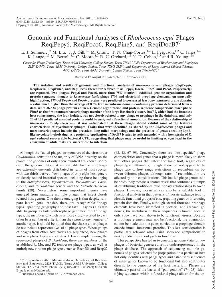
Transmission electron microscopy revealed that Poco6,DocB7, Pine5, and Pepy6 had characteristic siphophage mor-phologies, with icosahedral heads (average diameters of 82 nm,75 nm, 66 nm, and 80 nm, respectively) and long flexible,noncontractile tails (281 nm, 489 nm, 236 nm, and 285 nm,respectively) (Fig. 1 and Table 1). The International Commit-tee on Taxonomy of Viruses (ICTV) taxonomic prefix of thesephages would be vB_ReqS (39). The host range of thesephages was assessed by spotting dilutions of lysates onto lawnsof 37 R. equi strains, including clinical and environmentalisolates collected from different epidemiological regions acrossthe world (Table 2). These hosts include representativeVapA� and VapA� strains (73). Each of the phages was ableto generate clearing spots on multiple hosts, including repre-sentatives from clinical strains. DocB7 exhibited the broadesthost range, forming clearing spots on 35 isolates. Pine5 showedclearing on only 3 isolates, while Poco6 and Pepy6 showedclearing on 8 and 19 isolates, respectively. There was not acorrelation between phage sensitivity and the presence or ab-sence of the virulence plasmid.
Genome organization of ReqiPepy6 and ReqiPoco6 and re-lationship to phage 1706. The genomic sequences of Pepy6
FIG. 1. Negative-stained images of new rhodococcal siphophages. ReqiDocB7 (A), ReqiPine5 (B), ReqiPoco6 (C), and ReqiPepy6 (D) neg-atively stained with 2% (wt/vol) aqueous uranyl acetate. Bars, 200 nm.
VOL. 77, 2011 GENOMIC AND FUNCTIONAL ANALYSES OF R. EQUI PHAGES 671
Dow
nloa
ded
from
http
s://j
ourn
als.
asm
.org
/jour
nal/a
em o
n 07
Feb
ruar
y 20
22 b
y 22
1.15
0.18
1.24
8.

(76,806 bp) and Poco6 (78,064 bp) were closely related andcolinear with isolated blocks of nonidentity (Fig. 2). The ma-jority of the Pepy6 and Poco6 genomes could be aligned, withmultiple segments larger than 5 kb with up to 92% identity atthe DNA level. Pepy6 and Poco6 were predicted to encode 107proteins and a single tRNA of unknown specificity, all on thesame coding strand (Fig. 3). Both phages possessed 3� ex-tended cos ends, with the sequence 5�-CGCCGCCCT. This isrelated to the 3� extended cos termini (GCCCTGTCT [variantbases underlined]) of Lactococcus phage 1706, whose termi-nase large subunit (TerL) (LaP1706_gp8) was 60% identical tothe predicted TerL subunits of Pepy6 and Poco6 (gp14 andgp16, respectively). The Pepy6 and Poco6 cos sites were lo-cated approximately 13 kb upstream of TerL (Fig. 3).
Based on the total number and amino acid similarity ofshared proteins, the phage in the public database identified asbeing most closely related to Pepy6 and Poco6 was Lactococcuslactis phage 1706, a virulent siphophage initially isolated froman industrial cheese production site (Fig. 3 and Table 3) (17,20). Homologues of 23 proteins encoded by genes carried onphages Pepy6 and Poco6 could be identified among the 76proteins encoded by phage 1706, with individual amino acididentities from 47% to 75% (Fig. 3 and data not shown). Thesehomologues included nine out of the 15 phage 1706 proteinsdemonstrated to be virion associated (20). Many of the pro-teins encoded by genes carried on phage 1706 predicted to beinvolved in virion morphogenesis and DNA packaging hadhomologues in Pepy6 and Poco6, include LaP1706_gp13 (ma-jor capsid subunit, corresponding to Pepy6 gp28 and gp30 andPoco6 gp29 and gp31), LaP1706_gp10 (prohead protease, cor-responding to Pepy6 gp25 and Poco6 gp26), LaP1706_gp18(tape measure protein, corresponding to Pepy6 gp35 andPoco6 gp36), and LaP1706_gp9 (portal protein, correspondingto Pepy6 gp22 and Poco6 gp24) as well as LaP1706_gp8(TerL).
Domain shuffling in DNA metabolism genes. Two of theadditional Pepy6 and Poco6 predicted proteins with identifi-able homologues in phage 1706 had conserved domains sug-
gestive of functions in DNA replication, but the novel combi-nation of domains meant that specific functions could not berobustly assigned. The first of these were the Pepy6 and Poco6gene72 products, which were similar over their entire length tophage 1706 gp55. Two distinct conserved domains were iden-tified in these proteins: a COG3378 domain at the N terminusand a COG0417 domain at the C terminus. The COG3378domain, originally identified as a C-terminal conserved domainin DNA primase, is one of a number of domains that haveundergone extensive domain shuffling and thus can be foundeither by themselves or in combination with several other typesof conserved domains (36). Likewise, Pepy6 gp102 and Poco6gp102, which are homologues of phage 1706 gp67, had anatypical association of conserved domains likely to function inDNA replication (Fig. 4). The Rhodococcus phage proteinswere significantly longer (542 residues compared to 412 resi-dues) than phage 1706 gp67, and the amino-terminal extensionappeared to be derived from LigA, a DNA ligase domain-containing protein. This fusion may have occurred during theacquisition of an 11-kb insertion between Pepy6 gene77 andgene102, encoding homologues of phage 1706 gene66 andgene67, respectively (Fig. 4 and 5 and see below). The phage1706 protein contained an SSL2 domain commonly present inDEAD box helicase (Fig. 4). The region of Pepy6 gp102 thataligned with 1706 gp67 included the SSL2 domain-containingregion but was different enough such that a conserved domainwas not identified in the Rhodococcus proteins. SSL2 and re-lated DEAD box helicase domains have been found associatedwith other domains. For example, one group of bacterial pro-teins has carboxy-terminal DEAD box helicase domains andamino-terminal AE-Prim_S domains. A permuted arrange-ment of these domains was observed in hypothetical proteinCLOSS21_01549, encoded by the CLOSS21 prophage elementrelated to Pepy6 and Poco6 (Fig. 4 and see below).CLOSS21_1549 had an amino-terminal DEAD box helicasedomain (that aligned with Pepy6 gp102 residues 137 to 540,with 47% identity) and a carboxy-terminal LigC domain (that
TABLE 1. Rhodococcus equi phage characteristics and features
Phage
Virionheaddiam(nm)
Taillength(nm)
TMPa
Genomelength(bp)b
Genomicterminic
No. ofCDSd
No. of CDSwith TMD
(%)e
Pre-tmpframeshift/slip. seq.f
tRNA Notable feature(s)Length(aa)
% -helix
Pred.tail
length(nm)
ReqiDocB7 75 489 3,101 58 465 75,772 pac 105 9 Yes (GGAAAAA)
No Unique; broadhost range
ReqiPepy6 80 285 1,950 67 292 76,806 3� cos 107 29 (29) No Yes Phage 1706 like;many TMDproteins
ReqiPoco6 82 281 1,951 72 293 78,064 3� cos 107 26 (24) No Yes Phage 1706 like;many TMDproteins
ReqiPine5 66 236 1,732 68 259 59,231 pac 85 13 (15) No No Rosebush like
a TMP, tape measure protein. The measured length (in amino acids aa�), % alpha-helical content (% -helix), and predicted tail length (Pred. tail length) (innanometers) of the tape measure protein is shown.
b Genome length, genome nucleotide base pair length.c The structure of the packaged genomic termini is shown. pac, headful packaging; cos, cohesive ends.d The number of protein-coding genes (CDS) is shown.e The number and percentage of protein-coding genes with predicted transmembrane domains (CDS with TMD) is shown.f The presence or absence of a pre-tape measure protein ribosomal frameshift slippery sequence (Pre-tmp frameshift/slip. seq.) is shown.
672 SUMMER ET AL. APPL. ENVIRON. MICROBIOL.
Dow
nloa
ded
from
http
s://j
ourn
als.
asm
.org
/jour
nal/a
em o
n 07
Feb
ruar
y 20
22 b
y 22
1.15
0.18
1.24
8.

aligned with Pepy6 gp102 LigC domain-containing residues 15to 86, with 38% identity).
Prophage elements related to Pepy6, Poco6, and 1706. Fourprophage elements related to Pepy6, Poco6, and Lactococcusphage 1706 were identified in the genome assemblies from fourmembers of the Firmicutes order Clostridiales: Bryantella for-matexigens DSM 14469, Clostridium leptum DSM 753, Rumi-nococcus torques ATCC 27756, and Clostridium sp. strain SS2/1(Fig. 3 and Table 3). The Bryantella formatexigens DSM 14469,Clostridium leptum DSM 753, Ruminococcus torques ATCC27756, and Clostridium sp. strain SS2/1 prophage elementsare referred to as BRYFOR, CLOLEP, RUMTOR, andCLOSS21 prophage elements, respectively. The relationship ofphage 1706 to the RUMTOR and CLOLEP prophage ele-ments was described previously (20). Each of these phage andprophage elements encoded a common set of 22 proteins thatincluded most of the virion-associated and structural proteins,
TerL, and proteins containing various DNA metabolism-re-lated conserved domains (Fig. 3). Integrase genes could beidentified at the beginning of each prophage element, whichwould correspond to a genome location between the Pepy6gp35 and gp72 equivalents (Fig. 4). As in phage 1706, nointegrase gene candidate was identified in Poco6 or Pepy6 (20).
Tail fiber gene complexity. Pepy6 and Poco6 are �20 kblarger than phage 1706 and the clostridial prophage, includinga segment of �8 kb of novel sequence inserted between TerLand cosL (Fig. 3 and 5). This region carried genes encoding anovel set of proteins, 5 in Pepy6 and 6 in Poco6, that althoughwidely variant in size (278 to 856 amino acids [aa]), have anunusual, modular relationship to each other. The first 53 res-idues of each of these proteins were 56% to 94% identical, with21 invariant residues present in all 11 proteins (Fig. 5). Nu-merous other proteins in the public database with relatedamino-terminal domains were identified by BLAST, and align-ments reveled 11 invariant residues in proteins from phagesand prophages from taxonomically diverse hosts such as cyano-bacteria (e.g., Prochlorococcus phage P-SSM4 gp87), gamma-proteobacteria (e.g., XF_2114 in Xylella prophage Xfp6), andFirmicutes (e.g., Streptococcus phage phi3396 gp48). However,except for Pepy6 gp2, Poco6 gp3, and Poco6 gp6, these pro-teins were not recognizably related to proteins in the publicdatabase beyond the conserved amino-terminal domain. Poco6gp3 and Pepy6 gp2 C-terminal domains were 31% identical tothe pectate lyase conserved domain of gp20, the tail fiberprotein Salmonella phage ε15 (38). Several other Poco6 andPepy6 proteins in these clusters also contained pectin-lyasedomains and other carbohydrate-interaction domains as well,including SGNH hydrolase and concanavalin A-like lectins/glucanases. Likewise, the C terminus of Poco6 gp6 was related
FIG. 2. Dot plot alignment of the nucleotide sequences of Pepy6and Poco6. Pepy6 and Poco6 are closely related, colinear phages withisolated blocks of nonidentity. The percentages of identity of thelonger aligned blocks are indicated.
TABLE 2. Host ranges of phages Poco6, DocB7, Pine5, and Pepy6a
Host strain orparameter
Host range of phageb:
Poco6 DocB7 Pine5 Pepy6
ATCC 33701 X X XATCC 33701- X XATCC 33704 XMillB X XHDP5B XHDP1C X XS1 X XS2 X X XS3 XL1 XL1p- X XATCC 33703 X XATCC 33705 X98-099 X X99-110 X X X99-171 X X X99-180 X X99-228 X X X99-232 X X99-120 X99-134 X99-15800-051 X00-050 X X X01-116 X X X02-125 X X X04-172 X04-181 X04-195 X X04-200 X04-239 X05-300 X X05-305 X X05-306 X X05-338 X05-373 X06-383 X
Total no. of strainswith clearing
8 35 3 19
a The host ranges of phages Poco6, DocB7, Pine5, and Pepy6 were determinedby spotting a routine test dilution onto overlays of the indicated host strains.
b X indicates clearing on the indicated host strain. A boldface letter X corre-sponds to the original enrichment host for that phage.
VOL. 77, 2011 GENOMIC AND FUNCTIONAL ANALYSES OF R. EQUI PHAGES 673
Dow
nloa
ded
from
http
s://j
ourn
als.
asm
.org
/jour
nal/a
em o
n 07
Feb
ruar
y 20
22 b
y 22
1.15
0.18
1.24
8.

to lipases from a number of Actinomycetes. GDSL/SGNH hy-drolase domain proteins are involved in the hydrolysis of widevariety of substrates, including fatty acids, aromatic esters, andamino acid derivatives, and are particularly abundant in Actino-mycetes (3, 6). These relationships suggested that this geneclusters might encode proteins that interact with the cell en-velope in Rhodococcus.
A second large insertion in the genomes of Pepy6 and Poco6relative to phage 1706 was identified. This 11-kb insertion,encompassing Pepy6 genes gene77 to gene102, corresponds toan insertion between gene66 and gene67 in phage 1706 (Fig. 4).The BRYFOR and CLOSS21 prophage elements containedfour genes and one gene, respectively, between their 1706gene66 and gene67 homologues (Fig. 4). As previously dis-cussed, rearrangements associated with this region may havebeen the source of the novel domain structure of Pepy6 and
Poco6 gp102. The genes present in the Pepy6 and Poco6 11-kbinsertion were predicted to encode primarily small, hypothet-ical novel proteins, although one gene encoding a conservedthymidylate synthase was identified.
Poco6 and Pepy6 lysis genes. In addition to the cytoplasmicmembrane and the peptidoglycan cell wall, the envelope of theRhodococcus and the other genera of the mycolata, a suprage-neric taxon including Nocardia, Corynebacterium, and Myco-bacterium, has a third layer comprised of an arabinogalactanpolymer linked to an “outer membrane” of mycolic acid chains(70). Extrapolating from the lysis systems of mycobacterio-phages, rhodococcal phages can be expected to encode notonly the canonical holin and endolysin (LysA) lysis proteinsbut also an enzyme, LysB, that attacks this outer layer (21, 22,54). The LysA and holin genes were readily identified as ad-jacent reading frames, gene10 and gene11 in Pepy6, and as a
FIG. 3. Genome maps of Pepy6 and Poco6 and related phages and prophages. (A) Genome maps of phages ReqiPepy6 and ReqiPoco6. Purpleshading between the genome maps indicates segments of DNA that align with greater than 75% identity. The colors of the genes or proteins inthe gene maps indicate the relationship of the encoded protein with other proteins as follows. Yellow indicates a homologue is encoded by phage1706. Yellow genes with green outlines encode homologues of phage 1706 virion-associated proteins. The 1706 gene is indicated below theRhodococcus phage gene. Turquoise indicates that there was no homologue in 1706, nor was a functional annotation possible. Dark blue indicatesproteins for which a functional annotation could be made but lacked homologues in 1706. Green proteins possess 56 invariant, N-terminal aminoacid residues, indicated by peach bars. Genes outlined in red encode proteins with predicted transmembrane helices. Regions shaded in peachindicate locations of gene insertions relative to phage 1706. (B) Genome maps of Lactococcus lactis phage 1706 and prophage elements BRYFORand CLOSS21, present in the genomic sequences of Bryantella formatexigens DSM 14469 and Clostridium sp. strain SS2/1 genome, respectively. Thephage 1706 genome map was derived from the GenBank entry with accession no. EU081845. The BRYFOR prophage encompasses the regionencoding locus tags BRYFOR_08504 to BRYFOR_08569. The CLOSS21 prophage encompasses the region encoding locus tags CLOSS21_01492to CLOSS21_01580. In order to align with phage 1706, the BRYFOR and CLOSS21 maps are drawn with the order of prophages changed. Thelocations of the prophage genomic termini, between the coding region of BRYFOR gene73 and gene1 and CLOSS21 gene89 and gene1, areindicated on the genome maps by turquoise shading. The colors of the genes or proteins indicate the relationship of the encoded protein with otherproteins as follows: yellow, homologue encoded by Pepy6 and Poco6; gray, no homologue encoded by Pepy6 or Poco6. A green outline indicatesthat the protein encoded by the gene is a homologue of phage 1706 virion-associated protein. A red outline indicates that the protein encoded bythe gene has predicted transmembrane helices. The gene number of the corresponding protein encoded by Pepy6 and 1706 is indicated below eachmap (-, no protein). Regions shaded in peach indicate locations of gene insertions in the map relative to phage 1706. Select functional annotationsare shown above genes by the following abbreviations: e15, Salmonella phage epsilon15 tail fiber protein; lip, lipase; hol, holin; tmp, tape measureprotein; Prim, primase; pol, DNA polymerase; HNH, homing endonuclease; Int, integrase; endo, endonuclease; ligA, ligase A domain; CMPdeaminase, deoxycytidinylate deaminase. Homologous gene products in Pepy6 and 1706 (Pepy6yp and 1706yp, respectively) are indicated beloweach map.
674 SUMMER ET AL. APPL. ENVIRON. MICROBIOL.
Dow
nloa
ded
from
http
s://j
ourn
als.
asm
.org
/jour
nal/a
em o
n 07
Feb
ruar
y 20
22 b
y 22
1.15
0.18
1.24
8.
nearly identical pair of genes gene12 and gene13 (�90% iden-tity with the Pepy6 proteins) found in the same position inPoco6. The LysA annotation was made particularly robust bythe presence of a conserved N-terminal peptidoglycan-bindingdomain and N-acetylmuramoyl-L-alanine amidase catalytic res-idues and by similarity to actinobacterial amidases (data notshown). The holin annotation was based on gene location,prediction of a transmembrane domain, and the weak butsignificant primary sequence similarity (46%) to the experi-mentally confirmed holin encoded by Brevibacterium flavumphage BFK20 (10). The predicted length and topology of thetwo transmembrane domains (TMDs) and the distribution ofcharged residues are remarkably conserved between thePepy6/Poco6 proteins and the BFK20 holin (not shown). Thepresence of two unambiguous TMDs allowed these proteins tobe assigned as class II holins. Moreover, the absence of asecretion signal in the LysA sequence indicated that theseholins must form large holes, unlike many other members ofclass II, which have pinholin character (i.e., make only smallholes in the membrane) and serve only to effect a temporallyscheduled depolarization of the host membrane (53).
In mycobacteriophage genomes, LysB genes are invariablyclosely linked, or adjacent to, the holin-endolysin loci (54).However, the only candidates for LysB in Pepy6 and Poco6,gp33 and gp34, respectively, were located in the virion struc-tural protein cluster. The protein most closely related (49%similar) to these candidates was ROP_59440, a PE-PPE do-main-containing protein encoded by Rhodococcus opacus B4.ROP_59440 was a member of a small cluster of related pro-teins encoded by several Nocardia and Rhodococcus species(data not shown). However, psi-BLAST analysis detected asignificant relationship between these proteins and mycobacte-riophage LysB proteins, primarily Cjw1 gp35 (score of 58.5)but also to D29 gp12 (54). As was found for the mycobacterio-phage proteins, the Pepy6 and Poco6 putative LysB equiva-lents were predicted to be cytoplasmic and thus may be re-leased by holin function along with LysA.
Abundance of predicted membrane-associated proteins inPepy6, Poco6, and related prophages. The Pepy6 and Poco6genomes carried an unusually high fraction of genes predictedto contain at least one TMD (Tables 3 and 4). For Pepy6, 29out of 107 proteins (29%) and for Poco6, 26 out of 107 proteins(24%) were predicted to have TMDs. In order to determinehow these values compare to other phages, the predicted pro-teins encoded by 406 Caudovirales genomes were screened forTMDs. Out of a total of 36,324 proteins encoded by phagegenes, 3,081 (8.5%) were predicted to have at least one TMD(Table 4). When the data set was broken down by host clade,there was very little difference in the percentage of TMDproteins among the Proteobacteria (8.3%), Actinobacteria(8.0%), Firmicutes (9.5%) and “other” (which included Cyano-bacteria and Flavobacterium) (7.0%). All phages encoded atleast one potential TMD protein. Vibrio phage VP5 encodedthe lowest percentage of TMD proteins (2.1%), and whenPepy6 and Poco6 were excluded, Bacillus phage SPP1 encodedthe highest percentage of TMD proteins (17.8%). The clos-tridial prophages related to Pepy6 and Poco6 as well as phage1706 were enriched in TMD protein-coding regions (14% to23%) (Table 3).
Pine5 genome organization and relationship to mycobacte-riophage Rosebush. At 59 kb, Pine5 was the smallest of theRhodococcus phages analyzed. Pine5 genes were predicted toencode 85 proteins (Fig. 6), of which 41 (52%) were small,hypothetical novel proteins with no recognizable homologuesin the public database. Thirty-one proteins encoded by genescarried on Pine5 (39%) were related in sequence and geneorder to proteins encoded by a cluster of 13 closely relatedmycobacteriophages that included Rosebush, Qyrzula, Phlyer,Phaedrus, Pipefish, Nigel, Cooper, Chah, PG1, Orion, Colbert,Puhltonio, and UncleHowie (29–31). Rosebush was the firstmember of this group to be described (56). The 13 Rosebush-like phages exhibited an average of 62 to 99% DNA sequenceidentity to each other across the majority of the length of theirgenome (30, 31). The regions of Pine5 genomic DNA that
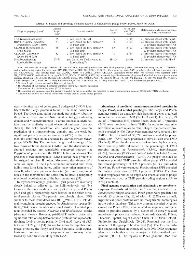
TABLE 3. Phages and prophage elements related to Rhodococcus phage Pepy6, Poco6, Pine5, or DocB7
Phage or prophage (host)aGenome
length(kb)b
Genomic terminic No. ofCDSd
No. of CDSwith TMD
(%)e
Relationship to Rhodococcus phage(no. of shared proteins)f
1706 (Lactococcus lactis) 55 3� cos GCCCTGTCT 76 12 (16) 21 proteins shared with Pepy6BRYFOR.phiA (Bryantella
formatexigens DSM 14469)57 3� cos (based on TerL similarity
to Pine5 gp14)73 17 (23) 23 proteins shared with Pepy6;
21 proteins shared with 1706CLOSS21 (Clostridium sp.
strain SS2/1)66 3� cos (based on TerL similarity
to Pine5 gp14)89 18 (20) 24 proteins shared with Pepy6;
22 proteins shared with 1706CLOLEP (Clostridium
leptum DSM 753)52 3� cos (based on TerL similarity
to Pine5 gp14)78 11 (14) 34 proteins shared with Pepy6;
21 proteins shared with 1706Mycobacteriophage
Rosebush-like phages67–71 pac (based on TerL related to
Rosebush gp7)81–104 (�10) 31 proteins shared with Pine5
a 1706, Lactococcus lactis phage 1706 (NC_010576); BRYFOR, Bryantella formatexigens DSM 14469 prophage (derived from GenBank entry NZ_ACCL02000018.1and includes coding region for locus tags BRYFOR_08569 to BRYFOR_08504); CLOSS21, Clostridium sp. strain SS2/1 prophage (derived from GenBank entryNZ_ABGC03000034 and includes locus tags CLOSS21_01580 to CLOSS21_01492); CLOLEP, Clostridium leptum DSM 753 (derived from GenBank entryNZ_ABCB02000017 and includes locus tags CLOLEP_01347 to CLOLEP_01425). Mycobacteriophage Rosebush-like phages (with GenBank entries in parentheses)included Rosebush (NC_004684), Qyrzula (NC_008204), Cooper (NC_008195), PG1 (NC_005259), Orion (NC_008197), Pipefish (NC_008199), Chah (NC_011284),Colbert (GQ303259.1), Nigel (NC_011044), Puhltonio (GQ303264.1), Phaedrus (NC_011057), Phlyer (NC_012027), and UncleHowie (GQ303266.1).
b Genome length, genome nucleotide kilobase pair length.c The structure of the packaged genomic termini is shown. pac, headful packaging.d The number of protein-coding genes (CDS) is shown.e The numbers and percentages of the proteins encoded by the element that are predicted to have transmembrane domains (CDS with TMD) are shown.f Maximum E values of 1e�10 and scores over 50 were used to define shared proteins.
VOL. 77, 2011 GENOMIC AND FUNCTIONAL ANALYSES OF R. EQUI PHAGES 675
Dow
nloa
ded
from
http
s://j
ourn
als.
asm
.org
/jour
nal/a
em o
n 07
Feb
ruar
y 20
22 b
y 22
1.15
0.18
1.24
8.

aligned with Rosebush genomic DNA were located primarilyin nine blocks, ranging from 0.4 to 1.1 kb, with up to 73%identity, which were predicted to encode proteins involved invirion morphogenesis and DNA metabolism (Fig. 6). Whilesignificant, this level of similarity falls below the threshold usedto define the clusters within the mycobacteriophages (30).However, Pine5 is the first nonmycobacteriophage that exhibitsshared synteny with this group of Mycobacterium phage.
Morphogenesis protein homologues in Pine5 and the Rose-bush-like mycobacteriophage included the tape measure pro-tein, portal protein, capsid subunit, and major tail subunit.Candidate genes encoding the frameshift tail chaperone, pro-head protease, and scaffolding subunit were not identified inPine5. Like Rosebush, the Pine5 genome assembled into acircular map, and no end sequences were identified, stronglysuggesting that the phage utilizes a pac-type headful packagingmechanism (56). Additional TerL proteins significantly relatedto the TerL protein from Pine5 and Rosebush encoded byother phages were demonstrated to have circularly permutedgenomes, included Bcep781, Aaphi23, and PY100 (59, 61, 68).
Pine5 and Rosebush carried genes encoding a related set of
proteins with conserved domains, suggesting that they playvarious roles in DNA metabolism. These proteins includedPine5 gp45 and Rosebush gp54, which possessed SSL2/helicasedomains (Fig. 4). Pine5 gp46 and Rosebush gp54 proteins hadN-terminal cd04859 primase-polymerase (Prim_Pol) domainand a carboxy-terminal region that includes Walker A andWalker B nucleotide-binding sites as part of a RepA helicasedomain. Proteins identified by BLAST searches that could bealigned to Pine5 gp46 across their entire length included onlythe mycobacteriophage homologues and gp43 of Corynebacte-rium phage BFK20. However, proteins unrelated to Pine5 gp46at a primary structural level but with conserved domain archi-tecture are widespread among prokaryotes (36).
The only readily identifiable component of lysis predicted tobe encoded by Pine5 was the LysA endolysin candidate, Pine5gp40. Pine5 gp40 has two domains that support this annota-tion, an N-terminal peptidase domain and a muramidase/gly-cosyl hydrolase domain. Some proteins encoded by genes car-ried on mycobacteriophages, including LysA, were groupedinto protein families characterized by members having com-plex chimeric interrelationships (31). The relationship of Pine5
FIG. 4. Novel combinations and permutations of DNA metabolism-associated conserved domains in phages and proteins encoded by bacterialgenes. (A) Lactococcus phage P087 (gp4); (B) Pepy6 (gp72), Poco6 (gp72), and Lactococcus phage 1706 (gp55); (C) satellite phage P4 (RPBPP4);(D) Xylella phage Xfas53 (gp23); (E) Vibrio phage Vp2 (VP2p2); (F) Burkholderia phage Bcep781 (gp53); (G) Pine5 (gp46) and Rosebush (gp54);(H) DocB7 (gp105); (I) Escherichia coli O157:H7 EDL933 (z1129); (J) DocB7 (gp48); (K) Pine5 (gp45) and mycobacteriophage Rosebush (gp50);(L) CLOS21 (CLOSS21_01549); (M) Pepy6 (gp102) and Poco6 (gp102); (N) Lactococcus phage 1706 (gp67). Related domains are shaped andshaded similarly. Domain abbreviations are based on the Conserved Domain Database entries as follows: PolB, COG0417; Prim_Cterm,COG3378; AE_Prim, cl01287; DEAD HELIC, CD00046; SSL2, COG1061; LigA, CL03295; PriCT-2D5N, pfam08707; D5_N superfamily, cl07360;Prim Z, cl07038; VirE, COG5545; RepA, COG3598; COG4643, COG4643.
676 SUMMER ET AL. APPL. ENVIRON. MICROBIOL.
Dow
nloa
ded
from
http
s://j
ourn
als.
asm
.org
/jour
nal/a
em o
n 07
Feb
ruar
y 20
22 b
y 22
1.15
0.18
1.24
8.

LysA to the mycobacteriophage LysA proteins was also chi-meric. No holin gene candidate could be identified for Pine5.In Rosebush, the holin is encoded by the gene immediatelydownstream of the LysA gene, but there is no downstreamgene in Pine5 at this location (Fig. 6, ReqiPine5 genome map).Thirteen Pine5-encoded proteins were predicted to have trans-membrane domains, but most of these proteins lacked homo-
logues in the public database and none possessed sequencesimilarity to a holin. However, the predicted topology of oneprotein, Pine5 gp17, was markedly similar to that of theBKF20, Pepy6, and Poco6 holin proteins and is thus the bestcandidate holin (data not shown). No LysB equivalent wasidentified for Pine5. This was consistent with the results of aglobal search of LysB homologues in 60 mycobacteriophage
FIG. 5. Complex relationships between potential tail fibers encoded by Pepy6 and Poco6. (A) Alignment of the first 56 residues of Pepy6 gp1,gp2, gp4, gp5, and gp7 with Poco6 gp1, gp2, gp3, gp5, gp6, and gp8 and the amino-terminal residues of PSSM4_087 (encoded by Prochlorococcusphage P-SSM4 genes), XF_2114 (encoded by Xylella fastidiosa prophage Xfp6 genes), and 3396_48 (encoded by Streptococcus phage phi3396genes). Invariant residues are shaded. (B) Schematic alignment of Pepy6 gp1, gp2, gp4, gp5, and gp7 with Poco6 gp1, gp2, gp3, gp5, gp6, and gp8and also to e15 (tail fiber, encoded by Salmonella phage epsilon15 gene), PSSM4_087 (encoded by Prochlorococcus phage P-SSM4 gene), 3396_48(encoded by Streptococcus phage phi3396 gene) and (FRAAL2677 encoded by Frankia alni ACN14a gene). Alignment scores are color coded asindicated. Conserved domains are indicated by shaded regions.
TABLE 4. Analysis of the presence of transmembrane domains (TMD) in proteins encoded by phage genes broken down by host taxonomy
Host clade No. ofphage
Total no. ofproteinsa
No. of proteinswith TMDb
% proteinswith TMD
Phage encoding the lowest or highest % of TMD proteins(% TMD proteins)c
Lowest Highest
Proteobacteria 178 16,407 1,360 8.3 Vibrio VP5 (2.1) Klebsiella K11 (19.6)Actinobacteriad 69 7,478 603 8.0 Mycobacterium Porky (5.4) Corynebacterium P1201 (12.4)Firmicutes 141 9,958 943 9.5 Bacillus BCJA1c (2.1) Bacillus SPP1 (17.8)Othere 18 2,481 175 7.0 Cyanophage P60 (2.5) Flavobacterium 11b (12.3)
Total 406 36,324 3,081 8.5 VP5 BCJA1c (2.1) Bacillus SPP1 (17.8)
a Total number of proteins encoded by phage genes.b Number of proteins encoded by phage genes and predicted to have at least one TMD.c The phage with genes encoding the lowest or highest percentage of proteins predicted to have at least one TMD. The percentage of proteins predicted to have at
least one TMD is shown in parentheses.d Actinobacteria does not include Rhodococcus phages Pepy6, Poco6, Pine5, and DocB7.e Includes Cyanobacteria and Flavobacterium.
VOL. 77, 2011 GENOMIC AND FUNCTIONAL ANALYSES OF R. EQUI PHAGES 677
Dow
nloa
ded
from
http
s://j
ourn
als.
asm
.org
/jour
nal/a
em o
n 07
Feb
ruar
y 20
22 b
y 22
1.15
0.18
1.24
8.

genomes in which only four phages, Rosebush, Qyrzula,Myrna, and Che12 lacked identifiable LysB equivalents (54).While not related to any protein encoded by genes carried ona phage, Pine5 gp21 possessed a SGNH-hydrolase domain,found in numerous lipases and esterases, and thus may play arole in lysis.
DocB7, a new phage type. The DocB7 genome was 75,772 bpand predicted to carry genes encoding 105 proteins (Fig. 6).DocB7 represents the first isolate of a new phage type with adistinct genome organization unrelated to any entries in thepublic database. Moreover, only 31% of DocB7 proteins hadsignificant similarity to known proteins, and no genetic ele-ment that shared significant gene content with DocB7 could beidentified in bacterial genomes. Functional assignments couldbe made for only 23 DocB7 proteins. Even with so few func-tional annotations, it was apparent that the genome was orga-nized into two convergent transcriptional arrays, with virionmorphogenesis loci located on the left arm and DNA metab-olism and regulatory genes largely encoded on the right arm.The left arm of DocB7, from gene1 to gene30, encoded proteinswith functions in DNA packaging and virion morphogenesis.The DocB7 TerL homologue, encoded by gene3, showed onlydistant relationships to other TerL proteins. Only two signifi-cantly related proteins were identified in the public database—the TerL homologues from Thermus phage phiYS40, andMicrocystis phage Ma-LMM01, both large myophages (51, 81).Defined termini were not identified for DocB7, indicating thatDocB7 utilizes a headful packaging strategy, as does Ma-LMM01 (81). DocB7 gp4 had some similarity to proteins of
unknown function encoded by genes in a number of bacteriapresent in the human gut microbial community, includingEubacterium rectale, Clostridium methylpentosum, and Rumino-coccus (45). This protein contained a conserved domain(DUF935) also present in many proteins encoded by phagegenes, notably Mu gp29, and probably functions in portal mor-phogenesis. DocB7 gp5 was significantly related to the phageT7 gene 17 tail fiber (63). While lacking identifiable conserveddomains, DocB7 gp7 was 47% identical to Gordonia terraephage GTE5 gp1, predicted to have an endopolygalacturonase(PGU1) domain (7). The amino-terminal 80 residues of DocB7gp7 were significantly related to the amino termini of hundredsof bacterial and fungal glycoside hydrolase family proteins,such as Lam55a from Phanerochaete chrysosporium (35).
DocB7 gene20 and gene21 were annotated as the � G/G-Ttail chaperone equivalents due to their location upstream ofthe tape measure protein gene (gene22) and presence of apredicted ribosomal frameshift sequence (80). Ribosomal slip-page at the sequence GGAAAAA (nucleotide [nt] 17359)would direct a �1 translational reading frameshift, leading tothe DocB7 gp20-21 frameshift protein. The tape measure pro-tein candidate gp22 had a mosaic relationship with other pro-teins in the public database, as was reported for mycobacterio-phage tape measure proteins (31). At 3,101 residues, theunusual length of DocB7 gp22 is consistent with the unusuallylong tail (489 nm) of the virion (Fig. 1). Three conserveddomains were identified in gp22: (i) a tape_meas_TP901 do-main near the amino terminus; (ii) a region that was recog-nized by multiple conserved domain clusters, including
FIG. 6. Genome maps of Pine5 and DocB7. Phage genome maps are drawn to scale with the coding strand indicated by the position of the genesabove (rightwards) and below (leftwards) the central ruler. (A) Pine5 aligned with mycobacteriophage Rosebush. The Rosebush map wasgenerated from the GenBank entry with accession no. AY129334.1. Purple lines between Pine5 and Rosebush indicate regions of greater than 60%DNA identity between the two phages. Genes are color coded to indicate similarity with other proteins in the public database as follows: yellow,homologues are found in the mycobacteriophages Rosebush, Qyrzula, Phlyer, Phaedrus, Pipefish, Nigel, Cooper, Chah, PG1, Orion, Colbert,Puhltonio, and UncleHowie; green, homologues in the public database but not in Rosebush; blue, no homologue in the public database; gray,Rosebush genes without homologues in Pine5. Genes with red outlines encode proteins with predicted transmembrane domains. Annotations ofselected genes are abbreviated as follows: PAPSr, phosphoadenosine phosphosulfate reductase; sub, subunit; RuvC, Holliday junction resolvases.(B) DocB7.Genes are color coded to indicate similarity with other proteins in the public database as follows: yellow genes encode proteins withat least one homologue in the public database; blue genes, no homologue in the public database. Genes with red outlines encode proteins withpredicted transmembrane domains. The 2,483-bp region highlighted in peach lacks recognizable protein-coding genes (no cds, no protein-codingsequences) and contains 13 perfect inverted repeats and one direct repeat.
678 SUMMER ET AL. APPL. ENVIRON. MICROBIOL.
Dow
nloa
ded
from
http
s://j
ourn
als.
asm
.org
/jour
nal/a
em o
n 07
Feb
ruar
y 20
22 b
y 22
1.15
0.18
1.24
8.

PRK01156, chromosome segregation protein; and finally, (iii)near the carboxy terminus, a “COG5412, Phage-related pro-tein of unknown function” domain.
The right arm of DocB7 carried genes encoding proteinsimplicated in regulation and DNA replication. The proteinencoded by gene31 was identified as a possible WhiB transcrip-tion factor. WhiB transcription factors were first identified inStreptomyces aureofaciens as factors involved in the develop-ment of the mycelium (37). WhiB-related proteins are com-mon among the mycobacteriophages (56). Two DocB7 geneswere predicted to encode proteins with some similarity to sub-units of DNA polymerase III, including the beta clamp domainsubunit (gp44) and the epsilon subunit (gp45). While T4phages frequently encode functional equivalents of DNA poly-merase III subunits, the DocB7 proteins were more closelyrelated to bacterial homologues than phage homologues. How-ever, the low similarity scores suggested that these genes werenot the results of a recent acquisition.
Like the other Rhodococcus phages, a gene on DocB7 en-coded a protein with conserved helicase and primase domains(Fig. 4). The helicase gp105 has a typical DEAD box helicasedomain. gp105, has a C-terminal AE_Prim_S domain and ex-hibits only weak similarity to three other proteins in the publicdatabase. Two additional proteins have notable conserved do-mains. gp68 is a large protein (644 amino acids) with a C-terminal Von Willebrand factor type A (vWA) domain. vWAdomains have a metal ion-dependent adhesion site that pro-motes oligomerization (79). gp69 (535 aa) has a C-terminalAAA domain. AAA domains are ATP-hydrolyzing domains.Both vWA and AAA domains are found in numerous, other-wise functionally unrelated, and widely diverse proteins. Thelack of other significant relationships in the large N-terminaldomains of gp68 and gp69 to other proteins precluded anyfunctional assignment. Additional proteins predicted to be en-coded by DocB7 genes include a homing endonuclease, gp54,and a tyrosine recombinase/possible integrase, gp59. The pres-ence of a putative integrase and a WhiB transcription factorhomologue raised the possibility that DocB7 might be a tem-perate phage, although DocB7 plaques were not detectablyturbid.
The region between DocB7 gene30 and gene31, separatingthe two coding arms of DocB7, contained a putative conver-gent transcription terminator domain. The 46 bp separatingthe stop codons of these two genes, were completely symmet-ric, which if transcribed, would form an extremely large stem-loop structure (34519-TAAGAAAACCCCCGGTACCTAATGAAAGGTACCGGGGGTTTTCTTA-34564 [potential stemformation residues are underlined]), likely promoting bidirec-tional, rho-independent transcription termination.
DocB7 lysis genes. Compared to the other Rhodococcusphages, DocB7 possessed the most complete lysis cassette.DocB7 lysis genes encoding LysA (gp27), LysB (gp38), andthree holin candidates (gp26, gp28, and gp30) were identified.Like Pepy6 and Poco6, the DocB7 gp27 LysA had an N-ter-minal peptidoglycan recognition protein conserved domain(cd06583) and conserved catalytic residues required for ami-dase activity. Moreover, gp27 is related to the mycobacterio-phage LysA equivalents, including PG1 gp49, as well as theBFK20 gp24 lysin. Like these proteins, DocB7 gp27 was pre-dicted to be a cytoplasmic protein and would thus require holin
activity to gain access to the cell wall. Proteins encoded bygene26, gene28, and gene30 were predicted to possess two, one,and four transmembrane domains, respectively. The predictedmembrane orientation and charge distribution of DocB7 gp28were compared to those of the BFK20 gp26 holin (data notshown). Of these, only gp26 exhibited significant sequencesimilarity to proteins in the public database and were related tomembers of mycobacteriophage Pham107 members, like gp27of phage Bxb1 (31). Some lysis systems in phages of Gram-negative hosts are fairly well characterized, allowing definitiveidentification and functional characterizations not only of ho-lins of several different topologies but also of antiholins, whichare holin-specific negative regulators. However, holin functionin Gram-positive bacteria in general, and especially in theActinomycetes, is poorly understood. It is thus not possible todiscern which of these proteins might be a holin or which mightserve as an antiholin regulator.
DocB7 gene38 encodes a protein with a PE-PPE domain,believed to be associated with cell surface features (2). Thisprotein was weakly similar to cutinases and hypothetical pro-teins from numerous Mycobacterium and other Corynebacte-rium species, for example exhibiting 40% similarity to Myco-bacterium smegmatis cutinase GI:302566037. gp38 lacks thesecretory signals common to most mycobacterial cutinases(78). When gp38 was queried against the database of Caudovi-rales proteins, similarity to mycobacteriophage LysB proteins,especially Bethlehem gp9, was detected. Thus, gp38 may be aLysB protein dependent on holin permeabilization of themembrane for release.
Gene-free region in DocB7. A notable feature of the DocB7genome is an unusually long region, 2,484 bp (from 68369 to70852; Fig. 6B) lacking any predicted protein- or RNA-encod-ing genes. There were no homologues to this region in thedatabase at a DNA level, and a translation BLAST algorithmdid not detect any significant amino acid similarity with pro-teins in the public database. Manual inspection identified anumber of small open reading frames with potential startcodons, but none had a reasonable Shine-Dalgarno motif. Thisregion was contained 14 inverted repeats, 13 perfect and onewith a single mismatch, and one direct repeat (data notshown). The dinucleotide composition of this region is signif-icantly different from the rest of the genome, implying that itmight be recently acquired (data not shown).
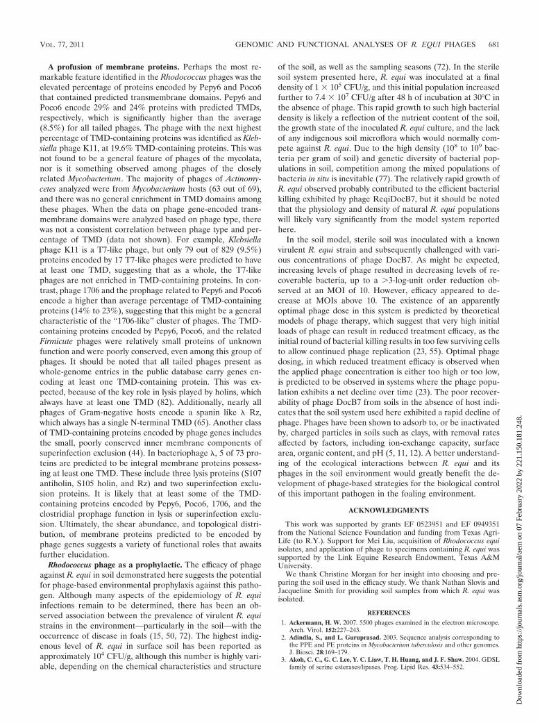
Phage efficacy at reducing R. equi in soil matrix. PhageDocB7 was chosen to determine whether application of phageto soil samples containing R. equi would result in a reduction inbacterial CFU. The soil matrix used in these assays was a moistsandy loam. Preliminary experiments were conducted to deter-mine the stability and recoverability of DocB7 from soils indi-cated that approximately 10% of the applied phage could berecovered following 48 h of incubation in soil at 30°C in theabsence of host cells (data not shown). In two independentexperiments, an R. equi suspension was applied to sterile soilsamples and incubated at 30°C prior to the application ofphage. Phage was applied by spraying, and then the number ofsurviving CFU in the soil was assessed. Beginning at an initialR. equi concentration at 1.0 � 105 CFU/g, viable counts in-creased by nearly 3 orders of magnitude (to 7.4 � 107 CFU/g)in 48 h at 30°C (Fig. 7). In contrast, all phage-treated samplesexhibited a reduction in CFU after incubation at all levels of
VOL. 77, 2011 GENOMIC AND FUNCTIONAL ANALYSES OF R. EQUI PHAGES 679
Dow
nloa
ded
from
http
s://j
ourn
als.
asm
.org
/jour
nal/a
em o
n 07
Feb
ruar
y 20
22 b
y 22
1.15
0.18
1.24
8.

initial input phage. The reduction was not colinear with in-creasing concentrations of phage. Application of phage at anapproximate multiplicity of infection (MOI) of 10 resulted inthe lowest recoverable CFU (6.0 � 104 CFU/g) after the 48-hincubation, a level even lower than the initial R. equi inoculums(1.0 � 105 CFU/g).
DISCUSSION
Rhodococcus phage diversity. The sequences of Pepy6,Poco6, DocB7, and Pine5 represent the first complete genomicdata available for phages that infect Rhodococcus. It is rele-vant, therefore, to discuss the relationship of these Rhodococ-cus phages to the �450 complete phage genomes, and evenmore prophage elements located in bacterial genome entries,present in the public database. The relationships of thesephages to each other and to phages that infect other hostclades follow several of the patterns noted when genomic datafrom other host-defined groups of phages have been analyzed.One of the four phages, DocB7, fit the criteria of being a newphage type. DocB7 not only has a distinct genome organizationbut encodes primarily novel proteins of unknown function andhas only a few proteins in common with any other phages orprophage sequences available at the time of analysis. This isexpected, as it is still common to isolate completely new phagetypes, especially for a host for which there is only limited phagegenomic data available. It is anticipated, however, that as moreRhodococcus and other phages are analyzed, DocB7 will ulti-mately become part of a cluster of related phages whose hostrange spans multiple genera. This is the case for Pine5, forwhich over 30% of encoded proteins are present in members ofa cluster of at least 13 mycobacteriophages, including Rose-
bush. While the mycobacteriophage members of this clustershare significant DNA similarity to each other, Pine5 has onlylimited DNA sequence similarity, well below the thresholdused to define the mycobacteriophage clusters (30). Consider-ing the taxonomic proximity of Rhodococcus to Mycobacteriumand the relative abundance of mycobacteriophage genomicdata, it was expected that at least some Rhodococcus phageswould be similar to mycobacteriophages. Finally, over 20% ofthe proteins encoded by Pepy6 and Poco6 shared similarity toproteins from phages that infect Firmicutes hosts, includingLactococcus phage 1706 and Clostridium prophage elementssequenced as part of the Human Gut Microbiome Initiative(24). The somewhat atypical genome organization of thesephages might contribute to the maintenance of genome iden-tity. Unusual genome features identified in 1706, Poco6, andPepy6 include the atypical location of the cos termini (i.e., notadjacent to the terminase genes) and the lack of clustering oflysis genes. There also appear to be two preferred spots forgenome expansion, which accounted for the majority of thebroad genome size range observed in this cluster of phages(from 52 kb to 78 kb). This relationship in which otherwiseclosely related phages differ considerably in gene content dueto the insertion of large clusters of small genes encoding mostlyhypothetical novel proteins is seen in some other phage fam-ilies. For example, Burkholderia phage BcepF1 has an �20-kbregion that encodes 62 small hypothetical proteins, represent-ing the majority of gene content differences between BcepF1and Pseudomonas phage F8 (42).
While there are phage types whose hosts span broad taxo-nomic lineages, it is also expected that regardless of phagetype, phages that infect a specific host clade will have special-ized, host-specific, adaptations. One of the features thatRhodococcus shares with Mycobacterium is the presence of anouter layer composed of mycolic acids covalently linked to thepeptidoglycan (71). This layer presents an additional challengeto phages of the mycolata, both in terms of adsorption andDNA injection process as well as host cell lysis. Evidence hasbeen provided recently that efficient lysis by mycobacterio-phage requires, in addition to the holin and endolysin, a my-colylarabinogalactan esterase (LysB) in order to disrupt thisouter membrane (21, 22, 54). Analogies can be made betweenthe function of LysB in disruption of the outer membrane withthe Rz-Rz1 equivalents of Gram-negative hosts, which func-tion in outer membrane disruption (54, 65). Most mycobacte-riophages encode LysB equivalents, so it was anticipated thatphages that infect other members of the Corynebacterineae,including Rhodococcus, would also carry genes encoding LysBequivalents (31, 54). DocB7, Pepy6, and Poco6 carry genesencoding potential LysB homologues, despite otherwise insig-nificant similarity to mycobacteriophages. However, the ge-netic structure of the lysis genes of the Rhodococcus phagesdiffered significantly from that of the mycobacteriophage inthat they lacked well-defined lysis gene cassettes. Rosebushwas among the few mycobacteriophages in which LysB geneswere not identified, and the lack of recognizable LysB homo-logues is one of the many genome features shared by Pine5 andRosebush. It is possible that these phages have a functionallyequivalent but mechanistically unrelated strategy for disrup-tion of the mycolate layer.
FIG. 7. Effect of phage application on recovery of R. equi from soil.Phage effect on R. equi was evaluated via 7 treatment groups. Eachtreatment group contained 3 parallel subsamples, where 3 sterile soilmatrixes were inoculated independently with R. equi strain ATCC33701 and were subjected to further treatment. R. equi was inoculatedat an initial concentration of 1.0 � 105 CFU/g soil. The R. equi levelswere determined before (0-h) and after (48-h) incubation in the ab-sence of phage treatment (No phage). The R. equi CFU levels werealso determined following 48 h of incubation in the presence of phageDocB7. Phage DocB7 was applied at initial multiplicity of infections(MOIs) of 1,000, 100, 10, 1, and 0.1 PFU/CFU. After each subsamplewas analyzed in two replicate samples, the mean values� standarddeviations of the subsamples (error bars) were plotted. Two indepen-dent experiments were carried out, and the results of one representa-tive experiment are shown.
680 SUMMER ET AL. APPL. ENVIRON. MICROBIOL.
Dow
nloa
ded
from
http
s://j
ourn
als.
asm
.org
/jour
nal/a
em o
n 07
Feb
ruar
y 20
22 b
y 22
1.15
0.18
1.24
8.
A profusion of membrane proteins. Perhaps the most re-markable feature identified in the Rhodococcus phages was theelevated percentage of proteins encoded by Pepy6 and Poco6that contained predicted transmembrane domains. Pepy6 andPoco6 encode 29% and 24% proteins with predicted TMDs,respectively, which is significantly higher than the average(8.5%) for all tailed phages. The phage with the next highestpercentage of TMD-containing proteins was identified as Kleb-siella phage K11, at 19.6% TMD-containing proteins. This wasnot found to be a general feature of phages of the mycolata,nor is it something observed among phages of the closelyrelated Mycobacterium. The majority of phages of Actinomy-cetes analyzed were from Mycobacterium hosts (63 out of 69),and there was no general enrichment in TMD domains amongthese phages. When the data on phage gene-encoded trans-membrane domains were analyzed based on phage type, therewas not a consistent correlation between phage type and per-centage of TMD (data not shown). For example, Klebsiellaphage K11 is a T7-like phage, but only 79 out of 829 (9.5%)proteins encoded by 17 T7-like phages were predicted to haveat least one TMD, suggesting that as a whole, the T7-likephages are not enriched in TMD-containing proteins. In con-trast, phage 1706 and the prophage related to Pepy6 and Poco6encode a higher than average percentage of TMD-containingproteins (14% to 23%), suggesting that this might be a generalcharacteristic of the “1706-like” cluster of phages. The TMD-containing proteins encoded by Pepy6, Poco6, and the relatedFirmicute phages were relatively small proteins of unknownfunction and were poorly conserved, even among this group ofphages. It should be noted that all tailed phages present aswhole-genome entries in the public database carry genes en-coding at least one TMD-containing protein. This was ex-pected, because of the key role in lysis played by holins, whichalways have at least one TMD (82). Additionally, nearly allphages of Gram-negative hosts encode a spanin like � Rz,which always has a single N-terminal TMD (65). Another classof TMD-containing proteins encoded by phage genes includesthe small, poorly conserved inner membrane components ofsuperinfection exclusion (44). In bacteriophage �, 5 of 73 pro-teins are predicted to be integral membrane proteins possess-ing at least one TMD. These include three lysis proteins (S107antiholin, S105 holin, and Rz) and two superinfection exclu-sion proteins. It is likely that at least some of the TMD-containing proteins encoded by Pepy6, Poco6, 1706, and theclostridial prophage function in lysis or superinfection exclu-sion. Ultimately, the shear abundance, and topological distri-bution, of membrane proteins predicted to be encoded byphage genes suggests a variety of functional roles that awaitsfurther elucidation.
Rhodococcus phage as a prophylactic. The efficacy of phageagainst R. equi in soil demonstrated here suggests the potentialfor phage-based environmental prophylaxis against this patho-gen. Although many aspects of the epidemiology of R. equiinfections remain to be determined, there has been an ob-served association between the prevalence of virulent R. equistrains in the environment—particularly in the soil—with theoccurrence of disease in foals (15, 50, 72). The highest indig-enous level of R. equi in surface soil has been reported asapproximately 104 CFU/g, although this number is highly vari-able, depending on the chemical characteristics and structure
of the soil, as well as the sampling seasons (72). In the sterilesoil system presented here, R. equi was inoculated at a finaldensity of 1 � 105 CFU/g, and this initial population increasedfurther to 7.4 � 107 CFU/g after 48 h of incubation at 30°C inthe absence of phage. This rapid growth to such high bacterialdensity is likely a reflection of the nutrient content of the soil,the growth state of the inoculated R. equi culture, and the lackof any indigenous soil microflora which would normally com-pete against R. equi. Due to the high density (108 to 109 bac-teria per gram of soil) and genetic diversity of bacterial pop-ulations in soil, competition among the mixed populations ofbacteria in situ is inevitable (77). The relatively rapid growth ofR. equi observed probably contributed to the efficient bacterialkilling exhibited by phage ReqiDocB7, but it should be notedthat the physiology and density of natural R. equi populationswill likely vary significantly from the model system reportedhere.
In the soil model, sterile soil was inoculated with a knownvirulent R. equi strain and subsequently challenged with vari-ous concentrations of phage DocB7. As might be expected,increasing levels of phage resulted in decreasing levels of re-coverable bacteria, up to a �3-log-unit order reduction ob-served at an MOI of 10. However, efficacy appeared to de-crease at MOIs above 10. The existence of an apparentlyoptimal phage dose in this system is predicted by theoreticalmodels of phage therapy, which suggest that very high initialloads of phage can result in reduced treatment efficacy, as theinitial round of bacterial killing results in too few surviving cellsto allow continued phage replication (23, 55). Optimal phagedosing, in which reduced treatment efficacy is observed whenthe applied phage concentration is either too high or too low,is predicted to be observed in systems where the phage popu-lation exhibits a net decline over time (23). The poor recover-ability of phage DocB7 from soils in the absence of host indi-cates that the soil system used here exhibited a rapid decline ofphage. Phages have been shown to adsorb to, or be inactivatedby, charged particles in soils such as clays, with removal ratesaffected by factors, including ion-exchange capacity, surfacearea, organic content, and pH (5, 11, 12). A better understand-ing of the ecological interactions between R. equi and itsphages in the soil environment would greatly benefit the de-velopment of phage-based strategies for the biological controlof this important pathogen in the foaling environment.
ACKNOWLEDGMENTS
This work was supported by grants EF 0523951 and EF 0949351from the National Science Foundation and funding from Texas Agri-Life (to R.Y.). Support for Mei Liu, acquisition of Rhodococcus equiisolates, and application of phage to specimens containing R. equi wassupported by the Link Equine Research Endowment, Texas A&MUniversity.
We thank Christine Morgan for her insight into choosing and pre-paring the soil used in the efficacy study. We thank Nathan Slovis andJacqueline Smith for providing soil samples from which R. equi wasisolated.
REFERENCES
1. Ackermann, H. W. 2007. 5500 phages examined in the electron microscope.Arch. Virol. 152:227–243.
2. Adindla, S., and L. Guruprasad. 2003. Sequence analysis corresponding tothe PPE and PE proteins in Mycobacterium tuberculosis and other genomes.J. Biosci. 28:169–179.
3. Akoh, C. C., G. C. Lee, Y. C. Liaw, T. H. Huang, and J. F. Shaw. 2004. GDSLfamily of serine esterases/lipases. Prog. Lipid Res. 43:534–552.
VOL. 77, 2011 GENOMIC AND FUNCTIONAL ANALYSES OF R. EQUI PHAGES 681
Dow
nloa
ded
from
http
s://j
ourn
als.
asm
.org
/jour
nal/a
em o
n 07
Feb
ruar
y 20
22 b
y 22
1.15
0.18
1.24
8.

4. Alvarez, V. M., et al. 2008. Bioremediation potential of a tropical soil con-taminated with a mixture of crude oil and production water. J. Microbiol.Biotechnol. 18:1966–1974.
5. Ashelford, K. E., M. J. Day, and J. C. Fry. 2003. Elevated abundance ofbacteriophage infecting bacteria in soil. Appl. Environ. Microbiol. 69:285–289.
6. Bielen, A., et al. 2009. The SGNH-hydrolase of Streptomyces coelicolor has(aryl)esterase and a true lipase activity. Biochimie 91:390–400.
7. Blanco, P., C. Sieiro, N. M. Reboredo, and T. G. Villa. 1998. Cloning,molecular characterization, and expression of an endo-polygalacturonase-encoding gene from Saccharomyces cerevisiae IM1-8b. FEMS Microbiol.Lett. 164:249–255.
8. Borodovsky, M., R. Mills, J. Besemer, and A. Lomsadze. 2003. Prokaryoticgene prediction using GeneMark and GeneMark.hmm. Curr. Protoc. Bioin-formatics Chapter 4, Unit 4.5. doi:10.1002/0471250953.bi0405s01.
9. Brodie, R., R. L. Roper, and C. Upton. 2004. JDotter: a Java interface tomultiple dotplots generated by dotter. Bioinformatics 20:279–281.
10. Bukovska, G., et al. 2006. Complete nucleotide sequence and genome anal-ysis of bacteriophage BFK20–a lytic phage of the industrial producer Bre-vibacterium flavum. Virology 348:57–71.
11. Burge, W. D., and N. K. Enkiri. 1978. Virus adsorption by five soils. J.Environ. Qual. 7:73–76.
11a.Casjens, S. R. 2008. Diversity among the tailed-bacteriophages that infectthe Enterobacteriaceae. Res. Microbiol. 159:340–348.
12. Chattopadhyay, S., and R. W. Puls. 2000. Forces dictating colloidal interac-tions between viruses and soil. Chemosphere 41:1279–1286.
13. Cohen, N. D., et al. 2008. Association of soil concentrations of Rhodococcusequi and incidence of pneumonia attributable to Rhodococcus equi in foalson farms in central Kentucky. Am. J. Vet. Res. 69:385–395.
14. Cohen, N. D., and R. J. Martens. 2007. Rhodococcus equi foal pneumonia, p.355–366. In B. C. McGorum, P. M. Dixon, N. E. Robinson, and J. Schuma-cher (ed.), Equine respiratory medicine and surgery. Elsevier Limited, Phil-adelphia, PA.
15. Cohen, N. D., M. S. O’Conor, M. K. Chaffin, and R. J. Martens. 2005. Farmcharacteristics and management practices associated with development ofRhodococcus equi pneumonia in foals. J. Am. Vet. Med. Assoc. 226:404–413.
16. Cortez, D., P. Forterre, and S. Gribaldo. 2009. A hidden reservoir of inte-grative elements is the major source of recently acquired foreign genes andORFans in archaeal and bacterial genomes. Genome Biol. 10:R65.
17. Deveau, H., S. J. Labrie, M. C. Chopin, and S. Moineau. 2006. Biodiversityand classification of lactococcal phages. Appl. Environ. Microbiol. 72:4338–4346.
18. Field, J. A., and R. Sierra-Alvarez. 2008. Microbial degradation of chlori-nated benzenes. Biodegradation 19:463–480.
19. Field, J. A., and R. Sierra-Alvarez. 2008. Microbial degradation of chlori-nated dioxins. Chemosphere 71:1005–1018.
20. Garneau, J. E., D. M. Tremblay, and S. Moineau. 2008. Characterization of1706, a virulent phage from Lactococcus lactis with similarities to prophagesfrom other Firmicutes. Virology 373:298–309.
21. Gil, F., et al. 2008. The lytic cassette of mycobacteriophage Ms6 encodes anenzyme with lipolytic activity. Microbiology 154:1364–1371.
22. Gil, F., et al. 2010. Mycobacteriophage Ms6 LysB specifically targets theouter membrane of Mycobacterium smegmatis. Microbiology 156:1497–1504.
23. Gill, J. J. 2008. Modeling bacteriophage therapy, p. 439–464. In S. Abedon(ed.), Bacteriophage ecology. Cambridge University Press, Cambridge,United Kingdom.
24. Gordon, J. I., et al. 2005, posting date. Extending our view of self: the humangut microbiome initiative (HGMI). National Human Genome ResearchInstitute white paper. http://www.genome.gov/Pages/Research/Sequencing/SeqProposals/HGMISeq.pdf.
25. Grimm, M. B., et al. 2007. Evaluation of fecal samples from mares as asource of Rhodococcus equi for their foals by use of quantitative bacteriologicculture and colony immunoblot analyses. Am. J. Vet. Res. 68:63–71.
26. Hamamura, N., M. Fukui, D. M. Ward, and W. P. Inskeep. 2008. Assessingsoil microbial populations responding to crude-oil amendment at differenttemperatures using phylogenetic, functional gene (alkB) and physiologicalanalyses. Environ. Sci. Technol. 42:7580–7586.
27. Hamamura, N., S. H. Olson, D. M. Ward, and W. P. Inskeep. 2006. Microbialpopulation dynamics associated with crude-oil biodegradation in diversesoils. Appl. Environ. Microbiol. 72:6316–6324.
28. Hatfull, G. F. 2008. Bacteriophage genomics. Curr. Opin. Microbiol. 11:447–453.
29. Hatfull, G. F., S. G. Cresawn, and R. W. Hendrix. 2008. Comparative geno-mics of the mycobacteriophages: insights into bacteriophage evolution. Res.Microbiol. 159:332–339.
30. Hatfull, G. F., et al. 2010. Comparative genomic analysis of sixty mycobacte-riophage genomes: genome clustering, gene acquisition and gene size. J.Mol. Biol. 397:119–143.
31. Hatfull, G. F., et al. 2006. Exploring the mycobacteriophage metaproteome:phage genomics as an educational platform. PLoS Genet. 2:e92.
32. Hiddema, R., M. D. Curran, N. P. Ferreira, J. N. Coetzee, and G. Lecatsas.
1985. Characterization of phages derived from strains of Rhodococcus aus-tralis and R. equii. Intervirology 23:109–111.
33. Hondalus, M. K. 1997. Pathogenesis and virulence of Rhodococcus equi. Vet.Microbiol. 56:257–268.
34. Horowitz, M. L., et al. 2001. Application of Sartwell’s model (lognormaldistribution of incubation periods) to age at onset and age at death of foalswith Rhodococcus equi pneumonia as evidence of perinatal infection. J. Vet.Intern. Med. 15:171–175.
35. Ishida, T., et al. 2009. Crystal structure of glycoside hydrolase family 55 -1,3-glucanase from the basidiomycete Phanerochaete chrysosporium.J. Biol. Chem. 284:10100–10109.
36. Iyer, L. M., E. V. Koonin, D. D. Leipe, and L. Aravind. 2005. Origin andevolution of the archaeo-eukaryotic primase superfamily and related palm-domain proteins: structural insights and new members. Nucleic Acids Res.33:3875–3896.
37. Kormanec, J., and D. Homerova. 1993. Streptomyces aureofaciens whiB geneencoding putative transcription factor essential for differentiation. NucleicAcids Res. 21:2512.
38. Kropinski, A. M., et al. 2007. The genome of epsilon15, a serotype-convert-ing, group E1 Salmonella enterica-specific bacteriophage. Virology 369:234–244.
39. Kropinski, A. M., D. Prangishvili, and R. Lavigne. 2009. Position paper: thecreation of a rational scheme for the nomenclature of viruses of Bacteria andArchaea. Environ. Microbiol. 11:2775–2777.
40. Larkin, M. J., L. A. Kulakov, and C. C. Allen. 2005. Biodegradation andRhodococcus–masters of catabolic versatility. Curr. Opin. Biotechnol. 16:282–290.
41. Larkin, M. J., L. A. Kulakov, and C. C. Allen. 2006. Biodegradation bymembers of the genus Rhodococcus: biochemistry, physiology, and geneticadaptation. Adv. Appl. Microbiol. 59:1–29.
42. Lavigne, R., et al. 2009. Classification of Myoviridae bacteriophages usingprotein sequence similarity. BMC Microbiol. 9:224.
43. Lavigne, R., D. Seto, P. Mahadevan, H. W. Ackermann, and A. M. Kropinski.2008. Unifying classical and molecular taxonomic classification: analysis ofthe Podoviridae using BLASTP-based tools. Res. Microbiol. 159:406–414.
44. Lu, M. J., and U. Henning. 1989. The immunity (imm) gene of Escherichiacoli bacteriophage T4. J. Virol. 63:3472–3478.
45. Mahowald, M. A., et al. 2009. Characterizing a model human gut microbiotacomposed of members of its two dominant bacterial phyla. Proc. Natl. Acad.Sci. U. S. A. 106:5859–5864.
46. Marchler-Bauer, A., et al. 2009. CDD: specific functional annotation withthe Conserved Domain Database. Nucleic Acids Res. 37:D205–D210.
47. McLeod, M. P., et al. 2006. The complete genome of Rhodococcus sp. RHA1provides insights into a catabolic powerhouse. Proc. Natl. Acad. Sci. U. S. A.103:15582–15587.
48. Meijer, W. G., and J. F. Prescott. 2004. Rhodococcus equi. Vet. Res. 35:383–396.
49. Muscatello, G., G. A. Anderson, J. R. Gilkerson, and G. F. Browning. 2006.Associations between the ecology of virulent Rhodococcus equi and theepidemiology of R. equi pneumonia on Australian thoroughbred farms.Appl. Environ. Microbiol. 72:6152–6160.
50. Muscatello, G., et al. 2007. Rhodococcus equi infection in foals: the scienceof ‘rattles’. Equine Vet. J. 39:470–478.
51. Naryshkina, T., et al. 2006. Thermus thermophilus bacteriophage phiYS40genome and proteomic characterization of virions. J. Mol. Biol. 364:667–677.
52. Nordmann, P., M. Keller, F. Espinasse, and E. Ronco. 1994. Correlationbetween antibiotic resistance, phage-like particle presence, and virulence inRhodococcus equi human isolates. J. Clin. Microbiol. 32:377–383.
53. Park, T., D. K. Struck, C. A. Dankenbring, and R. Young. 2007. The pinholinof lambdoid phage 21: control of lysis by membrane depolarization. J. Bac-teriol. 189:9135–9139.
54. Payne, K., Q. Sun, J. Sacchettini, and G. F. Hatfull. 2009. Mycobacterio-phage Lysin B is a novel mycolylarabinogalactan esterase. Mol. Microbiol.73:367–381.
55. Payne, R. J., and V. A. Jansen. 2001. Understanding bacteriophage therapyas a density-dependent kinetic process. J. Theor. Biol. 208:37–48.
56. Pedulla, M. L., et al. 2003. Origins of highly mosaic mycobacteriophagegenomes. Cell 113:171–182.
57. Prescott, J. F. 1991. Rhodococcus equi: an animal and human pathogen. Clin.Microbiol. Rev. 4:20–34.
58. Rahman, M. T., et al. 2003. Partial genome sequencing of Rhodococcus equiATCC 33701. Vet. Microbiol. 94:143–158.
59. Resch, G., E. M. Kulik, F. S. Dietrich, and J. Meyer. 2004. Completegenomic nucleotide sequence of the temperate bacteriophage Aa Phi 23 ofActinobacillus actinomycetemcomitans. J. Bacteriol. 186:5523–5528.
60. Rutherford, K., et al. 2000. Artemis: sequence visualization and annotation.Bioinformatics 16:944–945.
61. Schwudke, D., et al. 2008. Broad-host-range Yersinia phage PY100: genomesequence, proteome analysis of virions, and DNA packaging strategy. J.Bacteriol. 190:332–342.
62. Siew, N., and D. Fischer. 2003. Twenty thousand ORFan microbial proteinfamilies for the biologist? Structure 11:7–9.
682 SUMMER ET AL. APPL. ENVIRON. MICROBIOL.
Dow
nloa
ded
from
http
s://j
ourn
als.
asm
.org
/jour
nal/a
em o
n 07
Feb
ruar
y 20
22 b
y 22
1.15
0.18
1.24
8.

63. Steven, A. C., et al. 1988. Molecular substructure of a viral receptor-recog-nition protein. The gp17 tail-fiber of bacteriophage T7. J. Mol. Biol. 200:351–365.
64. Summer, E. J. 2009. Preparation of a phage DNA fragment library for wholegenome shotgun sequencing. Methods Mol. Biol. 502:27–46.
65. Summer, E. J., et al. 2007. Rz/Rz1 lysis gene equivalents in phages of Gram-negative hosts. J. Mol. Biol. 373:1098–1112.
66. Summer, E. J., et al. 2010. Genomic and biological analysis of phage Xfas53and related prophages of Xylella fastidiosa. J. Bacteriol. 192:179–190.
67. Summer, E. J., J. J. Gill, C. Upton, C. F. Gonzalez, and R. Young. 2007. Roleof phages in the pathogenesis of Burkholderia, or ‘Where are the toxin genesin Burkholderia phages?’. Curr. Opin. Microbiol. 10:410–417.
68. Summer, E. J., et al. 2006. Divergence and mosaicism among virulent soilphages of the Burkholderia cepacia complex. J. Bacteriol. 188:255–268.
69. Summer, E. J., et al. 2004. Burkholderia cenocepacia phage BcepMu and afamily of Mu-like phages encoding potential pathogenesis factors. J. Mol.Biol. 340:49–65.
70. Sutcliffe, I. C. 1998. Cell envelope composition and organisation in the genusRhodococcus. Antonie Van Leeuwenhoek 74:49–58.
71. Sutcliffe, I. C. 1997. Macroamphiphilic cell envelope components of Rhodo-coccus equi and closely related bacteria. Vet. Microbiol. 56:287–299.
72. Takai, S., et al. 2001. Prevalence of virulent Rhodococcus equi in soil fromfive R. equi-endemic horse-breeding farms and restriction fragment lengthpolymorphisms of virulence plasmids in isolates from soil and infected foalsin Texas. J. Vet. Diagn. Invest. 13:489–494.
73. Takai, S., et al. 1995. Identification of virulent Rhodococcus equi by ampli-
fication of gene coding for 15- to 17-kilodalton antigens. J. Clin. Microbiol.33:1624–1627.
74. Tettelin, H., et al. 2005. Genome analysis of multiple pathogenic isolates ofStreptococcus agalactiae: implications for the microbial “pan-genome.” Proc.Natl. Acad. Sci. U. S. A. 102:13950–13955.
75. Tettelin, H., D. Riley, C. Cattuto, and D. Medini. 2008. Comparative geno-mics: the bacterial pan-genome. Curr. Opin. Microbiol. 11:472–477.
76. Thomas, J. A., J. A. Soddell, and D. I. Kurtboke. 2002. Fighting foam withphages? Water Sci. Technol 46:511–518.
77. Torsvik, V., and L. Ovreas. 2002. Microbial diversity and function in soil:from genes to ecosystems. Curr. Opin. Microbiol. 5:240–245.
78. West, N. P., et al. 2009. Cutinase-like proteins of Mycobacterium tuberculosis:characterization of their variable enzymatic functions and active site identi-fication. FASEB J. 23:1694–1704.
79. Whittaker, C. A., and R. O. Hynes. 2002. Distribution and evolution of vonWillebrand/integrin A domains: widely dispersed domains with roles in celladhesion and elsewhere. Mol. Biol. Cell 13:3369–3387.
80. Xu, J., R. W. Hendrix, and R. L. Duda. 2004. Conserved translational frame-shift in dsDNA bacteriophage tail assembly genes. Mol. Cell 16:11–21.
81. Yoshida, T., et al. 2008. Ma-LMM01 infecting toxic Microcystis aeruginosailluminates diverse cyanophage genome strategies. J. Bacteriol. 190:1762–1772.
82. Young, R., and I. N. Wang. 2006. Phage lysis, p. 104–126. In R. Calendar(ed.), The bacteriophages, 2nd ed. Oxford University Press, Oxford, UnitedKingdom.
VOL. 77, 2011 GENOMIC AND FUNCTIONAL ANALYSES OF R. EQUI PHAGES 683
Dow
nloa
ded
from
http
s://j
ourn
als.
asm
.org
/jour
nal/a
em o
n 07
Feb
ruar
y 20
22 b
y 22
1.15
0.18
1.24
8.