genomic and functional profiling of gastric cancer and functional profiling of gastric cancer samuel...
TRANSCRIPT

Samuel MyllykangasHelsinki 2008
Genomic and functional profiling of
gastric cancer


Genomic and functional profiling of gastric cancer
Samuel Myllykangas
Department of PathologyHaartman Institute and HUSLABUniversity of Helsinki and Helsinki University Central HospitalHelsinki, Finland
Academic DissertationTo be presented, with the permission of the Faculty of Medicine, University of Helsinki, for public examination in the Niilo Hallman Hall, Childers’s Hospital, Helsinki University Central Hospital, Stenbäckinkatu 11, on September 19th, 2008, at 12 noon.
Helsinki Biomedical Dissertations no. 113

Supervised byProfessor Sakari Knuutila, PhDDepartment of PathologyHaartman Institute and HUSLABUniversity of Helsinki and Helsinki University Central HospitalHelsinki, Finland
Reviewed byProfessor Pentti Sipponen, MD, PhDDivision of PathologyHUSLABHelsinki University Central Hospital JorviEspoo, Finland
Research Professor Matej Orešič, PhDQuantitative Biology and BioinformaticsVTT Technical Research Centre of FinlandEspoo, Finland
Official opponentProfessor Juha Kere, MD, PhDDepartment of Biosciences and NutritionKarolinska InstitutetHuddinge, Sweden
ISSN 1457-8433ISBN 978-952-10-4927-9 ISBN 978-952-10-4928-6 (PDF)


”�Tietä�käyden�tien�on�vanki.��Vapaa�on�vain�umpihanki.”
����������������������Aaro Hellaakoski
Rakkailleni, Ninalle, Saaralle ja Viljamille
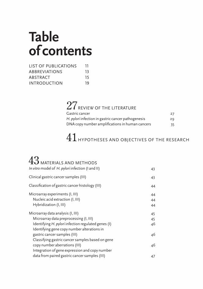
LIST oF PUBLICATIoNS 11ABBREVIATIoNS 13ABSTRACT 15INTRoDUCTIoN 19
Table of contents
REVIEw oF THE LITERATUREGastric cancer 27H. pylori infection in gastric cancer pathogenesis 29DNA copy number amplifications in human cancers 35
HyPoTHESES AND oBJECTIVES oF THE RESEARCH
27
41
43 MATERIALS AND METHoDSIn vitro model of H. pylori infection (I and II) 43
Clinical gastric cancer samples (III) 43
Classification of gastric cancer histology (III) 44
Microarray experiments (I, III) 44 Nucleic acid extraction (I, III) 44 Hybridization (I, III) 44
Microarray data analysis (I, III) 45 Microarray data preprocessing (I, III) 45 Identifying H. pylori infection regulated genes (I) 46 Identifying gene copy number alterations in gastric cancer samples (III) 46 Classifying gastric cancer samples based on gene copy number aberrations (III) 46 Integration of gene expression and copy number data from paired gastric cancer samples (III) 47

RESULTS AND DISCUSSIoNGene expression changes and regulatory networks in H. pylori infected cells 57Putative gastric cancer target genes 59DNA copy number amplifications in cancer 61
Validation of the microarray results (I, III) 47 Quantitative real-time polymerase chain reaction analysis (I) 47 Immunohistochemistry using tissue microarray (III) 48
Analysis of H. pylori infection regulated transcription factors and signaling pathways in AGS cells (II) 48 Computational analysis of H. pylori infection regulated signal transduction pathways in AGS cells (II) 48 Validation of NF-κB transcription factor activation after H. pylori stimulation of AGS cells using electrophoretic mobility shift assay (II) 49
In silico analysis of DNA copy number amplifications in human cancers (IV, V) 49 Collection of DNA copy number amplifications in human cancer (IV, V) 49 DNA copy number amplification profiling (IV) 50 Identification of amplification hot spots in the human genome (IV) 50 Modeling and clustering DNA copy number amplifications ( V) 51 Finite descriptions of the DNA copy number amplification models (V) 51 Data mining of the DNA copy number amplifications (IV, V) 52
Summary of the Methods (I – V) 52
57
CoNCLUSIoNS AND FUTURE PRoSPECTS65
ACKNowLEDGEMENTS 69CoNCEPTS 75REFERENCES 81PUBLICATIoNS 95


This thesis consists of an introductory part and the following publications:
I Myllykangas S, Monni o, Nagy B, Rautelin H, Knuutila S. Helicobacter
pylori infection activates FOS and stress- response genes and alters expression of genes in gastric cancer-specific loci. Genes Chromosomes Cancer 2004, 40:334 – 341.
II Myllykangas S, Saharinen J, Veckman V, Knuutila S. Helicobacter pylori stimulation
regulated cellular signaling in AGS human gastric cancer cells. Submitted.
List of publications
III Myllykangas S*, Junnila S*, Kokkola A, Autio R, Scheinin I, Kiviluoto T,
Karjalainen-Lindsberg M-L, Hollmén J, Knuutila S, Puolakkainen P, Monni o. Integrated gene copy number and expres-sion microarray analysis of gastric cancer highlights potential target genes. Int J Cancer 2008, 123:817 – 825.
IV Myllykangas S, Himberg J, Böhling T, Nagy B, Hollmén J, Knuutila S. DNA
copy number amplification profiling of human neoplasms. oncogene 2006, 25:7324 – 7332.
V Myllykangas S, Tikka J, Böhling T, Knuutila S, Hollmén J. Classification of
human cancers based on DNA copy number amplification modeling. BMC Medical Genomics 2008, 1:15.
The roman numbers (I – V) are used in the text when referring to the publications.
* Equal contribution
11Genomic and functional
profiling of gastric cancer


(a)CGH, (Array) comparative genomic hybridizationBFB, Breakage-fusion-bridgeCML, Chronic myeloid leukemiaEMSA, Electrophoretic mobility shift assayHSR, Homogeneously staining regionICA, Independent component analysisPCA, Principal component analysisPCR, Polymerase chain reaction RNS, Reactive nitrogen speciesROS, Reactive oxygen speciesROC, Receiver operating characteristics
Gene symbols are marked in italics and according to the guidelines of the Human Genome organization nomenclature committee (HGNC). More detailed description can be found at http://www.genenames.org/.
Abbreviations
13Genomic and functional
profiling of gastric cancer


15Genomic and functional
profiling of gastric cancer
Abstract
BackgroundHelicobacter�pylori infection is a risk factor for gastric cancer, which is a major health issue worldwide. Gastric cancer has a poor prog-nosis due to the unnoticeable progression of the disease and sur-gery is the only available treatment in gastric cancer. Therefore, gas-tric cancer patients would greatly benefit from identifying biomarker genes that would improve diagnostic and prognostic prediction and provide targets for molecular therapies. DNA copy number amplifications are the hallmarks of cancers in various anatomical locations. Mechanisms of amplification pre-dict that DNA double-strand breaks occur at the margins of the amplified region.
ObjectivesThe first objective of this thesis was to iden-tify the genes that were differentially ex-pressed in H.�pylori infection as well as the transcription factors and signal transduc-tion pathways that were associated with the gene expression changes. The second ob-jective was to identify putative biomarker genes in gastric cancer with correlated ex-pression and copy number, and the last ob-jective was to characterize cancers based on DNA copy number amplifications.
MethodsDNA microarrays, an in�vitro model and re-al-time polymerase chain reaction were used to measure gene expression changes in H.�pylori infected AGS cells. In order to identi-fy the transcription factors and signal trans-duction pathways that were activated after H.� pylori infection, gene expression pro-filing data from the H.�pylori experiments and a bioinformatics approach accompa-nied by experimental validation were used.
Genome-wide expression and copy number microarray analysis of clinical gastric can-cer samples and immunohistochemistry on tissue microarray were used to identify putative gastric cancer genes. Data mining and machine learning techniques were ap-plied to study amplifications in a cross-sec-tion of cancers.
ResultsFOS and various stress response genes were regulated by H.�pylori infection. H.�pylori reg-ulated genes were enriched in the chromo-somal regions that are frequently changed in gastric cancer, suggesting that molecu-lar pathways of gastric cancer and prema-lignant H.�pylori infection that induces gas-tritis are interconnected. 16 transcription factors were identified as being associated with H.�pylori infection induced changes in gene expression. NF-κB transcription fac-tor and p50 and p65 subunits were verified using elecrophoretic mobility shift assays. ERBB2 and other genes located in 17q12-q21 were found to be up-regulated in asso-ciation with copy number amplification in gastric cancer. Cancers with similar cell type and origin clustered together based on the genomic localization of the amplifications. Cancer genes and large genes were co-local-ized with amplified regions and fragile sites, telomeres, centromeres and light chromo-some bands were enriched at the amplifica-tion boundaries.
ConclusionsH.�pylori activated transcription factors and signal transduction pathways function in cellular mechanisms that might be capable of promoting carcinogenesis of the stom-ach. Intestinal and diffuse type gastric can-cers showed distinct molecular genetic pro-files. Integration of gene expression and copy number microarray data allowed the identification of genes that might be in-
volved in gastric carcinogenesis and have clinical relevance. Gene amplifications were demonstrated to be non-random ge-nomic instabilities. Cell lineage, properties of precursor stem cells, tissue microenvi-ronment and genomic map localization of specific oncogenes define the site specific-ity of DNA amplifications, whereas labile genomic features define the structures of amplicons. These conclusions suggest that the definition of genomic changes in cancer is based on the interplay between the cancer cell and the tumor microenvironment. ❦
16

Abstract


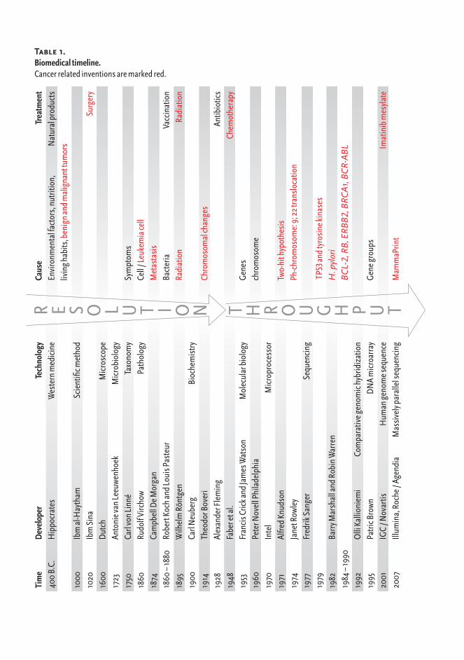
Understanding the mechanisms underlying diseases furthers the development of treatments. Throughout the history of human illness, technological progress has played a central role in determining causes of diseases and laying the foundations for treatment (Table 1). A major trend in technological advancement in medicine has been that the causes of diseases are interrogated in in-creasing resolution. Hippocrates defined diseases as having natural causes and treated patients with natural products to restore the humoral balance, whereas the development of microscope enabled the identification of bacteria as disease causing agents that lead to the discovery of antibiotics to treat infectious diseases. Know-ledge about DNA structure (Watson and Crick, 1953) started a new molecular era in biology. Molecular biology is based on the theory of sequential information flow in living organisms, “The central dogma of molecular biology” (Crick, 1970; Crick, 1958). The dogma presents the information transfer events between three main bio-molecules: DNA, RNA (nucleic acids) and proteins (Figure 1). Nucleic acids and proteins are biopolymers, which are made up of single building blocks. Nucleic acids are composed of nucleo-
tides (Adenine, Guanine, Cytosine, Urasil, Thymine; also referred to as A, G, C, U, T or bases) and proteins are made up of 20 different amino acids. Genetic informa-tion is translated from nucleotide sequence into amino acid sequence according to the genetic code, in which nucleotide codons (words of three nucleotides or triplets) correspond to specific amino acids. The genetic information is stored and distri- buted in DNA, transmitted via RNA and to be put into practice by proteins. The information may be transferred from one biopolymer compartment to another according to the rules formulated by Francis Crick (Crick, 1970; Crick, 1958). General transfer operations, which occur in
Introduction
19Genomic and functional
profiling of gastric cancer
Tim
eD
evel
oper
Te
chno
logy
Ca
use
Trea
tmen
t4
00B
.C.
Hip
pocr
ates
W
este
rnm
edici
ne
Envi
ronm
enta
lfac
tors
,nut
ritio
n,
Nat
ural
prod
ucts
livin
ghab
its, b
enig
nand
mal
igna
nttu
mor
s1
000
Ib
mal
-Hay
tham
Sc
ient
ificm
etho
d1
020
Ib
mSi
na
Surg
ery
160
0D
utch
M
icros
cope
172
3An
toni
evan
Leeu
wenh
oek
Micr
obio
logy
175
0Ca
rlvo
nLin
né
Taxo
nom
ySy
mpt
oms
186
0Ru
dolf
Virc
how
Path
olog
yCe
ll/ Le
ukem
iace
ll1
874
Cam
pbel
lDeM
orga
n
Met
asta
sis
186
0–
1880
Ro
bert
Koch
andL
ouis
Pas
teur
Bact
eria
Va
ccin
atio
n1
895
Wilh
elm
Rön
tgen
Radi
atio
nR
adia
tion
190
0Ca
rlN
eube
rg
Bioc
hem
istry
191
4Th
eodo
rBov
eri
Ch
rom
osom
alch
ange
s1
928
Alex
ande
rFle
min
g
An
tibio
tics
194
8Fa
bere
tal.
Chem
othe
rapy
195
3Fr
ancis
Cric
kand
Jam
esW
atso
nM
olec
ular
biol
ogy
Gen
es1
960
Pete
rNov
ellP
hila
delp
hia
ch
rom
osom
e1
970
Inte
lM
icrop
roce
ssor
197
1Al
fredK
nuds
on
Tw
o-hi
thyp
othe
sis
197
4Ja
netR
owle
y
Ph-c
hrom
osom
e:9;
22tr
anslo
catio
n1
977
Fred
rikSa
nger
Se
quen
cing
197
9
TP
53an
dtyr
osin
ekin
ases
198
2Ba
rryM
arsh
alla
ndR
obin
War
ren
H
. pyl
ori
198
4–19
90
BC
L-2,
RB
, ERB
B2,
BRC
A1,
BC
R-A
BL
199
2O
lliK
allio
niem
iCo
mpa
rativ
egen
omic
hybr
idiz
atio
n1
995
Patri
cBro
wn
DN
Am
icroa
rray
Gen
egro
ups
200
1IG
C/N
ovar
tis
Hum
ange
nom
eseq
uenc
e
Imat
inib
mes
ylate
200
7Ill
umin
a,R
oche
/Ag
endi
aM
assi
vely
para
llels
eque
ncin
gM
amm
aPrin
t
R E S O L u T I O N T H R O u G H P u T
Table 1. Biomedicaltimeline.Cancerrelatedinventionsaremarkedred.
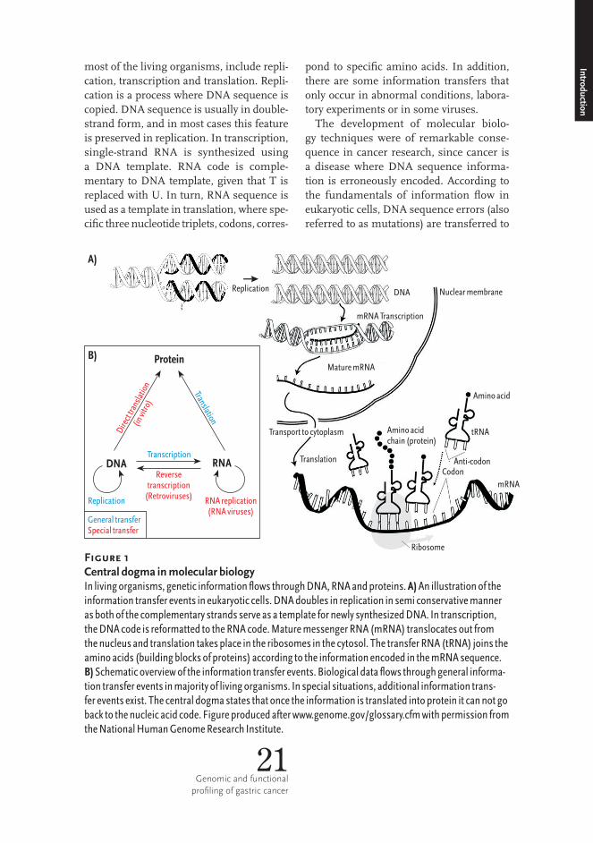
most of the living organisms, include repli- cation, transcription and translation. Repli- cation is a process where DNA sequence is copied. DNA sequence is usually in double- strand form, and in most cases this feature is preserved in replication. In transcription, single-strand RNA is synthesized using a DNA template. RNA code is comple-mentary to DNA template, given that T is replaced with U. In turn, RNA sequence is used as a template in translation, where spe-cific three nucleotide triplets, codons, corres-
pond to specific amino acids. In addition, there are some information transfers that only occur in abnormal conditions, labora-tory experiments or in some viruses.
The development of molecular biolo-gy techniques were of remarkable conse-quence in cancer research, since cancer is a disease where DNA sequence informa-tion is erroneously encoded. According to the fundamentals of information flow in eukaryotic cells, DNA sequence errors (also referred to as mutations) are transferred to
Introduction
21Genomic and functional
profiling of gastric cancer
Figure 1 Central dogma in molecular biologyInlivingorganisms,geneticinformationflowsthroughDNA,RNAandproteins.A)Anillustrationoftheinformationtransfereventsineukaryoticcells.DNAdoublesinreplicationinsemiconservativemannerasbothofthecomplementarystrandsserveasatemplatefornewlysynthesizedDNA.Intranscription,theDNAcodeisreformattedtotheRNAcode.MaturemessengerRNA(mRNA)translocatesoutfromthenucleusandtranslationtakesplaceintheribosomesinthecytosol.ThetransferRNA(tRNA)joinstheaminoacids(buildingblocksofproteins)accordingtotheinformationencodedinthemRNAsequence.B)Schematicoverviewoftheinformationtransferevents.Biologicaldataflowsthroughgeneralinforma-tiontransfereventsinmajorityoflivingorganisms.Inspecialsituations,additionalinformationtrans-fereventsexist.Thecentraldogmastatesthatoncetheinformationistranslatedintoproteinitcannotgobacktothenucleicacidcode.Figureproducedafterwww.genome.gov/glossary.cfmwithpermissionfromtheNationalHumanGenomeResearchInstitute.
Replication
Protein
Transcription
Reversetranscription
(Retroviruses)
Tr
ion
anslat
RNADNA
Replication RNA replication(RNA viruses)
rt
to
Di e
ctra
nsla
in
vitr
(ino)
A)
B)
General transferSpecial transfer
A)
B)
Replication DNA Nuclearmembrane
mRNATranscription
MaturemRNA
Aminoacid
tRNAAminoacidchain(protein)
Translation
Ribosome
CodonAnti-codon
mRNA
Protein
DNA RNA
Dire
cttra
nslat
ion(in
vitro
)
Translation
Transcription
Reversetranscription
(Retroviruses)Replication RNAreplication(RNAviruses)
GeneraltransferSpecialtransfer
Transporttocytoplasm
RNA in transcription and to daughter cells through DNA replication during cell divi-sion. Altered RNA sequence translates into altered amino acid sequence, and the abnor-mal protein structure has an altered func-tion in the cell. Eventually, DNA mutation encoded perturbations in protein function lead to the transformation of a normal cell to a cancer cell. In healthy tissue, cell death and renewal are balanced and controlled.
Tight surveillance of cell division and dis-posal enable the proper cell function and number in tissues and organs. In cancer cells, the mechanisms that execute the sur-veillance of cell existence have been turned off, which leads to uncontrolled growth, limitless replication, acquisition of nutri-ents and expansion. Hanahan and Wein-berg have defined the physiologic changes, six hallmarks, that characterize the cancer cell phenotype (Hanahan and Weinberg, 2000). Uncontrolled growth requires that 22
A) B)
CCGGTC CGTACGTA
CCGGTC CGTACGTG
CCGGTC CGTACGTA
CCGGTC CGTACGT..
CCGGTC CGTACGTA
CCGGTC CGTACGTAG
Substitution
Deletion
Insertion
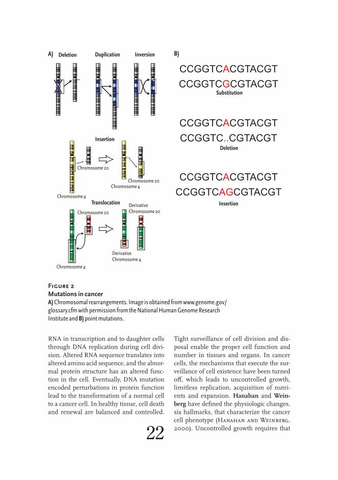
Figure 2Mutations in cancerA)Chromosomalrearrangements.Imageisobtainedfromwww.genome.gov/glossary.cfmwithpermissionfromtheNationalHumanGenomeResearchInstituteandB)pointmutations.
A) B)Deletion Duplication Inversion
Insertion
Chromosome20
Chromosome20Chromosome4
Chromosome4Translocation
Chromosome20
Chromosome4
DerivativeChromosome20
DerivativeChromosome4
Substitution
Deletion
Insertion

23Genomic and functional
profiling of gastric cancer
cancer cells are (1) independent of growth supporting signaling and (2) notwithstand-ing growth restraints. Cancer cells acquire limitless replication potential by becom-ing (3) immune to programmed cell death (apoptosis) and (4) free from natural repli-cation restraints (telomere shortening). In order to proliferate cancer cells need nutri- ents. Thereby, (5) activation of neovascu-larization and vascular maintenance are required for tumor growth. Because of this, cancer cells must possess the capability to promote angiogenesis, generation of new blood vessels into solid tumor mass. Malig-nant capacity is achieved when cancer cells (6) lose the control of their primary anatom-ical location and begin tissue invasion and to metastasize to distant sites.
Cancer cells acquire the hallmarks of can-cer and the capacity to grow unsupervised when mutations change the function of can-cer genes (Hanahan and Weinberg, 2000). Cancer genes are originally normal human genes that function in cell growth, replica-tion, energy metabolism and microenviron-ment modeling. By definition, when mutated, they are capable of promoting and partici- pating in cancerous cell growth. Cancer genes may be activated by gain-of-function mutations (oncogenes) and silenced by loss-of-function mutations (tumor suppressor genes). Even when inherited germ line muta- tions and their induced cancer syndromes are known to promote the emergence of a subset of cancers, mutations of cancer genes are usually somatic, sporadic changes in DNA sequence of non-gamete cells. The mechanisms that induce genomic changes include chromosomal rearrangements (Al-bertson et al., 2003) and nucleotide point mutations (Figure 2). These mutations can cause both, gain and loss of function of genes, depending on the end point. The point mutations that alter DNA sequence at a specific nucleotide can activate an
Introduction
oncogene, if it encodes a change in oncop-rotein that renders it immune to expression regulation or whether it has an increased activity. Nucleotide point mutations can alter the reading frame in the gene or introduce a new stop codon that destroys the original protein product. Structural chro-mosomal rearrangements, such as trans- location and inversion, can produce gene fusions that activate oncogenes or break and suppress function of genes. Inversion is a situation in which a genomic segment flips around to another direction. In addition to translocations, fusion genes can emerge also when a genomic sequence is deleted and the ends of a broken chromosome are rejoined. Translocation refers to a situation where genetic materials from two different chro-mosomes are joined and form a novel geno-type at the site of the junction. Translocation can be either balanced, when no DNA mate-rial is lost and no additional chromosomal fragments are present in the cell, or unbal-anced, if genomic material is lost from the cell during the rearrangement. Alterations that change the DNA copy number are fre-quently observed in a variety of human can-cers (Mitelman et al., 1994). Normal dip-loid human genome contains two copies of each gene and alterations in gene copy number are one of the main mechanisms that activate oncogenes.
Previously, the treatment of cancer has re-lied on surgery and radiation therapies as well as on chemotherapies. Although they are useful in treating locally occurring tu-mors, these techniques are ineffective against tumors that grow in unapproachable locations and metastasize to distant sites. It has become evitable that targeted therapies are needed to treat wide-spread disease. Ad-vances in molecular biology provided new means to determine mutated genes and at-tack cancer cells by targeting the oncopro-teins that they encode. Major breakthroughs have been established using molecular biology techniques in exploring genetic

basis of cancer, such as identification of TP53 and RB tumor suppressor genes and mole-cular changes in chronic myeloid leukemia (CML). The use of molecular targeted drugs in CML is one of the greatest successes in the treatment of cancer. CML is charac-terized by a chromosomal rearrangement, Philadelphia chromosome, which is formed in translocation between chromosomes 9 and 22. In the site of the translocation a fusion of the BCR and ABL genes takes place. Gene fusion causes increased acti-vation of the ABL tyrosine kinase. Imatinib mesylate (also known as Gleevec® or Clivec®) is a small molecule inhibitor of the kinase activity of ABL and in 2001 it was approved as a therapeutic agent for the treat-ment of CML. In addition, Imatinib mesylate has been successfully used as a therapeu-tic agent in the treatment of gastrointestinal
stromal tumor, a mesenchymal tumor char-acterized by KIT positivity, as, besides ABL, Imatinib mesylate has been shown to inhibit the KIT tyrosine kinase activity (Miettinen and Lasota, 2006).
In spite of the great improvements that have been made in order to unreveal the molecular backgrounds of cancer and of the fact that some cancers with specific genetic changes can be treated using target drugs, most of the cancers still remain untreatable. The majority of the most lethal forms of can-cer is multifactorial, and forms a heteroge-neous set of diseases that do not have spe-cific mutations that could be targeted with drugs. Instead of one specific target there might be diverse sets of genes that are per-turbed and are causing disease. Traditional molecular biology provides a reductionist approach that tries to resolve the complexity
24
Figure 3 PrinciplesofamicroarrayexperimentFromwww.genome.gov/glossary.cfm.ImageisusedwithpermissionfromtheNationalHumanGenomeResearchInstitute.

Introduction
in living organisms by studying individual molecular objects separately. It is not pos-sible to identify groups of disease genes using traditional molecular biology tech-niques, since it is difficult to be sure wheth-er the objects of research are the right ones or whether there are combinatorial effects between the biological components. To cir-cumvent these limitations, a second major technological trend in biomedicine has been to increase the throughput of the experimental measurements. In this perspec- tive, comparative genomic hybridization (CGH) (Kallioniemi et al., 1992) had an unprecedented impact on the field of can-cer research because it introduced new tech-nological concepts that allowed up-scaling of the measurement capacity. Firstly, CGH was a genome-wide technology for high-throughput examination of the DNA copy number. Secondly, in CGH, a hybridization probe was placed on a solid support and samples were labeled using fluorescent dyes. Thirdly, CGH was based on measur-ing relative changes in fluorescence inten-sities between the test sample and the refe- rence sample. Further development of genome-wide technologies has been largely relying on these principles.
Another true milestone in the develop-ment of biomedical technologies was the sequencing of the human genome (Lander et al., 2001; Venter et al., 2001). In addi-tion to revealing the organisms’ building in-structions, a detailed knowledge of genome sequences has facilitated the genome-wide analysis of gene expression (RNA synthesis) patterns in the field of functional genomics using DNA microarrays (DeRisi et al., 1996; Schena et al., 1995). Whereas in CGH, in which metaphase chromosomes were used as probes, DNA microarrays consist of col-lections of single-strand DNA probes spot-ted on a glass surface (Monni et al., 2002). Specific probes with sequences that are com-plementary to different genomic counter-
parts, genes or genomic map locations, can be produced. Oligonucleotide probes from 20 to 60 nucleotides (Agilent Technologies and Affymetrix, Palo Alto, Ca) can be synthe-sized and cDNA probes were collected from DNA libraries (Monni et al., 2002). Micro-array experiment is based on the hybridiza-tion of complementary, single-strand DNA fragments and detection is made possible by labeling the sample using fluorescent dyes (Figure 3). Microarray experiments are semi-quantitative as the sample is co-hybridized with a reference, and changes in fluores-cence intensity ratios are measured. Either DNA or RNA may be used as a sample in microarray experiment, although, RNA has to be reverse-transcribed to complementary DNA (cDNA) using specific viral enzymes (Monni et al., 2002). The amount of RNA in a given condition is a measure for gene expression and functionality. While post-transcriptional regulation of gene expres-sion exists, the intensity of transcription is one of the main regulators of gene function. Similarly, the amount of DNA corresponds to the gene copy number. In addition to the basic research, microarray technologies are starting to penetrate the clinical practice as products like MammaPrint®, the Food and Drug Administration of the United States approved diagnostic test, which uses measure- ments of some 70 genes to predict the metastatic behavior of breast tumors, are being developed (Buyse et al., 2006). Genome-wide technologies have exponen-tially increased the amount of quantita-tive data produced by biomedical research. Large-scale datasets can not be processed using human resources, but the develop-ment of microprocessor and propagation of computers have allowed high-through-put analysis of genome-wide biomedical data. There are databases for storing and managing genome-wide datasets (Edgar et al., 2002) and genomic information (Flicek et al., 2008) as well as bioinformatics methods are needed in order to analyze genome-wide measurements and to interpret multifactori-al biological phenomena. ❦25
Genomic and functional profiling of gastric cancer


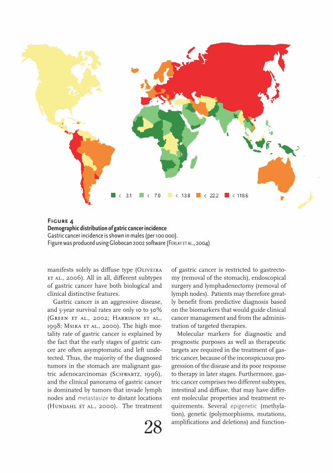
Gastric cancer is a major health issue in Finland and world-wide. Close to one million new gastric cancer cases, 9% of all can-cers, were diagnosed in the year 2002 alone (Ferlay et al., 2004). As an example of the scope of the problem, gastric cancer was the second most common cause of cancer-related death. In Finland, gastric cancer ranked sixth in mortality with close to 600 annu-al deaths and eighth in prevalence (over 700 new cases diagnosed yearly). Gastric cancer is the fourth most common cancer (after lung, breast, and colorectal cancers) (Ferlay et al., 2004). The high-est incidence areas are Asia, Eastern Europe and the Andean region in South America, whereas low rates are found in America, North-ern Europe, South-East Asia and Africa (Figure 4). Incidence and risk in high risk areas are usually as much as ten times larger than in low risk areas (Figure 4 and Table 2). Gastric cancer is more fre-quent in males than in females, and usually affects elderly, as 75% of gastric cancer patients are over 54 years old (Figure 5).
There are two distinct subtypes of gastric cancer, intestinal and diffuse (Lauren, 1965). Undifferentiated diffuse type is character-ized by non-cohesive and scattered cancer cells that grow by in-filtrating deep into the stroma (Werner et al., 2001). Intestinal gastric cancer forms distinguishable and distorted glandular struc-tures and grows by expansion (Werner et al., 2001). The Intesti-
nal type gastric cancer progresses through sequence of premalignant lesions (Figure 6). Generally, diffuse type gastric cancer is not characterized by stepwise progression and intermediate steps but there is evi- dence that premalignant polyps are found in gastric crypts preceding hereditary dif-fuse type gastric cancer (Oliveira et al., 2005). Moreover, premalignant stages, chronic gastritis, atrophy and achlorhydric stomach and intestinal metaplasia, are risk factors in the pathogenesis of intestinal gastric cancer, while simple inflammato-ry stress is associated with the diffuse type (Fenoglio-Preiser et al., 2000). The in-testinal type gastric cancer affects older pa-tients and it has slightly better prognosis than the diffuse type, which is more preva-lent in younger patients (Fenoglio-Preiser et al., 2000). The intestinal type is more common in males than in females but simi-lar gender bias is not observed in the diffuse type (Teh and Lee, 1987). Hereditary gastric cancer, associated with CDH1 mutations, 27
Genomic and functional profiling of gastric cancer
Review of the literature
Review of the literature
28
manifests solely as diffuse type (Oliveira et al., 2006). All in all, different subtypes of gastric cancer have both biological and clinical distinctive features.
Gastric cancer is an aggressive disease, and 5-year survival rates are only 10 to 30% (Green et al., 2002; Harrison et al., 1998; Msika et al., 2000). The high mor-tality rate of gastric cancer is explained by the fact that the early stages of gastric can-cer are often asymptomatic and left unde-tected. Thus, the majority of the diagnosed tumors in the stomach are malignant gas-tric adenocarcinomas (Schwartz, 1996), and the clinical panorama of gastric cancer is dominated by tumors that invade lymph nodes and metastasize to distant locations (Hundahl et al., 2000). The treatment
of gastric cancer is restricted to gastrecto-my (removal of the stomach), endoscopical surgery and lymphadenectomy (removal of lymph nodes). Patients may therefore great-ly benefit from predictive diagnosis based on the biomarkers that would guide clinical cancer management and from the adminis-tration of targeted therapies.
Molecular markers for diagnostic and prognostic purposes as well as therapeutic targets are required in the treatment of gas-tric cancer, because of the inconspicuous pro-gression of the disease and its poor response to therapy in later stages. Furthermore, gas-tric cancer comprises two different subtypes, intestinal and diffuse, that may have differ-ent molecular properties and treatment re-quirements. Several epigenetic (methyla-tion), genetic (polymorphisms, mutations, amplifications and deletions) and function-
Gastric cancer incidence in males (per 100 000)
Gastric cancer incidence in males (per 100 000)
Figure 4 DemographicdistributionofgatriccancerincidenceGastriccancerincidenceisshowninmales(per100000).FigurewasproducedusingGlobocan2002software(Ferlay et al., 2004)
29Genomic and functional
profiling of gastric cancerReview of the literature
al (overexpression) alterations have been as-sociated with gastric carcinogenesis, trans-formation and cancer progression (Table 3). Even when many genetic and epigenetic de-fects have been identified in gastric cancer, molecular biology based applications in clini- cal practice used to treat gastric cancer are still few.
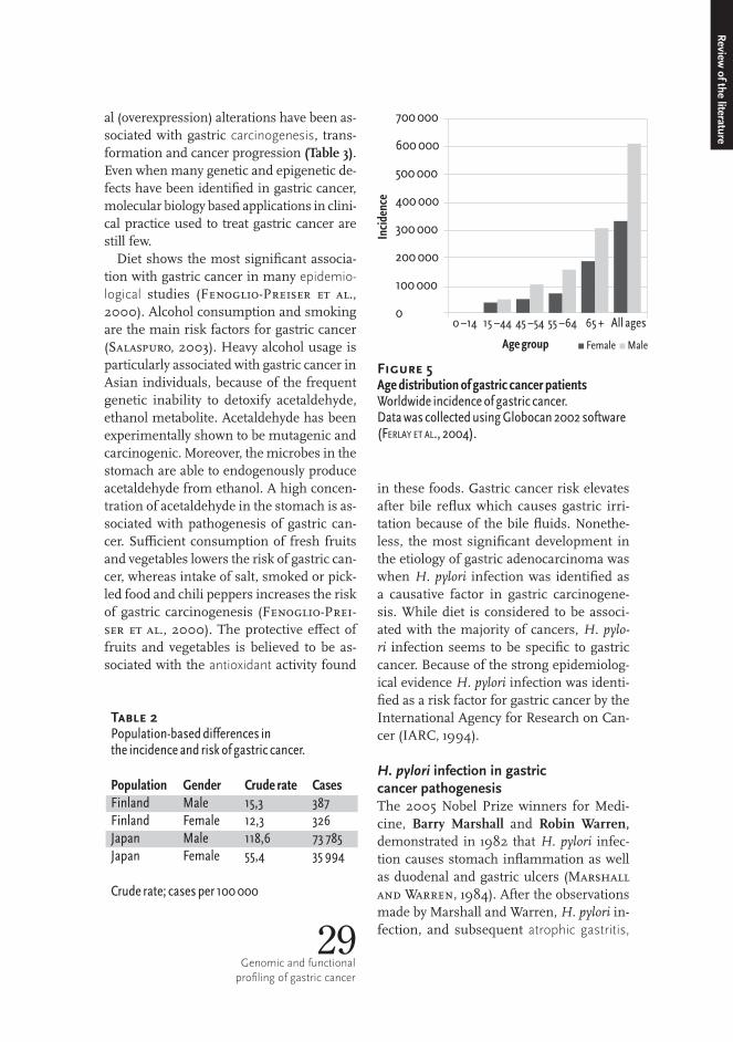
Diet shows the most significant associa-tion with gastric cancer in many epidemio-logical studies (Fenoglio-Preiser et al., 2000). Alcohol consumption and smoking are the main risk factors for gastric cancer (Salaspuro, 2003). Heavy alcohol usage is particularly associated with gastric cancer in Asian individuals, because of the frequent genetic inability to detoxify acetaldehyde, ethanol metabolite. Acetaldehyde has been experimentally shown to be mutagenic and carcinogenic. Moreover, the microbes in the stomach are able to endogenously produce acetaldehyde from ethanol. A high concen-tration of acetaldehyde in the stomach is as-sociated with pathogenesis of gastric can-cer. Sufficient consumption of fresh fruits and vegetables lowers the risk of gastric can-cer, whereas intake of salt, smoked or pick-led food and chili peppers increases the risk of gastric carcinogenesis (Fenoglio-Prei-ser et al., 2000). The protective effect of fruits and vegetables is believed to be as-sociated with the antioxidant activity found
in these foods. Gastric cancer risk elevates after bile reflux which causes gastric irri-tation because of the bile fluids. Nonethe-less, the most significant development in the etiology of gastric adenocarcinoma was when H.�pylori infection was identified as a causative factor in gastric carcinogene-sis. While diet is considered to be associ-ated with the majority of cancers, H.�pylo-ri infection seems to be specific to gastric cancer. Because of the strong epidemiolog-ical evidence H.�pylori infection was identi-fied as a risk factor for gastric cancer by the International Agency for Research on Can-cer (IARC, 1994).
H. pylori infection in gastric cancer pathogenesisThe 2005 Nobel Prize winners for Medi-cine, Barry Marshall and Robin Warren, demonstrated in 1982 that H.�pylori infec-tion causes stomach inflammation as well as duodenal and gastric ulcers (Marshall and Warren, 1984). After the observations made by Marshall and Warren, H.�pylori in-fection, and subsequent atrophic gastritis,
Review of the literature
Table 2Population-baseddifferencesintheincidenceandriskofgastriccancer.
Population Gender Cruderate CasesFinland Male 15,3 387Finland Female 12,3 326Japan Male 118,6 73785Japan Female 55,4 35994
Cruderate;casesper100000
700000
600000
500000
400000
300000
200000
100000
0
Agegroup
Inci
denc
e
0–14 15–44 45–54 55–64 65+ Allages
▪Female▪Male
Figure 5AgedistributionofgastriccancerpatientsWorldwideincidenceofgastriccancer.DatawascollectedusingGlobocan2002software(Ferlay et al., 2004).

30
Gene Genefunction Roleincancer Changes Reference
APC wnt/β-cateninandTGFβ-pathways
Tumorsuppressorgene
Mutations (Fang et al., 2002)
AURKA Cellcycle,mitosis Progression Functionalpolymorphism
(Ju et al., 2006; Kamada et al., 2004)
BTRC wnt/β-cateninpathway
Mutations (Kim et al., 2007)
CCND1 Cellcycle Highlevelsareassociatedwithalco-holconsumption
Amplifiedandoverexpressed,polymorphisms
(Bizari et al., 2006)
CCNE1 Cellcycle Amplificationandoverexpression
(Varis et al., 2003)
CDH1 TGFβ-pathway Hereditarydiffusegastriccancer
Mutations (PeeK and reddy, 2007)
CDKN2A Cellcycle InactivationbymethylationorLOH
(zhang et al., 2003; zhao et al., 2007)
CTNNB1 wnt/β-catenin/TGFβ-pathway,cellcycle
Associatedwithinvasiveandaggressivedisease
Mutations (Cheng et al., 2005)
RHOBTB2 Tumorsuppressorgenethathasgrowthinhibitoryfunction
MutationsandLOH (Cho et al., 2007a)
EGFR Transmembranereceptorproteinkinase
Associatedwithpoorsurvival,targetfortyrosinekinaseinhibitors
Increasedexpression
(galizia et al., 2007)
ERBB2 Transmembranereceptorproteinkinase
Targetforinhibitors Mutations,amplification
(lee et al., 2006)
GAST Gastrinhormone,mitogen
Associatedwithintestinalmetaplasiaandgastriccarcinoma
Amplifiedandelevatedexpression
(doCKray, 2004)
HRAS Signaltransduction Overexpression (Kim et al., 2000)
KRAS Signaltransduction Mutations (lee et al., 2006)
MET Tyrosinekinase ActivatedinGCandintestinalmetaplasia,inducesproliferation
Differentialexpression
(inoue et al., 2004; tang et al., 2004)
NRAS Membraneprotein Mutation (sasaKi et al., 2004)
Table 3Gastriccancerassociatedgenes.LOH;lossofheterozygocity,GC;gastriccancer.

Gene Genefunction Roleincancer Changes Reference
PIK3CA PTENpathway Mutations,up-regulation
(li et al., 2005a)
PPP1R1B Kinaseorphospha-taseinhibitor,regu-lationofapoptosis
Amplifiedandoverexpressed
(BelKhiri et al., 2005; Varis et al., 2004)
PTEN Cellcycle Tumorsuppressorgene
Mutations,down-regulation
(li et al., 2005B)
PTGS2 prostaglandinsyn-thesis,inflamma-tion,mitogenesis
Targetforinhibitor,prognosticfactor
Polymorphisms,over-expression
Numerous
PTPN11 Membraneproteintyrosinephospha-tase,cellgrowth,dif-ferentiation,mitosis
H. pylori target TyrosinephosphorylationofCagAtoxin
(yamazaKi et al., 2003)
RB1 Cellcycle,tumorsuppressorgene,H. pylori target
Mutations,differentialexpression
(lan et al., 2003)
RUNX3 Transcriptionfactor Tumorsuppressorgene,H. pylori related,apoptosis,potentialprognosticfactor
Inactivebydown-regulationormethylated
(homma et al., 2006; ito et al., 2005)
STK11 Proteinkinase Tumorsuppressorgene,Peutz-Jegherssyndromeassociat-edgastriccancer
Mutations (shinmura et al., 2005)
TP53 DNAbinding,tran-scriptionalactiva-tion,DNArepair,cellcyclearrest,apopto-sis,senescence
Tumorsuppressorgene,Li-Fraumenisyndrome
Mutations Numerous
ZFHX3 Transcriptionfactor Aggressiveform Geneticalterations (Cho et al., 2007B)
31Genomic and functional
profiling of gastric cancer
Review of the literature
has been identified as the most significant single environmental factor associated with the increased risk for developing gastric ad-enocarcinoma in many epidemiological, case and animal model studies (Forman et al., 1991; Parsonnet et al., 1991a; Parson-net et al., 1991b; Recavarren-Arce et al.,
1991; Watanabe et al., 1998). H.�pylori in-fection has been shown to cause changes that lead to development of pre-cancerous conditions and lesions, i.e., chronic gastritis, gastric atrophy, intestinal metaplasia and dysplasia (Craanen et al., 1992; Rugge et al., 1996; Watanabe et al., 1998). H.�pylo-ri strains, which carry the virulence factor cagA and produce vacuolating cytotoxin A,

are particularly associated with gastric can-cer (Atherton, 1997). Although the associ-ation of H.�pylori infection with gastric can-cer is undisputable, the molecular genet-ic mechanisms of how H.�pylori infection promotes the gastric carcinogenesis still re-main unknown.
Prolonged H.�pylori infection and sequen-tial precancerous process precedes gastric cancer. A multistep progression of gastric cancer includes chronic gastritis, atrophic gastritis, intestinal metaplasia, dysplasia and adenocarcinoma (Correa, 1992) (Fig-ure 6). Gastritis and gastric atrophy elim-inate gastric mucosa, which reduces gas-tric acid secretion and neutralizes gastric juice. Elevated gastric pH allows chang-es in the gastric flora and it also allows anaerobic bacteria to colonize in the stom-ach. Anaerobic bacteria produce reductase enzymes that catalyze the nitrite synthesis from food nitrate. Nitrite reacts with amines and urea to produce carcinogenic N-nitroso compounds, which are capable of interacting
with DNA. Carcinogenic agents initiate the progression from metaplasia to adenocar-cinoma as DNA damage accumulates in the cells. H.�pylori infection is involved in gastric carcinogenesis as it is the most fre-quent cause of chronic gastritis and it de-creases acid-pepsin secretion and ascorbic acid (dietary antioxidant) concentration in the stomach along with progressing muco-sal atrophy. Furthermore, H.�pylori colonies are observed overlaying and preceding le-sions of the intestinal metaplasia.
H.�pylori can adapt to the acidic environ-ment of the stomach by secreting urease enzyme that metabolizes urea into ammo-nium, which neutralizes hydrochloric acid and produces a neutral microenvironment around the organism (Dunn et al., 1990). What is more, H.�pylori bacteria secrete vir-ulence factors, which damage gastric epithe-lial cells. Urease not only is a survival fac-tor, it also functions as a virulence factor by inducing an inflammatory reaction (Har-ris et al., 1996) and toxic effect in gastric epithelial cells (Smoot et al., 1990). In ad-dition to urease, H.�pylori virulence factors 32
Figure 6 SequentialprogressionofgastriccancerFigureafter(Correa, 1992; PeeK and Blaser, 2002).
Change Conditions andlesions
Risk Molecular geneticaberration
Normalmucosa
Chronicgastritis
Atrophicgastritis
Intestinalmetaplasia
Dysplasia
Carcinoma
pHrises
Tobacco
anaerobicbacteria
nitrosoaminesacetaldehyde
Mutations
H. pyloriinfection
NaClNutritionMalabsorption
TP53mutation(30–50%)
IL1Bpolymorphisms
RASmutation(10%)
DCCmutation(20–60%)

include vacuolating cytotoxin A (VacA) and genes in the cytotoxicity associated gene pathogenicity island (cagPAI). VacA induces mucosal damage and is associated with gas-tric carcinogenesis (de Figueiredo Soares et al., 1998). CagPAI genes encode a se-cretion system (Covacci et al., 1999) that transfers an effector protein, cagA, into a host cell (Odenbreit et al., 2000). The cel-lular membrane tyrosine kinase phosphory-lates cagA (Covacci and Rappuoli, 2000). Phosphorylated cagA interacts with host signaling molecules, including SHP-2 (Higashi et al., 2002), and causes mor-phological changes in the epithelial cells (Moese et al., 2004). Especially cagA-posi-tive strains are associated with gastric ade-nocarcinoma (Blaser et al., 1995). Never-theless, bacterial strains carrying the Cag-PAI genes induce more intense inflamma-tion than strains that lack cag genes (Kolho
et al., 1999; Yamaoka et al., 1997). The in-teraction between cag secretory system and host cell induces the inflammatory reaction (gastritis). Notwithstanding, production of pro-inflammatory cytokines (interleukin-8) in epithelial cells (Odenbreit et al., 2000) also occur without cagA involvement (Fischer et al., 2001).
In addition to direct H.�pylori assault, en-dogenous host responses are crucial in de-termining the progression of H.�pylori in-fection in to pre-neoplastic lesions and ul-timately gastric adenocarcinoma (Correa and Houghton, 2007) (Figure 7). H.�py-lori infection induces activation of NF-κB (Maeda et al., 2000), which then activates growth factors (e.g., Cyclin D1 and Myc) and suppressors of apoptosis (e.g., BCL-XL) (Karin et al., 2002). Although in-crease in cell turn-over is not directly associ-ated with malignancy, excess propagation of cells increases the probability of mutations by inducing DNA damage from shortened 33
Genomic and functional profiling of gastric cancer
Review of the literature
Figure 7 AmodelofH. pyloriinfectioninducedinflammationandgastriccancerpromotion
STROMA
ADENOCARCINOMA CARCINOMA IN SITU EPITHELIUM
Endothelialcells
Angiogenesis
Angiogenicfactors Inflammatorycells CagA
Reactiveoxygen/nitrogen
species
CytokinesChemokines
TNFα
Hp
NF-κB Promotermethylation
Bcl-XL,IAP-1 CyclinD1,Myc COX-2
DNAdamage
Protectionfrom
apoptosis
Increasedproliferation
Lossofcontactinhibition
Anchorage-independence
LossofE-cadherin
Tumorsuppressioninactivation
ProstagalndinE2
Telomereshortening

telomeres. Furthermore, NF-κB activation initiates COX-2 and prostaglandin pathways (Williams et al., 1999) that introduce mor-phological changes in the epithelium. COX-2 expression induces production of prosta-glandin E2, which is a strong inducer of vol-atile and detached phenotype that is charac-
34
teristic of cancer cells. PGE2 production leads to the suppression of E-cadherin func-tion and induces anchorage independence and contact inhibition (Weinberg, 2007).
Pathogenic insult usually leads to an acute inflammation response and to a com-plete clearance of the microbe, but in H.�py-lori infection, inflammatory process fails to eradicate the microbe, infection sustains
B)
A)
HSR Normal LadderB)
A)
HSR Normal Ladder
Figure 8 ManifestationsofDNAcopynumberamplificationA)HomogeneouslystainingregionandB)Doubleminutechromosomes.Imagesfrom(sChwaB, 1998)andwww.wikipedia.org.
HSR
A)
Normal Ladder
B)

35Genomic and functional
profiling of gastric cancer
and a state of chronic inflammation devel-ops (Correa and Houghton, 2007). A pro-longed inflammation (chronic gastritis) and the subsequent atrophic gastritis serve as a platform for carcinogenesis by providing a suitable microenvironment for inducing DNA damage. The inflammation induces mutagenic hits by causing excess cell pro-liferation (De Luca et al., 2003; De Luca et al., 2004), changes in the epigenetic reg-ulation of the host gene expression (Nar-done et al., 2007) and oxidative/nitrosa-tive stress by inducing the production of re-active oxygen and nitrogen species (Ernst, 1999). The inflammatory cells produce cy-tokines and chemokines (e.g., TNF-α) that act via NF-κB pathway (Karin et al., 2002; Weinberg, 2007) and increase the prolifer-ation of epithelial cells. In the chronic H.�py-lori induced gastritis, the CpG islands of the promoter regions of several tumor suppres-sor genes have been found to be hypermeth-ylated (Kang et al., 2003). The hypermeth-ylation of a promoter region is an epigene-tic change that inhibits transcription and in-duces gene silencing by blocking the bind-ing of a transcription factor to its target se-quence in the promoter. Activated leukocytes produce reactive oxygen species, namely ox-ygen ions, free radicals and peroxides, and reactive nitrogen species as a means to at-tack colonizing pathogens (Ernst, 1999). Oxygen and nitrogen metabolites are highly reactive due to the presence of an unpaired electron and they cause damage by reacting randomly and extremely rapidly with vari-ous biomolecules, such as DNA, proteins and lipids. H.�pylori infection promoted in-flammation is amplified by the expansion of the inflammatory cell pool via NF-κB – TNF-α – cytokine pathways and DNA dama-ge induced feedback loops.
DNA copy number amplifications in human cancersMany human cancers of different anatomi-
cal locations are characterized by DNA copy number amplifications. Amplification is a chromosomal change that results in an in-crease of the copy number of a specific DNA region (Albertson et al., 2003; Lengauer et al., 1998). Even hundred-fold elevation in the gene copy number may be present in some tumors. Such high-level amplifica-tions of MYC and EGFR oncogenes occur in neuroblastoma (Schwab et al., 2003) and glioma (Vogt et al., 2004), respectively. DNA copy number amplification is a local, intra-chromosomal mutation that affects a DNA segment of less than 20 million bp in length. DNA copies generated in amplifica-tion manifest as concatenate homogenous-ly staining chromosomal regions (HSRs) and extra-chromosomal acentric DNA frag-ments (double minutes and episomes) (Albertson et al., 2003; Schwab, 1998). HSRs form a ladder-like structure of invert-ed repeats within chromosomes (Schwab, 1998) (Figure 8A). Double minute chromo-some bodies are extra-chromosomal, circu-lar DNA segments (Hahn, 1993) (Figure 8B). An excess chromosome or chromo-some segments, HSRs and double minute chromosomes can be detected using stan-dard microscopic techniques (Schwab et al., 2003; Vogt et al., 2004) and compara-tive genomic hybridization (CGH) (Kallio- niemi et al., 1992). Episomes are sub- microscopic extra-chromosomal DNA seg-ments of ~250 bp in length, which can be detected with molecular biology methods, such as fluorescent in�situ hybridization and microarray CGH (Graux et al., 2004; Mau-rer et al., 1987). The amplified region may contain gene fusions, i.e., DNA from dif-ferent genomic sites (Graux et al., 2004; Guan et al., 1994).
DNA copy number amplification resulting in the formation of a homogeneously staining re-gion has been proposed to have occurred accor- ding to the Breakage-Fusion-Bridge (BFB) model (Figure 9). The event initiating in BFB sequence is a DNA double-strand break. Before cell division the uncapped chromosome
Review of the literature

replicates. BFB sequence progresses if fusion of the uncapped sister chromosomes occur before chromosome segregation. During mi-tosis the two centromeres of the dicentric fu-sion chromosome are drawn to the oppo-site poles of the mitotic spindle forming an anaphase bridge. DNA double-strand break in the new site during mitosis inflicts in-verted duplication and loss of the genomic
region between the two breakpoints in the daughter cells. The BFB cycle proceeds with every cell division, and the duplicated DNA region proliferates until the open chromo-some ends are sealed. The steps of the BFB sequence, DNA double-strands break due to telomere loss (Murnane and Saba-tier, 2004) and chromosomal breakage (Coquelle et al., 1997), sister chromatin fusion and breaking of the anaphase bridge (Shimizu et al., 2005) as well as ladder-36
Breakage
Replication
Fusion
Anaphase bridge
Cell division
Normal
Breakage
Replication
Fusion
Oncogene
Centromere
Fusion
Figure 9 Breakage-Fusion-BridgemodelofDNAamplificationFigureproducedafterSchwab,1998(sChwaB, 1998)
OncogeneCentromereFusion
Normal
Breakage
Replication
Fusion
Anaphasebridge
BreakageCelldivision
Replication
Fusion

like structure of inverted repeats (Toledo et al., 1992), have been proven to exist in�vivo.
The disintegration of circular extra-chro-mosomal chromatin bodies (double min-utes and episomes) from chromosome, un-equal segregation in cell division and selec-tion in tumor tissue is another mechanism which results in DNA copy number ampli-fication (Graux et al., 2004; Hahn, 1993). Extra-chomosomal amplification most like-ly occurs by unequal segregation in cell di-vision and selection in tumor tissue, since double minutes and episomes rarely con-tain replication origins or centrosome that would enable propagation by replication and mitotic segregation. Extra-chromosom-al DNA elements are thought to rise either before or after replication. Chromosomal deletion occurs if a DNA segment loops out from the chromosome before replication and chromosome ends are joined. There is evidence that supports the model that ex-tra-chromosomal DNA is looped out from the genome, since original genomic archi-tecture has been shown to remain together in the MYCN amplification (Schneider et al., 1992) and excised double minute (Tole-do et al., 1993). It has also been shown that episomes (Graux et al., 2004) may carry DNA that is deleted in the original genomic region. If double minute excision happens after replication, the excised DNA segment stays also in the chromosome. There are two models for postreplicative multiplication of double minutes (Vogt et al., 2004) (Figure 10A and B). Double minutes may unfold via breakage of the replication bubbles and multiply due to unequal segregation during mitosis. Furthermore, double minutes may contain inverted, repeated sequences sug-gesting that double minute formation may happen by HSR breakdown and the circu-larization of the derivative DNA fragments (Fakharzadeh et al., 1993; Singer et al.,
37Genomic and functional
profiling of gastric cancer
Review of the literature
OR
1’1
2’2
Replication
Mitosis
A) B) C)
Oncogene
Homogeneouslystaining region
OR
1’1
2’2
Replication
Mitosis
A) B) C)
Oncogene
Homogeneouslystaining region
OR
1’1
2’2
Replication
Mitosis
A) B) C)
Oncogene
Homogeneouslystaining region
Figure 10 ModelsofdoubleminuteformationA)Segregationafterreplication,B)BreakageofthereplicationbublesandC)HSRbreakdown.FigureproducedafterVogtetal.,2004(Vogt et al., 2004).
A)
B) OR
1’1
2’2
Replication
Mitosis
A) B) C)
Oncogene
Homogeneouslystaining regionReplication
C)
Mitosis
OR
1
2 2’
1’
Oncogene
Homogeneouslystainingregion
2000) (Figure 10C). In proportion, DNA double-strand breaks may induce genomic relocation of double minutes and episomes resulting in formation of HSRs or distribut-ed insertions (Coquelle et al., 1998). Even though current data supports the excision-segregation-selection models of extrachro-mosomal amplification, also episomal pro-liferation (autonomous plasmid-like replica-tion of sub-microscopic circular molecules) (Von Hoff et al., 1988) and recombination to form double minutes that reinsert into the chromosomes (Von Hoff et al., 1990) have been proposed to result in DNA am-plification.
Models of DNA amplification mecha-nisms, BFB sequence and excision of extra-chromosomal DNA segments, stipulate that two independent DNA double-strand breaks that flank the amplified region are required to occur in order to make DNA amplification possible. There are numerous agents and processes that have been shown to induce DNA double-strand breaks. Tobacco, etha-nol and caffeine contain clastogenic chem-icals that cause chromosomal breaks (Ban et al., 1995; Kuwano and Kajii, 1987; Rao et al., 1988). Asbestos is a group of silicate fiber, which is associated with the risk of de-veloping lung cancer. Asbestos fibers block cell division, puncture into the nucleus and physically irritate the chromatin, suggest-ing a mechanism for inducing chromosome breaks (Jensen et al., 1996). Exposure to radiation is an established extrinsic cause of chromosomal breaks (Ban et al., 1995). The Vpr�gene encoded protein induces chro-mosomal breaks leading to amplifications in patients infected with human immuno-deficiency virus (Shimura et al., 1999). Fo-late and oxygen deficiencies (Blount et al., 1997; Coquelle et al., 1998) and release of oxygen-free radicals (Marnett, 2000) are intrinsic cellular processes that have been associated with the DNA damage.
Human genome is not consistently dura-ble and resistance to DNA breaks varies be-tween different regions. Specific DNA se-quence features have been proposed to in-crease damage susceptibility. Fragile sites are genomic regions, which are damage-prone when cells are treated with chemicals that interfere with replication (Schwartz et al., 2006). There are 120 fragile sites in the human genome according to the Nation-al Center for Biotechnology Information database (http://www.ncbi.nlm.nih.gov/). Eighty-eight fragile sites are classified as common, since they are found in all indi-viduals, and 31 fragile sites, which are found in less than 5 % of the population, as rare. Damage susceptibility of the fragile sites varies and it has been shown that 15 % of all the common fragile sites contained 75 % of the DNA breaks (Glover et al., 1984). Fragile sites harbor large genes. WWOX and FHIT genes are 1.1 Mb and 0.8 Mb in length and located in two most damage-prone com-mon fragile sites (FRA16D in 16q23.1 and FRA3B in 3p14.2) (Iliopoulos et al., 2006). In addition, fragile sites in general are en-riched with extremely large genes (Smith et al., 2006). Chromosomal fragility induced breaks have been proposed to initiate the amplification pathway (Coquelle et al., 1997). Specifically, the common fragile sites were shown to coincide with amplification boundaries in a cell line experiment (Hell-man et al., 2002). Furthermore, MET onco-gene amplifications in six primary esopha-geal adenocarcinomas contained at least one break point that was located within a fragile site (Miller et al., 2006).
According to the central dogma, DNA in-formation, including copy number, is trans-ferred to RNA. The information encoded in the DNA copy number does not transfer in-to RNA quantity in a strict proportion be-cause transcription is in many ways regu-lated. However, increased gene copy num-ber generally induces elevated gene expres-sion but the impact of gene amplification 38

on gene expression differs between cancer types, and it also depends on the structure of the amplicon. It has been estimated that a 2-fold change in DNA copy number causes a 1.5-fold difference in the mRNA level (Pol-lack et al., 2002). In studies of primary breast tumors (Hyman et al., 2002; Pol-lack et al., 2002) 44% to 62% of highly amplified genes were found to be overex-pressed. In contrast, only 19.3% of the am-plified genes in prostate cancer cell lines were shown to be overexpressed (Wolf et al., 2004).
Oncogene amplification promotes carci-nogenesis as an amplified ERBB2 gene was found to induce abnormal development of mammary glands and eventually initiate the tumorigenesis of the breast in a mouse mod-el (Andrechek et al., 2000). According to a very conservative estimate, there are over 40 oncogenes that have been identified to be activated by copy number amplification in human cancers (causes increased gene ex-pression or specifically associates with clin-ical outcome or tumor biology) (Myllykan-gas et al., 2007). Most studied amplified genes in human cancers include AKT2 in ovarian cancer, ERBB2 in breast and ovari-an tumors, MYCL1�in small cell lung cancer, MYCN in neuroblastoma, REL in Hodgkin Lymphoma, EGFR in glioma and non-small cell lung cancer and MYC in various cancers (Futreal et al., 2004).
Drug target gene amplifications fre-quently induce resistance to drug thera-py as asserted in laboratory experiments using cell lines as well as in clinical stud-ies (Goker et al., 1995; Johnston et al., 1983; Shannon, 2002; Wahl et al., 1979). The CAD gene amplification is a common mechanism in conducting resistance to N-(phosphonacetyl)-L-aspartate mediated genotoxicity (Wahl et al., 1979). Methotrexate is an inhibitor of the DHFR enzyme that is used in treatment of various cancers.
Methotrexate resistance has been shown to result from the DHFR gene amplification in hamster cell lines (Johnston et al., 1983) as well as in patients with acute lymphoplas-tic leukemia (Goker et al., 1995). Imatinib mesylate is a BCR-ABL fusion protein in-hibitor, which has been successfully applied in the treatment of CML patients. Imatinib mesylate treatment resistance occurs in some of the patients due to�BCR-ABL fu-sion gene amplification or mutations in the ABL�gene (Shannon, 2002). Additionally, changes in metabolite, substrate and com-petitive enzyme fluxes may underlie some cases of Imatinib mesylate resistance (Bo-ren et al., 2001; Boros et al., 2002).
Oncogene amplifications occur frequent-ly in advanced cancers and in the majori-ty of organs and they usually are associated with poor prognosis (Myllykangas et al., 2007). Amplifications are more common in solid tumors than hematologic malignan-cies (Myllykangas et al., 2007). Although the gain-of-function effect of gene amplifi-cations is very a appealing target for modern small molecule inhibitors, only therapeutic agents targeted at ERBB2 and EGFR ampli-fications are currently available (Myllykan-gas et al., 2007). The EGFR gene is fre-quently amplified in colorectal cancers and there is a monoclonal antibody (panitumab) that binds to EGFR and inhibits its function. The RAS oncogene encodes a signal trans-duction protein that is downstream of EGFR. Nevertheless, patients with mutations in the RAS gene do not respond to EGFR antibody treatment and, in fact, the latest re-ports indicate that patients with RAS muta-tions have worse prognosis using EGFR an-tibodies than with traditional therapies. ❦
39Genomic and functional
profiling of gastric cancer
Review of the literature


The treatment of cancer ultimately requires a detailed knowl-edge about the molecular properties of tumors. The complexity and heterogeneity of gastric cancer suggests that the genome-wide ap-proach is well justified to study its molecular properties. The ge-nome-wide methods can be used to detect genomic changes in can-cer and to link them with cellular functionality and cancer pheno-type. Novel microarray technologies for measuring gene expression and copy number allow molecular-level dissection of the function-al and structural genomic changes in cancer. It is possible to define the cancer biomarkers and genes associated with the cancer pheno-type using a multi-level, high-throughput genomic profiling, clini-cally and biologically relevant background data and computational data mining. Gastric cancer is a well-suited model for studying can-cer since it is fairly common, it has well-documented epidemiolog-ical and biological background and it mani-fests many characteristics that are common to human cancer. More explicitly, the asso-ciation of gastric cancer with H.�pylori infec-tion allows the research of carcinogenesis and cancer initiation. The biological back-ground of cancer can only be understood if different cancer types can simultaneous-ly be studied. Exploring DNA copy number amplifications in a landscape of cancers al-lows the uncovering of biomolecular funda-mentals behind cancer and the confluences between different cancer types. ❦
Hypotheses and objectives of the research
The scientific objectives of the thesis were to:•Identifygeneexpressionchangesandgeneregulatory
networksinH. pylori infectedcells(IandII)•Identifyputativetargetgenesingastriccancer(III)•DiscoverbiologicalfundamentalsbehindDNAcopy
numberamplificationsincancer(IVandV)
41Genomic and functional
profiling of gastric cancer

A brief overview of the materials and methods is presented in the following sub-sections. More detailed technical descriptions can be found in the corresponding publications.
In vitro model of H. pylori infection (I and II)Laboratory experiments (in�vitro) are needed in order to study the specific properties in biological systems. Laboratory experiments al-low the researchers to have control over different biological, phys-ical, and chemical properties, and focus the measurements on the specific biological properties. H.�pylori infection induced gene ex-pression changes in human epithelial cells and they were studied in�vitro using H.�pylori strain NCTC 11637 and AGS cell line (I). H.�pylori strain NCTC 11637 met the requirements of a pathogen in the experiment, since the virulence factors cagA and vacA were present (Jiang et al., 1996). AGS cell line (the American Type Culture Col-
lection, Manassas, MA) was chosen to be the host in the in�vitro ex-periments as its growth characteristics, monolayer, resembled the gastric epithelium. Moreover, AGS cell line contains less genomic changes compared to other stomach cancer cell lines. AGS cell line has previously been used as a model host in H.�pylori studies. There-fore, it is possible to compare results with the previous data.
Clinical gastric cancer samples (III)Experiments carried out in the laboratory are not equivalent to the natural phenomenon. That is why it is also important to study the original samples. Clinical gastric cancer samples were collected pro-spectively in the Department of Surgery (Helsinki University Cen-tral Hospital, Helsinki, Finland) (III). Ethical issues had to be met so that the privacy of the patients who participated in the study was
Materials and Methods
43Genomic and functional
profiling of gastric cancer
Materials and M
ethods

protected. The study was authorized by the Clinical Review Board of Helsinki Univer-sity Central Hospital and the research proj-ect was reviewed and approved by the Ethi-cal Committee of the Department of Medi-cal Genetics. Each participant signed a letter of consent prior to entering the study. Gas-tric cancer samples were collected between 1999 and 2005 during surgical removal of the stomach (gastrectomy). Extra precau-tions were taken in the sample preparation, since downstream analysis using microar-rays is very sensitive. Sample quality deter-mines the quality of the measurement data. In order to preserve the integrity of the nu-cleic acids, samples were taken immediately after the gastrectomy. Cancer samples were taken from the tumor site and normal tissue was obtained as far away from the tumor site as possible. Subsequently, samples were fresh frozen in liquid nitrogen and stored in -80 °C in as to preserve DNA and RNA from degradation. The collection of stomach spec-imens included a total of 375 samples. In or-der to ensure the anonymity of the partici-pating patients, generic sample codes were given to each collected tissue sample by the attending clinician and all personal data was eliminated when tissue samples were sub-jected to research.
Classification of gastric cancer histology (III)The classification of gastric cancer was de-fined according to Laurén (LaurÉn, 1965). The tissue samples used in the histopa-thology were surgical specimens from gas-tric cancer patients (see previous section). The classification was performed by a pro-fessional pathologist in Helsinki Universi-ty Central Hospital (HUSLAB). The histo-pathological classification was performed using the frozen section preparations from the samples. The main stains used in the classification experiments were HE and
Alcian blue-PAS. The patient records at the department of pathology were used to col-lect the clinical data.
Microarray experiments (I, III)Nucleic acid extraction (I, III)Total RNA was extracted from infected and non-infected cell lines using the Qiagen RNeasy method (I). DNA was extracted from the homogenized clinical gastric samples us-ing the DNeasy tissue extraction kit (Qiagen) and total RNA was extracted using the RNeasy midi kit (III). The same homogenates were used in RNA and DNA extraction. The con-centrations of DNA and RNA samples were measured using Eppendorf Biophotometer (Eppendorf AG, Hamburg, Germany) and the nucleic acid quality was measured using gel electrophoresis and Agilent’s 2100 Bioan-alyzer (Agilent Technologies) (I and III).
Hybridization (I, III)The gene expression profile of H.� pylori NCTC 11637 infected AGS cell line was mea-sured using custom-printed microarray con-taining 12,000 cDNA probes (I). For each hybridization, 50 µg of the total RNA was extracted from H.�pylori infected cell lines, and non-infected cell lines were labeled with fluorescent dyes using reverse transcription enzyme. The labeled cDNAs were hybrid-ized on a cDNA microarray as previously de-scribed (Mousses et al., 2000).
The integration of gene copy number and the expression of microarray data is also re-quired to understand the effects of gene reg-ulation and transcription in the manifesta-tion of genomic copy number changes. For-ty-six gastric tissue samples were selected based on the sample quality and were ana-lyzed using array comparative genomic hy-bridization (aCGH) and gene expression microarrays (III). The gene expression in 46 gastric tissues (8 normal and 38 cancer samples) was studied using a commercial microarray kit with 44,000 oligonucleotide probes. Twenty μg of total RNA was used in 44
hybridization. An RNA pool of 10 non-gas-tric cancer cell lines was used as a reference. Using a universal reference that contains a genome-wide coverage of transcribed genes allows the measuring of more genes. If the gene that is expressed in the test sample is not expressed in the reference sample, it is not possible to get a result, since ra-tio cannot be calculated if the reference is zero. The labeling of the test and reference RNAs, hybridizations and wa shings were carried out according to the manufacturer’s instructions.
For performing aCGH, DNA samples were labeled as previously described (Hyman et al., 2002; Wolf et al., 2004). Pooled DNA extracted from the gender-matched healthy individuals’ blood’s buffy coat fraction ob-tained from the Finnish Red Cross was used as a reference. Twenty μg of genomic DNA from test sample and reference sample were used in the hybridization experiment. DNAs were labeled using fluorescent dyes and re-verse transcription enzyme. The manufac-turer’s instructions were followed in the hy-bridization and washing of the slides.
Microarray data analysis (I, III)Microarray data preprocessing (I, III)After the hybridization experiments, micro-array slides were scanned using a confocal laser scanner and the fluorescence intensi-ties of the DNA spots were measured. Two scans were performed for each microarray slide to produce fluorescence intensity im-ages for both, test and reference channels. Image analysis software is required in or-der to interpret scanned images from TIFF-format into numeric values as well as to ob-tain a text file of raw intensity values for test (Cy5) and reference (Cy3) channels. The im-age analysis was performed using DEARRAY software (Chen et al., 1997) (I) or Feature Extraction software 8.1 (Agilent Techno- logies) (III).
Data from different measurements and arrays had to be normalized to be able to compare the different data from the differ-ent samples. All normalizations were done using GeneSpring software. Lowess nor-malization was applied on the data set using GeneSpring 5.0 data analysis software (Sili-con Genetics, Redwood City, CA) (I). aCGH and gene expression microarray normal-izations were carried out using the Feature Extraction data import plug-in with default parameters for 2-color microarrays (Agilent Technologies) (III). Outlier features were disregarded in the normalization.
Filtering the results by quality parameters is required to make certain that all the da-ta that enters the downstream analysis is of good quality. Intensity data were imported into the GeneSpring data analysis software (Agilent Technologies). Data were filtered by requiring the measurement quality score of the DEARRAY software to be more than 0.5 (Chen et al., 2002) (I). The genes that passed the quality control had spot fluores-cence intensity over two times higher than the variance of the background, the intensi-ty of the test sample was over 200, and the area of the spot was over 25. Microarray re-sults in publication III were quality filtered using outlier and control flags determined by the Feature Extraction Software.
In order to be able to interpret the results from the microarray experiments it is im-portant to know exactly what the arrayed probes are measuring. One efficient and stable technique is to match the probe se-quences in the human genome sequence and extract gene annotations through the genomic databases. In publication III, the DNA sequence information of the probes was provided by the manufacturers of the microarrays that were used in the study. The corresponding genomic map positions of the microarray probes were retrieved us-ing MegaBlast analysis against the human genome sequence. Each microarray probe was assigned with an Ensembl gene identi-
Materials and M
ethods
45Genomic and functional
profiling of gastric cancer

fier according to the map positions of the probe sequences. The gene identifications of Ensembl were applied to collect gene an-notation data.
Identifying H. pylori infection regulated genes (I)The genes that were regulated in AGS cells by co-culture experiments of H.�pylori stim-ulation were identified by filtering data in different time points (I). A cut-off of two-fold change in gene expression ratios was used to select genes regulated during the H.�pylori stimulation time series. In addition to this, t-test (p<0.05) was used to select differ-entially expressed genes in individual time points. The accumulation of H.�pylori reg-ulated genes in chromosomal regions that contain frequent changes in gastric cancer was assessed using a statistical hypothesis testing. P-value correction (e.g., False Discov-ery Rate) was not used when determining statistical significance in the analysis of the result genes since the identified genes were used for hypothesis generation and the in-dividual findings were further validated us-ing alternative methods. On the other hand, downstream-analysis was carried out using the whole set of genes.
Identifying gene copy number alterations in gastric cancer samples (III)In microarray-based comparative genomic hybridization, the copy number of the gene is quantified and reported by a relative and continuous scale. Microarray data is noisy and when thousands of genes are measured simultaneously, individual measurements can vary unintentionally. Consequently, it is difficult to determine gene copy number changes based on a single measurement. In aCGH, measurements are not independent but interconnected by genomic location. As a result the measurements of the neighbor-ing genomic positions have a higher prob-
ability of representing the same amount of genomic DNA. The dependency between probes representing close-by genomic ele-ments can be taken into account when the genomic regions of copy number changes are determined. The CGH Explorer soft-ware (Lingjaerde et al., 2005) was applied to identify DNA copy number changes in 38 gastric tumors (III). Analysis of copy er-rors algorithm and false discovery rate of 0.002 were used in the analysis. Copy num-ber gains and losses were determined for each tumor sample and the frequency pro-file across the genome was reported.
Classifying gastric cancer samples based on gene copy number aberrations (III)The classification of samples is an elemen-tal problem in clinical practice. Proper clas-sification and diagnosis are the foundation of any effective treatment, as it is the in-formative follow-up of the disease. Gener-al concept in diagnostics is to determine, based on some continuous measurement value, whether a patient has a given condi-tion or not. For example, what bodily tem-perature is considered as fever or does a pa-tient have a specific gastric cancer sub-type based on the genomic properties of the tu-mor. There are four different outcomes of a two-class classification problem, such as in-testinal or diffuse gastric cancer sub-type. True outcomes are achieved, when predic-tion and actual value are the same. This can be the case when an actual intestinal type sample is predicted to be intestinal or if a diffuse type is predicted to be diffuse. Con-versely, false outcomes ensue, if an actual value is different from the predicted value, when intestinal type is predicted to be dif-fuse type and vice versa.
Receiver operating characteristics (ROC) analysis (Swets, 1998) was used to assess the values of gene copy number changes as classifiers of intestinal (n=25) or diffuse (n=13) type gastric tumors and cancers locat-ed in corpus (n=19) or antrum (n=19) (III). 46

The gains and losses were treated separately in the analysis. ROC curve can be drawn by calculating the true and false positive rates. The area under the ROC curve was calcu- lated and these values were used to estimate the diagnostic value of a given gene.
ROC analysis identified gene groups that were too large for diagnostics use, since in clinical applications, patterns of no more than a few genes are sought after. More-over, identifying a small subset of classi-fier genes would be more informative in determining the biological significance of the genomic changes that determine the phenotypes of different subtypes. Forward selection algorithm and a Naïve Bayes classifier (Duda et al., 2001) were applied to identi-fy the patterns of genes that would be ideal when classifying gastric cancer patients based on the tumor location or subtype. Ten-fold cross-validation procedure was per-formed and repeated 50 times to ensure the stability of the identified patterns. Ten-fold cross-validation was chosen as it is optimal for such a small sample size. The number of genes in each pattern was determined by minimizing the validation error of 500 rep-etitions with different subsets of data in the framework of maximum likelihood. The sig-nificance of the identified patterns was as-sessed by comparing them to randomly se-lected variables. Ten thousand Naïve Bayes classifers were trained with randomly se-lected variables and the performance of the true classifier was evaluated using a statis-tical hypothesis test.
Integration of gene expression and copy number data from paired gastric cancer samples (III)DNA copy number change does not nec-essarily mean that all the genes inside the changed region would be implicated in can-cer. It is more likely that there is a subset of genes within the changed region that con-
tributes to the manifestation of the cancer-ous phenotype. Integrating the knowledge of gene expression intensity with the copy number is a powerful tool for separating the biologically relevant driving genes from the by-standers. Paired gene expression and copy number data from 38 gastric cancer tissues were integrated in order to identify genes that were differentially expressed due to copy number aberration according to the method reported by Hautaniemi et al. (Hau-taniemi et al., 2004) (III). Array CGH and gene expression microarray probes were combined using the genomic map positions of the arrayed probes. Gains and losses were treated separately in the analysis. The copy number aberration labels were used to clas-sify samples into two groups. Subsequently the mean gene expression levels were calcu-lated for each group. Signal-to-noise statis-tics and 10,000 random permutations of the label vectors were used to assess the signifi-cance of the difference in the mean gene ex-pression intensities between the compared groups.
Validation of the microarray results (I, III)Gene discovery approach using microarrays is often disturbed by noise in the data and the candidate genes need to be further ex-amined in order to validate the results and estimate the relevance of the findings. Gene expression changes in AGS cells caused by H.�pylori stimulation were verified using real-time PCR and target genes in gastric cancer were validated in protein level by immuno- histochemistry and tissue microarray.
Quantitative real-time polymerase chain reaction analysis (I)Quantitative real-time polymerase chain reaction (PCR) is a technique that can be used to de-tect and quantify the amount of a specific DNA sequence in a sample. The procedure follows basic PCR but the accumulation of a PCR product is measured after each amplifi- 47
Genomic and functional profiling of gastric cancer
Materials and M
ethods

cation cycle. When RNA is used as a tem-plate it has to be reversely transcribed in-to cDNA before quantitative PCR reaction. Quantitative real-time PCR was applied to validate the expression of ten genes reg-ulated by H.�pylori infection (I). Gene ex-pression was measured in two time points and the untreated cell line was used as a control. For each analyzed gene a specific pair of primers were designed and synthe-sized. Quantitative real-time PCR was run using Light Cycler thermal cycler and SYBR green detection system. Duplicate PCR re-actions were executed for each gene. A neg-ative control accompanied each run. Stan-dard curves were obtained by performing serial dilutions of beta-globulin gene. The ACTB gene (beta actin) was used as an in-ternal control and for the normalization of RNA quantity and quality differences in all samples.
Immunohistochemistry using tissue microarray (III)Immunohistochemistry is a laboratory technique, which utilizes antibodies to de-tect specific antigens in tissues. Antibodies (immunoglobulins) are proteins that are produced by cells of the immune system to identify and neutralize foreign agents. Each immunoglobulin binds to a specific anti-gen. Physiological antigens are often biolog-ical components that are specific to the inva-sion of bacteria and viruses, but specific an-tibodies against various antigens can be pro-duced. Tissue microarrays consist of hun-dreds of separate tissue specimens punc-tured into a paraffin block. Using immuno-histochemistry on tissue microarray enables simultaneous analysis of specific antigens in multiple samples. The specific antibodies that attach to ERBB2 and MUC-1 proteins were used on gastric cancer tissue microar-ray of 78 specimens to validate cancer bio-marker discoveries made using integrated
gene copy number and expression analysis of the clinical gastric cancer samples (III). The tissue microarray used in the study was independent of the samples that were used in the microarray profiling.
Analysis of H. pylori infection regulated transcription factors and signaling pathways in AGS cells (II)Rapid changes in the gene expression of AGS cells after H.�pylori infection suggest-ed that the host responses are controlled by signal transduction pathways that regulate the activity of specific transcription factors. Transcription factors are proteins that bind to specific DNA sequence after being acti-vated by specific signals and regulate, either by increasing or decreasing, the expression of specific target genes. Transcription fac-tor activation is a rapid mechanism for cells to respond to environmental stimuli by ac-cordingly changing their gene expression. Consequently, the hypothesis was that us-ing the gene expression data from H.�py-lori experiments and a bioinformatics ap-proach, it would be possible to reverse engi-neer the gene regulatory networks activated after H.�pylori infection. The transcription factors and signal transduction pathways that were associated with H.�pylori infection were charted by making use of phylogenetic footprinting and transcription factor bind-ing site mapping on the regulated genes´ promoter regions (II). More reliable conclu-sions about the regulatory backgrounds of the gene expression changes were allowed to be made by studying the entire H.�pylori transcriptome.
Computational analysis of H. pylori infection regulated signal transduction pathways in AGS cells (II)H.�pylori target genes (n=200) in AGS cells were previously identified using an in�vitro model and gene expression microarrays (I). Two kilobases of DNA upstream from the predicted transcription start site were 48

extracted in order to collect the regulatory DNA elements, promoters, of the H.�pylori infection target genes. Similarly, the puta-tive promoter regions were collected from 400 randomly picked reference genes. Orthologous mouse genes were selected for H.�pylori target and reference genes and their promoter regions were extracted using Ensembl database (Birney et al., 2004). Phylogenetic footprinting was applied to identify the conserved regions in the paired human-mouse promoter sequences (Lenhard et al., 2003). The DNA sequences that are evolutionary conserved are likely to have function and, thus, contain regulatory DNA elements (Lenhard et al., 2003). Transcription factor binding sites were mapped to the promoter sites that were evo-lutionary conserved using the application on Consite website (Lenhard et al., 2003). The transcription factor binding sites were collected from the JASPAR database (Sand-elin et al., 2004). The frequencies of tran-scription factor binding sites in H.�pylori in-fection regulated transcriptome and refer-ence gene set were counted and the statisti-cal hypothesis testing was applied in order to test the enrichment of the binding sites of specific transcription factors. BioCarta pathway database (http://www.biocarta.com/genes/index.asp) and database of Santa Cruz Biotechnology, Inc. (Santa Cruz, Ca) were used to collect annotation data for tran-scription factors.
Validation of NF-κB transcription factor activation after H. pylori stimulation of AGS cells using electrophoretic mobility shift assay (II)An electrophoretic mobility shift assay (EMSA) is a laboratory technique that is employed when studying protein-DNA interactions. Proteins and radio-labeled DNA are mixed, and radioactivity is detected using gel elec-trophoresis separation. Specific antibodies
are used to identify proteins that bind to radio-labeled DNA. EMSA technique was used to identify binding of NF-κB transcrip-tion factor to its target DNA sequence after H.�pylori stimulation of AGS cells. A simi-lar in�vitro model was utilized as previous-ly described (I). Upon activation, transcrip-tion factors translocate from cytosol to the nucleus, where they bind to a specific DNA sequence. The nuclear proteins of H.�pylo-ri infected cells were extracted in order to study the transcription factor activation of NF-κB. Nuclear protein extracts were mixed with radio-labeled target DNA sequence of NF-κB. Shift assays and specific antibodies against p50 and p65 subu nits were used to determine NF-κB transcription factor com-plex activation.
In silico analysis of DNA copy number amplifications in human cancers (IV, V)The in�silico analysis of DNA copy number amplifications in human cancer present-ed in this thesis can be classified as applied computational biology. Various computa-tional tasks were executed in order to dis-cover novel information from a data collec-tion of DNA copy number amplifications in a cross-section of human cancers.
Collection of DNA copy number amplifications in human cancer (IV, V)DNA copy number amplifications were ob-tained from a publicly available data collec-tion (Knuutila et al., 2000) (IV and V). The data was curated from 838 original peer-reviewed research articles of chromo-somal comparative genomic hybridization studies in cancer that were published be-tween 1992 and 2002. The data collection contained 23 284 cases of which 4 590 cas-es had amplifications. The amplifications were mapped to chromosome sub-band res-olution (n=393) (Shaffer and Tommerup, 2005). The amplifications were presented for each case by binary vectors of amplifi-49
Genomic and functional profiling of gastric cancer
Materials and M
ethods

cation observations, where zero denoted a non-amplified chromosome band and one amplified chromosome band. Data matrix was assembled by piling data vectors.
DNA copy number amplification profiling (IV)The cancer cases were classified according to the World Health Organization guide-lines (Kleihues and Sobin, 2000 – 2006) and neoplasm groups (n=73) were formed in relation to the collected classification da-ta (IV). The genome-wide amplification fre-quency profiles were defined for each neo-plasm type. A hierarchical clustering was applied to evaluate the similarities of am-plification frequency profiles between the different neoplasms. Average amplification
frequencies for each neoplasm group were used in the clustering.
Identification of amplification hot spots in the human genome (IV)Computational methods can be very use-ful when extracting meaningful data from large data sources that are often affected by noise. The database of DNA copy number amplification is somewhat affected by noise due to inconsistencies in the literature and limitations in the traditional CGH meth-od. The independent component analysis (ICA) (Hyvärinen et al., 2001) is a compu-tational method that aims at finding statis-tically independent components in the data. The independent components are the mul-tivariate factors that are composed of mul-tiple signal sources. The components are 50
Figure 11 DNAcopynumberamplificationsinchromosome1Originaldatafromover400caseswithamplificationsthatwerecollectedfromtheliterature(IV).Cancercasesareinrandomorder.

formed by maximizing their statistical in-dependence. The ICA method was applied in order to identify preferential amplifica-tion sites in the human genome as it was presented earlier by Himberg et al. (Him-berg et al., 2004) (IV). For an optimal per-formance, it is important to reduce noise by lowering the dimensionality in the data. The principal component analysis (PCA) (Duda et al., 2001) was used to reduce noise and to select the 60 components with the largest eigenvalues. After PCA whitening, the ICA analysis was performed and, the 30 most stable components in the data were selected and sorted using the stability indexes. The identified ICA components represented the regions in the human genome that are pref-erentially amplified.
Modeling and clustering DNA copy number amplifications (V)In science, models are often used to explore new ideas and concepts and to disseminate information. The models are often helpful when it is difficult to comprehend the prop-erties of the studied phenomenon, or if it is difficult to circulate the findings among peers. In traditional molecular biology, where single isolated events were studied, it was possible to present the measurement data or experiment outcomes in raw format, as simple tables or images, which were com-prehensible and informative as a basis of in-terpretation. Modern genomics applications measure tens of thousands of data points si-multaneously, and raw measurement data is presented in high-resolution images of ar-rayed hybridization signals and data files of tens of megabytes in size. It is evident that these array images and result tables no lon-ger are comprehensible and useful for hu-mans when interpreting the results. As a re-sult, by definition, genome-wide microarray data are incomprehensible and impossible to distribute in its original format and data
needs to be described with the appropriate visual or exploratory models. Fortunately, computers are fast and precise in handling large images and data files. It is therefore natural that computational methods have become instrumental in modeling genomic -scale data.
DNA copy number amplifications (Figure 11) were modeled in order to explore the bi-ological fundamentals in the amplifications and form a classification based on amplifica-tions (V). Machine learning techniques were applied to model inherent structures of the DNA copy number amplifications. DNA copy number amplifications were modeled for each chromosome separately. Finite mixture models of Bernoulli distributions were used in the modeling experiment. Expection- Maximization algorithm (Everitt and Hand, 1981) was applied in estimating the models and in selecting the complexity of the mod-els in a chromosome (Tikka et al., 2007). DNA copy number amplification models can be used to determine sample groups with a similar amplification pattern. The clustering of the cancer cases based on the amplification models was executed as pre-sented earlier.
Finite descriptions of the DNA copy number amplification models (V)Even when probabilistic models are effec-tive in describing the data, they may not be optimal when interpreting or distribut-ing the findings. The models are represent-ed as arrays of probability values that may be readily visualized. In order to fully inter-pret them, it is required for the models to be described in the original language of the chromosome bands. This ensures that fur-ther exploration becomes possible and rele-vant background data can be integrated in the models. Moreover, after translating the models into finite and comprehensible de-scriptions, the cancer specialists are able to relate to them. Maximal frequent itemset mining (HollmÉn and Tikka, 2007) was 51
Genomic and functional profiling of gastric cancer
Materials and M
ethods

used in the identification of the finite and comprehensible descriptions of the identi-fied DNA copy number amplification mod-els (V). Amplification patterns depict the ranges of the amplified areas and the gen-eral structure of the amplicons.
Data mining of the DNA copy number amplifications (IV, V)Data mining is a computational discipline for extracting meaningful knowledge from large data sets (Hand et al., 2001). In ad-dition to the original quantitative measure-ments and observations, data miners use metadata (data about the data or background data) to improve their inferences. Meaning-fulness of the information is usually mea-sured using statistical methods and explor-ing the non-random dependencies in the data.
The amplification hot spots (IV) and am-plification patterns (V) were fused with rele-vant cytogenetic and genomic metadata and statistical hypothesis testing methods were applied to enlighten the elemental factors contributing to the mechanisms and the se-lectivity of amplification. Chromosomal fea-tures and genomic annotations were collect-ed from the literature and databases. In gen-eral, the frequencies of chromosomal and genomic features on amplification hot spots and amplification patterns were compared to a random reference. The significance of the differences in the frequencies was as-sessed using a statistical hypothesis test and permutation analysis.
An inherent structure in the amplifica-tion model-based clustering of cancer cases was compared to the clinicohistopatholog-ical classification of cancers (V). The clas-sification data were collected from World Health Organization (Kleihues and So-bin, 2000 – 2006) and a data set of 101 at-tributes and 29 different parameters from 95 different neoplasms and 82 cancer types
was compiled. A comparison of the propor-tion of an attribute in a specific cluster to a random reference was used to test the accu-mulation of specific attributes in individu-al clusters. A statistical hypothesis test was used to measure the significance of the dif-ference in the proportions and random per-mutation analysis was applied to determine the empirical p-value.
Summary of the Methods (I – V)The methods have been summarized in Fig-ure 12. ❦
52
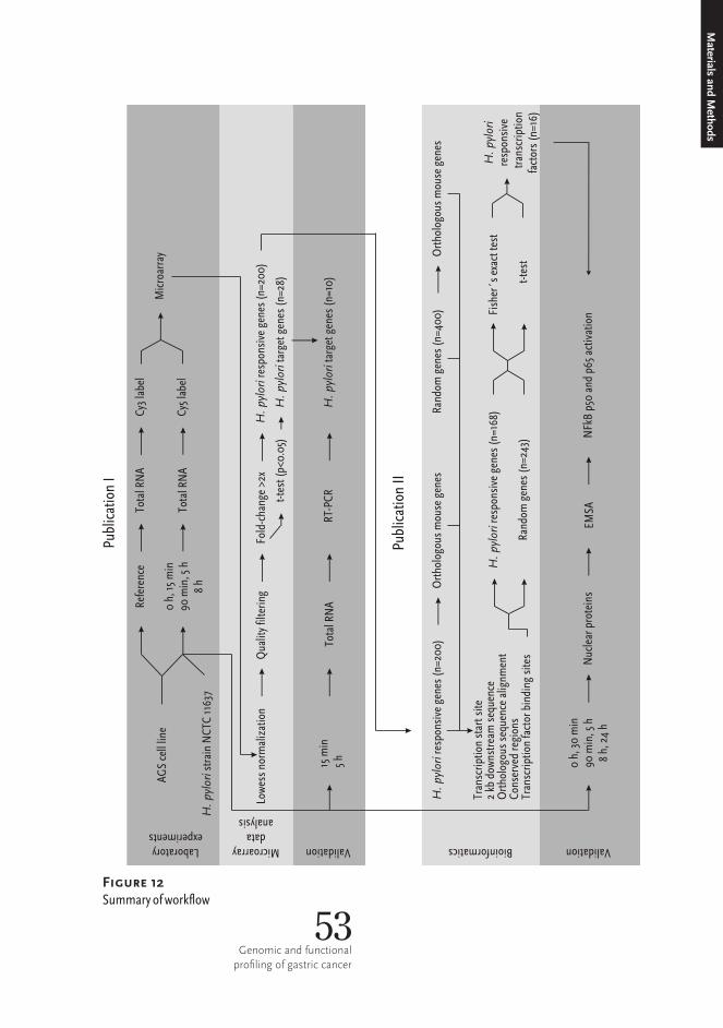
AGS c
ell lin
e
H. p
ylori s
train
NCTC
1163
7
0 h, 1
5 min
90 m
in, 5
h8 h
Refer
ence
Total
RNA
Total
RNA
Cy3 l
abel
Cy5 l
abel
Micro
array
Lowe
ss no
rmali
zatio
nQu
ality
filteri
ngt-t
est (
p<0.0
5)Fo
ld-ch
ange
>2x
H. py
lori ta
rget g
enes
(n=2
8)
H. py
lori re
spon
sive g
enes
(n=2
00)
Laboratoryexperiments
Microarraydata
analysis
Validation
RT-PC
RTo
tal RN
A15
min
5 hH
. pylo
ri targ
et ge
nes (
n=10
)
Lowe
ss no
rmali
zatio
nQu
ality
filteri
ngt-t
est (
p<0.0
5)Fo
ld-ch
ange
>2x
H. p
ylori t
arget
gene
s (n=
28)
H. p
ylori r
espo
nsive
gene
s (n=
200)
Microarraydata
analysis
H. p
ylori r
espo
nsive
gene
s (n=
200)
Ortho
logou
s mou
se ge
nes
Rand
om ge
nes (
n=40
0)Or
tholog
ous m
ouse
gene
s
Publi
catio
n I
Publi
catio
n II
Trans
cripti
on st
art si
te2 k
b dow
nstre
am se
quen
ceOr
tholog
ous s
eque
nce a
lignm
ent
Cons
erved
regio
nsTra
nscri
ption
facto
r bind
ing si
tesRa
ndom
gene
s (n=
243)
H. p
ylori r
espo
nsive
gene
s (n=
168)
Bioinformatics
t-tes
t
Fishe
r´s ex
act te
stH
. pylo
rires
pons
ive tr
anscr
iption
factor
s (n=
16)
EMSA
0 h, 3
0 min
90 m
in, 5
h8 h
, 24 h
Nucle
ar pro
teins
NFkB
p50 a
nd p6
5 acti
vatio
n
Validation
53Genomic and functional
profiling of gastric cancer
Materials and M
ethods
Figure 12 Summaryofworkflow
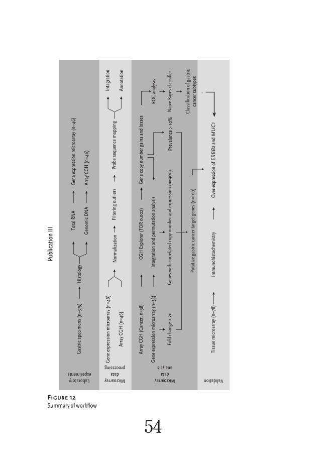
Publi
catio
n III
Microarraydata
processing
Laboratoryexperiments
Gastr
ic sp
ecim
ens (
n=37
5)Hi
stolog
yTo
tal RN
A
Geno
mic D
NA
Gene
expre
ssion
micr
oarra
y (n=
46)
Array
CGH
(n=4
6)
Gene
expre
ssion
micr
oarra
y (n=
46)
Array
CGH
(n=4
6)No
rmali
zatio
nFil
tering
outlie
rsPr
obe s
eque
nce m
appin
g
Microarraydata
analysis
Array
CGH
(Can
cer, n
=38)
CGH
Explo
rer (F
DR 0.
002)
Gene
copy
numb
er ga
ins an
d los
ses
Gene
expre
ssion
micr
oarra
y (n=
38)
Integ
ration
and p
ermuta
tion a
nalys
is
Gene
s with
corre
lated
copy
numb
er an
d exp
ressio
n (n=
900)
Fold
chan
ge >
2xPr
evale
nce >
10%
Putat
ive ga
stric
canc
er tar
get g
enes
(n=1
00)
Validation
Tissu
e micr
oarra
y (n=
78)
Immu
nohis
toche
mistr
yOv
er-exp
ressio
n of E
RBB2
and M
UC1
ROC a
nalys
is
Naive
Bayes
clas
sifier
Classi
ficati
on of
gastr
icca
ncer
subty
pes
Intag
ration
Anno
tation
54
Figure 12 Summaryofworkflow

Datab
ase o
f amp
lifica
tions
in ca
ncer
(450
0 cas
es)
Publi
catio
n IV
Publi
catio
n V
WHO
classi
ficati
onAm
plific
ation
frequ
ency
profili
ng(73
neop
lasms
)Hi
erarch
ical
cluste
ring
Indep
ende
nt co
mpon
ent
analy
sisAm
plific
ation
hot s
pots
Fragil
e site
s, vir
us in
tegrat
ion si
tesca
ncer
gene
s, ge
ne le
nght
Geno
mic d
istrib
ution
oflab
ile ch
romos
omal
featur
es
Intern
ation
al sys
tem of
chrom
osom
al an
notat
ion
NCBI
datab
ases
Statis
tical
hypo
thesis
testin
g
WHO
classi
ficati
on
Datab
ase o
f amp
lifica
tions
in ca
ncer
(450
0 cas
es)
Canc
er typ
es (n
=82)
Etiolo
gical
factor
s (29
attri
butes
)
Deve
lopme
ntal o
rigin
Mixtu
re mo
delin
gAm
plific
ation
mode
ls (n
=111)
Canc
erclu
sters
Statis
tical
hypo
thesis
testin
g
Ampli
ficati
on-ba
sed
classi
ficati
onof
canc
ers
Intern
ation
al sys
tem of
chrom
osom
al an
notat
ion
NCBI
datab
ases
Fragil
e site
s, da
rk/lig
ht ch
romos
ome
band
s, tel
omere
s, ce
ntrom
eres,
varia
ble re
gions
Maxim
al fre
quen
tite
msets
Statis
tical
hypo
thesis
testin
g
Geno
mic d
istrib
ution
oflab
ile ch
romos
omal
featur
es
Ampli
ficati
on-ba
sed
asso
ciatio
nsbe
twee
n neo
plasm
s
Comp
ilatio
n
55Genomic and functional
profiling of gastric cancer
Materials and M
ethods
Figure 12 Summaryofworkflow


Gene expression changes and regulatory networks in H. pylori infected cellsThe pathogenesis of the H.� pylori partly comes across via the host responses, and gene expression changes in the host cells are likely to contribute to the promotion of the malignant phenotype. In�vitro model and DNA microarrays were used to screen the target genes of H.�pylori in the host. H.�pylori infection induced differential expression of several target genes. According to the time series experiments and microarray analysis, FOS was the primary target in the infection and was up-regulated after 30 min of co-cul-ture (Table 1 in I). Furthermore, FOSL1 and ATF3 were rapidly up-regulated in H.�pylo-ri stimulated AGS cells (Table 1 in I). Many genes encoding heat shock (e.g., DNAJB1 and HSPA1A) and DNA damage response proteins (e.g., GADD45A) were up-regulat-ed by H.�pylori infection (I). Heat shock pro-teins are expressed when cells are exposed to stress. The transcription of GADD45A has been shown to increase when cells are
exposed to DNA damaging agents in environmental stress, and to go to growth arrest (Jackman et al., 1994) and ATF3 protein level increases rapidly as a response to diverse stress conditions (Jiang et al., 2004). ATF3 expression has been shown to be associated with CagA positivity (Zhang et al., 2007). The up-regulation of ATF3 and GADD45A has been validated using a H.�pylori infection mod-el on the human gastric epithelial immortalized GES-1 cells (Liu et al., 2006). H.�pylori regulated gene network functions in mecha-nisms that are activated in response to cellular damage and stress.
Gene expression changes induced by H.�pylori infection are reg-ulated by the action of specific transcription factors. The modula-tion of the transcription factor activity is the main mechanism by which H.�pylori is able to hijack cellular functions and abuse the host. A gene expression profile data and a bioinformatics approach
Results and Discussion
57Genomic and functional
profiling of gastric cancer
were used to decipher the transcription fac-tors that are activated after H.�pylori stimu-lation. Computational analysis revealed 16 transcription factors that were significant-ly (p<0.01) enriched in H.�pylori regulated genes’ promoter regions (Table 1 in II). EMSA assay demonstrated that H.�pylori stimula-tion of AGS cells resulted in an increased activation of NF-κB transcription factor and p50 and p65 subunits in the 3 h time point (Figure 1 in II). Previously, the activation of FOS (Meyer-ter-Vehn et al., 2000), NF-κB (p50 and p65 subunits) (Maeda et al., 2000), USF and CREB (Juttner et al., 2003) transcription factors have been shown to associate with H.�pylori infection. More-over, transcription factors that have not been linked with H.�pylori infection were identi-fied (ARNT, ELF1, EPAS1, E2F, MAX, MYC, MYC-MAX, SNAI1, TBP, and TCF3). H.�py-lori activated transcription factors function in cellular processes that are associated with carcinogenesis. NF-κB and FOS function in the production of the pro-inflammatory cyto-kines and in the activation of primary epithe-lial innate immunity response to pathogens (Eferl and Wagner, 2003; Karin et al., 2002) as well as the regulation of the anti-apoptotic and proliferative signaling (Karin et al., 2002). MYC is involved in cell-cycle progression, it promotes cell proliferation and is also associated with cancer initia-tion (Pelengaris et al., 2002). ARNT and CREB regulate cell response to hypoxia and promote angiogenesis (Harris, 2002). E2F controls cell-cycle and is associated with cell growth (Nevins, 2001). SNAI1 promotes tu-morigenesis by repressing E-cadherin, in-creasing cell motility and by promoting in-vasive growth (Thiery, 2002). USF1/2 tran-scription factors control telomerase reverse transcriptase (TERT) activity, which is re-quired for immortalization and continu-ous replication (Goueli and Janknecht, 2003). The transcription factors of H.�pylori
response are involved in cancer-related cel-lular mechanisms.
In eukaryotic cells, transcription activi-ty is controlled by the signal transduction pathways that consist of proteins that are sequentially activated so as to transfer sig-nals from outside the cell into the nucle-us, where response is executed at the level of gene expression. Cells have many recep-tors that they use to monitor their surround-ings. Some of these receptors are used to detect pathogens. For example, toll-like receptor 4 binds to lipopolysaccharide of H.�pylori (Kawahara et al., 2001). In addi-tion to the surveillance proteins of the host, pathogens have evolved proteins that mimic cellular signaling proteins and evoke phony cell responses. For instance, H.�pylori secret-ed cagA toxin interacts with the host cell’s signaling proteins (Atherton et al., 2001; Hatakeyama, 2004). Even when cross-talk between different signaling pathways might exist, both, pertinent physiological respons-es to bacteria as well as H.�pylori secreted disturbance signals are likely to contribute to specific pathway activation in H.�pylori infected cells. Due to the fact that H.�pylori strains that carry bacterial toxins are more carcinogenic, there might be specific path-way targets for the toxins that are related to the progression of the malignancy. H.�pylo-ri activated transcription factors were used to chart the signal transduction pathways that were associated with the infection (II). H.�pylori stimulation regulated transcription factors were shown to bind to specific tar-get sequences and function as downstream targets of Ahr signal transduction pathway, MAPK signaling pathway, NF-κB signaling pathway, RB pathway, and Wnt signaling pathway (Table 1 in II). Additionally, using the information from Biocarta, a model explaining the gene regulatory network of H.� pylori infection in a host cell could be drafted (Figure 2 in II). The activation of MAPK and NF-κB signal transduction pathways have been shown to occur after 58

H.�pylori infection (Juttner et al., 2003; Kawahara et al., 2001; Maeda et al., 2000; Meyer-ter-Vehn et al., 2000; Mitsuno et al., 2001; Nakayama et al., 2004; Su et al., 2003). NF-κB signaling is most likely acti-vated through lipopolysaccharide sensing by Toll-like receptor 4 (Kawahara et al., 2001), whereas cagA and VacA toxins are taught to activate MAPK signaling (Atherton et al., 2001; Hatakeyama, 2004). Apart from the known H.�pylori regulated pathways, novel signal transduction pathways, namely Ahr, WNT and E2F, were identified. H.� pylori pathogenecity probably acts through specific signaling pathways and the identification of cellular responses with malignant potential would serve as biomarkers for predicting the outcome of the infection.
H.�pylori induced reactive changes in the host cell’s gene expression are not explicitly cancer-related; rather, these changes affect cellular mechanisms that have the poten-tial to lead to perturbations that cause can-cer. H.�pylori stimulation activated NF-κB transcription factor, which is a key regula-tor of inflammation and cancer promoting cellular mechanisms. Among others, H.�pylori target genes and signal transduc-tion pathways regulate cell-proliferation, apoptosis and cell motility. Increased cell turnover can lead to increased mutation rate and carcinogenesis. Similarly, failures in executing apoptosis in control may have cancer-promoting implications. Cell prolife-ration involves DNA replication, which has a low but significant error rate, and H.�pylori infection, which favors cell proliferation, is indirectly mutagenic. In the view of the fact that H.�pylori infection regulated pathways that are constitutively active in gastritis and operate critical cellular functions, the genes encoding for proteins involved in these activities are likely targets for mutations. It could be further postulated that those path-ways that are hijacked in H.� pylori infec-
tion and the genes that they control might be preferential targets for genomic changes in gastric cancer initiation. H.� pylori infection regulated genes in AGS cells were enriched in the chromosomal regions that are frequently implicated in gastric cancer (Table 2 in I). The transcriptional activation of H.� pylori infection target genes involves introducing changes in the chromatin struc-ture. Histone coiling needs to be disassembled in order to get the transcription protein complex to access DNA. In open forma-tion, the physical protection of chromatin reduces and DNA gets exposed to muta-genic agents. DNA in the open chroma-tin formation is more likely to be damaged than DNA in the inactive regions of the genome. All in all, H.�pylori infection leads to activation of the cellular mechanisms that increase the probability of DNA damage, mutations and cancer promotion.
Putative gastric cancer target genesBiomarkers are measurable properties that can be used to indicate a biological char-acteristic of the sample object. Genome-wide microarray technologies are excel-lent tools for screening the genome for anomalies that could indicate a malignancy state or a certain biological process that is involved in cancer. Microarrays were exploited to identify the putative biomarkers and the biologically relevant target genes in gastric cancer. Putative gastric cancer biomarker genes were identified by integrating the gene expression and the copy number microarray data (III). The rationale behind integrating the gene expression with the copy number is that those genes that are dif-ferentially expressed due to a change in the copy number are more likely to be involved in cancer promoting processes. The integra-tion of gene expression and copy number measurements revealed genes that were up-regulated in association with copy number gain or down-regulated in association with copy number loss (Figure 2 and Table 1 in III).
Materials and M
ethods
59Genomic and functional
profiling of gastric cancer

The 17q12-q21 region contained many genes that were up-regulated in association with amplification (Figure 2 in III). Amplifica-tion and expression of ERBB2 as well as of other genes in the 17q12-q21 region have previously been identified (Maqani et al., 2006; Varis et al., 2004). In addition, several novel gastric cancer candidate genes were identified. These novel putative gas-tric cancer biomarker genes provide a pri-oritized selection of candidate genes for fur-ther studies.
Gastric cancer is divided into two main histological subtypes, namely intestinal and diffuse, which have different clinical and biological features (Lauren, 1965). The information of the histology of the tumors was used in fusion with gene copy num-ber profiling data in order to identify sub-type specific changes. Intestinal type gas-tric cancer was characterized by amplifica-tion of ERBB2 and gains at 20q13 and Xp (III). Previously, 17q12-q21 amplifications have been linked to intestinal type gastric cancer (Kokkola et al., 1997; Maqani et al., 2006; Vauhkonen et al., 2006). More specifically, the ERBB2 gene was shown to be activated, in RNA (Figure 2 in III) and protein levels (Figure 3 in III), by an am-plification in the intestinal type gastric can-cer (III). ERBB2 is a transmembrane recep-tor tyrosine kinase, which functions in cel-lular signal transduction and regulates pro-cesses that are frequently perturbed in can-cer. There is a new category of cancer ther-apies which target protein kinases. ERBB2 amplification and overexpression have been proposed as a therapeutic biomarker in gas-tric cancer and Trastuzumab (ERBB2 tyro-sine kinase domain binding monoclonal antibody) treatment was shown to inhibit tu-mor growth in gastric cancer cell lines and in a patient (Gong et al., 2004; Rebisc-hung et al., 2005). According to amplifica-tion modeling analysis, the 17q12-q21 region,
which contains the ERBB2 gene, is ampli-fied in gastric and esophageal adenocarci-nomas (Figure 5A in V). 17q12-q21 amplifica-tion was also typical for cancers of epithelial origin, more specifically adenocarcinomas. These data suggest that the 17q12-21 amplifi-cations are specific for epithelial type cancer of the gastroesophageal tract and amplifica-tion and over-expression of ERBB2 could be used as a biomarker for intestinal type gas-tric cancer in the proximal stomach.
There were interconnections between H.�pylori infection regulated genes and gas-tric cancer genome. MUC1 was shown to be amplified and over-expressed in intes-tinal type gastric cancer (Figures 2 and 3 in III). ERBB2 has been shown to phos-phorylate MUC1 (Li et al., 2001; Schroe-der et al., 2001) and regulate its interac-tion with β-catenin (Yamamoto et al., 1997). In conjunction, H.� pylori binds to MUC1 on the surface of gastric epithelium (Vinall et al., 2002) and activates β-catenin in gastric epithelial cells (Franco et al., 2005). β-catenin has been shown to enhance invasiveness and promote prolifer-ation in gastric cancer cell lines (Lowy et al., 2006). Based on these observations, there are multiple mechanisms – gene copy num-ber gain, ERBB2 kinase phosphorylation and H.�pylori binding – that are capable of ac-tivating MUC1. Furthermore, MUC1 might be involved in gastric carcinogenesis by pro-moting proliferation and invasion through interaction with β-catenin. PPP1R1B was one of the target genes at 17q21.1 that was up-regulated by a copy number gain (Table 1 and Figure 2 in III). PPP1R1B transcription was also up-regulated by H.�pylori infection (Supplementary data in I). Because H.�py-lori infection and gene copy number gain are both potential activators of PPP1R1B that further strengthens its central role in gas-tric carcinogenesis. Depending on the phos-phorylation pattern of specific amino acid residues, PPP1R1B functions as a kinase or phosphatase inhibitor (Bibb et al., 1999; 60
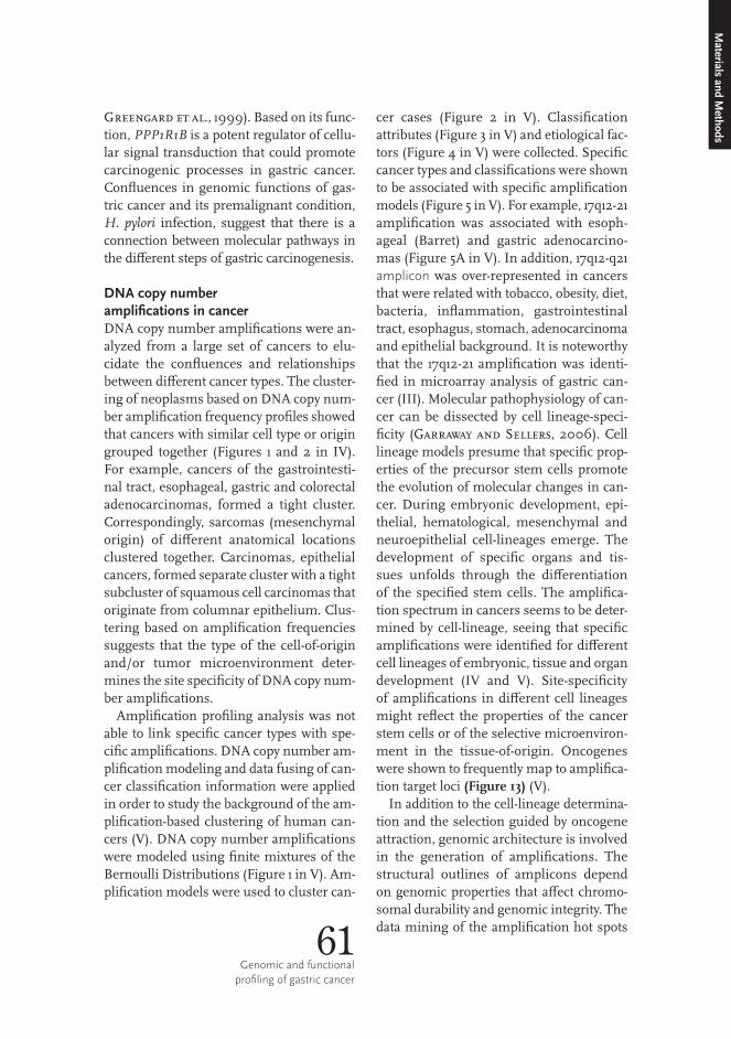
Greengard et al., 1999). Based on its func-tion, PPP1R1B is a potent regulator of cellu-lar signal transduction that could promote carcinogenic processes in gastric cancer. Confluences in genomic functions of gas-tric cancer and its premalignant condition, H.� pylori infection, suggest that there is a connection between molecular pathways in the different steps of gastric carcinogenesis.
DNA copy number amplifications in cancerDNA copy number amplifications were an-alyzed from a large set of cancers to elu-cidate the confluences and relationships between different cancer types. The cluster-ing of neoplasms based on DNA copy num-ber amplification frequency profiles showed that cancers with similar cell type or origin grouped together (Figures 1 and 2 in IV). For example, cancers of the gastrointesti-nal tract, esophageal, gastric and colorectal adenocarcinomas, formed a tight cluster. Correspondingly, sarcomas (mesenchymal origin) of different anatomical locations clustered together. Carcinomas, epithelial cancers, formed separate cluster with a tight subcluster of squamous cell carcinomas that originate from columnar epithelium. Clus-tering based on amplification frequencies suggests that the type of the cell-of-origin and/or tumor microenvironment deter-mines the site specificity of DNA copy num-ber amplifications.
Amplification profiling analysis was not able to link specific cancer types with spe-cific amplifications. DNA copy number am-plification modeling and data fusing of can-cer classification information were applied in order to study the background of the am-plification-based clustering of human can-cers (V). DNA copy number amplifications were modeled using finite mixtures of the Bernoulli Distributions (Figure 1 in V). Am-plification models were used to cluster can-
cer cases (Figure 2 in V). Classification attributes (Figure 3 in V) and etiological fac-tors (Figure 4 in V) were collected. Specific cancer types and classifications were shown to be associated with specific amplification models (Figure 5 in V). For example, 17q12-21 amplification was associated with esoph-ageal (Barret) and gastric adenocarcino-mas (Figure 5A in V). In addition, 17q12-q21 amplicon was over-represented in cancers that were related with tobacco, obesity, diet, bacteria, inflammation, gastrointestinal tract, esophagus, stomach, adenocarcinoma and epithelial background. It is noteworthy that the 17q12-21 amplification was identi-fied in microarray analysis of gastric can-cer (III). Molecular pathophysiology of can-cer can be dissected by cell lineage-speci-ficity (Garraway and Sellers, 2006). Cell lineage models presume that specific prop-erties of the precursor stem cells promote the evolution of molecular changes in can-cer. During embryonic development, epi-thelial, hematological, mesenchymal and neuroepithelial cell-lineages emerge. The development of specific organs and tis-sues unfolds through the differentiation of the specified stem cells. The amplifica-tion spectrum in cancers seems to be deter-mined by cell-lineage, seeing that specific amplifications were identified for different cell lineages of embryonic, tissue and organ development (IV and V). Site-specificity of amplifications in different cell lineages might reflect the properties of the cancer stem cells or of the selective microenviron-ment in the tissue-of-origin. Oncogenes were shown to frequently map to amplifica-tion target loci (Figure 13) (V).
In addition to the cell-lineage determina-tion and the selection guided by oncogene attraction, genomic architecture is involved in the generation of amplifications. The structural outlines of amplicons depend on genomic properties that affect chromo-somal durability and genomic integrity. The data mining of the amplification hot spots 61
Genomic and functional profiling of gastric cancer
Materials and M
ethods
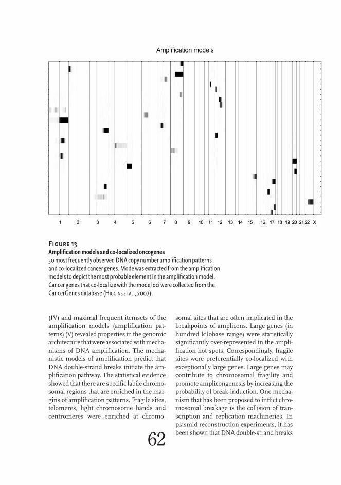
(IV) and maximal frequent itemsets of the amplification models (amplification pat-terns) (V) revealed properties in the genomic architecture that were associated with mecha- nisms of DNA amplification. The mecha-nistic models of amplification predict that DNA double-strand breaks initiate the am-plification pathway. The statistical evidence showed that there are specific labile chromo- somal regions that are enriched in the mar-gins of amplification patterns. Fragile sites, telomeres, light chromosome bands and centromeres were enriched at chromo-
somal sites that are often implicated in the breakpoints of amplicons. Large genes (in hundred kilobase range) were statistically significantly over-represented in the ampli-fication hot spots. Correspondingly, fragile sites were preferentially co-localized with exceptionally large genes. Large genes may contribute to chromosomal fragility and promote amplicongenesis by increasing the probability of break-induction. One mecha-nism that has been proposed to inflict chro-mosomal breakage is the collision of tran-scription and replication machineries. In plasmid reconstruction experiments, it has been shown that DNA double-strand breaks 62
Figure 13 Amplificationmodelsandco-localizedoncogenes30mostfrequentlyobservedDNAcopynumberamplificationpatternsandco-localizedcancergenes.Modewasextractedfromtheamplificationmodelstodepictthemostprobableelementintheamplificationmodel.Cancergenesthatco-localizewiththemodelociwerecollectedfromtheCancerGenesdatabase(higgins et al., 2007).

63Genomic and functional
profiling of gastric cancer
Materials and M
ethods
occurred and amplification process was ini-tiated by synchronized and site-directed transcription and replication (Shimizu et al., 2003). The transcription and replication of large genes considerably takes more time, which widens the window for an occasional collision between the two processes. Light chromosome bands contained multiple amplicon boundaries. Light chromosome bands are gene rich and usually in open conformation (Gilbert et al., 2004). The enrichment of amplification boundaries on light chromosome bands suggests that open
chromatin would be more vulnerable and DNA damage prone than inactive closed chromatin. Telomeres were also among the chromosomal regions that were over- represented by amplification breakpoints. Telomere erosion in uncontrollably repli-cating cancer cells is a mechanism that can produce DNA double-strand breaks in the telomere regions. Non-random distribution of labile genomic features indicated that the human genome is not evenly resilient, and that specific sites are more prone to DNA double-strand breaks. ❦

H. pylori activated the genes, transcription factors and sig-nal transduction pathways that were identified in studies I and II. The molecular targets of H.�pylori infection were associated with the cellular functions that might be capable of promoting carcino- genesis of the stomach. These targets could be useful when predicting the outcome of the infection and the designing novel treat-ments. Intestinal and diffuse type gastric cancers showed distinct molecular genetic profiles and the integration of gene expression and copy number microarray data allowed the identification of genes that might be involved in gastric carcinogenesis and have clinical relevance (III). Even when some subtypes with concordant genomic changes were identified in gastric cancer, it is likely that most of the carcinomas of the stomach are unique diseases that do not share a specific genomic change. Gene amplifications were shown to be
non-random genomic instabilities, and cell lineage, properties of precursor stem cells, tissue microenviron-ment and genomic map localization of specific oncogenes to define the site specificity of DNA amplifications, whereas labile genomic features defined the structures of the amplicons (IV and V). These conclusions suggest that the definition of genomic changes in can-cer is based on the interplay between cancer cell and the tumor microenvironment. Categorizing cancers according to their under- lying genetic properties, rather than looking at the appearance or behavior of the tumor, is likely to improve cancer treatment. Morphologically similar tumors may arise in the same anatomical location and still have entirely different genetic anomalies. Conversely, distinct genomic changes can cause cancers in different parts of the body as well as of diverse morphologies. Regardless of the site and behavior of the tumor, genetically coherent cancer subtypes
Conclusions and future prospects
65Genomic and functional
profiling of gastric cancer
are likely to depend on the same molecular perturbations, and the understanding of the genetic underpinnings of cancers facilitates the development of the targeted treatments.
In the future, the identified putative bio-markers for gastric cancer and H.� pylori infection related carcinogenic processes need to be validated in a lager set of samples using alternative approaches and their roles in gastric cancer should be explored using functional experiments. Moreover, there might be a concordance among gastric cancers in higher hierarchy of genomic functionality, namely in the level of biologi-cal processes and pathways, which could be revealed using a systems biology approach. In order to treat heterogeneous disease, such as gastric cancer, genomic properties of indivi-dual tumors need to be decoded. Novel, high-throughput, massively parallel sequencing technologies enable rapid and near genome- wide examination of the tumor DNA sequence, which is fundamental for the development of personalized treatments for gastric cancer. To fully understand the mechanisms and backgrounds of genomic changes, a large-scale datamining effort using DNA microarray or sequencing data should be executed. ❦
66

Conclusions and future prospects

This work was carried out at the Department of Pathology in Haartman Institute and HUSLAB, University of Helsinki and Helsinki University Central Hospital during the years 2003 – 2008. I am grateful for the financial support received from the Academy of Finland (SYSBIO Research Program), the State Appropriations of Helsinki University Central Hospital, the Helsinki University grants, the Sigrid Jusélius Foundation and the Finnish Cancer Organizations. I wish to express my gratitude to all those who have contributed to the accomplishment of this study, and I especially acknowledge:
Professors Veli-Pekka Lehto and Leif Andersson, present and for-mer Heads of the Department of Pathology, Haartman Institute, University of Helsinki, for their positive attitude towards my studies and for providing excellent research facilities. The former and present Heads of Department of Medical Genetics, Haartman Insti-tute, University of Helsinki and Department of Pathology, HUSLAB, Helsinki University Central Hospital, are also acknowledged.
Professor Sakari Knuutila, the supervisor of this thesis, is highly appreciated for his caring and positive attitude, encouragement, and not only for providing resources but also inspiration, and for sharing his intellect and passion for cancer research and cycling. His ideas, such as the one of riding around the Bodom Lake, or going to work over the frozen sea, reflect a freedom of thoughts and a willingness to take detours – state-of-mind – that I have learned to embrace when facing scientific challenges. I respect his ability to lead by setting an example, rather than by having expectations and being enthusiastic and not overly keen on following the traditional ways. A novice researcher could not have hoped for a better mentor than him. Above all, I value his friendship and rich personality.
Acknowledgements
69Genomic and functional
profiling of gastric cancer
Professor Pentti Sipponen and Docent Ari Ristimäki, my thesis committee mem-bers, provided great help by overseeing my research activities, reviewing my work as well as the manuscript of this thesis, and by providing much appreciated critique and constructive comments over the years. I thank them for their positive attitude, warm support and ample attention. I am especially grateful to Professor Sipponen for being an official reviewer of this thesis, for the enlightening lessons given on gastric can-cer and for providing wise comments that truly helped to improve my thesis. Research Professor Matej Orešič, another official reviewer of this thesis, is thanked for his all-round scientific expertise, which promoted an emergence of great revelations and new ideas when finishing this thesis. I was also fortunate to obtain his insight regarding the presentational clarity of the thesis.
I have had the privilege of having two tal-ented researchers as additional, unofficial supervisors. Docent Outi Monni is acknow-ledged for mentoring and teaching DNA microarray experiments (I and III). As the senior author, she was essential in the un-raveling of publication III. What is more, she has taught me in great lengths about scientific writing and conducting genomic- scale research, which I really appreciate. I would like to thank her for her encourage-ment and endorsement in the search for new research opportunities. Jaakko Hollmén is acknowledged for bringing top-of-the-line statistical knowledge for publications III, IV and V. Moreover, he was the senior author in publication V and did an excellent job in navigating that work. I am thankful for his outstanding teaching about proba- bilistic methods and statistical analysis, reviewing various research papers – especially this thesis – and for always giv-ing such positive, constructive and almost Rainmanishly detailed feedback. His quick-
witted humor and observant nature have made our collaboration an incredibly pleas-ant experience.
I am grateful to my co-authors for their commitment, scientific and intellectual inputs and for providing a comfortable and supportive work atmosphere. I would like to sincerely acknowledge their contributions to the completion of this thesis work: Bálint Nagy for carrying out real-time PCR experi-ments (I) and for collecting data (IV); Pro-fessor Hilpi Rautelin for her guidance and training in H.�pylori�culture experiments (I) and for her encouragement when I had my first international presentations; Ville Veck-man for tutoring and assisting the EMSA experiments (II), the traditional publication lunches and friendship; Juha Saharinen for his collaboration in bioinformatics studies (II), as well as for the analytical discussions and opinions regarding both, professional and everyday matters; Siina Junnila for her excellent collaboration in gastric cancer study (III), good spirits in the lab, amusing coffee breaks and friendship; Professor Pau-li Puolakkainen and Docent Arto Kokkola for providing gastric cancer samples, clini-cal data and clinicians’ point of view for the research (III); Reija Autio, for integration analysis (III); Ilari Scheinin for microarray probe matching (III), fruitful scientific off-thesis collaborations, iPhone help and con-tinuous bioinformatic support; Tuula Kivi-luoto for providing gastric cancer specimens (III); Docent Marja-Liisa Karjalainen-Linds-berg for histopathological evaluation of the gastric preparations (III) and for teaching gastric histology; Johan Himberg for bio- informatic analyses (IV); Docent Tom Böh-ling for reviewing cancer classifications (IV and V) and for educating me regarding can-cer pathology; and Jarkko Tikka for executing computational analyses (V).
There are a number of people without whom this thesis would not have been pos-sible. Pirjo Kosonen is thanked for her prac-tical assistance in H.� pylori culturing and 70
Ulla Kiiski for the preparation of the gastric cancer slides. The personnel of the Mole-cular Pathology Laboratory, especially Tiina Wirtanen, Leena Rossi, Paula Kvick, Tarja Salonen, Mikael Lindlöf and Kirsi Autio, are thanked for their aid in the lab and good company at work, as well as in cycling and skiing trips, Christmas parties and other rec-reational events. I am in gratitude to Pirjo Pennanen for the language revision of indivi- dual manuscripts and linguistic assistance when drafting numerous reports, grant appli- cations and various obscure writings. Laura Villella-Tolonen deserves my gratitude for the prompt language revision when finalizing the thesis and Tomi Metsä-Heikkilä for designing the layout of the thesis.
My colleagues, especially former and present CMG-group members, Ioana Borze, Mohamed Guled, Kowan Jee, Sippy Kaur, Eeva Kettunen, Leo Lahti, Pamela Lind-holm, Tuija Lundan, Tarja Nieminen, Tarja Niini, Shinsuke Ninomiya, Penny Nymark, Fabrizio Passador-Santos, Salla Ruosaari, Suvi Savola, Linda Siggberg, Anu Usvasalo, Hanna Vauhkonen, and Harriet Wikman are thanked for their exciting scientific discus-sions, aid in the lab, creating an inspiring working atmosphere and pleasant company in different work trips. My gratitude goes to the people in the SYSBIO research consor-tium, especially to Professor Samuel Kaski for heading the program. I am grateful for Jukka Vakkila for inspiring lessons on cancer immunology and inflammation- related diseases. Special thanks go to Perttu Salmenperä for his good company and for engaging in interesting scientific conver-sations at lunch and coffee breaks as well as in Snowbird. Massimiliano Gentile is thanked for all those hours of patiently and straightforwardly teaching me how to use GeneSpring in microarray data analysis. In addition to that, our scientific side proj-ects have been interesting, educational and
rewarding. Most importantly, enjoyable lunches at the Mountain restaurant and those at-least-twice-a-week doses of Nepalese food have been important ingredients for this thesis. His thorough and wide know-ledge about mountain bike and camera gear truly amazes me, and I’m glad that he has introduced me to the mountain biking grounds in the Keskuspuisto. I wish to thank him for the skiing photos in Snow-bird and especially the one that covers this thesis. Thanks are due to “the Microbes”: Anu Jääskeläinen, Anna Katz, Tiina Par-tanen, Hanna Sinkko and Ville Veckman for good times during studying and ever since. I greatly appreciate your friendship and all the peculiar traditions we have managed to maintain.
I wish to thank all my friends, particularly Tommi Sneck, Ville and Kristiina Laakso, Tuomas and Monika Kivimäki, Lotta Haik-kola and Jussi Kulonpalo, Niina Sihto and Panu Laaksonen, Pauliina Arvela and Mark Fletcher, Mari Kiviharju and Kari Rantavuori, Sari Lankinen, Laura Villella-Tolonen and Mikko Tolonen, Risto Sarvas and Tuula Savola, Mari Erma, Tania Ruutu and Anna Elmgren for showing an interest in my scientific undertakings as well as good times during the years. Special words of thanks go to the band members, Timo Erma, Olli Ruutu and Jaakko Vehanen, for their almost life-long friendship and continuous sup-port. Even when domestic beer has been re-placed by fancy exports and sessions often begin with salmon canapé servings, I still think that we occasionally truly ROCK! It is a privilege to have been able to play and share that passion for music (and whatever comes along with it) with them.
Sincere thanks to my wife’s family, Timo and Riikka Elvo, Annikki Elvo, Jenni and Luke Bland, Sanna, Petri, Patrik, Anni and Emma Viro, and Heidi, Sami, Topias, Ella and Iris Saurén, for caring and having faith in me.
71Genomic and functional
profiling of gastric cancer
Acknowledgem
ents

I am grateful for my parents, Hannele and Antti Myllykangas, for their unconditional love, support and encouragement. I think my mother is the one that set the spark for my interest in writing and pursuing a career in a field that enables me to express myself with words. I believe it was because of my father’s influence, endless curiosity and desire of knowledge, that I have developed a fascination for research. I thank my parents, for their intuition and guidance in many occa- sions when making decisions seemed diffi-cult. I wish to thank my brothers for their love and support. I respect Jani’s no-nonsense attitude and I look up to Miika for his cour-age and upright search for his own way in life. In many ways, their examples have been comforting to follow.
Foremost thanks go to my wife Nina, for her love and for being such a strong com-panion, since it has been her who had to put up with my non-productive loitering on the living room sofa. To me she is a tower of strength. Without her endless effort in running our family, my accomplishments would not be worth anything. I think that she is by far the most efficient person that I know and it is altogether her merit that I generally get anything done. I thank my children, Viljami and Saara, for their love. From them I get the most rewarding feed-back, as well as the most intense criticism, and I truly cherish them for that. ❦
72
Helsinki, August 29th, 2008

Acknowledgem
ents

Achlorhydric: A state when production of
gastric acid in the stomach is low.
Adenocarcinoma: A type of cancer that originates
from glandular epithelial tissue.
Amplicon: A genomic region
containing multiple DNA copies.
Anaerobic organism: An organism that does
not need oxygen to grow.
Anaphase: A late stage in cell division where
sister chromatin moves towards oppo-site poles of the dividing nucleus.
Antioxidant: A molecule that is capable of scaveng-
ing electrons and preventing oxidation of other molecules. Antioxidants protect cells from free radicals that are generated in oxidation reactions. Free radicals can damage cells.
Concepts Apoptosis: A programmed cell death of
a complex system of biological events leading to disposal of the cell.
Bernoulli distribution: A discrete probability distribution
with two outcomes. Given outcome 1 has a probability of p, outcome 0 has a probability of p’ = 1-p.
Biomarker: An indicator of a specific
biological state.
Carcinogenesis: A process where a normal cell
is transformed into a cancer cell.
Centromere: A region of specific and repetitive
DNA in the chromosomes that function in chromosomal segregation during cell division.
Chromatin: A composite of DNA and proteins
forming the chromosome.
Confocal: An optic system that includes one
lens for both focusing of the emission laser to the object and fluorescence to the image detector.
75Genomic and functional
profiling of gastric cancer
Cross-validation: A procedure where data is divided
into subsets, training and validation sets, that are used to executing the initial analysis and validating and conforming the result.
Data mining: “Analysis of large observational data
sets to find unsuspected relationships and to summarize the data in novel ways that are both understandable and useful to the data owner.” (Hand et al., 2001)
DNA hybridization: A process of combining single-
stranded complementary DNAs into one molecule.
DNA sequencing: A method of determining the order
of the nucleotides in a DNA molecule.
Electrophoretic mobility shift assay: A laboratory technique that is used
to study protein-DNA interactions. Proteins and radio-labeled DNA are mixed and radioactivity is detected using gel electrophoresis separation. Specific antibodies are used to identify proteins that bind to radio- labeled DNA.
Ensembl: A publicly available database
for genomic information that can be accessed at www.ensembl.org.
Epidemiology: A discipline of studying factors
that affect health and illnesses in populations.
Epigenetic: A change in the gene function without
changing the basic structure of the un-derlying DNA sequence. Epigenetic changes can remain after cell division.
Epithelial cell: In general, epithelial cells may
originate from ectodermal (e.g., the epidermis), endodermal (e.g., the intestinal tract) and mesodermal (e.g. wall of gut) germ layers of the developing embryo. Epithelium is a classification of the tissue forming glands and layers that line the cavities and structures of the body, for example skin, mouth, esophagus, gastroin- testinal tract as well as reproductive and urinary tracts. Cancers that origi-nate from epithelium are called carcinomas.
Expection-Maximization algorithm: An iterative maximum likelihood
estimation algorithm, to be used, for instance, in mixture models.
Finite mixture model: In statistics and machine learning, a
mixture model is a linear superposition of simple distributions (components), that together form a more complex distribution. Mixture models are used for example in clustering.
Forward selection algorithm: An algorithm that proceeds by
combining the currently best set of variables with the remaining single variables and chooses the best combi-nation as the new current best set.
Fusion gene: A combination of previously
separate genes.
Gamete: A haploid egg and sperm cell.76
Gastric atrophy: Partial or complete disappearance
of the gastric mucosa, glands and gastric pits.
Gastric ulcer: A gastric wound that derives
from the necrotic tissue removal.
Gastritis: An inflammation of the mucous
membrane layer of the stomach.
Genome: organism’s hereditary information
encoded in DNA.
Glioma: A brain tumor arising from glial cells.
Hybridization probe: A fragment of DNA used to detect
presence of complementary DNA or RNA in the sample.
Immunohistochemistry: A laboratory technique, which utilizes
antibodies to detect specific antigens in tissues.
Intestinal metaplasia: A premalignant condition where gastric
epithelium has been replaced with cells of intestinal morphology.
Kinase: An enzyme that catalyzes a reaction
called phosphorylation, the transfer of a phosphate group from a donor molecule, e.g., ATP, to a target (sub-strate). Protein kinases are modifying the function of target proteins by phosphorylation.
Lesion: A pathologic change in tissue
structure and function.
Leukocyte: A cell of the immune system.
white blood cells are specialized to neutralize microbe infections and foreign material. Leukocytes derive from hematopoietic stem cells in the bone marrow.
Machine learning: A computational discipline of
identifying patterns and extracting rules from large data sets using statistical methods and algorithms that are able to learn from the data.
Massively parallel sequencing: A method that allows high-throughput
DNA sequencing. The sequencing by extension platforms (Thomas et al., 2006) from 454 Life Sciences (Bradford, CT) and Illumina (Hayward, CA) can sequence over 100 megabase s per day.
Maximum likelihood: A principle that estimates parameters
to maximize the probability of the data.
Mesenchymal cell: A type of cell originating from the me-
sodermal germ layer of the developing embryo. Given a proper extracellular environment, the mesenchymal cells are able to migrate individually. This class of cells includes typically the cells in the connective tissue, bone, muscle and fat. Mesenchymal tumors arise from mesenchymal stem cells.
Metadata: Background data about the
observational measurement data.
Metaphase: A stage in mitosis, in the eukaryotic
cell cycle, in which chromosomes are condensed and aligned in the middle of the nucleous.77
Genomic and functional profiling of gastric cancer
Concepts

Metaplasia: Replacement of the original
differentiated cell type with another differentiated cell type.
Metastasis: Spreading of the malignant tumor cells
from a primary anatomical location to another site that is not adjacent to the original location.
Monoclonal antibody: Antibodies, also called immunoglobu-
lins, are proteins that are produced by specialized immune system cells, B-cells. Antibodies bind to specific mi-crobial structures, antigens, and initi-ate the immune response to neutral-ize the infection. Each cell produces a unique antibody and monoclonal an-tibodies refer to proteins produced by one type of B-cell that has been clonally multiplied. Monoclonal antibodies are used in cancer therapy to neutralize de-regulated oncoproteins.
Naïve Bayes classifier: A probabilistic classifier, which
assumes, given the class variable, that the variables in the model are independent.
Neuroblastoma: The most common solid tumor
in children arising from sympathetic nervous system.
Oncogenes: Class of genes that encode proteins
that promote transformation of cells to cancer cells when mutations increase or dysregulate their activity.
Orthologous: Corresponding elements
between two species.
Permutation analysis: A method of generating the empirical
probability distribution by randomly mixing data. The obtained permuted probability distribution can be then used to estimate the significance of the true observation.
Promoter: A DNA region of specific sequence that
is involved in controlling the expression of the adjacent gene by binding specific transcription factors.
P-value: Probability of obtaining a value
by random that is at least as extreme as the measured value.
P-value correction: when multiple hypotheses are
measured, some p-values may become significant by chance. There are differ-ent methods that can be used to correct the p-values obtained from a statistical test repeated many times.
Real-time polymerase chain reaction: A technique that can be used to detect
and quantify the amount of a specific DNA sequence in a sample. The proce-dure follows basic PCR but the accumu-lation of a PCR product is measured after each amplification cycle. when RNA is used as a template it has to be reverse transcribed into cDNA before quantitative PCR reaction.
Receiver operating characteristic: A curve depicting the trade-off between
the false positives and true positives for a diagnostic system for every cut-off point for decision.
Somatic cells: All other cells in the body
except the cells of the germ line.
78
Stromal cells: Cells that form the supportive
framework of connective tissue.
Systems biology: An approach to study the complex
interactions between components of the biological system.
Telomere: A region of repetitive DNA in the
ends of the chromosomes. Telomeres function, for example, in protecting chromosomes from breakage during replication and cell division.
Transcription factor: A protein that binds specific DNA
sequence and is part of the protein complex that controls transcription.
T-test: A statistical test that is used to compare
mean values in two groups of normally distributed random variables.
Tumor suppressor genes: A class of genes that encode proteins
that function to prevent cells from transformation into cancer cells.
Concepts
79Genomic and functional
profiling of gastric cancer

Albertson, D. G., Collins, C., McCormick, F., and Gray, J. w. (2003). Chromosome
aberrations in solid tumors. Nat Genet 34, 369 – 376.
Andrechek, E. R., Hardy, w. R., Siegel, P. M., Rudnicki, M. A., Cardiff, R. D., and
Muller, w. J. (2000). Amplification of the neu/erbB-2 oncogene in a mouse model of mammary tumorigenesis. Proc Natl Acad Sci U S A 97, 3444 – 3449.
Atherton, J. C. (1997). The clinical relevance of strain types of Helicobacter pylori. Gut 40,
701-703.Atherton, J. C., Timothy, L. C., Papini, E., and Telford, J. L. (2001). Vacuolating cyto-
toxin, In Helicobacter pylori: physiology and genetics, H. L. T. Mobley, G. L. Mendz, and S. L. Hazell, eds. (washington: ASM Press), pp. 97 – 110.
Ban, S., Cologne, J. B., and Neriishi, K. (1995). Effect of radiation and cigarette smok-
ing on expression of FUdR-inducible common fragile sites in human peripheral lymphocytes. Mutat Res 334, 197 – 203.
Belkhiri, A., Zaika, A., Pidkovka, N., Knuutila, S., Moskaluk, C., and El-Ri-
fai, w. (2005). Darpp-32: a novel antiapop-totic gene in upper gastrointestinal carcino-mas. Cancer Res 65, 6583 – 6592.
References Bibb, J. A., Snyder, G. L., Nishi, A., yan, Z., Meijer, L., Fienberg, A. A., Tsai, L. H.,
Kwon, y. T., Girault, J. A., Czernik, A. J., et al. (1999). Phosphorylation of DARPP-32 by Cdk5 modulates dopamine signalling in neurons. Nature 402, 669 – 671.
Birney, E., Andrews, T. D., Bevan, P., Caccamo, M., Chen, y., Clarke, L.,
Coates, G., Cuff, J., Curwen, V., Cutts, T., et al. (2004). An overview of Ensembl. Ge-nome Res 14, 925 – 928.
Bizari, L., Borim, A. A., Leite, K. R., Goncalves Fde, T., Cury, P. M., Tajara,
E. H., and Silva, A. E. (2006). Alterations of the CCND1 and HER-2/neu (ERBB2) pro-teins in esophageal and gastric cancers. Can-cer Genet Cytogenet 165, 41 – 50.
Blaser, M. J., Perez-Perez, G. I., Klean- thous, H., Cover, T. L., Peek, R. M., Chy-
ou, P. H., Stemmermann, G. N., and No-mura, A. (1995). Infection with Helico-bacter pylori strains possessing cagA is associ-ated with an increased risk of developing ade-nocarcinoma of the stomach. Cancer Res 55, 2111 – 2115.
Blount, B. C., Mack, M. M., wehr, C. M., MacGregor, J. T., Hiatt, R. A., wang, G.,
wickramasinghe, S. N., Everson, R. B., and Ames, B. N. (1997). Folate deficiency causes uracil misincorporation into human DNA and chromosome breakage: implica-tions for cancer and neuronal damage. Proc Natl Acad Sci U S A 94, 3290 – 3295.
Boren, J., Cascante, M., Marin, S., Comin-Anduix, B., Centelles, J. J., Lim,
S., Bassilian, S., Ahmed, S., Lee, w. N., and Boros, L. G. (2001). Gleevec (STI571) influences metabolic enzyme activities and glucose carbon flow toward nucleic acid and fatty acid synthesis in myeloid tumor cells. J Biol Chem 276, 37747 – 37753.81
Genomic and functional profiling of gastric cancer

Boros, L. G., Lee, w. N., and Cascante, M. (2002). Imatinib and chronic-phase leuke-
mias. N Engl J Med 347,67 – 68.Buyse, M., Loi, S., van’t Veer, L., Viale, G., Delorenzi, M., Glas, A. M., d’Assignies,
M. S., Bergh, J., Lidereau, R., Ellis, P., et al. (2006). Validation and clinical utility of a 70-gene prognostic signature for women with node-negative breast cancer. J Natl Cancer Inst 98, 1183 – 1192.
Chen, y., Dougherty, E. R., and Bittner, M. L. (1997). Ratio-based decisions and the
quantitative analysis of cDNA microarray im-ages. J Biomed opt 2, 364 – 374.
Chen, y., Kamat, V., Dougherty, E. R., Bittner, M. L., Meltzer, P. S., and Trent,
J. M. (2002). Ratio statistics of gene expres-sion levels and applications to microarray da-ta analysis. Bioinformatics 18, 1207 – 1215.
Cheng, X. X., wang, Z. C., Chen, X. y., Sun, y., Kong, Q. y., Liu, J., and Li, H. (2005).
Correlation of Wnt-2 expression and beta-catenin intracellular accumulation in Chinese gastric cancers: relevance with tumour dis-semination. Cancer Lett 223, 339 – 347.
Cho, y. G., Choi, B. J., Kim, C. J., Song, J. H., Zhang, C., Nam, S. w., Lee, J. y., and Park,
w. S. (2007a). Genetic analysis of the DBC2 gene in gastric cancer. Acta oncol, 1 – 6.
Cho, y. G., Song, J. H., Kim, C. J., Lee, y. S., Kim, S. y., Nam, S. w., Lee, J. y., and Park,
w. S. (2007b). Genetic alterations of the AT-BF1 gene in gastric cancer. Clin Cancer Res 13, 4355 – 4359.
Coquelle, A., Pipiras, E., Toledo, F., Buttin, G., and Debatisse, M. (1997). Ex-
pression of fragile sites triggers intrachromo-somal mammalian gene amplification and sets boundaries to early amplicons. Cell 89, 215 – 225.
Coquelle, A., Toledo, F., Stern, S., Bieth, A., and Debatisse, M. (1998). A new role
for hypoxia in tumor progression: induction of fragile site triggering genomic rearrangements and formation of complex DMs and HSRs. Mol Cell 2, 259 – 265.
Correa, P. (1992). Human gastric carcinogenesis: a multistep
and multifactorial process – First American Cancer Society Award Lecture on Cancer Ep-idemiology and Prevention. Cancer Res 52, 6735 – 6740.
Correa, P., and Houghton, J. (2007). Carcinogenesis of Helicobacter pylori. Gastro-
enterology 133, 659 – 672.Covacci, A., and Rappuoli, R. (2000). Tyrosine-phosphorylated bacterial proteins:
Trojan horses for the host cell. J Exp Med 191, 587 – 592.
Covacci, A., Telford, J. L., Del Giudice, G., Parsonnet, J., and Rappuoli, R.
(1999). Helicobacter pylori virulence and ge-netic geography. Science 284, 1328 – 1333.
Craanen, M. E., Blok, P., Dekker, w., Ferwerda, J., and Tytgat, G. N. (1992).
Subtypes of intestinal metaplasia and Heli-cobacter pylori. Gut 33, 597 – 600.
Crick, F. (1970). Central dogma of molecular biology. Nature 227, 561 – 563.——— (1958). On protein synthesis. Symp Soc Exp Biol 12, 138 – 163.de Figueiredo Soares, T., de Magalhaes Queiroz, D. M., Mendes, E. N., Rocha,
G. A., Rocha oliveira, A. M., Alvares Cabral, M. M., and de oliveira, C. A. (1998). The interrelationship between He-licobacter pylori vacuolating cytotoxin and gastric carcinoma. Am J Gastroenterol 93, 1841 – 1847.
De Luca, A., Baldi, A., Russo, P., Todisco, A., Altucci, L., Giardullo, N., Pasquale, L.,
Iaquinto, S., D’onofrio, V., Parodi, M. C., et al. (2003). Coexpression of Helico-bacter pylori’s proteins CagA and HspB in-duces cell proliferation in AGS gastric epi-thelial cells, independently from the bacteri-al infection. Cancer Res 63, 6350 – 6356.
De Luca, A., De Falco, M., Iaquinto, S., and Iaquinto, G. (2004). Effects of Helico-
bacter pylori infection on cell cycle progression and the expression of cell cycle regulatory pro-teins. J Cell Physiol 200, 334 – 342.
82

DeRisi, J., Penland, L., Brown, P. o., Bittner, M. L., Meltzer, P. S., Ray, M.,
Chen, y., Su, y. A., and Trent, J. M. (1996). Use of a cDNA microarray to analyse gene ex-pression patterns in human cancer. Nat Genet 14, 457 – 460.
Dockray, G. J. (2004). Clinical endocrinology and metabolism. Gas-
trin. Best Pract Res Clin Endocrinol Metab 18, 555 – 568.
Duda, R. o., Hart, P. E., and Stork, D. G. (2001). Pattern Classification.(Hobokan:
John wiley & Sons).Dunn, B. E., Campbell, G. P., Perez-Perez, G. I., and Blaser, M. J. (1990). Purification
and characterization of urease from Helico-bacter pylori. J Biol Chem 265, 9464 – 9469.
Edgar, R., Domrachev, M., and Lash, A. E. (2002). Gene Expression Omnibus: NCBI
gene expression and hybridization array data repository. Nucleic Acids Res 30, 207 – 210.
Eferl, R., and wagner, E. F. (2003). AP-1: a double-edged sword in tumorigenesis.
Nat Rev Cancer 3, 859 – 868.Ernst, P. (1999). Review article: the role of inflammation in the
pathogenesis of gastric cancer. Aliment Phar-macol Ther 13 Suppl 1, 13 – 18.
Everitt, B. S., and Hand, D. J. (1981). Finite Mixture Distributions (Monographs on
Applied Probability and Statistics) (Boca Ra-ton: Chapman & Hall).
Fakharzadeh, S. S., Rosenblum-Vos, L., Murphy, M., Hoffman, E. K., and
George, D. L. (1993). Structure and organi-zation of amplified DNA on double minutes containing the mdm2 oncogene. Genomics 15, 283 – 290.
Fang, D. C., Luo, y. H., yang, S. M., Li, X. A., Ling, X. L., and Fang, L. (2002). Mutation
analysis of APC gene in gastric cancer with mi-crosatellite instability. world J Gastroenterol 8, 787 – 791.
Fenoglio-Preiser, C., Carneiro, F., Correa, P., Guilford, P., Lambert, R., Megraud,
F., Muñoz, N., Powell, S. M., Rugge, M.,
Sasako., M., et al. (2000). Gastric carcino-ma, In Pathology and Genetics. Tumors of the Digestive System, S. R. Hamilton, and L. A. Aaltonen, eds. (Lyon: IARC Press), pp. 39 – 52.
Ferlay, J., Bray, F., Pisani, P., and Parkin, D. (2004). GLOBOCAN 2002: Cancer Inci-
dence, Mortality and Prevalence Worldwide. IARC CancerBase No. 5. version 2.0. (Lyon: IARC Press).
Fischer, w., Puls, J., Buhrdorf, R., Gebert, B., odenbreit, S., and Haas, R. (2001).
Systematic mutagenesis of the Helicobacter pylori cag pathogenicity island: essential genes for CagA translocation in host cells and in-duction of interleukin-8. Mol Microbiol 42, 1337 – 1348.
Flicek, P., Aken, B. L., Beal, K., Ballester, B., Caccamo, M., Chen, y., Clarke, L.,
Coates, G., Cunningham, F., Cutts, T., et al. (2008). Ensembl 2008. Nucleic Acids Res 36, D707 – 714.
Forman, D., Newell, D. G., Fullerton, F., yarnell, J. w., Stacey, A. R., wald, N.,
and Sitas, F. (1991). Association between infection with Helicobacter pylori and risk of gastric cancer: evidence from a prospective in-vestigation. Bmj 302, 1302 – 1305.
Franco, A. T., Israel, D. A., washington, M. K., Krishna, U., Fox, J. G., Rogers, A.
B., Neish, A. S., Collier-Hyams, L., Per-ez-Perez, G. I., Hatakeyama, M., et al. (2005). Activation of beta-catenin by carcino-genic Helicobacter pylori. Proc Natl Acad Sci U S A 102, 10646 – 10651.
Futreal, P. A., Coin, L., Marshall, M., Down, T., Hubbard, T., wooster, R., Rah-
man, N., and Stratton, M. R. (2004). A census of human cancer genes. Nat Rev Can-cer 4, 177 – 183.
Galizia, G., Lieto, E., orditura, M., Castellano, P., Mura, A. L., Impera-
tore, V., Pinto, M., Zamboli, A., De Vi-ta, F., and Ferraraccio, F. (2007). Epider-mal growth factor receptor (EGFR) expression is associated with a worse prognosis in gastric cancer patients undergoing curative surgery. world J Surg 31, 1458 – 1468.83
Genomic and functional profiling of gastric cancer
References

Garraway, L. A., and Sellers, w. R. (2006). Lineage dependency and lineage-survival on-
cogenes in human cancer. Nat Rev Cancer 6, 593 – 602.
Gilbert, N., Boyle, S., Fiegler, H., woodfine, K., Carter, N. P., and Bick-
more, w. A. (2004). Chromatin architecture of the human genome: gene-rich domains are enriched in open chromatin fibers. Cell 118, 555 – 566.
Glover, T. w., Berger, C., Coyle, J., and Echo, B. (1984). DNA polymerase alpha
inhibition by aphidicolin induces gaps and breaks at common fragile sites in human chro-mosomes. Hum Genet 67, 136 – 142.
Goker, E., waltham, M., Kheradpour, A., Trippett, T., Mazumdar, M., Elisseyeff,
y., Schnieders, B., Steinherz, P., Tan, C., Berman, E., and et al. (1995). Amplifica-tion of the dihydrofolate reductase gene is a mechanism of acquired resistance to metho-trexate in patients with acute lymphoblastic leukemia and is correlated with p53 gene mu-tations. Blood 86, 677 – 684.
Gong, S. J., Jin, C. J., Rha, S. y., and Chung, H. C. (2004). Growth inhibitory effects of
trastuzumab and chemotherapeutic drugs in gastric cancer cell lines. Cancer Lett 214, 215 – 224.
Goueli, B. S., and Janknecht, R. (2003). Regulation of telomerase reverse transcriptase
gene activity by upstream stimulatory factor. oncogene 22, 8042 – 8047.
Graux, C., Cools, J., Melotte, C., Quentmeier, H., Ferrando, A., Levine,
R., Vermeesch, J. R., Stul, M., Dutta, B., Boeckx, N., et al. (2004). Fusion of NUP214 to ABL1 on amplified episomes in T-cell acute lymphoblastic leukemia. Nat Genet 36, 1084 – 1089.
Green, D., Ponce de Leon, S., Leon- Rodriguez, E., and Sosa-Sanchez, R.
(2002). Adenocarcinoma of the stomach: univariate and multivariate analysis of factors associated with survival. Am J Clin oncol 25, 84 – 89.
Greengard, P., Allen, P. B., and Nairn, A. C. (1999). Beyond the dopamine receptor: the
DARPP-32/protein phosphatase-1 cascade. Neuron 23, 435 – 447.
Guan, X. y., Meltzer, P. S., Dalton, w. S., and Trent, J. M. (1994). Identification of
cryptic sites of DNA sequence amplification in human breast cancer by chromosome micro-dissection. Nat Genet 8, 155 – 161.
Hahn, P. J. (1993). Molecular biology of double-minute chromo-
somes. Bioessays 15, 477 – 484.Hanahan, D., and weinberg, R. A. (2000). The hallmarks of cancer. Cell 100, 57 – 70.Hand, D., Mannila, H., and Smyth, P. (2001). Principles of Data Minin.
(Cambridge: MIT Press).Harris, A. L. (2002). Hypoxia – a key regulatory factor in tumour
growth. Nat Rev Cancer 2, 38 – 47.Harris, P. R., Mobley, H. L., Perez-Perez, G. I., Blaser, M. J., and Smith, P. D.
(1996). Helicobacter pylori urease is a potent stimulus of mononuclear phagocyte activa-tion and inflammatory cytokine production. Gastroenterology 111, 419 – 425.
Harrison, L. E., Karpeh, M. S., and Brennan, M. F. (1998). Extended lymphad-
enectomy is associated with a survival benefit for node-negative gastric cancer. J Gastroin-test Surg 2, 126 – 131.
Hatakeyama, M. (2004). Oncogenic mechanisms of the Helicobacter
pylori CagA protein. Nat Rev Cancer 4, 688 – 694.
Hautaniemi, S., Ringnèr, M., Kauraniemi, P., Autio, R., Edgren, H., yli-Harja, o.,
Astola, J., Kallioniemi, A., and Kallion-iemi, o.-P. (2004). A strategy for identifying putative causes of gene expression variation in human cancers. J Franklin Institute 341, 77 – 88.
Hellman, A., Zlotorynski, E., Scherer, S. w., Cheung, J., Vincent, J. B., Smith, D. I.,
Trakhtenbrot, L., and Kerem, B. (2002). A role for common fragile site induction in amplification of human oncogenes. Cancer Cell 1, 89 – 97.84

Higashi, H., Tsutsumi, R., Muto, S., Sugiyama, T., Azuma, T., Asaka, M., and
Hatakeyama, M. (2002). SHP-2 tyrosine phosphatase as an intracellular target of He-licobacter pylori CagA protein. Science 295, 683 – 686.
Higgins, M. E., Claremont, M., Major, J. E., Sander, C., and Lash, A. E. (2007). Can-
cerGenes: a gene selection resource for can-cer genome projects. Nucleic Acids Res 35, D721 – 726.
Himberg, J., Hyvärinen, A., and Esposito, F. (2004). Validating the independent com-
ponents of neuroimaging time-series via clus-tering and visualization. Neuroimage 22, 1214 – 1222.
Hollmén, J., and Tikka, J. (2007). Compact and Understandable Descriptions
of Mixtures of Bernoulli Distributions, In Pro-ceedings of the 7th International Symposium on Intelligent Data Analysis (IDA 2007): Sep-tember 2007; Ljubljana, M. R. Berthold, J. Shawe-Taylor, and N. Lavrac, eds. (Heidel-berg: Springer-Verlag), pp. 1 – 12.
Homma, N., Tamura, G., Honda, T., Matsumoto, y., Nishizuka, S., Kawata,
S., and Motoyama, T. (2006). Spreading of methylation within RUNX3 CpG island in gastric cancer. Cancer Sci 97, 51 – 56.
Hundahl, S. A., Phillips, J. L., and Menck, H. R. (2000). The National Cancer Da-
ta Base Report on poor survival of U.S. gas-tric carcinoma patients treated with gastrec-tomy: Fifth Edition American Joint Commit-tee on Cancer staging, proximal disease, and the “different disease” hypothesis. Cancer 88, 921 – 932.
Hyman, E., Kauraniemi, P., Hautaniemi, S., wolf, M., Mousses, S., Rozenblum,
E., Ringner, M., Sauter, G., Monni, o., Elkahloun, A., et al. (2002). Im-pact of DNA amplification on gene expres-sion patterns in breast cancer. Cancer Res 62, 6240 – 6245.
Hyvärinen, A., oja, E., and Karhunen, J. (2001). Independent Component Analysis
(New york: John wiley & Sons).IARC (1994). Schistosomes, liver flukes and Helicobacter
pylori. IARC Working Group on the Evalua-tion of Carcinogenic Risks to Humans. Lyon, 7 – 14 June 1994. IARC Monogr Eval Carcinog Risks Hum 61, 1 – 241.
Iliopoulos, D., Guler, G., Han, S. y., Druck, T., ottey, M., McCorkell, K. A., and
Huebner, K. (2006). Roles of FHIT and WWOX fragile genes in cancer. Cancer Lett 232, 27 – 36.
Inoue, T., Kataoka, H., Goto, K., Nagaike, K., Igami, K., Naka, D., Kitamura, N.,
and Miyazawa, K. (2004). Activation of c-Met (hepatocyte growth factor receptor) in human gastric cancer tissue. Cancer Sci 95, 803 – 808.
Ito, K., Liu, Q., Salto-Tellez, M., yano, T., Tada, K., Ida, H., Huang, C., Shah, N.,
Inoue, M., Rajnakova, A., et al. (2005). RUNX3, a novel tumor suppressor, is frequent-ly inactivated in gastric cancer by protein mis-localization. Cancer Res 65, 7743 – 7750.
Jackman, J., Alamo, I., Jr., and Fornace, A. J., Jr. (1994). Genotoxic stress confers pref-
erential and coordinate messenger RNA sta-bility on the five gadd genes. Cancer Res 54, 5656 – 5662.
Jensen, C. G., Jensen, L. C., Rieder, C. L., Cole, R. w., and Ault, J. G. (1996). Long
crocidolite asbestos fibers cause polyploidy by sterically blocking cytokinesis. Carcinogene-sis 17, 2013 – 2021.
Jiang, H. y., wek, S. A., McGrath, B. C., Lu, D., Hai, T., Harding, H. P., wang, X.,
Ron, D., Cavener, D. R., and wek, R. C. (2004). Activating transcription factor 3 is integral to the eukaryotic initiation fac-tor 2 kinase stress response. Mol Cell Biol 24, 1365 – 1377.
Jiang, Q., Hiratsuka, K., and Taylor, D. E. (1996). Variability of gene order in different
Helicobacter pylori strains contributes to ge-nome diversity. Mol Microbiol 20, 833 – 842.
85Genomic and functional
profiling of gastric cancer
References
References

Johnston, R. N., Beverley, S. M., and Schimke, R. T. (1983). Rapid spontaneous
dihydrofolate reductase gene amplification shown by fluorescence-activated cell sorting. Proc Natl Acad Sci U S A 80, 3711 – 3715.
Ju, H., Cho, H., Kim, y. S., Kim, w. H., Ihm, C., Noh, S. M., Kim, J. B., Hahn, D. S., Choi,
B. y., and Kang, C. (2006). Functional poly-morphism 57Val > Ile of aurora kinase A asso-ciated with increased risk of gastric cancer pro-gression. Cancer Lett 242, 273 – 279.
Juttner, S., Cramer, T., wessler, S., walduck, A., Gao, F., Schmitz, F., wun-
der, C., weber, M., Fischer, S. M., Schmidt, w. E., et al. (2003). Helicobacter pylori stimulates host cyclooxygenase-2 gene transcription: critical importance of MEK/ERK-dependent activation of USF1/-2 and CREB transcription factors. Cell Microbiol 5, 821 – 834.
Kallioniemi, A., Kallioniemi, o. P., Sudar, D., Rutovitz, D., Gray, J. w.,
waldman, F., and Pinkel, D. (1992). Com-parative genomic hybridization for molecular cytogenetic analysis of solid tumors. Science 258, 818 – 821.
Kamada, K., yamada, y., Hirao, T., Fujimoto, H., Takahama, y., Ueno, M., Takayama,
T., Naito, A., Hirao, S., and Nakajima, y. (2004). Amplification/overexpression of Au-rora-A in human gastric carcinoma: potential role in differentiated type gastric carcinogene-sis. oncol Rep 12, 593 – 599.
Kang, G. H., Lee, H. J., Hwang, K. S., Lee, S., Kim, J. H., and Kim, J. S. (2003). Aberrant
CpG island hypermethylation of chronic gas-tritis, in relation to aging, gender, intestinal metaplasia, and chronic inflammation. Am J Pathol 163, 1551 – 1556.
Karin, M., Cao, y., Greten, F. R., and Li, Z. w. (2002). NF-kappaB in cancer: from innocent
bystander to major culprit. Nat Rev Cancer 2, 301 – 310.
Kawahara, T., Kuwano, y., Teshima-Kondo, S., Sugiyama, T., Kawai, T., Nikawa, T., Ki-
shi, K., and Rokutan, K. (2001). Helico-bacter pylori lipopolysaccharide from type I, but not type II strains, stimulates apoptosis of cultured gastric mucosal cells. J Med Invest 48, 167 – 174.
Kim, C. J., Song, J. H., Cho, y. G., Kim, y. S., Kim, S. y., Nam, S. w., yoo, N. J., Lee, J. y.,
and Park, w. S. (2007). Somatic mutations of the beta-TrCP gene in gastric cancer. Apmis 115, 127 – 133.
Kim, y. B., Han, J. y., Kim, T. S., Kim, P. S., and Chu, y. C. (2000). Overexpression of c-H-
ras p21 is correlated with vascular endothelial growth factor expression and neovasculariza-tion in advanced gastric carcinoma. J Gastro-enterol Hepatol 15, 1393 – 1399.
Kleihues, P., and Sobin, L. H., eds. (2000 – 2006). World Health Organization
Classification of Tumours (Lyon: IARCPress).Knuutila, S., Autio, K., and Aalto, y. (2000). Online access to CGH data of DNA
sequence copy number changes. Am J Pathol 157, 689.
Kokkola, A., Monni, o., Puolakkainen, P., Larramendy, M. L., Victorzon, M., Nor-
dling, S., Haapiainen, R., Kivilaakso, E., and Knuutila, S. (1997). 17q12 – 21 ampli-con, a novel recurrent genetic change in intes-tinal type of gastric carcinoma: a comparative genomic hybridization study. Genes Chromo-somes Cancer 20, 38 – 43.
Kolho, K. L., Karttunen, R., Heikkila, P., Lindahl, H., and Rautelin, H. (1999).
Gastric inflammation is enhanced in children with CagA-positive Helicobacter pylori infec-tion. Pediatr Infect Dis J 18, 337 – 341.
Kuwano, A., and Kajii, T. (1987). Synergistic effect of aphidicolin and etha-
nol on the induction of common fragile sites. Hum Genet 75, 75 – 78.
Lan, J., Xiong, y. y., Lin, y. X., wang, B. C., Gong, L. L., Xu, H. S., and Guo, G. S.
(2003). Helicobacter pylori infection generat-ed gastric cancer through p53-Rb tumor-sup-pressor system mutation and telomerase reac-tivation. world J Gastroenterol 9, 54 – 58.86
Lander, E. S., Linton, L. M., Birren, B., Nusbaum, C., Zody, M. C., Baldwin, J.,
Devon, K., Dewar, K., Doyle, M., Fit-zHugh, w., et al. (2001). Initial sequencing and analysis of the human genome. Nature 409, 860 – 921.
Lauren, P. (1965). The two histological main types of gastric car-
cinoma: diffuse and so-called intestinal-type carcinoma. An attempt at a histo-clinical clas-sification. Acta Pathol Microbiol Scand 64, 31 – 49.
Lee, J. w., Soung, y. H., Seo, S. H., Kim, S. y., Park, C. H., wang, y. P., Park, K., Nam, S.
w., Park, w. S., Kim, S. H., et al. (2006). Somatic mutations of ERBB2 kinase domain in gastric, colorectal, and breast carcinomas. Clin Cancer Res 12, 57 – 61.
Lengauer, C., Kinzler, K. w., and Vogelstein, B. (1998). Genetic instabilities
in human cancers. Nature 396, 643 – 649.Lenhard, B., Sandelin, A., Mendoza, L., Engstrom, P., Jareborg, N., and was-
serman, w. w. (2003). Identification of con-served regulatory elements by comparative ge-nome analysis. J Biol 2, 13.
Li, V. S., wong, C. w., Chan, T. L., Chan, A. S., Zhao, w., Chu, K. M., So, S., Chen, X.,
yuen, S. T., and Leung, S. y. (2005a). Mu-tations of PIK3CA in gastric adenocarcinoma. BMC Cancer 5, 29.
Li, y., Ren, J., yu, w., Li, Q., Kuwahara, H., yin, L., Carraway, K. L., 3rd, and Kufe, D.
(2001). The epidermal growth factor recep-tor regulates interaction of the human DF3/MUC1 carcinoma antigen with c-Src and be-ta-catenin. J Biol Chem 276, 35239 – 35242.
Li, y. L., Tian, Z., wu, D. y., Fu, B. y., and Xin, y. (2005b). Loss of heterozygosity on 10q23.3
and mutation of tumor suppressor gene PTEN in gastric cancer and precancerous lesions. world J Gastroenterol 11, 285 – 288.
Lingjaerde, o. C., Baumbusch, L. o., Liestol, K., Glad, I. K., and Borresen-
Dale, A. L. (2005). CGH-Explorer: a program for analysis of array-CGH data. Bioinformat-ics 21, 821 – 822.
Liu, Z. F., Chen, C. y., Tang, w., Zhang, J. y., Gong, y. Q., and Jia, J. H. (2006). Gene-ex-
pression profiles in gastric epithelial cells stim-ulated with spiral and coccoid Helicobacter pylori. J Med Microbiol 55, 1009 – 1015.
Lowy, A. M., Clements, w. M., Bishop, J., Kong, L., Bonney, T., Sisco, K., Aronow,
B., Fenoglio-Preiser, C., and Groden, J. (2006). beta-Catenin/Wnt signaling regu-lates expression of the membrane type 3 ma-trix metalloproteinase in gastric cancer. Can-cer Res 66, 4734 – 4741.
Maeda, S., yoshida, H., ogura, K., Mitsuno, y., Hirata, y., yamaji, y.,
Akanuma, M., Shiratori, y., and omata, M. (2000). H. pylori activates NF-kappaB through a signaling pathway involving Ikap-paB kinases, NF-kappaB-inducing kinase, TRAF2, and TRAF6 in gastric cancer cells. Gastroenterology 119, 97 – 108.
Maqani, N., Belkhiri, A., Moskaluk, C., Knuutila, S., Dar, A. A., and El-Rifai, w.
(2006). Molecular dissection of 17q12 ampli-con in upper gastrointestinal adenocarcino-mas. Mol Cancer Res 4, 449 – 455.
Marnett, L. J. (2000). Oxyradicals and DNA damage. Carcinogene-
sis 21, 361 – 370.Marshall, B. J., and warren, J. R. (1984). Unidentified curved bacilli in the stomach of
patients with gastritis and peptic ulceration. Lancet 1, 1311 – 1315.
Maurer, B. J., Lai, E., Hamkalo, B. A., Hood, L., and Attardi, G. (1987). Novel submicro-
scopic extrachromosomal elements contain-ing amplified genes in human cells. Nature 327, 434 – 437.
Meyer-ter-Vehn, T., Covacci, A., Kist, M., and Pahl, H. L. (2000). Helicobacter pylo-
ri activates mitogen-activated protein kinase cascades and induces expression of the proto-oncogenes c-fos and c-jun. J Biol Chem 275, 16064 – 16072.
Miettinen, M., and Lasota, J. (2006). Gastrointestinal stromal tumors: review on
morphology, molecular pathology, prognosis, and differential diagnosis. Arch Pathol Lab Med 130, 1466 – 1478.87
Genomic and functional profiling of gastric cancer
References

Miller, C. T., Lin, L., Casper, A. M., Lim, J., Thomas, D. G., orringer, M. B., Chang,
A. C., Chambers, A. F., Giordano, T. J., Glover, T. w., and Beer, D. G. (2006). Ge-nomic amplification of MET with boundaries within fragile site FRA7G and upregulation of MET pathways in esophageal adenocarcino-ma. oncogene 25, 409 – 418.
Mitelman, F., Johansson, B., and Mertens, F. (1994). Catalog of Chromosome Aberra-
tions in Cancer, Vol 2 (New york: wiley-Liss).Mitsuno, y., yoshida, H., Maeda, S., ogura, K., Hirata, y., Kawabe, T., Shira-
tori, y., and omata, M. (2001). Helico-bacter pylori induced transactivation of SRE and AP-1 through the ERK signalling pathway in gastric cancer cells. Gut 49, 18 – 22.
Moese, S., Selbach, M., Kwok, T., Brinkmann, V., Konig, w., Meyer, T. F.,
and Backert, S. (2004). Helicobacter pylo-ri induces AGS cell motility and elongation via independent signaling pathways. Infect Im-mun 72, 3646 – 3649.
Monni, o., Hautaniemi, S., and Kallioniemi, o. (2002). Geenisirutekniikka
ja siihen liittyvä bioinformatiikka. Duodecim 118, 1157 – 1166.
Mousses, S., Bittner, M. L., Chen, y., Dougherty, E. R., Baxevanis, A., Melt-
zer, P. S., and Trent, J. M. (2000). Gene expression analysis by cDNA microarrays, In Functional genomics, F. J. Livesey, and S. P. Hunt, eds. (oxford: oxford University Press), pp. 113–137.
Msika, S., Benhamiche, A. M., Jouve, J. L., Rat, P., and Faivre, J. (2000). Prognostic
factors after curative resection for gastric can-cer. A population-based study. Eur J Cancer 36, 390 – 396.
Murnane, J. P., and Sabatier, L. (2004). Chromosome rearrangements resulting from
telomere dysfunction and their role in cancer. Bioessays 26, 1164 – 1174.
Myllykangas, S., Böhling, T., and Knuutila, S. (2007). Specificity, selection
and significance of gene amplifications in can-cer. Semin Cancer Biol 17, 42 – 55.
Nakayama, M., Kimura, M., wada, A., yahiro, K., ogushi, K., Niidome, T., Fu-
jikawa, A., Shirasaka, D., Aoyama, N., Kurazono, H., et al. (2004). Helicobacter pylori VacA activates the p38/activating tran-scription factor 2-mediated signal pathway in AZ-521 cells. J Biol Chem 279, 7024 – 7028.
Nardone, G., Compare, D., De Colibus, P., de Nucci, G., and Rocco, A. (2007). He-
licobacter pylori and epigenetic mechanisms underlying gastric carcinogenesis. Dig Dis 25, 225 – 229.
Nevins, J. R. (2001). The Rb/E2F pathway and cancer. Hum Mol
Genet 10, 699 – 703.odenbreit, S., Puls, J., Sedlmaier, B., Gerland, E., Fischer, w., and Haas, R.
(2000). Translocation of Helicobacter pylori CagA into gastric epithelial cells by type IV se-cretion. Science 287, 1497 – 1500.
oliveira, C., Moreira, H., Seruca, R., de oliveira, M. C., and Carneiro, F. (2005).
Role of pathology in the identification of he-reditary diffuse gastric cancer: report of a Por-tuguese family. Virchows Arch 446, 181 – 184.
oliveira, C., Seruca, R., and Carneiro, F. (2006). Genetics, pathology, and clinics of
familial gastric cancer. Int J Surg Pathol 14, 21 – 33.
Parsonnet, J., Friedman, G. D., Vandersteen, D. P., Chang, y., Vogel-
man, J. H., orentreich, N., and Sibley, R. K. (1991a). Helicobacter pylori infection and the risk of gastric carcinoma. N Engl J Med 325, 1127 – 1131.
Parsonnet, J., Vandersteen, D., Goates, J., Sibley, R. K., Pritikin, J., and Chang, y.
(1991b). Helicobacter pylori infection in intes-tinal- and diffuse-type gastric adenocarcino-mas. J Natl Cancer Inst 83, 640 – 643.
Peek, R., and Reddy, K. R. (2007). Genetic mutations identified for hereditary
diffuse gastric cancer. Gastroenterology 133, 379 – 380.
Peek, R. M., Jr., and Blaser, M. J. (2002). Helicobacter pylori and gastrointestinal tract
adenocarcinomas. Nat Rev Cancer 2, 28 – 37.
88

Pelengaris, S., Khan, M., and Evan, G. (2002). c-MYC: more than just a matter of
life and death. Nat Rev Cancer 2, 764 – 776.Pollack, J. R., Sorlie, T., Perou, C. M., Rees, C. A., Jeffrey, S. S., Lonning, P. E., Tibshi-
rani, R., Botstein, D., Borresen-Dale, A. L., and Brown, P. o. (2002). Microar-ray analysis reveals a major direct role of DNA copy number alteration in the transcriptional program of human breast tumors. Proc Natl Acad Sci U S A 99, 12963 – 12968.
Rao, P. N., Heerema, N. A., and Palmer, C. G. (1988). Fragile sites induced by FU-
dR, caffeine, and aphidicolin. Their frequen-cy, distribution, and analysis. Hum Genet 78, 21 – 26.
Rebischung, C., Barnoud, R., Stefani, L., Faucheron, J. L., and Mousseau, M.
(2005). The effectiveness of trastuzumab (Herceptin) combined with chemotherapy for gastric carcinoma with overexpression of the c-erbB-2 protein. Gastric Cancer 8, 249 – 252.
Recavarren-Arce, S., Leon-Barua, R., Cok, J., Berendson, R., Gilman, R. H.,
Ramirez-Ramos, A., Rodriguez, C., and Spira, w. M. (1991). Helicobacter pylori and progressive gastric pathology that predisposes to gastric cancer. Scand J Gastroenterol Sup-pl 181, 51 – 57.
Rugge, M., Cassaro, M., Leandro, G., Baffa, R., Avellini, C., Bufo, P., Stracca,
V., Battaglia, G., Fabiano, A., Guerini, A., and Di Mario, F. (1996). Helicobacter pylori in promotion of gastric carcinogenesis. Dig Dis Sci 41, 950 – 955.
Salaspuro, M. P. (2003). Acetaldehyde,microbes, and cancer of the
digestive tract. Crit Rev Clin Lab Sci 40, 183 – 208.
Sandelin, A., Alkema, w., Engstrom, P., wasserman, w. w., and Lenhard, B.
(2004). JASPAR: an open-access database for eukaryotic transcription factor binding pro-files. Nucleic Acids Res 32 Database issue, D91 – 94.
Sasaki, y., Niu, C., Makino, R., Kudo, C., Sun, C., watanabe, H., Matsunaga, J.,
Takahashi, K., Tagami, H., Aiba, S., and Horii, A. (2004). BRAF point mutations in primary melanoma show different prev-alences by subtype. J Invest Dermatol 123, 177 – 183.
Schena, M., Shalon, D., Davis, R. w., and Brown, P. o. (1995). Quantitative monitor-
ing of gene expression patterns with a com-plementary DNA microarray. Science 270, 467 – 470.
Schneider, S. S., Hiemstra, J. L., Zehnbauer, B. A., Taillon-Miller, P., Le
Paslier, D. L., Vogelstein, B., and Bro-deur, G. M. (1992). Isolation and structur-al analysis of a 1.2-megabase N-myc amplicon from a human neuroblastoma. Mol Cell Biol 12, 5563 – 5570.
Schroeder, J. A., Thompson, M. C., Gardner, M. M., and Gendler, S. J.
(2001). Transgenic MUC1 interacts with epi-dermal growth factor receptor and correlates with mitogen-activated protein kinase acti-vation in the mouse mammary gland. J Biol Chem 276, 13057 – 13064.
Schwab, M. (1998). Amplification of oncogenes in human cancer
cells. Bioessays 20, 473 – 479.Schwab, M., westermann, F., Hero, B., and Berthold, F. (2003). Neuroblastoma: biol-
ogy and molecular and chromosomal pathol-ogy. Lancet oncol 4, 472 – 480.
Schwartz, G. K. (1996). Invasion and metastases in gastric cancer: in
vitro and in vivo models with clinical correla-tions. Semin oncol 23, 316 – 324.
Schwartz, M., Zlotorynski, E., and Kerem, B. (2006). The molecular basis of
common and rare fragile sites. Cancer Lett 232, 13 – 26.
Shaffer, L., and Tommerup, N., eds. (2005). ISCN (2005): An International System for
Human Cytogenetic Nomenclature (Basel: S. Karger).
Shannon, K. M. (2002). Resistance in the land of molecular cancer
therapeutics. Cancer Cell 2, 99 – 102.89Genomic and functional
profiling of gastric cancer
References

Shimizu, N., Hashizume, T., Shingaki, K., and Kawamoto, J. K. (2003). Amplification
of plasmids containing a mammalian replica-tion initiation region is mediated by controlla-ble conflict between replication and transcrip-tion. Cancer Res 63, 5281 – 5290.
Shimizu, N., Shingaki, K., Kaneko- Sasaguri, y., Hashizume, T., and Kanda,
T. (2005). When, where and how the bridge breaks: anaphase bridge breakage plays a cru-cial role in gene amplification and HSR gener-ation. Exp Cell Res 302, 233 – 243.
Shimura, M., onozuka, y., yamaguchi, T., Hatake, K., Takaku, F., and Ishizaka, y.
(1999). Micronuclei formation with chromo-some breaks and gene amplification caused by Vpr, an accessory gene of human immunodefi-ciency virus. Cancer Res 59, 2259 – 2264.
Shinmura, K., Goto, M., Tao, H., Shimizu, S., otsuki, y., Kobayashi, H., Ushida, S.,
Suzuki, K., Tsuneyoshi, T., and Sugimu-ra, H. (2005). A novel STK11 germline mu-tation in two siblings with Peutz-Jeghers syn-drome complicated by primary gastric cancer. Clin Genet 67, 81 – 86.
Singer, M. J., Mesner, L. D., Friedman, C. L., Trask, B. J., and Hamlin, J. L. (2000). Am-
plification of the human dihydrofolate reduc-tase gene via double minutes is initiated by chromosome breaks. Proc Natl Acad Sci U S A 97, 7921 – 7926.
Smith, D. I., Zhu, y., McAvoy, S., and Kuhn, R. (2006). Common fragile sites, extremely
large genes, neural development and cancer. Cancer Lett 232, 48 – 57.
Smoot, D. T., Mobley, H. L., Chippendale, G. R., Lewison, J. F., and Resau, J. H.
(1990). Helicobacter pylori urease activity is toxic to human gastric epithelial cells. Infect Immun 58, 1992 – 1994.
Su, B., Ceponis, P. J., Lebel, S., Huynh, H., and Sherman, P. M. (2003). Helicobacter
pylori activates Toll-like receptor 4 expression in gastrointestinal epithelial cells. Infect Im-mun 71, 3496 – 3502.
Swets, J. A. (1998). Measuring the accuracy of diagnostic systems.
Science 240, 1285 – 1293.Tang, Z., Zhao, M., Ji, J., yang, G., Hu, F., He, J., Shen, H., Gao, Z., Zhao, A., Li, J., and
Lu, y. (2004). Overexpression of gastrin and c-met protein involved in human gastric carci-nomas and intestinal metaplasia. oncol Rep 11, 333 – 339.
Teh, M., and Lee, y. S. (1987). Intestinal and diffuse carcinoma of the stom-
ach among the ethnic and dialect groups in Singapore. Cancer 60, 921 – 925.
Thiery, J. P. (2002). Epithelial-mesenchymal transitions in tu-
mour progression. Nat Rev Cancer 2, 442 – 454.
Thomas, R. K., Nickerson, E., Simons, J. F., Janne, P. A., Tengs, T., yuza, y., Gar-
raway, L. A., LaFramboise, T., Lee, J. C., Shah, K., et al. (2006). Sensitive mutation detection in heterogeneous cancer specimens by massively parallel picoliter reactor sequenc-ing. Nat Med 12, 852 – 855.
Tikka, J., Hollmén, J., and Myllykangas, S. (2007). Mixture modeling of DNA copy num-
ber amplification patterns in cancer, In Pro-ceedings of the 9th International Work-Conference on Artificial Neural Networks (IWANN’2007): June 2007; San Sebastián, F. Sandoval, A. Prieto, J. Cabestany, and M. Graña, eds. (Heidelberg: Springer-Verlag), pp. 972 – 979.
Toledo, F., Buttin, G., and Debatisse, M. (1993). The origin of chromosome rearrange-
ments at early stages of AMPD2 gene ampli-fication in Chinese hamster cells. Curr Biol 3, 255 – 264.
Toledo, F., Le Roscouet, D., Buttin, G., and Debatisse, M. (1992). Co-amplified markers
alternate in megabase long chromosomal in-verted repeats and cluster independently in in-terphase nuclei at early steps of mammalian gene amplification. Embo J 11, 2665 – 2673.
90
wahl, G. M., Padgett, R. A., and Stark, G. R. (1979). Gene amplification causes overpro-
duction of the first three enzymes of UMP synthesis in N-(phosphonacetyl)-L-aspar-tate-resistant hamster cells. J Biol Chem 254, 8679 – 8689.
Varis, A., van Rees, B., weterman, M., Ristimaki, A., offerhaus, J., and Kn-
uutila, S. (2003). DNA copy number chang-es in young gastric cancer patients with special reference to chromosome 19. Br J Cancer 88, 1914 – 1919.
Varis, A., Zaika, A., Puolakkainen, P., Nagy, B., Madrigal, I., Kokkola, A., Vayrynen,
A., Karkkainen, P., Moskaluk, C., El-Ri-fai, w., and Knuutila, S. (2004). Coampli-fied and overexpressed genes at ERBB2 locus in gastric cancer. Int J Cancer 109, 548 – 553.
watanabe, T., Tada, M., Nagai, H., Sasaki, S., and Nakao, M. (1998). Helicobacter py-
lori infection induces gastric cancer in mongo-lian gerbils. Gastroenterology 115, 642 – 648.
watson, J. D., and Crick, F. H. (1953). Molecular structure of nucleic acids; a struc-
ture for deoxyribose nucleic acid. Nature 171, 737 – 738.
Vauhkonen, H., Vauhkonen, M., Sajantila, A., Sipponen, P., and Knuuti-
la, S. (2006). DNA copy number aberrations in intestinal-type gastric cancer revealed by ar-ray-based comparative genomic hybridiza-tion. Cancer Genet Cytogenet 167, 150 – 154.
weinberg, w. A. (2007). In The biology of cancer (New york: Garland
Science), pp. 441 – 453.Venter, J. C., Adams, M. D., Myers, E. w., Li, P. w., Mural, R. J., Sutton, G. G., Smith,
H. o., yandell, M., Evans, C. A., Holt, R. A., et al. (2001). The sequence of the human genome. Science 291, 1304 – 1351.
werner, M., Becker, K. F., Keller, G., and Hofler, H. (2001). Gastric adenocarcino-
ma: pathomorphology and molecular pathol-ogy. J Cancer Res Clin oncol 127, 207 – 216.
williams, C. S., Mann, M., and DuBois, R. N. (1999). The role of cyclooxygenases in
inflammation, cancer, and development. on-cogene 18, 7908 – 7916.
Vinall, L. E., King, M., Novelli, M., Green, C. A., Daniels, G., Hilkens, J., Sarner,
M., and Swallow, D. M. (2002). Altered ex-pression and allelic association of the hyper-variable membrane mucin MUC1 in Helico-bacter pylori gastritis. Gastroenterology 123, 41 – 49.
Vogt, N., Lefevre, S. H., Apiou, F., Dutrillaux, A. M., Cor, A., Leuraud, P.,
Poupon, M. F., Dutrillaux, B., Deba-tisse, M., and Malfoy, B. (2004). Molecu-lar structure of double-minute chromosomes bearing amplified copies of the epidermal growth factor receptor gene in gliomas. Proc Natl Acad Sci U S A 101, 11368 – 11373.
wolf, M., Mousses, S., Hautaniemi, S., Karhu, R., Huusko, P., Allinen, M.,
Elkahloun, A., Monni, o., Chen, y., Kallioniemi, A., and Kallioniemi, o. P. (2004). High-resolution analysis of gene copy number alterations in human prostate cancer using CGH on cDNA microarrays: impact of copy number on gene expression. Neoplasia 6, 240 – 247.
Von Hoff, D. D., Forseth, B., Clare, C. N., Hansen, K. L., and VanDevanter, D.
(1990). Double minutes arise from circu-lar extrachromosomal DNA intermediates which integrate into chromosomal sites in hu-man HL-60 leukemia cells. J Clin Invest 85, 1887 – 1895.
Von Hoff, D. D., Needham-VanDevanter, D. R., yucel, J., windle, B. E., and wahl,
G. M. (1988). Amplified human MYC onco-genes localized to replicating submicroscopic circular DNA molecules. Proc Natl Acad Sci U S A 85, 4804 – 4808.
yamamoto, M., Bharti, A., Li, y., and Kufe, D. (1997). Interaction of the DF3/MUC1
breast carcinoma-associated antigen and be-ta-catenin in cell adhesion. J Biol Chem 272, 12492 – 12494.
91Genomic and functional
profiling of gastric cancer
References

yamaoka, y., Kita, M., Kodama, T., Sawai, N., Kashima, K., and Imanishi, J. (1997). In-
duction of various cytokines and development of severe mucosal inflammation by cagA gene positive Helicobacter pylori strains. Gut 41, 442 – 451.
yamazaki, S., yamakawa, A., Ito, y., ohtani, M., Higashi, H., Hatakeyama, M., and
Azuma, T. (2003). The CagA protein of Heli-cobacter pylori is translocated into epithelial cells and binds to SHP-2 in human gastric mu-cosa. J Infect Dis 187, 334 – 337.
Zhang, Q. X., Ding, y., Le, X. P., and Du, P. (2003). Studies on microsatellite instability in
p16 gene and expression of hMSH2 mRNA in human gastric cancer tissues. world J Gastro-enterol 9, 437 – 441.
Zhang, y., Takeuchi, H., Nishioka, M., Morimoto, N., Kamioka, M., Kumon, y.,
and Sugiura, T. (2007). Relationship of IL-8 production and the CagA status in AGS cells infected with Helicobacter pylori exposed to low pH and activating transcription factor 3 (ATF3). Microbiol Res.
Zhao, y. F., Zhang, y. G., Tian, X. X., Juan, D., and Jie, Z. (2007). Aberrant methylation
of multiple genes in gastric carcinomas. Int J Surg Pathol 15, 242 – 251. ❦
92

References

Graphic designTomi Metsä-Heikkilä
Cover photographyMassimiliano Gentile
Typography: Scala Pro, Scala Sans
Paper: Navigator 90 g


ISSN 1457-8433ISBN 978-952-10-4927-9 ja ISBN 978-952-10-4928-6 (PDF)
yliopistopainoHelsinki, 2008Finland