genomic diversity and relatedness of bifidobacteria ... · jcm 7031 14-44 biotype b pig feces jcm...
TRANSCRIPT

JOURNAL OF BACTERIOLOGY, Apr. 2003, p. 2571–2581 Vol. 185, No. 80021-9193/03/$08.00�0 DOI: 10.1128/JB.185.8.2571–2581.2003Copyright © 2003, American Society for Microbiology. All Rights Reserved.
Genomic Diversity and Relatedness of Bifidobacteria Isolated from aPorcine Cecum
P. J. Simpson,1 C. Stanton,1 G. F. Fitzgerald,2 and R. P. Ross1*Teagasc, Dairy Products Research Centre, Moorepark, Fermoy, County Cork,1 and Department of Microbiology,
University College Cork, Cork,2 Ireland
Received 20 November 2002/Accepted 3 February 2003
This study initially involved the isolation of a number of bifidobacteria from either the lumen or theepithelium of a porcine cecum. A total of 160 isolates were selected at random on MRS plates containingcysteine hydrochloride (0.5 g/liter) and mupirocin (50 mg/liter). All were identified as bifidobacteria based onfructose-6-phosphate phosphoketolase activity. Following genomic digestion with the restriction enzyme XbaIand pulsed-field gel electrophoresis (PFGE), the isolates produced 15 distinct macro-restriction patterns.Several of the PFGE patterns differed by only 1, 2, or 3 DNA fragments and were grouped as related patternsinto seven PFGE types, termed A through G. The related patterns appeared to show genomic plasticity withinthe isolates arising from chromosomal mutations or possibly horizontal transfer of plasmids. The relativefrequency of each PFGE type was maintained within each cecal sample, with PFGE type E representingapproximately 50% of the isolates. Randomly amplified polymorphic DNA PCR, cell morphology, whole-cellprotein profiling, 16S ribosomal DNA sequencing, and DNA-DNA hybridization were used to determine if theseven apparently unrelated PFGE types represented genetically distinct isolates. Four groups were identified:PFGE types A, C/D/G, B/E, and F, and these appeared to represent Bifidobacterium minimum, Bifidobacteriumpseudolongum subsp. pseudolongum, and Bifidobacterium pseudolongum subsp. globosum and two new species,respectively. The data demonstrate the presence of considerable genomic diversity within a relatively simplebifidobacteria population, consisting of 15 distinct strains representing four groups, which was maintainedthroughout the porcine cecal contents and epithelial layer.
The genus Bifidobacterium consists of gram-positive anaer-obes with a variety of rod morphologies that appear to beamong the most prevalent microflora in the gastrointestinaltract (GIT) of humans and animals (5, 41). The contribution ofthese bacteria to good health has been recognized for quitesome time and has led to the widespread exploitation of bi-fidobacteria as probiotics for maintaining or improving humanand animal health (15, 27, 48).
At present, the genus Bifidobacterium includes 34 species (5,16). Approximately 20 species have been identified from fecalsources, of which only 10 appear to have been recovered fromGIT samples (5, 24, 29). In addition, three species, Bifidobac-terium ruminatium and Bifidobacterium merycicum from bovinerumen (4) and Bifidobacterium gallinarum from chicken cecum(51), do not appear to have been recovered from fecal samples.Therefore, it seems likely that the examination of GIT sampleswill identify more Bifidobacterium species. Indeed, a potentialnew species, based on 16S ribosomal DNA (rDNA) sequencecomparisons, was isolated from the porcine intestine (24), andrecently, bifidobacteria were recovered from the chicken crop,a habitat not considered suitable for the growth of strict anaer-obes (31).
Bifidobacteria have been cultured from the cecal contents ofchickens (31, 35, 51), rats (43), rabbits (20, 35, 46), pigs (37),and humans (21). Isolates were confirmed as belonging to thegenus Bifidobacterium in all but the rat and pig studies, throughthe detection of fructose-6-phosphate phosphoketolase (F-6-
PPK) (41). However, only isolates from chicken and rabbitcecum were characterized to the species level. In the former anew species, B. gallinarum, was proposed (51), and in the lattertwo subspecies, Bifidobacterium pseudolongum subsp. pseud-olongum and Bifidobacterium pseudolongum subsp. globosum,were identified (20, 46). Although none of the studies includedan assessment of genomic diversity within the bifidobacterialpopulation, the use of the restriction enzyme XbaI followed bypulsed-field gel electrophoresis (PFGE) was reported to be aneffective method for discriminating human fecal Bifidobacte-rium isolates (17, 23).
In addition, a variety of molecularly based techniques havebeen used to establish the genetic relatedness and speciesidentity of Bifidobacterium isolates. Randomly amplified poly-morphic DNA (RAPD) PCR was used to group bifidobacteriarecovered from rat intestines (13), human feces (50), and hu-man GIT samples (22). Whole-cell protein profiling (WCPP)(3, 54), 16S rDNA sequencing (11, 16, 26), and DNA-DNAhybridization (39, 47) have been used to identify Bifidobacte-rium species.
Bifidobacterium strains are reported to vary in their ability toadhere to cultured epithelial cells, and this has been attributedto specific protein, polysaccharide, lipoteichoic acid, hydropho-bic surface, and autoaggregation factors (2, 6, 9, 10, 19, 30). Inaddition, studies on the general microflora of the epitheliumand contents of the porcine GIT have suggested that distinctbacterial populations exist within each region (1, 44). However,to our knowledge no study has determined the genomic diver-sity of bifidobacteria isolated from the contents and epitheliumof the same GIT sample.
The present study had two aims: to determine the level of
* Corresponding author. Mailing address: Teagasc, Dairy ProductsResearch Centre, Moorepark, Fermoy, Co. Cork, Ireland. Phone: 35325 42229. Fax: 353 25 42340. E-mail: [email protected].
2571
on August 28, 2019 by guest
http://jb.asm.org/
Dow
nloaded from

genomic diversity of Bifidobacterium isolates recovered fromdifferent fractions of a porcine cecum, using PFGE; and toestablish genetic relatedness and species identity of isolatesbased on PFGE, cell morphology, RAPD PCR, WCPP, 16SrDNA sequencing, and DNA-DNA hybridization.
MATERIALS AND METHODS
Strains, media, and growth conditions. The Bifidobacterium strains used in thestudy are listed in Table 1. Cultures were grown under anaerobic conditions(anaerobic jars with Anaerocult A gas packs; Merck, Darmstadt, Germany) at37°C in modified MRS (mMRS), comprising Lactobacilli MRS medium (Difco,Detroit, Mich.) supplemented with 0.05% (wt/vol) cysteine-hydrochloride. Tofacilitate the recovery of bifidobacteria from a pig cecum, 50 mg of mupirocin(Oxoid Inc., New York, N.Y.)/liter was added to mMRS from antimicrobialsusceptibility test discs, as previously described (36). For WCPP (see below),selected isolates were cultured on Columbia medium and on mMRS. Lactobacillusrhamnosus ATTC 7469 was cultured on mMRS as outlined above. Cell morphologywas determined by phase-contrast microscopy (cells from colonies) using a BX51microscope and DP50 digital camera (Olympus Optical Co. Ltd., Tokyo, Japan).Cell sizes were determined from a minimum of 25 cells using the program ANAL-YSIS (Soft Imaging System GmbH, Munster, Germany).
Pig cecum samples. The cecum from a 24-week-old pig was removed atslaughter, and the contents were secured by ligature and placed on ice. Within
1 h, duplicate samples were diluted 1 in 10 with Maximum Recovery Diluent(MRD) (Oxoid Inc., New York, N.Y.) from three cecum fractions representingbacterial isolates from the cecal contents (sample I) and bacterial isolates con-sidered to be weakly (sample II) or strongly (sample III) adherent to the cecalepithelial layer. To obtain adhering isolates, a mid-region of the cecal epithelialtissue, �8 cm2, was aseptically removed, washed with MRD to remove looselyadhering contents, weighed, and diluted accordingly. To obtain weakly adherentbacteria, the tissue sample was gently shaken for 5 min before serial dilution. Toobtain strongly adherent bacteria, the same piece of tissue was reweighed, re-suspended with fresh MRD, and vigorously shaken using a Stomacher 400(Seward, London, United Kingdom), set at high speed, for 5 min before serialdilution (sample III). Diluents from each cecal sample were pour plated withmMRS with and without 50 mg of mupirocin/liter and incubated for 2 days at37°C under anaerobic conditions. From each sample, 50 to 60 colonies wererandomly picked, inoculated into mMRS broth, and incubated as before for 48 h,from which 1-ml samples were taken for the preparation of frozen stocks, de-tection of F-6-PPK activity, and PFGE preparations. Selected isolates weredeposited in the Dairy Products Centre (DPC) Collection at Teagasc,Moorepark, Fermoy, County Cork, Ireland.
F-6-PPK activity. A protocol for detecting F-6-PPK activity in the intracellularextracts from 1 ml of mMRS broth cultures using Triton X-100 detergent-mediated cell lysis was employed based on protocols previously described (7, 28).Bifidobacterium thermophilum NCIMB 702553 and Lactobacillus rhamnosusATCC 7469 were used as positive and negative controls, respectively.
TABLE 1. Bifidobacterium strains used in this study
Species Collection namea Strain name Origin
B. thermophilum NCIMB 702253T J18-P2-91 biotype a Pig fecesJCM 7027 PNA-1-24 biotype a Pig fecesJCM 7028 PN-Ro-3 biotype a Pig fecesJCM 7031 14-44 biotype b Pig fecesJCM 7033 K30-P16-6 biotype b Poultry fecesJCM 7034 I27-P23-116 biotype c Poultry fecesJCM 7035 PN-Nu-1-6 biotype c Poultry fecesJCM 7036 P25-111 biotype c Poultry fecesATCC 25867 RU445 Cow rumenNCIMB 702554 RU326 Cow rumenDSMZ 20209 C3/12 Cow rumen
B. ruminantium DSMZ 6489T RU687 Cow rumenLMG 18895 RU728 Cow rumenLMG 18896 RU679 Cow rumen
B. pseudolongum subsp. globosum JCM 5820T RU224 Cow rumenJCM 7092 RU256 Cow rumen
B. pseudolongum subsp. pseudolongum NCIMB 702244T PNC-2-9G biotype a Pig fecesDSMZ 20094 28T biotype b Chicken fecesDSMZ 20095 29Sr-T biotype c Chicken feces
B. adolescentis DSMZ 20087 RU424 Cow rumenB. merycicum LMG 11341T RU915B Cow rumen
DSMZ 6493 RU767 Cow rumenB. boum LMG 10736T RU917 Cow rumenB. suis ATCC 27533T SU859 Pig feces
ATCC 27531 SU868 Pig fecesATCC 27532 SU901 Pig feces
B. pullorum DSMZ 20433T P145 Chicken fecesB. gallinarum DSMZ 20670T CH 206-5 Chicken cecumB. magnum DSMZ 20222T RA 3 Rabbit feces
JCM 7120 RA 206 Rabbit fecesDSMZ 20220 RA 76 Rabbit feces
B. animalis DSMZ 20104T R101-8 biotype a Rat fecesJCM 7117 RA20 Rabbit fecesDSMZ 20097 C10-45 biotype b Calf fecesDSMZ 20105 P23 Chicken feces
B. saeculare DSMZ 6531T RA161 Rabbit fecesDSMZ 6532 Ra158 Rabbit fecesDSMZ 6533 RA159 Rabbit feces
a Culture collection. NCIMB, National Collection of Industrial and Marine Bacteria, Aberdeen, United Kingdom. JCM, Japanese Collection of Microorganisms,Saitama, Japan. ATCC, American Type Culture Collection. DSMZ, German Microorganism Collection, Braunschweig, Germany. LMG, Laboratorium voor Micro-biology en microbiele Genetica, Ghent, Belgium. Superscript T, type strain.
2572 SIMPSON ET AL. J. BACTERIOL.
on August 28, 2019 by guest
http://jb.asm.org/
Dow
nloaded from

PFGE. Preparation of high-molecular-weight DNA from mMRS broth cul-tures was as previously described (45) for pediococci except that 40 �g ofmutanolysin/ml was added to the cell lysis buffer and the restriction enzymesXbaI and SpeI were used (New England Biolabs, Beverly, Mass.). DNA frag-ments were resolved using a CHEF-DR III pulsed-field system (Bio-Rad Labo-ratories, Richmond, Calif.) at 6 V/cm for 18 h with a 1- to 15-s linear ramp pulsetime. A 0.5� Tris base-borate-EDTA buffer was maintained at 14°C duringelectrophoresis. Molecular size markers were included in each gel (#N0350S;New England Biolabs). Gels were stained in distilled water containing 0.5 �g ofethidium bromide/ml for 30 min and destained for 60 min in distilled water.
RAPD PCR. RAPD PCR was performed on DNA from selected pig cecalisolates. Details on the isolation of DNA from mMRS broth cultures, randomprimers P1 and P2, RAPD PCR conditions, and RAPD pattern comparisonsusing Gelcompar software were as previously described (45).
Whole-cell protein profile. Whole-cell protein extraction, sodium dodecyl sul-fate-polyacrylamide gel electrophoresis, and computer-based comparisons of theresulting protein profiles were performed by BCCM/LMG (Laboratory for Mi-crobiology, University of Ghent, Ghent, Belgium) as previously described (32).The normalized and digitized protein patterns were numerically analyzed andclustered with the reference profiles of 33 Bifidobacterium type strains.
16S rDNA sequencing. Two 16S rDNA primers, CO1, for the 5� end (5�AGTTTGATCCTGGCTCAG3�), and CO2, for the 3� end (5�TACCTTGTTACGACT3�), were used to generate an approximately 1.5-kb 16S rDNA product.The PCRs were performed in a 50-�l (each) solution containing 0.25 �M of eachdeoxynucleoside triphosphate (supplied as a 50� dNTP Mastermix; Bioline,USA Inc., Randolph, Mass.), 10 mM (NH4)2SO4, 67 mM Tris-HCl (pH 8.8),0.1% (vol/vol) Tween-20 (supplied as a 10� NH4 reaction buffer; Bioline), 5 mMMgCl2 (supplied as a 50 mM stock; Bioline), 1 �M (each) primer, 2 �l of DNA,and 1 U of BIOTAQ DNA polymerase (Bioline). The mixture was denatured at94°C for 5 min in a DNA thermal cycler (Hybaid Ltd., Middlesex, UnitedKingdom) followed by 30 amplification cycles, each consisting of three 1-minstages at 94°C (denaturation), 60°C (annealing), and 72°C (extension).
PCR amplicons were separated through a 1.5% (wt/vol) agarose gel using a 1�
Tris base-acetate-EDTA running buffer containing 0.5 �g of ethidium bro-mide/ml and gel electrophoresis conditions of 100 V for 2 h. DNA was visualizedby UV trans-illumination and sized using a 100-bp ladder (Amersham-Pharma-cia-Biotech, Uppsala, Sweden) as a molecular weight standard. The �1.5-kbfragment was extracted from the gel using a QIAquick gel extraction kit (Qiagen,Valencia, Calif.). The recovered rDNA was sequenced using two forward prim-ers: CO1 (5� AGT TTG ATC CTG GCT CAG 3�) and MG3f (5� CTA CGGGAG GCA G 3�) and three reverse primers, CO2 (5� TAC CTT GTT ACG ACTT 3�), 782R (5� ACC AGG GTA TCT AAT CCT GT 3�), and 765R (5� CTGTTT GCT CCC CAC GCT TTC 3�) by MWG sequencing service (Ebersberg,Germany). Overlapping contigs and a consensus sequence were established usingSeqmanager (DNAstar).
Phylogenetic analysis. The analysis of 16S rDNA sequences was as previouslydescribed (8). In brief, the 16S rDNA sequences obtained in the present studyand those for each Bifidobacterium type strain (obtained from the NationalCenter for Biotechnology Information database [www.ncbi.nlm.nih.gov]) werealigned using CLUSTAL X. Using the PHYLIP package, version 3.6 (obtainedfrom www.evolutionary.genetics.washington.edu/phylip.html), the evolutionarydistance matrices were generated with the Cantor and Juke coefficient by theDNADIST program. A phylogenetic tree was constructed according to theneighbor-joining method, using the program NEIGHBOR. The stability of thegrouping was estimated by bootstrap analysis (1,000 replicons) using the pro-grams SEQBOOT, DNADIST, NEIGHBOR, and CONSENSE. The phyloge-netic tree was viewed using TREEVIEW software. The root of the unrooted treebased on the neighbor-joining method was estimated by using the outgroupEubacterium combesii. Gardnerella vaginalis, Actinobaculum suis, Arcanobacte-rium haemolyticum, Mobiluncus curtisii, and Actinomyces bovis were also includedas outgroups (16).
DNA-DNA hybridization reactions. For selected isolates, DNA-DNA hybrid-ization reactions to Bifidobacterium type strains were performed by BCCM/LMG(Laboratory for Microbiology, University of Ghent, Ghent, Belgium) as previ-ously described (12). Hybridization reactions with Bifidobacterium inopinatumand Bifidobacterium denticolens were performed at 44°C; all others were per-formed at 47°C.
Nucleotide sequence accession numbers. The GenBank accession numbers forPFGE types A through G are AY174103, AY174104, AY174105, AY174106,AY174107, AY174108, and AY174109, respectively.
RESULTS
The isolation of bifidobacteria from the porcine cecum. The38 Bifidobacterium strains representing 13 animal-derived spe-cies (Table 1) showed comparable growth on mMRS agar withand without 50 mg of mupirocin/liter. Therefore, the mupiro-cin medium was considered to have good elective properties.Cecal samples I, II, and III yielded 4.3 � 105 � 0.5, 1.8 � 104
� 0.02, and 1.9 � 103 � 0.6 CFU/g on the mupirocin medium,respectively. On mMRS without mupirocin, the samples I, II,and III yielded 6.7 � 108 � 1.6, 3.4 � 107 � 0.5, and 1.9 � 106
� 0.6 CFU/g, respectively, indicating that the bifidobacteriapopulation in each of the three cecal samples represented�0.1% of the total microflora enumerated. All of the 160selected isolates tested positive for F-6-PPK activity, indicatingtheir bifidobacterium status (41).
PFGE strain discrimination. Previous studies have reportedthat PFGE following genomic digestion with the restrictionenzyme XbaI can effectively discriminate strains from a num-ber of Bifidobacterium species (29, 38). However, no strains ofanimal origin were included. In the present study, the discrim-ination of 38 animal-derived strains, including 11 type strains(Table 1), was examined based on genomic digests with XbaIand PFGE. The 38 strains produced 35 distinct macro-restric-tion patterns. The PFGE patterns for three Bifidobacteriumsuis strains (ATCC 27531, ATCC 27532, and ATCC 27533)were identical, as were those for two Bifidobacterium magnumstrains (JCM 7120 and DSMZ 20220). In each case, followinggenomic digestion with the restriction enzyme SpeI, identicalPFGE patterns within each group of strains were again ob-served, suggesting that they represented only two strains. Inaddition, Bifidobacterium thermophilum ATCC 25867 and Bi-fidobacterium saeculare DSMZ 6532 differed from their respec-tive type strains by two extra fragments (at �40 and 80 kb) andone extra fragment (at �170 kb), respectively.
Given the effective level of strain discrimination achieved byXbaI and PFGE, 160 pig cecal isolates were examined. A totalof 15 distinct macro-restriction patterns were observed (Fig. 1
FIG. 1. PFGE macro-restriction patterns following genomic DNAdigests with the restriction enzyme XbaI. (A) Lanes 2 through 10represent PFGE types G, F, E, D, C, B, and, PFGE subtype Ea, A, andPFGE type E, respectively. (B) Lanes 2 through 12 represent PFGEtypes and PFGE subtypes G, Caa, Ca, Aa, Ga, F, G, Cb, Ab, A, andAa, respectively. Lanes 1, molecular size markers, lambda HindIIIfragments with lambda concatemers.
VOL. 185, 2003 GENOMIC DIVERSITY OF PORCINE BIFIDOBACTERIA 2573
on August 28, 2019 by guest
http://jb.asm.org/
Dow
nloaded from

and Table 2). Examples of different isolates having the samePFGE patterns are shown in Fig. 1 (see panel A, lanes 4 and10, panel B, lanes 2 and 8, panel A, lane 5, and panel B, lane7). In some instances, apparent differences were not recordedbecause they were considered to be an artifact of the weakstaining associated with the low DNA content of fragmentsbelow 20 kb (see Fig. 1A, lane 8 and Fig. 1B, lane 11).
A number of the macro-restriction patterns differed by one,two, or three fragments (Table 2), and in these cases thedifferences could be explained by a chromosomal mutation(49) or the presence of a putative plasmid. Grouping the re-lated macro-restriction patterns produced seven PFGE types,termed A through G (Fig. 1A, lanes 8 through 2, respectively).Four groups, A, C, E, and G, included related PFGE subtypes(Fig. 1A, lane 9, subtype Ea; Fig. 1B, lanes 3 to 6, subtypes Caa,Ca, Aa, and Ga, respectively; lanes 9, 10, and 12, subtypes Cb,Ab, and Aa, respectively). Representative isolates from thesame type were also found to have identical PFGE patternsfollowing genomic digests with SpeI. The actual nature of theproposed chromosomal mutations, although based on estab-lished methods of interpretation, can be attributed to recipro-cal rearrangements. For example, if the PFGE patterns for twostrains show a two-fragment difference, the rearrangementcould be interpreted as a deletion or insertion depending onwhich strain was the originator of the mutation. In the descrip-tion of the subtypes, a convention was adopted that relatedpatterns based on a minimum number of genomic rearrange-ments needed to link the subtypes. “Parental” strains wereidentified, and these were assigned as the PFGE type and theothers as the related subtypes, using a lowercase letter. Wherea succession of changes was evident, a second lowercase letterwas used. For example, the three- fragment difference betweentype A and subtype Aa (Fig. 1B, lanes 11 and 12, respectively)could be explained by a mutation leading to a new XbaI site.The �280-kb fragment would then produce two smaller frag-ments at �170 and �110 kb, equal in size to the missing typeA fragment. A single fragment of �85 kb in type A and anadditional fragment of �60 kb, which comigrated with an ex-isting fragment resulting in a fragment with a greater stainingintensity, in subtype Ab, distinguished the two PFGE patterns
(Fig. 1B, lanes 10 and 11, respectively). The difference could beexplained by a �25-kb deletion in the �85-kb fragment. If thedifference was explained by a �25-kb insertion in the subtypeAb fragment at �60 kb, a further mutation to remove an XbaIsite would be required to produce subtype Aa.
The occurrence of the subtypes among the seven types wasnot equally distributed, with types A and C accounting for 39and 44% of the subtypes, respectively. Both of the type Asubtypes appeared to result from chromosomal mutations, un-like the majority of type C subtypes, which appeared to haveputative plasmid differences (Table 2). Assuming each of theputative plasmids had a single XbaI site, the following sizeswere estimated: �30 kb for E and Ea (Fig. 1A, lanes 10 and 9,respectively), �45 kb for G and Ga (Fig. 1B, lanes 8 and 6,respectively), �75 kb for C and Ca or Caa (Fig. 1A, lane 6, and1B, lanes 4 and 3, respectively) and �300 kb for C and Cb (Fig.1A, lane 6, and 1B, lane 9, respectively). The extreme size ofthe latter putative plasmid prompted analysis with a secondrestriction enzyme, SpeI, which also yielded a single fragmentdifference at �300 kb, suggesting that the fragment did in factrepresent a single megaplasmid.
Overall, the relative distribution of the seven types wasmaintained within the three cecal samples analyzed (Table 2).This suggested that if bifidobacteria with specific abilities toadhere and colonize the epithelial layer do exist, these are alsopresent at a similar relative frequency within the cecal con-tents, although in the latter sample, cell numbers were �200-fold higher. Type E was the most dominant isolate, accountingfor 48, 51, and 35% of the bifidobacteria isolated from the twocecal epithelial samples and contents, respectively.
Relatedness of PFGE types. Using five different methods ofanalysis, the relatedness between the seven PFGE types thatappeared to represent unrelated macro-restriction patternswere determined. Results for the following sections are sum-marized in Table 3.
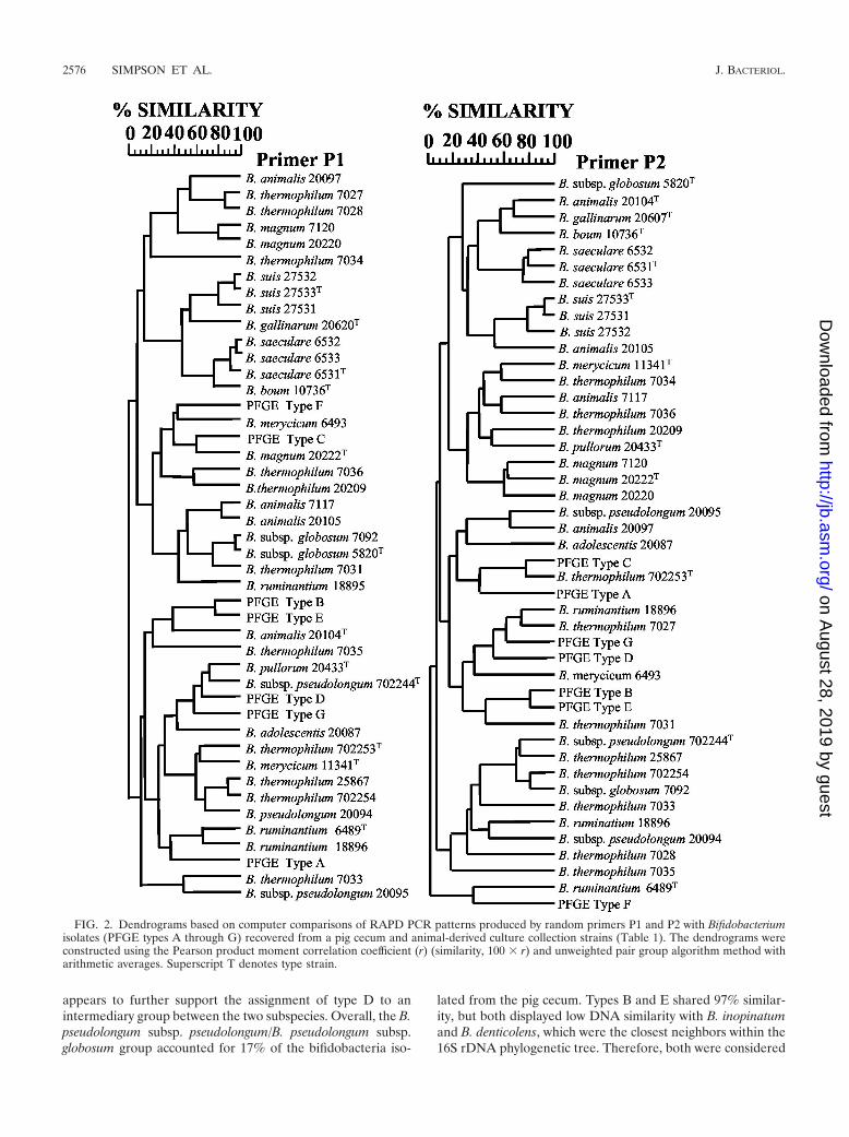
(i) RAPD PCR. A number of studies have reported the useof RAPD PCR to cluster related bifidobacteria isolates (13, 22,50). The dendrograms based on comparison of the RAPDPCR patterns produced by the primers P1 and P2 were con-sidered to be similar with respect to the seven PFGE types. In
TABLE 2. Frequency of PFGE types and subtypes representing 160 Bifidobacterium pig cecal isolates
PFGE typeaNo. of isolates for cecal sampleb: PFGE subtypes and the apparent macro-restriction patterns differences from
their related PFGE typecI II III
A 12 8 13 Aa, new XbaI site in a �280 kb fragment (5)Ab, 25-kb deletion in a 85-kb fragment (2)
B 1 2 2 NoneC 3 4 6 Ca, �75-kb plasmid (5)
Caa, �75-kb plasmid and �15-kb deletion in �130-kb fragment (1)Cb, �300-kb plasmid (1)Cc, �5-kb deletion in �130-kb fragment (2)
D 1 3 3 NoneE 25 25 22 Ea, �30-kb plasmid (1)F 6 6 11 NoneG 2 2 3 Ga, �40-kb plasmid (1)
Total 50 50 60 (18)
a Values include subtypes.b Representing isolates recovered from the cecal contents (I) and epithelial layer, mixed lightly (II) or vigorously (III).c Number in parentheses gives the number of isolates representing the subtype.
2574 SIMPSON ET AL. J. BACTERIOL.
on August 28, 2019 by guest
http://jb.asm.org/
Dow
nloaded from

both cases, the seven types showed the same interrelationship,forming five distinct clusters: types A, B/E, C, D/G, and F,respectively. Using a percentage similarity cutoff of 50%, thesame clusters were maintained when RAPD PCR patterns foreach culture collection strain were included (Fig. 2 and Table3). The segregation of collection strains from the same specieswas considered to be poor. Therefore, RAPD PCR, with prim-ers P1 and P2, was not considered to be a reliable tool forspecies identification.
(ii) Cell morphology. Representatives from the seven PFGEtypes and each subtype produced four distinct cell morphologygroups (Fig. 3). Group 1 cells were irregular rods, arrangedsingly or in pairs, with occasional bifurcated extremities andtapered ends, �0.7 to 1 �m wide and �2 to 3 �m long,represented by type A and subtypes Aa and Ab only (Fig. 3A).Group 2 cells were also irregular rods, arranged singly or inpairs, with occasional bifurcated extremities, �1 to 1.2 �mwide and �2.5 to 5 �m long, with a tendency to form largeaggregates (not shown), represented by types C, D, and G andall their related subtypes (Fig. 3C, D, and G). Group 3 cellswere irregular short rods, arranged singly or in pairs, �0.6 to0.9 �m wide and �0.8 to 1.5 �m long, represented by types Band E and subtype Ea (Fig. 3B and E). Group 4 cells were alsoshort rods but were generally more globular than group 3 cells,arranged singly or in pairs, �0.7 to 1 �m wide and �1 to 1.5�m long, represented by type F (Fig. 3F). Therefore, with theexception of type C, the morphology groups concurred with thegenetic relatedness observed following RAPD PCR (Table 3).
(iii) WCPP. WCPP has previously been demonstrated as auseful technique for the identification of Bifidobacterium spe-cies (3, 54). Based on WCPP after growth on Columbia and/ormMRS media, types A through G were segregated into fourgroups. Types C, D, and G all clustered together with a min-imum similarity value of 78%, compared to 65% similarity totypes A, B, E, and F. Types F and A were both distinct, withmaximum similarity values of 69 and 72% towards type B,respectively. Type B clustered with type E with a similarityvalue of 92%. These groupings were in good agreement withthose based on cell morphology (Table 3). Furthermore, basedon comparisons with all 33 type strains for the genus Bifidobac-terium, types C, D, and G were considered to belong to eitherB. pseudolongum subsp. pseudolongum or B. pseudolongumsubsp. globosum (25, 41, 52). Types A, B, E, and F remainedunidentified.
(iv) 16S rDNA sequencing. The comparison of Bifidobacte-rium 16S rDNA sequences has been shown to group all specieswithin the genus and identify isolates to specific species orgroups of species (11, 16, 26). In general, when 16S rDNA
similarity values exceed 97%, the strains are considered tobelong to the same species (47). Therefore, a tree based oncomparisons between almost complete 16S rDNA sequences(�1,400 bases) for types A through G and for each Bifidobac-terium type strain was constructed (Fig. 4). The seven PFGEtypes segregated in good agreement with the groupings basedon cell morphology, WCPP, and RAPD PCR (Table 3). PFGEType G shared 100% similarity with the B. pseudolongumsubsp. pseudolongum type strain, and PFGE types C and Dshared 99.93 and 100% similarity with the B. pseudolongumsubsp. globosum type strain, respectively. However, in generalagreement with WCPP, the assignments appeared to contra-dict the RAPD PCR-based data, which grouped types D and Gbut showed type C to be distinct. However, the 16S rDNAsequences for the two subspecies are themselves 99.14% sim-ilar, indicating the difficulty in distinguishing the two subspe-cies based on 16S rDNA sequence comparisons (26). Previ-ously strains considered as intermediates between the twosubspecies were proposed (52), and type D could represent anintermediate strain. Type A was identified as B. minimum sinceit was 99.57% similar to the species type strain. The inclusionof type F changed the position of B. minimum within thephylogenetic tree, replacing the three insect-derived Bifidobac-terium species as the closest neighbor (26). However, type Fwas only 95.08 and 94.85% similar to type A and B. minimum,respectively, and was considered to likely represent a newBifidobacterium species. Types B and E were 99.72% similar toeach other and were considered to be highly related and prob-ably from the same species. However, they were only 91.21 and91.36% similar, respectively, to their nearest neighbor, Bi-fidobacterium denticolens, and were considered to represent anew Bifidobacterium species.
(v) DNA-DNA similarity. DNA-DNA hybridization is con-sidered to be the “gold standard” for taxonomic evaluation(47). Bacteria that have DNA similarity values of 70% or aboveare generally considered to belong to the same species andusually have corresponding 16S rDNA similarity values of 98%or above (47). Therefore, the DNA similarity values between anumber of the PFGE types and type strains of species showinga phylogenetic grouping were measured. The results were ingood agreement with earlier findings (Table 4). Type A wasconfirmed as B. minimum, sharing 103% similarity, and thespecies accounted for 21% of the bifidobacteria isolated fromthe pig cecum. Type C and type D were 85% similar and wereclearly related. However, type C and type D shared 85 and100% similarity with the B. pseudolongum subsp. pseudolongumtype strain. This finding highlights the previous contradictionbetween RAPD PCR and 16S rDNA sequencing (Table 3) and
TABLE 3. Relatedness of PFGE types A through G
Group
Analysis
Species based on 16S rDNA sequence (% similarity)Cellmorphology
RAPDPCR WCPP
1 A A A A assigned to B. minimum (99.57)2 C, D, G D, G C, D, G G assigned to B. pseudolongum subsp. pseudolongum (100)3 B, E B, E B, E B and E related (99.72) assigned to a new species, B. aerophilum sp. nov.4 F F F F assigned to a new species, B. psychroaerophilum sp. nov.5 C C (99.93) and D (100) assigned to B. pseudolongum subsp. globosum
VOL. 185, 2003 GENOMIC DIVERSITY OF PORCINE BIFIDOBACTERIA 2575
on August 28, 2019 by guest
http://jb.asm.org/
Dow
nloaded from
appears to further support the assignment of type D to anintermediary group between the two subspecies. Overall, the B.pseudolongum subsp. pseudolongum/B. pseudolongum subsp.globosum group accounted for 17% of the bifidobacteria iso-
lated from the pig cecum. Types B and E shared 97% similar-ity, but both displayed low DNA similarity with B. inopinatumand B. denticolens, which were the closest neighbors within the16S rDNA phylogenetic tree. Therefore, both were considered
FIG. 2. Dendrograms based on computer comparisons of RAPD PCR patterns produced by random primers P1 and P2 with Bifidobacteriumisolates (PFGE types A through G) recovered from a pig cecum and animal-derived culture collection strains (Table 1). The dendrograms wereconstructed using the Pearson product moment correlation coefficient (r) (similarity, 100 � r) and unweighted pair group algorithm method witharithmetic averages. Superscript T denotes type strain.
2576 SIMPSON ET AL. J. BACTERIOL.
on August 28, 2019 by guest
http://jb.asm.org/
Dow
nloaded from

FIG. 3. Phase contrast cell morphology of Bifidobacterium isolates recovered from a pig cecum. Panels A through G represented PFGE typesA through G, respectively. Four morphology groups, 1 through 4, were considered to represent type A, B and E, C, D, and G, and F, respectively(Table 3). Each subtype segregated with its related type. Scale bars in the main and inset panels are 10 and 2 �m, respectively.
VOL. 185, 2003 GENOMIC DIVERSITY OF PORCINE BIFIDOBACTERIA 2577
on August 28, 2019 by guest
http://jb.asm.org/
Dow
nloaded from

FIG. 4. Phylogenetic tree based on 16S rDNA sequences for each Bifidobacterium species type strain and seven PFGE types, termed A throughG, representing bifidobacteria isolated from a pig cecum. Database accession numbers are given in parenthesis. The tree was rooted with E.combesii and constructed using the neighbor-joining method. Bootstrap values, expressed as percentages of 1,000 replicons, are given at eachbranch point where above 50%.
2578 SIMPSON ET AL. J. BACTERIOL.
on August 28, 2019 by guest
http://jb.asm.org/
Dow
nloaded from

to represent a new species which, based on phenotypic data(unpublished data) was provisionally termed Bifidobacteriumaerophilum sp. nov. The species accounted for 48% of theisolated cecal bifidobacteria. Type F displayed low similarity tothe nearest 16S rDNA neighbor, B. minimum (Fig. 4), and wasalso considered to represent a new species which, based onphenotypic data (unpublished data), was provisionally termedBifidobacterium psychroaerophilum sp. nov. The species ac-counted for 14% of the isolated cecal bifidobacteria.
Strain accession numbers. Isolates representing the PFGEtypes A through G were deposited in the Dairy Products Col-lection under accession numbers DPC 5735 through 5741, re-spectively.
DISCUSSION
The antibiotic mupirocin was previously found to effectivelyselect for bifidobacteria from other bacteria typically found inGIT and fecal samples (31, 34–36). The addition of mupirocinto mMRS has not been reported for the selective isolation ofbifidobacteria but, along with the culture conditions used in thepresent study, appeared to be selective for all Bifidobacteriumstrains tested. This medium yielded 4.3 � 105 CFU of bi-fidobacteria/g for the pig cecal contents. Only a limited numberof studies have reported the enumeration of bifidobacteriafrom cecal contents and values ranged from 3 � 103 CFU/g forrabbit (35), 5 � 106 CFU/ml for human (21), and 4 � 109
CFU/g for hen (35).PFGE has previously been found to discriminate between
Bifidobacterium strains (29, 38), a finding extended in thepresent study for a wide range of animal derived strains. Inaddition, PFGE was previously shown to be an effective tech-nique for determining the genomic diversity of bifidobacteriaisolated from human feces (17, 23). In these studies, the PFGEmacro-restriction patterns for 10 Bifidobacterium isolates re-covered at different times from a number of individuals werecompared. Each PFGE type was considered to represent adistinct strain, and a complex population was arbitrarily as-signed to subjects with four or more strains, while samples withfewer patterns were considered to be simple. Both kinds ofpopulation were observed with equal frequency. Following thisconvention our findings suggest that a simple bifidobacteriapopulation, dominated by 15 Bifidobacterium strains, wasmaintained within a pig cecum.
A variety of studies have reported that Bifidobacterium
strains vary in their ability to adhere to cultured epithelial cells(2, 6, 9, 10, 19, 30) and that the general microflora of theepithelium and contents of the porcine GIT are distinct (1, 44).However, in our study of bifidobacteria, although the totalnumbers of bifidobacteria associated with the cecal contentsand epithelium differed considerably, distinct populations werenot found. The data suggest that in vivo a relationship between“attached,” “loosely unattached,” and “unattached” cells ex-ists. However, it is not clear if the bifidobacteria proliferate inthe lumen and attach in a density-dependent manner or ifproliferation occurs when they are attached to the epitheliumwith the lumen fraction represented an accumulation of de-tached bacteria.
Representatives for each PFGE type were examined by cellmorphology, RAPD PCR, WCPP, 16S rDNA sequencing, andDNA-DNA hybridization to determine if a relatedness existedbetween the apparently unrelated macro-restriction patterns.Data from each technique were mostly complementary, andoverall the seven types appeared to represent four distinctgroups.
One group was identified as B. minimum, a species previ-ously isolated only from sewage (5, 40). Another group ap-peared to include strains from B. pseudolongum subsp. pseud-olongum and B. pseudolongum subsp. globosum (52). This studyappears to be the first to report the recovery of either subspe-cies from a pig cecum, although they have previously beenisolated from pig feces (25, 53), pork meat (14), and rabbitcecum (20, 46).
The two remaining groups were not assigned to any knownBifidobacterium species, although 16S rDNA sequencing didplace them within the Bifidobacterium genus. In addition, bothgroups were positive for F-6-PPK activity. Based on the pre-sented data and phenotypic analysis (unpublished data), PFGEtypes B, E, and subtype Ea strains were provisionally identifiedas a new species, termed Bifidobacterium aerophilum sp. nov.Similarly, the PFGE type F strain was provisionally identifiedas a new species termed Bifidobacterium psychroaerophilum sp.nov. The identification of additional species supports a recentstudy in which 16S rDNA sequences for 14 of 20 undefinedisolates from the pig cecum were less than 95% similar to anydatabase sequence (33).
A number of PFGE macro-restriction patterns differed by asingle DNA fragment. It is possible that the extra band seen inthese patterns represents an insertion, around or into a XbaI
TABLE 4. DNA-DNA hybridization homologies between selected PFGE types and Bifidobacterium type strainsa
Competitor strain Reference strain % DNA similarity
PFGE type A (DPC 5732) PFGE type F (DPC 5740T) 12PFGE type A (DPC 5732) B. minimum (LMG 11592T) 103PFGE type F (DPC 5740T) B. minimum (LMG 11592T) 14PFGE type D (DPC 5738) PFGE type C (DPC 5377) 85PFGE type C (DPC 5377) B. pseudolongum subsp. pseudolongum (LMG 11571T) 85PFGE type D (DPC 5738) B. pseudolongum subsp. pseudolongum (LMG 11571T) 100PFGE type B (DPC 5733) PFGE type E (DPC 5739T) 97PFGE type B (DPC 5733) B. denticolens (LMG 18312T) 8PFGE type E (DPC 5739T) B. denticolens (LMG 18312T) 7PFGE type B (DPC 5733) B. inopinatum (LMG 18313T) 2PFGE type E (DPC 5739T) B. inopinatum (LMG 18313T) 1
a Superscript T denotes type strain.
VOL. 185, 2003 GENOMIC DIVERSITY OF PORCINE BIFIDOBACTERIA 2579
on August 28, 2019 by guest
http://jb.asm.org/
Dow
nloaded from

site, of a fragment with two restriction sites separated by adistance equal to the size of the insertion. However, it wasconsidered more likely that the fragment represents the pres-ence of a putative plasmid. In support of this, the estimatedsizes for the majority of the suspected plasmids were within therange previously noted for the Bifidobacterium genus (41).However, two putative megaplasmids were also observedwhich have not previously been reported for the genus. Inter-estingly, most of the plasmids appeared to be associated withPFGE type C, which was identified as B. pseudolongum subsp.globosum, one of only four species in a survey of 25 Bifidobac-terium species reported to contained plasmids (42). Work isongoing to verify the PFGE interpretations and characterizethe possible plasmids.
One interesting aspect of this study is the discovery of strainswhich from macro-restriction pattern analysis appear to showgenomic rearrangements through reciprocal events involvingeither deletions or insertions. It is unclear if this genomicplasticity reflects a high rate of genomic change or a selectionfor very similar strains within the cecum. Interestingly, a recentstudy found that the commensal intestinal pathogen Bacte-roides fragilis modified the structure of exopolysaccharidesthrough chromosomal inversions within genetic promoter re-gions (18). The changes were considered to be part of a de-fense mechanism against the host immune response and allowfor the long-term colonization of the GIT. It is tempting tosuggest that the genomic plasticity observed in the presentstudy may also have some bearing on long-term colonization ofbifidobacteria.
In conclusion, the study presents a detailed examination ofgenomic diversity and relatedness of the bifidobacteria isolatedfrom a pig cecum. The results suggest that the same relativelysimple bifidobacteria population exists in the lumen and epi-thelium. Moreover, two potentially new species and evidencefor genomic plasticity within strains were reported.
ACKNOWLEDGMENTS
This work was supported by the Irish Government under the Na-tional Development Plan 2000-2006 and the European Union (SM&T-CT98-2235).
We are grateful to Tomotari Mitsuoka for providing B. thermophi-lum biotype information, Noelle Brennan for her assistance with 16SrDNA sequence comparisons and the determination of phylogeneticdifferences, and Shahenda Mohamed Mahmoud Al Abi for technicalassistance.
REFERENCES
1. Allison, M. J., I. M. Robinson, J. A. Bucklin, and G. D. Booth. 1979. Com-parison of bacterial populations of the pig cecum and colon based uponenumeration with specific energy sources. Appl. Environ. Microbiol. 37:1142–1151.
2. Bernet, M. F., D. Brassart, J. R. Neeser, and A. L. Servin. 1993. Adhesion ofhuman bifidobacterial strains to cultured human intestinal epithelial cellsand inhibition of enteropathogen-cell interactions. Appl. Environ. Microbiol.59:4121–4128.
3. Biavati, B., V. Scardovi, and W. E. C. Moore. 1982. Electrophoretic patternsof proteins in the genus Bifidobacterium and proposal of four new species.Int. J. Syst. Bacteriol. 32:368–373.
4. Biavati, B., and P. Mattarelli. 1991. Bifidobacterium ruminatium sp. nov. andBifidobacterium merycicum sp. nov. from the rumens of cattle. Int. J. Syst.Bacteriol. 41:163–168.
5. Biavati, B., M. Vescovo, S. Torriani, and V. Bottazzi. 2000. Bifidobacteria:history, ecology, physiology and applications. Ann. Microbiol. 50:117–131.
6. Bibiloni, R., P. F. Perez, and G. L. de Antoni. 1999. Factors involved inadhesion of bifidobacterial strains to epithelial cells in culture. Anaerobe5:483–485.
7. Bibiloni, R., A. G. Zavaglia, and G de Antoni. 2001. Enzyme-based mostprobable number for the enumeration of Bifidobacterium in dairy products.J. Food Prot. 64:2001–2006.
8. Brennan, N. M., R. Brown, M. Goodfellow, A. C. Ward, T. P. Beresford, P. J.Simpson, P. F. Fox, and T. M. Cogan. 2001. Corynebacterium mooreparkensesp. nov. and Corynebacterium casei sp. nov., isolated from the surface of asmear-ripened cheese. Int. J. Syst. Microbiol. 51:843–852.
9. Crociani, J., J.-P. Grill, M. Huppert, and J. Ballongue. 1995. Adhesion ofdifferent bifidobacteria strains to human enterocyte-like Caco-2 cells andcomparison with in vivo study. Lett. Appl. Microbiol. 21:146–148.
10. Del Re, B., A. Bussetto, G. Vignola, B. Sgorbati, and D. L. Palenzona. 1998.Autoaggregation and adhesion ability in a Bifidobacterium suis strain. Lett.Appl. Microbiol. 27:307–310.
11. Dong, X., Y. Xin, W. Jian, X. Liu, and D. Ling. 2000. Bifidobacteriumthermacidophilum sp. nov., isolated from an anaerobic digester. Int. J. Syst.Evol. Microbiol. 50:119–125.
12. Ezaki, T., Y. Hashimoto, and E. Yabuuchi. 1989. Flourometric deoxyribo-nucleic acid-deoxyribonucleic acid hybridization in microdilution wells as analternative to membrane filter hybridization in which radioisotopes are usedto determine genetic relatedness among bacterial strains. Int. J. Syst. Bac-teriol. 39:224–229.
13. Fanedl, L., F. V. Nekrep, and G. Avgustin. 1998. Random amplified poly-morphic DNA analysis and demonstration of genetic variability among bi-fidobacteria isolated from rats fed with raw kidney beans. Can. J. Microbiol.44:1094–1101.
14. Gavini, F., and H. Beerens. 1999. Origin and identification of bifidobacteriastrains isolated from meat and meat products. Int. J. Food Microbiol. 46:81–85.
15. Gomes, A. M. P., and F. X. Malcata. 1999. Bifidobacterium spp. and Lacto-bacillus acidophilus: biological, biochemical, technological and therapeuticalproperties relevant for use as probiotics. Trends Food Sci. Technol. 10:139–157.
16. Hoyles, L., E. Inganas, E. Falsen, M. Drancourt, N. Weiss, A. L. McCarney,and M. D. Collins. 2002. Bifidobacterium scardovii sp. nov., from humansources. Int. J. Syst. Evol. Microbiol. 52:995–999.
17. Kimura, K., A. L. McCartney, M. A. McConnell, and G. W. Tannock. 1997.Analysis of fecal populations of bifidobacteria and lactobacilli and investi-gation of the immunological responses of their human host to the predom-inant strain. Appl. Environ. Microbiol. 63:3394–3398.
18. Krinos, C. M., M. J. Coyne, K. G. Weinacht, A. O. Tzaianabos, D. L. Kasper,and L. E. Comstock. 2001. Extensive surface diversity of a commensal mi-croorganism by multiple DNA inversions. Nature 414:555–558.
19. Lankaputhra, W. E. V., and N. P. Shah. 1998. Adherence of probioticbacteria to human colonic cells. Biosci. Microflora 17:105–113.
20. Marounek, M., V. Rada, and V. Benda. 1998. Biochemical characteristics andfermentation of glucose and starch by rabbit caecal strains of Bifidobacteriumglobosum. Folia Microbiol. (Phara) 43:113–116.
21. Marteau, P., P. Pochart, J. Dore, C. Bera-Maillet, A. Bernalier, and G.Corthier. 2001. Comparative study of bacterial groups within the humancecal and fecal microbiota. Appl. Environ. Microbiol. 67:4939–4942.
22. McBrearty, S., P. J. Simpson, G. Fitzgerald, J. K. Collins, R. P. Ross, and C.Stanton. 2000. Probiotic bifidobacteria and their identification using molec-ular genetic techniques, p. 97–107. In J. Buttriss and M. Saltmarsh (ed.),Functional foods: claims and evidence. Royal Society of Chemistry, Cam-bridge, United Kingdom.
23. McCartney, A. L., W. Wenzhi, and G. W. Tannock. 1996. Molecular analysisof the composition of the bifidobacterial and Lactobacillus microflora ofhumans. Appl. Environ. Microbiol. 62:4608–4613.
24. Mikkelsen, L. L., and B. B. Jensen. 2001. Bifidobacteria in piglets, p. 285–288. In J. E. Lindberg and B. Ogle (ed.), Digestive physiology of pigs. CABInt., Oxford, United Kingdom.
25. Mitsuoka, T. 1969. Comparative studies on bifidobacteria isolated from thealimentary tract of man and animals, including descriptions of Bifidobacte-rium thermophilum nov. spec. and Bifidobacterium pseudolongum nov. spec.Infektionskrankhh. Hyg. Abt. 1 Orig. Reihe A 210:52–64.
26. Miyake, T., K. Watanabe, T. Watanabe, and H. Oyaizu. 1998. Phylogeneticanalysis of the genus Bifidobacterium and related genera based on 16S rDNAsequences. Microbiol. Immunol. 42:661–667.
27. Muirhead, S. 2000. Direct-fed products, p. 57–294. In S. Muirhead (ed.),2000–2001 direct-fed microbial, enzyme and forage additive compendium,vol 5. Miller Publishing Co., ●●●●, Minnesota.
28. Orban, J. J., and J. A. Patterson. 2000. Modification of the phosphoketolaseassay for rapid identification of bifidobacteria. J. Microbiol. Methods 40:221–224.
29. O’Riordan, K., and G. F. Fitzgerald. 1997. Determination of genetic diversitywithin the genus Bifidobacterium and estimation of chromosomal size. FEMSMicrobiol. Lett. 156:259–264.
30. Perez, P. F., Y. Minnaard, E. A. Disalvo, and G. L. de Antoni. 1998. Surfaceproperties of bifidobacterial strains of human origins. Appl. Environ. Micro-biol. 64:21–26.
31. Petr, J., and V. Rada. 2001. Bifidobacteria are obligate inhabitants of the
2580 SIMPSON ET AL. J. BACTERIOL.
on August 28, 2019 by guest
http://jb.asm.org/
Dow
nloaded from

crop of adult laying hens. J. Vet. Med. B. Infect. Dis. Public Health 48:227–233.
32. Pot, B., P. Vandamme, and K. Kersters. 1994. Analysis of electrophoreticwhole-organism protein fingerprints. M. Goodfellow and A. G. O’Donnell(ed.), Chemical methods in prokaryotic systematics. J. Wiley and Sons,Chichester, United Kingdom.
33. Pryde, S. E., A. J. Richardson, C. S. Stewart, and H. J. Flint. 1999. Molecularanalysis of the microbial diversity present in the colonic wall, colonic lumen,and cecal lumen of a pig. Appl. Environ. Microbiol. 65:5372–5377.
34. Rada, V. 1997. Detection of Bifidobacterium species by enzymatic methodsand antimicrobial susceptibility testing. Biotechnol. Tech. 11:909–912.
35. Rada, V., K. Sirotek, and J. Petr. 1999. Evaluation of selective media forbifidobacteria in poultry and rabbit caecal samples. J. Vet. Med. B 46:369–373.
36. Rada, V., and J. Koc. 2000. The use of mupirocin for selective enumerationof bifidobacteria in fermented milk products. Milchwissenchaft 55:65–67.
37. Robinson, I. M., M. J. Allison, and J. A. Bucklin. 1981. Characterization ofthe cecal bacteria of normal pigs. Appl. Environ. Microbiol. 41:950–955.
38. Roy, D., P. Ward, and G. Champagne. 1996. Differentiation of bifidobacteriaby use of pulsed-field gel electrophoresis and polymerase chain reaction. Int.J. Food Microbiol. 29:11–29.
39. Scardovi, V., G. Zani, and L. D. Trovatelli. 1970. Deoxyribonucleic acidhomology among the species of the genus Bifidobacterium isolated fromanimals. Arch. Mikrobiol. 72:318–325.
40. Scardovi, V., and L. D. Trovatelli. 1974. Bifidobacterium animalis (Mitsuoka)comb. nov. and the “minimum” and “subtile” groups of new bifidobacteriafound in sewage. Int. J. Syst. Bacteriol. 24:21–28.
41. Scardovi, V. 1986. Genus Bifidobacterium, p. 1418–1434. In P. H. A. Sneath,N. S. Mair, M. E. Sharpe, and J. G. Holt (ed.), Bergey’s manual of systematicbacteriology, vol. 2. Williams and Wilkins, Baltimore, Md.
42. Sgorbati, B., V. Scardovi, and D. J. Leblanc. 1982. Plasmids in the genusBifidobacterium. J. Gen. Microbiol. 128:2121–2131.
43. Shimizu, J., N. Tsuchihashi, K. Kudoh, M. Wada, T. Takita, and S. Innami.2001. Dietry curdlan increases proliferation of bifidobacteria in the cecum ofrats. Biosci. Biotechnol. Biochem. 65:466–469.
44. Simpson, J. M., V. J. McCracken, B. A. White, H. R. Gaskins, and R. I.
Mackie. 1999. Application of denaturant gradient gel electrophoresis for theanalysis of the porcine gastrointestinal microbiota. J. Microbiol. Methods36:167–179.
45. Simpson, P. J., C. Stanton, G. F. Fitzgerald, and R. P. Ross. 2002. Genomicdiversity within the genus Pediococcus as revealed by randomly amplifiedpolymorphic DNA PCR and pulsed-field gel electrophoresis. Appl. Environ.Microbiol. 68:765–771.
46. Slovakora, L., D. Duskova, and M. Marounek. 2002. Fermentation of pectinand glucose and activity of pectin-degrading enzymes in the rabbit caecalbacterium Bifidobacterium pseudolongum. Lett. Appl. Microbiol. 35:126–130.
47. Stackebrandt, E., and B. M. Goebel. 1994. Taxonomic note: a place forDNA-DNA reassociation and 16S rRNA sequence analysis in the presentspecies definition in bacteriology. Int. J. Syst. Bacteriol. 44:846–849.
48. Stanton, C., G. Gardiner, H. Meehan, K. Collins, G. Fitzgerald, P. B. Lynch,and R. P. Ross. 2001. Market potential for probiotics. Am. J. Clin. Nutr.73:476–483.
49. Tenover, F. C., R. D. Arbeit, R. V. Goering, P. A. Mickelsen, B. A. Murray,D. H. Persing, and B. Swaminathan. 1995. Interpreting chromosomal DNArestriction patterns produced by pulsed-field gel electrophoresis: criteria forbacterial strain typing. J. Clin. Microbiol. 33:2233–2239.
50. Vincent, D., D. Roy, F. Mondou, and C. Dery. 1998. Characterization ofbifidobacteria by random DNA amplification. Int. J. Food Microbiol. 43:185–193.
51. Watabe, J., Y. Benno, and T. Mitsuoka. 1983. Bifidobacterium gallinarum sp.nov.: a new species isolated from the ceca of chickens. Int. J. Syst. Bacteriol.33:127–132.
52. Yaeshima, T., T. Fujisawa, and T. Mitsuoka. 1992. Bifidobacterium globo-sum, subjective synonym of Bifidobacterium pseudolongum, and descriptionof Bifidobacterium pseudolongum subsp. pseudolongum comb. nov and Bi-fidobacterium pseudolongum subs. globosum comb. nov. Syst. Appl. Micro-biol. 15:380–385.
53. Zani, G., B. Biavati, F. Crociani, and D. Matteuzzi. 1974. Bifidobacteriafrom faeces of piglets. J. Appl. Bacteriol. 37:537–547.
54. Zavaglia, A. G., G. Kociubinski, P. Perez, and G. de Antoni. 1998. Isolationand characterisation of Bifidobacterium strains for probiotic formulation. J.Food Prot. 61:865–873.
VOL. 185, 2003 GENOMIC DIVERSITY OF PORCINE BIFIDOBACTERIA 2581
on August 28, 2019 by guest
http://jb.asm.org/
Dow
nloaded from