genomic microarray and whole exome sequencing · genomic microarray and whole exome sequencing ......
TRANSCRIPT
Genomic microarray
and whole exome sequencing
Final evidence report
December 19, 2017
Health Technology Assessment Program (HTA)
Washington State Health Care Authority
PO Box 42712 Olympia, WA 98504‐2712
(360) 725‐5126 www.hca.wa.gov/hta

Prepared by: RTI International–Evidence-based Practice Center Research Triangle Park, NC 27709 Lead Investigator: Nedra Whitehead, PhD
This health technology assessment is based on research conducted by RTI International under contract to the Washington Health Care Authority (HCA) (Contract No. K1959). The findings and conclusions in this document are those of the authors, who are responsible for its contents; the findings and conclusions do not necessarily represent the views of the Washington HCA. Therefore, no statement in this report should be construed as an official position of Washington HCA. The information in this report is intended to help the Washington HCA make well-informed coverage determinations and thereby improve the quality of health care services. This report is not intended to be a substitute for the application of clinical judgment. Anyone who makes decisions concerning the provision of clinical care should consider this report in the same way as any medical reference and in conjunction with all other pertinent information (i.e., in the context of available resources and circumstances presented by individual patients). This document is in the public domain and may be used and reprinted without permission except those copyrighted materials that are clearly noted in the document. Further reproduction of those copyrighted materials is prohibited without the specific permission of copyright holders. None of the investigators has any affiliations or financial involvement that conflicts with the material presented in this report.
Acknowledgments
The following individuals contributed to this report: Co-Investigators: Leila Kahwati, MD MPH; Sreelatha Meleth, PhD
Analysts: Rachel Clark, BA; Sonya Goode, MPH; Tracey Grant, MS; Josh Green, BA; Nana Haywood, MPH; Sara Kennedy, MPH; Jennifer Taylor, PhD
Library/Document Preparation:
Mark Howell, MLS; Loraine Monroe; Laura Small, BA

We thank the following reviewers for their thoughtful comments on the draft report:
Dan Jonas, MD, MPH. Associate Professor of Medicine, Section Chief for Research, Division of General Medicine & Clinical Epidemiology, Co-Director, RTI-UNC Evidence-based Practice Center, Director, Program on Medical Practice and Prevention, Cecil G. Sheps Center for Health Services Research
Christa Lese Martin, PhD, FACMG. Director & Professor, Autism & Developmental Medicine Institute, Geisinger
The members of the public who reviewed and commented on our draft report.
WA – Health Technology Assessment December 19, 2017
Genetic microarray and whole exome sequencing: Final evidence report i
Contents
Structured Abstract ...................................................................................................................... v Executive Summary ................................................................................................................ ES-1
Background ...................................................................................................................... ES-1 Methods............................................................................................................................ ES-7 Results .............................................................................................................................. ES-8 Discussion ...................................................................................................................... ES-15 Conclusion ..................................................................................................................... ES-17
Full Technical Report ................................................................................................................... 1
Background ............................................................................................................................ 1 Purpose ............................................................................................................................. 1 Condition Description ...................................................................................................... 1 Disease Burden ................................................................................................................ 1 Technology Description ................................................................................................... 2 Policy Context .................................................................................................................. 4 Washington State Agency Utilization Data ................................................................. 5 Regulatory Status ............................................................................................................. 7 Practice Guidelines .......................................................................................................... 7 Other Related HTAs ........................................................................................................ 8 Selected Payer Coverage Determinations ........................................................................ 8 Research Questions and Analytic Framework ............................................................... 11 What is Excluded from This HTA ................................................................................. 13
Methods................................................................................................................................ 13 Data Sources and Searches ............................................................................................ 13 Study Selection .............................................................................................................. 13 Data Abstraction and Quality Assessment ..................................................................... 15 Data Synthesis and Analysis .......................................................................................... 15
Results .................................................................................................................................. 15 Literature Search ............................................................................................................ 15 Safety ............................................................................................................................. 16 Efficacy .......................................................................................................................... 18 Cost and Cost-Effectiveness .......................................................................................... 26
Discussion ............................................................................................................................ 29 Summary of the Evidence .............................................................................................. 29 Additional Context for Interpreting Findings ................................................................ 30 Limitations of the Evidence Base .................................................................................. 31 Limitations of this HTA ................................................................................................. 32 Ongoing Research and Future Research Needs ............................................................. 33
Conclusion ........................................................................................................................... 33 References .................................................................................................................................... 35
WA – Health Technology Assessment December 19, 2017
Genetic microarray and whole exome sequencing: Final evidence report ii
List of Appendices
Appendix A. Search Strategy ........................................................................................................ 43
Appendix B. Additional Methods ................................................................................................. 45
Appendix C. Evidence Tables ....................................................................................................... 46
Appendix D. Excluded Studies ..................................................................................................... 61
Appendix E. Individual Study Risk of Bias Assessments ............................................................ 89
List of Figures
Figure ES-1 Analytic Framework for Chromosomal Microarray and Whole Exome Sequencing in Children with Developmental or Intellectual Disability, Autism, or Multiple Congenital Anomalies ........................................................................................ ES-5
Figure 1. Analytic Framework for the Detection of Chromosomal Abnormalities Chromosomal Microarray and Whole Exome Sequencing in Children with Intellectual Disability, Autism, or Birth Defects .................................................... 11
Figure 2. Study Flow Diagram .............................................................................................. 16
Figure 3. Summary Pooled Estimate for Diagnostic Yield of Chromosomal Microarray for All Included Phenotype .......................................................................................... 20
Figure 4. Evidence on Diagnostic Yield of Chromosomal Microarray for Autism Spectrum Disorders ................................................................................................................ 22
List of Tables
Table ES-1. Resolution and Detection of Chromosomal Microarray and Whole Exome Sequencing ......................................................................................................... ES-2
Table ES-2. Practice Guidelines Endorsing or Providing Guidance on Chromosomal Microarray Testing ............................................................................................. ES-4
Table ES-3. Payer Coverage for CMA and WES Testing ...................................................... ES-4
Table ES-4. Research Questions and Scoping Parameters for Chromosomal Microarray or Whole Exome Sequencing in Children with Intellectual Disability, Autism, or Birth Defects .................................................................................................. ES-6
Table ES-5. Summary of Findings for the Safety of Testing with Chromosomal Microarray or Whole Exome Sequencing ............................................................................. ES-9
Table ES-6. Strength of Evidence for Findings Related to the Safety of Testing with Chromosomal Microarray or Whole Exome Sequencing .................................. ES-9
Table ES-7. Summary of Findings for the Diagnostic Yield of Testing with Chromosomal Microarray or Whole Exome Sequencing ........................................................ ES-10
WA – Health Technology Assessment December 19, 2017
Genetic microarray and whole exome sequencing: Final evidence report iii
Table ES-8. Strength of Evidence for Findings Related to the Diagnostic Yield (EQ1) of Testing with Chromosomal Microarray or Whole Exome Sequencing ........... ES-10
Table ES-9. Summary of Findings for the Impact of Chromosomal Microarray Testing on Clinical Management ....................................................................................... ES-11
Table ES-10. Strength of Evidence for Findings Related to the Impact of Testing with Chromosomal Microarray on Clinical Management (EQ3) ............................. ES-13
Table ES-11. Summary of Findings of Studies Evaluating Cost Outcomes of Chromosomal Microarray Testing, Outcomes Reported in 2010 U.S. Dollars ES-14
Table ES-12. Strength of Evidence for Findings Related to the Cost-Effectiveness of Chromosomal Microarray Testing Compared to No Testing (CQ1) ............... ES-15
Table 1. Resolution and Detection of Chromosomal Microarray and Whole Exome Sequencing ............................................................................................................... 3
Table 2. Practice Guidelines Endorsing or Providing Guidance on Chromosomal Microarray Testing ................................................................................................... 4
Table 3. CPT Descriptions ..................................................................................................... 5
Table 4. Definitions for Utilization and Cost Tables ............................................................. 6
Table 5. Utilization Analysis – Genomic Micro-array and single exome sequencing ........... 6
Table 6. Payer Coverage for CMA and WES Testing ............................................................ 8
Table 7. Research Questions and Scoping Parameters for the Detection of Chromosomal Abnormalities by Chromosomal Microarray or Whole Exome Sequencing in Children with Intellectual Disability, Autism, or Birth Defects ............................. 12
Table 8. Summary of Findings for the Safety of Testing with Chromosomal Microarray or Whole Exome Sequencing ................................................................................. 17
Table 9. Strength of Evidence for Findings Related to the Safety of Testing with Chromosomal Microarray or Whole Exome Sequencing (SQ1) ............................ 17
Table 10. Summary of Findings for the Diagnostic Yield of Testing with Chromosomal Microarray or Whole Exome Sequencing .............................................................. 19
Table 11. Strength of Evidence for Findings Related to the Diagnostic Yield (EQ1) of Testing with Chromosomal Microarray or Whole Exome Sequencing ................. 21
Table 12. Summary of Findings for the Impact of Chromosomal Microarray Testing on Clinical Management ............................................................................................. 24
Table 13. Strength of Evidence for Findings Related to the Impact of Testing with Chromosomal Microarray on Clinical Management (EQ3) ................................... 25
Table 14. Summary of Findings of Studies Evaluating Cost Outcomes of Chromosomal Microarray Testing, Outcomes Reported in 2010 U.S. Dollars ............................. 27
Table 15. Strength of Evidence for Findings Related to the Cost-Effectiveness of Chromosomal Microarray Testing Compared to No Testing (CQ1) ..................... 29
WA – Health Technology Assessment December 19, 2017
Genetic microarray and whole exome sequencing: Final evidence report iv
List of Abbreviations
ASD autism spectrum disorder
CGH comparative genomic hybridization
CMA chromosomal microarray (also called genomic microarray)
CNV copy number variant
DD developmental disability
FISH fluorescent in situ hybridization
GDD global developmental delay
HTA health technology assessment
ID intellectual disability
MCA multiple congenital anomalies
UK United Kingdom
U.S. United States
WES whole exome sequencing
WA – Health Technology Assessment December 19, 2017
Genetic microarray and whole exome sequencing: Final evidence report v
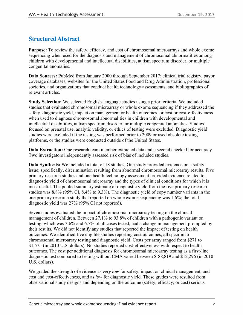
Structured Abstract
Purpose: To review the safety, efficacy, and cost of chromosomal microarrays and whole exome sequencing when used for the diagnosis and management of chromosomal abnormalities among children with developmental and intellectual disabilities, autism spectrum disorder, or multiple congenital anomalies.
Data Sources: PubMed from January 2000 through September 2017; clinical trial registry, payor coverage databases, websites for the United States Food and Drug Administration, professional societies, and organizations that conduct health technology assessments, and bibliographies of relevant articles.
Study Selection: We selected English-language studies using a priori criteria. We included studies that evaluated chromosomal microarray or whole exome sequencing if they addressed the safety, diagnostic yield, impact on management or health outcomes, or cost or cost-effectiveness when used to diagnose chromosomal abnormalities in children with developmental and intellectual disabilities, autism spectrum disorder, or multiple congenital anomalies. Studies focused on prenatal use, analytic validity, or ethics of testing were excluded. Diagnostic yield studies were excluded if the testing was performed prior to 2009 or used obsolete testing platforms, or the studies were conducted outside of the United States.
Data Extraction: One research team member extracted data and a second checked for accuracy. Two investigators independently assessed risk of bias of included studies.
Data Synthesis: We included a total of 18 studies. One study provided evidence on a safety issue; specifically, discrimination resulting from abnormal chromosomal microarray results. Five primary research studies and one health technology assessment provided evidence related to diagnostic yield of chromosomal microarray and the types of clinical conditions for which it is most useful. The pooled summary estimate of diagnostic yield from the five primary research studies was 8.8% (95% CI, 8.4% to 9.3%). The diagnostic yield of copy number variants in the one primary research study that reported on whole exome sequencing was 1.6%; the total diagnostic yield was 27% (95% CI not reported).
Seven studies evaluated the impact of chromosomal microarray testing on the clinical management of children. Between 27.1% to 93.8% of children with a pathogenic variant on testing, which was 3.6% and 6.7% of all cases tested, had a change in management prompted by their results. We did not identify any studies that reported the impact of testing on health outcomes. We identified five eligible studies reporting cost outcomes, all specific to chromosomal microarray testing and diagnostic yield. Costs per array ranged from $271 to $1,575 (in 2010 U.S. dollars). No studies reported cost-effectiveness with respect to health outcomes. The cost per additional diagnosis for chromosomal microarray testing as a first-line diagnostic test compared to testing without CMA varied between $-88,819 and $12,296 (in 2010 U.S. dollars).
We graded the strength of evidence as very low for safety, impact on clinical management, and cost and cost-effectiveness, and as low for diagnostic yield. These grades were resulted from observational study designs and depending on the outcome (safety, efficacy, or cost) serious
WA – Health Technology Assessment December 19, 2017
Genetic microarray and whole exome sequencing: Final evidence report vi
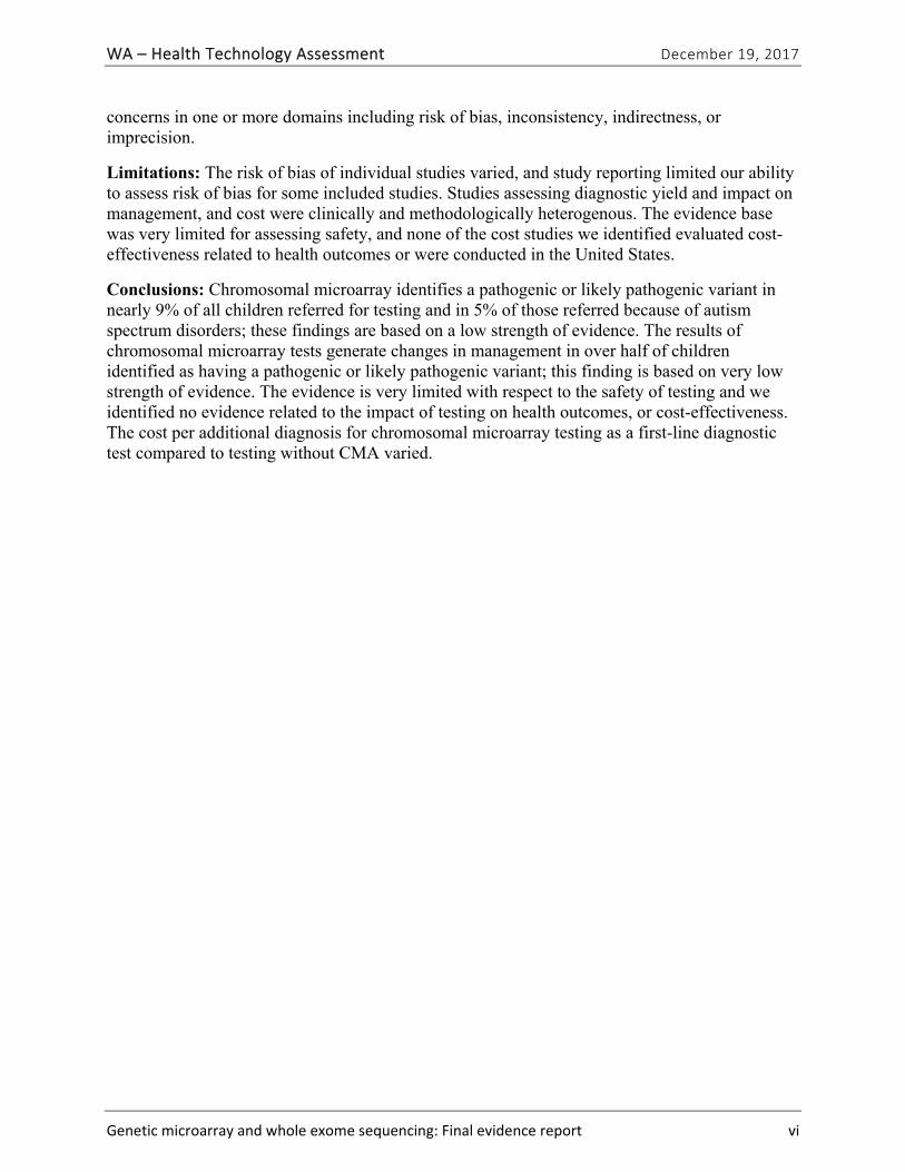
concerns in one or more domains including risk of bias, inconsistency, indirectness, or imprecision.
Limitations: The risk of bias of individual studies varied, and study reporting limited our ability to assess risk of bias for some included studies. Studies assessing diagnostic yield and impact on management, and cost were clinically and methodologically heterogenous. The evidence base was very limited for assessing safety, and none of the cost studies we identified evaluated cost-effectiveness related to health outcomes or were conducted in the United States.
Conclusions: Chromosomal microarray identifies a pathogenic or likely pathogenic variant in nearly 9% of all children referred for testing and in 5% of those referred because of autism spectrum disorders; these findings are based on a low strength of evidence. The results of chromosomal microarray tests generate changes in management in over half of children identified as having a pathogenic or likely pathogenic variant; this finding is based on very low strength of evidence. The evidence is very limited with respect to the safety of testing and we identified no evidence related to the impact of testing on health outcomes, or cost-effectiveness. The cost per additional diagnosis for chromosomal microarray testing as a first-line diagnostic test compared to testing without CMA varied.
WA – Health Technology Assessment December 19, 2017
Genetic microarray and whole exome sequencing: Final evidence report ES ‐1
Executive Summary
Background
Condition Description
Chromosomes, the genetic structures of a cell, are constructed of deoxyribose nucleic acid (DNA) and the proteins and other elements that protect, regulate, and package the DNA. Humans normally have 23 pairs of chromosomes, with half inherited from each parent. During cell replication, chromosomes are sometimes lost or broken and rearranged. Rearrangements vary in size and complexity, and may be balanced, with no loss of DNA, or unbalanced with loss or gain of DNA.
Disease Burden
Unbalanced chromosomal rearrangements that are present at conception or that occur during fetal development have profound consequences for the developing fetus, resulting in fetal death, structural defects, genetic diseases, or intellectual impairment.1 Chromosomal abnormalities occur in 43.8 per 10,000 births that survive to 20 weeks gestation or later.2 Trisomies 21 (Down syndrome), 18, and 13; 45, X (Turner syndrome), and other sex chromosome abnormalities account for most abnormalities. Excluding these, the prevalence of more rare abnormalities is 7.4 per 10,000 births.2 Small pathogenic duplications or deletions, called copy number changes or variants (CNV), occur in 1 of 270 pregnancies.3 The consequences of CNVs depend on the size and location within the genome.
Approximately 3% of infants born in the United States have a major structural defect,4 and almost 6% of children in the United States have an intellectual disability (ID), developmental delay (DD), or autism spectrum disorder (ASD).5 These conditions are expensive to manage: in 2004, U.S. hospitalization costs for birth defects totaled $2.6 billion.6 In Washington, 10.3% of adults living in the state in 2014 had a cognitive disability.7 State expenditures in caring for residents with an ID included over $600 million in Medicaid expenditures for long-term care and over $1 billion on special education programs.
Technology Description
The quest of genetics laboratories over the last 60 years has been to increase the resolution of genetic tests and to reduce the level of targeting required. This report discusses chromosomal microarray (CMA) and whole exome sequencing (WES), untargeted genome-wide tests that detect changes across the genome. CMA can detect unbalanced changes as small as 30,000 base pairs, and WES can detect changes as small as a single base pair.
Chromosomal Microarrays (CMA)
In the early 2000s, genome-wide microarrays for chromosomal analysis, commonly known as CMA, were introduced as an adjunct to karyotype and fluorescent in situ hybridization (FISH) testing for chromosome abnormalities. Comparative genomic hybridization (CGH) or single nucleotide polymorphism (SNP) arrays are used to evaluate the number of copies of portions of the chromosomes. In CGH, patient and control DNA samples are tagged with fluorescent
WA – Health Technology Assessment December 19, 2017
Genetic microarray and whole exome sequencing: Final evidence report ES ‐2
markers and hybridized to probes.8 Computer analysis determines the number of copies of each chromosomal region present based on the intensity and color of fluorescence. The first genome-wide platforms used bacterial artificial chromosome (BAC) probes that could detect deletions or duplications of approximately 1,000,000 base pairs (1 Mb).8 Around 2007, oligonucleotide arrays (‘oligo’) began replacing BAC arrays. Oligonucleotide probes are smaller, and oligo-based arrays have many more probes, enabling detection of smaller CNVs.9 SNP arrays tag individual base pairs throughout the genome that vary within the normal population with different fluorescent dyes. The number of alleles and whether the individual has the same allele on both chromosome or different alleles can be determined by analysis of the color and intensity of the bound fluorescent dyes.8 Many current CMA testing platforms use a combination of labeled SNPs and oligo-based probes to assess genetic bases or sequences throughout the genome.
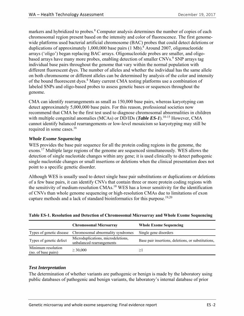
CMA can identify rearrangements as small as 150,000 base pairs, whereas karyotyping can detect approximately 5,000,000 base pairs. For this reason, professional societies now recommend that CMA be the first test used to diagnose chromosomal abnormalities in children with multiple congenital anomalies (MCAs) or DD/IDs (Table ES-1).10-15 However, CMA cannot identify balanced rearrangements or low-level mosaicism so karyotyping may still be required in some cases.16
Whole Exome Sequencing WES provides the base pair sequence for all the protein coding regions in the genome, the exons.17 Multiple large regions of the genome are sequenced simultaneously. WES allows the detection of single nucleotide changes within any gene; it is used clinically to detect pathogenic single nucleotide changes or small insertions or deletions when the clinical presentation does not point to a specific genetic disorder.
Although WES is usually used to detect single base pair substitutions or duplications or deletions of a few base pairs, it can identify CNVs that contain three or more protein coding regions with the sensitivity of medium-resolution CMAs.18 WES has a lower sensitivity for the identification of CNVs than whole genome sequencing or high-resolution CMAs due to limitations of exon capture methods and a lack of standard bioinformatics for this purpose.19,20
Table ES-1. Resolution and Detection of Chromosomal Microarray and Whole Exome Sequencing
Chromosomal Microarray Whole Exome Sequencing
Types of genetic disease Chromosomal abnormality syndromes Single gene disorders
Types of genetic defect Microduplications, microdeletions, unbalanced rearrangements
Base pair insertions, deletions, or substitutions,
Minimum resolution (no. of base pairs)
≥ 30,000 ≥1
Test Interpretation The determination of whether variants are pathogenic or benign is made by the laboratory using public databases of pathogenic and benign variants, the laboratory’s internal database of prior
WA – Health Technology Assessment December 19, 2017
Genetic microarray and whole exome sequencing: Final evidence report ES ‐3
test results, published literature, and consultation with other laboratories. The American College of Medical Genetics (ACMG) and the Association of Molecular Pathology published guidelines for the interpretation of sequence variants,21 and tools have been developed to aid in their use.22
Genome-wide testing can result in secondary or incidental findings. The ACMG recommends that laboratories conducting clinical exome or genome sequencing actively seek and report a list of specific, clinically actionable variants within 24 genes or classes of genes,23 regardless of the indication for testing. Approximately 2% of patients sequenced are expected have a reportable variant. The ACMG recommendations do not address copy number variants (CNVs) or structural abnormalities, but the apply to CNVs that delete or disable listed genes with autosomal dominant inheritance. Incidental findings from CMA have been studied less than those from sequencing; the studies that have examined them have found less than 1% of individuals tested have an incidental finding.24
Policy Context
The State of Washington Health Care Authority selected testing with CMA and WES as a topic based on medium, high, and high concerns for safety, efficacy, and cost, respectively. Several practice guidelines have been issued that call for CMA to replace G-banded karyotype as the first-tier test for diagnosis of individuals with DD, ID, or MCA, and for the clinical evaluation of ASD.13,25 These guidelines, combined with the increasing prevalence of autism,26 could greatly increase orders for CMA. The increased diagnostic yield of chromosomal abnormalities by CMA compared to karyotype underlies these guidelines.13,25 The degree of DD or ID for which CMA is most likely to yield a diagnosis is unclear, however, as is the effect of the testing results on the medical and educational management and health outcomes of affected children.
Regulatory Status
CMA and WES are considered laboratory-developed tests and are not regulated by the United States (U.S.) Food and Drug Administration (FDA). Clinical laboratories that conduct these tests must comply with regulatory standards for high complexity testing within the Clinical Laboratory Improvement Act (CLIA). Thus, these tests are generally only available through commercial diagnostic testing laboratories or hospital-based laboratories.
FDA approval is required when a company markets and sells a kit for CMA or WES testing. The FDA has approved two CMA kits for marketing in the United States: the Affymetrix CytoScan® Dx assay (Affymetrix, Inc., Santa Clara, CA), approved as a Class II test on January 21, 201427 and the Agilent GenetiSure Dx Postnatal Assay (Agilent Technologies, Inc., Santa Clara, CA),28 approved under a substantial equivalence determination on August 14, 2017. The FDA-approved indications for the kits include postnatal detection of CNVs associated with DD, ID, MCA, or dysmorphic features.
Practice Guidelines and Payer Coverage
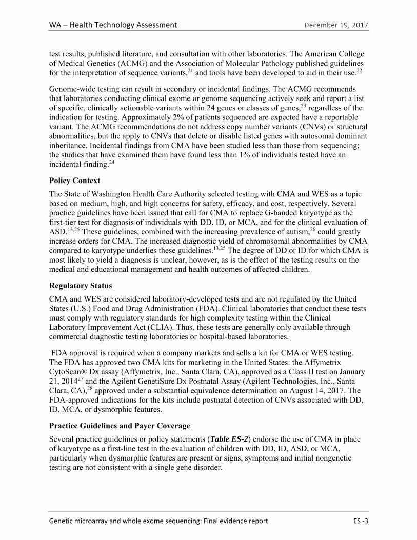
Several practice guidelines or policy statements (Table ES-2) endorse the use of CMA in place of karyotype as a first-line test in the evaluation of children with DD, ID, ASD, or MCA, particularly when dysmorphic features are present or signs, symptoms and initial nongenetic testing are not consistent with a single gene disorder.
WA – Health Technology Assessment December 19, 2017
Genetic microarray and whole exome sequencing: Final evidence report ES ‐4
Table ES-2. Practice Guidelines Endorsing or Providing Guidance on Chromosomal Microarray Testing
Organization Year
International Standard Cytogenomic Consortium25 2010
National Institute for Health and Care Excellence (UK)12 2011
American College of Medical Genetics and Genomics13 2013
American Academy of Pediatrics15 2014
American Academy of Neurology14 2015
The Centers for Medicare and Medicaid Services (CMS) has no national coverage determination for the use of CMA or WES. Table ES-3 summarizes selected payer coverage determinations for CMA and WES testing. Among payers, good alignment exists for the criteria under which CMA is covered. Typically, it is covered as first-line diagnostic for DD, ID, and ASD when relevant biochemical and metabolic diseases have been ruled out, the clinical presentation is not specific to a well-delineated genetic syndrome, and the results of CMA could impact the clinical management of the child.
Table ES-3. Payer Coverage for CMA and WES Testing
Payer CMA Testing WES Testing
Aetna29 Covered for specific indications Not covered
Blue Cross (Premera)30 Covered for specific indications Covered for specific indications
Regence Blue Shield Regence31,32 Covered for specific indications Not covered
Cigna33,34 Covered for specific indications Covered for specific indications
Humana35 Covered for specific indications Not covered
Kaiser Permanente36 Covered for specific indications Not covered
Medicare Fee for Service None None
Medicaid15,37,38 Not all states have policies; those that do typically cover for specific indications
Unknown
UnitedHealthcare39 Covered for specific indications Covered for specific indications
Research Questions
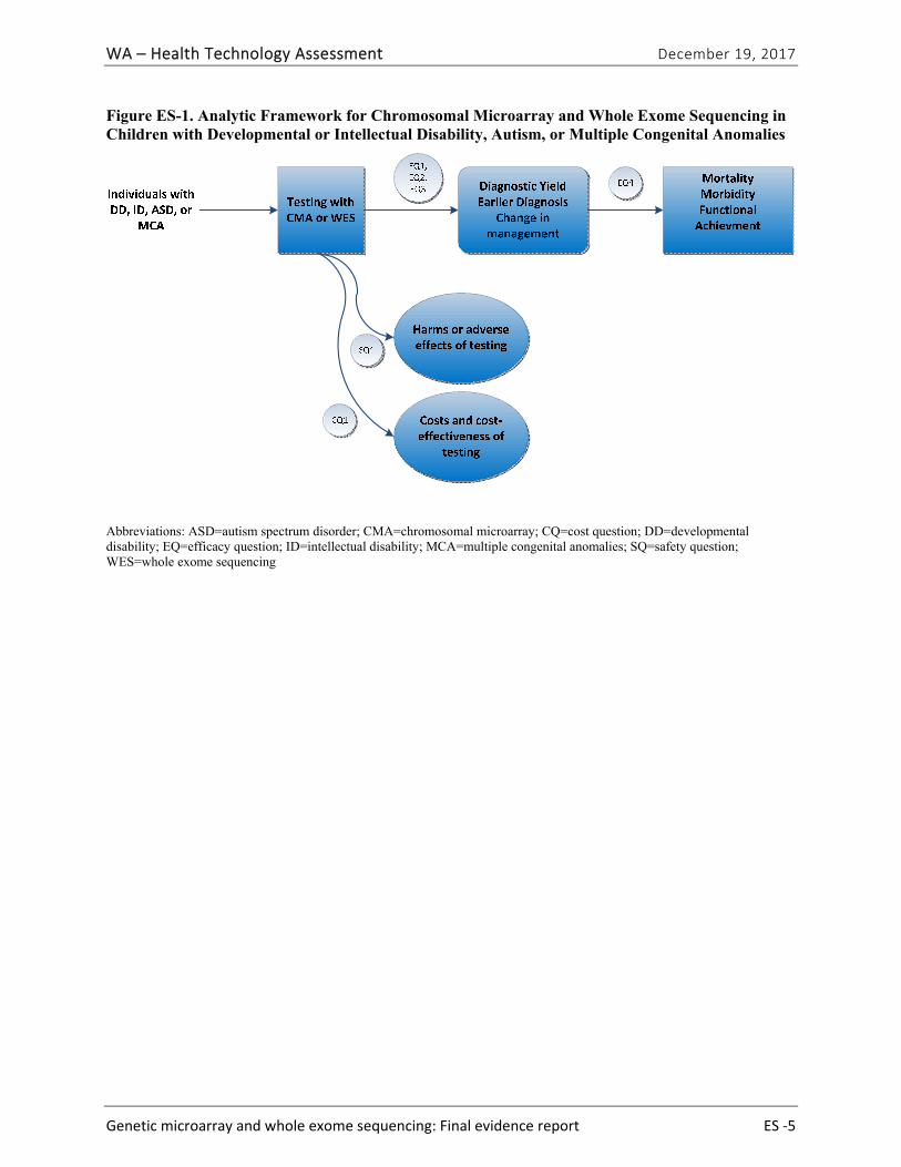
Figure ES-1 provides the analytic framework and Table ES-4 provides the final research questions and study selection criteria related to the population, intervention, comparator, outcomes, time period, and setting used to conduct this health technology assessment (HTA).
WA – Health Technology Assessment December 19, 2017
Genetic microarray and whole exome sequencing: Final evidence report ES ‐5
Figure ES-1. Analytic Framework for Chromosomal Microarray and Whole Exome Sequencing in Children with Developmental or Intellectual Disability, Autism, or Multiple Congenital Anomalies
Abbreviations: ASD=autism spectrum disorder; CMA=chromosomal microarray; CQ=cost question; DD=developmental disability; EQ=efficacy question; ID=intellectual disability; MCA=multiple congenital anomalies; SQ=safety question; WES=whole exome sequencing
WA – Health Technology Assessment December 19, 2017
Genetic microarray and whole exome sequencing: Final evidence report ES ‐6
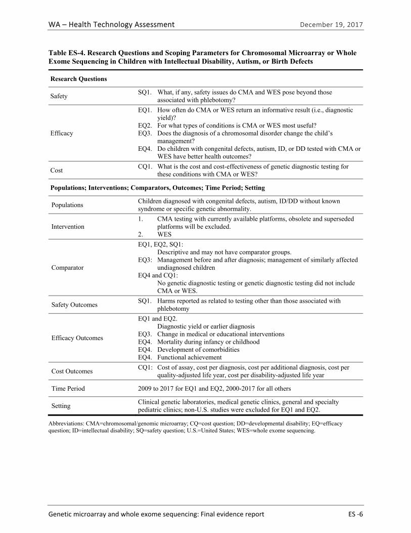
Table ES-4. Research Questions and Scoping Parameters for Chromosomal Microarray or Whole Exome Sequencing in Children with Intellectual Disability, Autism, or Birth Defects
Research Questions
Safety SQ1. What, if any, safety issues do CMA and WES pose beyond those
associated with phlebotomy?
Efficacy
EQ1. How often do CMA or WES return an informative result (i.e., diagnostic yield)?
EQ2. For what types of conditions is CMA or WES most useful? EQ3. Does the diagnosis of a chromosomal disorder change the child’s
management? EQ4. Do children with congenital defects, autism, ID, or DD tested with CMA or
WES have better health outcomes?
Cost CQ1. What is the cost and cost-effectiveness of genetic diagnostic testing for
these conditions with CMA or WES?
Populations; Interventions; Comparators, Outcomes; Time Period; Setting
Populations Children diagnosed with congenital defects, autism, ID/DD without known syndrome or specific genetic abnormality.
Intervention 1. CMA testing with currently available platforms, obsolete and superseded
platforms will be excluded. 2. WES
Comparator
EQ1, EQ2, SQ1: Descriptive and may not have comparator groups. EQ3: Management before and after diagnosis; management of similarly affected
undiagnosed children EQ4 and CQ1: No genetic diagnostic testing or genetic diagnostic testing did not include
CMA or WES.
Safety Outcomes SQ1. Harms reported as related to testing other than those associated with
phlebotomy
Efficacy Outcomes
EQ1 and EQ2. Diagnostic yield or earlier diagnosis EQ3. Change in medical or educational interventions EQ4. Mortality during infancy or childhood EQ4. Development of comorbidities EQ4. Functional achievement
Cost Outcomes CQ1: Cost of assay, cost per diagnosis, cost per additional diagnosis, cost per
quality-adjusted life year, cost per disability-adjusted life year
Time Period 2009 to 2017 for EQ1 and EQ2, 2000-2017 for all others
Setting Clinical genetic laboratories, medical genetic clinics, general and specialty pediatric clinics; non-U.S. studies were excluded for EQ1 and EQ2.
Abbreviations: CMA=chromosomal/genomic microarray; CQ=cost question; DD=developmental disability; EQ=efficacy question; ID=intellectual disability; SQ=safety question; U.S.=United States; WES=whole exome sequencing.
WA – Health Technology Assessment December 19, 2017
Genetic microarray and whole exome sequencing: Final evidence report ES ‐7
What is Excluded from This HTA
This HTA does not address the analytic validity of CMA or WES, because this testing is available within CLIA-licensed laboratories as a laboratory-developed test and analytic validity is assumed based on meeting those standards.40 This review is focused on the diagnosis of chromosomal abnormalities; therefore, we do not address single gene testing for these disorders, including the use of WES to identify mutations within single genes. The review does not assess either the ethical issues or the clinical utility of incidental findings not related to the health conditions for which the tests were ordered. Because of the large volume of studies on diagnostic yield (EQ1) and the rapidly evolving technology in use for CMA testing, we limited the studies considered for EQ1 to those conducted in the United States in 2009 or later that used current testing platforms.
Methods
Data Sources and Search
We searched MEDLINE® (via PubMed) and a clinical trials registry (clinicaltrials.gov) for relevant English-language studies published in 2000 or later. We searched the FDA website, selected payer and health care professional society websites, and other organizations that conduct and disseminate HTAs. In addition, we reviewed the reference lists of relevant studies, practice guidelines, and other HTAs on this topic to identify any relevant articles not found through the electronic search. The detailed search strategy is provided in Appendix A of the Full Report.
Study Selection
We screened titles and abstracts and full-text articles based on the study selection criteria listed in Table ES-3. We included all study designs except case reports. A single team member screened titles/abstracts following an initial set of 20 independent, dual reviews with the entire team to assess interrater reliability. The principal investigator reviewed all abstracts excluded for “ineligible intervention” and a sample of titles/abstracts excluded for other reasons to ensure continued consistency in application of study selection criteria. One senior team member screened each full-text article for inclusion, and the principal investigator confirmed the decisions.
Data Abstraction and Quality Assessment
One team member extracted relevant study data into a structured abstraction form. The principal investigator reviewed the abstractions for accuracy and consistency. Two senior team members conducted an independent risk of bias assessment on all included studies and met to reconcile any disagreements, in consultation with the principal investigator if needed. Because of the diverse types of studies included in this HTA, we adapted signaling questions from the QUADAS-2 instrument, a risk of bias assessment for diagnostics test studies, and items from the RTI item bank for observational studies.41,42 The signaling questions assessed the major sources of bias including selection bias (both how study population was selected and attrition/missing data), confounding, and measurement/information bias. We used the ROBIS instrument to assess the risk of bias for systematic reviews.43
WA – Health Technology Assessment December 19, 2017
Genetic microarray and whole exome sequencing: Final evidence report ES ‐8
Data Synthesis and Analysis
Study characteristics and results were qualitatively synthesized for each research question in tabular and narrative formats. For cost outcomes, we adjusted all reported outcomes to 2010 U.S. dollars (Appendix B).44,45 To determine whether quantitative synthesis was appropriate, we assessed the number of studies and the clinical and methodological heterogeneity present based on established guidance.46,47 We required three or more publications with similar approach and the same outcome measure to calculate a summary estimate. We estimated summary effects using a fixed effects model if the test for heterogeneity was nonsignificant and a random effects model if the test for heterogeneity was significant using OpenMetaAnalyst (for Windows 8, 64-bit) and the method of Hedges and Olkin to estimate between-study variance.48 We graded the strength of evidence for each research question using GRADE, which assesses the strength of evidence based on domains relating to risk of bias, inconsistency, imprecision, indirectness, and other considerations, such as reporting bias.49 Under GRADE, the strength of evidence can be graded as very low, low, moderate, or high.
Results
Literature Search
We identified and screened 2,717 unique citations. We excluded 2,375 after title and abstract review. We reviewed the full-text of 348 articles, and excluded 330 for the reasons listed in Figure 2 of the Full Report. We included 18 studies. One provided evidence on safety issues (SQ1), seven provided evidence on diagnostic yield (EQ1 and EQ2), seven on changes in management (EQ3), and five on costs (CQ1). No studies provided information on health outcomes (EQ4). Individual study characteristics for all included studies are summarized in Full Report Appendix C, Table C-1. The list of studies we screened at the full-text stage, but which were excluded from the review, is provided in Appendix D. Note that studies may have been excluded based on more than one reason but we report only one reason. Individual risk of bias assessments for all included studies are reported in Appendix E.
Safety
SQ1. What, if any, safety issues do CMA and WES pose beyond those associated with phlebotomy?
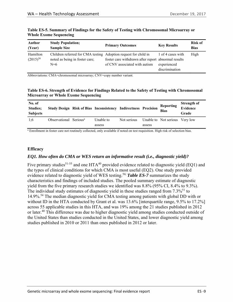
One study50 provided evidence on safety issues that arise in CMA testing. Table ES-5 summarizes the study characteristics and key outcomes related to discrimination resulting from testing. We graded the strength of evidence for this research question as very low (Table ES-6). We did not identify any studies reporting on safety outcomes related to WES testing.
WA – Health Technology Assessment December 19, 2017
Genetic microarray and whole exome sequencing: Final evidence report ES ‐9
Table ES-5. Summary of Findings for the Safety of Testing with Chromosomal Microarray or Whole Exome Sequencing
Author (Year)
Study Population; Sample Size
Primary Outcomes Key Results Risk of Bias
Hamilton (2015)50
Children referred for CMA testing noted as being in foster care; N=6
Adoption request for child in foster care withdrawn after report of CNV associated with autism
1 of 4 cases with abnormal results experienced discrimination
High
Abbreviations: CMA=chromosomal microarray; CNV=copy number variant.
Table ES-6. Strength of Evidence for Findings Related to the Safety of Testing with Chromosomal Microarray or Whole Exome Sequencing
No. of Studies; Subjects
Study Design Risk of Bias Inconsistency Indirectness PrecisionReporting Bias
Strength of Evidence Grade
1;6 Observational Seriousa Unable to assess
Not serious Unable to assess
Not serious Very low
a Enrollment in foster care not routinely collected, only available if noted on test requisition. High risk of selection bias.
Efficacy
EQ1. How often do CMA or WES return an informative result (i.e., diagnostic yield)?
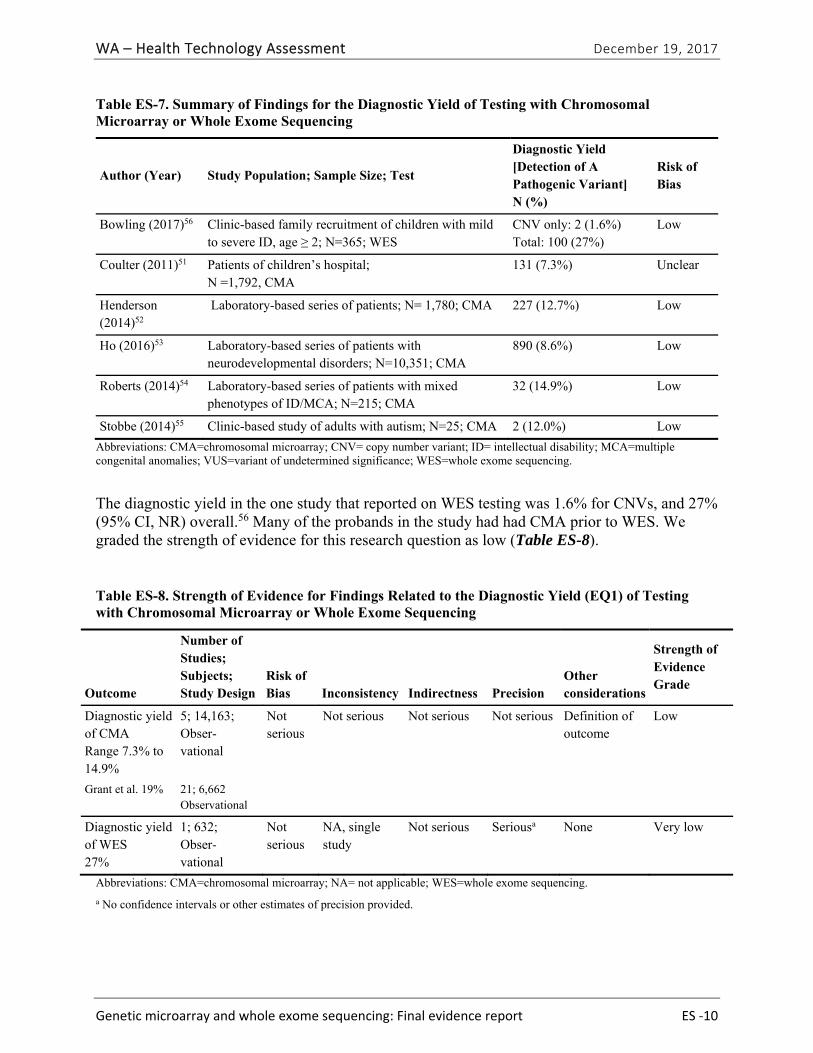
Five primary studies51-55 and one HTA40 provided evidence related to diagnostic yield (EQ1) and the types of clinical conditions for which CMA is most useful (EQ2). One study provided evidence related to diagnostic yield of WES testing.56| Table ES-7 summarizes the study characteristics and findings of included studies. The pooled summary estimate of diagnostic yield from the five primary research studies we identified was 8.8% (95% CI, 8.4% to 9.3%). The individual study estimates of diagnostic yield in these studies ranged from 7.3%51 to 14.9%.54 The median diagnostic yield for CMA testing among patients with global DD with or without ID in the HTA conducted by Grant et al. was 13.6% [interquartile range, 9.5% to 17.2%] across 55 applicable studies in this HTA, and was 19% among the 21 studies published in 2012 or later.40 This difference was due to higher diagnostic yield among studies conducted outside of the United States than studies conducted in the United States, and lower diagnostic yield among studies published in 2010 or 2011 than ones published in 2012 or later.
WA – Health Technology Assessment December 19, 2017
Genetic microarray and whole exome sequencing: Final evidence report ES ‐10
Table ES-7. Summary of Findings for the Diagnostic Yield of Testing with Chromosomal Microarray or Whole Exome Sequencing
Author (Year) Study Population; Sample Size; Test
Diagnostic Yield [Detection of A Pathogenic Variant] N (%)
Risk of Bias
Bowling (2017)56 Clinic-based family recruitment of children with mild to severe ID, age ≥ 2; N=365; WES
CNV only: 2 (1.6%) Total: 100 (27%)
Low
Coulter (2011)51 Patients of children’s hospital; N =1,792, CMA
131 (7.3%) Unclear
Henderson (2014)52
Laboratory-based series of patients; N= 1,780; CMA 227 (12.7%) Low
Ho (2016)53 Laboratory-based series of patients with neurodevelopmental disorders; N=10,351; CMA
890 (8.6%) Low
Roberts (2014)54 Laboratory-based series of patients with mixed phenotypes of ID/MCA; N=215; CMA
32 (14.9%) Low
Stobbe (2014)55 Clinic-based study of adults with autism; N=25; CMA 2 (12.0%) Low
Abbreviations: CMA=chromosomal microarray; CNV= copy number variant; ID= intellectual disability; MCA=multiple congenital anomalies; VUS=variant of undetermined significance; WES=whole exome sequencing.
The diagnostic yield in the one study that reported on WES testing was 1.6% for CNVs, and 27% (95% CI, NR) overall.56 Many of the probands in the study had had CMA prior to WES. We graded the strength of evidence for this research question as low (Table ES-8).
Table ES-8. Strength of Evidence for Findings Related to the Diagnostic Yield (EQ1) of Testing with Chromosomal Microarray or Whole Exome Sequencing
Outcome
Number of Studies; Subjects; Study Design
Risk of Bias Inconsistency Indirectness Precision
Other considerations
Strength of Evidence Grade
Diagnostic yield of CMA Range 7.3% to 14.9%
5; 14,163; Obser-vational
Not serious
Not serious Not serious Not serious Definition of outcome
Low
Grant et al. 19% 21; 6,662 Observational
Diagnostic yield of WES 27%
1; 632; Obser-vational
Not serious
NA, single study
Not serious Seriousa None Very low
Abbreviations: CMA=chromosomal microarray; NA= not applicable; WES=whole exome sequencing.
a No confidence intervals or other estimates of precision provided.
WA – Health Technology Assessment December 19, 2017
Genetic microarray and whole exome sequencing: Final evidence report ES ‐11
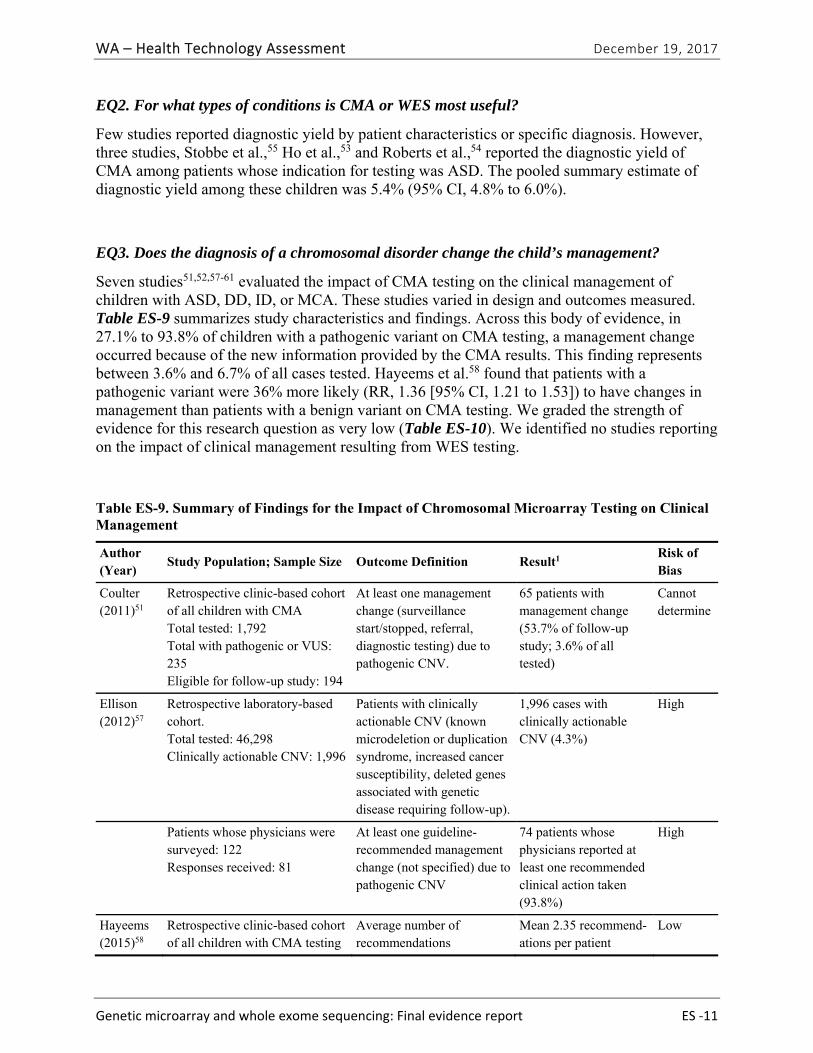
EQ2. For what types of conditions is CMA or WES most useful?
Few studies reported diagnostic yield by patient characteristics or specific diagnosis. However, three studies, Stobbe et al.,55 Ho et al.,53 and Roberts et al.,54 reported the diagnostic yield of CMA among patients whose indication for testing was ASD. The pooled summary estimate of diagnostic yield among these children was 5.4% (95% CI, 4.8% to 6.0%).
EQ3. Does the diagnosis of a chromosomal disorder change the child’s management?
Seven studies51,52,57-61 evaluated the impact of CMA testing on the clinical management of children with ASD, DD, ID, or MCA. These studies varied in design and outcomes measured. Table ES-9 summarizes study characteristics and findings. Across this body of evidence, in 27.1% to 93.8% of children with a pathogenic variant on CMA testing, a management change occurred because of the new information provided by the CMA results. This finding represents between 3.6% and 6.7% of all cases tested. Hayeems et al.58 found that patients with a pathogenic variant were 36% more likely (RR, 1.36 [95% CI, 1.21 to 1.53]) to have changes in management than patients with a benign variant on CMA testing. We graded the strength of evidence for this research question as very low (Table ES-10). We identified no studies reporting on the impact of clinical management resulting from WES testing.
Table ES-9. Summary of Findings for the Impact of Chromosomal Microarray Testing on Clinical Management
Author (Year)
Study Population; Sample Size Outcome Definition Result1 Risk of Bias
Coulter (2011)51
Retrospective clinic-based cohort of all children with CMA Total tested: 1,792 Total with pathogenic or VUS: 235 Eligible for follow-up study: 194
At least one management change (surveillance start/stopped, referral, diagnostic testing) due to pathogenic CNV.
65 patients with management change (53.7% of follow-up study; 3.6% of all tested)
Cannot determine
Ellison (2012)57
Retrospective laboratory-based cohort. Total tested: 46,298 Clinically actionable CNV: 1,996
Patients with clinically actionable CNV (known microdeletion or duplication syndrome, increased cancer susceptibility, deleted genes associated with genetic disease requiring follow-up).
1,996 cases with clinically actionable CNV (4.3%)
High
Patients whose physicians were surveyed: 122 Responses received: 81
At least one guideline- recommended management change (not specified) due to pathogenic CNV
74 patients whose physicians reported at least one recommended clinical action taken (93.8%)
High
Hayeems (2015)58
Retrospective clinic-based cohort of all children with CMA testing
Average number of recommendations
Mean 2.35 recommend-ations per patient
Low
WA – Health Technology Assessment December 19, 2017
Genetic microarray and whole exome sequencing: Final evidence report ES ‐12
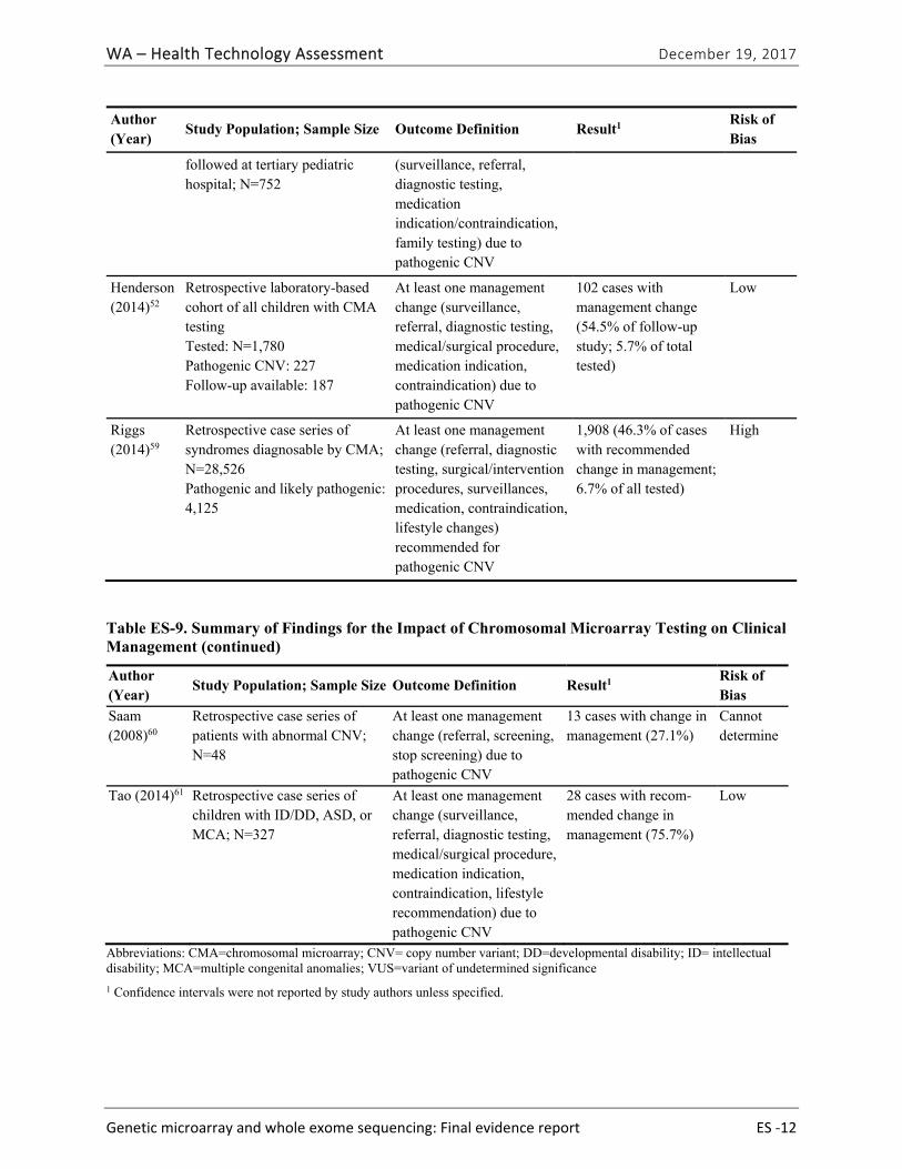
Author (Year)
Study Population; Sample Size Outcome Definition Result1 Risk of Bias
followed at tertiary pediatric hospital; N=752
(surveillance, referral, diagnostic testing, medication indication/contraindication, family testing) due to pathogenic CNV
Henderson (2014)52
Retrospective laboratory-based cohort of all children with CMA testing Tested: N=1,780 Pathogenic CNV: 227 Follow-up available: 187
At least one management change (surveillance, referral, diagnostic testing, medical/surgical procedure, medication indication, contraindication) due to pathogenic CNV
102 cases with management change (54.5% of follow-up study; 5.7% of total tested)
Low
Riggs (2014)59
Retrospective case series of syndromes diagnosable by CMA; N=28,526 Pathogenic and likely pathogenic: 4,125
At least one management change (referral, diagnostic testing, surgical/intervention procedures, surveillances, medication, contraindication, lifestyle changes) recommended for pathogenic CNV
1,908 (46.3% of cases with recommended change in management; 6.7% of all tested)
High
Table ES-9. Summary of Findings for the Impact of Chromosomal Microarray Testing on Clinical Management (continued)
Author (Year)
Study Population; Sample Size Outcome Definition Result1 Risk of Bias
Saam (2008)60
Retrospective case series of patients with abnormal CNV; N=48
At least one management change (referral, screening, stop screening) due to pathogenic CNV
13 cases with change in management (27.1%)
Cannot determine
Tao (2014)61 Retrospective case series of children with ID/DD, ASD, or MCA; N=327
At least one management change (surveillance, referral, diagnostic testing, medical/surgical procedure, medication indication, contraindication, lifestyle recommendation) due to pathogenic CNV
28 cases with recom-mended change in management (75.7%)
Low
Abbreviations: CMA=chromosomal microarray; CNV= copy number variant; DD=developmental disability; ID= intellectual disability; MCA=multiple congenital anomalies; VUS=variant of undetermined significance
1 Confidence intervals were not reported by study authors unless specified.
WA – Health Technology Assessment December 19, 2017
Genetic microarray and whole exome sequencing: Final evidence report ES ‐13
Table ES-10. Strength of Evidence for Findings Related to the Impact of Testing with Chromosomal Microarray on Clinical Management (EQ3)
Outcome
Number of Studies; Subjects; Study Design
Risk of Bias
Inconsistency Indirectness Precision Other Consider-ations
Strength of Evidence Grade
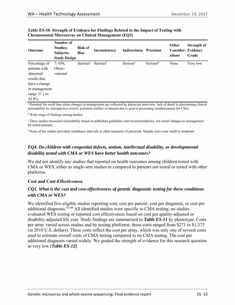
Percentage of patients with abnormal results that have a change in management range 27.1 to 93.8%
7; 658; Obser-vational
Seriousa Seriousb Seriousc Seriousd None Very low
a Potential for recall bias when changes in management are collected by physician interview, lack of detail in determining clinical actionability by retrospective review, potential conflict of interest due to goal of promoting reimbursement for CMA.
b Wide range of findings among studies.
c Three studies measured actionability based on published guidelines and recommendations, not actual changes in management for tested patients.
d None of the studies provided confidence intervals or other measures of precision. Sample sizes were small to moderate.
EQ4. Do children with congenital defects, autism, intellectual disability, or developmental disability tested with CMA or WES have better health outcomes?
We did not identify any studies that reported on health outcomes among children tested with CMA or WES, either as single-arm studies or compared to patients not tested or tested with other platforms.
Cost and Cost-Effectiveness
CQ1. What is the cost and cost-effectiveness of genetic diagnostic testing for these conditions with CMA or WES?
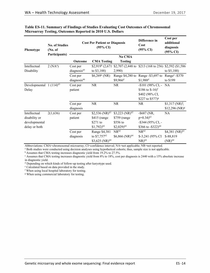
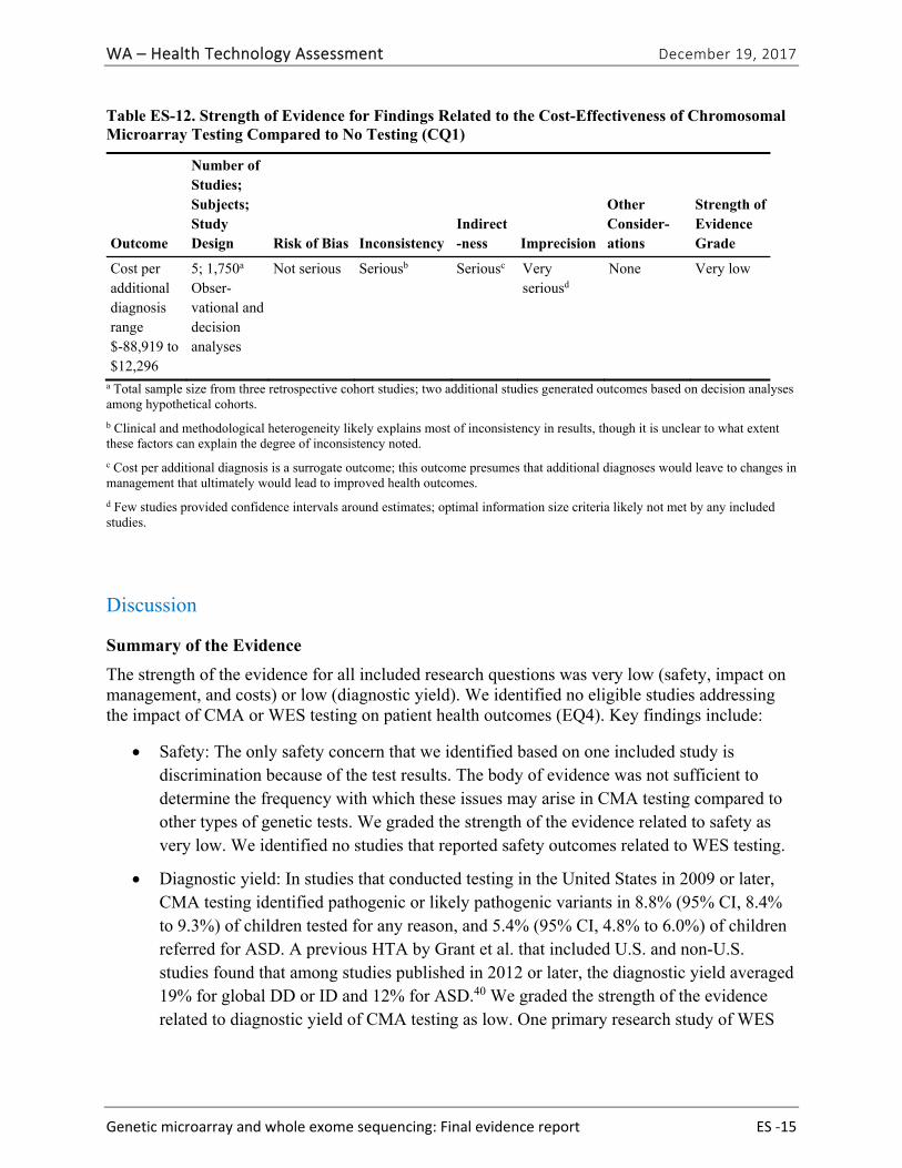
We identified five eligible studies reporting cost, cost per patient, cost per diagnosis, or cost per additional diagnosis.62-66 All identified studies were specific to CMA testing; no studies evaluated WES testing or reported cost effectiveness based on cost per quality-adjusted or disability-adjusted life year. Study findings are summarized in Table ES-11 by phenotype. Costs per array varied across studies and by testing platforms; these costs ranged from $271 to $1,575 (in 2010 U.S. dollars). These costs reflect the cost per array, which was only one of several costs used to estimate overall costs of CMA testing compared to no CMA testing. The cost per additional diagnosis varied widely. We graded the strength of evidence for this research question as very low (Table ES-12).
WA – Health Technology Assessment December 19, 2017
Genetic microarray and whole exome sequencing: Final evidence report ES ‐14
Table ES-11. Summary of Findings of Studies Evaluating Cost Outcomes of Chromosomal Microarray Testing, Outcomes Reported in 2010 U.S. Dollars
Phenotype No. of Studies (No. of Participants)
Cost Per Patient or Diagnosis (95% CI)
Difference in Cost (95% CI)
Cost per additional diagnosis (95% CI)
Outcome CMA TestingNo CMA Testing
Intellectual Disability
2 (NAa) Cost per diagnosis65
$2,919b (2,671 to $3,188)
$2,707 (2,448 to 2,990)
$213 (168 to 256) $2,592 ($1,586 to $5,188)
Cost per diagnosis66
$6,269c (NR) Range $4,280 to $9,966d
Range -$3,697 to $1,988d
Rangee -$370 to $199
Developmental Delay
1 (114)63 Cost per patient
NR NR -$101 (98% CI, -$186 to $-16)f $402 (98% CI, $227 to $577)g
NA
Cost per diagnosis
NR NR NR $1,317 (NR)f; $12,296 (NR)g
Intellectual disability or developmental delay or both
2(1,636) Cost per patient
$2,536 (NR)62 $415 (range $271 to $1,792)64
$3,223 (NR)62 $759 (range $556 to $2,029)64
-$687 (NR, p=0.34)62 -$344 (95% CI, -$366 to -$322)64
NA
Cost per diagnosis
Range $4,381 to $7,75762 $3,625 (NR)64
NR62 $6,866 (NR)64
NR62 $-3,241 (95% CI NR)64
$4,381 (NR)62 $-88,819 (NR)64
Abbreviations: CMA=chromosomal microarray; CI=confidence interval; NA=not applicable; NR=not reported. a Both studies were conducted using decision analyses using hypothetical cohorts; thus, sample size is not applicable. b Assumes that CMA testing increases diagnostic yield from 19.2% to 27.5%. c Assumes that CMA testing increases diagnostic yield from 8% to 18%, cost per diagnosis is 2440 with a 15% absolute increase in diagnostic yield. d Depending on which kinds of follow-up testing after karyotype used. e Calculated based on data provided in the study. f When using local hospital laboratory for testing. g When using commercial laboratory for testing.
WA – Health Technology Assessment December 19, 2017
Genetic microarray and whole exome sequencing: Final evidence report ES ‐15
Table ES-12. Strength of Evidence for Findings Related to the Cost-Effectiveness of Chromosomal Microarray Testing Compared to No Testing (CQ1)
Outcome
Number of Studies; Subjects; Study Design Risk of Bias Inconsistency
Indirect-ness Imprecision
Other Consider-ations
Strength of Evidence Grade
Cost per additional diagnosis range $-88,919 to $12,296
5; 1,750a Obser-vational and decision analyses
Not serious Seriousb Seriousc Very seriousd
None Very low
a Total sample size from three retrospective cohort studies; two additional studies generated outcomes based on decision analyses among hypothetical cohorts.
b Clinical and methodological heterogeneity likely explains most of inconsistency in results, though it is unclear to what extent these factors can explain the degree of inconsistency noted.
c Cost per additional diagnosis is a surrogate outcome; this outcome presumes that additional diagnoses would leave to changes in management that ultimately would lead to improved health outcomes.
d Few studies provided confidence intervals around estimates; optimal information size criteria likely not met by any included studies.
Discussion
Summary of the Evidence
The strength of the evidence for all included research questions was very low (safety, impact on management, and costs) or low (diagnostic yield). We identified no eligible studies addressing the impact of CMA or WES testing on patient health outcomes (EQ4). Key findings include:
Safety: The only safety concern that we identified based on one included study is discrimination because of the test results. The body of evidence was not sufficient to determine the frequency with which these issues may arise in CMA testing compared to other types of genetic tests. We graded the strength of the evidence related to safety as very low. We identified no studies that reported safety outcomes related to WES testing.
Diagnostic yield: In studies that conducted testing in the United States in 2009 or later, CMA testing identified pathogenic or likely pathogenic variants in 8.8% (95% CI, 8.4% to 9.3%) of children tested for any reason, and 5.4% (95% CI, 4.8% to 6.0%) of children referred for ASD. A previous HTA by Grant et al. that included U.S. and non-U.S. studies found that among studies published in 2012 or later, the diagnostic yield averaged 19% for global DD or ID and 12% for ASD.40 We graded the strength of the evidence related to diagnostic yield of CMA testing as low. One primary research study of WES
WA – Health Technology Assessment December 19, 2017
Genetic microarray and whole exome sequencing: Final evidence report ES ‐16
reported a diagnostic yield of 27% (95% CI, NR) and we graded the strength of this evidence as very low.
Impact on clinical management: CMA results prompted changes in clinical management in 27% to 94% of patients with a pathogenic variant, which was 3.6% to 6.7% of all patients tested. We graded the strength of this evidence as very low. We identified no studies reported on change in management related to WES testing.
Costs: The cost per additional diagnosis across this body of evidence ranged in 2010 U.S. dollars from $-88,819 to $12,296. No studies reported on cost per quality-adjusted or disability-adjusted life year. We graded the strength of the evidence on costs as very low.
Contextual Information
Our review revealed information related to CMA and WES that was not formally evaluated in our systematic review, but may add to the interpretation of our results.
CNVs in General Population
CMA testing on samples and phenotype information from the general population of Estonia found 0.7% had a DECIPHER-listed pathogenic variant, and 70% of individuals with a pathogenic variant reported clinical features consistent with their genetic findings.67
Analytic Validity
Compared to sequencing, the Affymetrix® CytoScan® Dx Assay identified 98.8% (95% CI: 93.5%, 99.8%) of duplications and 97.3% (92.3%, 99.1%) of deletions of 1000 base pairs or more, with a false positive rate of 1.2% (0.2%, 6.5%) for duplications and 2.7% (0.9%, 7.7%) for deletions.28 The Agilent GenetiSure Dx Postnatal Assay identified 97.6% (94.0%, 99.1%) of duplications and 96.9% (93.4%, 98.6%) of deletions of 10,000 base pairs or larger, with false positive rates of 2.4% (0.9%, 6.0%) and 3.1% (1.4%, 6.6%), respectively.28
Limitations and Applicability of the Evidence Base
Almost all studies we included focused on CMA. Clinical use of WES is still new, and the body of evidence regarding its impact is limited. Across the body of evidence for all research questions, study design, study population, and outcome measurement details were often sparse, resulting in our inability to assess the risk of bias for some studies. Most of the studies reporting on diagnostic yield included some cases for indications other than our population of interest. In addition, prior diagnostic testing received by the cases varied. The differences among studies likely increase the inconsistency between studies and the lack of precision in study estimates. Diagnostic yield among more homogenous case series may be more consistent within the same types of cases, but differ from our results.
Financial or intellectual conflicts of interest of the study authors were not addressed in the existing instruments we used; thus, we did not evaluate that aspect of the risk of bias. Authors of several included studies stated that a goal of the research was to provide evidence of clinical utility to get CMA covered by payors, potentially providing a strong incentive for analytic decisions that would increase the estimate of diagnostic yield or impact on management. Studies
WA – Health Technology Assessment December 19, 2017
Genetic microarray and whole exome sequencing: Final evidence report ES ‐17
evaluating the impact of testing on management were small, so each included only a small portion of known microduplication or microdeletion syndromes. The clinical features of these syndromes and the appropriate management actions vary accordingly, and are likely an explanation for the large heterogeneity of estimates on impact on management.
The body of evidence related to cost and cost-effectiveness is limited by the lack of studies conducted in the United States, the absence of a societal perspective in any of the analyses, and the absence of cost per quality-adjusted or disability-adjusted life year outcomes. Further, this body of evidence is limited by extreme clinical and methodological heterogeneity, which most likely explains the inconsistency in cost per additional diagnosis. The precise role of these tests in the overall sequence and approach to diagnostic evaluation in children with DD, ID, and ASD has also evolved; thus, the cost of the diagnostic journey with or without CMA testing reflected in the included studies may no longer be relevant to current clinical practice.
Limitations of this HTA
We did not include studies published in languages other than English and only searched two U.S.-based electronic databases. We used a single reviewer to screen most titles/abstracts, which may have led to studies inappropriately excluded. For the research question related to diagnostic yield (EQ1), we restricted eligibility to studies with CMA conducted in the United States in 2009 or later that used current testing platforms to reduce heterogeneity and provide results more applicable to what is in current clinical use. We did not assess analytic validity or reproducibility or conduct an in-depth analysis or synthesis of the cases, breakpoints, or other information related to CNV findings that were presented by study authors. In addition, our review was limited to the use of WES to detect chromosomal abnormalities. WES studies may have been more likely to be missed or inappropriately excluded because the distinction between test validation and clinical studies was unclear, and because we did not identify any systematic reviews or HTAs of this test.
Conclusion
CMA identifies a pathogenic or likely pathogenic variant in nearly 9% of all children referred for testing and in 5% of those referred because of ASDs; these findings are based on a low strength of evidence. The results of CMA tests generate changes in management in over half of children identified as having a pathogenic or likely pathogenic variant; this finding is based on very low strength of evidence. The evidence is very limited with respect to the safety of testing and we identified no evidence related to the impact of testing on health outcomes or cost-effectiveness. The cost per additional diagnosis for CMA testing as a first-line diagnostic test compared to no testing with CMA varied.
WA – Health Technology Assessment December 19, 2017
Genetic microarray and whole exome sequencing: Final evidence report Page 1
Full Technical Report
Background
Purpose
This health technology assessment (HTA) will review the efficacy, cost, and potential harms in the use of chromosomal microarray (CMA) or whole exome sequencing (WES) to identify chromosomal abnormalities, including aneuploidies, rearrangements, and copy number variants (CNVs) for the diagnosis and management of children with autism, intellectual disability, birth defects, or undiagnosed genetic disease. CMAs or WES can identify smaller rearrangements and CNVs than karyotype or fluorescent in-situ hybridization (FISH) analysis.8 When present at conception or acquired during prenatal development, chromosomal abnormalities can cause genetic diseases, congenital structural defects, or developmental disabilities.13,25
Condition Description
Chromosomes, the genetic structures of a cell, are constructed of deoxyribose nucleic acid (DNA) and the proteins and other elements that protect, regulate, and package the DNA. Humans normally have 23 pairs of chromosomes, with half inherited from each parent. During cell replication, chromosomes are sometimes lost or gained, or broken and rearranged. Rearrangements vary in size and complexity, and may be balanced, with no loss of genetic material, or unbalanced with loss or gain of DNA.
Unbalanced chromosomal rearrangements that are present at conception or that occur during fetal development can have profound consequences for the developing fetus or infant, including fetal or neonatal death.1 Chromosomal abnormalities account are a significant cause of congenital anomalies, intellectual disability (ID), developmental delay (DD), and autism. Among one cohort of 4-year old children with ID, the disability was due to a genetic cause in 20% (31 of 151) of the children. Twelve children (8%)68 had a specific chromosomal abnormality, 7 (4,6%) had a single gene disorder, and the remainder had a multifactorial condition.
Disease Burden
Chromosomal abnormalities occur in 43.8 per 10,000 births that survive to 20 weeks gestation or later.2 Trisomies 21, 18, and 13; 45, X, and other sex chromosome abnormalities account for most abnormalities. Excluding these, the prevalence of more rare abnormalities is 7.4 per 10,000 births.2 Small pathogenic duplications or deletions, called copy number changes or variants (CNVs), occur in 1 of 270 pregnancies.3 The consequences of CNVs depend on the size and location within the genome. The number of living children or adults with a chromosomal abnormality is unknown. Studies examining the prevalence of chromosomal abnormalities have focused on the prenatal period,2 the prevalence at birth,69 or the prevalence among individuals with specific structural defects70 or developmental disabilities.71 The life expectancy for individuals with a chromosomal abnormality may be significantly shortened by birth defects and other conditions, but for some such defects, life expectancy has increased in recent years.72
WA – Health Technology Assessment December 19, 2017
Genetic microarray and whole exome sequencing: Final evidence report Page 2
Approximately 3% of infants born in the United States have a major structural defect,4 and almost 6% of children in the United States have an ID, DD, or autism spectrum disorder (ASD).5 These conditions are expensive to manage: in 2004, U.S. hospitalization costs for birth defects totaled $2.6 billion dollars.6 In Washington, 10.3% of adults living in the state in 2014 had a cognitive disability.7 State expenditures in caring for residents with an ID included over $600 million in Medicaid expenditures for long-term care and over $1 billion on special education programs.
Technology Description
History The diagnosis of genetic disease began approximately 60 years ago, with the development of tests at the extremes of resolution and targeting. Karyotype, developed in 1959, initially only detected the largest of genomic changes, changes in the number of chromosomes, but is completely untargeted: a single test could detect changes in any chromosome. The Guthrie test for phenylketonuria, on the other hand, detected the results of a single base pair change, but only for one genetic disease. The quest of genetics laboratories over the last 60 years has been to increase the resolution of genetic tests and to reduce the level of targeting needed. This report discusses two recent developments in untargeted testing. CMA and WES detect changes across the genome. CMA can detect unbalanced changes as small as 30,000 base pairs and WES can detect changes as small as a single base pair.
Chromosomal Microarrays (CMA) Karyotyping and FISH have traditionally been used to identify unbalanced chromosomal rearrangements. In the early 2000s, genome-wide microarrays for chromosomal analysis, commonly known as CMA, which use comparative genomic hybridization (CGH) or single nucleotide polymorphism (SNP) arrays to evaluate the number of copies of portions of the chromosomes, were introduced as an adjunct to karyotype and FISH testing for chromosome abnormalities. CGH uses probes fixed to glass plates.8 Patient and control DNA samples are tagged with fluorescent markers and hybridized to the probes. Computer analysis uses the intensity and color of fluorescence to determine how many copies of each chromosomal region are present. For SNP arrays, individual base pairs throughout the genome that vary within the normal population are tagged with different fluorescent dyes. The number of alleles and whether the individual has the same allele on both chromosome or different alleles can be determined by analysis of the color and intensity of the bound fluorescent dyes.8
Although CMA can identify aneuploidies and large rearrangements, its strength lies in identifying small deletions and duplications.8 CMA can identify rearrangements as small as 30,000 base pairs, whereas karyotyping can detect approximately 5,000,000 base pairs. For this reason, professional societies now recommend CMA be the first test used to diagnose chromosomal abnormalities in children with multiple congenital anomalies (MCAs) or DD/IDs (Table 1).10-15 As a result, CMA has increasingly replaced karyotyping and FISH as the initial test for postnatal diagnosis of chromosomal abnormalities. However, CMA cannot identify balanced rearrangements or low-level mosaicism so karyotyping may still be required in some cases.16
WA – Health Technology Assessment December 19, 2017
Genetic microarray and whole exome sequencing: Final evidence report Page 3
The platforms used for CGH have changed since their introduction. The first genome-wide platforms used bacterial artificial chromosome (BAC) probes that could detect deletions or duplications of approximately 1,000,000 base pairs (1 Mb).8 Around 2007, arrays based on small synthesized oligonucleotides (‘oligo’) began replacing BAC arrays. Oligonucleotide probes are smaller, and oligo-based arrays have many more probes, enabling detection of smaller CNVs.9 Although some early platforms used SNP arrays, the probes were widely spaced, limiting the size of the CNVs that could be detected. Newer SNP arrays include probes for many more base pairs, and target the probes to gene-rich regions where CNVs are most likely to be detrimental. Many current CMA testing platforms use a combination of labeled SNPs and oligo-based probes to assess genetic bases or sequences throughout the genome.
CMA is more expensive than karyotyping. Greenwood Genetics Center, a nonprofit organization that provides clinical genetic services and diagnostic testing, charges $602 for routine resolution karyotyping, $794 for high-resolution karyotyping,73 and $1,950 for chromosomal analysis by CMA.74 The laboratory recommends karyotyping in conjunction with the CMA (charge $620) if not completed previously. A hospital-based genetics laboratory located in a midwestern academic medical center charges $1,905 for CMA testing.75 Several commercial diagnostic laboratories also provide this testing, but prices are not publicly available.
Whole Exome Sequencing WES is the provides the base pair sequence for all the protein coding regions in the genome, the exons.17 Multiple large regions of the genome are sequenced simultaneously. WES allows the detection of single nucleotide changes within any gene; it is used clinically to detect pathogenic single nucleotide changes or small insertions or deletions when the clinical presentation does not point to a specific genetic disorder. Defects in several genes may result in similar clinical presentations. In these cases, WES may be more efficient than sequential testing for single gene disorders.
Although WES is usually used for the detection of single base pair substitutions or duplications or deletions of a few base pairs, it can identify CNVs that contain three or more protein coding regions with the sensitivity of medium resolution chromosomal microarrays.18 WES has a lower sensitivity for the identification of CNVs than whole genome sequencing or high-resolution CMAs due to limitations of exon capture methods and a lack of standard bioinformatics for this purpose.19,20
Table 1. Resolution and Detection of Chromosomal Microarray and Whole Exome Sequencing
Chromosomal Microarray Whole Exome Sequencing
Types of genetic disease Chromosomal abnormality syndromes Single gene disorders
Types of genetic defect Microduplications, microdeletions, unbalanced rearrangements
Base pair insertions, deletions, or substitutions,
Minimum resolution (no. of base pairs)
≥ 30,000 ≥1
WA – Health Technology Assessment December 19, 2017
Genetic microarray and whole exome sequencing: Final evidence report Page 4
Test Interpretation After any genetic changes are identified, the laboratory determines if the variant is pathogenic, i.e., it is the likely cause of the patient’s symptoms or, if it is not the cause of the patient’s symptoms, it may have other clinical consequences, a secondary or incidental finding. These determinations are made through examining public databases of known pathogenic and benign variants, the laboratory’s internal database of prior test results, published literature, and consultation with other laboratories. The American College of Medical Genetics (ACMG) and the Association of Molecular Pathology published guidelines for the interpretation of sequence variants,21 and tools have been developed to aid in their use.22,76
Any genome-wide genetic testing can have secondary or incidental findings. What to do with these findings has been hotly debated. The ACMG recommended that laboratories conducting clinical exome or genome sequencing actively seek and report a list of specific, clinically actionable variants within 24 genes or classes of genes,23 regardless of the indication for testing. They estimated that approximately 2% of patients sequenced would have a reportable variant, and this number has been confirmed in clinical studies.56 The ACMG recommendations did not address CNVs or structural abnormalities, but they apply to CNVs that deleted or disabled listed genes with autosomal dominant inheritance. Incidental findings from CMA have been studied less than those from sequencing and studies have been limited to specific types of disease or genes. One study found 14 patients with a single gene CNV that were likely to cause adult-onset disease and 27 patients with a multi-gene CNV unrelated to their presenting symptoms that included a cancer predisposition gene among a total of 9,005 tested patients, a prevalence of 0.4%.24
Policy Context
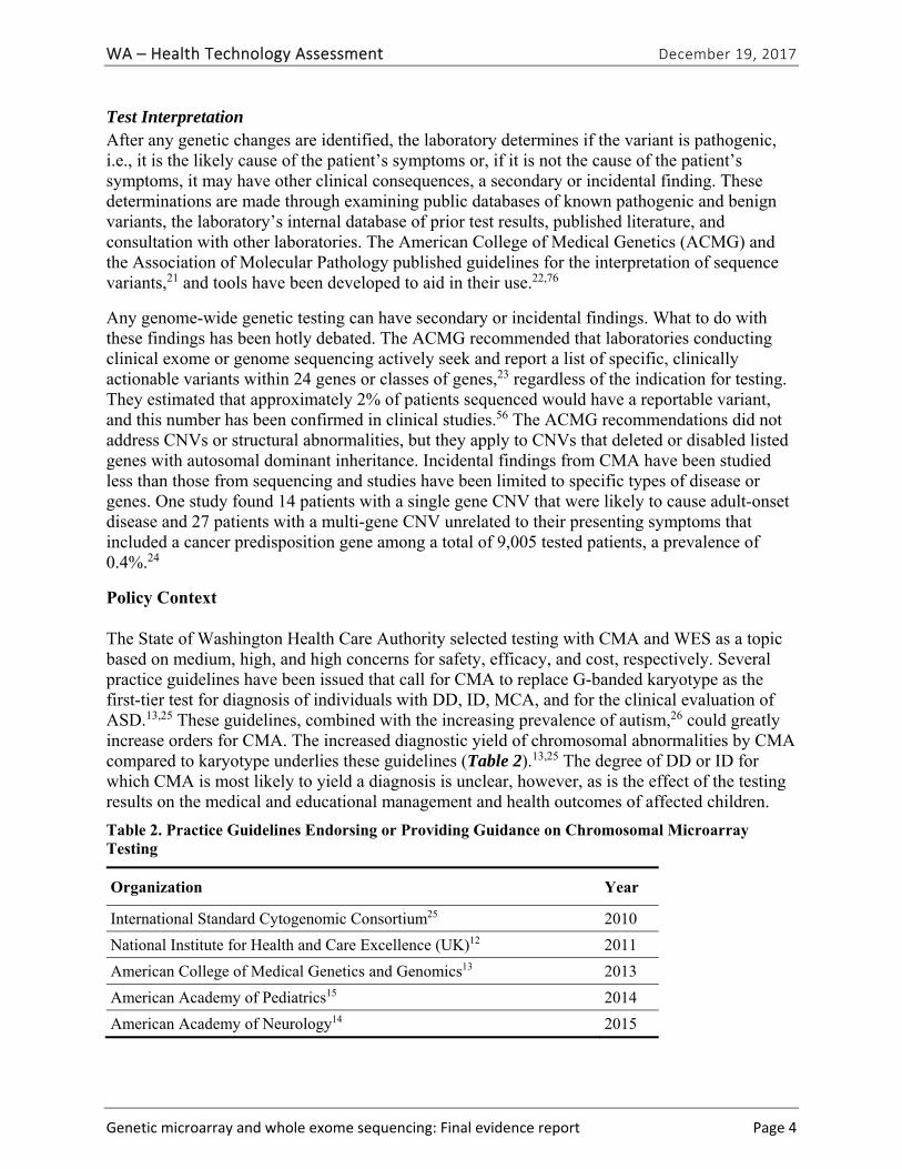
The State of Washington Health Care Authority selected testing with CMA and WES as a topic based on medium, high, and high concerns for safety, efficacy, and cost, respectively. Several practice guidelines have been issued that call for CMA to replace G-banded karyotype as the first-tier test for diagnosis of individuals with DD, ID, MCA, and for the clinical evaluation of ASD.13,25 These guidelines, combined with the increasing prevalence of autism,26 could greatly increase orders for CMA. The increased diagnostic yield of chromosomal abnormalities by CMA compared to karyotype underlies these guidelines (Table 2).13,25 The degree of DD or ID for which CMA is most likely to yield a diagnosis is unclear, however, as is the effect of the testing results on the medical and educational management and health outcomes of affected children.
Table 2. Practice Guidelines Endorsing or Providing Guidance on Chromosomal Microarray Testing
Organization Year
International Standard Cytogenomic Consortium25 2010
National Institute for Health and Care Excellence (UK)12 2011
American College of Medical Genetics and Genomics13 2013
American Academy of Pediatrics15 2014
American Academy of Neurology14 2015
WA – Health Technology Assessment December 19, 2017
Genetic microarray and whole exome sequencing: Final evidence report Page 5
Washington State Agency Utilization Data
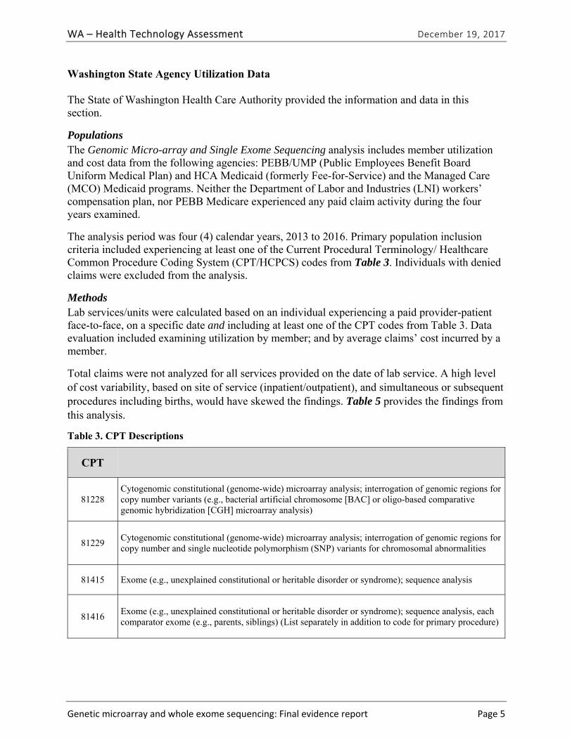
The State of Washington Health Care Authority provided the information and data in this section.
Populations The Genomic Micro-array and Single Exome Sequencing analysis includes member utilization and cost data from the following agencies: PEBB/UMP (Public Employees Benefit Board Uniform Medical Plan) and HCA Medicaid (formerly Fee-for-Service) and the Managed Care (MCO) Medicaid programs. Neither the Department of Labor and Industries (LNI) workers’ compensation plan, nor PEBB Medicare experienced any paid claim activity during the four years examined.
The analysis period was four (4) calendar years, 2013 to 2016. Primary population inclusion criteria included experiencing at least one of the Current Procedural Terminology/ Healthcare Common Procedure Coding System (CPT/HCPCS) codes from Table 3. Individuals with denied claims were excluded from the analysis.
Methods Lab services/units were calculated based on an individual experiencing a paid provider-patient face-to-face, on a specific date and including at least one of the CPT codes from Table 3. Data evaluation included examining utilization by member; and by average claims’ cost incurred by a member.
Total claims were not analyzed for all services provided on the date of lab service. A high level of cost variability, based on site of service (inpatient/outpatient), and simultaneous or subsequent procedures including births, would have skewed the findings. Table 5 provides the findings from this analysis.
Table 3. CPT Descriptions
CPT
81228 Cytogenomic constitutional (genome-wide) microarray analysis; interrogation of genomic regions for copy number variants (e.g., bacterial artificial chromosome [BAC] or oligo-based comparative genomic hybridization [CGH] microarray analysis)
81229 Cytogenomic constitutional (genome-wide) microarray analysis; interrogation of genomic regions for copy number and single nucleotide polymorphism (SNP) variants for chromosomal abnormalities
81415 Exome (e.g., unexplained constitutional or heritable disorder or syndrome); sequence analysis
81416 Exome (e.g., unexplained constitutional or heritable disorder or syndrome); sequence analysis, each comparator exome (e.g., parents, siblings) (List separately in addition to code for primary procedure)
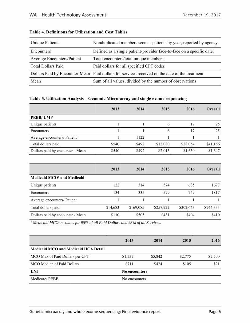
WA – Health Technology Assessment December 19, 2017
Genetic microarray and whole exome sequencing: Final evidence report Page 6
Table 4. Definitions for Utilization and Cost Tables
Unique Patients Nonduplicated members seen as patients by year, reported by agency
Encounters Defined as a single patient-provider face-to-face on a specific date.
Average Encounters/Patient Total encounters/total unique members
Total Dollars Paid Paid dollars for all specified CPT codes
Dollars Paid by Encounter-Mean Paid dollars for services received on the date of the treatment
Mean Sum of all values, divided by the number of observations
Table 5. Utilization Analysis – Genomic Micro-array and single exome sequencing
2013 2014 2015 2016 Overall
PEBB/ UMP
Unique patients 1 1 6 17 25
Encounters 1 1 6 17 25
Average encounters/ Patient 1 1122 1 1 1
Total dollars paid $540 $492 $12,080 $28,054 $41,166
Dollars paid by encounter - Mean $540 $492 $2,013 $1,650 $1,647
2013 2014 2015 2016 Overall
Medicaid MCO1 and Medicaid
Unique patients 122 314 574 685 1677
Encounters 134 335 599 749 1817
Average encounters/ Patient 1 1 1 1 1
Total dollars paid $14,683 $169,085 $257,922 $302,643 $744,333
Dollars paid by encounter - Mean $110 $505 $431 $404 $410
1 Medicaid MCO accounts for 95% of all Paid Dollars and 93% of all Services.
2013 2014 2015 2016
Medicaid MCO and Medicaid HCA Detail
MCO Max of Paid Dollars per CPT $1,537 $5,842 $2,775 $7,500
MCO Median of Paid Dollars $711 $424 $105 $21
LNI No encounters
Medicare/ PEBB No encounters
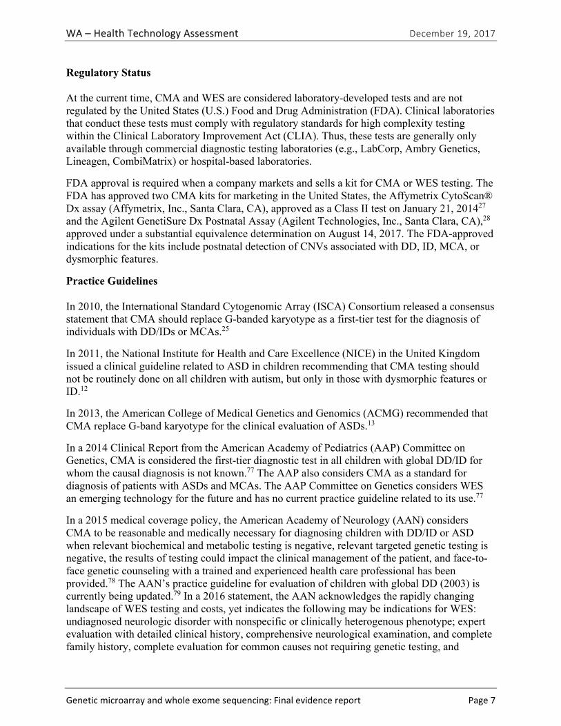
WA – Health Technology Assessment December 19, 2017
Genetic microarray and whole exome sequencing: Final evidence report Page 7
Regulatory Status
At the current time, CMA and WES are considered laboratory-developed tests and are not regulated by the United States (U.S.) Food and Drug Administration (FDA). Clinical laboratories that conduct these tests must comply with regulatory standards for high complexity testing within the Clinical Laboratory Improvement Act (CLIA). Thus, these tests are generally only available through commercial diagnostic testing laboratories (e.g., LabCorp, Ambry Genetics, Lineagen, CombiMatrix) or hospital-based laboratories.
FDA approval is required when a company markets and sells a kit for CMA or WES testing. The FDA has approved two CMA kits for marketing in the United States, the Affymetrix CytoScan® Dx assay (Affymetrix, Inc., Santa Clara, CA), approved as a Class II test on January 21, 201427 and the Agilent GenetiSure Dx Postnatal Assay (Agilent Technologies, Inc., Santa Clara, CA),28 approved under a substantial equivalence determination on August 14, 2017. The FDA-approved indications for the kits include postnatal detection of CNVs associated with DD, ID, MCA, or dysmorphic features.
Practice Guidelines
In 2010, the International Standard Cytogenomic Array (ISCA) Consortium released a consensus statement that CMA should replace G-banded karyotype as a first-tier test for the diagnosis of individuals with DD/IDs or MCAs.25
In 2011, the National Institute for Health and Care Excellence (NICE) in the United Kingdom issued a clinical guideline related to ASD in children recommending that CMA testing should not be routinely done on all children with autism, but only in those with dysmorphic features or ID.12
In 2013, the American College of Medical Genetics and Genomics (ACMG) recommended that CMA replace G-band karyotype for the clinical evaluation of ASDs.13
In a 2014 Clinical Report from the American Academy of Pediatrics (AAP) Committee on Genetics, CMA is considered the first-tier diagnostic test in all children with global DD/ID for whom the causal diagnosis is not known.77 The AAP also considers CMA as a standard for diagnosis of patients with ASDs and MCAs. The AAP Committee on Genetics considers WES an emerging technology for the future and has no current practice guideline related to its use.77
In a 2015 medical coverage policy, the American Academy of Neurology (AAN) considers CMA to be reasonable and medically necessary for diagnosing children with DD/ID or ASD when relevant biochemical and metabolic testing is negative, relevant targeted genetic testing is negative, the results of testing could impact the clinical management of the patient, and face-to-face genetic counseling with a trained and experienced health care professional has been provided.78 The AAN’s practice guideline for evaluation of children with global DD (2003) is currently being updated.79 In a 2016 statement, the AAN acknowledges the rapidly changing landscape of WES testing and costs, yet indicates the following may be indications for WES: undiagnosed neurologic disorder with nonspecific or clinically heterogenous phenotype; expert evaluation with detailed clinical history, comprehensive neurological examination, and complete family history, complete evaluation for common causes not requiring genetic testing, and
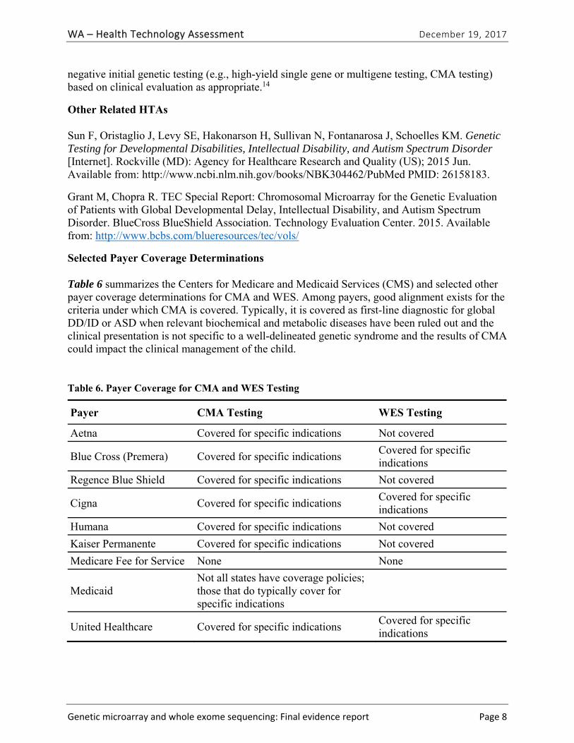
WA – Health Technology Assessment December 19, 2017
Genetic microarray and whole exome sequencing: Final evidence report Page 8
negative initial genetic testing (e.g., high-yield single gene or multigene testing, CMA testing) based on clinical evaluation as appropriate.14
Other Related HTAs
Sun F, Oristaglio J, Levy SE, Hakonarson H, Sullivan N, Fontanarosa J, Schoelles KM. Genetic Testing for Developmental Disabilities, Intellectual Disability, and Autism Spectrum Disorder [Internet]. Rockville (MD): Agency for Healthcare Research and Quality (US); 2015 Jun. Available from: http://www.ncbi.nlm.nih.gov/books/NBK304462/PubMed PMID: 26158183.
Grant M, Chopra R. TEC Special Report: Chromosomal Microarray for the Genetic Evaluation of Patients with Global Developmental Delay, Intellectual Disability, and Autism Spectrum Disorder. BlueCross BlueShield Association. Technology Evaluation Center. 2015. Available from: http://www.bcbs.com/blueresources/tec/vols/
Selected Payer Coverage Determinations
Table 6 summarizes the Centers for Medicare and Medicaid Services (CMS) and selected other payer coverage determinations for CMA and WES. Among payers, good alignment exists for the criteria under which CMA is covered. Typically, it is covered as first-line diagnostic for global DD/ID or ASD when relevant biochemical and metabolic diseases have been ruled out and the clinical presentation is not specific to a well-delineated genetic syndrome and the results of CMA could impact the clinical management of the child.
Table 6. Payer Coverage for CMA and WES Testing
Payer CMA Testing WES Testing
Aetna Covered for specific indications Not covered
Blue Cross (Premera) Covered for specific indications Covered for specific indications
Regence Blue Shield Covered for specific indications Not covered
Cigna Covered for specific indications Covered for specific indications
Humana Covered for specific indications Not covered
Kaiser Permanente Covered for specific indications Not covered
Medicare Fee for Service None None
Medicaid Not all states have coverage policies; those that do typically cover for specific indications
United Healthcare Covered for specific indications Covered for specific indications
WA – Health Technology Assessment December 19, 2017
Genetic microarray and whole exome sequencing: Final evidence report Page 9
CMS Medicare has no national coverage determination for the use of CMA or WES. Local coverage determinations vary.
Aetna Aetna considers CMA medically necessary and covered for diagnosing genetic abnormalities in children with MCAs, DD/ID, or ASD when relevant biochemical testing for metabolic diseases is negative; when targeted genetic testing if or when indicated by clinical and family history is negative; when the clinical presentation is not specific to a well-delineated genetic syndrome, and when the results of testing could impact the clinical management of the child.29 This coverage policy was last affirmed September 22, 2017.
Aetna considers WES testing to be experimental and investigational and, thus, not a covered benefit. This coverage policy was last affirmed on August 8, 2017.
Blue Cross (Premera) Premera considers CMA medically necessary as first-line testing of individuals with nonsyndromic DD/ID or ASD or two or more congenital anomalies not specific to a well-delineated genetic syndrome.30 Premera considers testing using next-generation sequencing to be investigational. This coverage policy was effective August 25, 2017.
Premera considers WES medically necessary for (1) the evaluation of unexplained congenital or neurodevelopmental disorders in children when the patient has been evaluated by a clinician with expertise in clinical genetics and counseled about the potential risks of genetic testing, (2) there is a potential for change in management and clinical outcome as a result of testing, and (3) a genetic etiology is considered the most likely explanation for the phenotype despite previous genetic testing or when prior genetic testing failed to yield a diagnosis and the individual is faced with invasive procedures or testing as the next diagnostic step.30 This coverage policy was effective February 1, 2017.
Blue Shield (Regence) Regence considers CMA medically necessary in children as first- or second-line assessment in children with apparent nonsyndromic cognitive DD/ID, ASD, or MCAs not specific to a well-delineated genetic syndrome.31 Further, Regence considers testing using next-generation sequencing to be investigational and not a covered benefit. This policy was last affirmed April 2017.
Regence considers all applications of WES to be investigational and not a covered benefit.31 This coverage policy was last effective August 1, 2017.
Cigna Cigna considers CMA medically necessary when phenotypic characteristics of a specific genetic disorder are absent for patients with ASD and nonsyndromic global DD/ID. Testing is also considered medically necessary when MCAs are present and cannot be ascribed to a specific genetic syndrome.33 In addition to these indications, testing must be recommended by independent board-certified or eligible medical geneticists, certified genetic counselors, or certified genetic nurses. The health professionals recommending testing (1) cannot be employed by a commercial genetic testing laboratory, (2) must have evaluated the individual, including a
WA – Health Technology Assessment December 19, 2017
Genetic microarray and whole exome sequencing: Final evidence report Page 10
three-generation pedigree, and (3) must intend to engage in post-test follow-up counseling. This coverage policy was effective November 15, 2016.
Cigna considers WES medically necessary when a genetic etiology is the most likely explanation for the phenotype demonstrated, no other cause can explain symptoms, and the clinical presentation does not fit a well-described syndrome for which single-gene or targeted panel testing is available; and the differential diagnosis and/or phenotype would require testing of multiple genes such that WES testing would be more practical than separate genetic tests or preclude the need for multiple and/or invasive procedures. In addition, the individual must have been evaluated by a board-certified medical geneticist or other physician specialist with expertise in the conditions and relevant genes for which testing is being considered, and WES results are expected to directly impact clinical decision-making and clinical outcome for the individual being tested.34 Pre- and post-test genetic counseling is required. This coverage policy was effective November 15, 2016.
Humana Humana covers CMA for the evaluation of children diagnosed with ASD, unexplained global DD/ID and the absence of a clinically identifiable single gene disorder, clinical syndrome, and no family history of chromosomal rearrangement or multiple miscarriages. Humana also covers CMA for the evaluation of multiple anomalies, the combination of which are not suggestive of a specific syndrome.35 This coverage policy was effective July 27, 2017.
Humana considers WES experimental/investigational, and this testing is not covered. This coverage policy was effective August 18, 2017.35
Kaiser Permanente Kaiser considers CMA medically necessary for the evaluation of ID for individuals with significant dysmorphic features or congenital anomalies, when results are expected to affect clinical management, and when genetic counseling by a health care professional with appropriate genetic training and experience has been conducted.36 The source documentation did not include an effective date for this coverage policy.
Kaiser does not cover WES.36 The source documentation did not include an effective date for this coverage policy.
Medicaid Some states have Medicaid coverage policies related to CMA testing. In Indiana, CMA testing is covered as a first-line test in postnatal evaluation of children with unexplained intellectual disability, development delay, or ASD.37 Similarly, CMA testing is also covered in North Carolina. In Massachusetts, testing is covered with prior authorization.15,38
United United Healthcare considers CMA medically necessary for evaluating patients with multiple anomalies not specific to a well-delineated genetic syndrome and that cannot be identified by a clinical evaluation alone, nonsyndromic DD/ID, and ASDs.39 This coverage determination was effective August 1, 1017.
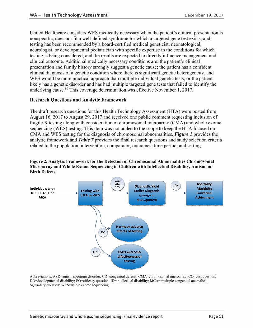
WA – Health Technology Assessment December 19, 2017
Genetic microarray and whole exome sequencing: Final evidence report Page 11
United Healthcare considers WES medically necessary when the patient’s clinical presentation is nonspecific, does not fit a well-defined syndrome for which a targeted gene test exists, and testing has been recommended by a board-certified medical geneticist, neonatological, neurologist, or developmental pediatrician with specific expertise in the conditions for which testing is being considered, and the results are expected to directly influence management and clinical outcome. Additional medically necessary conditions are: the patient’s clinical presentation and family history strongly suggest a genetic cause; the patient has a confident clinical diagnosis of a genetic condition where there is significant genetic heterogeneity, and WES would be more practical approach than multiple individual genetic tests; or the patient likely has a genetic disorder and has had multiple targeted gene tests that failed to identify the underlying cause.80 This coverage determination was effective November 1, 2017.
Research Questions and Analytic Framework
The draft research questions for this Health Technology Assessment (HTA) were posted from August 16, 2017 to August 29, 2017 and received one public comment requesting inclusion of fragile X testing along with consideration of chromosomal microarray (CMA) and whole exome sequencing (WES) testing. This item was not added to the scope to keep the HTA focused on CMA and WES testing for the diagnosis of chromosomal abnormalities. Figure 1 provides the analytic framework and Table 7 provides the final research questions and study selection criteria related to the population, intervention, comparator, outcomes, time period, and setting.
Figure 2. Analytic Framework for the Detection of Chromosomal Abnormalities Chromosomal Microarray and Whole Exome Sequencing in Children with Intellectual Disability, Autism, or Birth Defects
Abbreviations: ASD=autism spectrum disorder; CD=congenital defects; CMA=chromosomal microarray; CQ=cost question; DD=developmental disability; EQ=efficacy question; ID=intellectual disability; MCA= multiple congenital anomalies; SQ=safety question; WES=whole exome sequencing.
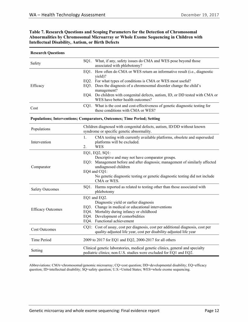
WA – Health Technology Assessment December 19, 2017
Genetic microarray and whole exome sequencing: Final evidence report Page 12
Table 7. Research Questions and Scoping Parameters for the Detection of Chromosomal Abnormalities by Chromosomal Microarray or Whole Exome Sequencing in Children with Intellectual Disability, Autism, or Birth Defects
Research Questions
Safety SQ1. What, if any, safety issues do CMA and WES pose beyond those
associated with phlebotomy?
Efficacy
EQ1. How often do CMA or WES return an informative result (i.e., diagnostic yield)?
EQ2. For what types of conditions is CMA or WES most useful? EQ3. Does the diagnosis of a chromosomal disorder change the child’s
management? EQ4. Do children with congenital defects, autism, ID, or DD tested with CMA or
WES have better health outcomes?
Cost CQ1. What is the cost and cost-effectiveness of genetic diagnostic testing for
these conditions with CMA or WES?
Populations; Interventions; Comparators, Outcomes; Time Period; Setting
Populations Children diagnosed with congenital defects, autism, ID/DD without known syndrome or specific genetic abnormality.
Intervention 1. CMA testing with currently available platforms, obsolete and superseded
platforms will be excluded. 2. WES
Comparator
EQ1, EQ2, SQ1: Descriptive and may not have comparator groups. EQ3: Management before and after diagnosis; management of similarly affected
undiagnosed children EQ4 and CQ1: No genetic diagnostic testing or genetic diagnostic testing did not include
CMA or WES.
Safety Outcomes SQ1. Harms reported as related to testing other than those associated with
phlebotomy
Efficacy Outcomes
EQ1 and EQ2. Diagnostic yield or earlier diagnosis EQ3. Change in medical or educational interventions EQ4. Mortality during infancy or childhood EQ4. Development of comorbidities EQ4. Functional achievement
Cost Outcomes CQ1: Cost of assay, cost per diagnosis, cost per additional diagnosis, cost per
quality-adjusted life year, cost per disability-adjusted life year
Time Period 2009 to 2017 for EQ1 and EQ2, 2000-2017 for all others
Setting Clinical genetic laboratories, medical genetic clinics, general and specialty pediatric clinics; non-U.S. studies were excluded for EQ1 and EQ2.
Abbreviations: CMA=chromosomal/genomic microarray; CQ=cost question; DD=developmental disability; EQ=efficacy question; ID=intellectual disability; SQ=safety question; U.S.=United States; WES=whole exome sequencing.
WA – Health Technology Assessment December 19, 2017
Genetic microarray and whole exome sequencing: Final evidence report Page 13
What is Excluded from This HTA
This HTA does not address the analytic validity of CMA or WES because this testing is available within Clinical Laboratory Improvement Act (CLIA)-licensed laboratories as a laboratory-developed test and analytic validity is assumed based on meeting CLIA standards.40 It also does not address the use of CMA or WES to identify or monitor chromosomal changes in tumor cells or its use for prenatal testing. This review is focused on the diagnosis of chromosomal abnormalities; therefore, we do not address single gene testing for these disorders, including the use of WES to identify mutations within single genes. The review does not assess either the ethical issues or the clinical utility of incidental findings not related to the health conditions for which the tests were ordered. Because of the large volume of studies on diagnostic yield (EQ1) and the rapidly evolving technology in use for CMA testing, we limited the studies considered for EQ 1 to studies that were conducted in the United States in 2009 or later and that used current testing platforms.
Methods
Data Sources and Searches
We searched MEDLINE® (via PubMed) and a clinical trials registry (clinicaltrials.gov) for relevant English-language studies published in 2000 or later. We searched the FDA website, selected payer and health care professional society websites, the U.S. Agency for Healthcare Research and Quality, and other organizations that conduct and disseminate HTAs. In addition, we reviewed the reference lists of relevant studies, practice guidelines, and other HTAs on this topic to identify any relevant articles not identified through the electronic search. The detailed search strategy is provided in Appendix A.
Study Selection
Study selection criteria for this HTA were as follows:
Population For all research questions, we included studies that reporting testing individuals (children or adults) with autism, cerebral palsy, global developmental delay (GDD), developmental disability(DD), intellectual disability (ID), autism spectrum disorder (ASD), including Asperger’s syndrome, mental retardation, mental deficiency, congenital defects, or birth defects. We excluded studies focused on testing among populations with syndromes known to be associated with single gene disorders or among populations with single, specific birth defects.
Intervention and Comparator For all research questions, we included studies that reported on the use of chromosomal microarray (CMA) testing (including the specific terms such as genome-wide array, genomic array, chromosomal array, chromosomal microarray, comparative genomic hybridization, deoxyribose nucleic acid (DNA) sequencing, or molecular karyotype) or whole exome sequencing (WES testing). Further, we required studies to be using these tests to identify imbalanced rearrangements, translocations, duplications, deletions, copy number variants (CNVs), or chromosomal aberrations and limited study selection for diagnostic yield (EQs1 and
WA – Health Technology Assessment December 19, 2017
Genetic microarray and whole exome sequencing: Final evidence report Page 14
2) to studies using current testing platforms. Studies that focused on fragile X testing, mutations in a single gene or a single gene panel or single gene sequencing, epigenetic testing, or whole genomic sequencing were excluded. Further, studies that used CMA or WES to identify mutations, trinucleotide repeats, or aneuploidy were excluded. For the research questions on comparative efficacy related to management and health outcomes (EQ3 and EQ4), we selected studies with comparison groups that were historical (management before/after testing) or concurrent controls (no CMA or WES testing or testing that did not include CMA or WES).
Outcomes For the research question on safety (SQ1), we included studies that reported on harms or adverse effects of testing with CMA or WES. We excluded studies solely reporting harms associated with phlebotomy, and did not include harms or adverse effects associated with incidental findings or ethical issues associated with this testing. For the research questions on diagnostic yield (EQ1 and EQ2), we included studies that reported on the proportion of CMA and WES testing that identified a known or likely pathogenic variant among all tested children or among children or adults with the conditions of interest. For EQ3, we included studies that reported on time to diagnosis and medical or educational interventions resulting from the results of testing. For EQ4, we included studies that reported mortality, morbidity, or functional achievement. For the research question on cost (CQ1), we included studies that reported on the cost of testing, cost per patient, cost per diagnosis, cost per additional diagnosis, or cost per quality-adjusted life year or disability-adjusted life year measures from either a payer or societal perspective. Studies that did not report at least one eligible outcome were excluded.
Time Period For EQ1 and EQ2 we selected studies that conducted testing in 2009 or later. Studies published in 2000 or later were eligible for all other research questions.
Settings We included studies conducted in clinical settings or where the CMA or WES was being evaluated as a clinical test; we excluded studies focused on test development or that occurred strictly within a research setting without any connection to clinical practice. For EQ1 and EQ2, we excluded studies conducted outside of the U.S.
Study Design We included all study designs except case reports.
We screened titles and abstracts and full-text articles based on these study selection criteria. Team members were trained on study selection criteria, and all team members independently screened an initial set of 20 titles and abstracts. Because we had excellent concordance among screeners on the initial set, a single team member screened the remaining titles/abstracts. The principal investigator reviewed all abstracts excluded for “ineligible intervention” and a sample of titles/abstracts excluded for other reasons to ensure continued consistency in application of study selection criteria by team members. A senior team member screened each full-text article for inclusion, and the principal investigator confirmed the decisions.
WA – Health Technology Assessment December 19, 2017
Genetic microarray and whole exome sequencing: Final evidence report Page 15
Data Abstraction and Quality Assessment
One team member extracted relevant study data into a structured abstraction form. The principal investigator reviewed the abstractions for accuracy and consistency. Two senior team members conducted independent risk of bias assessment on all included studies and met to reconcile any disagreements, in consultation with the principal investigator if needed. Because of the diverse types of studies included in this HTA, we adapted signaling questions from the QUADAS-2 instrument, a risk of bias assessment for diagnostics test studies, and items from the RTI item bank for observational studies.41,42 The signaling questions assessed the major sources of bias including selection bias (both how study population was selected and attrition/missing data), confounding, and measurement/information bias. We used the ROBIS instrument to assess the risk of bias for systematic reviews.43
Data Synthesis and Analysis
Study characteristics and results were qualitatively synthesized for each research question in tabular and narrative formats. For cost outcomes, we adjusted all reported outcomes in foreign currency to U.S. dollars based on the U.S. Department of Treasury mid-year exchange rate for the year reported by study authors and then used the chain-weighted consumer price index (CPI) to adjust to 2010 U.S. dollars (Appendix B).44,45 To determine whether quantitative synthesis was appropriate, we assessed the number of studies and the clinical and methodological heterogeneity present based on established guidance.46,47 We required three or more publications with similar approach and the same outcome measure to calculate a summary estimate. We estimated summary effects using a fixed effects model if the test for heterogeneity was nonsignificant and a random effects model if the test for heterogeneity was significant using OpenMetaAnalyst (For Windows 8, 64-bit) using the method of Hedges and Olkin to estimate between-study variance.48 We graded the strength of evidence for each research question using GRADE, which assesses the strength of evidence based on domains relating to risk of bias, inconsistency, imprecision, indirectness, and other considerations, such as reporting bias.49 Under GRADE, the strength of evidence can be graded as very low, low, moderate, or high.
Results
Literature Search
Figure 2 depicts the study flow diagram. We identified and screened 2,717 unique citations. We excluded 2,375 after title and abstract review. We reviewed the full-text of 348 articles, and excluded 330 for the reasons listed in Figure 2. We included a total of 18 studies. One provided evidence on safety issues (SQ1), seven provided evidence on diagnostic yield (EQ1 and EQ2), seven on changes in management (EQ3), and five on costs (CQ1). No studies provided information on health outcomes (EQ4). Individual study characteristics for all included studies are summarized in Appendix C, Table C-1. The list of studies we screened at the full text stage, but which were excluded from the review, is provided in Appendix D. Note that studies may have been excluded based on more than one reason but we report only one reason. Individual risk of bias assessments for all included studies are reported in Appendix E.
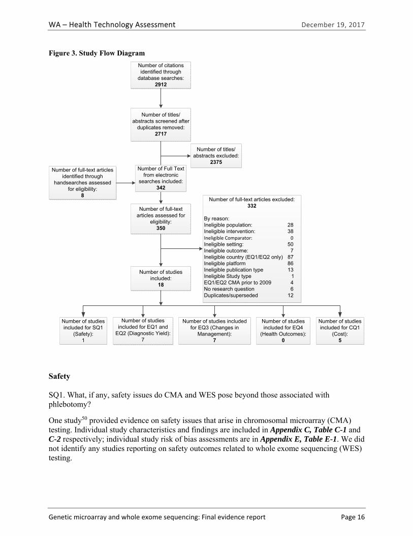
WA – Health Technology Assessment December 19, 2017
Genetic microarray and whole exome sequencing: Final evidence report Page 16
Figure 3. Study Flow Diagram
Safety
SQ1. What, if any, safety issues do CMA and WES pose beyond those associated with phlebotomy?
One study50 provided evidence on safety issues that arise in chromosomal microarray (CMA) testing. Individual study characteristics and findings are included in Appendix C, Table C-1 and C-2 respectively; individual study risk of bias assessments are in Appendix E, Table E-1. We did not identify any studies reporting on safety outcomes related to whole exome sequencing (WES) testing.
Number of citations identified through
database searches:2912
Number of titles/abstracts excluded:
2375
Number of titles/abstracts screened after
duplicates removed:2717
Number of full-text articles assessed for
eligibility:350
Number of full-text articles excluded: 332
By reason:Ineligible population: 28Ineligible intervention: 38Ineligible Comparator: 0Ineligible setting: 50Ineligible outcome: 7Ineligible country (EQ1/EQ2 only) 87Ineligible platform 86Ineligible publication type 13Ineligible Study type 1EQ1/EQ2 CMA prior to 2009 4 No research question 6Duplicates/superseded 12
Number of studies included for SQ1
(Safety):1
Number of studies included for EQ1 and
EQ2 (Diagnostic Yield):7
Number of studies included for EQ3 (Changes in
Management):7
Number of studies included for EQ4
(Health Outcomes):0
Number of studies included:
18
Number of studies included for CQ1
(Cost):5
Number of full-text articles identified through
handsearches assessed for eligibility:
8
Number of Full Text from electronic
searches included:342
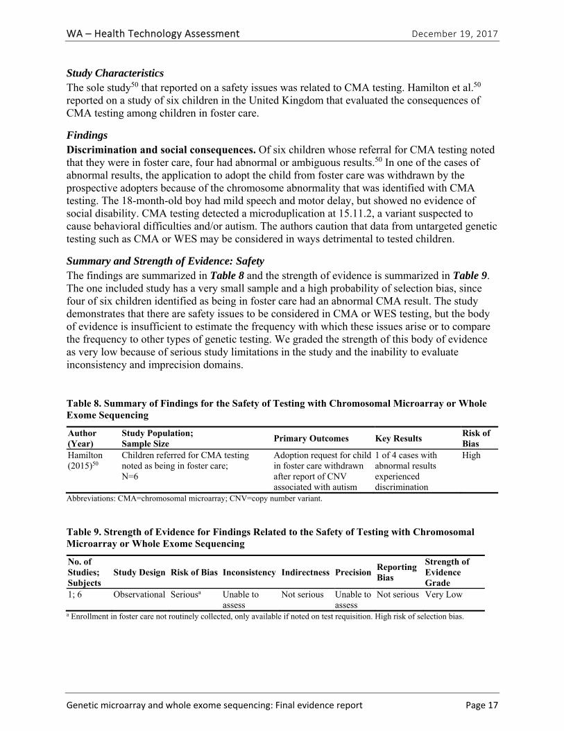
WA – Health Technology Assessment December 19, 2017
Genetic microarray and whole exome sequencing: Final evidence report Page 17
Study Characteristics The sole study50 that reported on a safety issues was related to CMA testing. Hamilton et al.50 reported on a study of six children in the United Kingdom that evaluated the consequences of CMA testing among children in foster care.
Findings Discrimination and social consequences. Of six children whose referral for CMA testing noted that they were in foster care, four had abnormal or ambiguous results.50 In one of the cases of abnormal results, the application to adopt the child from foster care was withdrawn by the prospective adopters because of the chromosome abnormality that was identified with CMA testing. The 18-month-old boy had mild speech and motor delay, but showed no evidence of social disability. CMA testing detected a microduplication at 15.11.2, a variant suspected to cause behavioral difficulties and/or autism. The authors caution that data from untargeted genetic testing such as CMA or WES may be considered in ways detrimental to tested children.
Summary and Strength of Evidence: Safety The findings are summarized in Table 8 and the strength of evidence is summarized in Table 9. The one included study has a very small sample and a high probability of selection bias, since four of six children identified as being in foster care had an abnormal CMA result. The study demonstrates that there are safety issues to be considered in CMA or WES testing, but the body of evidence is insufficient to estimate the frequency with which these issues arise or to compare the frequency to other types of genetic testing. We graded the strength of this body of evidence as very low because of serious study limitations in the study and the inability to evaluate inconsistency and imprecision domains.
Table 8. Summary of Findings for the Safety of Testing with Chromosomal Microarray or Whole Exome Sequencing
Author (Year)
Study Population; Sample Size
Primary Outcomes Key Results Risk of Bias
Hamilton (2015)50
Children referred for CMA testing noted as being in foster care; N=6
Adoption request for child in foster care withdrawn after report of CNV associated with autism
1 of 4 cases with abnormal results experienced discrimination
High
Abbreviations: CMA=chromosomal microarray; CNV=copy number variant.
Table 9. Strength of Evidence for Findings Related to the Safety of Testing with Chromosomal Microarray or Whole Exome Sequencing
No. of Studies; Subjects
Study Design Risk of Bias Inconsistency Indirectness PrecisionReporting Bias
Strength of Evidence Grade
1; 6 Observational Seriousa Unable to assess
Not serious Unable to assess
Not serious Very Low
a Enrollment in foster care not routinely collected, only available if noted on test requisition. High risk of selection bias.
WA – Health Technology Assessment December 19, 2017
Genetic microarray and whole exome sequencing: Final evidence report Page 18
Efficacy
EQ1. How often do CMA or WES return an informative result (i.e., diagnostic yield)?
Five primary studies51-55 and one health technology assessment (HTA)40 provided evidence related to diagnostic yield (EQ1) and the types of clinical conditions for which CMA is most useful (EQ2). One study provided evidence related to diagnostic yield of WES testing.56| Study characteristics and findings are summarized in Table 10. Appendix C, Tables C-1 and C-3 provide detailed individual study characteristics and findings, respectively; individual study risk of bias assessments are provided in Appendix E, Table 1.
Study Characteristics Grant et al. conducted an HTA of CMA testing that was published in 2015 and included a thorough review on the diagnostic yield of CMA in patients with global developmental delay (GDD), intellectual disability (ID), or autism spectrum disorders (ASDs).40 This review included 67 studies published prior to June 24, 2015 that provided evidence on the diagnostic yield of CMA or its impact on clinical management decisions or patient outcomes. They included case series or cohorts of at least 20 patients with GDD with or without ID, or ASD with or without negative karyotype results. We assessed this risk of bias of this HTA as “unclear”, primarily because it did not assess the risk of bias for studies included in its synthesis.
In addition to the Grant et al. HTA40, we included four primary research studies51,52,54,55 published from 2010 to 2015 that were included in the Grant et al. HTA, and one study53 published in 2016, for a total of five primary research studies that provided evidence on the diagnostic yield of CMA. We also included one study of the diagnostic yield of WES.56 Three52,54,55 studies used oligonucleotide CMA, two61,81 used SNP-oligo CMA, and two studies53,81 used high-resolution SNP CMA. The patient populations of the studies were mixed. Four studies51-54 of CMA testing reported findings for patients with congenital anomalies, DD, ID, or both DD and ID. Stobbe et al.55 reported on CMA results among adults with ASD. The single study reporting on diagnostic yield of WES testing56 reported findings for children with ID.
The methods for classifying a variant as pathogenic or likely pathogenic varied across studies. Coulter et al.51 classified a variant as pathologic if it was 1) associated with a known microduplication or deletion syndrome, 2) a deletion of genes that are known to cause disease when haploinsufficient, 3) or a large (size unspecified) duplication or deletion. A variant was classified as possibly pathogenetic if it overlapped a known syndromic region, or contained genes suspected of causing disease, with the potential unmasking of recessive alleles. Henderson et al.52 defined variants as pathologic if they were 1) associated with a known microduplication or deletion syndrome, 2) encompassed or interrupted genes associated with disease, 3) included numerous genes and were not found in healthy individuals, or 4) were a region of homozygosity greater than 10 Mb. Ho et al.53 classified variants as pathologic if they were found in less than 1% of the general population and there were at least two independent peer-reviewed reports that haploinsufficiency or triplosensitivity of the region or gene(s) caused clinical symptoms similar to those of the patient. Roberts et al. 54classified variants as pathologic if they had previously been associated with ASD or learning disability. They evaluated variants using the University of California at Santa Cruz (UCSC) Genome Browser,82 Database of Genomic Variants (DGV),83
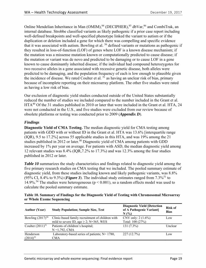
WA – Health Technology Assessment December 19, 2017
Genetic microarray and whole exome sequencing: Final evidence report Page 19
Online Mendelian Inheritance in Man (OMIM),84 (DECIPHER),85 dbVar,86 and CombiTrak, an internal database. Strobbe classified variants as likely pathogenic if a prior case report including well-defined breakpoints and well-specified phenotype linked the variant to autism or if the duplication or deletion included a gene for which there was compelling and specific evidence that it was associated with autism. Bowling et al. 56 defined variants or mutations as pathogenic if they resulted in loss-of-function (LOF) of genes where LOF is a known disease mechanism; if the mutation was a missense mutation known or computationally predicted to cause disease; if the mutation or variant was de novo and predicted to be damaging or to cause LOF in a gene known to cause dominantly inherited disease; if the individual had compound heterozygotes for two recessive alleles in a gene associated with recessive genetic disease, both alleles were predicted to be damaging, and the population frequency of each is low enough to plausible given the incidence of disease. We rated Coulter et al. 51 as having an unclear risk of bias, primary because of incomplete reporting on their microarray platform. The other five studies were rated as having a low risk of bias.
Our exclusion of diagnostic yield studies conducted outside of the United States substantially reduced the number of studies we included compared to the number included in the Grant et al. HTA40 Of the 31 studies published in 2010 or later that were included in the Grant et al. HTA, 24 were not conducted in the U.S., and five studies were excluded from our review because of obsolete platforms or testing was conducted prior to 2009 (Appendix D).
Findings Diagnostic Yield of CMA Testing. The median diagnostic yield for CMA testing among patients with GDD with or without ID in the Grant et al. HTA was 13.6% [interquartile range (IQR), 9.5 to 17.2%] across 55 applicable studies in this HTA, and was 19% among the 21 studies published in 2012 or later.40 Diagnostic yield of CMA among patients with GDD increased by 1% per year on average. For patients with ASD, the median diagnostic yield among 12 relevant studies was 8.4% (IQR,7.2% to 17.3%) and was 12.3% among the four studies published in 2012 or later.
Table 10 summarizes the study characteristics and findings related to diagnostic yield among the five primary research studies on CMA testing that we included. The pooled summary estimate of diagnostic yield, from these studies including known and likely pathogenic variants, was 8.8% (95% CI, 8.4% to 9.3%) (Figure 3). The individual study estimates ranged from 7.3%51 to 14.9%.54 The studies were heterogeneous (p < 0.001), so a random effects model was used to calculate the pooled summary estimate.
Table 10. Summary of Findings for the Diagnostic Yield of Testing with Chromosomal Microarray or Whole Exome Sequencing
Author (Year) Study Population; Sample Size, Test Diagnostic Yield [Detection of A Pathogenic Variant] N (%)
Risk of Bias
Bowling (2017)56 Clinic-based family recruitment of children with mild to severe ID, age ≥ 2; N=365, WES
CNV only: 2 (1.6%) Total: 100 (27%)
Low
Coulter (2011)51 Patients of children’s hospital; N =1,792, CMA
131 (7.3%) Unclear
Henderson (2014)52
Laboratory-based series of patients; N= 1780, CMA
227 (12.7%) Low
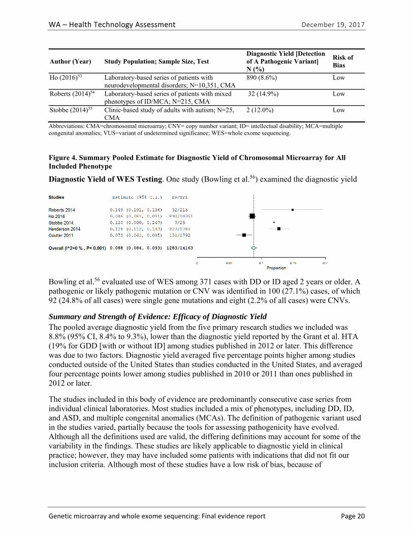
WA – Health Technology Assessment December 19, 2017
Genetic microarray and whole exome sequencing: Final evidence report Page 20
Author (Year) Study Population; Sample Size, Test Diagnostic Yield [Detection of A Pathogenic Variant] N (%)
Risk of Bias
Ho (2016)53 Laboratory-based series of patients with neurodevelopmental disorders; N=10,351, CMA
890 (8.6%) Low
Roberts (2014)54 Laboratory-based series of patients with mixed phenotypes of ID/MCA; N=215, CMA
32 (14.9%) Low
Stobbe (2014)55 Clinic-based study of adults with autism; N=25, CMA
2 (12.0%) Low
Abbreviations: CMA=chromosomal microarray; CNV= copy number variant; ID= intellectual disability; MCA=multiple congenital anomalies; VUS=variant of undetermined significance; WES=whole exome sequencing.
Figure 4. Summary Pooled Estimate for Diagnostic Yield of Chromosomal Microarray for All Included Phenotype
Diagnostic Yield of WES Testing. One study (Bowling et al.56) examined the diagnostic yield
Bowling et al.56 evaluated use of WES among 371 cases with DD or ID aged 2 years or older. A pathogenic or likely pathogenic mutation or CNV was identified in 100 (27.1%) cases, of which 92 (24.8% of all cases) were single gene mutations and eight (2.2% of all cases) were CNVs.
Summary and Strength of Evidence: Efficacy of Diagnostic Yield The pooled average diagnostic yield from the five primary research studies we included was 8.8% (95% CI, 8.4% to 9.3%), lower than the diagnostic yield reported by the Grant et al. HTA (19% for GDD [with or without ID] among studies published in 2012 or later. This difference was due to two factors. Diagnostic yield averaged five percentage points higher among studies conducted outside of the United States than studies conducted in the United States, and averaged four percentage points lower among studies published in 2010 or 2011 than ones published in 2012 or later.
The studies included in this body of evidence are predominantly consecutive case series from individual clinical laboratories. Most studies included a mix of phenotypes, including DD, ID, and ASD, and multiple congenital anomalies (MCAs). The definition of pathogenic variant used in the studies varied, partially because the tools for assessing pathogenicity have evolved. Although all the definitions used are valid, the differing definitions may account for some of the variability in the findings. These studies are likely applicable to diagnostic yield in clinical practice; however, they may have included some patients with indications that did not fit our inclusion criteria. Although most of these studies have a low risk of bias, because of
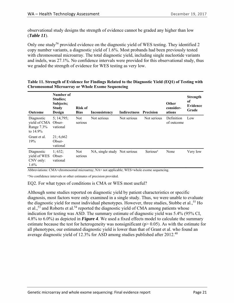
WA – Health Technology Assessment December 19, 2017
Genetic microarray and whole exome sequencing: Final evidence report Page 21
observational study designs the strength of evidence cannot be graded any higher than low (Table 11).
Only one study56 provided evidence on the diagnostic yield of WES testing. They identified 2 copy number variants, a diagnostic yield of 1.6%. Most probands had been previously tested with chromosomal microarray. The total diagnostic yield, including single nucleotide variants and indels, was 27.1%. No confidence intervals were provided for this observational study, thus we graded the strength of evidence for WES testing as very low.
Table 11. Strength of Evidence for Findings Related to the Diagnostic Yield (EQ1) of Testing with Chromosomal Microarray or Whole Exome Sequencing
Outcome
Number of Studies; Subjects; Study Design
Risk of Bias Inconsistency Indirectness Precision
Other consider-ations
Strength of Evidence Grade
Diagnostic yield of CMA Range 7.3% to 14.9%
5; 14,795; Obser-vational
Not serious
Not serious Not serious Not serious Definition of outcome
Low
Grant et al. 19%
21; 6,662 Obser-vational
Diagnostic yield of WES CNV only: 1.6%
1; 632; Obser-vational
Not serious
NA, single study Not serious Seriousa None Very low
Abbreviations: CMA=chromosomal microarray; NA= not applicable; WES=whole exome sequencing.
a No confidence intervals or other estimates of precision provided.
EQ2. For what types of conditions is CMA or WES most useful?
Although some studies reported on diagnostic yield by patient characteristics or specific diagnosis, most factors were only examined in a single study. Thus, we were unable to evaluate the diagnostic yield for most individual phenotypes. However, three studies, Stobbe et al.,55 Ho et al.,53 and Roberts et al.54 reported the diagnostic yield of CMA among patients whose indication for testing was ASD. The summary estimate of diagnostic yield was 5.4% (95% CI, 4.8% to 6.0%) as depicted in Figure 4. We used a fixed effects model to calculate the summary estimate because the test for heterogeneity was nonsignificant (p> 0.05). As with the estimate for all phenotypes, our estimated diagnostic yield is lower than that of Grant et al. who found an average diagnostic yield of 12.3% for ASD among studies published after 2012.40
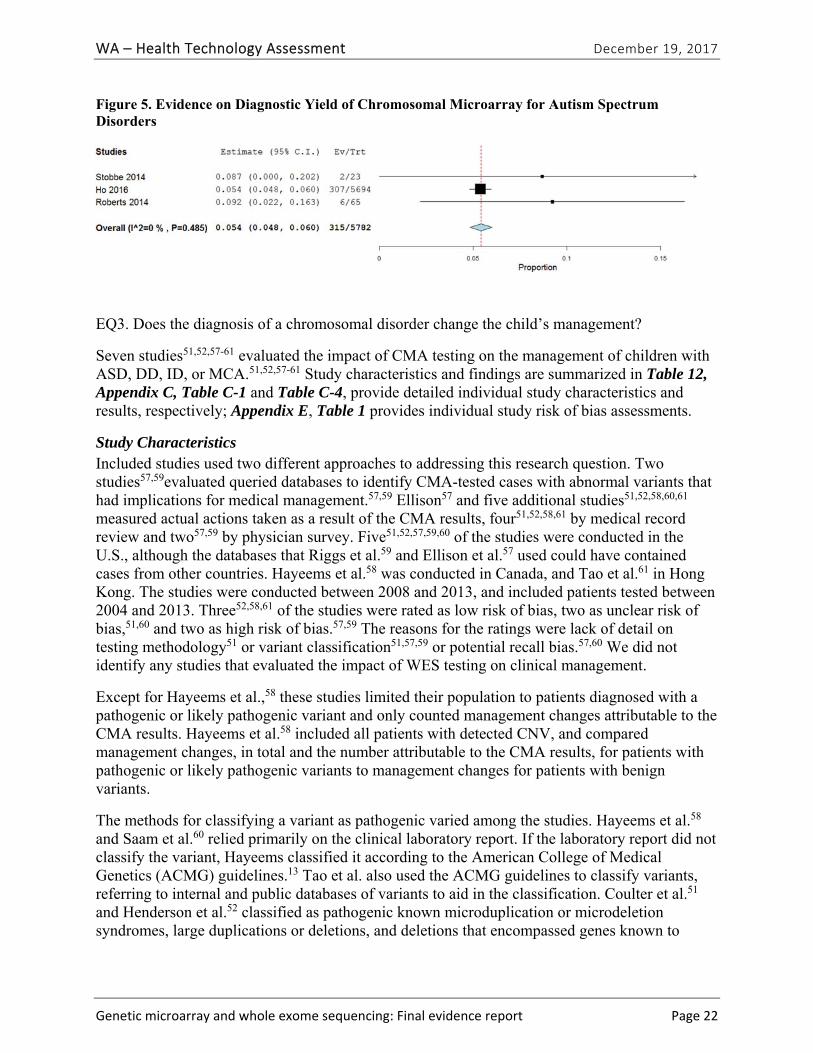
WA – Health Technology Assessment December 19, 2017
Genetic microarray and whole exome sequencing: Final evidence report Page 22
Figure 5. Evidence on Diagnostic Yield of Chromosomal Microarray for Autism Spectrum Disorders
EQ3. Does the diagnosis of a chromosomal disorder change the child’s management?
Seven studies51,52,57-61 evaluated the impact of CMA testing on the management of children with ASD, DD, ID, or MCA.51,52,57-61 Study characteristics and findings are summarized in Table 12, Appendix C, Table C-1 and Table C-4, provide detailed individual study characteristics and results, respectively; Appendix E, Table 1 provides individual study risk of bias assessments.
Study Characteristics Included studies used two different approaches to addressing this research question. Two studies57,59evaluated queried databases to identify CMA-tested cases with abnormal variants that had implications for medical management.57,59 Ellison57 and five additional studies51,52,58,60,61 measured actual actions taken as a result of the CMA results, four51,52,58,61 by medical record review and two57,59 by physician survey. Five51,52,57,59,60 of the studies were conducted in the U.S., although the databases that Riggs et al.59 and Ellison et al.57 used could have contained cases from other countries. Hayeems et al.58 was conducted in Canada, and Tao et al.61 in Hong Kong. The studies were conducted between 2008 and 2013, and included patients tested between 2004 and 2013. Three52,58,61 of the studies were rated as low risk of bias, two as unclear risk of bias,51,60 and two as high risk of bias.57,59 The reasons for the ratings were lack of detail on testing methodology51 or variant classification51,57,59 or potential recall bias.57,60 We did not identify any studies that evaluated the impact of WES testing on clinical management.
Except for Hayeems et al.,58 these studies limited their population to patients diagnosed with a pathogenic or likely pathogenic variant and only counted management changes attributable to the CMA results. Hayeems et al.58 included all patients with detected CNV, and compared management changes, in total and the number attributable to the CMA results, for patients with pathogenic or likely pathogenic variants to management changes for patients with benign variants.
The methods for classifying a variant as pathogenic varied among the studies. Hayeems et al.58 and Saam et al.60 relied primarily on the clinical laboratory report. If the laboratory report did not classify the variant, Hayeems classified it according to the American College of Medical Genetics (ACMG) guidelines.13 Tao et al. also used the ACMG guidelines to classify variants, referring to internal and public databases of variants to aid in the classification. Coulter et al.51 and Henderson et al.52 classified as pathogenic known microduplication or microdeletion syndromes, large duplications or deletions, and deletions that encompassed genes known to
WA – Health Technology Assessment December 19, 2017
Genetic microarray and whole exome sequencing: Final evidence report Page 23
cause disease when haploinsufficient51 or associated with genetic disease.52 Henderson et al. also classified as pathogenic large (>10 Mb) regions of homozygosity.52
Ellison et al.57 reviewed the results of all patients with CMA testing in Signature Genetics’ laboratory database to identify those with abnormal results that were clinically actionable. They defined a clinically actionable variant as one that was associated with a microduplication or microdeletion syndrome with features that require specific follow-up (40 variants), that were associated with increased cancer susceptibility (27 variants), or that involved known dosage sensitive genes that cause genetic disease requiring specific follow-up (38 variants).57
Riggs et al. identified genes covered by the International Standards for Cytogenomic Arrays (ISCA) Consortium 180k array design that were associated with a syndrome described in Gene Reviews, DECIPHER (https://decipher.sanger.ac.uk/syndromes), or the ISCA Consortium’s known pathogenic list (http://www.ncbi.nlm.nih.gov/dbvar/studies/nstd45/), a total of 205 syndromes for a total of 235 phenotypes.59 They excluded 49 phenotypes as not diagnosable by CMA. Of the 186 phenotypes diagnosable by CMA, 146 (79%) had specific medical management recommendations based on professional guidelines (level 1) or peer-reviewed publications (level 2) and were classified as clinically actionable.
All studies defined management change to include specialist referrals, diagnostic testing, changes in medical surveillance or screening, surgical or intervention procedures, and prescribed or contraindicated medications specifically related to the CMA results. Riggs et al.59 and Tao et al.61 also included recommendations for lifestyle changes. Saam et al.60 included changes in counseling on recurrence risk and improved access to services.
Findings In 27.1% to 93.8% of children with a pathogenic variant on CMA testing, a management change occurred because of the new information provided by the CMA results. Table 12 summarizes the findings related to the impact on clinical management of CMA testing for the seven primary research studies we included. Among the three studies that measured management changes using medical record review, in 53.7% to 75.7% of cases with a pathogenic variant at least one management change occurred because of the new information provided by the CMA results. The cases with management changes represented 3.6% to 6.7% of all cases tested, which is similar to the proportion of clinically actionable variants identified by Ellison et al.57 and Riggs et al.59 Of the 46,298 patients with CMA results in their laboratory database, 1,996 (4.3%) had variants defined by Ellison et al. as clinically actionable.57 Of 28,526 cases in the ISCA database, 1,908 (6.8%), were clinically actionable.59 Almost half, 48% of the pathogenic variants in the ISCA database were clinically actionable. Hayeems et al.58 found that patients with a pathogenic variant received an average of 2.4 medical management recommendations directly related to their CMA results and were 36% (RR, 1.36 [95% CI, 1.21 to 1.53]) more likely to have changes in management than patients with a benign variant on CMA testing.
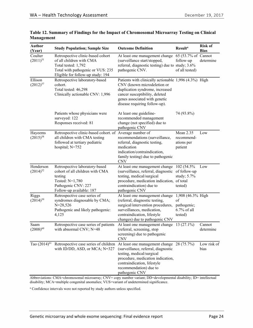
WA – Health Technology Assessment December 19, 2017
Genetic microarray and whole exome sequencing: Final evidence report Page 24
Table 12. Summary of Findings for the Impact of Chromosomal Microarray Testing on Clinical Management
Author (Year)
Study Population; Sample Size Outcome Definition Resulta Risk of Bias
Coulter (2011)51
Retrospective clinic-based cohort of all children with CMA Total tested: 1,792 Total with pathogenic or VUS: 235 Eligible for follow-up study: 194
At least one management change (surveillance start/stopped, referral, diagnostic testing) due to pathogenic CNV.
65 (53.7% of follow-up study; 3.6% of all tested)
Cannot determine
Ellison (2012)57
Retrospective laboratory-based cohort. Total tested: 46,298 Clinically actionable CNV: 1,996 Patients whose physicians were surveyed: 122 Responses received: 81
Patients with clinically actionable CNV (known microdeletion or duplication syndrome, increased cancer susceptibility, deleted genes associated with genetic disease requiring follow-up). At least one guideline- recommended management change (not specified) due to pathogenic CNV
1,996 (4.3%) 74 (93.8%)
High
Hayeems (2015)58
Retrospective clinic-based cohort. of all children with CMA testing followed at tertiary pediatric hospital; N=752
Average number of recommendations (surveillance, referral, diagnostic testing, medication indication/contraindication, family testing) due to pathogenic CNV
Mean 2.35 recommend-ations per patient
Low
Henderson (2014)52
Retrospective laboratory-based cohort of all children with CMA testing Tested: N=1,780 Pathogenic CNV: 227 Follow-up available: 187
At least one management change (surveillance, referral, diagnostic testing, medical/surgical procedure, medication indication, contraindication) due to pathogenic CNV
102 (54.5% of follow-up study; 5.7% of total tested)
Low
Riggs (2014)59
Retrospective case series of syndromes diagnosable by CMA; N=28,526 Pathogenic and likely pathogenic: 4,125
At least one management change (referral, diagnostic testing, surgical/intervention procedures, surveillances, medication, contraindication, lifestyle changes) due to pathogenic CNV
1,908 (46.3% of pathogenic; 6.7% of all tested)
High
Saam (2008)60
Retrospective case series of patients with abnormal CNV; N=48
At least one management change (referral, screening, stop screening) due to pathogenic CNV
13 (27.1%) Cannot determine
Tao (2014)61 Retrospective case series of children with ID/DD, ASD, or MCA; N=327
At least one management change (surveillance, referral, diagnostic testing, medical/surgical procedure, medication indication, contraindication, lifestyle recommendation) due to pathogenic CNV
28 (75.7%) Low risk of bias
Abbreviations: CMA=chromosomal microarray; CNV= copy number variant; DD=developmental disability; ID= intellectual disability; MCA=multiple congenital anomalies; VUS=variant of undetermined significance.
a Confidence intervals were not reported by study authors unless specified.
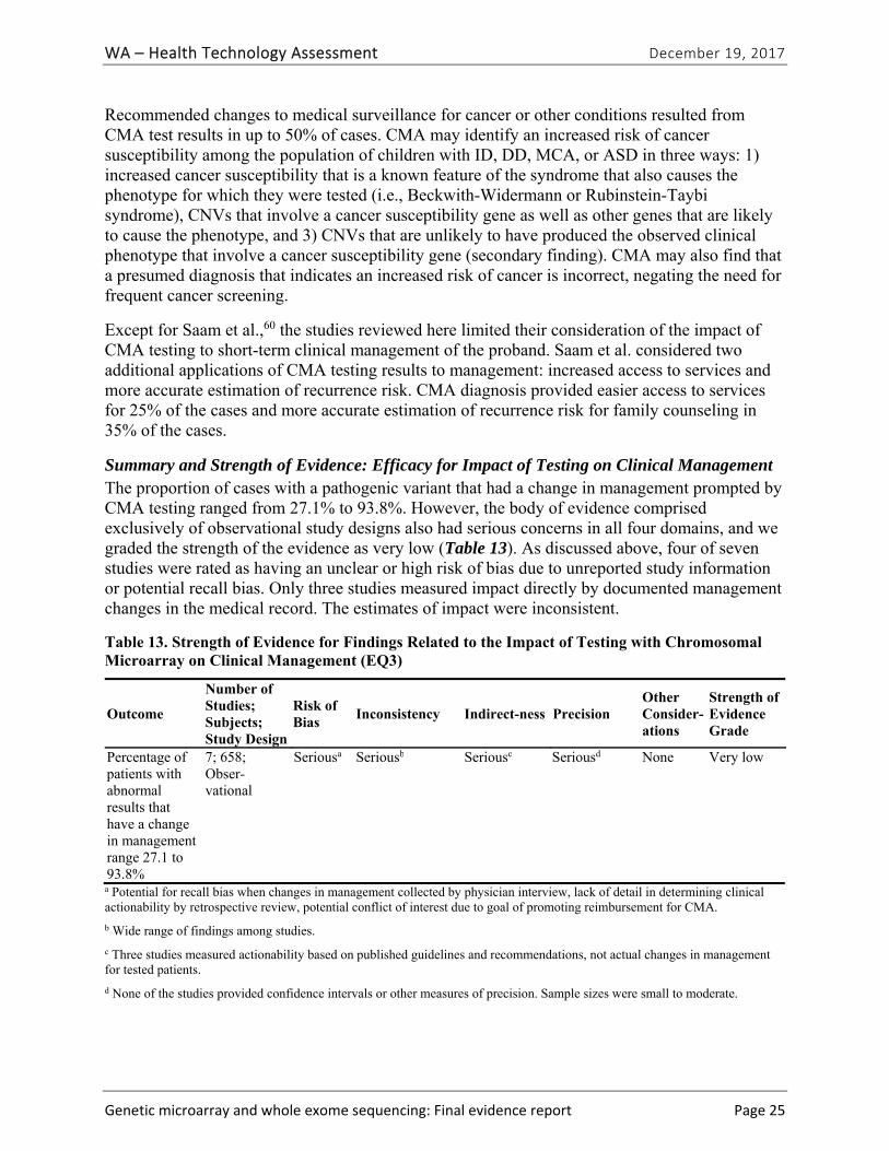
WA – Health Technology Assessment December 19, 2017
Genetic microarray and whole exome sequencing: Final evidence report Page 25
Recommended changes to medical surveillance for cancer or other conditions resulted from CMA test results in up to 50% of cases. CMA may identify an increased risk of cancer susceptibility among the population of children with ID, DD, MCA, or ASD in three ways: 1) increased cancer susceptibility that is a known feature of the syndrome that also causes the phenotype for which they were tested (i.e., Beckwith-Widermann or Rubinstein-Taybi syndrome), CNVs that involve a cancer susceptibility gene as well as other genes that are likely to cause the phenotype, and 3) CNVs that are unlikely to have produced the observed clinical phenotype that involve a cancer susceptibility gene (secondary finding). CMA may also find that a presumed diagnosis that indicates an increased risk of cancer is incorrect, negating the need for frequent cancer screening.
Except for Saam et al.,60 the studies reviewed here limited their consideration of the impact of CMA testing to short-term clinical management of the proband. Saam et al. considered two additional applications of CMA testing results to management: increased access to services and more accurate estimation of recurrence risk. CMA diagnosis provided easier access to services for 25% of the cases and more accurate estimation of recurrence risk for family counseling in 35% of the cases.
Summary and Strength of Evidence: Efficacy for Impact of Testing on Clinical Management The proportion of cases with a pathogenic variant that had a change in management prompted by CMA testing ranged from 27.1% to 93.8%. However, the body of evidence comprised exclusively of observational study designs also had serious concerns in all four domains, and we graded the strength of the evidence as very low (Table 13). As discussed above, four of seven studies were rated as having an unclear or high risk of bias due to unreported study information or potential recall bias. Only three studies measured impact directly by documented management changes in the medical record. The estimates of impact were inconsistent.
Table 13. Strength of Evidence for Findings Related to the Impact of Testing with Chromosomal Microarray on Clinical Management (EQ3)
Outcome
Number of Studies; Subjects; Study Design
Risk of Bias
Inconsistency Indirect-ness Precision Other Consider-ations
Strength of Evidence Grade
Percentage of patients with abnormal results that have a change in management range 27.1 to 93.8%
7; 658; Obser-vational
Seriousa Seriousb Seriousc Seriousd None Very low
a Potential for recall bias when changes in management collected by physician interview, lack of detail in determining clinical actionability by retrospective review, potential conflict of interest due to goal of promoting reimbursement for CMA.
b Wide range of findings among studies.
c Three studies measured actionability based on published guidelines and recommendations, not actual changes in management for tested patients.
d None of the studies provided confidence intervals or other measures of precision. Sample sizes were small to moderate.
WA – Health Technology Assessment December 19, 2017
Genetic microarray and whole exome sequencing: Final evidence report Page 26
EQ4. Do children with congenital defects, autism, intellectual disability, or developmental disability tested with CMA or WES have better health outcomes?
We did not identify any studies that reported on health outcomes among children tested with CMA or WES, either as single-arm studies or compared to patients not tested or tested with other platforms.
Cost and Cost-Effectiveness
CQ1. What is the cost and cost-effectiveness of genetic diagnostic testing for these conditions with CMA or WES?
We identified five eligible studies reporting cost, cost per patient, cost per diagnosis, or cost per additional diagnosis.62-66 All identified studies were specific to CMA testing; no studies evaluated WES testing or reported cost-effectiveness based on cost per quality-adjusted or disability-adjusted life year. Study characteristics and findings are summarized in Table 9. Appendix C, Tables C-1 and C-5A, provide individual study characteristics and findings respectively. Appendix E, Tables E-1 and E-2, provide individual study risk of bias assessments.
Study Characteristics The populations included across the five studies varied; two focused exclusively on children with ID65,66 one focused exclusively on children with DD,63 and two included a mixed population of children with ID, DD, or both.62,64 Studies evaluated the use of CMA as a first-line test for diagnostic evaluation, in comparison to karyotype as a first-line test in three studies,64-66 and in comparison to metabolic, genetic, imaging, or tissue biopsy testing in two studies.62,63 All studies reported cost-related outcomes from a payer perspective, and studies were highly varied as to what type of costs were included, and the currency year for which outcomes were reported. Because of this heterogeneity, we did not quantitatively synthesize findings. The authors conducted these studies from 2005 to 2009. Three studies used a retrospective cohort design with sample sizes ranging from 46 to 1,590,62-64 while two studies used decision analysis with a hypothetical cohort of participants.65,66 Three studies were conducted in the United Kingdom62,64,66 and two were conducted in Canada.63,65 We rated all included studies for the cost outcomes as having a low risk of bias.
Findings Costs per array varied across studies and by testing platforms; these costs ranged from $271 to $1,575 (in 2010 U.S. dollars). These costs reflect the cost per array, which was only one of several costs used to estimate overall costs of CMA testing compared to no CMA testing. The wide variation in costs per array can largely be attributed to the use of different testing platforms, in different years and the use of commercial laboratories (higher cost per array) compared with hospital-based laboratories (lower cost per array).
The cost per patient and cost per diagnosis estimated with CMA testing compared to no CMA testing is summarized in Table 14 and is organized by phenotypes evaluated. We converted and reported all outcomes in 2010 U.S. dollars for comparison across studies. Appendix C, Table C-5B, provides this same data in the currency and year reported by study authors. Studies reported cost per patient tested, cost per diagnosis rendered, and cost per additional diagnosis rendered,
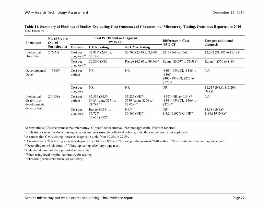
WA – Health Technology Assessment December 19, 2017
Genetic microarray and whole exome sequencing: Final evidence report Page 27
Table 14. Summary of Findings of Studies Evaluating Cost Outcomes of Chromosomal Microarray Testing, Outcomes Reported in 2010 U.S. Dollars
Phenotype No. of Studies (No. of Participants)
Cost Per Patient or Diagnosis (95% CI) Difference in Cost
(95% CI) Cost per additional diagnosis
Outcome CMA Testing No CMA Testing
Intellectual Disability
2 (NAa) Cost per diagnosis65
$2,919b (2,671 to $3,188)
$2,707 (2,448 to 2,990) $213 (168 to 256) $2,592 ($1,586 to $5,188)
Cost per diagnosis66
$6,269c (NR) Range $4,280 to $9,966d Range -$3,697 to $1,988d Rangee -$370 to $199
Developmental Delay
1 (114)63 Cost per patient
NR NR -$101 (98% CI, -$186 to -$16)f $402 (98% CI, $227 to $577)g
NA
Cost per diagnosis
NR NR NR $1,317 (NR)f; $12,296 (NR)g
Intellectual disability or developmental delay or both
2(1,636) Cost per patient
$2,536 (NR)62 $415 (range $271 to $1,792)64
$3,223 (NR)62 $759 (range $556 to $2,029)64
-$687 (NR, p=0.34)62 -$344 (95% CI, -$366 to -$322)64
NA
Cost per diagnosis
Range $4,381 to $7,75762 $3,625 (NR)64
NR62 $6,866 (NR)64
NR62 $-3,241 (95% CI NR)64
$4,381 (NR)62 $-88,819 (NR)64
Abbreviations: CMA=chromosomal microarray; CI=confidence interval; NA=not applicable; NR=not reported. a Both studies were conducted using decision analyses using hypothetical cohorts; thus, the sample size is not applicable b Assumes that CMA testing increases diagnostic yield from 19.2% to 27.5%
c Assumes that CMA testing increases diagnostic yield from 8% to 18%, cost per diagnosis is 2440 with a 15% absolute increase in diagnostic yield. d Depending on which kinds of follow-up testing after karyotype used. e Calculated based on data provided in the study. f When using local hospital laboratory for testing. g When using commercial laboratory for testing
WA – Health Technology Assessment December 19, 2017
Genetic microarray and whole exome sequencing: Final evidence report Page 28
which is the outcome that measures the incremental cost of first line CMA testing versus testing without CMA per the additional diagnoses rendered by CMA testing compared to testing without CMA. A negative value for this outcome suggests that first-line CMA testing compared to no CMA testing provides additional diagnoses at a cost savings. A positive value for this outcome suggests that testing does not save money, but could be cost-effective depending on the amount a decisionmaker is willing to pay for a higher diagnostic yield.
Cost Per Additional Diagnosis: Intellectual Disability. Regier et al. and Wordsworth et al. studied children with ID using decision analyses conducted with hypothetical cohorts.65,66 The cost per diagnosis was higher in both arms of one study66 compared with the other study.65 However, the study with lower costs per diagnosis estimated a higher cost per additional diagnosis ($2,592 [95% CI $1,586 to $5,188]) compared with the other study (range estimated to be between $-370 to $199 per additional diagnosis). These studies did not use similar cost inputs and assumed different diagnostic yield estimates, both of which explain the variation in estimates.
Cost Per Additional Diagnosis: DD. Trakadis et al., studied cost outcomes among children with DD.63 In this retrospective cohort of 114 children, eight additional diagnoses were made as a result of CMA testing compared with no CMA testing. This study estimated that the difference in cost per patient tested (CMA tested versus not CMA tested) was -$101 (98% CI, -$186 to -$16) when array testing was performed by a local hospital laboratory and $402 (98% CI, $227 to $577) when testing was performed by a commercial laboratory. This translated to a cost per additional diagnosis of $1,317 (local hospital laboratory) to $12,296 (commercial laboratory).
Cost Per Additional Diagnosis: Mixed Populations. Finally, Newman et al. and Sagoo et al. studied cost outcomes among children with ID, DD, or both. Although the costs per patient tested were much higher in both arms in one study62 compared with the other study,64 the difference in cost per patient (CMA tested versus not tested) between studies was similar (-$687 and -$344, respectively). Newman et al. reported a cost per additional diagnosis of $4,381 while Sagoo et al. reported a cost per additional diagnosis of $-88,819. The difference in findings between these two studies can largely be explained by a large difference in cost per diagnosis favoring CMA testing in the Sagoo et al. study since the absolute increase in diagnostic yield in Sagoo et al. was minimal (11.05% in non-CMA tested arm, 11.44% in CMA tested arm).
Summary and Strength of Evidence: Cost and Cost Effectiveness The cost per additional diagnosis across this body of evidence ranged from $-88,819 to $12,296 (in 2010 U.S. dollars). Table 15 summarizes the strength for evidence about the cost-effectiveness of diagnostic evaluations that use first-line CMA testing compared with those that do not use first-line CMA testing. Although this body of evidence did not have any serious concerns for risk of bias, we identified serious concerns related to inconsistency and imprecision. Further, we rated the indirectness the outcome reported (costs per additional diagnosis) as having very serious concerns given that this measure is a surrogate outcome and not a direct reflection of patient health outcomes. Because of these concerns, we graded the certainty of this estimate as very low.
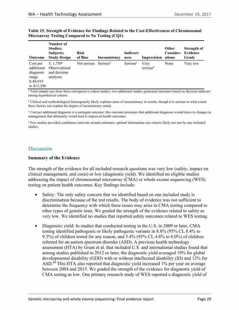
WA – Health Technology Assessment December 19, 2017
Genetic microarray and whole exome sequencing: Final evidence report Page 29
Table 15. Strength of Evidence for Findings Related to the Cost-Effectiveness of Chromosomal Microarray Testing Compared to No Testing (CQ1)
Outcome
Number of Studies; Subjects; Study Design
Risk of Bias Inconsistency
Indirect-ness Imprecision
Other Consider-ations
Strength of Evidence Grade
Cost per additional diagnosis range $-88,919 to $12,296
5; 1,750a Observational and decision analyses
Not serious Seriousb Seriousc Very seriousd
None Very low
a Total sample size from three retrospective cohort studies; two additional studies generated outcomes based on decision analyses among hypothetical cohorts.
b Clinical and methodological heterogeneity likely explains most of inconsistency in results, though it is unclear to what extent these factors can explain the degree of inconsistency noted.
c Cost per additional diagnosis is a surrogate outcome; this outcome presumes that additional diagnoses would leave to changes in management that ultimately would lead to improved health outcomes.
d Few studies provided confidence intervals around estimates; optimal information size criteria likely not met by any included studies.
Discussion
Summary of the Evidence
The strength of the evidence for all included research questions was very low (safety, impact on clinical management, and costs) or low (diagnostic yield). We identified no eligible studies addressing the impact of chromosomal microarray (CMA) or whole exome sequencing (WES) testing on patient health outcomes. Key findings include:
Safety: The only safety concern that we identified based on one included study is discrimination because of the test results. The body of evidence was not sufficient to determine the frequency with which these issues may arise in CMA testing compared to other types of genetic tests. We graded the strength of the evidence related to safety as very low. We identified no studies that reported safety outcomes related to WES testing.
Diagnostic yield: In studies that conducted testing in the U.S. in 2009 or later, CMA testing identified pathogenic or likely pathogenic variants in 8.8% (95% CI, 8.4% to 9.3%) of children tested for any reason, and 5.4% (95% CI, 4.8% to 6.0%) of children referred for an autism spectrum disorder (ASD). A previous health technology assessment (HTA) by Grant et al. that included U.S. and international studies found that among studies published in 2012 or later, the diagnostic yield averaged 19% for global developmental disability (GDD) with or without intellectual disability (ID) and 12% for ASD.40 This HTA also reported that diagnostic yield increased 1% per year on average between 2004 and 2015. We graded the strength of the evidence for diagnostic yield of CMA testing as low. One primary research study of WES reported a diagnostic yield of
WA – Health Technology Assessment December 19, 2017
Genetic microarray and whole exome sequencing: Final evidence report Page 30
27% (95% CI, NR) and we graded the strength of evidence on diagnostic yield of WES testing as very low.
Impact on clinical management: CMA results prompted changes in clinical management in 27% to 94% of patients with a pathogenic variant, which was 3.6% to 6.7% of all patients tested. Considering only studies that measured management changes by medical record review, CMA results changed clinical management in 54% to 76% of patients. We graded the strength of this evidence as very low. We identified no studies reported on change in management related to WES testing.
Costs: The cost per additional diagnosis across this body of evidence ranged from $-88,819 to $12,296 (in 2010 U.S. dollars). No studies reported on cost-effectiveness (i.e., cost per quality-adjusted or disability-adjusted life year). We graded the strength of evidence on costs as very low.
Additional Context for Interpreting Findings
Our review revealed some aspects of CMA and WES testing that, though outside the scope of our systematic review, may add to the interpretation of our results.
Diagnostic Yield of CMA in General Population CMA testing among biobank samples drawn from the general population of Estonia and tied to a database with phenotype information found 56 (0.7%) of 7,877 enrollees had a DECIPHER-listed pathogenic variant.67 The study did not include a clinical evaluation, but 70% of the 56 individuals with a pathogenic variant reported clinical features consistent with their genetic findings.
Analytic Validity of Commercial CMA Kits Compared to sequencing, the Affymetrix® CytoScan® Dx Assay identified 98.8% (95% CI: 93.5%, 99.8%) of duplications and 97.3% (92.3%, 99.1%) of deletions with a size of 1000 base pairs or more. The false positive rate, compared to sequencing, was 1.2% (0.2%, 6.5%) for duplications and 2.7% (0.9%, 7.7%) for deletions of 1000 base pairs or larger. The Agilent GenetiSure Dx Postnatal Assay identified 81.0% (74.3%, 86.2%) of duplications and 88.2% (84.6%, 91.0%) of deletions that were 1,000 to 10,000 base pairs. The false positive rate for 1,000 to 10,000 base pair duplications was 19.0% (13.8%, 25.7%) and 11.8% (9.0%, 15.4%) for deletions of this size. For copy number variants (CNVs) of 10,000 base pairs or larger, the kit identified 97.6% (94.0%, 99.1%) of duplications and 96.9% (93.4%, 98.6%) of deletions, with false positive rates of 2.4% (0.9%, 6.0%) and 3.1% (1.4%, 6.6%), respectively.
False Negatives As reported by D’Amours et al.,81 some array software may return false positive results by incorrectly calling a CNV that does not exist, or falsely identifying an inherited variant as de novo. None of the studies included in this review reported the number of false negative results, which is the failure to identify a chromosomal abnormality identified by karyotype or fluorescent in situ hybridization (FISH). Bi et al.16 reported that CMA failed to detect an abnormality in 0.24% of all cases tests by CMA and karyotype. CMA missed 6 of 43 cases of mosaicism and 29 of 30 balanced rearrangements; thus, testing with CMA may not identify low-level mosaicism or most balanced translocations.
WA – Health Technology Assessment December 19, 2017
Genetic microarray and whole exome sequencing: Final evidence report Page 31
False positives D’Amours et al.81 compared CMA results for 21 children with DD or ID across four high-resolution SNP arrays and the moderate-resolution oligo-array in use for clinical diagnosis at the time of the study. All four SNP arrays identified pathogenic abnormalities in six (28.6%) patients; the oligo-array only identified three (14.3%) of the six abnormalities. Three of the four SNP platforms identified a total of 17 false CNVs. The false positive rate among these arrays ranged from 0% to 5.8%. The false positives resulted from false calls by the platform software or incorrect assignment as de novo because the software did not detect the parental CNV. The authors concluded that high-resolution SNP arrays increase diagnostic yield, but that different platforms vary significantly regarding false positive CNV identification and breakpoint accuracy.
Incidental Findings Ethical issues concerning secondary findings from genetic sequencing have been widely discussed, resulting in American College of Medical Genetics (ACMG) guidelines on reporting clinically actionable findings.23,87 Bowling et al.56 found 55 (9%) pathogenic or likely pathogenic genetic variants unrelated to DD or ID among 605 tested parents of affected children. Nine variants (1.5%) were related to a disorder that the parent self-reported, 12 (2.0%) were one of 56 genes ACMG identified as having potentially clinically actionable variants, and 28 parents were carriers for an autosomal recessive disorder. One couple both carried an allele for the same autosomal recessive disorder, giving them a 25% risk of having a child with the disorder.
Impact on Management and Outcomes Although we did not identify any eligible studies that evaluated the impact of CMA and WES testing on health outcomes, Saam et al. reported that CMA testing increased access to services, such as public health insurance, among children with a pathogenic variant.60 Children referred for CMA testing have complex medical and developmental conditions. As reported in our results section, Hayeems et al.58 found 80% of children with a reportable variant and 62% of children with a benign variant received medical recommendations after their CMA testing. Improved access to insurance or to early invention services may improve long-term health or social outcomes for children with DD, ID, or ASD.88-90
Limitations of the Evidence Base
The body of evidence on safety, impact on management changes, and cost was limited in size, risk of bias, and applicability, limiting our ability to draw strong conclusions for these research questions. Further, almost all studies we included focused on CMA. Clinical use of WES is still new, and the body of evidence regarding its impact is limited.
Most of the studies reporting on diagnostic yield included some cases for indications other than our population of interest. In addition, prior diagnostic testing received by the cases varied. Diagnostic yield may differ among more homogenous case series. This factors may explain some of the difference between our summary estimate of 9% and the median diagnostic yield of 19% among studies published since 2012 that was observed by Grant et al.40 Differences in methods of calling pathogenic variants or access to family history or parent samples may contribute to the differences in diagnostic yield.
WA – Health Technology Assessment December 19, 2017
Genetic microarray and whole exome sequencing: Final evidence report Page 32
Studies evaluating the impact of testing on management were small, so each included only a small portion of known microduplication or microdeletion syndromes. The clinical features of these syndromes and the appropriate management actions vary accordingly, and are likely an explanation for the large heterogeneity of estimates on impact on management we observed across studies. Further, the estimated proportion of cases whose management is impacted by their CMA results may not apply across the population of children for whom CMA is ordered because most included studies were limited to patients followed by a single institution. The two studies that included a broader population of patients measured outcomes by physician survey or interview, which could be subject to recall bias, reporting bias, and response bias.
The body of evidence related to cost and cost-effectiveness is limited by the lack of studies conducted in the United States and the absence of a societal perspective in any of the analyses. Given large differences in the access to health care and its financing between the U.S. and non-U.S. countries, it is not clear whether the findings observed in the studies we identified apply to U.S. settings. Further, this body of evidence is limited by extreme clinical and methodological heterogeneity, which most likely explains the inconsistency in costs per additional diagnosis that we observed. Because CMA and WES tests have rapidly evolved over the past decade, the genetic assay costs used in the included studies may no longer be accurate. Further, the precise role of these tests in the overall sequence and approach to diagnostic evaluation in children with DD, ID, and ASD has also evolved: the cost of the diagnostic journey with or without CMA testing reflected in the included studies may no longer be relevant to current clinical practice.
Across the body of evidence for all research questions, study design, study population, and outcome measurement details were often sparse, resulting in our inability to assess the risk of bias for some studies. One aspect of evaluating the risk of bias not addressed in existing instruments we used are the financial or intellectual conflicts of interest of the study authors. Authors of several included studies stated that a goal of the research was to provide evidence of clinical utility to get CMA covered by payors, potentially providing a strong incentive for analytic decisions that would increase the estimate of diagnostic yield or impact on management.
Limitations of this HTA
We did not include studies published in languages other than English and only searched two U.S.-based electronic databases. These were pragmatic decisions but may have resulted in missing relevant studies; however, we conducted extensive hand searches of the reference lists of included studies and believe the possibility of missing a study that would have altered the findings to be low. For pragmatic reasons, we used a single reviewer to screen most titles/abstracts, which may have led to studies inappropriately excluded. However, we had an excellent concordance initial set of 20 independently dual-reviewed set of titles/abstracts; the principal investigator checked all studies excluded for ineligible intervention and randomly checked a subset of other excluded studies to minimize this possibility. WES studies may have been more likely to be missed or inappropriately excluded because the distinction between test validation and clinical studies was unclear, and because we did not identify any systematic reviews or HTAs of this test.
For the research question related to diagnostic yield (EQ1), we restricted eligibility to U.S. studies that conducted CMA testing in 2009 or later that used current testing platforms to reduce heterogeneity and provide results more applicable to what is in current clinical use. We did not
WA – Health Technology Assessment December 19, 2017
Genetic microarray and whole exome sequencing: Final evidence report Page 33
assess analytic validity or reproducibility and did not conduct an in-depth analysis or synthesis of the cases, breakpoints, or other information related to CNV findings that were presented by study authors.
In addition, our review was limited to the use of WES to detect chromosomal abnormalities. Although WES can detect chromosomal abnormalities, it is primarily used to detect pathogenic mutations or small insertions or deletions within single genes after chromosomal abnormalities have been ruled out by CMA.
Ongoing Research and Future Research Needs
We found no registered studies of research on this topic in clinical trials.gov registry. We identify the following future research needs:
We need randomized clinical trials or well-designed observational studies to evaluate comparative strategies for using CMA or WES as part of the diagnostic evaluation of children with DD, ID, ASD, or MCA. Clinical genetic testing will likely continue to incorporate WES and whole genome sequencing as replacements for single gene sequencing and mutation panels.
Karyotype is still the first-line test for the prenatal diagnosis of chromosomal abnormalities, but CMA is increasingly being emphasized as the first-tier test for prenatal diagnosis. In our search for this HTA, we identified many studies focused on the use in prenatal testing, and a systematic review focused on prenatal use of would synthesize the efficacy, safety, and costs when these tests are used in the prenatal context.
Conclusion
Chromosomal microarray identifies a pathogenic or likely pathogenic variant in nearly 9% of all children referred for testing and in 5% of those referred because of autism spectrum disorders; these findings are based on a low strength of evidence. The results of chromosomal microarray tests generate changes in management in over half of children who are identified as having a pathogenic or likely pathogenic variant; this finding is based on very low strength of evidence. The evidence is very limited with respect to the safety of testing and we identified no evidence related to the impact of testing on health outcomes or cost-effectiveness. The cost per additional diagnosis for chromosomal microarray testing as a first-line diagnostic test varies.
WA – Health Technology Assessment December 19, 2017
Genetic microarray and whole exome sequencing: Final evidence report Page 34
WA – Health Technology Assessment December 19, 2017
Genetic microarray and whole exome sequencing: Final evidence report Page 35
References
1. Watson CT, Marques-Bonet T, Sharp AJ, Mefford HC. The genetics of microdeletion and microduplication syndromes: an update. Annu Rev Genomics Hum Genet. 2014;15:215-244. PMID: 24773319. doi: 10.1146/annurev-genom-091212-153408
2. Wellesley D, Dolk H, Boyd PA, et al. Rare chromosome abnormalities, prevalence and prenatal diagnosis rates from population-based congenital anomaly registers in Europe. Eur J Hum Genet. 2012;20(5):521-526. PMID: 22234154. doi: 10.1038/ejhg.2011.246
3. Srebniak MI, Joosten M, Knapen M, et al. Frequency of submicroscopic chromosome aberrations in pregnancies without increased risk for structural chromosome aberrations: a systematic review of literature and meta-analysis. Ultrasound Obstet Gynecol. 2017. PMID: 28556491. doi: 10.1002/uog.17533
4. Rynn L, Cragan J, Correa A. Update on overall prevalence of major birth defects - Atlanta, Georgia, 1978-2005 (Reprinted from MMWR, vol 57,m pg 1-5, 2008). Journal of the American Medical Association. 2008;299(7):756-758. PMID: WOS:000253226900010.
5. Zablotsky B, Black LI, Maenner MJ, Schieve LA, Blumberg SJ. Estimated prevalence of autism and other developmental disabilities following questionnaire changes in the 2014 National Health Interview Survey. Natl Health Stat Report. 2015(87):1-20. PMID: 26632847.
6. Russo CA, Elixhauser A. Hospitalizations for Birth Defects, 2004. Rockville, MD: Agency for Healthcare Research and Quality; 2007.
7. Centers for Disease Control and Prevention. Disability and Health. Healthcare Cost Data. https://www.cdc.gov/ncbddd/disabilityandhealth/data-highlights.html. Published 2017. Updated August 3. Accessed December 14, 2017.
8. Patel A, Cheung SW. Application of DNA Microarray to Clinical Diagnostics. In: Li PCH, Sedighi A, Wang L, eds. Microarray Technology. Methods and Applications. New York, NY: Humana Press; 2016:111-132.
9. Hadjidekova S, Rukova B, Avdjieva-Tzavella D, Nesheva D, Tincheva R, Toncheva D. Comparison of Two Microarray CGH Platforms for Genome-Wide Copy Number Profilings: Oligo-Based Arrays Versus Bacterial Artificial Chromosome Arrays. Biotechnology & Biotechnological Equipment. 2014;25(3):2499-2504. PMID. doi: 10.5504/bbeq.2011.0064
10. Miller DT, Adam MP, Aradhya S. Consensus statement: chromosomal microarray is a first-tier clinical diagnostic test for individuals with developmental disabilities or congenital anomalies. Am J Hum Genet. 2010;86:749-764. PMID.
11. Manning M, Hudgins L. Array-based technology and recommendations for utilization in medical genetics practice for detection of chromosomal abnormalities. Genetics in Medicine. 2010;12(11):742-745. PMID. doi: 10.1097/gim.0b013e3181f8baad
12. Royal College of Obstetricians and Gynaecologists. Autism: recognition, referral and diagnosis of children and young people on the autism spectrum. W:\01_WES\01_Scope and Background\Coverage Determinations\NICE_Guideline.pdf. Published 2011. Accessed October 18, 2017.
WA – Health Technology Assessment December 19, 2017
Genetic microarray and whole exome sequencing: Final evidence report Page 36
13. Schaefer GB, Mendelsohn NJ, Professional P, Guidelines C. Clinical genetics evaluation in identifying the etiology of autism spectrum disorders: 2013 guideline revisions. Genet Med. 2013;15(5):399-407. PMID: 23519317. doi: 10.1038/gim.2013.32
14. Fogel BL, Satya-Murti S, Cohen BH. Clinical exome sequencing in neurologic disease. Neurol Clin Pract. 2016;6(2):164-176. PMID: 27104068. doi: 10.1212/CPJ.0000000000000239
15. Commonwealth of Massachusetts. 6. Service Codes. http://www.mass.gov/eohhs/docs/masshealth/servicecodes/sub6-lab.doc. Published 2017. Accessed October 18, 2017.
16. Bi W, Borgan C, Pursley AN, et al. Comparison of chromosome analysis and chromosomal microarray analysis: what is the value of chromosome analysis in today's genomic array era? Genet Med. 2013;15(6):450-457. PMID: 23238528. doi: 10.1038/gim.2012.152
17. Teer JK, Mullikin JC. Exome sequencing: the sweet spot before whole genomes. Hum Mol Genet. 2010;19(R2):R145-151. PMID: 20705737. doi: 10.1093/hmg/ddq333
18. de Ligt J, Boone PM, Pfundt R, et al. Detection of clinically relevant copy number variants with whole-exome sequencing. Hum Mutat. 2013;34(10):1439-1448. PMID: 23893877. doi: 10.1002/humu.22387
19. Vetro A, Goidin D, Lesende I, et al. Diagnostic application of a capture based NGS test for the concurrent detection of variants in sequence and copy number as well as LOH. Clin Genet. 2017. PMID: 28556904. doi: 10.1111/cge.13060
20. Ellingford JM, Campbell C, Barton S, et al. Validation of copy number variation analysis for next-generation sequencing diagnostics. Eur J Hum Genet. 2017;25(6):719-724. PMID: 28378820. doi: 10.1038/ejhg.2017.42
21. Richards S, Aziz N, Bale S, et al. Standards and guidelines for the interpretation of sequence variants: a joint consensus recommendation of the American College of Medical Genetics and Genomics and the Association for Molecular Pathology. Genet Med. 2015;17(5):405-424. PMID: 25741868. doi: 10.1038/gim.2015.30
22. Nykamp K, Anderson M, Powers M, et al. Sherloc: a comprehensive refinement of the ACMG-AMP variant classification criteria. Genet Med. 2017;19(10):1105-1117. PMID: 28492532. doi: 10.1038/gim.2017.37
23. Green RC, Berg JS, Grody WW, et al. ACMG recommendations for reporting of incidental findings in clinical exome and genome sequencing. Genet Med. 2013;15(7):565-574. PMID: 23788249. doi: 10.1038/gim.2013.73
24. Boone PM, Soens ZT, Campbell IM, et al. Incidental copy-number variants identified by routine genome testing in a clinical population. Genet Med. 2013;15(1):45-54. PMID: 22878507. doi: 10.1038/gim.2012.95
25. Miller DT, Adam MP, Aradhya S, et al. Consensus statement: chromosomal microarray is a first-tier clinical diagnostic test for individuals with developmental disabilities or congenital anomalies. Am J Hum Genet. 2010;86(5):749-764. PMID: 20466091. doi: 10.1016/j.ajhg.2010.04.006
WA – Health Technology Assessment December 19, 2017
Genetic microarray and whole exome sequencing: Final evidence report Page 37
26. Van Naarden Braun K, Christensen D, Doernberg N, et al. Trends in the prevalence of autism spectrum disorder, cerebral palsy, hearing loss, intellectual disability, and vision impairment, metropolitan atlanta, 1991-2010. PLoS One. 2015;10(4):e0124120. PMID: 25923140. doi: 10.1371/journal.pone.0124120
27. Chan MM. Letter to AFFYMETRIX, INC., C/O Eric Fung, M.D., Ph.D. https://www.accessdata.fda.gov/cdrh_docs/pdf13/k130313.pdf. Published January 21, 2014. Accessed October 18, 2019.
28. 510(k) Substantial Equivalence Determination Decision memorandum. https://www.accessdata.fda.gov/cdrh_docs/reviews/K163367.pdf. Published n.d. Accessed December 14, 2017.
29. Aetna. Comparative Genomic Hybridization (CGH). http://www.aetna.com/cpb/medical/data/700_799/0787.html. Published 2009. Updated September 22, 2017. Accessed October 18, 2017.
30. Premera Blue Cross. Whole Exome and Whole Genome Sequencing for Diagnosis of Genetic Disorders. Medical Policy - 12.04.102. Published February 1, 2017. Updated October 6, 2017. Accessed October 18, 2017.
31. Regence Bluecross Blueshield. Medical Policy Manual. Topic: Chromosomal Microarray Analysis (CMA) and Next-generation Sequencing Panels for the Genetic Evaluation of Patients with Developmental Delay/Intellectual Disability, Autism Spectrum Disorder, or Congenital Anomalies. http://blue.regence.com/trgmedpol/geneticTesting/gt58.pdf. Published August, 2010. Updated April 2017. Accessed October 18, 2017.
32. Regence Bluecross Blueshield. Medical Policy Manual. Topic: Whole Exome and Whole Genome Sequencing. http://blue.regence.com/trgmedpol/geneticTesting/gt76.pdf. Published August 1, 2017. Updated March 2017. Accessed October 18, 2017.
33. Cigna. Comparative Genomic Hybridization (CGH)/Chromosomal Microarray Analysis (CMA) for Autism Spectrum Disorders, Developmental Delay, Intellectual Disability and Congenital Anomalies. https://cignaforhcp.cigna.com/public/content/pdf/coveragePolicies/medical/mm_0493_coveragepositioncriteria_array_comparative_genomic_hybridization.pdf. Published November 15, 2016. Accessed October 18, 2017.
34. Cigna. Whole Exome and Whole Genome Sequencing. https://cignaforhcp.cigna.com/public/content/pdf/coveragePolicies/medical/mm_0519_coveragepositioncriteria_exome_genome_sequence.pdf. Published November 15, 2016. Accessed October 18, 2017.
35. Humana, Inc. Medical and Pharmacy Coverage Policies. http://apps.humana.com/tad/tad_new/home.aspx?type=provider Published 2013. Accessed November 1, 2017.
36. Kaiser Foundation Health Plan of Washington. Clinical Review Criteria. Genetic Screening and Testing. https://provider.ghc.org/all-sites/clinical/criteria/pdf/genetic_screening.pdf Published 2010. Accessed October 18, 2017.
WA – Health Technology Assessment December 19, 2017
Genetic microarray and whole exome sequencing: Final evidence report Page 38
37. Indiana Family & Social Services Administration. Indiana Health Coverage Programs. Genetic Testing. http://provider.indianamedicaid.com/media/155526/genetic%20testing.pdf. Published May 1, 2017. Accessed October 18, 2017.
38. NC Division of Medical Assistance. Genetic Testing. https://ncdma.s3.amazonaws.com/s3fs-public/documents/files/1S4_1.pdf. Published 2016. Accessed October 18, 2017.
39. UnitedHealthcare. Chromosome Microarray Testing. https://www.unitedhealthcareonline.com/ccmcontent/ProviderII/UHC/en-US/Assets/ProviderStaticFiles/ProviderStaticFilesPdf/Tools%20and%20Resources/Policies%20and%20Protocols/Medical%20Policies/Medical%20Policies/Chromosome_Microarray_Testing.pdf Published 2017. Accessed October 18, 2017.
40. Grant M, R. C. Special report: chromosomal microarray for the genetic evaluation of patients with global developmental delay, intellectual disability, and autism spectrum disorder. Technol Eval Cent Assess Program. 2015;30(2):1-4. PMID: 26376503.
41. Viswanathan M, Berkman ND, Dryden DM, Hartling L. Assessing Risk of Bias and Confounding in Observational Studies of Interventions or Exposures: Further Development of the RTI Item Bank. AHRQ Methods for Effective Health Care. Rockville, MD2013.
42. Viswanathan M, Berkman ND. Development of the RTI item bank on risk of bias and precision of observational studies. J Clin Epidemiol. 2012;65(2):163-178. PMID: 21959223. doi: 10.1016/j.jclinepi.2011.05.008
43. Whiting P, Savovic J, Higgins JP, et al. ROBIS: A new tool to assess risk of bias in systematic reviews was developed. J Clin Epidemiol. 2016;69:225-234. PMID: 26092286. doi: 10.1016/j.jclinepi.2015.06.005
44. Bureau of the Fiscal Service. Treasury Reporting Rates of Exchange. https://www.fiscal.treasury.gov/fsreports/rpt/treasRptRateExch/historicalRates.htm. Published 2017. Updated October 12. Accessed October 19, 2017.
45. United States Department of Labor, Bureau of Labor Statistics. Consumer Price Index. https://www.bls.gov/cpi/. Published n.d. Accessed October 19, 2017.
46. Gartlehner G, West SL, Mansfield AJ, et al. Clinical heterogeneity in systematic reviews and health technology assessments: synthesis of guidance documents and the literature. Int J Technol Assess Health Care. 2012;28(1):36-43. PMID: 22217016. doi: 10.1017/s0266462311000687
47. West SL, Gartlehner G, Mansfield AJ, et al. AHRQ Methods for Effective Health Care. Comparative Effectiveness Review Methods: Clinical Heterogeneity. Rockville (MD): Agency for Healthcare Research and Quality (US); 2010.
48. Hedges LV, Vevea JL. Fixed- and random-effects models in meta-analysis. Psychological Methods. 1998;3(4):486-504. PMID: WOS:000077640200006. doi: Doi 10.1037//1082-989x.3.4.486
49. Guyatt GH, Oxman AD, Vist GE, et al. GRADE: an emerging consensus on rating quality of evidence and strength of recommendations. BMJ. 2008;336(7650):924-926. PMID: 18436948. doi: 10.1136/bmj.39489.470347.AD
WA – Health Technology Assessment December 19, 2017
Genetic microarray and whole exome sequencing: Final evidence report Page 39
50. Hamilton MJ, Tolmie JL, Joss S, Morrison N, Suhaimi NM. Chromosomal microarray analysis for looked after children: a double-edged sword? Arch Dis Child. 2015;100(2):206-207. PMID: 25260514. doi: 10.1136/archdischild-2014-307189
51. Coulter ME, Miller DT, Harris DJ, et al. Chromosomal microarray testing influences medical management. Genet Med. 2011;13(9):770-776. PMID: 21716121. doi: 10.1097/GIM.0b013e31821dd54a
52. Henderson LB, Applegate CD, Wohler E, Sheridan MB, Hoover-Fong J, Batista DA. The impact of chromosomal microarray on clinical management: a retrospective analysis. Genet Med. 2014;16(9):657-664. PMID: 24625444. doi: 10.1038/gim.2014.18
53. Ho KS, Wassman ER, Baxter AL, et al. Chromosomal Microarray Analysis of Consecutive Individuals with Autism Spectrum Disorders Using an Ultra-High Resolution Chromosomal Microarray Optimized for Neurodevelopmental Disorders. Int J Mol Sci. 2016;17(12). PMID: 27941670. doi: 10.3390/ijms17122070
54. Roberts JL, Hovanes K, Dasouki M, Manzardo AM, Butler MG. Chromosomal microarray analysis of consecutive individuals with autism spectrum disorders or learning disability presenting for genetic services. Gene. 2014;535(1):70-78. PMID: 24188901. doi: 10.1016/j.gene.2013.10.020
55. Stobbe G, Liu Y, Wu R, Hudgings LH, Thompson O, Hisama FM. Diagnostic yield of array comparative genomic hybridization in adults with autism spectrum disorders. Genet Med. 2014;16(1):70-77. PMID: 23765050. doi: 10.1038/gim.2013.78
56. Bowling KM, Thompson ML, Amaral MD, et al. Genomic diagnosis for children with intellectual disability and/or developmental delay. Genome Med. 2017;9(1):43. PMID: 28554332. doi: 10.1186/s13073-017-0433-1
57. Ellison JW, Ravnan JB, Rosenfeld JA, et al. Clinical utility of chromosomal microarray analysis. Pediatrics. 2012;130(5):e1085-1095. PMID: 23071206. doi: 10.1542/peds.2012-0568
58. Hayeems RZ, Hoang N, Chenier S, et al. Capturing the clinical utility of genomic testing: medical recommendations following pediatric microarray. Eur J Hum Genet. 2015;23(9):1135-1141. PMID: 25491637. doi: 10.1038/ejhg.2014.260
59. Riggs ER, Wain KE, Riethmaier D, et al. Chromosomal microarray impacts clinical management. Clin Genet. 2014;85(2):147-153. PMID: 23347240. doi: 10.1111/cge.12107
60. Saam J, Gudgeon J, Aston E, Brothman AR. How physicians use array comparative genomic hybridization results to guide patient management in children with developmental delay. Genet Med. 2008;10(3):181-186. PMID: 18344707. doi: 10.1097/GIM.0b013e3181634eca
61. Tao VQ, Chan KY, Chu YW, et al. The clinical impact of chromosomal microarray on paediatric care in Hong Kong. PLoS One. 2014;9(10):e109629. PMID: 25333781. doi: 10.1371/journal.pone.0109629
62. Newman WG, Hamilton S, Ayres J, et al. Array comparative genomic hybridization for diagnosis of developmental delay: an exploratory cost-consequences analysis. Clin Genet. 2007;71(3):254-259. PMID: 17309648. doi: 10.1111/j.1399-0004.2007.00756.x
WA – Health Technology Assessment December 19, 2017
Genetic microarray and whole exome sequencing: Final evidence report Page 40
63. Trakadis Y, Shevell M. Microarray as a first genetic test in global developmental delay: a cost-effectiveness analysis. Dev Med Child Neurol. 2011;53(11):994-999. PMID: 21848878. doi: 10.1111/j.1469-8749.2011.04080.x
64. Sagoo GS, Mohammed S, Barton G, et al. Cost Effectiveness of Using Array-CGH for Diagnosing Learning Disability. Appl Health Econ Health Policy. 2015;13(4):421-432. PMID: 25894741. doi: 10.1007/s40258-015-0172-7
65. Regier DA, Friedman JM, Marra CA. Value for money? Array genomic hybridization for diagnostic testing for genetic causes of intellectual disability. Am J Hum Genet. 2010;86(5):765-772. PMID: 20398885. doi: 10.1016/j.ajhg.2010.03.009
66. Wordsworth S, Buchanan J, Regan R, et al. Diagnosing idiopathic learning disability: a cost-effectiveness analysis of microarray technology in the National Health Service of the United Kingdom. Genomic Med. 2007;1(1-2):35-45. PMID: 18923927. doi: 10.1007/s11568-007-9005-6
67. Mannik K, Magi R, Mace A, et al. Copy number variations and cognitive phenotypes in unselected populations. Jama. 2015;313(20):2044-2054. PMID: 26010633. doi: 10.1001/jama.2015.4845
68. Karam SM, Riegel M, Segal SL, et al. Genetic causes of intellectual disability in a birth cohort: a population-based study. Am J Med Genet A. 2015;167(6):1204-1214. PMID: 25728503. doi: 10.1002/ajmg.a.37011
69. Nielsen J, Wohlert M. Chromosome abnormalities found among 34,910 newborn children: results from a 13-year incidence study in Arhus, Denmark. Hum Genet. 1991;87(1):81-83. PMID: 2037286.
70. Lammer EJ, Chak JS, Iovannisci DM, et al. Chromosomal abnormalities among children born with conotruncal cardiac defects. Birth Defects Res A Clin Mol Teratol. 2009;85(1):30-35. PMID: 19067405. doi: 10.1002/bdra.20541
71. Mundhofir FE, Winarni TI, van Bon BW, et al. A cytogenetic study in a large population of intellectually disabled Indonesians. Genet Test Mol Biomarkers. 2012;16(5):412-417. PMID: 22191675. doi: 10.1089/gtmb.2011.0157
72. Oster ME, Lee KA, Honein MA, Riehle-Colarusso T, Shin M, Correa A. Temporal trends in survival among infants with critical congenital heart defects. Pediatrics. 2013;131(5):E1502-E1508. PMID: WOS:000318270700016. doi: 10.1542/peds.2012-3435
73. Greenwood Genetic Center. Peripheral Blood Chromosome Analysis. http://www.ggc.org/diagnostic/tests-costs/test-finder/peripheral-blood-chromosome-analysis.html. Published 2017. Accessed July 25, 2017.
74. Greenwood Genetic Center. CytoScan HD Microarray. http://www.ggc.org/diagnostic/tests-costs/test-finder/whole-genome-snp-microarray.html. Published 2017. Accessed July 25, 2017.
75. Genetics Laboratory. CPT Codes and Pricing. http://www.genetics.ouhsc.edu/molecularprice.asp. Published. Accessed October 18, 2017.
WA – Health Technology Assessment December 19, 2017
Genetic microarray and whole exome sequencing: Final evidence report Page 41
76. Kleinberger J, Maloney KA, Pollin TI, Jeng LJ. An openly available online tool for implementing the ACMG/AMP standards and guidelines for the interpretation of sequence variants. Genet Med. 2016;18(11):1165. PMID: 26986878. doi: 10.1038/gim.2016.13
77. Moeschler JB, Shevell M. Comprehensive evaluation of the child with intellectual disability or global developmental delays. Pediatrics. 2014;134(3):e903-918. PMID: 25157020. doi: 10.1542/peds.2014-1839
78. Satya-Murti S, Cohen BH, Michelson D. Chromosomal Microarray Analysis for Intellectual Disabilities. Published August 20, 2013. Accessed October 18, 2017.
79. Shevell M, Ashwal S, Donley D, et al. Practice parameter: evaluation of the child with global developmental delay: report of the Quality Standards Subcommittee of the American Academy of Neurology and The Practice Committee of the Child Neurology Society. Neurology. 2003;60(3):367-380. PMID: 12578916.
80. UnitedHealthcare. Whole Exome and Whole Genome Sequencing. https://www.uhcprovider.com/content/dam/provider/docs/public/policies/index/commercial/whole-exome-and-whole-genome-sequencing-11117.pdf. Published November 1, 2017. Accessed October 18, 2017.
81. D'Amours G, Langlois M, Mathonnet G, et al. SNP arrays: comparing diagnostic yields for four platforms in children with developmental delay. BMC Med Genomics. 2014;7:70. PMID: 25539807. doi: 10.1186/s12920-014-0070-0
82. University of California, Santa Cruz. UCSC Genome Browser. https://genome.ucsc.edu/. Published 2017. Accessed November 1, 2017.
83. Database of Genomic Variants. Keyword, Landmark or Region Search. Find DGV Variants. http://dgv.tcag.ca/dgv/app/home. Published 2016. Updated May. Accessed November 1, 2017.
84. Online Mendelian Inheritance in Man. An Online Catalog of Human Genes and Genetic Disorders. https://www.omim.org/. Published 2017. Updated October 31. Accessed November 1, 2017.
85. Firth HV, Richards SM, Bevan AP, et al. DECIPHER: Database of Chromosomal Imbalance and Phenotype in Humans Using Ensembl Resources. American Journal of Human Genetics. 2009;84(4):524-533. PMID: WOS:000265232800013. doi: 10.1016/j.ajhg.2009.03.010
86. National Center for Biotechnology Information. dbVar. https://www.ncbi.nlm.nih.gov/dbvar. Published n.d. Accessed November 1, 2017.
87. Roche MI, Berg JS. Incidental Findings with Genomic Testing: Implications for Genetic Counseling Practice. Curr Genet Med Rep. 2015;3(4):166-176. PMID: 26566463. doi: 10.1007/s40142-015-0075-9
88. Estes A, Munson J, Rogers SJ, Greenson J, Winter J, Dawson G. Long-Term Outcomes of Early Intervention in 6-Year-Old Children With Autism Spectrum Disorder. J Am Acad Child Adolesc Psychiatry. 2015;54(7):580-587. PMID: 26088663. doi: 10.1016/j.jaac.2015.04.005
89. Todd J, Armon C, Griggs A, Poole S, Berman S. Increased rates of morbidity, mortality, and charges for hospitalized children with public or no health insurance as compared with
WA – Health Technology Assessment December 19, 2017
Genetic microarray and whole exome sequencing: Final evidence report Page 42
children with private insurance in Colorado and the United States. Pediatrics. 2006;118(2):577-585. PMID: 16882810. doi: 10.1542/peds.2006-0162
90. Tonge BJ, Bull K, Brereton A, Wilson R. A review of evidence-based early intervention for behavioural problems in children with autism spectrum disorder: the core components of effective programs, child-focused interventions and comprehensive treatment models. Curr Opin Psychiatry. 2014;27(2):158-165. PMID: 24452070. doi: 10.1097/YCO.0000000000000043
91. Ho KS, Twede H, Vanzo R, et al. Clinical Performance of an Ultrahigh Resolution Chromosomal Microarray Optimized for Neurodevelopmental Disorders. Biomed Res Int. 2016;2016:3284534. PMID: 27975050. doi: 10.1155/2016/3284534
WA – Health Technology Assessment December 19, 2017
Genetic microarray and whole exome sequencing: Final evidence report Page 43
Appendix A. Search Strategy
PubMed searched from 1/1/200-9/18/2017
Condition being Diagnosed (#1) #1 "Chromosome Aberrations"[Mesh] OR "Chromosome Disorders"[Mesh] OR "Translocation, Genetic"[Mesh] OR "Chromosome Deletion"[Mesh] OR "DNA Copy Number Variations"[Mesh] OR "Gene Dosage"[Mesh] OR "Genomic Structural Variation"[Mesh] OR (("Allelic Imbalance"[Mesh] OR "Genomic Instability"[Mesh] OR "Chromosomes"[Mesh] OR "Aneuploidy"[Majr]) AND ("copy number"[Title/Abstract] OR "CNV"[Title/Abstract] OR "structural"[Title/Abstract] OR "rearrangement"[Title/Abstract] OR "translocation"[Title/Abstract] OR "imbalance"[Title/Abstract] OR "imbalances"[Title/Abstract] OR "inversion"[Title/Abstract] OR "deletion"[Title/Abstract] OR "duplication"[Title/Abstract])) OR "copy number variants"[Title/Abstract] OR "copy number changes"[Title/Abstract] OR "copy number variation"[Title/Abstract] OR "CNV"[Title/Abstract] OR "CNVs"[Title/Abstract] OR "chromosomal rearrangements"[Title/Abstract] OR "translocation"[Title/Abstract] OR "translocations"[Title/Abstract] OR "causal abnormality"[Title/Abstract] Filters: Publication date from 2000/01/01; English Yield: 174,578
Laboratory Testing Methods (Genetic Microarray or Whole Exome Sequencing) (#2) #2 "Microarray Analysis"[Mesh] OR "Comparative Genomic Hybridization"[Mesh] OR "Oligonucleotide Array Sequence Analysis"[Mesh] OR (("In Situ Hybridization"[Mesh] OR "Karyotyping"[Mesh] OR "Cytogenetics"[Mesh]) AND ("array"[Title/Abstract] OR "microarray"[Title/Abstract] OR "CGH"[Title/Abstract] OR "array-CGH"[Title/Abstract] OR "comparative genomic hybridization"[Title/Abstract] OR "fluorescent in situ hybridization"[Title/Abstract])) OR (("Genetic Testing"[Mesh] OR "Molecular Sequence Data"[Mesh] OR "Sequence Analysis"[Mesh] OR "Sequence Analysis, DNA"[Mesh]) AND ("exome"[Title/Abstract] OR "next generation sequencing"[Title/Abstract])) OR "array-CGH"[Title/Abstract] OR "CGH"[Title/Abstract] OR "comparative genomic hybridization"[Title/Abstract] OR "array-based comparative genomic hybridization"[Title/Abstract] OR "genome-wide array"[Title/Abstract] OR "exome"[Title/Abstract] OR "congenital anomaly registers"[Title/Abstract] OR "high resolution testing"[Title/Abstract] Filters: Publication date from 2000/01/01; English Yield: 131,811
Clinical Population (Children with Intellectual Disability, Autism, or Birth Defects) OR Non-Clinical Population (#3) #3 "Autistic Disorder"[Mesh] OR "Asperger Syndrome"[Mesh] OR "Intellectual Disability"[Mesh] OR "Abnormalities, Multiple"[Mesh] OR "Congenital Abnormalities"[Mesh] OR "Developmental Disabilities"[Mesh] OR "Learning Disorders"[Mesh] OR "ASD"[Title/Abstract] OR "autism"[Title/Abstract] OR "mental retardation"[Title/Abstract] OR "mentally retarded"[Title/Abstract] OR "birth defects"[Title/Abstract] OR "mental deficiency"[Title/Abstract] OR chromosome abnormalit*[Title/Abstract] OR "phenotypically abnormal"[Title/Abstract] OR genetic disease*[Title/Abstract] OR "Down syndrome"[Title/Abstract] OR (("Genetics, Population"[Mesh] OR "Prenatal Diagnosis"[Mesh] OR "Genome, Human"[Mesh] OR "Phenotype"[Mesh] OR "Pregnancy"[Mesh] OR "Genetic Variation"[Mesh]) AND ("not at increased risk"[Title/Abstract] OR "phenotypically normal"[Title/Abstract] OR "normal phenotype"[Title/Abstract] OR (("population"[Title/Abstract] OR "populations"[Title/Abstract] OR "individuals"[Title/Abstract]) AND
WA – Health Technology Assessment December 19, 2017
Genetic microarray and whole exome sequencing: Final evidence report Page 44
("general"[Title/Abstract] OR "unselected"[Title/Abstract] OR "normal"[Title/Abstract] OR "disease-free"[Title/Abstract]))) Filters: Publication date from 2000/01/01; English Yield: 311,528
Combining #1, #2, #3 #4 (#1 AND #2 AND #3) Filters: Publication date from 2000/01/01; English 4318 #5 #4 NOT (("Animals"[Mesh] NOT "Humans"[Mesh]) OR "Comment"[Publication Type] OR "Editorial"[Publication Type] OR "Case Reports"[Publication Type]) Filters: Publication date from 2000/01/01; English Total Yield: 2,264
ClinicalTrials.Gov Search from inception to 10/11/2017
Terms: Microarray, limit Child Total Yield: 101
Terms: Whole Exome, limit Child Total Yield: 4
Other Data
The following websites were searched using the terms genetic or chromosomal microarray, whole exome, WES, and CMA to identify information relevant to this health technology assessment.
U.S. Food and Drug Administration
Centers for Medicare and Medicaid Services
Aetna
UnitedHealth
Humana
BlueCross BlueShield (Premera and Regence)
Kaiser Permanente
National Institute for Health and Care Excellence (UK)
U.S. Agency for Healthcare Research and Quality
American Academy of Pediatrics
American Academy of Neurology
American College of Medical Genetics and Genomics
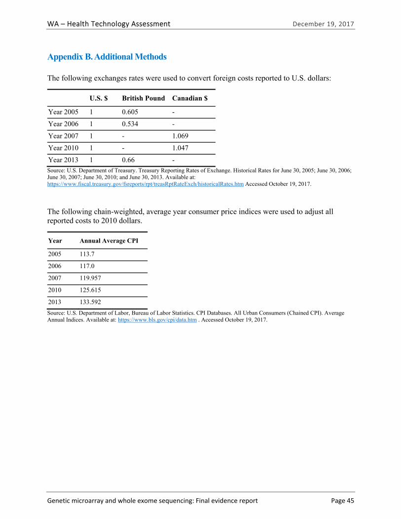
WA – Health Technology Assessment December 19, 2017
Genetic microarray and whole exome sequencing: Final evidence report Page 45
Appendix B. Additional Methods
The following exchanges rates were used to convert foreign costs reported to U.S. dollars:
U.S. $ British Pound Canadian $
Year 2005 1 0.605 -
Year 2006 1 0.534 -
Year 2007 1 - 1.069
Year 2010 1 - 1.047
Year 2013 1 0.66 -
Source: U.S. Department of Treasury. Treasury Reporting Rates of Exchange. Historical Rates for June 30, 2005; June 30, 2006; June 30, 2007; June 30, 2010; and June 30, 2013. Available at: https://www.fiscal.treasury.gov/fsreports/rpt/treasRptRateExch/historicalRates.htm Accessed October 19, 2017.
The following chain-weighted, average year consumer price indices were used to adjust all reported costs to 2010 dollars.
Year Annual Average CPI
2005 113.7
2006 117.0
2007 119.957
2010 125.615
2013 133.592
Source: U.S. Department of Labor, Bureau of Labor Statistics. CPI Databases. All Urban Consumers (Chained CPI). Average Annual Indices. Available at: https://www.bls.gov/cpi/data.htm . Accessed October 19, 2017.
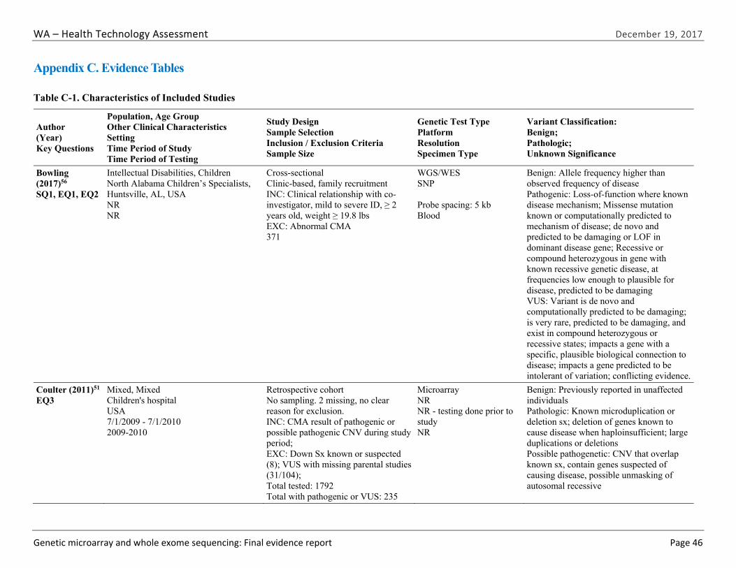
WA – Health Technology Assessment December 19, 2017
Genetic microarray and whole exome sequencing: Final evidence report Page 46
Appendix C. Evidence Tables
Table C-1. Characteristics of Included Studies
Author (Year) Key Questions
Population, Age Group Other Clinical Characteristics Setting Time Period of Study Time Period of Testing
Study Design Sample Selection Inclusion / Exclusion Criteria Sample Size
Genetic Test Type Platform Resolution Specimen Type
Variant Classification: Benign; Pathologic; Unknown Significance
Bowling (2017)56 SQ1, EQ1, EQ2
Intellectual Disabilities, Children North Alabama Children’s Specialists, Huntsville, AL, USA NR NR
Cross-sectional Clinic-based, family recruitment INC: Clinical relationship with co-investigator, mild to severe ID, ≥ 2 years old, weight ≥ 19.8 lbs EXC: Abnormal CMA 371
WGS/WES SNP Probe spacing: 5 kb Blood
Benign: Allele frequency higher than observed frequency of disease Pathogenic: Loss-of-function where known disease mechanism; Missense mutation known or computationally predicted to mechanism of disease; de novo and predicted to be damaging or LOF in dominant disease gene; Recessive or compound heterozygous in gene with known recessive genetic disease, at frequencies low enough to plausible for disease, predicted to be damaging VUS: Variant is de novo and computationally predicted to be damaging; is very rare, predicted to be damaging, and exist in compound heterozygous or recessive states; impacts a gene with a specific, plausible biological connection to disease; impacts a gene predicted to be intolerant of variation; conflicting evidence.
Coulter (2011)51 EQ3
Mixed, Mixed Children's hospital USA 7/1/2009 - 7/1/2010 2009-2010
Retrospective cohort No sampling. 2 missing, no clear reason for exclusion. INC: CMA result of pathogenic or possible pathogenic CNV during study period; EXC: Down Sx known or suspected (8); VUS with missing parental studies (31/104); Total tested: 1792 Total with pathogenic or VUS: 235
Microarray NR NR - testing done prior to study NR
Benign: Previously reported in unaffected individuals Pathologic: Known microduplication or deletion sx; deletion of genes known to cause disease when haploinsufficient; large duplications or deletions Possible pathogenetic: CNV that overlap known sx, contain genes suspected of causing disease, possible unmasking of autosomal recessive
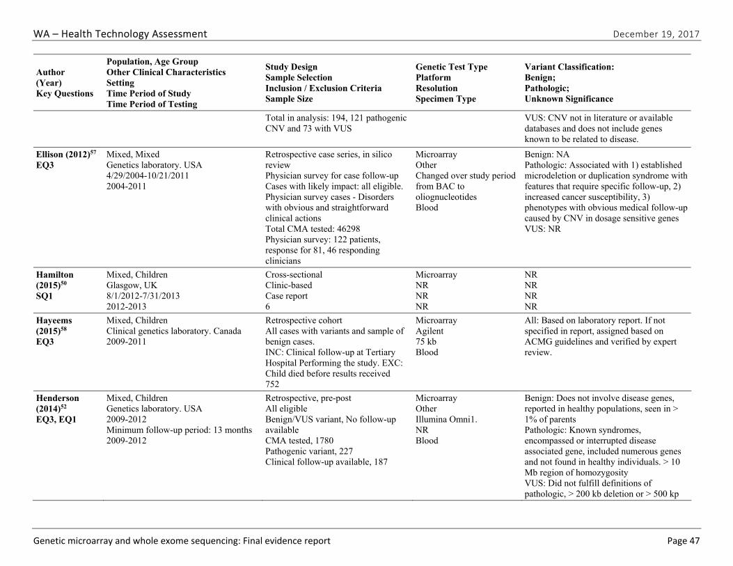
WA – Health Technology Assessment December 19, 2017
Genetic microarray and whole exome sequencing: Final evidence report Page 47
Author (Year) Key Questions
Population, Age Group Other Clinical Characteristics Setting Time Period of Study Time Period of Testing
Study Design Sample Selection Inclusion / Exclusion Criteria Sample Size
Genetic Test Type Platform Resolution Specimen Type
Variant Classification: Benign; Pathologic; Unknown Significance
Total in analysis: 194, 121 pathogenic CNV and 73 with VUS
VUS: CNV not in literature or available databases and does not include genes known to be related to disease.
Ellison (2012)57 EQ3
Mixed, Mixed Genetics laboratory. USA 4/29/2004-10/21/2011 2004-2011
Retrospective case series, in silico review Physician survey for case follow-up Cases with likely impact: all eligible. Physician survey cases - Disorders with obvious and straightforward clinical actions Total CMA tested: 46298 Physician survey: 122 patients, response for 81, 46 responding clinicians
Microarray Other Changed over study period from BAC to oliognucleotides Blood
Benign: NA Pathologic: Associated with 1) established microdeletion or duplication syndrome with features that require specific follow-up, 2) increased cancer susceptibility, 3) phenotypes with obvious medical follow-up caused by CNV in dosage sensitive genes VUS: NR
Hamilton (2015)50 SQ1
Mixed, Children Glasgow, UK 8/1/2012-7/31/2013 2012-2013
Cross-sectional Clinic-based Case report 6
Microarray NR NR NR
NR NR NR NR
Hayeems (2015)58 EQ3
Mixed, Children Clinical genetics laboratory. Canada 2009-2011
Retrospective cohort All cases with variants and sample of benign cases. INC: Clinical follow-up at Tertiary Hospital Performing the study. EXC: Child died before results received 752
Microarray Agilent 75 kb Blood
All: Based on laboratory report. If not specified in report, assigned based on ACMG guidelines and verified by expert review.
Henderson (2014)52 EQ3, EQ1
Mixed, Children Genetics laboratory. USA 2009-2012 Minimum follow-up period: 13 months 2009-2012
Retrospective, pre-post All eligible Benign/VUS variant, No follow-up available CMA tested, 1780 Pathogenic variant, 227 Clinical follow-up available, 187
Microarray Other Illumina Omni1. NR Blood
Benign: Does not involve disease genes, reported in healthy populations, seen in > 1% of parents Pathologic: Known syndromes, encompassed or interrupted disease associated gene, included numerous genes and not found in healthy individuals. > 10 Mb region of homozygosity VUS: Did not fulfill definitions of pathologic, > 200 kb deletion or > 500 kp
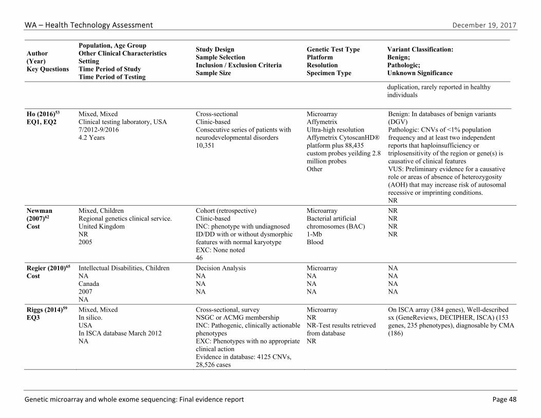
WA – Health Technology Assessment December 19, 2017
Genetic microarray and whole exome sequencing: Final evidence report Page 48
Author (Year) Key Questions
Population, Age Group Other Clinical Characteristics Setting Time Period of Study Time Period of Testing
Study Design Sample Selection Inclusion / Exclusion Criteria Sample Size
Genetic Test Type Platform Resolution Specimen Type
Variant Classification: Benign; Pathologic; Unknown Significance
duplication, rarely reported in healthy individuals
Ho (2016)53 EQ1, EQ2
Mixed, Mixed Clinical testing laboratory, USA 7/2012-9/2016 4.2 Years
Cross-sectional Clinic-based Consecutive series of patients with neurodevelopmental disorders 10,351
Microarray Affymetrix Ultra-high resolution Affymetrix CytoscanHD® platform plus 88,435 custom probes yeilding 2.8 million probes Other
Benign: In databases of benign variants (DGV) Pathologic: CNVs of <1% population frequency and at least two independent reports that haploinsufficiency or triplosensitivity of the region or gene(s) is causative of clinical features VUS: Preliminary evidence for a causative role or areas of absence of heterozygosity (AOH) that may increase risk of autosomal recessive or imprinting conditions. NR
Newman (2007)62 Cost
Mixed, Children Regional genetics clinical service. United Kingdom NR 2005
Cohort (retrospective) Clinic-based INC: phenotype with undiagnosed ID/DD with or without dysmorphic features with normal karyotype EXC: None noted 46
Microarray Bacterial artificial chromosomes (BAC) 1-Mb Blood
NR NR NR NR
Regier (2010)65 Cost
Intellectual Disabilities, Children NA Canada 2007 NA
Decision Analysis NA NA NA
Microarray NA NA NA
NA NA NA NA
Riggs (2014)59 EQ3
Mixed, Mixed In silico. USA In ISCA database March 2012 NA
Cross-sectional, survey NSGC or ACMG membership INC: Pathogenic, clinically actionable phenotypes EXC: Phenotypes with no appropriate clinical action Evidence in database: 4125 CNVs, 28,526 cases
Microarray NR NR-Test results retrieved from database NR
On ISCA array (384 genes), Well-described sx (GeneReviews, DECIPHER, ISCA) (153 genes, 235 phenotypes), diagnosable by CMA (186)
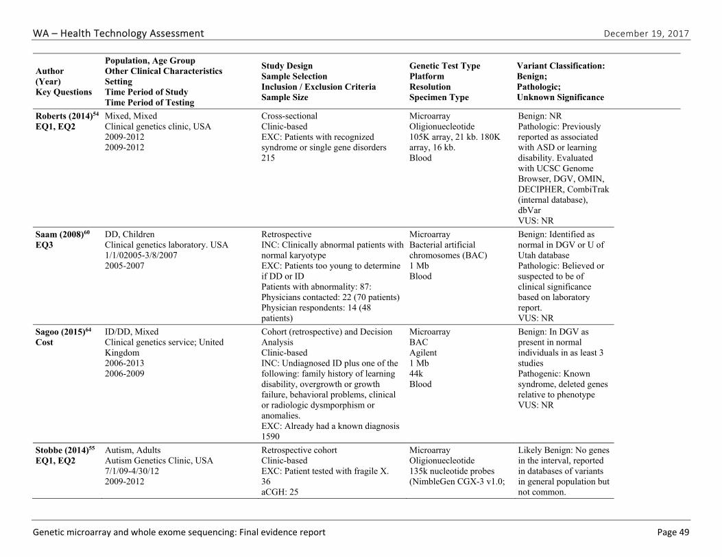
WA – Health Technology Assessment December 19, 2017
Genetic microarray and whole exome sequencing: Final evidence report Page 49
Author (Year) Key Questions
Population, Age Group Other Clinical Characteristics Setting Time Period of Study Time Period of Testing
Study Design Sample Selection Inclusion / Exclusion Criteria Sample Size
Genetic Test Type Platform Resolution Specimen Type
Variant Classification: Benign; Pathologic; Unknown Significance
Roberts (2014)54 EQ1, EQ2
Mixed, Mixed Clinical genetics clinic, USA 2009-2012 2009-2012
Cross-sectional Clinic-based EXC: Patients with recognized syndrome or single gene disorders 215
Microarray Oligionuecleotide 105K array, 21 kb. 180K array, 16 kb. Blood
Benign: NR Pathologic: Previously reported as associated with ASD or learning disability. Evaluated with UCSC Genome Browser, DGV, OMIN, DECIPHER, CombiTrak (internal database), dbVar VUS: NR
Saam (2008)60 EQ3
DD, Children Clinical genetics laboratory. USA 1/1/02005-3/8/2007 2005-2007
Retrospective INC: Clinically abnormal patients with normal karyotype EXC: Patients too young to determine if DD or ID Patients with abnormality: 87: Physicians contacted: 22 (70 patients) Physician respondents: 14 (48 patients)
Microarray Bacterial artificial chromosomes (BAC) 1 Mb Blood
Benign: Identified as normal in DGV or U of Utah database Pathologic: Believed or suspected to be of clinical significance based on laboratory report. VUS: NR
Sagoo (2015)64 Cost
ID/DD, Mixed Clinical genetics service; United Kingdom 2006-2013 2006-2009
Cohort (retrospective) and Decision Analysis Clinic-based INC: Undiagnosed ID plus one of the following: family history of learning disability, overgrowth or growth failure, behavioral problems, clinical or radiologic dysmporphism or anomalies. EXC: Already had a known diagnosis 1590
Microarray BAC Agilent 1 Mb 44k Blood
Benign: In DGV as present in normal individuals in as least 3 studies Pathogenic: Known syndrome, deleted genes relative to phenotype VUS: NR
Stobbe (2014)55 EQ1, EQ2
Autism, Adults Autism Genetics Clinic, USA 7/1/09-4/30/12 2009-2012
Retrospective cohort Clinic-based EXC: Patient tested with fragile X. 36 aCGH: 25
Microarray Oligionuecleotide 135k nucleotide probes (NimbleGen CGX-3 v1.0;
Likely Benign: No genes in the interval, reported in databases of variants in general population but not common.
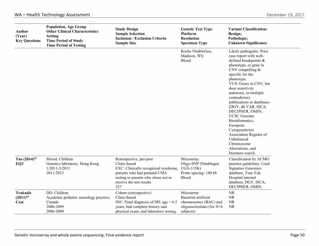
WA – Health Technology Assessment December 19, 2017
Genetic microarray and whole exome sequencing: Final evidence report Page 50
Author (Year) Key Questions
Population, Age Group Other Clinical Characteristics Setting Time Period of Study Time Period of Testing
Study Design Sample Selection Inclusion / Exclusion Criteria Sample Size
Genetic Test Type Platform Resolution Specimen Type
Variant Classification: Benign; Pathologic; Unknown Significance
Roche NimbleGen, Madison, WI) Blood
Likely pathogenic: Prior case report with well-defined breakpoints & phenotype, or gene in CNV compelling & specific for the phenotype. VUS: Genes in CNV, but dose sensitivity unknown, or multiple contradictory publications or databases (DGV, db VAR, ISCA, DECIPHER, OMIN, UCSC Genome Bioinformatics, European Cytogeneticists Association Register of Unbalanced Chromosome Aberrations, and literature search.
Tao (2014)61 EQ3
Mixed, Children Genetics laboratory. Hong Kong 1/2011-5/2013 2011-2013
Retrospective, pre-post Clinic-based EXC: Clinically recognized syndrome, patients who had prenatal CMA testing or parents who chose not to receive the test results 327
Microarray Oligo-SNP (Nimblegen CGX-135K) Probe spacing: 140 kb Blood
Classification by ACMG practice guidelines. Used Signature Genomics database, Tsan Yuk Hospital internal database, DGV, ISCA, DECIPHER, OMIN.
Trakadis (2011)63 Cost
DD, Children Academic pediatric neurology practice; Canada 2006-2009 2006-2009
Cohort (retrospective) Clinic-based INC: Final diagnosis of DD, age < 6.5 years, had complete history and physical exam, and laboratory testing
Microarray Bacterial artificial chromosomes (BAC) and oligonucleotides (for N=6 subjects)
NR NR NR NR
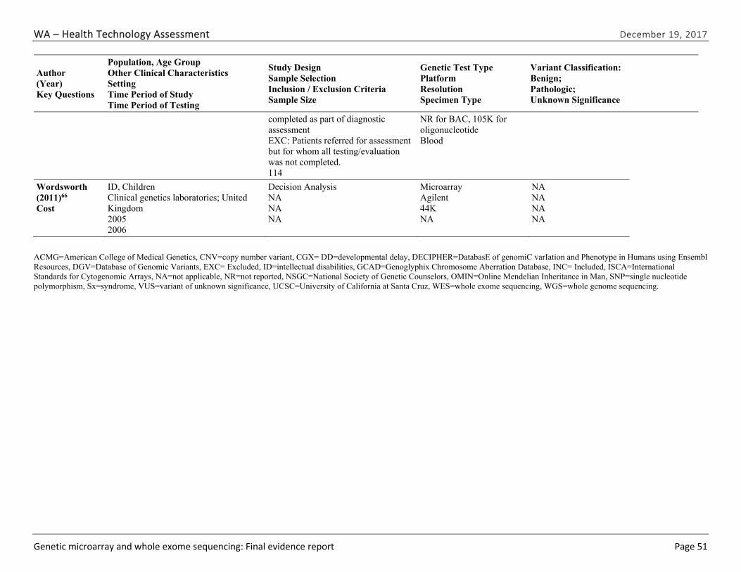
WA – Health Technology Assessment December 19, 2017
Genetic microarray and whole exome sequencing: Final evidence report Page 51
Author (Year) Key Questions
Population, Age Group Other Clinical Characteristics Setting Time Period of Study Time Period of Testing
Study Design Sample Selection Inclusion / Exclusion Criteria Sample Size
Genetic Test Type Platform Resolution Specimen Type
Variant Classification: Benign; Pathologic; Unknown Significance
completed as part of diagnostic assessment EXC: Patients referred for assessment but for whom all testing/evaluation was not completed. 114
NR for BAC, 105K for oligonucleotide Blood
Wordsworth (2011)66 Cost
ID, Children Clinical genetics laboratories; United Kingdom 2005 2006
Decision Analysis NA NA NA
Microarray Agilent 44K NA
NA NA NA NA
ACMG=American College of Medical Genetics, CNV=copy number variant, CGX= DD=developmental delay, DECIPHER=DatabasE of genomiC varIation and Phenotype in Humans using Ensembl Resources, DGV=Database of Genomic Variants, EXC= Excluded, ID=intellectual disabilities, GCAD=Genoglyphix Chromosome Aberration Database, INC= Included, ISCA=International Standards for Cytogenomic Arrays, NA=not applicable, NR=not reported, NSGC=National Society of Genetic Counselors, OMIN=Online Mendelian Inheritance in Man, SNP=single nucleotide polymorphism, Sx=syndrome, VUS=variant of unknown significance, UCSC=University of California at Santa Cruz, WES=whole exome sequencing, WGS=whole genome sequencing.
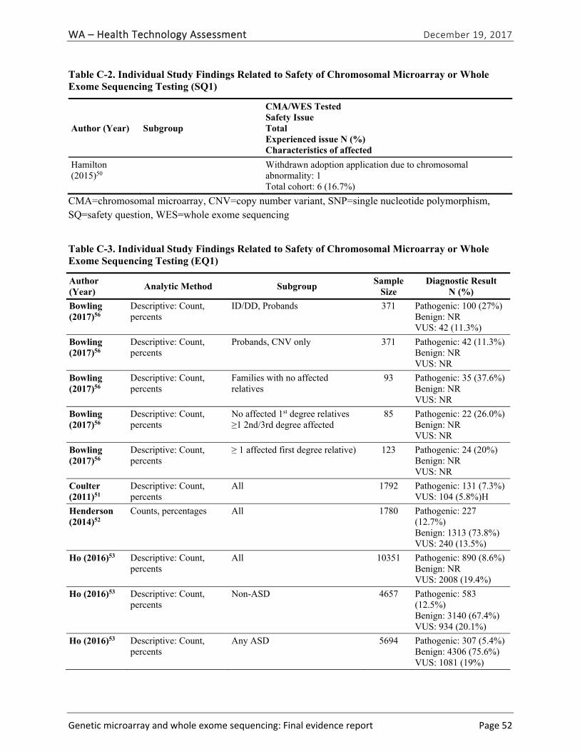
WA – Health Technology Assessment December 19, 2017
Genetic microarray and whole exome sequencing: Final evidence report Page 52
Table C-2. Individual Study Findings Related to Safety of Chromosomal Microarray or Whole Exome Sequencing Testing (SQ1)
Author (Year) Subgroup
CMA/WES Tested Safety Issue Total Experienced issue N (%) Characteristics of affected
Hamilton (2015)50
Withdrawn adoption application due to chromosomal abnormality: 1 Total cohort: 6 (16.7%)
CMA=chromosomal microarray, CNV=copy number variant, SNP=single nucleotide polymorphism, SQ=safety question, WES=whole exome sequencing
Table C-3. Individual Study Findings Related to Safety of Chromosomal Microarray or Whole Exome Sequencing Testing (EQ1)
Author (Year)
Analytic Method Subgroup Sample
SizeDiagnostic Result
N (%)
Bowling (2017)56
Descriptive: Count, percents
ID/DD, Probands 371 Pathogenic: 100 (27%) Benign: NR VUS: 42 (11.3%)
Bowling (2017)56
Descriptive: Count, percents
Probands, CNV only 371 Pathogenic: 42 (11.3%) Benign: NR VUS: NR
Bowling (2017)56
Descriptive: Count, percents
Families with no affected relatives
93 Pathogenic: 35 (37.6%) Benign: NR VUS: NR
Bowling (2017)56
Descriptive: Count, percents
No affected 1st degree relatives ≥1 2nd/3rd degree affected
85 Pathogenic: 22 (26.0%) Benign: NR VUS: NR
Bowling (2017)56
Descriptive: Count, percents
≥ 1 affected first degree relative) 123 Pathogenic: 24 (20%) Benign: NR VUS: NR
Coulter (2011)51
Descriptive: Count, percents
All 1792 Pathogenic: 131 (7.3%) VUS: 104 (5.8%)H
Henderson (2014)52
Counts, percentages All 1780 Pathogenic: 227 (12.7%) Benign: 1313 (73.8%) VUS: 240 (13.5%)
Ho (2016)53 Descriptive: Count, percents
All 10351 Pathogenic: 890 (8.6%) Benign: NR VUS: 2008 (19.4%)
Ho (2016)53 Descriptive: Count, percents
Non-ASD 4657 Pathogenic: 583 (12.5%) Benign: 3140 (67.4%) VUS: 934 (20.1%)
Ho (2016)53 Descriptive: Count, percents
Any ASD 5694 Pathogenic: 307 (5.4%) Benign: 4306 (75.6%) VUS: 1081 (19%)
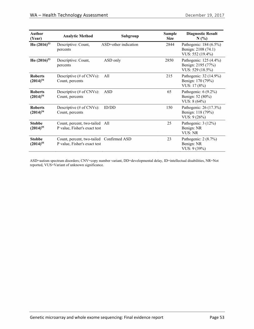
WA – Health Technology Assessment December 19, 2017
Genetic microarray and whole exome sequencing: Final evidence report Page 53
Author (Year)
Analytic Method Subgroup Sample
SizeDiagnostic Result
N (%)
Ho (2016)53 Descriptive: Count, percents
ASD+other indication 2844 Pathogenic: 184 (6.5%) Benign: 2108 (74.1) VUS: 552 (19.4%)
Ho (2016)53 Descriptive: Count, percents
ASD only 2850 Pathogenic: 125 (4.4%) Benign: 2195 (77%) VUS: 529 (18.5%)
Roberts (2014)54
Descriptive (# of CNVs): Count, percents
All 215 Pathogenic: 32 (14.9%) Benign: 170 (79%) VUS: 17 (8%)
Roberts (2014)54
Descriptive (# of CNVs): Count, percents
ASD 65 Pathogenic: 6 (9.2%) Benign: 52 (80%) VUS: 8 (64%)
Roberts (2014)54
Descriptive (# of CNVs): Count, percents
ID/DD 150 Pathogenic: 26 (17.3%) Benign: 118 (79%) VUS: 9 (26%)
Stobbe (2014)55
Count, percent, two-tailed P value, Fisher's exact test
All 25 Pathogenic: 3 (12%) Benign: NR VUS: NR
Stobbe (2014)55
Count, percent, two-tailed P value, Fisher's exact test
Confirmed ASD 23 Pathogenic: 2 (8.7%) Benign: NR VUS: 9 (39%)
ASD=autism spectrum disorders; CNV=copy number variant, DD=developmental delay, ID=intellectual disabilities, NR=Not reported, VUS=Variant of unknown significance.
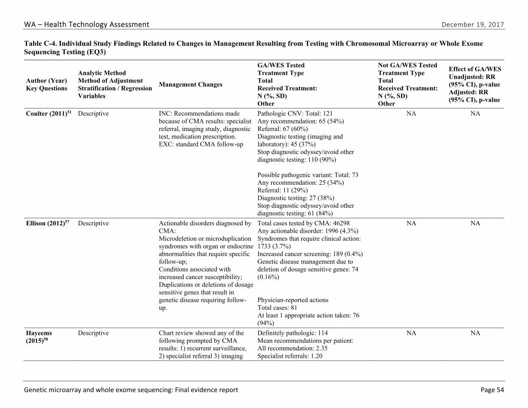
WA – Health Technology Assessment December 19, 2017
Genetic microarray and whole exome sequencing: Final evidence report Page 54
Table C-4. Individual Study Findings Related to Changes in Management Resulting from Testing with Chromosomal Microarray or Whole Exome Sequencing Testing (EQ3)
Author (Year) Key Questions
Analytic Method Method of Adjustment Stratification / Regression Variables
Management Changes
GA/WES Tested Treatment Type Total Received Treatment: N (%, SD) Other
Not GA/WES Tested Treatment Type Total Received Treatment: N (%, SD) Other
Effect of GA/WES Unadjusted: RR (95% CI), p-value Adjusted: RR (95% CI), p-value
Coulter (2011)51 Descriptive INC: Recommendations made because of CMA results: specialist referral, imaging study, diagnostic test, medication prescription. EXC: standard CMA follow-up
Pathologic CNV: Total: 121 Any recommendation: 65 (54%) Referral: 67 (60%) Diagnostic testing (imaging and laboratory): 45 (37%) Stop diagnostic odyssey/avoid other diagnostic testing: 110 (90%) Possible pathogenic variant: Total: 73 Any recommendation: 25 (34%) Referral: 11 (29%) Diagnostic testing: 27 (38%) Stop diagnostic odyssey/avoid other diagnostic testing: 61 (84%)
NA NA
Ellison (2012)57 Descriptive Actionable disorders diagnosed by CMA: Microdeletion or microduplication syndromes with organ or endocrine abnormalities that require specific follow-up; Conditions associated with increased cancer susceptibility; Duplications or deletions of dosage sensitive genes that result in genetic disease requiring follow-up.
Total cases tested by CMA: 46298 Any actionable disorder: 1996 (4.3%) Syndromes that require clinical action: 1733 (3.7%) Increased cancer screening: 189 (0.4%) Genetic disease management due to deletion of dosage sensitive genes: 74 (0.16%) Physician-reported actions Total cases: 81 At least 1 appropriate action taken: 76 (94%)
NA NA
Hayeems (2015)58
Descriptive
Chart review showed any of the following prompted by CMA results: 1) recurrent surveillance, 2) specialist referral 3) imaging
Definitely pathologic: 114 Mean recommendations per patient: All recommendation: 2.35 Specialist referrals: 1.20
NA NA
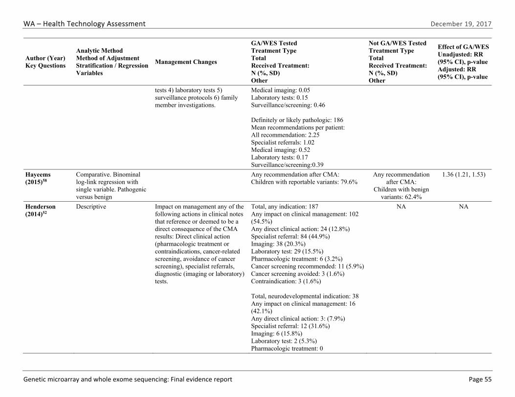
WA – Health Technology Assessment December 19, 2017
Genetic microarray and whole exome sequencing: Final evidence report Page 55
Author (Year) Key Questions
Analytic Method Method of Adjustment Stratification / Regression Variables
Management Changes
GA/WES Tested Treatment Type Total Received Treatment: N (%, SD) Other
Not GA/WES Tested Treatment Type Total Received Treatment: N (%, SD) Other
Effect of GA/WES Unadjusted: RR (95% CI), p-value Adjusted: RR (95% CI), p-value
tests 4) laboratory tests 5) surveillance protocols 6) family member investigations.
Medical imaging: 0.05 Laboratory tests: 0.15 Surveillance/screening: 0.46 Definitely or likely pathologic: 186 Mean recommendations per patient: All recommendation: 2.25 Specialist referrals: 1.02 Medical imaging: 0.52 Laboratory tests: 0.17 Surveillance/screening:0.39
Hayeems (2015)58
Comparative. Binominal log-link regression with single variable. Pathogenic versus benign
Any recommendation after CMA: Children with reportable variants: 79.6%
Any recommendation after CMA:
Children with benign variants: 62.4%
1.36 (1.21, 1.53)
Henderson (2014)52
Descriptive Impact on management any of the following actions in clinical notes that reference or deemed to be a direct consequence of the CMA results: Direct clinical action (pharmacologic treatment or contraindications, cancer-related screening, avoidance of cancer screening), specialist referrals, diagnostic (imaging or laboratory) tests.
Total, any indication: 187 Any impact on clinical management: 102 (54.5%) Any direct clinical action: 24 (12.8%) Specialist referral: 84 (44.9%) Imaging: 38 (20.3%) Laboratory test: 29 (15.5%) Pharmacologic treatment: 6 (3.2%) Cancer screening recommended: 11 (5.9%)Cancer screening avoided: 3 (1.6%) Contraindication: 3 (1.6%) Total, neurodevelopmental indication: 38 Any impact on clinical management: 16 (42.1%) Any direct clinical action: 3: (7.9%) Specialist referral: 12 (31.6%) Imaging: 6 (15.8%) Laboratory test: 2 (5.3%) Pharmacologic treatment: 0
NA NA
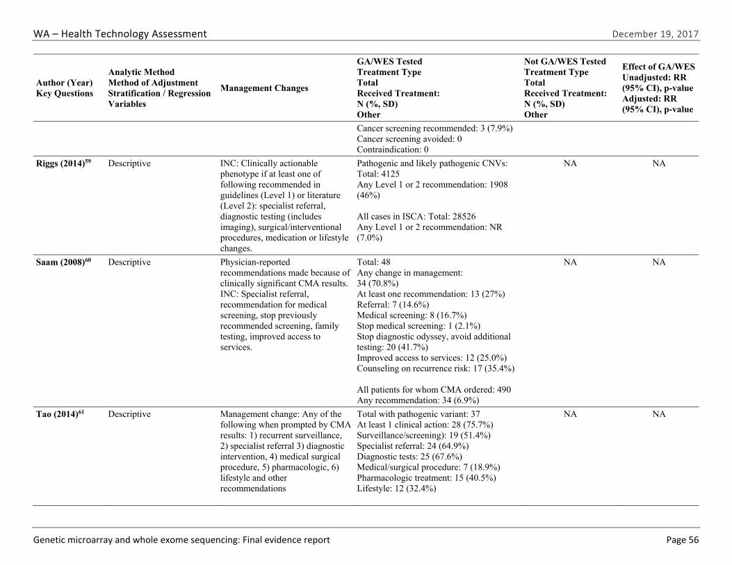
WA – Health Technology Assessment December 19, 2017
Genetic microarray and whole exome sequencing: Final evidence report Page 56
Author (Year) Key Questions
Analytic Method Method of Adjustment Stratification / Regression Variables
Management Changes
GA/WES Tested Treatment Type Total Received Treatment: N (%, SD) Other
Not GA/WES Tested Treatment Type Total Received Treatment: N (%, SD) Other
Effect of GA/WES Unadjusted: RR (95% CI), p-value Adjusted: RR (95% CI), p-value
Cancer screening recommended: 3 (7.9%) Cancer screening avoided: 0 Contraindication: 0
Riggs (2014)59 Descriptive INC: Clinically actionable phenotype if at least one of following recommended in guidelines (Level 1) or literature (Level 2): specialist referral, diagnostic testing (includes imaging), surgical/interventional procedures, medication or lifestyle changes.
Pathogenic and likely pathogenic CNVs: Total: 4125 Any Level 1 or 2 recommendation: 1908 (46%) All cases in ISCA: Total: 28526 Any Level 1 or 2 recommendation: NR (7.0%)
NA NA
Saam (2008)60 Descriptive Physician-reported recommendations made because of clinically significant CMA results. INC: Specialist referral, recommendation for medical screening, stop previously recommended screening, family testing, improved access to services.
Total: 48 Any change in management: 34 (70.8%) At least one recommendation: 13 (27%) Referral: 7 (14.6%) Medical screening: 8 (16.7%) Stop medical screening: 1 (2.1%) Stop diagnostic odyssey, avoid additional testing: 20 (41.7%) Improved access to services: 12 (25.0%) Counseling on recurrence risk: 17 (35.4%) All patients for whom CMA ordered: 490 Any recommendation: 34 (6.9%)
NA NA
Tao (2014)61 Descriptive Management change: Any of the following when prompted by CMA results: 1) recurrent surveillance, 2) specialist referral 3) diagnostic intervention, 4) medical surgical procedure, 5) pharmacologic, 6) lifestyle and other recommendations
Total with pathogenic variant: 37 At least 1 clinical action: 28 (75.7%) Surveillance/screening): 19 (51.4%) Specialist referral: 24 (64.9%) Diagnostic tests: 25 (67.6%) Medical/surgical procedure: 7 (18.9%) Pharmacologic treatment: 15 (40.5%) Lifestyle: 12 (32.4%)
NA NA
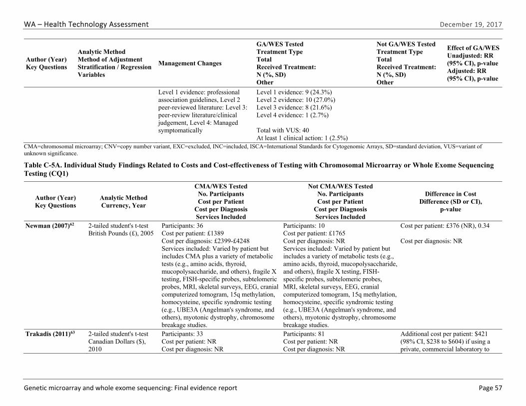
WA – Health Technology Assessment December 19, 2017
Genetic microarray and whole exome sequencing: Final evidence report Page 57
Author (Year) Key Questions
Analytic Method Method of Adjustment Stratification / Regression Variables
Management Changes
GA/WES Tested Treatment Type Total Received Treatment: N (%, SD) Other
Not GA/WES Tested Treatment Type Total Received Treatment: N (%, SD) Other
Effect of GA/WES Unadjusted: RR (95% CI), p-value Adjusted: RR (95% CI), p-value
Level 1 evidence: professional association guidelines, Level 2 peer-reviewed literature: Level 3: peer-review literature/clinical judgement, Level 4: Managed symptomatically
Level 1 evidence: 9 (24.3%) Level 2 evidence: 10 (27.0%) Level 3 evidence: 8 (21.6%) Level 4 evidence: 1 (2.7%) Total with VUS: 40 At least 1 clinical action: 1 (2.5%)
CMA=chromosomal microarray; CNV=copy number variant, EXC=excluded, INC=included, ISCA=International Standards for Cytogenomic Arrays, SD=standard deviation, VUS=variant of unknown significance.
Table C-5A. Individual Study Findings Related to Costs and Cost-effectiveness of Testing with Chromosomal Microarray or Whole Exome Sequencing Testing (CQ1)
Author (Year) Key Questions
Analytic Method Currency, Year
CMA/WES Tested No. Participants Cost per Patient
Cost per Diagnosis Services Included
Not CMA/WES Tested No. Participants Cost per Patient
Cost per Diagnosis Services Included
Difference in Cost Difference (SD or CI),
p-value
Newman (2007)62 2-tailed student's t-test British Pounds (£), 2005
Participants: 36 Cost per patient: £1389 Cost per diagnosis: £2399-£4248 Services included: Varied by patient but includes CMA plus a variety of metabolic tests (e.g., amino acids, thyroid, mucopolysaccharide, and others), fragile X testing, FISH-specific probes, subtelomeric probes, MRI, skeletal surveys, EEG, cranial computerized tomogram, 15q methylation, homocysteine, specific syndromic testing (e.g., UBE3A (Angelman's syndrome, and others), myotonic dystrophy, chromosome breakage studies.
Participants: 10 Cost per patient: £1765 Cost per diagnosis: NR Services included: Varied by patient but includes a variety of metabolic tests (e.g., amino acids, thyroid, mucopolysaccharide, and others), fragile X testing, FISH-specific probes, subtelomeric probes, MRI, skeletal surveys, EEG, cranial computerized tomogram, 15q methylation, homocysteine, specific syndromic testing (e.g., UBE3A (Angelman's syndrome, and others), myotonic dystrophy, chromosome breakage studies.
Cost per patient: £376 (NR), 0.34 Cost per diagnosis: NR
Trakadis (2011)63 2-tailed student's t-test Canadian Dollars ($), 2010
Participants: 33 Cost per patient: NR Cost per diagnosis: NR
Participants: 81 Cost per patient: NR Cost per diagnosis: NR
Additional cost per patient: $421 (98% CI, $238 to $604) if using a private, commercial laboratory to
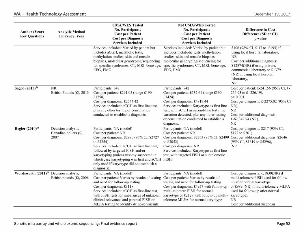
WA – Health Technology Assessment December 19, 2017
Genetic microarray and whole exome sequencing: Final evidence report Page 58
Author (Year) Key Questions
Analytic Method Currency, Year
CMA/WES Tested No. Participants Cost per Patient
Cost per Diagnosis Services Included
Not CMA/WES Tested No. Participants Cost per Patient
Cost per Diagnosis Services Included
Difference in Cost Difference (SD or CI),
p-value
Services included: Varied by patient but includes aCGH, metabolic tests, methylation studies, skin and muscle biopsies, molecular genotyping/sequencing for specific syndromes, CT, MRI, bone age, EEG, EMG.
Services included: Varied by patient but includes metabolic tests, methylation studies, skin and muscle biopsies, molecular genotyping/sequencing for specific syndromes, CT, MRI, bone age, EEG, EMG.
$106 (98% CI, $-17 to -$195) if using local hospital laboratory, NR Cost per additional diagnosis: $12874(NR) if using private, commercial laboratory to $1379 (NR) if using local hospital laboratory, NR
Sagoo (2015)64 NR British Pounds (£), 2013
Participants: 848 Cost per patient: £291.05 (range £190-£1258) Cost per diagnosis: £2544.42 Services included: aCGH as first line test, plus any other testing or consultation conducted to establish a diagnosis.
Participants: 742 Cost per patient: £532.61 (range £390-£1424) Cost per diagnosis: £4819.44 Services included: Karyotype as first line test, with aCGH as second-line test if no variation detected, plus any other testing or consultation conducted to establish a diagnosis.
Cost per patient: £-241.56 (95% CI, £-256.93 to £ -226.19), p< 0.001 Cost per diagnosis: £-2275.02 (95% CI NR), NR Cost per additional diagnosis: £-62,342.94 (NR), NR
Regier (2010)65 Decision analysis, Canadian dollars ($), 2007
Participants: NA (model) Cost per patient: NR Cost per diagnosis: $2980 (95% CI, $2727 to $3254) Services included: aCGH as first line test, followed by targeted FISH and/or karyotyping (unless trisomy suspected in which case karyotyping was first and aCGH only used if karyotype did not establish a diagnosis).
Participants: NA (model) Cost per patient: NR Cost per diagnosis: $2763 (95% CI, $2499 to $3052) Cost per diagnosis: NR Services included: Karyotype as first line test, with targeted FISH or subtelomeric FISH.
Cost per diagnosis: $217 (95% CI, $172 to $261) Cost per additional diagnosis: $2646 (95% CI, $1619 to $5296), NR
Wordsworth (2011)66 Decision analysis, British pounds (£), 2006
Participants: NA (model) Cost per patient: Varies by results of testing and need for follow-up testing. Cost per diagnosis: £3118 Services included: aCGH as first-line test, with FISH tests for imbalances of unknown clinical relevance, and parental FISH or MLPA testing to identify de novo variants.
Participants: NA (model) Cost per patient: Varies by results of testing and need for follow-up testing. Cost per diagnosis: £4957 with follow-up multi-telomere FISH for normal karyotype or £2129 with follow-up multi-telomere MLPA for normal karyotype
Cost per diagnosis: -£1839(NR) if multi-telomere FISH used for follow-up after normal karyotype or £989 (NR) if multi-telomere MLPA used for follow-up after normal karyotype), NR Cost per additional diagnosis:
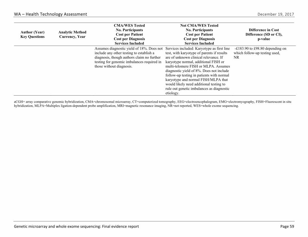
WA – Health Technology Assessment December 19, 2017
Genetic microarray and whole exome sequencing: Final evidence report Page 59
Author (Year) Key Questions
Analytic Method Currency, Year
CMA/WES Tested No. Participants Cost per Patient
Cost per Diagnosis Services Included
Not CMA/WES Tested No. Participants Cost per Patient
Cost per Diagnosis Services Included
Difference in Cost Difference (SD or CI),
p-value
Assumes diagnostic yield of 18%. Does not include any other testing to establish a diagnosis, though authors claim no further testing for genomic imbalances required in those without diagnosis.
Services included: Karyotype as first line test, with karyotype of parents if results are of unknown clinical relevance. If karyotype normal, additional FISH or multi-telomere FISH or MLPA. Assumes diagnostic yield of 8%. Does not include follow-up testing in patients with normal karyotype and normal FISH/MLPA that would likely need additional testing to rule out genetic imbalances as diagnostic etiology.
-£183.90 to £98.80 depending on which follow-up testing used, NR
aCGH= array comparative genomic hybridization, CMA=chromosomal microarray, CT=computerized tomography, EEG=electroencephalogram, EMG=electromyography, FISH=Fluorescent in situ hybridization, MLPA=Multiplex ligation-dependent probe amplification, MRI=magnetic resonance imaging, NR=not reported, WES=whole exome sequencing.
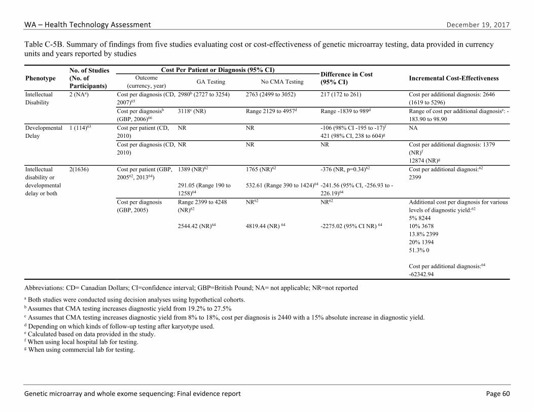
WA – Health Technology Assessment December 19, 2017
Genetic microarray and whole exome sequencing: Final evidence report Page 60
Table C-5B. Summary of findings from five studies evaluating cost or cost-effectiveness of genetic microarray testing, data provided in currency units and years reported by studies
Phenotype No. of Studies (No. of Participants)
Cost Per Patient or Diagnosis (95% CI) Difference in Cost (95% CI)
Incremental Cost-Effectiveness Outcome (currency, year)
GA Testing No CMA Testing
Intellectual Disability
2 (NAa) Cost per diagnosis (CD, 2007)65
2980b (2727 to 3254) 2763 (2499 to 3052) 217 (172 to 261) Cost per additional diagnosis: 2646 (1619 to 5296)
Cost per diagnosisb (GBP, 2006)66
3118c (NR) Range 2129 to 4957d Range -1839 to 989d Range of cost per additional diagnosise: -183.90 to 98.90
Developmental Delay
1 (114)63 Cost per patient (CD, 2010)
NR NR -106 (98% CI -195 to -17)f 421 (98% CI, 238 to 604)g
NA
Cost per diagnosis (CD, 2010)
NR NR NR Cost per additional diagnosis: 1379 (NR)f 12874 (NR)g
Intellectual disability or developmental delay or both
2(1636) Cost per patient (GBP, 200562, 201364)
1389 (NR)62 291.05 (Range 190 to 1258)64
1765 (NR)62 532.61 (Range 390 to 1424)64
-376 (NR, p=0.34)62 -241.56 (95% CI, -256.93 to -226.19)64
Cost per additional diagnosi:62 2399
Cost per diagnosis (GBP, 2005)
Range 2399 to 4248 (NR)62 2544.42 (NR)64
NR62 4819.44 (NR) 64
NR62 -2275.02 (95% CI NR) 64
Additional cost per diagnosis for various levels of diagnostic yield:62 5% 8244 10% 3678 13.8% 2399 20% 1394 51.3% 0 Cost per additional diagnosis:64 -62342.94
Abbreviations: CD= Canadian Dollars; CI=confidence interval; GBP=British Pound; NA= not applicable; NR=not reported
a Both studies were conducted using decision analyses using hypothetical cohorts. b Assumes that CMA testing increases diagnostic yield from 19.2% to 27.5%
c Assumes that CMA testing increases diagnostic yield from 8% to 18%, cost per diagnosis is 2440 with a 15% absolute increase in diagnostic yield. d Depending on which kinds of follow-up testing after karyotype used. e Calculated based on data provided in the study. f When using local hospital lab for testing. g When using commercial lab for testing.
WA – Health Technology Assessment December 19, 2017
Genetic microarray and whole exome sequencing: Final evidence report Page 61
Appendix D. Excluded Studies
List of Exclusion Codes
X1: Ineligible publication type
X2: Ineligible population
X3: Ineligible or no intervention
X4: Ineligible comparator
X5: Ineligible or no outcome
X6: EQ1 Study Published before 2010
X7: No Key Question
X8: Old Platform
X9: Ineligible setting
X10: EQ1 Ineligible country
X11: Duplicates/superseded by more recent publications
Abdelmoity AT, LePichon JB, Nyp SS, et al. 15q11.2 proximal imbalances associated with a diverse array of neuropsychiatric disorders and mild dysmorphic features. J Dev Behav Pediatr. 2012 Sep;33(7):570-6. doi: 10.1097/DBP.0b013e31826052ae. PMID: 22922608. Exclusion Code: X9.
Alamillo CL, Powis Z, Farwell K, et al. Exome sequencing positively identified relevant alterations in more than half of cases with an indication of prenatal ultrasound anomalies. Prenat Diagn. 2015 Nov;35(11):1073-8. doi: 10.1002/pd.4648. PMID: 26147564. Exclusion Code: X2.
Al-Mamari W, Al-Saegh A, Al-Kindy A, et al. Diagnostic Yield of Chromosomal Microarray Analysis in a Cohort of Patients with Autism Spectrum Disorders from a Highly Consanguineous Population. J Autism Dev Disord. 2015 Aug;45(8):2323-8. doi: 10.1007/s10803-015-2394-9. PMID: 25703031. Exclusion Code: X10.
Alvarez-Mora MI, Calvo Escalona R, Puig Navarro O, et al. Comprehensive molecular testing in patients with high functioning autism spectrum disorder. Mutat Res. 2016 Feb-Mar;784-785:46-52. doi: 10.1016/j.mrfmmm.2015.12.006. PMID: 26845707. Exclusion Code: X10.
Aradhya S, Manning MA, Splendore A, et al. Whole-genome array-CGH identifies novel contiguous gene deletions and duplications associated with developmental delay, mental retardation, and dysmorphic features. Am J Med Genet A. 2007 Jul 01;143a(13):1431-41. doi: 10.1002/ajmg.a.31773. PMID: 17568414. Exclusion Code: X8.
Aristidou C, Koufaris C, Theodosiou A, et al. Accurate Breakpoint Mapping in Apparently Balanced Translocation Families with Discordant Phenotypes Using Whole Genome Mate-Pair Sequencing. PLoS One. 2017;12(1):e0169935. doi: 10.1371/journal.pone.0169935. PMID: 28072833. Exclusion Code: X9.
WA – Health Technology Assessment December 19, 2017
Genetic microarray and whole exome sequencing: Final evidence report Page 62
Athanasakis E, Licastro D, Faletra F, et al. Next generation sequencing in nonsyndromic intellectual disability: from a negative molecular karyotype to a possible causative mutation detection. Am J Med Genet A. 2014 Jan;164a(1):170-6. doi: 10.1002/ajmg.a.36274. PMID: 24307393. Exclusion Code: X3.
Bachman KK, DeWard SJ, Chrysostomou C, et al. Array CGH as a first-tier test for neonates with congenital heart disease. Cardiol Young. 2015 Jan;25(1):115-22. doi: 10.1017/s1047951113001868. PMID: 24192140. Exclusion Code: X3.
Baldwin EL, Lee JY, Blake DM, et al. Enhanced detection of clinically relevant genomic imbalances using a targeted plus whole genome oligonucleotide microarray. Genet Med. 2008 Jun;10(6):415-29. doi: 10.1097/GIM.0b013e318177015c. PMID: 18496225. Exclusion Code: X8.
Baptista J, Mercer C, Prigmore E. Breakpoint mapping and genome-wide array analysis in translocations: comparison of a phenotypically normal and an abnormal cohort. Am J Hum Genet. 2008 //;82:927-36. Exclusion Code: X8.
Baptista J, Prigmore E, Gribble SM, et al. Molecular cytogenetic analyses of breakpoints in apparently balanced reciprocal translocations carried by phenotypically normal individuals. Eur J Hum Genet. 2005 Nov;13(11):1205-12. doi: 10.1038/sj.ejhg.5201488. PMID: 16118644. Exclusion Code: X8.
Baris HN, Tan WH, Kimonis VE, et al. Diagnostic utility of array-based comparative genomic hybridization in a clinical setting. Am J Med Genet A. 2007 Nov 01;143a(21):2523-33. doi: 10.1002/ajmg.a.31988. PMID: 17910064. Exclusion Code: X8.
Barth TF, Benner A, Bentz M, et al. Risk of false positive results in comparative genomic hybridization. Genes Chromosomes Cancer. 2000 Jul;28(3):353-7. PMID: 10862043. Exclusion Code: X2.
Bartnik M, Nowakowska B, Derwinska K, et al. Application of array comparative genomic hybridization in 256 patients with developmental delay or intellectual disability. J Appl Genet. 2014 Feb;55(1):125-44. doi: 10.1007/s13353-013-0181-x. PMID: 24297458. Exclusion Code: X10.
Bartnik M, Wisniowiecka-Kowalnik B, Nowakowska B, et al. The usefulness of array comparative genomic hybridization in clinical diagnostics of intellectual disability in children. Dev Period Med. 2014 Jul-Sep;18(3):307-17. PMID: 25182394. Exclusion Code: X10.
Battaglia A, Carey JC. Etiologic yield of autistic spectrum disorders: a prospective study. Am J Med Genet C Semin Med Genet. 2006 //;142C:3-7. Exclusion Code: X3.
Battaglia A, Doccini V, Bernardini L, et al. Confirmation of chromosomal microarray as a first-tier clinical diagnostic test for individuals with developmental delay, intellectual disability, autism spectrum disorders and dysmorphic features. Eur J Paediatr Neurol. 2013 Nov;17(6):589-99. doi: 10.1016/j.ejpn.2013.04.010. PMID: 23711909. Exclusion Code: X10.
Behjati F, Firouzabadi SG, Kariminejad R, et al. Genomic characterization of some Iranian children with idiopathic mental retardation using array comparative genomic hybridization. Indian J Hum Genet. 2013 Oct;19(4):443-8. doi: 10.4103/0971-6866.124373. PMID: 24497710. Exclusion Code: X3.
WA – Health Technology Assessment December 19, 2017
Genetic microarray and whole exome sequencing: Final evidence report Page 63
Ben-Shachar S, Lanpher B, German JR, et al. Microdeletion 15q13.3: a locus with incomplete penetrance for autism, mental retardation, and psychiatric disorders. J Med Genet. 2009 Jun;46(6):382-8. doi: 10.1136/jmg.2008.064378. PMID: 19289393. Exclusion Code: X9.
Bernardini L, Alesi V, Loddo S, et al. High-resolution SNP arrays in mental retardation diagnostics: how much do we gain? Eur J Hum Genet. 2010 Feb;18(2):178-85. doi: 10.1038/ejhg.2009.154. PMID: 19809473. Exclusion Code: X9.
Bi W, Borgan C, Pursley AN, et al. Comparison of chromosome analysis and chromosomal microarray analysis: what is the value of chromosome analysis in today's genomic array era? Genet Med. 2013 Jun;15(6):450-7. doi: 10.1038/gim.2012.152. PMID: 23238528. Exclusion Code: X8.
Blue Cross Blue Shield Technology Evaluation Center. Special report: aCGH for the genetic evaluation of patients with developmental delay/mental retardation or autism spectrum disorder. Technol Eval Cent Assess Program Exec Summ. 2009 Apr;23(10):1-5. PMID: 19824216. Exclusion Code: X11.
Blyth M, Maloney V, Beal S, et al. Pallister-Killian syndrome: a study of 22 British patients. J Med Genet. 2015 Jul;52(7):454-64. doi: 10.1136/jmedgenet-2014-102877. PMID: 25888713. Exclusion Code: X5.
Boggula VR, Agarwal M, Kumar R, et al. Recurrent benign copy number variants & issues in interpretation of variants of unknown significance identified by cytogenetic microarray in Indian patients with intellectual disability. Indian J Med Res. 2015 Dec;142(6):699-712. doi: 10.4103/0971-5916.174561. PMID: 26831419. Exclusion Code: X10.
Boggula VR, Shukla A, Danda S, et al. Clinical utility of multiplex ligation-dependent probe amplification technique in identification of aetiology of unexplained mental retardation: a study in 203 Indian patients. Indian J Med Res. 2014 Jan;139(1):66-75. PMID: 24604040. Exclusion Code: X3.
Brady PD, DeKoninck P, Fryns JP, et al. Identification of dosage-sensitive genes in fetuses referred with severe isolated congenital diaphragmatic hernia. Prenat Diagn. 2013 Dec;33(13):1283-92. doi: 10.1002/pd.4244. PMID: 24122781. Exclusion Code: X10.
Brady PD, Delle Chiaie B, Christenhusz G, et al. A prospective study of the clinical utility of prenatal chromosomal microarray analysis in fetuses with ultrasound abnormalities and an exploration of a framework for reporting unclassified variants and risk factors. Genet Med. 2014 Jun;16(6):469-76. doi: 10.1038/gim.2013.168. PMID: 24177055. Exclusion Code: X10.
Breckpot J, Thienpont B, Peeters H, et al. Array comparative genomic hybridization as a diagnostic tool for syndromic heart defects. J Pediatr. 2010 May;156(5):810-7, 7.e1-7.e4. doi: 10.1016/j.jpeds.2009.11.049. PMID: 20138633. Exclusion Code: X8.
Bremer A, Giacobini M, Eriksson M, et al. Copy number variation characteristics in subpopulations of patients with autism spectrum disorders. American Journal of Medical Genetics Part B: Neuropsychiatric Genetics. 2010 2010/12/08;156(2):115-24. doi: 10.1002/ajmg.b.31142. Exclusion Code: X10.
Bruno DL, Ganesamoorthy D, Schoumans J, et al. Detection of cryptic pathogenic copy number variations and constitutional loss of heterozygosity using high resolution SNP microarray analysis in 117 patients referred for cytogenetic analysis and impact on clinical practice. J
WA – Health Technology Assessment December 19, 2017
Genetic microarray and whole exome sequencing: Final evidence report Page 64
Med Genet. 2009 Feb;46(2):123-31. doi: 10.1136/jmg.2008.062604. PMID: 19015223. Exclusion Code: X8.
Bruno DL, White SM, Ganesamoorthy D, et al. Pathogenic aberrations revealed exclusively by single nucleotide polymorphism (SNP) genotyping data in 5000 samples tested by molecular karyotyping. J Med Genet. 2011 Dec;48(12):831-9. doi: 10.1136/jmedgenet-2011-100372. PMID: 22039585. Exclusion Code: X9.
Buckley JR, Kavarana MN, Chowdhury SM, et al. Current Practice and Utility of Chromosome Microarray Analysis in Infants Undergoing Cardiac Surgery. Congenit Heart Dis. 2015 May-Jun;10(3):E131-8. doi: 10.1111/chd.12241. PMID: 25494910. Exclusion Code: X2.
Busse T, Graham JM, Jr., Feldman G, et al. High-Resolution genomic arrays identify CNVs that phenocopy the chromosome 22q11.2 deletion syndrome. Hum Mutat. 2011 Jan;32(1):91-7. doi: 10.1002/humu.21395. PMID: 21120947. Exclusion Code: X5.
Buysse K, Delle Chiaie B, Van Coster R, et al. Challenges for CNV interpretation in clinical molecular karyotyping: lessons learned from a 1001 sample experience. Eur J Med Genet. 2009 Nov-Dec;52(6):398-403. doi: 10.1016/j.ejmg.2009.09.002. PMID: 19765681. Exclusion Code: X10.
Capalbo A, Bono S, Spizzichino L, et al. Sequential comprehensive chromosome analysis on polar bodies, blastomeres and trophoblast: insights into female meiotic errors and chromosomal segregation in the preimplantation window of embryo development. Hum Reprod. 2013 Feb;28(2):509-18. doi: 10.1093/humrep/des394. PMID: 23148203. Exclusion Code: X2.
Capobianco S, Lava SA, Bianchetti MG, et al. Chromosomal microarray among children with intellectual disability: a useful diagnostic tool for the clinical geneticist. Dev Med Child Neurol. 2014 Mar;56(3):290. doi: 10.1111/dmcn.12341. PMID: 24266756. Exclusion Code: X10.
Cappuccio G, Vitiello F, Casertano A, et al. New insights in the interpretation of array-CGH: autism spectrum disorder and positive family history for intellectual disability predict the detection of pathogenic variants. Ital J Pediatr. 2016 Apr 12;42:39. doi: 10.1186/s13052-016-0246-7. PMID: 27072107. Exclusion Code: X10.
Caramaschi E, Stanghellini I, Magini P, et al. Predictive diagnostic value for the clinical features accompanying intellectual disability in children with pathogenic copy number variations: a multivariate analysis. Ital J Pediatr. 2014 Apr 28;40:39. doi: 10.1186/1824-7288-40-39. PMID: 24775911. Exclusion Code: X10.
Carey AS, Liang L, Edwards J, et al. Effect of copy number variants on outcomes for infants with single ventricle heart defects. Circ Cardiovasc Genet. 2013 Oct;6(5):444-51. doi: 10.1161/circgenetics.113.000189. PMID: 24021551. Exclusion Code: X3.
Carreira IM, Ferreira SI, Matoso E, et al. Copy number variants prioritization after array-CGH analysis - a cohort of 1000 patients. Mol Cytogenet. 2015;8:103. doi: 10.1186/s13039-015-0202-z. PMID: 26719768. Exclusion Code: X10.
Celestino-Soper PB, Shaw CA, Sanders SJ, et al. Use of array CGH to detect exonic copy number variants throughout the genome in autism families detects a novel deletion in
WA – Health Technology Assessment December 19, 2017
Genetic microarray and whole exome sequencing: Final evidence report Page 65
TMLHE. Hum Mol Genet. 2011 Nov 15;20(22):4360-70. doi: 10.1093/hmg/ddr363. PMID: 21865298. Exclusion Code: X9.
Charan P, Woodrow N, Walker SP, et al. High-resolution microarray in the assessment of fetal anomalies detected by ultrasound. Aust N Z J Obstet Gynaecol. 2014 Feb;54(1):46-52. doi: 10.1111/ajo.12170. PMID: 24471846. Exclusion Code: X10.
Chatron N, Haddad V, Andrieux J, et al. Refinement of genotype-phenotype correlation in 18 patients carrying a 1q24q25 deletion. Am J Med Genet A. 2015 May;167a(5):1008-17. doi: 10.1002/ajmg.a.36856. PMID: 25728055. Exclusion Code: X9.
Cho SC, Yim SH, Yoo HK, et al. Copy number variations associated with idiopathic autism identified by whole-genome microarray-based comparative genomic hybridization. Psychiatr Genet. 2009 Aug;19(4):177-85. doi: 10.1097/YPG.0b013e32832bdafa. PMID: 19407672. Exclusion Code: X8.
Chong WW, Lo IF, Lam ST, et al. Performance of chromosomal microarray for patients with intellectual disabilities/developmental delay, autism, and multiple congenital anomalies in a Chinese cohort. Mol Cytogenet. 2014;7:34. doi: 10.1186/1755-8166-7-34. PMID: 24926319. Exclusion Code: X10.
Christian SL, Brune CW, Sudi J, et al. Novel submicroscopic chromosomal abnormalities detected in autism spectrum disorder. Biol Psychiatry. 2008 Jun 15;63(12):1111-7. doi: 10.1016/j.biopsych.2008.01.009. PMID: 18374305. Exclusion Code: X8.
Codina-Sola M, Rodriguez-Santiago B, Homs A, et al. Integrated analysis of whole-exome sequencing and transcriptome profiling in males with autism spectrum disorders. Mol Autism. 2015;6:21. doi: 10.1186/s13229-015-0017-0. PMID: 25969726. Exclusion Code: X9.
Connor JA, Hinton RB, Miller EM, et al. Genetic testing practices in infants with congenital heart disease. Congenit Heart Dis. 2014 Mar-Apr;9(2):158-67. doi: 10.1111/chd.12112. PMID: 23782710. Exclusion Code: X8.
Coppinger J, Alliman S, Lamb AN, et al. Whole-genome microarray analysis in prenatal specimens identifies clinically significant chromosome alterations without increase in results of unclear significance compared to targeted microarray. Prenat Diagn. 2009 Dec;29(12):1156-66. doi: 10.1002/pd.2371. PMID: 19795450. Exclusion Code: X8.
Coutton C, Dieterich K, Satre V, et al. Array-CGH in children with mild intellectual disability: a population-based study. Eur J Pediatr. 2015 Jan;174(1):75-83. doi: 10.1007/s00431-014-2367-6. PMID: 24985125. Exclusion Code: X10.
Cusco I, Medrano A, Gener B, et al. Autism-specific copy number variants further implicate the phosphatidylinositol signaling pathway and the glutamatergic synapse in the etiology of the disorder. Hum Mol Genet. 2009 May 15;18(10):1795-804. doi: 10.1093/hmg/ddp092. PMID: 19246517. Exclusion Code: X3.
D'Amours G, Kibar Z, Mathonnet G, et al. Whole-genome array CGH identifies pathogenic copy number variations in fetuses with major malformations and a normal karyotype. Clin Genet. 2012 Feb;81(2):128-41. doi: 10.1111/j.1399-0004.2011.01687.x. PMID: 21496010. Exclusion Code: X8.
WA – Health Technology Assessment December 19, 2017
Genetic microarray and whole exome sequencing: Final evidence report Page 66
D'Amours G, Langlois M, Mathonnet G, et al. SNP arrays: comparing diagnostic yields for four platforms in children with developmental delay. BMC Med Genomics. 2014 Dec 24;7:70. doi: 10.1186/s12920-014-0070-0. PMID: 25539807. Exclusion Code: X10.
D'Arrigo S, Gavazzi F, Alfei E, et al. The Diagnostic Yield of Array Comparative Genomic Hybridization Is High Regardless of Severity of Intellectual Disability/Developmental Delay in Children. J Child Neurol. 2016 May;31(6):691-9. doi: 10.1177/0883073815613562. PMID: 26511719. Exclusion Code: X10.
De Gregori M, Ciccone R, Magini P. Cryptic deletions are a common finding in /`balanced/' reciprocal and complex chromosome rearrangements: a study of 59 patients. J Med Genet. 2007 //;44:750-62. Exclusion Code: X8.
de Ligt J, Boone PM, Pfundt R, et al. Detection of clinically relevant copy number variants with whole-exome sequencing. Hum Mutat. 2013 Oct;34(10):1439-48. doi: 10.1002/humu.22387. PMID: 23893877. Exclusion Code: X9.
de Souza KR, Mergener R, Huber J, et al. Cytogenomic Evaluation of Subjects with Syndromic and Nonsyndromic Conotruncal Heart Defects. Biomed Res Int. 2015;2015:401941. doi: 10.1155/2015/401941. PMID: 26137477. Exclusion Code: X10.
de Vries BB, Pfundt R, Leisink M. Diagnostic genome profiling in mental retardation. Am J Hum Genet. 2005 //;77:606-16. Exclusion Code: X8.
Dharmadhikari AV, Kang SH, Szafranski P, et al. Small rare recurrent deletions and reciprocal duplications in 2q21.1, including brain-specific ARHGEF4 and GPR148. Hum Mol Genet. 2012 Aug 01;21(15):3345-55. doi: 10.1093/hmg/dds166. PMID: 22543972. Exclusion Code: X9.
Di Gregorio E, Gai G, Botta G, et al. Array-Comparative Genomic Hybridization Analysis in Fetuses with Major Congenital Malformations Reveals that 24% of Cases Have Pathogenic Deletions/Duplications. Cytogenet Genome Res. 2015;147(1):10-6. doi: 10.1159/000442308. PMID: 26658296. Exclusion Code: X10.
Di Gregorio E, Riberi E, Belligni EF, et al. Copy number variants analysis in a cohort of isolated and syndromic developmental delay/intellectual disability reveals novel genomic disorders, position effects and candidate disease genes. Clin Genet. 2017 Oct;92(4):415-22. doi: 10.1111/cge.13009. PMID: 28295210. Exclusion Code: X10.
Donnelly JC, Platt LD, Rebarber A, et al. Association of copy number variants with specific ultrasonographically detected fetal anomalies. Obstet Gynecol. 2014 Jul;124(1):83-90. doi: 10.1097/aog.0000000000000336. PMID: 24901266. Exclusion Code: X2.
Du L, Xie HN, Huang LH, et al. Prenatal diagnosis of submicroscopic chromosomal aberrations in fetuses with ventricular septal defects by chromosomal microarray-based analysis. Prenat Diagn. 2016 Dec;36(13):1178-84. doi: 10.1002/pd.4953. PMID: 27794163. Exclusion Code: X10.
Dugoff L, Norton ME, Kuller JA. The use of chromosomal microarray for prenatal diagnosis. Am J Obstet Gynecol. 2016 Oct;215(4):B2-9. doi: 10.1016/j.ajog.2016.07.016. PMID: 27427470. Exclusion Code: X1.
WA – Health Technology Assessment December 19, 2017
Genetic microarray and whole exome sequencing: Final evidence report Page 67
Dykes JC, Al-mousily MF, Abuchaibe EC, et al. The incidence of chromosome abnormalities in neonates with structural heart disease. Heart. 2016 Apr;102(8):634-7. doi: 10.1136/heartjnl-2015-308650. PMID: 26843533. Exclusion Code: X3.
El-Hattab AW, Fang P, Jin W, et al. Int22h-1/int22h-2-mediated Xq28 rearrangements: intellectual disability associated with duplications and in utero male lethality with deletions. J Med Genet. 2011 Dec;48(12):840-50. doi: 10.1136/jmedgenet-2011-100125. PMID: 21984752. Exclusion Code: X9.
Emy Dorfman L, Leite JC, Giugliani R, et al. Microarray-based comparative genomic hybridization analysis in neonates with congenital anomalies: detection of chromosomal imbalances. J Pediatr (Rio J). 2015 Jan-Feb;91(1):59-67. doi: 10.1016/j.jped.2014.05.007. PMID: 25203518. Exclusion Code: X10.
Erdogan F, Larsen LA, Zhang L, et al. High frequency of submicroscopic genomic aberrations detected by tiling path array comparative genome hybridisation in patients with isolated congenital heart disease. J Med Genet. 2008 Nov;45(11):704-9. doi: 10.1136/jmg.2008.058776. PMID: 18713793. Exclusion Code: X8.
Eriksson MA, Lieden A, Westerlund J, et al. Rare copy number variants are common in young children with autism spectrum disorder. Acta Paediatr. 2015 Jun;104(6):610-8. doi: 10.1111/apa.12969. PMID: 25661985. Exclusion Code: X10.
Espirito Santo LD, Moreira LM, Riegel M. Cri-Du-Chat Syndrome: Clinical Profile and Chromosomal Microarray Analysis in Six Patients. Biomed Res Int. 2016;2016:5467083. doi: 10.1155/2016/5467083. PMID: 27144168. Exclusion Code: X2.
Evangelidou P, Alexandrou A, Moutafi M, et al. Implementation of high resolution whole genome array CGH in the prenatal clinical setting: advantages, challenges, and review of the literature. Biomed Res Int. 2013;2013:346762. doi: 10.1155/2013/346762. PMID: 23555083. Exclusion Code: X10.
Ezugha H, Anderson CE, Marks HG, et al. Microarray analysis in children with developmental disorder or epilepsy. Pediatr Neurol. 2010 Dec;43(6):391-4. doi: 10.1016/j.pediatrneurol.2010.06.019. PMID: 21093728. Exclusion Code: X8.
Faas BH, Feenstra I, Eggink AJ, et al. Non-targeted whole genome 250K SNP array analysis as replacement for karyotyping in fetuses with structural ultrasound anomalies: evaluation of a one-year experience. Prenat Diagn. 2012 Apr;32(4):362-70. doi: 10.1002/pd.2948. PMID: 22467167. Exclusion Code: X3.
Fan Y-S, Jayakar P, Zhu H, et al. Detection of pathogenic gene copy number variations in patients with mental retardation by genomewide oligonucleotide array comparative genomic hybridization. Human Mutation. 2007 2007/11;28(11):1124-32. doi: 10.1002/humu.20581. Exclusion Code: X8.
Farina A, Volinia S, Arcelli D, et al. Evidence of genetic underexpression in chorionic villi samples of euploid fetuses with increased nuchal translucency at 10-11 weeks' gestation. Prenat Diagn. 2006 Feb;26(2):128-33. doi: 10.1002/pd.1373. PMID: 16470729. Exclusion Code: X3.
Feenstra I, Hanemaaijer N, Sikkema-Raddatz B, et al. Balanced into array: genome-wide array analysis in 54 patients with an apparently balanced de novo chromosome rearrangement and
WA – Health Technology Assessment December 19, 2017
Genetic microarray and whole exome sequencing: Final evidence report Page 68
a meta-analysis. Eur J Hum Genet. 2011 Nov;19(11):1152-60. doi: 10.1038/ejhg.2011.120. PMID: 21712853. Exclusion Code: X10.
Filges I, Suda L, Weber P, et al. High resolution array in the clinical approach to chromosomal phenotypes. Gene. 2012 Mar 10;495(2):163-9. doi: 10.1016/j.gene.2011.12.042. PMID: 22240311. Exclusion Code: X10.
Fiorentino F, Caiazzo F, Napolitano S, et al. Introducing array comparative genomic hybridization into routine prenatal diagnosis practice: a prospective study on over 1000 consecutive clinical cases. Prenat Diagn. 2011 Dec;31(13):1270-82. doi: 10.1002/pd.2884. PMID: 22034057. Exclusion Code: X8.
Fiorentino F, Napoletano S, Caiazzo F, et al. Chromosomal microarray analysis as a first-line test in pregnancies with a priori low risk for the detection of submicroscopic chromosomal abnormalities. Eur J Hum Genet. 2013 Jul;21(7):725-30. doi: 10.1038/ejhg.2012.253. PMID: 23211699. Exclusion Code: X8.
Firouzabadi SG, Kariminejad R, Vameghi R, et al. Copy Number Variants in Patients with Autism and Additional Clinical Features: Report of VIPR2 Duplication and a Novel Microduplication Syndrome. Mol Neurobiol. 2016 Oct 28doi: 10.1007/s12035-016-0202-y. PMID: 27796743. Exclusion Code: X10.
Friedman J, Adam S, Arbour L, et al. Detection of pathogenic copy number variants in children with idiopathic intellectual disability using 500 K SNP array genomic hybridization. BMC Genomics. 2009 Nov 16;10:526. doi: 10.1186/1471-2164-10-526. PMID: 19917086. Exclusion Code: X8.
Friedman JM, Baross A, Delaney AD, et al. Oligonucleotide microarray analysis of genomic imbalance in children with mental retardation. Am J Hum Genet. 2006 Sep;79(3):500-13. doi: 10.1086/507471. PMID: 16909388. Exclusion Code: X8.
Fu F, Chen F, Li R, et al. Prenatal diagnosis of fetal multicystic dysplastic kidney via high-resolution whole-genome array. Nephrol Dial Transplant. 2016 Oct;31(10):1693-8. doi: 10.1093/ndt/gfv465. PMID: 26932690. Exclusion Code: X10.
Gannon WT, Martinez JE, Anderson SJ, et al. Craniofacial dysmorphism and developmental disorders among children with chromosomal microdeletions and duplications of unknown significance. J Dev Behav Pediatr. 2011 Oct;32(8):600-4. doi: 10.1097/DBP.0b013e31823299d6. PMID: 21918468. Exclusion Code: X9.
Geddes GC, Butterly M, Sajan I. FISH for 22q11.2 deletion not cost-effective for infants with congenital heart disease with microarray. Pediatr Cardiol. 2015 Mar;36(3):531-6. doi: 10.1007/s00246-014-1045-9. PMID: 25304247. Exclusion Code: X3.
Geng J, Picker J, Zheng Z, et al. Chromosome microarray testing for patients with congenital heart defects reveals novel disease causing loci and high diagnostic yield. BMC Genomics. 2014 Dec 17;15:1127. doi: 10.1186/1471-2164-15-1127. PMID: 25516202. Exclusion Code: X3.
Gervasini C, Mottadelli F, Ciccone R, et al. High frequency of copy number imbalances in Rubinstein-Taybi patients negative to CREBBP mutational analysis. Eur J Hum Genet. 2010 Jul;18(7):768-75. doi: 10.1038/ejhg.2010.1. PMID: 20125191. Exclusion Code: X9.
WA – Health Technology Assessment December 19, 2017
Genetic microarray and whole exome sequencing: Final evidence report Page 69
Gervasini C, Pfundt R, Castronovo P, et al. Search for genomic imbalances in a cohort of 24 Cornelia de Lange patients negative for mutations in the NIPBL and SMC1L1 genes. Clin Genet. 2008 Dec;74(6):531-8. doi: 10.1111/j.1399-0004.2008.01086.x. PMID: 18798846. Exclusion Code: X8.
Ghasemi Firouzabadi S, Vameghi R, Kariminejad R, et al. Analysis of Copy Number Variations in Patients with Autism Using Cytogenetic and MLPA Techniques: Report of 16p13.1p13.3 and 10q26.3 Duplications. Int J Mol Cell Med. 2016 Fall;5(4):236-45. PMID: 28357200. Exclusion Code: X3.
Giannikou K, Fryssira H, Oikonomakis V, et al. Further delineation of novel 1p36 rearrangements by array-CGH analysis: narrowing the breakpoints and clarifying the "extended" phenotype. Gene. 2012 Sep 15;506(2):360-8. doi: 10.1016/j.gene.2012.06.060. PMID: 22766398. Exclusion Code: X9.
Gijsbers ACJ, Bosch CAJ, Dauwerse JG. Additional cryptic CNVs in mentally retarded patients with apparently balanced karyotypes. Eur J Med Genet. 2010 //;53:227-33. Exclusion Code: X8.
Gilissen C, Hehir-Kwa JY, Thung DT, et al. Genome sequencing identifies major causes of severe intellectual disability. Nature. 2014 Jul 17;511(7509):344-7. doi: 10.1038/nature13394. PMID: 24896178. Exclusion Code: X3.
Gillentine MA, Berry LN, Goin-Kochel RP, et al. The Cognitive and Behavioral Phenotypes of Individuals with CHRNA7 Duplications. J Autism Dev Disord. 2017 Mar;47(3):549-62. doi: 10.1007/s10803-016-2961-8. PMID: 27853923. Exclusion Code: X5.
Girirajan S, Brkanac Z, Coe BP, et al. Relative burden of large CNVs on a range of neurodevelopmental phenotypes. PLoS Genet. 2011 Nov;7(11):e1002334. doi: 10.1371/journal.pgen.1002334. PMID: 22102821. Exclusion Code: X9.
Girirajan S, Johnson RL, Tassone F, et al. Global increases in both common and rare copy number load associated with autism. Hum Mol Genet. 2013 Jul 15;22(14):2870-80. doi: 10.1093/hmg/ddt136. PMID: 23535821. Exclusion Code: X9.
Girirajan S, Rosenfeld JA, Coe BP, et al. Phenotypic heterogeneity of genomic disorders and rare copy-number variants. N Engl J Med. 2012 Oct 04;367(14):1321-31. doi: 10.1056/NEJMoa1200395. PMID: 22970919. Exclusion Code: X9.
Goldmuntz E, Paluru P, Glessner J, et al. Microdeletions and microduplications in patients with congenital heart disease and multiple congenital anomalies. Congenit Heart Dis. 2011 Nov-Dec;6(6):592-602. doi: 10.1111/j.1747-0803.2011.00582.x. PMID: 22010865. Exclusion Code: X3.
Grande M, Jansen FA, Blumenfeld YJ, et al. Genomic microarray in fetuses with increased nuchal translucency and normal karyotype: a systematic review and meta-analysis. Ultrasound Obstet Gynecol. 2015 Dec;46(6):650-8. doi: 10.1002/uog.14880. PMID: 25900824. Exclusion Code: X2.
Griswold AJ, Dueker ND, Van Booven D, et al. Targeted massively parallel sequencing of autism spectrum disorder-associated genes in a case control cohort reveals rare loss-of-function risk variants. Mol Autism. 2015;6:43. doi: 10.1186/s13229-015-0034-z. PMID: 26185613. Exclusion Code: X3.
WA – Health Technology Assessment December 19, 2017
Genetic microarray and whole exome sequencing: Final evidence report Page 70
Gruchy N, Decamp M, Richard N, et al. Array CGH analysis in high-risk pregnancies: comparing DNA from cultured cells and cell-free fetal DNA. Prenat Diagn. 2012 Apr;32(4):383-8. doi: 10.1002/pd.2861. PMID: 22025315. Exclusion Code: X3.
Gumuslu KE, Savli H, Sunnetci D, et al. A CGH array study in nonsyndromic (primary) autism patients: deletions on 16p13.11, 16p11.2, 1q21.1, 2q21.1q21.2, and 8p23.1. Turk J Med Sci. 2015;45(2):313-9. PMID: 26084120. Exclusion Code: X9.
Gupta AR, Westphal A, Yang DYJ, et al. Neurogenetic analysis of childhood disintegrative disorder. Mol Autism. 2017;8:19. doi: 10.1186/s13229-017-0133-0. PMID: 28392909. Exclusion Code: X9.
Hansson K, Szuhai K, Knijnenburg J, et al. Interstitial deletion of 6q without phenotypic effect. Am J Med Genet A. 2007 Jun 15;143a(12):1354-7. doi: 10.1002/ajmg.a.31783. PMID: 17506100. Exclusion Code: X1.
Harada N, Hatchwell E, Okamoto N, et al. Subtelomere specific microarray based comparative genomic hybridisation: a rapid detection system for cryptic rearrangements in idiopathic mental retardation. J Med Genet. 2004 Feb;41(2):130-6. PMID: 14757861. Exclusion Code: X8.
Hayashi S, Imoto I, Aizu Y, et al. Clinical application of array-based comparative genomic hybridization by two-stage screening for 536 patients with mental retardation and multiple congenital anomalies. J Hum Genet. 2011 Feb;56(2):110-24. doi: 10.1038/jhg.2010.129. PMID: 20981036. Exclusion Code: X8.
Hayashi S, Yagi M, Morisaki I, et al. Identical deletion at 14q13.3 including PAX9 and NKX2-1 in siblings from mosaicism of unaffected parent. J Hum Genet. 2015 Apr;60(4):203-6. doi: 10.1038/jhg.2014.123. PMID: 25608831. Exclusion Code: X1.
Helbig I, Swinkels ME, Aten E, et al. Structural genomic variation in childhood epilepsies with complex phenotypes. Eur J Hum Genet. 2014 Jul;22(7):896-901. doi: 10.1038/ejhg.2013.262. PMID: 24281369. Exclusion Code: X2.
Helsmoortel C, Vandeweyer G, Ordoukhanian P, et al. Challenges and opportunities in the investigation of unexplained intellectual disability using family-based whole-exome sequencing. Clin Genet. 2015 Aug;88(2):140-8. doi: 10.1111/cge.12470. PMID: 25081361. Exclusion Code: X1.
Hightower HB, Robin NH, Mikhail FM, et al. Array comparative genomic hybridisation testing in CHD. Cardiol Young. 2015 Aug;25(6):1155-72. doi: 10.1017/s1047951114001838. PMID: 25296170. Exclusion Code: X10.
Hillman SC, Barton PM, Roberts TE, et al. BAC chromosomal microarray for prenatal detection of chromosome anomalies in fetal ultrasound anomalies: an economic evaluation. Fetal Diagn Ther. 2014;36(1):49-58. doi: 10.1159/000358387. PMID: 24943865. Exclusion Code: X10.
Hillman SC, McMullan DJ, Hall G, et al. Use of prenatal chromosomal microarray: prospective cohort study and systematic review and meta-analysis. Ultrasound Obstet Gynecol. 2013 Jun;41(6):610-20. doi: 10.1002/uog.12464. PMID: 23512800. Exclusion Code: X8.
WA – Health Technology Assessment December 19, 2017
Genetic microarray and whole exome sequencing: Final evidence report Page 71
Hillman SC, Pretlove S, Coomarasamy A, et al. Additional information from array comparative genomic hybridization technology over conventional karyotyping in prenatal diagnosis: a systematic review and meta-analysis. Ultrasound Obstet Gynecol. 2011 Jan;37(1):6-14. doi: 10.1002/uog.7754. PMID: 20658510. Exclusion Code: X1.
Ho KS, Twede H, Vanzo R, et al. Clinical Performance of an Ultrahigh Resolution Chromosomal Microarray Optimized for Neurodevelopmental Disorders. Biomed Res Int. 2016;2016:3284534. doi: 10.1155/2016/3284534. PMID: 27975050. Exclusion Code: X11.
Hochstenbach R, van Binsbergen E, Engelen J. Array analysis and karyotyping: workflow consequences based on a retrospective study of 36[thinsp]325 patients with idiopathic developmental delay in the Netherlands. Eur J Med Genet. 2009 //;52:161-9. Exclusion Code: X3.
Honda S, Hayashi S, Imoto I, et al. Copy-number variations on the X chromosome in Japanese patients with mental retardation detected by array-based comparative genomic hybridization analysis. J Hum Genet. 2010 Sep;55(9):590-9. doi: 10.1038/jhg.2010.74. PMID: 20613765. Exclusion Code: X3.
Howell KB, Kornberg AJ, Harvey AS, et al. High resolution chromosomal microarray in undiagnosed neurological disorders. J Paediatr Child Health. 2013 Sep;49(9):716-24. doi: 10.1111/jpc.12256. PMID: 23731025. Exclusion Code: X10.
Hoyer J, Dreweke A, Becker C, et al. Molecular karyotyping in patients with mental retardation using 100K single-nucleotide polymorphism arrays. J Med Genet. 2007 Oct;44(10):629-36. doi: 10.1136/jmg.2007.050914. PMID: 17601928. Exclusion Code: X8.
Hu J, Liao J, Sathanoori M, et al. CNTN6 copy number variations in 14 patients: a possible candidate gene for neurodevelopmental and neuropsychiatric disorders. J Neurodev Disord. 2015;7(1):26. doi: 10.1186/s11689-015-9122-9. PMID: 26257835. Exclusion Code: X9.
Hua R, Barrett AN, Tan TZ, et al. Detection of aneuploidy from single fetal nucleated red blood cells using whole genome sequencing. Prenat Diagn. 2015 Jul;35(7):637-44. doi: 10.1002/pd.4491. PMID: 25178640. Exclusion Code: X5.
Iossifov I, O'Roak BJ, Sanders SJ, et al. The contribution of de novo coding mutations to autism spectrum disorder. Nature. 2014 Nov 13;515(7526):216-21. doi: 10.1038/nature13908. PMID: 25363768. Exclusion Code: X9.
Iossifov I, Ronemus M, Levy D, et al. De novo gene disruptions in children on the autistic spectrum. Neuron. 2012 Apr 26;74(2):285-99. doi: 10.1016/j.neuron.2012.04.009. PMID: 22542183. Exclusion Code: X9.
Iourov IY, Vorsanova SG, Kurinnaia OS, et al. Molecular karyotyping by array CGH in a Russian cohort of children with intellectual disability, autism, epilepsy and congenital anomalies. Mol Cytogenet. 2012 Dec 31;5(1):46. doi: 10.1186/1755-8166-5-46. PMID: 23272938. Exclusion Code: X8.
Itsara A, Cooper GM, Baker C, et al. Population analysis of large copy number variants and hotspots of human genetic disease. Am J Hum Genet. 2009 Feb;84(2):148-61. doi: 10.1016/j.ajhg.2008.12.014. PMID: 19166990. Exclusion Code: X8.
WA – Health Technology Assessment December 19, 2017
Genetic microarray and whole exome sequencing: Final evidence report Page 72
Jacquemont ML, Sanlaville D, Redon R. Array-based comparative genomic hybridisation identifies high frequency of cryptic chromosomal rearrangements in patients with syndromic autism spectrum disorders. J Med Genet. 2006 //;43:843-9. Exclusion Code: X8.
Jaillard S, Drunat S, Bendavid C, et al. Identification of gene copy number variations in patients with mental retardation using array-CGH: Novel syndromes in a large French series. Eur J Med Genet. 2010 Mar-Apr;53(2):66-75. doi: 10.1016/j.ejmg.2009.10.002. PMID: 19878743. Exclusion Code: X10.
Jalbrzikowski M, Lazaro MT, Gao F, et al. Transcriptome Profiling of Peripheral Blood in 22q11.2 Deletion Syndrome Reveals Functional Pathways Related to Psychosis and Autism Spectrum Disorder. PLoS One. 2015;10(7):e0132542. doi: 10.1371/journal.pone.0132542. PMID: 26201030. Exclusion Code: X2.
Jansen FA, Blumenfeld YJ, Fisher A, et al. Array comparative genomic hybridization and fetal congenital heart defects: a systematic review and meta-analysis. Ultrasound Obstet Gynecol. 2015 Jan;45(1):27-35. doi: 10.1002/uog.14695. PMID: 25319878. Exclusion Code: X3.
Jansen FA, Hoffer MJ, van Velzen CL, et al. Chromosomal abnormalities and copy number variations in fetal left-sided congenital heart defects. Prenat Diagn. 2016 Feb;36(2):177-85. doi: 10.1002/pd.4767. PMID: 26716421. Exclusion Code: X9.
Jehee FS, Krepischi-Santos AC, Rocha KM, et al. High frequency of submicroscopic chromosomal imbalances in patients with syndromic craniosynostosis detected by a combined approach of microsatellite segregation analysis, multiplex ligation-dependent probe amplification and array-based comparative genome hybridisation. J Med Genet. 2008 Jul;45(7):447-50. doi: 10.1136/jmg.2007.057042. PMID: 18456720. Exclusion Code: X3.
Jeon YJ, Zhou Y, Li Y, et al. The feasibility study of non-invasive fetal trisomy 18 and 21 detection with semiconductor sequencing platform. PLoS One. 2014;9(10):e110240. doi: 10.1371/journal.pone.0110240. PMID: 25329639. Exclusion Code: X5.
Jo HY, Park MH, Woo HM, et al. Application of whole-exome sequencing for detecting copy number variants in CMT1A/HNPP. Clin Genet. 2016 Aug;90(2):177-81. doi: 10.1111/cge.12714. PMID: 26662885. Exclusion Code: X9.
Joly G, Lapierre JM, Ozilou C, et al. Comparative genomic hybridisation in mentally retarded patients with dysmorphic features and a normal karyotype. Clin Genet. 2001 Sep;60(3):212-9. PMID: 11595023. Exclusion Code: X8.
Kan AS, Lau ET, Tang WF, et al. Whole-genome array CGH evaluation for replacing prenatal karyotyping in Hong Kong. PLoS One. 2014;9(2):e87988. doi: 10.1371/journal.pone.0087988. PMID: 24505343. Exclusion Code: X10.
Kariminejad R, Lind-Thomsen A, Tumer Z, et al. High frequency of rare copy number variants affecting functionally related genes in patients with structural brain malformations. Hum Mutat. 2011 Dec;32(12):1427-35. doi: 10.1002/humu.21585. PMID: 21882292. Exclusion Code: X8.
Kashevarova AA, Nazarenko LP, Skryabin NA, et al. Array CGH analysis of a cohort of Russian patients with intellectual disability. Gene. 2014 Feb 15;536(1):145-50. doi: 10.1016/j.gene.2013.11.029. PMID: 24291026. Exclusion Code: X10.
WA – Health Technology Assessment December 19, 2017
Genetic microarray and whole exome sequencing: Final evidence report Page 73
Kirchhoff M, Gerdes T, Brunebjerg S, et al. Investigation of patients with mental retardation and dysmorphic features using comparative genomic hybridization and subtelomeric multiplex ligation dependent probe amplification. Am J Med Genet A. 2005 Dec 15;139(3):231-3. doi: 10.1002/ajmg.a.31019. PMID: 16278905. Exclusion Code: X8.
Kirchhoff M, Pedersen S, Kjeldsen E, et al. Prospective study comparing HR-CGH and subtelomeric FISH for investigation of individuals with mental retardation and dysmorphic features and an update of a study using only HR-CGH. Am J Med Genet A. 2004 Jun 01;127a(2):111-7. doi: 10.1002/ajmg.a.20678. PMID: 15108196. Exclusion Code: X8.
Kirchhoff M, Rose H, Lundsteen C. High resolution comparative genomic hybridisation in clinical cytogenetics. J Med Genet. 2001 Nov;38(11):740-4. PMID: 11694545. Exclusion Code: X8.
Koczkowska M, Wierzba J, Smigiel R, et al. Genomic findings in patients with clinical suspicion of 22q11.2 deletion syndrome. J Appl Genet. 2017 Feb;58(1):93-8. doi: 10.1007/s13353-016-0366-1. PMID: 27629806. Exclusion Code: X10.
Kon M, Suzuki E, Dung VC, et al. Molecular basis of non-syndromic hypospadias: systematic mutation screening and genome-wide copy-number analysis of 62 patients. Hum Reprod. 2015 Mar;30(3):499-506. doi: 10.1093/humrep/deu364. PMID: 25605705. Exclusion Code: X10.
Konialis C, Pangalos C. Dilemmas in Prenatal Chromosomal Diagnosis Revealed Through a Single Center's 30 Years' Experience and 90,000 Cases. Fetal Diagn Ther. 2015;38(3):218-32. doi: 10.1159/000368604. PMID: 25659342. Exclusion Code: X8.
Kousoulidou L, Moutafi M, Nicolaides P, et al. Screening of 50 cypriot patients with autism spectrum disorders or autistic features using 400K custom array-CGH. Biomed Res Int. 2013;2013:843027. doi: 10.1155/2013/843027. PMID: 24260744. Exclusion Code: X10.
Krepischi-Santos AC, Vianna-Morgante AM, Jehee FS, et al. Whole-genome array-CGH screening in undiagnosed syndromic patients: old syndromes revisited and new alterations. Cytogenet Genome Res. 2006;115(3-4):254-61. doi: 10.1159/000095922. PMID: 17124408. Exclusion Code: X8.
Kriek M, Knijnenburg J, White SJ, et al. Diagnosis of genetic abnormalities in developmentally delayed patients: a new strategy combining MLPA and array-CGH. Am J Med Genet A. 2007 Mar 15;143a(6):610-4. doi: 10.1002/ajmg.a.31593. PMID: 17318845. Exclusion Code: X8.
Lalani SR, Shaw C, Wang X, et al. Rare DNA copy number variants in cardiovascular malformations with extracardiac abnormalities. Eur J Hum Genet. 2013 Feb;21(2):173-81. doi: 10.1038/ejhg.2012.155. PMID: 22929023. Exclusion Code: X3.
Lammer EJ, Chak JS, Iovannisci DM, et al. Chromosomal abnormalities among children born with conotruncal cardiac defects. Birth Defects Res A Clin Mol Teratol. 2009 Jan;85(1):30-5. doi: 10.1002/bdra.20541. PMID: 19067405. Exclusion Code: X8.
Lapierre JM, Cacheux V, Luton D, et al. Analysis of uncultured amniocytes by comparative genomic hybridization: a prospective prenatal study. Prenat Diagn. 2000 Feb;20(2):123-31. PMID: 10694684. Exclusion Code: X8.
WA – Health Technology Assessment December 19, 2017
Genetic microarray and whole exome sequencing: Final evidence report Page 74
Lay-Son G, Espinoza K, Vial C, et al. Chromosomal microarrays testing in children with developmental disabilities and congenital anomalies. J Pediatr (Rio J). 2015 Mar-Apr;91(2):189-95. doi: 10.1016/j.jped.2014.07.003. PMID: 25458876. Exclusion Code: X10.
149. Le Caignec C, Boceno M, Saugier-Veber P, et al. Detection of genomic imbalances by array based comparative genomic hybridisation in fetuses with multiple malformations. J Med Genet. 2005 Feb;42(2):121-8. doi: 10.1136/jmg.2004.025478. PMID: 15689449. Exclusion Code: X8.
Ledig S, Hiort O, Scherer G, et al. Array-CGH analysis in patients with syndromic and non-syndromic XY gonadal dysgenesis: evaluation of array CGH as diagnostic tool and search for new candidate loci. Hum Reprod. 2010 Oct;25(10):2637-46. doi: 10.1093/humrep/deq167. PMID: 20685758. Exclusion Code: X9.
Lee CG, Park SJ, Yun JN, et al. Array-based comparative genomic hybridization in 190 Korean patients with developmental delay and/or intellectual disability: a single tertiary care university center study. Yonsei Med J. 2013 Nov;54(6):1463-70. doi: 10.3349/ymj.2013.54.6.1463. PMID: 24142652. Exclusion Code: X8.
Lee CH, Wu CC, Wu YN, et al. Gene copy number variations in Asian patients with congenital bilateral absence of the vas deferens. Hum Reprod. 2009 Mar;24(3):748-55. doi: 10.1093/humrep/den413. PMID: 19095672. Exclusion Code: X9.
Lee CN, Lin SY, Lin CH, et al. Clinical utility of array comparative genomic hybridisation for prenatal diagnosis: a cohort study of 3171 pregnancies. Bjog. 2012 Apr;119(5):614-25. doi: 10.1111/j.1471-0528.2012.03279.x. PMID: 22313859. Exclusion Code: X8.
Lefkowitz RB, Tynan JA, Liu T, et al. Clinical validation of a noninvasive prenatal test for genomewide detection of fetal copy number variants. Am J Obstet Gynecol. 2016 Aug;215(2):227.e1-.e16. doi: 10.1016/j.ajog.2016.02.030. PMID: 26899906. Exclusion Code: X3.
Leppa VM, Kravitz SN, Martin CL, et al. Rare Inherited and De Novo CNVs Reveal Complex Contributions to ASD Risk in Multiplex Families. Am J Hum Genet. 2016 Sep 01;99(3):540-54. doi: 10.1016/j.ajhg.2016.06.036. PMID: 27569545. Exclusion Code: X9.
Leslie EJ, Carlson JC, Shaffer JR, et al. Association studies of low-frequency coding variants in nonsyndromic cleft lip with or without cleft palate. Am J Med Genet A. 2017 Jun;173(6):1531-8. doi: 10.1002/ajmg.a.38210. PMID: 28425186. Exclusion Code: X9.
Leung TY, Vogel I, Lau TK, et al. Identification of submicroscopic chromosomal aberrations in fetuses with increased nuchal translucency and apparently normal karyotype. Ultrasound Obstet Gynecol. 2011 Sep;38(3):314-9. doi: 10.1002/uog.8988. PMID: 21400624. Exclusion Code: X2.
Levy D, Ronemus M, Yamrom B, et al. Rare de novo and transmitted copy-number variation in autistic spectrum disorders. Neuron. 2011 Jun 09;70(5):886-97. doi: 10.1016/j.neuron.2011.05.015. PMID: 21658582. Exclusion Code: X9.
Liao C, Fu F, Li R, et al. Implementation of high-resolution SNP arrays in the investigation of fetuses with ultrasound malformations: 5 years of clinical experience. Clin Genet. 2014 Sep;86(3):264-9. doi: 10.1111/cge.12271. PMID: 24000829. Exclusion Code: X10.
WA – Health Technology Assessment December 19, 2017
Genetic microarray and whole exome sequencing: Final evidence report Page 75
Liao C, Fu F, Li R, et al. Loss-of-function variation in the DPP6 gene is associated with autosomal dominant microcephaly and mental retardation. Eur J Med Genet. 2013 Sep;56(9):484-9. doi: 10.1016/j.ejmg.2013.06.008. PMID: 23832105. Exclusion Code: X3.
Liao C, Li R, Fu F, et al. Prenatal diagnosis of congenital heart defect by genome-wide high-resolution SNP array. Prenat Diagn. 2014 Sep;34(9):858-63. doi: 10.1002/pd.4383. PMID: 24718970. Exclusion Code: X10.
Liu W, Zhang R, Wei J, et al. Rapid Diagnosis of Imprinting Disorders Involving Copy Number Variation and Uniparental Disomy Using Genome-Wide SNP Microarrays. Cytogenet Genome Res. 2015;146(1):9-18. doi: 10.1159/000435847. PMID: 26184742. Exclusion Code: X10.
Lovrecic L, Remec ZI, Volk M, et al. Clinical utility of array comparative genomic hybridisation in prenatal setting. BMC Med Genet. 2016 Nov 15;17(1):81. doi: 10.1186/s12881-016-0345-8. PMID: 27846804. Exclusion Code: X2.
Lu XY, Phung MT, Shaw CA, et al. Genomic imbalances in neonates with birth defects: high detection rates by using chromosomal microarray analysis. Pediatrics. 2008 Dec;122(6):1310-8. doi: 10.1542/peds.2008-0297. PMID: 19047251. Exclusion Code: X8.
Lund IC, Christensen R, Petersen OB, et al. Chromosomal microarray in fetuses with increased nuchal translucency. Ultrasound Obstet Gynecol. 2015 Jan;45(1):95-100. doi: 10.1002/uog.14726. PMID: 25393210. Exclusion Code: X2.
Macayran JF, Cederbaum SD, Fox MA. Diagnostic yield of chromosome analysis in patients with developmental delay or mental retardation who are otherwise nondysmorphic. Am J Med Genet A. 2006 Nov 01;140(21):2320-3. doi: 10.1002/ajmg.a.31459. PMID: 17022071. Exclusion Code: X3.
Machado IN, Heinrich JK, Barini R. Genomic imbalances detected through array CGH in fetuses with holoprosencephaly. Arq Neuropsiquiatr. 2011 Feb;69(1):3-8. PMID: 21359414. Exclusion Code: X8.
Machado IN, Heinrich JK, Barini R, et al. Copy number imbalances detected with a BAC-based array comparative genomic hybridization platform in congenital diaphragmatic hernia fetuses. Genet Mol Res. 2011 Feb 15;10(1):261-7. doi: 10.4238/vol10-1gmr1001. PMID: 21341218. Exclusion Code: X8.
Mademont-Soler I, Morales C, Soler A, et al. Prenatal diagnosis of chromosomal abnormalities in fetuses with abnormal cardiac ultrasound findings: evaluation of chromosomal microarray-based analysis. Ultrasound Obstet Gynecol. 2013 Apr;41(4):375-82. doi: 10.1002/uog.12372. PMID: 23233332. Exclusion Code: X8.
Magli MC, Pomante A, Cafueri G, et al. Preimplantation genetic testing: polar bodies, blastomeres, trophectoderm cells, or blastocoelic fluid? Fertil Steril. 2016 Mar;105(3):676-83.e5. doi: 10.1016/j.fertnstert.2015.11.018. PMID: 26658131. Exclusion Code: X2.
Mannik K, Magi R, Mace A, et al. Copy number variations and cognitive phenotypes in unselected populations. Jama. 2015 May 26;313(20):2044-54. doi: 10.1001/jama.2015.4845. PMID: 26010633. Exclusion Code: X9.
WA – Health Technology Assessment December 19, 2017
Genetic microarray and whole exome sequencing: Final evidence report Page 76
Manolakos E, Vetro A, Kefalas K, et al. The use of array-CGH in a cohort of Greek children with developmental delay. Mol Cytogenet. 2010 Nov 09;3:22. doi: 10.1186/1755-8166-3-22. PMID: 21062444. Exclusion Code: X10.
Marle N, Martinet D, Aboura A, et al. Molecular characterization of 39 de novo sSMC: contribution to prognosis and genetic counselling, a prospective study. Clin Genet. 2014 Mar;85(3):233-44. doi: 10.1111/cge.12138. PMID: 23489061. Exclusion Code: X2.
Marshall CR, Noor A, Vincent JB, et al. Structural variation of chromosomes in autism spectrum disorder. Am J Hum Genet. 2008 Feb;82(2):477-88. doi: 10.1016/j.ajhg.2007.12.009. PMID: 18252227. Exclusion Code: X8.
Marshall CR, Scherer SW, Zariwala MA, et al. Whole-Exome Sequencing and Targeted Copy Number Analysis in Primary Ciliary Dyskinesia. G3 (Bethesda). 2015 Jul 02;5(8):1775-81. doi: 10.1534/g3.115.019851. PMID: 26139845. Exclusion Code: X2.
Masurel-Paulet A, Andrieux J, Callier P, et al. Delineation of 15q13.3 microdeletions. Clin Genet. 2010 Aug;78(2):149-61. doi: 10.1111/j.1399-0004.2010.01374.x. PMID: 20236110. Exclusion Code: X9.
Mattioli F, Schaefer E, Magee A, et al. Mutations in Histone Acetylase Modifier BRPF1 Cause an Autosomal-Dominant Form of Intellectual Disability with Associated Ptosis. Am J Hum Genet. 2017 Jan 05;100(1):105-16. doi: 10.1016/j.ajhg.2016.11.010. PMID: 27939639. Exclusion Code: X2.
Maya I, Kahana S, Yeshaya J, et al. Chromosomal microarray analysis in fetuses with aberrant right subclavian artery. Ultrasound Obstet Gynecol. 2017 Mar;49(3):337-41. doi: 10.1002/uog.15935. PMID: 27063194. Exclusion Code: X10.
Mazina V, Gerdts J, Trinh S, et al. Epigenetics of autism-related impairment: copy number variation and maternal infection. J Dev Behav Pediatr. 2015 Feb-Mar;36(2):61-7. doi: 10.1097/dbp.0000000000000126. PMID: 25629966. Exclusion Code: X9.
McGregor TL, Misri A, Bartlett J, et al. Consanguinity mapping of congenital heart disease in a South Indian population. PLoS One. 2010 Apr 21;5(4):e10286. doi: 10.1371/journal.pone.0010286. PMID: 20422016. Exclusion Code: X9.
McGrew SG, Peters BR, Crittendon JA, et al. Diagnostic yield of chromosomal microarray analysis in an autism primary care practice: which guidelines to implement? J Autism Dev Disord. 2012 Aug;42(8):1582-91. doi: 10.1007/s10803-011-1398-3. PMID: 22089167. Exclusion Code: X8.
McMullan DJ, Bonin M, Hehir-Kwa JY. Molecular karyotyping of patients with unexplained mental retardation by SNP arrays: a multicenter study. Hum Mutat. 2009 //;30:1082-92. Exclusion Code: X8.
Menten B, Maas N, Thienpont B, et al. Emerging patterns of cryptic chromosomal imbalance in patients with idiopathic mental retardation and multiple congenital anomalies: a new series of 140 patients and review of published reports. J Med Genet. 2006 Aug;43(8):625-33. doi: 10.1136/jmg.2005.039453. PMID: 16490798. Exclusion Code: X8.
Meyer R, Soellner L, Begemann M, et al. Targeted Next Generation Sequencing Approach in Patients Referred for Silver-Russell Syndrome Testing Increases the Mutation Detection Rate
WA – Health Technology Assessment December 19, 2017
Genetic microarray and whole exome sequencing: Final evidence report Page 77
and Provides Decisive Information for Clinical Management. J Pediatr. 2017 Aug;187:206-12.e1. doi: 10.1016/j.jpeds.2017.04.018. PMID: 28529015. Exclusion Code: X2.
Michelson DJ, Shevell MI, Sherr EH, et al. Evidence Report: Genetic and metabolic testing on children with global developmental delay: Report of the Quality Standards Subcommittee of the American Academy of Neurology and the Practice Committee of the Child Neurology Society. Neurology. 2011 2011/09/28;77(17):1629-35. doi: 10.1212/wnl.0b013e3182345896. Exclusion Code: X8.
Mignon-Ravix C, Cacciagli P, Choucair N, et al. Intragenic rearrangements in X-linked intellectual deficiency: results of a-CGH in a series of 54 patients and identification of TRPC5 and KLHL15 as potential XLID genes. Am J Med Genet A. 2014 Aug;164a(8):1991-7. doi: 10.1002/ajmg.a.36602. PMID: 24817631. Exclusion Code: X10.
Milunsky JM, Huang XL. Unmasking Kabuki syndrome: chromosome 8p22-8p23.1 duplication revealed by comparative genomic hybridization and BAC-FISH. Clin Genet. 2003 Dec;64(6):509-16. PMID: 14986831. Exclusion Code: X1.
Mirzaa GM, Campbell CD, Solovieff N, et al. Association of MTOR Mutations With Developmental Brain Disorders, Including Megalencephaly, Focal Cortical Dysplasia, and Pigmentary Mosaicism. JAMA Neurol. 2016 Jul 01;73(7):836-45. doi: 10.1001/jamaneurol.2016.0363. PMID: 27159400. Exclusion Code: X9.
Miyake N, Shimokawa O, Harada N, et al. No detectable genomic aberrations by BAC array CGH in Kabuki make-up syndrome patients. Am J Med Genet A. 2006 Feb 01;140(3):291-3. doi: 10.1002/ajmg.a.31012. PMID: 16278908. Exclusion Code: X8.
Miyatake S, Koshimizu E, Fujita A, et al. Detecting copy-number variations in whole-exome sequencing data using the eXome Hidden Markov Model: an 'exome-first' approach. J Hum Genet. 2015 Apr;60(4):175-82. doi: 10.1038/jhg.2014.124. PMID: 25608832. Exclusion Code: X10.
Montgomery ND, Turcott CM, Tepperberg JH, et al. A 137-kb deletion within the Potocki-Shaffer syndrome interval on chromosome 11p11.2 associated with developmental delay and hypotonia. Am J Med Genet A. 2013 Jan;161a(1):198-202. doi: 10.1002/ajmg.a.35671. PMID: 23239541. Exclusion Code: X1.
Mosca-Boidron AL, Bouquillon S, Faivre L, et al. What can we learn from old microdeletion syndromes using array-CGH screening? Clin Genet. 2012 Jul;82(1):41-7. doi: 10.1111/j.1399-0004.2011.01747.x. PMID: 21722100. Exclusion Code: X1.
Muhlmann MC, Laudicina AO, Perandones C, et al. Uses and limitations of two molecular cytogenetic techniques for the study of arrested embryos obtained through assisted reproduction technology. Genet Mol Res. 2005 Jun 30;4(2):143-51. PMID: 16110436. Exclusion Code: X5.
Neill NJ, Ballif BC, Lamb AN, et al. Recurrence, submicroscopic complexity, and potential clinical relevance of copy gains detected by array CGH that are shown to be unbalanced insertions by FISH. Genome Res. 2011 Apr;21(4):535-44. doi: 10.1101/gr.114579.110. PMID: 21383316. Exclusion Code: X8.
Ness GO, Lybaek H, Houge G. Usefulness of high-resolution comparative genomic hybridization (CGH) for detecting and characterizing constitutional chromosome
WA – Health Technology Assessment December 19, 2017
Genetic microarray and whole exome sequencing: Final evidence report Page 78
abnormalities. Am J Med Genet. 2002 Nov 22;113(2):125-36. doi: 10.1002/ajmg.10593. PMID: 12407702. Exclusion Code: X8.
Nicholl J, Waters W, Mulley JC, et al. Cognitive deficit and autism spectrum disorders: prospective diagnosis by array CGH. Pathology. 2014 Jan;46(1):41-5. doi: 10.1097/pat.0000000000000043. PMID: 24300712. Exclusion Code: X10.
Nicholl J, Waters W, Suwalski S, et al. Epilepsy with cognitive deficit and autism spectrum disorders: prospective diagnosis by array CGH. Am J Med Genet B Neuropsychiatr Genet. 2013 Jan;162b(1):24-35. doi: 10.1002/ajmg.b.32114. PMID: 23184456. Exclusion Code: X10.
Nord AS, Roeb W, Dickel DE, et al. Reduced transcript expression of genes affected by inherited and de novo CNVs in autism. Eur J Hum Genet. 2011 Jun;19(6):727-31. doi: 10.1038/ejhg.2011.24. PMID: 21448237. Exclusion Code: X9.
Nowakowska B, Stankiewicz P, Obersztyn E, et al. Application of metaphase HR-CGH and targeted Chromosomal Microarray Analyses to genomic characterization of 116 patients with mental retardation and dysmorphic features. Am J Med Genet A. 2008 Sep 15;146a(18):2361-9. doi: 10.1002/ajmg.a.32475. PMID: 18698622. Exclusion Code: X8.
Nowakowska BA, de Leeuw N, Ruivenkamp CA, et al. Parental insertional balanced translocations are an important cause of apparently de novo CNVs in patients with developmental anomalies. Eur J Hum Genet. 2012 Feb;20(2):166-70. doi: 10.1038/ejhg.2011.157. PMID: 21915152. Exclusion Code: X9.
Oikonomakis V, Kosma K, Mitrakos A, et al. Recurrent copy number variations as risk factors for autism spectrum disorders: analysis of the clinical implications. Clin Genet. 2016 Jun;89(6):708-18. doi: 10.1111/cge.12740. PMID: 26777411. Exclusion Code: X10.
Okamoto N, Hayashi S, Masui A, et al. Deletion at chromosome 10p11.23-p12.1 defines characteristic phenotypes with marked midface retrusion. J Hum Genet. 2012 Mar;57(3):191-6. doi: 10.1038/jhg.2011.154. PMID: 22258158. Exclusion Code: X7.
O'Roak BJ, Vives L, Girirajan S, et al. Sporadic autism exomes reveal a highly interconnected protein network of de novo mutations. Nature. 2012 Apr 04;485(7397):246-50. doi: 10.1038/nature10989. PMID: 22495309. Exclusion Code: X9.
Osoegawa K, Iovannisci DM, Lin B, et al. Identification of novel candidate gene loci and increased sex chromosome aneuploidy among infants with conotruncal heart defects. Am J Med Genet A. 2014 Feb;164a(2):397-406. doi: 10.1002/ajmg.a.36291. PMID: 24127225. Exclusion Code: X3.
Pan M, Han J, Zhen L, et al. Prenatal diagnosis of fetuses with increased nuchal translucency using an approach based on quantitative fluorescent polymerase chain reaction and genomic microarray. Eur J Obstet Gynecol Reprod Biol. 2016 Feb;197:164-7. doi: 10.1016/j.ejogrb.2015.12.024. PMID: 26771907. Exclusion Code: X10.
Pani AM, Hobart HH, Morris CA, et al. Genome rearrangements detected by SNP microarrays in individuals with intellectual disability referred with possible Williams syndrome. PLoS One. 2010 Aug 31;5(8):e12349. doi: 10.1371/journal.pone.0012349. PMID: 20824207. Exclusion Code: X2.
WA – Health Technology Assessment December 19, 2017
Genetic microarray and whole exome sequencing: Final evidence report Page 79
Papoulidis I, Sotiriadis A, Siomou E, et al. Routine use of array comparative genomic hybridization (aCGH) as standard approach for prenatal diagnosis of chromosomal abnormalities. Clinical experience of 1763 prenatal cases. Prenat Diagn. 2015 Dec;35(13):1269-77. doi: 10.1002/pd.4685. PMID: 26289927. Exclusion Code: X10.
Peng HH, Chang NC, Chen KT, et al. Nonsynonymous variants in MYH9 and ABCA4 are the most frequent risk loci associated with nonsyndromic orofacial cleft in Taiwanese population. BMC Med Genet. 2016 Aug 15;17(1):59. doi: 10.1186/s12881-016-0322-2. PMID: 27527345. Exclusion Code: X3.
Pereira RR, Pinto IP, Minasi LB, et al. Screening for intellectual disability using high-resolution CMA technology in a retrospective cohort from Central Brazil. PLoS One. 2014;9(7):e103117. doi: 10.1371/journal.pone.0103117. PMID: 25061755. Exclusion Code: X10.
Piard J, Philippe C, Marvier M, et al. Clinical and molecular characterization of a large family with an interstitial 15q11q13 duplication. Am J Med Genet A. 2010 Aug;152a(8):1933-41. doi: 10.1002/ajmg.a.33521. PMID: 20635369. Exclusion Code: X1.
Pickering DL, Eudy JD, Olney AH, et al. Array-based comparative genomic hybridization analysis of 1176 consecutive clinical genetics investigations. Genet Med. 2008 Apr;10(4):262-6. doi: 10.1097/GIM.0b013e31816b64ad. PMID: 18414209. Exclusion Code: X8.
Pinto D, Pagnamenta AT, Klei L, et al. Functional impact of global rare copy number variation in autism spectrum disorders. Nature. 2010 Jul 15;466(7304):368-72. doi: 10.1038/nature09146. PMID: 20531469. Exclusion Code: X9.
Poot M, Eleveld MJ, van 't Slot R, et al. Recurrent copy number changes in mentally retarded children harbour genes involved in cellular localization and the glutamate receptor complex. Eur J Hum Genet. 2010 Jan;18(1):39-46. doi: 10.1038/ejhg.2009.120. PMID: 19623214. Exclusion Code: X8.
Pratte-Santos R, Ribeiro KH, Santos TA, et al. Analysis of chromosomal abnormalities by CGH-array in patients with dysmorphic and intellectual disability with normal karyotype. Einstein (Sao Paulo). 2016 Jan-Mar;14(1):30-4. doi: 10.1590/s1679-45082016ao3592. PMID: 27074231. Exclusion Code: X10.
Preiksaitiene E, Molyte A, Kasnauskiene J, et al. Considering specific clinical features as evidence of pathogenic copy number variants. J Appl Genet. 2014 May;55(2):189-96. doi: 10.1007/s13353-014-0197-x. PMID: 24535828. Exclusion Code: X10.
Qiao Y, Harvard C, Tyson C, et al. Outcome of array CGH analysis for 255 subjects with intellectual disability and search for candidate genes using bioinformatics. Hum Genet. 2010 Aug;128(2):179-94. doi: 10.1007/s00439-010-0837-0. PMID: 20512354. Exclusion Code: X3.
Qiao Y, Liu X, Harvard C, et al. Large-scale copy number variants (CNVs): distribution in normal subjects and FISH/real-time qPCR analysis. BMC Genomics. 2007 Jun 12;8:167. doi: 10.1186/1471-2164-8-167. PMID: 17565693. Exclusion Code: X8.
WA – Health Technology Assessment December 19, 2017
Genetic microarray and whole exome sequencing: Final evidence report Page 80
Qiao Y, Riendeau N, Koochek M, et al. Phenomic determinants of genomic variation in autism spectrum disorders. J Med Genet. 2009 Oct;46(10):680-8. doi: 10.1136/jmg.2009.066795. PMID: 19625284. Exclusion Code: X8.
Raca G, Artzer A, Thorson L, et al. Array-based comparative genomic hybridization (aCGH) in the genetic evaluation of stillbirth. Am J Med Genet A. 2009 Nov;149a(11):2437-43. doi: 10.1002/ajmg.a.33083. PMID: 19876905. Exclusion Code: X8.
Radhakrishna U, Nath SK, McElreavey K, et al. Genome-wide linkage and copy number variation analysis reveals 710 kb duplication on chromosome 1p31.3 responsible for autosomal dominant omphalocele. J Med Genet. 2012 Apr;49(4):270-6. doi: 10.1136/jmedgenet-2012-100826. PMID: 22499347. Exclusion Code: X9.
Ramalingam A, Zhou XG, Fiedler SD, et al. 16p13.11 duplication is a risk factor for a wide spectrum of neuropsychiatric disorders. J Hum Genet. 2011 Jul;56(7):541-4. doi: 10.1038/jhg.2011.42. PMID: 21614007. Exclusion Code: X5.
Rauch A, Hoyer J, Guth S. Diagnostic yield of various genetic approaches in patients with unexplained developmental delay or mental retardation. Am J Med Genet A. 2006 //;140:2063-74. Exclusion Code: X8.
Rauch A, Wieczorek D, Graf E, et al. Range of genetic mutations associated with severe non-syndromic sporadic intellectual disability: an exome sequencing study. Lancet. 2012 Nov 10;380(9854):1674-82. doi: 10.1016/s0140-6736(12)61480-9. PMID: 23020937. Exclusion Code: X10.
Redon R, Ishikawa S, Fitch KR, et al. Global variation in copy number in the human genome. Nature. 2006 Nov 23;444(7118):444-54. doi: 10.1038/nature05329. PMID: 17122850. Exclusion Code: X7.
Richards AA, Santos LJ, Nichols HA, et al. Cryptic chromosomal abnormalities identified in children with congenital heart disease. Pediatr Res. 2008 Oct;64(4):358-63. doi: 10.1203/PDR.0b013e31818095d0. PMID: 18535492. Exclusion Code: X8.
Riviere JB, van Bon BW, Hoischen A, et al. De novo mutations in the actin genes ACTB and ACTG1 cause Baraitser-Winter syndrome. Nat Genet. 2012 Feb 26;44(4):440-4, s1-2. doi: 10.1038/ng.1091. PMID: 22366783. Exclusion Code: X9.
Roesser J. Diagnostic yield of genetic testing in children diagnosed with autism spectrum disorders at a regional referral center. Clin Pediatr (Phila). 2011 Sep;50(9):834-43. doi: 10.1177/0009922811406261. PMID: 21525079. Exclusion Code: X7.
Rooryck C, Toutain J, Cailley D, et al. Prenatal diagnosis using array-CGH: a French experience. Eur J Med Genet. 2013 Jul;56(7):341-5. doi: 10.1016/j.ejmg.2013.02.003. PMID: 23454632. Exclusion Code: X10.
Rosa RF, Pilla CB, Pereira VL, et al. 22q11.2 deletion syndrome in patients admitted to a cardiac pediatric intensive care unit in Brazil. Am J Med Genet A. 2008 Jul 01;146a(13):1655-61. doi: 10.1002/ajmg.a.32378. PMID: 18512234. Exclusion Code: X3.
Rosello M, Martinez F, Monfort S, et al. Phenotype profiling of patients with intellectual disability and copy number variations. Eur J Paediatr Neurol. 2014 Sep;18(5):558-66. doi: 10.1016/j.ejpn.2014.04.010. PMID: 24815074. Exclusion Code: X10.
WA – Health Technology Assessment December 19, 2017
Genetic microarray and whole exome sequencing: Final evidence report Page 81
Rosenberg C, Knijnenburg J, Bakker E, et al. Array-CGH detection of micro rearrangements in mentally retarded individuals: clinical significance of imbalances present both in affected children and normal parents. J Med Genet. 2006 Feb;43(2):180-6. doi: 10.1136/jmg.2005.032268. PMID: 15980116. Exclusion Code: X8.
Rosenfeld JA, Ballif BC, Martin DM, et al. Clinical characterization of individuals with deletions of genes in holoprosencephaly pathways by aCGH refines the phenotypic spectrum of HPE. Hum Genet. 2010 Apr;127(4):421-40. doi: 10.1007/s00439-009-0778-7. PMID: 20066439. Exclusion Code: X9.
Rosenfeld JA, Ballif BC, Torchia BS. Copy number variations associated with autism spectrum disorders contribute to a spectrum of neurodevelopmental disorders. Genet Med. 2010 //;12:694-702. Exclusion Code: X8.
Ruiter EM, Koolen DA, Kleefstra T, et al. Pure subtelomeric microduplications as a cause of mental retardation. Clin Genet. 2007 Oct;72(4):362-8. doi: 10.1111/j.1399-0004.2007.00874.x. PMID: 17850634. Exclusion Code: X8.
Sagoo GS, Butterworth AS, Sanderson S, et al. Array CGH in patients with learning disability (mental retardation) and congenital anomalies: updated systematic review and meta-analysis of 19 studies and 13,926 subjects. Genet Med. 2009 Mar;11(3):139-46. doi: 10.1097/GIM.0b013e318194ee8f. PMID: 19367186. Exclusion Code: X8.
Sahoo T, Peters SU, Madduri NS, et al. Microarray based comparative genomic hybridization testing in deletion bearing patients with Angelman syndrome: genotype-phenotype correlations. J Med Genet. 2006 Jun;43(6):512-6. doi: 10.1136/jmg.2005.036913. PMID: 16183798. Exclusion Code: X9.
Sanders SJ, Ercan-Sencicek AG, Hus V, et al. Multiple recurrent de novo CNVs, including duplications of the 7q11.23 Williams syndrome region, are strongly associated with autism. Neuron. 2011 Jun 09;70(5):863-85. doi: 10.1016/j.neuron.2011.05.002. PMID: 21658581. Exclusion Code: X9.
Sansovic I, Ivankov AM, Bobinec A, et al. Chromosomal microarray in clinical diagnosis: a study of 337 patients with congenital anomalies and developmental delays or intellectual disability. Croat Med J. 2017 Jun 14;58(3):231-8. PMID: 28613040. Exclusion Code: X10.
Schaefer GB, Lutz RE. Diagnostic yield in the clinical genetic evaluation of autism spectrum disorders. Genet Med. 2006 //;8:549-56. Exclusion Code: X3.
Schaefer GB, Starr L, Pickering D, et al. Array comparative genomic hybridization findings in a cohort referred for an autism evaluation. J Child Neurol. 2010 //;25:1498-503. Exclusion Code: X8.
Schluth-Bolard C, Delobel B, Sanlaville D, et al. Cryptic genomic imbalances in de novo and inherited apparently balanced chromosomal rearrangements: array CGH study of 47 unrelated cases. Eur J Med Genet. 2009 Sep-Oct;52(5):291-6. doi: 10.1016/j.ejmg.2009.05.011. PMID: 19505601. Exclusion Code: X8.
Schmid M, Stary S, Blaicher W, et al. Prenatal genetic diagnosis using microarray analysis in fetuses with congenital heart defects. Prenat Diagn. 2012 Apr;32(4):376-82. doi: 10.1002/pd.2862. PMID: 22025351. Exclusion Code: X1.
WA – Health Technology Assessment December 19, 2017
Genetic microarray and whole exome sequencing: Final evidence report Page 82
Schou KV, Kirchhoff M, Nygaard U, et al. Increased nuchal translucency with normal karyotype: a follow-up study of 100 cases supplemented with CGH and MLPA analyses. Ultrasound Obstet Gynecol. 2009 Dec;34(6):618-22. doi: 10.1002/uog.7468. PMID: 19953565. Exclusion Code: X2.
Schoumans J, Anderlid BM, Blennow E, et al. The performance of CGH array for the detection of cryptic constitutional chromosome imbalances. J Med Genet. 2004 Mar;41(3):198-202. PMID: 14985382. Exclusion Code: X8.
Schoumans J, Ruivenkamp C, Holmberg E, et al. Detection of chromosomal imbalances in children with idiopathic mental retardation by array based comparative genomic hybridisation (array-CGH). J Med Genet. 2005 Sep;42(9):699-705. doi: 10.1136/jmg.2004.029637. PMID: 16141005. Exclusion Code: X8.
Sebat J, Lakshmi B, Malhotra D. Strong association of de novo copy number mutations with autism. Science. 2007 //;316:445-9. Exclusion Code: X8.
Sebat J, Lakshmi B, Troge J, et al. Large-scale copy number polymorphism in the human genome. Science. 2004 Jul 23;305(5683):525-8. doi: 10.1126/science.1098918. PMID: 15273396. Exclusion Code: X9.
Shaffer LG, Beaudet AL, Brothman AR, et al. Microarray analysis for constitutional cytogenetic abnormalities. Genet Med. 2007 Sep;9(9):654-62. doi: 10.1097GIM.0b013e31814ce3d9. PMID: 17873655. Exclusion Code: X3.
Shaffer LG, Bejjani BA. Using microarray-based molecular cytogenetic methods to identify chromosome abnormalities. Pediatr Ann. 2009 Aug;38(8):440-7. doi: 10.3928/00904481-20090723-08. PMID: 19711882. Exclusion Code: X3.
Shaffer LG, Bejjani BA, Torchia B, et al. The identification of microdeletion syndromes and other chromosome abnormalities: cytogenetic methods of the past, new technologies for the future. Am J Med Genet C Semin Med Genet. 2007 Nov 15;145c(4):335-45. doi: 10.1002/ajmg.c.30152. PMID: 17910076. Exclusion Code: X8.
Shaffer LG, Coppinger J, Alliman S, et al. Comparison of microarray-based detection rates for cytogenetic abnormalities in prenatal and neonatal specimens. Prenat Diagn. 2008 Sep;28(9):789-95. doi: 10.1002/pd.2053. PMID: 18646242. Exclusion Code: X8.
Shaffer LG, Kashork CD, Saleki R, et al. Targeted genomic microarray analysis for identification of chromosome abnormalities in 1500 consecutive clinical cases. J Pediatr. 2006 Jul;149(1):98-102. doi: 10.1016/j.jpeds.2006.02.006. PMID: 16860135. Exclusion Code: X8.
Shao L, Shaw CA, Lu XY, et al. Identification of chromosome abnormalities in subtelomeric regions by microarray analysis: a study of 5,380 cases. Am J Med Genet A. 2008 Sep 01;146a(17):2242-51. doi: 10.1002/ajmg.a.32399. PMID: 18663743. Exclusion Code: X8.
Sharma P, Gupta N, Chowdhury MR, et al. Application of chromosomal microarrays in the evaluation of intellectual disability/global developmental delay patients - A study from a tertiary care genetic centre in India. Gene. 2016 Sep 15;590(1):109-19. doi: 10.1016/j.gene.2016.06.020. PMID: 27291820. Exclusion Code: X10.
WA – Health Technology Assessment December 19, 2017
Genetic microarray and whole exome sequencing: Final evidence report Page 83
Shaw-Smith C, Redon R, Rickman L, et al. Microarray based comparative genomic hybridisation (array-CGH) detects submicroscopic chromosomal deletions and duplications in patients with learning disability/mental retardation and dysmorphic features. J Med Genet. 2004 Apr;41(4):241-8. PMID: 15060094. Exclusion Code: X8.
Shen Y, Dies KA, Holm IA, et al. Clinical genetic testing for patients with autism spectrum disorders. Pediatrics. 2010 Apr;125(4):e727-35. doi: 10.1542/peds.2009-1684. PMID: 20231187. Exclusion Code: X8.
Shevell MI, Bejjani BA, Srour M, et al. Array comparative genomic hybridization in global developmental delay. Am J Med Genet B Neuropsychiatr Genet. 2008 Oct 05;147b(7):1101-8. doi: 10.1002/ajmg.b.30730. PMID: 18361433. Exclusion Code: X8.
Shin S, Yu N, Choi JR, et al. Routine chromosomal microarray analysis is necessary in Korean patients with unexplained developmental delay/mental retardation/autism spectrum disorder. Ann Lab Med. 2015 Sep;35(5):510-8. doi: 10.3343/alm.2015.35.5.510. PMID: 26206688. Exclusion Code: X10.
Sinkey RG, Odibo AO. Cost-Effectiveness of Old and New Technologies for Aneuploidy Screening. Clin Lab Med. 2016 Jun;36(2):237-48. doi: 10.1016/j.cll.2016.01.008. PMID: 27235909. Exclusion Code: X1.
Sismani C, Kitsiou-Tzeli S, Ioannides M. Cryptic genomic imbalances in patients with de novo or familial apparently balanced translocations and abnormal phenotype. Mol Cytogenet. 2008 //;1:15. Exclusion Code: X8.
Siu WK, Lam CW, Mak CM, et al. Diagnostic yield of array CGH in patients with autism spectrum disorder in Hong Kong. Clin Transl Med. 2016 Dec;5(1):18. doi: 10.1186/s40169-016-0098-1. PMID: 27271878. Exclusion Code: X10.
Smith M, Flodman PL, Gargus JJ, et al. Mitochondrial and ion channel gene alterations in autism. Biochim Biophys Acta. 2012 Oct;1817(10):1796-802. doi: 10.1016/j.bbabio.2012.04.004. PMID: 22538295. Exclusion Code: X9.
Srebniak MI, Diderich KE, Joosten M, et al. Prenatal SNP array testing in 1000 fetuses with ultrasound anomalies: causative, unexpected and susceptibility CNVs. Eur J Hum Genet. 2016 May;24(5):645-51. doi: 10.1038/ejhg.2015.193. PMID: 26328504. Exclusion Code: X10.
Srebniak MI, Joosten M, Knapen M, et al. Frequency of submicroscopic chromosome aberrations in pregnancies without increased risk for structural chromosome aberrations: a systematic review of literature and meta-analysis. Ultrasound Obstet Gynecol. 2017 May 27doi: 10.1002/uog.17533. PMID: 28556491. Exclusion Code: X1.
Stankiewicz P, Beaudet AL. Use of array CGH in the evaluation of dysmorphology, malformations, developmental delay, and idiopathic mental retardation. Curr Opin Genet Dev. 2007 Jun;17(3):182-92. doi: 10.1016/j.gde.2007.04.009. PMID: 17467974. Exclusion Code: X8.
Stark Z, Behrsin J, Burgess T, et al. SNP microarray abnormalities in a cohort of 28 infants with congenital diaphragmatic hernia. Am J Med Genet A. 2015 Oct;167a(10):2319-26. doi: 10.1002/ajmg.a.37177. PMID: 26059276. Exclusion Code: X3.
WA – Health Technology Assessment December 19, 2017
Genetic microarray and whole exome sequencing: Final evidence report Page 84
Stegmann AP, Jonker LM, Engelen JJ. Prospective screening of patients with unexplained mental retardation using subtelomeric MLPA strongly increases the detection rate of cryptic unbalanced chromosomal rearrangements. Eur J Med Genet. 2008 Mar-Apr;51(2):93-105. doi: 10.1016/j.ejmg.2007.10.003. PMID: 18032123. Exclusion Code: X3.
Stoeva RE, Grozdanova LI, Vermeesch JR, et al. Clinical and molecular-cytogenetic studies of cryptic chromosome aberrations in individuals with idiopathic mental retardation and multiple congenital malformations. Folia Med (Plovdiv). 2008 Oct-Dec;50(4):55-62. PMID: 19209531. Exclusion Code: X8.
Striano P, Coppola A, Paravidino R, et al. Clinical significance of rare copy number variations in epilepsy: a case-control survey using microarray-based comparative genomic hybridization. Arch Neurol. 2012 Mar;69(3):322-30. doi: 10.1001/archneurol.2011.1999. PMID: 22083797. Exclusion Code: X2.
Sukenik-Halevy R, Sukenik S, Koifman A, et al. Clinical aspects of prenatally detected congenital heart malformations and the yield of chromosomal microarray analysis. Prenat Diagn. 2016 Dec;36(13):1185-91. doi: 10.1002/pd.4954. PMID: 27794172. Exclusion Code: X10.
Szczaluba K, Nowakowska B, Sobecka K, et al. Application of Array Comparative Genomic Hybridization in Newborns with Multiple Congenital Anomalies. In: Pokorski M, ed Advances in Experimental Medicine and Biology. 2016/03/19 ed. Vol. 912. 2016:1-9. Exclusion Code: X10.
Szczaluba K, Nowakowska BA, Sobecka K, et al. High-Resolution Array Comparative Genomic Hybridization Utility in Polish Newborns with Isolated Cleft Lip and Palate. Neonatology. 2015;107(3):173-8. doi: 10.1159/000368878. PMID: 25613075. Exclusion Code: X10.
Tang S, Lv J, Chen X, et al. Prenatal Diagnosis of DNA Copy Number Variations by Genomic Single-Nucleotide Polymorphism Array in Fetuses with Congenital Heart Defects. Fetal Diagn Ther. 2016;39(1):64-73. doi: 10.1159/000431320. PMID: 26184588. Exclusion Code: X10.
Tannour-Louet M, Han S, Corbett ST, et al. Identification of de novo copy number variants associated with human disorders of sexual development. PLoS One. 2010 Oct 26;5(10):e15392. doi: 10.1371/journal.pone.0015392. PMID: 21048976. Exclusion Code: X3.
Taylor MRG, Jirikowic J, Wells C, et al. High prevalence of array comparative genomic hybridization abnormalities in adults with unexplained intellectual disability. Genetics in Medicine. 2009 2009/12/18;12(1):32-8. doi: 10.1097/gim.0b013e3181c83de0. Exclusion Code: X8.
Teixeira WG, Marques FK, Freire MC. Retrospective karyotype study in mentally retarded patients. Rev Assoc Med Bras (1992). 2016 May-Jun;62(3):262-8. doi: 10.1590/1806-9282.62.03.262. PMID: 27310551. Exclusion Code: X3.
Tervo RC, Asis M. Parents' reports predict abnormal investigations in global developmental delay. Clin Pediatr (Phila). 2009 Jun;48(5):513-21. doi: 10.1177/0009922809332592. PMID: 19252105. Exclusion Code: X7.
WA – Health Technology Assessment December 19, 2017
Genetic microarray and whole exome sequencing: Final evidence report Page 85
Thauvin-Robinet C, Callier P, Franco B, et al. Search for genomic imbalances in a cohort of 20 patients with oral-facial-digital syndromes negative for mutations and large rearrangements in the OFD1 gene. Am J Med Genet A. 2009 Aug;149a(8):1846-9. doi: 10.1002/ajmg.a.32981. PMID: 19610098. Exclusion Code: X8.
Thienpont B, Mertens L, de Ravel T, et al. Submicroscopic chromosomal imbalances detected by array-CGH are a frequent cause of congenital heart defects in selected patients. Eur Heart J. 2007 Nov;28(22):2778-84. doi: 10.1093/eurheartj/ehl560. PMID: 17384091. Exclusion Code: X8.
Thuresson AC, Bondeson ML, Edeby C, et al. Whole-genome array-CGH for detection of submicroscopic chromosomal imbalances in children with mental retardation. Cytogenet Genome Res. 2007;118(1):1-7. doi: 10.1159/000106434. PMID: 17901693. Exclusion Code: X8.
Torkamani A, Bersell K, Jorge BS, et al. De novo KCNB1 mutations in epileptic encephalopathy. Ann Neurol. 2014 Oct;76(4):529-40. doi: 10.1002/ana.24263. PMID: 25164438. Exclusion Code: X2.
Tropeano M, Howley D, Gazzellone MJ, et al. Microduplications at the pseudoautosomal SHOX locus in autism spectrum disorders and related neurodevelopmental conditions. J Med Genet. 2016 Aug;53(8):536-47. doi: 10.1136/jmedgenet-2015-103621. PMID: 27073233. Exclusion Code: X10.
Tyreman M, Abbott KM, Willatt LR, et al. High resolution array analysis: diagnosing pregnancies with abnormal ultrasound findings. J Med Genet. 2009 Aug;46(8):531-41. doi: 10.1136/jmg.2008.065482. PMID: 19451135. Exclusion Code: X10.
Tyson C, Harvard C, Locker R, et al. Submicroscopic deletions and duplications in individuals with intellectual disability detected by array-CGH. Am J Med Genet A. 2005 Dec 15;139(3):173-85. doi: 10.1002/ajmg.a.31015. PMID: 16283669. Exclusion Code: X10.
Tzetis M, Kitsiou-Tzeli S, Frysira H, et al. The clinical utility of molecular karyotyping using high-resolution array-comparative genomic hybridization. Expert Rev Mol Diagn. 2012 Jun;12(5):449-57. doi: 10.1586/erm.12.40. PMID: 22702362. Exclusion Code: X10.
Tzschach A, Grasshoff U, Beck-Woedl S, et al. Next-generation sequencing in X-linked intellectual disability. Eur J Hum Genet. 2015 Nov;23(11):1513-8. doi: 10.1038/ejhg.2015.5. PMID: 25649377. Exclusion Code: X3.
Utine GE, Haliloglu G, Volkan-Salanci B, et al. Etiological yield of SNP microarrays in idiopathic intellectual disability. Eur J Paediatr Neurol. 2014 May;18(3):327-37. doi: 10.1016/j.ejpn.2014.01.004. PMID: 24508361. Exclusion Code: X10.
Uwineza A, Caberg JH, Hitayezu J, et al. Array-CGH analysis in Rwandan patients presenting development delay/intellectual disability with multiple congenital anomalies. BMC Med Genet. 2014 Jul 12;15:79. doi: 10.1186/1471-2350-15-79. PMID: 25016475. Exclusion Code: X10.
van Bon BW, Mefford HC, Menten B, et al. Further delineation of the 15q13 microdeletion and duplication syndromes: a clinical spectrum varying from non-pathogenic to a severe outcome. J Med Genet. 2009 Aug;46(8):511-23. doi: 10.1136/jmg.2008.063412. PMID: 19372089. Exclusion Code: X9.
WA – Health Technology Assessment December 19, 2017
Genetic microarray and whole exome sequencing: Final evidence report Page 86
Van Esch H, Bauters M, Ignatius J, et al. Duplication of the MECP2 region is a frequent cause of severe mental retardation and progressive neurological symptoms in males. Am J Hum Genet. 2005 Sep;77(3):442-53. doi: 10.1086/444549. PMID: 16080119. Exclusion Code: X3.
van Karnebeek CD, Scheper FY, Abeling NG, et al. Etiology of mental retardation in children referred to a tertiary care center: a prospective study. Am J Ment Retard. 2005 Jul;110(4):253-67. doi: 10.1352/0895-8017(2005)110[253:eomric]2.0.co;2. PMID: 15941363. Exclusion Code: X7.
Veerappa AM, Saldanha M, Padakannaya P, et al. Family based genome-wide copy number scan identifies complex rearrangements at 17q21.31 in dyslexics. Am J Med Genet B Neuropsychiatr Genet. 2014 Oct;165b(7):572-80. doi: 10.1002/ajmg.b.32260. PMID: 25139666. Exclusion Code: X2.
Vestergaard EM, Christensen R, Petersen OB, et al. Prenatal diagnosis: array comparative genomic hybridization in fetuses with abnormal sonographic findings. Acta Obstet Gynecol Scand. 2013 Jul;92(7):762-8. doi: 10.1111/aogs.12146. PMID: 23590624. Exclusion Code: X2.
Vialard F, Molina Gomes D, Leroy B, et al. Array comparative genomic hybridization in prenatal diagnosis: another experience. Fetal Diagn Ther. 2009;25(2):277-84. doi: 10.1159/000224112. PMID: 19521095. Exclusion Code: X8.
Vianna GS, Medeiros PF, Alves AF, et al. Array-CGH analysis in patients with intellectual disability and/or congenital malformations in Brazil. Genet Mol Res. 2016 Feb 19;15(1)doi: 10.4238/gmr.15017769. PMID: 26909975. Exclusion Code: X10.
Visser R, Gijsbers A, Ruivenkamp C, et al. Genome-wide SNP array analysis in patients with features of sotos syndrome. Horm Res Paediatr. 2010;73(4):265-74. doi: 10.1159/000284391. PMID: 20215773. Exclusion Code: X10.
Vissers LE, de Ligt J, Gilissen C, et al. A de novo paradigm for mental retardation. Nat Genet. 2010 Dec;42(12):1109-12. doi: 10.1038/ng.712. PMID: 21076407. Exclusion Code: X1.
Vuillaume ML, Naudion S, Banneau G, et al. New candidate loci identified by array-CGH in a cohort of 100 children presenting with syndromic obesity. Am J Med Genet A. 2014 Aug;164a(8):1965-75. doi: 10.1002/ajmg.a.36587. PMID: 24782328. Exclusion Code: X2.
Wagenstaller J, Spranger S, Lorenz-Depiereux B, et al. Copy-number variations measured by single-nucleotide-polymorphism oligonucleotide arrays in patients with mental retardation. Am J Hum Genet. 2007 Oct;81(4):768-79. doi: 10.1086/521274. PMID: 17847001. Exclusion Code: X6.
Wang LS, Hranilovic D, Wang K, et al. Population-based study of genetic variation in individuals with autism spectrum disorders from Croatia. BMC Med Genet. 2010 Sep 21;11:134. doi: 10.1186/1471-2350-11-134. PMID: 20858243. Exclusion Code: X10.
Wapner RJ, Martin CL, Levy B, et al. Chromosomal microarray versus karyotyping for prenatal diagnosis. N Engl J Med. 2012 Dec 06;367(23):2175-84. doi: 10.1056/NEJMoa1203382. PMID: 23215555. Exclusion Code: X2.
Warburton D, Ronemus M, Kline J, et al. The contribution of de novo and rare inherited copy number changes to congenital heart disease in an unselected sample of children with
WA – Health Technology Assessment December 19, 2017
Genetic microarray and whole exome sequencing: Final evidence report Page 87
conotruncal defects or hypoplastic left heart disease. Hum Genet. 2014 Jan;133(1):11-27. doi: 10.1007/s00439-013-1353-9. PMID: 23979609. Exclusion Code: X7.
Wat MJ, Veenma D, Hogue J, et al. Genomic alterations that contribute to the development of isolated and non-isolated congenital diaphragmatic hernia. J Med Genet. 2011 May;48(5):299-307. doi: 10.1136/jmg.2011.089680. PMID: 21525063. Exclusion Code: X9.
Wellesley D, Dolk H, Boyd PA, et al. Rare chromosome abnormalities, prevalence and prenatal diagnosis rates from population-based congenital anomaly registers in Europe. Eur J Hum Genet. 2012 May;20(5):521-6. doi: 10.1038/ejhg.2011.246. PMID: 22234154. Exclusion Code: X6.
Willemsen MH, Kleefstra T. Making headway with genetic diagnostics of intellectual disabilities. Clin Genet. 2014 Feb;85(2):101-10. doi: 10.1111/cge.12244. PMID: 23895455. Exclusion Code: X10.
Wincent J, Anderlid BM, Lagerberg M, et al. High-resolution molecular karyotyping in patients with developmental delay and/or multiple congenital anomalies in a clinical setting. Clin Genet. 2011 Feb;79(2):147-57. doi: 10.1111/j.1399-0004.2010.01442.x. PMID: 20486943. Exclusion Code: X10.
Wolfe K, Strydom A, Morrogh D, et al. Chromosomal microarray testing in adults with intellectual disability presenting with comorbid psychiatric disorders. Eur J Hum Genet. 2016 Jan;25(1):66-72. doi: 10.1038/ejhg.2016.107. PMID: 27650969. Exclusion Code: X10.
Wooderchak-Donahue W, VanSant-Webb C, Tvrdik T, et al. Clinical utility of a next generation sequencing panel assay for Marfan and Marfan-like syndromes featuring aortopathy. Am J Med Genet A. 2015 Aug;167a(8):1747-57. doi: 10.1002/ajmg.a.37085. PMID: 25944730. Exclusion Code: X2.
Wright D, Carey L, Battersby S, et al. Validation of a Chromosomal Microarray for Prenatal Diagnosis Using a Prospective Cohort of Pregnancies with Increased Risk for Chromosome Abnormalities. Genet Test Mol Biomarkers. 2016 Dec;20(12):791-8. doi: 10.1089/gtmb.2016.0117. PMID: 27690282. Exclusion Code: X10.
Xi Q, Zhu X, Wang Y, et al. Copy number variations in multicystic dysplastic kidney: update for prenatal diagnosis and genetic counseling. Prenat Diagn. 2016 May;36(5):463-8. doi: 10.1002/pd.4807. PMID: 26941192. Exclusion Code: X10.
Xiang B, Zhu H, Shen Y, et al. Genome-wide oligonucleotide array comparative genomic hybridization for etiological diagnosis of mental retardation: a multicenter experience of 1499 clinical cases. J Mol Diagn. 2010 Mar;12(2):204-12. doi: 10.2353/jmoldx.2010.090115. PMID: 20093387. Exclusion Code: X8.
Xie L, Chen JL, Zhang WZ, et al. Rare de novo copy number variants in patients with congenital pulmonary atresia. PLoS One. 2014;9(5):e96471. doi: 10.1371/journal.pone.0096471. PMID: 24826987. Exclusion Code: X9.
Yakut S, Cetin Z, M SI, et al. Rare structural chromosomal abnormalities in prenatal diagnosis; clinical and cytogenetic findings on 10125 prenatal cases. Turk Patoloji Derg. 2015;31(1):36-44. doi: 10.5146/tjpath.2014.01280. PMID: 25301051. Exclusion Code: X2.
WA – Health Technology Assessment December 19, 2017
Genetic microarray and whole exome sequencing: Final evidence report Page 88
Yan Y, Wu Q, Zhang L, et al. Detection of submicroscopic chromosomal aberrations by array-based comparative genomic hybridization in fetuses with congenital heart disease. Ultrasound Obstet Gynecol. 2014 Apr;43(4):404-12. doi: 10.1002/uog.13236. PMID: 24323407. Exclusion Code: X10.
Yang X, Li R, Fu F, et al. Submicroscopic chromosomal abnormalities in fetuses with increased nuchal translucency and normal karyotype. J Matern Fetal Neonatal Med. 2017 Jan;30(2):194-8. doi: 10.3109/14767058.2016.1168394. PMID: 26998667. Exclusion Code: X10.
Yatsenko SA, Davis S, Hendrix NW, et al. Application of chromosomal microarray in the evaluation of abnormal prenatal findings. Clin Genet. 2013 Jul;84(1):47-54. doi: 10.1111/cge.12027. PMID: 23020214. Exclusion Code: X2.
Yu S, Fiedler S, Stegner A, et al. Genomic profile of copy number variants on the short arm of human chromosome 8. Eur J Hum Genet. 2010 Oct;18(10):1114-20. doi: 10.1038/ejhg.2010.66. PMID: 20461109. Exclusion Code: X9.
Yuen RKC, Merico D, Bookman M, et al. Whole genome sequencing resource identifies 18 new candidate genes for autism spectrum disorder. Nat Neurosci. 2017 Apr;20(4):602-11. doi: 10.1038/nn.4524. PMID: 28263302. Exclusion Code: X9.
Zhu X, Li J, Ru T, et al. Identification of copy number variations associated with congenital heart disease by chromosomal microarray analysis and next-generation sequencing. Prenat Diagn. 2016 Apr;36(4):321-7. doi: 10.1002/pd.4782. PMID: 26833920. Exclusion Code: X10.
Zrnova E, Vranova V, Slamova I, et al. Analysis of chromosomal aberrations in patients with mental retardation using the array-CGH technique: a single Czech centre experience. Folia Biol (Praha). 2011;57(5):206-15. PMID: 22123463. Exclusion Code: X10.
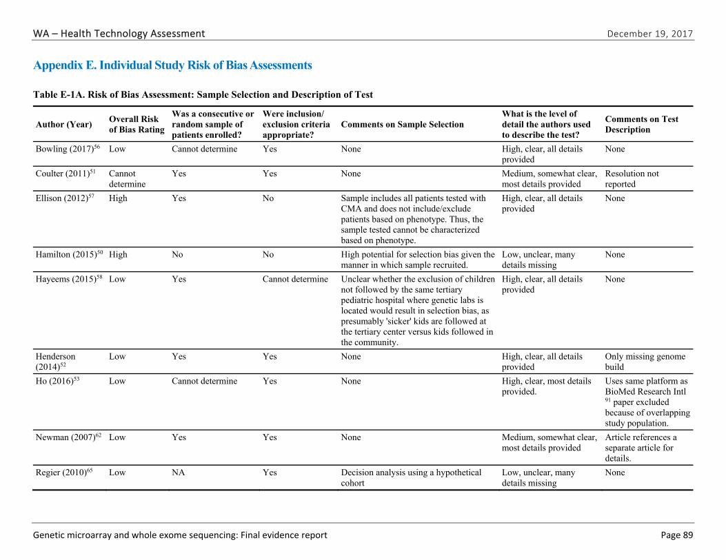
WA – Health Technology Assessment December 19, 2017
Genetic microarray and whole exome sequencing: Final evidence report Page 89
Appendix E. Individual Study Risk of Bias Assessments
Table E-1A. Risk of Bias Assessment: Sample Selection and Description of Test
Author (Year) Overall Risk of Bias Rating
Was a consecutive or random sample of patients enrolled?
Were inclusion/ exclusion criteria appropriate?
Comments on Sample Selection What is the level of detail the authors used to describe the test?
Comments on Test Description
Bowling (2017)56 Low Cannot determine Yes None High, clear, all details provided
None
Coulter (2011)51 Cannot determine
Yes Yes None Medium, somewhat clear, most details provided
Resolution not reported
Ellison (2012)57 High Yes No Sample includes all patients tested with CMA and does not include/exclude patients based on phenotype. Thus, the sample tested cannot be characterized based on phenotype.
High, clear, all details provided
None
Hamilton (2015)50 High No No High potential for selection bias given the manner in which sample recruited.
Low, unclear, many details missing
None
Hayeems (2015)58 Low Yes Cannot determine Unclear whether the exclusion of children not followed by the same tertiary pediatric hospital where genetic labs is located would result in selection bias, as presumably 'sicker' kids are followed at the tertiary center versus kids followed in the community.
High, clear, all details provided
None
Henderson (2014)52
Low Yes Yes None High, clear, all details provided
Only missing genome build
Ho (2016)53 Low Cannot determine Yes None High, clear, most details provided.
Uses same platform as BioMed Research Intl 91 paper excluded because of overlapping study population.
Newman (2007)62 Low Yes Yes None Medium, somewhat clear, most details provided
Article references a separate article for details.
Regier (2010)65 Low NA Yes Decision analysis using a hypothetical cohort
Low, unclear, many details missing
None
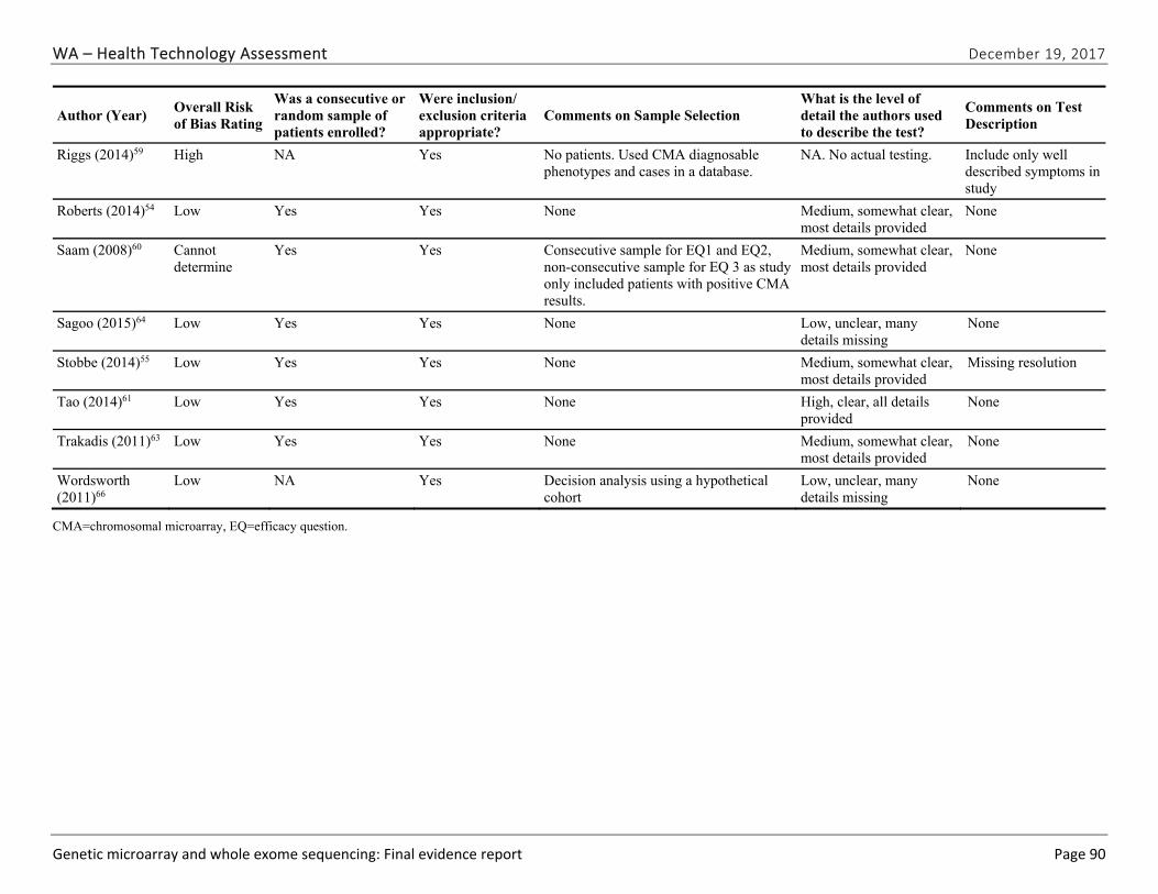
WA – Health Technology Assessment December 19, 2017
Genetic microarray and whole exome sequencing: Final evidence report Page 90
Author (Year) Overall Risk of Bias Rating
Was a consecutive or random sample of patients enrolled?
Were inclusion/ exclusion criteria appropriate?
Comments on Sample Selection What is the level of detail the authors used to describe the test?
Comments on Test Description
Riggs (2014)59 High NA Yes No patients. Used CMA diagnosable phenotypes and cases in a database.
NA. No actual testing. Include only well described symptoms in study
Roberts (2014)54 Low Yes Yes None Medium, somewhat clear, most details provided
None
Saam (2008)60 Cannot determine
Yes Yes Consecutive sample for EQ1 and EQ2, non-consecutive sample for EQ 3 as study only included patients with positive CMA results.
Medium, somewhat clear, most details provided
None
Sagoo (2015)64 Low Yes Yes None Low, unclear, many details missing
None
Stobbe (2014)55 Low Yes Yes None Medium, somewhat clear, most details provided
Missing resolution
Tao (2014)61 Low Yes Yes None High, clear, all details provided
None
Trakadis (2011)63 Low Yes Yes None Medium, somewhat clear, most details provided
None
Wordsworth (2011)66
Low NA Yes Decision analysis using a hypothetical cohort
Low, unclear, many details missing
None
CMA=chromosomal microarray, EQ=efficacy question.
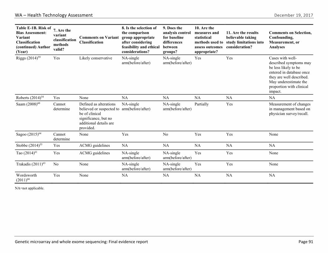
WA – Health Technology Assessment December 19, 2017
Genetic microarray and whole exome sequencing: Final evidence report Page 91
Table E-1B. Risk of Bias Assessment: Variant Classification (continued) Author (Year)
7. Are the variant classification methods valid?
Comments on Variant Classification
8. Is the selection of the comparison group appropriate after considering feasibility and ethical considerations?
9. Does the analysis control for baseline differences between groups?
10. Are the measures and statistical methods used to assess outcomes appropriate?
11. Are the results believable taking study limitations into consideration?
Comments on Selection, Confounding, Measurement, or Analyses
Riggs (2014)59 Yes Likely conservative NA-single arm(before/after)
NA-single arm(before/after)
Yes Yes Cases with well-described symptoms may be less likely to be entered in database once they are well described. May underestimate the proportion with clinical impact.
Roberts (2014)54 Yes None NA NA NA NA NA
Saam (2008)60 Cannot determine
Defined as alterations believed or suspected to be of clinical significance, but no additional details are provided.
NA-single arm(before/after)
NA-single arm(before/after)
Partially Yes Measurement of changes in management based on physician survey/recall.
Sagoo (2015)64 Cannot determine
None Yes No Yes Yes None
Stobbe (2014)55 Yes ACMG guidelines NA NA NA NA NA
Tao (2014)61 Yes ACMG guidelines NA-single arm(before/after)
NA-single arm(before/after)
Yes Yes None
Trakadis (2011)63 No None NA-single arm(before/after)
NA-single arm(before/after)
Yes Yes None
Wordsworth (2011)66
Yes None NA NA NA NA NA
NA=not applicable.
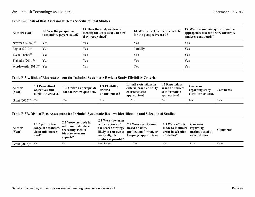
WA – Health Technology Assessment December 19, 2017
Genetic microarray and whole exome sequencing: Final evidence report Page 92
Table E-2. Risk of Bias Assessment Items Specific to Cost Studies
Author (Year) 12. Was the perspective (societal vs. payer) stated?
13. Does the analysis clearly identify the costs used and how they were valued?
14. Were all relevant costs included for the perspective used?
15. Was the analysis appropriate (i.e., appropriate discount rate, sensitivity analyses conducted)?
Newman (2007)62 Yes Yes Yes Yes
Regier (2010)65 Yes Yes Partially Yes
Sagoo (2015)64 Yes Yes Yes Yes
Trakadis (2011)63 Yes Yes Yes Yes
Wordsworth (2011)66 Yes Yes Yes Yes
Table E-3A. Risk of Bias Assessment for Included Systematic Review: Study Eligibility Criteria
Author (Year)
1.1 Pre-defined objectives and eligibility criteria?
1.2 Criteria appropriate for the review question?
1.3 Eligibility criteria unambiguous?
1.4. All restrictions in criteria based on study characteristics appropriate?
1.5 Restrictions based on sources of information appropriate?
Concerns regarding study eligibility criteria.
Comments
Grant (2015)40 Yes Yes Yes Yes Yes Low None
Table E-3B. Risk of Bias Assessment for Included Systematic Review: Identification and Selection of Studies
Author (Year)
2.1 Appropriate range of databases/ electronic sources used?
2.2 Were methods in addition to database searching used to identify relevant reports?
2.3 Were the terms and structure of the search strategy likely to retrieve as many eligible studies as possible?
2.4 Were restrictions based on date, publication format, or language appropriate?
2.5 Were efforts made to minimize error in selection of studies?
Concerns regarding methods used to select studies.
Comments
Grant (2015)40 Yes No Probably yes Yes Yes Low None
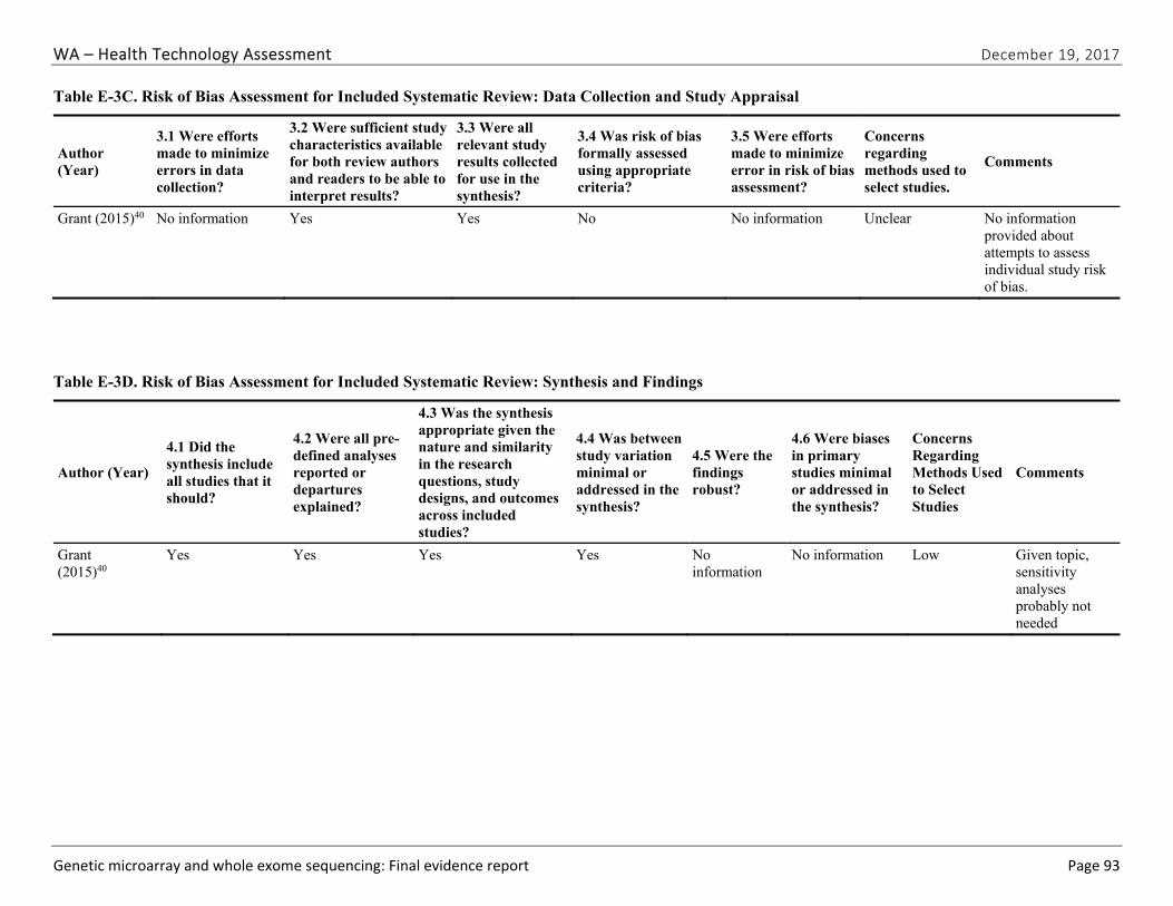
WA – Health Technology Assessment December 19, 2017
Genetic microarray and whole exome sequencing: Final evidence report Page 93
Table E-3C. Risk of Bias Assessment for Included Systematic Review: Data Collection and Study Appraisal
Author (Year)
3.1 Were efforts made to minimize errors in data collection?
3.2 Were sufficient study characteristics available for both review authors and readers to be able to interpret results?
3.3 Were all relevant study results collected for use in the synthesis?
3.4 Was risk of bias formally assessed using appropriate criteria?
3.5 Were efforts made to minimize error in risk of bias assessment?
Concerns regarding methods used to select studies.
Comments
Grant (2015)40 No information Yes Yes No No information Unclear No information provided about attempts to assess individual study risk of bias.
Table E-3D. Risk of Bias Assessment for Included Systematic Review: Synthesis and Findings
Author (Year)
4.1 Did the synthesis include all studies that it should?
4.2 Were all pre-defined analyses reported or departures explained?
4.3 Was the synthesis appropriate given the nature and similarity in the research questions, study designs, and outcomes across included studies?
4.4 Was between study variation minimal or addressed in the synthesis?
4.5 Were the findings robust?
4.6 Were biases in primary studies minimal or addressed in the synthesis?
Concerns Regarding Methods Used to Select Studies
Comments
Grant (2015)40
Yes Yes Yes Yes No information
No information Low Given topic, sensitivity analyses probably not needed
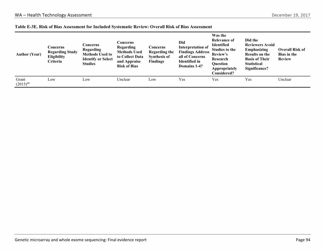
WA – Health Technology Assessment December 19, 2017
Genetic microarray and whole exome sequencing: Final evidence report Page 94
Table E-3E. Risk of Bias Assessment for Included Systematic Review: Overall Risk of Bias Assessment
Author (Year)
Concerns Regarding Study Eligibility Criteria
Concerns Regarding Methods Used to Identify or Select Studies
Concerns Regarding Methods Used to Collect Data and Appraise Risk of Bias
Concerns Regarding the Synthesis of Findings
Did Interpretation of Findings Address all of Concerns Identified in Domains 1-4?
Was the Relevance of Identified Studies to the Review’s Research Question Appropriately Considered?
Did the Reviewers Avoid Emphasizing Results on the Basis of Their Statistical Significance?
Overall Risk of Bias in the Review
Grant (2015)40
Low Low Unclear Low Yes Yes Yes Unclear