genotypes of klebsiella oxytoca isolates from patients ... · pneumothorax (1) n (15), o (6)...
TRANSCRIPT

Genotypes of Klebsiella oxytoca Isolates from Patients withNosocomial Pneumonia Are Distinct from Those of Isolates fromPatients with Antibiotic-Associated Hemorrhagic Colitis
Kathrin A. T. Herzog,a Georg Schneditz,b Eva Leitner,c Gebhard Feierl,c Karl Martin Hoffmann,d Ines Zollner-Schwetz,e
Robert Krause,e Gregor Gorkiewicz,f Ellen L. Zechner,b Christoph Högenauera
Division of Gastroenterology and Hepatology, Department of Internal Medicine, Medical University of Graz, Graz, Austriaa; Institute of Molecular Biosciences, University ofGraz, Graz, Austriab; Institute of Hygiene, Microbiology and Environmental Medicine, Medical University of Graz, Graz, Austriac; Division of General Pediatrics, Departmentof Pediatrics and Adolescent Medicine, Medical University of Graz, Graz, Austriad; Section of Infectious Diseases and Tropical Medicine, Department of Internal Medicine,Medical University of Graz, Graz, Austriae; Institute of Pathology, Medical University of Graz, Graz, Austriaf
Klebsiella oxytoca acts as a pathobiont in the dysbiotic human intestinal microbiota, causing antibiotic-associated hemorrhagiccolitis (AAHC), but it also infects other organs, resulting in pneumonia and urinary tract and skin infections. The virulence of K.oxytoca is still poorly understood. The production of a specific cytotoxin has been linked to AAHC pathogenesis. To investigatethe clonal relationships of K. oxytoca with regard to clinical origin and virulence attributes, we established a multilocus se-quence typing (MLST) method and analyzed 74 clinical K. oxytoca isolates from asymptomatic carriers and patients with AAHC,respiratory infections, and other infections. The isolates were phenotypically characterized, typed, and compared phylogeneti-cally based on the sequences of seven housekeeping genes. MLST analysis yielded 60 sequence types, 12 of which were repre-sented by more than one isolate. The phylogenetic tree distinguished clusters of K. oxytoca isolates between patients with AAHCand those with respiratory infections. Toxin-positive and -negative strains were observed within one sequence type. Our findingsindicate that AAHC isolates share a genetic background. Interestingly, K. oxytoca isolates from nosocomial pneumonia showeda different genetic clustering, suggesting that these strains do not originate from the intestines or that they are specialized forrespiratory tract colonization. Our results further indicate a polyphyletic origin and possible horizontal transfer of the genesinvolved in K. oxytoca cytotoxin production. This work provides evidence that K. oxytoca isolates colonizing the two main clini-cally relevant habitats (lower gastrointestinal [GI] tract and respiratory tract) of the human host are genetically distinct. Applica-tions of this MLST analysis should help clarify the sources of nosocomial infections.
Klebsiella oxytoca is a Gram-negative member of the human mi-crobiota. It can be detected in the intestines of about 2 to 10% of
healthy subjects, and until recently, K. oxytoca was considered to bea commensal member of the enteric microflora (1–3). However,we have shown that K. oxytoca is in fact an intestinal pathobiontand the causative agent of antibiotic-associated hemorrhagic coli-tis (AAHC) (2). Under conditions of intestinal dysbiosis, a state ofmicrobial imbalance, K. oxytoca unleashes its pathogenic poten-tial. Several factors can perturb the intestinal microbiota duringthe life span of an individual, including immune deficiency, infec-tions, dietary changes, and drugs, like antibiotics (4, 5). The con-sequences of antibiotic-induced intestinal dysbiosis range fromdiarrheal symptoms to intestinal inflammation and infection. Thecharacteristics of AAHC are sudden onset of bloody diarrhea andabdominal cramps during penicillin or cephalosporin therapy.The antibiotic penicillin is considered critical for triggering dys-biosis, as K. oxytoca exhibits a natural resistance to penicillins.Rapid colonic overgrowth of K. oxytoca follows during the acutephases of AAHC (3). The pathogenicity of K. oxytoca in colitis isnot understood, but a correlation has been observed between iso-lates originating from AAHC patients and the secretion of cyto-toxin(s) (1, 2, 6). Besides the potential to induce colitis undercertain circumstances, enteric carriage of K. oxytoca may be im-portant for the transmission of antibiotic resistance genes to otherbacteria and as a source of nosocomial infections (7, 8). Indeed,this bacterium and the closely related species Klebsiella pneu-moniae are important human pathogens causing hepatobiliary in-
fections and infections of the urinary tract and soft tissue, in ad-dition to nosocomial pneumonia (9–11). In recent years,multidrug-resistant strains of both species have emerged as animportant problem in the health care system (7, 12).
So far, no typing method has successfully identified a clonalrelationship between K. oxytoca isolates with respect to the partic-ular infections they cause, their isolation source, or their toxicity(6, 13). Here, we established a multilocus sequence typing (MLST)protocol to assess the genetic relatedness and population structureof clinical K. oxytoca isolates from patients with AAHC comparedto those of isolates from patients with nosocomial (respiratoryand urinary tract) and other infections. We further analyzedwhether distinct MLST sequence types (STs) are associated withparticular infections or with the production of a bacterial cyto-
Received 5 December 2013 Returned for modification 10 January 2014Accepted 22 February 2014
Published ahead of print 5 March 2014
Editor: W. M. Dunne, Jr.
Address correspondence to Ellen L. Zechner, [email protected], orChristoph Högenauer, [email protected].
Supplemental material for this article may be found at http://dx.doi.org/10.1128/JCM.03373-13.
Copyright © 2014, American Society for Microbiology. All Rights Reserved.
doi:10.1128/JCM.03373-13
May 2014 Volume 52 Number 5 Journal of Clinical Microbiology p. 1607–1616 jcm.asm.org 1607
on May 23, 2019 by guest
http://jcm.asm
.org/D
ownloaded from

toxin that is thought to contribute to virulence in colitis (2, 14,15). Tools to assess the genotype-virulence relationships of K. oxy-toca isolates will be useful for obtaining insights into the epidemi-ological patterns and evolution of the pathogenicity of this impor-tant opportunistic human pathogen (16).
MATERIALS AND METHODSBacterial isolates and their characterization. The study (approved by theinstitutional review board of the Medical University of Graz, Austria)analyzed 74 K. oxytoca strains and 1 K. pneumoniae strain isolated frompatients or healthy subjects. The details of the patient diagnoses and iso-lation sources are provided in Table 1 and Fig. 1; see also Table S1 in thesupplemental material. The antibiotic resistance profiles were determinedaccording to European Committee on Antimicrobial Susceptibility Test-ing (EUCAST) guidelines.
Cytotoxin testing. Bacterial cytotoxicity toward cultured Hep2 cellswas measured with an MTT [(3-(4,5-dimethyl-2-thiazolyl)-25-diphenyl-2H-tetrazolium bromide] assay using supernatants of bacterial culturemedium (6). The isolates were designated toxin positive when Hep2 via-bility was �50% compared to that of phosphate-buffered saline (PBS)-treated cells.
MLST scheme. Seven housekeeping genes (gapA, infB, mdh, pgi, phoE,rpoB, and tonB) analyzed in a published MLST protocol for K. pneu-moniae (17) were selected as targets for K. oxytoca (Table 2). To developthe MLST analysis for this species, the available genomic data in publicdatabases were compared (GenBank accession no. CP003683, CP003218,AGDI00000000.1, AGDJ00000000.1, AGDL00000000.1, AKCF00000000.1, andAGDP00000000.1). Neighboring genes were ruled out to be under selec-tive pressure. The primer sequences for PCR amplification and sequenc-ing primers (Tables 2 and 3) were adapted from the K. pneumoniae MLSTprimers (17). The primer annealing sites were chosen within highly con-served regions of the target genes of the K. oxytoca reference strains tomaximize the likelihood of amplification in all K. oxytoca strains. All allelicprimer sequences (except for rpoB reverse) therefore differ from K. pneu-moniae primers either in binding position within the gene or in the exactnucleotide sequence. The discriminatory index (D) was calculated as de-scribed by Hunter and Gaston (18) to verify the typing ability of thedeveloped MLST scheme. The allele sequences and sequence types areavailable on http://pubmlst.org/koxytoca/ (19).
PCR methods and sequencing. The 7 housekeeping genes were am-plified by PCR using the primers listed in Table 2. For amplification,
Phusion high-fidelity DNA polymerase (Thermo Scientific, USA) wasused in a total reaction volume of 30 �l. The PCR template was generatedby boiling a suspension of a single bacterial colony in H2O for 10 min,followed by centrifugation at 13,000 rpm for 30 s. PCRs were performed,starting with 30 s of initial denaturation at 98°C, followed by 35 cycles of10 s of denaturation (98°C), 30 s of primer annealing (54°C), and 1 min ofextension at 72°C, with a final extension of 5 min (72°C). The PCR prod-ucts were analyzed for fragment length and quality on an agarose gelstained with ethidium bromide.
Sanger cycle sequencing was performed using universal primers an-nealing to nonhomologous 5= ends of the PCR primers (Table 3), exceptfor the phoE target, for which nested primers were used. Each nucleotidesequence was supported by a forward and reverse chromatogram.
Bioinformatics and phylogenetic analysis. Sequence chromatogramswere edited using CLC Main Workbench 6, including the CLC MLSTmodule (CLC bio, Denmark). Using the same software, an MLST schemewas set up, and the obtained sequences were assembled and compared toalready existing allelic sequences of a given locus. A new allele number wasassigned to each distinct sequence of a locus. The distinct combination ofthe seven allele numbers, one for each locus, determined the sequencetype (ST).
For phylogenetic and nucleotide diversity analyses, the sequences ofthe 7 loci were concatenated. DnaSP 5.10.1 (20) was used to calculatepolymorphism statistics from the sequence alignments. Phylogenetictrees were drawn using MEGA5 (21) and CLC Main Workbench 6, basedon the Tamura-Nei parameter with gamma distribution and invariablesites (TN93 � G � I), according to Model test integrated in MEGA5. Onethousand random bootstrapping replicates were performed to assess thestability of the phylogenetic tree. One clinical K. pneumoniae isolate of thelocal strain collection was typed to be used as the outgroup in phylogeneticanalyses.
The likelihood of the outcome of two groups was determined usingthe odds ratio (OR) tool in Prism5 (GraphPad Software, USA). Statis-tical significance was assessed using the Fisher‘s exact test. The dis-criminatory index (D) was calculated as described by Hunter and Gas-ton (18) to quantify the typing ability of the developed MLST scheme.eBURST V3 (22) analysis was done to assess the presence of clonalcomplexes (CCs) that share 6 out of 7 alleles. SplitsTree version 4.12.6(23) was used to draw split decomposition trees of the concatenatedsequences of the STs to detect possible recombination-based networkstructures. As an additional recombination parameter, the index of
TABLE 1 Clinical and phenotypical attributes of K. oxytoca isolatesa
Isolation site Diagnosisb N and/or Oc Toxind Country of isolatione
Stool (40) AAHC (16), diarrhea/colitis of othercauses (11), IBD (3),asymptomatic carrier (7), follow-up/AAHC (1), asymptomaticcarrier/UTI (1), NA (1)
N (9), O (25),NA (6)
Positive (31), Negative (9) JPN (1), USA (1), NED (2),AUT (30), ESP (1), HKG(3), GER (2)
Respiratory tract (21) Nosocomial pneumonia/VAP (13),COPD (2), cystic fibrosis (1),pneumonia (2), pharyngitis (2),pneumothorax (1)
N (15), O (6) Positive (3), Negative (18) AUT (21)
Urinary tract (4) UTI (4) N (2), O (2) Negative (4) AUT (4)Blood (2) AAHC with bacteremia (1),
bacteremia (1)O (2) Positive (1), Negative (1) AUT (2)
Skin/mucousmembranes (7)
DFS (4), CSSTI (2), oral abscess (1) O (7) Positive (4), Negative (3) AUT (7)
a The number of isolates within a given category is shown in parentheses.b AAHC, antibiotic-associated hemorrhagic colitis; IBD, inflammatory bowel disease; UTI, urinary tract infection; VAP, ventilator-associated pneumonia; COPD, chronicobstructive pulmonary disease; DFS, diabetic foot syndrome; CSSTI, complicated skin and skin structure infection.c Isolates were classified as nosocomial (N) when infection occurred 48 h after hospitalization. O, outpatient; NA, information not available.d Cytotoxicity was assessed via an MTT-based cell culture assay (6).e JPN, Japan; NED, Netherlands; AUT, Austria; ESP, Spain; HKG, Hong Kong; GER, Germany.
Herzog et al.
1608 jcm.asm.org Journal of Clinical Microbiology
on May 23, 2019 by guest
http://jcm.asm
.org/D
ownloaded from
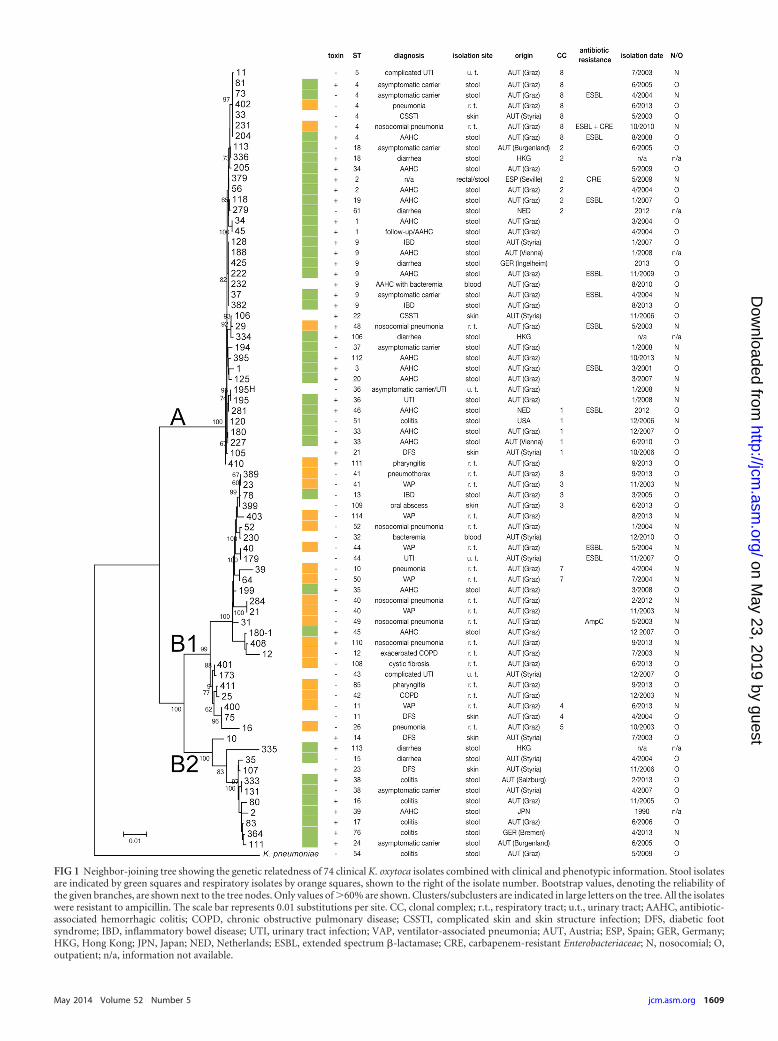
FIG 1 Neighbor-joining tree showing the genetic relatedness of 74 clinical K. oxytoca isolates combined with clinical and phenotypic information. Stool isolatesare indicated by green squares and respiratory isolates by orange squares, shown to the right of the isolate number. Bootstrap values, denoting the reliability ofthe given branches, are shown next to the tree nodes. Only values of �60% are shown. Clusters/subclusters are indicated in large letters on the tree. All the isolateswere resistant to ampicillin. The scale bar represents 0.01 substitutions per site. CC, clonal complex; r.t., respiratory tract; u.t., urinary tract; AAHC, antibiotic-associated hemorrhagic colitis; COPD, chronic obstructive pulmonary disease; CSSTI, complicated skin and skin structure infection; DFS, diabetic footsyndrome; IBD, inflammatory bowel disease; UTI, urinary tract infection; VAP, ventilator-associated pneumonia; AUT, Austria; ESP, Spain; GER, Germany;HKG, Hong Kong; JPN, Japan; NED, Netherlands; ESBL, extended spectrum �-lactamase; CRE, carbapenem-resistant Enterobacteriaceae; N, nosocomial; O,outpatient; n/a, information not available.
May 2014 Volume 52 Number 5 jcm.asm.org 1609
on May 23, 2019 by guest
http://jcm.asm
.org/D
ownloaded from

TA
BLE
2N
ucl
eoti
depo
lym
orph
ism
sam
ong
K.o
xyto
cais
olat
esan
dM
LST
targ
etan
dP
CR
prim
erin
form
atio
n
Gen
eP
uta
tive
gen
efu
nct
ion
Olig
onu
cleo
tide
Olig
onu
cleo
tide
sequ
ence
(5=t
o3=
)a
Size
ofan
alyz
edfr
agm
ent
(bp)
No.
ofal
lele
sb
No.
ofpo
lym
orph
icsi
tesb
Mea
n%
G�
Cco
nte
nt
Var
iati
onin
dice
sb
�c
dN/d
Sd
gapA
Gly
cera
ldeh
yde-
3-ph
osph
ate
deh
ydro
gen
ase
gapA
_fw
dG
TT
TT
CC
CA
GT
CA
CG
AC
GT
TG
TA
TG
AA
GT
AT
GA
CT
CC
AC
TC
AC
GG
450
8(9
)15
(16)
53.7
0.01
340
(0.0
1335
)0.
000
(0.0
00)
gapA
_rev
TT
GT
GA
GC
GG
AT
AA
CA
AT
TT
CA
AC
GC
CT
TT
CA
TT
GC
GC
CT
TC
GG
AA
infB
Tra
nsl
atio
nin
itia
tion
fact
or2
infB
_fw
dG
TT
TT
CC
CA
GT
CA
CG
AC
GT
TG
TA
CT
CT
CT
GC
TG
GA
CT
AC
AT
TC
G31
815
(17)
48(5
0)59
.20.
0561
5(0
.056
25)
0.13
9(0
.137
)
infB
_rev
TT
GT
GA
GC
GG
AT
AA
CA
AT
TT
CC
GC
TT
TC
AG
CT
CC
AG
AA
CT
TC
mdh
Mal
ate
deh
ydro
gen
ase
mdh
_fw
dG
TT
TT
CC
CA
GT
CA
CG
AC
GT
TG
TA
CC
CA
AC
TG
CC
TT
CA
GG
TT
CA
G47
725
(25)
86(8
6)52
.10.
0522
2(0
.052
33)
0.03
2(0
.031
)
mdh
_rev
TT
GT
GA
GC
GG
AT
AA
CA
AT
TT
CC
CT
TC
CA
CG
TA
GG
CG
CA
TT
CC
pgi
Ph
osph
oglu
cose
isom
eras
epg
i_fw
dG
TT
TT
CC
CA
GT
CA
CG
AC
GT
TG
TA
GA
GA
AA
AA
CC
TG
CC
GG
TG
CT
GC
TG
432
27(2
9)45
(45)
56.2
0.03
008
(0.0
3023
)0.
008(
0.00
8)
pgi_
rev
TT
GT
GA
GC
GG
AT
AA
CA
AT
TT
CC
GG
TT
AA
TC
AG
GC
CG
TT
AG
TG
GA
GC
phoE
Ph
osph
opor
ine
Eph
oE_f
wd
GT
TT
TC
CC
AG
TC
AC
GA
CG
TT
GT
AA
CC
TG
GC
GC
AA
CA
CC
GA
TT
TC
TT
C42
026
(28)
52(5
4)53
.70.
0331
5(0
.033
37)
0.02
0(0
.019
)
phoE
_rev
TT
GT
GA
GC
GG
AT
AA
CA
AT
TT
CT
TC
AG
CT
GG
TT
GA
TT
TT
GT
AA
TC
CA
C
rpoB
RN
Apo
lym
eras
esu
bun
it�
rpoB
_fw
dG
TT
TT
CC
CA
GT
CA
CG
AC
GT
TG
TA
GG
CG
AA
AT
GG
CG
GA
AA
AC
CA
501
19(2
1)40
(42)
54.3
0.01
777
(0.0
1812
)0.
001
(0.0
01)
rpoB
_rev
TT
GT
GA
GC
GG
AT
AA
CA
AT
TT
CG
AG
TC
TT
CG
AA
GT
TG
TA
AC
C
tonB
Per
ipla
smic
ener
gytr
ansd
uce
rto
nB
_fw
dG
TT
TT
CC
CA
GT
CA
CG
AC
GT
TG
TA
CT
CT
AT
AC
TT
CG
GT
AC
AT
CA
GG
TT
405
25(2
7)78
(79)
61.9
0.05
920
(0.0
6001
)0.
092
(0.0
95)
ton
B_r
evT
TG
TG
AG
CG
GA
TA
AC
AA
TT
TC
CC
TG
TT
TG
GC
GG
CC
AG
CA
CC
TG
GT
Con
cate
nat
edse
quen
ce3,
003
55.6
0.03
607
(0.0
3631
)
aSp
ecifi
col
igon
ucl
eoti
des:
bold
-typ
ese
quen
cebi
nds
targ
etge
ne,
un
derl
ined
sequ
ence
(ove
rhan
g)se
rves
asan
nea
ling
site
for
sequ
enci
ng
prim
er(p
hoE
was
sequ
ence
dw
ith
dist
inct
nes
ted
prim
ers)
.b
Pu
blic
lyav
aila
ble
K.o
xyto
caN
CB
Ise
quen
ceda
taw
ere
com
bin
edw
ith
the
San
ger
sequ
ence
sfr
omcl
inic
alis
olat
esto
gen
erat
ea
sepa
rate
alig
nm
ent
wh
ich
yiel
ded
the
valu
esgi
ven
inbr
acke
ts.
cD
iver
sity
inde
x(�
)is
equ
alto
the
aver
age
nu
mbe
rof
nu
cleo
tide
diff
eren
ces
per
site
.d
dN/d
S,ra
tio
ofn
onsy
non
ymou
sto
syn
onym
ous
subs
titu
tion
s.
Herzog et al.
1610 jcm.asm.org Journal of Clinical Microbiology
on May 23, 2019 by guest
http://jcm.asm
.org/D
ownloaded from

association (IAS) value was computed using LIAN 3.6 with 1,000 ran-
dom resamplings to provide a quantitative analysis of the recombina-tion and linkage disequilibrium rates within the K. oxytoca populationanalyzed. P values from the parametric and Monte Carlo methods wereassessed (24). For all statistical methods, a P value of �0.05 was consid-ered statistically significant.
RESULTSSequence types and genetic diversity. All genes included in theMLST scheme were affected by sequence variation. A range of 8(gapA) to 27 (pgi) distinct alleles was detected (Table 2). The iso-lates comprised 60 distinct sequence types (STs). All STs areshown in Table S1 in the supplemental material, along with theirisolation sources and details regarding patient diagnoses. Twelveof the STs are represented by more than one isolate (Table 4). Thedifferences between the STs were as small as a single nucleotidepolymorphism within the entire concatenated sequence (3,003bp). An alignment of the seven gene sequences for insertions/deletions identified solely isolate 2 in the tonB locus, which wasaffected by an insertion of four codons plus two downstream de-letion events involving two codons each. Synonymous substitu-tions were 1,000 (for rpoB) to 7 (for infB) times more frequentthan nonsynonymous substitutions. The clinical K. pneumoniaeisolate included in the typing scheme did not share any alleles withthe typed K. oxytoca isolates. The discriminatory index of theMLST scheme was calculated, using the method of Hunter andGaston (18), to be 0.9858.
Phylogenetic relationship of K. oxytoca isolates. The concat-enated sequences of all seven loci were used to draw a phylogenetictree with K. pneumoniae as the outgroup. The resulting neighbor-joining phylogeny (Fig. 1) comprises two major clusters, A and B,with the latter divided into subclusters B1 and B2. Cluster A showsoverall closer genetic relatedness (sum of branch lengths [SBL],0.019) than cluster B (SBL, 0.132) and subclusters B1 (SBL, 0.065)and B2 (SBL, 0.043). The majority of the AAHC isolates (13 of 16;OR, 5.7; P � 0.05) belong to cluster A. In contrast, respiratoryisolates are almost exclusively found in subcluster B1 (17 of 21;OR, 23.9; P � 0.0001). Accordingly, isolates originating in noso-comial pneumonia are also more abundant in subcluster B1 (11 of13; OR, 18.5; P � 0.0001). The predominance of AAHC isolates incluster A correlates with the overrepresentation of stool isolates inthis group (28 of 38; OR, 5.6; P � 0.005). Stool isolates were alsooverrepresented in subcluster B2. The strains of other isolationsources (urine, skin, and blood) were evenly distributed betweenthe clusters. Geographically diverse isolates were found to beclosely related (Fig. 1; see also Fig. S1 in the supplemental mate-rial) and even share the same ST (Table 4). K. oxytoca referencestrains with published sequences were found in all clusters (seeFig. S1 and Table S1 in the supplemental material). STs that showhigh genetic similarity were grouped into clonal complexes (CCs)by eBURST (22) analysis (Fig. 2; see also Table S1 in the supple-mental material). Each CC is made up of STs that differ from each
other by only one allele. CC1 includes ST21, ST33, ST46, andputative founder ST51. CC2 includes ST2 (putative founder),ST18, ST19, and ST61. CC3 includes ST109 (putative founder),ST41, and ST13. Clonal complexes 4 to 8 each contain two differ-ent STs. Thirty-nine STs are singletons that are not related to anyother ST.
Distribution of cytotoxicity and antibiotic resistance in theK. oxytoca population. The MLST-based phylogeny indicatesthat toxin-producing isolates are present in cluster A as well as incluster B, although their prevalence is higher in cluster A (27/38[71%]) and B2 (9/11 [82%]) than in subcluster B1 (3/25 [12%]).This association was strengthened when strains with publishedsequences were included in the analysis (see Fig. S1 in the supple-mental material). The larger number of stool isolates in cluster Aand B2 and the high frequency of toxin production in stool isolates(6) are consistent with this clustering. It is also interesting to notethat two of three toxin-positive isolates in subcluster B1 originatedin AAHC patients.
Our analysis also indicates that strains with identical sequencetypes can have different toxicity phenotypes. Twelve of the 60 STs arerepresented by more than one isolate (Table 4). The six isolates of ST4 include 4 toxin-negative and 2 toxin-positive isolates. Also, ST18,ST33, ST36, and ST38 each include a mixture of one toxin-negativeand one toxin-positive isolate. The remaining sequence types com-prising multiple isolates each contain strains with the same toxin phe-notype: ST1 (2 isolates), ST2 (2 isolates), ST9 (7 isolates), ST11 (2isolates), ST40 (2 isolates), ST41 (2 isolates), and ST44 (2 isolates).Isolates within the same ST also differed in isolation date, body site,geographic origin, and antibiotic resistance pattern.
Most (9 of 11) isolates producing extended-spectrum �-lacta-mases (ESBL) are located in cluster A (Fig. 1; see also Fig. S1 in thesupplemental material). Generally, antibiotic resistance did notcoincide with toxin production or any other parameter, such asgeographic origin, isolation date, or diagnosis.
K. oxytoca diversity within one patient. The isolation of mul-tiple K. oxytoca strains from the same patient allowed us to assessgenetic heterogeneity within individual human colonization orinfection cases. Isolates 180 and 180-1 were both cultured fromthe same stool sample from a patient with AAHC. While isolate180 does not produce the cytotoxin and is an ST33 strain of clusterB1, isolate 180-1 belongs to ST45 of cluster A and produces toxin.Isolates 195 and 195-H were also obtained at the same time fromthe same patient but from different body sources (stool versusurine). While these isolates share the same ST, only the stool iso-late is toxin positive. A case of temporal carriage of the identical K.oxytoca strain in a patient during acute AAHC as well as in remis-sion (follow-up isolate) is displayed by isolates 34 and 45. They areidentical in ST and toxicity and were obtained at a 1-month inter-val (Table 4).
Clonal diversity and relationships within the K. oxytoca pop-ulation. The calculated split graph (23) (Fig. 3) shows low levels ofrecombination, indicated by minor interconnected networks, in-volving the STs of cluster B. The branching displayed in the splitgraph correlates with the major clusters of the K. oxytoca MLSTneighbor-joining phylogeny. The index of association (IA
S) wascalculated to assess the amount of recombination within the pop-ulation and to detect possible associations between alleles. Theresulting IA
S of 0.2600 (P � 0.001) indicates significant linkagedisequilibrium within the K. oxytoca population. Intragenic re-combination affecting the separate MLST loci was then compared.
TABLE 3 Primers used for Sanger sequencing in this study
Primer name Sequence (5= to 3=)MLST_seq_fwd GTTTTCCCAGTCACGACGTTGTAMLST_seq_rev TTGTGAGCGGATAACAATTTCphoE_seq_fwd TTTCTTCGGCGTGGTAGATCCphoE_seq_rev GTAATCCACAAAGGCATTC
MLST for Klebsiella oxytoca
May 2014 Volume 52 Number 5 jcm.asm.org 1611
on May 23, 2019 by guest
http://jcm.asm
.org/D
ownloaded from

Our analysis showed congruency for gapA, infB, mdh, pgi, andtonB: all display two main branches, each comprising half of theisolates (see Fig. S2 in the supplemental material). The trees ofphoE and rpoB differed slightly from the others. Due to the ob-served congruency of the single-locus trees, we conclude that thephylogenetic signal is consistent between the loci. This in turnindicates a mutational evolutionary background within this pop-ulation of K. oxytoca isolates.
DISCUSSION
Previous attempts to type K. oxytoca strains using gyrA and parC,genes that are subject to antibiotic selection pressure (25), and
pulsed-field gel electrophoresis (PFGE) (6, 13) were unable todefine a clonal relationship for particular K. oxytoca pathotypes.We therefore established an MLST protocol specifically for K. oxy-toca to enable the phylogenetic-virulence relationships of clinicalK. oxytoca isolates to be analyzed. MLST tools decipher bacterialpopulation structures based on gene sequence comparison. Anadditional assessment of the distribution of virulence factorsacross the bacterial population provides insights into the possiblepresence of pathogenicity-associated subgroups (16, 26).
The discriminatory index (0.9858) determined for the MLSTscheme developed in this study is comparable to established
TABLE 4 Clinical, genetic, and phenotypic information of sequence types represented by more than one K. oxytoca isolate
Sequencetype Isolate Toxina Geographic originb Isolation site Diagnosisc
Isolationdate(mo/yr)
Antibioticresistancetyped
Nosocomial (N)/outpatient (O)e
Clonalcomplex Cluster
1 34 � AUT (Graz) Stool AAHC 3/2004 O A1 45 � AUT (Graz) Stool Follow-up/AAHC 4/2004 O A2 56 � AUT (Graz) Stool AAHC 4/2004 O 2 A2 379 � ESP Stool/rectal swab NA 5/2009 CRE N 2 A4 33 � AUT (Styria) Skin CSSTI 5/2004 O 8 A4 73 � AUT (Graz) Stool Asymptomatic
carrier4/2004 ESBL N 8 A
4 81 � AUT (Graz) Stool Asymptomaticcarrier
6/2005 O 8 A
4 204 � AUT (Graz) Stool AAHC 8/2008 ESBL O 8 A4 231 � AUT (Graz) Respiratory tract Nosocomial
pneumonia10/2010 ESBL � CRE N 8 A
4 402 � AUT (Graz) Respiratory tract Pneumonia 6/2013 O 8 A9 37 � AUT (Graz) Stool Asymptomatic
carrier4/2004 ESBL N A
9 128 � AUT (Styria) Stool IBD 3/2007 O A9 188 � AUT (Vienna) Stool AAHC 1/2008 NA A9 222 � AUT (Graz) Stool AAHC 11/2009 ESBL O A9 232 � AUT (Graz) Blood AAHC with
bacteremia8/2010 O A
9 382 � AUT (Graz) Stool IBD 8/2013 O A9 425 � GER Stool Diarrhea 2013 O A11 75 � AUT (Graz) skin DFS 4/2004 O 4 B111 400 � AUT (Graz) Respiratory tract VAP 6/2013 N 4 B118 113 � AUT (Burgenland) Stool Asymptomatic
carrier6/2005 O 2 A
18 336 � HKG Stool Diarrhea NA NA 2 A33 180 � AUT (Graz) Stool AAHC 12/2007 O 1 A33 227 � AUT (Vienna) Stool AAHC 6/2010 O 1 A36 195 � AUT (Graz) Stool UTI 1/2008 N A36 195-H � AUT (Graz) Urinary tract UTI 1/2008 N A38 131 � AUT (Styria) Stool Asymptomatic
carrier4/2007 O B2
38 333 � AUT (Salzburg) Stool Colitis 3/2014 O B240 21 � AUT (Graz) Respiratory tract VAP 11/2003 N B140 284 � AUT (Graz) Respiratory tract Nosocomial
pneumonia2/2012 N B1
41 23 � AUT (Graz) Respiratory tract VAP 11/2003 N 3 B141 389 � AUT (Graz) Respiratory tract Pneumothorax 9/2013 O 3 B144 40 � AUT (Graz) Respiratory tract VAP 5/2004 ESBL N B144 179 � AUT (Styria) Urinary tract UTI 11/2007 ESBL O B1a Cytotoxicity was assessed via an MTT-based cell culture assay (6).b AUT, Austria; ESP, Spain; GER, Germany; HKG, Hong Kong.c AAHC, antibiotic-associated hemorrhagic colitis; NA, information not available; CSSTI, complicated skin and skin structure infection; IBD, inflammatory bowel disease; DFS,diabetic foot syndrome; VAP, ventilator-associated pneumonia; UTI, urinary tract infection.d All isolates were resistant to ampicillin. ESBL, extended spectrum �-lactamase; CRE, carbapenem-resistant Enterobacteriaceae.e Isolates were classified as nosocomial when infection occurred after 48 h of hospitalization.
Herzog et al.
1612 jcm.asm.org Journal of Clinical Microbiology
on May 23, 2019 by guest
http://jcm.asm
.org/D
ownloaded from

MLST schemes for Enterobacteriaceae (27, 28) and thus is appro-priate for evolutionary population genetics. Additionally, whole-genome comparison of the eight publicly available K. oxytoca ge-nome sequences was done using genomic BLAST, followed bydendrogram construction based on genetic distance. The resultingdendrogram matches the phylogenetic clustering of these samereference K. oxytoca sequences achieved using this MLST method(see Fig. S3 in the supplemental material). Therefore, the subset ofconcatenated MLST sequences compared in this study providesrelated results similar to those of whole-genome-based compari-sons.
The observed congruency of the single-locus trees (see Fig. S2in the supplemental material) and the significant linkage disequi-librium (IA
S, 0.2600) provide evidence for a consistent phyloge-netic signal of the separate loci and for clonality within the K.oxytoca population analyzed (29). Evidence for only minor re-combination levels can also be seen in the split tree (Fig. 3). Itappears that the nucleotide diversity in this population can beattributed mainly to a mutational process. This consistency sug-gests a predominantly clonal long-term evolution of K. oxytoca,which makes phylogenetic and epidemiological interpretationsvalid (16).
A comparison of the SBL values of clusters A and B of theneighbor-joining phylogeny shows closer overall relatednesswithin cluster A. Cluster A harbors predominantly isolates fromstool samples, suggesting specialization for the gastrointestinal(GI) tract. This finding was independent of the geographic originof the isolates. Intrahospital spread and local epidemic dissemina-tion can therefore be ruled out as possible causes of the observednarrow genetic distances in cluster A. Cluster B shows more diver-sity regarding isolation site and less relatedness among the strains.
Subcluster B1 harbors the majority of respiratory isolates, mainlyderived from nosocomial pneumonia, while in subcluster B2,mainly fecal isolates are present. The niche adaptations of specificEscherichia coli genotypes were also revealed using MLST in pre-vious studies. While E. coli strains causing intestinal disease belongto phylogenetic groups A, B1, and E, strains causing extraintesti-nal diseases are found mainly in the phylogenetic group B2 (30,31). Future studies should therefore investigate the potential forrespiratory tract colonization by different phylotypes of other fac-ultative pathogenic enteric bacteria.
The gastropulmonary or rectopulmonary hypothesis (32) cur-rently proposes that nosocomial pneumonia, especially ventila-tor-associated pneumonia (VAP), is caused by Gram-negativebacteria originating from the GI tract (33, 34). In contrast, ourfindings provide evidence that the colonization of the two main K.oxytoca habitats (lower GI tract and respiratory tract) requiresdistinct genetic backgrounds. The STs of the majority of the lowerGI isolates in our analysis were not associated with respiratoryinfections, suggesting that bacterial translocation from the lowerGI tract is very unlikely. However, it is possible that K. oxytocaisolates from the upper GI tract (pharynx and stomach) are genet-ically distinct from the lower GI tract isolates and may represent asource of respiratory colonization and nosocomial pneumonia.Selective decontamination of the digestive tract (SDD) has beeninvestigated in several studies to reduce VAP and sepsis casescaused by enteric Gram-negative bacteria (35). In light of ourfindings, the concept of SDD should be reconsidered for avoidingbacterial translocation of Gram-negative rods from the lower GItract, since the majority of K. oxytoca respiratory isolates in thispopulation represent genotypes not associated with the large in-testine. The findings of this study support strategies using selective
FIG 2 Clonal diversity of K. oxytoca isolates. eBURST (22) was used to calculate clonal complexes (CC) that contain single-locus variants (SLVs) that share 6 of7 MLST alleles (STs connected by lines). The single-locus difference between SLVs is indicated by the gene name next to the connecting line. The relative positionsand spacing between the STs are not related to genetic distance. Each ST is represented by a dot, the size of which varies directly with the frequency of the ST inthe population.
MLST for Klebsiella oxytoca
May 2014 Volume 52 Number 5 jcm.asm.org 1613
on May 23, 2019 by guest
http://jcm.asm
.org/D
ownloaded from

oral decontamination rather than decontamination of the lowerintestine for avoiding VAP. Moreover, the prophylactic use ofantibiotics in SDD is subject to debate at present, since the in-crease in antibiotic resistance that would potentially result amongGram-negative bacteria is a major drawback of this method (34,36). Applications of MLST analyses to hospital and environmentalisolates will be important for better understanding the sourcesof nosocomial infections and thus improving prevention strat-egies. A public database has been established to facilitate epi-demiological studies of K. oxytoca populations (http://pubmlst.org/koxytoca/).
Toxin-positive K. oxytoca were found in both clusters A and B,although proportionally, the numbers of toxin-producing isolateswere higher in cluster A and subcluster B2. This finding correlateswith the higher prevalence of toxin production within stool iso-lates (which are predominantly found in the same clusters) thanfor isolates from other isolation sites (6). Given that stool isolates
show a higher frequency of toxin-positive phenotypes, it is con-ceivable that toxin production confers a fitness advantage to iso-lates of the GI tract. Multiple independent occurrences of toxin-producing isolates in the tree nonetheless indicate a polyphyleticorigin of the toxin. Thus, this finding implies a horizontal mode ofdissemination of the genes involved in toxin biosynthesis ratherthan vertical or clonal transmission. The finding that isolates shar-ing the same ST exhibit different toxicity phenotypes supports thisnotion (Table 4). A study of E. coli toxin genotypes did not linktoxin production to a specific genetic background. Instead, theacquisition of plasmid-carried genes for the heat-labile and heat-stabile enterotoxins was observed in phylogenetically closely anddistantly related strains (37). For K. oxytoca, this might be simi-larly explained, for example, by the localization of toxin genes ona mobile genetic element. However, our efforts to correlate thetoxicity of K. oxytoca strains with plasmid carriage did not supportthis link (data not shown). An alternative explanation that is con-
FIG 3 Split decomposition analysis for K. oxytoca strains. SplitsTree (23) was used for analysis of concatenated sequences of the seven MLST loci. Each K. oxytocaST (included in Fig. 1), regardless of frequency within the population, was included once in the analysis.
Herzog et al.
1614 jcm.asm.org Journal of Clinical Microbiology
on May 23, 2019 by guest
http://jcm.asm
.org/D
ownloaded from

sistent with a mechanism of horizontal gene transfer of the toxingenes might be their organization within a genomic island. Hori-zontal transfer on a mobile genomic island is known to drive dis-semination of several bacterial virulence factors, like the iron up-take system encoded by the E. coli high-pathogenicity island,which is widely distributed among the strains of different phylo-genetic groups (38, 39).
The relationships of genotypes and virulence attributes areeven more important in light of the observation that asymptom-atic carriers of K. oxytoca and AAHC patients can concurrentlyharbor genetically heterogeneous K. oxytoca strains (6). OurMLST analysis confirmed these previous PFGE typing results, thatone AAHC patient can simultaneously carry diverse K. oxytocastrains (isolates 180 and 180-1), which differ not only in theirgenetic background but also in toxin production (Table 4). Thisfinding makes it important to consider the possibility that multi-ple K. oxytoca strains exist in patient samples during clinical lab-oratory analysis. A diagnostic tool to check for the presence of K.oxytoca toxin biosynthesis genes would be useful for assessing thepresence of toxin-producing K. oxytoca isolates. In addition tocarriage of heterologous K. oxytoca genotypes, prolonged carriageof the same strain might also be observed during active AAHC(isolate 34) and later in remission (isolate 45). As shown previ-ously, the abundance of K. oxytoca in feces is several-fold higherduring active AAHC than in healthy carriers (4 106 CFU/mlcompared to �101 CFU/ml for healthy carriers) (3). Therefore,overgrowth of toxin-producing K. oxytoca strains already presentin the intestine at the start of antibiotic therapy might cause colitisin AAHC. The cessation of antibiotic therapy is usually sufficientto resolve AAHC. It follows that regrowth of the normal microbi-ota reduces the abundance of K. oxytoca to levels too low to causedisease. This assumption is also supported by the fact that mostAAHC isolates were found in similar clusters (A and B2) as isolatesfrom asymptomatic intestinal carriers. This pathophysiologicmodel is also consistent with known infection models in animalsfor AAHC (2); however, it is different from antibiotic colitis that isdue to Clostridium difficile, as patients who develop colitis arenewly infected with spores of the bacterium, whereas healthy car-riers rarely develop C. difficile colitis (40).
The MLST method established in this study revealed clusteringof clinical K. oxytoca isolates according to habitat (stool versusrespiratory tract) and to specific infections (AAHC and nosoco-mial pneumonia). Toxin production in K. oxytoca, however, wasnot associated with any genetic background. The results suggestspecific niche adaptation of genetically distinct K. oxytoca strainsthat cause different types of human infections. Efforts to sequencewhole genomes of K. oxytoca isolates should provide insight intothe underlying basis for the association of distinct genotypes withbody habitat adaptations.
ACKNOWLEDGMENTS
We thank Martina Joainig for her contribution to this study, ChristinaStrempfl and Bernadette Neuhold for their expert technical assistance,and A. Pascual (University Hospital Virgen Macarena), W. C. Yam (Uni-versity of Hong Kong), E. J. Kuijper (Leiden University Medical Center),and T. Chida (Medical and Dental University Tokyo) for providing bac-terial isolates.
All authors report no conflicts of interest.This work was supported by the funds of the Oesterreichische Nation-
albank (Anniversary Funds, project 14321 to C.H.) and grants from the
Austrian Science Fund (DK Molecular Enzymology W901 to E.L.Z.) andthe NAWI Graz fund (to E.L.Z.).
REFERENCES1. Beaugerie L, Metz M, Barbut F, Bellaiche G, Bouhnik Y, Raskine L,
Nicolas JC, Chatelet FP, Lehn N, Petit JC, Infectious Colitis StudyGroup. 2003. Klebsiella oxytoca as an agent of antibiotic-associated hem-orrhagic colitis. Clin. Gastroenterol. Hepatol. 1:370 –376. http://dx.doi.org/10.1053/S1542-3565(03)00183-6.
2. Högenauer C, Langner C, Beubler E, Lippe IT, Schicho R, GorkiewiczG, Krause R, Gerstgrasser N, Krejs GJ, Hinterleitner TA. 2006. Klebsiellaoxytoca as a causative organism of antibiotic-associated hemorrhagic coli-tis. N. Engl. J. Med. 355:2418 –2426. http://dx.doi.org/10.1056/NEJMoa054765.
3. Zollner-Schwetz I, Högenauer C, Joainig M, Weberhofer P, GorkiewiczG, Valentin T, Hinterleitner TA, Krause R. 2008. Role of Klebsiellaoxytoca in antibiotic-associated diarrhea. Clin. Infect. Dis. 47:e74 – e78.http://dx.doi.org/10.1086/592074.
4. Dethlefsen L, Relman DA. 2011. Incomplete recovery and individualizedresponses of the human distal gut microbiota to repeated antibiotic per-turbation. Proc. Natl. Acad. Sci. U. S. A. 108(Suppl 1):4554 – 4561. http://dx.doi.org/10.1073/pnas.1000087107.
5. Walker AW, Ince J, Duncan SH, Webster LM, Holtrop G, Ze X, BrownD, Stares MD, Scott P, Bergerat A, Louis P, McIntosh F, Johnstone AM,Lobley GE, Parkhill J, Flint HJ. 2011. Dominant and diet-responsivegroups of bacteria within the human colonic microbiota. ISME J. 5:220 –230. http://dx.doi.org/10.1038/ismej.2010.118.
6. Joainig MM, Gorkiewicz G, Leitner E, Weberhofer P, Zollner-SchwetzI, Lippe I, Feierl G, Krause R, Hinterleitner T, Zechner EL, HogenauerC. 2010. Cytotoxic effects of Klebsiella oxytoca strains isolated from pa-tients with antibiotic-associated hemorrhagic colitis or other diseasescaused by infections and from healthy subjects. J. Clin. Microbiol. 48:817–824. http://dx.doi.org/10.1128/JCM.01741-09.
7. Sievert DM, Ricks P, Edwards JR, Schneider A, Patel J, Srinivasan A,Kallen A, Limbago B, Fridkin S, National Healthcare Safety Network(NHSN) Team and Participating NHSN Families. 2013. Antimicrobial-resistant pathogens associated with healthcare-associated infections: sum-mary of data reported to the National Healthcare Safety Network at theCenters for Disease Control and Prevention, 2009 –2010. Infect. ControlHosp. Epidemiol. 34:1–14. http://dx.doi.org/10.1086/668770.
8. Molton JS, Tambyah PA, Ang BS, Ling ML, Fisher DA. 2013. The globalspread of healthcare-associated multidrug-resistant bacteria: a perspectivefrom Asia. Clin. Infect. Dis. 56:1310 –1318. http://dx.doi.org/10.1093/cid/cit020.
9. Brisse S, Grimont F, Grimont PD. 2006. The genus Klebsiella, p 159 –196.In Dworkin M, Falkow S, Rosenberg E, Schleifer KH, Stackebrandt E (ed),The prokaryotes. Springer, New York, NY.
10. Polage CR, Solnick JV, Cohen SH. 2012. Nosocomial diarrhea: evalua-tion and treatment of causes other than Clostridium difficile. Clin. Infect.Dis. 55:982–989. http://dx.doi.org/10.1093/cid/cis551.
11. Podschun R, Ullmann U. 1998. Klebsiella spp. as nosocomial pathogens:epidemiology, taxonomy, typing methods, and pathogenicity factors.Clin. Microbiol. Rev. 11:589 – 603.
12. Sbrana F, Malacarne P, Viaggi B, Costanzo S, Leonetti P, Leonildi A,Casini B, Tascini C, Menichetti F. 2013. Carbapenem-sparing antibioticregimens for infections caused by Klebsiella pneumoniae carbapenemase-producing K. pneumoniae in intensive care unit. Clin. Infect. Dis. 56:697–700. http://dx.doi.org/10.1093/cid/cis969.
13. Cheng VC, Yam WC, Tsang LL, Yau MC, Siu GK, Wong SC, Chan JF,To KK, Tse H, Hung IF, Tai JW, Ho PL, Yuen KY. 2012. Epidemiologyof Klebsiella oxytoca-associated diarrhea detected by Simmons citrate agarsupplemented with inositol, tryptophan, and bile salts. J. Clin. Microbiol.50:1571–1579. http://dx.doi.org/10.1128/JCM.00163-12.
14. Higaki M, Chida T, Takano H, Nakaya R. 1990. Cytotoxic component(s)of Klebsiella oxytoca on HEp-2 cells. Microbiol. Immunol. 34:147–151.http://dx.doi.org/10.1111/j.1348-0421.1990.tb00999.x.
15. Minami J, Katayama S, Matsushita O, Sakamoto H, Okabe A. 1994.Enterotoxic activity of Klebsiella oxytoca cytotoxin in rabbit intestinalloops. Infect. Immun. 62:172–177.
16. Urwin R, Maiden MC. 2003. Multi-locus sequence typing: a tool forglobal epidemiology. Trends Microbiol. 11:479 – 487. http://dx.doi.org/10.1016/j.tim.2003.08.006.
MLST for Klebsiella oxytoca
May 2014 Volume 52 Number 5 jcm.asm.org 1615
on May 23, 2019 by guest
http://jcm.asm
.org/D
ownloaded from

17. Diancourt L, Passet V, Verhoef J, Grimont PA, Brisse S. 2005. Multi-locus sequence typing of Klebsiella pneumoniae nosocomial isolates. J.Clin. Microbiol. 43:4178 – 4182. http://dx.doi.org/10.1128/JCM.43.8.4178-4182.2005.
18. Hunter PR, Gaston MA. 1988. Numerical index of the discriminatoryability of typing systems: an application of Simpson’s index of diversity. J.Clin. Microbiol. 26:2465–2466.
19. Jolley KA, Maiden MC. 2010. BIGSdb: scalable analysis of bacterial ge-nome variation at the population level. BMC Bioinformatics 11:595. http://dx.doi.org/10.1186/1471-2105-11-595.
20. Librado P, Rozas J. 2009. DnaSP v5: a software for comprehensive anal-ysis of DNA polymorphism data. Bioinformatics 25:1451–1452. http://dx.doi.org/10.1093/bioinformatics/btp187.
21. Tamura K, Peterson D, Peterson N, Stecher G, Nei M, Kumar S. 2011.MEGA5: molecular evolutionary genetics analysis using maximum likeli-hood, evolutionary distance, and maximum parsimony methods. Mol.Biol. Evol. 28:2731–2739. http://dx.doi.org/10.1093/molbev/msr121.
22. Feil EJ, Li BC, Aanensen DM, Hanage WP, Spratt BG. 2004. eBURST:inferring patterns of evolutionary descent among clusters of related bac-terial genotypes from multilocus sequence typing data. J. Bacteriol. 186:1518 –1530. http://dx.doi.org/10.1128/JB.186.5.1518-1530.2004.
23. Huson DH, Bryant D. 2006. Application of phylogenetic networks inevolutionary studies. Mol. Biol. Evol. 23:254 –267. http://dx.doi.org/10.1093/molbev/msj030.
24. Haubold B, Hudson RR. 2000. LIAN 3.0: detecting linkage disequilib-rium in multilocus data. Bioinformatics 16:847– 849. http://dx.doi.org/10.1093/bioinformatics/16.9.847.
25. Deguchi T, Yasuda M, Kawamura T, Nakano M, Ozeki S, Kanematsu E,Nishino Y, Kawada Y. 1997. Improved antimicrobial activity of DU-6859a, a new fluoroquinolone, against quinolone-resistant Klebsiellapneumoniae and Enterobacter cloacae isolates with alterations in GyrA andParC proteins. Antimicrob. Agents Chemother. 41:2544 –2546.
26. Brisse S, Verhoef J. 2001. Phylogenetic diversity of Klebsiella pneumoniaeand Klebsiella oxytoca clinical isolates revealed by randomly amplifiedpolymorphic DNA, gyrA and parC genes sequencing and automated ri-botyping. Int. J. Syst. Evol. Microbiol. 51:915–924. http://dx.doi.org/10.1099/00207713-51-3-915.
27. Nemoy LL, Kotetishvili M, Tigno J, Keefer-Norris A, Harris AD, Per-encevich EN, Johnson JA, Torpey D, Sulakvelidze A, Morris JG, Jr,Stine OC. 2005. Multilocus sequence typing versus pulsed-field gel elec-trophoresis for characterization of extended-spectrum beta-lactamase-producing Escherichia coli isolates. J. Clin. Microbiol. 43:1776 –1781. http://dx.doi.org/10.1128/JCM.43.4.1776-1781.2005.
28. Liu WB, Liu B, Zhu XN, Yu SJ, Shi XM. 2011. Diversity of Salmonella
isolates using serotyping and multilocus sequence typing. Food Microbiol.28:1182–1189. http://dx.doi.org/10.1016/j.fm.2011.04.001.
29. Maiden MC. 2006. Multilocus sequence typing of bacteria. Annu. Rev.Microbiol. 60:561–588. http://dx.doi.org/10.1146/annurev.micro.59.030804.121325.
30. Clermont O, Olier M, Hoede C, Diancourt L, Brisse S, Keroudean M,Glodt J, Picard B, Oswald E, Denamur E. 2011. Animal and humanpathogenic Escherichia coli strains share common genetic backgrounds.Infect. Genet. Evol. 11:654 – 662. http://dx.doi.org/10.1016/j.meegid.2011.02.005.
31. Le Gall T, Clermont O, Gouriou S, Picard B, Nassif X, Denamur E,Tenaillon O. 2007. Extraintestinal virulence is a coincidental by-productof commensalism in B2 phylogenetic group Escherichia coli strains. Mol.Biol. Evol. 24:2373–2384. http://dx.doi.org/10.1093/molbev/msm172.
32. Kallet RH, Quinn TE. 2005. The gastrointestinal tract and ventilator-associated pneumonia. Respir. Care. 50:910 –921; discussion 921–923.
33. Hurley JC. 1995. Prophylaxis with enteral antibiotics in ventilatedpatients: selective decontamination or selective cross-infection? Anti-microb. Agents Chemother. 39:941–947. http://dx.doi.org/10.1128/AAC.39.4.941.
34. Rotstein C, Evans G, Born A, Grossman R, Light RB, Magder S,McTaggart B, Weiss K, Zhanel GG. 2008. Clinical practice guidelines forhospital-acquired pneumonia and ventilator-associated pneumonia inadults. Can. J. Infect. Dis. Med. Microbiol. 19:19 –53.
35. Ramirez P, Bassi GL, Torres A. 2012. Measures to prevent nosocomialinfections during mechanical ventilation. Curr. Opin. Crit. Care. 18:86 –92. http://dx.doi.org/10.1097/MCC.0b013e32834ef3ff.
36. Bonten MJ, Krueger WA. 2006. Selective decontamination of the diges-tive tract: cumulating evidence, at last? Semin. Respir. Crit. Care Med.27:18 –22. http://dx.doi.org/10.1055/s-2006-933669.
37. Chaudhuri RR, Henderson IR. 2012. The evolution of the Escherichia coliphylogeny. Infect. Genet. Evol. 12:214 –226. http://dx.doi.org/10.1016/j.meegid.2012.01.005.
38. Schubert S, Darlu P, Clermont O, Wieser A, Magistro G, Hoffmann C,Weinert K, Tenaillon O, Matic I, Denamur E. 2009. Role of intraspeciesrecombination in the spread of pathogenicity islands within the Esche-richia coli species. PLoS Pathog. 5:e1000257. http://dx.doi.org/10.1371/journal.ppat.1000257.
39. Dobrindt U, Hochhut B, Hentschel U, Hacker J. 2004. Genomic islandsin pathogenic and environmental microorganisms. Nat. Rev. Microbiol.2:414 – 424. http://dx.doi.org/10.1038/nrmicro884.
40. Kyne L, Warny M, Qamar A, Kelly CP. 2000. Asymptomatic carriage ofClostridium difficile and serum levels of IgG antibody against toxin A. N.Engl. J. Med. 342:390 –397. http://dx.doi.org/10.1056/NEJM200002103420604.
Herzog et al.
1616 jcm.asm.org Journal of Clinical Microbiology
on May 23, 2019 by guest
http://jcm.asm
.org/D
ownloaded from