genotyping single-nucleotide polymorphisms by
TRANSCRIPT
4
Genotyping Single-Nucleotide Polymorphisms by Minisequencing Using Tag Arrays
Lovisa Lovmar and Ann-Christine Syvänen
SummaryThe need for large-scale and high-throughput methods for SNP genotyping has rapidly
increased during the last decade. Our system, presented here, combines the highly specific geno-typing principle of minisequencing with the advantages of a microarray format that allows highlymultiplexed and parallel analysis.
Cyclic minisequencing reactions with fluorescently labeled dideoxynucleotides (ddNTPs) areperformed in solution using multiplex PCR product as template and detection primers, designedto anneal immediately adjacent and upstream of the SNP site. The detection primers carry unique5′ tag sequences and oligonucleotides complementary to the tag sequence, cTags, are immobi-lized on a microarray. After extension, the tagged detection primers are allowed to hybridize tothe cTags; then the fluorescent signals from the array are measured, and the genotypes arededuced according to the label incorporated. The “array of arrays” format of the system, accom-plished by a silicon rubber grid giving separate reaction chambers, allows either 80 or 14 sam-ples to be analyzed for up to 200 or 600 SNPs, respectively, on a single microscope slide.
Key Words: Single-nucleotide polymorphism (SNP); genotyping; minisequencing; microarray;multiplex; PCR; “array of arrays.”
1. IntroductionGenomic nucleotide substitutions that are present in more than 1% of the
alleles in a population are denoted single-nucleotide polymorphisms (SNPs)and are the most abundant form of genetic variation (1). Following the com-pletion of the nucleotide sequence of the human genome (2,3), a large inter-est in SNPs has arisen owing to their potential use as markers when one issearching for genetic factors underlying complex, multifactorial disorders.Consequently, the need for high-throughput methods for SNP genotypinghas increased.
79
From: Methods in Molecular Medicine, Vol. 144, Microarrays in Clinical DiagnosticsEdited by: T. Joos and P. Fortina © Humana Press Inc., Totowa, NJ
04_chap_Joos.qxd 25/04/2005 02:03 pm Page 79
Several reaction principles and assay formats have been developed (see ref. 4for a review of genotyping techniques). One of the reaction principles mostoften used in high-throughput systems today is minisequencing, in which aDNA polymerase is allowed to extend a detection primer by a single nucleotideat the position of the SNP (5). The system, presented in this chapter, combinesthe highly specific genotyping principle of minisequencing with the advantagesof a microarray format that allows highly multiplex and parallel analysis.
The Tag array minisequencing system utilizes generic capture oligonucleotides(cTags) that are immobilized on a microarray. Multiplex cyclic minisequencingreactions with fluorescently labeled dideoxynucleotides (ddNTPs) are performedin solution using detection (minisequencing) primers, designed to anneal imme-diately adjacent and upstream of the SNP site. The primers carry 5′ Tag sequencescomplementary to one of the arrayed cTags, and the SNPs are genotyped byhybridizing the extended detection primers to their corresponding cTags withknown locations on the array. The incorporated fluorescently labeled ddNTPsallow deduction of the genotypes of each SNP based on measurement of the sig-nal intensities by fluorescence scanning of the arrays (6,7). The use of generic Tagsequences allows universal, non-SNP-specific array designs. The “array ofarrays” format described below is accomplished by a silicon rubber grid form-ing separate reaction chambers for multiple samples on a single microscopeslide. Either 80 or 14 samples can be simultaneously analyzed for up to 200 or600 SNPs, respectively (8–10) (Fig. 1).
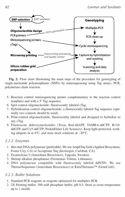
The procedure described in detail in the Methods section outlines (1) selec-tion of appropriate SNPs; (2) design of oligonucleotides for polymerase chainreaction (PCR), immobilization on the microarray, and SNP genotyping; (3)preparation of microarrays; (4) manufacturing of the silicon rubber grid; (5) thegenotyping reaction; and (6) data analysis and genotype interpretation. Themain steps of the assay are illustrated in Fig. 2.
Several alterations and modifications of the method are possible, and a num-ber of suggestions are given in the Notes section. The protocol is given underthe assumption that the reader will use the instrumentation, reagents, and con-sumables specified in the Materials section, but other equivalent procedures arealso feasible.
2. Materials 2.1. Instrumentation
1. Access to arraying facilities or purchased customized arrayed slides. We use aProSys 5510A instrument (Cartesian Technologies, Huntingdon, UK) with StealthMicro Spotting Pins (TeleChem, Sunnyvale, CA).
2. Facilities for programmed thermal cycling. 3. Multichannel pipete and a pipeting robot (optional).4. Centrifuge for microtiter plates (recommended).
80 Lovmar and Syvänen
04_chap_Joos.qxd 25/04/2005 02:03 pm Page 80
5. Minisequencing reaction rack (Fig. 3).6. Heat block at 42°C. 7. Array scanner and software for signal analysis. We use the ScanArray Express sys-
tem (PerkinElmer Lifesciences, Boston, MA).
2.2. Reagents and Consumables
All reagents should be of standard molecular biology grade. Use sterile dis-tilled or deionized water.
2.2.1. Oligonucleotide Primers and Nucleotides
1. PCR primers.2. Minisequencing primers with 5′ Tag sequences.3. cTags with a 3′ end 15-T residue spacer and a 3′ amino group.4. Reaction control templates: four oligonucleotides differing in one internal nucleotide
position.
Minisequencing Using Tag Arrays 81
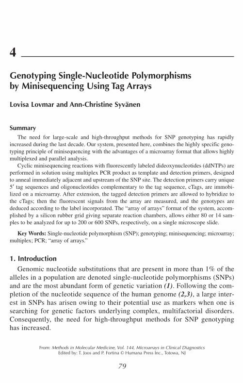
Fig. 1. Principle of Tag array minisequencing. Schematic views of about one-thirdof two arrayed slides with the subarrays in either a 384-well (A) or 96-well (B) con-formation. One of the subarrays for each slide containing up to 200 (A) or 600 (B)cTags is showed enlarged (C). The principle of the minisequencing reaction is illus-trated with a minisequencing primer carrying a 5′ Tag sequence that has annealed toits target and has been extended with a labeled ddCTP at the position of the single-nucleotide polymorphism (SNP). (D) The Tag sequences of the extended minisequenc-ing primers are allowed to hybridize to their complementary cTags arrayed as spots inthe subarrays. The genotypes are deduced by measuring the fluorescence of the incor-porated nucleotides. Part of one subarray, with the result for two SNPs, is shown. Thissample is homozygous (A/A) for SNP 1 and heterozygous (C/T) for SNP 2.
04_chap_Joos.qxd 25/04/2005 02:03 pm Page 81
5. Reaction control minisequencing primer complementary to the reaction controltemplates and with a 5′ Tag sequence.
6. Spot control oligonucleotide: fluorescently labeled cTag.7. Hybridization control oligonucleotide: a fluorescently labeled Tag sequence (opti-
mally two controls should be used).8. Print-control oligonucleotide, fluorescently labeled and designed to hybridize to
any cTag.9. Fluorescent dideoxynucleotides (Texas Red-ddATP, TAMRA-ddCTP, R110-
ddGTP, and Cy5-ddUTP; PerkinElmer Life Sciences). Keep light-protected, work-ing aliquots in at 4°C, and store stock solutions at −20°C.
2.2.2. Enzymes
1. Hot-start DNA-polymerase (preferable). We use AmpliTaq Gold (Applied Biosystems,Foster City, CA) or Accuprime Taq (Invitrogen, Carlsbad, CA).
2. Exonuclease I (Amersham Biosciences, Uppsala, Sweden).3. Shrimp alkaline phosphatase (Fermentas, Vilnius, Lithuania).4. DNA polymerase compatible with fluorescently labeled ddNTPs. We use
ThermoSequenase (Amersham Biosciences) or KlenThermase™ (GeneCraft).
2.2.3. Buffer Solutions
1. Standard PCR reagents or reagents optimized for multiplex PCR. 2. 2X Printing buffer: 300 mM phosphate buffer, pH 8.5. Store at room temperature
up to 1 month.
82 Lovmar and Syvänen
Fig. 2. Flow chart illustrating the main steps of the procedure for genotyping ofsingle-nucleotide polymorphisms (SNPs) by minisequencing using Tag arrays. PCR,polymerase chain reaction.
04_chap_Joos.qxd 25/04/2005 02:03 pm Page 82

3. Blocking solution: 50 mM ethanolamine, 100 mM Tris-HCl, pH 9.0, and 0.1%sodium dodecyl sulfate (SDS). Prepare directly before use. Ethanolamine is highlycorrosive and should be handled according to safety instructions.
4. Washing solutions: (I) 4X standard saline citrate (SSC) and 0.1% SDS; (II) 2X SSCand 0.1% SDS; and (III) 0. 2X SSC. (20X SSC: 3 M NaCl, 300 mM sodium citrate,pH 7.0.)
5. 1M Tris-HCl, pH 9.5.6. 50 mM MgCl2.7. 1% (v/v) Triton X-100.8. Hybridization solution: 6.25X SSC.
2.2.4. Consumables
1. 384- or 96-well V-bottomed microtiter plates (ABgene, Epsom, UK).2. CodeLink™ Activated Slides (Amersham Biosciences). 3. Elastosil RT 625 A and B (polydimethyl siloxan; Wacker-Chemie, München,
Germany).
Minisequencing Using Tag Arrays 83
Fig. 3. The arrayed slide is covered with a silicon rubber grid to give separate reac-tion chambers and placed in a custom-made, heat-conducting aluminum rack. Two sil-icon rubber grids, one 384-well (right) and the other 96-well (left), are shown on top ofa reaction rack. A plexiglas cover with drilled holes through which the reaction cham-bers are accessible is tightly screwed on top of the assembly, thus ensuring correct posi-tioning of the silicon grid during hybridization.
04_chap_Joos.qxd 25/04/2005 02:03 pm Page 83
3. Methods3.1. SNP Selection
SNPs can be identified either experimentally or in databases, for example indbSNP (http://www.ncbi.nlm.nih.gov/SNP/). Database searches for SNPs maybe aimed at genes of interest, candidate chromosomal regions, or randomlydistributed SNPs with known allele frequencies, depending on the aim of theproject (see Note 1).
3.2. Oligonucleotide Design
3.2.1. PCR Primers
The PCR fragments should optimally be short, i.e., 100–150 bp. Design PCRprimers flanking the SNPs of interest using available software. Primer3 is freelyavailable on the internet (http://frodo.wi.mit.edu/cgi-bin/primer3/primer3_www.cgi; see Note 2).
The sequence of each PCR fragment should be “blasted” against the genomesequence (http://www.ncbi.nlm.nih.gov/BLAST/) and give a single hit only tothe intended region (see Note 3).
3.2.2. Minisequencing Primers
Minisequencing primers are designed to anneal immediately adjacent to andupstream of the SNP position. The minisequencing primers should be approx20 bases long and have a melting temperature of 55–60°C to ensure specificityin the cyclic primer extension reaction.
At the 5′ end of each minisequencing primer, add the Tag sequences, whichshould be complementary to the cTags that will be arrayed onto the microarray.The Tags should be 20 bases long, have similar melting temperature, and not becomplementary to either each other, to the gene-specific part of the minisequenc-ing primers, or to the human genome (6). The Affymetrix GeneChip® TagCollection can be used as a source for Tag sequences (Affymetrix, Santa Clara,CA). Minisequencing primers from both forward and reverse strands are oftenhelpful as internal controls for the genotyping results (see Note 4).
3.2.3. Complementary Tag Sequences
The complementary Tag sequences (cTags) have 15 3′ T-residues as a spac-er and a 3′ amino group to allow covalent attachment of the cTags to the slides.
3.2.4. Control Oligonucleotides
We recommend the use of a number of oligonucleotides as controls for thedifferent steps of the procedure (10).
84 Lovmar and Syvänen
04_chap_Joos.qxd 25/04/2005 02:03 pm Page 84
1. To control for the spotting procedure, a fluorescently labeled cTag may be includedin the array. Also a print-control oligonucleotide designed to hybridize to any cTag(5′-AAA AAA AAA ANN NNN NNN NN— Fluorophore -3′) is recommended foruse on some subarrays or microarrays from each batch.
2. As a minisequencing reaction control, a minisequencing primer is useful that iscomplementary to four synthesized single-stranded oligonucleotide templates dif-fering at one nucleotide position mimicking the four possible alleles of a SNP. Addthe control templates to the minisequencing reaction up to a final concentration of1.5 nM. A corresponding cTag should be included in the array.
3. To control for the hybridization reaction, a fluorescently labeled oligonucleotidecomplementary to an arrayed cTag is used. Optimally use two differently labeledhybridization control oligonucleotides, and add them in an alternating pattern overthe microarray to ensure that no leaking between wells has occurred.
3.3. Preparation of Microarrays
3.3.1. Microarray Printing
1. Dissolve the cTags in printing buffer to a final concentration of 25 µM (see Note 5).2. If they are not used immediately or if they are to be reused, store the cTags at
–20°C, but limit freeze-thawing cycles to 10. 3. Prepare the arrays by contact-printing the cTag oligonucleotides onto CodeLink
Activated slides using the ProSys 5510A instrument with SMP3 pins that deliver1 nL of the cTag solution to the slides as spots with diameters of 125–150 µm anda center-to-center distance of, for example, 200 µM (see Note 6).
4. To use the “array of arrays” format, print spots in a subarray pattern correspondingto the spacing of wells in a 384-well microtiter plate (see Note 7 and Fig. 1).
5. After arraying, mark the position of some of the subarrays on the back side of theslides using a diamond pen.
3.3.2. Postprinting Processing of the Microarray Slides
Process the slides according to the instructions of the manufacturer. The pro-tocol for CodeLink Activated Slides is given below.
1. Prepare an incubation chamber with 75% relative humidity. Add as much solidNaCl to water as needed to form a 1-cm-deep slurry at the bottom of a plastic con-tainer with an airtight lid.
2. After printing, keep the arrays in the incubation chamber for 4–72 h.3. Prepare the blocking solution, and preheat it to 50°C. 4. Deactivate the excess of amine-reactive groups by immersing the arrayed slides for
30 min in the blocking solution at 50°C. 5. Rinse twice with dH2O. Immerse the slides in washing solution I for 30 min at
50°C. (At least 10 mL per slide should be used.) Rinse again with dH2O.6. Spin-dry the slides for 5 min at about 90 g. Store the slides desiccated at 20°C
until use.
Minisequencing Using Tag Arrays 85
04_chap_Joos.qxd 25/04/2005 02:03 pm Page 85

3.3.3. Quality Control of Printing Procedure
For each batch of printed slides, it is useful to analyze a few subarrays byhybridization as a quality control of the spots. After deactivation of the slides,hybridize the 3′ fluorescently labeled print-control oligonucleotide to somesubarrays at 300 nM concentration in 6X SSC for 5 min with subsequent wash-ing and scanning as described below under Subheadings 3.5.4. and 3.5.5.
3.4 Preparation of Reusable Silicon Rubber Grid
A grid of silicon rubber reaction chambers is made using inverted V-bottomedmicrotiter plates as the mold (Fig. 3).
1. Add the two Elastosil RT 625 components in a 50-mL Falcon tube in a mass ratioof 9:1 (i.e., 46.8 g of A and 5.2 g of B); then rotate and turn the tube by hand untilthe components are fully mixed (approx 30 min; see Note 8).
2. Pour the mixture onto an inverted V-bottomed 384-well microtiter plate, leavingabout 1–2 mm of the tip of the wells uncovered. Allow the silicon rubber to hardenat least overnight at room temperature (see Note 9).
3. Remove the silicon rubber grid from the plate, and use a scalpel to cut the siliconrubber into pieces of the same size as microscope slides, with the wells matchingthe printed subarrays.
The silicon rubber grid is reusable; wash it with water, and allow it to dryafter each use.
3.5. Genotyping
3.5.1. Multiplex PCR and Clean-Up
1. Amplify genomic DNA samples and PCR negatives by multiplex PCR accordingto an optimized protocol. The success of the amplification may be verified on a 1%agarose gel for a subset of the samples.
2. For each sample, pool the multiplex PCR products (see Note 10).3. Prepare a master mix of the exonuclease (ExoI) and alkaline phosphatase (sAP)
reagents for clean-up of the PCR products (see Table 1 and Note 11).4. Add 3.4 µL of the clean-up mixture to give a total volume of 10.5 µL. 5. Incubate at 37°C for 30–60 min. 6. Inactivate the enzymes by heating to 85°C for 15 min.
3.5.2. Cyclic Minisequencing
1. Prepare a master mix with minisequencing reagents (see Table 2 and Note 12).2. After the clean-up step, add 4.5 µL of minisequencing reaction mixture to give a
total volume of 15 µL. 3. Perform the minisequencing reactions in a thermal cycler using an initial 3-min
denaturation step at 96°C followed by, for example, 33 cycles of 20 s at 95°C and20 s of 55°C in a thermocycler (see Note 13).
86 Lovmar and Syvänen
04_chap_Joos.qxd 25/04/2005 02:03 pm Page 86
3.5.3. Capture by Hybridization
1. Position a silicon rubber grid over the arrayed slide according to the diamond penmarkings. Place the arrayed slides into the custom-made aluminum reaction rack,and tighten the plexiglas cover (Fig. 3). Preheat the assembly to 42°C on a heatblock (see Note 14).
2. Add 7 µL of the hybridization solution to each minisequencing reaction to a finalvolume of 22 µL. It is recommended to include hybridization control oligonu-cleotides at 0.25 nM concentrations in the hybridization mixture.
3. Transfer 20 µL of each sample to a separate reaction chamber on the microscopeslide. A multichannel pipete is convenient for this step.
4. Hybridize for 2.5–3 h at 42°C in a humid environment, formed, for example, byplacing a wet tissue on the plexiglas lid and covering it with plastic film and alu-minum foil.
Minisequencing Using Tag Arrays 87
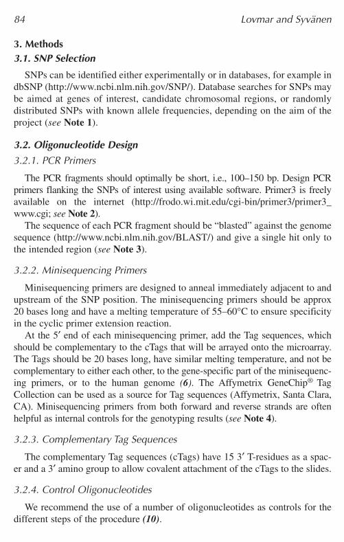
Table 1PCR Clean-Up Reagents
Volume per FinalReagent reaction (µL) concentration
PCR products 7.150 mM MgCl2 1.6 7.62 mM a
1 M Tris-HCl, pH 9.5 0.5 0.05 M20 U/µL Exonuclease I 0.3 0.57 U/µL1 U/µL Shrimp alkaline phosphatase 1.0 0.10 U/µLTotal volume 10.5
a The true final concentration of MgCl2 is higher depending on the contribution from the PCRproducts.
Table 2Minisequencing Reagents
Volume per FinalReagent reaction (µL) concentration
100 nM of each pooled minisequencing primer 1.50 10.0 nM100 µM Fluorescently labeled ddNTPs a 4 × 0.015 0.10 µM1% Triton X-100 0.30 0.02%32 U/µL ThermoSequenase 0.03 0.064 U/µLH2O 2.61Total volume 15.00
a Texas Red-ddATP, TAMRA-ddCTP, R110-ddGTP, and Cy5-ddUTP.
04_chap_Joos.qxd 25/04/2005 02:03 pm Page 87
3.5.4. Washing
1. Prepare the three washing solutions. Preheat solution II to 42°C.2. After hybridization, take the slides from the reaction rack, and rinse briefly with
solution I, at 20–25°C. 3. Wash the slides twice for 5 min with solution II at 42°C and twice for 1 min with
solution III, at 20–25°C in 50-mL Falcon tubes. 4. Spin-dry the slides for 5 min at about 90 g. and store them protected from light.
3.5.5. Fluorescence Scanning
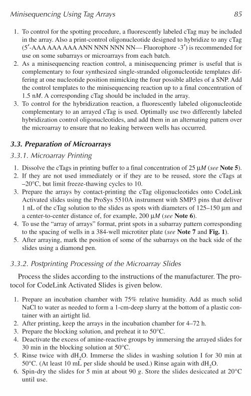
If allowed by the scanner used, balance the signal intensity from each laserchannel so that no signals are saturated and the signals from the four fluo-rophores are as equal as possible. Balancing is feasible if a reaction control withsignals from all four fluorophores has been included. Figure 4 shows an exampleof a scanned array.
3.6. Data Analysis and Genotype Assignment
A quantification program such as the one supplied with ScanArray Expresshandles the scanning images and quantitates the signals from each spot. The rawdata are collected in an Excel sheet.
1. Subtract the background, measured either around the spots or at negative controlspots, i.e., spotted cTags without corresponding tagged primers, from the signalsmeasured in each channel.
2. Assign the genotypes of the SNPs in each sample by calculating the ratios betweenthe signals from one of the alleles and the sum of the signals from both the alleles:Signal Allele 1/(Signal Allele 1 + Signal Allele 2).
3. A scatter plot with this ratio on the horizontal axis and the sum of the signalsfrom both alleles on the vertical axis may be used for assigning the genotypes(Fig. 5). This scatter plot should give three distinct genotype clusters with the
88 Lovmar and Syvänen
Fig. 4. Scanning results from genotyping one sample for 45 SNPs in one subarray,after cyclic minisequencing with ddATP, ddCTP, ddGTP, and ddUTP labeled withTexas Red, TAMRA, R110, and Cy5, respectively. Each cTag was spotted as horizontalduplicates, and both polarities of the SNPs were analyzed (10).
04_chap_Joos.qxd 25/04/2005 02:03 pm Page 88
homozygote samples clustering at each side and the heterozygotes in the middle.The ratios may vary between SNPs, depending on the sequence surrounding it,the type of nucleotide incorporated, and the signal intensity of the fluorophores(see Note 15).
4. When using the ScanArray Express or QuantArray program for signal analysis, orif the signal quantitation output files have been converted to fit their format, thegenotyping results can be visualized using the SNPSnapper software customizedfor this method (http://www.bioinfo.helsinki.fi/snpsnapper).
4. Notes1. Many of the SNPs in databases have not been validated or may not be polymorphic in
the population from which the study samples originate. The SNP allele frequencies ina particular population may be determined by analyzing pooled DNA samples usingquantitative minisequencing in microtiter plates, or in the microarray format (7,11).
2. A touchdown PCR procedure may be used (12). One strategy when designingprimers for multiplex PCR is to aim at as similar primer melting temperature andG/C content as possible. Complementary 3′ sequences in the primers can be avoidedby designing primers with the same 3′ terminal nucleotides (13). Other options areto introduce common tails on the 5′ ends of all PCR primers and to amplify subse-quently with one common primer for all the fragments at an elevated temperature(14) or to use universal 5′ sequences, making the PCR primers eligible for the samereaction conditions (15).
Minisequencing Using Tag Arrays 89
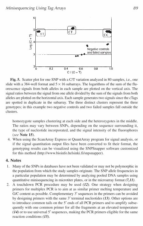
Fig. 5. Scatter plot for one SNP with a C/T variation analyzed in 80 samples, i.e., oneslide with a 384-well format and 5 × 16 subarrays. The logarithms of the sum of the flu-orescence signals from both alleles in each sample are plotted on the vertical axis. Thesignal ratios between the signal from one allele divided by the sum of the signals from bothalleles are plotted on the horizontal axis. Each sample generates two signals since the cTagsare spotted in duplicate in the subarray. The three distinct clusters represent the threegenotypes; in this example two negative controls and two failed samples fall outside theclusters.
04_chap_Joos.qxd 25/04/2005 02:03 pm Page 89
3. We recommend excluding SNPs located in repetitive elements identified by theRepeatMasker program (http://www.repeatmasker.org).
4. To avoid strong hairpin-loop structures, evaluate the complete minisequencing primer,including the Tag sequence. Secondary structures that involve the 3′ end of a primermay lead to misincorporation of nucleotides. A primer design software that predictssecondary structures (mfold:http://www.bioinfo.rpi.edu/~zukerm/ or NetPrimer:http://www.premierbiosoft.com/netprimer) can be used.
5. The array may also be manufactured with immobilized minisequencing primers.In this assay variant, the genotyping reaction is performed directly on the arraysurface (16).
6. Microarrays may be purchased from a commercial supplier or manufactured in-house. There are several different slide types and attachment chemistries. Some ofthem have been tested in our system (17).
7. The number of spots in each subarray can be varied by changing the subarray pat-tern from a 384- to a 96-well format; thus the number of spots in each subarray isincreased, but the maximum number of samples that can be simultaneously ana-lyzed is decreased (9).
8. Elastosil RT601 may be used instead of RT625 to give a slightly harder siliconrubber to decrease deformation of the wells when the rack lid is tightened. Iflarge subarrays, utilizing all available surface, are printed, deformation of thewells may cause the cTags in the corners to be covered by the silicon. The soft-er, RT625, silicon sticks better to the glass surface and decreases the risk of leak-age between wells.
9. Depending on the number of SNPs to be explored, an inverted 96-well microtiterplate may be used as well as a silicon rubber mold to allow larger subarrays (9).
10. Instead of multiplex PCR, single-fragment PCR can be used with subsequent pool-ing of the amplified fragments, possibly after concentration using ethanol precipita-tion or spin dialysis. Also, if large numbers of multiplex PCR products are pooled,it may be advantageous to concentrate the pool prior to the subsequent steps.
11. Alkaline phosphatase inactivates the remaining dNTPs, and exonuclease I degradesthe single-stranded PCR primers, which would disturb the subsequent minise-quencing reactions.
12. Cy5-ddUTP can be used at a 1.5–2-fold higher concentration than the other ddNTPsto compensate for its lower incorporation efficiency. Instead of using four differ-ently labeled nucleotides in the same reaction, depending on the available microar-ray scanner, a single label or two labels may be used in four or two separate reactions,respectively.
13. If the fluorescent signals obtained are weak, the number of cycles may be increased.We have used up to 99 cycles.
14. Background problems can arise if the hybridization chamber is not kept humid;lack of humidity causes the samples on the slide to dry out.
15. The flanking sequences as well as the fluorophores attached to the dideoxynu-cleotides affect the efficiency and sequence specificity of nucleotide incorporationby the DNA polymerase. The different properties of the fluorophores, such as
90 Lovmar and Syvänen
04_chap_Joos.qxd 25/04/2005 02:03 pm Page 90
molar extinction coefficients, emission spectra, and quantum yield, as well as non-specific background, also affect the signal intensities and signal ratios obtained (7).
AcknowledgmentsThis protocol is the result of the combined work effort of both former and
present members of the Molecular Medicine research group at the Departmentof Medical Sciences at Uppsala University. The group has received financialsupport from the Swedish Research Council, the Wallenberg Foundation, andthe European Commission (FP5 and FP6).
References1. Sachidanandam, R., Weissman, D., Schmidt, S. C., et al. (2001) A map of human
genome sequence variation containing 1.42 million single nucleotide polymor-phisms. Nature 409, 928–933.
2. Venter, J. C., Adams, M. D., Myers, E. W., et al. (2001). The sequence of the humangenome. Science 291, 1304–1351.
3. Lander, E. S., Linton, L. M., Birren, B., et al. (2001). Initial sequencing and anal-ysis of the human genome. Nature 409, 860–921.
4. Syvanen, A. C. (2001). Accessing genetic variation: genotyping single nucleotidepolymorphisms. Nat. Rev. Genet. 2, 930–942.
5. Syvanen, A. C., Aalto-Setala, K., Harju, L., Kontula, K., and Soderlund, H. (1990).A primer-guided nucleotide incorporation assay in the genotyping of apolipopro-tein E. Genomics 8, 684–692.
6. Hirschhorn, J. N., Sklar, P., Lindblad-Toh, K., et al. (2000). SBE-TAGS: an array-based method for efficient single-nucleotide polymorphism genotyping. Proc.Natl. Acad. Sci. USA 97, 12164–12169.
7. Lindroos, K., Sigurdsson, S., Johansson, K., Ronnblom, L., and Syvanen, A. C.(2002) Multiplex SNP genotyping in pooled DNA samples by a four-colour microar-ray system. Nucleic Acids Res. 30, e70.
8. Pastinen, T., Raitio, M., Lindroos, K., Tainola, P., Peltonen, L., and Syvanen, A. C.(2000). A system for specific, high-throughput genotyping by allele-specificprimer extension on microarrays. Genome Res. 10, 1031–1042.
9. Fredriksson, M., Barbany, G., Liljedahl, U., Hermanson, M., Kataja, M., andSyvanen, A. C. (2004). Assessing hematopoietic chimerism after allogeneic stemcell transplantation by multiplexed SNP genotyping using microarrays and quanti-tative analysis of SNP alleles. Leukemia 18, 255–266.
10. Lovmar, L., Fredriksson, M., Liljedahl, U., Sigurdsson, S., and Syvanen, A. C.(2003) Quantitative evaluation by minisequencing and microarrays reveals accu-rate multiplexed SNP genotyping of whole genome amplified DNA. Nucleic AcidsRes. 31, e129.
11. Lagerstrom-Fermer, M., Olsson, C., Forsgren, L., and Syvanen, A. C. (2001).Heteroplasmy of the human mtDNA control region remains constant during life.Am. J. Hum. Genet. 68, 1299–1301.
Minisequencing Using Tag Arrays 91
04_chap_Joos.qxd 25/04/2005 02:03 pm Page 91

12. Don, R. H., Cox, P. T., Wainwright, B. J., Baker, K., and Mattick, J. S. (1991).‘Touchdown’ PCR to circumvent spurious priming during gene amplification.Nucleic Acids Res. 19, 4008.
13. Zangenberg, A. P., Saiki, R. K., and Reynolds, R. (1999). Multiplex PCR: opti-mization guidelines, in PCR Applications (Innis, M. A., Gelfand, D. H., andSninsky, J. J., eds.), Academic Press, London, UK, pp. 73–94.
14. Brownie, J., Shawcross, S., Theaker, J., et al. (1997). The elimination of primer-dimer accumulation in PCR. Nucleic Acids Res. 25, 3235–3241.
15. Shuber, A. P., Grondin, V. J., and Klinger, K. W. (1995). A simplified procedure fordeveloping multiplex PCRs. Genome Res. 5, 488–493.
16. Liljedahl, U., Karlsson, J., Melhus, H., et al. (2003). A microarray minisequencingsystem for pharmacogenetic profiling of antihypertensive drug response.Pharmacogenetics 13, 7–17.
17. Lindroos, K., Liljedahl, U., Raitio, M., and Syvanen, A. C. (2001). Minisequencingon oligonucleotide microarrays: comparison of immobilisation chemistries. NucleicAcids Res. 29, E69.
92 Lovmar and Syvänen
04_chap_Joos.qxd 25/04/2005 02:03 pm Page 92