geometry of the semicircular canals and extraocular ...pcnjeffery/papers/janat2008.pdf · between...
TRANSCRIPT

J. Anat.
(2008)
213
, pp583–596 doi: 10.1111/j.1469-7580.2008.00983.x
© 2008 The Authors Journal compilation © 2008 Anatomical Society of Great Britain and Ireland
Blackwell Publishing Ltd
Geometry of the semicircular canals and extraocular muscles in rodents, lagomorphs, felids and modern humans
Philip G. Cox and Nathan Jeffery
Division of Human Anatomy and Cell Biology
,
School of Biomedical Sciences
,
University of Liverpool
,
Liverpool
,
UK
Abstract
The vestibulo-ocular reflex (VOR) exacts compensatory movements of the extraocular muscles in response tostimulation of the semicircular canals to allow gaze fixation during head movements. In this study, the spatialrelationships of these muscles and canals were investigated to assess their relative alignments in mammalian speciescommonly used in studies of the VOR. The head region of each specimen was scanned using magnetic resonanceimaging and 28 anatomical landmarks were recorded from the images to define the six extraocular muscles andthe anatomical planes of the three semicircular canals. The vector rotation of a semicircular canal that does notstimulate either of its two sister canals, referred to as the prime direction, was also calculated as an estimate of themaximal response plane. Significant misalignments were found between the extraocular muscles and the canals bywhich they are principally stimulated in most of the species under study. The deviations from parallel orientationwere most pronounced in the human and rabbit samples. There were also significant departures from orthogonalitybetween the semicircular canals in most species. Only the guinea pig displayed no significant difference from 90
°
in any of its three inter-canal angles, although humans and rabbits deviated from orthogonality in just one semi-circular canal pair – the anterior and posterior canals. The prime directions were found to deviate considerably fromthe anatomical canal planes (by over 20
°
in rats). However, these deviations were not always compensatory, i.e.prime planes were not always more closely aligned with the muscle planes. Results support the view that thevestibular frame remains relatively stable and that the spatial mismatch with the extraocular co-ordinate frame isprincipally driven by realignment of the muscles as a result of changes in the position of the orbits within the skull(orbital convergence and frontation).
Key words
cat; extraocular muscles; guinea pig; modern human; mouse; rabbit; rat; semicircular canals; squirrel;vestibulo-ocular reflex.
Introduction
The vestibulo-ocular reflex (VOR) enables the gaze to befocused on one point whilst the head is moving. The func-tional components of the VOR are the semicircular canalsof the inner ears, which sense angular accelerations anddecelerations of the head, and the extraocular muscles,which produce appropriate compensatory movements ofthe eyeballs (Cohen et al. 1964; Suzuki et al. 1964). Eachsemicircular canal principally activates one extraocularmuscle in each eye (Szentágothai, 1950; summarized in
Fig. 1, Cox & Jeffery, 2007), although other extraocularmuscles are also stimulated to lesser degrees. The spatialorientations of these primary functional couplings areof key interest to investigations of the organization andfunction of the VOR. This study examines the relativeorientations of the muscles and canals in seven mammalianspecies, many of which are commonly used in VOR studies.
The orientations of the planes of the semicircular canals,both to each other and to standard stereotaxic planes,have been measured in a number of different vertebratespecies. Early work in this area examined the inner earsof rabbits (de Burlet & Koster, 1916) and rats (Cummins,1925). Due to the inaccessible nature of the inner ears,the canals were studied using wax or plaster of Parisreconstructions modelled from serial sections. Anglesbetween the three canals and standard reference planeswere calculated visually using a protractor (Cummins, 1925),or mathematically by determining the three-dimensionalco-ordinates of three points on each canal in order todefine a plane (de Burlet & Koster, 1916).
Correspondence
Philip Cox
,
Division of Human Anatomy and Cell Biology
,
School of Biomedical Sciences
,
University of Liverpool
,
Sherrington Buildings
,
Ashton Street
,
Liverpool
,
L69 3GE
,
UK. T: +44 151 7945454; F: +44 151 7945517; E: [email protected]
Accepted for publication
5 August 2008
Article published online
14 October 2008

Semicircular canal and extraocular muscle geometry, P. G. Cox and N. Jeffery
© 2008 The AuthorsJournal compilation © 2008 Anatomical Society of Great Britain and Ireland
584
Later work tended to favour a technique in which thesemicircular canals were surgically exposed in a skullthrough careful removal of portions of the temporal bone,occipital bone and tympanic bulla. The skull was thensecurely mounted in a stereotaxic plane, and the
X
,
Y
and
Z
co-ordinates of a number of points along each canalwere recorded using a curved needle attached to a micro-manipulator. Using this method, the semicircular canalswere mapped in cats (Blanks et al. 1972; Ezure & Graf,1984), guinea pigs (Curthoys et al. 1975), humans (Blankset al. 1975), rabbits (Ezure & Graf, 1984; Mazza & Winterson,1984), monkeys (Blanks et al. 1985), rats (Daunicht & Pellionisz,1987), pigeons (Dickman, 1996), turtles (Brichta et al. 1988)and toadfish (Ghanem et al. 1998). Reisine et al. (1988)used a similar protocol on their study of rhesus monkeys,but determined the
X
,
Y
and
Z
co-ordinates from a resincast of the vestibular apparatus rather than directlyfrom the skull. Having obtained three-dimensional co-ordinate data for each canal, a plane of best fit can becalculated using either a least-squares technique or aprincipal components analysis in which the values of theeigenvectors corresponding to the smallest eigenvaluegive the directional cosines of the normal vector to theplane. In a number of the above studies (Blanks et al. 1972,1975, 1985; Curthoys et al. 1975; Ezure & Graf, 1984), bothplane-fitting techniques were applied and were found togive virtually identical results. An alternative indirectmethod is the null point technique employed by Blanks& Torigoe (1989) and Haque et al. (2004) to determine theplanes of the semicircular canals in the rat and rhesus monkey,respectively. In this method, the experimental animal ismounted in a three-axis rotator system, and is subjected tovarious angular accelerations whilst the afferent responsesat the vestibular nerve root are recorded. The ‘null point’ isthe orientation which produces no response, and indicatesthat the semicircular canal is perpendicular to the plane ofangular acceleration. Once the null points have been found
for each canal, the anatomical planes of the semicircularcanals can be approximated in the form a
X
+ b
Y
+ c
Z
= 0,referenced in the stereotaxic co-ordinate system.
With the development of more sophisticated imagingtechniques, recent investigations have utilized computerreconstructions of the labyrinth to analyse the planarrelationships of the semicircular canals. Reconstructions ofthe human labyrinth have been generated from serialsections (Takagi et al. 1989; Sato et al. 1993; Hashimotoet al. 2005), magnetic resonance images (Ichijo, 2002), andcomputed tomography (CT) scans (Spoor & Zonneveld,1998; Della Santina et al. 2005). CT data have also beenused to study the canals in mice (Calabrese & Hullar, 2006)and chinchillas (Hullar & Williams, 2006). As in earlierstudies, planes of best fit were calculated using a least-squares technique or a principal components analysis,except for Della Santina et al. (2005), who fitted the planesvisually using multi-slice planar reconstructions.
Many of the studies mentioned above have notedthat the semicircular canals are not precisely orthogonal,and in some cases depart widely from orthogonality (e.g.Curthoys et al. 1975; Ezure & Graf, 1984; Mazza & Winterson,1984). In such specimens, rotation of the head in theanatomical plane of a semicircular canal will necessarilystimulate at least one of the other two ipsilateral canals. Itwas thus proposed by Rabbitt (1999) that a more usefulmeasurement to derive would be the vector of rotation ofa canal that does not stimulate either of the other twocanals. This is termed the ‘prime direction’ of a canal.Prime directions have been derived for rhesus monkeys(Haque et al. 2004), mice (Calabrese & Hullar, 2006) andchinchillas (Hullar & Williams, 2006). Haque et al. (2004)suggested that the oculomotor system uses the primeco-ordinate frame to encode vestibular signals. However,this hypothesis is difficult to test in rhesus monkeys due tothe near-orthogonality of their semicircular canals and,therefore, the close alignment of the anatomical and
Fig. 1 Three-dimensional representation of the extraocular muscles and semicircular canals of a cat (specimen Cat3). Surface rendering performed using AMIRA 4.1 (Mercury Inc., USA). (A) Lateral view. (B) Dorsal view. SO, superior oblique; IO, inferior oblique; SR, superior rectus; IR, inferior rectus; MR, medial rectus; LR, lateral rectus; ASC, anterior canal; PSC, posterior canal; LSC, lateral canal.

Semicircular canal and extraocular muscle geometry, P. G. Cox and N. Jeffery
© 2008 The Authors Journal compilation © 2008 Anatomical Society of Great Britain and Ireland
585
the prime reference frames. Deviations of up to 15
°
and21
°
between the two co-ordinate systems were found inmice (Calabrese & Hullar, 2006) and chinchillas (Hullar &Williams, 2006), respectively. This is due to the greaterdeparture from orthogonality in the labyrinths of thesespecies. The orientations of the extraocular muscles werenot measured in these studies and so it is unknown if themuscles align more closely with the canal planes or theprime directions in mice and chinchillas.
Studies in which the orientations of the extraocularmuscles are also included are sparse. Simpson & Graf (1981)derived the orientations of the superior oblique andsuperior rectus muscles from direct measurements
in situ
and from photographs. They compared the orientations ofthese two muscles with the orientations of the anteriorand posterior semicircular canals to the midsagittal line inhumans, cats, guinea pigs and rabbits. However, all theangles were projected on to the dorsal plane, thus losingaccuracy in the reduction from three to two dimensions.Subsequently, Ezure & Graf (1984) employed a moresophisticated approach and determined the orientationsof all six extraocular muscles and canals in cats and rabbits.The three-dimensional co-ordinates of the muscular originsand insertions were taken using a three-axis micromani-pulator, allowing a three-dimensional vector to be calculatedfor each muscle. The muscle vectors were also combinedinto three muscle planes – the vertical recti, horizontalrecti and oblique muscles. Angles were then calculatedbetween the canal planes and both the individualmuscle vectors and the combined muscle planes. Daunicht& Pellionisz (1987) conducted a similar experiment on rats,but rejected the concept of combining the six extraocularmuscles into three planes.
The present study proposes to use recent advances innon-invasive image acquisition and analyses to build on thework of Simpson & Graf (1981), Ezure & Graf (1984) andlatterly Daunicht & Pellionisz (1987). As such this paper isprimarily concerned with the extent of the spatial mismatchbetween the vestibular and extraocular co-ordinate systemsrather than the compensatory systems. Specific aims include(1) to measure canal–muscle orientations that representthe VOR function as described by Szentágothai (1950) andto include prime anatomical directions (e.g. Rabbitt, 1999);(2) to report on these measurement data for large samplesof species commonly used in research; and (3) to provideresearchers with co-registered
X
,
Y
,
Z
co-ordinates for thenormals of the canal planes and the vectors of the extra-ocular muscles.
Materials and methods
Sample and imaging
The sample consists of 53 adult mammalian heads from sevenspecies. These comprise five grey squirrels (
Sciurus carolinensis
,
three male/two female), nine house mice (
Mus musculus
, fivemale/four female), eight brown rats (
Rattus norvegicus
, four male/four female), eight domestic guinea pigs (
Cavia porcellus
, fivemale/ three female), nine European rabbits (
Oryctolagus cuniculus
,four male/ five female), eight domestic cats (
Felis catus
, five male/three female), and six humans (
Homo sapiens
, three male/threefemale). Specimens of
Sciurus
,
Mus
and
Rattus
were imaged on a7.0-Tesla magnetic resonance imaging system (Magnex-SMIS, UK)at the University of Manchester with a T2-weighted spin-echomulti-slice sequence (TE = 55 ms; TR = 6000–7000 ms). After acqui-sition the data were zero-filled to 512
×
512 data points, Fouriertransformed and exported as raw binary files. The field of view(FOV) varied from 27 to 64 mm and slice thickness was 0.32 mm.The
Cavia
,
Oryctolagus
and
Felis
specimens were imaged on a 4.7-Tesla imaging and spectroscopy unit (Sisco-Varian, USA) at QueenMary, University of London, with a T2-weighted spin-echo multi-slice sequence (TE = 20–50 ms; TR = 8000–16 000 ms). Data werezero-filled to 256
×
256 data points, representing FOV from 67 to115.2 mm. Slice thickness ranged from 0.30 to 0.90 mm. Forcomparison we collated existing image datasets representing six
Homo
specimens. Data for one adult female and one adult malewere kindly provided by Jim Rilling (Emory University, USA). Imageswere acquired from volunteers on a 1.5 T Phillips NT system(Phillips Medical, Netherlands) with a T2-weighted inversionrecovery sequence (TR = 3000 ms; TE = 40 ms; TI = 200 ms). Fieldof view was 260 mm represented by a zero-filled matrix of512
×
512. Slices were 2 mm thick. Another two image sets (one maleand one female) were kindly provided by Dirk Bartz (University ofTübingen, Germany). Volunteers were imaged on a 1.5 T SonataSystem (Siemens, Germany) with a T2-weighted 3D ConstructiveInterference in Steady State (CISS) sequence (TR = 13 ms; TE = 5.9 ms).The image matrices were 256
×
256, representing FOV of 230 mmand slice thickness was 0.9 mm. Finally, we studied datasets fromtwo cryosectioned subjects (one male and one female) publishedby the Visible Human Project (http://www.nlm.nih.gov/research/visible/visible_human.html). Image matrices were optimized formulti-planar reformatting with 648
×
740 pixels representing FOVof 207
×
234 mm and an effective slice thickness of 1 mm. Slicesfor all 53 specimens were interpolated to form isometric voxels(vertices ranging from 0.05 to 0.9 mm) with the bicubic splinefunction in I
MAGE
J (W. Rasband, NIH of Mental Health, Bethesda).
Measurements
To capture the extraocular muscles and semicircular canals, anumber of anatomical landmarks were taken from images usingA
MIRA
4.1 (Mercury Inc., USA). A three-dimensional reconstructionof the semicircular canals and extraocular muscles of one of the
Felis
specimens (Cat3) is shown in Fig. 1. This was generated fromthe magnetic resonance (MR) images using the surface-renderingfunction in A
MIRA
. Overall, 28 landmarks were recorded from refor-matted planar MR images (illustrated in Fig. 2). Each extraocularmuscle was represented by two landmarks: the origin and theinsertion of that muscle. For the ipsilateral superior and medialrectus muscles, the same landmark was used for the origin (iO) –the centroid of the optic foramen – whereas the origin of theipsilateral superior oblique (iSOO) was taken where the muscleruns through the trochlea and abruptly changes direction, as thiswas deemed to be the functional origin of this muscle. The centroidof the optic foramen on the opposite side of the head was usedas the landmark for the origin of the contralateral inferior andmedial rectus muscles (cO), whilst the origin of the contralateral

Semicircular canal and extraocular muscle geometry, P. G. Cox and N. Jeffery
© 2008 The AuthorsJournal compilation © 2008 Anatomical Society of Great Britain and Ireland
586
inferior oblique (cIOO) was recorded on the medial orbital wall.The point at which each muscle inserts on the eyeball was alsolandmarked (iSRI, iMRI, iSOI, cIRI, cLRI and cIOI, respectively). Themembranous labyrinth is not seen consistently within and betweenimage sets. Hence, the vestibular system was represented by 18landmarks taken from the bony labyrinth, six per semicircular canal.According to Ifediba et al. (2007) the bony canals give a reasonableestimation of membranous labyrinth orientations in humans. Itseems unlikely that any difference in the orientations of the bonyand membranous planes in the other species studied here will beof any great significance in comparison with the interspecificvariances. One landmark was taken from each semicircular canalat its apex, i.e. the point of its curve furthest from the vestibule(ASCap, PSCap, LSCap); two mark the greatest radius of curva-ture of each canal (ASCwA, ASCwP, PSCwS, PSCwI, LSCwA, LSCwP);one was recorded on the margin of the vestibule diametricallyopposite the apex (ASCb, PSCb, LSCb); one was taken at the canal’sinferiormost arc diameter, as the canal duct enters the vestibule(ASCv, PSCv, LSCv); and one landmark was taken from the centroidof the ampulla (ASCam, PSCam, LSCam). All landmarks were recordedon the right-hand side of the specimen except those representingthe contralateral extraocular muscles. Measurements were onlytaken from one side of the skull as there is no evidence for anysignificant left–right asymmetry of the semicircular canals. Further-more, any asymmetry is likely to be insignificant compared withthe noise due to intraspecific variation.
Co-registration
For each individual, the co-ordinates for muscle and canal land-marks exist within the same spatial reference frame. However, theframe differs between individuals according to the position andorientation of the specimen during image acquisition. Landmarkco-ordinates from individual specimens were co-registered intothe same space using the generalized least-squares Procrustessuperimposition (Rohlf & Slice, 1990) method as implemented inthe software programme M
ORPHOLOGIKA
(P. O’Higgins, Hull YorkMedical School, UK). The co-registered landmark co-ordinateswere then used to calculate vectors and planes. This approach ispreferred to registration to planes defined arbitrarily or by a fewanatomical landmarks that could themselves vary between speciesand thereby bias findings.
Calculations
Vectors were calculated from the origin to the insertion of the sixextraocular muscles: the ipsilateral superior rectus, superior obliqueand medial rectus (iSR, iSO, iMR) and the contralateral inferiorrectus, inferior oblique and lateral rectus (cIR, cIO, cLR). Planes ofbest fit for the anterior, posterior and lateral semicircular canals(ASC, PSC, LSC) were calculated from the six landmarks taken fromeach canal using a principal components analysis (Blanks et al.
Fig. 2 MR images of guinea pig (A–C; specimen Cavia7) and cat head (D–F; specimen Cat3) illustrating landmarks (Table 1). (A) Lateral slice through optic axis showing vertical rectus muscles. (B) Transverse slice through optic axis showing horizontal rectus muscles. (C) Cross-sectional slice through optic axis showing oblique muscles. (D) Longitudinal section through plane of anterior semicircular canal. (E) Longitudinal section through plane of posterior semicircular canal. (F) Longitudinal section through plane of lateral semicircular canal. See Table 1 for abbreviations.
Semicircular canal and extraocular muscle geometry, P. G. Cox and N. Jeffery
© 2008 The Authors Journal compilation © 2008 Anatomical Society of Great Britain and Ireland
587
1972; Ezure & Graf, 1984). In this technique, the values of theeigenvectors corresponding to the smallest eigenvalue gavethe directional cosines of the normal vector to the plane of best fit. Thelandmarks, vectors and planes are summarized in Table 1. FollowingRabbitt (1999) and Calabrese & Hullar (2006), the prime directionsof each canal (ASC
′
, PSC
′
, LSC
′
) were obtained by calculating thecross product of the other two ipsilateral semicircular canals.
Angles between planes were calculated using the dot product:
θ
= arccos(
n
.
m
/|
n
||
m
|)
where
n
and
m
are the normal vectors of the two planes and
θ
isthe angle between them.
Angles between the muscle vectors and the canal and primeplanes were calculated using the following formula:
ϕ
= arcsin(
n
.
b
/|
n
||
b
|)
where
n
is the normal vector of the plane,
b
is the muscle vectorand
ϕ
is the angle between them.Angles were calculated between each canal plane or prime
direction and the vectors of the two muscles principally activatedby that canal (Szentágothai, 1950). Inter-canal angles were alsocalculated for each specimen, as well as the deviation betweenthe unit normal for each canal plane and its prime direction.Following the convention used by Spoor & Zonneveld (1998) andJeffery & Spoor (2004), angles are denoted by the abbreviations
of the two planes or vectors on which they are based, separated bythe < symbol. For example, ASC < iSR is the angle between the anteriorsemicircular canal and the ipsilateral superior rectus muscle.
As the principal components method can produce a normalvector in either direction, where necessary the
X
,
Y
and
Z
compo-nents for the normal vectors were multiplied by
−
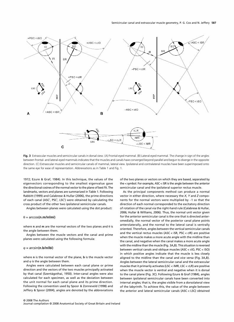
1 so that thedirection of each normal corresponded to the excitatory directionof rotation of the canal via the right-hand rule (Calabrese & Hullar,2006; Hullar & Williams, 2006). Thus, the normal unit vector givenfor the anterior semicircular canal is the one that is directed anter-omedially, the normal vector of the posterior canal plane pointsanterolaterally, and the normal to the lateral canal is ventrallyoriented. Therefore, angles between the vertical semicircular canalsand the vertical rectus muscles (ASC < iSR, PSC < cIR) are positivewhen the muscle makes a more acute angle with the midline thanthe canal, and negative when the canal makes a more acute anglewith the midline than the muscle (Fig. 3A,B). This situation is reversedbetween vertical canals and oblique muscles (ASC < cIO, PSC < iSO)in which positive angles indicate that the muscle is less closelyaligned to the midline than the canal and
vice versa
(Fig. 3A,B).Angles between the lateral semicircular canal and the extraocularmuscles that it primarily activates (LSC < iMR, LSC < cLR) are positivewhen the muscle vector is ventral and negative when it is dorsalto the canal plane (Fig. 3C). Following Ezure & Graf (1984), anglesbetween ipsilateral semicircular canals have been converted intointernal angles; that is, the angles visible from a dorsolateral viewof the labyrinth. To achieve this, the value of the angle betweenthe anterior and lateral semicircular canals (ASC < LSC) obtained
Fig. 3 Extraocular muscles and semicircular canals in dorsal view. (A) Frontal-eyed mammal. (B) Lateral-eyed mammal. The change in sign of the angles between frontal- and lateral-eyed mammals indicates that the muscles and canals have converged beyond parallel and begun to diverge in the opposite direction. (C) Extraocular muscles and semicircular canals of mammal, lateral view. Ipsilateral and contralateral muscles have been superimposed onto the same eye for ease of representation. Abbreviations as in Table 1 and Fig. 1.

Semicircular canal and extraocular muscle geometry, P. G. Cox and N. Jeffery
© 2008 The AuthorsJournal compilation © 2008 Anatomical Society of Great Britain and Ireland
588
from the dot product of their normal vectors was subtracted from180
°
.
Results
Angles between muscles and canals
The
X
,
Y
and
Z
components for the mean unit vectorsrepresenting the muscles and the mean normal unitvectors of the semicircular canal planes of each species
are given in Table 2. The angles between the extraocularmuscles and the semicircular canal by which they areprincipally activated vary widely across the species understudy. The results given in Table 3 show ranges of between50
°
and 62
°
for the canal–muscle angles. As also noted inprevious work (Ezure & Graf, 1984), the extraocular musclesand semicircular canals deviate significantly from parallelorientation among species, with misalignments of almost40
°
being measured between the posterior canal and thecontralateral inferior rectus muscle. The mean values and
Table 1 Abbreviations and descriptions of landmarks, muscle vectors and canal planes
Abbreviation Description
LandmarksASCam Centroid of the ampulla of the anterior semicircular canalASCap Centroid of anterior canal at point most distant from vestibuleASCb Point on margin of vestibule diametrically opposite the apex of the anterior semicircular canalASCv Centroid of the anterior part of the anterior semicircular canal at its inferiormost diameter as it enters the
vestibuleASCwA Centroid of the anterior part of the anterior semicircular canal at its greatest width in the transverse planeASCwP Centroid of the posterior part of the anterior semicircular canal at its greatest width in the transverse planecIOI Centroid of the inferior oblique muscle as it inserts on the eyeball on the contralateral sidecIOO Centroid of the inferior oblique muscle near its origin on the orbital wall on the contralateral sidecIRI Centroid of the inferior rectus muscle as it inserts on the eyeball on the contralateral sidecLRI Centroid of the lateral rectus muscle as it inserts on the eyeball on the contralateral sidecO Centroid of the optic nerve as it passes through the optic foramen on the contralateral sideiMRI Centroid of the medial rectus muscle as it inserts on the eyeballiO Centroid of the optic nerve as it passes through the optic forameniSOI Centroid of the superior oblique muscle as it inserts on the eyeballiSOO Point on the medial orbital wall at which the superior oblique muscle abruptly changes direction (trochlea)iSRI Centroid of the superior rectus muscle as it inserts on the eyeballLSCam Centroid of the ampulla of the lateral semicircular canalLSCap Centroid of lateral canal at point most distant from vestibuleLSCb Point on margin of vestibule diametrically opposite the apex of the lateral semicircular canalLSCv Centroid of the anterior part of the lateral semicircular canal at its medialmost diameter as it enters the vestibuleLSCwA Centroid of the anterior part of the lateral semicircular canal at its greatest width in the sagittal planeLSCwP Centroid of the posterior part of the lateral semicircular canal at its greatest width in the sagittal planePSCam Centroid of the ampulla of the posterior semicircular canalPSCap Centroid of posterior canal at point most distant from vestibulePSCb Point on margin of vestibule diametrically opposite the apex of the posterior semicircular canalPSCv Centroid of the superior part of the posterior semicircular canal at its medialmost diameter as it enters the
vestibulePSCwI Centroid of the inferior part of the posterior semicircular canal at its greatest width in the coronal planePSCwS Centroid of the superior part of the posterior semicircular canal at its greatest width in the coronal plane
Muscle vectorscIO Contralateral inferior oblique muscle: cIOO to cIOIcIR Contralateral inferior rectus muscle: cO to cIRIcLR Contralateral lateral rectus muscle: cO to cLRIiMR Ipsilateral medial rectus muscle: iO to iMRIiSO Ipsilateral superior oblique muscle: iSOO to iSOIiSR Ipsilateral superior rectus muscle: iO to iSRI
Semicircular canal planesASC Anterior semicircular canal: plane of best fit of ASCam, ASCap, ASCb, ASCv, ASCwA, ASCwPLSC Lateral semicircular canal: plane of best fit of LSCam, LSCap, LSCb, LSCv, LSCwA, LSCwPPSC Posterior semicircular canal: plane of best fit of PSCam, PSCap, PSCb, PSCv, PSCwI, PSCwS

Semicircular canal and extraocular muscle geometry, P. G. Cox and N. Jeffery
© 2008 The Authors Journal compilation © 2008 Anatomical Society of Great Britain and Ireland
589
Table 2
X
,
Y
and
Z
components of mean values (± SD) of muscle unit vectors and normal unit vectors of semicircular canal planes
Species
iSR cIO
X Y Z X Y Z
Homo
−
0.342 ± 0.051
−
0.242 ± 0.071
−
0.904 ± 0.028 0.807 ± 0.076
−
0.360 ± 0.118 0.438 ± 0.116
Felis
−
0.373 ± 0.088
−
0.498 ± 0.046
−
0.776 ± 0.048 0.601 ± 0.087
−
0.288 ± 0.142 0.728 ± 0.042
Cavia
−0.533 ± 0.064 −0.633 ± 0.041 −0.555 ± 0.046 0.349 ± 0.067 −0.322 ± 0.070 0.875 ± 0.040Rattus −0.437 ± 0.031 −0.595 ± 0.041 −0.672 ± 0.034 0.335 ± 0.093 −0.125 ± 0.097 0.925 ± 0.039Mus −0.520 ± 0.088 0.640 ± 0.051 −0.554 ± 0.060 0.443 ± 0.110 0.001 ± 0.116 0.882 ± 0.052Sciurus −0.678 ± 0.028 −0.490 ± 0.051 −0.543 ± 0.053 0.561 ± 0.052 0.005 ± 0.140 0.816 ± 0.038Oryctolagus −0.657 ± 0.099 −0.585 ± 0.038 −0.444 ± 0.145 0.140 ± 0.114 −0.199 ± 0.182 0.947 ± 0.054
Species
iSO cIR
X Y Z X Y Z
Homo −0.817 ± 0.073 −0.373 ± 0.056 0.410 ± 0.149 0.400 ± 0.052 0.417 ± 0.045 −0.813 ± 0.042Felis −0.833 ± 0.097 −0.200 ± 0.188 0.444 ± 0.187 0.625 ± 0.091 0.064 ± 0.102 −0.763 ± 0.088Cavia −0.622 ± 0.050 −0.341 ± 0.119 0.691 ± 0.077 0.859 ± 0.027 −0.106 ± 0.024 −0.497 ± 0.051Rattus −0.726 ± 0.093 −0.131 ± 0.124 0.651 ± 0.113 0.894 ± 0.044 −0.096 ± 0.042 −0.427 ± 0.086Mus −0.634 ± 0.052 −0.140 ± 0.117 0.749 ± 0.065 0.828 ± 0.028 −0.223 ± 0.041 −0.510 ± 0.051Sciurus −0.726 ± 0.056 −0.007 ± 0.157 0.669 ± 0.061 0.900 ± 0.017 0.054 ± 0.026 −0.431 ± 0.035Oryctolagus −0.423 ± 0.133 −0.587 ± 0.162 0.657 ± 0.082 0.982 ± 0.009 0.115 ± 0.118 −0.056 ± 0.090
Species
iMR cLR
X Y Z X Y Z
Homo −0.052 ± 0.062 0.104 ± 0.082 −0.989 ± 0.010 0.714 ± 0.042 0.098 ± 0.049 −0.689 ± 0.047Felis 0.021 ± 0.059 −0.211 ± 0.099 −0.971 ± 0.014 0.792 ± 0.080 −0.292 ± 0.098 −0.512 ± 0.113Cavia −0.204 ± 0.043 −0.361 ± 0.043 −0.908 ± 0.018 0.950 ± 0.021 −0.299 ± 0.069 −0.025 ± 0.047Rattus −0.252 ± 0.030 −0.381 ± 0.045 −0.888 ± 0.018 0.945 ± 0.030 −0.281 ± 0.076 −0.130 ± 0.078Mus −0.339 ± 0.067 −0.496 ± 0.056 −0.794 ± 0.032 0.905 ± 0.030 −0.402 ± 0.062 −0.110 ± 0.060Sciurus −0.353 ± 0.034 −0.274 ± 0.087 −0.891 ± 0.021 0.966 ± 0.015 −0.208 ± 0.030 0.146 ± 0.050Oryctolagus −0.476 ± 0.055 −0.146 ± 0.118 −0.858 ± 0.032 0.839 ± 0.043 −0.389 ± 0.084 0.361 ± 0.087
Species
ASC PSC
X Y Z X Y Z
Homo 0.829 ± 0.037 −0.201 ± 0.081 −0.512 ± 0.063 −0.647 ± 0.064 −0.159 ± 0.127 −0.734 ± 0.027Felis 0.824 ± 0.061 −0.369 ± 0.138 −0.394 ± 0.101 −0.668 ± 0.060 −0.111 ± 0.122 −0.724 ± 0.043Cavia 0.849 ± 0.040 −0.357 ± 0.079 −0.370 ± 0.095 −0.497 ± 0.051 −0.274 ± 0.111 −0.815 ± 0.029Rattus 0.889 ± 0.024 −0.322 ± 0.095 −0.310 ± 0.046 −0.530 ± 0.075 −0.282 ± 0.121 −0.786 ± 0.073Mus 0.905 ± 0.039 −0.34 ± 0.098 −0.194 ± 0.138 −0.519 ± 0.067 −0.364 ± 0.046 −0.769 ± 0.047Sciurus 0.865 ± 0.012 −0.364 ± 0.058 −0.337 ± 0.049 −0.463 ± 0.087 −0.391 ± 0.092 −0.785 ± 0.067Oryctolagus 0.894 ± 0.045 −0.090 ± 0.150 −0.405 ± 0.084 −0.526 ± 0.043 −0.149 ± 0.135 −0.826 ± 0.039
Species
LSC
X Y Z
Homo −0.003 ± 0.065 0.969 ± 0.016 −0.228 ± 0.077Felis 0.073 ± 0.046 0.956 ± 0.044 −0.219 ± 0.185Cavia 0.170 ± 0.059 0.853 ± 0.025 −0.487 ± 0.050Rattus −0.019 ± 0.064 0.982 ± 0.020 −0.159 ± 0.086Mus 0.035 ± 0.037 0.940 ± 0.024 −0.331 ± 0.068Sciurus 0.096 ± 0.032 0.961 ± 0.030 −0.226 ± 0.135Oryctolagus −0.050 ± 0.088 0.978 ± 0.034 0.010 ± 0.190

Semicircular canal and extraocular muscle geometry, P. G. Cox and N. Jeffery
© 2008 The AuthorsJournal compilation © 2008 Anatomical Society of Great Britain and Ireland
590
standard deviations for each species are given in Table 4a–f.Again, it can be seen that even within species there is frequentnon-alignment between the muscles and canals. With theexception of the rat, orientations of the posterior and anteriorcanals to the oblique muscles are always significantlymisaligned. There are also significant deviations for orien-tations involving the lateral canal. Interestingly, the angleof the posterior canal to the contralateral inferior rectuslies close to spatial alignment for all but two species(rabbits and humans). Results from an analysis of varianceindicate that there are highly significant differences(P < 0.001) in the orientations of the muscles to the canalsbetween the seven species studied here. Further analysisof these results using a post-hoc Duncan test (Table 4a–f)elucidates precisely which species are differing fromwhich. The only primate in the analysis, Homo, generallyforms a group on its own, indicating that the degree ofmisalignment between the canals and muscles in humansis significantly different from that measured in the otherspecies analysed here. For two angles (ASC < iSR, PSC < iSO;Table 4a,c), there is no difference between the mean valuemeasured in the cats and that calculated for the humans,and hence they group together. In four of the six anglesmeasured here (ASC < iSR, ASC < cIO, PSC < cIR, LSC < cLR;Table 4a,b,d,f), the rabbit was determined to be signifi-cantly different from every other species in the analysis, andthus formed a group on its own. The four rodents in theanalysis (rat, mouse, squirrel, guinea pig) show few consistenttrends. Despite their close phylogenetic relationship, the ratsand the mice are only grouped together by the orientationsof the posterior canal to the ipsilateral superior obliquemuscle and the lateral canal to the medial and lateralrectus muscles (Table 4c,e,f). There are significant differencesbetween their mean values of the other three angles(ASC < iSR, ASC < cIO, PSC < cIR; Table 4a,b,d). Indeed,when considering these angles, the rats often group withthe guinea pigs, and the mice are frequently associatedwith the squirrels. The angle of the posterior semicircularcanal to the ipsilateral superior oblique muscle (Table 4c)is notable because, for this measurement, the Duncan testsplits the species into just three groups – humans and cats,and two overlapping groups of rodents and rabbits –whereas four to six groups are needed to capture thevariation seen in the other five canal–muscle angles.
Angles between semicircular canals
Across all species, the variation in the angles between thethree semicircular canals is relatively small compared tothe variation seen in the angles between the canals and theextraocular muscles. The range across the whole dataset is32.8° for ASC < PSC, 38.7° for ASC < LSC, and 41.1° forPSC < LSC (see Table 3), whereas ranges of up to 62° aremeasured in the canal–muscle angles, as noted above.This suggests that the morphology of the labyrinth variesrelatively little across the mammals. The mean anglesbetween the three semicircular canals for each speciesare given in Table 4g–i. As with the canal–muscle angles,an analysis of variance showed the intraspecific variationto be significantly less (P < 0.001) than the interspecificvariation. However, it can be seen from the Duncan teststhat the interspecific variation is less than that seen forthe canal–muscle angles. These post-hoc tests split thespecies into only three groups with a great deal of overlap,suggesting that the species means are clustered closetogether. There are few discernible trends within thegroupings, and there is no separation of the humansample from the other species, as seen in the canal–muscleangles. Previous work (Haque et al. 2004; Hullar & Williams,2006) has suggested that the semicircular canals of primatesmore closely approximate orthogonality than those ofother mammalian species such as rodents, but this was notfound to be true here. Although the humans were shownto deviate from orthogonality in just one semicircularcanal pair – the anterior and posterior canals – this wasalso the case for the rabbits, and the guinea pigs showedno significant deviations from 90° between any of thethree semicircular canals. Overall, the posterior and lateralcanals were most often measured to be orthogonal, theangle between them departing significantly from 90° inonly the mouse and squirrel samples.
Prime directions
The X, Y and Z components for the mean unit prime direc-tions of each species are given in Table 5. It should benoted that these are prime directions of the anatomicalplanes, not of the afferent sensitivity planes. It can be seenthat the prime directions deviate from the unit normal
Table 3 Minimum and maximum values, ranges, mean values and SD of muscle–canal and inter-canal angles for whole dataset
ASC < iSR ASC < cIO PSC < iSO PSC < cIR LSC < iMR LSC < cLR ASC < PSC ASC < LSC PSC < LSC
Minimum −36.31 −19.67 −21.16 −39.27 −22.01 −31.31 83.17 63.43 75.35Maximum 24.15 36.84 30.12 22.86 28.12 24.04 115.97 102.12 116.41Range 60.46 56.51 51.28 62.13 50.13 55.35 32.80 38.69 41.06Mean −3.21 8.74 −3.13 −3.18 −2.74 −10.53 97.26 80.67 94.03SD 13.07 14.12 13.31 14.88 12.33 12.43 7.36 9.32 9.44

Semicircular canal and extraocular muscle geometry, P. G. Cox and N. Jeffery
© 2008 The Authors Journal compilation © 2008 Anatomical Society of Great Britain and Ireland
591
Table 4 Mean values (± SD), and significance of deviation from 0° or 90°, of angles between extraocular muscles and semicircular canals, and angles between semicircular canals. Groups indicate those species which show no significant differences between means, as determined by Duncan’s test (***P < 0.001; **P < 0.01; *P < 0.05; ns, not significant)A
B
C
D
E
Species ASC < iSR P (x = 0°) Groups
Homo 13.20 ± 5.52 ** AFelis 10.75 ± 9.66 * ARattus 0.62 ± 3.53 ns BCavia −1.13 ± 5.89 ns BMus −8.60 ± 4.77 *** CSciurus −13.07 ± 1.91 *** COryctolagus −20.93 ± 10.14 *** D
Species ASC < cIO P (x = 0°) Groups
Homo 31.25 ± 7.29 *** AFelis 19.44 ± 4.35 *** BMus 13.82 ± 6.74 *** CSciurus 12.01 ± 2.79 *** CCavia 5.03 ± 5.02 * DRattus 3.03 ± 3.89 ns DOryctolagus −14.27 ± 3.88 *** E
Species PSC < iSO P (x = 0°) Groups
Homo 16.64 ± 8.07 ** AFelis 15.36 ± 12.5 * ARattus −4.78 ± 4.39 * BCavia −9.44 ± 6.01 ** B CSciurus −11.32 ± 4.83 *** B CMus −11.40 ± 4.87 ** B COryctolagus −12.82 ± 3.40 *** C
Species PSC < cIR P (x = 0°) Groups
Homo 15.61 ± 4.66 *** AFelis 7.48 ± 9.35 ns BMus 2.55 ± 3.30 ns B CCavia 0.42 ± 3.73 ns C DSciurus −5.70 ± 4.77 ns D ERattus −6.32 ± 8.42 ns EOryctolagus −29.92 ± 5.72 *** F
Species LSC < iMR P (x = 0°) Groups
Homo 19.20 ± 5.79 *** ACavia 5.69 ± 3.57 ** BFelis 0.78 ± 11.24 ns B CSciurus −5.63 ± 9.25 ns C DOryctolagus −7.36 ± 7.62 * DMus −12.49 ± 5.32 *** DRattus −13.19 ± 7.09 ** D
F
G
H
I
Species LSC < cLR P (x = 0°) Groups
Homo 14.64 ± 6.60 ** ACavia −4.77 ± 3.12 ** BFelis −6.63 ± 7.92 * BSciurus −8.20 ± 4.22 * BRattus −15.95 ± 6.24 *** CMus −18.13 ± 4.13 *** COryctolagus −24.78 ± 4.82 *** D
Species ASC < PSC P (x = 90°) Groups
Felis 103.34 ± 9.70 ** AMus 101.54 ± 6.32 *** ARattus 97.57 ± 4.80 ** A BHomo 97.14 ± 4.82 * A B COryctolagus 97.05 ± 5.60 ** A B CCavia 91.20 ± 5.78 ns B CSciurus 89.52 ± 4.40 ns C
Species ASC < LSC P (x = 90°) Groups
Cavia 91.22 ± 6.97 ns AHomo 85.30 ± 5.81 ns A BOryctolagus 81.73 ± 11.00 ns B CSciurus 78.97 ± 6.61 * B CFelis 78.36 ± 10.05 * B CMus 76.63 ± 6.02 *** B CRattus 73.35 ± 6.37 *** C
Species PSC < LSC P (x = 90°) Groups
Sciurus 104.41 ± 9.64 * ARattus 98.12 ± 10.89 ns A BOryctolagus 97.52 ± 9.82 ns A BMus 96.08 ± 6.06 * A BFelis 89.49 ± 6.94 ns B CHomo 88.96 ± 6.33 ns B CCavia 85.58 ± 5.39 ns C
Table 4 (Continued)
vectors of the canals quite considerably – from 7.5° inguinea pigs to over 20° in rats (Table 6). In general, theprime directions are not orthogonal. Species that departthe furthest from orthogonality show the greatest devia-tions between prime directions and normal vectors. Theorientations of the prime directions to each other, as wellas to the extraocular muscles, are given in Table 7. The ori-entations of the extraocular muscles with the anatomicalcanal planes and with the prime directions were comparedusing Student’s t-test. The results (Table 8) show that, ofthe 42 mean muscle vectors under study (six mean vectorsin seven species), 14 are significantly more aligned with

Semicircular canal and extraocular muscle geometry, P. G. Cox and N. Jeffery
© 2008 The AuthorsJournal compilation © 2008 Anatomical Society of Great Britain and Ireland
592
the relevant prime direction than with the anatomicalcanal plane – both oblique muscles of the humans and cats,as well as the lateral rectus of the cat; the inferior rectusand both horizontal recti in the rat; the superior rectus,inferior oblique and both horizontal recti in the mouse;and the inferior and medial recti of the rabbit. However,11 mean muscle vectors are significantly less alignedwith the prime direction, and the remaining 17 vectorsshow no significant change either way. Thus there is not aconsistent trend for the extraocular muscles to be moreclosely aligned to the prime directions.
Discussion
The angles between each extraocular muscle and thesemicircular canal by which it is principally activated werecalculated from three-dimensional co-ordinate data. It wasdetermined that the muscles and canals are frequently not
in alignment, and vary widely in their relative orientationsbetween mammalian species commonly used in research.Similar patterns of variation have also been observedduring human development (Cox & Jeffery, 2007). Acrossall seven species studied, the rat and the guinea pig showthe closest spatial alignment between the vertical canalsand the extraocular muscles, particularly with regard tothe superior and inferior recti. The least aligned axes werefound among the rabbits and modern humans. Proposedcompensatory mechanisms for the spatial integration ofvestibular signals with the activation of the extraocularmuscles include changes in the maximal response planes(e.g. Estes et al. 1975; Reisine et al. 1988; Haque et al. 2004).These studies demonstrate that the plane of rotation thatproduces the maximum response of the ampullary nerve inthe cat and rhesus monkey only differs by about 10° fromthe anatomical plane. In the present study, estimationsof these maximal response planes using prime directions
Table 5 X, Y and Z components of mean values of (± SD) of unit prime directions
Species
ASC′ PSC′
X Y Z X Y Z
Homo 0.755 ± 0.054 −0.144 ± 0.076 −0.630 ± 0.074 −0.546 ± 0.055 −0.190 ± 0.064 −0.812 ± 0.032Felis 0.730 ± 0.060 −0.197 ± 0.139 −0.634 ± 0.083 −0.469 ± 0.118 −0.148 ± 0.144 −0.850 ± 0.081Cavia 0.834 ± 0.052 −0.385 ± 0.062 −0.383 ± 0.055 −0.491 ± 0.074 −0.353 ± 0.054 −0.790 ± 0.049Rattus 0.833 ± 0.051 −0.069 ± 0.051 −0.539 ± 0.086 −0.370 ± 0.039 −0.154 ± 0.091 −0.911 ± 0.025Mus 0.850 ± 0.045 −0.198 ± 0.045 −0.481 ± 0.066 −0.307 ± 0.112 −0.306 ± 0.078 −0.892 ± 0.026Sciurus 0.879 ± 0.046 −0.186 ± 0.075 −0.426 ± 0.078 −0.412 ± 0.051 −0.165 ± 0.120 −0.888 ± 0.039Oryctolagus 0.822 ± 0.062 0.048 ± 0.180 −0.537 ± 0.044 −0.415 ± 0.078 −0.010 ± 0.145 −0.896 ± 0.037
Species
LSC′
X Y Z
Homo −0.071 ± 0.056 −0.953 ± 0.033 0.263 ± 0.131Felis −0.228 ± 0.132 −0.894 ± 0.056 0.352 ± 0.095Cavia −0.194 ± 0.082 −0.882 ± 0.049 0.410 ± 0.103Rattus −0.168 ± 0.085 −0.873 ± 0.061 0.425 ± 0.152Mus −0.194 ± 0.116 −0.819 ± 0.053 0.523 ± 0.060Sciurus −0.155 ± 0.068 −0.840 ± 0.058 0.507 ± 0.097Oryctolagus −0.010 ± 0.140 −0.964 ± 0.029 0.186 ± 0.144
Table 6 Mean values (± SD), and significance of deviation from 0°, of angles between canal normals and prime directions
Species ASC < ASC′ P (x = 0°) PSC < PSC′ P (x = 0°) LSC < LSC′ P (x = 0°)
Homo 9.94 ± 5.27 ** 9.34 ± 4.83 ** 8.79 ± 3.37 ***Felis 19.19 ± 12.73 ** 16.07 ± 9.56 ** 14.82 ± 9.71 **Cavia 7.58 ± 5.17 ** 7.74 ± 5.00 ** 7.93 ± 6.07 **Rattus 20.89 ± 6.44 *** 16.90 ± 7.28 *** 22.94 ± 6.33 ***Mus 19.25 ± 8.03 *** 16.35 ± 5.14 *** 17.45 ± 5.66 ***Sciurus 12.51 ± 5.91 ** 15.80 ± 8.58 * 18.78 ± 10.63 *Oryctolagus 15.32 ± 8.19 *** 14.08 ± 8.34 *** 15.86 ± 10.74 **
***P < 0.001; **P < 0.01; *P < 0.05; ns, not significant.

Semicircular canal and extraocular muscle geometry, P. G. Cox and N. Jeffery
© 2008 The Authors Journal compilation © 2008 Anatomical Society of Great Britain and Ireland
593
(see Rabbitt, 1999) revealed differences of about 7–23°relative to the anatomical planes. Although some of thesedifferences were compensatory in that the prime planeswere closer than the anatomical planes to the muscleorientations, the findings were inconsistent. Indeed, in anumber of cases, a muscle was found to be less closelyaligned to the prime direction than to the semicircularcanal by which it is primarily activated (see Table 8). These
findings would appear to favour the idea that the spatialmismatch is corrected for elsewhere, probably along the three-neuron arc and most likely involving signal transformationswithin the vestibular nuclei with reference to the flocculus (Ito,1982; Ito et al. 1982; Brettler & Baker, 2001; Billig & Balaban,2005). Nevertheless, there remain several other structuralpossibilities for helping to resolve differences between thevestibular and extraocular spatial frameworks among
Table 7 Mean values (± SD) and significance of deviation from 90° or 0°, of angles between prime directions, and between extraocular muscles and prime directions
Species ASC′ < PSC′ P (x = 90°) ASC′ < LSC′ P (x = 90°) PSC′ < LSC′ P (x = 90°)
Homo 82.59 ± 4.61 * 94.39 ± 6.31 ns 89.68 ± 6.62 nsFelis 75.66 ± 10.20 ** 102.2 ± 11.28 * 93.43 ± 8.89 nsCavia 88.34 ± 6.28 ns 88.5 ± 7.44 ns 85.38 ± 5.88 nsRattus 78.75 ± 6.71 ** 108.71 ± 7.44 *** 101.10 ± 12.38 *Mus 76.54 ± 6.82 *** 104.94 ± 6.83 *** 99.53 ± 6.19 **Sciurus 86.95 ± 4.77 ns 101.55 ± 6.65 * 104.59 ± 10.00 *Oryctolagus 80.65 ± 6.14 ** 99.31 ± 12.15 ns 98.72 ± 11.00 *
Species ASC′ < iSR P (x = 0°) ASC′ < cIO P (x = 0°) PSC′ < iSO P (x = 0°)
Homo 20.26 ± 5.42 *** 22.51 ± 6.85 *** 10.79 ± 10.04 *Felis 18.87 ± 5.78 *** 2.36 ± 8.98 ns 3.09 ± 16.93 nsCavia 0.72 ± 5.15 ns 4.54 ± 4.92 * −7.16 ± 7.13 *Rattus 2.22 ± 4.55 ns −12.21 ± 10.76 * −17.94 ± 8.31 ***Mus −2.75 ± 5.31 ns −2.82 ± 9.80 ns −25.68 ± 7.36 ***Sciurus −15.98 ± 7.32 ** 8.81 ± 2.91 ** −16.87 ± 6.03 **Oryctolagus −19.8 ± 7.16 *** −23.30 ± 6.40 *** −25.08 ± 7.21 ***
Species PSC′ < cIR P (x = 0°) LSC′ < iMR P (x = 0°) LSC′ < cLR P (x = 0°)
Homo 21.25 ± 4.08 *** −20.91 ± 9.61 ** −18.95 ± 7.80 **Felis 20.35 ± 10.60 ** − 9.09 ± 9.55 * −5.62 ± 8.51 nsCavia 0.53 ± 5.74 ns −0.84 ± 5.78 ns 4.12 ± 6.45 nsRattus 4.18 ± 6.25 ns −0.12 ± 8.74 ns 1.69 ± 6.75 nsMus 15.79 ± 6.19 *** 3.33 ± 5.19 ns 5.64 ± 9.20 nsSciurus 0.06 ± 3.93 ns −9.64 ± 7.79 ns 5.66 ± 4.56 nsOryctolagus −21.24 ± 6.07 *** −1.05 ± 12.34 ns 26.12 ± 10.52 ***
***P < 0.001; **P < 0.01; *P < 0.05; ns, not significant.
Table 8 Probabilities that the mean orientations of the extraocular muscles to the semicircular canals and to the prime directions are equal
Species ASC/ASC′ < iSR ASC/ASC′ < cIO PSC/PSC′ < iSO PSC/PSC′ < cIR LSC/LSC′ < iMR LSC/LSC′ < cLR
Homo ** (a) * (p) * (p) * (a) ns nsFelis * (a) ** (p) * (p) * (a) ns * (p)Cavia ns ns ns ns ns nsRattus ns ** (a) ** (a) ** (p) * (p) ** (p)Mus * (p) *** (p) *** (a) *** (a) ** (p) *** (p)Sciurus ns ns ns ns * (a) nsOryctolagus ns *** (a) ** (a) *** (p) * (p) ns
***P < 0.001; **P < 0.01; *P < 0.05; ns, not significant; a, anatomical plane more closely aligned with muscle; p, prime direction more closely aligned with muscle.
Semicircular canal and extraocular muscle geometry, P. G. Cox and N. Jeffery
© 2008 The AuthorsJournal compilation © 2008 Anatomical Society of Great Britain and Ireland
594
mammals. These include canal shape, canal torsion andfluid coupling, particularly in the common crus (Oman et al.1987; Hullar, 2006).
Comparing values of optic divergence from Hughes(1977) with the muscle–canal angles reported here, it canbe seen that the two broadly correlate. Table 4a–f showthat, for each angle, the species tend to be arranged withthe highly optically convergent humans (binocular vision)at one extreme, and the very optically divergent rabbits(optic axis approximately 85° from midline) at the other.This suggests that it is realignment of the extraocularmuscles, rather than of the semicircular canals, that is drivingthe variation in their relative orientations. This is indicatedby Simpson & Graf (1981) who found a range of just 4° inthe angle of the anterior semicircular canal to the midlinein a sample of humans, cats, guinea pigs and rabbits, butmeasured a range of 42° in the angle of the superior rectusmuscle to the midline. Similarly, the superior obliquevaried over 39° in its orientation to the midline, whereasthe posterior canal had a much more restricted range ofjust 14°. Mediolateral realignment of the optic axes onlyaccounts for variation in the orientation of the verticalsemicircular canals to the relevant extraocular muscles(Table 4a–d). Angles measured between the lateral canaland the horizontal muscles indicate misalignment in thedorsoventral direction, which is not affected by the diverg-ence of the eyes. This can be seen in the results for LSC < iMRand LSC < cLR (Table 4e,f), which differ from those of theother four angles. For instance, the lateral-eyed guinea pigsare more similar to the cats in these angles but tend to groupwith the other rodents when considering angles involvingthe vertical canals. However, angles of the lateral canalto the medial and lateral recti could be influenced bydorsoventral realignment of the optic axes, a processreferred to as frontation (see Noble et al. 2000). For example,in humans the eyes face forwards relative to the cranium,whereas in rabbits the eyes are tilted upwards.
Changes in the relative positions of the muscles couldalso be influenced by changes in eye size. Increasing ordecreasing the size of the eye will tend to displace theinsertion points of the extraocular muscles, changing theirorientations relative to the semicircular canals. Eye sizedoes not have a simple linear relationship with body sizeacross all mammals. At small size (< 1 kg), eye mass scaleshyperallometrically with body mass, at medium size (1–79 kg)the two masses are related isometrically, and at large size(> 80 kg) they are relatively independent (Hughes, 1977;Kiltie, 2000). Similarly, Burton (2006) found an isometricrelationship between eye size and brain size up to approxi-mately 200 g brain mass, but evidence of independencebetween the two variables beyond this size. Hence, thedifference in eye size in the rabbits, cats and humans isproportional to their difference in body size, but the fourrodents in the analysis have smaller eyes than might beexpected for their size, which may account for some of
the variation seen in the relative orientations of theextraocular muscles.
Despite showing a great deal of variation in their orien-tation relative to the extraocular muscles, the semicircularcanals do not vary as much in their orientations to eachother. The canals are rarely perpendicular, with deviationsof almost 30° from orthogonality being measured in someindividuals. The one exception here is the guinea pig, inwhich all three inter-canal angles show no significantdifference from 90°. This is in direct contradiction to Curthoyset al. (1975), who calculated mean deviations from 90° ofat least 8° in all three angles in the guinea pig, and a meanangle of 58° between the anterior and lateral canals [actu-ally reported as 122° because Curthoys et al. (1975) weremeasuring the supplementary angle ventral to the lateralcanal]. Similarly, Blanks et al. (1975) measured a meanangle of 68° (reported as 112°) between the same twocanals in humans, whereas the mean value calculated herewas just over 85°. The lack of correspondence betweenthese results may possibly be attributed to differences inthe method of mapping the semicircular canals. Blanks et al.(1972, 1975) and Curthoys et al. (1975) exclude points fromthe common crus and ampullae from their measurements,whereas this study follows the more recent work ofCalabrese & Hullar (2006) and Hullar & Williams (2006) andincludes them. Also, older studies that employed a micro-manipulator to map the canals (Blanks et al. 1972, 1975;Curthoys et al. 1975) tend to use three-dimensional co-ordinates from the medial-most wall of the osseous canals.In contrast, this study based on MR images was able to use theco-ordinates of centroids of each semicircular canal lumen.
To summarize, the present study finds significant varia-tions in the spatial arrangement of the semicircular canalsand the extraocular muscles both within and betweenspecies commonly used as experimental models of thevestibulo-ocular reflexes. The spatial arrangement of thecanals and muscles in the cat and the arrangement amongthe canals in the guinea pig most closely resemble theconditions found in modern humans. The rabbit closelymatches humans in terms of the severity of the functionalchallenge of integrating different extraocular and vesti-bular co-ordinate frames. However, it is important to notethat humans are distinct from all the other species studiedin terms of the angles between vertical canals and contra-lateral muscles (i.e. PSC < cIR & ASC < cIO) and the anglesbetween the lateral canal and horizontal muscles (LSC < iMR& LSC < cLR). These angles may have some deeper functionalor structural significance. For example, Spoor et al. (2007)have recently demonstrated a strong statistical linkbetween lateral canal arc size and agility among a largesample of mammals, based on the presumed importanceof arc size to calibrate mechanical sensitivity levels. Yang& Hullar (2007) report that canal orientation may also havea substantial impact on sensitivity, and varying canalangles may thus be linked with locomotor agility.
Semicircular canal and extraocular muscle geometry, P. G. Cox and N. Jeffery
© 2008 The Authors Journal compilation © 2008 Anatomical Society of Great Britain and Ireland
595
Our findings support the view that the vestibular refer-ence frame remains relatively stable and that spatialdisparities with the extraocular co-ordinate system arisedue to changes of orbit position. However, the exact natureof the variation remains unclear and important questionshave yet to be answered. For instance, does the extraocularframe move with the orbit as one rigid unit, or are thereperhaps significant changes within the extraocular framesuch as a convergence in the line of action of the superioroblique and superior rectus muscles (see Simpson & Graf,1981)? Also, in species with extreme spatial mismatchingdoes the functional capacity of the three-neuron arcimpede the high frequency and irregular head movementsnormally used during agile forms of locomotion (see Spooret al. 2007)? At present we can only surmise the answers,as the limited range of species studied to date do not givesufficient data resolution to map out statistically meaning-ful interspecific trends. Other possible constraints or per-turbations to the extraocular co-ordinate frame and to itslinks with the vestibular frame include changes of basicra-nial architecture (Ross & Ravosa, 1993), orbital frontation(Noble et al. 2000) as well as phylogeny (Garland et al. 2005)and body size scaling. We are currently repeating the currentstudy with a broader range of over 40 mammalian species toclarify the above questions.
Acknowledgements
We thank the Biotechnology and Biological Sciences ResearchCouncil for funding (grant no. BBD0000681). We also thank FayPenrose, University of Liverpool Veterinary School, for providingspecimens and Fred Spoor, University College London, for suggestingimprovements to an earlier draft of this paper. Karen Davies andSteve Williams, University of Manchester, and Alasdair Preston,Queen Mary, University of London, kindly assisted with the imagingexperiments. Images of modern human subjects were generouslyprovided by Dirk Bartz, University of Leipzig, and James Rilling,Emory University.
References
Billig I, Balaban CD (2005) Zonal organization of the vestibulo-cerebellar pathways controlling the horizontal eye muscles usingtwo recombinant strains of pseudorabies virus. Neuroscience133, 1047–1059.
Blanks RHI, Torigoe Y (1989) Orientation of the semicircular canalsin rat. Brain Res 487, 278–287.
Blanks RHI, Curthoys IS, Markham CH (1972) Planar relationshipsof semicircular canals in the cat. Am J Physiol 223, 55–62.
Blanks RHI, Curthoys IS, Markham CH (1975) Planar relationshipsof the semicircular canals in man. Acta Otolaryngol 80, 185–196.
Blanks RHI, Curthoys IS, Bennett ML, Markham CH (1985) Planarrelationships of the semicircular canals in rhesus and squirrelmonkeys. Brain Res 340, 315–324.
Brettler SC, Baker JF (2001) Directional sensitivity of anterior,posterior and horizontal canal vestibule-ocular neurons in thecat. Exp Brain Res 140, 432–442.
Brichta AM, Acuna DL, Peterson EH (1988) Planar relations ofsemicircular canals in awake, resting turtles, Pseudemys scripta.Brain Behav Evol 32, 236–245.
de Burlet HM, Koster JJJ (1916) Zur Bestimmung des Standes derBogengänge unde der Maculae acusticae im Kaninchenschädel.Arch Anat Physiol 1916, 59–100.
Burton RF (2006) A new look at the scaling of size in mammalianeyes. J Zool 269, 225–232.
Calabrese DR, Hullar TE (2006) Planar relationships of the twosemicircular canals in two strains of mice. J Assoc Res Otolaryngol7, 151–159.
Cohen B, Suzuki J, Bender MB (1964) Eye movements from semicircularcanal nerve stimulation in the cat. Ann Otol Rhinol Laryngol 73,153–169.
Cox PG, Jeffery N (2007) Morphology of the mammalian vestibulo-ocular reflex: the spatial arrangement of the human fetalsemicircular canals and extraocular muscles. J Morphol 268,878–890.
Cummins H (1925) The vestibular labyrinth of the albino rat: formand dimensions and the orientation of the semicircular canals,cristae and maculae. J Comp Neurol 38, 399–445.
Curthoys IS, Curthoys EJ, Blanks RHI, Markham CH (1975) Theorientation of the semicircular canals in the guinea pig. ActaOtolaryngol 80, 197–205.
Daunicht WJ, Pellionisz AJ (1987) Spatial arrangement of thevestibular and oculomotor system in the rat. Brain Res 435,48–56.
Della Santina CC, Potyagaylo V, Migliaccio AA, Minor LB, Carey JP(2005) Orientation of human semicircular canals measured bythree-dimensional multiplanar CT reconstruction. J Assoc ResOtolaryngol 6, 191–206.
Dickman JD (1996) Spatial orientation of semicircular canals andafferent sensitivity vectors in pigeons. Exp Brain Res 111, 8–20.
Estes MS, Blanks RH, Markham CH (1975) Physiologic characteristicsof vestibular first-order canal neurons in the cat. I. Response planedetermination and resting discharge characteristics. J Neurophysiol38, 1232–1249.
Ezure K, Graf W (1984) A quantitative analysis of the spatialorganization of the vestibulo-ocular reflexes in lateral- andfrontal-eyed animals – I. Orientation of semicircular canals andextraocular muscles. Neuroscience 12, 85–93.
Garland T, Bennett AF, Rezende EL (2005) Phylogenetic approachesin comparative physiology. J Exp Biol 208, 3015–3035.
Ghanem T, Rabbitt RD, Tresco PA (1998) Three-dimensionalreconstruction of the membranous vestibular labyrinth in thetoadfish, Opsanus tau. Hearing Res 124, 27–43.
Haque A, Angelaki DE, Dickman JD (2004) Spatial tuning anddynamics of vestibular semicircular canal afferents in rhesusmonkeys. Exp Brain Res 155, 81–90.
Hashimoto S, Naganuma H, Tokumasu K, Itoh A, Okamoto M (2005)Three-dimensional reconstruction of the human semicircularcanals and measurement of each membranous canal planedefined by Reid’s stereotactic co-ordinates. Ann Otol RhinolLaryngol 114, 934–938.
Hughes A (1977) The topography of vision in mammals of contrastinglife style: comparative optics and retinal organisation. In Handbookof Sensory Physiology, Vol. VII/5 (ed. Crescitelli, F.), pp. 613–756.Berlin-Heidelberg-New York: Springer.
Hullar TE (2006) Semicircular canal geometry, afferent sensitivity,and animal behavior. Anat Rec 288A, 466–472.
Hullar TE, Williams CD (2006) Geometry of the semicircular canalsof the chinchilla (Chinchilla laniger). Hearing Res 213, 17–24.

Semicircular canal and extraocular muscle geometry, P. G. Cox and N. Jeffery
© 2008 The AuthorsJournal compilation © 2008 Anatomical Society of Great Britain and Ireland
596
Ichijo H (2002) Angles between left and right vertical semicircularcanals. Nippon Jibi Inkoka Gakkai Kaiho 105, 1138–1142.
Ifediba MA, Rajguru SM, Hullar TE, Rabbitt RD (2007) The role of3-canal biomechanics in angular motion transduction by thehuman vestibular labyrinth. Ann Biomed Eng 35, 1247–1263.
Ito M (1982) Cerebellar control of the vestibulo-ocular reflex –around the flocculus hypothesis. Ann Rev Neurosci 5, 275–296.
Ito M, Orlov I, Yamamoto M (1982) Topographical representationof vestibulo-ocular reflexes in rabbit cerebellar flocculus.Neuroscience 7, 1657–1664.
Jeffery N, Spoor F (2004) Prenatal growth and development of themodern human labyrinth. J Anat 204, 71–92.
Kiltie RA (2000) Scaling of visual acuity with body size in mammalsand birds. Funct Ecol 14, 226–234.
Mazza D, Winterson BJ (1984) Semicircular canal orientation in theadult resting rabbit. Acta Otolaryngol 98, 472–480.
Noble VE, Kowalski EM, Ravosa MJ (2000) Orbit orientation and thefunction of the mammalian postorbital bar. J Zool 250, 405–418.
Oman CM, Marcus EN, Curthoys IS (1987) The influence of semicircularcanal morphology on endolymph flow dynamics. An anatomicallydescriptive mathematical model. Acta Otolaryngol 103, 1–13.
Rabbitt RD (1999) Directional coding of three-dimensionalmovements by the vestibular semicircular canals. Biol Cybern 80,417–431.
Reisine H, Simpson JI, Henn V (1988) A geometric analysis ofsemicircular canals and induced activity in their peripheralafferents in the rhesus monkey. Ann N Y Acad Sci 545, 10–20.
Rohlf FJ, Slice D (1990) Extensions of the Procrustes method for theoptimal superimposition of landmarks. Syst Zool 39, 40–59.
Ross CF, Ravosa MJ (1993) Basicranial flexion, relative brain size,and facial kyphosis in nonhuman primates. Am J Phys Anthropol91, 305–324.
Sato H, Sando I, Takahashi H, Fujita S (1993) Torsion of the humansemicircular canals and its influence on their angular relationships.Acta Otolaryngol 113, 171–175.
Simpson JI, Graf W (1981) Eye-muscle geometry and compensatoryeye movements in lateral-eyed and frontal-eyed animals. AnnN Y Acad Sci 374, 20–30.
Spoor F, Zonneveld F (1998) Comparative review of the humanbony labyrinth. Yearb Phys Anthropol 41, 211–251.
Spoor F, Garland T, Krovitz G, Ryan T, Silcox M, Walker A (2007)The primate semicircular canal system and locomotion. Proc NatlAcad Sci U S A 104, 10808–10812.
Suzuki J, Cohen B, Bender MB (1964) Compensatory eye movementsinduced by vertical semicircular canal stimulation. Exp Neurol 9,137–160.
Szentágothai J (1950) The elementary vestibulo-ocular reflex arc.J Neurophysiol 13, 395–407.
Takagi A, Sando I, Takahashi H (1989) Computer-aided three-dimensional reconstruction and measurement of semicircularcanals and their cristae in man. Acta Otolaryngol 107, 362–365.
Yang AZ, Hullar TE (2007) Relationship of semicircular canal size tovestibular-nerve afferent sensitivity in mammals. J Neurophysiol98, 3197–3205.