ghassan soleiman abu-sittah jamal j. hoballah joseph ...€¦ · ghassan soleiman abu-sittah and...
TRANSCRIPT

Reconstructing the War Injured Patient
123
Ghassan Soleiman Abu-SittahJamal J. HoballahJoseph Bakhach Editors

Reconstructing the War Injured Patient
Ghassan Soleiman Abu-Sittah Jamal J. Hoballah • Joseph BakhachEditors
Reconstructing the War Injured Patient
ISBN 978-3-319-56885-0 ISBN 978-3-319-56887-4 (eBook)DOI 10.1007/978-3-319-56887-4
Library of Congress Control Number: 2017943689
© Springer International Publishing AG 2017This work is subject to copyright. All rights are reserved by the Publisher, whether the whole or part of the material is concerned, specifically the rights of translation, reprinting, reuse of illustrations, recitation, broadcasting, reproduction on microfilms or in any other physical way, and transmission or information storage and retrieval, electronic adaptation, computer software, or by similar or dissimilar methodology now known or hereafter developed.The use of general descriptive names, registered names, trademarks, service marks, etc. in this publication does not imply, even in the absence of a specific statement, that such names are exempt from the relevant protective laws and regulations and therefore free for general use.The publisher, the authors and the editors are safe to assume that the advice and information in this book are believed to be true and accurate at the date of publication. Neither the publisher nor the authors or the editors give a warranty, express or implied, with respect to the material contained herein or for any errors or omissions that may have been made. The publisher remains neutral with regard to jurisdictional claims in published maps and institutional affiliations.
Printed on acid-free paper
This Springer imprint is published by Springer NatureThe registered company is Springer International Publishing AGThe registered company address is: Gewerbestrasse 11, 6330 Cham, Switzerland
EditorsGhassan Soleiman Abu-SittahDivision of Plastic and Reconstructive
SurgeryAmerican University of Beirut
Medical CenterBeirut, Lebanon
Joseph BakhachDivision of Plastic and Reconstructive
SurgeryAmerican University of Beirut Medical
Centre-Faculty of MedicineBeirut, Lebanon
Jamal J. HoballahDepartment of SurgeryAmerican University of Beirut
Medical CenterBeirut, Lebanon

v
1 Ballistics of Gunshot Wounds . . . . . . . . . . . . . . . . . . . . . . . . . . . . . 1Fadel M. Chahine
2 Biodynamics of Blast Injuries � � � � � � � � � � � � � � � � � � � � � � � � � � � � � 7Ghassan Soleiman Abu-Sittah and Odette M. Abou Ghanem
3 Management of Craniomaxillofacial Injuries � � � � � � � � � � � � � � � 19Ghassan Soleiman Abu-Sittah and Joe S. Baroud
4 The Management of Penetrating Neck Injuries � � � � � � � � � � � � � � 31Mohammad Rachad Wehbe and Jamal J. Hoballah
5 Reconstruction of Periocular War Injuries � � � � � � � � � � � � � � � � � 39Riad Maluf and Rouba Maluf
6 Management of the Upper Limb Soft-Tissue Injuries � � � � � � � � � 49Joseph Bakhach and Hamed Janom
7 Management of Brachial Plexus Injuries � � � � � � � � � � � � � � � � � � � 59Ghassan Soleiman Abu-Sittah, Joseph Bakhach and Arij El Khatib
8 Management of Upper Limb Fractures � � � � � � � � � � � � � � � � � � � � 67Said S. Saghieh and Naji S. Madi
9 Lower Extremity Reconstruction � � � � � � � � � � � � � � � � � � � � � � � � � 79Amir Ibrahim and Ahmad Oneisi
10 Management of Peripheral Nerve Injuries � � � � � � � � � � � � � � � � � � 89Joseph Bakhach and Arij El Khatib
11 Management of Lower Limb Fractures � � � � � � � � � � � � � � � � � � � � 97Karim Z. Masrouha and Said S. Saghieh
12 Abdominal Wall Reconstruction � � � � � � � � � � � � � � � � � � � � � � � � � 111Ghassan Soleiman Abu-Sittah and Firas Abiad
13 Contemporary Management of Urogenital Injuries � � � � � � � � � 119Mohammed Shahait and Rami Wajih Nasr
14 Management of Central Nervous System War Injuries � � � � � � 131Ghassan S. Skaf and Elias Elias
15 Vascular Trauma � � � � � � � � � � � � � � � � � � � � � � � � � � � � � � � � � � � � � � 141Hasan Al Harakeh and Jamal J. Hoballah
Contents

vi
16 Early Microsurgical Management of Blast Injuries � � � � � � � � � 157Joseph Bakhach and Odette M. Abou Ghanem
17 Amputations and Prostheses � � � � � � � � � � � � � � � � � � � � � � � � � � � � 165Reem Karami and Jamal J. Hoballah
18 Healing the Scars Within: Psychological Support for the War-Injured � � � � � � � � � � � � � � � � � � � � � � � � � � � � � � � � � � � 181Brigitte Khoury and Sariah Daouk
19 Infections in Combat-Related Wounds � � � � � � � � � � � � � � � � � � � � 191Abdul Rahman Bizri and Zeyad Tamim Sahli
Index � � � � � � � � � � � � � � � � � � � � � � � � � � � � � � � � � � � � � � � � � � � � � � � � � � � � 203
Contents
vii
Contributors
Firas Abiad Department of Plastic and Reconstructive Surgery, American University of Beirut Medical Center, Beirut, Lebanon
Odette M� Abou Ghanem, M�D� Department of Plastic and Reconstructive Surgery, American University of Beirut Medical Center, Beirut, Lebanon
Ghassan Soleiman Abu-Sittah, MD, MBChB, FRCS (Plast) Division of Plastic and Reconstructive Surgery, American University of Beirut Medical Center, Beirut, Lebanon
Joseph Bakhach, M�D�, Ph�D� Division of Plastic and Reconstructive Surgery, American University of Beirut Medical Centre - Faculty of Medicine, Beirut, Lebanon
Joe S� Baroud, M�D�, M�R�C�S� Department of General Surgery, Surgery Intern, American University of Beirut, Beirut, Lebanon
Abdul Rahman Bizri, M�D�, M�Sc�, D�L�S�H�T�M� Division of Infectious Diseases, Department of Internal Medicine, American University of Beirut Medical Center, Beirut, Lebanon
Fadel M� Chahine, M�D� Division of Plastic & Reconstructive Surgery, American University of Beirut Medical Center, Beirut, Lebanon
Sariah Daouk, M�A� Department of Psychiatry, American University of Beirut, Beirut, Lebanon
Elias Elias, M�D�, M�P�H� Division of Neurosurgery, Department of Surgery, American University of Beirut Medical Center, Beirut, Lebanon
Christopher Alain Hakim, M�D� Department of General Surgery, Surgery Intern, American University of Beirut, Beirut, Lebanon
Hasan Al Harakeh, M�D� Department of Surgery, American University of Beirut Medical Center, Beirut, Lebanon
Jamal J� Hoballah, M�D�, M�B�A�, F�A�C�S� Department of Surgery, American University of Beirut Medical Center, Beirut, Lebanon

viii
Amir Ibrahim, M�D� Division of Plastic and Reconstructive Surgery, Department of Surgery, American University of Beirut Medical Center, Beirut, Lebanon
Hamed Janom, M�D�, M�R�C�S� Division of Plastic and Reconstructive Surgery, Department of Clinical Surgery, American University of Beirut Medical Center - Faculty of Medicine, Beirut, Lebanon
Reem Karami, M�D� Division of Plastic and Reconstructive Surgery, Department of Surgery, American University of Beirut Medical Center, Beirut, Lebanon
Arij El Khatib, M�D� Plastic and Reconstructive Surgeon, Cosmetic Surgery Center, Beirut, Lebanon
Brigitte Khoury, Ph�D� Department of Psychiatry, American University of Beirut, Beirut, Lebanon
Naji S� Madi, M�D� Division of Orthopaedics, Department of Surgery, American University of Beirut Medical Center, Beirut, Lebanon
Riad Maluf, M�D� Department of Ophthalmology, American University of Beirut Medical Center, Beirut, Lebanon
Rouba Maluf Medical Mathematics (May 2016), Sioux Falls, SD, USA
Karim Z� Masrouha, M�D� Department of Surgery, American University of Beirut Medical Center, Beirut, Lebanon
Rami Wajih Nasr, M�D�, F�A�C�S� Assistant Professor of Clinical Surgery, Department of Surgery/Urology, American University of Beirut Medical Center, Beirut, Lebanon
Ahmad Oneisi, M�D� Division of Plastic and Reconstructive Surgery, Department of Surgery, American University of Beirut Medical Center, Beirut, Lebanon
Said S� Saghieh, M�D� Division of Orthopaedics, Department of Surgery, American University of Beirut Medical Center, Beirut, Lebanon
Zeyad Tamim Sahli, M�D� Department of Surgery, American University of Beirut Medical Center, Beirut, Lebanon
Mohammed Shahait, M�B�B�S� Urology Resident, Department of Surgery, American University of Beirut Medical Center, Beirut, Lebanon
Ghassan S� Skaf, M�D�, F�R�C�S�C�, F�A�C�S� Division of Neurosurgery, Department of Surgery, American University of Beirut Medical Center, Beirut, Lebanon
Mohammad Rachad Wehbe, M�D� Department of Surgery, American University of Beirut Medical Center, Beirut, Lebanon
Contributors

1© Springer International Publishing AG 2017 G.S. Abu-Sittah et al. (eds.), Reconstructing the War Injured Patient, DOI 10.1007/978-3-319-56887-4_1
Ballistics of Gunshot Wounds
Fadel M. Chahine
Introduction
The incidence of gunshot wounds and blast injuries parallels the global rise in wars, conflicts, and ter-rorism. As such, the devastating power of weapons poses a new worldwide surgical challenge to sur-geons dealing with penetrating trauma.
“Wound ballistics” is the study of the wound-ing mechanism of missiles [1], a term which usu-ally designates diverse projectiles (bullets, shrapnel, fragments, etc.) with sufficient kinetic energy to penetrate a living target [1].
As such, the severity of gunshot wounds and tissue damage is related to the amount of energy transmitted [2]. Specifically, following impact, a complex projectile-tissue interaction occurs dur-ing penetration. This is related physically to the projectile’s dynamics, and biologically to the local tissue reaction, although the severity of injury is ultimately related to the proximity of the wound track to vital organs and large vessels [1].
Ballistics of Bullets and Projectiles
Once the trigger is pulled, a quick expansion of gas ensues from combustion of the propellant, with concomitant rise in temperature up to 2800 °C, resulting in pressures as high as 25 tons per square foot. This is translated into launching the bullet with enough kinetic energy and devastating potentials [2, 3].
Characteristics of Firearms
The general design of a gun is that of a long tube referred to as the barrel, along with a chamber, which receives the bullet and contains the propel-lant, and the primer [3].
The BarrelWith a longer barrel, more time is available for bullet acceleration by the expanding gases, which signifies that for identical bullets, guns with a longer barrel produce a higher velocity bullet [2].
RiflingThe barrel may contain internal groovings, a characteristic referred to as rifling, and allows for the bullet’s spin, which is necessary for appropriate orientation during flight with its nose forward [3].
F.M. Chahine, M.D. (*) Division of Plastic & Reconstructive Surgery, American University of Beirut Medical Center, Hamra, Cairo Street, Beirut 11072020, Lebanone-mail: [email protected]
1

2
Low- Versus High-Velocity/Energy FirearmsProjectiles were traditionally labeled as “low” or “high” velocity, in relation to the speed of sound in air (350 m/s). This classification was pertinent to match small arms (<350 m/s), handguns (350–600 m/s), and explosive effects seen with rifles at speeds above 600–700 m/s [3].
Characteristics of Bullets
CaliberThis is a measure of the width of a bullet. In the metric system, it refers to the diameter of the bullet in millimeters (e.g., 9 mm), whereas a 30- caliber ammunition by American manu-facturers is a label of English origin that refers to a diameter of 30 hundredth of an inch [4].
Nose Profile/ContourThe shape of the projectile’s nose is important for maintenance of velocity and energy inflight [3]. Designs vary from the round tip of pistols to the slender/pointed profile of military assault rifles, with various effects on ballistics performance.
CompositionMost bullets are composed of a lead alloy, although lead-free (nontoxic) metallic bullets are available [5].
Shell/JacketBullets may include a lead or steel core covered by a “jacket” of a harder metal such as cupro-nickel or steel alloy [5].
ConstructionPartially jacketed bullets may either refer to an exposed or a hollowed-out tip, which flattens upon impact. Full metal-jacketed (FMJ) bul-lets on the other hand are immune to tip defor-mation thanks to the jacket enclosing the tip [5].
From Barrel to Target: How the Bullet Travels
YawThis is defined as the deviation of the long axis of the bullet from its line of flight [6].
SpinRotatory movement of the bullet secondary to rifling, which is necessary for appropriate orien-tation during flight with its nose forward [3].
PrecessionRotation of the bullet around the center of mass due to spin (Fig. 1.1).
NutationSmall circular motions at the bullet tip (Fig. 1.2).
Energy Transfer in Gunshot Wounds
The Fallacy of Equating Wound Severity with Velocity
A better understanding of gunshot wounds eventually uncovered the direct relationship between the severity of the gunshot injury and the amount of energy transferred by the projec-tile, which is ultimately related to the velocity and distance travelled. As such, a more perti-nent classification regards “high-” versus “low”-energy injuries [2]. For instance, pub-lished ballistics data reveals that the muzzle energy drops markedly beyond 45 m for the majority of handgun bullets, and beyond 100 m for rifle bullets [3]. However, most civilian gunshot wounds occur at ranges of 10 m [3].Fig. 1.1 Precession or rotation of the bullet around the
center of mass due to spin
F.M. Chahine

3
• High/low energy inaccuracy—importance of energy deposited in tissue
Nevertheless, describing gunshot wounds as high “versus” low energy was a misleading esti-mate because impact energy (kinetic energy) is not the only factor. In reality, tissue disruption is due to the amount of energy dissipated and trans-ferred from the bullet to the tissues, and quanti-fied as E = 1/2M(Ventering
2 − Vexiting2) [2, 3].
• Energy transfer and tissue resistance—relation to presented surface area
The amount of energy transferred from the bullet to the tissues, which generates the damage, depends on four main variables [6].
The first factor is the amount of kinetic energy possessed by the bullet at the time of impact, which is a function of its velocity and mass.
The angle of yaw of a bullet at the time of impact, which is defined as the deviation of the long axis of the bullet from its line of flight, also influences the amount of energy transferred to the tissues. The greater the angle of yaw of a bullet when it strikes the body, the greater is the contact surface area, and hence the greater is the loss of kinetic energy.
In fact, as the bullet moves further from the muzzle and with its destabilizing gas effects, the maxi-mum amplitude of yaw gradually decreases. This correlates with the observations that close-
up wounds are often more destructive than dis-tant wounds because of increased bullet stability with increasing range. In addition, this explana-tion supports the observation that a rifle bullet penetrates deeper at 100 m than at 3 m [6].
With tumbling of the bullet, a much larger cross- sectional area of the bullet to be presented to the target is needed. Hence, a shorter projectile will tumble sooner than a larger projectile [6].
The third factor that governs the amount of kinetic energy lost and transferred to the tis-sues in the body is the bullet’s characteristics: its configuration, caliber, and construction. Bullets with a blunt nose, which are less streamlined than pointed spitzer bullets, are more retarded by the tissues, and subsequently lose greater amounts of its kinetic energy. By contradistinction to the fully-jacketed bullets, an expanding ammunition disintegrates in the tissues. Consequently, by shattering and mushrooming they are more retarded than fully-jacketed bullets [6].
Of note, the caliber of a bullet and its shape are important determinants of the initial value of the area of interphase between the bullet and the tissues, and subsequently influence the drag of the bullet. Once the bullet is deformed, the shape and caliber decrease in importance [6].
The fourth and final characteristic that quantifies the amount of kinetic energy lost by a bullet is the density, strength, and elasticity of tissue struck, as well as the length of the wound track. Retardation and loss of kinetic energy are directly proportional to the density of the penetrated tissue [6].
Mechanism of Gunshot Wounds
Once a bullet has lost all its kinetic energy, it can no longer move forward. Hence, a bullet found in the tissues has already transferred all its energy. The resulting track is a blind-end wound with only an entry hole. Otherwise, a piercing wound may result, with the bullet exiting the body through another hole, which tends to be larger and more irregular than the entrance wound, secondary to the projectile’s tumbling [3].
Fig. 1.2 Nutation, small circular motions at the bullet tip
1 Ballistics of Gunshot Wounds

4
Direct Tissue Damage
A permanent wound channel is formed due to crush injury from overpressure in front of the pro-jectile, followed by breakup of the tissue encoun-tered by the bullet [3]. This track is surrounded by an area of irreversible tissue damage that ulti-mately undergoes necrosis, and an outer extrava-sation or hemorrhagic zone with no evidence of gross tissue damage [3].
Other mechanisms of injury also apply in close-range gunshot wounds, whereby the dam-age is worsened by the blast effect of the gases escaping through the muzzle [3], and tissue burn-ing may be a consequence of bullets retained in the wound [3].
Cavitation
The soft tissues have a limited capacity to react to the pressure wave changes created by the penetrat-ing bullet, which explains how tissue displacement lags behind the bullet, and the ensuing deformity is termed the temporary cavity [3]. Should the dis-placed tissues be elastic and accumulate enough energy, the cavity walls may collapse, resulting in pulsations of expansion and contraction which wane until the “permanent wound channel” settles following the “temporary cavitation” [3].
The rate of energy transfer as well as the dimensions of the bullet along the track modulate the size of the temporary cavity. In fact, the spindle- shaped temporary cavity becomes more
apparent with increasing yaw angle, and reaches its peak at 90°, which as outlined earlier corre-sponds to a marked rise in energy transfer to the tissues [3] (Fig. 1.3). In addition, the bullet’s size, design, and resistance to deformation affect the size of the temporary cavity as some are smaller than rifle bullets, and their surface area increases negligibly with yawing, and hence do not elicit a remarkable cavitation [3].
Bone Injuries
When it comes to ballistic bone injuries, the higher density and particularly its hardness compared to soft tissues impede and retard the penetrating bullet markedly. The physical and mechanical properties of bone underlie the complex ballistic interaction, which often leads to the bullet’s deformation and breakup [3].
In general, important considerations include the projectile’s energy at impact, angle of interaction between the projectile and bony surface, as well as bone thickness [3]. In particular, cancellous bone is associated with a greater energy- absorptive capac-ity, and limits the extension of a fracture line. Cancellous bone is usually more abundant in the metaphyseal regions of long bones, where “drill-hole” defects—a characteristic of low-energy bal-listic penetration—are more common [3].
As for bone marrow, its fluid properties allow for more cavitation [3], especially in cases of explosive high-energy ballistic impacts, which translate into comminuted fractures [3].
Nevertheless, bone comminution may occur with low-energy ballistic penetration.
While the radiologic picture is indiscernible from high-energy impacts, clinical evaluation of the associated soft-tissue injury is helpful [3].
Head Injuries
The bone–projectile interaction is an important factor to examine in cranial vault penetrations.
“Gutter wound” refers to tangential bullet wounds of the skull, and may include the outer table or the entire bone thickness [3].
Fig. 1.3 Cavitation of the bullet during tissue penetration
F.M. Chahine

5
In general, if a bullet penetrates the skull, it may undergo deformation, and carries enough remaining energy to reach the opposite side with-out necessarily exiting [3]. The travelling bullet may also lead to secondary missiles in the form of bone fragments.
Interestingly, the dimension of the wound channel in the brain is not directly related to the muzzle energy of the bullet, nor its caliber [3].
The peculiarity of gunshot wounds to the head lies in the limited and constricted volume, which prevents expansion of the temporary cavity. The pressure buildup boosts the effects of cavitation even in low-energy penetrations, and may only be dissipated by bursting the skull [3]. The mag-nitude of temporary cavitation can be visualized as parenchymal changes that extend beyond the permanent wound channel [3].
Contamination
The vacuum created by the travelling bullet acts to suck foreign material and debris into the wound, in addition to the contamination
already present on the surface of the bullet tra-versing the dirty battlefield, soiled clothes, and colonized skin [3]. Of note, the bullet sur-face is not sterilized by the heating incurred, as previously believed [3].
References
1. Hollerman J, Fackler M, Coldwell D, Ben-Menachem Y. Gunshot wounds: bullets, ballistics, and mechanisms of injury. AJR Am J Roentgenol. 1990;155:685–90.
2. Rozen N, Dudkiewicz I. Wound ballistics and tissue damage. In: Lerner A, Soudry M, editors. Armed con-flict injuries to the extremities. New York: Springer- Verlag; 2011. p. 21–33.
3. Stefanopoulos P, Hadjigeorgiou F, Filippakis K, Gyftokostas D. Gunshot wounds: a review of bal-listics related to penetrating trauma. J Acute Dis. 2014;3:178–85.
4. Siegel JA, Mirakovits K. Forensic science: the basics. 2nd ed. Boca Raton: CRC Press; 2010.
5. Stefanopoulos P, Filippakis K, Soupiou O, Pazarakiotis V. Wound ballistics of firearm-related injuries—Part 1: Missile characteristics and mechanisms of soft tissue wounding. Int J Oral Maxillofac Surg. 2014;43:1445–58.
6. DiMaio V. Gunshot wounds: practical aspects of fire-arms, ballistics and forensic techniques. Philadelphia: Elsevier Science; 1986.
1 Ballistics of Gunshot Wounds

7© Springer International Publishing AG 2017 G.S. Abu-Sittah et al. (eds.), Reconstructing the War Injured Patient, DOI 10.1007/978-3-319-56887-4_2
Biodynamics of Blast Injuries
Ghassan Soleiman Abu-Sittah and Odette M. Abou Ghanem
2
Introduction
Explosive devices have become a major weapon in current armed conflicts, antipersonnel land-mines, and terrorist bombing. This has changed the trends of prevalence of the wounding mecha-nisms over the past several decades. Shrapnel injuries are now more common than bullet inju-ries in wars between armies and can cause up to 80% of casualties due to the preponderance of blasts and explosive devices in conflicts [1]. In addition, these explosive weapon systems have a greater distance range of injury compared to the close-range firearm systems [1]. The detonated explosives generate high winds and propel debris causing conventional blunt and penetrating trauma. However, explosive devices do not only cause injury through fragmentation which has similar wound ballistics as gunshot injuries dis-cussed in the previous chapter. Explosive sys-tems can cause a special set of lesions that have
particular pathology, their own diagnostic chal-lenges, and specific management requirements known as primary blast injuries. This chapter dis-cusses the biodynamics of blasts and their mech-anisms of injury with an overview of the current understanding of primary blast injuries and their effects primarily on the respiratory, gastrointesti-nal, and auditory system.
Blast Physics
An explosive is a substance, solid or liquid, that once detonates will chemically convert instanta-neously into gas through an intense exothermic reaction releasing large amounts of energy [2]. The gas expands radially outward from the loca-tion of explosion at supersonic speeds (usually greater than 5000 m/s) in a process termed deto-nation [3]. This expanding gas causes an instan-taneous acute rise in pressure creating a supersonic wave called the blast wave or shock wave. The blast wave displaces the surrounding medium, be it air or water, generating winds of enormous velocity called blast winds that propel people and objects [4]. The displaced medium in front of the blast wave is compressed which heats and accelerates its molecules creating a pressure that exceeds atmospheric pressure called blast overpressure (BOP) [5]. The air molecules are compressed to such a density that the blast wave itself acts like a solid hitting the victim [6]. The
G.S. Abu-Sittah, M.D., M.B.Ch.B., F.R.C.S.Division of Plastic and Reconstructive Surgery, American University of Beirut Medical Center, Beirut, Lebanone-mail: [email protected]
O.M. Abou Ghanem, M.D. (*) Department of Plastic and Reconstructive Surgery, American University of Beirut Medical Center, Riad el Solh, Beirut 11-0236, Lebanone-mail: [email protected]

8
blast pressure dissipates over time and space. These changes in pressure due to the blast wave vary depending on whether the detonation took place in open air or closed space. The classic Friedlander wave describes the characteristic pressure changes over time of a blast wave out-doors, the so-called free-field wave (Fig. 2.1a). It
is an idealized blast overpressure waveform, with an acute instantaneous rise in pressure to a peak overpressure and then dissipation exponentially over time until back to atmospheric pressure in what is called the positive blast phase. This peak overpressure is the maximum pressure reached and is commonly referred to as BOP. It decreases
Pre
ssur
e
Time
Blast overpressure(BOP)
a
b
c
Positive phase
AtmosphericpressureNegative
phase
Pre
ssur
e
Time
Blast overpressure(BOP)
Positive phase
Atmosphericpressure
Negativephase
Pre
ssur
e
Time
Blast overpressure(BOP)
Atmosphericpressure
Fig. 2.1 (a) Free-field wave—open-space wave. Classic Friedlander wave: An idealized blast overpressure waveform. (b) Simple free-field wave. A more realistic waveform. (c) Enclosed- space waveform. Blast overpressure is amplified, and positive pressure wave is prolonged
G.S. Abu-Sittah and O.M. Abou Ghanem

9
so rapidly (inversely proportional to the cube of the distance) as the distance from the detonation center increases, persons must be within tens of meters close to the epicenter to sustain a primary blast injury [2]. However, pressure keeps decreas-ing to subatmospheric pressures in what is called the negative-pressure suction wave before return-ing to ambient pressure. A more realistic wave-form of a simple free-field wave has both positive and negative phases roughly very similar to the Friedlander but with multiple peaks and troughs, very close in amplitudes, that represent vibration or reflection of the surrounding surfaces, at least the ground (Fig. 2.1b). In enclosed space explo-sion, however, the blast overpressure is signifi-cantly amplified and the positive pressure phase is prolonged. This is due to the confinement of the pressure waves that reflect back from the mul-tiple surrounding solid surfaces which increases their force and causes multiple pressure peaks and troughs (Fig. 2.1c) [7]. This understanding of blast overpressure magnitude, positive pressure phase, and propagation speed of a blast wave is fundamental for the understanding of the bio-logic effects and management of blast injuries.
Many factors affect the likelihood and severity of blast injuries. One important factor is the medium in which the explosion takes place. For example, water molecules do not get as com-pressed by the blast wave as the air molecules do. Therefore, the blast wave propagates more rap-idly and dissipates more slowly in a water medium causing more injury than an explosion does in an air medium [8]. Another important factor to consider is the distance at which a per-son or an object is from the detonation epicenter. This distance determines how exposed the victim is to the blast overpressure [9]. The blast energy dissipates and the pressure drops inversely pro-portional to the distance cubed. For example, if individual A is at a distance d from the detonation and individual B is at a distance 2d double that of A’s, then the BOP that individual B is exposed to is 1/8 that individual A is exposed to. A 1-kg explosive will generate blast overpressure of 500 Kpa at the site of detonation which is fatal and drops exponentially to 20 Kpa at 3 m from the center which causes minimal injury [4]. Another
substantial factor that determines blast overpres-sure exposure is the surrounding solid surfaces. These surfaces reflect the pressure waves and amplify their forces, hence exposing people next to them to a higher blast overpressure compared to those away from them and at the same radius from the detonation center. It is the reason behind which closed-space explosions have the potential to cause more severe injuries and higher mortal-ity that open-field explosion [10, 11].
Mechanisms of Blast Injuries
Traditionally, blast injuries have been classified into four categories according to the mechanism by which the blast wave causes these injuries. A fifth type of blast injuries has been recently suggested.
Primary blast injuries (PBI) are the direct effects of the interaction of different organs in the body with the pressure changes of the blast wave. These injuries are unique to higher order explo-sives which make most civilian physicians unfa-miliar with them. The organ damage in PBI is produced by the interaction of the blast wave at the interface between tissues of different densi-ties or the interface between tissues and trapped air. Consequently, gas-containing structures, like the lung, GI tract, and ear, are most commonly affected by PBI [12]. These types of injuries are the main focus of this chapter and are discussed in great details in the following section.
Secondary blast injuries occur when objects energized by the explosion strike an individual, causing either blunt or penetrating trauma (e.g., bomb fragments, shrapnel). Fragments displaced by the blast winds travel a much longer distance than that traveled by the blast overpressure. This is why secondary blast injuries can occur up to thousands of meters away from the explosion site while PBI occurs within tens of meters only [13]. Penetrating secondary blast injuries from frag-mentation of the detonated weapon or the sec-ondary fragments resulting from the explosion are a leading cause of morality in terrorist attacks not including building collapse [14].
Tertiary blast injuries occur when the victim’s body or body parts are displaced by the blast winds
2 Biodynamics of Blast Injuries
10
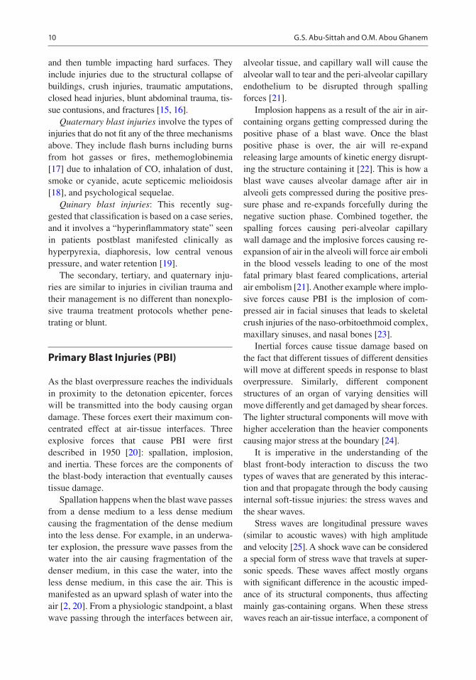
and then tumble impacting hard surfaces. They include injuries due to the structural collapse of buildings, crush injuries, traumatic amputations, closed head injuries, blunt abdominal trauma, tis-sue contusions, and fractures [15, 16].
Quaternary blast injuries involve the types of injuries that do not fit any of the three mechanisms above. They include flash burns including burns from hot gasses or fires, methemoglobinemia [17] due to inhalation of CO, inhalation of dust, smoke or cyanide, acute septicemic melioidosis [18], and psychological sequelae.
Quinary blast injuries: This recently sug-gested that classification is based on a case series, and it involves a “hyperinflammatory state” seen in patients postblast manifested clinically as hyperpyrexia, diaphoresis, low central venous pressure, and water retention [19].
The secondary, tertiary, and quaternary inju-ries are similar to injuries in civilian trauma and their management is no different than nonexplo-sive trauma treatment protocols whether pene-trating or blunt.
Primary Blast Injuries (PBI)
As the blast overpressure reaches the individuals in proximity to the detonation epicenter, forces will be transmitted into the body causing organ damage. These forces exert their maximum con-centrated effect at air-tissue interfaces. Three explosive forces that cause PBI were first described in 1950 [20]: spallation, implosion, and inertia. These forces are the components of the blast-body interaction that eventually causes tissue damage.
Spallation happens when the blast wave passes from a dense medium to a less dense medium causing the fragmentation of the dense medium into the less dense. For example, in an underwa-ter explosion, the pressure wave passes from the water into the air causing fragmentation of the denser medium, in this case the water, into the less dense medium, in this case the air. This is manifested as an upward splash of water into the air [2, 20]. From a physiologic standpoint, a blast wave passing through the interfaces between air,
alveolar tissue, and capillary wall will cause the alveolar wall to tear and the peri-alveolar capillary endothelium to be disrupted through spalling forces [21].
Implosion happens as a result of the air in air- containing organs getting compressed during the positive phase of a blast wave. Once the blast positive phase is over, the air will re-expand releasing large amounts of kinetic energy disrupt-ing the structure containing it [22]. This is how a blast wave causes alveolar damage after air in alveoli gets compressed during the positive pres-sure phase and re-expands forcefully during the negative suction phase. Combined together, the spalling forces causing peri-alveolar capillary wall damage and the implosive forces causing re- expansion of air in the alveoli will force air emboli in the blood vessels leading to one of the most fatal primary blast feared complications, arterial air embolism [21]. Another example where implo-sive forces cause PBI is the implosion of com-pressed air in facial sinuses that leads to skeletal crush injuries of the naso- orbitoethmoid complex, maxillary sinuses, and nasal bones [23].
Inertial forces cause tissue damage based on the fact that different tissues of different densities will move at different speeds in response to blast overpressure. Similarly, different component structures of an organ of varying densities will move differently and get damaged by shear forces. The lighter structural components will move with higher acceleration than the heavier components causing major stress at the boundary [24].
It is imperative in the understanding of the blast front-body interaction to discuss the two types of waves that are generated by this interac-tion and that propagate through the body causing internal soft-tissue injuries: the stress waves and the shear waves.
Stress waves are longitudinal pressure waves (similar to acoustic waves) with high amplitude and velocity [25]. A shock wave can be considered a special form of stress wave that travels at super-sonic speeds. These waves affect mostly organs with significant difference in the acoustic imped-ance of its structural components, thus affecting mainly gas-containing organs. When these stress waves reach an air-tissue interface, a component of
G.S. Abu-Sittah and O.M. Abou Ghanem

11
the compressive stress wave is reflected back at the interface as a tension wave [7]. It is when these stress waves equal and exceed the tensile strength of the tissue interface that their work done on the organs becomes an irreversible work of damage [2]. A stress wave also compresses air in air-filled organs that re- expands forcefully causing damage to the walls through implosive forces. All this interprets how for example small bowel wall injury or alveolar septum injury happens in thoracic and abdominal wall PBI.
Shear waves are transverse waves with long duration and low velocity, traveling perpendicu-larly to the longitudinal stress waves and tangen-tially to body surfaces. They are generated from body wall displacement. Different solid organs with different densities move asynchronously with different inertias causing shearing of solid organs [25].
Biological Effects of Primary Blast Injuries
The true incidence of primary blast injuries is unknown despite the various reports of incidence published. This is because PBI tend to be com-monly overlooked especially in situations of mass casualty where the health care teams are faced with amputations, crush injuries, burns, toxic inhalations, and penetrating trauma. Delay in the diagnosis of PBI can complicate patient care especially in patients with isolated PBI who do not manifest external body trauma [26].
Primary blast injuries involve mainly gas- containing structures, namely the pulmonary, gastrointestinal, and auditory systems. The ear is the most commonly affected organ because for primary blast injury of the ear to occur, the blast overpressure threshold required is lower than that required for lungs and the bowels to be injured [27]. However, blast injuries are not exclusive to gas-containing structures. Other systems are affected as well though less common: the heart, vascular system, eye and orbit, and central ner-vous system among others.
Other than specific organ injuries, PBI have a systemic effect, a global physiologic response in
the form of a cardiogenic shock in the absence of hemorrhage uncompensated by vasoconstric-tion. It is mediated by pulmonary C-fiber recep-tors that are thought to initiate this vagal reflex. It usually occurs following thoracic PBI within seconds and lasts between minutes to hours but often resolves by 2 h. It is characterized by tran-sient bradycardia, bradypnea, and hypotension [28, 29].
Pulmonary SystemAs with all primary blast injuries, the lungs are more likely to get injured after a blast whereby the blast overpressure is high and the positive blast phase is prolonged. Uncomplicated blast lung injury has a favorable prognosis at 1-year follow-up. Hirshberg et al. reported that people who are discharged after surviving a lung blast injury had no pulmonary complaints, normal pul-monary function tests, and resolution of the chest radiography findings at 1-year follow-up [30]. Pulmonary PBI is essentially manifested as pul-monary contusions [5]. The spallation and implo-sion of the stress wave at the different air-alveolar-capillary wall interfaces cause alveo-lar wall, capillary wall, and interalveolar space disruption [2, 22]. This causes the pooling of blood perivascularly and alveolar hemorrhage. It can range all the way from petechiae to confluent hemorrhage [31]. Also, the extravascular fluid is compressed and driven into the alveolar space which causes pulmonary edema manifested as bilateral pulmonary infiltrates on chest radiogra-phy [32]. Implosive forces can also drive air into the interstitial spaces causing interstitial emphy-sema [26]. Shearing forces can disrupt the bron-chovascular tree and create bronchopulmonary fistulas. These tears in the air-tissue interface can lead to arterial air emboli (AAE) development either immediately after the blast causing rapid death or delayed with the initiation of positive pressure ventilation [33]. AAE when big enough can cause MI, stroke, spinal cord infarction, intestinal ischemia, and death [34]. Even when microscopic, AAE can still cause symptoms like confusion, mental status changes, vision distur-bances, pain, and weakness. Clinical signs like air in the retinal arteries, tongue blanching, or
2 Biodynamics of Blast Injuries

12
livedo reticularis can be indicators of emboli [35]. Pleural tears and lacerations can also be caused by pulmonary barotrauma as a blast effect or due to positive pressure ventilation and can give rise to pneumothoraces, hemothoraces, or pneumomediastinum [7].
Pulmonary contusions are usually bilateral in closed-space explosions but tend to be worse on the side of the impact of the blast wave in open- field explosions [5]. The ribs protect the lung parenchyma from the full force of the blast over-pressure. This results in stripes of hemorrhagic congestion corresponding to the intercostal spaces where there is no rib protection. These parallel bands of ecchymoses used to be called mistakenly “rib markings” as they were thought to be occurring under the ribs but were proven to occur along the intercostal spaces with the ribs providing protection to the underlying paren-chyma. Perimediastinal lung parenchyma espe-cially the azygos lobe and lung regions in the costophrenic angles are more severely involved by the blast injury. This unequal distribution is justified by the reflection and augmentation of the stress wave within the chest [7].
Specific ultrastructural manifestations have been reported in lung primary blast injury. On light microscopy, pulmonary capillaries are seen dilated [36]. On electron microscopy, increased pinocytosis, blebbing, and ballooning in pulmo-nary capillary endothelial and type I epithelial cells are seen in experiments done on rats exposed to a blast. Also, loss of structure or enlargement was noted in the lamellated bodies of type II epi-thelial cells [37]. These changes occurred not only in areas of the lung with apparent damage but also in apparently normal regions of lung parenchyma. So patients with no clinical or radiologic evidence of injury could still have sus-tained a lung blast.
At the macroscopic level, respiratory mucosa is very sensitive to blast effect. Damage occurs at overpressures below those that would cause parenchymal lung injury. Mucosal injury includes loss of cilia and flattening of epithelial cells. More severe injury can also occur with stripping of the mucosal epithelium off the basal lamina the so-called stripped epithelium lesion, with the
resultant intraluminal hemorrhage. This stripping of the epithelium is postulated to be due to the spalling forces at the epithelial tissue-air surface. These injuries generally resolve spontaneously and should be sought while examining a patient subjected to a blast. Their presence is an indica-tor of possible primary parenchymal lung blast injury and other organ blast injuries [38].
Clinically, the triad of dyspnea, cough, and hypoxia is referred to as “blast lung syndrome” and is due to ventilation mismatch, vascular shunt-ing, and impaired gas exchange [39]. Focal pulmo-nary edema and hemorrhage in the alveoli cause ventilation perfusion mismatch with increased intrapulmonary shunt, hypoxia, reduced lung compliance, and increased work of breathing [40]. Clinical symptoms include dyspnea, cough, hemoptysis, chest pain, or discomfort. Clinical signs include tachypnea, cyanosis, reduced breath sounds and dullness to percussion, coarse crepita-tions, rhonchi, subcutaneous emphysema, features of hemopneumothorax or pneumothorax, retroster-nal crunch, or retinal artery emboli.
Any blast-exposed patient is worth a chest radiograph. Bilateral pulmonary infiltrate is typi-cally seen on chest radiography in primary blast injuries [32]. Usually, these infiltrates develop within few hours, become maximal at 24–48 h, and tend to resolve within a week. Infiltrates that continue to worsen beyond 48 h may be indica-tive of pneumonia or adult respiratory distress syndrome [7]. Pneumothorax, hemopneumotho-rax, interstitial emphysema, subcutaneous emphysema, pneumomediastinum, or pneumo-peritoneum might be evident on chest radiogra-phy. Most blast injuries develop immediately, but sometimes, progressive vascular leak and inflam-matory changes develop over 12–24 even up to 48 h contributing to delayed presentation [31]. Hence, patients with pulmonary symptoms and negative chest radiographs should be observed for 8 h before discharge [14]. However, the majority of patients with blast lung injury will manifest radiologic or clinical findings on admis-sion [41]. If the symptoms are persistent or severe with a negative chest radiograph then a chest CT should be done [42]. A study showed that the ratio of PaO2 to FiO2, the presence or absence of
G.S. Abu-Sittah and O.M. Abou Ghanem

13
chest radiograph infiltrates, and bronchopleural fistulas can help identify the severity of the lung injury in terms of mortality or progression to adult respiratory distress syndrome (ARDS) and help determine the respiratory management [43].
Management of primary blast injuries of the lung can be quiet challenging. On one side, these patients are most often hemodynamically unsta-ble requiring volume resuscitation yet excessive fluid resuscitation can lead to or exacerbate pul-monary edema in patients suffering from contu-sions [32]. To optimize the patient’s respiratory status adequate pain management and noninva-sive ventilation techniques are used. Avoiding positive pressure ventilation (PPV) as much as possible could not be emphasized enough. Positive pressure ventilation especially with high positive end-expiratory pressure (PEEP) is thought to increase the risk of pulmonary baro-trauma, namely pneumothorax and arterial air emboli [9, 44]. Drainage of air, fluid, and blood through chest tube thoracostomy is very impor-tant in optimizing the pulmonary status of the patient and helps minimize the need for PPV. Prophylactic chest tube thoracostomy is recommended for patients who suffer from severe lung blast injury that will need positive pressure ventilation or need air transportation [45]. In blast lung injuries, lung compliance is poor and if positive pressure ventilation is to be needed then protective measures must be used like low PEEP, low O2 saturation, low tidal volumes, and permis-sive hypercapnia [43, 46]. Reversion to spontane-ous breathing by intermittent mechanical ventilation and continuous positive airway pres-sure should be done as soon as the patient’s pul-monary status allows it.
With arterial air embolism, if the patient is not intubated administration of oxygen should be ini-tiated promptly and if available hyperbaric oxy-gen is the definitive treatment [7]. If the patient is intubated, then the ventilator settings should be adjusted to low PEEP and 100% fiO2. Some rec-ommend putting the patient in left lateral decubi-tus position to decrease the risk of systemic embolization [35, 47]. Organ transplant teams should be aware of the fact that normally looking organs could be unusable due to AAE [48].
Other less conventional techniques including extracorporeal membrane oxygenation, indepen-dent lung ventilation, nitric oxide ventilation, and high-frequency jet ventilation have been used in a small number of patients with varying degrees of success [43].
Gastrointestinal SystemThe gastrointestinal system like the pulmonary sys-tem is at an increased risk of primary blast injury due to its gas-containing structures and similar blast overpressure to the lung [5]. The most com-mon site of the GI tract of both hemorrhage and perforation is the colon and ileocecal region where gas accumulates most in the tract and ruptures the wall due to implosive forces of the blast [49]. Solid abdominal organs injuries can be PBI but arise more commonly as secondary or tertiary blunt and penetrating blast injuries [50, 51]. Solid intra-abdominal organs like the spleen, liver, or kidneys are made of relatively similar liquid densities [52]. Therefore, solid organ injuries during a blast are less likely due to compression by the stress wave but rather due to body-wall displacements causing acceleration effects at organ attachments. Shear forces can therefore cause subcapsular petechiae, contusions, or even frank ruptures [25]. Bowel PBI are caused by the implosive and shearing forces rupturing the bowel walls. These forces cause the wall’s structural layer to separate resulting in intra-mural edema and hemorrhage with microthrombo-ses [49]. Hemorrhages can range in size from small petechiae to large confluent submucosal hemato-mas and can progress into more severe transmural hemorrhages [2]. This can compromise the perfu-sion putting the bowels at risk for delayed perfora-tion. At laparotomy it was found that small bowel contusions more than 15 mm in diameter and large bowel contusions of more than 20 mm were at sig-nificantly higher risk of perforation. Some consid-ered that finding such contusions intraoperatively warrants resection while a more conservative approach is reserved to smaller contusions [53]. Mesenteric, retroperitoneal, and scrotal hemor-rhages can also occur [2]. Shearing forces can dis-rupt the blood supply leading to intestinal ischemia. Arterial air embolism can be a cause of intestinal ischemia as well [35].
2 Biodynamics of Blast Injuries
14
Clinically, abdominal PBI can present with abdominal pain, hematemesis, nausea and vomit-ing, rectal pain, tenesmus, and testicular pain. Clinical signs include peritoneal signs, absent bowel sounds, and evidence of hypovolemia [7]. In some cases the diagnosis of intra-abdominal injury can be obvious, yet in most cases as with all PBI it is a diagnostic challenge. Focused abdominal sonography can be used to assess for free fluid in the abdomen when assessing abdom-inal complaints for abdominal life-threatening injuries [54]. But a negative FAST does not exclude an abdominal primary blast injury. If hemodynamically stable, an abdominal CT can be of help [55]. However, CT is specific for solid organ damage and perforation but it lacks sensi-tivity to rule out intestinal contusions and mesen-teric injury [56]. Doppler can also be used for investigation of abdominal perforating injury. Patients subjected to blast with abdominal com-plaints should be observed for 8 h and reexam-ined even if the mentioned imaging modalities are negative for findings [26]. Victims found to have an abdominal primary blast injury requiring operative intervention should be assessed for pri-mary lung blast injury as these patients will require intubation and positive pressure ventila-tion intraoperatively [4].
Patients with suspected abdominal injury that could not be stabilized and have unexplained signs of hemorrhage would need urgent laparot-omy. Pneumoperitoneum, diaphragmatic rupture, signs of peritoneal irritation on physical exam, and significant persistent GI bleed are all indica-tions for urgent laparotomy. Tension pneumo-peritoneum has also been reported. It is a complication of pulmonary barotrauma due to blast. It causes severe cardiovascular and respira-tory collapse with severe hypoxemia, hypercar-bia, and shock [57].
The EarAs discussed before, the auditory system is the most sensitive to blast overpressure injury [58]. The part of the tympanic membrane most fre-quently injured is the pars tensa [59]. Small tym-panic membrane ruptures can be managed conservatively as they usually heal spontane-
ously. Ruptures involving beyond 5% of the membrane’s surface will more likely require sur-gical intervention [60]. Isolated eardrum perfora-tion has been shown to be a poor marker of latent pulmonary or gastrointestinal PBI and explosion survivors with eardrum perforations and no signs of PBIs can be discharged after monitoring and normal chest radiography [61]. Because the tym-panic membrane ruptures at BOP lower than that required to cause lung or intestinal injury, it has been postulated that patients with intact tympanic membrane are probably exposed to little BOP and will not need further assessment. However, studies have shown that victims with and without tympanic membrane rupture were found to have lung blast injury [62, 63].
A temporary shift in the threshold for audible noises can cause a transient tinnitus or sensori-neural deafness that resolves over several hours to days [64]. This is due to stunning of the recep-tor organs of the inner ear. The severity of these symptoms decreases as the distance from the detonation epicenter increases [65]. However, injury to the inner ear can sometimes cause per-manent hearing loss in case of severe structural damage to the organ of Corti causing permanent threshold shifts [66]. This sensorineural hearing loss is different which is usually of high fre-quency and is a different entity than the usual 4-kHz noise-induced and reported trauma- induced hearing loss [67].
Ossicular injuries including incudomalleolar and incudostapedial joint disruption, fractures of the stapes superstructure, dislocation of the stapes footplate, and dislocation of the incus are also features of middle-ear primary blast injuries [68, 69]. Cholesteatoma of the middle ear and mastoid air-cell system is a late compli-cation in blast-induced tympanic membrane perforations. The incidence of cholesteatoma is related to the grade of perforation. For exam-ple, a grade 1 perforation (<2 mm in diameter) has a 2% incidence of cholesteatoma whereas a grade 4 perforation (subtotal) has a 20% inci-dence [70]. Vertigo postblast can be due to benign paroxysmal positional vertigo, peri-lymph fistulas, or more commonly associated head injuries [4, 7].
G.S. Abu-Sittah and O.M. Abou Ghanem