gland cell responses to feeding in drosera capensis, a
TRANSCRIPT

ORIGINAL ARTICLE
Gland cell responses to feeding in Drosera capensis,a carnivorous plant
Irene Lichtscheidl1 & Sue Lancelle2& Marieluise Weidinger1 & Wolfram Adlassnig1
& Marianne Koller-Peroutka1 &
Sonja Bauer1 & Stefanie Krammer1 & Peter K. Hepler2
Received: 4 March 2021 /Accepted: 11 May 2021# The Author(s) 2021
AbstractGlands of Drosera absorb and transport nutrients from captured prey, but the mechanism and dynamics remain unclear. In thisstudy, we offered animal proteins in the form of fluorescent albumin (FITC-BSA) and observed the reactions of the glands by livecell imaging and fluorescence microscopy. The ultrastructure of these highly dynamic processes was also assessed in high-pressure frozen and freeze substituted (HPF-FS) cells. HPF-FS yielded excellent preservation of the cytoplasm of all cell types,although the cytosol looked different in gland cells as compared to endodermoid and stalk cells. Especially prominent were theER and its contacts with the plasma membrane, plasmodesmata, and other organelles as well as continuities between organelles.Also distinct were actin microfilaments in association with ER and organelles. Application of FITC-BSA to glands caused theformation of fluorescent endosomes that pinched off the plasma membrane. Endosomes fused to larger aggregates, and accu-mulated in the bulk cytoplasm around the nucleus. They did not fuse with the cell sap vacuole but remained for at least three days;in addition, fluorescent vesicles also proceeded through endodermoid and transfer cells to the epidermal and parenchymal cells ofthe tentacle stalk.
Keywords Gland cell .Drosera capensis . Carnivorous
Introduction
Drosera is one of the largest and most diverse genera ofcarnivorous plants (CPs). The structure and physiology ofthese plants have been of great interest ever since Darwin(1875) described their carnivorous nature, not only because
of the exceptional way they acquire nutrition, but also becauseof their economic importance in pharmacy due to their anti-microbial metabolites and their sticky, trapping glue(Grevenstuk et al. 2009; Sprague-Piercy et al. 2020). Plantsare characterized by leaves bearing protuberances on the mar-gin and upper surface of their blades; they consist of a slender
In memory of Ursula Lütz-Meindl
Handling Editor: Andreas Holzinger
* Irene [email protected]
Marieluise [email protected]
Wolfram [email protected]
Marianne [email protected]
Sonja [email protected]
Stefanie [email protected]
Peter K. [email protected]
1 Cell Imaging and Ultrastructure Research, University of Vienna,Althanstrasse 14, A-1090 Vienna, Austria
2 Biology Department, University of Massachusetts Amherst, 221Morrill Science Center III; 611 North Pleasant Street,Amherst, MA 01003-9297, USA
https://doi.org/10.1007/s00709-021-01667-5
/ Published online: 21 June 2021
Protoplasma (2021) 258:1291–1306

stalk, or tentacle, that bears a multicellular glandular head. Inaddition, several types of small glandular trichomes occur onthe leaf blade and the tentacle stalk (Naidoo and Heneidak2013). The glands of the tentacles secrete a sticky mucilagethat lures insects and holds them tight. The tentacles furtherenwrap the prey, which is degraded by secreted digestive en-zymes. The tentacles then absorb the digested nutrients, asthey actively move over the prey. This remarkable multiplefunction of the Drosera tentacles has made them an importantobject of investigations about general questions of morpholo-gy, physiology, ecology, and evolution of carnivorous plants.Despite a wealth of information, which has been summarized(Lloyd 1942; Juniper et al. 1989; Ellison and Adamec 2018),many vexing questions about absorption and transport of preysubstances remain unsolved.
Although questions have been raised concerning the bene-fit of prey consumption (Daubenmire 1974), even early stud-ies suggested that Drosera profits from carnivorous nutritionby the formation of additional flowers (Francis Darwin 1878),and more recently that those plants have a faster rate of growth(Chandler and Anderson 1976a, b). Studies have furthershown that minerals are absorbed from digested prey(Adamec 1997; Shibata and Komiya 1972, 1973) and thatthe plant nutrient status and photosynthetic performance wereincreased (Pavlovic et al. 2014; Pavlovic and Saganova 2015).
Concerning secretion and absorption in Drosera glandcells, the Golgi apparatus has been shown to play a majorrole in the production and secretion of the trapping muci-lage (Schnepf 1961, 1969; Outenreath and Dauwalder1986). The production and secretion of lytic enzymes as-sociated with feeding have also received considerable at-tention (see Dexheimer 1976, 1978; Outenreath andDauwalder 1982, 1986; McNally et al. 1988; Muravnik2000; Plachno et al. 2006). Further studies on the absorp-tion of fluorescently labeled proteins showed that nutri-ents can enter the cells by endocytosis, in addition totransport through the plasma membrane (Adlassnig et al.2012). Membrane recycling to pre-vacuolar compartmentsand vacuoles occurs for arabinogalactan proteins as de-tected by immunolocalization (Samaj et al. 2000).
For further transport of nutrients from glands to leaves,different routes through the tentacle stalk have been sug-gested as being either through the central xylem strand orthrough the inner or the epidermal stalk cells (Juniperet al. 1989); however, definitive experiments are stillmissing. Nevertheless, the conclusion that stalk cells ab-sorb nutrients is strongly supported by the observation of“aggregation,” a remarkable reaction first described byDarwin (1875), which includes swelling of the cytoplasmand acceleration of organelle movement.
The ultrastructure study of gland cells has yielded impor-tant information about Golgi bodies and the production ofmucilage (Schnepf 1961; Ragetli et al. 1972; Dexheimer
1972, 1976, 1978; Gilchrist and Juniper 1974; Juniper andGilchrist 1976; Outenreath and Dauwalder 1982, 1986;Muravnik 2000); however, they fall short in representingfaithfully the instantaneous situation of organelle interactionsand membrane organization during various physiological ac-tivities. Especially the cells of the tentacle stalk, which arecovered by a thick cuticle, have been extremely difficult topreserve due to long fixation times; fixation can take morethan ten minutes (our own observations) and lead to structuralreorganizations and reorientation of the cytoplasm and its or-ganelles, a phenomenon that becomes even more important indeeper layers of the tissue.
In this study, we investigated the ultrastructure ofDrosera capensis glands during absorption and transportof proteins, a highly dynamic process as seen in studiesusing fluorescently labeled proteins (Adlassnig et al.2012), and applied freeze fixation techniques for faithfulreflection of the in vivo state in unstimulated glands andin cells after feeding. We subjected Drosera tentacles tohigh-pressure freezing (HPF, Moor 1987) followed byfreeze substitution, a technique that had been shown topreserve delicate details in Drosera tentacle stalks, includ-ing notably cytoplasmic microtubules (MTs), actin micro-filaments (MFs), and closely associated elements of endo-plasmic reticulum (ER) (Lichtscheidl et al. 1990). We alsoanalyzed the ultrastructure of the various glandular celltypes during absorption and further processing of pro-teins, and relate the results to parallel observations of cellsin the living state by fluorescence microscopy. We admin-istered fluorescent bovine albumin (BSA) to Droseraleaves to mimic the chemical situation of the prey that isrequired for enzyme discharge and causes further secre-tion of digestive enzymes (Dexheimer 1978). We visual-ized this marker within the cells, and observed the fateand dynamics of the plasma membrane with the fluores-cent membrane marker FM4-64.
Material and methods
Plant material
We investigated young but fully developed leaves of Droseracapensis that had been grown in the greenhouses of theUniversities of Massachusetts and of Vienna without fertilizerin a mixture of sand and sphagnum. The long slender leaves beartentacles the heads of which were each surrounded by a dropletof mucilage (Fig. 1a). According to the classification ofOutenreath and Dauwalder (1982), they were in their intermedi-ate to mature state. For our investigations, we used only healthyleaves that had had no contact with insects; we used them assuch, or fed them for various times with BSA (5–10% in water)as a model for animal prey.
1292 I. Lichtscheidl et al.

Light microscopy
Leaves were studied macroscopically with a stereomicroscope(Nikon) equipped with a Nikon digital camera. This allowedus to feed individual tentacles with a glass capillary mountedon a micromanipulator. Dynamic properties of the glands andstalk cells were observed in a Leica DMIRE 2 and a NikonEclipse microscope both equipped with differential interfer-ence contrast (DIC) and epifluorescence. Chloroplasts weredetected by their auto-fluorescence. The fluorescence of ERwas observed by staining tentacles with 20 μM DiOC6 (3,3′-dihexyloxacarbocyanine iodide; Thermo Fisher) dissolved inwater or in BSA. For fluorescence studies, in addition, weused a Leica DM 6000CS confocal laser scanning micro-scope. The tonal range adjustment of the micrographs andarrangement of plates was carried out in Adobe PhotoshopCS6.
Experiments on endocytosis
Uptake of proteins from animal prey was simulated by thesoluble protein BSA. For fluorescence studies, 10 μl of 2%BSA coupled to FITC (FITC-BSA, Sigma) was deposited onthe leaves for 5 min and up to 72 h, as described by Adlassniget al. (2012). Thereafter, leaves were dissected and washedbriefly in 2% BSA (Sigma) before observation. The conjugatemade visible the passage of the protein into the gland andfurther into the tentacle. Alternatively, for localization of up-take in individual glands, FITC-BSA was loaded into a mi-cropipette and placed over a single head with the aid of amicromanipulator (Fig. 1b).
As an alternative indicator for endocytosis, we marked theplasma membrane of the glandular cells with the styryl dyeFM 4 - 6 4 ( N - ( 3 - t r i e t h y l a mm o n i u m p r o p y l ) -4-(8-(4-(diethylamino)phenyl) (hexatrienyl) pyridinium-
dibromide; Molecular Probes) at a final concentration of4 μM either in water or in BSA in concentrations between 5and 10%. With this probe, we could track the formation ofendosomes (Jelinkova et al. 2019).
Transmission electron microscopy by high-pressurefreezing and freeze substitution
The central portion of the middle leaf zone bearing short ten-tacles at the surface was dissected and sandwiched as quicklyas possible (20 s) in a mold between two lecithin-coated goldsample holders (Balzers BB1131242-1). For mechanical pro-tection as well as for maximum heat conductivity, we filledthe remaining space within the sample holder with a droplet of1% ultralow gelling (< 15 °C) agarose (Type IX, SigmaChemical Co.). Freezing of the tissue was accomplished withthe Balzers HPM 010 high-pressure freezer.
Freeze substitution followed the procedure described byLancelle et al. (1986, 1987) and employed by Lichtscheidlet al. (1990). It thus resembled the method applied by Kisset al. (1990) and Staehelin et al. (1990). Briefly,cryoimmobilized material was transferred to acetone contain-ing 2% osmium tetroxide at approximately − 80 °C and freezesubstituted for 36–40 h, then brought to room temperaturegradually over a period of 5–6 h. Before embedding in amixture of Epon and Araldite, the tissue was transferred tomethanol and stained in 5% uranyl acetate in methanol for 2h, then brought back to acetone.
After staining with lead citrate, ultra-thin sections wereobserved in a Jeol 100 CX or a Zeiss 902 electron microscopeoperated at 80 kV.
Results
General morphology of the glandular heads
Tentacles cover the upper surface and margins of the leaves.Their thin cylindrical stalk is fused into a glandular head by aconnecting zone called the neck area, a ring of epidermal andparenchymal transfer cells. Glandular cells in the head pro-duce trapping mucilage, a viscous solution of polysaccharidesthat can be drawn out into threads several centimeters long(Rost and Schauer 1977). The length of the tentacles variesand depends on their position on the leaf, with the marginaltentacles being up to ten times longer than the central ones(Fig. 1a). The cellular composition of the cylindrical glandularheads, however, is relatively constant, with the exception ofsome outermost tentacles carrying asymmetrical heads.Figure 2 shows two layers of glandular cells, the outer andinner gland cells; they cover the surface and are separatedfrom a core of spongy tracheids with spiral-shaped thicken-ings by a bell-shaped layer of endodermoid cells. Their radial
Fig. 1 Leaf of Drosera capensis. Upper leaf surface and margin arecovered with tentacles of different lengths that produce trappingmucilage (a). Nutrients and fluorescent indicator dyes are supplied toindividual glands with the aid of a micropipette (b)
1293Gland cell responses to feeding in Drosera capensis, a carnivorous plant

walls are impregnated with a waxy substance, presumablycutin or suberin, which inhibits apoplastic passage of smallmolecules between gland and stalk cells. The whole gland andalso the stalk are covered by a cuticle. Pores with osmiophilliccontent are found only around secretory cells; these allow forpassage of liquids (Fig. 3c).
Gland heads are connected to the tentacle stalks by a layerof cells described as transfer cells according to the definitionof Gunning and Pate (1974), which were named as epidermalneck cells (ENC) and as parenchymal neck cells (PNC). Theycontain numerous plasmodesmata in their cell walls. Contactbetween tracheids of the gland head and the vascular supply ofthe leaf is given by a row of slender spiral tracheids that runsthrough the stalk. This xylem is surrounded by two rows ofcells, an inner course of parenchymal and an external layer ofepidermal cells. The different cell types are shown in Fig. 2.
Ultrastructure of glandular cells before and afterfeeding with BSA
The periphery of the gland head is composed of two layers ofclosely fitting secretory cells. In this study, we only brieflysummarize the composition of the cytoplasm and draw ourfocus on the endomembrane compartments involved in ab-sorption, digestion, and transport of nutrients.
Fig. 2 Overview of Drosera tentacles in TEM: the gland head is formedby an outer and inner layer of secretory cells (outer gland cells, OGC;inner gland cells, IGC). They are supported by a bell-shaped layer ofendodermoid cells (EC) that curve out to the surface at the base of thehead. They surround the core of tracheids (Tr) and connect to the stalk bya ring of epidermal and parenchymal neck cells (ENC; PNC). These neckcells are continuous with the cells of the tentacle stalk, the epidermal andparenchymal stalk cells (ESC; PSV). Transfer of substances from theglandular cells to the stalk is provided by plasmodesmata betweenglandular cells, endodermis, neck cells, and tentacle stalk
Fig. 3 Outer (a) and inner (b) glandular cell. Cells contain a large vacuolesurrounded by a layer of cytoplasm. The main part of the cytoplasm,including the nucleus and the organelles, is concentrated along theinterior surfaces of the cell. The adjacent lateral walls as well as theperipheral tangential wall possess only a thin layer of cytoplasm withfew organelles, mainly ER and vesicles. Leucoplasts with dense matrixand mitochondria are frequently found closely appressed to the nuclearenvelope and in close contact with each other. Pores in the cuticle arefilled with fluffy osmiophilic material (c)
1294 I. Lichtscheidl et al.

Outer and inner gland cells contain large eccentric vacu-oles, which are traversed by strands of cytoplasm (Fig. 3a, b).Red pigment gives rise to osmiophilic content, presumablyflavonoids and anthocyanins that are concentrated inelectron-dense particles. Vacuoles are surrounded by cyto-plasm that forms a thin layer underneath the outer peripheralcell walls where it contains mainly elements of ER and smallvesicles. The main part of the cytoplasm occupies the interiorpart of the cells; here, the nucleus and most of the organellesare located. External and radial cell walls increase their surfaceby cellulose buttresses that form septa and in addition increasetheir surface with a fingerlike labyrinth of wall ingrowths, thusresembling the cell walls of transfer cells. They appear to besites where vesicles make contact with the plasma membrane(PM, Fig. 4a, b) and endoplasmic reticulum (ER, Fig. 4d, e).Fluorescence labeling of ER with DiOC6 shows the three-dimensional arrangement of tubular ER in the outer peripheryof the cytoplasm (Fig. 4f). Staining the plasmamembranewithFM4-64 exhibits increased intensity where the cell wall pro-trudes into the cytoplasm and vesicles accumulate (Fig. 4c).
The nucleus contains a large nucleolus and condensed chromatinconcentrating inside the nuclear envelop. Leucoplasts with densematrix andmitochondria are frequently found closely appressed tothe nuclear envelope and in close contact with each other (Fig. 3).
Regarding elements of the cytoskeleton, we occasionally findstraight or curved MTs mainly in the periphery of the cell (Fig.4g), but also extending into the cytoplasm. By contrast, we havenot observed actin MFs in either the outer or the inner gland cell.In agreement with this observation are those with the light mi-croscope, showing that far-reaching organellemovements are notobserved in these gland cells, only saltatory movement.
The ultrastructure of the endomembrane compartments such asER, Golgi bodies, and microbodies depends on the physiologicalstate of the tentacles. In unfed young tentacles, the most notewor-thy feature of apical outer gland cells is well-developed Golgibodies, which are responsible for slime production. They mayconsist of only a few lamellae, or may be multi-stacked, withstraight or oblique arrangement of the lamellae (Fig. 5a–d).Golgi lamellae of the maturing face form dark-stained sphericalor elongated slime vesicles of variable diameter with mostly
Fig. 4 Outer glandular cell.Protuberances of the cell wallhave different sizes and offercontact points for vesicles andER. Contact between the plasmamembrane and vesicles is seen inTEM (a, b) and after staining ofthe membrane with thefluorescent dye FM4-64 (c);arrows. Single ER cisternae aredecorated with ribosomes andattached to the fingerlikeinvaginations of peripheral andlateral cell walls (d, e). Stainingwith DiOC6 shows the net of ERtubules in the peripheralcytoplasm of the outer tangentialcell wall of gland cells (f).Occasional microtubules in theperipheral cytoplasm contactorganelles, in this case amitochondrion M (g); arrowhead
1295Gland cell responses to feeding in Drosera capensis, a carnivorous plant
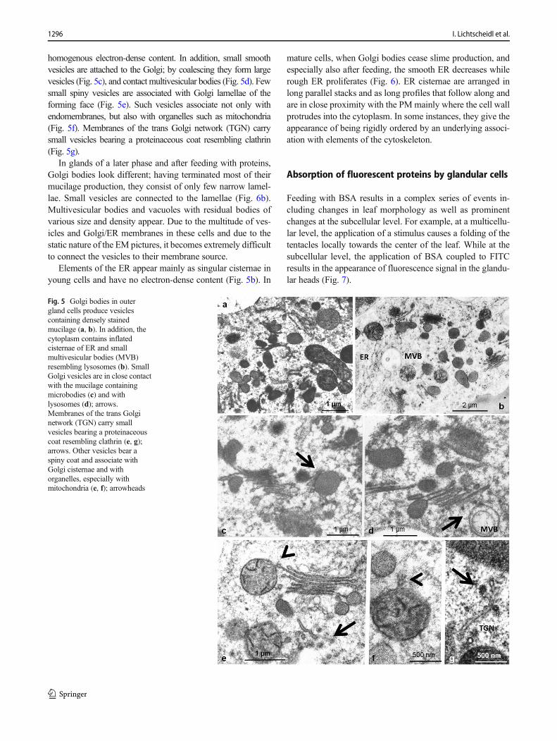
homogenous electron-dense content. In addition, small smoothvesicles are attached to the Golgi; by coalescing they form largevesicles (Fig. 5c), and contact multivesicular bodies (Fig. 5d). Fewsmall spiny vesicles are associated with Golgi lamellae of theforming face (Fig. 5e). Such vesicles associate not only withendomembranes, but also with organelles such as mitochondria(Fig. 5f). Membranes of the trans Golgi network (TGN) carrysmall vesicles bearing a proteinaceous coat resembling clathrin(Fig. 5g).
In glands of a later phase and after feeding with proteins,Golgi bodies look different; having terminated most of theirmucilage production, they consist of only few narrow lamel-lae. Small vesicles are connected to the lamellae (Fig. 6b).Multivesicular bodies and vacuoles with residual bodies ofvarious size and density appear. Due to the multitude of ves-icles and Golgi/ER membranes in these cells and due to thestatic nature of the EM pictures, it becomes extremely difficultto connect the vesicles to their membrane source.
Elements of the ER appear mainly as singular cisternae inyoung cells and have no electron-dense content (Fig. 5b). In
mature cells, when Golgi bodies cease slime production, andespecially also after feeding, the smooth ER decreases whilerough ER proliferates (Fig. 6). ER cisternae are arranged inlong parallel stacks and as long profiles that follow along andare in close proximity with the PMmainly where the cell wallprotrudes into the cytoplasm. In some instances, they give theappearance of being rigidly ordered by an underlying associ-ation with elements of the cytoskeleton.
Absorption of fluorescent proteins by glandular cells
Feeding with BSA results in a complex series of events in-cluding changes in leaf morphology as well as prominentchanges at the subcellular level. For example, at a multicellu-lar level, the application of a stimulus causes a folding of thetentacles locally towards the center of the leaf. While at thesubcellular level, the application of BSA coupled to FITCresults in the appearance of fluorescence signal in the glandu-lar heads (Fig. 7).
Fig. 5 Golgi bodies in outergland cells produce vesiclescontaining densely stainedmucilage (a, b). In addition, thecytoplasm contains inflatedcisternae of ER and smallmultivesicular bodies (MVB)resembling lysosomes (b). SmallGolgi vesicles are in close contactwith the mucilage containingmicrobodies (c) and withlysosomes (d); arrows.Membranes of the trans Golginetwork (TGN) carry smallvesicles bearing a proteinaceouscoat resembling clathrin (e, g);arrows. Other vesicles bear aspiny coat and associate withGolgi cisternae and withorganelles, especially withmitochondria (e, f); arrowheads
1296 I. Lichtscheidl et al.

The use of the fluorescent dye FM4-64 provides a cleardemarcation of the plasma membrane and permits the analysisof endocytotic trafficking. The plasma membrane becomespunctuated with small dots at the limit of resolution (Fig. 8a,b), initially in the thin surface layer of the cytoplasm at theperiphery of the outer gland cells and at anticlinal walls
between outer gland cells (10–20 min incubation). Here, cellshave the largest absorptive surface due to numerous invagina-tions of the cell wall. TEM images reveal these endosomes atthe cell wall as well as their fusion products, chains of tubule-vesicular compartments (Fig. 8c–e). Time lapse showed theappearance of vesicular and tubular microbodies and a time-dependent increase of labeling inside enlarging clusters in thebulk cytoplasm as well as vesicles transformed intomultivesicular bodies (Figs. 8f, 9, and 10). In a similar way,FITC-BSA becomes visible within small fluorescent vesicles,which gradually increase in number and size, and give rise to anew type of globular or elongated pleiomorphic vacuoles.These are independent of the original cell sap vacuoles andwith time become heavily stained. In the EM, we see theseendosomes as multivesicular bodies from variable size andstructure with more or less residual content. Their content ofsmall vesicles increases with time.
This uptake of FM4-64-labeled plasma membrane occursin control conditions suggesting spontaneous endocytosis, butappears to be amplified in cells treated with BSA.
Further export of fluorescent BSA to the leaf was analyzedby pulse-labeling; we offered FITC-BSA to the leaves for 3–6h, and then carefully washed it away. After 24 h, there was still
Fig. 6 Gland cells after feedingwith proteins. Cisternae of roughER proliferate (a). Gradually thenumber of Golgi cisternaereduces and large Golgi vesiclesdisappear. Golgi stacks insteadare in close contact with smallvesicles which partly bear a coatof spiny proteins (b)
Fig. 7 Leaf of Drosera capensis after feeding with FITC-BSA: thetentacles fold towards the center of the leaf. The glandular heads arebrightly fluorescing in green because they absorbed the fluorescentprotein. Chloroplasts in the leaf give red auto-fluorescence
1297Gland cell responses to feeding in Drosera capensis, a carnivorous plant

Fig. 8 Staining of gland cellswith FM4-64 shows membraneflow in the fluorescencemicroscope. a: Initially, theplasma membrane is decoratedwith fluorescence dots at the limitof resolution. b: larger vesicles ofglobular or tubular shape form.Spontaneous endocytosis occursin untreated cells, but we observeit in higher frequency afterfeeding with BSA. c, d, e, f: EMsections show these vesiclesclosely attached to the plasmamembrane and also deeper withinthe cytoplasm
Fig. 9 Outer gland cells indifferent levels of the cytoplasmduring feeding with BSA: a, d, gperipheral cytoplasm in thesurface of the gland; b, c, e, f, h, icentral and inner side of the outergland cells. TEM pictures showthe tubulo-vesicular net ofendosomes that form in theperiphery; by fusing, they giverise to large vacuolarcompartments (a, b, c). Stainingof the plasma membrane withFM4-64 shows the fluorescentmembranes of endosomes andtheir final destination intomultivesicular bodies (d, e, f). Byfeeding with FITC-BSA, theabsorbed proteins become visiblewithin these endosomes (g, h, i)
1298 I. Lichtscheidl et al.

intensive fluorescence of vesicular compartments mainly inthe bulk cytoplasm similar to that seen also immediately afterstaining. In addition, some vesicles occurred in the cytoplasmof the neck cells and the tentacle stalk.
Transport of proteins to the tentacle stalk
For exploitation of absorbed proteins by the leaves, nutrientsmust be transported from outer gland cells to inner gland cells,and further to the endodermis. From there, these substances needto pass through epidermal and/or parenchymal neck cells to thetentacle stalk, and alternatively also transport through the centraltracheid of the stalk was suggested (Figs. 2 and 11a). We ana-lyzed the ultrastructure of the relevant cell types by EM andfollowed the path of FITC-BSA by fluorescence microscopy.
Endodermal cells
A layer of endodermal cells forms the “parenchymal bell” sepa-rating the inner gland cells from the tracheary elements in thecenter of the gland head. Rather short in the apical zone of thegland, lateral endodermal cells are elongated and flat, and theycurve out to the surface at the base (Fig. 2; Suppl. Fig. 2a, b).
Their radial cell walls are suberized similar to endodermal cellsof roots, which contain a Casparian strip. The cells comprise alarge central vacuole and a thin layer of cytoplasmwith only feworganelles including Golgi bodies and mitochondria. Very fewplastids occur which, depending on the individual plants whoseglands were sectioned, resembled either leucoplasts or chloro-plasts. If no protein digestion takes place, the cytoplasm containsonly few additional small vacuoles.
In these innermost living cells, during freeze fixation, icecrystal formation occurs more often than in the gland cells, butstill we find sufficient amounts of well-frozen cytoplasm. Yet,the structure of the cytoplasm differs from that of the glandcells; it looks more loose and extracted. Bundles of MFs areclosely coaligned with ER and oriented parallel to the longi-tudinal axis of the cell (Suppl. Fig. 2c, d). They are found deepwithin the cytoplasm, but approach the plasma membrane andcell wall as well. Accordingly, in video light microscopy, fastlong-distance movements of organelles are observed.
The transfer cells of the neck region
The neck area completes the gland head and fuses it to thetentacle stalk. It consists of a peripheral cell layer of eight to
Fig. 10 Multivesicular bodies(MVB) originate fromendosomes. They fill a largeportion of the cytoplasm of glandcells. In TEM pictures, thedegradation of their contentbecomes visible: vesicles are stillpresent (a), but graduallydisappear (b). They feed on thetubulo-vesicular endosomes (g)and engulf also other organellessuch as mitochondria (f).Absorption of FITC-BSA showsthe content of endosomes ofvarious sizes and MVBs withdegrading substance (b, c).Comparison of such lateendosomes in bright-fieldsuggests that these compartmentsare different from the original cellsap vacuole (V) (b, d). Stainingwith FM4-64 shows theirmembranes in the fluorescencemicroscope (h)
1299Gland cell responses to feeding in Drosera capensis, a carnivorous plant

ten cells forming a circle that make contact with the above out-curving endodermal cell and to the underneath positioned epi-dermal stalk cells (“epidermal neck cells”). A second layerwithin is a ring of parenchymal cells that connects to the abovetracheids and endodermis of the glandular heads and impingesat its base on parenchymal cells of the tentacle stalk surround-ing the central tracheid (Fig. 2 and 11b). Large numbers ofplasmodesmata suggest symplastic connections between en-dodermis of the cells of the stalk (Fig. 11e–g). Both cell typeshave large central vacuoles surrounded by a thin layer of cy-toplasm containing an often tapered nucleus and chloroplastsin addition to the usual organelles. Dark osmiophilic precipi-tates in the vacuoles represent most probably the red anthocy-anin known in these cells from light microscopy. In addition,extensively stained homogenous round bodies cluster in thevacuole of parenchymal neck cells (Suppl. Fig. 3d, e). Theyhave strong auto-fluorescence with a maximum emission be-tween 540 and 570 nm, and are in constant Brownian motion(Suppl. Fig. 3a, b). They continue also in the neighboringparenchymal cells of the stalk in its upper region and areindependent of protein supply in both unstimulated andBSA-treated cells.
The preservation of the cytoplasm in both cell types ap-pears to be quite good (Suppl. Figs. 4; 5): the nucleoplasmhas a fine, granular texture with homogenous nucleoli and asmooth nuclear envelope. The somewhat extracted appear-ance of the cytosol is similar to that of endodermal cells.Organelles of both epidermal and parenchymal neck cells
often make close contact, and this is also the situation in theadjacent epidermal and parenchymal cells of the tentacle stalk:mitochondria and chloroplasts tightly appress to nucleus andmicrobodies; mitochondria are attached to chloroplasts andeven penetrate them (Fig. 13g; Suppl. Fig. 5a, b, c). In additionto intruding organelles, inclusions of cytosol and extensionsresembling stromules are observed in the pleiomorphic chlo-roplasts (Fig. 13h, i; Suppl. Fig. 5c)
Epidermal and parenchymal cells of the stalk
Similar to the neck cells, a ring of parenchymal cells en-sheathes the central spiral tracheid in the stalk. It is surroundedby a second ring of cells forming the epidermis (Fig. 12). Theepidermis carries occasional glandular trichomes consisting of2 to 6 glandular cells (not shown).
Between endodermis and neck cells as well as in the trans-verse walls between the stalk cells, plasmodesmata group intopit fields (Fig. 11e–g). In the adjacent cytoplasm, vesicles ofdifferent size and complexity accumulate in clusters (Fig.11e). Feeding leaves with proteins causes re-organization ofthe whole cytoplasm, a complex reaction described much ear-lier as “aggregation” (Darwin 1875). The cytoplasm starts toswell immediately after feeding, the velocity of organellesincreases, and the cell sap vacuole segregates into many smallparts that move through the cell (Fig. 11d). While vacuolesand most organelles accelerate their movement and are inconstant cyclosis, chloroplasts remain stably anchored to the
Fig. 11 Distribution of FITC-BSA from gland head throughneck cells to stalk cells. FITC-BSA occurs in endosomes ofglandular cells (a) and further inepidermal and parenchymal cellsof the stalk (c). In epidermal andparenchymal neck cells, it seemsto accumulate within large roundbodies (a). After feeding withBSA, the colorless cytoplasm ofthe stalk cells is swollen, whereasred vacuoles decreased in size (d;differential interference contrast).In BSA-treated stalk cells, thecytoplasm contains a largeamount of small vesicles (e). Agrazing section through neck andstalk in TEM shows theorganization of the stalk cells (b).Plasmodesmata in transverse cellwalls anchor ER (f, g). EC,endodermoid cell; ENC,epidermal neck cell; PNC,parenchymal neck cell; ESC,epidermal stalk cell; PSC,parenchymal stalk cell
1300 I. Lichtscheidl et al.

cell wall, mainly along the inner longitudinal wall of epider-mal and parenchymal stalk cells.
Some vacuoles contain spherical inclusions similar to thosefound in the parenchymal stalk cells. Also, some vacuolesexhibit strong auto-fluorescence with similar spectral charac-teristics as those spherical inclusions found in the neck cells,and which also occur in the vacuoles of a few stalk cells(Suppl. Fig. 3c).
Rhabdoids, elongated protein bodies, are present in theepidermal cells of untreated leaves and occur in BSA-treatedcells as well. They are in close contact with Golgi vesicles.From these static pictures, we cannot say if proteins are addedto or consumed from the protein bodies (Fig. 13a, b).
Progress of FITC-BSA from glandular heads to stalk
Feeding leaves with FITC-BSA leads to the formation of fluo-rescent endosomes in the endodermis after 1 to 2 h; after 24 h,these endosomes are observed in the parenchymal and epider-mal neck and further in the living cells of the tentacle stalk,mainly in the epidermis. Large amounts of FITC-BSA causefluorescence of the cytosol of living stalk cells in addition tofluorescent organelles. The central tracheids of the gland headand stalk occasionally showed fluorescence in young not fullydeveloped leaves.
Progress of vacuolar staining by DiOC6 from glandular headsto stalk
In an attempt to stain ER and mitochondria, we appliedDiOC6, a fluorescent indicator for membrane potential. It suc-cessfully allowed observation of ER and mitochondria in out-er and inner gland cells (Fig. 4f), but in addition, it gradually
stained the cell sap of the vacuoles, clearly showing the closecontacts of outer and inner gland cells with the endodermoidcells (Suppl. Fig. 6a). This dual staining also extended into thetentacle stalk where it mainly occurred in epidermal stalk cells(Suppl. Fig. 6b). Some notable characteristics included a sta-ble cortical net of ER that stained strongly; fast-moving endo-plasmic ER tubules, which gave a diffuse fluorescence to thecytoplasm, where mitochondria shone brightly (not shown);and the vacuoles, which were heavily labeled. In control cells,the vacuoles were uniform and filled the cells (Suppl. Fig. 6c),whereas in BSA-treated cells, they were disintegrated intotubes and vesicles, which moved quickly through the swollencytoplasm (Suppl. Fig. 6d).
Discussion
In this study, we provide new information about the dynamicproperties of gland cells of carnivorous plants when stimulat-ed by exogeneously applied protein. Using both live cell andfixed preparations, we followed the absorption of proteins intogland cells of Drosera capensis, as well as the ensuing prog-ress of nutrients from gland cells to tentacle stalk, includingthe “aggregation” of cytoplasm in stalk cells during feeding.In this phase, we used fluorescence staining as a means toprovide information about the underlying physiological pro-cesses. In the studies of fixed cells, we took advantage of thehigh fidelity and extent of preservation in cells prepared byhigh-pressure freeze fixation and freeze substitution, and byexamination at high resolution in the transmission electronmicroscope. By relating the observations from both tech-niques, a new look at dynamic events and structural propertieshas become possible.
Absorption of proteins by endocytosis
Stimulation of gland cells through the application of proteins(FITC-BSA) led to the proliferation of elements of rough ER;this stood in contrast to unstimulated gland cells that hadmainly smooth ER and only few cisternae lined by ribosomes.In addition, prominent associations and apparent connectionswere observed between these rough ER cisternae and the PMat sites of cell wall invaginations. These observations strength-en the suggestions of Heslop-Harrison and Heslop-Harrison(1981; reviewed by Juniper et al. (1989)) that digestive en-zymes are produced in the ER cisternae and might be trans-ferred directly from ER across the PM to the apoplast.
Application of FITC-BSA led to an immediate bendingreaction of the tentacles within the first 10 min. During thisinterval, fluorescent vesicles formed on the plasma membranein the outer periphery and the lateral cell walls of gland cells.They pinch off mainly from the fingerlike invaginations of thecell wall in the process of endocytosis. These observations
Fig. 12 Cross section through a tentacle stalk of Drosera capensis. Acentral tracheid (Tr) is surrounded by an inner ring of long parenchymalstalk cells (PSC) and an outer ring of shorter epidermal stalk cells (ESC)
1301Gland cell responses to feeding in Drosera capensis, a carnivorous plant

support the report of Adlassnig et al. (2012) concerning endo-cytosis in several glands of carnivorous plants includingDrosera. They clearly underline the findings of Baluskaet al. (2004), Samaj et al. (2004), Exteberria et al. (2009),Exteberria (2012), and recently Narasimhan et al. (2020) thatfluid-phase endocytosis is a primary route for the exchange ofsolutes between the apoplast and cytoplasm, and the furthertrafficking of endomembranes as was summarized, e.g., byHuet al. (2020). Clathrin-mediated endocytosis similar to animalcells was discussed also for plant cells, and indeed, we did findvesicles in the cytoplasm bearing proteins structurally similarto clathrin (Fig. 5).
A question remained if endocytosis of early endosomeswas triggered by the BSA fused to the fluorescent dye, andwe therefore also offered the fluorescent styryl dye FM4-64, awell-established membrane marker for endocytosis (Jelinkovaet al. 2019), together with and without a stimulating protein. Inthis instance, the plasma membrane also became decorated byfluorescent dots, close to the limit of resolution, whichpinched off and fused to form larger aggregates. Theseobservations suggest that endocytosis is not necessarilydependent on the presence of nutrients in the apoplast, butthat spontaneous membrane invaginations and endocytosis
occur as well. A possible explanation could be that in glandcells, which have the main function of secreting trappingmucilage and digestive enzymes, the endocytotic machineryis active for membrane retrieval after excessive secretion.
Outenreath and Dauwalder (1986) showed the incorpora-tion of tritiated (3H)-galactose into tentacles of Droseracapensis, and from their results, it appeared that the outergland cells are not uniform in the activity; thus, radioactivematerial accumulated in apical rather than in radial outer glandcells and the apical cells were also more active in secretingtrapping mucilage than the lateral cells (Juniper et al. 1989;Ellison and Adamec 2018). In the experiments presented hereregarding absorption by Drosera capensis, we could not findsuch a difference between lateral and apical gland cells, asthey both had similar staining of the PM with FM4-64 andendocytosis with FITC-BSA; we cannot exclude, however,that such micro-morphological features could depend on theage of the leaves or on the position of the tentacles on eithercenter (short tentacles) or margin (long tentacles) of theleaves, or on various Drosera species.
Fluorescent vesicles fuse to form larger structures, similar tothe vesiculo-tubular complexes observed in the pictures fromEM (Figs. 8 and 9). These give rise to multivesicular bodies
Fig. 13 Organelles and proteinbodies in epidermal andparenchymal cells of tentaclestalks. Protein bodies, earlierdescribed as rhabdoids, occur inuntreated and in BSA-treated cells(a, b). Golgi vesicles connect withrhabdoids (b, c). Golgi vesiclesalso connect to multivesicularbodies (d, e) and to mitochondria(f). Organelles such asmitochondria and chloroplastsclosely interconnect (g).Chloroplasts interconnect withER and form stromules (h, i)
1302 I. Lichtscheidl et al.
and to a set of large complex compartments seen in both fluo-rescence and in the EM. In live cell imaging, these new organ-elles can bewell discriminated from the original cell sap vacuolesbecause of the red anthocyanins in the original cell sap. Thus,they are independent of the original cell sap vacuole and more-over form de novo. This could be a reason for the swelling of thecytoplasm described as “aggregation” (Darwin 1875), while theoriginal vacuoles decrease in size.
Translocation of absorbed products to tentacles
Transport of absorbed substances from the glandular headthrough the tentacle stalk to the leaf has been proven, but theroute that the substances take is still not clear. The large numberof plasmodesmata in cell walls between gland cells,endodermoid cells, neck cells, and the two layers of livingcells in the stalk support a mechanism of symplastic transport(Williams and Pickard 1974). On the other hand, Gilchrist andJuniper (1974) found blebs in the endodermoid cells, whichevaginated towards the spongy tracheid mass in the center ofthe gland head andmight suggest movement through the xylem.
Absorbed FITC-BSA distributed within one hour betweenall cells of the gland including the endodermoid cell, but wedid not observe it in the tracheid center of the gland head.Further transport through transfer cells in the neck gave riseto fluorescent vacuoles and vesicles in the cytoplasm of theliving stalk cells, mainly the epidermis. However, neither thecytoplasm per se nor the vacuoles were fluorescent, and sim-ilarly, the tracheids in the stalk were not fluorescent either.Despite the progress of fluorescence to the stalk, the glandularcells in the head remained fluorescent, suggesting that not allproteins are necessarily exported. In EM pictures, we see ves-icles of different sizes distributed within the whole swollencytoplasm, but also accumulating around plasmodesmata intransfer cells, epidermal stalk cells, and parenchymal stalkcells. These observations support the symplastic continuityof cells in Drosera tentacles.
A similar symplastic movement can be assumed for the ERstain DiOC6. In addition to the plasma membrane and ER(depending on concentration), it also stained mitochondria inthe gland cell of the head, and gradually the mitochondria inthe transfer cells of the neck and in the stalk cells. However,for reasons unknown to us, the vacuoles of the gland cellsstained intensively after some time. We therefore presumecontinuity through plasmodesmata not only of the cytoplasmbut also of vacuoles. In the EM, we never saw vacuoles indirect contact with plasmodesmata, but ER connects to plas-modesmata and could mediate the transport.
Lessons learned from EM and quality of the fixation
Good-quality fixation is essential for EM studies in order toprovide reliable information. In Drosera tentacles, chemical
fixation is a problem because of the thick cuticle lining thestalk: fixatives such as formaldehyde and glutaraldehyde pen-etrate so slowly that substantial rearrangements of the cyto-plasm occur during the fixation process (our own observa-tions). Cryo-fixation circumvents this problem and preservesthe cytoplasm in a fraction of a second; however, flawlessvitrification is difficult to achieve except for the outermostmicrometers (e.g., 10 μm) of tissues; in deeper layers of cellsand tissues, ice crystal damage occurs. The technique of high-pressure freeze fixation followed by freeze substitution greatlyincreases the depth and extent of high-quality fixation as de-scribed by Knoll et al. (1987), Moor (1987), Studer et al.(2001), and McDonald et al. (2007) and since then yieldedsome excellent results in plant cells (e.g., Donohoe et al.2007; Wilson and Bacic 2012; Karahara and Kang 2014;Gergely et al. 2018) and animal cells (e.g., Hess et al. 2018).
In Drosera tentacles, many cells were well preserved, de-spite the thickness of the cuticle and tissue (up to 100 μm). Icecrystal damage was found in few cells or parts of cells; it wasnot necessarily confined to inner zones of the tissue, althoughit happened there more frequently. Due to unaccounted differ-ences in staining, we found that neighboring cells, which lookvery much alike, may be contrasted either smoothly or appearvery dark and overstained. Unrelated to the quality of thecytoplasmic preparation, we found cracks and breaks in cellwalls and cytoplasm that mainly appear in the periphery of thetissue, but may occur within as well. In addition, we occasion-ally found burst and ruptured nuclei. This may be due either tomechanical damage during high-pressure freezing (Kaeseret al. 1989; Kiss et al. 1990) or to conversion of Ice II or IIIto Ice I during substitution as was suggested by M. Mueller(ETH Zuerich, pers. communication) and Craig and Staehelin(1988); the latter is less dense, and thus, the conversion isaccompanied by a volume expansion that may break the cellwall. In general, we gained the impression that the frozenmaterial becomes more brittle and inelastic than chemicallyfixed material.
The form of plastids and mitochondria is different to manypublished pictures from chemical fixation: thus, after freezefixation, mitochondria are branched and interconnected, andalso chloroplasts are interconnected and show stromules thathad been described earlier, e.g., by Natesan et al. (2005),Holzinger et al. (2008), and Hanson and Hines (2017). Thus,the rapid preservation achieved during freezing may preventchanges of organelle morphology.
Elements of the cytoskeleton such as MFs andMTs as wellas ER were visible, and interrelation between them as well aswith other organelles could be very well studied (seeLichtscheidl et al. (1990). In addition, in all cell types, organ-elles and ER are in close contact with each other; indeed, werarely observe single individual organelles when we look atserial sections but we see complex associations between or-ganelles of the same sort, e.g., plastids, and between
1303Gland cell responses to feeding in Drosera capensis, a carnivorous plant

organelles of different kinds, e.g., plastids, mitochondria, andthe nucleus, and the ER is closely aligning and surrounding all(e.g., Volland et al. 2012). Close connections between organ-elles have been reported from other plant cells (e.g., Brownet al. 1983, reviewed, e.g., by Douce 1985), but compared tothe large number of investigations with chemically fixed plantcells, these reports are rather few. Concerning endomembranecompartments, Mersey andMcCully (1978) and McCully andCanny (1985) showed that the ER belongs to the most labilecomponents of plant cytoplasm and undergoes drastic changesduring chemical fixation that is avoided by freeze fixationproviding a life-like structure of the cytoplasm. Strengthenedby the good preservation of highly labile MFs, we thereforeexpect that the close interactions of membrane systems andorganelles seen in Drosera after HPF-FS represent the situa-tion as it occurs in living cells. The importance of contactsbetween ER and organelles has been shown recently in animalcells (Wu et al. 2018) and plant cells (Izumi and Nakamura2018) and was reviewed for plant cells by Ye et al. (2020).Wepredict that HPF-FS will be a suitable technique for furtherresearch, especially when combining with newly developedtechniques of accelerated freeze substitution (Reipert et al.2018).
Conclusions
In this study, absorption and distribution of proteins by glandsof Drosera capensis were studied by administering the fluo-rescently labeled protein FITC-BSA and by analyzing the ul-trastructure of the cells in EM after high-pressure freezing andfreeze fixation. Fluorescent proteins are absorbed by glandcells through endocytosis. Endosomes fuse and form specialvacuoles different from the cell sap vacuole. Membrane stain-ing with FM4-64 shows that this is an autonomous processand not necessarily triggered by the presence of proteins.Fluorescent proteins progress from glands through neck cellsto epidermal and parenchymal tentacle cells in the form ofvesicles within the cytoplasm. Large amounts of proteins leadto additional staining of the cytoplasm. These observationsindicate that plasmodesmata provide a symplastic routethrough which transport is possible. HPF-FS further revealsthe close interactions of organelles, ER and the cytoskeletonduring these dynamic events of protein absorption.
Supplementary Information The online version contains supplementarymaterial available at https://doi.org/10.1007/s00709-021-01667-5.
Acknowledgements We thank the gardeners Manfred Edlinger fromBundesgaerten Schoenbrunn and Thomas Joch and Andreas SchroeflfromUniversity of Vienna for providing plant material of the best quality.We thank Michael Hess from Medical University of Innsbruck, A,Lubomir Adamec from the Institute of Botany in Trebon, CZ, andWalter Url and Ingeborg Lang fromUniversity of Vienna, A, for constant
help and fruitful discussion. CIUS (Core Facility Cell Imaging andUtrastructure Research of the University of Vienna) is a member ofVLSI (Vienna Life-Science Instruments) and of the Austrian Node ofEuroBioImaging.
Authors’ contribution All authors whose names appear on the submis-sion made substantial contributions to the conception or design of thework, or the acquisition, analysis, or interpretation of data. (See also theseparate file.)
Funding Open access funding provided by University of Vienna.
Data Availability Data supporting the results reported in the article can befound in the Core Facility of Cell Imaging and Ultrastructure Research,University of Vienna, Althanstrasse 14, A-1090 Vienna, Austria
All data generated or analysed during this study are included in thispublished article.
Code availability Not applicable
Declarations Not applicableAll authors agree with the following statementsThe work described has not been published before; that it is not under
consideration for publication anywhere else;
Consent for publication All authors approve this work for publication inPROTOPLASMA. All authors agreed with the content, all gave explicitconsent to submit and they obtained consent from the responsible author-ities at the institutes where the work has been carried out, before the workis submitted.
Data transparency All data and materials as well as software applica-tion support our published claims comply with field standards
Financial or Non-financial interests None.The authors have no relevant financial or non-financial interests to
disclose.The authors have no conflicts of interest to declare that are relevant to
the content of this article.All authors certify that they have no affiliations with or involvement in
any organization or entity with any financial interest or non-financialinterest in the subject matter or materials discussed in this manuscript.
The authors have no financial or proprietary interests in any materialdiscussed in this article.
Conflict of interest The authors declare that they have no conflict ofinterest.
Open Access This article is licensed under a Creative CommonsAttribution 4.0 International License, which permits use, sharing,adaptation, distribution and reproduction in any medium or format, aslong as you give appropriate credit to the original author(s) and thesource, provide a link to the Creative Commons licence, and indicate ifchanges weremade. The images or other third party material in this articleare included in the article's Creative Commons licence, unless indicatedotherwise in a credit line to the material. If material is not included in thearticle's Creative Commons licence and your intended use is notpermitted by statutory regulation or exceeds the permitted use, you willneed to obtain permission directly from the copyright holder. To view acopy of this licence, visit http://creativecommons.org/licenses/by/4.0/.
1304 I. Lichtscheidl et al.

References
Adamec L (1997) Mineral nutrition of carnivorous plants: a review. BotRev 63:273–299. https://doi.org/10.1007/s12229-021-09259
Adlassnig W, Koller-Peroutka M, Bauer S, Koshkin E, Lendl T,Lichtscheidl IK (2012) Endocytotic uptake of nutrients in carnivo-rous plants. Plant J 71:303–313. https://onlinelibrary.wiley.com/doi/full/10.1111/j.1365-313X.2012.04997.x
Baluska F, Samaj J, Hlavacka A, Kendrick-Jones J, Volkmann D (2004)Actin-dependent fluid-phase endocytosis in inner cortex cells ofmaize root apices. J Exp Bot 55:463–473. https://academic.oup.com/jxb/article/55/396/463/489084
Brown RH, Rigsby LL, Akin DE (1983) Enclosure of mitochondria bychloroplasts. Plant Physiol 71:437–439. https://academic.oup.com/plphys/article-abstract/71/2/437/6078952?redirectedFrom=fulltext
Chandler GE, Anderson JW (1976a) Studies on the nutrition and growthof Drosera species with reference to the carnivorous habit. NewPhytol 76:129–141. https://doi.org/10.1111/j.1469-8137.1976.tb01445.x
Chandler GE, Anderson JW (1976b) Uptake and metabolism of insectmetabolites by leaves and tentacles of Drosera species. New Phytol77:625–634. https://doi.org/10.1111/j.1469-8137.1976.tb04655.x
Craig S, Staehelin LA (1988) High pressure freezing of intact plant tis-sues. Evaluation and characterization of novel features of the endo-plasmic reticulum and associated membrane systems. Eur J CellBiol 46:80–93
Darwin C (1875) Insectivorous plants. John Murray, LondonDarwin F (1878) Experiments on the nutrition of Drosera rotundifolia. J
Linn Soc Bot 17:17–31. https://doi.org/10.1111/j.1095-8339.1878.tb00454.x
Daubenmire RF (1974) Plants and environment. A textbook of plantautecology, 3rd edn. Wiley, New York
Dexheimer J (1972) Quelques aspects ultrastructuraux de la secretion demucilage par les glandes digestives deDrosera rotundifolia. L.C.R.Acad Sci (Paris) 275:1983–1986
Dexheimer J (1976) Etude de la secretion de mucilage par les cellules desglandes digestives de Drosera (D. rotundifolia L.; et D. capensisL.)- Application de quelques techniques cytochimiques.Cytobiologie 13:307–321
Dexheimer J (1978) Study of mucilage secretion by the cells of digestiveglands of Drosera capensis L. using staining of the plasmalemmaand mucilage by phototungstic acid. Cytologia 43:45–52
Donohoe BS, Kang BH, Staehelin LA (2007) Identification and charac-terization of COPIa- and COPIb-type vesicle classes associated withplant and algal Golgi. PNAS 104:163–168. https://doi.org/10.1073/pnas.0609818104
Douce R (1985) Mitochondria in higher plants. Academic Press, Inc.,Orlando
Ellison A, Adamec L (2018) Carnivorous plants: physiology, ecology,and evolution. Oxford Scholarsh. https://doi.org/10.1093/oso/9780198779841.001.0001
Exteberria E (2012) Fluid-phase endocytosis in plant cells. In: Samaj J(ed) Endocytosis in plants. Springer-Verlag, Berlin Heidelberg, pp107–122. https://link.springer.com/chapter/10.1007%2F978-3-642-32463-5_5
Exteberria E, Gonzalez P, Pozueta J (2009) Evidence for two endocytotictransport pathways in plant cells. Plant Sci 177:341–348. https://doi.org/10.1016/j.plantsci.2009.06.014
Gergely ZR, Martinez DR, Donohoe BS, Mogelsvang S, Herder R,Staehelin LA (2018) 3D electron tomographic and biochemicalanalysis of ER, Golgi and trans Golgi network membrane systemsin stimulated Venus fytrap (Dionaea muscipula) glandular cells. JBiol Res-Thessaloniki 25, 15. https://doi.org/10.1186/s40709-018-0086-2
Gilchrist AJ, Juniper BE (1974) An excitable membrane in the stalkedglands ofDrosera capensis. Planta 119:143–147. https://doi.org/10.1007/BF00390887
Grevenstuk T, Goncalves S, Almeida S, Coelho N, Quintas C, GasparMN, Romano A (2009) Evaluation of the antioxidant and antimi-crobial properties of in vitro cultured Drosera intermedia extracts.Nat Prod Commun 4:1063–1068. https://doi.org/10.1177/1934578X0900400809
Gunning BES, Pate JS (1974) Transfer cells. In: Robards AW (ed)Dynamic aspects of plant ultrastructure. Maidenhead, McGraw-Hill, pp 441–481
Hanson MR, Hines KM (2017) Stromules: probing formation and func-tion. Plant Physiol 176:128–137. https://doi.org/10.1104/pp.17.01287
Heslop-Harrison Y, Heslop-Harrison J (1981) The digestive glands ofPinguicula: structure and cytochemistry. Ann Bot 47:293–319.https://doi.org/10.1093/oxfordjournals.aob.a086022
Hess MW, Vogel GF, Yordanov TE, Witting B, Gutleben K, Ebner HL,Araujo MEG, Filipek PA, Huber LA (2018) Combining high-pressure freezing with pre-embedding immunogold electron micros-copy and tomography. Traffic 19:639–649. https://doi.org/10.1111/tra.12575
Holzinger A, Kwok EY, Hanson MR (2008) Effects of arc3, arc5 andarc6 mutations on plastid morphology and stromule formation ingreen and nongreen tissues of Arabidopsis thaliana. PhotochemPhotobiol 84:1324–1335. https://doi.org/10.1111/j.1751-1097.2008.00437.x
Hu S, Li Y, Shen J (2020) A diverse membrane interaction network forplant multivesicular bodies: roles in proteins vacuolar delivery andunconventional secretion. Front Plant Sci 11:article 425. https://doi.org/10.3389/fpls.2020.00425
Izumi M, Nakamura S (2018) Chloroplast protein turnover: the influenceof extraplastidic processes, including autophagy. Int J Mol Sci 19:828. https://doi.org/10.3390/ijms19030828
Jelinkova A, Malinska K, Petrasek J (2019) Using FM dyes to studyendomembranes and their dynamics in plants and cell suspensions.In: Cvrčková F, Žárský V (eds) Plant cell morphogenesis. Methodsin molecular biology, vol 1992. Humana, NewYork. https://doi.org/10.1007/978-1-4939-9469-4_11
Juniper BE, Gilchrist AJ (1976) Absorption and transport of calcium inthe stalked glands ofDrosera capensis L. In: Perspectives of exper-imental biology. Sunderland N (ed) Vol II:477-486
Juniper BE, Robins RJ, Joel DM (1989) The carnivorous plants.Academic Press
Kaeser H, KoyroW,Moor H (1989) Cryofixation of plant tissues withoutpretreatment. J Microsc 154:279–288. https://doi.org/10.1111/j.1365-2818.1989.tb00591.x
Karahara I, Kang B-H (2014) High-pressure freezing and low-temperature processing of plant tissue samples for elecctron micros-copy. In: Plant cell morphogenesis: methods and protocols. SpringerProtocols, pp 147-157. https://doi.org/10.1007/978-1-62703-643-6_12
Kiss JZ, Giddings TH, Staehelin LA, Sack FD (1990) Comparison of theultrastructure of conventionally fixed and high pressure frozen /freeze substituted root tips of Nicotiana and Arabidopsis.Protoplasma 157:64–74. https://doi.org/10.1007/BF01322639
Knoll G, Verkleij AJ, Plattner H (1987) Cryofixation of dynamic process-es in cells and organelles. In: Steinbrecht RA, Zeirold K (eds)Cryotechniques in biological electron microscopy. Springer-Verlag, Berlin Heidelberg, pp 258–271. https://doi.org/10.1007/978-3-642-72815-0
Lancelle SA, Callaham DA, Hepler PK (1986) A method for rapid freezefixation of plant cells. Protoplasma 131:153–165. https://doi.org/10.1007/BF01285037
1305Gland cell responses to feeding in Drosera capensis, a carnivorous plant

Lancelle SA, Cresti M, Hepler PK (1987) Ultrastructure of the cytoskel-eton in freeze-substituted pollen tubes of Nictotiana alata.Protoplasma 140:141–150. https://doi.org/10.1007/BF01273723
Lichtscheidl IK, Lancelle SA, Hepler PK (1990) Actin-endoplasmic re-ticulum complexes in Drosera: their structural relationship withplasmalemma, nucleus and organelles in cells prepared by highpressure freezing. Protoplasma 155:116–126. https://doi.org/10.1007/BF01322621
Lloyd FE (1942) The carnivorous plants. The Chronica BotanicaCompany, Pubishers, Waltham
McCully ME, CannyMJ (1985) The stabilization of labile configurationsof plant cytoplasm by freeze-substitution. J Microsc 139:27–33.https://doi.org/10.1111/j.1365-2818.1985.tb04657.x
McDonald KL, Morphew M, Verkade P, Müller-Reichert T (2007)Recent advances in high-pressure freezing. In: Kup J (ed) Methodsin molecular biology vol. 369, Electron microscopy: methods andprotocols, second edition. Humana Press Inc., Totowa, pp 143–173.https://doi.org/10.1007/978-1-59745-294-6_8
McNally SF, Stewart A,Wilson UE (1988) The stimulation of acid phos-phatase activity in the stalked gland of Drosera rotundifolia. AnnBot 61:289–292. https://doi.org/10.1093/oxfordjournals.aob.a087556
Mersey B,McCully ME (1978)Monitoring the course of fixation of plantcells. J Microsc 114:49–76. https://doi.org/10.1111/j.1365-2818.1978.tb00116.x
Moor H (1987) Theory and practice of high pressure freezing. In:Steinbrecht RA, Zeirold K (eds) Cryotechniques in biological elec-tron microscopy. Springer-Verlag, Berlin Heidelberg, pp 175–191.https://doi.org/10.1007/978-3-642-72815-0
Muravnik LE (2000) The ultrastructure of the secretory cells of glandularhairs in two Drosera species as affected by chemical stimulation.Russ J Plant Physiol 47:540–548
Naidoo Y, Heneidak S (2013) Morphological investigation of glandularhairs on Drosera capensis leaves with an ultrastructural study ofsessile glands. J Bot 91:234–241. https://doi.org/10.1139/cjb-2012-0255
Narasimhan M, Johnson A, Prizak R, Kaufmann WA, Tan S, Casillas-Perez B, Friml J (2020) Evolutionarily unique mechanistic frame-work of clathrin-mediated endocytosis in plants. eLife 9:e52067.https://doi.org/10.7554/eLife.52067
Natesan SKA, Sullivan JA, Gray JC (2005) Stromules: a characteristiccell-specific feature of plastid morphology. J Exp Bot 56:787–797.https://doi.org/10.1093/jxb/eri088
Outenreath R, Dauwalder M (1982) Ultrastructural and radioautographicstudies of the digestive gland cells of Drosera capensis. I.Development and mucilage secretion. J Ultrastruct Mol Struct Res80:71–88. https://doi.org/10.1016/S0022-5320(82)80033-6
Outenreath R, Dauwalder M (1986) Ultrastructural and radioautographicstudies of the digestive gland cells ofDrosera capensis. II. Changesinduced by stimulation. J Ultrastruct Mol Struct Res 95:164–174
Pavlovic A, Saganova M (2015) A novel insight into the cost-benefitmodel for the evolution of botanical carnivory. Ann Bot 115:1075–1092. https://doi.org/10.1093/aob/mcv050
Pavlovic A, KrauskoM, LibiakovaM, Adamec L (2014) Feeding on preyincreases photosynthetic efficiency in the carnivorous sundewDrosera capensis. Ann Bot 113:69–78. https://doi.org/10.1093/aob/mct254
Plachno B, Adamec L, Lichtscheidl I, Peroutka M, Adlassni W, Vrba J(2006) Fluorescence labelling of phosphatase activity in digestiveglands of carnivorous plants. Plant Biol 8:813–820. https://doi.org/10.1055/s-2006-924177
Ragetli HWJ, Weintraub M, Lo E (1972) Characteristics of Droseratentacles. I. Anatomical and cytological detail. Can J Bot 50:159–168. https://doi.org/10.1139/b72-021
Reipert S, Goldammer H, Richardson C, Goldberg MW, Hawkins TJ,Hollergschwandtner E, Kaufmann WA, Antreich S, Stierhof YD(2018) Agitation modules: flexible means to accelerate automatedfreeze substitution. J Histochem Cytochem 66:903–921. https://doi.org/10.1369/0022155418786698
Rost K, Schauer R (1977) Physical and chemical properties of the mucinsecreted by Drosera capensis. Phytochemistry 16:1365–1368.https://doi.org/10.1016/S0031-9422(00)88783-X
Samaj J, Samajova O, Peters M, Baluska F, Lichtscheidl I, Knox JP,Volkmann D (2000) Immunolocalization of LM2 arabinogalactanprotein epitope associated with endomembranes of plant cells.Protoplasma 212:186–196. https://doi.org/10.1007/BF01282919
Samaj J, Baluska F, Voigt B, Schlicht M, Volkmann D,Menzel D (2004)Endocytosis, actin cytoskeleton, and signaling. Plant Physiol 135:1150–1161. https://doi.org/10.1104/pp.104.040683
Schnepf E (1961) Licht- und elektronenmikroskopische Beobachtungenan Insektivoren-Drüsen über die Sekretion des Fangschleimes. Flora151:73–87. https://doi.org/10.1016/S0367-1615(17)33241-X
Schnepf E (1969) Sekretion und Exkretion bei Pflanzen.Protoplasmatologia 8:1–181. https://doi.org/10.1002/jpln.19691230311
Shibata C, Komiya S (1972) Increase of nitrogen content in the leaves ofDrosera rotundifolia fed with protein. Jpn Bull Nippon Dent CollGen Educ 1:55–75
Shibata C, Komiya S (1973) Changes of nitrogen content in the leaves ofDrosera rotundifolia during feeding with protein. Jpn Bull NipponDent Coll Gen Educ 2:89–100
Sprague-Piercy MA, Bierma JC, Crosby MG, Carpenter BP, TakahasiGR, Paullino J, Hung I, Zhang R, Kelly JE, Kozlyuk N, Chen X,Butts CT, Martin RW (2020) The Droserasin 1 PSI: a membrane-interacting antimicrobial peptide from the carnivorous plantDrosera capensis. Biomolecules 10:1069. https://doi.org/10.3390/biom10071069
Staehelin LA, Giddings TH, Kiss JZ, Sack FD (1990) Macromoleculardifferentiation of Golgi stacks in root tips of Arabidopsis andNicotiana seedlings as visualized in high pressure frozen and freezesubstituted samples. Protoplasma 157:75–91. https://doi.org/10.1007/BF01322640
Studer D, Graber W, Al-Amoudi EP (2001) A new approach forcryofixation by high-pressure freezing. J Microsc 203:285–294.https://doi.org/10.1046/j.1365-2818.2001.00919.x
Volland S, Lütz C, Michalke B, Lütz-Meindl U (2012) Intracellular chro-mium localization and cell physiological response in the unicellularalga Micrasterias. Aquat Toxicol 109:59–69. https://doi.org/10.1016/j.aquatox.2011.11.013
Williams SE, Pickard BG (1974) Connections and barriers between cellsof Drosera tentacles in relation to their electrophysiology. Planta116:1–16. https://doi.org/10.1007/BF00390198
Wilson SM, Bacic A (2012) Preparation of plant cells for transmissionelectron microscopy to optimize immunogold labelling of carbohy-drate and protein epitopes. Nat Protoc 7:1716–1727. https://doi.org/10.1038/nprot.2012.096
Wu H, Carvalho P, Voeltz GK (2018) Here, there, and everywhere: theimportance of ER membrane contact sites. Science 361:466. https://doi.org/10.1126/science.aan5835
YeH, Ji C, Guo R, Jiang L (2020)Membrane contact sites and organellesinteraction in plant autophagy. Front Plant Sci 11:477. https://doi.org/10.3389/fpls.2020.00477
Publisher’s note Springer Nature remains neutral with regard to jurisdic-tional claims in published maps and institutional affiliations.
1306 I. Lichtscheidl et al.