glass displaced into the infratemporal region from ... · glass displaced into the infratemporal...
TRANSCRIPT

Page 1 of 3
Case report
Licensee OA Publishing London 2012. Creative Commons Attribution License (CC-BY)
Com
petin
g in
tere
sts:
non
e de
clar
ed. C
onfli
ct o
f int
eres
ts: n
one
decl
ared
.A
ll au
thor
s co
ntrib
uted
to th
e co
ncep
tion,
des
ign,
and
pre
para
tion
of th
e m
anus
crip
t, a
s w
ell a
s re
ad a
nd a
ppro
ved
the
final
man
uscr
ipt.
All
auth
ors
abid
e by
the
Ass
ocia
tion
for M
edic
al E
thic
s (A
ME)
eth
ical
rule
s of
dis
clos
ure.
F�� �������� ��������: Hamdoon Z, Jerjes W, Al-Delayme RM, Upile T, Hopper C. Glass displaced into the infratemporal region from submandibular injury: a case report. Hard Tissue. 2012 Nov 10;1(1):6.
Glass displaced into the infratemporal region from submandibular injury: case report
Z Hamdoon1, W Jerjes1,2,3*, RM Al-Delayme1, T Upile2, C Hopper1,2
AbstractIntroductionThis report describes glass displaced into the infratemporal region from submandibular injury.Case ReportThis report describes an unusual cas-e with foreign body displacement fr-om submandibular region to infrate-mporal fossa. An appropriate surgic-al approach to retrieve the object us-ing a trans-oral approach is disc-ussed.ConclusionRetrieving a foreign body in the ITF using an intro-oral approach should be guided by the precise location and size of the object, the signs and sym-ptoms presented by the patient, and the surgeons knowledge and skill.
IntroductionMany surgical approaches have been suggested in the literature to recover a foreign body displaced into the ITF, such as long incision in the buccal su-lcus, Caldwell–Luc approach through the maxillary sinus after removal of the whole posterior wall and resecti-on of the coronoid process1. We rep-ort an unusual case with foreign bod-y displacement from submandibular region to infratemporal fossa.
Case ReportAn 18-year-old intoxicated female was admitted to our hospital at the A&E department after falling in the
Cross-sectional scanning detected a moderately dense irregular mass located in the infratemporal fossa (ITF). The thickness (1 mm) and shape of the mass suggested a glass fragment. The glass fragment superiorly extended to the skull base and sphenoid sinus, inferiorly to the level of para-pharyngeal space and anteriorly to the lateral pterygoid process.
In the Head and Neck Trauma Multi-Discipline meeting (MDM), discussion about leaving or removal of the glass fragment was raised, with final consensus decision toward operative exploration and removal. This was predicated on the following factors: a high-risk location with proximity to the maxillary artery and size of the object (2 × 2 cm) and facial weakness.
The glass fragment was successfully removed via a trans-tuberosity approach under general anaesthesia. A vertical incision was made starting in the gingivolabial sulcus posteriorly, close to the max-illary tuberosity, and then extended to the retro-molar area and proceed-ing down to the mandibular ridge, then along the ascending ramus towards mandibular angle. An osteotomy was performed in the
To allow maximum sensitivity and control, the surgeon performed a fin-ger dissection to facilitate the move-ment of the fragment through various anatomic spaces, starting from below and moving upward. The pathway of egress was mapped by keeping close to the lateral pterygoid plate and then moving along the lateral surface of the medial pterygoid muscle toward the lateral pterygoid muscle. Extreme care was taken to avoid an inferior alveolar nerve injury. The Howarth periosteal elevator is used transorally, both for exposure and haptic feedback. Once the intraor-al elevator palpated the fragment, int-estinal clamping forceps clamped the fragment whilst avoiding the local soft tissue. The fragment was gently remo-ved with malleable copper retract-ors placed medially to the fragment to protect the vasculature during ma-noeuvring (Figure 1). By 2 weeks postoperatively, the patient fully recovered, without adverse sequel.
DiscussionIn our case, the glass fragment (for-
eign body) was displaced at the lower part of the ITF; therefore, an intraoral approach using partial maxillectomy (tubersectomy) was chosen, which showed satisfactory results without any additional procedure. This ap-proach has 3 advantages: (1) it offers a good cosmetic result without obvious scarring (2) it provides adequate expo-sure and (3) it protects the facial nerves.
Acute removal of fragments is easier and less hazardous than at a later
* Corresponding authorEmail: [email protected] Unit of Oral and Maxillofacial Surgery, UCL
Eastman Dental Institute, London, UK2 Department of Surgery, UCL Medical School,
London, UK3 Leeds Institute of Molecular Medicine,
University of Leeds, Leeds, UK
the tuberosity to provide a wide exp-osure of the fragment. The pterygoid process was trimmed whilst prese-rving the coronoid process.
bar holding a wine glass. She fell onto the glass. On physical examination, the patient was presented with a 1.5 cm laceration in the submandibular region. The buccal mucosa was normal without any signs of tearing or fistula. The wound was cleaned, debrided and sutured. The patient was kept under observation in the hospital for 12 hours and discharged with a Glasgow Coma Scale (GCS) of 15. In the follow-up clinic, the patient reported swelling, pain, and impaired mouth opening for over 2 days after the discharge.
Oral
& C
rani
o-M
axill
ofac
ial
Surg
ery
Page 2 of 3
Case report
Licensee OA Publishing London 2012. Creative Commons Attribution License (CC-BY)
Com
petin
g in
tere
sts:
non
e de
clar
ed. C
onfli
ct o
f int
eres
ts: n
one
decl
ared
.A
ll au
thor
s co
ntrib
uted
to th
e co
ncep
tion,
des
ign,
and
pre
para
tion
of th
e m
anus
crip
t, a
s w
ell a
s re
ad a
nd a
ppro
ved
the
final
man
uscr
ipt.
All
auth
ors
abid
e by
the
Ass
ocia
tion
for M
edic
al E
thic
s (A
ME)
eth
ical
rule
s of
dis
clos
ure.
F�� �������� ��������: Hamdoon Z, Jerjes W, Al-Delayme RM, Upile T, Hopper C. Glass displaced into the infratemporal region from submandibular injury: a case report. Hard Tissue. 2012 Nov 10;1(1):6.
stage, when adhesion of normal ana-tomic planes by fibrous tissue as well as fragment encapsulation by fibrous tissue takes place. The size of the for-eign body was a factor in the decision for or against its removal. Removal of small shell fragments (≤1 cm in diam-eter) is considered when surgical damage is determined to be minimal; if not, they are left alone.
Irregular shape, deformation, hooks, multiple edges and position increase the number of expected com-plications during surgery. Shuker2,3 discussed projectile retrieval of high risk bullets/fragments lodged in the sphenoid sinuses and ITF. This injury has a high potential for infection and more complications in the ITF, be-cause along its pathway, the projectile may carry with it a bone or tooth frag-ments that are frequently observed in its course and around the shell.
Malignant processes have been reported in conjunction with the pro-longed presence of a foreign body4. Cases of delayed aneurysm and abscess at the site of a retained foreign body5 as well as foreign body granulomas and reactions have been described6. In addi-tion, there remains a possibility of late infection and erosion of the vessel walls in this dangerous region. Moreover, the patients’ fears may result in psychologi-cal consequences7,8.
These arguments support the need for an early removal of foreign bodies whenever possible. Foreign bodies in the head and neck are occasionally difficult to manage even when a plan has been formulated from static pre-operative images. The surgeon should be prepared for unpredictable com-plications, such as haemorrhage, and pre-emptively consider an alternative surgical approach.
In this case report, a trans-oral ap-proach was found to be the safest way to retrieve the foreign body. Using a sub-mandibular approach, utilizing the entry path of the foreign body was not feasible because of adjacent vital structures.
For removal (extraction), the for-eign body should be localized exactly
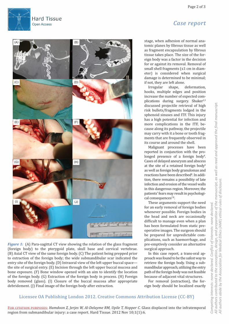
Figure 1: (A) Para-sagittal CT view showing the relation of the glass fragment (foreign body) to the pterygoid plate, skull base and cervical vertebrae. (B) Axial CT view of the same foreign body. (C) The patient being prepped prior to extraction of the foreign body; the wide submandibular scar indicated the entry site of the foreign body. (D) Intraoral view of the left upper buccal space—the site of surgical entry. (E) Incision through the left upper buccal mucosa and bone exposure. (F) Bone window opened with an aim to identify the location of the foreign body. (G) Extraction of the foreign body in process. (H) Foreign body removed (glass). (I) Closure of the buccal mucosa after appropriate debridement. (J) Final image of the foreign body after extraction.
(B)(A)
(D)(C)
(F)(E)
(H)(G)
(J)(I)
Page 3 of 3
Case report
Licensee OA Publishing London 2012. Creative Commons Attribution License (CC-BY)
Com
petin
g in
tere
sts:
non
e de
clar
ed. C
onfli
ct o
f int
eres
ts: n
one
decl
ared
.A
ll au
thor
s co
ntrib
uted
to th
e co
ncep
tion,
des
ign,
and
pre
para
tion
of th
e m
anus
crip
t, a
s w
ell a
s re
ad a
nd a
ppro
ved
the
final
man
uscr
ipt.
All
auth
ors
abid
e by
the
Ass
ocia
tion
for M
edic
al E
thic
s (A
ME)
eth
ical
rule
s of
dis
clos
ure.
F�� �������� ��������: Hamdoon Z, Jerjes W, Al-Delayme RM, Upile T, Hopper C. Glass displaced into the infratemporal region from submandibular injury: a case report. Hard Tissue. 2012 Nov 10;1(1):6.
intraoperatively. Different methods can be used for detection and localiza-tion. MRI is not a suitable imaging mo-dality for detecting missile fragments, as particles with metallic content give rise to powerful interference artefacts, and since the fragments can be con-ductive or ferromagnetic, they present a potential hazard for the patient.
Conventional radiography in two planes can quite accurately define a foreign object, and computed tomog-raphy (CT) is even better in exact three-dimensional location of the fragment preoperatively (with a small cross marker on the skin for co-registration). Ultrasound is sensi-tive and specific in detecting foreign bodies in soft tissues9. Its intraopera-tive use is possible with special probes. The technique is unreliable when me-tallic objects are close to bones, as in a missile wound with comminute bone fracture. In such cases, the distinction between metal and bone is very diffi-cult or even impossible.
ConlcusionThe decision to retrieve a foreign body in the ITF using an intra-oral approach should be guided by the precise location and size of the object, the signs and symp-toms presented by the patient, and the surgeon’s knowledge and skill.
ConsentWritten informed consent was ob-tained from the patient for publication of this case study and accompanying images. A copy of the written consent is available for review by the Editor-in-Chief of this journal.
References1. Paoli JR, Gence E, Vives P, Boutault F,Dupui D. Removal through the coronal approach of the upper wisdom teeth. Apropos of a case of bilateral migration into the temporal fossa. Rev Stomatol Chir Maxillofac. 1995;96(6):392–5.2. Shuker ST. Management of transcranial orbital penetrating shrapnel/bullet war injuries. J Oral Maxillofac Surg. 2008 Sep;66(9):1927–31.3. Shuker ST. Management of penetrating war injuries: bullet/shell fragments in the sphenoid sinuses. J Oral Maxillofac Surg. 2008 Oct;66(10):2067–72.4. Kalinich JF, Emond CA, Dalton TK, Mog SR, Coleman GD, Kordell JE, et al. Embed-ded weapons-grade tungsten alloy shrap-nel rapidly induces metastatic high-grade rhabdomyosarcomas in F344 rats. Envi-ron Health Perspect. 2005 Jun;113(6):729–34.5. Chedid MK, Vender JR, Harrison SJ, McDonnell DE. Delayed appearance of a traumatic intracranial aneurysm. Case report and review of the literature. J Neu-rosurg. 2001 Apr;94(4):637–41.
6. Veselko M, Trobec R. Intraoperativelocalization of retained metallic fragments in missile wounds. J Trauma. 2000 Dec;49(6):1052–8.7. Ghislain PD. Spontaneous extrusion of hand grenade fragments from the face 60 years after injury. JAMA. 2003 Sep 10;290(10):1317–8.8. Wittich AC. Diagnosis and removal of agrenade fragment from the Vietnam war 35 years after injury. Mil Med. 2002 Jun;167(6):519–20.9. Oikarinen KS, Nieminen TM, Mäkäräinen H, Pyhtinen J. Visibility of foreign bodies in soft tissue in plain radiographs, computed tomography, magnetic resonance imaging, and ultrasound. An in vitro study. Int J Oral Maxillofac Surg. 1993 Apr;22(2):119–24.