glaucoma update - aventri
TRANSCRIPT

Glaucoma Update
Dr. James Thimons, Founding Partner, Medical Director
Ophthalmic Consultants of Connecticut
DDisclosures
SpeakerAlconAllerganPRNTear LabShireZeissB&LDiopsysReichartGlaukosInFocusAerie


Welcome to Connecticut

TThe Glaucoma Explosion
New Technologies for the Primary Care ClinicianFinally, New Weapons for Medical Management!The Next Generation of TherapeuticsMIGS: The Evolving Science of Surgery In Glaucoma

AAdvances in OCT Technology: AAutomated Intelligence for the ECPGanglion Cell Analysis: A New Horizon in Primary CareHD SD/OCT Anterior SegmentOCT Angiography in Glaucoma

Ganglion Cell Anatomy

“WWiper” Defect
GGanglion Cell Anatomy
Analysis of VF in RGC loss in Glaucoma24-2 protocol has 6 degrees separation allowing for thinning the RGC to be missed to due point placement
Drazdo t al: Vision Research 200710-2 testing substantially improves correlation with RGC analysis
Hood and Raza; Vis Science 2011Stamper( 1984) identified the relationship between NTG and macular damage with typically near fixation visual field loss.Heijl & Lundqvist 1984
45 patients followed from normal to abnormal VF’s using test points at 5,10,15 & 20 degrees from fixationLargest number at 15 degrees but a surprising number at 5 degrees confirming Hood’s work showing that early damage occurs in the macula as well as more traditional arcuate zones

15
“Green Disease”
“Red Disease”

OOptical Coherence Tomography as a Biomarker for DDiagnosis, Progression, and Prognosis of Neurodegenerative DiseasesSatue, etal AJO 2016
Recent research using the latest SD OCT imaging technology has demonstrated that an early damage of the anterior visual pathway occurs in MS, PD, and AD and that the ganglion cell layer is the ultimate biomarker for disease diagnosis, severity, and progression. Thus, OCT technology should be used as a common and very useful clinical complement in the diagnosis and control of neurodegenerative disorders.85 Citations

AAmerican Journal of OphthalmologyDDecember 2017
Baseline Fourier-Domain Optical Coherence Tomography Structural Risk Factors for Visual Field Progression in the Advanced Imaging for Glaucoma Study
David Huang, MD etal
AAIG/ 2016
A total of 277 eyes of 188 participants were followed up for 3.7 ± 2.1 years. VF progression was observed in 83 eyes (30%). Several baseline NFL and GCC parameters, but not disc parameters, were found to be significant predictors of progression on univariate Cox regression analysis. The most accurate single predictors were the GCC focal loss volume (FLV), followed closely by NFL-FLV. An abnormal GCC-FLV at baseline increased risk of progression by a hazard ratio of 3.1

NNew Perspectives on Disease Management
SD-OCT is superior in identifying progression in glaucoma suspects, pre-perimetric glaucoma, mild glaucoma and early moderate disease compared with SAP are superior in identifying progression, after an initial VF to set baseline. Average time to identification of statistically significant progression is 2-3 years with SD-OCT and up 6 years with SAPIntra-test variability is up to 10x less with OCT( 3%) than VF( 20%)
NNew Perspectives on Disease Management
RNFL “Floor” limits usefulness in late moderate to advanced glaucoma ( 50-60 microns)GCC progression analysis can continue to be useful in late moderate to advanced glaucoma due to density of fibers in the macula and the later involvement of central vision in the disease

GGCC Progression Analysis
Case courtesy of Dr. Shamika GuneCCCase courtttesy of Df Df Dr. ShShSha ikikmikkk GGa Gune
Normal/Shallow Chamber

PPrimary Angle Closure
BBleb Morphology

CCasia Swept Source AS-OCT (Tomey)
• 245 B-Scans (cuts)• Each Repeated 4x
w/FastTrac™ LSO Lock-On
3 mm X 3 mm Angio
Total = 240,000 A-scans, ~ 5.0 secs
Zeiss AngioPlex™ = One Fast Cubic Scan x4
• 245 axial A-Scans per B-Scan, e 1024 voxels deep

Normal 3x3 Angio Cube OD - Full Retina (L) and Deep Plexus (R)
Glaucoma
This case courtesy of Carolyn Majcher, OD. Incarnate Word, San Antonio

Glaucoma
GlaucomaSuperficial

GlaucomaSuperficial
GlaucomaSuperficial

GlaucomaSuperficial
Telemetrics: The Future of Medicine

Triggerfish:Tracking the Elusive Diurnal!
Sensimed: Swiss medical device company. Jean Marc Wismer CEOTracks fluid pressure in the eye and beams data to palm size recorder. Uses a circular antenna taped around the eye and connected to a battery powered portable recorder.This transmits radio frequency energy to an utlra thin gold ring in the CL. This powers a chip embedded in the lens.Additionally on the lens in an ultra thin platinum ring that stretches in response in variation in eye shape secondary to pressure.Available in Europe. Primary trial at University Hospitals of Geneva
“TTriggerfish”

TriggerFish
Based on assumption that IOP and corneal curvature radius are relatedMeausurements are taken every 5 minutes for 30 sec secondsResults are presented as an arbitrary unit not mmHg
Launch Point Technologies
IOL Tonometry
http://www.launchpnt.com/portfolio/biomedical/intraocular-pressure-sensor

TThere’s an App for That
Nature Medicine 2014Yossi Mandel, Bar-Ilan/ Stephen Quake, StanfordUtilizes a variable float tube in the IOLSmart Phone app allows acquisition of dataIn development
TThere’s an App for That

AA Comparison of Perimetric Results from a Tablet Perimeter and Humphrey Field Analyzer in Glaucoma Patients
Y. Kong, M. He, J Crowston, A VingrysTransl Vis Sci Technol. 2016 Nov; 5(6):2University of Melbourne College of Optometry
Melbourne Rapid Fields

MRF
MRF
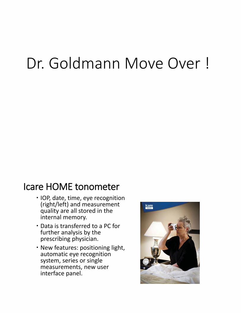
Dr. Goldmann Move Over !
IIcare HOME tonometerIOP, date, time, eye recognition (right/left) and measurement quality are all stored in the internal memory. Data is transferred to a PC for further analysis by the prescribing physician.New features: positioning light, automatic eye recognition system, series or single measurements, new user interface panel.
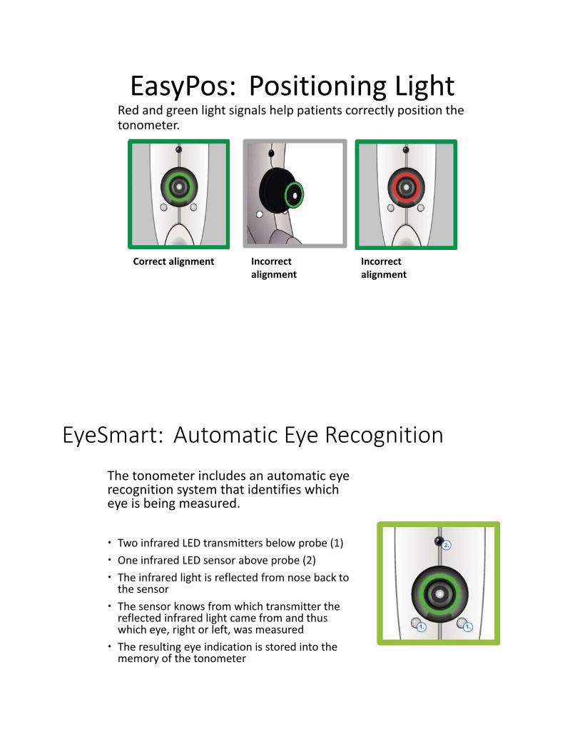
EasyPos: Positioning LightRed and green light signals help patients correctly position the tonometer.
Correct alignment Incorrectalignment
Incorrectalignment
EyeSmart: Automatic Eye RecognitionThe tonometer includes an automatic eye recognition system that identifies which eye is being measured.
Two infrared LED transmitters below probe (1)One infrared LED sensor above probe (2)The infrared light is reflected from nose back to the sensorThe sensor knows from which transmitter the reflected infrared light came from and thus which eye, right or left, was measuredThe resulting eye indication is stored into the memory of the tonometer

CCORNEAL HYSTERESIS: The Newest Disruptive Technology
In Glaucoma

2002: Clinical research with ORA commences
2005: The 1st generation ORA was made commercially available
2012: Generation II ORA was launched
3rd Generation “ORA G3” introduced September 2015Measures:
Corneal Hysteresis (CH)
Goldmann-correlated IOP (IOPg)
Corneal compensated IOP (IOPCC)

CH: Average Values in Normal Subjects
1. Fontes BM J Refract Surg. 2008 Nov;24(9):941-5.2. Carbonaro. The Heritability of Corneal Hysteresis and Ocular Pulse Amplitude A Twin
Study doi:10.1016/j.ophtha.2008.02.0113. Lam A. Et Al. Optom Vis Sci. 2007 Sep;84(9):909-144. Kamiya Et Al. J Refract Surg. 2009 Oct;25(10):888-935. Ortiz Et Al. J Cataract Refract Surg. 2007 Aug;33(8):1371-56. John Et. Al. 2007 Spring;39(1):9-14
CH Values in Normals around the world N CH*
Brazil1 105 10.1 ± 1.8
UK2 272 pairs 10.2 ± 1.2
China3 125 10.9 ± 1.5
Japan4 204 10.2 ± 1.3
Spain5 88 10.8 ± 1.5
USA6 44 10.5 ± 1.2
*CH units are mmHg
Clinical Evidence – Study 1Corneal Hysteresis found to be associated with progression
The first observational study to investigate the relationship of Corneal Hysteresis to a variety of other parameters in a glaucoma population
230 POAG or suspected POAG patients were included in the studyPOAG was defined by a reliable visual field that was abnormal according to OHTS criteria, with an optic nerve image, photo, or CDR thought to be consistent with the field damage by a fellowship-trained glaucoma specialist.GAT, ORA, CCT and Axial Length measurements (IOL master) were recordedAmong persons with three or more reliable fields over three or more years, or with five reliable fields in less than three years, progression was defined as having achieved the OHTS standard of “conversion” (if previously normal), or (if previously damaged as evidenced by an abnormal GHT or PSD) having worsened by 1 dB or greater per year in either MD or PSD.A stepwise model was not used nor were any hypotheses about interactions made.
56
POAG Primary Open Angle Glaucoma; GAT Goldmann Applanation Tonometry; IOP intraocular pressure; ence limit.
CCT Central Corneal Thickness; CH Corneal Hysteresis,Congdon NG et al. Am J Ophthalmol. 2006;141:868-875.

Congdon NG et al. Am J Ophthalmol. 2006;141:868-875.
Conclusions: Corneal Hysteresis was the parameter most associated with progressive field worsening
GAT Goldmann Applanation Tonometry; IOP intraocular pressure; OR odds ratio; LCL lower confidence limit; UCL upper confidence limit.CCT Central Corneal Thickness; CH Corneal Hysteresis
OR LCL UCL P-value
Age per year <65 1.12 1.01 1.24 .03
Age per year >65 1.08 1.01 1.15 .02
GAT IOP per mmHg 1.22 0.95 1.58 .12
Treatment 1847.6 3.16 106 .02
IOP by treatment interaction 0.79 0.61 1.03 .08
CCT per 100 microns 1.65 0.66 0.98 .30
Years with glaucoma 1.00 0.96 1.04 .98
Baseline IOP 0.99 0.93 1.06 .79
CH per mmHg 0.81 0.66 0.98 .03
Clinical Evidence – Study 1Corneal Hysteresis found to be associated with progression
A Prospective Longitudinal Study to Investigate Corneal Hysteresis as a Risk Factor for Predicting Development of GlaucomaAJOPHT 10365 – in pressAuthor Block: Feilin Zhu , Alberto DinizFilho, Linda M. Zangwill , Felipe A. Medeiros
58
Each 1mmHg lower CH was associated with an increase of 21% in the risk of developing glaucoma during follow up
Purpose: To investigate the role of CH as a risk factor for development of glaucoma in a prospective longitudinal study.
Results: Fifty four (19%) of the 287 eyes developed repeatable visual field defects during a 4 year follow-up.
CH was independently predictive of conversion to glaucoma even when adjusted for age, IOP, and CCT.
Corneal Hysteresis in GlaucomaPredictive of conversion to Glaucoma in pre-perimetric Glaucoma Suspects

Medeiros FA et al. Ophthalmology. 2013;120:1533-1540.
59
Eyes with low CH Eyes with high CH
Time (years)
“The prospective longitudinal design of this study supports the role of CH as an important factor to be considered in the assessment of risk for glaucoma progression”
Note – NO rapid progressors in CH
mmHG group!
Corneal Hysteresis in GlaucomaPredictive of Progression in Prospective, Longitudinal Study (DIGS)
• Each 1 mmHg lower CH was associated with a 0.25% per year increase in rate of Visual Field loss
• 2X more predictive of VF loss than GAT IOP (IOP associated w 0.11% per year loss)
• CH was more than 3X more associated with rate of VF loss than CCT (explained 17.4% vs 5.2%)
Actual Rate of VF progression over time
60
“The Effect of IOP on rates of progression was dependent upon
Corneal Hysteresis”
• For eyes with lower CH, the impact of IOP on VF loss was significantly greater
• IOP of 30 is not so bad with a CH of 11.
• IOP of 20 is very bad with a CH of 6
Corneal Hysteresis in GlaucomaPredictive of Progression in Prospective, Longitudinal Study (DIGS)
Medeiros FA et al. Ophthalmology. 2013;120:1533-1540.
Gol
dman
nIO
P (m
mH
g)
Corneal Hysteresis (mmHg)
30
25
20
15
104 6 10 119875
Percentage per year change in Visual Field
-6%
0%
Population Avg CH & IOP
-5%-4%
-3%-2%
-1%

Evaluation of the Influence of Corneal Biomechanical Properties on Intraocular Pressure Measurements Using the Ocular Response Analyzer.Felipe A. Medeiros, MD and Robert N. Weinreb, MDJ Glaucoma 2006;15:364–370.
GAT vs CCT
IOPcc vs CCT
IOPcc – a superior indicator of IOPNot correlated with CCT
IOPcc provides an estimate of IOP that agrees with GAT on average but is less influenced by corneal properties
IOPcc R2= 27.4%
GAT R2= 12.8% iCare R2= 6.5%
IOPcc had the strongest association with visual field loss vs GAT & iCare
IOPcc – a superior indicator of IOPMore associated with actual glaucoma progressionAssociation between Rates of Visual Field Progression and IOP Measurements obtained by different Tonometers
Association between IOP by 3 different Tonometers and rates of Glaucoma Visual Field Progression over time

66 yo, African American Female w 15yr history various practices
Pre Tx highest IOP: 39 OD 35 OSPoor compliance. Intermittently using meds.
IOP 1-month after recent attempt at Tx: 20 OD / 19 OS
1 month later discontinued meds (burning): IOP 25 OD / 28 OSCCT: 589 OD 612 OS microns
V Cup to Disc Ratio: 0.67 OD / 0.57 OS
Patient States IOP “always been high” doesn’t want meds
Courtesy of: Michael Chaglasian, OD
Case: What would you do?The Non-compliant Patient
July 2017 OD PSD: 0.95, VFI 97%
July 2017 OS PSD: 1.89, VFI 95%Patient States IOP always beORA Measurements• CH = 12.7 mmHg• IOP cc = 23.5 mmHg
• HIGH CH indicates low risk of visual field loss• In agreement with 15+ years of actual findings.• Decision made to monitor patient without meds
CCorneal Compensated IOP
Superior to Goldmann in all forms of post Refractive Surgery IOP measurements

GAT and IOPcc357 Normal Eyes
IOPcc 155 POAG 102 NTG Eyes
GAT155 POAG 102 NTG Eyes
GAT
IOPcc
Goldmann applanation tonometry compared with corneal-compensated intraocular pressure in the
evaluation of primary open-angle GlaucomaJoshua R Ehrlich, Nathan M Radcliffe, and Mitsugu
ShimmyoBMC O h h l l 2012 12 52
Not shown here from this study:• 39% of NTG eyes would be re-classified as POAG with IOPcc• Average IOPcc was 5 mmHg higher than GAT in NTG eyes
AUC .93 for IOPcc vs .78 for GAT
GAT measures GAT meaprox
easures mexx 20% of aproxx 0% of 2
glaucoma eyes glaucomatoo low
“the results of this study suggest that IOPcc may represent a superior test for the evaluation of glaucoma”
IOPcc is clinically superior to GAT, other NCTs, and iCare because it is more associated with Glaucoma risk, status of glaucoma, and glaucoma progression
IOPcc Key Benefit #2IOPcc is superior for glaucoma risk assessment
In conclusion, IOPcc measurements were more strongly associated with rates of visual field progression in glaucoma patients as compared to GAT and RBT. By correcting for corneal-induced artifacts, IOPcc measurements may present significant advantages for predicting clinically relevant outcomes in glaucoma patients

2017: A Great Year for Glaucoma Therapy

Inflow
22-2-agonists22-
gg22-blockers
CAIs
22-2-agonists22 go stsagaCholinergicsC g
Prostaglandins
i tOutflow
Pathways to Lower IOP

RhopressaInhibitor of Rho Kinase (ROCK)
and Norepinephrine Transporter (NET)
Potentially lower IOP by three mechanisms1.Increasing TM outflow2.Reducing episcleral venous pressure3.Reducing aqueous production (via NET inhibition


CCorneal Verticillata
Corneal VerticillataCorneal verticillata occurred in approximately 20% of the patients in controlled clinical studies. The corneal verticillata seen in RHOPRESSA-treated patients were first noted at 4 weeks of daily dosing. This reaction did not result in any apparent visual functional changes in patients. Most corneal verticillata resolved upon discontinuation of treatment.

RRhopressa
RHOPRESSA 0.02% was evaluated in three randomized and controlled clinical trials in patients with open-angle glaucoma or ocular hypertension.
Studies 301 and 302 enrolled subjects with baseline IOP lower than 27mmHgStudy 304 enrolled subjects with baseline IOP lower than 30 mmHg. The treatment duration was 3 months in Study 301, 12 months in Study 302, and 6 months in Study 304.

RRhopressa 0.02%: Two Sides to Every Story
For patients with baseline IOP < 25 mmHg, the IOP reductions with RHOPRESSA 0.02% dosed once daily were similar to those with timolol0.5% dosed twice daily (see Table 1). Patients with baseline IOP equal to or above 25 mmHg RHOPRESSA 0.02% resulted in smaller mean IOP reductions at the morning time points than timolol 0.5% for study visits on Days 43 and 90 TThe difference in mean IOP reduction between the two treatment groups was as high as 3 mmHg, favoring timolol.
NNetarsudil Mesylate in Development
Phase III studies with mixed results
Current development plan is in combination with latanoprost.
Katz J. ARVO 2016. E-abstract 4279.Bacharach AGS 2015.
N=298 total Latanoprost Netarsudilmesylate
Fixedcombination
Baseline IOP 26.0 mmHg 25.4 mmHg 25.1 mmHg
Final IOP 18.4 mmHg 19.1 mmHg 16.5 mmHg
IOP Reduction 7.6 mmHg 6.3 mmHg 8.6 mmHg

Vyzulta(Latanoprostene Bunod)
NNitric Oxide and GlaucomaPatients with primary open-angle glaucoma (POAG) have lower levels of NO synthase activity in the trabecular meshwork (TM), Schlemm’s canal, and ciliary muscle1
NO donors lower IOP in normal and POAG eyesA major site of action for NO donors is the TM
NO relaxes the TM and ciliary muscleNO donors increase outflow facility in anterior segments, mediated by a decrease in TM cell volumeEndothelial NO synthase (eNOS) overexpression increases conventional outflow and lowers IOP in a mouse eye model

LLatanoprostene Bunod:NO-Donating Latanoprost
NO plays key roles in both health and disease throughout the body, including the eye
VVyzulta
Nitric Oxide (NO)-donating
rapidly metabolized in situ to latanoprost acid and BDMN, a NO-donating moiety
Exhibited potent and effective intraocular pressure (IOP)-lowering activity in 3 ocular hypertensive glaucoma animal models1
BDMN: Butanediol mononitrate.1Krauss AH, Impagnatiello F, Toris CB, et al. Ocular hypotensive activity of BOL-303259-X, a nitric oxide donating prostaglandin F2 agonist, in preclinical models. Exp Eye Res. 2011;93: 250-5.
Ester Linkage
Latanoprost AcidNitri
c Oxid
e
BOL-303259-X

LLatanoprostene-bunod:Mechanism of Action
Latanoprostene = latanoprostIncreases uveoscleral outflow
Bunod modification donates nitric oxide Exerts its effect in trabecular smooth muscleActivating cGMP signaling pathwayResulting in trabecular relaxation and increased conventional outflow
Mechanisms would be expected to additive
Cavet ME, Vittitow JL, Impagnatiello F, et al. Invest Ophthalmol Vis Sci. 2014;55:5005-5015; Ellis DZ, Dismuke WM, Chokshi BM. Invest Ophthalmol Vis Sci 2009; 50: 1808–1813.
VOYAGER Clinical Trial Design
88
Primary Objectiveo To assess the efficacy and safety of various
doses of LBN QD compared with latanoprost0.005% QD
o Determine the optimum concentration of LBN in reducing IOP
Primary endpointo Reduction in mean diurnal IOP on Day 28
Phase 2, multicenter, single-masked, parallel-group dose finding study in subjects with OAG or OHT
1. Weinreb RN, et al. Br J Ophthalmol 2015;99:738–45.
LBN 0.012% (n=85)
28-day study
LBN 0.024% (n=83)
LBN 0.040% (n=81)
Latanoprost 0.005 % (n=82)
LBN 0.006% (n=82)

EEfficacy Results: Primary Endpoint Voyager Study
REDUCTION IN MEAN DIURNAL IOP ON VISIT 6 (DAY 28)
* †
At highest doses, lowered IOP 1-1.5 mmHg more than latanoprostMost common AE: pain upon instillation
1. Weinreb RN et al. Br J Ophthalmol. 2015;99(6):738-45
APOLLO and LUNAR Clinical Trial Design
90
Phase 3, randomized, multicenter, double-masked, parallel-group studies in patients with OAG or OHT
Primary Objectiveo Evaluate noninferiority of LBN 0.024%
QD in the evening vs timolol maleate 0.5% BID
Primary endpointo IOP measured at 9 assessment time
points in the study eye
Secondary endpoints o Change from baseline (CFB) in IOP at
9 assessment time pointso CFB in diurnal IOP at Week 2, Week
6, and Month 3
1. Weinreb RN, et al. Ophthalmology 2016;123(5):965-73. 2. Medeiros FA, et al. Am J Ophthalmol. 2016;168:250-9.
LBN 0.024% (n=284)
Timolol 0.5% (n=133)
LBN 0.024% (open-label)
3-month efficacy phase 3-month (LUNAR) or 6-month (APOLLO) safety
extension phase

o LBN 0.024% was non-inferior to timolol in both studies and demonstrated significantly greater IOP lowering over timolol at all but one time point in the two studies.
APOLLO and LUNAR: Change from Baseline by Visit (ITT, LOCF)
91
Week 2 Week 6 Month 38 AM 12 PM 4 PM 8 AM 12 PM 4 PM 8 AM 12 PM 4 PM
APOLLOLBN mean CFB (mm Hg) -9.0 -8.5 -7.7 -9.1 -8.7 -7.9 -9.0 -8.7 -7.9
Timolol mean CFB (mm Hg) -7.8 -7.2 -6.6 -8.0 -7.4 -6.7 -7.9 -7.4 -6.6Treatment difference -1.21 -1.37 -1.11 -1.03 -1.24 -1.26 -1.02 -1.27 -1.33
Primary objective NI NI NI NI NI NI NI NI NISecondary objective <.001 <.001 <.001 .002 <.001 <.001 .002 <.001 <.001
LUNARLBN mean CFB (mm Hg) -8.3 -8.1 -7.5 -8.8 -8.5 -7.8 -8.8 -8.6 -7.9
Timolol mean CFB (mm Hg) -7.9 -7.3 -6.9 -7.9 -7.7 -6.8 -7.9 -7.4 -6.6Treatment difference -0.44 -0.76 -0.69 -0.92 -0.84 -0.98 -0.88 -1.29 -1.34
Primary objective NI NI NI NI NI NI NI NI NISecondary objective .216 .022 .025 .005 .007 .003 .006 <.001 <.001
1. Weinreb RN, et al. Ophthalmology 2016;123(5):965-73. 2. Medeiros FA, et al. Am J Ophthalmol. 2016;168:250-9.
Clinical Studieso The most common ocular adverse reactions observed in
patients treated with VyzultaTM (n=811, across both studies) were
conjunctival hyperemia (6%)eye irritation (4%)eye pain (3%)instillation site pain (2%)
o Approximately 0.6% of patients discontinued therapy due to ocular adverse reactions including ocular hyperemia, conjunctival irritation, eye irritation, eye pain, conjunctival edema, vision blurred, punctate keratitis and foreign body sensation.
92VYZULTA™ [prescribing information]. Bridgewater, NJ; Bausch + Lomb, 2017.
APOLLO and LUNAR: Adverse Events

Study Week
JUPITER: Sustained IOP-lowering Efficacy through One Year
o -treatment visit vs. baseline (P<0.001 for all).
1. Kawase K, et al. Adv Ther 2016;33:1612-27
Study Week
all).
K l Ad Th 2016 33 1612 27

TThe Next Generation of Medical MManagement in Glaucoma
Sustained Release Systems
MMati Therapeutics
The Evolute has an L-shaped design and is inserted into the nasolacrimal duct. The device is cosmetically invisible, but can be easily seen with eversion of the lower lid.The glaucoma product has a core of latanoprost-polymer matrix that is surrounded by silicone, and it delivers the medication into the tear film at a constant rate.In a phase II clinical trial, the latanoprost punctal plug was found to be comfortable. It was associated with a 20% lowering from baseline IOP over a 3-month period, and in two separate clinical trials.Retention rate of 92% and 96%, respectively.

MMati Therapeutics
Evolute® Punctal Plug Delivery System
StableFit™ Design
Successful By Design1. Easy to place and remove
2. Cosmetically invisible – easy to identify
3. Tolerable
4. Consistent, sustained efficacy
5. Use in multiple disease states
Targeted Delivery
Drug Core
Polymer Sleeve
Cyanoacrylate FilmProven Sustained Elution
98

Excellent Plug Retention Rates Over 12 Weeks
Study Week 4 Week 8 Week 12
Glau 12 (n = 92) 98% 97% 96%
Glau 13 (n = 87) 98% 96% 92%
U.S. Phase II Multi-center Trials – Lower Puncta
99
Evolute® Tearing & Comfort Scores
18.923 21 19.6
18.9 20.7 20 19.3
0
10
20
30
40
50
60
70
80
90
100
Baseline Week 4 Week 8 Week 12
Tearing Comfort
VAS
scor
ing,
0 =
no
resp
onse
, 100
= in
tole
rabl
e N=179
100

OOcular Therapeutix
OOcular Therapeutix
Phase II study randomly assigned 73 patients into two groups to receive either the travoprost plug with twice daily artificial tears or timolol 0.5% twice daily with placement of a drug-free punctal plug.At 90 days, there was a 4.5 to 5.7 mm Hg reduction from baseline IOP in patients who had the travoprost punctal plug, which was clinically meaningful.However, the control group had an average IOP lowering of 6.4 to 7.6 mm Hg.The safety profile was good—no hyperemia was seen. The retention rate at 60, 75, and 90 days was 91%, 88%, and 48%, respectively.

CContact Lens Embedded IOP Lowering Drug
Dean Ho, UCLA Dentistry School Research TeamNanogel that is embedded in CL’sIOP lowering capacityUses nanotechnolgy with small diamonds that timolol until tear enzymes (lysozyme) activate it The octohedron structure of the nanodiamonds has a unique charge that binds drugs to its surface Chitosan, A natural polymer is used to bind the drugAnticipated NDA is 2016
LLatanoprost--EEluting Contact Lenses in GGlaucomatous Monkeys.Ciolino, J, Kohane,DS etal Ophthalmology 2016
RESULTS:Latanoprost ophthalmic solution resulted in IOP reduction of 5.4±1.0 mmHg on day 3 and peak IOP reduction of 6.6±1.3 mmHg on day 5.The CLLO reduced IOP by 6.3±1.0, 6.7±0.3, and 6.7±0.3 mmHg on days 3, 5, and 8,
respectively. The CLHI lowered IOP by 10.5±1.4, 11.1±4.0, and 10.0±2.5 mmHg on days 3, 5, and 8, respectively.For the CLLO and CLHI, the IOP was statistically significantly reduced compared with the
untreated baseline at most time points measured. The CLHI demonstrated greater IOP reduction than latanoprost ophthalmic solution on day 3 (P = 0.001) and day 5 (P = 0.015), and at several time points on day 8 (P < 0.05).Coating Polylactic co-glycolic acid (PLGA) is coated with films containing Polyhydroxy-methacrylate by UV polymerization

BBimatoprost SR
BBimatoprost SR
Bimatoprost SR is currently in phase 3 clinical trials. In phase 2 trials, the device produced a mean IOP reduction:
7.2 to 9.5 mm Hg from baseline in 75 eyes 4 months after the injection.
Patients’ fellow eyes received qd topical bimatoprost 0.03% IOP reduction of 8.4 mm Hg at 4 months.
The implant lowered IOP; 92% of patients at 4 months 71% at 6 months. No serious adverse ocular events, and the most common adverse events were related to the injection procedure

BBimatoprost Ring
BBimatoprost Ring
The bimatoprost-impregnated insert produced:More than 20% IOP lowering at all time pointsIt was slightly (0-1.5 mm Hg) less efficacious than twice-daily timolol at the nine time points.
The pharmacokinetics of prostaglandin analogues; constant dosing tends to produce a lesser effect than pulsed dosing regimens.Maintenance of the insert in place without a physician’s reintervention:
93.1% at 12 weeks 88.5% at 6 months.
Adverse events were relatively low. Of the 161 patients who wore a nonmedicated insert for the first month, 151 reported no discomfort.

GGlaukos iDose
The iDose is a titanium implant that is comparable in size to Glaukos’ proprietary devices for microinvasive glaucoma surgeryThe 150-patient, multicenter, randomized, double-blind phase 2 trial evaluated two models of the iDose delivery system with different travoprost elution rates in comparison to a topical timolol maleate ophthalmic solution, 0.5%. The unit is filled with a formulation of travoprost specific to the device and capped with a membrane designed for continuous controlled drug elution into the anterior chamber.
GGlaukos iDose

MMIGS: Not Just a Soviet Fighter Jet Anymore

Effect of Cataract Surgery onIOP Reduction
According to the AAO Preferred Practice Patterns, cataract surgery with IOL implantation alone results in a modest reduction in IOP of less than 2mm Hg on average.1
Chart review of 588 normotensive andOHT subjects2
53% had a mean reduction of 1.6 to2.5 mm Hg2
1American Academy of Ophthalmology, Preferred Practice Patterns, 2010.2Poley BJ, Lindstrom RL, et al. Long-term effects of phacoemulsification with intraocular lens implantation in normotensive and ocular hypertensive eyes. J Cataract Refract Surg .2008;34(5):735-42.
Baseline IOP (mm Hg)
6.5
4.7
2.5
1.60.2
-7
-6
-5
-4
-3
-2
-1
0
1
IOP
(mm
Hg)
23-31n=19
20-22n=62
18-19n=86
15-17n=223
9-14n=198
Current OAGTreatment Algorithm1
Drug therapy has been the standard of care in glaucoma for over 30 years.Approximately 50% of patients are taking 2 or more medications increasing the disease management challenges of glaucoma and financial burden to patients and the healthcare system.2,3
1AAO Preferred Practice Pattern; Primary Open Angle Glaucoma. AAO committee 2003.2Stein J, Newman-Casey P, Niziol L, et. al. Association between the use of glaucoma medications and mortality. Arch Ophthalmol. 2010;128(2):235-245.3Market Scope Quarterly Glaucoma Report, 4th quarter 2013.
Add More Rx Therapy
Prescription Therapy(30 – 90 Days)
y
AAAO committee 2003.AAO committee 2003
Invasive SurgeryTrabeculectomy
Switch orAdd Rx Therapy
Laser Trabeculoplastyty
Newly Diagnosed POAG Patient

TTop MIG”s in 2018
Glaukos iStentXen Glaucoma ImplantCyPassCanaloplastyTrabectome
116
iiStent® Indication for Use(US Label)
The iStent Trabecular Micro-Bypass Stent is indicated for use in conjunction with cataract surgery for the reduction of intraocular pressure (IOP) in adult patients with mild to moderate open-angle glaucoma currently treated with ocular hypotensive medication
117
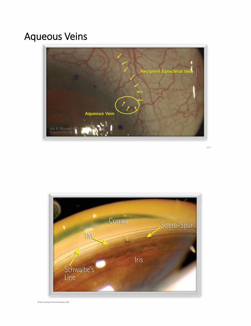
AAqueous Veins
Aqueous Vein
Recipient t Episcleralal Vein
Photo courtesy of Tom Samuelson, MD

25% decrease
Intraocular Pressure
Preop 2 Years
16.8.8±8±4.0 12.6.6±6±1.8
Results – All Eyes (n=104 eyes)Preop and 2-Year IOP and Medication
All Eyes with 2-Year Follow-up
57% decrease
Medication Burden
Preop 2 Years
2.3.3±3±1.0
1.0.0±0±1.2
Control and Reduced Medications Two Years Following Cataract Surgery (M. Gallardo)
Mark Gallardo, MD El Paso Eye Surgeons El Paso, TX. Presented at ASCRS 2017, Los Angeles, CA.
•• 50% were on 0 medications, compared to 6% preop
Retrospective Case Series (Ferguson, Berdahl)
Ferguson TJ, Berdahl JP, Schweitzer JA, Sudhagoni RG. Clinical evaluation of trabecular microbypass stents with phacoemulsification in patients with open-angle glaucoma and cataract. Clinical Ophthalmology 2016:10 1767-1773
19.33
21.96
19.56
16.37 16.12 15.67 16.44 16.2915.17
1.40 0.63 0.81 0.95 0.44 0.51 0.68 0.75 0.610
5
10
15
20
25
Preop (n=107) 1 Day (n=107) 1 Week (n=103) 1 Month (n=102)
3 Months (n=90)
6 Months (n=82)
1 Year (n=98) 18 Months (n=77)
2 Years (n=107)
IOP
(mm
Hg)
IOP and Medications – 1 iStent® + Cataract – Consistent Cohort
IOP (mm Hg) Glaucoma Meds P<0.0001
• Large series (n=107)• At 2 years, mean IOP reduction was 22% with a 56% reduction in mean medications

Ferguson TJ, Berdahl JP, Schweitzer JA, Sudhagoni RG. Clinical evaluation of trabecular microbypass stents with phacoemulsification in patients with open-angle glaucoma and cataract. Clinical Ophthalmology 2016:10 1767-1773
Retrospective Case Series (Ferguson, Berdahl)
4.16
0.69
3.48
7.69
11.28
0
2
4
6
8
10
12
14
All Eyes (n=107) 18-21 (n=27) 22-25 (n=16)
IOP
(mm
Hg)
Postoperative IOP Reduction Comparison (mm Hg)
• IOP reduction higher with higher baseline IOP• Patients with pre-
36%
Neuhann TH. Trabecular micro-bypass stent implantation during small-incision cataract surgery for open-angle glaucoma or ocular hypertension: Long-term results. J Cataract Refract Surg 2015; 41:2664–2671.
666666
iiStent®® + Cataract Surgery TThrough 3 Years (T) . NeuhannAt 3 years mean IOP was < 15 mm Hg with an 86% reduction in medications
• Consecutive series of 62 eyes: decision to implant based on patient desire to reduce topical meds and intent to offer surgical treatment with favorable safety profile

CCo-Management CodingiStent implantation is described by CPT code 0191T
0191T is a Category III (new technology) code0191T has no assigned Relative Value Units or Global Period There is no postop co-management fee for any T-codeMedicare carriers will not recognize modifiers -54 & -55 for 0191T
Modifiers -54 & -55 can still be appended to CPT code 66984Modifier -54: surgical care onlyModifier -55: all/part of outpatient postoperative careSurgeon MUST initiate the notification to Medicare by using modifier -54 with the claimIn localities where Medicare has a higher physician payment for 0191T than for 66984 and where 66984 is reduced by 50%, payment for 66984-550 will be reduced by 50%
AAllergan:XEN

Ab Interno Sub-Conjunctival DrainageSurgical “Gold Standard” IOP reduction in minimally invasively procedureClinically proven outflow pathwayBypasses all potential outflow obstructionsConjunctiva sparing: alternative surgical options remainSingle implant delivers desired effectiveness
XEN Glaucoma Implantnt™™ Mechanism of Action
Gelatin Material is Tissue Conforming
POAG Only
*Mean preoperative IOP is best medicated. Patients were not washed out prior to surgery.
Summed patients: primary, combined and refractory

IInitial Clinical Results: From A Multi-Center Study on Early Moderate Stage Population
* Washout IOP calculated at +30% from medicated
N = 62Pre op
IOP 21.8 ± 3.5 mmHg Meds 2.612M
(N=37)18M
(N=14)24M(N=8)
Mean IOP mmHgStd. Dev.
15.73.5
14.72.9
14.92.8
Mean Post Op MedsMean Meds % Reduction
0.9-65%
1.0-61%
1.0-61%
% IOP reduction from Best RxFrom Washout IOP*
-28%-45%
-33%-48%
-32%-47%
% <21 mmHgand/or -20%
100% 100% 100%
% <18 mmHgand/or -20%
100% 86% 100%
% <16 mmHgand/or -30%
84% 86% 63%
IInitial Clinical Results: From A Multi-Center Study on Severe/Refractory Population
* Washout IOP calculated at +30% from medicated
N = 39Pre op
IOP 22.6 ± 4.3 mmHg Meds 3.112M
(N=29)18M
(N=12)24M
(N=12)
Mean IOP mmHgStd. Dev.
13.94.6
12.93.4
13.74.7
Mean Post Op MedsMean Meds % Reduction
1.1-66%
1.1-66%
1.3-57%
% IOP reduction from Best RxFrom Washout IOP*
-38%-52%
-43%-55%
-39%-53%
% <21 mmHgand/or -20%
100% 100% 100%
% <18 mmHgand/or -20%
97% 100% 100%
% <16 mmHgand/or -30%
79% 92% 83%

CCypass: Suprachoroidal Stent
CCypass: Suprachoroidal Stent

CCyPass
IInitial Clinical Experience With the CCyPass MMicro--SStent: Safety and Surgical Outcomes of a Novel Supraciliary Microstent.
Mean±SD follow-up was 294±121 days.Preoperative baseline mean IOP was 20.2±6.0 mm Hg and mean number of IOP-lowering medications was 2.0±1.1. Cohort 1 ( >21 mmHg) showed a 35% decrease in mean IOP and a 49% reduction in mean glaucoma medication usage; Cohort 2 ( < 21 mmHg) demonstrated a 75% reduction in mean medication usage while maintaining mean IOP<21 mm Hg. For all eyes, mean IOP at 12 months was 15.9±3.1 mm Hg (14% reduction from baseline). Early and late postoperative IOP elevation occurred in 1.2% and 1.8% of eyes. Two subjects demonstarted mild transient hyphema, andNone exhibited prolonged inflammation, persistent hypotony, or hypotonymaculopathy

Alcon Voluntarily Withdraws CyPass Micro-Stent For Surgical Glaucoma From MarketAugust 29, 2018, 01:21:00 AM EDT By RTT News
•
Shutterstock photo(RTTNews.com) - Alcon, the eye care unit of Novartis ( NVS ), announced Wednesday an immediate, voluntary market withdrawal of the CyPass Micro-Stent from the global market. Alcon also advised surgeons to immediately cease further implantation with the CyPass Micro-Stent and to return any unused devices to Alcon.The move is based on an analysis of five-year post-surgery data from the COMPASS-XT long-term safety study. The COMPASS-XT study was designed to collect safety data on the subjects who participated in the COMPASS study for an additional three years, with analysis of the completed data set at five years post-surgery. At five years, the CyPass Micro-Stent group experienced statistically significant endothelial cell loss compared to the group who underwent cataract surgery alone.The US Food and Drug Administration or FDA approved the CyPass Micro-Stent in July 2016 for use in conjunction with cataract surgery in adult patients with mild-to-moderate primary open-angle glaucoma based on the results of the landmark two-year COMPASS study.The COMPASS study demonstrated a statistically significant reduction in intraocular pressure at two years post-surgery in subjects implanted with the CyPass Micro-Stent at the time of cataract surgery, as compared to subjects undergoing cataract surgery alone.At two years post- surgery, there was little difference in endothelial cell loss between the CyPass Micro-Stent and cataract surgery-only groups, and results were consistent with peer-review literature benchmarks of cataract-related endothelial cell loss.Stephen Lane, Chief Medical Officer, Alcon, said, "Although we are removing the product from the market now out of an abundance of caution, we intend to partner with the FDA and other regulators to explore labeling changes that would support the reintroduction of the CyPass Micro-Stent in the future."The voluntary market withdrawal applies to all versions of the CyPass Micro- Stent.
PPreliminary ASCRS CyPass Withdrawal Consensus StatementASCRS CyPass Withdrawal Task ForceLeads: Douglas Rhee, MD; Nathan Radcliffe, MD; and Francis Mah, MD Glaucoma: Leon Herndon, MD; Marlene Moster, MD; Thomas Samuelson, MD; Steven Vold, MDCornea: Ken Beckman, MD, FACS; John Berdahl, MD; Marjan Farid,, MD; Preeya Gupta, MD
The COMPASS XT study followed a smaller number of patients than the COMPASS trial. By 60 months, there were roughly 200 CyPasspatients and 53 control patients. Of note, as the study was being assembled at 36 months, there are too few (n=36 patients) presenting at 36 months to make any meaningful comparisons. Aside from ECL, outlined below, there were no other significant safety concerns.

CCyPass Recall
At 5 years, there was more ECL in CE with CyPass compared to CE alone (control). Baseline endothelial cell counts (ECCs) were 2432 for CyPass and 2434 for control, falling at 48 months to 1992 in CE with CyPass (n=116) vs 2303 in control (n=33) and at 60 months to 1931 in CE with CyPass (n=163) vs 2189 in the control group (n=40). This represented an 18.4% reduction in ECL in CE with CyPass vs 7.5% ECL in the control group at month 48, and to a 60-month CE with CyPass ECL of 20.5% compared to 10.1% in the control group. The difference in ECL between CE with CyPass and control decreased slightly between 48 and 60 months. ANSI Z80:27 standards consider 30% ECL at 5 years to be meaningful. The percentage loss was 27.2% in CyPassvs 10% in controls.
CCyPass Recall
For eyes with no rings showing (n=69), the rate was 1.39%/year, for 1 ring showing (n=98) 2.74%/year, and for 2-3 rings showing (n=27) 6.96%/year. No patients in COMPASS XT required corneal surgery by 5 years. Four patients underwent a CyPass trimming procedure for a CyPass with 3 rings visible in the anterior chamber that was observed in the first postoperative week. In all cases, the corneas remained clear and the ECC remained stable at month 60. One patient in the COMPASS trial (two-year follow up) did undergo a Descemet’s stripping endothelial keratoplasty (DSEK) at month 13 with the procedure being thought to be related to the CE and not to the CyPass, which was well positioned with 1 ring visible. Some eyes with >2 rings visible in the anterior chamber experienced minimal ECL. Thus, clinically relevant, judicious, and periodic monitoring of corneal health is advised.

CCyPass Recall
CCyPass Recall

CyPass Recall
CConsiderations for Device Revision
If corneal decompensation develops and >1 ring of the device is visible, the surgeon may consider CyPass repositioning, removal, or proximal end trimming. It was the consensus of the group that implant repositioning i.e. deeper implantation, would be most safe if performed within 7-10 days of implantation. Beyond this time period, there was concern expressed by the group that fibrosis around and/or through the filtration holes of the device may create a higher risk of complications with device repositioning. Due to the potential for fibrosis around and possibly investing the device, device removal was not favored by the group. Trimming of the proximal end is likely to be the preferred procedure if the patient and physician desire intervention. Technical descriptions of the procedures are described in the CyPass micro-stent Instructions for Use “IFU.”
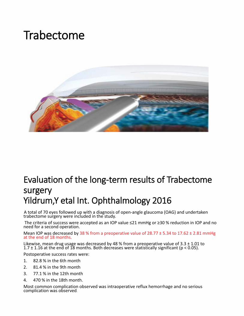
TTrabectome
EEvaluation of the long-term results of TrabectomesurgeryYildrum,Y etal Int. Ophthalmology 2016A total of 70 eyes followed up with a diagnosis of open-angle glaucoma (OAG) and undertaken trabectome surgery were included in the study.
% reduction in IOP and no need for a second operation. Mean IOP was decreased by 38 % from a preoperative value of 28.77 ± 5.34 to 17.62 ± 2.81 mmHg at the end of 18 months. Likewise, mean drug usage was decreased by 48 % from a preoperative value of 3.3 ± 1.01 to 1.7 ± 1.16 at the end of 18 months. Both decreases were statistically significant (p < 0.05). Postoperative success rates were:1. 82.8 % in the 6th month2. 81.4 % in the 9th month3. 77.1 % in the 12th month4. 470 % in the 18th month. Most common complication observed was intraoperative reflux hemorrhage and no serious complication was observed.

CCanaloplasty
VViscodilation
Preoperative Dilation of Schlemm’s canal
Dilation of Schlemm’scanal and collector
channels
Dilation of Schlemm’s canal visualized with UltraSound Imaging

EEffects of Suture TensionEx-Vivo Perfusion Study, Utilizing Morton Grant Flow
Model– Pressurize globe to a range of physiologic pressures– Apply tension to a suture implanted through the canal– Measure outflow facility (uL/Min / mmHg)
CCanaloplasty, Suture Tension
Histology (H&E staining) Close-up of suture
Distension of Trabecular Meshwork Histology (ex-vivo)

CCanaloplasty
IOP All Enrolled Eyes
0.0
5.0
10.0
15.0
20.0
25.0
30.0
35.0
Baseline 1D 1W 1M 3M 6M 12M 18M 24M
IOP
[mm
Hg]
CCombined Procedure ResultsMedication Results
-0.5
0.0
0.5
1.0
1.5
2.0
2.5
3.0
3.5
Baseline 1D 1W 1M 3M 6M 12M 18M 24M
# Med
icatio
ns
Combined Canaloplaslty Alone
Baseline 3 Months 6 Months 12 Months 18 Months 24 Months
Meds/Pt 1.5 0.1 0.1 0.2 0.2 0.2
Meds SD 1.0 0.3 0.4 0.4 0.4 0.4
N 50 41 43 36 26 26
Meds/Pt 2.0 0.4 0.5 0.6 0.6 0.8
Meds SD 0.9 0.7 0.7 0.8 0.8 0.9
N 146 126 118 108 88 66
CombinedMean Meds Drop: 87%
Canaloplasty AloneMean Meds Drop: 61.4%
0.8
0.2

TTrabeculectomy with Express Minishunt
RRetrospective Case SeriesFinal percent IOP lowering was similarMoorefields Bleb Grading System
Less vascularity and height but more diffuse area associated with the Ex-PRESS blebs
Fewer cases of early postoperative hypotony and hyphemaQuicker visual recovery
The Ex-PRESS group required fewer postoperative visits compared with the trabeculectomy group (P < .000).
Good TJ. Assessment of bleb morphologic features and postoperative outcomes after Ex-PRESSdrainage device implantation versus trabeculectomy. Am J Ophthalmol. 2011 Mar;151(3):507-13.e1. Epub 2011 Jan 13.

EEx-PRESS in prior operated eyesSuccess complete in 60(60%) and qualified in 24 (24%) eyes Mean IOP
27.7 ± 9.2 mm Hg with 2.73 ± 1.1 14.02 ± 5.1 mm Hg with 0.72 ± 1.06 drugs (p < 0.0001)
FailureUncontrolled IOP (11%)bleb needling (4%)persistent hypotony (1%)
Lankaranian D. Intermediate-term results of the Ex-PRESS(TM) miniature glaucoma implant under a scleral flap in previously operated eyes. Clin Experiment Ophthalmol. 2010 Dec 22.
55 year study Ex-press vs Trabeculoectomy
EX-PRESS more effective without medicationAt year 1 12.8% of patients required IOP meds after EX-PRESS implantation vs 35.9% after trabeculectomyAt year 5 (41% versus 53.9%)
Responder rate was higher with EX-PRESSTime to failure was longerSurgical interventions for complications were fewer after EX-PRESS implantation
deJong et al. Five-year extension of a clinical trial comparing the EX-PRESS glaucomafiltration device and trabeculectomy in primary open-angle glaucoma. Clin Ophthalmol. 2011;5:527-33. Epub 2011 Apr 29.