global proteome and phospho-proteome analysis of merlin...
TRANSCRIPT

EBioMedicine 16 (2017) 76–86
Contents lists available at ScienceDirect
EBioMedicine
j ourna l homepage: www.eb iomed ic ine.com
Research Paper
Global Proteome and Phospho-proteome Analysis of Merlin-deficientMeningioma and Schwannoma Identifies PDLIM2 as a NovelTherapeutic Target
Kayleigh Bassiri a,1, Sara Ferluga a,1, Vikram Sharma b, Nelofer Syed c, Claire L. Adams a,Edwin Lasonder b, C Oliver Hanemann a,⁎a Institute of Translational and StratifiedMedicine, PlymouthUniversity Peninsula Schools ofMedicine and Dentistry, John Bull Building, Plymouth Science Park, ResearchWay, Derriford, PlymouthPL6 8BU, UKb School of Biomedical and Healthcare Sciences, Plymouth University, Drakes Circus, Plymouth PL4 8AA, UKc John Fulcher Neuro-oncology Laboratory, Division of Brain Sciences, Faculty of Medicine, Imperial College London, London W6 8RP, UK
⁎ Corresponding author at: John Bull Building, PlymouDerriford, Plymouth PL6 8BU, UK.
E-mail address: [email protected] (C.1 These authors equally contributed to the manuscript.
http://dx.doi.org/10.1016/j.ebiom.2017.01.0202352-3964/© 2017 The Authors. Published by Elsevier B.V
a b s t r a c t
a r t i c l e i n f oArticle history:Received 10 October 2016Received in revised form 13 January 2017Accepted 13 January 2017Available online 18 January 2017
Loss or mutation of the tumour suppressor Merlin predisposes individuals to develop multiple nervous systemtumours, including schwannomas andmeningiomas, sporadically or as part of the autosomal dominant inheritedcondition Neurofibromatosis 2 (NF2). These tumours display largely low grade features but their presence canlead to significant morbidity. Surgery and radiotherapy remain the only treatment options despite years of re-search, therefore an effective therapeutic is required.Unbiased omics studies have become pivotal in the identification of differentially expressed genes and proteinsthat may act as drug targets or biomarkers. Here we analysed the proteome and phospho-proteome of these ge-netically defined tumours using primary human tumour cells to identify upregulated/activated proteins and/orpathways.We identified over 2000 proteins in comparative experiments betweenMerlin-deficient schwannomaand meningioma compared to human Schwann and meningeal cells respectively. Using functional enrichmentanalysis we highlighted several dysregulated pathways and Gene Ontology terms.We identified several proteinsand phospho-proteins that are more highly expressed in tumours compared to controls. Among proteins jointlydysregulated in both tumourswe focused in particular on PDZ and LIMdomain protein 2 (PDLIM2) and validatedits overexpression in several tumour samples, while not detecting it in normal cells. We showed that shRNAme-diated knockdown of PDLIM2 in both primary meningioma and schwannoma leads to significant reductions incellular proliferation.To our knowledge, this is the first comprehensive assessment of the NF2-related meningioma and schwannomaproteome and phospho-proteome. Taken together, our data highlight several commonly deregulated factors, andindicate that PDLIM2 may represent a novel, common target for meningioma and schwannoma.
© 2017 The Authors. Published by Elsevier B.V. This is an open access article under the CC BY-NC-ND license(http://creativecommons.org/licenses/by-nc-nd/4.0/).
Keywords:MeningiomaSchwannomaNF2MerlinProteomePhospho-proteome
1. Introduction
Neurofibromin 2 (Merlin, NF2) is a tumour suppressor proteinexpressed during embryonic development and thereafter (Gronholm etal., 2005). In adults, significant levels of expression are found in Schwannand meningeal cells, nerve and lens (Claudio et al., 1997; Sakuda et al.,1996; Scherer and Gutmann, 1996). Mutations in the encoding gene(NF2) lead to formation of schwannomas and meningiomas, and lessoften of ependymomas and retinal astrocytic hamartomas (Hanemann,2008; Martin et al., 2010; Rouleau et al., 1993). These tumours originate
th Science Park, Research Way,
O. Hanemann).
. This is an open access article under
sporadically or as part of the genetic condition Neurofibromatosis type 2(NF2) (Hanemann, 2008). They are largely unresponsive to classic che-motherapeutic agents, leaving surgery and radiotherapy as the only re-maining treatment options which can leave the patient with mild toseveremorbidity (Hanemann, 2008). Additionally, NF2 patients often de-velop multiple tumours simultaneously (Hanemann, 2008), strengthen-ing the need for effective systemic therapeutic options. Loss of Merlinhas also been related to a variety of other cancers, including glioblasto-mas, malignant mesotheliomas and thyroid carcinomas, highlighting itsrole as tumour suppressor (Garcia-Rendueles et al., 2015; Guerrero etal., 2015; Lee et al., 2016; Morrow et al., 2016; Sheikh et al., 2004).
Merlin shares structural similarity with the Ezrin/Radixin/Moesin(ERM) family of proteins that link the cytoskeleton with componentsof the cell membrane (Bretscher et al., 2000; McClatchey, 2003;McClatchey and Giovannini, 2005). Although Merlin lacks the C-
the CC BY-NC-ND license (http://creativecommons.org/licenses/by-nc-nd/4.0/).

77K. Bassiri et al. / EBioMedicine 16 (2017) 76–86
terminal actin-binding domain present in the other members of theERM family, it can localize to the cortical cytoskeleton and interact di-rectly with the actin-binding protein α-catenin (Gladden et al., 2010).At sites of cell-cell contact Merlin acts as tumour suppressor controllingcadherin-mediated contact-dependent inhibition of proliferation andadherens junction formation (Flaiz et al., 2008; Lallemand et al.,2003). Several receptor tyrosine kinases (RTKs) have been found to beMerlin-dependent (Curto et al., 2007; Lallemand et al., 2009).Our group and others showed overexpression and reduced degradationof the platelet-derived growth factor receptor β (PDGFRβ) inschwannoma compared to normal Schwann cells which, together withthe loss of Merlin, leads to increased cellular proliferation and aberrantactivation of the MAPK and PI3K signalling pathways (Ammoun et al.,2008; Fraenzer et al., 2003). RTKs are found to be linked to Merlin andthus the cytoskeleton via the PDZ domain–containing adapter NHERF-1 (Na+/H+ exchanger regulatory factor) (Maudsley et al., 2000;Weinman et al., 2000). Merlin loss further contributes to tumorigenesisvia the activation of a number of other pathways including the Hippo,Ras and Wnt/β-catenin (Li et al., 2014; Mohler et al., 1999; Zhao et al.,2010, 2011). Merlin activity is also in the nucleus, where it binds tothe E3 ubiquitin ligase CRL4 (DCAF1) suppressing its activity. Depletionof DCAF1 in Merlin-deficient schwannoma cells was sufficient to blockproliferation (Cooper et al., 2011).
Unbiased genomic studies have been performed aiming to identifynovel differentially-expressed genes in schwannomas and meningio-mas (Fevre-Montange et al., 2009; Hanemann et al., 2006;Torres-Martin et al., 2013a,b, 2014; Wang et al., 2012) as well as noveldriver mutations, exclusive of NF2 (Clark et al., 2013).
Mass spectrometry (MS) is a powerful, high-throughput techniqueto identify thousands of proteins aberrantly expressed and regulated.Recently Sharma and colleagues performed comparative proteomicanalysis on different grades of meningiomas to investigate alterationsin the meningioma tissue and in the human serum of meningioma pa-tients compared to normal brain tissue. They identified severalderegulated proteins including transgelin-2 and caveolin in tissue,plus apoliopoproteins A and E in serum (Sharma et al., 2014, 2015).
Here we analysed by label free quantitative proteomics both the pro-teomeandphospho-proteomeofmeningiomaand schwannomaprimarytumour cells. By analysing proteomes and phospho-proteomes together,we identify overexpressed proteins in tumour cells and regulatory signal-ling pathways that may be ‘switched off’with therapeutic intervention.
We also compared protein abundances in primary Merlin-deficienthuman meningioma cells against human meningeal cells, and primaryhuman schwannoma cells against primary human Schwann cells. Weidentified numerous novel upregulated and downregulated proteinsand phospho-proteins, performed Gene Ontology (GO) mapping andfunctional enrichment analyses for GO and pathway terms. We identi-fied proteins common to both Merlin-deficient tumour types. Severalof the upregulated proteins contained either a PDZ/LIM domain, orboth. These proteins have been shown to have awide range of biologicalfunctions including roles in cell signalling (Te Velthuis et al., 2007). Wefound PDZ and LIM domain protein 2 (PDLIM2/ mystique/SLIM) com-monly upregulated in both tumour types compared to the normal con-trols. Previous experiments on PDLIM2 suggested a role in cytoskeletalorganization as it was co-immunoprecipitated together with alpha-actinin-1, alpha-actinin-4, filamin A, and myosin heavy polypeptide 9in rat corneal epithelial cells (Loughran et al., 2005a; Torrado et al.,2004). PDLIM2 was also identified at the nuclear level exerting tumoursuppressive functions by terminating NF-κB activation during inflam-mation (Tanaka et al., 2007) and in breast cancer (Qu et al., 2010).PDLIM2 overexpression was found in metastatic cancer cells(Loughran et al., 2005b) and androgen-independent prostate cancercell lines (Kang et al., 2016). Using our primary human cultures we per-formed PDLIM2 silencing in primary human schwannomas and menin-giomas and observed a statistically significant reduction in cellproliferation in both tumour types.
To our knowledge, this work is the first proteomic study aiming todecipher common deregulated elements in the proteome andphospho-proteome of Merlin-deficient schwannomas and meningiomas.
2. Materials and Methods
2.1. Clinical Samples
Meningioma and schwannoma specimens were collected after pa-tients consented to the study and given a unique MOT identificationnumber. This study was granted full national ethics approval by theSouthWest research ethics committee (RECNo: 14/SW/0119; IRAS pro-ject ID: 153,351) and local research and development approval (Plym-outh Hospitals NHS Trust: R&D No: 14/P/056 and North Bristol NHSTrust: R&D No: 3458). Normal human Schwann cells were collectedafter ethical approval under the REC number REC6/Q2103/123. Thebrain tumour material was obtained from the Imperial brain tumourbank and this sub-collection is covered by Imperial College Tissuebank ethics. All meningioma samples used in this study were grade I.
2.2. Cell Culture
Human meningeal cells (HMC) were obtained from Sciencell™ andmaintained in the manufacturer's recommended media at 5% CO2.Human primary Schwann/schwannoma cells were maintained as de-scribed previously (Rosenbaum 2000). Ben-Men-1 cells and primarymeningioma cells were routinely grown in DMEM, 10% FBS and100 U/ml Penicillin/Streptomycin, and were kept at 5% CO2/37 °C.
2.3. Phospho-protein Purification
Phospho-proteinswere isolated from cell lysates using the commer-cially available phospho-protein purification kit from Qiagen®. Themanufacturers reported an enrichment of over 80% with less than 5%phosphorylation in the flow-through fraction. Similarly Meimoun etal. reported an enrichment of 88% using this kit (Meimoun et al.,2007). The protocol was carried out according to themanufacturer's in-structions using 2.5mgof startingmaterial. Protein concentrationsweredetermined by the BCA protein assay according to the manufacturer'sinstructions.
2.4. In-gel Digestion
Cells were lysed in the buffer provided with the phospho-proteinpurification kit. 50 μg of protein and corresponding isolated phospho-protein were separated via SDS-PAGE. Gels were stained with colloidalcoomassie blue stain (Life Technologies) for 3 h at room temperature(RT). Destaining was performed using MS grade water (Fisher) over-night at RT. Individual lanes were cut into small 1 mm × 1 mm piecesbefore in-gel digestion as described previously (Lasonder et al., 2002).The protocol was performed as follow per slice: equilibration in 200 μlof 50 mM ammonium bicarbonate (ABC) for 5 min at 37 °C, destainingin 200 μl of 50% acetonitrile (ACN)/50% H2O for 5 min at 37 °C then200 μl of 100% ACN for 5 min at 37 °C. These steps were performed intriplicate. 200 μl of reduction buffer (10 mM dithiothreitol in ABC)was added to the gel slices and incubated for 20 min at 56 °C. Sliceswere then shrunk using 100% ACN for 5 min at RT and alkylated using200 μl of alkylation buffer (23.35 mg 2-choloroacetamide, 5 ml 50 mMABC) for 20 min at RT in the dark. The gel pieces were incubated withdigestion buffer (12.5 ng/μl trypsin in ABC) overnight at 37 °C. Digestedpeptides were extracted by the addition of 2% Trifluoroacetic acid (TFA)to the digestion buffer incubated for 20min on a shaker at 37 °C. Peptidesolutions were transferred to fresh tubes, and 100 μl of buffer B (80%ACN, 0.5% acetic acid, 1% TFA)was added to the gel pieces and incubatedfor a further 20 min on a shaker at 37 °C. The buffer B solution was thencombined with the solution from the first peptide extraction, and

78 K. Bassiri et al. / EBioMedicine 16 (2017) 76–86
samples were concentrated in a DNA centrifuge (Labconco CentriVap®)until less than 40 μl of sample was left. Samples were then dissolved inbuffer A (0.5% acetic acid, 1% TFA) prior to MS analysis.
2.5. Peptide Purification With Stage Tips
Stage tips were assembled by placing high performance C18 extrac-tion disks into pipette tips as described (Rappsilber et al., 2003). 50 μl ofmethanolwas added to theprepared stage tips and centrifuged until thewhole volume passed through. This was repeated with buffer B (80%acetonitrile, 0.5% acetic acid) and then twice with buffer A (0.5% aceticacid). Samples were added to stage tips and centrifuged (1 min;10,000 ×g at RT). 50 μl of buffer A was then added and centrifugeduntil all the volume had passed through. Peptides were eluted by addi-tion of 20 μl of buffer B and centrifugation. The samples were concen-trated using a speed vac before resuspension in buffer A to give a finalvolume of approximately 25 μl (Rappsilber et al., 2003).
2.6. Liquid Chromatography Tandem Mass Spectrometry
MS was carried out using an Ultimate 3000 UPLC system (ThermoFisher, Germany) connected to anOrbitrapVelos Promass spectrometer(ThermoFisher, Bremen, Germany). The prepared peptideswere loadedon to a 2 cm Acclaim™ PepMap™ 100 Nano-Trap Column (ThermoFisher, Germany) and separated by a 25 cm Acclaim™ PepMap™ 100Nano LC column (Thermo Fisher, Germany) packed with C18 beads of3 μmand running a 120min gradient of 95% buffer A/5% buffer B (bufferA contains 0.5% acetic acid and buffer B contains 0.5% acetic acid in 100%acetonitrile) to 65% buffer A/35% buffer B and a flow rate of 300 nl/min.Eluted peptides were electrosprayed into the mass spectrometer at aspray voltage of 2.5 kV. The Orbitrap instrument performs data acquisi-tion in a data dependent mode to switch between MS and MS2. TheOrbitrap cell with a resolution of 60,000 acquires a full-scan MS spec-trum of intact peptides (m/z 350–1500)with an automated gain controlaccumulation target value of 1000,0000 ions. In the linear ion trap theten most abundant ions are isolated and fragmented by applying colli-sion induced dissociation using an accumulation target value of10,000, a capillary temperature of 275 °C, and normalized collision ener-gy of 30%. A dynamic exclusion of ions previously sequencedwithin 45 swas applied. Any singly charged ions and unassigned charged stateswere excluded from sequencing and a minimum of 10,000 counts wasrequired for MS2 selection. Dynamic exclusion is a widely used tool inmass spectrometry data acquisition software enabling more proteinsto be identified and increase proteome coverage (Zhang et al., 2009).
2.7. Protein Identification
Andromeda search engine integrated in MaxQuant version 1.3.05programme was used to identify the proteins in the Uniprot database(www.uniprot.org/downloads, November 2015) and supplementedwith sequences of frequently observed contaminants. A mass toleranceof 6 ppm for the parental peptide and 0.5 Da for fragmentation spectraand a trypsin specificity allowing up to 2 mis-cleaved sites wereset as the Andromeda search parameters. Fixed modifications ofcarboxyamidomethylation of cysteines and variable modifications ofoxidation of methionine, deamidation of glutamine and asparaginewere set. A minimal peptide length of 7 amino acids was set. MaxQuantperformed an internal mass calibration of measured ions and peptidevalidation by the target decoy approach as described. Proteins and pep-tides with a better than 1% false discovery rate (FDR) were accepted ifthey had been identified by at least 2 peptides in one of the samples.Shared peptide sequences (razor peptides) were mapped to proteinsby the principle of maximum parsimony in MaxQuant. Proteins werequantified by normalized summed peptide intensities computed aslabel free quantification (LFQ) values in MaxQuant 1.3.05  (Cox et
al., 2014) LFQ data was generated in triplicate for all samples. LFQ datawas generated in triplicate for all samples.
2.8. Quantification Analysis
LFQ data generated by Maxquant were processed using MicrosoftExcel and specially developed proteomics software, Perseus (Tyanovaet al., 2016). LFQ values for proteins and phospho-proteins were Log2transformed and fold change (FC) was calculated based on the equa-tion: Average Log2 LFQ tumour - Average Log2 LFQ control. Entrieswith 0 for LFQ were kept and included in the fold change calculations.A 2 sample t-test was performed generating p-values for each identifiedprotein/phospho-protein. The proteins with a p-value b 0.05 were con-sidered differentially expressed and included in further analysis. Signif-icantly changed phospho-proteins were compared against respectiveprotein changes to identify those that are relatively highly upregulatedi.e. displaying a large significant change in phosphorylation and a small-er increase or a decrease in protein abundance.
2.9. Functional Enrichment Analysis
Functional enrichment analysis was performed using Benjamini-Hochberg multiple correction testing integrated in to the database forannotation, visualization and integrated discovery (DAVID) software(Huang da et al., 2009) for Gene Ontology (GO) annotations and forKEGG pathways annotations. Functional enrichment analysis comparescoverage of GO and pathway terms from significantly differentiallyexpressed proteins with coverage of these terms in a defined controlbackground – in this case the entire human proteome. This allows path-ways, biological processes, molecular functions and proteins of particu-lar cellular components to be identified that are proportionally overrepresented in the experimental dataset than they are in the back-ground dataset and calculated as fold enrichment. We acceptedenriched GO and pathway terms with p adjusted b 0.05 and FoldEnrichment N 2.
The representative steps involved in target identification are pre-sented in Fig. S1.
2.10. Western Blotting
Cells were lysed in RIPA buffer consisting of (150 mM NaCl, 1% Tri-ton-X, 0.5% Sodium deoxycholate, 0.1% SDS and 50 mM Tris pH 8.0) be-fore protein concentration was determined using a colorimetric BCAprotein assay (Pierce), and immunoblotting proceeded as describedpreviously (Kaempchen et al., 2003). Samples intended for MS mea-surement were separated using 4–15% gradient pre-cast gels (Bio-rad). The antibodies used in the study included: Merlin (1:1000),pMerlin (1:500), HDAC1 (1:1000) and PDLIM2 (1:500) from Cell Sig-naling Technology; PDLIM2 (1:500) from Santa Cruz Biotechnologyand GAPDH (1:50.000) from Millipore.
2.11. Immunofluorescence Microscopy
For immunofluorescence, cells were grown O/N on glass slides. Thefollowing day slides were washed twice with PBS and fixed with 4%Paraformaldehyde (PFA)/PBS for 10 min. Slides were then washedtwice with PBS and cells were permeabilized with 0.2% Triton X-100/PBS for 5 min at RT. Slides were washed three times with PBS andblocked for 1 h in 10% BSA/PBS at RT. Primary antibodies werediluted in 5% BSA/PBS and incubated O/N at 4 °C. Slides were thenwashed thrice in PBS for 5 min each and incubated with secondaryantibodies (1:200, Alexa Fluor®, Life Technologies), nuclear counter-stained (DAPI, 4 μg/ml) and mounted with ProLong Diamond antifademountant (Life Technologies). Confocal microscopy was performedusing a Leica DMI6000B microscope.

79K. Bassiri et al. / EBioMedicine 16 (2017) 76–86
2.12. shRNA Mediated Gene Silencing
Cultured cells were seeded at 80% confluency before transfectionwith lentiviral particles (10 μl/6 well, 2 μl labtek) directed towardsPDLIM2 (Sigma) in the presence of 5 μg/ml polybrene (Santa Cruz bio-technology). Lentivirus was applied for 24 h, at which point mediumwas removed and replaced with normal medium for a further 24 h. Pu-romycin was then applied to cells at a concentration of 5 μg/ml for cellselection. Selection took place over 4–5 days, at which point cells werelysed for Western blot analysis, or fixed and stained for Ki-67 expres-sion. Five different shRNA clones were tested (sequence clone 1:CCGGCTCGGAAGTCTTCAAGATGCTCTCGAGAGCATCTTGAAGACTTCCG-AGTTTTTTG; sequence clone 2: CCGGGCTCTTACATGAGCTAAGTTTCTCGAGAAACTTAGCTCATGTAAGAGCTTTTTTG; sequence clone 3:CCGGGAGGACATACACTGAGAGTCACTCGAGTGACTCTCAGTGTATGTCC-TCTTTTTTG; sequence clone 4: CCGGCCACTGCCTTTGATCAACCTTCTCGAGAAGGTTGATCAAAGGCAGTGGTTTTTTG; sequence clone 5:CCGGGAGCTGTACTGTGAGAAGCATCTCGAGATGCTTCTCACA GTACAGCTCTTTTTTG), cloned into the plasmid pLKO.1-puro. Clone 5 was themost successful in knocking down PDLIM2.
2.13. λ-Phosphatase Treatment and Cytoplasmic-nuclear Extraction
Cellswere lysed in RIPA buffer containing protease inhibitors but notphosphatase inhibitors. Protein dephosphorylation was achieved bytreating 20 μg of protein lysate with λ-phosphatase (New EnglandBiolabs) following the instructions of the supplier. The reaction wasallowed to proceed for 2 h at 30 °C. Non treated sample was incubatedin the same buffer and for the same amount of time at 30 °C but waterwas added in place of λ-phosphatase.
To ascertain the cellular location of PDLIM2, a cytoplasmic and nu-clear extraction assay (Thermo Scientific) was performed. Primary ad-herent meningioma cells were harvested with trypsin and centrifugedat 500 g for 5 min. The cell pellet was then washed once in PBS, trans-ferred to a microcentrifuge tube and centrifuged for 3 min at 500 g. Icecold CER I reagent (Cytoplasmic Extraction Reagent, provided with thekit) was added to the pellet, vortexed vigorously for 15 s and incubatedon ice for 10 min. Ice cold CER II was then added to the tube andvortexed for 5 s on the highest setting before incubation on ice for1 min. The tube was then centrifuged for 5 min at 16,000 g and the su-pernatant immediately transferred to a pre-chilled tube (the cytoplas-mic fraction). Ice cold NER (Nuclear Extraction Reagent, provided withkit) was added to the remaining pellet and vortexed for 15 s. After incu-bation on ice for 40min with rigorous vortexing every 10min, the tubewas centrifuged atmaximum speed for 10min. The supernatant (nucle-ar fraction) was transferred to a clean tube and both extracts werestored at−80 °C until analysis byWestern blot. The experimentwas re-peated in triplicate on three different meningioma cell populations.Total HDAC1 and GAPDH were included as reference proteins for thenuclear and cytoplasmic fractions respectively.
3. Results
3.1. Differential Protein and Phospho-protein Expression in Schwannomavs. Schwann Cells
Three primary Merlin-deficient schwannoma-derived cell popula-tions were analysed vs. human primary Schwann cells. Merlin statuswas confirmed byWestern blot prior toMS analysis (Fig. 1A). The globalproteome and the isolated phospho-proteomewere measured in paral-lel to allow an indirect comparison between proteome and phospho-proteome data, and also to identify both phosphorylated and non-phos-phorylated potential targets. Over 1559 proteins (Table S1a) (peptidesin Table S1c) and over 2455 phospho-proteins (Table S1b) (peptidesin Table S1d) were identified in primary schwannoma vs. Schwanncells with a 32% overlap (Fig. S5a). Only 16 proteins in the proteome
dataset were found to be significantly upregulated with a Log2FC N 1,while 93 proteins were downregulated with a Log2FC b −1. A list ofthe significant differentially expressed proteins is summarized inTable S2. The top three upregulated include the fructose-bisphosphatealdolase C (ALDOC), the proteasome subunit beta type-5 (PSMB5) andtransgelin (TAGLN), the latter identified also in previous studies(Sharma et al., 2015). All upregulated proteins were grouped based onprotein class and are represented by a pie chart (Fig. 1B). The largestproportion of upregulated proteins were cytoskeletal (50%). Interest-ingly, 11 of the 16 upregulated proteins interact with one another, asidentified by string.db (Fig. S3). In the phospho-proteome dataset, 122were significantly upregulated with a log fold-change over 1 and101 phospho-proteins were significantly downregulated with aLog2FC b −1 (Table S3). Among the most upregulated phospho-pro-teins was the Yorkie homolog (YAP), previously shown to be active inschwannoma (Li et al., 2014) as well as members associated to the Raspathway (Ammoun et al., 2008; Morrison et al., 2007).
In order to identify individual proteins aberrantly regulated thatmay be involved in protein signalling and pathway activation, weanalysed the phospho-proteome dataset with respect to whole path-ways and/or biological processes that are significantly representedusingDAVID (Huang da et al., 2009). The upregulated phospho-proteinswere mapped to several pathways (Fig. 1C). Among the statisticallyenriched pathways (Benjamini-Hochberg Adjusted p b 0.05) represent-ed by the upregulated proteins were focal adhesion (18%, Fold enrich-ment (FE) 5), the MAPK pathway (16%, FE 3) and regulation of theactin cytoskeleton (12%, FE 3), pathways that have previously beenshown to be activated in schwannoma (Ammoun et al., 2014; Schulzeet al., 2002). Among the other deregulated pathways identified wereendocytosis (12%, FE 3), vascular smooth muscle contraction (12%, FE6), neurotrophin signalling (9%, FE 4), glycolysis/gluconeogenesis (7%,FE 6). We also performed functional enrichment analysis on the upreg-ulated phospho-protein dataset to identify the most significant GOterms (Fig. 1D). RAS protein signal transduction was identified as themost enrichedbiological process in linewith the role of the Ras pathwayin schwannoma (Ammoun et al., 2008; Morrison et al., 2007). Themostenriched GO term overall corresponding to upregulated phospho-pro-teins is ‘AP-2 adaptor complex’, linked to clathrin-mediated endocyto-sis. Among the downregulated phospho-proteins, there wassignificant enrichment of lysosomal proteins (Fig. S3). These are ARSA,AGA, CTSD, GUSB, PSAP and SMPD1. CTSD, or Cathepsin D, in particularis associated with caspase-3 induction of cell death and its downregula-tion may be related to schwannoma cell survival (Pranjol et al., 2015).
In order to identify proteins thatwere highly activatedwewanted toidentify those that displayed a relatively small change in protein abun-dance relative to phospho-protein expression. The simplest way ofperforming this analysis was to plot both datasets against each otheras Log2FC, allowing for fast visual identification of the highly upregulat-ed phospho-proteins (Fig. 1E). Fold changes of significantly changedphospho-proteins (p-value b 0.05) are plotted on the y axis, againsttheir respective protein fold changes (irrespective of p-value). Themost relevant differences were found on the top part of the graph;among them we found several cytoskeletal-related proteins likePDLIM2, PDLIM5 and PDLIM7, the regulator of cell polarity Rho-associ-ated protein kinase 1 (ROCK1), Filamin-B and Vinculin. Numerouswere also involved in vesicular transport like the alpha-soluble NSF at-tachment protein (NAPA), the Charged Multivascular Body Protein 2B(CHMP2B) and the Vacuolar Protein Sorting-associated 29 (VPS29).
3.2. Differential Protein and Phospho-protein Expression inMeningioma vs.Meningeal Cells
Three primary human meningioma-derived cell populations (MN)and the meningioma cell line Ben Men-1 (Puttmann et al., 2005),were analysed against Human Meningeal Cells (HMC) as normal

Fig. 1. Functional comparative analysis of schwannoma vs. normal Schwann cells. (A) Western blot showing Merlin expression in normal human Schwann cells and loss of Merlinexpression in schwannomas. (B) Pie chart, created using PANTHER.db, showing the upregulated proteins grouped based on protein class. About 50% of the total upregulated proteinsin schwannomas were cytoskeletal. (C) Pie chart showing the upregulated phospho-proteins submitted for functional enrichment analysis using DAVID, the figure highlights a numberof activated pathways in schwannoma cells but not in normal Schwann cells. Focal adhesion and MAPK signalling were the most enriched (18% and 16% respectively). (D) Mostsignificantly enriched GO terms in the protein classes ‘molecular function’ (green), ‘cellular component’ (blue) and ‘biological process’ (red). As cellular component, the AP-2 adaptorcomplex was found highly enriched (about 80%) as well as clathrin-mediated endocytosis (nearly 70% and 50%) (E) Significantly changed phospho-proteins in schwannoma cells vs.phospho-proteins in normal Schwann cells plotted against their respective protein and phospho-protein amounts. Data were plotted as a Log2FC LFQ tumour/normal.
80 K. Bassiri et al. / EBioMedicine 16 (2017) 76–86
control. All samples were analysed for Merlin status by Western blotprior MS (Fig. 2A).
In the comparison between grade I meningioma primary cells vs.HMC, 2582 proteins were identified (Table S4a) (peptides in TableS4c), and after phospho-protein enrichment, we identified 2505phospho-proteins (Table S4b) (peptides in Table S4d)with a 6% overlap(Fig. S5b). 186 proteins were upregulated (Log2FC N1) and 494 weredownregulated (Log2FC b−1) (Table S5). Of the identified phosphopro-teins, 478 were significantly changed between the two cell types; 35proteins were upregulated (Log2FC N1) and 443 were downregulated(Log2FC b −1) (Table S6). Due to the relatively low number of signifi-cantly changed phosphoproteins (35), it was not feasible to detect sta-tistically significant enriched GO and pathway terms by functionalenrichment analysis in DAVID. We also tested the benign meningiomacell line and compared Ben Men-1 cells vs. HMC, grown and processedin triplicate separately and saw a 39% overlap between identified pro-teins and phosphoproteins (Fig. S5c). In this analysis 3129 proteinswere identified (Table S7a) (peptides in Table S7c), 176 were signifi-cantly upregulated (Log2FC N1), and 232 were significantly downregu-lated (Log2FC b−1) (Table S8). Among themost upregulated we foundthe tumour necrosis factor receptor superfamily member 10D(TNFRSF10D), and few integrins (ITGB3, ITGA8, ITGA4, ITGA1). The up-regulated proteins were grouped based on protein class as before; alarge number of them were nucleic acid binding (29%), cytoskeletal(17%) or receptor proteins (14%) (Fig. 2B). GO enrichment analysis of
upregulated proteins in the proteome dataset identified terms relatinglargely to ECM interaction, collagen and integrin mediated signalling(Fig. S4).
After phospho-enrichment we identified 2770 proteins (Table S7b)(peptides in Table S7d) and a total of 240 phospho-proteins werefound significantly upregulated,whilst 195were significantly downreg-ulated (p b 0.05, Log2FC N 1/b−1, Table S9). The upregulated phospho-proteins were submitted for functional enrichment analysis usingDAVID. The top enriched pathways were spliceosome (25%), ribosome(13%) and cell cycle (13%) (Fig. 2C). Proteasome is represented by11%, meaning a quite significant aberration in the protein degradationmachinery, as well as antigen processing (11%), suggesting a possibleimpaired immune response. There was also significant representationof phospho-proteins involved in non-homologous end joining (NHEJ)(6%) and nucleotide excision repair (10%). The data therefore also indi-cates there may be alterations in DNA repair mechanisms. GO enrich-ment analysis identified significant enrichment of proteasomeactivator complex (~80 fold) and proteasome activator activity (~60fold), as well as positive regulation of ubiquitin-protein ligase activity,in line with functional enrichment analysis (Fig. 2D).
Significantly changed phospho-proteins were plotted in a graphagainst their respective total protein abundances (Fig. 2E). Amongthe most interesting phospho-proteins identified were transgelin-2(TAGLN2), previously found overexpressed in meningioma(Sharma et al., 2015); calcyclin binding protein (CACYBP), that can
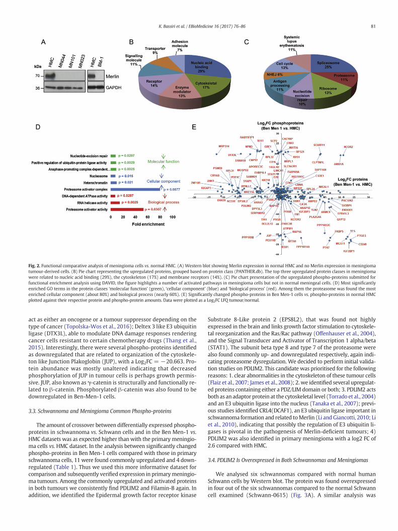
Fig. 2. Functional comparative analysis of meningioma cells vs. normal HMC. (A) Western blot showing Merlin expression in normal HMC and no Merlin expression in meningiomatumour-derived cells. (B) Pie chart representing the upregulated proteins, grouped based on protein class (PANTHER.db). The top three upregulated protein classes in meningiomawere related to nucleic acid binding (29%), the cytoskeleton (17%) and membrane receptors (14%). (C) Pie chart presentation of the upregulated phospho-proteins submitted forfunctional enrichment analysis using DAVID, the figure highlights a number of activated pathways in meningioma cells but not in normal meningeal cells. (D) Most significantlyenriched GO terms in the protein classes ‘molecular function’ (green), ‘cellular component’ (blue) and ‘biological process’ (red). Among them the proteasome was found the mostenriched cellular component (about 80%) and biological process (nearly 60%). (E) Significantly changed phospho-proteins in Ben Men-1 cells vs. phospho-proteins in normal HMCplotted against their respective protein and phospho-protein amounts. Data were plotted as a Log2FC LFQ tumour/normal.
81K. Bassiri et al. / EBioMedicine 16 (2017) 76–86
act as either an oncogene or a tumour suppressor depending on thetype of cancer (Topolska-Wos et al., 2016); Deltex 3 like E3 ubiquitinligase (DTX3L), able to modulate DNA damage responses renderingcancer cells resistant to certain chemotherapy drugs (Thang et al.,2015). Interestingly, there were several phospho-proteins identifiedas downregulated that are related to organization of the cytoskele-ton like Junction Plakoglobin (JUP), with a Log2FC = −20.663. Pro-tein abundance was mostly unaltered indicating that decreasedphosphorylation of JUP in tumour cells is perhaps growth permis-sive. JUP, also known as γ-catenin is structurally and functionally re-lated to β-catenin. Phosphorylated β-catenin was also found to bedownregulated in Ben-Men-1 cells.
3.3. Schwannoma and Meningioma Common Phospho-proteins
The amount of crossover between differentially expressed phospho-proteins in schwannoma vs. Schwann cells and in the Ben Men-1 vs.HMC datasets was as expected higher than with the primary meningio-ma cells vs. HMC dataset. In the analysis between significantly changedphospho-proteins in Ben Men-1 cells compared with those in primaryschwannoma cells, 11 were found commonly upregulated and 4 down-regulated (Table 1). Thus we used this more informative dataset forcomparison and subsequently verified expression in primarymeningio-ma tumours. Among the commonly upregulated and activated proteinsin both tumours we consistently find PDLIM2 and Filamin-B again. Inaddition, we identified the Epidermal growth factor receptor kinase
Substrate 8-Like protein 2 (EPS8L2), that was found not highlyexpressed in the brain and links growth factor stimulation to cytoskele-tal reorganization and the Ras/Rac pathway (Offenhauser et al., 2004),and the Signal Transducer and Activator of Transcription 1 alpha/beta(STAT1). The subunit beta type 8 and type 7 of the proteasome werealso found commonly up- and downregulated respectively, again indi-cating proteasome dysregulation. We decided to perform initial valida-tion studies on PDLIM2. This candidate was prioritised for the followingreasons: 1. clear abnormalities in the cytoskeleton of these tumour cells(Flaiz et al., 2007; James et al., 2008); 2. we identified several upregulat-ed proteins containing either a PDZ/LIM domain or both; 3. PDLIM2 actsboth as an adaptor protein at the cytoskeletal level (Torrado et al., 2004)and an E3 ubiquitin ligase into the nucleus (Tanaka et al., 2007); previ-ous studies identified CRL4(DCAF1), an E3 ubiquitin ligase important inschwannoma formation and related toMerlin (Li and Giancotti, 2010; Liet al., 2010), indicating that possibly the regulation of E3 ubiquitin li-gases is pivotal in the pathogenesis of Merlin-deficient tumours; 4)PDLIM2 was also identified in primary meningioma with a log2 FC of2.6 compared with HMC.
3.4. PDLIM2 Is Overexpressed in Both Schwannomas and Meningiomas
We analysed six schwannomas compared with normal humanSchwann cells by Western blot. The protein was found overexpressedin four out of the six schwannomas compared to the normal Schwanncell examined (Schwann-0615) (Fig. 3A). A similar analysis was
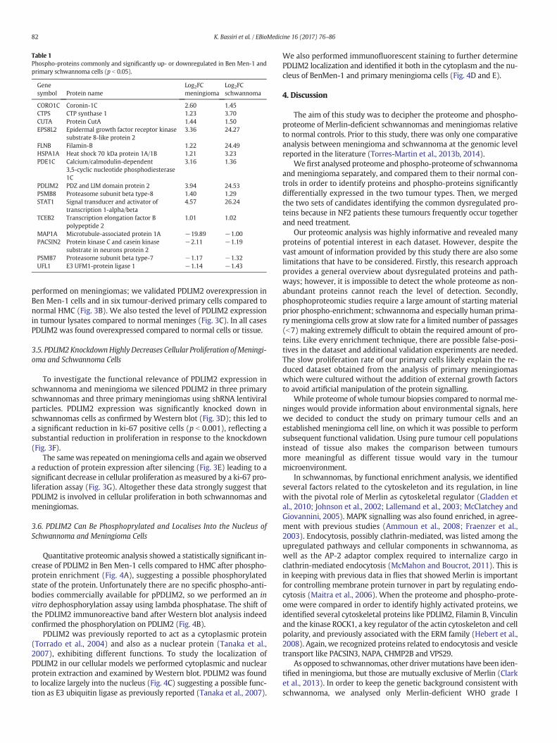
Table 1Phospho-proteins commonly and significantly up- or downregulated in Ben Men-1 andprimary schwannoma cells (p b 0.05).
Genesymbol Protein name
Log2FCmeningioma
Log2FCschwannoma
CORO1C Coronin-1C 2.60 1.45CTPS CTP synthase 1 1.23 3.70CUTA Protein CutA 1.44 1.50EPS8L2 Epidermal growth factor receptor kinase
substrate 8-like protein 23.36 24.27
FLNB Filamin-B 1.22 24.49HSPA1A Heat shock 70 kDa protein 1A/1B 1.21 3.23PDE1C Calcium/calmodulin-dependent
3,5-cyclic nucleotide phosphodiesterase1C
3.16 1.36
PDLIM2 PDZ and LIM domain protein 2 3.94 24.53PSMB8 Proteasome subunit beta type-8 1.40 1.29STAT1 Signal transducer and activator of
transcription 1-alpha/beta4.57 26.24
TCEB2 Transcription elongation factor Bpolypeptide 2
1.01 1.02
MAP1A Microtubule-associated protein 1A −19.89 −1.00PACSIN2 Protein kinase C and casein kinase
substrate in neurons protein 2−2.11 −1.19
PSMB7 Proteasome subunit beta type-7 −1.17 −1.32UFL1 E3 UFM1-protein ligase 1 −1.14 −1.43
82 K. Bassiri et al. / EBioMedicine 16 (2017) 76–86
performed on meningiomas; we validated PDLIM2 overexpression inBen Men-1 cells and in six tumour-derived primary cells compared tonormal HMC (Fig. 3B). We also tested the level of PDLIM2 expressionin tumour lysates compared to normal meninges (Fig. 3C). In all casesPDLIM2 was found overexpressed compared to normal cells or tissue.
3.5. PDLIM2KnockdownHighlyDecreases Cellular Proliferation ofMeningi-oma and Schwannoma Cells
To investigate the functional relevance of PDLIM2 expression inschwannoma and meningioma we silenced PDLIM2 in three primaryschwannomas and three primary meningiomas using shRNA lentiviralparticles. PDLIM2 expression was significantly knocked down inschwannomas cells as confirmed by Western blot (Fig. 3D); this led toa significant reduction in ki-67 positive cells (p b 0.001), reflecting asubstantial reduction in proliferation in response to the knockdown(Fig. 3F).
The samewas repeated onmeningioma cells and again we observeda reduction of protein expression after silencing (Fig. 3E) leading to asignificant decrease in cellular proliferation asmeasured by a ki-67 pro-liferation assay (Fig. 3G). Altogether these data strongly suggest thatPDLIM2 is involved in cellular proliferation in both schwannomas andmeningiomas.
3.6. PDLIM2 Can Be Phosphoprylated and Localises Into the Nucleus ofSchwannoma and Meningioma Cells
Quantitative proteomic analysis showed a statistically significant in-crease of PDLIM2 in Ben Men-1 cells compared to HMC after phospho-protein enrichment (Fig. 4A), suggesting a possible phosphorylatedstate of the protein. Unfortunately there are no specific phospho-anti-bodies commercially available for pPDLIM2, so we performed an invitro dephosphorylation assay using lambda phosphatase. The shift ofthe PDLIM2 immunoreactive band after Western blot analysis indeedconfirmed the phosphorylation on PDLIM2 (Fig. 4B).
PDLIM2 was previously reported to act as a cytoplasmic protein(Torrado et al., 2004) and also as a nuclear protein (Tanaka et al.,2007), exhibiting different functions. To study the localization ofPDLIM2 in our cellular models we performed cytoplasmic and nuclearprotein extraction and examined by Western blot. PDLIM2 was foundto localize largely into the nucleus (Fig. 4C) suggesting a possible func-tion as E3 ubiquitin ligase as previously reported (Tanaka et al., 2007).
We also performed immunofluorescent staining to further determinePDLIM2 localization and identified it both in the cytoplasm and the nu-cleus of BenMen-1 and primary meningioma cells (Fig. 4D and E).
4. Discussion
The aim of this study was to decipher the proteome and phospho-proteome of Merlin-deficient schwannomas and meningiomas relativeto normal controls. Prior to this study, there was only one comparativeanalysis between meningioma and schwannoma at the genomic levelreported in the literature (Torres-Martin et al., 2013b, 2014).
We first analysed proteome andphospho-proteome of schwannomaand meningioma separately, and compared them to their normal con-trols in order to identify proteins and phospho-proteins significantlydifferentially expressed in the two tumour types. Then, we mergedthe two sets of candidates identifying the common dysregulated pro-teins because in NF2 patients these tumours frequently occur togetherand need treatment.
Our proteomic analysis was highly informative and revealed manyproteins of potential interest in each dataset. However, despite thevast amount of information provided by this study there are also somelimitations that have to be considered. Firstly, this research approachprovides a general overview about dysregulated proteins and path-ways; however, it is impossible to detect the whole proteome as non-abundant proteins cannot reach the level of detection. Secondly,phosphoproteomic studies require a large amount of starting materialprior phospho-enrichment; schwannoma and especially human prima-ry meningioma cells grow at slow rate for a limited number of passages(b7) making extremely difficult to obtain the required amount of pro-teins. Like every enrichment technique, there are possible false-posi-tives in the dataset and additional validation experiments are needed.The slow proliferation rate of our primary cells likely explain the re-duced dataset obtained from the analysis of primary meningiomaswhich were cultured without the addition of external growth factorsto avoid artificial manipulation of the protein signalling.
While proteome of whole tumour biopsies compared to normalme-ninges would provide information about environmental signals, herewe decided to conduct the study on primary tumour cells and anestablished meningioma cell line, on which it was possible to performsubsequent functional validation. Using pure tumour cell populationsinstead of tissue also makes the comparison between tumoursmore meaningful as different tissue would vary in the tumourmicroenvironment.
In schwannomas, by functional enrichment analysis, we identifiedseveral factors related to the cytoskeleton and its regulation, in linewith the pivotal role of Merlin as cytoskeletal regulator (Gladden etal., 2010; Johnson et al., 2002; Lallemand et al., 2003; McClatchey andGiovannini, 2005). MAPK signalling was also found enriched, in agree-ment with previous studies (Ammoun et al., 2008; Fraenzer et al.,2003). Endocytosis, possibly clathrin-mediated, was listed among theupregulated pathways and cellular components in schwannoma, aswell as the AP-2 adaptor complex required to internalize cargo inclathrin-mediated endocytosis (McMahon and Boucrot, 2011). This isin keeping with previous data in flies that showed Merlin is importantfor controlling membrane protein turnover in part by regulating endo-cytosis (Maitra et al., 2006). When the proteome and phospho-prote-ome were compared in order to identify highly activated proteins, weidentified several cytoskeletal proteins like PDLIM2, Filamin B, Vinculinand the kinase ROCK1, a key regulator of the actin cytoskeleton and cellpolarity, and previously associated with the ERM family (Hebert et al.,2008). Again, we recognized proteins related to endocytosis and vesicletransport like PACSIN3, NAPA, CHMP2B and VPS29.
As opposed to schwannomas, other drivermutations have been iden-tified in meningioma, but those are mutually exclusive of Merlin (Clarket al., 2013). In order to keep the genetic background consistent withschwannoma, we analysed only Merlin-deficient WHO grade I

Fig. 3. PDLIM2 overexpressed in schwannomas and meningiomas is linked to increased proliferation of tumour cells. (A) Western blot analysis of PDLIM2 expression in primaryschwannoma cells compared to primary human Schwann cell. (B–C) Western blot analysis of PDLIM2 expression in Ben Men-1 and primary meningioma cells compared to HMC (B),and meningioma tumour specimens compared to normal human meninges (C). (D) PDLIM2 shRNA-mediated knockdown in three primary schwannomas, confirmed by the absenceof immunoreactive band in Western blot analysis compared to the sh-scramble control. The samples analysed were; (1) NF1: NF1115 (Fig. 3A), Merlin-positive and pMerlin-positive,NF0116; (2) NF2: NF0116, Merlin-positive and pMerlin faint band (data not shown); (3) NF3: NF0216, Merlin-negative and pMerlin-negative (data not shown). (E) PDLIM2 shRNA-mediated knockdown in three primary meningioma cells, confirmed by the reduction of intensity of the immunoreactive band detected by Western blot analysis compared to the sh-Scramble control. The samples analysed were; (1) MN1: MN026, Merlin-negative and pMerlin-negative (data not shown), (2) MN2: MN028, and (3) MN3: MN031, both Merlin-negative and pMerlin-negative (Fig. 3B). (F) Ki-67 immunofluorescent staining (green) of the three schwannoma cell populations after PDLIM2 shRNA knockdown compared to sh-Scramble control. On the left side the histogram showing the highly statistically significant (***, p b 0.001) reduced proliferation in PDLIM2 knockdown cells. (G) Ki-67immunofluorescent staining (green) of the three primary meningioma cells after PDLIM2 shRNA knockdown compared to sh-Scramble control. On the left side the histogram showingthe statistically significant (*, p b 0.05) reduced proliferation in PDLIM2 knockdown cells. Nuclei are stained with DAPI (Blue). Micrographs are taken at 20× magnification. SC-Scramble; KD-knockdown.
83K. Bassiri et al. / EBioMedicine 16 (2017) 76–86
meningiomas. Comparative functional enrichment analysis in the me-ningiomadatasets identifiedpathways thatmight be of particular impor-tance; among them we found proteasome activation to be a recurringtheme throughout, highlighting it as an important target inmeningioma.A 2014 study looking at the proteasome inhibitor bortezomib showedthat itwas effective in sensitizingmeningioma cells to TRAIL-induced ap-optosis (Koschny et al., 2014). Further, the proteasome inhibitor MG132was also found to increase levels of N-cadherin in schwannoma cells,which in turn decreased proliferation (Zhou et al., 2011). Our data andprevious reports thus suggest proteasome inhibition as a potential ther-apy, either alone or in combination with drugs targeting other relevantpathways. The phosphorylated protein with the largest fold change inprimary meningioma cells was TGM2, or transglutaminase 2. The ex-pression of this protein has been previously studied in meningiomaand was found to be highly upregulated and suggested as a therapeutictarget. The authors also showed that loss of the NF2 gene was associatedwith high expression of TGM2 (Huang et al., 2014). We also foundTAGLN2 as upregulated in meningioma cells, in keeping with previousproteomic studies on meningioma (Sharma et al., 2015). It is similar inits function to transgelin (TAGLN), which we identified as highlyexpressed in schwannoma. The transgelins are a family of proteins able
to influence a diverse range of cellular processes, including proliferation,migration and apoptosis (Dvorakova et al., 2014). The study by Sharmaet al. used a similar proteomic approach to identify potential therapeutictargets usingmeningioma tissue (compared to normal brain) as opposedto cells. Therewere 12 proteins significantly upregulated and common toboth datasets including the LIM domain containing protein FHL1,drebrin, fibronectin and translationally controlled tumour protein(TCTP), all linked with structural regulation.
We also identified possible alterations in DNA repair mechanisms,consistently with previous results showing chromosome instabilityand defects in the mitotic apparatus in meningioma (van Tilborg et al.,2005), in particular in the NF2-mutated (Goutagny et al., 2010). Studiesby Yang et al. (2012) showed that the tumour suppressor CHEK2 onchromosome 22q is often deleted together with Merlin, thus impairingDNA repair mechanisms and increasing chromosomal instability inme-ningiomas (Yang et al., 2012).
BenMen-1 cells, which have a knownNF2mutation, have been usedas a WHO grade I meningioma cell line model and compared to HMC,bearing in mind possible modifications due to immortalization(Puttmann et al., 2005), however helping the study by being an homo-geneous population of cells. The comparison between Ben Men-1 and

Fig. 4. PDLIM2 acts as phosphoprotein and localises into the nucleus. (A) Histogram showing PDLIM2 MS quantification as Log2 LFQ value in Ben Men-1 (BM) cells vs. HMC afterphosphoenrichment. Phosphorylated PDLIM2 was statistically significantly enriched in Ben Men-1 cells (**, p b 0.012) compared to HMC. (B) Western blot analysis confirming thephosphorylated status of PDLIM2 in Ben Men-1 cells. Lambda phosphatase treatment (λ-Ph) induced indeed a shift in PDLIM2 immunoreactive band compared to non-treated (NT)control. (C) Representative Western blot showing PDLIM2 localization after nuclear and cytoplasmic protein fractionation. Total HDAC1 and GAPDH are shown as reference protein forthe nuclear and cytoplasmic fraction respectively. (D) Confocal microscopy (Z-stacks) of PDLIM2 (red) in Ben Men-1 cells and in primary meningioma cells (MN028, MN033, MN036)(E). Nuclei were stained with DAPI (blue).
84 K. Bassiri et al. / EBioMedicine 16 (2017) 76–86
schwannoma datasets compared with controls revealed several com-mon upregulated proteins. Among them we identified the epidermalgrowth factor receptor kinase substrate 8-like protein 2 (EPS8L2), partof the EPS family of proteins related to actin cytoskeleton reorganizationunder growth factors stimulation (Offenhauser et al., 2004); the cyto-skeletal protein Filamin-B (FLNB); and the signal transducer and activa-tor of transcription 1-alpha/beta (STAT1), part of the JAK/STAT1activated in response to interferon and previously found expressed inmeningiomas (Magrassi et al., 1999), currently under validation.
Here we decided to further analyse PDLIM2 for several reasons; weidentified several PDZ/LIM domains proteins throughout the study, in-dicating a possibly important role of this family of proteins in Merlin-deficient tumours. PDLIM2 was first described in 2004 as an adaptorprotein linking other proteins to the cytoskeleton (Torrado et al.,2004), so its dysregulation inMerlin-deficient tumours appeared highlyplausible. Since, it has been found to have a number of different rolesand has been particularly well studied in breast cancer where it hasbeen identified as a driver of tumour progression and invasion (Deeviet al., 2014; Loughran et al., 2005a). In 2007, for the first time PDLIM2was shown to possess nuclear ubiquitin E3 ligase activity negatively
regulating NF-kappaB by targeting the p65 subunit during inflamma-tion (Tanaka et al., 2007). Previous studies already identified anotherE3 ubiquitin ligase, CRL4(DCAF1), involved in the formation of Merlin-deficient tumours (Cooper et al., 2011; Li et al., 2010). Finally, the dys-regulated ubiquitin ligase activity, together with the dysregulatedproteasomal activity found in meningiomas in our study, can suggestnovel therapeutic strategies.
We first confirmed PDLIM2 overexpression in primary meningiomaand schwannoma samples and showed that it is not expressed in HMCor normal meningeal tissue and minimally expressed in the Schwanncell examined. PDLIM2 was significantly knocked down in three prima-ry meningioma and three primary schwannoma cell populations. Thisled to significant reductions in cell proliferation in both cell types.These results are in line with a previous study which showed howPDLIM2 suppression leads to decreased proliferation in androgen-inde-pendent prostate cancer cell lines (Kang et al., 2016). On the other hand,other studies have identified PDLIM2 as an important tumour suppres-sor (Sun et al., 2015; Zhao et al., 2016). Interestingly enough, PDLIM5,that we found highly overexpressed in the schwannoma phospho-pro-teome, was found overexpressed in gastric cancer cells and its siRNA-

85K. Bassiri et al. / EBioMedicine 16 (2017) 76–86
mediated silencing significantly reduced cellular proliferation (Li et al.,2015), highlighting a possible common role for this family as regulatorsof cell proliferation.
Our results showed that PDLIM2 can be phosphorylated. Recentlyone proteomic study identified specific phosphoserine sites onPDLIM2 (Bian et al., 2014); however, no phosphospecific antibodiesare available and the result needs further validation.
Upon subcellular fractionation, PDLIM2 was found to localize intothe nucleus, possibly exploiting E3 ubiquitin ligase activity (Tanaka etal., 2007). ICC analysis showed it localised to both the nucleus and thecytoplasm. It may be that PDLIM2 associates with the cytoskeletonand is thus rendered insoluble during subcellular fractionation, as isthe case with some cytoskeletal proteins e.g. intermediate filaments,explaining why only nuclear PDLIM2 was detectable via Western blot.Our overall results indicate that PDLIM2 has both nuclear and cytoplas-mic functions inmeningioma cells. Additional studieswill be performedto verify whether the protein acts on p65 even in Merlin-negative me-ningiomas and schwannomas, and the role of the phosphorylation onPDLIM2 activity.
In conclusion, we performed a comprehensive analysis of proteomeand phosphoproteome expression in Merlin-deficient schwannomasand meningiomas, found several dysregulated proteins/pathways ineach dataset and underlying known and novel candidates involved inthe pathogenesis of both tumours. Additionally, we validated the over-expression of PDLIM2 which was found involved in the proliferation ofbothmeningioma and schwannoma cells, confirming that PDLIM2war-rants further investigation as a potential common target in Merlin-defi-cient meningiomas and schwannomas.
Supplementary data to this article can be found online at http://dx.doi.org/10.1016/j.ebiom.2017.01.020.
Funding Sources
This study was supported by grants from DHT (Dr. Hadwen Trust);Brain Tumour Research and the Biochemical Society (Eric Reid Fundfor Methodology).
Acknowledgement
We thankDr. David Hilton for providing tumour specimens, the sur-geons from Plymouth and Bristol hospitals and Dr. Emanuela Ercolanofor helping with primary cell cultures.
References
Ammoun, S., Flaiz, C., Ristic, N., Schuldt, J., Hanemann, C.O., 2008. Dissecting and targetingthe growth factor-dependent and growth factor-independent extracellular signal-regulated kinase pathway in human schwannoma. Cancer Res. 68, 5236–5245.
Ammoun, S., Provenzano, L., Zhou, L., Barczyk, M., Evans, K., Hilton, D.A., Hafizi, S.,Hanemann, C.O., 2014. Axl/Gas6/NFkappaB signalling in schwannoma pathologicalproliferation, adhesion and survival. Oncogene 33, 336–346.
Bian, Y., Song, C., Cheng, K., Dong, M., Wang, F., Huang, J., Sun, D., Wang, L., Ye, M., Zou, H.,2014. An enzyme assisted RP-RPLC approach for in-depth analysis of human liverphosphoproteome. J. Proteome 96, 253–262.
Bretscher, A., Chambers, D., Nguyen, R., Reczek, D., 2000. ERM-Merlin and EBP50 proteinfamilies in plasmamembrane organization and function. Annu. Rev. Cell Dev. Biol. 16,113–143.
Clark, V.E., Erson-Omay, E.Z., Serin, A., Yin, J., Cotney, J., Ozduman, K., Avşar, T., Li, J.,Murray, P.B., Henegariu, O., Yilmaz, S., Günel, J.M., Carrión-Grant, G., Yilmaz, B.,Grady, C., Tanrikulu, B., Bakircioğlu, M., Kaymakçalan, H., Caglayan, A.O., Sencar, L.,Ceyhun, E., Atik, A.F., Bayri, Y., Bai, H., Kolb, L.E., Hebert, R.M., Omay, S.B., Mishra-Gorur, K., Choi, M., Overton, J.D., Holland, E.C., Mane, S., State, M.W., Bilgüvar, K.,Baehring, J.M., Gutin, P.H., Piepmeier, J.M., Vortmeyer, A., Brennan, C.W., Pamir,M.N., Kiliç, T., Lifton, R.P., Noonan, J.P., Yasuno, K., Günel, M., 2013. Genomic analysisof non-NF2 meningiomas reveals mutations in TRAF7, KLF4, AKT1, and SMO. Science339, 1077–1080.
Claudio, J.O., Veneziale, R.W., Menko, A.S., Rouleau, G.A., 1997. Expression ofschwannomin in lens and Schwann cells. Neuroreport 8, 2025–2030.
Cooper, J., Li, W., You, L., Schiavon, G., Pepe-Caprio, A., Zhou, L., Ishii, R., Giovannini, M.,Hanemann, C.O., Long, S.B., Erdjument-Bromage, H., Zhou, P., Tempst, P., Giancotti,F.G., 2011. Merlin/NF2 functions upstream of the nuclear E3 ubiquitin ligaseCRL4DCAF1 to suppress oncogenic gene expression. Sci. Signal. 4 (pt6).
Cox, J., Hein, M.Y., Luber, C.A., Paron, I., Nagaraj, N., Mann, M., 2014. Accurate proteome-wide label-free quantification by delayed normalization and maximal peptide ratioextraction, termed MaxLFQ. Mol. Cell. Proteomics 13, 2513–2526.
Curto, M., Cole, B.K., Lallemand, D., Liu, C.H., McClatchey, A.I., 2007. Contact-dependent in-hibition of EGFR signaling by Nf2/Merlin. J. Cell Biol. 177, 893–903.
Deevi, R.K., Cox, O.T., O'Connor, R., 2014. Essential function for PDLIM2 in cell polarizationin three-dimensional cultures by feedback regulation of the beta1-integrin-RhoA sig-naling axis. Neoplasia 16, 422–431.
Dvorakova, M., Nenutil, R., Bouchal, P., 2014. Transgelins, cytoskeletal proteins implicatedin different aspects of cancer development. Expert Rev. Proteomics 11, 149–165.
Fevre-Montange, M., Champier, J., Durand, A., Wierinckx, A., Honnorat, J., Guyotat, J.,Jouvet, A., 2009. Microarray gene expression profiling in meningiomas: differentialexpression according to grade or histopathological subtype. Int. J. Oncol. 35,1395–1407.
Flaiz, C., Kaempchen, K., Matthies, C., Hanemann, C.O., 2007. Actin-rich protrusions andnonlocalized GTPase activation in Merlin-deficient schwannomas. J. Neuropathol.Exp. Neurol. 66, 608–616.
Flaiz, C., Utermark, T., Parkinson, D.B., Poetsch, A., Hanemann, C.O., 2008. Impaired inter-cellular adhesion and immature adherens junctions in merlin-deficient human pri-mary schwannoma cells. Glia 56, 506–515.
Fraenzer, J.T., Pan, H., Minimo Jr., L., Smith, G.M., Knauer, D., Hung, G., 2003. Overexpres-sion of the NF2 gene inhibits schwannoma cell proliferation through promotingPDGFR degradation. Int. J. Oncol. 23, 1493–1500.
Garcia-Rendueles, M.E., Ricarte-Filho, J.C., Untch, B.R., Landa, I., Knauf, J.A., Voza, F.,Smith, V.E., Ganly, I., Taylor, B.S., Persaud, Y., Oler, G., Fang, Y., Jhanwar, S.C.,Viale, A., Heguy, A., Huberman, K.H., Giancotti, F., Ghossein, R., Fagin, J.A., 2015.NF2 loss promotes oncogenic RAS-induced thyroid cancers via YAP-dependenttransactivation of RAS proteins and sensitizes them to MEK inhibition. CancerDiscov. 5, 1178–1193.
Gladden, A.B., Hebert, A.M., Schneeberger, E.E., McClatchey, A.I., 2010. The NF2 tumor sup-pressor, Merlin, regulates epidermal development through the establishment of ajunctional polarity complex. Dev. Cell 19, 727–739.
Goutagny, S., Yang, H.W., Zucman-Rossi, J., Chan, J., Dreyfuss, J.M., Park, P.J., Black, P.M.,Giovannini, M., Carroll, R.S., Kalamarides, M., 2010. Genomic profiling reveals alterna-tive genetic pathways of meningioma malignant progression dependent on the un-derlying NF2 status. Clin. Cancer Res. 16, 4155–4164.
Gronholm, M., Teesalu, T., Tyynela, J., Piltti, K., Bohling, T., Wartiovaara, K., Vaheri, A.,Carpen, O., 2005. Characterization of the NF2 protein merlin and the ERM proteinezrin in human, rat, and mouse central nervous system. Mol. Cell. Neurosci. 28,683–693.
Guerrero, P.A., Yin, W., Camacho, L., Marchetti, D., 2015. Oncogenic role of Merlin/NF2 inglioblastoma. Oncogene 34, 2621–2630.
Hanemann, C.O., 2008. Magic but treatable? Tumours due to loss of merlin. Brain 131,606–615.
Hanemann, C.O., Bartelt-Kirbach, B., Diebold, R., Kampchen, K., Langmesser, S., Utermark,T., 2006. Differential gene expression between human schwannoma and controlSchwann cells. Neuropathol. Appl. Neurobiol. 32, 605–614.
Hebert, M., Potin, S., Sebbagh, M., Bertoglio, J., Breard, J., Hamelin, J., 2008. Rho-ROCK-de-pendent ezrin-radixin-moesin phosphorylation regulates Fas-mediated apoptosis inJurkat cells. J. Immunol. 181, 5963–5973.
Huang da, W., Sherman, B.T., Lempicki, R.A., 2009. Systematic and integrative analysis oflarge gene lists using DAVID bioinformatics resources. Nat. Protoc. 4, 44–57.
Huang, Y.C., Wei, K.C., Chang, C.N., Chen, P.Y., Hsu, P.W., Chen, C.P., Lu, C.S., Wang, H.L.,Gutmann, D.H., Yeh, T.H., 2014. Transglutaminase 2 expression is increased as a func-tion of malignancy grade and negatively regulates cell growth in meningioma. PLoSOne 9, e108228.
James, M.F., Lelke, J.M., Maccollin, M., Plotkin, S.R., Stemmer-Rachamimov, A.O., Ramesh,V., Gusella, J.F., 2008. Modeling NF2 with human arachnoidal and meningioma cellculture systems: NF2 silencing reflects the benign character of tumor growth.Neurobiol. Dis. 29, 278–292.
Johnson, K.C., Kissil, J.L., Fry, J.L., Jacks, T., 2002. Cellular transformation by a FERM domainmutant of the Nf2 tumor suppressor gene. Oncogene 21, 5990–5997.
Kaempchen, K., Mielke, K., Utermark, T., Langmesser, S., Hanemann, C.O., 2003. Upregula-tion of the Rac1/JNK signaling pathway in primary human schwannoma cells. Hum.Mol. Genet. 12, 1211–1221.
Kang, M., Lee, K.H., Lee, H.S., Park, Y.H., Jeong, C.W., Ku, J.H., Kim, H.H., Kwak, C., 2016.PDLIM2 suppression efficiently reduces tumor growth and invasiveness of humancastration-resistant prostate cancer-like cells. Prostate 76, 273–285.
Koschny, R., Boehm, C., Sprick, M.R., Haas, T.L., Holland, H., Xu, L.X., Krupp, W., Mueller,W.C., Bauer, M., Koschny, T., Keller, M., Sinn, P., Meixensberger, J., Walczak, H.,Ganten, T.M., 2014. Bortezomib sensitizes primary meningioma cells to TRAIL-in-duced apoptosis by enhancing formation of the death-inducing signaling complex.J. Neuropathol. Exp. Neurol. 73, 1034–1046.
Lallemand, D., Curto, M., Saotome, I., Giovannini, M., McClatchey, A.I., 2003. NF2 deficiencypromotes tumorigenesis and metastasis by destabilizing adherens junctions. GenesDev. 17, 1090–1100.
Lallemand, D., Manent, J., Couvelard, A., Watilliaux, A., Siena, M., Chareyre, F., Lampin, A.,Niwa-Kawakita, M., Kalamarides, M., Giovannini, M., 2009. Merlin regulates trans-membrane receptor accumulation and signaling at the plasma membrane in primarymouse Schwann cells and in human schwannomas. Oncogene 28, 854–865.
Lasonder, E., Ishihama, Y., Andersen, J.S., Vermunt, A.M., Pain, A., Sauerwein, R.W., Eling,W.M., Hall, N., Waters, A.P., Stunnenberg, H.G., Mann, M., 2002. Analysis of the Plas-modium falciparum proteome by high-accuracy mass spectrometry. Nature 419,537–542.
Lee, H., Hwang, S.J., Kim, H.R., Shin, C.H., Choi, K.H., Joung, J.G., Kim, H.H., 2016. Neurofi-bromatosis 2 (NF2) controls the invasiveness of glioblastoma through YAP-

86 K. Bassiri et al. / EBioMedicine 16 (2017) 76–86
dependent expression of CYR61/CCN1 andmiR-296-3p. Biochim. Biophys. Acta 1859,599–611.
Li, W., Giancotti, F.G., 2010. Merlin's tumor suppression linked to inhibition of the E3ubiquitin ligase CRL4 (DCAF1). Cell Cycle 9, 4433–4436.
Li, W., You, L., Cooper, J., Schiavon, G., Pepe-Caprio, A., Zhou, L., Ishii, R., Giovannini, M.,Hanemann, C.O., Long, S.B., Erdjument-Bromage, H., Zhou, P., Tempst, P., Giancotti,F.G., 2010. Merlin/NF2 suppresses tumorigenesis by inhibiting the E3 ubiquitin ligaseCRL4(DCAF1) in the nucleus. Cell 140, 477–490.
Li, W., Cooper, J., Zhou, L., Yang, C., Erdjument-Bromage, H., Zagzag, D., Snuderl, M.,Ladanyi, M., Hanemann, C.O., Zhou, P., Karajannis, M.A., Giancotti, F.G., 2014. Mer-lin/NF2 loss-driven tumorigenesis linked to CRL4(DCAF1)-mediated inhibition ofthe hippo pathway kinases Lats1 and 2 in the nucleus. Cancer Cell 26, 48–60.
Li, Y., Gao, Y., Xu, Y., Sun, X., Song, X., Ma, H., Yang, M., 2015. si-RNA-mediated knockdownof PDLIM5 suppresses gastric cancer cell proliferation in vitro. Chem. Biol. Drug Des.85, 447–453.
Loughran, G., Healy, N.C., Kiely, P.A., Huigsloot, M., Kedersha, N.L., O'Connor, R., 2005a.Mystique is a new insulin-like growth factor-I-regulated PDZ-LIM domain proteinthat promotes cell attachment and migration and suppresses Anchorage-indepen-dent growth. Mol. Biol. Cell 16, 1811–1822.
Loughran, G., Huigsloot, M., Kiely, P.A., Smith, L.M., Floyd, S., Ayllon, V., O'Connor, R.,2005b. Gene expression profiles in cells transformed by overexpression of the IGF-Ireceptor. Oncogene 24, 6185–6193.
Magrassi, L., De-Fraja, C., Conti, L., Butti, G., Infuso, L., Govoni, S., Cattaneo, E., 1999. Expres-sion of the JAK and STAT superfamilies in human meningiomas. J. Neurosurg. 91,440–446.
Maitra, S., Kulikauskas, R.M., Gavilan, H., Fehon, R.G., 2006. The tumor suppressors Merlinand expanded function cooperatively to modulate receptor endocytosis and signal-ing. Curr. Biol. 16, 702–709.
Martin, K., Rossi, V., Ferrucci, S., Pian, D., 2010. Retinal astrocytic hamartoma. Optometry81, 221–233.
Maudsley, S., Zamah, A.M., Rahman, N., Blitzer, J.T., Luttrell, L.M., Lefkowitz, R.J., Hall, R.A.,2000. Platelet-derived growth factor receptor association with Na(+)/H(+) ex-changer regulatory factor potentiates receptor activity. Mol. Cell. Biol. 20, 8352–8363.
McClatchey, A.I., 2003. Merlin and ERM proteins: unappreciated roles in cancer develop-ment? Nat. Rev. Cancer 3, 877–883.
McClatchey, A.I., Giovannini, M., 2005. Membrane organization and tumorigenesis–theNF2 tumor suppressor, Merlin. Genes Dev. 19, 2265–2277.
McMahon, H.T., Boucrot, E., 2011. Molecular mechanism and physiological functions ofclathrin-mediated endocytosis. Nat. Rev. Mol. Cell Biol. 12, 517–533.
Meimoun, P., Ambard-Bretteville, F., Colas-des Francs-Small, C., Valot, B., Vidal, J., 2007.Analysis of plant phosphoproteins. Anal. Biochem. 371, 238–246.
Mohler, P.J., Kreda, S.M., Boucher, R.C., Sudol, M., Stutts, M.J., Milgram, S.L., 1999. Yes-as-sociated protein 65 localizes p62(c-Yes) to the apical compartment of airway epithe-lia by association with EBP50. J. Cell Biol. 147, 879–890.
Morrison, H., Sperka, T., Manent, J., Giovannini, M., Ponta, H., Herrlich, P., 2007. Merlin/neurofibromatosis type 2 suppresses growth by inhibiting the activation of Ras andRac. Cancer Res. 67, 520–527.
Morrow, K.A., Das, S., Meng, E., Menezes, M.E., Bailey, S.K., Metge, B.J., Buchsbaum, D.J.,Samant, R.S., Shevde, L.A., 2016. Loss of tumor suppressor Merlin results in aberrantactivation of Wnt/beta-catenin signaling in cancer. Oncotarget 7, 17991–18005.
Offenhauser, N., Borgonovo, A., Disanza, A., Romano, P., Ponzanelli, I., Iannolo, G., Di Fiore,P.P., Scita, G., 2004. The eps8 family of proteins links growth factor stimulation toactin reorganization generating functional redundancy in the Ras/Rac pathway.Mol. Biol. Cell 15, 91–98.
Pranjol, M.Z., Gutowski, N., Hannemann, M., Whatmore, J., 2015. The potential role of theproteases cathepsin D and cathepsin L in the progression and metastasis of epithelialovarian cancer. Biomolecules 5, 3260–3279.
Puttmann, S., Senner, V., Braune, S., Hillmann, B., Exeler, R., Rickert, C.H., Paulus, W., 2005.Establishment of a benignmeningioma cell line by hTERT-mediated immortalization.Lab. Investig. 85, 1163–1171.
Qu, Z., Fu, J., Yan, P., Hu, J., Cheng, S.Y., Xiao, G., 2010. Epigenetic repression of PDZ-LIM do-main-containing protein 2: implications for the biology and treatment of breast can-cer. J. Biol. Chem. 285, 11786–11792.
Rappsilber, J., Ishihama, Y., Mann, M., 2003. Stop and go extraction tips for matrix-assistedlaser desorption/ionization, nanoelectrospray, and LC/MS sample pretreatment inproteomics. Anal. Chem. 75, 663–670.
Rouleau, G.A., Merel, P., Lutchman, M., Rouleau, G.A., Merel, P., Lutchman, M., Sanson, M.,Zucman, J., Marineau, C., Hoang-Xuan, K., Demczuk, S., Desmaze, C., Plougastel, B., etal., 1993. Alteration in a new gene encoding a putative membrane-organizing proteincauses neuro-fibromatosis type 2. Nature 363, 515–521.
Sakuda, K., Kohda, Y., Matsumoto, T., Park, C., Seto, A., Tohma, Y., Hasegawa, M., Kida, S.,Nitta, H., Yamashima, T., Yamashita, J., 1996. Expression of NF2 gene product merlinin arachnoid villi and meningiomas. Noshuyo Byori 13, 145–148.
Scherer, S.S., Gutmann, D.H., 1996. Expression of the neurofibromatosis 2 tumor suppres-sor gene product, Merlin, in Schwann cells. J. Neurosci. Res. 46, 595–605.
Schulze, K.M., Hanemann, C.O., Muller, H.W., Hanenberg, H., 2002. Transduction of wild-type merlin into human schwannoma cells decreases schwannoma cell growth andinduces apoptosis. Hum. Mol. Genet. 11, 69–76.
Sharma, S., Ray, S., Moiyadi, A., Sridhar, E., Srivastava, S., 2014. Quantitative proteomicanalysis of meningiomas for the identification of surrogate protein markers. Sci.Rep. 4, 7140.
Sharma, S., Ray, S., Mukherjee, S., Moiyadi, A., Sridhar, E., Srivastava, S., 2015. Multi-pronged quantitative proteomic analyses indicate modulation of various signal trans-duction pathways in human meningiomas. Proteomics 15, 394–407.
Sheikh, H.A., Tometsko,M., Niehouse, L., Aldeeb, D., Swalsky, P., Finkelstein, S., Barnes, E.L.,Hunt, J.L., 2004. Molecular genotyping of medullary thyroid carcinoma can predicttumor recurrence. Am. J. Surg. Pathol. 28, 101–106.
Sun, F., Xiao, Y., Qu, Z., 2015. Oncovirus Kaposi sarcoma herpesvirus (KSHV) repressestumor suppressor PDLIM2 to persistently activate nuclear factor kappaB (NF-kappaB)and STAT3 transcription factors for tumorigenesis and tumor maintenance. J. Biol.Chem. 290, 7362–7368.
Tanaka, T., Grusby, M.J., Kaisho, T., 2007. PDLIM2-mediated termination of transcriptionfactor NF-kappaB activation by intranuclear sequestration and degradation of thep65 subunit. Nat. Immunol. 8, 584–591.
Te Velthuis, A.J., Isogai, T., Gerrits, L., Bagowski, C.P., 2007. Insights into the molecular evo-lution of the PDZ/LIM family and identification of a novel conserved protein motif.PLoS One 2, e189.
Thang, N.D., Yajima, I., Kumasaka, M.Y., Iida, M., Suzuki, T., Kato, M., 2015. Deltex-3-like(DTX3L) stimulates metastasis of melanoma through FAK/PI3K/AKT but not MEK/ERK pathway. Oncotarget 6, 14290–14299.
Topolska-Wos, A.M., Chazin, W.J., Filipek, A., 2016. CacyBP/SIP—structure and variety offunctions. Biochim. Biophys. Acta 1860, 79–85.
Torrado, M., Senatorov, V.V., Trivedi, R., Fariss, R.N., Tomarev, S.I., 2004. Pdlim2, a novelPDZ-LIM domain protein, interacts with alpha-actinins and filamin A. Invest.Ophthalmol. Vis. Sci. 45, 3955–3963.
Torres-Martin, M., Lassaletta, L., de Campos, J.M., Isla, A., Gavilan, J., Pinto, G.R., Burbano,R.R., Latif, F., Melendez, B., Castresana, J.S., Rey, J.A., 2013a. Global profiling in vestib-ular schwannomas shows critical deregulation of microRNAs and upregulation inthose included in chromosomal region 14q32. PLoS One 8, e65868.
Torres-Martin, M., Lassaletta, L., San-Roman-Montero, J., De Campos, J.M., Isla, A., Gavilan,J., Melendez, B., Pinto, G.R., Burbano, R.R., Castresana, J.S., Rey, J.A., 2013b. Microarrayanalysis of gene expression in vestibular schwannomas reveals SPP1/MET signalingpathway and androgen receptor deregulation. Int. J. Oncol. 42, 848–862.
Torres-Martin, M., Lassaletta, L., Isla, A., De Campos, J.M., Pinto, G.R., Burbano, R.R.,Castresana, J.S., Melendez, B., Rey, J.A., 2014. Global expression profile in low grademeningiomas and schwannomas shows upregulation of PDGFD, CDH1 and SLIT2compared to their healthy tissue. Oncol. Rep. 32, 2327–2334.
Tyanova, S., Temu, T., Sinitcyn, P., Carlson, A., Hein, M.Y., Geiger, T., Mann, M., Cox, J., 2016.The Perseus computational platform for comprehensive analysis of (prote)omicsdata. Nat. Methods.
van Tilborg, A.A., Al Allak, B., Velthuizen, S.C., de Vries, A., Kros, J.M., Avezaat, C.J., de Klein,A., Beverloo, H.B., Zwarthoff, E.C., 2005. Chromosomal instability in meningiomas.J. Neuropathol. Exp. Neurol. 64, 312–322.
Wang, X., Gong, Y., Wang, D., Xie, Q., Zheng, M., Zhou, Y., Li, Q., Yang, Z., Tang, H., Li, Y., Hu,R., Chen, X., Mao, Y., 2012. Analysis of gene expression profiling in meningioma:deregulated signaling pathways associated with meningioma and EGFL6 overexpres-sion in benign meningioma tissue and serum. PLoS One 7, e52707.
Weinman, E.J., Steplock, D., Donowitz, M., Shenolikar, S., 2000. NHERF associations withsodium-hydrogen exchanger isoform 3 (NHE3) and ezrin are essential for cAMP-me-diated phosphorylation and inhibition of NHE3. Biochemistry 39, 6123–6129.
Yang, H.W., Kim, T.M., Song, S.S., Shrinath, N., Park, R., Kalamarides, M., Park, P.J., Black,P.M., Carroll, R.S., Johnson, M.D., 2012. Alternative splicing of CHEK2 and codeletionwith NF2 promote chromosomal instability in meningioma. Neoplasia 14, 20–28.
Zhang, Y., Wen, Z., Washburn, M.P., Florens, L., 2009. Effect of dynamic exclusion durationon spectral count based quantitative proteomics. Anal. Chem. 81, 6317–6326.
Zhao, B., Li, L., Lei, Q., Guan, K.L., 2010. The Hippo-YAP pathway in organ size control andtumorigenesis: an updated version. Genes Dev. 24, 862–874.
Zhao, L., Yu, C., Zhou, S., Lau, W.B., Lau, B., Luo, Z., Lin, Q., Yang, H., Xuan, Y., Yi, T., Zhao, X.,Wei, Y., 2016. Epigenetic repression of PDZ-LIM domain-containing protein 2 pro-motes ovarian cancer via NOS2-derived nitric oxide signaling. Oncotarget 7,1408–1420.
Zhou, L., Ercolano, E., Ammoun, S., Schmid, M.C., Barczyk, M.A., Hanemann, C.O., 2011.Merlin-deficient human tumors show loss of contact inhibition and activation ofWnt/beta-catenin signaling linked to the PDGFR/Src and Rac/PAK pathways. Neopla-sia 13, 1101–1112.