glutathione peroxidase-like activity of caeruloplasmin as an important lung antioxidant
TRANSCRIPT
Glutathione peroxidase-like activity of caeruloplasmin as an importantlung antioxidant
Y.S. Parka, K. Suzukia, N. Taniguchia, John M.C. Gutteridgeb;*aDepartment of Biochemistry, Osaka University Medical School, Suita, Osaka 565-0871, Japan
bOxygen Chemistry Laboratory, Directorate of Anaesthesia and Critical Care, Royal Brompton and Hare¢eld NHS Trust, Sydney Street,London SW3 6NP, UK
Received 25 June 1999; received in revised form 16 August 1999
Abstract The copper-containing plasma protein caeruloplasmin(Cp) has been shown to possess several oxidase activities, butwith the exception of its ferrous ion oxidising (ferroxidase)activity which so far appear to be of minor biological relevance.Recently, Kim and colleagues (Kim et al. (1998) FEBS Lett. 431,pp. 473^475) observed that Cp can catalytically removehydrogen peroxide in the presence of thiols. Here, we show thatCp can remove both hydrogen peroxide and lipid hydroperoxidesat physiologically relevant concentrations of reduced glutathioneknown to be present in lung and lung lining fluid. The glutathioneperoxidase-like activity of Cp together with its ferroxidaseactivity would completely remove the primary reactants requiredfor both Fenton chemistry and lipid peroxidation.z 1999 Federation of European Biochemical Societies.
Key words: Caeruloplasmin; Antioxidant; Glutathioneperoxidase; Fenton reaction; Lipid peroxidation; Free radical
1. Introduction
The major copper-containing protein of normal humanplasma is caeruloplasmin (Cp), an acute-phase protein withseveral oxidase activities (reviewed in [2]) that is synthesised inthe liver, although several other tissues can also make theprotein [3^5]. In spite of an intensive research interest inCp, its biological functions remain an enigma. The proteinis able to catalyse the oxidation of ferrous ions to the lessreactive ferric state (ferroxidase activity) [6,7]. This observa-tion led us several years ago to propose that Cp functions asan important plasma antioxidant when redox active iron isinvolved in molecular damage [8^9]. Cp contains six tightlybound copper atoms and one loosely associated with the pro-tein which can be removed by chelation (reviewed in [2]). Ithas recently been suggested that native Cp carries this seventhcopper and that this copper makes Cp pro-oxidant towardsthe oxidation of low density lipoproteins [10]. Debate con-cerning the pro-oxidant properties of copper associated withCp continues, however (reviewed in [11]).
Recently, Kim and colleagues observed that Cp destroyshydrogen peroxide (H2O2) when thiols are present [1]. Here,we extend this ¢nding to show that Cp can remove H2O2 andlipid hydroperoxides (LOOH) at concentrations of reducedglutathione (GSH) reported as present in lung tissue [12]and lung lining £uid [12^15]. The lung has recently been iden-ti¢ed as an important site of Cp synthesis in primates [16]with the protein, subsequently, entering lung lining £uid
[16]. Lung lining £uid also contains a mixture of the selenium-containing extracellular glutathione peroxidase (EC-GSHPase) and cellular glutathione peroxidase (C-GSHPase)[17] with the former predominating [17]. Since EC-GSHPaserequires GSH in the mM range [18] for its activity, Cp may bethe more important protein for removing peroxides in lunglining £uid.
2. Materials and methods
2.1. MaterialsCp, linolenic acid, GSH, conalbumin (egg white apotransferrin) and
horseradish peroxidase (HRP) were obtained from Sigma (MO,USA). Myeloperoxidase (MPO), 5,5P-dinitro-bis-(2-nitrobenzoicacid) (DTNB), taurine, trichloroacetic acid (TCA), potassium thiocya-nate (KSCN), FeSO4W7H2O, HEPES and sodium hypochlorite(HOCI) were purchased from Wako (Osaka, Japan), Sephadex G25was from Pharmacia (Uppasla, Sweden) and Amplex Red (N-acetyl-3-7-dihydroxyphenoxazine) was obtained from Molecular Probes (OR,USA).
2.2. Puri¢cation of human CpCp obtained as a solution (50 mg/ml) was further puri¢ed by pass-
ing it through a column of Sephadex G25 and eluting with 50 mMsodium phosphate bu¡er (pH 7.4). The protein concentration wasdetermined with the Bradford method using bovine serum albuminas a standard.
2.3. Preparation of LOOHsA thin ¢lm layer of methyl linolenic acid was formed on the inner
surface of a £ask by rotation [19]. This was allowed to auto-oxidise inair at 37³C. After 48 h, it was suspended in methanol and used with-out further puri¢cation. The amount of hydroperoxide present wascalculated from the UV absorption at 234 nm (O= 27 400 M31 cm31).
2.4. Measurement of thiol-linked peroxidase activities2.4.1. Hydrogen peroxide. The peroxidase activity of Cp was de-
termined by two methods using KSCN [20] and a £uorescence probe[21]. The reaction was started by addition of 500 WM H2O2 into a 50 Wlreaction mixture containing 0.5^500 WM GSH, 50 mM HEPES bu¡er(pH 7.0) and an appropriate amount of sample, for incubation at37³C. After 40 min, the 40 Wl reaction mixture was added to 0.8 mlTCA solution to stop the reaction, followed by addition of 200 Wl10 mM FeSO4 and 100 Wl KSCN to develop the purple colour. Meas-urement of the remaining H2O2 was performed by monitoring thedecrease in absorbance at 480 nm. A 10 Wl residue was assayed bythe £uorescence method using an Amplex Red probe. Amplex Red,10 WM, was incubated with the reaction mixture and 1 U/ml HRP in50 mM Tris, pH 7.4, at room temperature for 5 min before a 10-folddilution prior to measurement. The £uorescence intensity of the re-action mixture was measured using a £uorescence microplate readerwith a ¢lter set for excitation and emission at 560 and 595 nm, re-spectively (Biolumin 960, Molecular Dynamic).
2.4.2. LOOHs. The reaction was started by addition of 180 WMLOOH into 50 Wl of the reaction mixture containing 0.5^500 WMHEPES bu¡er (pH 7.0) and an appropriate amount of sample forincubation at 37³C. After 40 min, the remaining hydroperoxide wasdetermined as above using the KSCN method.
0014-5793 / 99 / $20.00 ß 1999 Federation of European Biochemical Societies. All rights reserved.PII: S 0 0 1 4 - 5 7 9 3 ( 9 9 ) 0 1 1 4 2 - 4
*Corresponding author. Fax: (44) (171) 351 8524.
FEBS 22585 8-9-99
FEBS 22585 FEBS Letters 458 (1999) 133^136

2.5. Measurement of ferroxidase activity of CpFerroxidase activity of Cp was assayed by measuring the oxidation
of ferrous ions to the ferric state, at pH 5.5. Ferric ions bind toapotransferrin to produce a pink complex (A460 nm) [22]. In this assay,conalbumin (egg white apotransferrin) was substituted for apotrans-ferrin.
2.6. Measurement of hypochlorous acid formationGeneration of HOCI was determined as taurine-chloramine forma-
tion [23]. 1 mM thio-bis-(2-nitrobenzoic acid) (TNB) was prepared bydissolving 2 mM DTNB in 50 mM phosphate bu¡er, pH 7.4. Thesolution of DTNB was titrated to pH 12.0 with sodium hydroxide to
promote its hydrolysis and after 5 min, the pH was brought back topH 7.4 with hydrochloric acid. Reactions were started by addingH2O2, Cp and NaCI in bu¡er containing 10 mM taurine and incu-bated for 10 min at 37³C. TNB was added to the sample solution andincubated for 5 min. The A412 nm was determined and the amount ofHOCI generated was calculated from a standard curve using aliquotsof 10 mM taurine bu¡er to which known amounts of HOCI (A292 nmO= 350 M31 cm31) were added.
3. Results and discussion
Intact human Cp has recently been ascribed a thiol-linkedperoxidase activity [1] which we here show can remove H2O2
and LOOH at physiologically relevant concentrations of GSH(Figs. 1 and 2) known to be present in lung tissue [12] andlung lining £uid [12^15]. GSH at ¢nal reaction concentrationsof 50^500 WM supported the peroxidase activities of Cp.Whilst H2O2 was being removed, chloride ions enhanced theperoxidase activity as previously observed [1], but made no
Fig. 1. GSH-dependent removal of H2O2 by Cp. 10 WM Cp (65 Wg)and 500 WM H2O2 were incubated with various GSH concentrationsfor 40 min. The peroxidase activities were assayed by the KSCNmethod. The amount of hydrogen peroxide removed was calculatedfrom a standard curve using known amounts of H2O2. Data aremeans þ S.D. of triplicate experiments.
Fig. 2. GSH-dependent removal of LOOH by Cp. 10 WM Cp and180 WM hydroperoxide were incubated with various GSH concentra-tions for 40 min. The peroxidase activities were assayed by theKSCN method. The amount of hydroperoxide removed was calcu-lated from a standard curve using known amounts of LOOH thatwere added. Data are means þ S.D. of triplicate experiments.
Fig. 3. The e¡ect of chloride ions on peroxide removal (H2O2 andLOOH). Reaction conditions were as described under Section 2. (A)H2O2 : The black bars and hatched bars correspond to the KSCNand the £uorescence assays, respectively. Concentrations in the reac-tion solution were 10 WM Cp, 500 WM H2O2, 500 WM GSH, 150mM NaCl, respectively. (B) LOOH: Reaction solution contained 10WM Cp, 180 WM LOOH, 500 WM GSH, 150 mM NaCl, respectively.Data are means þ S.D. of triplicate experiments.
FEBS 22585 8-9-99
Y.S. Park et al./FEBS Letters 458 (1999) 133^136134
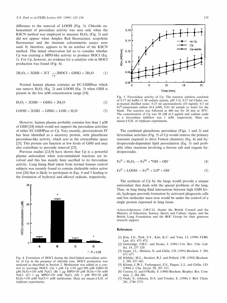
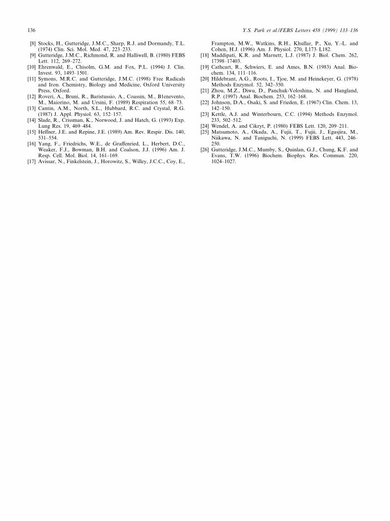
di¡erence to the removal of LOOH (Fig. 3). Chloride en-hancement of peroxidase activity was seen only when theKSCN method was employed to measure H2O2 (Fig. 3) anddid not appear when Amplex Red £uorescence, scopoletin£uorescence and the titanium colourimetric assays wereused. It, therefore, appears to be an artefact of the KSCNmethod. This initial observation led us to consider whetherCp was exerting a MPO-like activity to produce HOCI (Eq.1). For Cp, however, no evidence for a catalytic role in HOCIproduction was found (Fig. 4).
2H2O2 � 2GSH� 2CI3ÿ!Cp
2HOCI�GSSG� 2H2O �1�
Normal human plasma contains an EC-GSHPase whichcan remove H2O2 (Eq. 2) and LOOH (Eq. 3) when GSH ispresent in the low mM concentration range [18].
H2O2 � 2GSH! GSSG� 2H2O �2�
LOOH� 2GSH! GSSG� LOH�H2O �3�
However, human plasma probably contains less than 1 WMof GSH [24] which would not support the peroxidase activitiesof either EC-GSHPase or Cp. Very recently, peroxiredoxin IVhas been identi¢ed as a secretory protein, with glutathioneperoxidase-like activity, which acts in the extracellular space[25]. This protein can function at low levels of GSH and mayalso contribute to peroxide removal [25].
Previous studies [2,8,9] have shown that Cp is a powerfulplasma antioxidant when iron-stimulated reactions are in-volved and this has mainly been ascribed to its ferroxidaseactivity. Lung lining £uid taken from normal human controlsubjects was recently found to contain chelatable redox activeiron [26] that is likely to participate in Eqs. 4 and 5 leading tothe formation of hydroxyl and alkoxyl radicals, respectively.
The combined glutathione peroxidase (Figs. 1 and 2) andferroxidase activities (Fig. 5) of Cp would remove the primaryreactants required to drive Fenton chemistry (Eq. 4) and hy-droperoxide-dependent lipid peroxidation (Eq. 5) and prob-ably other reactions involving a ferrous salt and organic hy-droperoxides.
FeII �H2O2 ! FeIII � �OH�OH3 �4�
FeII � LOOH! FeIII � LO� �OH3 �5�
The synthesis of Cp by the lungs would provide a uniqueantioxidant that deals with the special problems of the lung.Thus, in lung lining £uid interactions between high GSH lev-els, hydrogen peroxide formation by activated phagocytic cellsand low molecular mass iron would be under the control of asingle protein expressed in lung tissue.
Acknowledgements: J.M.C.G. thanks the British Council and theMinistry of Education, Science, Sports and Culture, Japan, and theBritish Lung Foundation and the BOC Group for their generousresearch support.
References
[1] Kim, I.G., Park, S.Y., Kim, K.C. and Yum, J.J. (1998) FEBS.Lett. 431, 473^475.
[2] Gutteridge, J.M.C. and Stocks, J. (1981) Crit. Rev. Clin. Lab.Sci. 14, 257^329.
[3] Jaeger, J.L., Shimizu, N. and Gitlin, J.D. (1991) Biochem. J. 280,671^677.
[4] Schilsky, M.L., Stockert, R.J. and Pollard, J.W. (1992) Biochem.J. 288, 657^661.
[5] Klomp, L.W.J., Farhangrazi, Z.S., Dugan, L.L. and Gitlin, J.D.(1996) J. Clin. Invest. 98, 207^215.
[6] Curzon, G. and O'Reilly, S. (1960) Biochem. Biophys. Res. Com-mun. 2, 284^286.
[7] Osaki, S., Johnson, D.A. and Frieden, E. (1996) J. Biol. Chem.241, 2746^2751.
Fig. 4. Formation of HOCI during the thiol-linked peroxidase activ-ity of Cp in the presence of chloride ions. HOCI production wasanalysed as described in Section 2. Methionine was added as a con-trol to scavenge HOCI. (A) 1 WM Cp (130 Wg)+500 WM GSH+50WM H2O2+150 mM NaCl. (B) 1 Wg MPO+50 WM H2O2+150 mMNaCl. (C) 1 Wg MPO+150 mM NaCl. (D) 1 WM PO+50 WMH2O2+150 mM NaCl+1 mM methionine. Data are means þ S.D. oftriplicate experiments.
Fig. 5. Ferroxidase activity of Cp. The reaction solution consistedof 0.17 ml bu¡er (1 M sodium acetate, pH 5.5), 0.27 ml Chelex res-in-treated distilled water, 0.25 ml apotransferrin (10 mg/ml), 0.3 mlFeII-ammonium sulfate (0.4 mM), 0.01 ml sample or water for theblank. The reaction was followed at 460 nm for 20 min at 30³C.The concentration of Cp was 50 nM (6.5 Wg/ml) and sodium azideas a ferroxidase inhibitor was 1 mM, respectively. Data aremeans þ S.D. of triplicate experiments.
FEBS 22585 8-9-99
Y.S. Park et al./FEBS Letters 458 (1999) 133^136 135
[8] Stocks, H., Gutteridge, J.M.C., Sharp, R.J. and Dormandy, T.L.(1974) Clin. Sci. Mol. Med. 47, 223^233.
[9] Gutteridge, J.M.C., Richmond, R. and Halliwell, B. (1980) FEBSLett. 112, 269^272.
[10] Ehrenwald, E., Chisolm, G.M. and Fox, P.L. (1994) J. Clin.Invest. 93, 1493^1501.
[11] Symons, M.R.C. and Gutteridge, J.M.C. (1998) Free Radicalsand Iron. Chemistry, Biology and Medicine, Oxford UniversityPress, Oxford.
[12] Roveri, A., Bruni, R., Baristussio, A., Coassin, M., B1enevento,M., Maiorino, M. and Ursini, F. (1989) Respiration 55, 68^73.
[13] Cantin, A.M., North, S.L., Hubbard, R.C. and Crystal, R.G.(1987) J. Appl. Physiol. 63, 152^157.
[14] Slade, R., Crissman, K., Norwood, J. and Hatch, G. (1993) Exp.Lung Res. 19, 469^484.
[15] He¡ner, J.E. and Repine, J.E. (1989) Am. Rev. Respir. Dis. 140,531^554.
[16] Yang, F., Friedrichs, W.E., de Gra¡enried, L., Herbert, D.C.,Weaker, F.J., Bowman, B.H. and Coalson, J.J. (1996) Am. J.Resp. Cell. Mol. Biol. 14, 161^169.
[17] Avissar, N., Finkelstein, J., Horowitz, S., Willey, J.C.C., Coy, E.,
Frampton, M.W., Watkins, R.H., Khullar, P., Xu, Y.-L. andCohen, H.J. (1996) Am. J. Physiol. 270, L173^L182.
[18] Maddipati, K.R. and Marnett, L.J. (1987) J. Biol. Chem. 262,17398^17403.
[19] Cathcart, R., Schwiers, E. and Ames, B.N. (1983) Anal. Bio-chem. 134, 111^116.
[20] Hildebrant, A.G., Roots, I., Tjoe, M. and Heinekeyer, G. (1978)Methods Enzymol. 52, 342^350.
[21] Zhou, M.Z., Diwu, D., Panchuk-Voloshina, N. and Hangland,R.P. (1997) Anal. Biochem. 253, 162^168.
[22] Johnson, D.A., Osaki, S. and Frieden, E. (1967) Clin. Chem. 13,142^150.
[23] Kettle, A.J. and Winterbourn, C.C. (1994) Methods Enzymol.233, 502^512.
[24] Wendel, A. and Cikryt, P. (1980) FEBS Lett. 120, 209^211.[25] Matsumoto, A., Okada, A., Fujii, T., Fujii, J., Egasjira, M.,
Niikawa, N. and Taniguchi, N. (1999) FEBS Lett. 443, 246^250.
[26] Gutteridge, J.M.C., Mumby, S., Quinlan, G.J., Chung, K.F. andEvans, T.W. (1996) Biochem. Biophys. Res. Commun. 220,1024^1027.
FEBS 22585 8-9-99
Y.S. Park et al./FEBS Letters 458 (1999) 133^136136