glutathione supplementation attenuates
DESCRIPTION
"Glutathione supplementation attenuates lipopolysaccharide-induced mitochondrial dysfunction and apoptosis in a mouse model of acute lung injury" appeared in a 2012 issue of 'Physiology' and summarized the research Stephen M. Black and his team conducted into acute lung injury. Learn more about his research on his blog: http://stephenmblack.com/blog/TRANSCRIPT
ORIGINAL RESEARCH ARTICLEpublished: 28 May 2012
doi: 10.3389/fphys.2012.00161
Glutathione supplementation attenuateslipopolysaccharide-induced mitochondrial dysfunction andapoptosis in a mouse model of acute lung injurySaurabh Aggarwal 1, Christiana Dimitropoulou2, Qing Lu1, Stephen M. Black 1 and Shruti Sharma1*
1 Vascular Biology Center, Georgia Health Sciences University, Augusta, GA, USA2 Department of Pharmacology and Toxicology, Georgia Health Sciences University, Augusta, GA, USA
Edited by:
Gregg Rokosh, University ofLouisville, USA
Reviewed by:
Scarlet Y. Shi, Medical College ofWisconsin, USAWenbo Zhang, The University of TexasMedical Branch, USA
*Correspondence:
Shruti Sharma, Vascular BiologyCenter, Georgia Health SciencesUniversity, 1459 Laney Walker Blvd,Augusta, GA 30912, USA.e-mail: [email protected]
Acute lung injury (ALI) is a life threatening condition associated with hypoxemia, diffusealveolar damage, inflammation, and loss of lung function. Lipopolysaccharide (LPS; endo-toxin) from the outer membrane of Gram-negative bacteria is a major virulence factorinvolved in the development of ALI. The depletion of glutathione (GSH), an essential intra-and extra-cellular protective antioxidant, by LPS is an important event that contributes tothe elevation in reactive oxygen species. Whether restoring GSH homeostasis can effec-tively ameliorate mitochondrial dysfunction and cellular apoptosis in ALI is unknown andtherefore, was the focus of this study. In peripheral lung tissue of LPS-treated mice, hydro-gen peroxide and protein nitration levels were significantly increased. Pre-treatment withGSH-ethyl ester (GSH-EE) prevented this increase in oxidative stress. LPS also increasedthe lactate/pyruvate ratio, attenuated SOD2 protein levels, and decreased ATP levels in themouse lung indicative of mitochondrial dysfunction. Again, GSH-EE treatment preservedthe mitochondrial function. Finally, our studies showed that LPS induced an increase in themitochondrial translocation of Bax, caspase 3 activation, and nuclear DNA fragmentationand these parameters were all prevented with GSH-EE. Thus, this study suggests thatGSH-EE supplementation may reduce the mitochondrial dysfunction associated with ALI.
Keywords: acute lung injury, mitochondrial dysfunction, apoptosis, lipopolysaccharide, glutathione ethyl ester
INTRODUCTIONAcute lung injury (ALI) is an acute inflammatory disorder associ-ated with both high morbidity and mortality in afflicted patientseven under intensive care. ALI is characterized by non-cardiogenicdyspnea, hypoxemia, pulmonary neutrophil sequestration, andlow lung compliance (Martinez et al., 2009). ALI can occur inresponse to direct insults, such as viral or bacterial infections ofthe lung, hyperoxia, and acid aspiration, or indirect insults likesepsis, multiple transfusions, and pancreatitis. Lipopolysaccha-ride (LPS) from the outer cell wall of Gram-negative bacteria thatcause sepsis is the most common indirect pulmonary insult lead-ing to ALI (Erickson et al., 2009). LPS provokes damage to thealveolar-capillary membrane and the adhesion, activation, andsequestration of polymorphonuclear neutrophils (PMN), whichresult in the deterioration of gas exchange (Nagase et al., 2003).Although several therapies, such as corticosteroids, prostacyclins,exogenous surfactants, ketoconazole, and nitric oxide, have shownpromising outcomes, the estimated mortality rate in ALI is 38.5%(Rubenfeld et al., 2005). Therefore, there is a further need to studythe pathophysiology of ALI and identify therapeutic targets toimprove patient outcome.
Glutathione (GSH), a tripeptide thiol, is the most potentantioxidant found in the cell and plays a protective role againstreactive oxygen and nitrogen species (ROS and RNS) mediatedinjury and lung inflammation (Li et al., 1994; van Klaveren et al.,
1997). This antioxidant is also involved in the regulation of apop-tosis, cell proliferation, and gene transcription (Rahman and Mac-Nee, 2000b; Luppi et al., 2005). GSH also modifies protein sulfdrylgroups by a number of reactions: reduction of protein sulfenicacids, formation of protein mixed disulfides, and their subsequentreduction. The conjugation of GSH with electrophilic compoundsmediated by glutathione S-transferases (GSTs) and the subsequentexcretion of these conjugates from the cell also serve to protectagainst toxins, such as LPS. GSH and GSH-associated enzymespresent in the lower respiratory tract act as the first line of defenseagainst external agents (DeLeve and Kaplowitz, 1990; Pacht et al.,1991). Alterations in alveolar and lung GSH metabolism have beenshown to be a central feature of many inflammatory lung diseases,such as acute respiratory distress syndrome (ARDS), cystic fibro-sis, and asthma (Rahman and MacNee, 2000a). GSH levels aresignificantly reduced in bronchoalveolar lavage fluid (BALF) frompatients with ALI (Pacht et al., 1991). Further, it has been reportedthat disruptions in thiol status are associated with ALI (Quinlanet al., 1994, 1997).
Apoptosis, or programmed cell death, is an essential phys-iological phenomenon responsible for the selective eliminationof cells. However, the dysregulation of apoptotic signaling path-ways is thought to play an important role in the developmentof ALI (Z’Graggen et al., 2010). Several studies have shown com-pelling evidence that increased epithelial/endothelial cell apoptosis
www.frontiersin.org May 2012 | Volume 3 | Article 161 | 1
Aggarwal et al. Glutathione supplementation and acute lung injury
significantly contributes to the damage to the pulmonary alveolar-capillary interface in ALI (Kitamura et al., 2001). The inhibition ofapoptosis increases survival in animal models of LPS-induced ALI(Kawasaki et al., 2000; Ma et al., 2010). Previously, we have shownthat key mechanisms of zinc-mediated apoptotic induction inpulmonary endothelial cells include the disruption of cellular glu-tathione homeostasis and decreased mitochondrial ATP synthesis(Wiseman et al., 2010). We found that the acute increases in bothoxidative and nitrosative stress (similar to that occurring in ALI)in endothelial cells led to a significant increase in cellular apopto-sis, which was attenuated by pre-treatment with GSH-ethyl ester(GSH-EE) (Wiseman et al.,2010). Therefore, the focus of this studywas to determine whether GSH supplementation can prevent LPS-mediated ROS/RNS generation, mitochondrial dysfunction, andcellular apoptosis in mouse model of LPS-induced ALI.
MATERIALS AND METHODSANIMAL TREATMENTSAdult male C57BL/6NHsd mice (7–8 weeks; Harlan, Indianapo-lis, IN, USA) were used in all experiments and housed in atemperature-controlled animal facility with 12-h light–dark cycles.All animal care and experimental procedures were approved bythe Committee on Animal Use in Research and Education of theGeorgia Health Sciences University (Augusta, GA, USA). Micewere divided into four experimental groups (n = 6/group); Vehi-cle, LPS, GSH-EE + LPS, and GSH-EE alone, respectively. Micewere injected intraperitoneally either with saline (vehicle) or LPS(6.75 × 10⁴ EU/g body wt), as previously published (Chatterjeeet al., 2008). The group of mice receiving both GSH-EE and LPSwere injected with two separate doses of 2 mmol GSH-EE/kg bodywt (Sigma, St. Louis, MO, USA; Singhal and Jain, 2000). Dose1 was given 2 h prior to LPS and dose 2 was given along withLPS injection. The pH of the GSH-EE solution was adjusted to6.8 by the addition of 2 M NaOH immediately before injection.Mice belonging to GSH-EE alone group received similar GSH-EEdoses. LPS only and GSH-EE + LPS groups of mice were euth-anized 12 h after LPS injection, and the lungs were flushed withice-cold EDTA-PBS, excised, snap-frozen in liquid nitrogen, andstored at −80˚C until used.
LUNG TISSUE HOMOGENATESLung protein extracts were prepared by homogenizing mouselung tissues in Triton lysis buffer (50 mM Tris–HCL, pH 7.6,0.5% Triton X-100, 20% glycerol) containing a protease inhibitorcocktail (Sigma). Extracts were then clarified by centrifugation(14,000 rpm for 10 min at 4˚C). Supernatant fractions were thenassayed for protein concentration using the Bradford reagent (Bio-Rad, Richmond, CA, USA). Mitochondria were isolated from allfour groups of mice lungs using the Mitochondria Isolation Kit(Thermo Fisher Scientific Inc., Rockford, IL, USA) and analyzedby western blot to detect the Bax protein levels.
MEASUREMENT OF GSH LEVELSTissue samples were homogenized in 5% sulfosalicylic acid (SSA)and centrifuged at 12,000 × g for 15 min. Hundred microliters ofsupernatant was brought to neutral pH by using triethanolamineand split into two tubes, one for the total GSH/GSSG and another
to measure the GSSG levels alone (Griffith, 1980). Two microlitersof 2-vinylpyridine (2-VP) was added to the sample tube measuringGSSG levels alone and placed on a rocker for an hour. 2-VP masksGSH and prevents its reaction with 5,5′-dithio-bis(2-nitrobenzoicacid; DTNB), thereby measuring only oxidized GSSG levels. Fivemicroliters of 50 U/mL glutathione reductase was added to boththe tubes to convert GSSG to GSH. The reaction was then initi-ated in both tubes by adding, 50 μL of 6 mM DTNB and 350 μL of0.3 mM NADPH. All solutions were made with 125 mM NaHPO4buffer (pH 7.5) containing 6.3 mM EDTA. The increase in theabsorbance was measured at 412 nm. Reduced GSH levels wereextrapolated from the GSH/GSSG ratio and the oxidized GSSGlevels and then normalized to tissue wet weight.
IN SITU DETECTION OF H2O2 LEVELSThe Amplex Red Reagent (Molecular Probes) was used to detectH2O2 levels in fresh lung tissue samples obtained from micetreated with LPS in the presence or absence of GSH-EE supple-mentation. Briefly, an equal amount (∼10 mg/sample) of lungtissue was incubated at 37˚C for 30 min in master mix solution con-taining Amplex Red reagent, horseradish peroxidase, and a buffersolution. Supernatant was then collected, fluorescence was readat excitation/emission 530/590 nm, and concentrations of H2O2
were determined through extrapolation from a standard curve.Protein in the tissue samples was estimated by BCA assay and usedto normalize the detected H2O2 levels.
MEASUREMENT OF PROTEIN NITRATIONThe total nitrated protein levels were measured in the lunghomogenates of mice treated with LPS in the presence or absenceof GSH-EE via a dot blot procedure. Briefly, 30 μg proteinwas applied to a nitrocellulose membrane pre-soaked with Tris-buffered saline (TBS). After the protein samples were completelytransferred, the membrane was blocked in 5% fat-free milk for 1 h,washed with TBS, and incubated with mouse anti-3-nitrotyrosine(1:100, Calbiochem) antibody overnight. Finally, the membranewas incubated with goat anti-mouse IgG for 2 h. The reactive dotswere visualized using chemiluminescence (Pierce Laboratories) ona Kodak 440CF image station (New Haven, CT, USA). The bandintensity was quantified using Kodak 1D image processing soft-ware. The protein expression was normalized by re-probing withmouse anti β-actin antibody.
WESTERN BLOT ANALYSESThe lung tissue sample homogenates containing 25–50 μg pro-teins were separated on 4–20% denaturing polyacrylamide gelsand transferred to Immunoblot-PVDF membranes (Bio-Rad Lab,Hercules, CA, USA). The membranes were blocked with 5%non-fat dry milk in TBS containing 0.1% Tween (TBST). Afterblocking, each membrane was incubated overnight at 4˚C witheither SOD1 (1:500, custom-made), SOD2 (1:1000, LS Biochem-icals), Bax (1:1000, Cell Signaling, Danvers, MA, USA), cleavedcaspase 3 (1:1000, Cell Signaling, Danvers, MA, USA), cleavedcaspase 7 (1:1000, Cell Signaling, Danvers, MA, USA), Voltage-dependent anion channels (VDAC; 1:500, Cell Signaling, Danvers,MA, USA), or mouse β-actin (1:5000, Sigma), washed with TBST,and then incubated with a IgG-horseradish peroxidase. The reac-tive bands were visualized using chemiluminescence (SuperSignal
Frontiers in Physiology | Oxidant Physiology May 2012 | Volume 3 | Article 161 | 2
Aggarwal et al. Glutathione supplementation and acute lung injury
West Femto substrate kit; Pierce) on a Kodak 440CF image sta-tion. The band intensity was quantified using Kodak 1D imageprocessing software. To normalize for equal protein loading, theblots were re-probed with either β-actin or VDAC.
DETERMINATION OF LACTATE AND PYRUVATE LEVELSThe lung tissues were homogenized in ice-cold 0.5 M perchlo-ric acid and centrifuged at 14,000 rpm for 20 min. The collectedsupernatants were then neutralized with 3 M KHCO3 and usedfor the lactate and pyruvate assays. The relative changes in lac-tate levels were measured using a lactate assay kit (BioVision).The pyruvate levels were determined using the spectrophotometricenzymatic measurement assay at 340 nm, as previously published(Sharma et al., 2008). NADH was used as a cofactor and lactatedehydrogenase as the coenzyme.
ATP DETERMINATIONThe ATP levels were quantified with a commercially available kit(Invitrogen) based on the firefly luciferin-luciferase reaction. Inthis reaction, ATP is consumed and light is emitted when fireflyluciferase catalyzes the oxidation of luciferin. The amount of lightemitted during the reaction is proportional to the availability ofATP. The luminescence was determined using a Fluoroscan Ascentplate luminometer (Thermo Electron, Corp.), and the ATP levelsreported in nanomoles per milligram protein.
MEASUREMENT OF CASPASE 3/7 ACTIVITYCaspase 3/7 activity was detected with the caspase-Glo 3/7 assaykit (Promega Corporation, Australia). Briefly, 100 μL caspase 3/7reagents were added to each well with 50 μL peripheral lunghomogenates containing equal amount of protein and incubatedfor 1 h on rotary shaker at room temperature. The lumines-cence intensity was measured using a Multiskan Microplate readeraccording to the manufacturer’s instructions.
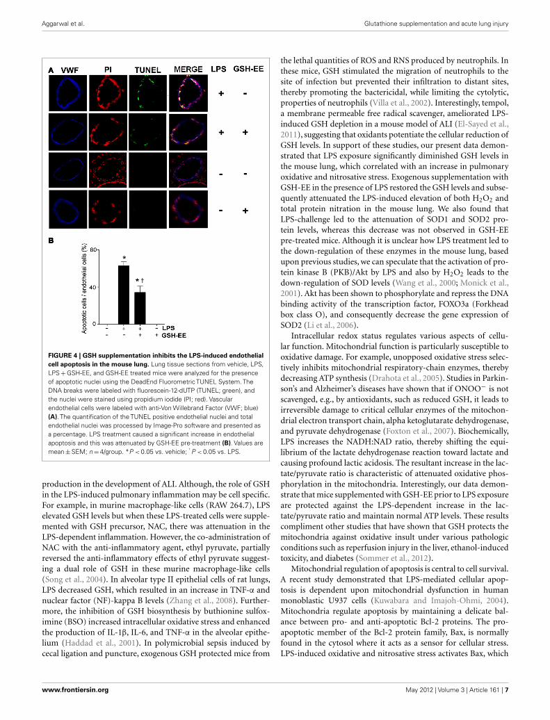
DETERMINATION OF LUNG APOPTOSISThe lung tissues were washed in PBS, fixed in 4% paraformalde-hyde (RT, 1 h), then placed in 30% sucrose (RT, 1 h), embeddedin O.C.T embedding medium (Tissue-Tek, Sakura Finetechnical,Tokyo, Japan), and stored at −80˚C overnight. The embeddedtissue blocks were sectioned to 5 μm slices and were analyzedfor the presence of apoptotic nuclei using the DeadEnd Fluoro-metric TUNEL System (Promega, Madison, WI, USA). The DNAbreaks were labeled with fluorescein-12-dUTP (green), and thenuclei were stained with propidium iodide (red). The vascularendothelial cell nuclei were labeled with anti-Von Willebrand Fac-tor (1:500; Abcam, Cambridge, MA, USA), and Alexa Fluor 350(blue) was used as the secondary antibody (Invitrogen, Carls-bad, CA, USA). The sections were examined under a fluorescencemicroscope (Olympus, Japan). All images were captured by Image-Pro Plus ver 5.0 (Media Cybernetics, Silver Spring, MD, USA).The quantification of the TUNEL positive endothelia nuclei andtotal endothelia nuclei was processed by Image-Pro software andpresented as a percentage.
STATISTICAL ANALYSISThe experimental results are expressed as mean ± standard errorof the mean (SEM) for n = 5–6 for each group. The statistical
analysis was done using GraphPad Prism version 4.01 for Windows(GraphPad Software, San Diego, CA, USA) using one-way analysisof variance (ANOVA) and a Newman–Keuls multiple comparisonpost hoc test. A value of P < 0.05 was considered significant.
RESULTSGSH-EE SUPPLEMENTATION ATTENUATES LPS-INDUCED OXIDATIVEAND NITROSATIVE STRESS IN THE MOUSE LUNGSTo determine the effect of LPS exposure on oxidant and antiox-idant homeostasis, we first measured the glutathione levels inthe peripheral lung tissue of mice treated with or without LPS.There was a significant (approximately threefold) reduction inGSH levels in LPS-treated mouse lung when compared to controls(Figure 1A) that was preserved with GSH-EE supplementation.Earlier studies have provided evidence indicating that ROS andRNS, particularly H2O2 and ONOO−, contributes to the patho-genesis of ALI (Lang et al., 2002; Tasaka et al., 2008). Therefore,we determined whether GSH-EE pre-treatment attenuated LPS-induced oxidative and nitrosative stress in the mouse lung mice.We found elevated H2O2 levels (Figure 1B) and increased pro-tein nitration (Figure 1C). GSH-EE pre-treatment preserved theLPS-induced H2O2, and nitrated protein levels in the mouse lung(Figure 1).
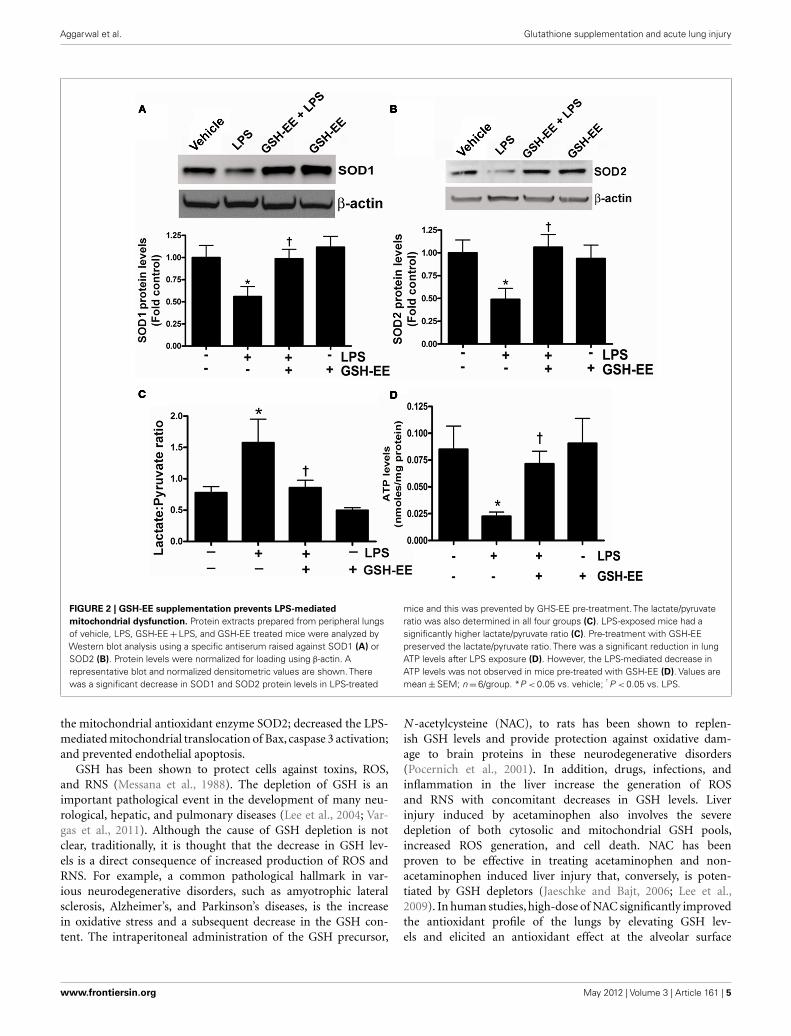
GSH-EE SUPPLEMENTATION PRESERVES MITOCHONDRIAL FUNCTIONThe loss of mitochondrial function upon exposure to LPS has beenimplicated in the development of ALI (Kuwabara and Imajoh-Ohmi, 2004). Superoxide dismutase-1 and 2 (SOD1 and SOD2)are the two important antioxidant enzymes actively involved inquenching harmful superoxide radicals. The lungs of LPS-treatedmice had a approximately twofold decrease in SOD1 and SOD2protein levels (Figures 2A,B), while pre-treatment with GSH-EEpreserved both SOD1 and SOD2 protein levels. Under physiologi-cal conditions where mitochondrial function is optimal, pyruvatelevels are higher than lactate, and thus any increase in lactate levelswill decrease the pyruvate-to-lactate ratio and can be extrapolatedto suggest a switch in mitochondrial ATP generation from glu-coneogenesis to glycolysis (Shinde et al., 2005). The LPS-exposedmice had a significantly higher lactate/pyruvate ratio, thus indi-cating a disruption in lung mitochondrial ATP generation fromgluconeogenesis (Figure 2C) and this was confirmed by an overalldecrease in ATP levels in the LPS-treated mouse lung (Figure 2D).Pre-treatment with GSH-EE blocked the increase in the lac-tate/pyruvate ratio (Figure 2C) and preserved ATP generation(Figure 2D).
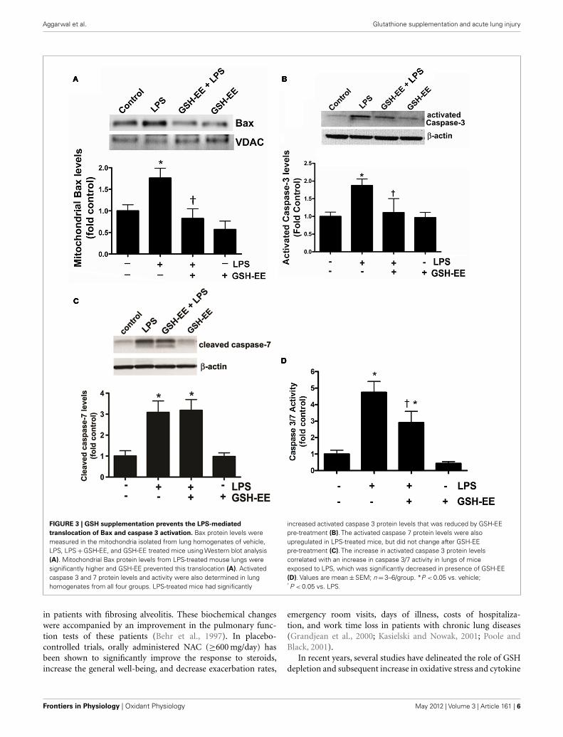
GSH-EE SUPPLEMENTATION PREVENTS THE LPS-INDUCEDMITOCHONDRIAL TRANSLOCATION OF Bax AND CASPASE 3/7ACTIVATIONBax, a member of the Bcl-2 family of proteins, promotes apoptosis.The translocation of Bax from the cytoplasm to the outer mito-chondrial membrane stimulates apoptosis (Sarkar et al., 2003). Wefound higher Bax protein levels in the mitochondria isolated fromthe LPS-treated mice compared to the control mice (Figure 3A).GSH-EE prevented the LPS-induced mitochondrial translocationof Bax (Figure 3A). Activated caspases 3 and 7 are the execu-tors of apoptotic cell death. Western blot analysis demonstrated a
www.frontiersin.org May 2012 | Volume 3 | Article 161 | 3

Aggarwal et al. Glutathione supplementation and acute lung injury
FIGURE 1 | GSH-EE supplementation improves LPS-mediated decrease in
GSH levels and attenuates LPS-induced oxidative and nitrosative stress.
Lungs from vehicle, LPS, GSH-EE + LPS, and GSH-EE treated mice were usedfor the analysis of GSH, H2O2, and nitrated proteins levels. LPS significantlydecreased GSH levels but GSH-EE pre-treatment prevented the loss of GSHin these animals (A). The Amplex Red assay measurement indicated a twofold
increase in H2O2 levels in LPS-treated mice lungs as compared to controls(B). Total nitrated protein levels were measured by dot blot analysis (C). LPScaused significant increases in the formation of H2O2 and nitrated proteins,whereas GSH-EE pre-treatment prevented the LPS-induced increase in theseoxidative and nitrosative stress parameters. Values are mean ± SEM;n = 6/group. *P < 0.05 vs. vehicle; † P < 0.05 vs. LPS alone.
twofold increase in activated caspase 3 (Figure 3B), and a three-fold increase in activated caspase 7 (Figure 3C), protein levels afterLPS exposure. However, pre-treatment with GSH-EE significantlyreduced only activated caspase 3, and not activated caspase 7, levels(Figures 3B,C). Furthermore, using a luminescent assay, we founda fivefold increase in caspase 3/7 activity in LPS-treated mice, ascompared to the controls (Figure 3D). GSH-EE pre-treatmentpartially decreased caspase 3/7 activity (Figure 3D). These resultssuggest that GSH-EE pre-treatment attenuated the LPS-mediatedincrease in caspase 3/7 activity primarily by decreasing activecaspase 3 levels.
GSH-EE SUPPLEMENTATION PROTECTS AGAINST LPS-INDUCEDAPOPTOSISTUNEL staining was carried out to determine the effectof GSH-EE pre-treatment on apoptosis in the LPS-exposed
mouse lungs. Sections were co-stained with TUNEL, a nucleimarker (propidium iodide), and endothelial cell marker (VWF;Figure 4A). The quantitation of TUNEL positive cells indicatedincreased endothelial cell apoptosis in LPS-treated mice lungs,which was attenuated with GSH-EE administration (Figure 4B).In animals receiving vehicle or GSH-EE alone, no apoptotic nucleiwere observed (Figure 4).
DISCUSSIONThe data presented in this study demonstrate that GSH-EE supple-mentation can protect the mouse lung against LPS-induced mito-chondrial dysfunction and subsequent endothelial cell apoptosis.The prior administration of GSH-EE: preserved LPS-mediatedattenuation of GSH levels; reduced the LPS-induced oxidativeand nitrosative stress; mitigated the LPS-mediated disruption ofmitochondrial ATP generation; prevented the LPS-induced loss of
Frontiers in Physiology | Oxidant Physiology May 2012 | Volume 3 | Article 161 | 4
Aggarwal et al. Glutathione supplementation and acute lung injury
FIGURE 2 | GSH-EE supplementation prevents LPS-mediated
mitochondrial dysfunction. Protein extracts prepared from peripheral lungsof vehicle, LPS, GSH-EE + LPS, and GSH-EE treated mice were analyzed byWestern blot analysis using a specific antiserum raised against SOD1 (A) orSOD2 (B). Protein levels were normalized for loading using β-actin. Arepresentative blot and normalized densitometric values are shown. Therewas a significant decrease in SOD1 and SOD2 protein levels in LPS-treated
mice and this was prevented by GHS-EE pre-treatment. The lactate/pyruvateratio was also determined in all four groups (C). LPS-exposed mice had asignificantly higher lactate/pyruvate ratio (C). Pre-treatment with GSH-EEpreserved the lactate/pyruvate ratio. There was a significant reduction in lungATP levels after LPS exposure (D). However, the LPS-mediated decrease inATP levels was not observed in mice pre-treated with GSH-EE (D). Values aremean ± SEM; n = 6/group. *P < 0.05 vs. vehicle; † P < 0.05 vs. LPS.
the mitochondrial antioxidant enzyme SOD2; decreased the LPS-mediated mitochondrial translocation of Bax, caspase 3 activation;and prevented endothelial apoptosis.
GSH has been shown to protect cells against toxins, ROS,and RNS (Messana et al., 1988). The depletion of GSH is animportant pathological event in the development of many neu-rological, hepatic, and pulmonary diseases (Lee et al., 2004; Var-gas et al., 2011). Although the cause of GSH depletion is notclear, traditionally, it is thought that the decrease in GSH lev-els is a direct consequence of increased production of ROS andRNS. For example, a common pathological hallmark in var-ious neurodegenerative disorders, such as amyotrophic lateralsclerosis, Alzheimer’s, and Parkinson’s diseases, is the increasein oxidative stress and a subsequent decrease in the GSH con-tent. The intraperitoneal administration of the GSH precursor,
N -acetylcysteine (NAC), to rats has been shown to replen-ish GSH levels and provide protection against oxidative dam-age to brain proteins in these neurodegenerative disorders(Pocernich et al., 2001). In addition, drugs, infections, andinflammation in the liver increase the generation of ROSand RNS with concomitant decreases in GSH levels. Liverinjury induced by acetaminophen also involves the severedepletion of both cytosolic and mitochondrial GSH pools,increased ROS generation, and cell death. NAC has beenproven to be effective in treating acetaminophen and non-acetaminophen induced liver injury that, conversely, is poten-tiated by GSH depletors (Jaeschke and Bajt, 2006; Lee et al.,2009). In human studies, high-dose of NAC significantly improvedthe antioxidant profile of the lungs by elevating GSH lev-els and elicited an antioxidant effect at the alveolar surface
www.frontiersin.org May 2012 | Volume 3 | Article 161 | 5
Aggarwal et al. Glutathione supplementation and acute lung injury
FIGURE 3 | GSH supplementation prevents the LPS-mediated
translocation of Bax and caspase 3 activation. Bax protein levels weremeasured in the mitochondria isolated from lung homogenates of vehicle,LPS, LPS + GSH-EE, and GSH-EE treated mice using Western blot analysis(A). Mitochondrial Bax protein levels from LPS-treated mouse lungs weresignificantly higher and GSH-EE prevented this translocation (A). Activatedcaspase 3 and 7 protein levels and activity were also determined in lunghomogenates from all four groups. LPS-treated mice had significantly
increased activated caspase 3 protein levels that was reduced by GSH-EEpre-treatment (B). The activated caspase 7 protein levels were alsoupregulated in LPS-treated mice, but did not change after GSH-EEpre-treatment (C). The increase in activated caspase 3 protein levelscorrelated with an increase in caspase 3/7 activity in lungs of miceexposed to LPS, which was significantly decreased in presence of GSH-EE(D). Values are mean ± SEM; n = 3–6/group. *P < 0.05 vs. vehicle;† P < 0.05 vs. LPS.
in patients with fibrosing alveolitis. These biochemical changeswere accompanied by an improvement in the pulmonary func-tion tests of these patients (Behr et al., 1997). In placebo-controlled trials, orally administered NAC (≥600 mg/day) hasbeen shown to significantly improve the response to steroids,increase the general well-being, and decrease exacerbation rates,
emergency room visits, days of illness, costs of hospitaliza-tion, and work time loss in patients with chronic lung diseases(Grandjean et al., 2000; Kasielski and Nowak, 2001; Poole andBlack, 2001).
In recent years, several studies have delineated the role of GSHdepletion and subsequent increase in oxidative stress and cytokine
Frontiers in Physiology | Oxidant Physiology May 2012 | Volume 3 | Article 161 | 6
Aggarwal et al. Glutathione supplementation and acute lung injury
FIGURE 4 | GSH supplementation inhibits the LPS-induced endothelial
cell apoptosis in the mouse lung. Lung tissue sections from vehicle, LPS,LPS + GSH-EE, and GSH-EE treated mice were analyzed for the presenceof apoptotic nuclei using the DeadEnd Fluorometric TUNEL System. TheDNA breaks were labeled with fluorescein-12-dUTP (TUNEL; green), andthe nuclei were stained using propidium iodide (PI; red). Vascularendothelial cells were labeled with anti-Von Willebrand Factor (VWF; blue)(A). The quantification of the TUNEL positive endothelial nuclei and totalendothelial nuclei was processed by Image-Pro software and presented asa percentage. LPS treatment caused a significant increase in endothelialapoptosis and this was attenuated by GSH-EE pre-treatment (B). Values aremean ± SEM; n = 4/group. *P < 0.05 vs. vehicle; † P < 0.05 vs. LPS.
production in the development of ALI. Although, the role of GSHin the LPS-induced pulmonary inflammation may be cell specific.For example, in murine macrophage-like cells (RAW 264.7), LPSelevated GSH levels but when these LPS-treated cells were supple-mented with GSH precursor, NAC, there was attenuation in theLPS-dependent inflammation. However, the co-administration ofNAC with the anti-inflammatory agent, ethyl pyruvate, partiallyreversed the anti-inflammatory effects of ethyl pyruvate suggest-ing a dual role of GSH in these murine macrophage-like cells(Song et al., 2004). In alveolar type II epithelial cells of rat lungs,LPS decreased GSH, which resulted in an increase in TNF-α andnuclear factor (NF)-kappa B levels (Zhang et al., 2008). Further-more, the inhibition of GSH biosynthesis by buthionine sulfox-imine (BSO) increased intracellular oxidative stress and enhancedthe production of IL-1β, IL-6, and TNF-α in the alveolar epithe-lium (Haddad et al., 2001). In polymicrobial sepsis induced bycecal ligation and puncture, exogenous GSH protected mice from
the lethal quantities of ROS and RNS produced by neutrophils. Inthese mice, GSH stimulated the migration of neutrophils to thesite of infection but prevented their infiltration to distant sites,thereby promoting the bactericidal, while limiting the cytolytic,properties of neutrophils (Villa et al., 2002). Interestingly, tempol,a membrane permeable free radical scavenger, ameliorated LPS-induced GSH depletion in a mouse model of ALI (El-Sayed et al.,2011), suggesting that oxidants potentiate the cellular reduction ofGSH levels. In support of these studies, our present data demon-strated that LPS exposure significantly diminished GSH levels inthe mouse lung, which correlated with an increase in pulmonaryoxidative and nitrosative stress. Exogenous supplementation withGSH-EE in the presence of LPS restored the GSH levels and subse-quently attenuated the LPS-induced elevation of both H2O2 andtotal protein nitration in the mouse lung. We also found thatLPS-challenge led to the attenuation of SOD1 and SOD2 pro-tein levels, whereas this decrease was not observed in GSH-EEpre-treated mice. Although it is unclear how LPS treatment led tothe down-regulation of these enzymes in the mouse lung, basedupon previous studies, we can speculate that the activation of pro-tein kinase B (PKB)/Akt by LPS and also by H2O2 leads to thedown-regulation of SOD levels (Wang et al., 2000; Monick et al.,2001). Akt has been shown to phosphorylate and repress the DNAbinding activity of the transcription factor, FOXO3a (Forkheadbox class O), and consequently decrease the gene expression ofSOD2 (Li et al., 2006).
Intracellular redox status regulates various aspects of cellu-lar function. Mitochondrial function is particularly susceptible tooxidative damage. For example, unopposed oxidative stress selec-tively inhibits mitochondrial respiratory-chain enzymes, therebydecreasing ATP synthesis (Drahota et al., 2005). Studies in Parkin-son’s and Alzheimer’s diseases have shown that if ONOO− is notscavenged, e.g., by antioxidants, such as reduced GSH, it leads toirreversible damage to critical cellular enzymes of the mitochon-drial electron transport chain, alpha ketoglutarate dehydrogenase,and pyruvate dehydrogenase (Foxton et al., 2007). Biochemically,LPS increases the NADH:NAD ratio, thereby shifting the equi-librium of the lactate dehydrogenase reaction toward lactate andcausing profound lactic acidosis. The resultant increase in the lac-tate/pyruvate ratio is characteristic of attenuated oxidative phos-phorylation in the mitochondria. Interestingly, our data demon-strate that mice supplemented with GSH-EE prior to LPS exposureare protected against the LPS-dependent increase in the lac-tate/pyruvate ratio and maintain normal ATP levels. These resultscompliment other studies that have shown that GSH protects themitochondria against oxidative insult under various pathologicconditions such as reperfusion injury in the liver, ethanol-inducedtoxicity, and diabetes (Sommer et al., 2012).
Mitochondrial regulation of apoptosis is central to cell survival.A recent study demonstrated that LPS-mediated cellular apop-tosis is dependent upon mitochondrial dysfunction in humanmonoblastic U937 cells (Kuwabara and Imajoh-Ohmi, 2004).Mitochondria regulate apoptosis by maintaining a delicate bal-ance between pro- and anti-apoptotic Bcl-2 proteins. The pro-apoptotic member of the Bcl-2 protein family, Bax, is normallyfound in the cytosol where it acts as a sensor for cellular stress.LPS-induced oxidative and nitrosative stress activates Bax, which
www.frontiersin.org May 2012 | Volume 3 | Article 161 | 7
Aggarwal et al. Glutathione supplementation and acute lung injury
then translocates to the outer mitochondrial membrane where itinteracts with anti-apoptotic proteins (Mishra and Dhali, 2007).This interaction between Bax and anti-apoptotic proteins disruptstheir normal function, which leads to the formation of pores in themitochondria, allowing the exit of cytochrome c. In addition, ROSindependently oxidize mitochondrial pores further contributingto cytochrome c release upon GSH depletion (Costantini et al.,1996). Cytochrome c released from the mitochondria activates thecaspase cascade, which then induces apoptosis. Recent studies haveshown that LPS triggers the release of cytochrome c from the mito-chondria to the cytoplasm, thereby activating the caspase cascadeand DNA fragmentation (Chuang et al., 2011). The pre-treatmentof human alveolar epithelial A549 cells with NAC significantlydecreased LPS-mediated caspase activation and DNA damage, sug-gesting an important role of GSH in ameliorating LPS-inducedcell death (Chuang et al., 2011). The loss of GSH and subsequentdecrease in ATP and ensuing cell death by dopamine metabolitesis prevented by GSH-EE and NAC supplementation, suggestinga critical role of GSH in maintaining functional mitochondria inParkinson’s disease (Nunes et al., 2011). GSH also exhibited anti-apoptotic effects in selenium induced apoptosis in HSC-3 humanoral squamous cell carcinoma cells and arsenic trioxide-inducedapoptosis in lymphoma cells (Takahashi et al., 2005). Thus, ourfindings in the mouse model of ALI concur with these studies and
demonstrate that GSH-EE pre-treatment prevents LPS-inducedpulmonary endothelial cell apoptosis.
In conclusion, the results presented in this study suggest thatthe modulation of the cellular redox equilibrium by GSH pre-serves oxidative phosphorylation in the mitochondria in responseto LPS-induced oxidative damage and subsequent cellular apopto-sis. Although clinical trials involving the use of antioxidants havenot shown promising results in chronically ill patients, the acuteburst of oxidative, and nitrosative stress seen in ALI may be moresusceptible to antioxidant therapy. Based upon past studies andour present data, we conclude that GSH depletion plays a vital rolein the pathogenesis of ALI, and therefore, GSH supplementationmay act as an important adjuvant therapy to reverse the diseaseprocess.
ACKNOWLEDGMENTSThe authors thank Imran Rehmani, Suphin Kallarackal, JohnnyWright, and Sumant Ponnala for their excellent technical assis-tance. This research was supported in part by a Beginning Grant inAid (09BGIA2310050) from the Southeast Affiliates, and a ScientistDevelopment Grant (11SDG7460024; all to Shruti Sharma) fromthe American Heart Association; 1P01HL101902-01A, HL60190,HL67841, and HL101902 (all to Stephen M. Black) from theNational Institutes of Health.
REFERENCESBehr, J., Maier, K., Degenkolb, B.,
Krombach, F., and Vogelmeier, C.(1997). Antioxidative and clinicaleffects of high-dose N-acetylcysteinein fibrosing alveolitis. Adjunctivetherapy to maintenance immuno-suppression. Am. J. Respir. Crit. CareMed. 156, 1897–1901.
Chatterjee, A., Snead, C., Yetik-Anacak,G., Antonova, G., Zeng, J., andCatravas, J. D. (2008). Heat shockprotein 90 inhibitors attenuate LPS-induced endothelial hyperperme-ability. Am. J. Physiol. Lung Cell. Mol.Physiol. 294, L755–L763.
Chuang, C. Y., Chen, T. L., Cherng,Y. G., Tai, Y. T., Chen, T. G., andChen, R. M. (2011). Lipopolysac-charide induces apoptotic insults tohuman alveolar epithelial A549 cellsthrough reactive oxygen species-mediated activation of an intrinsicmitochondrion-dependent pathway.Arch. Toxicol. 85, 209–218.
Costantini, P., Chernyak, B. V., Petron-illi, V., and Bernardi, P. (1996). Mod-ulation of the mitochondrial perme-ability transition pore by pyridinenucleotides and dithiol oxidation attwo separate sites. J. Biol. Chem. 271,6746–6751.
DeLeve, L. D., and Kaplowitz, N.(1990). Importance and regulationof hepatic glutathione. Semin. LiverDis. 10, 251–266.
Drahota, Z., Krivakova, P., Cervinkova,Z., Kmonickova, E., Lotkova, H.,
Kucera, O., and Houstek, J. (2005).Tert-butyl hydroperoxide selectivelyinhibits mitochondrial respiratory-chain enzymes in isolated rathepatocytes. Physiol. Res. 54,67–72.
El-Sayed, N. S., Mahran, L. G., andKhattab, M. M. (2011). Tem-pol, a membrane-permeableradical scavenger, ameliorateslipopolysaccharide-induced acutelung injury in mice: a key role forsuperoxide anion. Eur. J. Pharmacol.663, 68–73.
Erickson, S. E., Martin, G. S., Davis, J.L., Matthay, M. A., and Eisner, M. D.(2009). Recent trends in acute lunginjury mortality: 1996-2005. Crit.Care Med. 37, 1574–1579.
Foxton, R. H., Land, J. M., andHeales, S. J. (2007). Tetrahydro-biopterin availability in Parkin-son’s and Alzheimer’s disease; poten-tial pathogenic mechanisms. Neu-rochem. Res. 32, 751–756.
Grandjean, E. M., Berthet, P., Ruff-mann, R., and Leuenberger, P.(2000). Efficacy of oral long-termN-acetylcysteine in chronic bron-chopulmonary disease: a meta-analysis of published double-blind,placebo-controlled clinical trials.Clin. Ther. 22, 209–221.
Griffith, O. W. (1980). Determinationof glutathione and glutathione disul-fide using glutathione reductase and2-vinylpyridine. Anal. Biochem. 106,207–212.
Haddad, J. J., Safieh-Garabedian,B., Saade, N. E., and Land, S. C.(2001). Thiol regulation of pro-inflammatory cytokines revealsa novel immunopharmacologicalpotential of glutathione in thealveolar epithelium. J. Pharmacol.Exp. Ther. 296, 996–1005.
Jaeschke, H., and Bajt, M. L. (2006).Intracellular signaling mecha-nisms of acetaminophen-inducedliver cell death. Toxicol. Sci. 89,31–41.
Kasielski, M., and Nowak, D. (2001).Long-term administration of N-acetylcysteine decreases hydrogenperoxide exhalation in subjectswith chronic obstructive pul-monary disease. Respir. Med. 95,448–456.
Kawasaki, M., Kuwano, K., Hagi-moto, N., Matsuba, T., Kunitake, R.,Tanaka, T., Maeyama, T., and Hara,N. (2000). Protection from lethalapoptosis in lipopolysaccharide-induced acute lung injury in miceby a caspase inhibitor. Am. J. Pathol.157, 597–603.
Kitamura, Y., Hashimoto, S., Mizuta,N., Kobayashi, A., Kooguchi,K., Fujiwara, I., and Nakajima,H. (2001). Fas/FasL-dependentapoptosis of alveolar cells afterlipopolysaccharide-induced lunginjury in mice. Am. J. Respir. Crit.Care Med. 163, 762–769.
Kuwabara, T., and Imajoh-Ohmi, S.(2004). LPS-induced apoptosis is
dependent upon mitochondrial dys-function. Apoptosis 9, 467–474.
Lang, J. D., McArdle, P. J., O’reilly, P.J., and Matalon, S. (2002). Oxidant-antioxidant balance in acute lunginjury. Chest 122, 314S–320S.
Lee, T. D., Sadda, M. R., Mendler, M.H., Bottiglieri, T., Kanel, G., Mato, J.M., and Lu, S. C. (2004). Abnormalhepatic methionine and glutathionemetabolism in patients with alco-holic hepatitis. Alcohol. Clin. Exp.Res. 28, 173–181.
Lee, W. M., Hynan, L. S., Rossaro, L.,Fontana, R. J., Stravitz, R. T., Lar-son, A. M., Davern, T. J. II, Mur-ray, N. G., McCashland, T., Reisch,J. S., and Robuck, P. R. (2009). Intra-venous N-acetylcysteine improvestransplant-free survival in early stagenon-acetaminophen acute liver fail-ure. Gastroenterology 137, 856.e1–864.e1.
Li, M., Chiu, J. F., Mossman, B.T., and Fukagawa, N. K. (2006).Down-regulation of manganese-superoxide dismutase through phos-phorylation of FOXO3a by Akt inexplanted vascular smooth musclecells from old rats. J. Biol. Chem. 281,40429–40439.
Li, X. Y., Donaldson, K., Rahman, I.,and MacNee, W. (1994). An inves-tigation of the role of glutathionein increased epithelial permeabilityinduced by cigarette smoke in vivoand in vitro. Am. J. Respir. Crit. CareMed. 149, 1518–1525.
Frontiers in Physiology | Oxidant Physiology May 2012 | Volume 3 | Article 161 | 8
Aggarwal et al. Glutathione supplementation and acute lung injury
Luppi, F., Aarbiou, J., Van Wetering, S.,Rahman, I., De Boer, W. I., Rabe, K.F., and Hiemstra, P. S. (2005). Effectsof cigarette smoke condensate onproliferation and wound closure ofbronchial epithelial cells in vitro:role of glutathione. Respir. Res. 6,140.
Ma, X., Xu, D., Ai, Y., Ming, G.,and Zhao, S. (2010). Fas inhibi-tion attenuates lipopolysaccharide-induced apoptosis and cytokinerelease of rat type II alveolarepithelial cells. Mol. Biol. Rep. 37,3051–3056.
Martinez, O., Nin, N., and Esteban,A. (2009). Prone position for thetreatment of acute respiratory dis-tress syndrome: a review of currentliterature. Arch. Bronconeumol. 45,291–296.
Messana, J. M., Cieslinski, D. A.,O’connor, R. P., and Humes, H. D.(1988). Glutathione protects againstexogenous oxidant injury to rab-bit renal proximal tubules. Am. J.Physiol. 255, F874–F884.
Mishra, D. P., and Dhali, A. (2007).Endotoxin induces luteal cell apop-tosis through the mitochondrialpathway. Prostaglandins Other LipidMediat. 83, 75–88.
Monick, M. M., Carter, A. B., Rob-eff, P. K., Flaherty, D. M., Peterson,M. W., and Hunninghake, G. W.(2001). Lipopolysaccharide activatesAkt in human alveolar macrophagesresulting in nuclear accumula-tion and transcriptional activityof beta-catenin. J. Immunol. 166,4713–4720.
Nagase, T., Uozumi, N., Aoki-Nagase,T., Terawaki, K., Ishii, S., Tomita,T., Yamamoto, H., Hashizume, K.,Ouchi, Y., and Shimizu, T. (2003). Apotent inhibitor of cytosolic phos-pholipase A2, arachidonyl trifluo-romethyl ketone, attenuates LPS-induced lung injury in mice. Am. J.Physiol. Lung Cell. Mol. Physiol. 284,L720–L726.
Nunes, C., Barbosa, R. M., Almeida,L., and Laranjinha, J. (2011). Nitricoxide and DOPAC-induced celldeath: from GSH depletion to mito-chondrial energy crisis. Mol. Cell.Neurosci. 48, 94–103.
Pacht, E. R., Timerman, A. P.,Lykens, M. G., and Merola, A.J. (1991). Deficiency of alveolar
fluid glutathione in patients withsepsis and the adult respiratorydistress syndrome. Chest 100,1397–1403.
Pocernich, C. B., Cardin, A. L., Racine,C. L., Lauderback, C. M., and But-terfield, D. A. (2001). Glutathioneelevation and its protective rolein acrolein-induced protein damagein synaptosomal membranes: rele-vance to brain lipid peroxidationin neurodegenerative disease. Neu-rochem. Int. 39, 141–149.
Poole, P. J., and Black, P. N. (2001). Oralmucolytic drugs for exacerbations ofchronic obstructive pulmonary dis-ease: systematic review. BMJ 322,1271–1274.
Quinlan, G. J., Evans, T. W., and Gut-teridge, J. M. (1994). Oxidative dam-age to plasma proteins in adult respi-ratory distress syndrome. Free Radic.Res. 20, 289–298.
Quinlan, G. J., Lamb, N. J., Tilley, R.,Evans, T. W., and Gutteridge, J. M.(1997). Plasma hypoxanthine levelsin ARDS: implications for oxida-tive stress, morbidity, and mortality.Am. J. Respir. Crit. Care Med. 155,479–484.
Rahman, I., and MacNee, W. (2000a).Oxidative stress and regulation ofglutathione in lung inflammation.Eur. Respir. J. 16, 534–554.
Rahman, I., and MacNee, W. (2000b).Regulation of redox glutathionelevels and gene transcription inlung inflammation: therapeuticapproaches. Free Radic. Biol. Med.28, 1405–1420.
Rubenfeld, G. D., Caldwell, E., Peabody,E., Weaver, J., Martin, D. P., Neff,M., Stern, E. J., and Hudson, L. D.(2005). Incidence and outcomes ofacute lung injury. N. Engl. J. Med.353, 1685–1693.
Sarkar, F. H., Rahman, K. M., and Li, Y.(2003). Bax translocation to mito-chondria is an important event ininducing apoptotic cell death byindole-3-carbinol (I3C) treatmentof breast cancer cells. J. Nutr. 133,2434S–2439S.
Sharma, S., Sud, N., Wiseman, D. A.,Carter, A. L., Kumar, S., Hou, Y.,Rau, T., Wilham, J., Harmon, C.,Oishi, P., Fineman, J. R., and Black, S.M. (2008). Altered carnitine home-ostasis is associated with decreasedmitochondrial function and altered
nitric oxide signaling in lambs withpulmonary hypertension. Am. J.Physiol. Lung Cell. Mol. Physiol. 294,L46–L56.
Shinde, S., Golam, K., Kumar, P., Patil,N., and Sadacharan, K. (2005). Peri-operative blood lactate levels, pyru-vate levels, and lactate-pyruvate ratioin children undergoing cardiopul-monary bypass for congenital heartdisease. Indian J. Crit. Care Med. 9,145–150.
Singhal, R. K., and Jain, A. (2000). Glu-tathione ethyl ester supplementationprevents mortality in newborn ratsexposed to hyperoxia. Biol. Neonate77, 261–266.
Sommer, S. P., Sommer, S., Sinha, B.,Walter, D., Aleksic, I., Gohrbandt, B.,Otto, C., and Leyh, R. G. (2012).Glutathione preconditioning ame-liorates mitochondria dysfunctionduring warm pulmonary ischemia-reperfusion injury. Eur. J. Cardiotho-rac. Surg. 41, 140–148.
Song, M., Kellum, J. A., Kaldas, H.,and Fink, M. P. (2004). Evidencethat glutathione depletion is a mech-anism responsible for the anti-inflammatory effects of ethyl pyru-vate in cultured lipopolysaccharide-stimulated RAW 264.7 cells. J. Phar-macol. Exp. Ther. 308, 307–316.
Takahashi, M., Sato, T., Shinohara, F.,Echigo, S., and Rikiishi, H. (2005).Possible role of glutathione in mito-chondrial apoptosis of human oralsquamous cell carcinoma caused byinorganic selenium compounds. Int.J. Oncol. 27, 489–495.
Tasaka, S., Amaya, F., Hashimoto, S., andIshizaka,A. (2008). Roles of oxidantsand redox signaling in the pathogen-esis of acute respiratory distress syn-drome. Antioxid. Redox Signal. 10,739–753.
van Klaveren, R. J., Demedts, M., andNemery, B. (1997). Cellular glu-tathione turnover in vitro, withemphasis on type II pneumocytes.Eur. Respir. J. 10, 1392–1400.
Vargas, M. R., Johnson, D. A., andJohnson, J. A. (2011). Decreasedglutathione accelerates neuro-logical deficit and mitochondrialpathology in familial ALS-linkedhSOD1(G93A) mice model.Neurobiol. Dis. 43, 543–551.
Villa, P., Saccani, A., Sica, A., and Ghezzi,P. (2002). Glutathione protects mice
from lethal sepsis by limitinginflammation and potentiatinghost defense. J. Infect. Dis. 185,1115–1120.
Wang, X., McCullough, K. D., Franke,T. F., and Holbrook, N. J. (2000).Epidermal growth factor receptor-dependent Akt activation by oxida-tive stress enhances cell survival. J.Biol. Chem. 275, 14624–14631.
Wiseman, D. A., Sharma, S., and Black,S. M. (2010). Elevated zinc inducesendothelial apoptosis via disruptionof glutathione metabolism: role ofthe ADP translocator. Biometals 23,19–30.
Z’Graggen, B. R., Tornic, J., Muller-Edenborn, B., Reyes, L., Booy, C., andBeck-Schimmer, B. (2010). Acutelung injury: apoptosis in effectorand target cells of the upper andlower airway compartment. Clin.Exp. Immunol. 161, 324–331.
Zhang, F., Wang, X., Wang, W.,Li, N., and Li, J. (2008). Glu-tamine reduces TNF-alpha byenhancing glutathione synthesisin lipopolysaccharide-stimulatedalveolar epithelial cells of rats.Inflammation 31, 344–350.
Conflict of Interest Statement: Theauthors declare that the research wasconducted in the absence of any com-mercial or financial relationships thatcould be construed as a potential con-flict of interest.
Received: 06 February 2012; accepted:07 May 2012; published online: 28 May2012.Citation: Aggarwal S, Dimitropoulou C,Lu Q, Black SM and Sharma S (2012)Glutathione supplementation attenuateslipopolysaccharide-induced mitochondr-ial dysfunction and apoptosis in a mousemodel of acute lung injury. Front. Physio.3:161. doi: 10.3389/fphys.2012.00161This article was submitted to Frontiersin Oxidant Physiology, a specialty ofFrontiers in Physiology.Copyright © 2012 Aggarwal, Dim-itropoulou, Lu, Black and Sharma. Thisis an open-access article distributed underthe terms of the Creative Commons Attri-bution Non Commercial License, whichpermits non-commercial use, distribu-tion, and reproduction in other forums,provided the original authors and sourceare credited.
www.frontiersin.org May 2012 | Volume 3 | Article 161 | 9