glypican and biglycan in the nuclei of neurons and glioma cells
TRANSCRIPT

The Rockefeller University Press, 0021-9525/97/11/851/14 $2.00The Journal of Cell Biology, Volume 139, Number 4, November 17, 1997 851–864http://www.jcb.org 851
Glypican and Biglycan in the Nuclei of Neurons and GliomaCells: Presence of Functional Nuclear LocalizationSignals and Dynamic Changes in Glypican During the Cell Cycle
Yu Liang,* Monika Häring,
§
Peter J. Roughley,
‡
Renée K. Margolis,
§
and Richard U. Margolis*
*Department of Pharmacology, New York University Medical Center, New York 10016;
‡
Shriners Hospital for Crippled Children, McGill University, Montreal, Canada; and
§
Department of Pharmacology, State University of New York, Health Science Center, Brooklyn, New York 11203
Abstract.
We have investigated the expression patterns and subcellular localization in nervous tissue of glypi-can, a major glycosylphosphatidylinositol-anchored heparan sulfate proteoglycan that is predominantly synthesized by neurons, and of biglycan, a small, leu-cine-rich chondroitin sulfate proteoglycan. By laser scanning confocal microscopy of rat central nervous tis-sue and C6 glioma cells, we found that a significant por-tion of the glypican and biglycan immunoreactivity colocalized with nuclear staining by propidium iodide and was also seen in isolated nuclei. In certain regions, staining was selective, insofar as glypican and biglycan immunoreactivity in the nucleus was seen predomi-nantly in a subpopulation of large spinal cord neurons. The amino acid sequences of both proteoglycans con-tain potential nuclear localization signals, and these were demonstrated to be functional based on their abil-ity to target
b
-galactosidase fusion proteins to the nu-clei of transfected 293 cells. Nuclear localization of
glypican
b
-galactosidase or Fc fusion proteins in trans-fected 293 cells and C6 glioma cells was greatly reduced or abolished after mutation of the basic amino acids or deletion of the sequence containing the nuclear local-ization signal, and no nuclear staining was seen in the case of heparan sulfate and chondroitin sulfate pro-teoglycans that do not possess a nuclear localization signal, such as syndecan-3 or decorin (which is closely related in structure to biglycan). Transfection of COS-1 cells with an epitope-tagged glypican cDNA demon-strated transport of the full-length proteoglycan to the nucleus, and there are also dynamic changes in the pat-tern of glypican immunoreactivity in the nucleus of C6 cells both during cell division and correlated with dif-ferent phases of the cell cycle. Our data therefore sug-gest that in certain cells and central nervous system re-gions, glypican and biglycan may be involved in the regulation of cell division and survival by directly par-ticipating in nuclear processes.
G
lypican,
whose primary structure was first reportedbased on its cloning from human lung fibroblasts(David et al., 1990), was the initial member of a
rapidly expanding family of glycosylphosphatidylinositol-anchored heparan sulfate proteoglycans that currently in-cludes four other vertebrate proteins (cerebroglycan, Stippet al., 1994; OCI-5, Filmus et al., 1995; K-glypican, Wa-tanabe et al., 1995; and glypican-5, Veugelers et al., 1997)as well as the
dally
gene product of
Drosophila
(Nakato et al.,1995). In earlier biochemical studies of the brain (Klingeret al., 1985) and PC12 pheochromocytoma cells (Gowdaet al., 1989), we described a major heparan sulfate proteo-glycan that we later cloned and identified as the rat homo-logue of glypican (Karthikeyan et al., 1992). Immunocyto-
chemical studies (Karthikeyan et al., 1994) demonstratedthat glypican is present in the marginal layer (prospectivewhite matter) and in the dorsal root entry zone of E13-16spinal cord, as well as in the optic nerve and retina at thisstage, but does not appear in significant levels in the brainuntil approximately E19. The proteoglycan shows a widedistribution in the gray matter and axonal projections ofpostnatal brain, including the hippocampal formation, theparallel fibers of cerebellar granular cells, and in the me-dulla and brainstem. Northern analysis demonstrated highlevels of glypican mRNA in the brain, skeletal muscle, andin rat PC12 pheochromocytoma cells, and in situ hybrid-ization histochemistry showed that glypican mRNA wasespecially prominent in cerebellar granule cells, large mo-tor neurons in the brainstem, and CA3 pyramidal cells ofthe hippocampus (Karthikeyan et al., 1994). Based on thesefindings, we concluded that glypican is predominantly aneuronal membrane proteoglycan in the late embryonic
Address all correspondence to Richard U. Margolis, Department of Phar-macology, New York University Medical Center, 550 First Avenue, NewYork, NY 10016. Tel.: (212) 263-7113; Fax: (212) 263-8632.

The Journal of Cell Biology, Volume 139, 1997 852
and postnatal rat central nervous system, and in situ hy-bridization studies of glypican expression in adult rat ner-vous tissue led to similar conclusions (Litwack et al., 1994).Although glia do not appear to be a major source of glypi-can in the central nervous system, its mRNA has been de-tected in cultured oligodendroglia (Bansal et al., 1996),and glypican is present on peripheral nerve Schwann cells(Carey et al., 1993).
We have now extended our studies of glypican in ner-vous tissue to an analysis of its subcellular localization, andin view of evidence for a nuclear pool, we carried out par-allel studies of biglycan, a small, leucine-rich chondroitin sul-fate proteoglycan for which we have also found nuclearimmunoreactivity. Biglycan and the closely related proteo-glycan decorin (for reviews see Kresse et al., 1993; Iozzoand Murdoch, 1996) are most prominently associated withconnective tissue and related elements such as endothelialcells, fibroblasts, and blood vessels, but they have alsobeen detected in the central and peripheral nervous tissue.Immunocytochemical studies have shown that both deco-rin and biglycan occur in adult rat brain parenchyma andthat their expression levels are increased after injury, al-though these changes in expression occur in somewhat dif-ferent locations and follow different time courses (Stichelet al., 1995). Biglycan is synthesized by astrocytes (Koopset al., 1996), and has also been identified as the chon-droitin sulfate proteoglycan secreted by meningeal cellsthat is capable, at nanomolar concentrations, of sustainingthe survival of cultured rat neocortical neurons (Junghanset al., 1995). The potent neurotrophic activity of biglycan,even after its isolation and purification in the presence ofdenaturing agents such as 8 M urea and 4 M guanidineHCl, together with the nuclear localization demonstratedin the studies described here, suggest that in certain cellsand central nervous system regions, both biglycan andglypican may be involved in the regulation of cell divisionand survival.
Materials and Methods
Antibodies
Antibodies were raised to amino acids 1–423 of glypican, expressed inpGEX2T as a glutathione
S
-transferase fusion protein from a Kspl-Kpnl re-striction fragment of rat glypican cDNA (Karthikeyan et al., 1992), andthen isolated by SDS-PAGE after thrombin cleavage of the fusion pro-tein. For the initial immunization, rabbits were injected with crushed gelslices containing the recombinant glypican band, as well as with glypicantransferred to a nitrocellulose membrane after electrophoresis and un-transferred protein electroeluted from gel slices and emulsified in Freund’scomplete adjuvant (
z
100
m
g total protein/rabbit). The first and secondboosts (at 2-wk intervals, 40–50
m
g protein/rabbit) used gel slices and ni-trocellulose membrane, and only gel slices (
z
25
m
g protein/rabbit) wereused for subsequent boosts at 1-mo intervals. Antibody titers were fol-lowed by a dot-binding assay (Rauch et al., 1991) and showed serum dilu-tion endpoints in the 1:1,000,000 range.
Antipeptide antibodies were raised to a COOH-terminal sequence ofhuman and bovine biglycan (Roughley et al., 1993) in which all of the 13biglycan residues are also identical to the corresponding rat sequence(Dreher et al., 1990). These antibodies do not cross-react with decorin(Roughley et al., 1993) and were used as affinity-purified IgG. A rabbitantiserum to decorin from cultured human skin fibroblasts (Glössl et al.,1984) was provided by Dr. Hans Kresse (University of Münster) and hasbeen shown to also recognize rat decorin (Stichel et al., 1995). Affinity-puri-fied rabbit antibodies to a 16-kD recombinant fragment of rat syndecan-3(Carey et al., 1992) were provided by Dr. David Carey (Geisinger Clinic,
Danville, PA). Monoclonal and rabbit polyclonal antibodies to
Esche-richia coli
b
-galactosidase were obtained from Promega (Madison, WI)and 5 Prime—3 Prime, Inc. (Boulder, CO), respectively, and were usedfor immunocytochemistry at a dilution of 1:500. mAbs to the HSV and hu-man Fc tags were obtained from Novagen (Madison, WI) and Jackson Im-munoResearch Laboratories (West Grove, PA), respectively.
Electrophoresis and Western Blotting
Proteins were electrophoresed on 10% SDS-PAGE minigels and trans-ferred to nitrocellulose membranes. After blocking in 5% BSA for 2 h atroom temperature, membranes were incubated with primary antibodies atdilutions of 1:1,000 (glypican) or 1:500 (biglycan) for 1–1.5 h at room tem-perature or at 4
8
C overnight. Bound antibody was then detected usingperoxidase-conjugated second antibody (1 hr at room temperature) fol-lowed by enhanced chemiluminescence (Pierce Chemical Co., Rockford, IL).
Cell lysates or tissue homogenates were treated with heparitinase (E.C.4.2.2.8; Seikagaku America, Rockville, MD) in 0.1 M Tris-HCl buffer (pH7.2) containing protease inhibitors (Kato et al., 1985) for 4–6 h at 37
8
C,and treatment with protease-free chondrointinase ABC (SeikagakuAmerica) was performed for 1 h at 37
8
C in 0.1 M Tris-HCl buffer (pH 8.0)in the presence of protease inhibitors.
Isolation of Nuclei
Cells were lysed in a hypotonic 10 mM Tris-HCl buffer (pH 7.2) contain-ing protease inhibitors (1
m
g/ml pepstatin A, 10 mM
N
-ethylmaleimide,1 mM PMSF) by two freeze–thaw cycles at
2
70
8
C, and the pellet waswashed once with the same buffer, followed by centrifugation for 5 min at4
8
C. For isolation of nuclei (Mazzoni et al., 1992), the cells were allowedto stand for 2 min at room temperature in hypotonic buffer containing2 mM MgCl
2
, and then for 5 min at 4
8
C. NP-40 was added to a concentra-tion of 0.5%, and the solution was passed twice through a 22-gauge nee-dle. The MgCl
2
concentration was then adjusted to 5 mM, and the crudenuclei were pelleted by centrifugation (3 min, 700
g
) followed by twowashes with Tris buffer containing 5 mM MgCl
2
. Possible contaminationof the isolated nuclei with plasma membranes was minimized by mildtrypsin treatment before cell lysis and by detergent treatment of the iso-lated nuclei. C6 cells were treated with trypsin (10
m
g/ml) in serum-freeF10 medium for 15 min at 37
8
C. Cell layers were then washed twice withcold PBS containing 50
m
g/ml each of egg white trypsin inhibitor andtosyllysine chloromethyl ketone before isolation of nuclei, which weretreated with 1% followed by 2% Triton X-100 before being plated onslides and fixed with methanol for immunocytochemistry.
Transfections
293 cells and COS-1 cells were plated overnight in 35-mm dishes andtransfected with 0.5
m
g DNA using Lipofectamine (GIBCO BRL, Gaith-ersburg, MD). After 6 h, the transfection medium was replaced with freshcomplete medium for 6 h, and cells were moved overnight to 26-well slides(Cel-Line Associates, Newfield, NJ) before using for immunocytochemis-try. Transfection of C6 cells followed the same procedure, with the excep-tion that the recovery time on slides was decreased to 5–6 h.
Cell Synchronization and Flow Cytometry
For serum starvation, C6 cells were plated overnight in 60-mm dishes at80–90% of confluency, and were then grown for 3 d in F10 medium con-taining 0.4% serum. For some experiments, basic fibroblast growth factor(bFGF)
1
(10 ng/ml) or cAMP (1 mM) was added for 1 d in the low serummedium. Serum-starved cells were released for different periods by re-placing the low serum medium with complete medium. To obtain cells ar-rested at the G
1
/S boundary, serum release was in the presence of 1 mMhydroxyurea for 24 h.
To determine the relative proportions of C6 cells in different phases ofthe cell cycle by flow cytometry, cells synchronized at different phaseswere collected and washed once with PBS. Thy were then resuspended in200
m
l of PBS, overlayed with 1.8 ml cold methanol followed by gentlemixing, and allowed to stand for 15 min at 4
8
C. The cells were thenwashed once with cold PBS, incubated for 0.5–1 h in the dark at 37
8
C inPBS containing 100
m
g/ml RNase B and 20
m
g/ml propidium iodide, andstored at 4
8
C.
1.
Abbreviation used in this paper
: bFGF, basic fibroblast growth factor.

Liang et al.
Glypican and Biglycan Expression in Nervous Tissue
853
Construction of Plasmids
To generate
b
-galactosidase fusion proteins, cDNAs encoding amino ac-ids 356–423 of glypican and amino acids 70–147 of biglycan were amplifiedby PCR. A partial rat biglycan DNA (Dreher et al., 1990) was kindly pro-vided by Dr. Kevin Dreher (U.S.-E.P.A., Research Triangle Park, NC).The sense and antisense primers used were 5
9
-TCTGAATTCGAG-GAGAAGCGT-3
9
and 5
9
-AACCTCGCCGGCTTACCGGCCCTT-3
9
(forglypican) and 5
9
-CCCTGAATTCCGCAAGGAT-3
9
and 5
9
-AATGCA-GCCGGCTTACCGGAGCCC-3
9
(for biglycan). Both antisense primerscontained a stop codon. The PCR products were purified by Qiagen gel(QIAGEN Inc., Chatsworth, CA) and ligated into the COOH-terminalEcoRI site of the
LacZ
gene. PCR amplifications were performed for 30cycles with denaturation for 30 s at 94
8
C, primer annealing for 1.5 min at57
8
C, and extension for 1 min at 72
8
C. All constructs were in the pcDNA3vector (Invitrogen, La Jolla, CA) under the control of a CMV promoter.
For mutation of the glypican basic cluster, the primers 5
9
-CCTGAAT-TCAACAGTAGCAGTGCCAAACTG-3
9
(sense) and 5
9
AGTCCACA-TCATTTCCACC-3
9
(antisense) were used (annealing, 56
8
C, 1.5 min; ex-tension, 72
8
C, 1.5 min; 40 cycles). A deletion mutant was also constructedby PstI digestion to remove the sequence after the basic cluster that wasmutated in the construct described above (see Fig. 11).
To construct a full-length glypican–Fc fusion protein including the signalpeptide (designed pcGLYPFc), the cDNA encoding amino acids 424–499of glypican was amplified by PCR (annealing, 55
8
C, 1.5 min; extension,72
8
C, 1 min; 30 cycles) using as sense and antisense primers 5
9
-CCGCT-GCTGGAATGGG-3
9
and 5
9
-ACGGATCCACTTACCTGTACAGGCG-TCATCT-3
9
, respectively. The PCR product and the BamHI-Notl frag-ment from the PIG-1 vector (kindly provided by Dr. James Salzer, NewYork University Medical Center) containing the human IgG1 Fc regionwere ligated into the KpnI site of the glypican cDNA.
The 68–amino acid glypican sequence used to produce the
b
-galactosi-dase fusion protein described above was deleted from the glypican–Fc fu-sion protein (construct designated pcGDFc) by a PCR reaction (annealing,62
8
C, 1.5 min; extension, 72
8
C, 1.5 min; 40 cycles) using 5
9
-GGGGATGC-CCCTCGAGAACTG-3
9
and 5
9
-ACGCTGGTACCCAGGCCCAGAG-CCATG-3
9
as sense and antisense primers, respectively.To produce an HSV epitope–tagged glypican, the NH
2
-terminal signalsequence of glypican and the HSV-tag (Novagen) were amplified by PCR(annealing, 70
8
C, 1 min; extension, 72
8
C, 1 min; 30 cycles), and the prod-uct was ligated into the KspI site of glypican. The sense and antisenseprimers used were 5
9
-CGAATTCGCCATGGCCATCCGGGCCCGAG-3
9
and 5
9
-CGAATCCCGCGGTCTTCCGGATCCTCTGGAGCGAGTTC-3
9
.
Immunocytochemistry
C6 cells were plated for 3–5 h on 26-well Cel-Line slides coated with poly-
d
-lysine (10
m
g/ml, Sigma, mol wt
.
300,000; Sigma Immunochemicals, St.Louis, MO). After fixation for 10 min in 4% formalin or paraformalde-hyde, cells were permeabilized with methanol for 2 min at room tempera-ture, or in some cases, were treated directly with methanol without previ-ous fixation. After fixation/permeabilization, the slides were rinsed withPBS and blocked with 1% BSA/PBS for 1 h at room temperature or over-night at 4
8
C, and were incubated with primary antibody for 1.5–2 h atroom temperature (1:400 dilution for antiglypican and 1:200 for antibigly-can). The cells were washed again with PBS and incubated for 1 h at roomtemperature with FITC- or rhodamine-conjugated second antibody (1:100dilution) before coverslipping under Vectashield (Vector Laboratories,Burlingame, CA).
For propidium iodide staining of nuclei, cells or tissue sections wereblocked with BSA and treated for 20–30 min at 37
8
C with RNase B (1 mg/ml in PBS; Sigma). After incubation with the primary antibody and block-ing, propidium iodide was mixed with the second antibody (at a concen-tration of 1
m
g/ml for tissue sections and 20
m
g/ml for cells) followed bystaining for 1 h at room temperature.
Fresh frozen sections of adult spinal cord were mounted on slides andprocessed for immunocytochemistry as described by Meyer-Puttlitz et al.(1996). Confocal optical sections of cultured cells and spinal cord were re-corded using a laser scanning microscope (Molecular Dynamics, Inc., Sun-nyvale, CA). The 488-nm laser line was used for simultaneous propidiumiodide and fluorescein staining, and in other cases, double immunofluores-cence microscopy was performed using rhodamine and fluorescein. Vi-bratome sections were prepared from perfusion-fixed brain and stainedwith peroxidase-diaminobenzidine, as described previously (Rauch et al.,1991).
Results
Characterization of Antibodies toGlypican and Biglycan
The specificity of antibodies to glypican and biglycan wasdemonstrated by several methods. In rat brain homoge-nates and rat C6 glioma cells, antibodies to recombinantglypican recognized a major 64-kD glypican core protein
Figure 1. Reactivity of antis-era to glypican and biglycan.(A) Glypican immunoreac-tivity in a C6 glioma cell ly-sate (lanes 1–3), in isolatedC6 cell nuclei (lanes 4 and 5),in a homogenate of 1-mopostnatal rat brain (lanes 6and 7), and in a partially pu-rified preparation of ratbrain heparan sulfate pro-teoglycans (lanes 8–10),which contains glypican andtwo proteolytic degradationproducts of its core protein(Karthikeyan et al., 1994).
Samples in lanes 1, 3, 5, 7, 9, and 10 have been treated with heparitinase, whereas those in lanes 2, 4, 6, and 8 were incubated for thesame period in buffer alone. Detection was by enhanced chemiluminescence after transfer to nitrocellulose, and antibody that had beenpreadsorbed with recombinant rat glypican gave no signal (lanes 1 and 10). (B) Biglycan immunoreactivity in a chondroitinase-treatedpostnuclear supernatant fraction of C6 glioma cells in the absence (lane 11) and presence (lane 12) of the biglycan peptide used for im-munization (100 mg/ml). Lane 13 shows biglycan immunoreactivity in purified C6 cell nuclei without chondroitinase treatment (addi-tional bands at 38 and 42 kD were seen in postnuclear fractions of C6 cells), and lanes 14 and 15 show purified biglycan before and afterincubation with a C6 cell lysate. Lanes 16 and 17 show the reactivity of proteins in a nuclear fraction of C6 cells with IgG purified on apeptide affinity matrix after subsequent adsorption to and elution (by 100 mM glycine, pH 2.5) from nitrocellulose-immobilized carti-lage biglycan (lane 16), compared with that of the unbound antibodies (lane 17).

The Journal of Cell Biology, Volume 139, 1997 854
that was generated by heparitinase treatment, and this im-munoreactivity could be abolished by adsorbing the anti-bodies with the glypican fusion protein (Fig. 1
A
). Immu-nocytochemical staining was not seen using adsorbed serum(Figs. 2 and 3) or preimmune serum (data not shown).
Affinity-purified IgG raised to a synthetic peptide se-quence from the COOH terminus of biglycan predomi-nantly stained several bands on immunoblots of C6 cells(Fig. 1
B
) and brain (data not shown) that apparently rep-resent partial proteolysis products of the proteoglycan, in-sofar as they were smaller than the size of the biglycancore protein (Fig. 1, lane
11
), and bands of less than theexpected size were also obtained without chondroitinasetreatment (Fig. 1, lane
13
). The high susceptibility of bigly-can to proteolytic degradation by C6 cell extracts was fur-ther demonstrated by incubating bovine cartilage biglycan(kindly provided by Dr. Lawrence Rosenberg, MontefioreHospital and Medical Center, New York) with a C6 cell ly-sate in the presence of protease inhibitors under the condi-tions used for chondroitinase treatment (Fig. 1, lanes 14and 15).
In all cases, staining of immunoblots, cultured cells, andtissue sections was prevented by preincubation of the anti-body with the biglycan peptide (Figs. 1, 3, and 4). The bi-glycan antibodies also recognized a doublet that migratedat z75 kD, which is above the 51-kD biglycan core protein(Fig. 1 B). Although these appear to be proteolytic prod-ucts of biglycan dimers (see below), it was possible thatthey were not derived from biglycan but instead repre-sented other proteins that contained an epitope present ina neoprotein (i.e., the synthetic peptide immunogen) withan atypical linkage. We therefore demonstrated that ad-sorption of the antibodies with an equivalent molar con-centration of authentic cartilage biglycan also abolishedstaining (Fig. 4 J).
In another experiment, IgG that had been purified on apeptide affinity column was further purified by binding tocartilage biglycan on a nitrocellulose membrane followedby elution with 100 mM glycine (pH 2.5). The eluted anti-bodies (after neutralization with 1 M Tris) still showed re-activity with the z75-kD proteins in a nuclear fraction ofC6 cells, whereas the unbound fraction produced no signif-icant staining of immunoblots (Fig. 1, lanes 16 and 17). Inimmunocytochemical studies, the bound and eluted anti-bodies retained the ability to strongly stain C6 cell nuclei,whereas the unbound fraction produced little or no nu-clear staining (Fig. 5). These results indicate that all of theimmunoreactive bands, including those larger than the bi-glycan core protein, are derived from biglycan. The bigly-can core protein is known to self associate to dimers andhigher oligomers under physiological conditions, and al-though based on Coomassie blue staining it would appearthat aggregates of purified cartilage biglycan are dissoci-ated on SDS-PAGE (Liu et al., 1994), our results suggestthat (at least in tissue extracts) a small proportion of pro-teolytic products derived from these dimers, and that areresistant to dissociation by SDS, can be revealed by moresensitive immunochemical detection techniques.
Presence of Glypican and Biglycan in the Nucleus
Immunocytochemical studies of spinal cord, brain, and cul-
Figure 2. Confocal laser scanning photomicrographs of coronalsections of adult rat spinal cord stained with propidium iodide (A,C, E, and G) and antiglypican antibodies (B, D, F, and H). Onlylarge neurons representing a small portion of the cells that can beidentified by propidium iodide staining of their nuclei (seen asbright dots at lower magnification) also show nuclear glypicanimmunoreactivity. Very faint staining is seen using adsorbed anti-body (D and H), and the nuclei of neurons showing glypican im-munoreactivity are usually larger than average and stain weaklywith propidium iodide. Bars, A and C, 100 mm; E and G, 5 mm.
tured cells showed unexpectedly that both glypican andbiglycan immunoreactivity was present in the nucleus. Wehave previously demonstrated that in nervous tissue, gly-pican is a component of the neuronal plasma membrane(Karthikeyan et al., 1994), and it is known that the small,

Liang et al. Glypican and Biglycan Expression in Nervous Tissue 855
leucine-rich proteoglycan biglycan is secreted into the ex-tracellular matrix or culture medium by fibroblasts andother cell types (Kresse et al., 1993). However, examina-tion of coronal sections of adult rat spinal cord demon-strated that glypican and biglycan immunoreactivity wasalso present in a relatively small proportion of cell bodiesand their nuclei, being found most frequently in large cellsof the ventral gray matter that are probably motor neu-rons (Figs. 2 and 4). These cells have correspondinglylarge nuclei that were less intensely and uniformly stainedby nuclear stains such as propidium iodide (Figs. 2 and 4)or DAPI in comparison with the nuclei of surroundingcells. In other central nervous system areas (e.g., the Pur-kinje cells and deep nuclei of the cerebellum, the brain-stem, and the cerebral cortex, hippocampus, and thala-mus), most cells showed nuclear staining of glypican orbiglycan (Fig. 6).
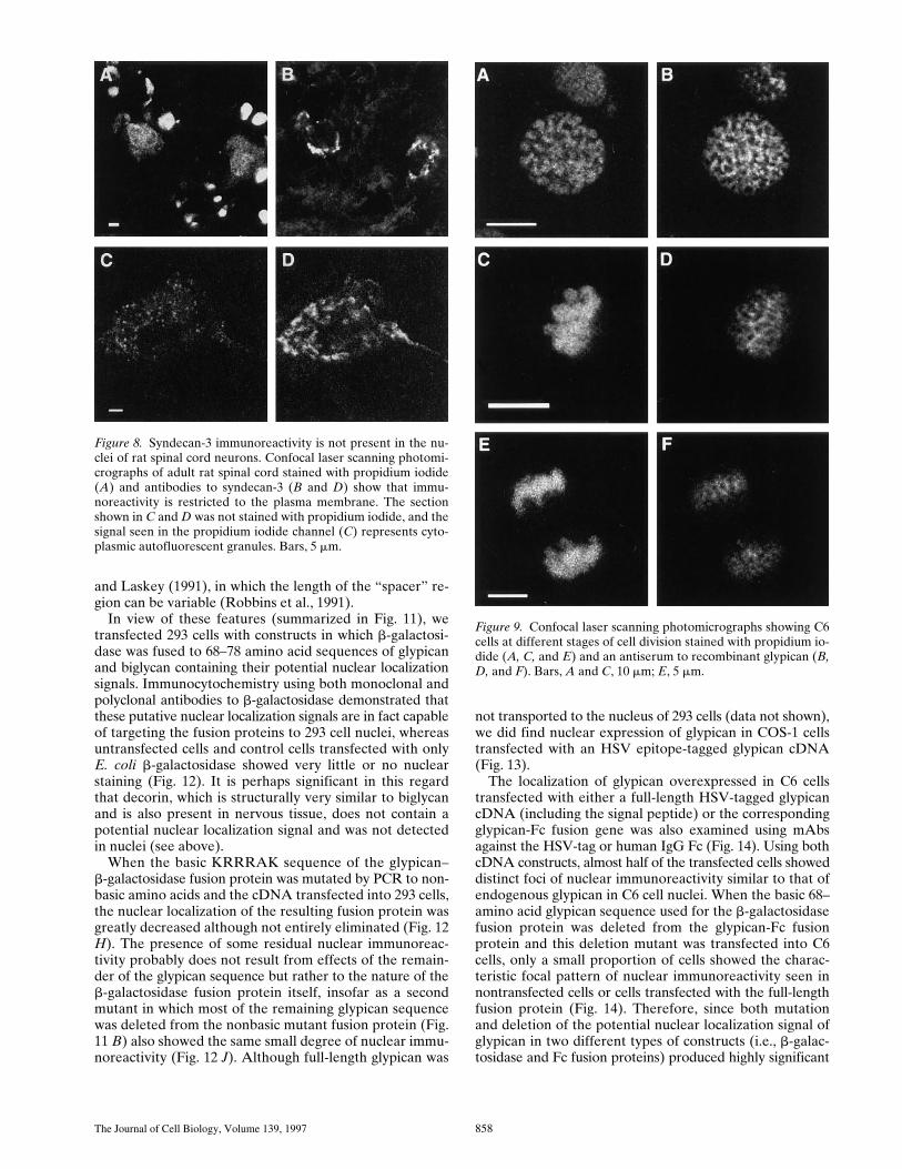
Horizontal sections obtained by confocal laser scanningmicroscopy after combined immunostaining and propid-ium iodide staining of nuclei demonstrated that the nu-clear staining was distinct from the cytoplasm and intracel-lular membranes (Figs. 2, 3, and 4). This conclusion wasfurther supported both by examination of vertical sections,which showed that the glypican and biglycan immunoreac-tivity was present primarily in the nuclear matrix ratherthan in the surrounding membrane (data not shown), andby staining of isolated nuclei prepared by various types ofdetergent treatment (Fig. 7). However, the matrix usuallyshowed a heterogeneous rather than uniform staining pat-tern, frequently with one or more intensely stained spots,some of which are probably nucleoli. No nuclear stainingwas observed with antibodies to N-syndecan/syndecan-3,another heparan sulfate proteoglycan of nervous tissue(Fig. 8), or to decorin, a small, leucine-rich chondroitin/dermatan sulfate proteoglycan with z55% sequence iden-tity to biglycan (data not shown).
Dynamics of Glypican Immunoreactivity in C6Cell Nuclei
Although glypican immunoreactivity in interphase C6 cellnuclei was seen throughout the nuclear matrix, when thechromosomes begin to condense the focal staining disap-pears and glypican immunoreactivity, which increases inintensity, is excluded from the prophase chromosomes (Fig.9 B). This perichromosomal pattern continues throughmetaphase (Fig. 9 D) and anaphase (Fig. 9 F) until thechromosomes are decondensed after separation into twodaughter cells.
Because there was considerable variability in the patternof glypican nuclear immunoreactivity in asynchronous cul-tures of C6 cells, we examined possible correlations be-tween the nuclear staining pattern and the cell cycle. Afterserum starvation, the pattern of focal staining seen in cellsfrom a regular culture almost disappeared, whereas nearly
Figure 3. Rat C6 glioma cells stained with antibodies to eitherglypican (A, B, D, and F) or biglycan (H and J). Nuclei of the an-tibody-stained cells shown in D, F, H, and J were also stainedwith propidium iodide (C, E, G, and I). Glypican immunoreactiv-
ity in the plasma membrane is seen in horizontal and vertical sec-tions of nonpermeabilized cells (A and B, respectively), whereasall other cells were permeabilized with methanol. In F, the anti-bodies were adsorbed with recombinant glypican before use, andin J, the antibiglycan antibodies were used in the presence of 25mg/ml biglycan peptide. Bars, A, E, and I, 5 mm; C and G, 10 mm.
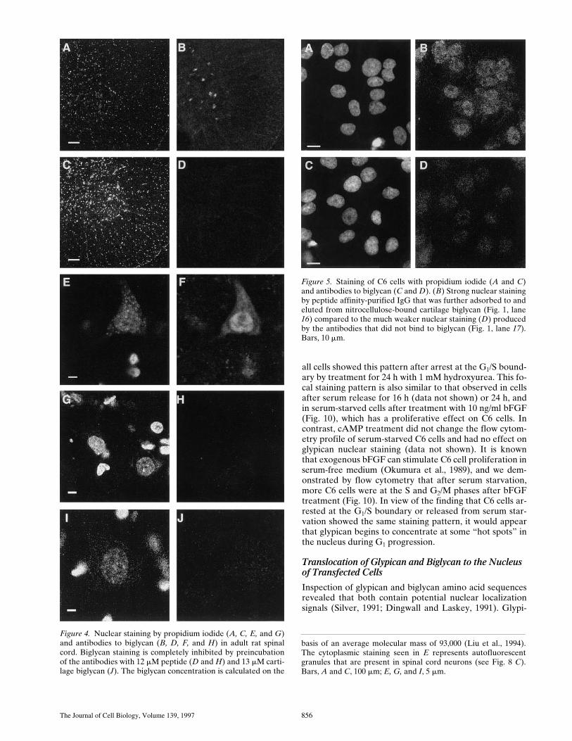
The Journal of Cell Biology, Volume 139, 1997 856
all cells showed this pattern after arrest at the G1/S bound-ary by treatment for 24 h with 1 mM hydroxyurea. This fo-cal staining pattern is also similar to that observed in cellsafter serum release for 16 h (data not shown) or 24 h, andin serum-starved cells after treatment with 10 ng/ml bFGF(Fig. 10), which has a proliferative effect on C6 cells. Incontrast, cAMP treatment did not change the flow cytom-etry profile of serum-starved C6 cells and had no effect onglypican nuclear staining (data not shown). It is knownthat exogenous bFGF can stimulate C6 cell proliferation inserum-free medium (Okumura et al., 1989), and we dem-onstrated by flow cytometry that after serum starvation,more C6 cells were at the S and G2/M phases after bFGFtreatment (Fig. 10). In view of the finding that C6 cells ar-rested at the G1/S boundary or released from serum star-vation showed the same staining pattern, it would appearthat glypican begins to concentrate at some “hot spots” inthe nucleus during G1 progression.
Translocation of Glypican and Biglycan to the Nucleus of Transfected Cells
Inspection of glypican and biglycan amino acid sequencesrevealed that both contain potential nuclear localizationsignals (Silver, 1991; Dingwall and Laskey, 1991). Glypi-
Figure 4. Nuclear staining by propidium iodide (A, C, E, and G)and antibodies to biglycan (B, D, F, and H) in adult rat spinalcord. Biglycan staining is completely inhibited by preincubationof the antibodies with 12 mM peptide (D and H) and 13 mM carti-lage biglycan (J). The biglycan concentration is calculated on the
basis of an average molecular mass of 93,000 (Liu et al., 1994).The cytoplasmic staining seen in E represents autofluorescentgranules that are present in spinal cord neurons (see Fig. 8 C).Bars, A and C, 100 mm; E, G, and I, 5 mm.
Figure 5. Staining of C6 cells with propidium iodide (A and C)and antibodies to biglycan (C and D). (B) Strong nuclear stainingby peptide affinity-purified IgG that was further adsorbed to andeluted from nitrocellulose-bound cartilage biglycan (Fig. 1, lane16) compared to the much weaker nuclear staining (D) producedby the antibodies that did not bind to biglycan (Fig. 1, lane 17).Bars, 10 mm.

Liang et al. Glypican and Biglycan Expression in Nervous Tissue 857
can contains the sequence KRRRAK, which is in excellentagreement with the proposed hexapeptide signal consist-ing of four K or R residues that are not interrupted byacidic (D or E) or bulky (W, F, or Y) amino acids (Bouli-kas, 1994), whereas rat, mouse, and human biglycan all con-tain two basic amino acids (RK, residues 102/103 in ma-ture rat biglycan) followed by a 30–amino acid spacer anda cluster of seven amino acids consisting of four basic resi-dues (RIRKVPK). This corresponds to the “bipartite mo-tif” for nuclear localization signals proposed by Dingwall
Figure 7. Staining of isolated C6 cell nuclei with propidium io-dide (A, C, and E) and antibodies to glypican (B) or biglycan (F).For the experiment shown in A and B, C6 cells were trypsinizedbefore isolation of nuclei, which were then treated successivelywith 1% and 2% Triton X-100 to further reduce contaminationby intracellular and plasma membranes. (C and D) Nuclei iso-lated from nontrypsinized C6 cells that were first incubated withantibodies to glypican (1 h at room temperature), after which theisolated nuclei were stained with second antibody alone. Bar, 5 mm.
Figure 6. Peroxidase-diaminobenzidine staining of glypican (A–C)and biglycan (D–F) in Vibratome sections of 9-d rat cerebrumand cerebellum: A, cerebral cortex; B, Purkinje cell layer of cere-bellum; C, deep cerebellar nuclei; D, thalamus; E, hippocampus(hilus); F, cingulum. Bar, 20 mm.
The Journal of Cell Biology, Volume 139, 1997 858
and Laskey (1991), in which the length of the “spacer” re-gion can be variable (Robbins et al., 1991).
In view of these features (summarized in Fig. 11), wetransfected 293 cells with constructs in which b-galactosi-dase was fused to 68–78 amino acid sequences of glypicanand biglycan containing their potential nuclear localizationsignals. Immunocytochemistry using both monoclonal andpolyclonal antibodies to b-galactosidase demonstrated thatthese putative nuclear localization signals are in fact capableof targeting the fusion proteins to 293 cell nuclei, whereasuntransfected cells and control cells transfected with onlyE. coli b-galactosidase showed very little or no nuclearstaining (Fig. 12). It is perhaps significant in this regardthat decorin, which is structurally very similar to biglycanand is also present in nervous tissue, does not contain apotential nuclear localization signal and was not detectedin nuclei (see above).
When the basic KRRRAK sequence of the glypican–b-galactosidase fusion protein was mutated by PCR to non-basic amino acids and the cDNA transfected into 293 cells,the nuclear localization of the resulting fusion protein wasgreatly decreased although not entirely eliminated (Fig. 12H). The presence of some residual nuclear immunoreac-tivity probably does not result from effects of the remain-der of the glypican sequence but rather to the nature of theb-galactosidase fusion protein itself, insofar as a secondmutant in which most of the remaining glypican sequencewas deleted from the nonbasic mutant fusion protein (Fig.11 B) also showed the same small degree of nuclear immu-noreactivity (Fig. 12 J). Although full-length glypican was
not transported to the nucleus of 293 cells (data not shown),we did find nuclear expression of glypican in COS-1 cellstransfected with an HSV epitope-tagged glypican cDNA(Fig. 13).
The localization of glypican overexpressed in C6 cellstransfected with either a full-length HSV-tagged glypicancDNA (including the signal peptide) or the correspondingglypican-Fc fusion gene was also examined using mAbsagainst the HSV-tag or human IgG Fc (Fig. 14). Using bothcDNA constructs, almost half of the transfected cells showeddistinct foci of nuclear immunoreactivity similar to that ofendogenous glypican in C6 cell nuclei. When the basic 68–amino acid glypican sequence used for the b-galactosidasefusion protein was deleted from the glypican-Fc fusionprotein and this deletion mutant was transfected into C6cells, only a small proportion of cells showed the charac-teristic focal pattern of nuclear immunoreactivity seen innontransfected cells or cells transfected with the full-lengthfusion protein (Fig. 14). Therefore, since both mutationand deletion of the potential nuclear localization signal ofglypican in two different types of constructs (i.e., b-galac-tosidase and Fc fusion proteins) produced highly significant
Figure 9. Confocal laser scanning photomicrographs showing C6cells at different stages of cell division stained with propidium io-dide (A, C, and E) and an antiserum to recombinant glypican (B,D, and F). Bars, A and C, 10 mm; E, 5 mm.
Figure 8. Syndecan-3 immunoreactivity is not present in the nu-clei of rat spinal cord neurons. Confocal laser scanning photomi-crographs of adult rat spinal cord stained with propidium iodide(A) and antibodies to syndecan-3 (B and D) show that immu-noreactivity is restricted to the plasma membrane. The sectionshown in C and D was not stained with propidium iodide, and thesignal seen in the propidium iodide channel (C) represents cyto-plasmic autofluorescent granules. Bars, 5 mm.

Liang et al. Glypican and Biglycan Expression in Nervous Tissue 859
decreases in their nuclear localization, it is likely that thisbasic sequence is a functional nuclear localization signal.
DiscussionBy now, there is extensive literature concerning glycos-aminoglycans in the nucleus (Bhavanandan and Davidson,
1975; Stein et al., 1975; Margolis et al., 1976; Fromme et al.,1976; Furukawa and Terayama, 1977; Fedarko and Con-rad, 1986; Ishihara et al., 1986; Ripellino et al., 1988; Buschet al., 1992). Most of these reports have described bio-chemical studies in which labeled glycosaminoglycans weredetected in purified nuclei. Although it has recently beendemonstrated that a large portion of the labeled glycos-
Figure 10. C6 cells were serum-starved for 2 d and transferred for 24 h to either new medium also containing 0.4% serum (A and B),complete medium (C and D), complete medium with 1 mM hydroxyurea (E and F), or medium containing 0.4% serum and 10 ng/mlbFGF (G and H). Cell aliquots were then used either for double staining with propidium iodide (A, C, E, and G) and antiglypican anti-bodies (B, D, F, and H) or for flow cytometry. The right-hand portion of the figure shows the corresponding flow cytometry profiles,presented as cell number on the ordinate and propidium iodide fluorescence intensity (in arbitrary units) on the abscissa. Each graphrepresents the analysis of 10,000 cells. Bars, 10 mm.

The Journal of Cell Biology, Volume 139, 1997 860
aminoglycans found in nuclei isolated from primary cul-tures of rat ovarian granulosa cells may represent materialderived from the plasma membrane (Hiscock et al., 1994),our studies of glypican and biglycan demonstrate consider-able cellular specificity with regard to their nuclear localiza-tion. Even in ovarian granulosa cells, not all of the glycos-aminoglycans associated with the nuclei could be ascribedto possible contamination from other subcellular fractions,and earlier mass analytical studies on the concentrationsof glycoconjugates in rat brain nuclei demonstrated negli-gible contamination of an unlabeled crude nuclear fractionafter mixing with [35S]sulfate-labeled proteoglycans presentin an 850-g postnuclear supernatant fraction before purifi-cation of the nuclei (Margolis et al., 1976). Moreover, auto-
Figure 11. The location of potential nuclear localization signalsin biglycan (A) and glypican (B) is indicated by the shaded re-gions, that were used for the construction of b-galactosidase fu-sion proteins. The clusters of basic amino acids are underlined. Inone glypican mutant, the basic cluster was mutated to NSSSAN,and the sequence indicated by italics was deleted in a second mu-tant. The attachment sites for the phosphatidylinositol anchorand the COOH-terminal heparan sulfate chain of glypican are in-dicated by an open triangle and an arrow, respectively. In two fu-sion proteins, amino acids 499–558 of glypican were replaced bythe human IgG Fc sequence (shown as hatched regions in C andD), and in a deletion mutant, the potential nuclear localizationsignal used for the b-galactosidase fusion protein was removedfrom the glypican–Fc fusion protein (D).
Figure 12. Localization of b-galactosidase immunoreactivity intransfected 293 cells. 293 cells were transfected with b-galactosi-dase alone (A and B) or b-galactosidase fusion proteins contain-ing either the putative nuclear localization signals of glypican (Cand D) or biglycan (E and F). In a third fusion protein, the basiccluster of the glypican fragment was mutated to nonbasic aminoacids (G and H), and in a fourth fusion protein (I and J), therewas a further deletion of the amino acids shown in italics in Fig.11 B. A, C, E, G, and I show propidium iodide staining; B, D, F,H, and J show b-galactosidase immunoreactivity. Bars, 10 mm.

Liang et al. Glypican and Biglycan Expression in Nervous Tissue 861
radiographic (Fromme et al., 1976), immunocytochemical(Aquino et al., 1984; Ripellino et al., 1989), and histo-chemical studies using a specific biotinylated probe for hy-aluronan (Ripellino et al., 1988) have also demonstratedthe presence of sulfated glycosaminoglycans, chondroitinsulfate proteoglycans, and hyaluronan in the nucleus.
Glypican was detected both on Western blots and immu-nocytochemically in nuclei isolated from C6 cells treatedwith trypsin to reduce possible contamination by cell-sur-face proteoglycans, and in nuclei prepared from trypsin-ized cells, there was no reduction in glypican immunoreac-tivity after their treatment with 25 mM potassium iodideto depolymerize actin and thereby reduce cytoskeletal–membrane interactions (Hiscock et al., 1994), or by incu-
Figure 13. Transport of HSV-tagged glypican to the nuclei oftransfected COS-1 cells. Cells were transfected with full-lengthglypican cDNA containing an HSV epitope tag and stained withpropidium iodide (A and C) or an mAb to the HSV tag (B andD). Staining is seen in the nuclei and in the surrounding ER andGolgi membranes. Bars, 50 mm.
Figure 14. Localization of glypican fusion proteins in C6 gliomacells. Cells were transfected with an HSV fusion protein (A andB) or Fc fusion proteins containing either the complete glypicancDNA (pcGLYPFc, C and D), or glypican cDNA from which thenuclear localization signal had been deleted (pcGDFc, E and F).The cells were then stained with both propidium iodide (A, C,and E) and mAbs to either the HSV tag (B) or to the human Fcregion (D and F). For the Fc fusion protein studies, two indepen-dent transfections were performed, and more than 200 cells werescored in each experiment. The transfected cells were dividedinto two groups; one in which the nuclei contained discrete foci ofglypican immunoreactivity (e.g., D, in which only the cell on theleft side shows nuclear staining), and a second group with eitherno nuclear staining or a very weak immunoreactivity (F). A quan-titation of these results is shown in the bar graph (G), where itcan be seen that whereas z40% of the cells transfected with the
full-length cDNA (pcGLYPFc) show the characteristic pattern ofdiscrete nuclear immunoreactivity (solid bars), this pattern wasseen in only 5% of cells transfected with DNA from which theglypican nuclear localization signal had been deleted (pcGDFc).The hatched bars represent the proportion of cells that do notshow foci of nuclear staining. Cells with abnormal morphology orcondensed nuclei were not scored. Bars, 10 mm.

The Journal of Cell Biology, Volume 139, 1997 862
bation of the isolated nuclei with phosphatidylinositol-spe-cific phospholipase C, 0.5 M NaCl, heparin (10 mg/ml), orheparitinase (data not shown). Because nuclei isolatedeven from nontrypsinized cells pretreated with antibodiesto glypican showed no significant plasma membrane con-tamination after staining with secondary antibody (Fig. 7D), an adventitious association of cell-surface glypicanwith isolated nuclei appears very unlikely, and the conclu-sions from these studies are in excellent agreement withour confocal immunocytochemical data demonstratingglypican immunoreactivity in the nuclear matrix in situ.
The recent cloning of two hyaluronan-binding proteinshas identified one of these (which also binds chondroitinsulfate and heparin in vitro) as the vertebrate homologueof the essential cell cycle control protein Cdc37 (Gramma-tikakis et al., 1995), while a 68-kD hyaluronan-bindingprotein from human fibroblasts was shown to be identicalto P-32 (Deb and Datta, 1996), a protein that copurifieswith the human pre-mRNA splicing factor SF2. Both ofthese proteins contain glycosaminoglycan-binding motifspreviously described in several hyaluronan-binding pro-teins, and they provide further support for a role for gly-cosaminoglycans and proteoglycans in nuclear processessuch as the control of cell division. Our finding that glypi-can and biglycan are components of some nuclei raises theobvious possibility that they might modulate the functionsof other nuclear proteins with which they may interact,such as transcription factors or histones.
The nuclear localization of chondroitin sulfate and hepa-ran sulfate proteoglycans extends the rapidly growing num-ber of proteins that have been found at unexpected sites.Examples include the cell surface and/or nuclear localiza-tion of cytoskeletal proteins such as actin, tubulin, tau, andglycosyltransferases and kinases (reviewed by Smalheiser,1996), and the nuclear translocation or localization of growthfactors and their receptors (Maher et al., 1996), structuralprotein 4.1 (Krauss et al., 1997), myelin basic protein (Pe-draza et al., 1997), myosin I (Nowak et al., 1997), and fer-ritin (Cai et al., 1997), to mention only a few of the manyproteins that fall into this category. It has also recently be-come apparent that a number of generally and genuinelynuclear proteins can be recruited to contribute to the as-sembly of different types of junctional plaques. These in-clude the desmosomal plaque proteins plakophilin 1 andplakophilin 2 (Mertens et al., 1996), the adherens junctionplaque protein b-catenin (Funayama et al., 1994; Kar-novsky and Klymkowsky, 1995), and symplekin, a noveltype of tight junction plaque protein (Keon et al., 1996).
It is not clear how glypican and biglycan are transportedacross membranes insofar as they both contain NH2-termi-nal signal sequences and cotranslationally traverse the lu-men of the ER. However, the HSV-1 structural proteinVP22, which (like a small group of unusual proteins suchas IL-1b, the HIV-1 Tat protein, and basic fibroblast growthfactor) contains no signal peptide, was recently found tobe transported intercellularly to neighboring cells after en-dogenous synthesis (Elliott and O’Hare, 1997). Althoughglypican and biglycan both contain signal sequences, theymay share certain other features with these proteins thatpermit their translocation across the plasma membrane.Numerous other cell membrane and secreted proteins alsocontain nuclear localization signals (Boulikas, 1994), but
whether any of these are actually transported to the nu-cleus remains to be examined.
Because the HSV tag in glycosylphosphatidylinositol-anchored glypican (which is continually shed into the cul-ture medium) and the Fc tag in the secreted form bothshowed a similar pattern of nuclear immunoreactivity, it islikely that soluble rather than membrane-anchored glypi-can is the species that is transported into the nucleus.However, because cells surrounding transfected C6 cellsexpressing the glypican Fc fusion protein did not show anyFc immunoreactivity, it would appear that glypican re-leased from the cell surface uses an autocrine pathway forentry into the nucleus.
C6 cells expressing the glypican deletion mutant showeda significant reduction in the number of cells with foci ofnuclear immunoreactivity, indicating the importance ofthe basic cluster in its nuclear targeting. However, the cy-toplasmic form of glypican lacking the signal peptide showeda rather uniform distribution throughout the nucleus (datanot shown), suggesting that in addition to the nuclear lo-calization signal, certain posttranslational modificationsmay be necessary for its targeting to nuclear subdomains.Although glypican immunoreactivity was not seen in thenuclei of 293 cells transfected with either tagged full-lengthor cytoplasmic glypican, tagged full-length glypican wastransported to the nuclei of COS-1 cells (Fig. 13), indicat-ing a cell type specificity for this process. The absence of anydetectable expression of glypican in 293 cells suggests thatits nuclear targeting may require an interaction with oneor more other proteins expressed only by certain cell types.
The glypican and biglycan immunoreactivity seen in theventral horn of the spinal cord is most prominent in thenuclei of large cell bodies with a morphology typical ofmotor neurons, and their large nuclei are also distin-guished by a relatively weaker staining with both DAPI(data not shown) and propidium iodide (Figs. 2 and 4). Al-though we are not aware of any previous reports calling at-tention to different patterns of nuclear staining in spinalcord neurons, differences in propidium iodide staininghave been described in relation to the organization of nu-clear DNA and as a function of different physiologicalconditions of the cells. For example, propidium iodidestaining is increased after 0.7 M NaCl or low pH extractionof histone H1 (Giangarè et al., 1989) or mild nuclease di-gestion to relax DNA supercoils (Prosperi et al., 1991),and both DAPI and propidium iodide staining of nuclei de-creases after erythroid differentiation of Friend leukemiacells (Darzynkiewicz et al., 1984). It is therefore possiblethat the pattern of nuclear glypican and biglycan immu-noreactivity that we observed in the spinal cord is associ-ated with a subpopulation of more highly differentiatedneurons having a more compact chromatin structure.
C6 cells entering the G1 phase acquire prominent foci ofnuclear glypican immunoreactivity, suggesting that thisnuclear localization may be related to cell proliferation.The dynamic changes in the glypican staining pattern re-semble those of certain conventional nuclear components,as well as that of some unexpected proteins, such as pro-tein 4.1, which was first identified as a crucial protein inthe mature red cell membrane skeleton but redistributesto several structural zones during the cell cycle (Krauss etal., 1997). Glypican may also serve as a structural compo-

Liang et al. Glypican and Biglycan Expression in Nervous Tissue 863
nent of the nucleus, although the specific compartment(s)corresponding to the foci of glypican immunmoreactivityseen in C6 cell nuclei have not yet been identified. The de-tailed pathways leading to the nuclear localization of gly-pican and biglycan and their physiological role in the nu-cleus therefore remain to be elucidated, as does the basisfor the cellular specificity that we have observed, whichmay be regulated either by cell type–specific mechanismsfor entry of the proteoglycans into the cytoplasm or at thenuclear level. However, the similar nuclear localization ofglypican and biglycan, but not of related proteoglycanssuch as decorin and syndecan-3, emphasizes the potentialimportance of both chondroitin sulfate and heparan sul-fate proteoglycans in nuclear processes.
We thank Drs. David Carey, Hans Kresse, Kevin Dreher, James Salzer,and Lawrence Rosenberg for generously providing antibodies, DNAs,and proteins, and Markus Mevissen and Susanna Popp for skillful techni-cal assistance.
This work was supported by research grants NS-09348, NS-13876, andMH-00129 from the National Institutes of Health.
Received for publication 3 June 1997 and in revised form 2 September 1997.
References
Aquino, D.A., R.U. Margolis, and R.K. Margolis. 1984. Immunocytochemicallocalization of a chondroitin sulfate proteoglycan in nervous tissue. I. Adultbrain, retina, and peripheral nerve. J. Cell Biol. 99:1117–1129.
Bansal, R., M. Kumar, K. Murray, and S.E. Pfeiffer. 1996. Developmental andFGF-2-mediated regulation of syndecans (1–4) and glypican in oligodendro-cytes. Mol. Cell Neurosci. 7:276–288.
Bhavanadan, V.P., and E.A. Davidson. 1975. Mucopolysaccharides associatedwith nuclei of cultured mammalian cells. Proc. Natl. Acad. Sci. USA. 72:2032–2036.
Boulikas, T. 1994. Putative nuclear localization signals (NLS) in protein tran-scription factors. J. Cell. Biochem. 55:32–58.
Busch, S.J., G.A. Martin, R.L. Barnhart, M. Mano, A.D. Cardin, and R.L. Jack-son. 1992. Trans-repressor activity of nuclear glycosaminoglycans on Fos andJun/Ap-1 oncoprotein-mediated transcription. J. Cell Biol. 116:31–42.
Cai, C.X., D.E. Birk, and T.F. Linsenmayer. 1997. Ferritin is a developmentallyregulated nuclear protein of avian corneal epithelial cells. J. Biol. Chem. 272:12831–12839.
Carey, D.J., D.M. Evans, R.C. Stahl, V.K. Asundi, K.J. Conner, P. Garbes, andG. Cizmeci-Smith. 1992. Molecular cloning and characterization of N-synde-can, a novel transmembrane heparan sulfate proteoglycan. J. Cell Biol. 117:191–201.
Carey, D.J., R.C. Stahl, V.K. Asundi, and B. Tucker. 1993. Processing and sub-cellular distribution of the Schwann cell lipid-anchored heparan sulfate pro-teoglycan and identification as glypican. Exp. Cell Res. 208:10–18.
Darzynkiewicz, Z., F. Traganos, J. Kapuscinski, L. Staiano-Coico, and M.R.Melamed. 1984. Accessibility of DNA in situ to various fluorochromes: rela-tionship to chromatin changes during erythroid differentiation of friend leu-kemia cells. Cytometry. 5:355–363.
David, G., V. Lories, B. Decock, P. Marynen, J.-J. Cassiman, and H. Van denBerghe. 1990. Molecular cloning of a phosphatidylinositol-anchored mem-brane heparan sulfate proteoglycan from human lung fibroblasts. J. CellBiol. 111:3165–3176.
Deb, T.B., and K. Datta. 1996. Molecular cloning of human fibroblast hyal-uronic acid-binding protein confirms its identity with P-32, a protein co-puri-fied with splicing factor SF2. J. Biol. Chem. 271:2206–2212.
Dingwall, C., and R.A. Laskey. 1991. Nuclear targeting sequences—a consen-sus? Trends Biochem. Sci. 16:478–481.
Dreher, K.L., V. Asundi, D. Matzura, and K. Cowan. 1990. Vascular smoothmuscle biglycan represents a highly conserved proteoglycan within the arte-rial wall. Eur. J. Cell Biol. 53:296–304.
Elliott, G., and P. O’Hare. 1997. Intercellular trafficking and protein deliveryby a herpesvirus structural protein. Cell. 88:223–233.
Fedarko, N.S., and H.E. Conrad. 1986. A unique heparan sulfate in the nucleiof hepatocytes: structural changes with the growth state of the cells. J. CellBiol. 102:587–599.
Filmus, J., W. Shi, Z.M. Wong, and M.J. Wong. 1995. Identification of a newmembrane-bound heparan sulphate proteoglycan. Biochem. J. 311:561–565.
Fromme, H.G., E. Buddecke, K.V. Figura, and H. Kresse. 1976. Localization ofsulfated glyocsaminoglycans within cell nuclei by high-resolution autora-diography. Exp. Cell Res. 102:445–449.
Funayama, N., F. Fagotto, P. McCrea, and B.M. Gumbiner. 1994. Embryonicaxis induction by the armadillo repeat domain of b-catenin: evidence for in-
tracellular signaling. J. Cell Biol. 128:959–968.Furukawa, K., and H. Terayama. 1977. Isolation and identification of glycos-
aminoglycans associated with purified nuclei from rat liver. Biochim. Bio-phys. Acta. 499:278–289.
Giangarè, M.C., E. Prosperi, G. Pedrali-Noy, and G. Bottiroli. 1989. Flow cyto-metric evaluation of DNA stainability with propidium iodide after histoneH1 extraction. Cytometry. 10:726–730.
Glössl, J., M. Beck, and H. Kresse. 1984. Biosynthesis of proteodermatan sul-fate in cultured human fibroblasts. J. Biol. Chem. 259:14144–14150.
Gowda, D.C., B. Goossen, R.K. Margolis, and R.U. Margolis. 1989. Chon-droitin sulfate and heparan sulfate proteoglycans of PC12 pheochromocy-toma cells. J. Biol. Chem. 264:11436–11443.
Grammatikakis, N., A. Grammatikakis, M. Yondea, Q. Yu, S.D. Banerjee, andB.P. Toole. 1995. A novel glycosaminoglycan-binding protein is the verte-brate homologue of the cell cycle control protein, Cdc37. J. Biol. Chem. 270:16198–16205.
Hiscock, D.R.R., M. Yanagishita, and V.C. Hascall. 1994. Nuclear localizationof glycosaminoglycans in rat ovarian granulosa cells. J. Biol. Chem. 269:4539–4546.
Iozzo, R.V., and A.D. Murdoch. 1996. Proteoglycans of the extracellular envi-ronment: clues from the gene and protein side offer novel perspectives inmolecular diversity and function. FASEB J. 10:598–614.
Ishihara, M., N.S. Fedarko, and H.E. Conrad. 1986. Transport of heparan sul-fate into the nuclei of hepatocytes. J. Biol. Chem. 261:13575–13580.
Junghans, U., A. Koops, A. Westmeyer, J. Kappler, H.E. Meyer, and H.W.Müller. 1995. Purification of a meningeal cell-derived chondroitin sulphateproteoglycan with neurotrophic activity for brain neurons and its identifica-tion as biglycan. Eur. J. Neurosci. 7:2341–2350.
Karnovsky, A., and M.W. Klymkowsky. 1995. Anterior axis duplication in Xe-nopus induced by the over-expression of the cadherin-binding protein plako-globin. Proc. Natl. Acad. Sci. USA. 92:4522–4526.
Karthikeyan, L., P. Maurel, U. Rauch, R.K. Margolis, and R.U. Margolis. 1992.Cloning of a major heparan sulfate proteoglycan from brain and identifica-tion as the rat form of glypican. Biochem. Biophys. Res. Commun. 188:395–401.
Karthikeyan, L., M. Flad, M. Engel, B. Meyer-Puttlitz, R.U. Margolis, and R.K.Margolis. 1994. Immunocytochemical and in situ hybridization studies of theheparan sulfate proteoglycan, glypican, in nervous tissue. J. Cell Sci. 107:3213–3222.
Kato, M., Y. Oike, S. Suzuki, and K. Kimata. 1985. Selective removal of hepa-ran sulfate chains from proteoheparan sulfate with a commercial prepara-tion of heparitinase. Anal. Biochem. 148:479–484.
Keon, B.H., S. Schäfer, C. Kuhn, C. Grund, and W.W. Franke. 1996. Symple-kin, a novel type of tight junction plaque protein. J. Cell Biol. 134:1003–1018.
Klinger, M.M., R.U. Margolis, and R.K. Margolis. 1985. Isolation and charac-terization of the heparan sulfate proteoglycans of brain. Use of affinity chro-matography on lipoprotein lipase-agarose. J. Biol. Chem. 260:4082–4090.
Koops, A., J. Kappler, U. Junghans, H.G. Kuhn, H. Kresse, and H.W. Müller. 1996.Cultured astrocytes express biglycan, a chondroitin/dermatan sulfate pro-teoglycan supporting the survival of neocortical neurons. Mol. Brain Res. 41:65–73.
Krauss, S.W., C.A. Larabell, S. Lockett, P. Gascard, S. Penman, N. Mohandas,and J.A. Chasis. 1997. Structural protein 4.1 in the nucleus of human cells:dynamic rearrangements during cell division. J. Cell Biol. 137:275–289.
Kresse, H., H. Hausser, and E. Schönherr. 1993. Small proteoglycans. Experien-tia (Basel). 49:403–416.
Litwack, E.D., C.S. Stipp, A. Kumbasar, and A.D. Lander. 1994. Neuronal ex-pression of glypican, a cell-surface glycosylphosphatidylinositol-anchoredheparan sulfate proteoglycan, in the adult rat nervous system. J. Neurosci.14:3713–3724.
Liu, J., T.M. Laue, H.U. Choi, L.-H. Tang, and L. Rosenberg. 1994. The self-association of biglycan from bovine articular cartilage. J. Biol. Chem. 269:28366–28373.
Maher, P.A. 1996. Nuclear translocation of fibroblast growth factor (FGF) re-ceptors in response to FGF-2. J. Cell Biol. 134:529–536.
Margolis, R.K., C.P. Crockett, W.-L. Kiang, and R.U. Margolis. 1976. Gly-cosaminoglycans and glycoproteins associated with rat brain nuclei. Bio-chim. Biophys. Acta. 451:465–469.
Mazzoni, M., V. Bertagnolo, L.M. Neri, C. Carini, M. Marchisio, D. Milani,F.A. Manzoli, and S. Capitani. 1992. Discrete subcellular localization ofphosphoinositidase C b g and d in PC12 rat pheochromocytoma cells. Bio-chem. Biophys. Res. Commun. 187:114–120.
Mertens, C., C. Kuhn, and W.W. Franke. 1996. Plakophilins 2a and 2b: constitu-tive proteins of dual location in the karyoplasm and the desmosomal plaque.J. Cell Biol. 135:1009–1025.
Meyer-Puttlitz, B., E. Junker, R.U. Margolis, and R.K. Margolis. 1996. Chon-droitin sulfate proteoglycans in the developing central nervous system. II.Immunocytochemical localization of neurocan and phosphacan. J. Comp.Neurol. 366:44–54.
Nakato, H., T.A. Futch, and S.B. Selleck. 1995. The division abnormally de-layed (dally) gene: a putative integral membrane proteoglycan required forcell division patterning during postembryonic development of the nervoussystem in Drosophila. Development (Camb.). 121:3687–3702.
Nowak, G., L. Pestic-Dragovich, P. Hozak, A. Philimonenko, C. Simerly, G.Schatten, and P. de Lanerolle. 1997. Evidence for myosin I in the nucleus. J.Biol. Chem. 272:17176–17181.

The Journal of Cell Biology, Volume 139, 1997 864
Okumura, N., K. Takimoto, M. Okada, and H. Nakagawa. 1989. C6 glioma cellsproduce basic fibroblast growth factor that can stimulate their own prolifera-tion. J. Biochem. 106:904–909.
Pedraza, L., L. Fidler, S.M. Staugaitis, and D.R. Colman. 1997. The active trans-port of myelin basic protein into the nucleus suggests a regulatory role inmyelination. Neuron. 18:579–589.
Prosperi, E., M.C. Giangarè, and G. Bottiroli. 1991. Nuclease-induced DNAstructural changes assessed by flow cytometry with the intercalating dye pro-pidium iodide. Cytometry. 12:323–329.
Rauch, U., P. Gao, A. Janetzko, A. Flaccus, L. Hilgenberg, H. Tekotte, R.K.Margolis, and R.U. Margolis. 1991. Isolation and characterization of devel-opmentally-regulated chondroitin sulfate and chondroitin/keratan sulfateproteoglycans of brain identified with monoclonal antibodies. J. Biol. Chem.266:14785–14801.
Ripellino, J.A., M. Bailo, R.U. Margolis, and R.K. Margolis. 1988. Light andelectron microscopic studies on the localization of hyaluronic acid in devel-oping rat cerebellum. J. Cell Biol. 106:845–855.
Ripellino, J.A., R.U. Margolis, and R.K. Margolis. 1989. Immunoelectron mi-croscopic localization of hyaluronic acid-binding region and link proteinepitopes in brain. J. Cell Biol. 108:1899–1907.
Robbins, J., S.M. Dilworth, R.A. Laskey, and C. Dingwall. 1991. Two interde-pendent basic domains in nucleoplasmin nuclear targeting sequence: identi-fication of a class of bipartite nuclear targeting sequence. Cell 64:615–623.
Roughley, P.J., R.J. White, M.-C. Magny, J. Liu, R.H. Pearce, and J.S. Mort.1993. Non-proteoglycan forms of biglycan increase with age in human articu-lar cartilage. Biochem. J. 295:421–426.
Silver, P.A. 1991. How proteins enter the nucleus. Cell. 64:489–497.Smalheiser, N.R. 1996. Proteins in unexpected locations. Mol. Biol. Cell. 7:
1003–1014.Stein, G.S., R.M. Roberts, J.L. Davis, W.J. Head, J.L. Stein, C.L. Thrall, J. Van
Veen, and D.W. Welch. 1975. Are glycoproteins and glycosaminoglycanscomponents of the eukaryotic genome? Nature (Lond.). 258:639–641.
Stichel, C.C., J. Kappler, U. Junghans, A. Koops, H. Kresse, and H.W. Müller.1995. Differential expression of the small chondroitin/dermatan sulfate pro-teoglycans decorin and biglycan after injury of the adult rat brain. Brain Res.704:263–274.
Stipp, C.S., E.D. Litwack, and A.D. Lander. 1994. Cerebroglycan: an integralmembrane heparan sulfate proteoglycan that is unique to the developingnervous system and expressed specifically during neuronal differentiation. J.Cell Biol. 124:149–160.
Veugelers, M., J. Vermeesch, G. Reekmans, R. Steinfeld, P. Marynen, and G.David. 1997. Characterization of glypican-5 and chromosomal localization ofhuman GPC5, a new member of the glypican gene family. Genomics. 40:24–30.
Watanabe, K., H. Yamada, and Y. Yamaguchi. 1995. K-Glypican: a novel GPI-anchored heparan sulfate proteoglycan that is highly expressed in develop-ing brain and kidney. J. Cell Biol. 130:1207–1218.