gold nanoparticles: synthesis and application for halal
TRANSCRIPT

Vol.8 (2018) No. 4-2
ISSN: 2088-5334
Gold Nanoparticles: Synthesis and application for Halal Authentication in Meat and Meat Products
Deni Subara#, Irwandi Jaswir* # Department of Biotechnology Engineering, Faculty of Engineering, International Islamic University Malaysia 53100 Kuala Lumpur,
Malaysia E-mail: [email protected]
* International Institute for Halal Research and Training (INHART), International Islamic University Malaysia Gombak, Kuala Lumpur,
Malaysia E-mail: [email protected]
Abstract—The Muslim community has recently become more aware of the food they eat, especially Muslim minorities in secular areas. This awereness is increased along with increasing adulteration and substitution of expensive meat for cheaper material. Hence, meat authentication has become a major concern for both consumers and researchers alike. Many analytical methods for detecting Halal meat have been developed to address issues with meat authentication, such as HPLC, FTIR, GCMS and PCR. Nanotechnology has recently shown great potential in biosensing applications for Halal meat products. Rapid testing, cheaper materials and portability of gold nanoparticle-based sensors are expected to change conventional sensing of Halal meat. Selected molecules and types of meat was one of the target for developing gold nanoparticles in the detection. Detection of meat products using gold nanoparticles is based on the colour changed of its optical properties. This article reviewed the application of gold nanoparticles for Halal authentication of meat and meat products. Furthermore, the recent development in preparation of gold nanoparticles and current challenges are also discussed. Gold nanoparticles shows great potential to detect specific meats in meat products. Several traditional and modern meat processed products have been tested on the application of gold nanoparticles, such as meat ball and burgers. However, detection of meat adulteration on real food samples should be done before the comercialization. Limit of detection of gold nanoparticles in analysis shows an increment. Gold nanoparticles offer new alternative to expensive method. In adition, specific detector for different animal meats of interest need to be designed. Keywords—gold nanoparticle; halal detection; halal meat; food adulteration; nanotechnology; hazard detection.
I. INTRODUCTION
The development of sensors for detection of meat adulteration especially for Halal meat has gained considerable attention in the Muslim community. In the field of food technology, adulteration of meat and meat products has become a major concern for Muslim consumers. The substitution of expensive materials for cheaper ingredient such as pork for beef is on the rise. At the same time, there is an increasing global market for Halal food that is expected reach 1.24 trillion dollars [1], [2]. Therefore, many studies have been conducted to protect the Muslim community from haram ingredients.
Recently, the number of publication on detection methods of specific meat in food samples were raised. Wolf and colleagues used PCR for detecting porcine DNA in sausage samples [3]. Meanwhile, Xu et al. used FTIR and chemometrics for detecting certain amino acids in sausage
[4]. However, these established methods require advanced technologies and materials.
On the other hand, the application of nanotechnology in biosensing has shown significant promise and growth [5], [6]. One of the growing research areas is the application of metallic nanoparticles for food analysis using visual properties depending on size, shape, and state of nanoparticle itself [7]. Rapid detection, ease of use and low cost has further advanced the application of metallic nanoparticles in food adulteration detection. Gold nanoparticles (AuNPs) provide an excellent nanobiosensing platform.and are extensively used in sensing applications. Various study on the application of AuNPs for detecting Halal meat have also been published. In this review, the types of AuNPs used in Halal meat detection as well as their properties are elaborated. This review highlights the methods for synthesis of AuNPs and more importantly, highlights current research into innovative application of AuNPs for
1633
detecting meat adulteration. Finally, the challenges and future prospects of AuNP use in detecting adulteration of meat were also explained.
II. MATERIAL AND METHOD
The term Halal is from the Arabic word meaning permissible, permitted, approved, lawful, legal and legitimate. Most Halal terms are related to food and consumer goods. The opposite of Halal in Arabic is Haram. The guideline for Haram and Halal is clearly stated in the Holy Quran. Carrion, pig and its derivatives, Khamr or liquor and its derivatives, blood and its derivatives and parts of the human body are classified as Haram.
Due to poor economic situation and technological advances, adulteration or mixtures in food products have become more common. International food regulations dictate that it is mandatory for sellers to label their food before offering to consumers in the market place [8]. However, adulteration is still rampant. The food products most subjected to adulteration are meat and bakery products [9]. Accidental adulteration has also been on the rise, for example the horse meat scandal in Europe in 2013 [10]. Therefore, rapid and easy tools for specific sample testing are essential. In order to arranged the information, this review was able to answer the primary research question includes WHAT (what is definition of gold nanoparticles), WHY (why the nanoparticles use in the detection), and HOW (how to use and produce gold nanoparticles). This paper also explained what have been publish in the field of application of gold nanoparticles for detection adulteration in Halal meat product.
III. RESULTS AND DISCUSSION
A. Gold Nanoparticles
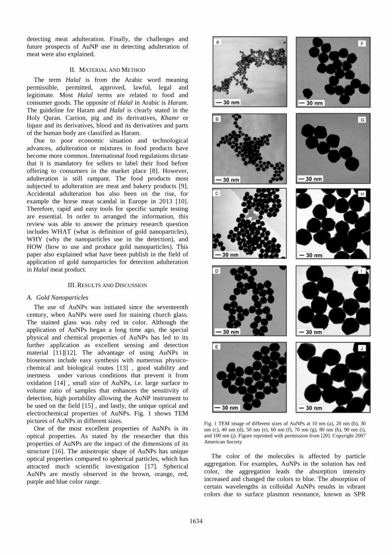
The use of AuNPs was initiated since the seventeenth century, when AuNPs were used for staining church glass. The stained glass was ruby red in color. Although the application of AuNPs began a long time ago, the special physical and chemical properties of AuNPs has led to its further application as excellent sensing and detection material [11][12]. The advantage of using AuNPs in biosensors include easy synthesis with numerous physico-chemical and biological routes [13] , good stability and inertness under various conditions that prevent it from oxidation [14] , small size of AuNPs, i.e. large surface to volume ratio of samples that enhances the sensitivity of detection, high portability allowing the AuNP instrument to be used on the field [15] , and lastly, the unique optical and electrochemical properties of AuNPs. Fig. 1 shows TEM pictures of AuNPs in different sizes.
One of the most excellent properties of AuNPs is its optical properties. As stated by the researcher that this properties of AuNPs are the impact of the dimensions of its structure [16]. The anisotropic shape of AuNPs has unique optical properties compared to spherical particles, which has attracted much scientific investigation [17]. Spherical AuNPs are mostly observed in the brown, orange, red, purple and blue color range.
Fig. 1 TEM image of different sizes of AuNPs at 10 nm (a), 20 nm (b), 30 nm (c), 40 nm (d), 50 nm (e), 60 nm (f), 70 nm (g), 80 nm (h), 90 nm (i), and 100 nm (j). Figure reprinted with permission from [20]. Copyright 2007 American Society
The color of the molecules is affected by particle
aggregation. For examples, AuNPs in the solution has red color, the aggregation leads the absorption intensity increased and changed the colors to blue. The absorption of certain wavelengths in colloidal AuNPs results in vibrant colors due to surface plasmon resonance, known as SPR
1634

absorption [18], [19]. The SPR absorption peak of AuNPs is dependent on AuNP particle size. For example, 13nm spherical AuNPs absorb the highest peak at around 520 nm, while 99 nm AuNPs absorb at 600 nm (Fig. 2) [20] [18]. Kneipp et al. [21] and Nie et al. [22] further apply the unique SPR effect of AuNPs in probes for single molecule surface-enhanced Raman scattering or SERS detection. This characteristic makes AuNPs effective tools in protein detection [23].
Fig. 2 Colors of gold nanoparticles at different particle sizes. Figure reprinted with permission from [20]. Copyright 2007 American Society
Researcher has differentiated three AuNPs based on the
shape or dimension,namely one-dimensional, two-dimensional and three-dimensional AuNPs. One-dimensional of AuNPs includes nanorods, nanowires, nanotubes, and nanobelts. Whereas two-dimensional AuNPs which are quite similar to one-dimensional AuNPs but have a complex dimensional structure such as pentagonal, square, rectangle, hexagonal, truncated triangle and dimpled nanoplates. Meanwhile the three-dimensional consists gold nanotadpoles, gold nanodumbbells (AuNDs), nanostars and gold nanodendrites. Anisotropic gold nanoparticles greatly effect multiple surface plasmon resonance compared to regular gold nanoparticles [24]. Anisotropic AuNPs are useful in biomedical applications such as imaging due to its color range closer to the infrared range that allows differentiation of blood and tissue. A study on anisotropic AuNPs has been published previously by Hu et al [25].
In addition, the physical properties of AuNPs has value added to its application for the detection. The properties include density, melting point, the ability to deliver electron and mechanical strength. Different size AuNPs have different mechanical properties; the smaller the particle, the higher its mechanical strength. The mechanical strength is increased due to smaller gaps between particles, which narrows the valence and conduction bands. In smaller AuNPs, increased surface area is also an important property.
Safaei et al. report that nanomaterials have lower melting points compared to its precursor because the electron are moving out at lower temperature, the movement was initiated because the low electron interation in the surface [26]. Normally, bulk gold melts at around 1063 °C, whereas 2 nm gold nanoparticles melt at half that temperature. The large surface area of AuNPs also increased its conductivity compared to bulk gold.
The AuNPs is more active compared to the its bulk gold, whereas the bulk gold has an characteristic to be less active material. However, in nanometer size, gold nanoparticles show great catalytic activity [27]. AuNPs have been used in many catalytic reactions due to their cost-efficiency compared to platinum and palladium. AuNPs also provide better catalytic reaction yields. AuNPs are especially effective catalytic agents in low-temperature reactions, as reported by Ayastuy et al [28]. This is also due to high surface area.
The different wavelength absorption peaks in the visible region obtained from different morphology of nanoparticles has many applications in medicine, such as for therapy and diagnostics [29]. The principle used in AuNP sensing is the detection of changes in the nanoparticle aggregation states based on its absorbance peak [30]. As explained previously, SPR absorption peak is highly affected by nanoparticle size. Verma et al. (2014) used star-shaped AuNPs for colorimetric sensing of pathogens [30].
B. Synthesis of Gold Nanoparticles
Many studies have been carried out to develop methods for producing isotropic and anisotropic AuNPs with controllable size and shape [31]–[33]. Based on starting material, the production of AuNPs were differentiated into two general categories: firstly, top-down method which involves applying grinding, milling, laser ablation or electrolytic procedures to bulk gold to produce AuNPs [34], whereas the second method involves building or assembling nanoparticles from atoms or molecules (bottom- up method) [33], [35].
Based on the method used, synthesis of AuNPs is carried out via three procedures: chemical procedures, physical procedures and biological procedures. The first method is chemical methods. Most bottom-up methods are typically chemical procedures. The chemical synthesis method is performed in aqueous media.
The most widely utilized chemical method is the Turkevich-Frens citrate reduction of gold (III) derivative method (Fig. 3) [36],[37]. This method retains AuNPs in particle size between 10 to 150 nm. The method is favorable due to its simplicity, stable colloidal nanoparticles produced and controllable size [38]. Till today, the method is often used and modified to contain desired ligands with specific functions [30], [39]. In theTurkevich-Frens method, HAuCl4 is used as a precursor. The HAuCl4 solution is boiled at 75 to 150 °C, then citrate dehydrate solution were mixed to the boiling solution under vigorous stirring to stabilize the products from aggregation. The gold nanoparticles are collected after the appearance of a wine-red colloidal solution is observed. The addition of citrate dihydrate is quite crutial to the product. It has functions to stabilize the nano size. Low amounts of citrate dihydrate are not able to
1635

prevent particle aggregation, hence producing bigger gold nanoparticle sizes. The critical factors affecting particle size and shape are concentration of HAuCl4, concentration of citrate dihydrate, pH, and temperature.
.
Fig. 3 Ilustration of Turkevich-Frens Method Synthesis of AuNPs The second most favorable method is the Brust-Schiffrin
method (Fig. 4) [40] which could develop the hydrophobic AuNPs with desirable size of between 1 to 8 nm in range. Because of the hydrophobicity, toluene and chloroform are the well-dissolve solvent to dissolve the AuNPs, this AuNPs products are also highly stable, easily transferred to powder form without any changes in properties, and amenable to further modification.
Fig. 4 Ilustration of Brust-Schiffrin Method
The Brust-Schiffrin method is different to the previous
method in that this method uses a two-phase liquid/liquid system [41]. There are three independent factor affect the particles size of AuNPs. Firstly, the ration between precursors and thiol that leads to the smaller product. Secondly, slower the reductant addition affects the aggregation and produced huge particles, and lastly warm solutions and heat environtment produced larger particles [41]. More recently, several modified methods have been published, such as Aslam et al [42] producing AuNPs using a one-step process in two phases, while Kim et al [43] produced AuNPs in a one-phase synthesis method. Malikova et al [44] produced AuNPs using salicylic acid in order to reduce gold salt and Otsuka et al [45] produced 1 to 10 nm AuNPs by reducing the concentration of HAuCL4. A high yield and rapid method was presented by Martin et al [46]. The advantages of this method are the absence of a cleaning step, and low amounts of chemical used. This method uses HAuCl4 as a precursor and are mixed with toluene. Tetraoctylammonium bromide (TOAB) was used as a catalyst. Then, the organic phase is mixed with a protecting agent such as dodecanethiol. The Au3+ ion is reduced using sodium borohydride. The AuNPs are collected in a deep brown-colored toluene solution. The critical factors for particle size and shape are ratio of thiol to gold precursor, addition of reducing agent, and processing temperature. Physical methods for the synthesis of AuNPs employ physical procedures such as γ-radiation, microwave irradiation, thermolytic processes and photochemical
processes [47]. Mostly, reducing agent are mixed within the process. The agent has function as a electron donor to create an atoms. The physical method produces particles of various sizes by optimizing processing conditions [48]. Wang et al. report that high purity nanoparticles and controllable nanoparticles size ranging from 5 to 40 nm were produced using microwave irradiation [49]. The significant factors for producing small AuNP size are molar mass of the protecting agent and reaction time. This method does not require the use of external reducing agents.
Green synthesis methods have also been recently reported. The methods used are favorable to the environment because they are nontoxic, use low amounts of solvent and are safe [50]. The method employs a biological resource which includes bacteria, algae, fungi, yeast and viruses [38], [51], [52]. Green synthesis methods are advantageous as they produce larger yields, are easy to scale up, and are economically viable [53], [54].
As mentioned previously, that size and shape has the independent significant effect on its optical properties. Several ways have been published to produce AuNPs of different shapes. Zhou and colleagues produced different shaped AuNPs using ultraviolet reduction [55]. They produced the nanoparticles without any effect of temperature, but with the addition of polymer capping agents. The procedure was conducted using HAuCl4 as a precursor, polyvinylacetate and poly(ethylene glycol) as a capping agent. Irradiation time was the determining factor for producing different AuNPs morphology, besides concentration of precursor and capping agent. About 15 nm AuNPs with triangular shape was produced via irradiation using low concentration of precursor, and 25 nm hexagonal AuNPs were produced using high concentration of precursor [55]. Another simple method was presented by Zhou and colleagues in 2002 using a combination of ascorbic acid and polyvinylpyrrolidone as a capping agent and KAu(CN)2 as a precursor [56]. Other approaches to producing different AuNPs morphology includes template synthesis for producing gold nanowires [57], seed-mediated synthesis for producing gold nanorods [58], and silver assisted growth procedure [59].
C. Colorimetric Sensing for Halal Authentication Using Gold Nanoparticle
In order to produce high-accuracy sensors, the device should be selective towards certain analytes by attaching to specific ligands and producing measurable change such as color intensity or wavelength. Gold nanoparticle sensors are highly amenable to modifications depending on the measured analyte and other parameters such as size and shape. The principle of colorimetric sensing using AuNPs is based on the characteristic aggregation of AuNP particles upon interaction with specific proteins. The aggregation is due to the different electrostatic properties of DNA, which results in different SPR. The application of gold nanoparticles (AuNPs) in sensing of protein was first initiated by Elghanian and colleagues in 1997 [60]. Elghanian used modified AuNPs to identify polynucleotides based on colorimetric detection. The AuNPs were modified using mercaptoalkyloligonucleotide [60]. About 13 nm sized AuNPs were used because they were
1636

easy to prepare, had narrow deviation of about ± 2 nm, and showed sharp absorption bands at wavelength 520 nm. The AuNPs were able to detect 10 fentomoles of an oligonucleotide or approximately 1 µl of aliquot using reverse-phase thin layer chromatography. However, the ultimate sensitivity of this experiment was not determined.
Subsequently, several research papers have been published focusing on developing AuNPs as sensing agents [61]–[63]. In 2000, James and colleagues studied the factors controlling AuNP properties during aggregation [64]. In this study, 15 nm AuNPs were used. The results suggested that the type of DNA linker affected the optical properties of AuNPs. Hence, DNA linkers were more effective at controlling aggregate growth compared to spacer units [64].
Later, AuNPs was developed to analyse the nucleotide sequences. The method was used without any crosslinking process. The method was developed by Li and Robert in 2004 [65]. They used 13 nm AuNPs as colorimetric sensors. Kim Thanh and colleagues further report that particle aggregation was more rapid at low temperatures [63]. They reported a new aggregation immunoassay technique that was much simpler, cheaper and sensitive. This method was able to detect 1 µg/ml quantities of anti-protein [63].
The application of AuNPs for biological samples was first carried out by Ali and colleagues [66] (Table 1). The authors studied whether 13 nm AuNPs could be used for sequence identification and whether AuNPs could be used to directly detect small samples.
In this experiment, AuNPs were modified for detecting eleven types of meat mixtures such as pork-shad, pork venison and shad-venison. About 40 ± 5 nm of AuNPs was prepared as colorimetric sensor. Graber Freeman method with monobasic anhydrous sodium citrate as a precursor was used. No additional equipment was used for measuring the results except the eye. The AuNP detector appeared from pinkish-red to gray purple in appearance upon contact with swine-specific nucleotide. The alteration of color indicated a reduction of surface-plasmon-resonance peak from the 530 nm to 620 nm region. The authors claim that the method could replace conventional detection methods such as gel electrophoresis, RFLP and southern blotting [66]. However, the limit of detection of AuNPs in the study was higher than in real-time PCR.
Ali continued the experiment by producing AuNP sensors in 2011 [67]. In this experiment, 3 nm AuNPs were coated with citrate-tannate and were set to detect specific targets. Raw meat as well as processed meat were used as a samples. In order to vary the experiment, the samples also were cooked with the high temperature and pressure for 2.5 h. As a results, the biosensor detected as low as 1% of pork in mixed meats in raw and cooked meats. This results is special, even a PCR has lack of the ability to give the correct results because the DNA has already degraded. The challenge with AuNP sensors however still remained that the limit of detection was higher than qPCR and the results was failed to detect in very concentrate samples [67].
Ali and colleagues also produced nanobioprobes in 2011 [68]. In the study, Ali addressed the issue of heterogeneous or commercial samples. Ali and colleagues used 3 nm AuNPs for detecting pork adulteration in instant beef burgers.
The AuNP nanobioprobe was integrated with fluorophre-labeled 27-swine cytochrome nucleotide.
TABLE I SUMMARY OF AUNPS SENSOR FOR HALAL MEAT AUTHENTICATION IN MEAT
PRODUCTS
Size Samples LOD Comments Ref 13 nm Pork-venision
Pork-shad Shad-venison
6 µg ml˗1 The LOD was higher than real-time PCR
[66]
3 nm Pork Pork-beef binary mxtures Autoclaved Pork-beef
58.6 pM 230 µg l-1
The LOD was lower than real-time PCR, however relative higher with raw pork
[67]
3 nm Pork adulteration in beef burger
1 % of pork adulteration in beef burger
The accuracy was lower in high content of pork.
[68]
20 nm Pork adulteration in beef meatball and chicken meatball products
4 µg ml˗1 The LOD was higher than PCR based method
[69]
3 nm Autoclaved pork-beef mixture
1 % of pork in autoclaved pork-beef mixture
The accuracy was lower for high content of pork and longer autoclaved meats
[70]
15 nm Chicken tissue in meats and meat products
28 ng µl-1 The PCR amplification affected the colometric assay
[71]
20 nm Processed meat product, meatballs
6 µg ml˗1 The LOD was lower in 30% of pork-containing samples.
[72]
20 nm Horsemeat adulteration in meat products. Fresh meat cut, slices roasted beef and pork, ready to eat meatballs, country style sausages.
12.3 fg µl-1 The efficiency was lower in highly degraded samples
[73]
11 nm Pork contaminations in meat products
0.1 µmol l-1 Not in real food samples,
[74]
The results is interesting because the probe could detect
until 1% mixed meats with 90% accuracy of detection. However, the accuracy was changed and decreased in 3% of pork mixed in burger. The authors believed that the experiment offered a new alternative to expensive methods such as PCR [68].
In 2012, Ali and colleagues produced 20 nm AuNP sensors without proper surface modification [69]. The sensors were tested on adulterated processed food such as
1637
beef and chicken meatballs as samples. Ali reported that the AuNP sensors at 20 nm produced clear color changes compared to 40 nm of AuNPs [69]. The sensitivity of the results was increased by using absorption spectroscopy. Detection limit in 20 nm AuNP was also lower than 40 nm AuNP. The 20 nm AuNPs did not require sample preparation and had a rapid testing duration of about less than 10 min [69]. This finding reports that 20 nm AuNPs were more sensitive than 40 nm AuNPs.
Ali and colleagues further studied swine biomarkers for biosensors in 2012 [70]. The target of this experiment is to use the biomarkers for detection the adulteration. The biosensor was constructed using 3 nm AuNPs. The probe successfully detected adulteration of 1% pork DNA in meat products.
The authentication of chicken tissue using AuNP was reported by Han and colleagues in 2015 [71]. The samples were analyzed using 15 nm AuNPs. Han prepared the detector using the sodium citrate reduction method. It was modified using thiol to prevent salt-induced aggregation. Han et al. reported that the identification method could be used without any sophisticated instrumentation. They also report high accuracy detection using AuNP [71].
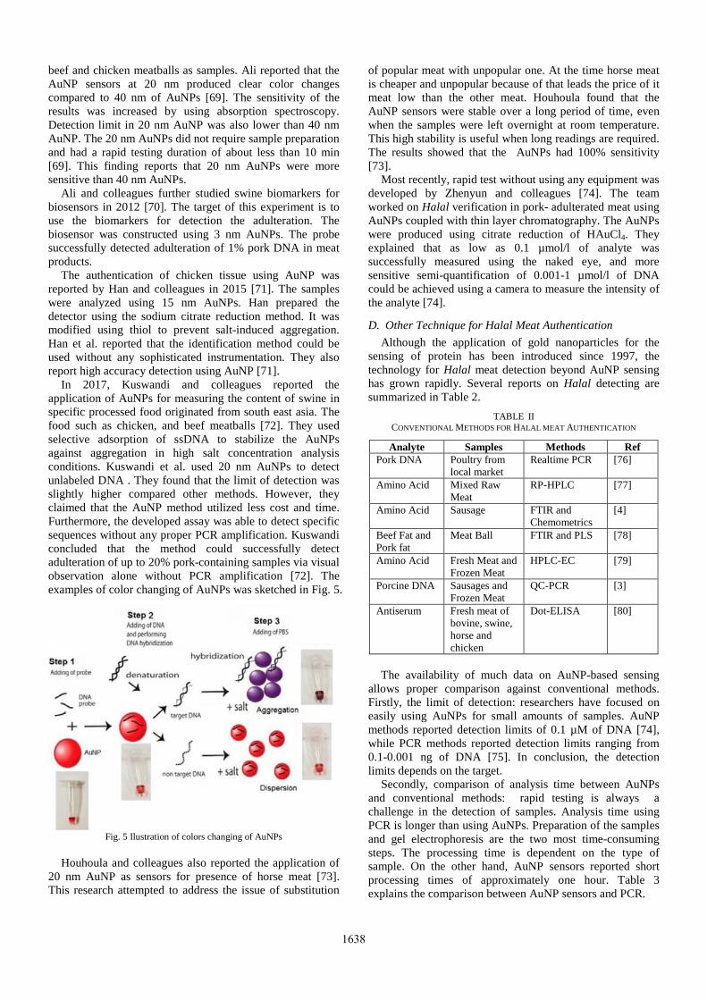
In 2017, Kuswandi and colleagues reported the application of AuNPs for measuring the content of swine in specific processed food originated from south east asia. The food such as chicken, and beef meatballs [72]. They used selective adsorption of ssDNA to stabilize the AuNPs against aggregation in high salt concentration analysis conditions. Kuswandi et al. used 20 nm AuNPs to detect unlabeled DNA . They found that the limit of detection was slightly higher compared other methods. However, they claimed that the AuNP method utilized less cost and time. Furthermore, the developed assay was able to detect specific sequences without any proper PCR amplification. Kuswandi concluded that the method could successfully detect adulteration of up to 20% pork-containing samples via visual observation alone without PCR amplification [72]. The examples of color changing of AuNPs was sketched in Fig. 5.
Fig. 5 Ilustration of colors changing of AuNPs
Houhoula and colleagues also reported the application of
20 nm AuNP as sensors for presence of horse meat [73]. This research attempted to address the issue of substitution
of popular meat with unpopular one. At the time horse meat is cheaper and unpopular because of that leads the price of it meat low than the other meat. Houhoula found that the AuNP sensors were stable over a long period of time, even when the samples were left overnight at room temperature. This high stability is useful when long readings are required. The results showed that the AuNPs had 100% sensitivity [73].
Most recently, rapid test without using any equipment was developed by Zhenyun and colleagues [74]. The team worked on Halal verification in pork- adulterated meat using AuNPs coupled with thin layer chromatography. The AuNPs were produced using citrate reduction of HAuCl4. They explained that as low as 0.1 µmol/l of analyte was successfully measured using the naked eye, and more sensitive semi-quantification of 0.001-1 µmol/l of DNA could be achieved using a camera to measure the intensity of the analyte [74].
D. Other Technique for Halal Meat Authentication
Although the application of gold nanoparticles for the sensing of protein has been introduced since 1997, the technology for Halal meat detection beyond AuNP sensing has grown rapidly. Several reports on Halal detecting are summarized in Table 2.
TABLE II CONVENTIONAL METHODS FOR HALAL MEAT AUTHENTICATION
Analyte Samples Methods Ref Pork DNA Poultry from
local market Realtime PCR [76]
Amino Acid Mixed Raw Meat
RP-HPLC [77]
Amino Acid Sausage FTIR and Chemometrics
[4]
Beef Fat and Pork fat
Meat Ball FTIR and PLS [78]
Amino Acid Fresh Meat and Frozen Meat
HPLC-EC [79]
Porcine DNA Sausages and Frozen Meat
QC-PCR [3]
Antiserum Fresh meat of bovine, swine, horse and chicken
Dot-ELISA [80]
The availability of much data on AuNP-based sensing
allows proper comparison against conventional methods. Firstly, the limit of detection: researchers have focused on easily using AuNPs for small amounts of samples. AuNP methods reported detection limits of 0.1 µM of DNA [74], while PCR methods reported detection limits ranging from 0.1-0.001 ng of DNA [75]. In conclusion, the detection limits depends on the target.
Secondly, comparison of analysis time between AuNPs and conventional methods: rapid testing is always a challenge in the detection of samples. Analysis time using PCR is longer than using AuNPs. Preparation of the samples and gel electrophoresis are the two most time-consuming steps. The processing time is dependent on the type of sample. On the other hand, AuNP sensors reported short processing times of approximately one hour. Table 3 explains the comparison between AuNP sensors and PCR.
1638

TABLE III |COMPARISON BETWEEN AUNPS SENSING AND PCR
Categories PCR AuNPs Ref Detection Limit 0.1-0.001 ng of
DNA 0.1 µM of DNA
[74], [75]
Analysis time 5-48 h 1 h Specificity Excellent Excellent Technical requirement
Gel electrophoresis equipment
Minimal equipment, TLC.
IV. CONCLUSION
Research and development in Halal meat sensing using AuNPs is meant to detect the presence of doubtful meat products for Muslims. AuNP sensors are able to detect very small quantities of analyte i.e. as small as 1 µmol/l and have the potential to replace conventional techniques for meat authentication such as gel electrophoresis. AuNP sensing is rapid and does not require any equipment during the analysis. Meat authentication using AuNPs is becoming more important and many challenges in the method are available to future research. It is suggested that researchers start to apply AuNP analysis on real food samples. Unlike PCR, AuNP analysis employs small and portable kits that can be carried everywhere for rapid and effective testing. However, AuNP systems need to be calibrated for each analyte before use in detection. Moreover, specific probes for different animal meats of interest need to be designed. Finally, researchers need to produce stable nanoparticles that are suitable for use in all media.
ACKNOWLEDGMENT
This study was funded by KIHIM grant No. MOHE 18-002-0002
REFERENCES [1] H. N. Lubis, N. F. Mohd-Naim, N. N. Alizul, and M. U. Ahmed,
“From market to food plate: Current trusted technology and innovations in halal food analysis,” Trends Food Sci. Technol., vol. 58, no. December 2016, pp. 55–68, 2016.
[2] T. Reuters and D. Standard, “State of the global Islamic economy report 2016/17,” Dubai: Thomson Reuters, 2016.
[3] C. Wolf and J. Lüthy, “Quantitative competitive (QC) PCR for quantification of porcine DNA,” Meat Sci., vol. 57, no. 2, pp. 161–168, 2001.
[4] L. Xu, C.-B. Cai, H.-F. Cui, Z.-H. Ye, and X.-P. Yu, “Rapid discrimination of pork in Halal and non-Halal Chinese ham sausages by Fourier transform infrared (FTIR) spectroscopy and chemometrics,” Meat Sci., vol. 92, no. 4, pp. 506–510, 2012.
[5] J. González-Sálamo, B. Socas-Rodriguez, J. Hernández-Borges, and M. Á. Rodriguez-Delgado, “Core-shell poly (dopamine) magnetic nanoparticles for the extraction of estrogenic mycotoxins from milk and yogurt prior to LC--MS analysis,” Food Chem., vol. 215, pp. 362–368, 2017.
[6] R. Hou, Z. Zhang, S. Pang, T. Yang, J. M. Clark, and L. He, “Alteration of the nonsystemic behavior of the pesticide ferbam on tea leaves by engineered gold nanoparticles,” Environ. Sci. Technol., vol. 50, no. 12, pp. 6216–6223, 2016.
[7] V. V Mody, R. Siwale, A. Singh, and H. R. Mody, “Introduction to metallic nanoparticles,” J. Pharm. Bioallied Sci., vol. 2, no. 4, p. 282, 2010.
[8] U. H. F. Bunz and V. M. Rotello, “Gold nanoparticle--fluorophore complexes: sensitive and discerning noses for biosystems sensing,” Angew. Chemie Int. Ed., vol. 49, no. 19, pp. 3268–3279, 2010.
[9] S. Eustis and M. A. El-Sayed, “Why gold nanoparticles are more precious than pretty gold: noble metal surface plasmon resonance and its enhancement of the radiative and nonradiative properties of nanocrystals of different shapes,” Chem. Soc. Rev., vol. 35, no. 3, pp. 209–217, 2006.
[10] A. Agrawal, R. A. Tripp, L. J. Anderson, and S. Nie, “Real-time detection of virus particles and viral protein expression with two-color nanoparticle probes,” J. Virol., vol. 79, no. 13, pp. 8625–8628, 2005.
[11] M. A. Syed and S. Bokhari, “Gold nanoparticle based microbial detection and identification,” J. Biomed. Nanotechnol., vol. 7, no. 2, pp. 229–237, 2011.
[12] C. Wang and C. Yu, “Detection of chemical pollutants in water using gold nanoparticles as sensors: a review,” Rev. Anal. Chem., vol. 32, no. 1, pp. 1–14, 2013.
[13] Y.-Y. Yu, S.-S. Chang, C.-L. Lee, and C. R. C. Wang, “Gold nanorods: electrochemical synthesis and optical properties,” J. Phys. Chem. B, vol. 101, no. 34, pp. 6661–6664, 1997.
[14] X. Zhang, “Gold nanoparticles: recent advances in the biomedical applications,” Cell Biochem. Biophys., vol. 72, no. 3, pp. 771–775, 2015.
[15] S. Link and M. A. El-Sayed, “Size and temperature dependence of the plasmon absorption of colloidal gold nanoparticles,” J. Phys. Chem. B, vol. 103, no. 21, pp. 4212–4217, 1999.
[16] S. Link and M. A. El-Sayed, “Spectral properties and relaxation dynamics of surface plasmon electronic oscillations in gold and silver nanodots and nanorods.” ACS Publications, 1999.
[17] P. N. Njoki, I S Lim, D Mott, H Park, B Khan, S Mishra, R Sujakumar, J Luo and C Zhong, “Size correlation of optical and spectroscopic properties for gold nanoparticles,” J. Phys. Chem. C, vol. 111, no. 40, pp. 14664–14669, 2007.
[18] K. Kneipp, Y Wang, H Kneipp, L T Perelman, I Itzkan, R R Dasari, and M S Feld, “Single molecule detection using surface-enhanced Raman scattering (SERS),” Phys. Rev. Lett., vol. 78, no. 9, p. 1667, 1997.
[19] S. Nie and S. R. Emory, “Probing single molecules and single nanoparticles by surface-enhanced Raman scattering,” Science (80-.86 )., vol. 275, no. 5303, pp. 1102–1106, 1997.
[20] S. Alex and A. Tiwari, “Functionalized gold nanoparticles: synthesis, properties and applications a review,” J. Nanosci. Nanotechnol., vol. 15, no. 3, pp. 1869–1894, 2015.
[21] K. L. Shuford, M. A. Ratner, and G. C. Schatz, “Multipolar excitation in triangular nanoprisms,” J. Chem. Phys., vol. 123, no. 11, p. 114713, 2005.
[22] M. Hu, J Chen, ZY li, L Au, G Hartland, X Li, M Marquez, and Y Xia, “Gold nanostructures: engineering their plasmonic properties for biomedical applications,” Chem. Soc. Rev., vol. 35, no. 11, pp. 1084–1094, 2006.
[23] A. Safaei, “The effect of the averaged structural and energetic features on the cohesive energy of nanocrystals,” J. Nanoparticle Res., vol. 12, no. 3, pp. 759–776, 2010.
[24] M. Haruta and M. Daté, “Advances in the catalysis of Au nanoparticles,” Appl. Catal. A Gen., vol. 222, no. 1–2, pp. 427–437, 2001.
[25] J. L. Ayastuy, U. Iriarte-Velasco, A. Gurbani, and M. A. Gutierrez-Ortiz, “Investigation of the calcination temperature effect on the interaction between Au nanoparticles and the catalytic support α-Fe2O3 for the low temperature CO oxidation,” J. Taiwan Inst. Chem. Eng., vol. 75, pp. 18–28, 2017.
[26] W. Zhang, F Wang, Y Wang, J Wang, Y Yu, S Guo, R Chen, and D Zhou, “pH and near-infrared light dual-stimuli responsive drug delivery using DNA-conjugated gold nanorods for effective treatment of multidrug resistant cancer cells,” J. Control. Release, vol. 232, pp. 9–19, 2016.
[27] M.-C. Daniel and D. Astruc, “Gold nanoparticles: assembly, supramolecular chemistry, quantum-size-related properties, and applications toward biology, catalysis, and nanotechnology,” Chem. Rev., vol. 104, no. 1, pp. 293–346, 2004.
[28] Q. Wang and C. Yu, “Chemical and biological sensing and imaging using plasmonic nanoparticles and nanostructures,” Biomed. nanosensors, p. 59, 2012.
[29] C. J. Murphy, T. K. Sau, A. Gole, and C. J. Orendorff, “Surfactant-directed synthesis and optical properties of one-dimensional
1639

plasmonic metallic nanostructures,” Mrs Bull., vol. 30, no. 5, pp. 349–355, 2005.
[30] M. E. Stewart, C. R. Anderton, L Thompson, J. Maria, S. K. Gray, J. A. Rogers, and R. G. Nuzzo, “Nanostructured plasmonic sensors,” Chem. Rev., vol. 108, no. 2, pp. 494–521, 2008.
[31] Z. Zhang, J. Wang, X. Nie, T. Wen, Y. Ji, X. Wu, Y. Zhao, and C. Chen, “Near infrared laser-induced targeted cancer therapy using thermoresponsive polymer encapsulated gold nanorods,” J. Am. Chem. Soc., vol. 136, no. 20, pp. 7317–7326, 2014.
[32] E. M. Hicks, O. Lyandres, W. P. Hall, S. Zou, M. R. Glucksberg, and R. P. Van Duyne, “Plasmonic properties of anchored nanoparticles fabricated by reactive ion etching and nanosphere lithography,” J. Phys. Chem. C, vol. 111, no. 11, pp. 4116–4124, 2007.
[33] J. Turkevich, P. C. Stevenson, and J. Hillier, “A study of the nucleation and growth processes in the synthesis of colloidal gold,” Discuss. Faraday Soc., vol. 11, pp. 55–75, 1951.
[34] G. Frens, “Controlled nucleation for the regulation of the particle size in monodisperse gold suspensions,” Nat. Phys. Sci., vol. 241, no. 105, p. 20, 1973.
[35] J. Wang, G. Zhu, M. You, E. Song, M. I , Shukoor, K. Zhang, M. B. Altman, Y. Chen, and Z. Zhu, “Assembly of aptamer switch probes and photosensitizer on gold nanorods for targeted photothermal and photodynamic cancer therapy,” ACS Nano, vol. 6, no. 6, pp. 5070–5077, 2012.
[36] N. L. Rosi and C. A. Mirkin, “Nanostructures in biodiagnostics,” Chem. Rev., vol. 105, no. 4, pp. 1547–1562, 2005.
[37] Z. Wang and L. Ma, “Gold nanoparticle probes,” Coord. Chem. Rev., vol. 253, no. 11–12, pp. 1607–1618, 2009.
[38] M. Brust, M. Walker, D. Bethell, D. J. Schiffrin, and R. Whyman, “Synthesis of thiol-derivatised gold nanoparticles in a two-phase liquid--liquid system,” J. Chem. Soc. Chem. Commun., no. 7, pp. 801–802, 1994.
[39] M. Aslam, L. Fu, M. Su, K. Vijayamohanan, and V. P. Dravid, “Novel one-step synthesis of amine-stabilized aqueous colloidal gold nanoparticles,” J. Mater. Chem., vol. 14, no. 12, pp. 1795–1797, 2004.
[40] K.-S. Kim, D. Demberelnyamba, and H. Lee, “Size-selective synthesis of gold and platinum nanoparticles using novel thiol-functionalized ionic liquids,” Langmuir, vol. 20, no. 3, pp. 556–560, 2004.
[41] N. Malikova, I. Pastoriza-Santos, M. Schierhorn, N. A. Kotov, and L. M. Liz-Marzán, “Layer-by-layer assembled mixed spherical and planar gold nanoparticles: control of interparticle interactions,” Langmuir, vol. 18, no. 9, pp. 3694–3697, 2002.
[42] H. Otsuka, Y. Akiyama, Y. Nagasaki, and K. Kataoka, “Quantitative and reversible lectin-induced association of gold nanoparticles modified with $α$-lactosyl-$ω$-mercapto-poly (ethylene glycol),” J. Am. Chem. Soc., vol. 123, no. 34, pp. 8226–8230, 2001.
[43] M. N. Martin, J. I. Basham, P. Chando, and S.-K. Eah, “Charged gold nanoparticles in non-polar solvents: 10-min synthesis and 2D self-assembly,” Langmuir, vol. 26, no. 10, pp. 7410–7417, 2010.
[44] Z. Yang, Z. Li, X. Lu, F, He, X. Zhu, Y. Ma, R. He, F. Gao, W. Ni, and Y. Yi, “Controllable Biosynthesis and Properties of Gold Nanoplates Using Yeast Extract,” Nano-Micro Lett., vol. 9, no. 1, p. 5, 2017.
[45] S. Wang, X. Zhao, S. Wang, J. Qian, and S. He, “Biologically inspired polydopamine capped gold nanorods for drug delivery and light-mediated cancer therapy,” ACS Appl. Mater. Interfaces, vol. 8, no. 37, pp. 24368–24384, 2016.
[46] Y. Wang, K. C. L. Black, H. Luehman, W. Li, Y. Zhang, X. Cai, D. Wan, S. Liu, and P. Kim, “Comparison study of gold nanohexapods, nanorods, and nanocages for photothermal cancer treatment,” ACS Nano, vol. 7, no. 3, pp. 2068–2077, 2013.
[47] L. Vigderman, B. P. Khanal, and E. R. Zubarev, “Functional gold nanorods: synthesis, self-assembly, and sensing applications,” Adv. Mater., vol. 24, no. 36, pp. 4811–4841, 2012.
[48] R. Venkatesan, A. Pichaimani, K. Hari, P. K. Balasubramanian, J. Kulandaivel, and K. Premkumar, “Doxorubicin conjugated gold nanorods: a sustained drug delivery carrier for improved anticancer therapy,” J. Mater. Chem. B, vol. 1, no. 7, pp. 1010–1018, 2013.
[49] A. K. Vala, “Exploration on green synthesis of gold nanoparticles by a marine-derived fungus Aspergillus sydowii,” Environ. Prog. Sustain. Energy, vol. 34, no. 1, pp. 194–197, 2015.
[50] S. Menon, S. Rajeshkumar, and V. Kumar, “A review on biogenic synthesis of gold nanoparticles, characterization, and its applications,” Resour. Technol., 2017.
[51] A. M. Fayaz, M. Girilal, M. Rahman, R. Venkatesan, and P. T. Kalaichelvan, “Biosynthesis of silver and gold nanoparticles using thermophilic bacterium Geobacillus stearothermophilus,” Process Biochem., vol. 46, no. 10, pp. 1958–1962, 2011.
[52] Y. Zhou, C. Y. Wang, Y. R. Zhu, and Z. Y. Chen, “A novel ultraviolet irradiation technique for shape-controlled synthesis of gold nanoparticles at room temperature,” Chem. Mater., vol. 11, no. 9, pp. 2310–2312, 1999.
[53] Q. F. Zhou, J. C. Bao, and Z. Xu, “Shape-controlled synthesis of nanostructured gold by a protection--reduction technique,” J. Mater. Chem., vol. 12, no. 2, pp. 384–387, 2002.
[54] C. R. Martin, “Membrane-based synthesis of nanomaterials,” Chem. Mater., vol. 8, no. 8, pp. 1739–1746, 1996.
[55] N. R. Jana, L. Gearheart, and C. J. Murphy, “Wet chemical synthesis of high aspect ratio cylindrical gold nanorods,” J. Phys. Chem. B, vol. 105, no. 19, pp. 4065–4067, 2001.
[56] M. L. Personick, M. R. Langille, J. Zhang, N. Harris, G. C. Schatz, and C. A. Mirkin, “Synthesis and isolation of {110}-faceted gold bipyramids and rhombic dodecahedra,” J. Am. Chem. Soc., vol. 133, no. 16, pp. 6170–6173, 2011.
[57] M. Lees, Food authenticity and traceability. Elsevier, 2003. [58] Y. B. C. Man, A. A. Aida, A. R. Raha, and R. Son, “Identification of
pork derivatives in food products by species-specific polymerase chain reaction (PCR) for halal verification,” Food Control, vol. 18, no. 7, pp. 885–889, 2007.
[59] C. von Bargen, J. Brockmeyer, and H.-U. Humpf, “Meat authentication: a new HPLC--MS/MS based method for the fast and sensitive detection of horse and pork in highly processed food,” J. Agric. Food Chem., vol. 62, no. 39, pp. 9428–9435, 2014.
[60] R. Elghanian, J. J. Storhoff, R. C. Mucic, R. L. Letsinger, and C. A. Mirkin, “Selective colorimetric detection of polynucleotides based on the distance-dependent optical properties of gold nanoparticles,” Science (80-. )., vol. 277, no. 5329, pp. 1078–1081, 1997.
[61] R. A. Reynolds, C. A. Mirkin, and R. L. Letsinger, “Homogeneous, nanoparticle-based quantitative colorimetric detection of oligonucleotides,” J. Am. Chem. Soc., vol. 122, no. 15, pp. 3795–3796, 2000.
[62] J. J. Storhoff, R. Elghanian, R. C. Mucic, C. A. Mirkin, and R. L. Letsinger, “One-pot colorimetric differentiation of polynucleotides with single base imperfections using gold nanoparticle probes,” J. Am. Chem. Soc., vol. 120, no. 9, pp. 1959–1964, 1998.
[63] N. T. Kim Thanh and Z. Rosenzweig, “Development of an aggregation-based immunoassay for anti-protein A using gold nanoparticles,” Anal. Chem., vol. 74, no. 7, pp. 1624–1628, 2002.
[64] J. J. Storhoff, A. A. Lazarides, R. C. Music, C. A. Mirkin, R. L. Letsinger, and G. C. Schatz, “What controls the optical properties of DNA-linked gold nanoparticle assemblies?,” J. Am. Chem. Soc., vol. 122, no. 19, pp. 4640–4650, 2000.
[65] H. Li and L. J. Rothberg, “Label-free colorimetric detection of specific sequences in genomic DNA amplified by the polymerase chain reaction,” J. Am. Chem. Soc., vol. 126, no. 35, pp. 10958–10961, 2004.
[66] M. E. Ali, U. Hashim, S. Mustafa, Y. B. Man, M. H. M. Yusop, M. F. Bari, Kh. N. Islam, and M. F. Hasan, “Nanoparticle sensor for label free detection of swine DNA in mixed biological samples,” Nanotechnology, vol. 22, no. 19, 2011.
[67] M. E. Ali, U. Hashim, S. Mustafa, Y. B. Man, M. H. M. Yusop, M. Kashif, Th. S. Dhahi, M. F. Bari, M. A. Hakim, and M. A. Latif, “Nanobiosensor for detection and quantification of DNA sequences in degraded mixed meats,” J. Nanomater., vol. 2011, p. 32, 2011.
[68] M. E. Ali, S. Mustafa, U. Hashim, Y. B. Che Man, and K. L. Foo, “Nanobioprobe for the determination of pork adulteration in burger formulations,” J. Nanomater., vol. 2012, 2012.
[69] M. E. Ali, U. Hashim, S. Mustafa, Y. B. Che Man, and K. N. Islam, “Gold nanoparticle sensor for the visual detection of pork adulteration in meatball formulation,” J. Nanomater., vol. 2012, 2012.
[70] M. E. Ali, U. Hashim, M. Kashif, S. Mustafa, Y. B. Che Man, and S. B. Abd Hamid, “Development of swine-specific DNA markers for biosensor-based halal authentication,” Genet Mol Res, vol. 11, no. 2, pp. 1762–1772, 2012.
[71] H. Han, W. Yi, D. Hou, T. Huang, and Z. Hao, “AuNPs-Based colorimetric assay for identification of chicken tissues in meat and meat products,” J. Nanomater., vol. 16, no. 1, p. 276, 2015.
[72] B. Kuswandi, A. A. Gani, N. Kristiningrum, and M. Ahmad, “Simple Colorimetric DNA Biosensor Based on Gold Nanoparticles for Pork Adulteration Detection in Processed Meats,” Sensors & Transducers, vol. 208, no. 1, p. 7, 2017.
1640

[73] D. P. Houhoula, M. Kouzilou, C. Tzogias, V. Kyrana, C. Sflomos, J. Tsaknis, and V. P. Lougovois, “Effectual Gold Nanoprobe Sensor for Screening Horse Adulteration in Meat Products,” J. Food Res., vol. 6, no. 4, p. 34, 2017.
[74] Z. He and H. Yang, “Colourimetric detection of swine-specific DNA for halal authentication using gold nanoparticles,” Food Control, vol. 88, no. January, pp. 9–14, 2018.
[75] S. Soares, J. S. Amaral, M. B. P. P. Oliveira, and I. Mafra, “A SYBR Green real-time PCR assay to detect and quantify pork meat in processed poultry meat products,” Meat Sci., vol. 94, no. 1, pp. 115–120, 2013.
[76] R. Jorfi, S. Mustafa, Y. B. Man, D. B. M. Hashim, A. Q. Sazili, A. S. Farjam, L. Nateghi, and P. Kashiani, “Differentiation of pork from beef, chicken, mutton and chevon according to their primary amino acids content for halal authentication,” African J. Biotechnol., vol. 11, no. 32, pp. 8160–8166, 2012.
[77] A. Rohman, Y. Erwanto, Y. B. C. Man, and others, “Analysis of pork adulteration in beef meatball using Fourier transform infrared (FTIR) spectroscopy,” Meat Sci., vol. 88, no. 1, pp. 91–95, 2011.
[78] C.-C. Chou, S.-P. Lin, K.-M. Lee, C.-T. Hsu, T. W. Vickroy, and J.-M. Zen, “Fast differentiation of meats from fifteen animal species by liquid chromatography with electrochemical detection using copper nanoparticle plated electrodes,” J. Chromatogr. B, vol. 846, no. 1–2, pp. 230–239, 2007.
[79] A. Macedo-Silva, S. F. C. Barbosa, M. G. A. Alkmin, A. J. Vaz, M. Shimokomaki, and A. Tenuta-Filho, “Hamburger meat identification by dot-ELISA,” Meat Sci., vol. 56, no. 2, pp. 189–192, 2000.
[80] M. S. Verma, J. L. Rogowski, L. Jones, and F. X. Gu, “Colorimetric biosensing of pathogens using gold nanoparticles,” Biotechnol. Adv., vol. 33, no. 6, pp. 666–680, 2015.
1641