gram-negative sepsis: a dilemmaofmodernmedicine · rev. table 1. definition ofterms useda term...
TRANSCRIPT
CLINICAL MICROBIOLOGY REVIEWS, Jan. 1993, p. 57-68 Vol0893-8512/93/010057-12$02.00/0Copyright © 1993, American Society for Microbiology
Gram-Negative Sepsis: a Dilemma of Modern MedicineROGER C. BONE
Section ofPulmonary Diseases, Department of Internal Medicine, Rush-Presbyterian-St. Luke'sMedical Center, 1653 West Congress Parkway, Chicago, Illinois 60612
INTRODUCTION....................................... 57DEFINITIONS....................................... 57INCIDENCE AND NATURAL HISTORY ....................................... 57RISK FACTORS....................................... 57DIAGNOSIS....................................... 58PATHOGENESIS....................................... 59MANAGEMENT....................................... 60ADVANCES IN THE MANAGEMENT OF SEPSIS ....................................... 63CONCLUSION ....................................... 66REFERENCES ...................................... 66
INTRODUCTION
Gram-negative sepsis, a relatively rare clinical diagnosisonly a few decades ago, is perhaps the most importantinfectious disease problem in hospitals today. Despite recentadvances in our understanding of the pathophysiologicalmechanisms of sepsis and improved antimicrobial therapy,the mortality rate from gram-negative sepsis remains frus-tratingly high, particularly after the onset of shock.
Unfortunately, many of the therapeutic methods proposedover the years for the management of sepsis and its compli-cations have either failed to meet their initial expectations or
remain unproved, despite many anecdotal reports. Recently,however, the development of new monoclonal antibody-based treatments, together with earlier recognition of andintervention in the pathogenetic process, has raised the hopethat a significant reduction in deaths from gram-negativesepsis can be achieved.
This article reviews the epidemiology, diagnosis, andcurrent management of gram-negative sepsis and examinesthe therapeutic potentials of new treatment modalities beingdeveloped.
DEFINITIONS
The American College of Chest Physicians/Society ofCritical Care Medicine Consensus Conference (12) was thelatest in a series of ongoing attempts (7-10) to provide a
conceptual and practical framework in which to define thesystemic inflammatory response to infection that often un-derlies sepsis. The term sepsis has traditionally been used todescribe this progressive process, which is also associatedwith organ damage. Acceptance of the broad definitionsproposed at the consensus conference would make earlydetection and treatment of disease possible and would facil-itate the standardization of research protocols. The interpre-tation of clinical trials designed to evaluate conventional andinnovative therapies for sepsis can be expected to improve ifthe use of disparate definitions for such terms as infection,bacteremia, sepsis, septicemia, sepsis syndrome, and septicshock can be avoided. The new terms and definitions pro-posed by the conference can be found in Table 1.
INCIDENCE AND NATURAL HISTORY
Sepsis is not a reportable disease, and it is possible thatmany deaths due to sepsis are attributed to underlyingdiseases when mortality statistics are compiled (86). Pub-lished estimates of up to 300,000 cases of sepsis per year inthe United States may be realistic (47, 86). Estimatedmortality from sepsis of gram-negative etiology ranges from20 to 50% of the overall total number of septic deaths (75,86); the fraction is notably higher among the approximately40% of septic patients who develop shock. Among patientswho develop the complications of shock and organ failure,mortality can reach 90% (9). Sepsis therefore represents aleading cause of death in the United States, and its incidencehas increased significantly over the past decade (20).A significant proportion of sepsis cases are caused by
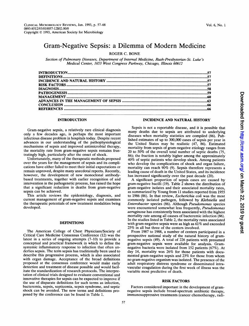
gram-negative bacilli (19). Table 2 shows the distribution ofgram-negative isolates and their associated mortality rates,as summarized by Young from 11 studies reported from 1955to 1986 (86). In that review, Escherichia coli was the mostcommonly isolated pathogen, followed by Klebsiella andEnterobacter species (86). Although Pseudomonas specieswere encountered somewhat less frequently, Pseudomonasaenrginosa has consistently been associated with the highestmortality rate among all causes of bacteremic infection (86).In the studies listed in Table 2, the mortality rates associatedwith gram-negative sepsis were as high as 61% and exceeded25% in all but three of the centers involved.From 1987 to 1988, a number of centers participated in a
prospective national study of the natural history of gram-negative sepsis (49). A total of 226 patients with presumedgram-negative sepsis were available for analysis. Gram-negative bacteria were isolated from 152 patients (67%). Atday 14, mortality was 26% for those patients with docu-mented gram-negative sepsis and 23% for those from whomno gram-negative organism was isolated. The presence of theadult respiratory distress syndrome or disseminated intra-vascular coagulation during the first week of illness was thevariable most predictive of death.
RISK FACTORSFactors considered important in the development of gram-
negative sepsis include broad-spectrum antibiotic therapy,immunosuppressive treatments (cancer chemotherapy, radi-
57
I. 6, No. 1
on March 22, 2019 by guest
http://cmr.asm
.org/D
ownloaded from
on M
arch 22, 2019 by guesthttp://cm
r.asm.org/
Dow
nloaded from
on March 22, 2019 by guest
http://cmr.asm
.org/D
ownloaded from
on M
arch 22, 2019 by guesthttp://cm
r.asm.org/
Dow
nloaded from

CLIN. MICROBIOL. REV.
TABLE 1. Definition of terms useda
Term Definition
Infection Microbial phenomenon characterized byan inflammatory response to thepresence of microorganisms or theinvasion of normally sterile hosttissue by those organisms
Presence of viable bacteria in blood
Systemic inflammatory response to avariety of severe clinical insults (seetext). The response is manifested bytwo or more of the followingconditions: temperature, >38 or<36°C; heart rate, >90 beats permin; respiratory rate, >20 breathsper min, or PaCO2, <32 mm Hg;leukocyte count, >12,000 cells perm13, <4,000 cells per m13, or >10%immature (band) forms.
Systemic response to infection. Thissystemic response is manifested bytwo or more of the followingconditions as a result of infection:temperature, >38 or <36°C; heartrate, >90 beats per min; respiratoryrate, >20 breaths per min, or PaCO2,<32 mm Hg; leukocyte count,>12,000 cells per m13, <4,000 cellsper ml3, or >10% immature (band)forms.
Sepsis associated with organdysfunction, hypoperfusion, or
hypotension. Hypoperfusion andperfusion abnormalities may include,but are not limited to, lactic acidosis,oliguria, or an acute alteration inmental status.
Sepsis with hypotension despiteadequate fluid resuscitation, alongwith the presence of perfusionabnormalities which may include, butare not limited to, lactic acidosis,oliguria, or an acute alteration inmental status. Patients who are on
inotropic or vasopressor agents maynot be hypotensive at the time thatperfusion abnormalities aremeasured.
Systolic blood pressure of <90 mm Hgor a reduction of >40 mm Hg frombaseline in the absence of othercauses of hypotension
Presence of altered organ function in an
acutely ill patient such thathomeostasis cannot be maintainedwithout intervention
a Reprinted from reference 12 with the permission of the publisher.
ation therapy, agents to reduce transplant rejection, andsteroids), invasive devices or procedures (surgery, vascularand bladder catheters, prosthetic devices, drainage tubes,and inhalation therapy equipment), penetrating wounds,
burns or other trauma, anatomic obstruction, intestinalulceration, age (the very young and the very old), andprogressive clinical conditions (malignancy, diabetes, AIDS,and other serious chronic diseases) (14, 50, 53, 83). Theseforms of treatment may either serve as a source or a focus ofinfection that foments sepsis or they may prolong the life ofa critically ill patient who is vulnerable to the disorder.Contaminated intravenous fluids have been implicated in a
number of nationwide epidemics of bacteremia. Between1965 and 1978, Maki recorded 33 epidemics traceable tosome form of infusion therapy; 7 of these were related to acontaminated commercial product (48). Nearly 80% of alldocumented epidemics were caused by gram-negative ba-cilli.Although the survival of patients with progressive or
chronic illnesses has been prolonged by better treatments forthe primary disease, debilitation eventually occurs, andthese patients become the immunocompromised targets ofsystemic infection.
DIAGNOSIS
In recent studies on the efficacy of methylprednisolonetreatment for septic patients (13, 14), the inclusion criteriafor study subjects included the following: a presumed site ofinfection, hyper- or hypothermia, tachycardia, tachypnea,and inadequate organ perfusion or function. Manifestationsof insufficient perfusion included altered mental state, hy-poxemia, elevated levels of plasma lactate, and oliguria.Bacteremia and hypotension were not essential for thediagnosis of sepsis syndrome in those studies.
Fever, the most common sign of sepsis, is believed to becaused by the actions of a number of endogenous substanceson prostaglandin E2 synthesis (3, 25, 26, 37). Hypothermia isseen principally in older patients (33, 37). Cardiac manifes-tations of sepsis range from tachycardia and increasedcardiac output to myocardial failure (37). Respiratory signsof sepsis include respiratory alkalosis, hyperventilation,failure of respiratory muscles, and the adult respiratorydistress syndrome, considered a catastrophic complication(6, 42, 54, 86).An increase in cardiac output is often seen early in the
course of the systemic inflammatory response syndrome(SIRS) but is usually offset by decreased peripheral resis-tance in the preshock state (37). Early shock is accompaniedby a significant decline in systemic vascular resistance thatmay precede the fall in blood pressure (37). In later shock,declining cardiac output, vasoconstriction, and refractoryhypotension may occur; alternatively, vasodilation may per-sist even in late shock (37, 86).
Renal manifestations of SIRS include azotemia and oligu-ria that result from renal tubular injury (37). Liver dysfunc-tion may be revealed by a rise in serum bilirubin levels thatfrequently precedes the clinical signs of infection (29). He-matological abnormalities associated with SIRS include eosi-nopenia (37), vacuolization of neutrophils (91), reducedlevels of iron in the serum (41), and the disseminatedintravascular coagulation syndrome (37). Thrombocytopeniais often noted at an early stage of SIRS (58), as is hypergly-cemia in diabetic patients (36, 37). A variety of changes inmental status is possible in the septic patient, includingdisorientation, lethargy, confusion, agitation, and obtunda-tion (37, 86).
Bacteremia
SIRS
Sepsis
Severe sepsis
Septic shock
Hypotension
Multiple organdysfunctionsyndrome
58 BONE
on March 22, 2019 by guest
http://cmr.asm
.org/D
ownloaded from
GRAM-NEGATIVE SEPSIS 59
TABLE 2. Distribution of gram-negative bacteremic isolates excluding polymicrobial infections'
Period of observa- No. of isolates (% mortality) Total episodestion E. coli Klebsiella spp. Enterobacter spp. Serratia spp. P. aeruginosa Proteus spp. (% mortality)
1983-1986 63 9 34 129 (12)1981-1983 38 8 23 83 (17)1975-1977 76 (35) 25 (48) 22 (72) 123 (39.8)1968-1974 127 (38.6) 233 (31.8) 67 (35.8) 37 (32.4) 74 (68.9)b 30 (36.7) 568 (38.9)1965-1974 189 (19.5) 74 (24.3) 47 (17.0) 11 (18.0) 60 (36.6) 49 (16.3) 430 (22.1)1972-1973 86 (17.4) 37 (43.2) 4 (0) 30 (60) 14 (35.7) 171 (31.6)1967-1972 68 (26.4) 72 (26.9) 35 (14.3) 8 (25) 29 (48.3) 11 (27.3) 233 (27.3)1968-1969 58 (18) 23 (13) 9 (0) 27 (54) 21 (57)b 13 (16) 151 (27.8)1965-1968 83 (21) 57 (33)b 45 (71) 19 (16) 204 (34.8)1958-1966 190 (42) 138 (55)" 67 (67)b 63 (33) 458 (50.7)1955-1967 93 (48) 68 (66)b 39 (77) 42 (67) 242 (61.2)
Usual rank or- 1 2 5C 4C 3 6Cder frequency
Mortality 4C 2 6C 5C 1 3Ca Reprinted from reference 86 with the permission of the publisher.b Species are grouped.c No significant differences in rank order.
PATHOGENESIS
At the pathophysiological level, the development of gram-negative sepsis involves a complicated series of effects basedon the composition of the bacterial cell wall. Pfeiffer firstrecognized the heat-stable toxic component of gram-nega-tive bacteria at the end of the 19th century (15, 56). In hisexperiments, Pfeiffer noted that lysates of heat-inactivatedVibrio cholerae caused shock and death in laboratory ani-mals. He called the toxic substance, not yet characterized,"endotoxin" on the assumption that it was found inside thebacterium. This also served to distinguish it from toxinssecreted during bacterial growth in culture (51, 56).
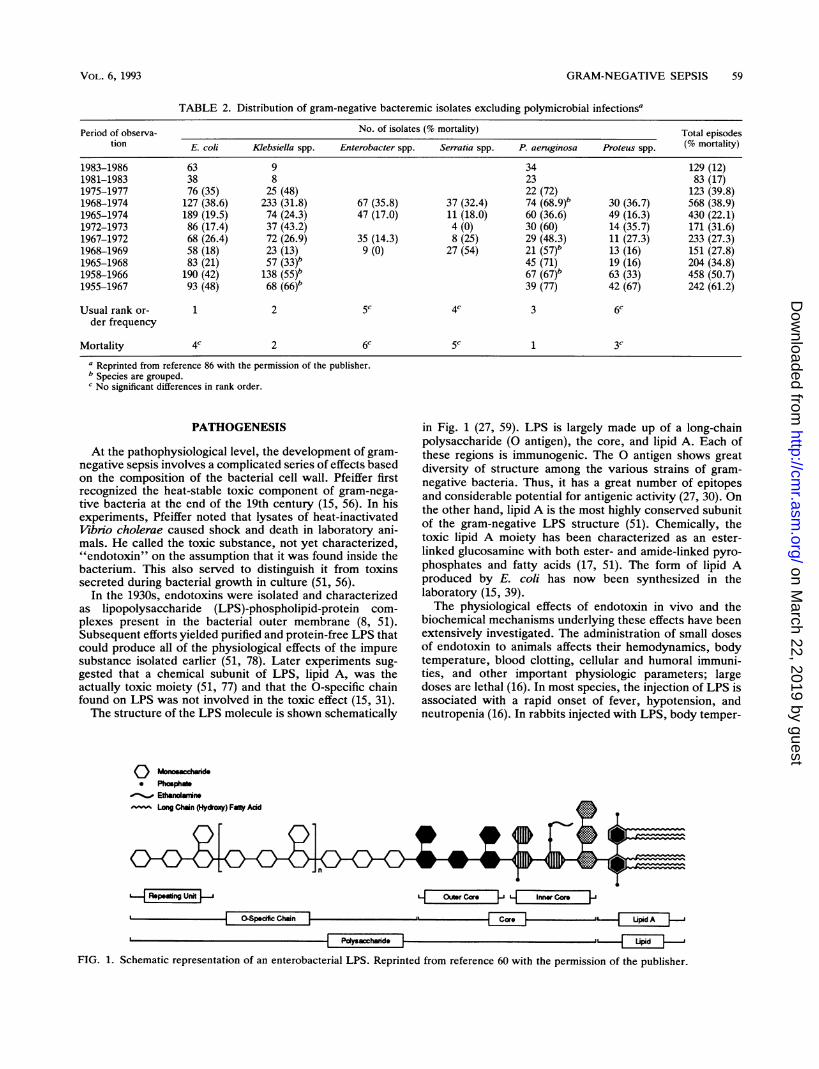
In the 1930s, endotoxins were isolated and characterizedas lipopolysaccharide (LPS)-phospholipid-protein com-plexes present in the bacterial outer membrane (8, 51).Subsequent efforts yielded purified and protein-free LPS thatcould produce all of the physiological effects of the impuresubstance isolated earlier (51, 78). Later experiments sug-gested that a chemical subunit of LPS, lipid A, was theactually toxic moiety (51, 77) and that the 0-specific chainfound on LPS was not involved in the toxic effect (15, 31).The structure of the LPS molecule is shown schematically
in Fig. 1 (27, 59). LPS is largely made up of a long-chainpolysaccharide (O antigen), the core, and lipid A. Each ofthese regions is immunogenic. The 0 antigen shows greatdiversity of structure among the various strains of gram-negative bacteria. Thus, it has a great number of epitopesand considerable potential for antigenic activity (27, 30). Onthe other hand, lipid A is the most highly conserved subunitof the gram-negative LPS structure (51). Chemically, thetoxic lipid A moiety has been characterized as an ester-linked glucosamine with both ester- and amide-linked pyro-phosphates and fatty acids (17, 51). The form of lipid Aproduced by E. coli has now been synthesized in thelaboratory (15, 39).The physiological effects of endotoxin in vivo and the
biochemical mechanisms underlying these effects have beenextensively investigated. The administration of small dosesof endotoxin to animals affects their hemodynamics, bodytemperature, blood clotting, cellular and humoral immuni-ties, and other important physiologic parameters; largedoses are lethal (16). In most species, the injection of LPS isassociated with a rapid onset of fever, hypotension, andneutropenia (16). In rabbits injected with LPS, body temper-
O Moniooacaide* Phosphate%_ Ethanoramin
Long Chnin (Hydroxy) Fatly Add
Rapsaing Unit |- L OutrCare L InrwCore |
a | ~OS~paciaicChoina|"d[_I= [ Placad _
7
FIG. 1. Schematic representation of an enterobacterial LPS. Reprinted from reference 60 with the permission of the publisher.
VOL. 6, 1993
on March 22, 2019 by guest
http://cmr.asm
.org/D
ownloaded from
CLIN. MICROBIOL. REV.
ature begins to rise in 10 to 20 min and peaks at approxi-mately 70 min (16). Humans are more sensitive to thepyrogenic activity of LPS and demonstrate fever at a smallfraction of the LPS dosage required to cause the febrileresponse in rabbits (82).Marked hypotension is observed in most species about 30
min after the administration of LPS (16). Recently, Suffred-ini et al. characterized the hemodynamic effects of endotoxinin humans by administering purified LPS to healthy volun-teers (68). Three hours after dosing, systemic vascularresistance and mean arterial pressure had decreased by 46and 18%, respectively, while the cardiac index had increasedby 53% and the heart rate had increased by 36%. Leftventricular function, both before and after volume loading,was consistent with the hemodynamic alterations observedin septic shock.The profound effects of endotoxin on clotting are demon-
strated by both local and generalized Schwartzman reactions(65). In animal studies, these reactions have been incited bytwo injections of endotoxin 12 to 18 h apart. In the localreaction, an intradermal injection followed by an intrave-nous injection produces hemorrhagic necrosis at the extra-dermal injection site. In the generalized reaction, sequentialintravenous injections produce bilateral renal cortical necro-sis in the test animals. This occurs as a result of theocclusion of small vessels by fibrin and intravascular coag-ulation (16).Endotoxin can also affect the blood cells, inducing neu-
tropenia, leukocytosis, and a reduction in circulating plate-lets (16). The proliferation of B lymphocytes and macro-phages is also stimulated by endotoxin.
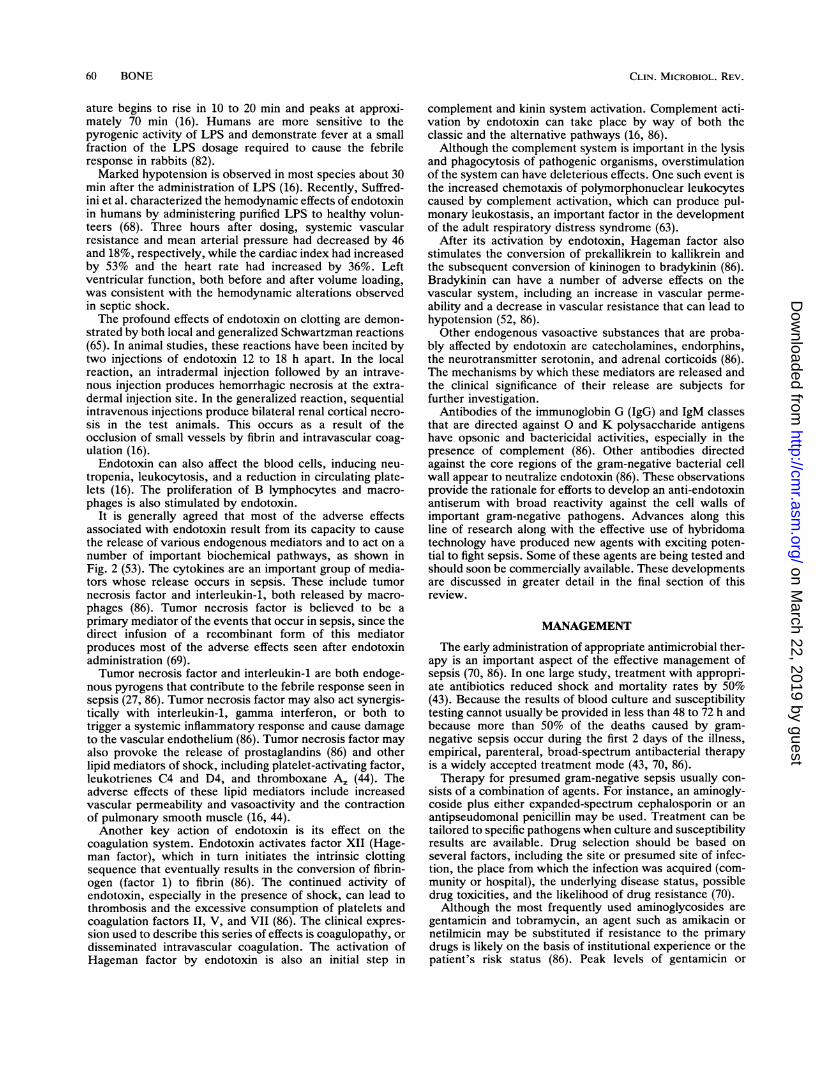
It is generally agreed that most of the adverse effectsassociated with endotoxin result from its capacity to causethe release of various endogenous mediators and to act on anumber of important biochemical pathways, as shown inFig. 2 (53). The cytokines are an important group of media-tors whose release occurs in sepsis. These include tumornecrosis factor and interleukin-1, both released by macro-phages (86). Tumor necrosis factor is believed to be aprimary mediator of the events that occur in sepsis, since thedirect infusion of a recombinant form of this mediatorproduces most of the adverse effects seen after endotoxinadministration (69).Tumor necrosis factor and interleukin-1 are both endoge-
nous pyrogens that contribute to the febrile response seen insepsis (27, 86). Tumor necrosis factor may also act synergis-tically with interleukin-1, gamma interferon, or both totrigger a systemic inflammatory response and cause damageto the vascular endothelium (86). Tumor necrosis factor mayalso provoke the release of prostaglandins (86) and otherlipid mediators of shock, including platelet-activating factor,leukotrienes C4 and D4, and thromboxane A, (44). Theadverse effects of these lipid mediators include increasedvascular permeability and vasoactivity and the contractionof pulmonary smooth muscle (16, 44).Another key action of endotoxin is its effect on the
coagulation system. Endotoxin activates factor XII (Hage-man factor), which in turn initiates the intrinsic clottingsequence that eventually results in the conversion of fibrin-ogen (factor 1) to fibrin (86). The continued activity ofendotoxin, especially in the presence of shock, can lead tothrombosis and the excessive consumption of platelets andcoagulation factors II, V, and VII (86). The clinical expres-sion used to describe this series of effects is coagulopathy, ordisseminated intravascular coagulation. The activation ofHageman factor by endotoxin is also an initial step in
complement and kinin system activation. Complement acti-vation by endotoxin can take place by way of both theclassic and the alternative pathways (16, 86).Although the complement system is important in the lysis
and phagocytosis of pathogenic organisms, overstimulationof the system can have deleterious effects. One such event isthe increased chemotaxis of polymorphonuclear leukocytescaused by complement activation, which can produce pul-monary leukostasis, an important factor in the developmentof the adult respiratory distress syndrome (63).
After its activation by endotoxin, Hageman factor alsostimulates the conversion of prekallikrein to kallikrein andthe subsequent conversion of kininogen to bradykinin (86).Bradykinin can have a number of adverse effects on thevascular system, including an increase in vascular perme-ability and a decrease in vascular resistance that can lead tohypotension (52, 86).Other endogenous vasoactive substances that are proba-
bly affected by endotoxin are catecholamines, endorphins,the neurotransmitter serotonin, and adrenal corticoids (86).The mechanisms by which these mediators are released andthe clinical significance of their release are subjects forfurther investigation.
Antibodies of the immunoglobin G (IgG) and IgM classesthat are directed against 0 and K polysaccharide antigenshave opsonic and bactericidal activities, especially in thepresence of complement (86). Other antibodies directedagainst the core regions of the gram-negative bacterial cellwall appear to neutralize endotoxin (86). These observationsprovide the rationale for efforts to develop an anti-endotoxinantiserum with broad reactivity against the cell walls ofimportant gram-negative pathogens. Advances along thisline of research along with the effective use of hybridomatechnology have produced new agents with exciting poten-tial to fight sepsis. Some of these agents are being tested andshould soon be commercially available. These developmentsare discussed in greater detail in the final section of thisreview.
MANAGEMENT
The early administration of appropriate antimicrobial ther-apy is an important aspect of the effective management ofsepsis (70, 86). In one large study, treatment with appropri-ate antibiotics reduced shock and mortality rates by 50%(43). Because the results of blood culture and susceptibilitytesting cannot usually be provided in less than 48 to 72 h andbecause more than 50% of the deaths caused by gram-negative sepsis occur during the first 2 days of the illness,empirical, parenteral, broad-spectrum antibacterial therapyis a widely accepted treatment mode (43, 70, 86).Therapy for presumed gram-negative sepsis usually con-
sists of a combination of agents. For instance, an aminogly-coside plus either expanded-spectrum cephalosporin or anantipseudomonal penicillin may be used. Treatment can betailored to specific pathogens when culture and susceptibilityresults are available. Drug selection should be based onseveral factors, including the site or presumed site of infec-tion, the place from which the infection was acquired (com-munity or hospital), the underlying disease status, possibledrug toxicities, and the likelihood of drug resistance (70).Although the most frequently used aminoglycosides are
gentamicin and tobramycin, an agent such as amikacin ornetilmicin may be substituted if resistance to the primarydrugs is likely on the basis of institutional experience or thepatient's risk status (86). Peak levels of gentamicin or
60 BONE
on March 22, 2019 by guest
http://cmr.asm
.org/D
ownloaded from

GRAM-NEGATIVE SEPSIS 61
Invasive InfectionBactererniaFungemia
RicketsemiaViremia
Mediator ReleasHistamine Kinin Activation
Complement ProstaglandinsActivation ? Others
Severe DecreaseIn Systemic
Vascular ResistanceProducing
Refractory Hypotenslon
Severe
Cardiac Output
IDeath
FIG. 2. The septic cascade: pathogenesis of septic shock. Reprinted from reference 53 with the permission of the publisher.
tobramycin in the blood should be maintained at between 6and 10 ,g/dl, while trough levels should fall below 2 ,ug/dl todecrease the chances of ototoxicity or nephrotoxicity (62).Frequent monitoring of drug levels in the blood is thereforerequired.For hospital-acquired infections in non-neutropenic pa-
tients, an expanded-spectrum cephalosporin rather than anaminoglycoside is often used because the etiologic organismis more likely to be a Kiebsiella sp. than a Pseudomonas sp.In patients with presumed P. aeruginosa infection, includingthose with neutropenia, burns, or infection related to respi-ratory therapy, an antipseudomonal penicillin such as me-zlocillin, piperacillin, ticarcillin, or azlocillin may be substi-tuted for the cephalosporin and used in combination with anaminoglycoside (85). In the case of resistance to both ceph-
alosporins and penicillins, imipenem may be used with theaminoglycoside (86).The rationale for using a combination of two antibiotics is
based on several considerations, including the broad cover-age of potential pathogens, the frequency of polymicrobialinfections, and the possibility of antibacterial synergy be-tween the two agents. Such synergistic combinations havebeen associated with improved clinical results (2). In addi-tion, such combinations may reduce the chances of emergentresistance by eliminating secondary bacterial populationsthat are resistant to one drug but not both (86). Currentevidence does not support the use of triple-drug combina-tions, and combinations of bactericidal and bacteriostaticagents should generally be avoided (40).The appropriate management of fluid and electrolyte bal-
Peripheral Vascular Effects1. Artenolar and Venular
Vasodilatation2. Leukocyte Aggregation
and Microembolization3. Endothelial Cell Injury
Direct Myocardial Effects1. Depressed Response to Fluid
Loading in Nonsurvivors ofSepic Shock
2. Depressed Ejection Fraction(Although Cardiac OutputElevated)
VOL. 6, 1993
I I
on March 22, 2019 by guest
http://cmr.asm
.org/D
ownloaded from

CLIN. MICROBIOL. REV.
TABLE 3. Sympathomimetic amines for circulatory support inseptic shock'
Drug Dose g/ Commentskg/min)
Dopamine 2-25 Increase infusion rate (D5W orsaline) every 15-20 min untilsystolic blood pressureexceeds 90 mm Hg and urineoutput exceeds 30 ml/h.
Dobutamine 2-25 Titrate as for dopamine.
Isoproterenol 5 Observe effect within 15-25 min;double infusion rate ifnecessary.
Norepinephrine 0.05 Start with test dose of 0.1-0.2pLg/kg and observe within 5min; administer via plasticcatheter into large peripheralor central vein.
a Adapted from reference 86 with the permission of the publisher. Drugs arelisted in order of preference and are to be used after volume replacement andwith careful electrocardiograph, central venous pressure, and blood pressuremonitoring. D5W, 5% dextrose in water.
ance is an important supportive measure in the treatment ofsepsis, particularly when shock ensues. Sympathomimeticamines may also be administered to manage the hemody-namic complications encountered in septic shock. Table 3summarizes the recommended sympathomimetic amines foruse to control shock. Dopamine raises the heart rate andsystolic blood pressure at higher infusion rates. Many clini-cians prefer to use low-dose dopamine (1 to 10 ,ug/kg of bodyweight per min) for its effect on renal perfusion (dopaminer-gic effect). Dobutamine may be added to the therapeuticregimen to increase myocardial contractility. If systolicblood pressure is still not adequate, norepinephrine is ti-trated to increase blood pressure through an increase insystemic vascular resistance. Compared with dopamine,dobutamine has less influence on heart rate and causes adecrease in pulmonary capillary wedge pressure. Isoproter-enol does not markedly elevate blood pressure, but it doesincrease the cardiac index (79). Adequate volume replace-ment must be achieved before any sympathomimetic amineis administered (86).
Despite extensive investigation, the utility of a number ofdrugs in the treatment of septic shock remains controversial.Disagreement concerning the use of glucocorticoids haspersisted for many years. The finding that corticosteroidtreatment improved survival in laboratory animal models ofsepsis was supported by the results of a 1976 clinical trial(64). Although this study was prospective and randomized,concerns were raised regarding certain aspects of the trialdesign. Sprung et al. compared the effect of a two-dosesteroid regimen with that of placebo in patients with septicshock and found no significant between-treatment differ-ences in mortality rates (66). In 1987, two large, controlledtrials of glucocorticoid therapy in sepsis were publishedsimultaneously, one by The Veterans Administration Sys-temic Sepsis Cooperative Study Group (72) and the other bythe Methylprednisolone Severe Sepsis Study Group (13).Both trials were prospective, randomized comparisons ofhigh-dose methylprednisolone sodium succinate and pla-cebo. In the Veterans Administration study, 14-day mortal-
ity was similar in the glucocorticoid (21%) and placebo (22%)groups. The resolution of secondary infection was signifi-cantly higher in patients who received placebo (P = 0.03).
In the second study of methylprednisolone, mortality at 14days was not improved by steroids, nor were treatment-related differences in the reversal of shock observed. Theauthors of both of these important studies concluded thathigh-dose glucocorticoid therapy provides no benefit inpatients with sepsis and septic shock and should not be used.It is now widely accepted that glucocorticoids should not beused in the treatment of septic shock (57).The opiate antagonist naloxone has attracted interest
because of its capacity to alter endotoxic shock in animals(28). In a small trial published in 1981, Peters et al. observeda 45% increase in systolic blood pressure after the adminis-tration of 0.4 to 1.2 mg of naloxone to eight patients withsepsis who were not receiving corticosteroids (55). In-creased blood pressure was evident within a few minutes ofthe intravenous injection and lasted for about 45 min. Fouryears later, however, DeMaria and associates performed adouble-blind, placebo-controlled study of intravenous bolusnaloxone in septic shock patients and found no significantbetween-treatment differences in either blood pressure ele-vation or survival (24). In the most recent study of naloxonein the treatment of sepsis, Roberts et al. gave either placeboor a 30-pg/kg intravenous bolus injection plus an additional30-p,g/kg infusion of naloxone to 14 patients with septicshock who required the support of inotropes, vasopressors,or both (61). The infusions of naloxone or placebo wereadministered over a period of 16 to 18 h. Pulmonary wedgepressure and pH were kept constant, and inotrope or vaso-pressor therapy was titrated to maintain a fixed mean bloodpressure. Inotrope or vasopressor requirements were signif-icantly lower in the naloxone-treated group than in theplacebo group at 8 (P < 0.005) and 16 (P < 0.02) h.Significant improvements were also seen in stroke volumeand heart rate in the group that received naloxone comparedwith those who received placebo. Because the positivehemodynamic effects seen in the naloxone group wereobserved only after 4 h, earlier studies utilizing bolus injec-tions may not have provided an optimal drug regimen andobservation period. Additional studies with naloxone areneeded to clarify its role in the management of septic shock.
Anticoagulants, particularly heparin, have been widelyused in the management of disseminated intravascular coag-ulation. Although these agents can ameliorate the clinicalexpressions of coagulopathy (23), they have not been shownto reduce mortality, and their use is probably best reservedfor other indications (86).
Transfusions of granulocytes for both the prophylaxis andtreatment of sepsis have been studied. In trials involvingprophylactic transfusions, investigators augmented the cir-culating granulocyte pool in patients scheduled to undergoaggressive chemotherapy for bone marrow transplantationor leukemia (22, 67, 81). The results of these studies sug-gested that any modest reductions in the incidence of gram-negative infection due to the infusions were countered by anincreased incidence of pulmonary problems. This prophylac-tic strategy cannot be recommended.
Therapeutic granulocyte transfusions have yielded moreencouraging results than have prophylactic transfusions;however, the evidence to date does not support the use ofgranulocytes in the routine treatment of neutropenic pa-tients. A survival benefit noted in some small studies (1, 38)was not confirmed by a larger, randomized study by Winstonet al. (80). In this large trial, granulocytopenic patients
62 BONE
on March 22, 2019 by guest
http://cmr.asm
.org/D
ownloaded from
GRAM-NEGATIVE SEPSIS 63
TABLE 4. Dependence of mortality rate on presence or absenceof shock at study admissiona
No. of patients with symptoms/total no. of patients(% affected)
Study groupOnly sepsis syn- Shock at Shock developeddrome present admission after admission
Totalb 10/77 (13) 19/69 (28)C 19/44 (43)dNonbacteremic 8/50 (16) 7/34 (21) 9/20 (45)dBacteremic 2/26 (8) 11/34- (32) 10/24 (42{Gram-negative 1/16 (6) 8/23 (35) 7/16 (43)CGram-positive 1/10 3/11 (27) 3/8 (38)
a Reprinted from reference 14 with the permission of the publisher.b Follow-up data were available for 190 of 191 patients (mortality data were
not available for one patient in the sepsis-syndrome-only group).c P < 0.05 compared with the sepsis-syndrome-only group.d p < 0.01 compared with the sepsis-syndrome-only group.I One patient with shock present on admission died from a primary
fungemia.fP < 0.001 compared with the sepsis-syndrome-only group.
(granulocyte count of <0.5 x 10⁹) with proven infectionswere randomly assigned to receive either a daily granulocytetransfusion or a control treatment. In addition to this treat-ment, antimicrobial therapy was also instituted. Amongpatients with gram-negative sepsis who did not demonstratebone marrow recovery, the survival rate was 48% in thetransfused group and 50% in the control group. The authorsconcluded that granulocyte transfusion conferred no signif-icant benefit over that of optimal antimicrobial therapyalone.
It has been suggested that the therapeutic use of granulo-cytes might prove more efficacious if larger quantities ofcells could be administered (85). While currently unfeasable,improved techniques in the harvest of granulocytes couldsomeday make this possible. In the meantime, these trans-fusions should be reserved for patients with reversiblydefective granulocyte production who have not responded toappropriate antimicrobial therapy.The prognosis for patients with sepsis becomes signifi-
cantly more grave at the onset of septic shock. In our ownprospective study (Table 4), patients with sepsis and withoutshock had a mortality rate of 13% (13). The mortality ratewas 28% for septic patients with shock at trial entry and 43%for those who developed shock after entry. These valueshighlight a need for the aggressive treatment of sepsis at theearliest possible time during its course. This has beenidentified as a means of preventing shock and improvingoutcome. These efforts should be helped considerably by thetherapeutic modalities now being developed.
ADVANCES IN THE MANAGEMENT OF SEPSIS
Immunotherapy for infection was first practiced more than100 years ago when von Behring used an equine antiserum totreat patients with diphtheria (21). The early years of thiscentury saw the development of antisera against a number ofimportant bacteria. The antibodies against a particular bac-terium recognized and acted against that organism only,however (21). This line of research was almost entirelydiscontinued after the introduction of effective antimicrobialagents, but interest in serum therapy reemerged when it wasrealized that broad-spectrum agents were not having theexpected impact on mortality caused by bacterial sepsis.The promise of immunotherapy in the treatment of sepsis
was underscored in 1982, when Ziegler and colleagues
published the results of their clinical study on a polyclonalantiserum against gram-negative bacteria (89). Earlier re-searchers had determined that, whereas the oligosaccharideside chains of gram-negative bacterial LPS differ widelyfrom strain to strain, the core regions of most strains arequite similar (45). Thus, it was conjectured, broadly effectiveimmunotherapy for gram-negative sepsis in the form ofantibodies raised against LPS might be developed throughthe use of a bacterial strain with an outer membrane thatfeatures no side chains, instead bearing only the conservedcore elements in its LPS. The strain selected was the J5mutant of E. coli O111:B4, whose LPS contains only thecore determinants, primarily lipid A.
After encouraging results were obtained in animal exper-iments, the researchers prepared human J5 antiserum byimmunizing healthy donors with a J5 boiled-cell vaccine (91).Over a period of 7 years, 304 patients with clinical symptomsthat suggested gram-negative bacteremia were entered intothe trial and randomized to treatment with either 1 U ofimmune serum or the same quantity of preimmune serum asa control. Of the 212 patients in whom the diagnosis ofgram-negative bacteremia was subsequently confirmed, 103received immune serum and 109 received control serum.Mortality was significantly lower (22%) in the J5 antiserumgroup than in the control group (39%). In the subset ofpatients with profound shock who needed vasopressors formore than 6 h, mortality was 77% in the control group and44% in the antiserum group. The authors concluded that J5antiserum reduced mortality from gram-negative sepsis byapproximately 50% and that this protection was apparenteven in the presence of optimal antimicrobial therapy andmedical-surgical management (91).These findings led to a prophylactic trial of J5 antibody in
which high-risk surgical patients were treated with eitherimmune or preimmune plasma at the time of randomizationand every 5 days thereafter until they were no longerconsidered to be at high risk (5). Because the previoustherapeutic trial involving J5 had not shown a significantdecrease in mortality among the patients with abdominalinfections, the effect of prophylaxis in this group was ofconsiderable interest. In addition, the study allowed re-searchers to assess the value of this strategy in patients atsubstantial risk, such as those with multiple trauma, theelderly, and immunocompromised patients undergoing lungsurgery.A total of 262 evaluable patients were entered into the
study and observed daily. Although there was no significantdifference in the incidence of infection in the J5 antibodystudy group (36%) compared with the control group (40%),the antibody clearly reduced the serious consequences ofgram-negative infection. Patients in the control group had arisk of developing septic shock that was more than twice thatin the antibody group. A twofold decrease in mortality wasobserved among patients in shock who received J5 plasma.The antibody's efficacy was also demonstrated in patientswho underwent abdominal surgery. The study also con-firmed the specificity of J5's affinity for lipid A, the injuriousportion of the bacterial outer membrane, since neither thedirect consequences of gram-positive infection nor the infec-tion rate was altered, while the complications of gram-negative infection, such as the inflammatory response, wereaffected. This suggests that the J5 antibody bound directly tothe lipid A portion of the bacteria (5).
In patients with SIRS, retrospective analysis had demon-strated a significant correlation between the serum titers ofantibody to core glycolipid and survival (47). Nevertheless,
VOL. 6, 1993
on March 22, 2019 by guest
http://cmr.asm
.org/D
ownloaded from

CLIN. MICROBIOL. REV.
the relationship between the level of J5 antibody achieved inseptic patients and the improvement seen in their outcomeremained to be demonstrated (4, 91). Important follow-upstudies indicated that a human IgG antibody to E. coli J5 wasnot effective in reducing complications of gram-negativesepsis (18) and that the protective activity of the antiserumlay almost entirely in the IgM antibody (46).
Despite these encouraging results, polyclonal J5 antiserumis not suitable for commercial development for a number ofreasons. These include the adverse effects of vaccination on
serum donors; the variability of antiserum activity; the needto use pooled human blood, thereby creating a risk fortransmission of viral disease; and the difficulty of producinglarge quantities of antiserum.
Fortunately, the discovery and elaboration of hybridomatechnology have allowed the mass production of IgM mon-
oclonal antibodies that may prove to be useful in themanagement of gram-negative sepsis. Preclinical and clinicalstudies on both murine and human monoclonal antibodieshave been encouraging. A detailed analysis of these mono-
clonal antibodies follows.On the basis of the obvious promises held out by anti-
endotoxin therapy, the anti-endotoxin antibody E5 was
developed, using the following procedure (32). After micewere immunized with boiled E. coli J5 cells, their spleencells were fused with murine myeloma cells to yield a single(monoclonal) cell line producing an antibody directed spe-cifically against lipid A. Selection of the proper cell lineprovided a culture that continued to produce an IgM anti-body that reacts with the core region of gram-negativebacterial outer membranes (71). A recent in vitro study thatemployed boiled bacterial cells and a large panel of LPS andlipid A preparations found that lipid A was the apparentepitope on LPS to which E5 binds (84).The results seen in preclinical and clinical trials of E5 have
been encouraging. After significantly improved survival wasseen in mice challenged with gram-negative organisms andtreated with E5 (87), and following human pharmacokineticstudies (74), two major clinical trials of E5 were undertaken.In the first trial, a double-blind comparison of E5 andplacebo in patients with suspected gram-negative sepsisshowed that mortality among those in the subgroup withdocumented gram-negative infection was 22% in patientsgiven routine therapy alone but only 7% in patients givenroutine therapy plus E5 (34).
In a subsequent large, double-blind trial in which 33centers participated, 486 patients with suspected gram-negative sepsis were enrolled (35). To be eligible, patientswere required to show at least two of the following signs ofgram-negative infection: a core temperature of >38 or
<35°C; a peripheral blood leukocyte count of >12 x 109 or
<3 x 10 Iliter (not resulting from other treatments), or
.20% immature forms; growth of gram-negative bacteriafrom a blood culture taken within the preceding 48 h; anddocumented or suspected site of gram-negative infection. Inaddition, evidence of a systemic response was required(Table 5). The presence of at least one of the followingsymptoms was taken as evidence of such a response: arterialhypotension; metabolic acidosis; decreased systemic vascu-
lar resistance; tachypnea; or otherwise unexplained dysfunc-tions of the kidney, central nervous system, lung, or coagu-lation systems.Grounds for exclusion from the study included the occur-
rence of any one of the following: uncomplicated transientbacteremia; granulocyte count of <109/liter, late septicshock, core temperature of >41.7°C, Child's class C liver
TABLE 5. Evidence of systemic response in the E5 trial
Parameter Response
Arterial hypotension Systolic blood pressure of <90 mm Hg oracute drop of 30 mm Hg
Metabolic acidosis Base deficit of >5 meq/literDecreased systemic Resistance of <800 dynes/s/cm5vascular resistance
Tachypnea Respiratory rate of <30 breaths/min ormechanical ventilation of >10 liters/min
Unexplaineddysfunction of
Kidney Urine output of <30 mlLung PaO2 of <0.1 atm on room air or PaO2/
F102 of <0.37 atmaCentral nervous Confusion, disorientation, obtundationsystem
Coagulation Thrombocytopenia (25% decrease inplatelet count), prolonged prothrombin,or partial thromboplastin times
a PaO2, oxygen tension of arterial blood; F102, oxygen fraction of inspiredair. 1 atm = 101.29 kPa.
disease, AIDS, burn infections, pregnancy or lactation,previous treatment with or a known allergy to murineproducts, treatment with any other investigational agent, ageunder 18 years, or lack of commitment to full life support bythe primary physician.
Patients were randomly assigned to receive treatment witheither placebo (D5NS) or 200 ml of a solution containing theE5 anti-endotoxin antibody at a dose of 2 mg/kg. The 1-hinfusion was administered intravenously. A second infusionof the study drug was administered at the same dose 1 daylater. Patients also continued to receive the antimicrobialagent and supportive therapy deemed appropriate by theprimary physician. Because 18 patients were excluded fromefficacy analysis, 468 were considered evaluable. Of these,316 had documented gram-negative sepsis, as demonstratedby a positive culture result for any normally sterile body siteduring the period of 2 days before to 3 days after theadministration of the study drug.E5 was not significantly more effective than placebo in
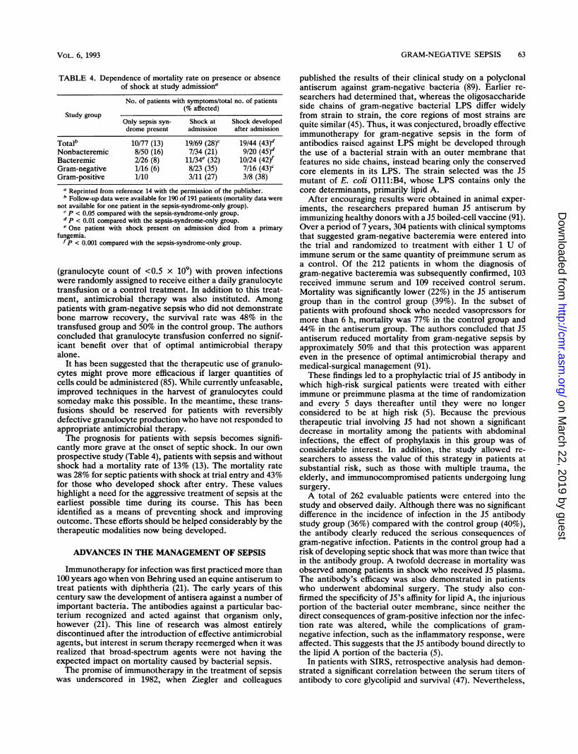
improving survival among the 316 patients with documentedgram-negative sepsis. In the subgroup (n = 137) of patientswith gram-negative sepsis who were not in refractory shockat study entry, however, patients taking E5 had a signifi-cantly lower mortality rate (hazard ratio = 2.3; P = 0.01).Both bacteremic and nonbacteremic patients contributed toimproved survival in the E5 group as a whole, since theywere associated with relative risks of 2.3 and 2.1, respec-tively. Survival data are summarized in Fig. 3.Among the 137 patients with sepsis who were not in
shock, the number of times that individual organ failures(disseminated intravascular coagulation, adult respiratorydistress syndrome, or acute renal failure) resolved ap-proached significance (P = 0.05) among patients taking E5(19 of 35 resolved [54%]) compared with those taking pla-cebo (8 of 27 resolved [30%]).The two E5 studies have shown that the drug is well
tolerated. Adverse reactions were seen in 5.9% of the E5group, although these were not necessarily related to E5administration, and in 2.1% of the control group. Rash andan anaphylactoid reaction were the most common adverseevents probably related to E5 administration, with a fre-quency of 1.2 and 1.0%, respectively. The presence of a
64 BONE
on March 22, 2019 by guest
http://cmr.asm
.org/D
ownloaded from

GRAM-NEGATIVE SEPSIS 65
c:CO,
0
co.0
20-
1.0 -
0.9 -
0.8 -
0.7 -
0.6-
0.5 -
0 10 20
Study Day
FIG. 3. Thirty-day Kaplan-Meier survival curves of patientswith gram-negative sepsis. Solid line represents survival probabilityin the E5 group; dotted line represents survival probability in theplacebo group. Reprinted from reference 35 with the permission ofthe publisher.
human antimurine antibody response was evaluated by an
enzyme immunoassay method with a sensitivity of <1.0ng/ml. A fourfold increase over baseline was considered a
positive result. Serial determinations of human antimurineantibody response among 182 patients treated with E5showed positive IgG responses in 86 patients (47%). Anti-body responses generally occurred 2 weeks after the initialdose, however, and were not associated with clinical ad-verse events.
Encouraging results have also been reported with a humanhybrid monoclonal antibody, HA-1A, derived from E. coli J5vaccine (88). HA-1A was evaluated in 543 patients withsepsis syndrome and presumed gram-negative infection. Thetherapeutic regimen consisted of 100 mg of HA-1A or
placebo (human serum albumin) administered in a singleintravenous infusion. Patients were monitored for 28 days or
until death. HA-1A was significantly more effective (P =
0.014) than placebo at reducing mortality in the subset ofpatients with documented gram-negative bacteremia (n =
200). HA-1A also reduced mortality by 42% in the patientswith both bacteremia and shock (P = 0.017 compared withplacebo). HA-1A did not offer significant protection to allpatients entered into the study, however; treatment was notsignificantly protective in nonbacteremic patients.During the first 7 days after treatment with HA-1A, all
evidence of any major complications of sepsis present atbaseline (such as shock, disseminated intravascular coagu-
lation, acute renal failure, acute hepatic failure, or adultrespiratory distress syndrome) resolved in 26 of 62 patients(42%) given placebo and in 38 of 61 (62%; P = 0.024) givenHA-1A.The use of HA-1A, too, was well tolerated by patients.
One patient developed hives at the infusion site, and anotherexperienced flushing and mild hypotension. These events
were mild and transient in both patients. No patient haddetectable anti-HA-lA antibodies.The results of these studies indicate that both the murine
and the human hybrid monoclonal anti-endotoxin antibodiesimprove survival in patients with gram-negative sepsis. E5appears to be effective whether or not the patient is bacter-emic, while HA-1A appears to be effective only in thepresence of bacteremia. On this note, it is unfortunate that
the confirmation of bacteremia frequently occurs after theoptimum time for pharmacologic intervention, by which timethe patient may already be showing signs of SIRS. Anotherdifference between the two antibodies is that HA-1A waseffective in patients with shock refractory to treatment withfluids or inotropes, while E5 was not. However, directcomparisons between the two studies are difficult because oftheir different methodologies. For instance, the definitionsfor shock used by the two studies were different: some 40patients who were characterized as not being in shock in theE5 study may have met the criteria for shock in the otherstudy. In addition, the different antibody responses seen inthe two studies might be related to the detection limits of theassays used. Limited sampling and the use of a less sensitiveassay probably decreased the likelihood that significantantibody responses could be detected in the HA-1A study. Itis important to note that antibody responses to E5 appearedwell after completion of the 2-day course of therapy andwere not associated with clinical sequelae.
While the results of these trials into the use of J5, E5, andHA-1A on patients with sepsis appear to be supportive, notall writers have agreed. For instance, an editorial by Wenzelconcluded that these studies failed to show an effectivenessthat would yield an acceptable cost-benefit ratio (76). Hefurther pointed out that various confounding factors werepresent in the studies that may have resulted in differencesbetween the control and treatment groups. These includesupplemental treatment with antimicrobial agents and thepresence of varying levels of disease at entrance.
In an editorial for the Sounding Board section in the sameissue of the New England Journal of Medicine, however,Warren et al. come to a different conclusion on the issue ofHA-1A anti-endotoxin antibody use (73). First, they thinkthere is a need for more clinical research into the effects ofthe HA-1A antibody with stress on the idea that theseantibodies are intended for adjunctive use along with appro-priate antimicrobial therapy. However, they believe thatthere is not enough in vitro evidence of HA-1A's ability toeffectively bind endotoxin. They quoted in vitro and animalstudies of the antibody that bring into doubt its ability to bindto endotoxin and decrease the host's systemic response.They also had familiar critiques on the differences betweenthe control and treatment groups in the clinical studies.These articles were rebutted by a letter from Ziegler and
Smith (90) found in the same issue. They think that there hasbeen much in vitro work in the last 6 years showing thespecificity and strength of HA-1A binding to endotoxin.They also pointed out that animal studies cannot be appliedto human use of the antibody because of the divergentresponses of animals to endotoxin.To summarize the much discussed results of the trials into
E5 and HA-1A use in septic patients, it is easy to criticizeany study because of imbalances between the placebo andtreatment groups. These will inevitably occur by chance ifenough baseline criteria are analyzed. However, in myopinion, these studies of anti-endotoxin antibodies havebeen rigorously planned and well executed. I believe that,ultimately, both murine and human monoclonal antibodieswill be highly useful. If these agents become generallyavailable and commonly used, they should herald a signifi-cant advance in the management of gram-negative sepsis.
It should also be remembered that imagination is the onlylimit to the types of new agents that may be used to fightgram-negative sepsis. Currently, several are being devel-oped for use by researchers (11). While the use of antibodiesto endotoxin may be an important adjunct to the treatment of
VOL. 6, 1993
?--l
I- -
L
------
I------I
------
ILL------
L---
on March 22, 2019 by guest
http://cmr.asm
.org/D
ownloaded from

CLIN. MICROBIOL. REV.
gram-negative sepsis, many other key molecular interactionsinvolved in the systemic inflammatory response may beaffected through the use of novel agents. For instance,antibodies that bind to the cytokines themselves coulddirectly affect the endogenous mechanisms by which sepsisoccurs. Monoclonal antibodies to exotoxins, phospholipaseA2, C5a (a complement fragment), adhesion molecules, andcontact factors could also become important in the fight tocontrol SIRS. Similarly, agents can be found that block thetumor necrosis factor, interleukin-1, platelet-activating fac-tor, thromboxane A2, or bradykinin receptors, thus dimin-ishing the deleterious effects of these mediators. Agentswhich inhibit neutrophil activation could be effective inblocking the inflammatory response. These include pentox-ifyline, adenosine, dapsone, antioxidants, heavy-metal che-lators, oxygen radical scavengers, and protease inhibitors.Coagulopathy is an important and deleterious part of theinflammatory response. Because of the great complexity ofcoagulation, there are numerous points at which the re-sponse may be controlled. Some inhibitors of this processinclude antithrombin III, protein C, thrombomodulin, hiru-din, al-antitrypsin Pittsburgh, aprotinin, soybean trypsininhibitor, and plasminogen activators. Other therapeuticmeasures that may prove helpful in SIRS include gut decon-tamination, antihistamines, naloxone, thyroid releasing hor-mone, glucagon, surfactant, extracorporeal membrane oxy-genation, calcium channel blockers, growth factors, andgrowth hormone. I think that all such agents should besubjected to intense investigation and scrutiny, although it isalso important that we not reject potentially importantadvances in therapy that may provide improvements inpatient management.
CONCLUSION
Gram-negative sepsis remains a significant cause of mor-bidity and mortality in spite of the ongoing development ofnew antimicrobial agents. This may be because antimicrobialtherapy fails to address the underlying pathogenetic mecha-nism involved in the systemic inflammatory response. It isthe triggering of mediators by bacterial endotoxin that pro-duces the symptoms of gram-negative sepsis. Monoclonalantibody technology has made possible the development ofpreparations that, theoretically, will bind to and neutralizeendotoxin, thereby inactivating it.The anti-endotoxin antibody E5 has been shown to im-
prove survival and enhance the resolution of major morbid-ities in patients with gram-negative sepsis who are not inshock. Similarly, HA-1A, a human hybrid monoclonal anti-body, enhanced the resolution of organ failures and im-proved survival in bacteremic patients with gram-negativesepsis. These encouraging clinical results suggest that theadministration of these antibodies early in the course ofgram-negative sepsis as part of a therapeutic regimen thatalso includes antibiotics and appropriate supportive careshould significantly affect morbidity and mortality.
REFERENCES
1. Alavi, J. B., R. K. Root, I. Djerassi, A. E. Evans, S. J.Gluckman, R. R. MacGregor, D. Guerry, A. D. Schreiber, J. M.Shaw, P. Koch, and R. A. Cooper. 1977. A randomized clinicaltrial of granulocyte transfusions for infection in acute leukemia.N. Engl. J. Med. 296:706-711.
2. Anderson, E. T., L. S. Young, and W. L. Hewitt. 1978. Antimi-crobial synergism in the therapy of gram-negative bacteremia.Chemotherapy (Basel) 24:45-54.
3. Atkins, E. 1984. Fever: the old and the new. J. Infect. Dis.149:339-348.
4. Baumgartner, J.-D., and M. P. Glauser. 1987. Controversies inthe use of passive immunotherapy for bacterial infections in thecritically ill patient. Rev. Infect. Dis. 9:194-205.
5. Baumgartner, J.-D., J. A. McCutchan, G. von Melle, M. Vogt,R. Luethy, M. P. Glauser, E. J. Ziegler, M. R. Klauber, E.Muehlen, R. Chiolero, and S. Geroulanos. 1985. Prevention ofgram-negative shock and death in surgical patients by antibodyto endotoxin core glycolipid. Lancet ii:59-63.
6. Blair, E. 1970. Hypocapnia and gram-negative bacteremicshock. Am. J. Surg. 119:433-439.
7. Boivin, A., and L. Mesrobeanu. 1935. Recherches sur les anti-genes somatiques et sur les endotoxines des bacteries. I. Con-siderations generales, et expose des techniques utilisees. Rev.Immunol. (Paris) 1:553-569.
8. Bone, R. C. 1991. Let's agree on terminology: definitions ofsepsis. Crit. Care Med. 19:973-976.
9. Bone, R. C. 1991. Sepsis syndrome. Part 1. The diagnosticchallenge. J. Crit. Illness 6:525-539.
10. Bone, R. C. 1991. Sepsis syndrome. Part 2. Coping with thetherapeutic challenge. J. Crit. Illness 6:650-664.
11. Bone, R. C. 1991. A critical evaluation of new agents for thetreatment of sepsis. JAMA 266:1686-1691.
12. Bone, R. C., R. A. Balk, F. B. Cerra, R. P. Dellinger, A. M. Fein,W. A. Knaus, R. M. H. Schein, W. J. Sibbald, et al. 1992.ACCP/SCCM consensus conference: definitions for sepsis andorgan failure and guidelines for the use of innovative therapiesin sepsis. Chest 101:1644-1655.
13. Bone, R. C., C. J. Fisher, Jr., T. P. Clemmer, G. J. Slotman,C. A. Metz, R. A. Balk, and the Methylprednisolone SevereSepsis Study Group. 1987. A controlled clinical trial of high-dosemethylprednisolone in the treatment of severe sepsis and septicshock. N. Engl. J. Med. 317:653-658.
14. Bone, R. C., C. J. Fisher, Jr., T. P. Clemmer, G. J. Slotman,C. A. Metz, R. A. Balk, and the Methylprednisolone SevereSepsis Study Group. 1989. Sepsis syndrome: a valid clinicalentity. Crit. Care Med. 17:389-393.
15. Brade, H., L. Brade, U. Schade, U. Zahringer, 0. Holst, H.-M.Kuhn, A. Rozalski, E. Rohrscheidt, and E. T. Rietschel. 1988.Structure, endotoxicity, immunogenicity and antigenicity ofbacterial lipopolysaccharides (endotoxins, 0-antigens), p. 17-45. In J. Levin, J. W. tenCate, H. R. Buller, S. J. H. vanDeventer, and A. Sturk (ed.), Bacterial endotoxins: pathophys-iological effects, clinical significance, and pharmacological con-trol. Alan R. Liss, New York.
16. Braude, A. I. 1986. Bacterial endotoxins, p. 51-60. In A. I.Braude (ed.), Infectious diseases and medical microbiology, 2nded. The W. B. Saunders Co., Philadelphia.
17. Burton, A. J., and H. E. Carter. 1964. Purification and charac-terization of the lipid A component of the lipopolysaccharidesfrom Escherichia coli. Biochemistry 3:411-418.
18. Calandra, T., M. P. Glauser, J. Shellekens, and J. Verhoef. 1988.Treatment of gram-negative septic shock with human IgGantibody to Escherichia coli J5: a prospective, double-blind,randomized trial. J. Infect. Dis. 158:312-319.
19. Centers for Disease Control. 1978. National nosocomial infec-tions study report. Annual summary 1976. U.S. Department ofHealth, Education, and Welfare, Washington, D.C.
20. Centers for Disease Control. 1990. Increase in national hospitaldischarge survey rates for septicemia-United States, 1979-1987. Morbid. Mortal. Weekly Rep. 39:31-34.
21. Chmel, H. 1990. Role of monoclonal antibody therapy in thetreatment of infectious disease. Am. J. Hosp. Pharmacy47(Suppl. 3):S11-S15.
22. Clift, R. A., J. E. Sanders, E. D. Thomas, B. Williams, and C. D.Buckner. 1978. Granulocyte transfusions for the prevention ofinfection in patients receiving bone marrow transplants. N.Engl. J. Med. 298:1052-1057.
23. Corrigan, J. J., Jr., and J. F. Kiernat. 1975. Effect of heparin in
66 BONE
on March 22, 2019 by guest
http://cmr.asm
.org/D
ownloaded from

GRAM-NEGATIVE SEPSIS 67
experimental gram-negative septicemia. J. Infect. Dis. 131:138-143.
24. DeMaria, A., D. E. Craven, J. J. Heffernan, T. K. McIntosh,G. A. Grindlinger, and W. R. McCabe. 1985. Naloxone versusplacebo in treatment of septic shock. Lancet i:1363-1365.
25. Dinarello, C. A., J. G. Cannon, S. M. Wolff, H. A. Bernheim, B.Beutler, A. Cerami, I. S. Figari, M. A. Palladino, Jr., and J. V.O'Connor. 1986. Tumor necrosis factor (cachectin) is an endo-genous pyrogen and induces production of interleukin 1. J. Exp.Med. 163:1433-1450.
26. Dinarello, C. A., G. H. A. Clowes, Jr., A. H. Gordon, C. A.Saravis, and S. M. Wolff. 1984. Cleavage of human interleu-kin-1: isolation of a peptide fragment from plasma of febrilehumans and activated monocytes. J. Immunol. 133:1332-1338.
27. DiPiro, J. T. 1990. Pathophysiology and treatment of gram-negative sepsis. Am. J. Hosp. Pharmacy 47(Suppl. 3):S6-S10.
28. Faden, A. I., and J. W. Holaday. 1979. Opiate antagonists: a rolein the treatment of hypovolemic shock. Science 205:317-318.
29. Franson, T. R., W. J. Hierholzer, and D. R. LaBrecque. 1985.Frequency and characteristics of hyperbilirubinemia associatedwith bacteremia. Rev. Infect. Dis. 7:1-9.
30. Galanos, C., M. A. Freudenberg, F. Jay, D. Nerkar, K. Veleva,H. Brade, and W. Strittmatter. 1984. Immunogenic properties oflipid A. Rev. Infect. Dis. 6:546-552.
31. Galanos, C., 0. Luderitz, E. T. Rietschel, and 0. Westphal.1977. Newer aspects of the chemistry and biology of bacteriallipopolysaccharides, with special reference to their lipid Acomponent, p. 239-335. In T. W. Goodwin (ed.), Internationalreview of biochemistry. Biochemistry of lipids II, vol. 14.University Park Press, Baltimore.
32. Gascon, R. L., S. Alam, L. E. Bermudez, and L. S. Young. 1989.Development of monoclonal antibodies against lipopolysaccha-ride and their evaluation in animal models. In Abstr., Sympo-sium on Major Developments in the Management of Bacterialand Fungal Infections: A Look to the Future, Vancouver,Canada, 19 to 23 August 1989.
33. Gleckman, R., and D. Hibert. 1982. Afebrile bacteremia: aphenomenon in geriatric patients. JAMA 248:1478-1481.
34. Gorelick, K. J., N. I. Wedel, A. Y. Kunz, K. M. Wilson, andR. N. Greenberg. 1990. E5 antiendotoxin antibody in gram-negative sepsis: report of a phase II study, abstr. Crit. CareMed. 18(Suppl.):S261.
35. Greenman, R. L., R. M. H. Schein, M. A. Martin, R. P. Wenzel,N. R. Maclntyre, G. Emmanuel, H. Chmel, R. B. Kohler, M.McCarthy, J. Plouffe, J. A. Russell, and the XOMA Sepsis StudyGroup. 1991. A controlled clinical trial of E5 murine monoclonalIgM antibody to endotoxin in the treatment of gram-negativesepsis. JAMA 266:1097-1102.
36. Gump, F. E., C. Long, P. Killian, and J. M. Kinney. 1974.Studies of glucose intolerance in septic injured patients. J.Trauma 14:378-388.
37. Harris, R. L., D. M. Musher, K. Bloom, J. Gathe, L. Rice, B.Sugarman, T. W. Williams, Jr., and E. J. Young. 1987. Mani-festations of sepsis. Arch. Intern. Med. 147:1895-1906.
38. Herzig, R. H., G. P. Herzig, R. G. Graw, Jr., M. I. Bull, andK. K. Ray. 1977. Successful granulocyte transfusion therapy forgram-negative septicemia. N. Engl. J. Med. 296:701-705.
39. Imoto, M., H. Yoshimura, N. Sakaguchi, S. Kusumoto, and T.Shiba. 1985. Total synthesis of Escherichia coli lipid A. Tetra-hedron Lett. 26:1545-1548.
40. International Antimicrobial Therapy Project Group of the Euro-pean Organization for Research and Treatment of Cancer. 1983.Combination of amikacin and carbenicillin with or withoutcefazolin as empiracal treatment of febrile neutropenic patients.J. Clin. Oncol. 1:597-603.
41. Kluger, M. J., and B. A. Rothenburg. 1979. Fever and reducediron: their interaction as a host defense response to bacterialinfection. Science 203:374-376.
42. Krausz, M. M., A. Perel, D. Eimerl, and S. Cotev. 1977.Cardiopulmonary effects of volume loading patients in septicshock. Ann. Surg. 185:429-434.
43. Kreger, B. E., D. E. Craven, and W. R. McCabe. 1980. Gram-negative bacteremia. IV. Re-evaluation of clinical features and
treatment in 612 patients. Am. J. Med. 68:344-355.44. Lefer, A. M. 1989. Significance of lipid mediators in shock
states. Circ. Shock 27:3-12.45. Luderitz, O., A. M. Staub, and 0. Westphal. 1966. Immuno-
chemistry of 0 and R antigens of Salmonella and relatedEnterobacteniaceae. Bacteriol. Rev. 30:192-255.
46. McCabe, W. R., A. DeMaria, Jr., H. Berberich, and M. A.Johns. 1988. Immunization with rough mutants of Salmonellaminnesota: protective activity of IgM and IgG antibody to theR595 (Re chemotype) mutant. J. Infect. Dis. 58:291-300.
47. McCabe, W. R., B. E. Kreger, and M. Johns. 1972. Type-specific and cross-reactive antibodies in gram-negative bactere-mia. N. Engl. J. Med. 287:261-267.
48. Maki, D. G. 1981. Nosocomial bacteremia: an epidemiologicoverview. Am. J. Med. 70:719-732.
49. Martin, M. A., R. P. Wenzel, K. J. Gorelick, and the XOMASepsis Study Group. 1989. Program Abstr. 29th Intersci. Conf.Antimicrob. Agents Chemother., abstr. 317.
50. Mizock, B. 1984. Septic shock. A metabolic perspective. Arch.Intern. Med. 144:579-585.
51. Morrison, D. C. 1983. Bacterial endotoxins and pathogenesis.Rev. Infect. Dis. 5(Suppl. 4):S733-S747.
52. Nies, A. S., R. P. Forsyth, H. E. Williams, and K. L. Melmon.1968. Contribution of kinins to endotoxin shock in unanesthe-tized rhesus monkeys. Circ. Res. 22:155-164.
53. Parker, M. M., and J. E. Parrillo. 1983. Septic shock. Hemo-dynamics and pathogenesis. JAMA 250:3324-3327.
54. Pepe, P., R. T. Potkin, D. H. Reus, L. D. Hudson, and C. J.Carrico. 1982. Clinical predictors of the adult respiratory dis-tress syndrome. Am. J. Surg. 144:124-130.
55. Peters, W. P., M. W. Johnson, P. A. Friedman, and W. E. Mitch.1981. Pressor effect of naloxone in septic shock. Lancet i:529-532.
56. Pfeiffer, R. 1892. Untersuchungen uber das Cholera Gift. Z.Hyg. Infectionskr. 11:393-412.
57. Rackow, E. C., and M. E. Astiz. 1991. Pathophysiology andtreatment of septic shock. JAMA 266:548-554.
58. Riedler, G. F., P. W. Straub, and P. G. Frick. 1971. Thrombo-cytopenia in septicemia: a clinical study for the evaluation of itsincidence and diagnostic value. Helv. Med. Acta 36:23-38.
59. Rietschel, E. T., U. Schade, M. Jensen, H.-W. Wollenweber, 0.Luderitz, and S. G. Greisman. 1984. Bacterial endotoxins:chemical structure, biological activity and role in septicemia.Scand. J. Infect. Dis. 31:8-21.
60. Rietschel, E. T., H. W. Wollenweber, H. Brade, U. Zahringer, B.Lindner, U. Seydel, H. Bradaczek, G. Barnickel, H. Labischin-ski, and P. Giesbrecht. 1984. Structure and conformation of thelipid A component of lipopolysaccharides, p. 187-220. In R. A.Proctor (ed.), Handbook of endotoxin, vol. 1. Chemistry ofendotoxin. Elsevier, Amsterdam.
61. Roberts, D. E., K. E. Dobson, K. W. Hall, and R. B. Light. 1988.Effects of prolonged naloxone infusion in septic shock. Lancetii:699-702.
62. Sathe, S., and M. P. Weinstein. 1987. Etiology, diagnosis, andtreatment of bacteremia. Comp. Ther. 13:24-31.
63. Schedel, I. 1988. New aspects in the treatment of gram-negativebacteraemia and septic shock. Infection 16:8-11. (Editorial.)
64. Schumer, W. 1976. Steroids in the treatment of clinical septicshock. Ann. Surg. 184:333-341.
65. Schwartzman, G., and L. E. Gerber. 1948. Hemorrhagic mani-festations of bacterial and virus infections: experimental studiesand pathological interpretations. Ann. N.Y. Acad. Sci. 49:627-640.
66. Sprung, C. L., P. V. Caralis, E. H. Marcial, M. Pierce, M. A.Gelbard, W. M. Long, R. C. Duncan, M. D. Tendler, and M.Karpf. 1984. The effects of high-dose corticosteroids in patientswith septic shock: a prospective, controlled study. N. Engl. J.Med. 311:1137-1143.
67. Strauss, R. G., J. E. Connett, R. P. Gale, C. D. Bloomfield, G. P.Herzig, J. McCullough, L. C. Maguire, D. J. Winston, W. Ho,D. C. Stump, W. V. Miller, and J. A. Koepke. 1981. A controlledtrial of prophylactic granulocyte transfusions during initial in-duction of chemotherapy for acute myelogenous leukemia. N.
VOL. 6, 1993
on March 22, 2019 by guest
http://cmr.asm
.org/D
ownloaded from
CLIN. MICROBIOL. REV.
Engi. J. Med. 305:597-603.68. Suffredini, A. F., R. E. Fromm, M. M. Parker, M. Brenner, J. A.
Kovacs, R. A. Wesley, and J. E. Parrillo. 1989. The cardiovas-cular response of normal humans to the administration ofendotoxin. N. Engl. J. Med. 321:280-287.
69. Tracey, K. J., B. Beutler, S. F. Lowry, J. Merryweather, S.Wolpe, I. W. Milsark, R. J. Hariri, T. J. Fahey III, A. Zentella,J. D. Albert, G. T. Shires, and A. Cerami. 1986. Shock andtissue injury induced by recombinant human cachectin. Science234:470-474.
70. Treadwell, T. L. 1988. Gram-negative bacteremia: the currentsetting. Hosp. Pract. 23(July):117-123.
71. Trown, P. W., and the XOMA Research Group. 1989. E5(Xomen-E5TM): aspects of preclinical development. In Abstr.,Symposium on Major Developments in the Management ofBacterial and Fungal Infections: A Look to the Future, Van-couver, Canada, 19 to 23 August 1989.
72. Veterans Administration Systemic Sepsis Cooperative StudyGroup. 1987. Effect of high-dose glucocorticoid therapy onmortality in patients with clinical signs of systemic sepsis. N.Engl. J. Med. 317:659-665.
73. Warren, H. S., R. L. Danner, and R. S. Munford. 1992.Sounding board: anti-endotoxin monoclonal antibodies. N.Engl. J. Med. 326:1153-1157.
74. Wedel, N. I., K. J. Gorelick, E. A. Saria, D. J. Weidler, and T. F.Blaschke. 1990. Pharmacokinetics and safety of antiendotoxinantibody E5 in normal subjects. Crit. Care Med. 18(Suppl.):S212.
75. Wenzel, R. P. 1988. The mortality of hospital-acquired blood-stream infections: need for a new vital statistic? Int. J. Epide-miol. 17:225-227.
76. Wenzel, R. P. 1992. Anti-endotoxin monoclonal antibodies-asecond look. N. Engl. J. Med. 326:1151-1153.
77. Westphal, O., and 0. Luderitz. 1954. Chemische Erforschungvon Lipopolysaccharide gram negativer Bakterien. Angew.Chem. Int. Ed. Engl. 66:404-417.
78. Westphal, O., 0. Luderitz, and F. Bister. 1952. Uber derExtraktion von Bakterien mit phenol Wasser. Z. Naturforsch.Teil B 7:148-155.
79. Winslow, E. J., H. S. Loeb, S. H. Pahimtoola, et al. 1973.Hemodynamic studies and results of therapy in 50 patients withbacteremic shock. Am. J. Med. 54:421.
80. Winston, D. J., W. G. Ho, and R. P. Gale. 1982. Therapeuticgranulocyte transfusions for documented infections. Ann. In-tern. Med. 97:509-515.
81. Winston, D. J., W. G. Ho, L. S. Young, and R. P. Gale. 1980.Prophylactic granulocyte transfusions during human bone mar-row transplantation. Am. J. Med. 68:893-897.
82. Wolff, S. M. 1973. Biologic effects of bacterial endotoxins inman. J. Infect. Dis. 128(Suppl.):251-264.
83. Wolff, S. M., and J. C. Bennett. 1974. Gram-negative rodbacteremia. N. Engl. J. Med. 291:733-734.
84. Wood, D. M., E. Spoor, P. T. Pruyne, H.-M. Wu, D. T.Reardon, P. W. Trown, and P. J. Conlon. 1990. Determinationof the binding of XomenTM-E5 to a spectrum of smooth andrough lipopolysaccharides and to lipid A, abstr. III-P-79, p. 86.Program Abstr. 1st Congr. Int. Endotoxin Soc., San Diego,Calif., 10 to 12 May 1990.
85. Young, L. S. 1983. The role of granulocytes transfusions intreating and preventing infection. Cancer Treat. Rep. 67:109.
86. Young, L. S. 1990. Gram-negative sepsis, p. 611-636. In G. L.Mandell, R. G. Douglas, Jr., and J. E. Bennett (ed.), Principlesand practice of infectious diseases, 3rd ed. Churchill Living-stone, New York.
87. Young, L. S., R. Gascon, S. Alam, and L. E. Bermudez. 1989.Monoclonal antibodies for treatment of gram-negative infec-tions. Rev. Infect. Dis. 11(Suppl. 7):S1564-S1571.
88. Ziegler, E. J., C. J. Fisher, C. L. Sprung, R. C. Straube, J. C.Sadoff, G. E. Foulke, C. H. Wortel, M. P. Fink, R. P. Dellinger,N. N. H. Teng, I. E. Allen, H. J. Berger, G. L. Knatterud, A. F.LoBuglio, C. R. Smith, and the HA-1A Sepsis Study Group. 1991.Treatment of gram-negative bacteremia and septic shock withHA-1A human monoclonal antibody against endotoxin. N.Engl. J. Med. 324:429-436.
89. Ziegler, E. J., A. McCutchan, J. Fierer, M. P. Glauser, J. C.Sadoff, H. Douglas, and A. I. Braude. 1982. Treatment ofgram-negative bacteremia and shock with human antiserum to amutant Escherichia coli. N. Engl. J. Med. 307:1225-1230.
90. Ziegler, E. J., and C. R. Smith. 1992. Anti-endotoxin mono-clonal antibodies. N. Engl. J. Med. 326:1165. (Letter.)
91. Zieve, P. D., M. Haghshenass, M. Blanks, and J. R. Krevans.1966. Vacuolization of the neutrophil. Arch. Intern. Med.118:356-357.
68 BONE
on March 22, 2019 by guest
http://cmr.asm
.org/D
ownloaded from
Vol. 6, No. 4CLINICAL MICROBIOLOGY REVIEWS, October 1993, p. 443 4440893-8512/93/040443-02$02.00/0
Letter to the EditorGram-Negative Sepsis: What Dilemma?
Bone reviewed the syndrome of gram-negative sepsis withan emphasis on aspects of its clinical management and thepromise of antiendotoxin immunotherapy (3). He suggestedthat the triggering of mediators by bacterial endotoxin,which antimicrobial therapy fails to address, is the basis forthe continuing high mortality and complications associatedwith this condition.The new terminology presented is an important step
toward progress in this area, as it is based on the recognitionthat patient outcome in this syndrome is more closely relatedto the degree of organ damage than to the documentation ofinfection (i.e., bacteremia) by the microbiology laboratory.For example, in the Veterans Administration systemic sepsisstudy, alterations in mental state were a powerful predictorof outcome (odds ratio for mortality, 2.36; P < 0.0001)whereas gram-negative bacteremia was not (odds ratio formortality, 1.32; P was not significant [12]). As a conse-quence, recent efforts have focused on the mediators of thesepsis cascade, with particular attention to endotoxin.From three perspectives, however, the role of endotoxin
in the sepsis syndrome is far from clear. First, the neologism"endotoxinemia" emphasizes that the detection of endo-toxin in blood (previously termed "endotoxemia") does notautomatically imply "toxemia" (10).
Second, experimental data demonstrate that antibiotics doin fact induce the release of endotoxin in patients (5, 9).However, it is difficult to find documented cases in whichthis release of endotoxin could be of any clinical conse-quence (6).
Third, the effects of endotoxin cannot account for themultiple organ failure (MOF) paradoxes. The process mayinvolve multiple organs; the lag period; the lack of microbi-ological documentation for many clinically septic patients,even those with a fatal outcome; and the lack of response tocurrently applied therapy (4).There is increasing recognition that mechanisms, such as
lipopolysaccharide-binding proteins (11), which mediatemuch of the response to endotoxin in sepsis, are regulated.Hence, the mass action response that follows the acuteadministration of endotoxin to volunteers may not accu-rately represent the pathophysiology of sepsis. In particular,these acute effects of endotoxin cannot explain the MOFparadoxes.
It is difficult to establish the experimental evidence onwhich Bone has based the assertion that monoclonal antien-dotoxin antibodies bind to endotoxin or neutralize its ad-verse effects, as much of his cited literature does notcorrespond to pertinent entries in his list of references.Moreover, there is now in vitro (13) and in vivo (2) evidenceto refute this assertion. In the clinical evaluations of both E5and HA-1A, no overall benefit was noted. Rather, the benefitwas confined to subgroups of patients that were defined onlyin retrospect. Hence, the value of antiendotoxin therapy (1)and the mechanism of its benefit (7) are questionable.
This is not to deny that these immunotherapeutic ap-proaches may yet have an important role in that subgroupof patients which we are not yet able to identify prospec-tively. However, reappraisal of the concept of endotoxin
suggests that its role in the mediation of sepsis is unproven,and other components of gram-negative bacteria, for exam-ple, L-forms (8), have yet to be examined.
REFERENCES1. Baumgartner, J.-D. 1991. Immunotherapy with antibodies to
core lipopolysaccharide: a critical appraisal. Infect. Dis. Clin.N. Am. 5:915-927.
2. Baumgartner, J.-D., D. Heumann, J. Gerain, P. Weinbreck,G. E. Grau, and M. P. Glauser. 1990. Association betweenprotective efficacy of anti-lipopolysaccharide (LPS) antibodiesand suppression of LPS-induced tumour necrosis factor alphaand interleukin 6. Comparison of 0 side chain-specific antibod-ies with core LPS antibodies. J. Exp. Med. 171:889-896.
3. Bone, R. C. 1993. Gram-negative sepsis: a dilemma of modernmedicine. Clin. Microbiol. Rev. 6:57-68.
4. Deitch, E. A. 1990. Multiple organ failure: summary and over-view, p. 285-289. In E. A. Deitch (ed.), Multiple organ failure:pathophysiology and basic concepts of therapy. Thieme Medi-cal Publishers, Inc., New York.
5. Hurley, J. C. 1991. antibiotic action and endotoxin. Ph.D.thesis. University of Melbourne, Parkville, Victoria, Australia.
6. Hurley, J. C. 1992. Antibiotic-induced release of endotoxin: areappraisal. Clin. Infect. Dis. 15:840-854.
7. Hurley, J. C. Bacteremia, endotoxemia and mortality in Gramnegative sepsis. J. Infect. Dis., in press. (Letter.)
8. Hurley, J. C. 1993. Reappraisal of the role of endotoxin in thesepsis syndrome. Lancet 341:1133-1135.
9. Hurley, J. C., W. J. Louis, F. A. Tosolini, and J. B. Carlin. 1991.Antibiotic-induced release of endotoxin in chronically bacteriu-ric patients. Antimicrob. Agents Chemother. 35:2388-2394.
10. The Lancet. 1992. Endotoxaemia or endotoxinaemia? Lancet340:1323.
11. Raetz, C. R. H., R. J. Ulevitch, S. D. Wright, C. H. Sibley, A.Ding, and C. F. Nathan. 1991. Gram negative endotoxin: anextraordinary lipid with profound effects on eukaryotic signaltransduction. FASEB J. 5:2652-2660.
12. Sprung, C. L., P. N. Peduzzi, C. H. Shatney, R. M. H. Schein,M. F. Wilson, J. N. Sheagren, L. B. Hinshaw, and The VeteransAdministration Systemic Sepsis Cooperative Study Group. 1990.Impact of encephalopathy on mortality in the sepsis syndrome.Crit. Care Med. 18:801-806.
13. Warren, H. S., S. F. Amato, C. Fitting, K. M. Black, P. M.Loiselle, M. S. Pasternack, and J. M. Cavaillon. 1993. Assess-ment of ability of murine and human anti-lipid A monoclonalantibodies to bind and neutralize lipopolysaccharide. J. Exp.Med. 177:83-97.
James C. HurleyDivision of Infectious DiseasesChildren's Hospital & Medical Center4800 Sand Point WayP.O. Box C5371Seattle, Washington 98105
Author's ReplyThe dilemma that I spoke of in my article is one of both
terminology and clinical trial conduct. Problems with the oldterminology resulted in much confusion, as various individ-uals defined such a clinical entity as "sepsis" in differentways. The terms developed by the ACCP/SCCM ConsensusConference provide a standard reference and should resolvesome of this confusion. The use of poorly defined terms was
443

444 LETlTER TO THE EDITOR
No. of Patients80
7060
50
40
30
20
10
0(10 10- 20- 30- 40- 50- 60- 70- 80- 90-
ICU Day 1 Mortality Risk
SIRS (N-503) No SIRS (N-16)
FIG. 1. Risk distribution of 519 sepsis patients according towhether they met criteria for SIRS. Reproduced with permission ofthe publishers (1).
also an important stumbling block to clinical researchers;with inclusion criteria and disease definitions that were notcomparable, and without the ability to stratify patients bythe severity of illness, trial results were very difficult tointerpret. Nonstandard terminology made it especially diffi-cult to compare results from study to study. In many cases,patients with high risk did not fulfill the criteria for sepsissyndrome.An ACCP/SCCM study group examined the risk factors
and classifications of 519 patients admitted to intensive careunits with a diagnosis of sepsis. Figure 1 shows that 503 ofthese 519 patients fulfilled the definition for septic inflamma-tory response syndrome (SIRS) and indicates their riskdistributions (1). Figure 2 shows those 503 patients withSIRS stratified by mortality risk, along with their eventualmortality (1).These data demonstrate the difference that definitions can
have on the results obtained by clinical trials.The essential issue is that, because of the equivocal results
obtained in clinical trials to date, we cannot use certainpharmacological agents that may, nonetheless, be effectivetreatments. This is a result of our inability to prospectivelydefine the populations that might benefit from their use. Todate, this factor has influenced the results of clinical trials ofmonoclonal antibodies to endotoxin and the interleukin-1receptor antagonist. For example, in trials of the interleu-kin-1 receptor antagonist, when a population with an ex-pected mortality of more than 25% was used, the dataindicated that a significant, beneficial effect resulted from the
80 _ 100
70 90
2°440~~~~~~~~~~~~8O~~~~~~~~~~~~~~~~~so
60- 7060 102 3
40 50s
30-4030
20-2010 10
410 10- 20- 30- 40- 50- 60- 70- 80- 90-ICU Day 1 Mortality Risk
Hospital Mortality - No. of Patients
FIG. 2. Risk distribution of 503 septic patients who met thecriteria for SIRS. This demonstrates the relationship between risk ofhospital mortality, calculated on the first day of stay in the intensivecare unit, and actual hospital mortality rates. Reproduced withpermission of the publishers (1).
treatment. However, this is a post hoc analysis and it couldbe questioned. In such a case, the enrollment of largenumbers of patients with low risk would make the hypothe-sis of a decreased mortality difficult to prove. The same thingwas seen in the results of the second clinical trial of E5, amonoclonal antibody against endotoxin. In that study, theplacebo group had a lower mortality than would have beenexpected on the basis of previous studies.
It is hoped that future studies will use terminology thatpromotes the inclusion of larger numbers of patients who areappropriately stratified. This should allow us to prospec-tively define populations likely to benefit from this treatmentand to exclude those that are less likely to benefit. Addition-ally, it would greatly improve our ability to interpret andcompare clinical results.
REFERENCE1. Bone, R. C., R. A. Balk, F. B. Cerra, R. P. Dellinger, A. M. Fein,W. A. Knaus, R. M. H. Schein, W. J. Sibbald, et al. 1992.ACCP/SCCM consensus conference: definitions for sepsis andorgan failure and guidelines for the use of innovative therapies insepsis. Chest 101:1644-1655. (Also published in Crit. Care Med.20:864-874.)
Roger C. BoneRush-Presbyterian-St. Luke's Medical Center1653 West Congress ParkwayChicago, Illinois 60612-3864
CLIN. MICROBIOL. REV.