grand rounds presentation - suny downstate medical center
TRANSCRIPT

Grand Rounds
PresentationGabriel Schaab MD
SUNY Downstate
May 12, 2011

HPI 42 yr old AA female presented to ER c/o decreased
vision in her left eye for the last 2 months. She reports
mild waxing/waning of symptoms over the last 2 month
with recent 3 day history of persistent worsening vision.
She also reports very mild nonspecific left eye pain
on/off during this time. She denies any recent illness or
trauma.
Interviewing and communication skills, professionalism

ROS ROS: occasional palm and foot numbness, otherwise neg.
PMH: No DM, No HTN, + 2 previous spontaneous abortions
POH: Denies
Social Hx: Denies x 3
NKDA
Meds/Gtts: none
Fam Hx: No Glaucoma/blindness
Professionalism, Interviewing and communication skills
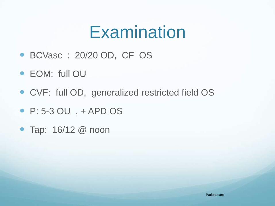
Examination BCVasc : 20/20 OD, CF OS
EOM: full OU
CVF: full OD, generalized restricted field OS
P: 5-3 OU , + APD OS
Tap: 16/12 @ noon
Patient care
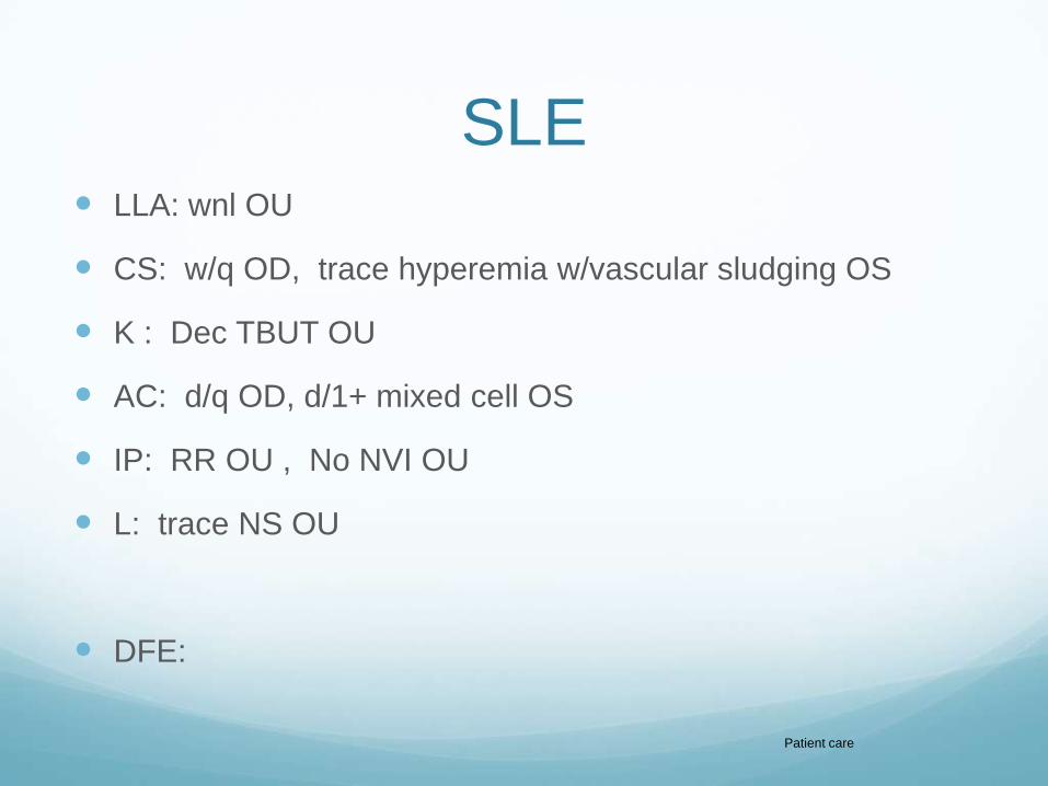
SLE LLA: wnl OU
CS: w/q OD, trace hyperemia w/vascular sludging OS
K : Dec TBUT OU
AC: d/q OD, d/1+ mixed cell OS
IP: RR OU , No NVI OU
L: trace NS OU
DFE:
Patient care
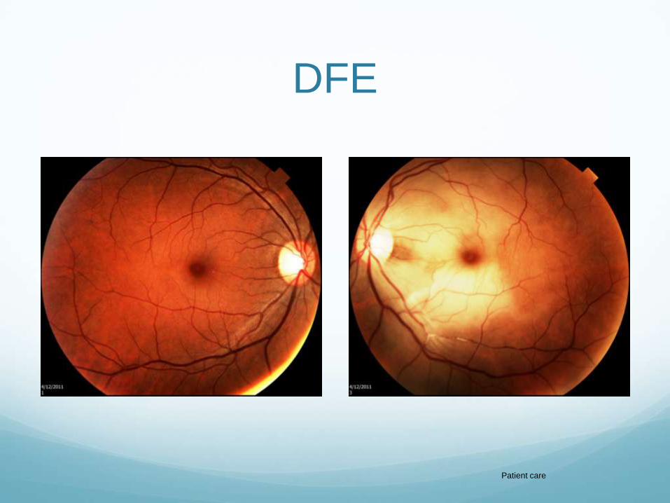
DFE
Patient care

DDx?

DDx? Vascular:
CRAO
BRAO
Ophthalmic artery occlusion
Inflammatory
GCA
SLE
Syphillis
Embolic
Carotid plaque
Mural thrombus
Vasculitic / Inflammatory
GCA
SLE
Syphillis
Neoplastic
Hematologic
Hypercoagulable
Medical knowledge

What Next?
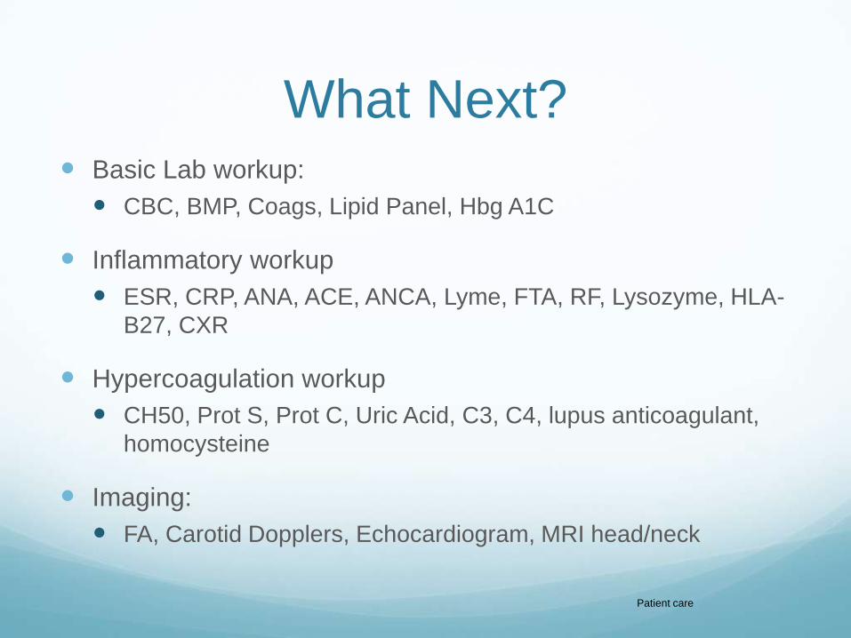
What Next? Basic Lab workup:
CBC, BMP, Coags, Lipid Panel, Hbg A1C
Inflammatory workup
ESR, CRP, ANA, ACE, ANCA, Lyme, FTA, RF, Lysozyme, HLA-
B27, CXR
Hypercoagulation workup
CH50, Prot S, Prot C, Uric Acid, C3, C4, lupus anticoagulant,
homocysteine
Imaging:
FA, Carotid Dopplers, Echocardiogram, MRI head/neck
Patient care
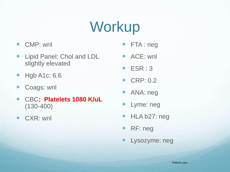
Workup CMP: wnl
Lipid Panel: Chol and LDL slightly elevated
Hgb A1c: 6.6
Coags: wnl
CBC: Platelets 1080 K/uL(130-400)
CXR: wnl
FTA : neg
ACE: wnl
ESR : 3
CRP: 0.2
ANA: neg
Lyme: neg
HLA b27: neg
RF: neg
Lysozyme: neg
Patient care
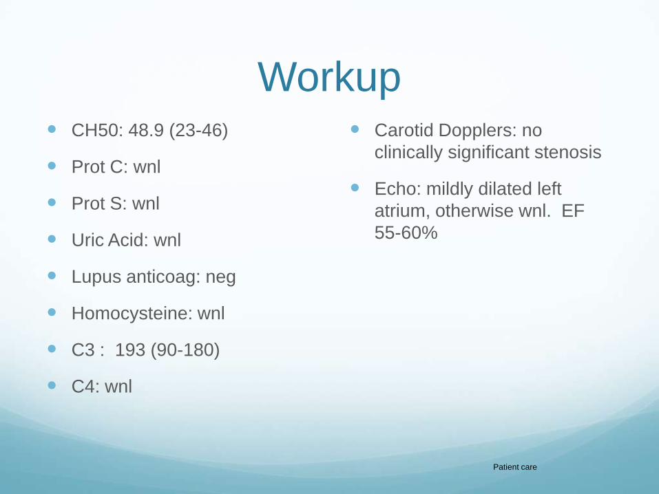
Workup CH50: 48.9 (23-46)
Prot C: wnl
Prot S: wnl
Uric Acid: wnl
Lupus anticoag: neg
Homocysteine: wnl
C3 : 193 (90-180)
C4: wnl
Carotid Dopplers: no
clinically significant stenosis
Echo: mildly dilated left
atrium, otherwise wnl. EF
55-60%
Patient care

FA
Patient care

Now What?
Now What? Review of previous records revealed a platelet level of
643 in 2005.
Started pt on daily 81mg ASA
Referred to Hematology
Recommended FISH chromosomal analysis
Referred to Neurology
Agreed with starting asprin and recommended f/up after
MRI studies.
Patient care, systems based practice

Fluorescence In Situ
Hybridization (FISH) Patient DNA is denatured
Flourescently labeled
specific probes are
introduced
DNA is allowed to hybridize
with probes
Excess probe is washed off
Chromosomes are analyzed
under flourescent
microscope
Medical knowledge

FISH FISH analysis revealed a Jak2+ gene mutation
Patient care
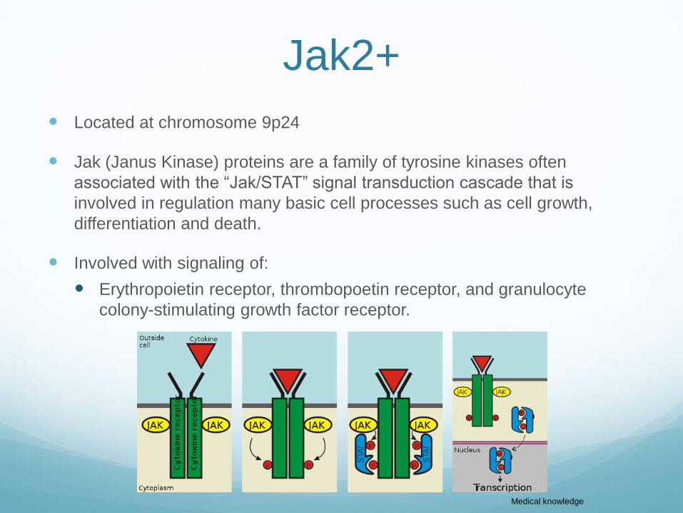
Jak2+
Located at chromosome 9p24
Jak (Janus Kinase) proteins are a family of tyrosine kinases often
associated with the “Jak/STAT” signal transduction cascade that is
involved in regulation many basic cell processes such as cell growth,
differentiation and death.
Involved with signaling of:
Erythropoietin receptor, thrombopoetin receptor, and granulocyte
colony-stimulating growth factor receptor.
Medical knowledge
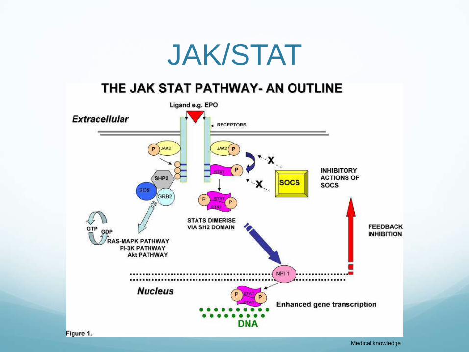
JAK/STAT
Medical knowledge
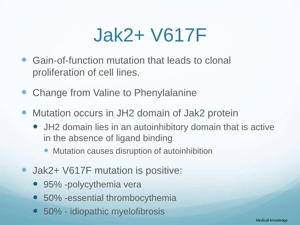
Jak2+ V617F Gain-of-function mutation that leads to clonal
proliferation of cell lines.
Change from Valine to Phenylalanine
Mutation occurs in JH2 domain of Jak2 protein
JH2 domain lies in an autoinhibitory domain that is active
in the absence of ligand binding
Mutation causes disruption of autoinhibition
Jak2+ V617F mutation is positive:
95% -polycythemia vera
50% -essential thrombocythemia
50% - idiopathic myelofibrosisMedical knowledge
Essential Thrombocythemia A member of a family of myeloproliferative disorders
Polycythemia Vera
Primary myelofibrosis
Was previously a diagnosis of exclusion
Has been reported to rarely transform into acute
leukemia
May actually represent a forme frust form of
polycythemia vera
Share common genetic defect (V617F)
Medical knowledge

Essential Thrombocythemia Females 2x > Males
Usually diagnosed in the 7th and 8th decade of life
Medical knowledge

Essential Thrombocythemia Diagnostic Criteria:
Diagnosis requires A1-A3 or A1 + A3-A5
A1 Sustained platelet count >450 × 109/L.
A2 Presence of an acquired pathogenetic mutation (eg, in
JAK2 or MPL).
A3 No other myeloid malignancy, especially polycythemia
vera (PV), primary myelofibrosis (PMF), chronic myeloid
leukemia (CML) or myelodysplastic syndrome (MDS).
A4 No reactive cause for thrombocytosis and normal iron
stores.
A5 Bone marrow trephine histology showing increased
megakaryo- cytes with prominent large hyperlobated forms;
reticulin is generally not increased (≤ 2 on a 0-4 scale).
Medical knowledge
Essential Thrombocythemia Complications
Headache, lightheadedness, atypical chest pains, distal
paresthesias, transient occlusions of microcirculation,
visual blurring or diplopia
Major source of mortality is thrombosis of large arteries
(cardiac, DVT/PE, renal, cerebral).
Can get morbidity and mortality from associated
hemorrhagic phenomenon (GI bleeds, secondary to
trauma, epistaxis, gum bleeding, hematoma formation)
Acute leukemia and myelodysplasia are rare late-onset
transformation events.
Medical knowledge
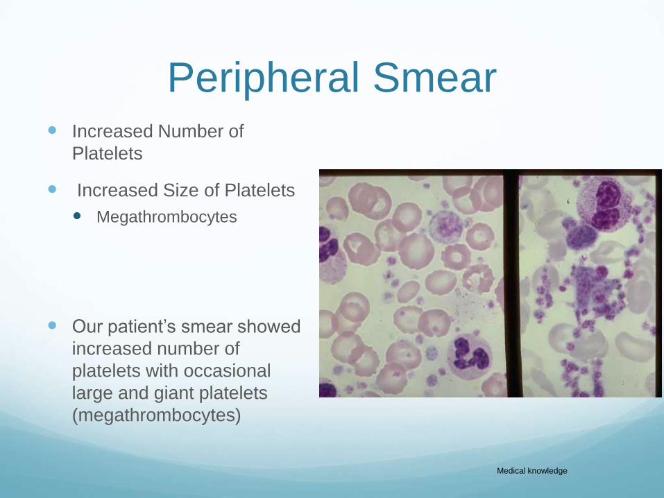
Peripheral Smear Increased Number of
Platelets
Increased Size of Platelets
Megathrombocytes
Our patient’s smear showed
increased number of
platelets with occasional
large and giant platelets
(megathrombocytes)
Medical knowledge
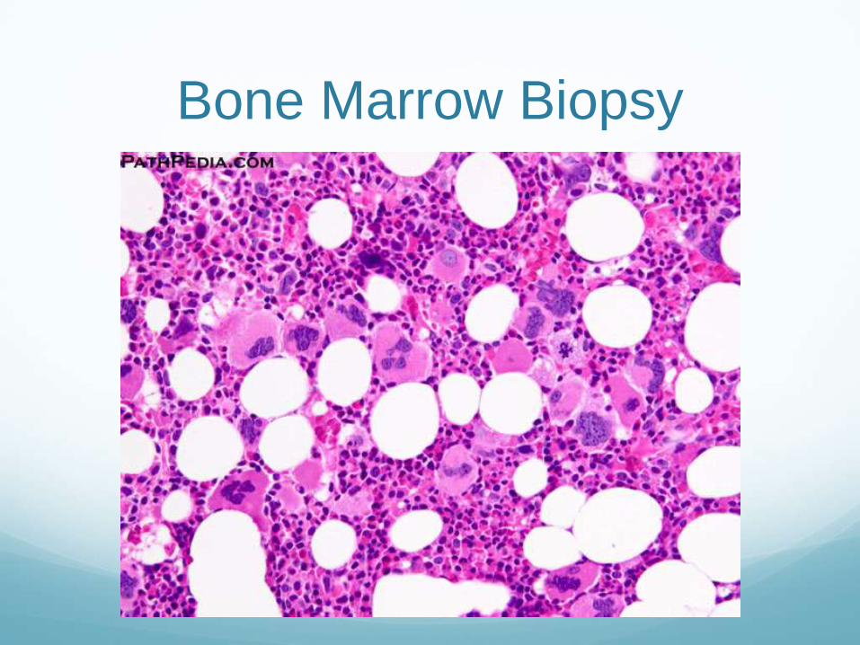
Bone Marrow Biopsy
Polycythemia Vera Member of group of myelodysplastic disorders in which
largely (or solely) there is marked elevation in red blood
cells (primarily)
Can also get elevation in platelets or wbc’s
Men>women
Rare <40 yrs old
Some symptoms: headache, SOB, dizziness, fatigue,
bluish skin discoloration, itchiness, red skin spots,
bleeding.
Medical knowledge
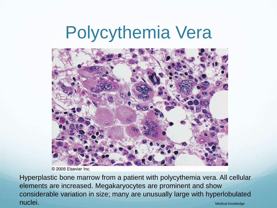
Polycythemia Vera
Hyperplastic bone marrow from a patient with polycythemia vera. All cellular
elements are increased. Megakaryocytes are prominent and show
considerable variation in size; many are unusually large with hyperlobulated
nuclei. Medical knowledge
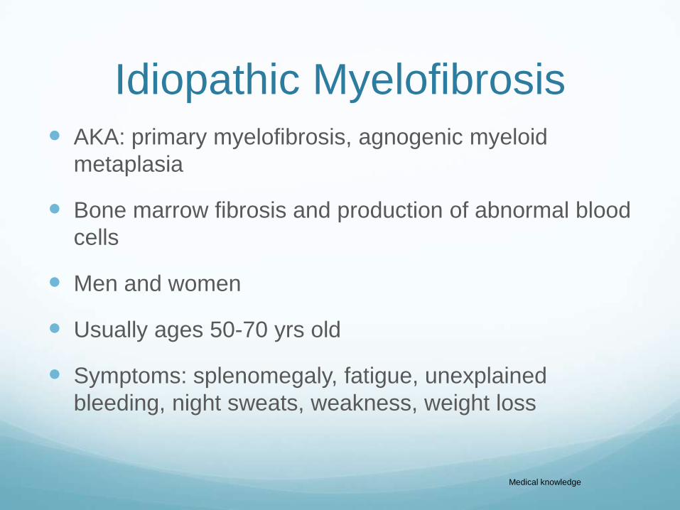
Idiopathic Myelofibrosis AKA: primary myelofibrosis, agnogenic myeloid
metaplasia
Bone marrow fibrosis and production of abnormal blood
cells
Men and women
Usually ages 50-70 yrs old
Symptoms: splenomegaly, fatigue, unexplained
bleeding, night sweats, weakness, weight loss
Medical knowledge

Idiopathic Myelofibrosis
Biopsy from an adult with a 7-year history of chronic idiopathic myelofibrosis
(agnogenic myeloid metaplasia). There is extensive marrow fibrosis with
marked reduction in hematopoietic tissue. Medical knowledge

Treatment Options Daily low dose Asprin (81mg)
Hydroxyurea
Interferon-alpha
Anagrelide
Pipobroman
Busulfan
Melphalan
Radioactive Phosphorus (32P)Medical knowledge
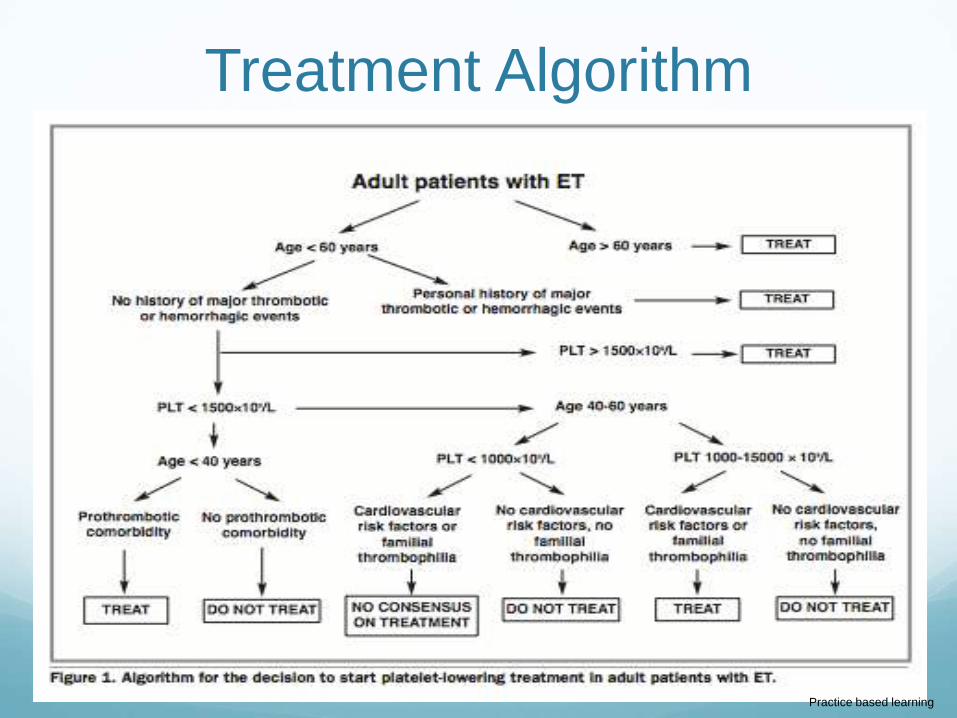
Treatment Algorithm
Practice based learning
Treatment Target of therapy is to lower platelets to a level of 400.
A level of 600 may be an apropriate target for pts who
require high doses of medication/ high risk of toxicity.
Should be followed every 3-4 mo by PCP for the first
year then twice yearly.
Should monitor CBC with diff, cardiovascular risk factors,
signs/symptoms of embolic and hemorrhagic events.
Medical knowledge
Hydroxyurea Inhibits ribonucleosidase reductase.
Inhibits DNA synthesis
Causes generalized myelosupression of all
myelogenous cell lineages
Can cause increased risk of malignancy when used in
conjunction with other myelosupressive agents (eg.
Busulphan)
Generally accepted as first line therapy for essential
thrombocythemia.
Medical knowledge
Interferon-alpha Direct acting cytokine that can inhibit hematopoeic
progenitor cells and megakaryocytic forming units.
Main side effect is flu-like symptoms upon induction of
therapy
Medical knowledge

Anagrelide Acts by inhibiting megakaryocyte maturation
Phosphodiesterase inhibitor with unknown exact
mechanism of action
Medical knowledge

Other Treatments Pipobroman – alkylating agent. Has been shown to
carry a aprox. 2.5% risk of leukemic transformation.
Busulfan –alkylating agent used commonly in CML.
Has been shown to have higher incidence of secondary
malignancy in pt’s treated with busulfan and
hydroxyurea.
Medical knowledge
CRAO Interventions and
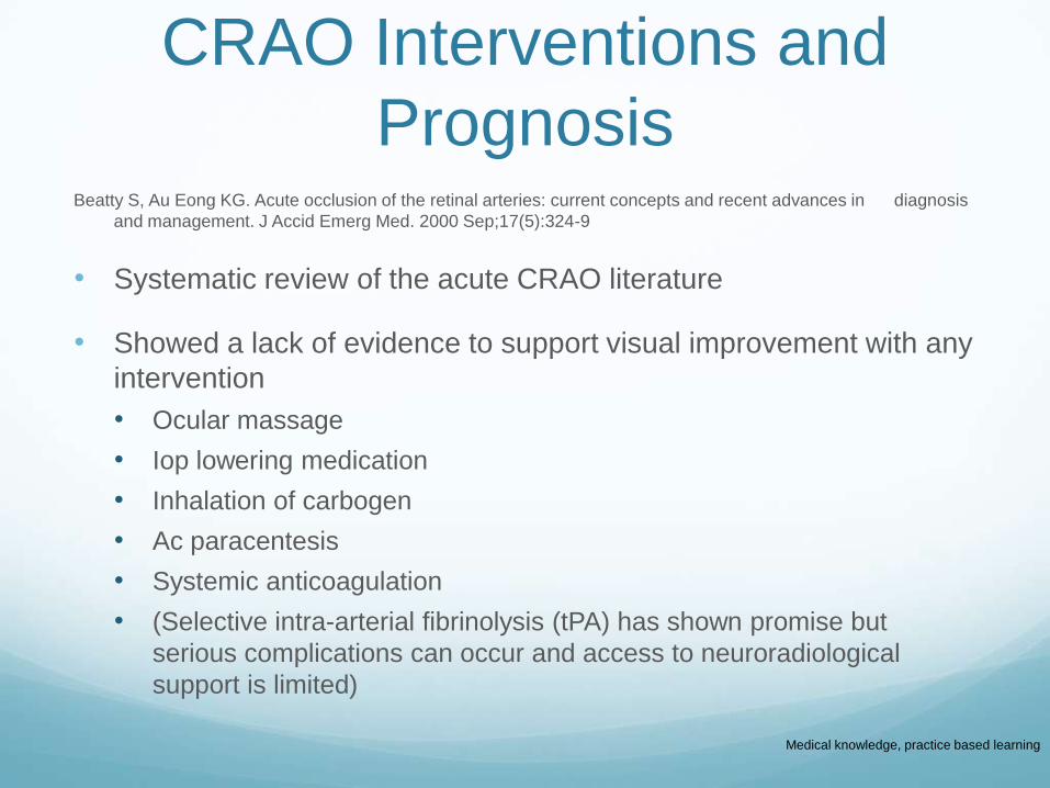
PrognosisBeatty S, Au Eong KG. Acute occlusion of the retinal arteries: current concepts and recent advances in diagnosis
and management. J Accid Emerg Med. 2000 Sep;17(5):324-9
• Systematic review of the acute CRAO literature
• Showed a lack of evidence to support visual improvement with any
intervention
• Ocular massage
• Iop lowering medication
• Inhalation of carbogen
• Ac paracentesis
• Systemic anticoagulation
• (Selective intra-arterial fibrinolysis (tPA) has shown promise but
serious complications can occur and access to neuroradiological
support is limited)
Medical knowledge, practice based learning
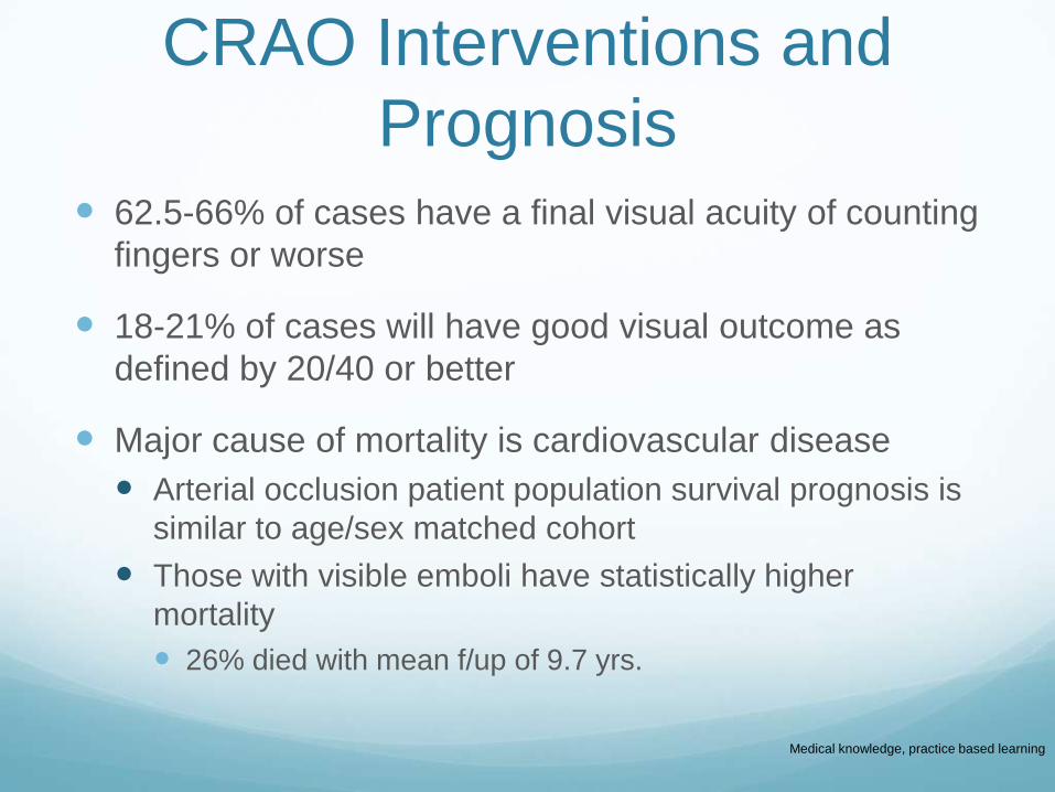
CRAO Interventions and
Prognosis 62.5-66% of cases have a final visual acuity of counting
fingers or worse
18-21% of cases will have good visual outcome as
defined by 20/40 or better
Major cause of mortality is cardiovascular disease
Arterial occlusion patient population survival prognosis is
similar to age/sex matched cohort
Those with visible emboli have statistically higher
mortality
26% died with mean f/up of 9.7 yrs.
Medical knowledge, practice based learning
Our Patient After the Jak2+ lab result our patient returned to
Hematology clinic
Recommended:
Continuing daily ASA
Starting daily hydroxyurea
No need for further anticoagulation at this time other than
daily ASA
Return to Hematology clinic in 1 mo
Systems based practice, patient care

Our Patient Continue to closely follow at KCH eye clinic
Has had no clinical deterioration in her vision OU
Va continues to be 20/20 OD and CF OS
Will obtain MRIs soon and continue to follow with
Hematology and Neurology
Patient care, professionalism
Reflective Practice• Here we have presented a rare case of Jak2+ Essential
Thrombocythemia induced retinal arterial occlusion. I
believe that the patient was evaluated, and treated
apropriately. Additionally It highlights the necessity and
advantage of a well coordinated multidisciplinary
approach to patient care.
Practice based learning, professionalism
Core Competencies Patient Care – throughout the patient encounters the patient was appropriately treated with
compassion and the patient’s best interest in mind.
Interviewing and Communication Skills- A thorough ROS and history was obtained from the
patient and open communication between consulting services was maintained at all times.
Professionalism – the patient was treated with kindness and in a respectful professional manner
at all times.
Medical Knowledge – The scientific literature was reviewed and then was applied to the patient
encounter.
Systems Based Practice – The ophthalmologists were able to work within the framework of the
hospital system to call upon proper consult services and arrange to get certain testing done
while the patient was in the hospital.
Practice Based Learning – The patient was monitored closely and interval changes were noted
at each visit. Changes in patient care were made based on patient performance and testing
results.
Practice based learning
References Ma W, Kantarjian H, Zhang X, et al. Higher detection rate of JAK2 mutation using plasma. Blood.
2008;111:3906-390
McLornan, D. et. al. JAK2 V617F: A Single Mutation in the Myeloproliferative Group of Disorders.
Ulster Medical Journal. 2006; 75:112-119.
Barbui, T. et. al. Practice guidelines for the therapy of essential thrombocythemia. A statement from the
Italian Society of Hematology, the Italian Society of Experimental Hematology and the Italian Group for
Bone Marrow Transplantation. Haematologica. 2004; 89:215-232.
Beatty S, Au Eong KG. Acute occlusion of the retinal arteries: current concepts and recent advances in
diagnosis and management. J Accid Emerg Med. 2000;17:324-9
Briere, J. Essential Thrombocythemia. Orphanet J Rare Dis. 2007; 2:3
Najfeld, V. et al. Numerical gain and structural rearrangements of JAK2, identified by FISH,
characterize both JAK2617V>F-positive and -negative patients with Ph-negative MPD,
myelodysplasia, and B-lymphoid neoplasms. Experimental Hematology. 2007;35:1668-1676.
http://www.pathconsultddx.com – images
Thank You Dr. Glatman
Dr. Olumba
Dr. Rand
The Departments of Hematology and Neurology