granular tricalcium phosphate in large cancellous … · 470 lange, zerwekh, peek, mooney, and...
TRANSCRIPT

ANNALS O F CLINICAL AND LABORATORY SCIEN CE, Vol. 16, No. 6Copyright © 1986, Institute for Clinical Science, Inc.
Granular Tricalcium Phosphate in Large Cancellous DefectsTHOMAS A. LANGE, M .D.,* JOE E. ZERWEKH, PH.D.,t RICHARD D. PEEK, M .D .,* VERT MOONEY, M .D .,tand BARRY H. HARRISON, B.S.*
*Department o f Orthopaedic Surgery, University o f Arkansas fo r Medical Sciences,
Little Rock, AR 72205 and
fDepartment o f Orthopaedic Surgery, University o f Texas Health Science Center,
Dallas, TX 72005
ABSTRACTTricalcium phosphate (TCP) is a porous ceramic which has biological
properties of being non-reactive and resorbable, and acts as a scaffolding for bone ingrowth, undergoing progressive degradation and replacement by bone. Tricalcium phosphate has been shown to be comparable to autogenous bone graft in small periodontal defects. However, orthopedic defects are much larger. This prompted us to review the bone ingrowth potential in large cancellous bone defects (up to 12 cm3) in adult pigs. To quantitate bone ingrowth potential, three skeletally mature pigs had metaphyseal defects created in the tibia and femur of each hind limb, for 12 total sites. Twelve-cc defects in the distal femur and eight cc defects in the proximal tibia were made. Bone curetted was saved to be used as autogenous graft in the control, while the other ipsilateral defect was packed with tricalcium phosphate.
Four months following the initial defect, the opposite hind extemity was similarly operated. All animals were sacrificed at nine months. Specimens were imbedded in methyl-methacrylate, cut at 120 microns, and stained.The quantity of regenerated bone was measured by histomorphometric techniques. Qualitative assessment at four months revealed absence of inflammation and TCP surrounded by trabecular bone, which was uniformly viable. There was very little TCP left by nine months. Quantitative analysis revealed the tibias to have a higher percent net bone replacement with TCP as compared to the control (32 percent versus 13 percent). The femoral TCP-filled defects were comparable to autogenous bone (both measured 29 percent). The combined results in tibia and femur revealed that TCP had a higher percent net bone (30.5 percent versus 21 percent).Both TCP and autogenous bone-grafted sites had a higher percent bone replacement with the passage of time. In both qualitative and quantitative analysis, TCP compared favorably to autogenous bone graft in terms of trabecular bone formation.
4670091-7370/86/1100-0467 $00.90 © Institute for Clinical Science, Inc.

4 6 8 LANGE, ZERW EKH, PEEK , MOONEY, AND HARRISON
IntroductionThe study was undertaken to deter
mine the degree of replacement of granular tricalcium phosphate by repair bone w h en p laced in artific ia lly crea ted defects in the cancellous bone of adult pigs. The product utilized is currently under an FDA-controlled clinical investigation for use in small, six- to ten-cc sized defects in humans. Since approval for use in larger defects is important, the adult p ig was chosen as our anim al model to attempt to extend the size limitations.
Tricalcium ph osp h ate is a porous ceramic which has biologic properties of being non-reactive, resorbable, and acts as a scaffo ld ing for b on e in grow th , undergoing progressive degradation and replacement by bone.3,7,9 Its principal limitation is its mechanical property of being quite brittle.1,9 This feature does not interfere with its use strictly as cance llo u s b on e rep la cem en t sin ce its strengths are comparable to cancellous bone.9
The tricalcium phosphate used* will be called by the generic term tricalcium phosphate, later referred to as TCP. It is classified as a polycrystalline ceramic which is sintered to 2000°C from a beta- tricalcium phosphate. The desired particle size is approxim ately 0.44m m to 2m m w ith a p orosity o f 250 to 400 microns. Minimal pore size for ingrowth has been shown to be 100 microns.Methods and Materials
To quantitate bone ingrowth potential, three skeletally mature pigs had metaphyseal defects created in the tibia and femur of each hind limb for 12 total sites; however, one pig sustained a femur fracture, leaving 11 sites for review. The tri
* Supplied by DePuy Company and called O rthograft®.
calcium phosphate (TCP) was used for all studies, f It was sterilized prior to use by dry heating at 175°C for four hours. The pigs were given a pre-anesthetic agent, Innovar, and a spinal anesthetic was performed using one percent Lidocaine.
In itia lly , all a n im a ls’ r igh t h ind extremities were operated through a longitudinal lateral skin-splitting and muscle-splitting incision. Then a one-centim e te r c o r tic a l w in d o w w as m ade through the nonarticular bone of the lateral condyle of the distal femur and the lateral cortex o f the proxim al tibia. Through these holes there were created 12 cubic-centimeter defects in the distal femur, and six-cc or eight-cc defects in the tibia, depending on the size of the animal. The size of the defect was docum ented by m easuring the volum e of fluid to fill it. Bone curetted was saved to be used as autogenous graft in the control, while the other ipsilateral defect was packed with tricalcium phosphate. Four months following the initial defect, the opposite hind extremity was operated. All animals were sacrificed at nine months after the first surgical procedure.
To determine the microvascular circulation, one specim en had the femoral artery injected with silicone rubber injection compound.:}: After sacrifice all specimens were cut into one-centimeter sagittal or transverse sections and radiographed on a Faxitron high-resolution x-ray unit. Fluorochrome labeling was employed throughout the experiment by giving oxytetracycline 50 mg during the first three days of each month.
The central section was fixed in alcohol. The specim ens were dehydrated and em bedded in methylmethacrylate. Each specimen was cut into thin sections approximately 120 microns thick. Two staining procedures were used. Alizorin
t Supplied by the DePuy Company and identical to the product “Orthograft® Large Granular. ”
$ Microfil*®.

TRICALCIUM PHOSPHATE IN CANCELLOUS D EFECTS 469Red S. was used to identify mineralized bone and TCR Toluidine-blue chroma- trope was used to identify osteoid and collagen. All slides underwent qualitative analysis by light microscopic examination.
For quantitative analysis, se lected central sections were submitted to the hard-tissue laboratory at the University of Texas Medical Science Center, Dallas, for processing. From the large, one-centimeter-thick bone sections, the central zone of repair was harvested as a 1 X 1 X 2 cm section and imbedded in methacrylate. After hardening, a Buchler Isomet diamond-tipped saw cut sections at 120 microns, and stained as noted previously. Utilizing a Bioquant digitizing pad interfaced to a Z eiss binocular m icroscope and Bioquant bone histo-
morphometry program in an Apple He computer, six random low-power fields equal to 0.9 mm per field were counted from multiple slides per specimen. Data obtained were: (1) total measured area; (2) percent of total as bone; and (3) percent of total as void area (without bone or marrow elements).Results
On light m icroscopy there was an absence of inflammation. The TCP was appositioned with trabecular bone which was uniformly viable. In the four-month specimen, extensive bone ingrowth was noted into the center of the specimen. E ven by th e fourth m on th , th ere appeared to be normalization of bone trabeculae in areas and already extensive
V/
a
- » > Y /”
/
* ' - v • J
FIGURE 1. M ic r o sc o p y o f tr ic a lc iu m p h o s p h a te (TCP) a n d a u to g ra ft a t n in e m o n th s .

470 LANGE, ZERW EKH, PEEK , MOONEY, AND HARRISON
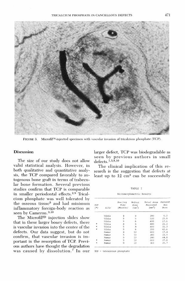
conversion of the TCP, still recognized as amorphous blue-staining crystals in small areas. The control specimen had a similar appearance, but with narrower trabeculae, suggesting further maturity of the autograft. Throughout the specimens there was very little TCP left by nine m onths. The regenerated bone appeared to be normal trabecular bone. Again, the controls had similar appearance, with somewhat narrower trabeculae (figure 1). Microscopic examination of th e M ic r o fil® -in je c te d sp e c im e n s revealed that vascularity extended to the central portion of defects filled with TCP (figure 3). Also, the fluorochrome bone label specimen revealed boney activity to the center of the lesions. Vascular invasion appeared to be com plete and n ecessa ry for reso rp tio n and b on e ingrowth.
Radiographic examination revealed resorption as indicated by loss of granular detail and replacement of TCP with
b o n e
F i g u r e 2 . Initial and nine-m onth radiograph of : autograft.
trabecular bone, as indicated by normalization of the trabecular pattern in a specimen x-ray (figure 2).
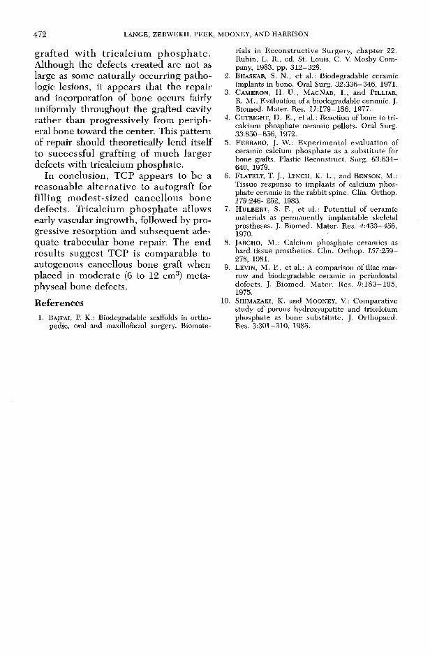
The q u an tita tive an alysis resu lts obtained by histomorphometry are listed on table I. The results analyzed by the size o f the d e fec t sh ow ed that the smaller defects in the tibias had a higher percent net bone replacement with TCP as compared to the control (32 percent versus 13 percent). The femoral TCP- filled defects were comparable to autogenous bone (both m easured 29 percent). The combined results in tibia and femur revealed the TCP had a higher percent net bone (30 percent versus 21 p e r c e n t) . T h ere a p p ea rs to b e an increase in net bone formed with duration of observation for both TCP and bone-grafted sites. The TCP-stimulated bone volum e com pared favorably to autogenous bone at four months (26 percent versus 16 percent) and at nine months (33 percent versus 26 percent).
BONE J
tibial tricalcium phosphate (TCP) graft and femoral
TRICALCIUM PHOSPHATE IN CANCELLOUS DEFECTS 471
F i g u r e 3. Microfilm-injected specimen with vascular invasion of tricalcium phosphate (TCP).
DiscussionThe size of our study does not allow
valid statistical analysis. However, in both qualitative and quantitative analysis, the TCP compared favorably to autogenous bone graft in terms of trabecular bone form ation. Several previous studies confirm that TCP is comparable in smaller periodontal effects.2,9 Tricalcium phosphate was well tolerated by the osseous tissue5 and had minimum inflammatory foreign-body reaction as seen by Cameron.3,10
The M icrofil® injection slides show that in these larger boney defects, there is vascular invasion into the center of the defects. Our data suggest, but do not confirm, that vascular invasion is important in the resorption of TCP. Previous authors have thought the degradation was cau sed by d is so lu t io n .7 In our
larger defect, TCP was biodegradable as se e n by p rev io u s authors in sm all defects.1'3,8,10
The clinical im plication of this research is the suggestion that defects at least up to 12 cm3 can be successfully
TABLE I
Histomorphometric Results
TCP(*) S i t e
H e a lin gTime
(M onths)D e fe c tS i z e(cm3)
T o ta l A rea M easured
(mm? )P e r c e n t
N e tBone
Tibia 4 8 280 6.2Tibia 9 6 520 15.8Tibia 9 6 400 17.6
* Tibia 4 6 480 21.2* Tibia 5 8 400 31.3* Tibia 9 6 200 42.6
Femur 4 12 400 17.4Femur 5 10 280 23.9Femur 9 12 240 44.8
* Femur 9 12 400 27.8* Femur 9 12 360 29.7
TCP = tricalcium phosphate
4 7 2 LANGE, ZERW EKH, PEEK, MOONEY, AND HARRISON
grafted w ith tricalcium p h osp h ate. Although the defects created are not as large as some naturally occurring pathologic lesions, it appears that the repair and incorporation of bone occurs fairly uniformly throughout the grafted cavity rather than progressively from peripheral bone toward the center. This pattern of repair should theoretically lend itself to successful grafting of much larger defects with tricalcium phosphate.
In conclusion, TCP appears to be a reasonable alternative to autograft for filling m odest-sized cancellous bone defects. Tricalcium phosphate allows early vascular ingrowth, followed by progressive resorption and subsequent adequate trabecular bone repair. The end results suggest TCP is comparable to autogenous cancellous bone graft when placed in moderate (6 to 12 cm3) metaphyseal bone defects.References
1 . BAJPAI, P. K.: Biodegradable scaffolds in orthopedic, oral and maxillofacial surgery. Biomate
rials in R econstructive Surgery, chap ter 22. Rubin, L. R., ed. St. Louis, C. V. Mosby Company, 1983, pp. 312-328.
2. B h a s k a r , S . N., e t a l . : B i o d e g r a d a b l e c e r a m i c i m p l a n t s i n b o n e . O r a l S u r g . 32:336-346, 1971.
3. C a m e r o n , H. U., M a c N a b , I ., and P i l l i a r , R. M.: Evaluation of a biodegradable ceramic. J. Biomed. Mater. Res. ii:1 7 9 -1 8 6 , 1977.
4 . C u t r i g h t , D . E . , et al.: Reaction of bone to tri- calcium phosphate ceramic pellets. Oral Surg. 33:850-856, 1972.
5. F e r r a r o , J. W . : E xperim ental evaluation of ceramic calcium phosphate as a substitu te for bone grafts. Plastic Reconstruct. Surg. 63:634- 640, 1979.
6. F l a t e l y , T. J., Ly n c h , K. L . , and B e n s o n , M.: Tissue response to im plants of calcium phosphate ceramic in the rabbit spine. Clin. Orthop. 779:246-252, 1983.
7. H U L B E R T , S. F., e t al.: Potential of ceram ic materials as perm anently implantable skeletal prostheses. J. Biomed. Mater. Res. 4:433-456, 1970.
8. JA RCH O , M.: Calcium phosphate ceramics as hard tissue prosthetics. Clin. Orthop. 157:259- 278, 1981.
9. L e v i n , M. P., et al.: A comparison of iliac marrow and biodegradable ceramic in periodontal defects. J. Biom ed. M ater. Res. 9:183—195,1975.
10. S h i m a z a k i , K. and M o o n e y , V .: Comparative study of porous hydroxyapatite and tricalcium phosphate as bone substitute. J. Orthopaed. Res. 3:301-310, 1985.