greater than 50 kgs protocol - starship · cvvh protocol. potassium should be ... set up calcium +...
TRANSCRIPT

Greater than 50 kgs protocol

EXTRACORPOREAL FILTRATION
2
WELCOME TO THE WORLD OF CONTINUOUS RENAL REPLACEMENT THERAPY
This clinical guideline package has been put together to endeavour to supply the user (both medical and nursing) with the protocols and information to safely and effectively manage continuous renal replacement therapy or therapeutic plasma exchange. In PICU we have been using continuous renal replacement therapy (CRRT) since 1993. Our primary methods of filtration within PICU are Continuous Veno-Venous Haemofiltration (CVVH) and Continuous Veno-Venous Plasmafiltration (CVVP)/ Therapeutic Plasma Exchange (TPE). Our total numbers of continuous renal replacement therapy remain very low when compared against the adult world. From 1993 - 2007 there have been156 children filtered in PICU. This is an average of 11 patients per year.

EXTRACORPOREAL FILTRATION
CATHETERS, FILTERS & CIRCUITS CATHETERS Maximum blood flow rate achievable will vary with
catheter size & machine.
CIRCUIT
ALL PATIENTS > 50KGS - ST100 – 152MLS IN SET
REPLACEMENT FLUIDS 1. Baxter Haemofiltration Solution Citrate 14mmol/litre
5 litres Citrate based buffer replacement solution Hang time 24 hrs ALWAYS infuse pre-filter (predilution)
Reconstitutes as: Sodium 140mmol/L Potassium 1mmol/L Chloride 99mmol/L Citrate 14mmol/L
3Osmolality 254mOsm/L
Approximate ranges for the different catheters are:
11.5F 90-250mls/min
13F Up to 350mls/min

EXTRACORPOREAL FILTRATION
4
2. Baxter Accusol
5 litres Bicarbonate based buffer replacement solution Hang time = 24hrs ALWAYS infuse pre filter (predilution)
Reconstitutes as: Sodium 140mmol/L Potassium 0mmol/L Calcium 1.75mmol/L Magnesium 0.5mmol/L Chloride 109.5mmol/L Bicarbonate 33mmol/L Osmolality 287mOsm/L
RATIONALE FOR CRRT Introduction The concept behind continuous renal replacement techniques is to mimic the renal function of patients in a physiologic way, with continuous filtration, until no longer required. Intensive care patients are particularly suited to this technique as they are, by definition, bed bound, and, when acutely sick, intolerant of the fluid swings associated with intermittent haemodialysis (IHD). Common reasons for Filtration in PICU are: Renal Failure with
• Fluid overload • Hyperkalemia • Acidemia • Severe uremia
Removal of Toxins
• Drug toxicity (non-plasma bound) • Inborn errors of metabolism

EXTRACORPOREAL FILTRATION
5
Advantages: Well tolerated cardiovascularly Fine control over fluid and electrolyte shifts Effective urea clearance and controlled fluid removal. Creates room for essential fluids such as blood products and
nutrition.
Setup and Programming The configuration of the haemofiltration circuit will vary slightly, depending which anticoagulation protocol is selected. CRRT WITH CITRATE-BASED REPLACEMENT FLUID Setting up the Circuit using the Haemofiltration Solution Citrate (14mmol/litre) based Anticoagulation Protocol 1 x prismaflex 1 x ST100 1 x bag spike 2 x 1000ml 0.9% Sodium Chloride 1 x 10ml syringe, posiflush and blind end cap 2 x smartsites (1 blue, 1 red) 1 x 5l bag Citrate replacement fluid (PBP) 1x 5l bag bicarbonate solution (replacement line)

EXTRACORPOREAL FILTRATION
6
CRRT WITH CITRATE-BASED SUBSTITUTION FLUID
Programming The PICU Consultant/Fellow must write the filtration prescription on
the Filtration Record.
The prismaflex requires the patients NHI. Enter this on the patient ID screen. Enter the patients weight as instructed.
Enter the haematocrit as a fraction, eg, 0.35. take the reading from the arterial blood gas sample prior to initiation. Update the patient haematocrit daily from the gas taken around midday each day. Also update the haematocrit after a blood transfusion or a large drop in haemoglobin.
The PBP or white line is the citrate administration line.
The purple line is the replacement line. This is programmed to run post filter into the deaeration chamber. The amount does not change through the course of the therapy. This fluid provides an air – blood barrier to reduce clotting in the deaeration chamber.
The dialysate line or green line is always programmed as 0mls.
The prismaflex will account for all fluids on the scales plus the circuit heparin infusion (if running), but not the patient IV & enteral fluids & drugs. Therefore, to achieve the hourly fluid balance target - add up all fluids (this includes bolus medications – e.g. drugs & flushes) infusing into the patient. This total plus the desired hourly fluid balance of the patient, gives you the total to set for the Fluid Loss Rate (ml/h). This is the number you titrate to pull off more or less fluid each hour.
CITRATE ANTICOAGULATION FOR HAEMOFILTRATION Patients on continuous modes of renal replacement therapy require anticoagulation to prevent clotting of blood in the extracorporeal circuit. Adequate anticoagulation prolongs circuit life but how long the membrane remains effective is debatable. Citrate is the anti-coagulant of choice in PICU. Citrate anticoagulation prolongs circuit life and causes less bleeding when compared with Heparin.

EXTRACORPOREAL FILTRATION
7
Citrate anticoagulation Citrate acts by chelating (binding) calcium ions that are essential in the clotting cascade. It also chelates other divalent cations including magnesium and aluminium. A plasma citrate concentration of about 5-6mmol/L is required in order to reduce ionised calcium concentration to less than 0.6mmol/L, which is required for anticoagulation. Adding approximately 2-3 mmol of citrate per litre of blood flowing through the filter usually achieves this.
Most of the added citrate returns to the patient and is metabolised rapidly by the liver, though there is a continuously slightly elevated systemic plasma citrate level which chelates some calcium in the systemic circulation. This can lead to a low systemic ionised calcium level even with normal total calcium. Also some chelated calcium is filtered, and as the substitution fluid contains no calcium there is a net loss of approx 2-3mmol/kg/day in the filtrate.
In order to avoid systemic ionised hypocalcaemia, a separate infusion of calcium is required at a rate of approximately 2mmol/kg/day for a blood flow of 5ml/kg/min, and this is adjusted to maintain systemic plasma ionised calcium level of approximately. 0.9-1.2mmol/L.
To achieve optimal anticoagulation within the circuit using a citrate based substitution solution there must be a balance between circuit blood flow and substitution fluid flow rate. This ratio between citrate dose and circuit blood flow remains reasonably fixed allowing the prediction of what citrate dose (substitution flow rate) is needed for a particular blood flow rate.
Magnesium supplementation is also necessary. This is achieved using the calcium / magnesium bags.
Citrate is metabolised to bicarbonate (three molecules per molecule of citrate), mostly in the liver. A metabolic alkalosis may develop in protocols which add citrate separately and also use a bicarbonate replacement solution. This is minimised if sodium citrate is an integral component of the substitution fluid where the citrate acts as the base compound instead of bicarbonate.

EXTRACORPOREAL FILTRATION
8
CITRATE ANTICOAGULATION FOR HAEMOFILTRATION
Relative Contraindications Citrate anticoagulation MAY NOT be an option for: Patients with severe hepatic dysfunction – e.g. fulminant liver failure. Patients with coagulopathy (ACT >200 sec, or APTT > twice normal)
may be filtered without any anticoagulant using the bicarbonate-based substitution fluid Accusol and a maximum tolerated Blood flow rate (BFR). E.g. liver failure, overwhelming sepsis.
Patients with “Citrate lock”, where citrate has accumulated during citrate anticoagulation. These patients should use Heparin as an alternative anticoagulant (see page 25).
Replacement Fluid Made commercially in 5 Litre bags by Baxter/Edwards Life Sciences. Composition cannot be adjusted, except for the addition of potassium chloride, potassium phosphate, or sodium phosphate, if needed, as per CVVH protocol. Potassium should be added to the substitution fluid to maintain the serum potassium @ 4mmol/l. This can be achieved by adding either KCl or KH2PO4 to each 5 litre bag to achieve a concentration of 20mMol in a 5l bag.) The composition is: Sodium 140mmol/L Potassium 1mmol/L Chloride 99mmol/L Citrate 14mmol/L (42meq/L) Trisodium citrate dihydrate.
ALL the citrate replacement fluid must be added Pre-Filter, never Post-Filter to ensure that the filter and circuit are anti-coagulated.

EXTRACORPOREAL FILTRATION
9
PERFORMING CVVH with CITRATE ANTICOAGULATION
Step 1 Blood Flow Rates For citrate anti-coagulation to be effective it is essential to match the PBP and replacement flow rate to the blood flow rate, as per the table below Initial Flow Rates for Citrate Based CVVH
Patient Weight (kg)
Blood Flow (ml/min)
PBP Citrate Rate (ml/hr)
Replacement post filter rate
CaCl + MgCl Infusion Rate (ml/hr)
50 -60 150 1650 160 8
60.1-70 200 1950 200 10
70.1-80 200 2250 200 12
80.1-90 250 2550 200 14
>90 250 2850 200 16 Fluid Balance
Net fluid balance for the PATIENT is the sum of ALL ingoing fluid minus ALL outgoing fluid.
Step 2 Calcium and Magnesium Infusion The calcium chloride and magnesium infusion is available from CVICU. They are aware that we use their stock when we need to source it. Set up calcium + magnesium infusion using Edwards Life Sciences
4.9% Calcium Chloride with 3.4% Magnesium Chloride. This must be infused via a central venous line with the initial rate set according to the table below (1ml of this solution contains 0.33 mmol of calcium). If the patient’s serum ionized calcium is below 1 mmol/L it should be corrected prior to commencing haemofiltration by giving the bolus of 10% calcium gluconate 0.5mls/kg over 10 minutes.
Magnesium Citrate also causes magnesium chelation. Intravenous magnesium replacement in addition to that present with
the calcium + magnesium infusion may be required.

EXTRACORPOREAL FILTRATION
10
Step 3 Monitoring and Adjusting the Circuit (Post-Filter) Ionised Calcium Circuit calcium must be measured to ensure effective anticoagulation. Using an arterial blood gas syringe, take blood samples for circuit
plasma ionised calcium from the blue sample port in the return limb of the circuit (post-filter).
Analyse it in the blood gas machine to determine the circuit ionised calcium level. Take a blood sample 1 hour after starting treatment, and then 6-hourly thereafter. Adjust as necessary. REPEAT this sampling regimen whenever changes are made to the calcium infusion rate or the substitution fluid rate.
A circuit post-filter (blue port) ionized calcium level of between 0.3 and 0.5 mmol/L confirms adequate chelation. Repeat 6 hourly if in range
A level < 0.3 mmol/L may be an indication of excessive citrate administration. This should prompt a check for signs of systemic citrate accumulation (see below). If such signs are present the citrate haemofiltration solution infusion rate should be reduced by 300 ml/hr (consult registrar).
A level > 0.5 mmol/L indicates inadequate citrate administration. In this case the citrate haemofiltration solution infusion rate should be increased by 300 ml/hr (consult registrar).
If the calcium + magnesium infusion rate needs to be above
20ml/hr to maintain the serum ionized calcium (arterial blood gas) between 0.9 and 1.2 mmol/L then the citrate haemofiltration fluid infusion rate should be decreased by 300ml/hr. If on the next arterial blood gas serum ionized calcium the calcium + magnesium infusion still needs to be above 20ml/hr to maintain the serum ionized calcium between 0.9 and 1.2 mmol/L consider changing some of the solution to bicarbonate substitution fluid.
A circuit ionised calcium less than 0.3mmol/L may be acceptable provided that the patient has an acceptable ionised calcium level, they are not receiving an excessive amount of calcium via infusion, and they are not developing a citrate gap. If there is an excess delivery of citrate at a substitution rate of 30ml/kg/hr you may need to replace some of the substitution fluid with non-citrate containing fluid (see notes at end) or switch to heparin anticoagulation with Accusol replacement fluid.
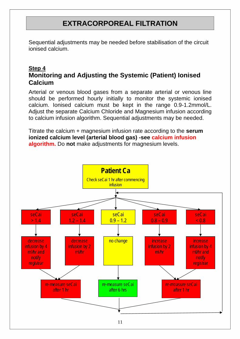
EXTRACORPOREAL FILTRATION Sequential adjustments may be needed before stabilisation of the circuit ionised calcium. Step 4 Monitoring and Adjusting the Systemic (Patient) Ionised Calcium Arterial or venous blood gases from a separate arterial or venous line should be performed hourly initially to monitor the systemic ionised calcium. Ionised calcium must be kept in the range 0.9-1.2mmol/L. Adjust the separate Calcium Chloride and Magnesium infusion according to calcium infusion algorithm. Sequential adjustments may be needed. Titrate the calcium + magnesium infusion rate according to the serum ionized calcium level (arterial blood gas) -see calcium infusion algorithm. Do not make adjustments for magnesium levels. Patient Ca
Check seCai 1 hr after commencing infusion
11
seCai > 1.4
seCai 1.2
seCai 0.9
seCai 0.8
seCai < 0.8– 1.4 – 1.2 – 0.9
no change decrease infusion by 4
ml/hr and notify
registrar
decrease infusion by 2
ml/hr
increase infusion by 2
ml/hr
increase infusion by 4
ml/hr and notify
registrar
re-measure seCai
after 1 hr re-measure seCai
after 6 hrs re-measure seCai
after 1 hr

EXTRACORPOREAL FILTRATION
12
PERFORMING CVVH with CITRATE ANTICOAGULATION CONT. Record both the circuit ionised calcium and the patient’s ionised calcium on the filtration record sheet. The following Biochemistry requires immediate attention - Inform the PICU registrar or Consultant Ca++ < 0.8mmol/L or >1.5mmol/L Total serum Ca > 3mmol/L Na+ < 130mmol/L or Na+ > 150mmol/L HCO3
- > 35mmol/L pH < 7.25 or pH > 7.5 Base Excess < - 5 Patient Anion Gap > 8mmol/L [Na-(HCO3 + CI)] The plasma magnesium should be measured 12hrly. If it falls to
0.7mmol/L give an extra 10mmol of Magnesium Sulphate and recheck in 4 hours. If the plasma magnesium remains low regular magnesium should be charted.
Step 5 Other monitoring Acid-Base Citrate is completely metabolised in most patients with normal liver function. A rising anion gap in a patient with a rising total calcium but a falling ionised calcium (despite increasing the calcium infusion) is caused by citrate accumulation (“citrate lock”).This is more likely in patients who have poor liver function and those receiving large amounts of blood products (these can contain large amounts of citrate). Acidaemia may also develop, with falling bicarbonate, an increasing anion gap, and a decreasing base excess. (Under normal circumstances the citrate in the circuit blood will cause an anion gap which is 5-7mmol/L greater than the patient’s anion gap. Therefore in assessing an anion gap it is important to do so on the patients blood NOT a circuit blood sample.) Citrate lock can only be managed by decreasing the citrate infusion rate. REMEMBER that the substitution fluid rate should not be less than 30ml/kg/hr. If you need to decrease the citrate fluid rate to less than

EXTRACORPOREAL FILTRATION
13
30ml/kg/hr because of a developing citrate lock then replace SOME of the replacement fluid with either Accusol. In some patients this situation continues to worsen and citrate anticoagulation may have to be abandoned and replaced by heparin/Accusol regime. Some patients with normal liver function may become alkalotic due to overproduction of bicarbonate from the citrate load. If this occurs use 0.45% or 0.9% Sodium Chloride as some of the replacement solution. This must be discussed with the Intensivist. Electrolytes Magnesium (total) should be checked 12 hourly. Sodium should be checked every 6-12 hours. Total Serum Calcium should be checked 12 hourly, and more frequently if more than two sequential increases in calcium chloride infusion rate have been required, or if a metabolic acidosis with rising anion gap occurs. See Acid/base section (above). Note Citrate Lock
If citrate lock is developing as evidenced by acidosis, increased anion gap, and increasing ratio of total:ionised calcium then you need to decrease citrate delivery. It is important that the replacement fluid rate is kept at not less than 30ml/kg/hr.
To achieve this and not give excess citrate then some of the replacement fluid will need to be given as either Accusol (if patient acidotic), 0.9% Sodium Chloride (if patient alkalotic and serum sodium normal), or 0.45% Sodium Chloride (if patient alkalotic and serum sodium high).
Start by replacing a proprtion of the citrate substitution fluid with one of the above fluids. The citrate rate is reduced and the additional solution is run on the replacement pump at the rate the citrate has been turned down by. It will usually be accusol.
Changing Citrate Substitution to ONLY Accusol Substitution Cease the Calcium infusion immediately after ceasing ALL citrate based substitution fluid. Recheck plasma total and ionised Calcium and total Magnesium at 1 hour and 6 hours post cessation.
EXTRACORPOREAL FILTRATION
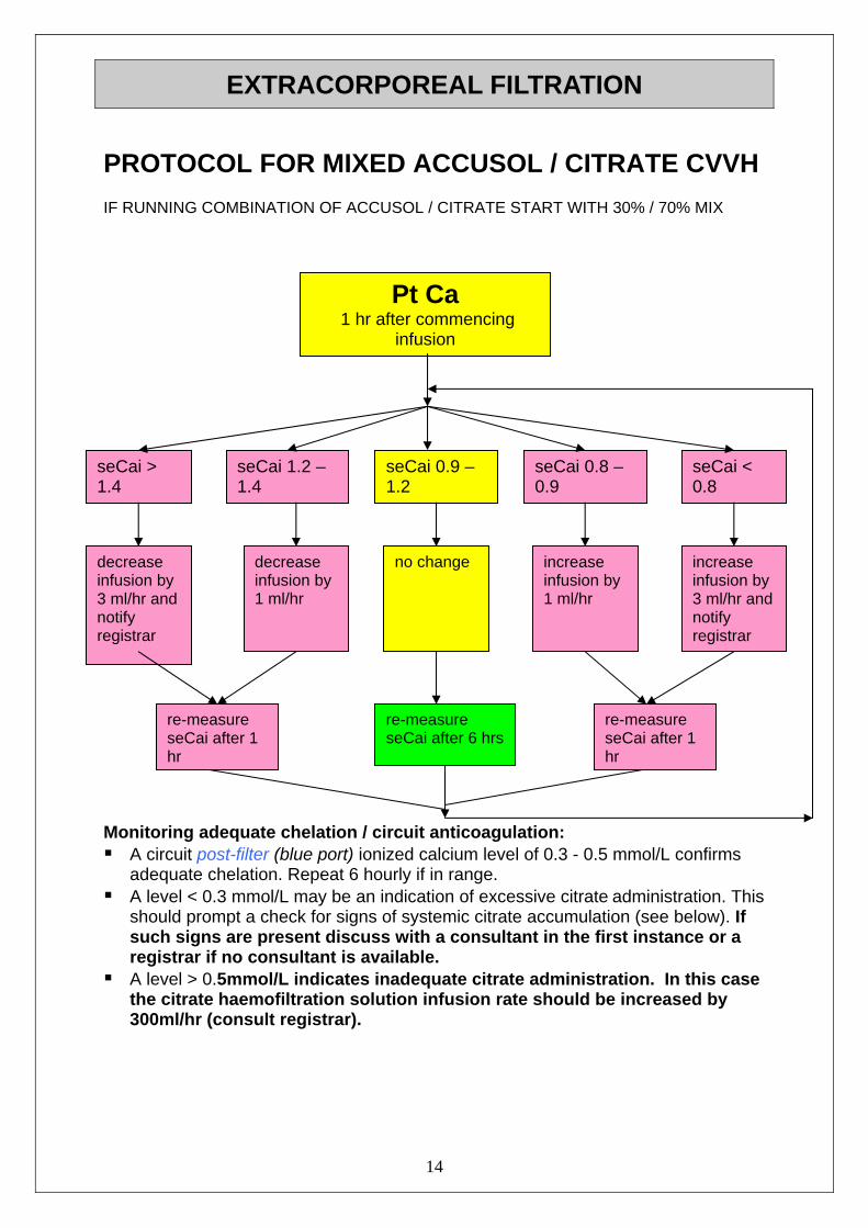
PROTOCOL FOR MIXED ACCUSOL / CITRATE CVVH IF RUNNING COMBINATION OF ACCUSOL / CITRATE START WITH 30% / 70% MIX
Monitoring adequate chelation / circuit anticoagulation: A circuit post-filter (blue port) ionized calcium level of 0.3 - 0.5 mmol/L confirms
adequate chelation. Repeat 6 hourly if in range. A level < 0.3 mmol/L may be an indication of excessive citrate administration. This
should prompt a check for signs of systemic citrate accumulation (see below). If such signs are present discuss with a consultant in the first instance or a registrar if no consultant is available.
A level > 0.5mmol/L indicates inadequate citrate administration. In this case the citrate haemofiltration solution infusion rate should be increased by 300ml/hr (consult registrar).
14
Pt Ca 1 hr after commencing
infusion
seCai > 1.4
seCai 1.2 – 1.4
seCai 0.9 – 1.2
seCai 0.8 – 0.9
seCai < 0.8
decrease infusion by 3 ml/hr and notify registrar
decrease infusion by 1 ml/hr
no change increase infusion by 1 ml/hr
increase infusion by 3 ml/hr and notify registrar
re-measure seCai after 1 hr
re-measure seCai after 6 hrs
re-measure seCai after 1 hr
EXTRACORPOREAL FILTRATION
15
CRRT WITH HEPARIN Setting up the Circuit using the Heparin based Anticoagulation Protocol 1 x prismaflex 1 x ST100 2 x large bore 2-way taps and 2 x Smartsites (1 red, 1 blue) 2 x 5l bag Accusol fluid 2x 1000ml 0.9% Sodium Chloride (priming fluid) 1 x 10ml posiflush Heparin infusion in 50ml BD Precise syringe
200 u X weight (1ml = 4u/kg/hr) ACT machine & LR cartridges Once connected the BFR is increased to run as fast as is tolerated by
the patient. This is determined by the patient’s haemodynamic status and the circuit pressures. A guide to achievable BFR’s is available.
Manage the Heparin infusion as per the Heparin protocol.

EXTRACORPOREAL FILTRATION
16
HEPARIN INFUSION PROTOCOL Dose Adjustments for CVVH
ACT (sec) Bolus
(u/kg) Stop
Infusion (min)
% Rate Change Repeat ACT
< 120
120 – 140 140 – 160 160 – 180 180 – 220
>220
20 0 0 0 0 0
0 0 0 0 30 60
+ 15 % + 10 %
0 − 10% − 10% − 15%
2 hours 2 hours 4 hours 2 hours 2 hours 2 hours
Start the hepain infusion at 10u/kg/hr check ACT red port at 30 minutes post commencement of the
circuit
Make any changes necessary, as per table above.
recheck ACT hourly and 30 minutes post any infusion bolus or change in rate.
Blood Flow and Substitution Fluid Rates
Patient weight (kg) PBP fluid rate (ml/hr)
Replacement fluid rate (mls/hr)
Blood Flow (ml/min)
50 – 60 1900 200 150
60 – 70 2300 200 200
70 – 80 2600 200 200
80 – 90 3000 200 250
> 90 3300 200 250
ALL SUBSTITUTION FLUID IS ACCUSOL

EXTRACORPOREAL FILTRATION
17
HEPARIN ANTI-COAGULATION Make up a Heparin infusion to 50mls total volume in a 50ml BD
Precise syringe. Add Heparin 200iu x patient weight to 0.9% Sodium Chloride. Thus 1ml/hr = 4iu/kg/hr.
Administer heparin in an alaris syringe driver using guardrails profile ‘heparin treatment’.
Adjust Heparin as per table overleaf to maintain ACT = 140 - 160. Take blood for ACT testing from the red sample port of the circuit
(pre filter). For an INR greater than 2 correct with FFP 10-20ml/kg. Ask
Consultant For a fibrinogen < 1g/L correct with cryoprecipitate 10ml/kg (usually
a minimum of 1 unit). Ask Consultant
Consider limiting Heparin administration and boluses for patients with severe coagulopathies such as fulminant liver failure and
severe sepsis.
FFP and cryoprecipitate can also have variable effects on the ACT - so check the ACT after they have been given. Large falls in the platelet count may mean that HITT is occurring – check with a HITT screen. For the patients who bleed excessively while on the circuit, use Accusol and no heparin or switch to citrate. For those patients with an absolute contraindication to Heparin in whom citrate cannot be used, an anticoagulant free circuit with the blood flow rate as high as is tolerated by the circuit and patient is an acceptable means of managing the filter.
Consider an anti-coagulant–free circuit for patients with a severe non-correcting coagulopathy (i.e. fulminant liver failure, severe
sepsis).

EXTRACORPOREAL FILTRATION
PREPARING an ACCUSOL BAG
18
Accusol: Lactate free, bicarbonate base substitution fluid
Squeeze from edges at the seal between compartments A & B until the seal breaks. Mix well.
Remove Seal Unscrew Cap
Connect Substitution Fluid Line

EXTRACORPOREAL FILTRATION PREPARING an ACCUSOL BAG CONT.
19
Squeeze ‘wings’ until they snap flush with the access port – this causes the bag to be pierced.
Medication port – on rear of bag

EXTRACORPOREAL FILTRATION
CONNECTING Double lumen connection Required: Sterile gloves (in appropriate size) Sterile guard Dressing pack with sterile gauze 2 x 10ml syringes 2 x 10ml posiflush 2% Chlorhexidine solution
Before attaching the circuit to the patient ensure that: baseline: FBC, coags, full U&E’s + an ABG have been obtained a PICU Consultant is present appropriate volume is present e.g. Red Blood Cells (as above) resuscitation sheet and drugs are available continuous ECG, SaO2, BP and core temperature monitoring are in
place. the filtration orders have been written
Important: If there is a Heparin lock in the catheter of a concentration greater
than 10 iu/ml then it is essential that the Heparin is ASPIRATED out of the catheter lumens. If you are unable to do this then medical
staff must be notified. Using a sterile technique throughout the procedure clamp both
lumens of the catheter.
Clean the catheter hubs with the 2% Chlorhexidine solution and allow to dry.
Using the 0.9% Sodium Chloride and 10ml syringes check that both the access (red) and return (blue) lumens aspirate and flush freely.
20
Ensure the blood pump is stopped and clamp both ends of the extracorporeal circuit.

EXTRACORPOREAL FILTRATION
21
Attach the end of the access line of the circuit to the red lumen of the catheter and the return end of the circuit to the blue lumen of the catheter, ensuring secure, bubble-free connections.
Recheck the circuit for air bubbles, loose connections, cracks or deformities.
Open all clamps on the blood path of the extracorporeal circuit.
Start the blood pump slowly (30ml/min), watching for any sign of catheter obstruction or flow impedance.
Increase the blood pump speed until the circuit is thoroughly filled with the patient’s blood.
Increase the flow to the required rate, ensuring that the patient’s blood pressure and heart rate and circuit pressures remain within acceptable parameters. The circuit should be fully running within 15 minutes of connection.

EXTRACORPOREAL FILTRATION
22
DISCONNECTING CRRT CIRCUIT
This procedure reflects a planned disconnection from the patient and circuit. 1. Inform medical staff.
2. Stop the prismaflex using ‘end treatment’ (plus any circuit related infusions e.g. Calcium, Heparin).
3. Clamp access and return lines at catheter point closest to patient. For extra security, use GIZMO clamps.
4. At the catheter turn the 3-way taps OFF to patient.
5. Using an aseptic non-touch technique, swab the access and return ends of the circuit with 2% Chlorhexidine. Disconnect, including the 3-way taps.
6. Flush each lumen with 0.9% Sodium Chloride.
7. Fill each catheter lumen with Heparin 1000iu/ml to the volume stipulated on each lumen of the catheter.
8. Cap each lumen with a sterile ‘blind end luer lock’ (Combi lock).
9. Clearly document on each lumen, the drug, concentration, volume, time and write “DO NOT FLUSH”. Also document on PICU 24hr flowchart that the catheter is “Heplocked” with time and date. See CVL manual for further information on heplocking vascaths.
10. THE CIRCUIT VOLUME IS NOT ROUTINELY RETURNED TO THE PATIENT WHEN A CIRCUIT IS DISCONTINUED.
11. Ensure the Heparin used for the heplocking is prescribed in the medication chart.

EXTRACORPOREAL FILTRATION
23
POTENTIAL COMPLICATIONS OF CONTINUOUS RENAL REPLACEMENT THERAPY Hypovolaemia Have colloid readily available at beside for emergencies Manage fluid balance corrections by adjusting ultrafiltration rate only Monitor haemodynamics continuously
Fluid Overload Ensure that replacement fluid is as prescribed vs. delivery amount If PBP fluid pump stopped, address cautionary troubleshooting
options on the screen Observe for signs of pulmonary oedema
Hypothermia As the CRRT circuits are extra-corporeal, a fall in temperature of 0.5-1.C is expected. Continuously monitor core temperature Use the Bair Hugger to warm the patient if core temperature is less
than 36.C Infection Avoid contamination of the exposed ends of sterile equipment when
priming the circuit Avoid breaking the circuit wherever possible Redress cannula site 48 hourly/prn and record on the procedures
chart Monitor central temperature and observe cannulation site for
inflammation Manage venous catheter in accordance with central line RBP

EXTRACORPOREAL FILTRATION
DOCUMENTATION Use a new Filtration Record for every 24hr period (0800-0700)
All orders, interventions and trouble shooting are to be documented
on the Filtration Record sheet. At the end of a ‘CRRT’ duty enter a shift summary into the clinical
notes. Orders are to be written daily by Medical staff on the Filtration
Record. Enter the delivered PBP, replacement fluid, fluid removal and blood
pump speed on the observation chart hourly. Enter the access, return and TMP hourly on the observation chart.
Enter the heparin or calcium / magnesium infusion rate in mls/hr on
the observation chart hourly. Only the Fluid Loss (ml/hr) is transcribed from the Filtration Record
sheet to the PICU 24hr Flowchart. Enter the haematocrit every day around midday or after a blood
transfusion. Note this on the chart.
24