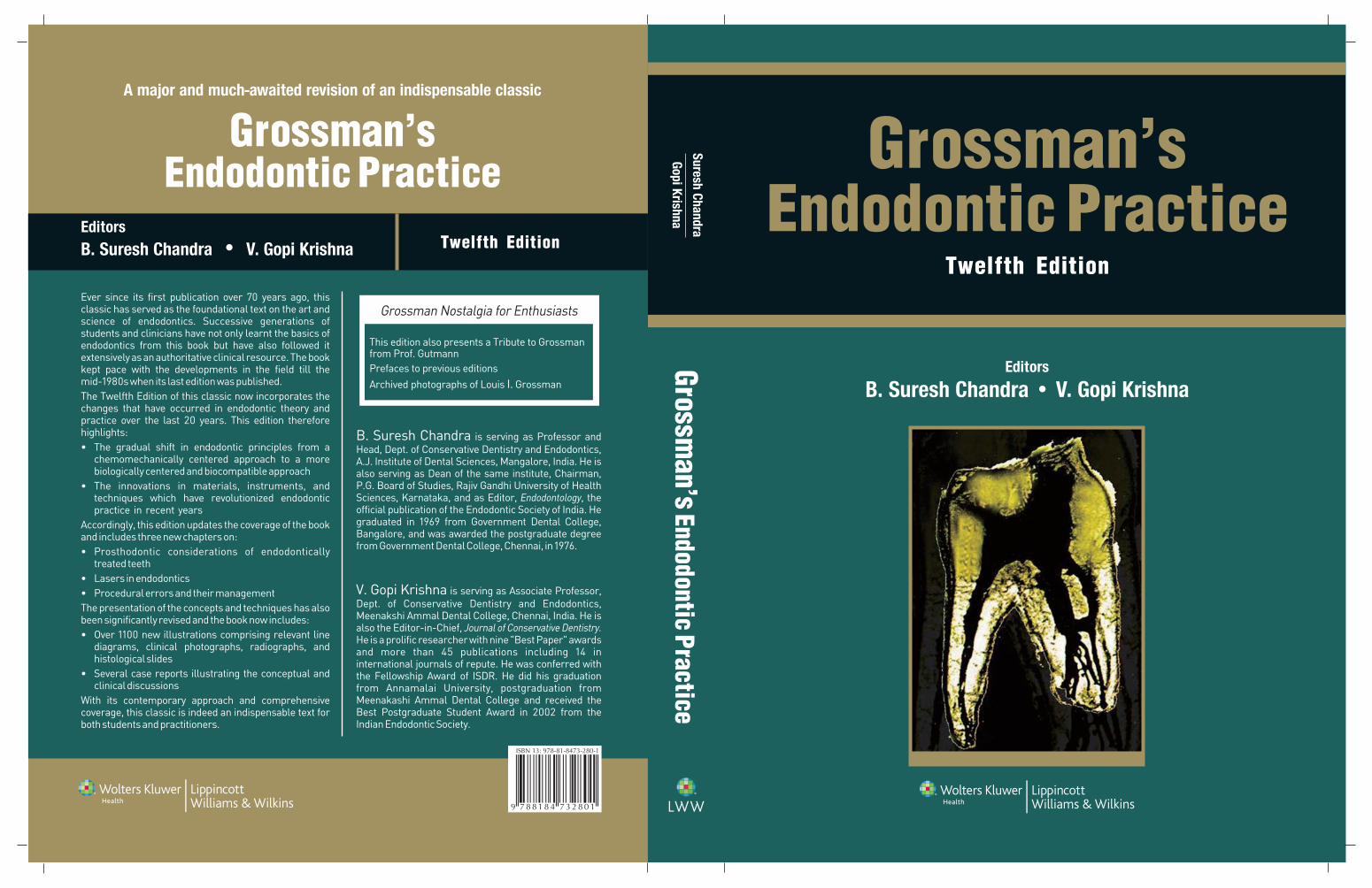
grossmans endodontic practice 12th edition preview
DESCRIPTION
Grossmans Endodontic Practice 12th Edition PreviewTRANSCRIPT
v
Dedicated to my parents, wife, and children.
–B. Suresh Chandra
Dedicated toMy beloved mother, Sulochana Velayutham, for being the
source and nurturer of my life and intellect …My beloved ardhangini, Lakshmi, for being the source of love,
support, and inspiration ….My beloved angels, Sanjana and Sidharth, for being the source of my happiness….
And above all to the Almighty for being the source of my creativity…
–V. Gopi Krishna
Endodontic Practice_FM.indd vEndodontic Practice_FM.indd v 2/18/10 9:12:30 PM2/18/10 9:12:30 PM
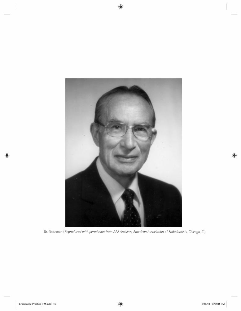
Dr. Grossman (Reproduced with permission from AAE Archives, American Association of Endodontists, Chicago, IL.)
Endodontic Practice_FM.indd viiEndodontic Practice_FM.indd vii 2/18/10 9:12:31 PM2/18/10 9:12:31 PM

Dr. Grossman presenting the AAE Louis I. Grossman Award to its fi rst recipient Dr. Birger Nygaard-Østby (Reproduced with permission from AAE Archives, American Association of Endodontists, Chicago, IL.)
Endodontic Practice_FM.indd viiiEndodontic Practice_FM.indd viii 2/18/10 9:12:31 PM2/18/10 9:12:31 PM
ix
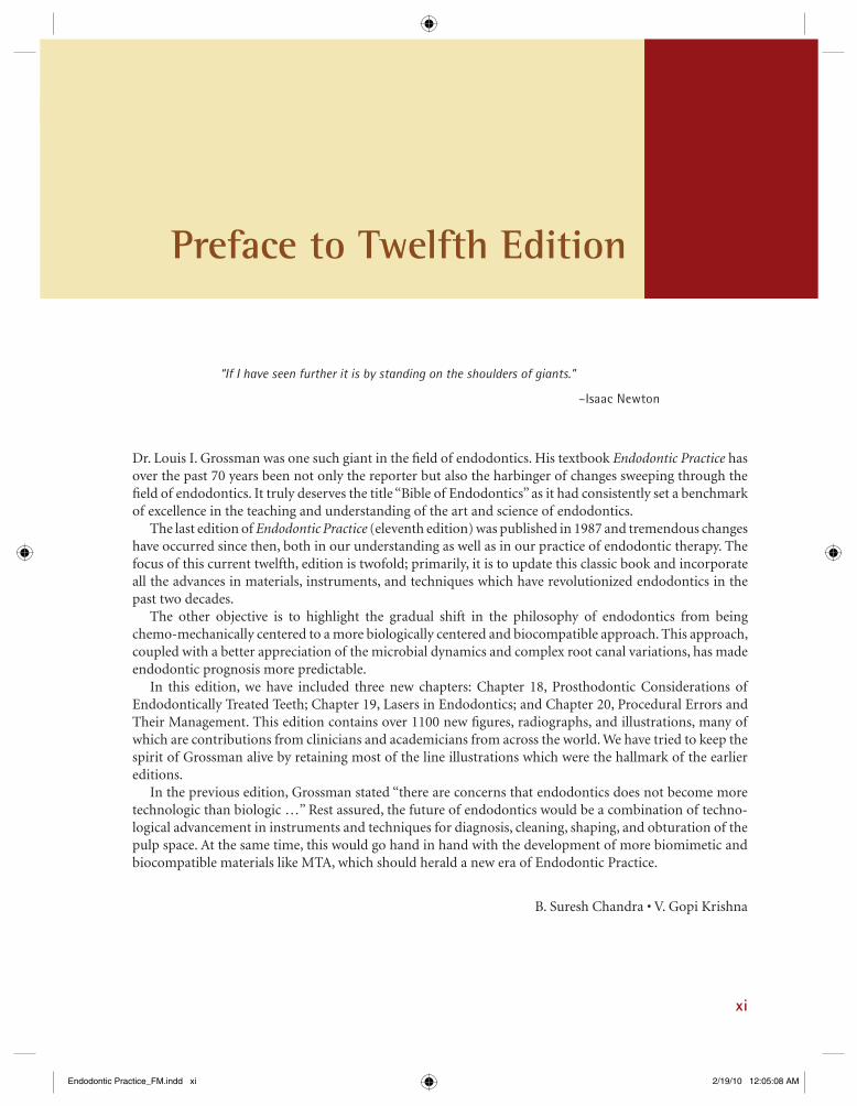
Louis I. Grossman: The Visionary Father of
Modern Endodontics
Dr. Louis I. Grossman was born in a Ukranian village near Odessa on December 16, 1901, and was brought to the United States by his family as a boy. He grew up in Philadelphia and completed his high school education at South Philadelphia High School in 1919. He earned a doctorate in dental surgery at the University of Pennsylvania in 1923 and a doctorate in medical dentistry (Dr. Med Dent) at the University of Rostock in Germany in 1928.
On December 21, 1928, he married Emma May MacIntyre and they had two children, a daughter Clara Ruth Grossman in 1939 and a son Richard Alan Grossman in 1943.
Dr. Grossman began his teaching career as an Instructor in Operative Dentistry at the University of Pennsylvania in 1927, in addition to being appointed as a Fellow in Research at the American Dental Association. In 1941 he was an Associate in Oral Medicine; he became Assistant Professor of Oral Medicine in 1947, Associate Professor of Oral Medicine in 1950, and Professor in 1954.
His achievements and honors were extensive in many sectors of dentistry with a prime focus in endodontics. He was an honorary member of the Association of Licentiates in Dental Surgery and University of Dentists of Belgium; Montreal Endodontia Society; Vancouver Endodontic Study Club, Brazilian Dental Association; Dental Association of Medellin (Colombia); and the Japanese Endodontic Association. He received an honorary Doctor of Science (ScD) from the University of Pennsylvania.
His major publication and crowning achievement was his textbook Root Canal Therapy published in 1940 (now known as Endodontic Practice) with multiple editions appearing worldwide. Subsequently trans-lated into eight languages, the book has served as a benchmark for the development of modern endodontic philosophy and practice. Dr. Grossman also authored Dental Formulas and Aids to Dental Practice, fi rst published in 1952, and the Handbook of Dental Practice published in 1948.
He was a chairman of the American Board of Endodontics, was a charter member of the American Association of Endodontists (AAE) and served as its President from 1948 to 1949. He was a Fellow of the American Association for the Advancement of Science.
Dr. Grossman passed away at the age of 86 in 1988. The University of Pennsylvania has honored Dr. Grossman with an endowed Professorship, usually given to the department chairperson. The AAE has honored him with the Louis I. Grossman Award that recognizes an author for cumulative publication of signifi cant research studies that have made an extraordinary contribution to endodontology. This award is given at the AAE meeting when warranted.
A study club was formed in Philadelphia in the honor of Dr. Louis I. Grossman for his unyielding dedication and commitment towards facilitating the recognition of endodontics as a specialty in the fi eld of dentistry. The purpose of the Louis I. Grossman Study Club was to provide an opportunity to the endodontists as well as other interested dentists to meet, share ideas, and expand and update our knowledge in the fi eld of endodontics and dental medicine.
Endodontic Practice_FM.indd ixEndodontic Practice_FM.indd ix 2/18/10 9:12:32 PM2/18/10 9:12:32 PM

x Ü Louis I. Grossman – The Visionary Father of Modern Endodontics
Dr. Louis I. Grossman was the founder of the fi rst Root Canal Study Club. It was established in 1939 in Philadelphia, Pennsylvania, at a time when the Focal Infection Theory threatened the future of endodontics. The purpose of the Root Canal Study Club as stated in the original letter compiled by Dr. Grossman was “to study problems connected with root canal therapy and to present clinics so as to help others in practic-ing this important phase of dentistry more adequately.” Endodontists from as far away as Massachusetts chose Philadelphia as the hub for scientifi c and educational learning in the fi eld of endodontics.
James L. Gutman
Endodontic Practice_FM.indd xEndodontic Practice_FM.indd x 2/18/10 9:12:32 PM2/18/10 9:12:32 PM
xi
Preface to Twelfth Edition
Dr. Louis I. Grossman was one such giant in the fi eld of endodontics. His textbook Endodontic Practice has over the past 70 years been not only the reporter but also the harbinger of changes sweeping through the fi eld of endodontics. It truly deserves the title “Bible of Endodontics” as it had consistently set a benchmark of excellence in the teaching and understanding of the art and science of endodontics.
The last edition of Endodontic Practice (eleventh edition) was published in 1987 and tremendous changes have occurred since then, both in our understanding as well as in our practice of endodontic therapy. The focus of this current twelfth, edition is twofold; primarily, it is to update this classic book and incorporate all the advances in materials, instruments, and techniques which have revolutionized endodontics in the past two decades.
The other objective is to highlight the gradual shift in the philosophy of endodontics from being chemo-mechanically centered to a more biologically centered and biocompatible approach. This approach, coupled with a better appreciation of the microbial dynamics and complex root canal variations, has made endodontic prognosis more predictable.
In this edition, we have included three new chapters: Chapter 18, Prosthodontic Considerations of Endodontically Treated Teeth; Chapter 19, Lasers in Endodontics; and Chapter 20, Procedural Errors and Their Management. This edition contains over 1100 new fi gures, radiographs, and illustrations, many of which are contributions from clinicians and academicians from across the world. We have tried to keep the spirit of Grossman alive by retaining most of the line illustrations which were the hallmark of the earlier editions.
In the previous edition, Grossman stated “there are concerns that endodontics does not become more technologic than biologic …” Rest assured, the future of endodontics would be a combination of techno-logical advancement in instruments and techniques for diagnosis, cleaning, shaping, and obturation of the pulp space. At the same time, this would go hand in hand with the development of more biomimetic and biocompatible materials like MTA, which should herald a new era of Endodontic Practice.
B. Suresh Chandra • V. Gopi Krishna
“If I have seen further it is by standing on the shoulders of giants.”
–Isaac Newton
Endodontic Practice_FM.indd xiEndodontic Practice_FM.indd xi 2/19/10 12:05:08 AM2/19/10 12:05:08 AM
xii
Ninth Edition
From Prefaces to Previous Editions
Louis I. GrossmanPhiladelphia, Pennsylvania
Endodontic Practice_FM.indd xiiEndodontic Practice_FM.indd xii 2/18/10 9:12:32 PM2/18/10 9:12:32 PM
Preface Ö xiii
Louis I. GrossmanPhiladelphia
Tenth Edition
Louis I. GrossmanPhiladelphia
Eleventh Edition
Endodontic Practice_FM.indd xiiiEndodontic Practice_FM.indd xiii 2/18/10 9:12:34 PM2/18/10 9:12:34 PM
xiv
Preface to First Edition
Endodontic Practice_FM.indd xivEndodontic Practice_FM.indd xiv 2/18/10 9:12:35 PM2/18/10 9:12:35 PM
Preface to First Edition Ö xv
Louis I. GrossmanPhiladelphia, PA
Endodontic Practice_FM.indd xvEndodontic Practice_FM.indd xv 2/18/10 9:12:37 PM2/18/10 9:12:37 PM
xvi
Acknowledgments
This, twelfth, edition, after nearly 23 years of the previous edition, continues the work and legacy of Dr. L.I. Grossman, the legendary endodontist from the University of Pennsylvania. I have toiled for several months along with Dr. Gopi Krishna to bring out this updated edition. I would like this edition to be my tribute to Thai Universal mother god almighty, my spiritual guru Sri Swami Narendranath Kotekar, and Amma Shakuntala Kotekar for all their blessings, inspiration, and guidance for this project.
I am grateful to my teachers as well as my beloved students and colleagues for all that they have taught me. They have truly made me what I am today. A special note of gratitude to Sir Prof. A. Parameshwaran and his beloved wife Mrs. Seetha Parameshwaran who have continued to be everything for me in my professional and personal life. In Dr. Parameshwaran, I have always found a teacher par excellence and a friend, philosopher, and guide. Thanks to Mr. A.J. Shetty and Mr. Prashanth Shetty, President and Vice President, respectively, A.J. Institute of Dental Sciences, Mangalore, for all their encouragement.
I would like to express my gratitude to my coeditor Dr. Gopi Krishna for all his efforts, dedication, and perseverance.
I would also like to appreciate the efforts of my postgraduate students Roma, Naveen, Arun, Meeta, Saurav, and Gautam signifi cantly helped me in this project.
My special thanks to my wife Suryakanthi, daughter Sowmya, and son Shravan for their wonderful patience and support throughout the development of this prestigious project.
B. Suresh Chandra
Endodontic Practice_FM.indd xviEndodontic Practice_FM.indd xvi 2/18/10 9:12:38 PM2/18/10 9:12:38 PM

Acknowledgements xvii
Thank you are two little words which would probably never completely convey the sense of gratitude and regards which I feel for each of the following wonderful people who have made Grossmans Endodontic Practice, – twelfth edition, a reality.
First and foremost, my sincere gratitude to Dr. Suresh Chandra, P. Sangeetha, and Rajiv Banerji for inviting me to be a part of this monumental work. At the same time, I would not have been part of this book but for the kind permission granted by Dr. Anil Kohli, President, DCI, and Ritu Sharma, Reed Elsevier.
I would like to take this opportunity to thank each one of my teachers who have helped in my growth as an endodontist. My pranams to my Gurus Dr. A. Parameswaran, Dr. B. Suresh Chandra, Dr. E. Munirathnam Naidu, Dr. D. Kandaswamy, and Dr. L. Lakshmi Narayanan.
I would like to specially thank two people who have been instrumental in my growth as an academician and as a clinician: James “Jim” Gutmann, for being a perennial source of inspiration, motivation, and support in my academic endeavors; Dr. Vijailakshmi Acharya, for motivating me to give the very best to our patients and inspiring me to be a quality conscious clinician.
The true soul of this edition has been the numerous images and clinical contributions by eminent researchers and clinicians from across the world. I thank each one for accepting my invitation to contribute and for their kindness and generosity in sharing their knowledge and expertise.
I would like to compliment the wonderful team at Wolters Kluwer Health for showing genuine passion and professionalism in giving life and body to this edition. Thank you Rajiv Banerji, P. Sangeetha, Eti Dinesh, Munish Khanna, and Honey Pal for your support. Many thanks to Dr. Harini Swaminathan for her meticulous editing of the manuscript.
A special thanks to Siju Jacob and Vivek Hegde for being my friends.My sincere thanks to each one of the following people at the places of my work for helping me in
various ways during the genesis of this edition. Your favors, big and small, assistance and support made this possible!
Meenakshi Ammal Dental College—Dr. P. Jayakumar, Dr. Krithika Datta, Dr. Abarajithan, Dr. Ruben Joseph, Dr. Vijayalakshmi, Dr. Krishnamurthi, Dr. Santosh, Dr. Smita Surendran, Dr. Anusha, Dr. Tarun, and Dr. Denzil.
Root Canal Centre—Dr. Fazila, Dr. Krithika, and Bose.Acharya Dental—Dr. Aby John, Dr. Ramesh, Amutha, Chiranjib, Poonkuzhali, and Jayalakshmi.Last but not the least, my indebtedness to my parents and family for their understanding and support
during this long journey.
V. Gopi Krishna
Endodontic Practice_FM.indd xviiEndodontic Practice_FM.indd xvii 2/19/10 12:13:58 AM2/19/10 12:13:58 AM

Endodontic Practice_FM.indd xviiiEndodontic Practice_FM.indd xviii 2/18/10 9:12:39 PM2/18/10 9:12:39 PM
xix
Australia
! Peter Parashos, BDSC, LDS, MDSc, FRACDS, PhD, FACD, FICD, University of Melbourne
! Geoff Young, BDS (Syd), DCD (Melb), Uni-versity of Melbourne
Brazil
! Alessandra Sverberi Carvalho, São Paulo State University
! Prof. Carlos Estrela, DDS, MSc, PhD, Federal University of Goiás
! Prof. Carlos Jose Soares, Federal University of Uberlândia
Canada
! Anil Kishen, MDS, PhD, University of Toronto
China
! Prof. Bing Fan, DDS, PhD, University of Wuhan
England
! Julian Webber, BDS, MSc DGDP FICD, The Harley Street Centre For Endodontics
France
! Wilhem J. Pertot, DDS, Endodontie Exclusive
Contributors
Germany
! Sebastian Horvath, Dr. Med. Dent, University Hospital Freiburg
! Domonkos Horvath, Dr. Med. Dent, University Hospital Freiburg
India
! Anil Kohli, BDS, MDS (Lko), FDS, RCS (Eng)DLit (Honoris Causa), DSc
! Siju Jacob, MDS, Root Canal Clinic! B. Sivapathasundharam, MDS, Meenakshi
Ammal Dental College ! Vivek Hegde, MDS, Rangoonwala Dental
College! Naseem Shah, MDS, All India Institute of
Medical Sciences! Arvind Shenoy, MDS, Bapuji Dental College! K. Manjunath, MDS, Meenakshi Ammal Dental
College ! Krithika Datta, MDS, Meenakshi Ammal Dental
College ! Abarajithan, MDS, Meenakshi Ammal Dental
College ! Ruben Joseph, MDS, Meenakshi Ammal Dental
College ! Priya Ramani, MDS, Meenakshi Ammal Dental
College ! Jojo Kottoor, MDS, Meenakshi Ammal Dental
College ! Pradeep Naidu, MDS, Meenakshi Ammal
Dental College ! Jaisimha Raj, MDS, Meenakshi Ammal Dental
College
Endodontic Practice_FM.indd xixEndodontic Practice_FM.indd xix 2/18/10 9:12:39 PM2/18/10 9:12:39 PM
xx Ü Contributors
! Anbu, MDS, Meenakshi Ammal Dental College ! Nandini S., MDS, Meenakshi Ammal Dental
College ! Roheet Khatavkar, MDS, Rangoonwala Dental
College! Harsh Vyas, MDS, Paediatric Dentist! Sanjay Miglani, MDS, Jamia Millia Islamia! Hemalatha Hiremath, MDS, Loni Institute of
Dental Sciences! S. Karthiga Kannan, MDS, Sree Mookambika
Institute of Dental Sciences! Nagesh Bolla, MDS! R. Prakash, MDS, CSI College of Dental
Sciences and Research! T. Sarumathi, MDS, Adhiparasakthi Dental
College and Hospital! Tarek Frank Fessali, Rajan Dental Institute
Iran
! Saeed Asgary, DDS, MS, Shahid Beheshti Uni-versity of Medical Sciences
Italy
! Arnaldo Castellucci, MD, DDS
Jamaica
! Sashi Nalapatti, BDS, Cert. Endo, Private Practice & Nova Southeastern University
Netherlands
! Niek Opdam, Radboud University
Norway
! Mathias Nordvi, University of Oslo! Randi F. Klinge, University of Oslo
Switzerland
! P.N.R. Nair, BVSc, DVM, PhD (Hon.), Univer-sity of Zurich
Thailand
! Jeeraphat Jantarat, DDS, MS, PhD, Mahidol University
United States of America
! James L. Gutmann, DDS, PhD (Honoris Causa), Cert. Endo, FACD, FICD, FADI
! Louis H. Berman, DDS, FACD! Syngcuk Kim, DDS, PhD, MD (Hon.), Uni-
versity of Pennsylvania! Meetu Kohli, DMD, University of Pennsylvania! Jason J. Hales, DDS, MS! Dean Baugh, DDS! Martin S. Spiller, DMD! J.M. Brady! Samuel I. Kratchman, DMD, University of
Pennsylvania
Endodontic Practice_FM.indd xxEndodontic Practice_FM.indd xx 2/18/10 9:12:39 PM2/18/10 9:12:39 PM
xxi
Preface to Twelfth Edition xiFrom Preface to Previous Editions xiiPreface to First Editions xivAcknowledgements xviContributors xix
CHAPTER 1 The Dental Pulp and Periradicular Tissues 1
Part 1: Embryology 1Development of the Dental Lamina and Dental Papilla 1Dentinogenesis 9Amelogenesis 10Development of the Root 10Development of the Periodontal Ligament and Alveolar Bone 15Circulation and Innervation of Developing Tooth 16
Part 2: Normal Pulp 16Functions of the Pulp 17Zones of the Pulp 17Mineralizations 33Effects of Aging on the Pulp 34
Part 3: Normal Periradicular Tissues 34Cementum 36Periodontal Ligament 36Alveolar Process 40Bibliography 41
CHAPTER 2 Microbiology 43
Bacterial Pathways into the Pulp 43Endodontic Microfl ora 44Types of Endodontic Infections 45Biofi lms 47Culture of Microorganisms 48Bacteriologic Examination by Culture Techniques prior to Obturation 49
Contents
Endodontic Practice_FM.indd xxiEndodontic Practice_FM.indd xxi 2/19/10 5:32:45 PM2/19/10 5:32:45 PM

Molecular Biology Methods 50Bibliography 50
CHAPTER 3 Clinical Diagnostic Methods 53
History and Record 53Symptoms 56Recent Trends in Vitality Assessment 71Bibliography 72
CHAPTER 4 Diseases of the Dental Pulp 74
Causes of Pulp Disease 76Diseases of the Pulp 82Bibliography 95
CHAPTER 5 Diseases of the Periradicular Tissues 97
Acute Periradicular Diseases 97Chronic Periradicular Diseases 106Condensing Osteitis 122External Root Resorption 122Diseases of the Periradicular Tissues of Nonendodontic Origin 127Bibliography 129
CHAPTER 6 Rationale of Endodontic Treatment 131
Infl ammation 131Endodontic Implications 137Bibliography 139
CHAPTER 7 Selection of Cases for Treatment 141
Assessment of the Patient’s Systemic Status 142Case Diffi culty Assessment Form 146Factors Infl uencing Healing after Endodontic Treatment 149Considerations Warranting Removal of Tooth 152Endodontics and Prosthodontic Treatment 152Endodontics and Orthodontic Treatment 153Endodontics and Single-Tooth Implants 153Bibliography 155
CHAPTER 8 Principles of Endodontic Treatment 157
Rubber Dam Isolation 157
xxii Ü Contents
Endodontic Practice_FM.indd xxiiEndodontic Practice_FM.indd xxii 2/19/10 5:32:45 PM2/19/10 5:32:45 PM
Components of Rubber Dam Kit 158Techniques of Rubber Dam Application 163Sterilization of Instruments 170Cold Sterilization 172Glass Bead (Hot Salt) Sterilizers 174Biological Monitoring 174Bibliography 174
CHAPTER 9 Anatomy of Pulp Cavity and its Access Opening 176
Pulp Cavity 176Tooth Anatomy and Its Relation to the Preparation of Access Opening 182Anomalies of Pulp Cavities 216Temporary Filling 218Bibliography 219
CHAPTER 10 Preparation of the Radicular Space: Instruments 221and Techniques
Cleaning and Shaping of Radicular Space 221Preparation of an Apical Matrix 222Local Anesthesia 223Endodontic Instruments for Cleaning and Shaping 226Pulpectomy 239Working Length 243Cleaning and Shaping Protocol 250Techniques of Root Canal Shaping 253Bibliography 259
CHAPTER 11 Irrigants and Intracanal Medicaments 263
Irrigants 266Irrigation Guidelines 270Intracanal Medicaments 272Temporary Filling Materials 273Bibliography 276
CHAPTER 12 Obturation of the Radicular Space 278
When to Obturate the Root Canal 279Requirements for an Ideal Root Canal Filling Material 279Core Materials 279Gutta-Percha Obturation Techniques 282Root Canal Sealers 301Reactions to Obturating Materials 304
Contents Ö xxiii
Endodontic Practice_FM.indd xxiiiEndodontic Practice_FM.indd xxiii 2/19/10 5:32:46 PM2/19/10 5:32:46 PM
Overfi lling and Underfi lling 304Repair Following Endodontic Treatment 304Success and Failure in Endodontics 305Bibliography 307
CHAPTER 13 Vital Pulp Therapy, Pulpotomy, and Apexifi cation 310
Pulpal Infl ammation and Its Sequelae 310Vital Pulp Therapy 312Pulp Capping Agents and their Treatment Protocols 316Pulpotomy 323Apexifi cation 331Revascularization to Induce Apexifi cation/Apexogenesis in 336 Infected, Nonvital, Immature TeethBibliography 339
CHAPTER 14 Bleaching of Discolored Teeth 342
Classifi cation of Tooth Discoloration 342Causes of Tooth Discoloration 344Bleaching 346Microabrasion Technique 358Tetracycline Discoloration 359Macroabrasion 360Bibliography 360
CHAPTER 15 Treatment of Traumatized Teeth 361
Causes and Incidence of Dental Injuries 361Fractures of Teeth 361Diagnosis in Traumatic Dental Injuries 362Enamel Infraction and Enamel Fractures 363Crown Fracture without Pulp Exposure 364Crown Fracture with Pulp Exposure 365Crown–Root Fractures 365Root Fractures 369Vertical Fracture 372Luxated Teeth 375Avulsion 378Response of Pulp to Trauma 384Effect of Trauma on Supporting Tissues 386Bibliography 388
CHAPTER 16 Endodontic Surgery 390
Objectives and Rationale for Surgery 390Microsurgery 392Treatment Planning and Presurgical Notes for Periradicular Surgery 397
xxiv Ü Contents
Endodontic Practice_FM.indd xxivEndodontic Practice_FM.indd xxiv 2/19/10 5:32:46 PM2/19/10 5:32:46 PM

Stages in Surgical Endodontics 399Additional Surgical Procedures 416Bibliography 422
CHAPTER 17 Endodontic–Periodontic Interrelationship 425
Pulpoperiodontal Pathways 425Etiology of Endo–Perio Lesions 425Classifi cation 426Sequence of Treatment 433Radisectomy and Hemisection 434Bibliography 437
CHAPTER 18 Prosthodontic Considerations in Endodontically Treated Teeth 439
Assessment of Restorability 439Anatomical, Biological, and Mechanical Considerations in 441 Restoring Endodontically Treated TeethRestorative Treatment Planning of Nonvital Teeth 444Core 445Evaluation of Teeth 445Factors Determining Post Selection 447Clinical Recommendations 456Bibliography 457
CHAPTER 19 Lasers in Endodontics 460
Basics of Laser Physics 460Characteristics of a Laser Beam 461Dental Laser Delivery Systems 462Tissue Response to Lasers 463Laser Wavelengths Used in Dentistry 464Applications of Lasers in Endodontics 465Bibliography 467
CHAPTER 20 Procedural Errors: Prevention and Management 469
Procedural Errors Related to Access Opening of the Pulp Space 470Procedural Errors in Canal Cleaning and Shaping 484Procedural Errors with Obturation 492Other Procedural Errors 493Bibliography 494
Appendix A Radiographic Technique for Endodontics 497
Appendix B Root Canal Confi guration 507
Index 511
Contents Ö xxv
Endodontic Practice_FM.indd xxvEndodontic Practice_FM.indd xxv 2/19/10 5:32:46 PM2/19/10 5:32:46 PM

Endodontic Practice_FM.indd xxviEndodontic Practice_FM.indd xxvi 2/19/10 5:32:46 PM2/19/10 5:32:46 PM
The beginning of all things are small….
1
PART 1: EMBRYOLOGY
The pulp and dentin are different components of a tooth which remain closely integrated, both func-tionally and anatomically, throughout the life of the tooth. The two tissues are referred to as the pulp–dentin organ or the pulp–dentin complex.
Development of the Dental Lamina and Dental Papilla
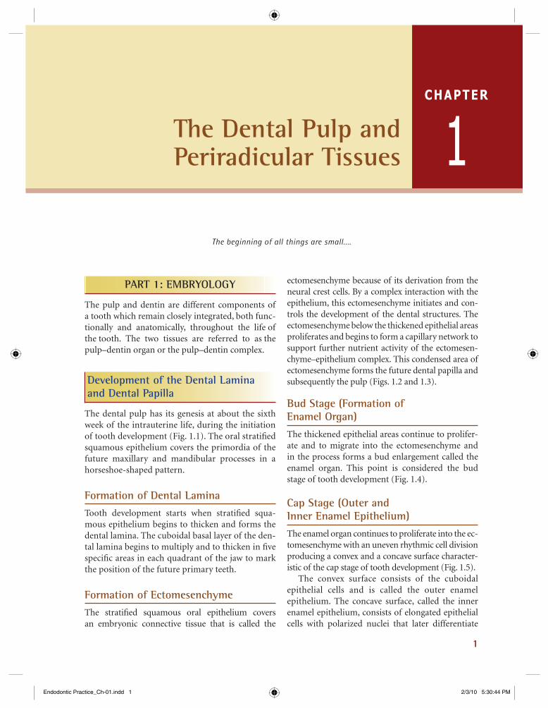
The dental pulp has its genesis at about the sixth week of the intrauterine life, during the initiation of tooth development (Fig. 1.1). The oral stratifi ed squamous epithelium covers the primordia of the future maxillary and mandibular processes in a horseshoe-shaped pattern.
Formation of Dental LaminaTooth development starts when stratifi ed squa-mous epithelium begins to thicken and forms the dental lamina. The cuboidal basal layer of the den-tal lamina begins to multiply and to thicken in fi ve specifi c areas in each quadrant of the jaw to mark the position of the future primary teeth.
Formation of EctomesenchymeThe stratifi ed squamous oral epithelium covers an embryonic connective tissue that is called the
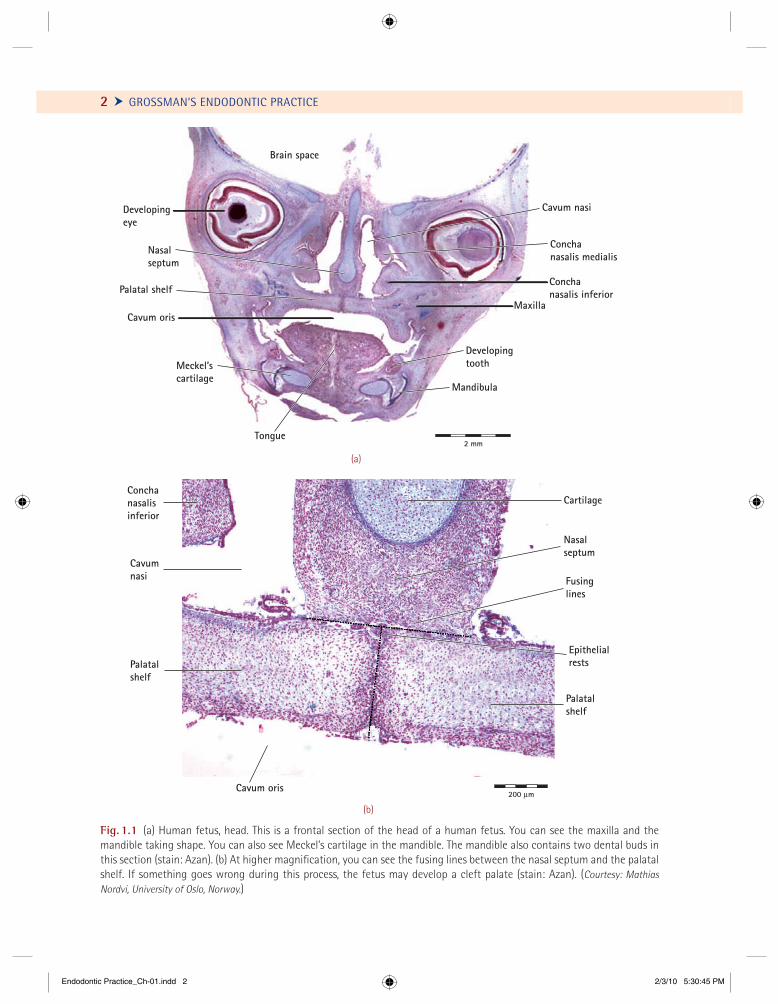
ectomesenchyme because of its derivation from the neural crest cells. By a complex interaction with the epithelium, this ectomesenchyme initiates and con-trols the development of the dental structures. The ectomesenchyme below the thickened epithelial areas proliferates and begins to form a capillary network to support further nutrient activity of the ectomesen-chyme–epithelium complex. This condensed area of ectomesenchyme forms the future dental papilla and subsequently the pulp (Figs. 1.2 and 1.3).
Bud Stage (Formation of Enamel Organ)The thickened epithelial areas continue to prolifer-ate and to migrate into the ectomesenchyme and in the process forms a bud enlargement called the enamel organ. This point is considered the bud stage of tooth development (Fig. 1.4).
Cap Stage (Outer and Inner Enamel Epithelium)The enamel organ continues to proliferate into the ec-tomesenchyme with an uneven rhythmic cell division producing a convex and a concave surface character-istic of the cap stage of tooth development (Fig. 1.5).
The convex surface consists of the cuboidal epithelial cells and is called the outer enamel epithelium. The concave surface, called the inner enamel epithelium, consists of elongated epithelial cells with polarized nuclei that later differentiate
CHAPTER
1The Dental Pulp and Periradicular Tissues
Endodontic Practice_Ch-01.indd 1Endodontic Practice_Ch-01.indd 1 2/3/10 5:30:44 PM2/3/10 5:30:44 PM
2 Ü GROSSMAN’S ENDODONTIC PRACTICE
Fig. 1.1 (a) Human fetus, head. This is a frontal section of the head of a human fetus. You can see the maxilla and the mandible taking shape. You can also see Meckel’s cartilage in the mandible. The mandible also contains two dental buds in this section (stain: Azan). (b) At higher magnifi cation, you can see the fusing lines between the nasal septum and the palatal shelf. If something goes wrong during this process, the fetus may develop a cleft palate (stain: Azan). (Courtesy: Mathias Nordvi, University of Oslo, Norway.)
Brain space
DevelopingeyeDevelopingeye
Developingtooth
Mandibula
Conchanasalis medialis
Conchanasalis inferior
Nasalseptum
Palatal shelf
Meckel’scartilage
Tongue
Cavum orisMaxilla
2 mm
Cavum nasi
(a)
Nasalseptum
Cavumnasi
Conchanasalisinferior
Cartilage
200 µm
Fusinglines
Epithelialrests
Palatalshelf
Cavum oris
Palatalshelf
(b)
Endodontic Practice_Ch-01.indd 2Endodontic Practice_Ch-01.indd 2 2/3/10 5:30:45 PM2/3/10 5:30:45 PM
CHAPTER 1 The Dental Pulp and Periradicular Tissues Ö 3
Developing brainBrain space
Developing eye
Concha nasalismedia
Concha nasalisinferior
Tooth bud(cap stage)
Tooth bud(bud stage)
Mandibula
Muscle
Muscle
Muscle
Meckel’scartilage
Mandibula
Cavumoris
Maxilla
Cavumnasi
Muscle
Cartilagothyroidea
Developingthyroid gland
2 mm
Nasalseptum
Tongue
Fig. 1.2 Human fetus, head. This is a frontal section of the head of a human fetus. The nasal cavity (Latin cavum nasi ) is divided into two by the nasal cartilage within the nasal septum. At both sides of the septum, you can see the nasal conchae (Latin concha nasalis media et inferior). They are made up of cartilage at this stage of development. The palate and the maxilla also contain a few spicules of bone. (Courtesy: Mathias Nordvi, University of Oslo, Norway.)
Endodontic Practice_Ch-01.indd 3Endodontic Practice_Ch-01.indd 3 2/3/10 5:30:46 PM2/3/10 5:30:46 PM

That it will never come again is what makes life so sweet.
—Emily Dickinson
43
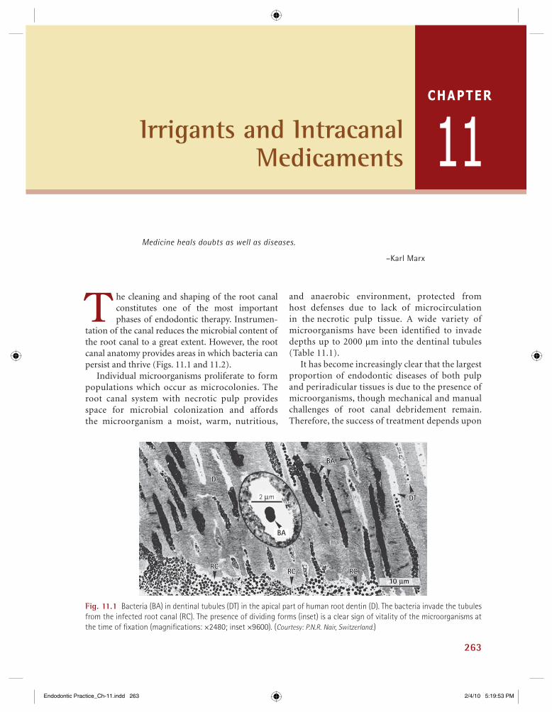
Microorganisms virtually cause all the pathoses of the pulpal and periapical tis-sues. Endodontic infection is the infec-
tion of the root canal system and is the major etiologic factor of apical periodontitis. The root ca-nal infection usually develops after pulpal necrosis, which can occur as a sequel of caries, trauma, and periodontal diseases or operative procedures.
The role of microbiology in endodontic prac-tice, although clearly important, has remained controversial through most of the twentieth cen-tury. Onderdenk suggested the need for bacterio-logic examination of the root canal in 1901. Shortly thereafter, in 1910, Hunter made his his-toric address in Montreal, in which he condemned the “golden traps of sepsis,” the ill-fi tting crowns and bridgework of his day that inexplicably re-sulted in the extraction of countless numbers of treated pulpless teeth and the inception of the “focal infection theory.”
Within 25 years, nearly 2000 papers on focal in-fection were published, many concerned with oral focal infection. During this period, a few voices were raised to stem the hysterical tide and to return endo-dontic care to its proper role in the healing arts. La Roche and Coolidge suggested that bacteriologic ex-amination be used in treating the root canal. Histo-logic studies of repair were reported by Blayney in 1932, Coolidge in 1931, Kronfeld in 1939, Aisenberg in 1931, Hatton and associates in 1928, Orban in 1932, Gottlieb and colleagues in 1928, and others.
Another study was published in 1936 by Fish and MacLean, who demonstrated that the pulp and periapical tissues of vital healthy teeth are invari-ably free of the evidence of microorganisms when examined histologically. In 1935, Okell and Elliot reported a transient bacteremia following extrac-tion; Appleton suggested that without bacteria no need would exist for endodontic treatment, a hypo-thesis supported by the classic study of Kakehashi and colleagues, who reported that exposed pulps in gnotobiotic rats healed without treatment in a germ-free environment.
In the last few decades, many reports have been published on the bacterial fl ora of the pulp and periapical and periodontal tissues, the pathways of infection, the immunologic reactions, and the infl ammatory responses. Although treatment pro-cedures have changed radically, they refl ect a bet-ter understanding of the host–parasite relationship and of the way in which such reactions are managed more effectively.
Bacterial Pathways into the Pulp
Bacteria enter the pulp in various ways:
! Through dentinal tubules following carious invasion
! Through crown or root following traumatic exposure of the pulp
! Coronal leakage following restorative proced-ures and restorations
CHAPTER
2Microbiology
Endodontic Practice_Ch-02.indd 43Endodontic Practice_Ch-02.indd 43 2/3/10 5:34:42 PM2/3/10 5:34:42 PM
46 Ü GROSSMAN’S ENDODONTIC PRACTICE
Microorganisms Detected in Root-Filled Teeth Associated with Persistent Apical Peridodontitis
TaxonomyEnterococcus faecalisPseudoramibacter alactolyticusPropionibacterium propionicumFilifactor alocisDialister pneumosintesStreptococcus spp.T. forsythiaDialister invisusCampylobacter rectusP. gingivalisTreponema denticolaFusobacterium nucleatumP. intermediaCandida albicans
TABLE 2.3
Fig. 2.2 Radiographic appearance of a secondary intra-radicular infection in a root-fi lled tooth.
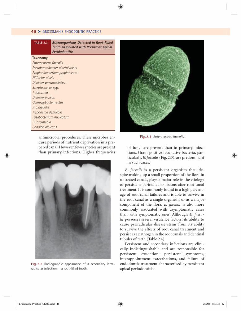
Fig. 2.3 Enterococcus faecalis.antimicrobial procedures. These microbes en-dure periods of nutrient deprivation in a pre-pared canal. However, fewer species are present than primary infections. Higher frequencies
of fungi are present than in primary infec-tions. Gram-positive facultative bacteria, par-ticularly, E. faecalis (Fig. 2.3), are predominant in such cases.
E. faecalis is a persistent organism that, de-spite making up a small proportion of the fl ora in untreated canals, plays a major role in the etiology of persistent periradicular lesions after root canal treatment. It is commonly found in a high percent-age of root canal failures and is able to survive in the root canal as a single organism or as a major component of the fl ora. E. faecalis is also more commonly associated with asymptomatic cases than with symptomatic ones. Although E. faeca-lis possesses several virulence factors, its ability to cause periradicular disease stems from its ability to survive the effects of root canal treatment and persist as a pathogen in the root canals and dentinal tubules of teeth (Table 2.4).
Persistent and secondary infections are clini-cally indistinguishable and are responsible for persistent exudation, persistent symptoms, inter appoint ment exacerbations, and failure of endo dontic treatment characterized by persistent apical periodontitis.
Endodontic Practice_Ch-02.indd 46Endodontic Practice_Ch-02.indd 46 2/3/10 5:34:43 PM2/3/10 5:34:43 PM

CHAPTER 2 Microbiology Ö 47
Extraradicular InfectionsMicrobial invasion of the infl amed periradicular tissue is invariably a sequel of intraradicular infec-tion. Acute alveolar abscess is an example of extra-radicular extension or a sequel to intraradicular infection (Fig. 2.4).
Sometimes extraradicular infection can be inde-pendent of intraradicular infection. For example,
Survival and Virulence Factors of E. faecalis
! Endures prolonged periods of nutritional deprivation! Binds to dentin and profi ciently invades dentinal tubules! Alters host responses! Suppresses the action of lymphocytes! Possesses lytic enzymes, cytolysin, aggregation
substance, pheromones, and lipoteichoic acid! Utilizes serum as a nutritional source! Resists intracanal medicaments (i.e., Ca(OH)2) Maintains pH homeostasis Properties of dentin lessen the effect of calcium
hydroxide! Competes with other cells! Forms a biofi lm
TABLE 2.4 apical actinomycosis caused by Actinomyces sp. and P. propionicum is a pathological disease which can be treated only by periapical surgery. Other patho-gens implicated in such infections are as follows:
! Treponema spp.! T. forsythia! P. endodontalis! P. gingivalis! F. nucleatum
Biofi lms (Fig. 2.5)
Biofi lm is defi ned as a community of microcolonies of microorganisms in an aqueous solution that is surrounded by a matrix made of glycocalyx, which also attaches the bacterial cells to a solid substratum. A biofi lm is one of the basic survival methods em-ployed by bacteria in times of starvation. According to Caldwell et al., a biofi lm has the following attributes:
! Autopoiesis. Ability to self-organize! Homeostasis. Ability to resist environmental
disturbances! Synergy. Effective in association with fellow
microorganisms than in isolation
Fig. 2.4 Radiographic appearance, clinical view, and aspiration of serous exudate from an extraradicular infection.(Courtesy: S. Karthiga Kannan, India.)
(b)
(c)(a)
Endodontic Practice_Ch-02.indd 47Endodontic Practice_Ch-02.indd 47 2/3/10 5:34:44 PM2/3/10 5:34:44 PM
Listen to your patient .… The patient will give you the diagnosis.
—Sir William Osler
53
Diagnosis is the correct determination, discriminative estimation, and logical ap-praisal of conditions found during exami-
nation as evidenced by distinctive signs, marks, and symptoms. Diagnosis is also defi ned as the art of distinguishing one disease from another. Correct treatment begins with a correct diagnosis. Arriving at a correct diagnosis requires knowledge, skill, and art: knowledge of the diseases and their symptoms, skill to apply proper test procedures, and the art of synthesizing impressions, facts, and experience into understanding.
Diagnostic procedures should follow a consis-tent, logical order which includes comprehensive medical and dental history, radiographic examina-tion, extraoral and intraoral clinical examination including histopathological examination to arrive at the fi nal diagnosis when required.
The process begins with the initial call request-ing an appointment for some specifi c reason, usu-ally a complaint of pain. Subjective information is supplied by the written history or questionnaire that each patient completes and signs. Further informa-tion is obtained by the clinician, who reviews the questionnaire and asks specifi c questions regarding the patient’s chief complaint, past medical history, past dental history, and present medical and dental status. The clinician should not hesitate to consult the patient’s physician whenever the patient appears
to be medically compromised or when the gained information is inadequate or unclear. More often than not, a patient’s medical problem affects the course of treatment, especially concerning the use of anesthet-ics, antibiotics, and analgesics. Occasionally, the pa-tient’s medical status bears a direct relation to the clinical diagnosis. For example, diffuse pain in the mandibular left molars may be a referred pain caused by angina pectoris, or bizarre symptoms may be the result of psychogenic or neurologic disorders.
History and Record
Case history is defi ned as the data concerning an individual and his or her family and environment, including the individual medical history that may be useful in analyzing and diagnosing his or her case or for instructional purposes. Because many diseases have similar symptoms, the clinician must be astute in determining the correct diagnosis. Dif-ferential diagnosis is the most common procedure. This technique distinguishes one disease from sev-eral other similar disorders by identifying their dif-ferences. Diagnosis by exclusion, on the other hand, eliminates all possible diseases under consideration until one remaining disease correctly explains the patient’s symptoms. Although proper clinical diag-nosis may appear to be simple, it can tax the most experienced clinician.
CHAPTER
3Clinical Diagnostic Methods
Endodontic Practice_Ch-03.indd 53Endodontic Practice_Ch-03.indd 53 2/3/10 6:26:46 PM2/3/10 6:26:46 PM
58 Ü GROSSMAN’S ENDODONTIC PRACTICE
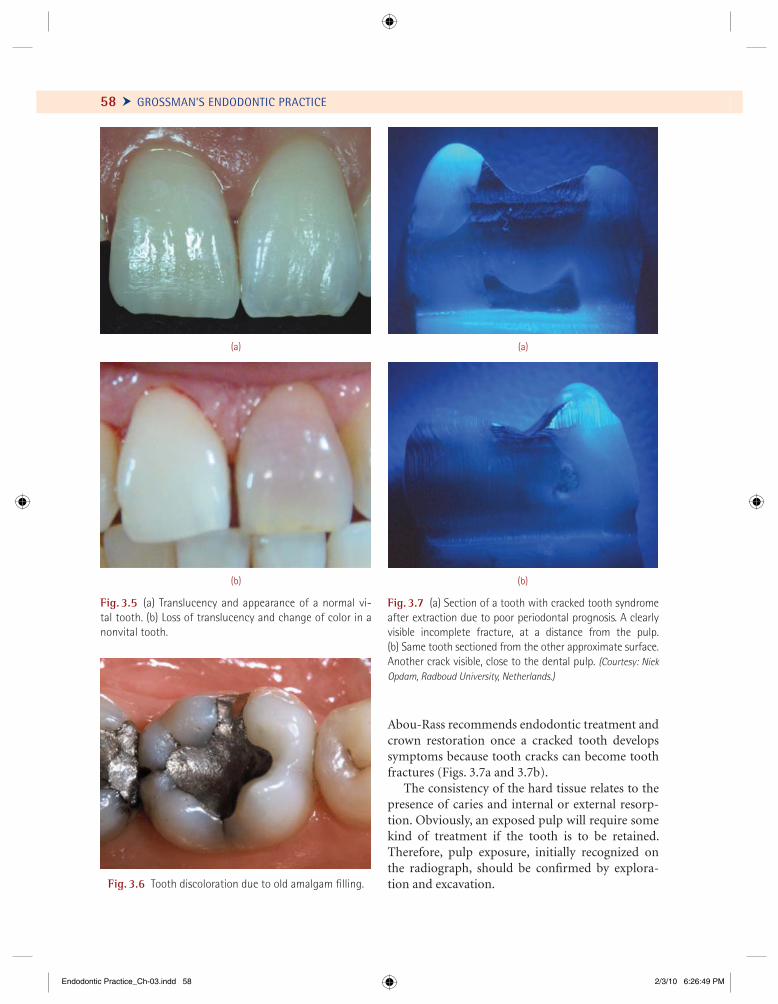
Abou-Rass recommends endodontic treatment and crown restoration once a cracked tooth develops symptoms because tooth cracks can become tooth fractures (Figs. 3.7a and 3.7b).
The consistency of the hard tissue relates to the presence of caries and internal or external resorp-tion. Obviously, an exposed pulp will require some kind of treatment if the tooth is to be retained. Therefore, pulp exposure, initially recognized on the radiograph, should be confi rmed by explora-tion and excavation.
Fig. 3.5 (a) Translucency and appearance of a normal vi-tal tooth. (b) Loss of translucency and change of color in a nonvital tooth.
(b)
(a)
Fig. 3.6 Tooth discoloration due to old amalgam fi lling.
(b)
(a)
Fig. 3.7 (a) Section of a tooth with cracked tooth syndrome after extraction due to poor periodontal prognosis. A clearly visible incomplete fracture, at a distance from the pulp. (b) Same tooth sectioned from the other approximate surface. Another crack visible, close to the dental pulp. (Courtesy: Niek Opdam, Radboud University, Netherlands.)
Endodontic Practice_Ch-03.indd 58Endodontic Practice_Ch-03.indd 58 2/3/10 6:26:49 PM2/3/10 6:26:49 PM
CHAPTER 3 Clinical Diagnostic Methods Ö 59
The technique of visual and tactile examination is simple. One uses one’s eyes, fi ngers, an explorer, and the periodontal probe. The patient’s teeth and periodontium should be examined in good light under dry conditions. For example, a sinus tract (fi stula) might escape detection if it is covered by saliva or an interproximal cavity may escape notice if it is fi lled with food.
Loss of translucency, slight color changes, and cracks may not be apparent in poor light. In fact, a transilluminator may aid in detecting enamel cracks or crown fractures.
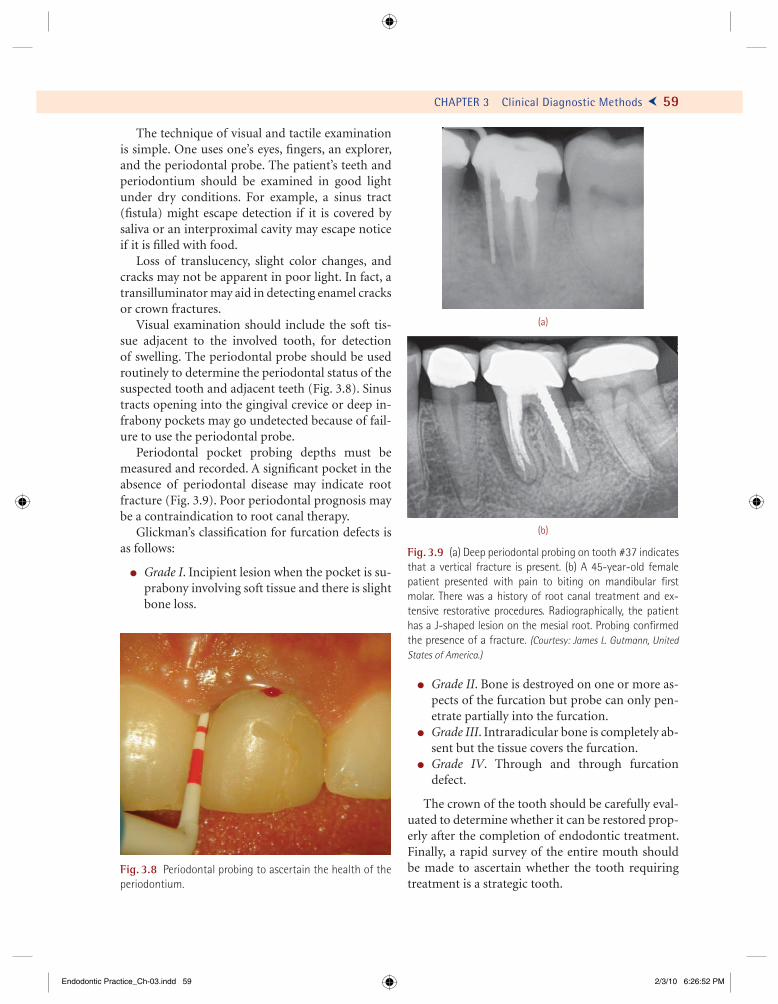
Visual examination should include the soft tis-sue adjacent to the involved tooth, for detection of swelling. The periodontal probe should be used routinely to determine the periodontal status of the suspected tooth and adjacent teeth (Fig. 3.8). Sinus tracts opening into the gingival crevice or deep in-frabony pockets may go undetected because of fail-ure to use the periodontal probe.
Periodontal pocket probing depths must be measured and recorded. A signifi cant pocket in the absence of periodontal disease may indicate root fracture (Fig. 3.9). Poor periodontal prognosis may be a contraindication to root canal therapy.
Glickman’s classifi cation for furcation defects is as follows:
! Grade I. Incipient lesion when the pocket is su-prabony involving soft tissue and there is slight bone loss.
! Grade II. Bone is destroyed on one or more as-pects of the furcation but probe can only pen-etrate partially into the furcation.
! Grade III. Intraradicular bone is completely ab-sent but the tissue covers the furcation.
! Grade IV. Through and through furcation defect.
The crown of the tooth should be carefully eval-uated to determine whether it can be restored prop-erly after the completion of endodontic treatment. Finally, a rapid survey of the entire mouth should be made to ascertain whether the tooth requiring treatment is a strategic tooth.
Fig. 3.8 Periodontal probing to ascertain the health of the periodontium.
Fig. 3.9 (a) Deep periodontal probing on tooth #37 indicates that a vertical fracture is present. (b) A 45-year-old female patient presented with pain to biting on mandibular fi rst molar. There was a history of root canal treatment and ex-tensive restorative procedures. Radiographically, the patient has a J-shaped lesion on the mesial root. Probing confi rmed the presence of a fracture. (Courtesy: James L. Gutmann, United States of America.)
(a)
(b)
Endodontic Practice_Ch-03.indd 59Endodontic Practice_Ch-03.indd 59 2/3/10 6:26:52 PM2/3/10 6:26:52 PM
74
For there was never yet a philosopher who could endure the toothache patiently.
–William Shakespeare, Much Ado About Nothing, Act V
T he pulp is the formative organ of the tooth. It builds primary dentin during the development of the tooth, secondary
dentin after tooth eruption, and reparative den-tin in response to stimulation as long as the odontoblasts remain intact (Figs. 4.1a and 4.1b). The pulp responds to hot and cold stimuli which are perceived only as pain. Heat at temperatures between 60°F (16°C) and 130°F (55°C) when ap-plied directly to an intact tooth surface is usually well tolerated by the pulp, but foodstuffs and beverages above and below this temperature range can also be endured. Cavity preparation also produces temperature changes, with an in-crease of 20°C in temperature during dry cavity preparation 1 mm from the pulp and a 30°C in-crease 0.5 mm from the pulp. A theoretical model has shown that the sensory reaction to thermal stimulation is registered before a temperature change occurs at the pulpodentinal junction, where the nerve endings are located. The sensa-tion of pain, a warning signal that the pulp is en-dangered, is a protective reaction, as it is elsewhere in the body.
The pulp has been described as a highly resistant organ and as an organ with little resistance or re-cuperating ability. Its resistance depends on cellular
activity, nutritional supply, age, and other meta-bolic and physiologic parameters. This variability has led to the remark that “some pulps will die if you look crossly at them, while others can’t be killed with an axe.” On the whole, the resistance of the pulp to injury is slight in certain cases, but evidence of unusual persistence of vitality following injury has also been observed.
The desirability of maintaining a vital pulp and protecting it from injury was recognized by the ear-liest practitioners of dentistry. The value of the pulp as an integral part of the tooth, both anatomic and functional, should be recognized and every effort made to conserve it.
Accurate pulpal diagnosis is the key to all end-odontic treatments. Unfortunately, there has been poor correlation between the clinical symptoms and histopathology of the pulp. Previous attempts made to diagnose the condition of the pulp based on either clinical signs (or symptoms), electric pulp test, and/or thermal tests and radiography have not always been successful. The endodontist is expected to understand the various causative fac-tors of pulpal diseases, collect information about the presentation and history of symptoms, and conduct many practical tests before formulating the fi nal diagnosis.
4 Diseases of the Dental Pulp
CHAPTER
Endodontic Practice_Ch-04.indd 74Endodontic Practice_Ch-04.indd 74 2/3/10 6:43:17 PM2/3/10 6:43:17 PM
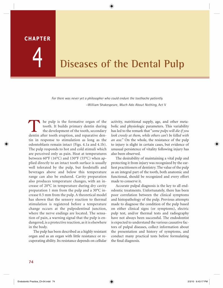
CHAPTER 4 Diseases of the Dental Pulp Ö 75
Fig. 4.1 (a) Demineralized tooth, longitudinal section: this is a section through a premolar. The pulp (Latin pulpa) comprises loose connective tissue, blood vessels, and nerves (stain: H + E). (b) Demineralized tooth, cross-section: this is a section through the coronal part of a premolar. The enamel has been lost during the preparation of the section. The pulp (Latin pulpa) is visible at the center of the tooth (stain: H + E). (Courtesy: Mathias Nordvi, University of Osla, Norway.)
Odontoblasts
Fibroblasts
Pulpa
Nerve
Odontoblasts
Fibroblasts
Predentin
Blood vessels
Dentin
Enamel space
1 mm
(a)
2 mm
Pulpa
Pulpa
Dentin
2 mm
(b)
Endodontic Practice_Ch-04.indd 75Endodontic Practice_Ch-04.indd 75 2/3/10 6:43:18 PM2/3/10 6:43:18 PM

Life tells you nothing … it shows you everything.
—Richard Bach
97
P ulpal disease is only one of several possible causes of diseases of the periradicular tis-sues. Because of the inter-relationship be-
tween the pulp and the periradicular tissues, pulpal infl ammation causes infl ammatory changes in the periodontal ligament even before the pulp becomes totally necrotic. Bacteria and their toxins, immuno-logic agents, tissue debris, and products of tissue necrosis from the pulp reach the periradicular area through the various foramina of the root canals and give rise to infl ammatory and immunologic reac-tions. Neoplastic disorders, periodontal conditions, developmental factors, and trauma can also cause periradicular diseases.
The diseases of periradicular tissues can be clas-sifi ed on the basis of the etiology, symptoms, and histopathological features. The WHO has classifi ed diseases of periradicular tissues into various catego-ries (Table 5.1).
Periradicular diseases of pulpal origin may also be classifi ed as acute or chronic periradicular diseases based on symptoms, etiology, and histo-pathology (Table 5.2).
Acute Periradicular Diseases
These disorders include acute apical periodontitis, acute alveolar abscess, and acute exacerbation of a chronic lesion.
CHAPTER
5Diseases of the Periradicular Tissues
TABLE 5.1 WHO Classifi cation of Periradicular Diseases
Code Number Category
KO4.4 Acute apical periodontitis
KO4.5 Chronic apical periodontitis (apical granuloma)
KO4.6 Periapical abscess with sinus (dentoalveolar abscess with sinus, periodontal abscess of pulpal origin)
KO4.60 Periapical abscess with sinus to maxillary antrum
KO4.61 Periapical abscess with sinus to nasal cavity
KO4.62 Periapical abscess with sinus to oral cavity
KO4.63 Periapical abscess with sinus to skin
KO4.7 Periapical abscess without sinus (dental abscess without sinus, dentoalveolar abscess without sinus, periodontal abscess of pulpal origin without sinus)
KO4.8 Radicular cyst (apical periodontal cyst, periapical cyst)
KO4.80 Apical and lateral cyst
KO4.81 Residual cyst
KO4.82 Infl ammatory periodontal cyst
Endodontic Practice_Ch-05.indd 97Endodontic Practice_Ch-05.indd 97 2/4/10 10:03:16 AM2/4/10 10:03:16 AM

108 Ü GROSSMAN’S ENDODONTIC PRACTICE
may be present. The root surface may show external root resorption due to cementoclastic activity or hypercementosis due to cementoblast activity.
Treatment
Root canal therapy may suffi ce for the treatment of a chronic apical periodontitis. Removal of the cause of infl ammation is usually followed by resorption of the granulomatous tissue and repair with tra-beculated bone (Fig. 5.13).
Prognosis
The prognosis for long-term retention of the tooth is excellent.
(a) (b)
(c)
Fig. 5.13 (a) Chronic apical periodontitis in an asymptomatic poorly obturated mandibular molar. (b) Radiographic view after completion of retreatment and obturation of the root canals. (c) Two-year follow-up shows complete resolution of the periradicular pathology. (Courtesy: Julian Webber, England.)
Chronic Alveolar AbscessA chronic alveolar abscess is a long-standing, low-grade infection of the periradicular alveolar bone characterized by the presence of an abscess draining through a sinus tract.
Synonym
Chronic suppurative apical periodontitis.
Causes
The source of the infection is in the root canal. Chronic alveolar abscess is a natural sequela of death of the pulp with extension of the infective
Endodontic Practice_Ch-05.indd 108Endodontic Practice_Ch-05.indd 108 2/4/10 10:03:24 AM2/4/10 10:03:24 AM

CHAPTER 5 Diseases of the Periradicular Tissues Ö 109
process periapically, or it may result from a pre-existing acute abscess.
Symptoms
A tooth with a chronic alveolar abscess is generally asymptomatic, or only mildly painful. At times, such an abscess is detected only during routine radiographic examination or because of the presence of a sinus tract, which can be either intraoral (Figs. 5.14a–5.14c) or extraoral (Fig. 5.15). The sinus tract usually pre-vents exacerbation or swelling by providing continual drainage of the periradicular lesion.
Diagnosis
The fi rst sign of osseous breakdown is radiographic evidence seen during routine examination or
(a) (b)
(c)
Fig. 5.14 (a) Intraoral labial sinus opening in relation to the carious maxillary lateral incisor. (b) Intraoral palatal sinus opening in relation to the carious maxillary central incisor. (c) Intraoral buccal sinus opening in relation to the carious mandibular premolar.
Fig. 5.15 Extraoral sinus opening. (Courtesy: Karthiga Kannan, India.)
Endodontic Practice_Ch-05.indd 109Endodontic Practice_Ch-05.indd 109 2/4/10 10:03:24 AM2/4/10 10:03:24 AM
CHAPTER 5 Diseases of the Periradicular Tissues Ö 121
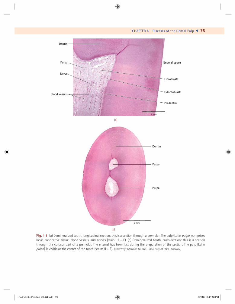
Fig. 5.26 Photomicrographs of axial semithin sections through the surgically removed apical portion of the root with a persistent apical periodontitis. Note the adhesive biofi lm (BF) in the root canal. Consecutive sections [(a)–(b)] reveal the emerging widened profi le of an accessory canal (AC) that is clogged with the biofi lm. The AC and the biofi lm are magnifi ed in (c) and (d), respectively. Magnifi cations: (a) !75, (b) !70, (c) !110, and (d) !300. (Adapted from Nair, P.N.R., et al.: Intrara-dicular bacteria and fungi in root-fi lled, asymptomatic human teeth with therapy-resistant periapical lesions: A long-term light and electron microscopic follow-up study. J. Endod., 16:580–88, 1990.)
D
BF
BF BF
BF
AC
AC
AP AP
DD D
(a) (b)
(c) (d)
0.5 mm 0.5 mm
50 µµm100 µm
Endodontic Practice_Ch-05.indd 121Endodontic Practice_Ch-05.indd 121 2/4/10 10:03:32 AM2/4/10 10:03:32 AM
When the only tool you own is a hammer, every problem begins to resemble a nail.
—Abraham Maslow
131
Injury to the calcifi ed structure of teeth and to the supporting tissues by noxious stimuli may cause changes in the pulp and the periradicular
tissues. Noxious stimuli can be physical, chemical, or bacterial. They can produce changes that are either reversible or irreversible, depending on duration, in-tensity, and pathogenicity of the stimulus and the host’s ability to resist the stimulus and to repair tissue damage.
On the basis of these premises, we can general-ize that mild-to-moderate noxious stimuli to the pulp may produce sclerosis of the dentinal tubules, formation of reparative dentin, or reversible infl am-mation. Irreversible infl ammatory changes caused by severe injury can lead to necrosis of the pulp and subsequent pathologic changes in the periradicular tissues.
The infl ammatory response of the connective tissue of the dental pulp is modifi ed because of its milieu. Because the pulp is encased in hard tissues with limited portals of entry, it is an organ of ter-minal and limited circulation with no effi cient col-lateral circulation and with limited space to expand during the infl ammatory reaction. A clear concept of the fundamentals of infl ammation is necessary for the understanding of the diseases of the pulp and their extension to the periradicular tissues.
Infl ammation
Infl ammation is the local physiologic reaction of the body to noxious stimuli or irritants. Any irri-tant, whether of traumatic, chemical, or bacterial origin, produces a sequence of basic physiologic and morphologic reactions in vascular, lymphatic, and connective tissues. Host-resistance factors and intensity, duration, and virulence of the irritant modify the ultimate character, extent, and severity of the tissue changes and, to some degree, the clini-cal manifestations.
The objective of infl ammation is to remove or destroy the irritant and to repair damage to the tissue. Infl ammation brings to the area phagocytic cells to digest bacteria or cellular debris, anti bodies to recognize, attack, and destroy foreign matter, edema or fl uid to dilute and neutralize the irritant, and fi brin to limit the spread of infl ammation.
Repair of the tissues depends on the severity of injury and host resistance. The injurious agent may cause reversible or irreversible changes to the tissues. Irreversible damage leads to tissue ne-crosis, whereas reversible damage leads to repair. Repair, or the return of the tissue to normal struc-ture and function, begins as the tissue becomes involved in the infl ammatory process. Removal of
CHAPTER
6Rationale of Endodontic Treatment
Endodontic Practice_Ch-06.indd 131Endodontic Practice_Ch-06.indd 131 2/4/10 10:18:58 AM2/4/10 10:18:58 AM

138 Ü GROSSMAN’S ENDODONTIC PRACTICE
Zone of StimulationThis zone is characterized by fi broblasts and osteo-blasts. At the periphery, Fish noted that the toxin was mild enough to be a stimulant. In response to this stimulation, collagen fi bers were laid down by the fi broblasts, which acted both as a wall of defense around the zone of irritation and as a scaffolding on which the osteoblasts built new bone. This new bone was built in irregular fashion.
By analogy, we can apply the knowledge gained in Fish’s experiment to understand better the reaction of the periradicular tissues to a pulp-less tooth. The root canal is the site of infection (Fig. 6.3). The microorganisms in the root canal are rarely motile and do not move from the root canal to the periradicular tissues; however, they can multiply suffi ciently to grow out of the root canal, or the metabolic products of these micro-organisms or the toxic products of tissue necrosis
may be diffused to the periradicular tissues. As the microorganisms gain access to the periradicular area, they are destroyed by the polymorphonu-clear leukocytes. When the microorganisms are suffi ciently virulent, or when enough are present, they overwhelm the defensive mechanism, and a periradicular lesion results. When they are of low virulence and numbers, however, a stalemate oc-curs. The polymorphonuclear leukocytes destroy the microorganisms as rapidly as they gain access to the periradicular tissues. The result is a chronic abscess. The toxic products of the microorganisms and the necrotic pulp in the root canal are irritat-ing and destructive to the periradicular tissue and, together with the proteolytic enzymes released by the dead polymorphonuclear leukocytes, help to produce pus.
At the periphery of the destroyed area of osse-ous tissue, toxic bacterial products may be diluted
Zone of stimulation
Zone of irritation
Zone of contamination
Zone of necrosis
Fig. 6.3 Schematic diagram showing bacteria in the root canal and the zones of infection, contamination, irritation, and stimulation.
Endodontic Practice_Ch-06.indd 138Endodontic Practice_Ch-06.indd 138 2/4/10 10:19:00 AM2/4/10 10:19:00 AM

Education is a progressive discovery of our own ignorance.
—Will Durant
141
Proper selection of cases avoids pitfalls during endodontic treatment and helps to ensure success. Not every tooth is suitable for endo-
dontic treatment. Errors in case selection, some of which could have been avoided, constituted 22% of failures reported in a study by Ingle and Beveridge.
Many more root canals are treated today than before because of a greater interest in endodontics by the practitioner not only to save endodontically involved teeth, but also to use them as abutments for bridges or partial dentures. Unfortunately, a general practitioner’s best effort may not be good enough because of a mistaken diagnosis, such as a curved canal not capable of being instrumented to the apex, with a resulting persistence of the area of rarefaction. On the other hand, certain cases for-merly contraindicated for endodontic treatment, such as a sinus tract discharging into the gingi-val sulcus, are treated successfully today because of advances in both endodontic and periodontal therapy. To depend on root canal treatment alone for all endodontic cases is bound to meet with a certain degree of failure. If such treatment is com-bined with other endodontic procedures, such as apexifi cation with mineral trioxide aggregate (MTA) to induce the formation of a calcifi c bar-rier in root canals with open apex or the use of periapical microsurgery to remove the periapical
pathological tissues, the possibilities of it being successful are high. On the other hand, micro-surgery is not indicated simply because an area of rarefaction is present.
The selection of cases for endodontic treat-ment has been discussed by a number of authors, including Bender, Grossman and associates, and Strindberg. Advances in the understanding of endodontics, better techniques, and principles of canal preparation and obturation have led to signifi cantly increased and predictable healing rates for endodontic treatment—95% and higher under ideal conditions according to current lit-erature. However, before selecting a case for endo-dontic therapy the clinician should consider the following factors that infl uence the outcome of the treatment:
! Health and systemic status of the patient! Anatomy of the root canal system! Extent of previous tooth restoration! Presence or absence of periradicular pathosis! Radiographic interpretation! Degree of diffi culty in locating, cleaning , shap-
ing, and obturating the complete root canal system
! Periodontal status of the tooth! Presence of crown or root fractures
CHAPTER
7Selection of Cases for Treatment
Endodontic Practice_Ch-07.indd 141Endodontic Practice_Ch-07.indd 141 2/4/10 10:37:30 AM2/4/10 10:37:30 AM
CHAPTER 7 Selection of Cases for Treatment Ö 143
Situation Agent Adults Children
TABLE 7.2 AHA-Recommended Antibiotic Prophylaxis Regimens for Dental Procedures
Oral
Unable to take oral medication
Amoxicillin
Ampicillinorcefazolin or ceftriaxone
2 g
2 g IM or IV
1 g IM or IV
50 mg/kg
50 mg/kg IM or IV
50 mg/kg IM or IV
Allergic to penicillin or ampicillin—oral
Cephalexin*†orclindamycinorazithromycin or clarithromycin
2 g
600 mg
500 mg
50 mg/kg
20 mg/kg
15 mg/kg
Allergic to penicillinor ampicillin and unable to take oral medication
Cefazolin or ceftriaxone†orclindamycin
1g IM or IV
600 mg IM or IV
50 mg/kg IM or IV
20 mg/kg IM or IV
IM, intramuscular; IV, intravenous.* Or other first or second generation in equivalent adult or pediatric dosage.† Cephalosporins should not be used in an individual with a history of anaphylaxis, angioderma, or urticaria with penicillins or ampicillin.
(b)(a)
(d)(c)
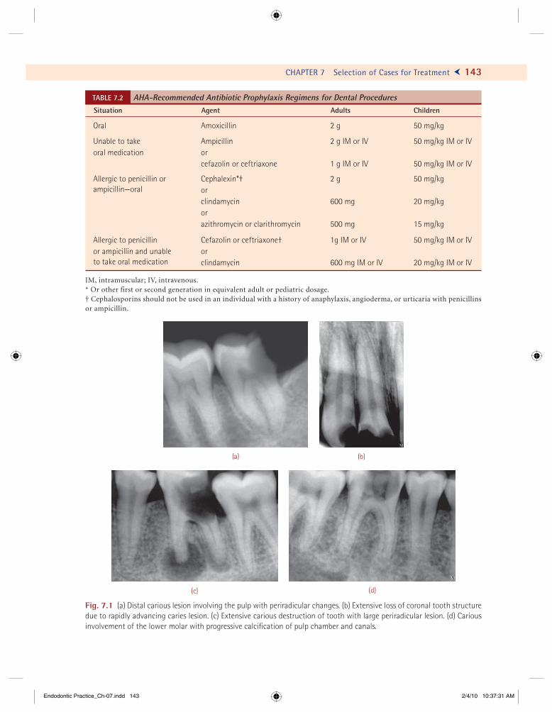
Fig. 7.1 (a) Distal carious lesion involving the pulp with periradicular changes. (b) Extensive loss of coronal tooth structure due to rapidly advancing caries lesion. (c) Extensive carious destruction of tooth with large periradicular lesion. (d) Cariousinvolvement of the lower molar with progressive calcifi cation of pulp chamber and canals.
Endodontic Practice_Ch-07.indd 143Endodontic Practice_Ch-07.indd 143 2/4/10 10:37:31 AM2/4/10 10:37:31 AM
Quality is, doing the little things right when nobody is looking.
—George Bernard Shaw
157
T he basic principles underlying the treatment of teeth with endodontic problems are those underlying surgery in general. An aseptic
technique, debridement of the wound, drainage, and gentle treatment of the tissues with both instru-ments and drugs—all are cardinal principles of sur-gery. Specifi cally, pain must be controlled, if present. During treatment, all pulp tissue must be removed, the root canal enlarged and irrigated, the canal sur-face rendered sterile as determined by bacteriologic examination, and the root canal well obturated to prevent the possibility of reinfection.
Rubber Dam Isolation
To achieve the fi rst principle of endodontic treat-ment a safe and aseptic operating technique needs to be maintained. For this, application of rubber dam is imperative. It is the only sure safeguard against bacterial contamination from saliva and ac-cidental swallowing of root canal instruments. All endodontic operations should be performed under the rubber dam. Treatment of any tooth should not be attempted under cotton rolls. The risk of losing a reamer or fi le down the patient’s trachea or esopha-gus is too great to warrant this practice.
In some cases, it is fi rst necessary to replace a missing wall with a restoration or to cement a stainless steel band to prevent the rubber dam clamp from slipping off the tooth. In other cases, a gingivectomy may need to be done, with removal of about 2-mm gingival tissue to provide enough tooth structure for application of a rubber dam clamp. Gingivectomy may be necessary in any event for restoration of the crown of the tooth.
In a survey of the use of the rubber dam in endo-dontic treatment by general practitioners, Going and Sawinski found that 36% of dentists never, or seldom, used the rubber dam. Their survey em-phasizes the need for more general use of the rub-ber dam by dentists, especially in view of the more than threefold increase in ingestion or aspiration of instruments during endodontic treatment in the last decade. To risk operating without a rub-ber dam is to risk one’s professional reputation. A variety of objects used in endodontic practice may be swallowed accidentally if the rubber dam is not applied. Root canal instruments swallowed during endodontic treatment has been reported (Fig. 8.1).
If an instrument is swallowed or aspirated during endodontic treatment without a rubber dam, one is likely to be confronted with a lawsuit. Grossman
CHAPTER
8Principles of Endodontic Treatment
Endodontic Practice_Ch-08.indd 157Endodontic Practice_Ch-08.indd 157 2/4/10 10:46:57 AM2/4/10 10:46:57 AM
164 Ü GROSSMAN’S ENDODONTIC PRACTICE
dam is held in the left hand and is kept from obstruct-ing the view, while the clamp is slipped over the tooth with the right hand. The forceps are then disengaged from the clamp, and the rubber dam is slipped un-der the anterior arms of the clamp. If a wing clamp is used, the wing of the clamp is inserted into the hole of the rubber dam, the clamp is applied to the tooth, the clamp forceps are removed, and the rubber dam is slipped under the arms of the clamp.
To facilitate slipping the rubber dam over the tooth, especially if the contact point is tight, the surface of the rubber dam adjacent to the hole should be wiped with liquid soap, or a wet fi nger
may be rubbed on a cake of soap and applied to the rubber dam around the punched hole. Petrolatum or cocoa butter should not be used for this purpose because these substances soften and weaken the rubber dam, and leakage may result.
The following methods are used while applying rubber dam in different teeth in the oral cavity:
! Molar tooth isolation using rubber dam (Figs. 8.7a–8.7f)
! Isolation of multiple teeth—clamp fi rst fol-lowed by dam (Figs. 8.8a–8.8i)
! Optradam application (Figs. 8.9a–8.9c)
(d)(c)
(b)(a)
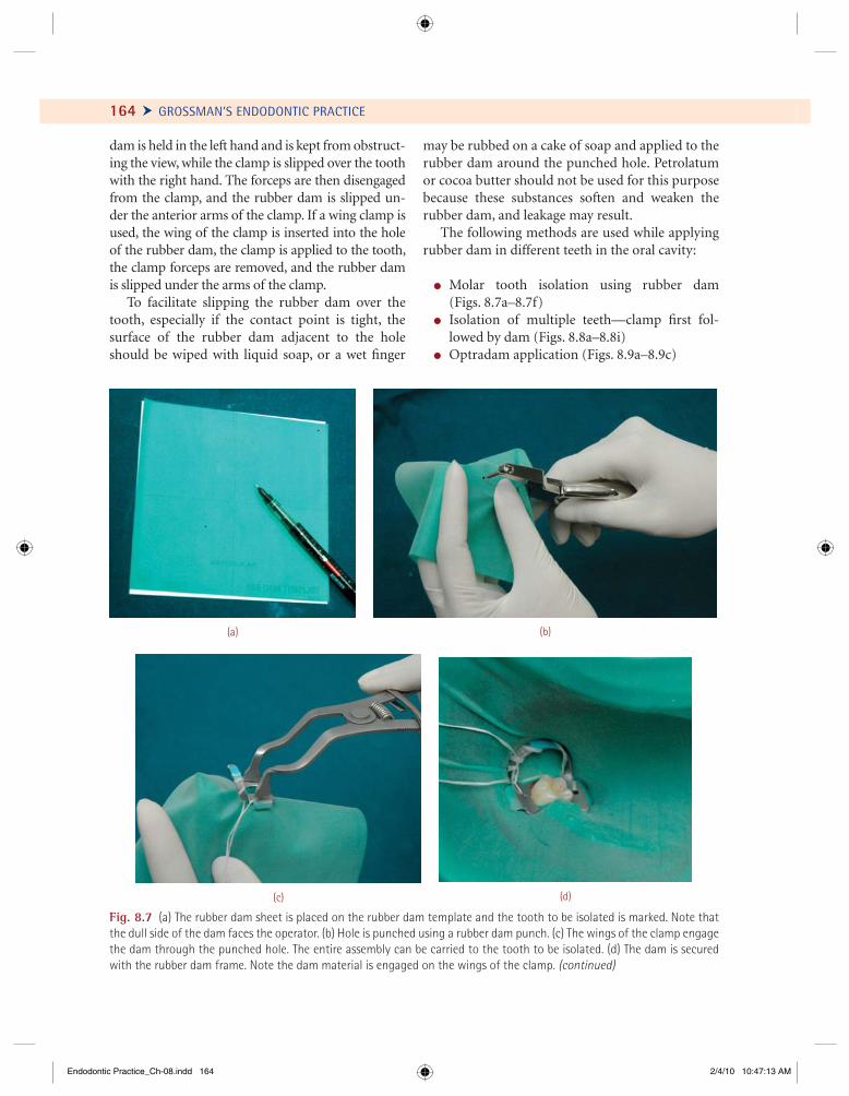
Fig. 8.7 (a) The rubber dam sheet is placed on the rubber dam template and the tooth to be isolated is marked. Note that the dull side of the dam faces the operator. (b) Hole is punched using a rubber dam punch. (c) The wings of the clamp engage the dam through the punched hole. The entire assembly can be carried to the tooth to be isolated. (d) The dam is secured with the rubber dam frame. Note the dam material is engaged on the wings of the clamp. (continued)
Endodontic Practice_Ch-08.indd 164Endodontic Practice_Ch-08.indd 164 2/4/10 10:47:13 AM2/4/10 10:47:13 AM

CHAPTER 8 Principles of Endodontic Treatment Ö 165
Fig. 8.7 (continued ) (e) The dam is gently teased away from the wing of the clamp using a blunt hand instrument. (f) Dental fl oss is passed mesially and distally to ensure inversion of the dam for isolation.
(e) (f)
Fig. 8.8 (a) Testing and lubricating the proximal contact. (b) The clamp is engaged with the clamp forceps and the forceps is secured with the lock. (c) The clamp is transferred to the tooth. The jaws of the clamp engage the tooth gingival to the heightof contour along the four axial line angles of the tooth. (d) Check the stability of the dam with index fi nger on the bow of the clamp. The clamp should not rock. (continued)
(a) (b)
(c) (d)
Endodontic Practice_Ch-08.indd 165Endodontic Practice_Ch-08.indd 165 2/4/10 10:47:16 AM2/4/10 10:47:16 AM
176
Anatomy of Pulp Cavity and Its Access Opening
Of all the phases of anatomic study in the human system,One of the most complex is the pulp cavity morphology.
–M.T. Barrett
The journey of a thousand miles begins with a single small step.
–Lao Tzu
T he external morphologic features of the crowns of teeth vary according to the shape and size of the head. The length of
the crown differs with the size and sex of the per-son and is generally shorter in females than in males. As the external morphology of the tooth varies from person to person, so does the internal morphology of the crown and root. Changes in pulp cavity anatomy result from age, disease, and trauma. Although morphologic variations occur, clinical experience indicates that these changes usually follow a general pattern, and thus the study of pulp cavity morphology is a feasible undertaking.
Pulp Cavity
The pulp cavity is the central cavity within a tooth and is entirely enclosed by dentin except at the apical foramen (Fig. 9.1). The pulp cav-ity may be divided into a coronal portion, the pulp chamber, and a radicular portion, the root canal.
Pulp ChamberIn anterior teeth, the pulp chamber gradually merges into the root canal, and this division becomes indis-tinct. In multirooted teeth, the pulp cavity consists of a single pulp chamber and usually three root ca-nals, although the number of canals can vary from one to four or more. The roof of the pulp chamber consists of dentin covering the pulp chamber oc-clusally or incisally (Fig. 9.1).
The pulp horn is an accentuation of the roof of the pulp chamber directly under a cusp or devel-opmental lobe. The term refers more commonly to the prolongation of the pulp itself directly under a cusp. The fl oor of the pulp chamber runs parallel to the roof and consists of dentin bounding the pulp chamber near the cervical area of the tooth, partic-ularly dentin forming the furcation area. The canalorifi ces are openings in the fl oor of the pulp cham-ber leading into the root canals. The canal orifi ces are not separate structures, but are continuous with both the pulp chamber and the root canals. The walls of the pulp chamber derive their names from the corresponding walls of the tooth surface, such
9CHAPTER
Endodontic Practice_Ch-09.indd 176Endodontic Practice_Ch-09.indd 176 2/4/10 4:05:46 PM2/4/10 4:05:46 PM
198 Ü GROSSMAN’S ENDODONTIC PRACTICE
(a) (b)
(c) (d)
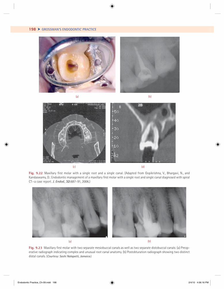
Fig. 9.22 Maxillary fi rst molar with a single root and a single canal. (Adapted from Gopikrishna, V., Bhargavi, N., and Kandaswamy, D.: Endodontic management of a maxillary fi rst molar with a single root and single canal diagnosed with spiral CT—a case report. J. Endod., 32:687–91, 2006.)
(a) (b)
Fig. 9.23 Maxillary fi rst molar with two separate mesiobuccal canals as well as two separate distobuccal canals: (a) Preop-erative radiograph indicating complex and unusual root canal anatomy. (b) Postobturation radiograph showing two distinct distal canals. (Courtesy: Sashi Nalapatti, Jamaica.)
Endodontic Practice_Ch-09.indd 198Endodontic Practice_Ch-09.indd 198 2/4/10 4:06:16 PM2/4/10 4:06:16 PM
CHAPTER 9 Anatomy of Pulp Cavity and Its Access Opening Ö 211
In cross-section, all three canals are ovoid in the cervical and middle thirds and round in the apical third. Two canals present in the distal root are usu-ally round in cross-section from the cervical third to the apical third.
Clinical Signifi canceThe mesial root of the mandibular fi rst molar is in close proximity to the buccal cortical plate, whereas the distal root is centrally located. The apex of the roots of mandibular fi rst molars may be close to the mandibular canal, or they may be at some distance
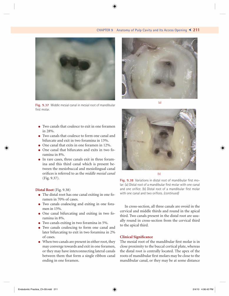
Fig. 9.37 Middle mesial canal in mesial root of mandibular fi rst molar.
(a)
(b)
Fig. 9.38 Variations in distal root of mandibular fi rst mo-lar: (a) Distal root of a mandibular fi rst molar with one canal and one orifi ce. (b) Distal root of a mandibular fi rst molar with one canal and two orifi ces. (continued)
! Two canals that coalesce to exit in one foramen in 28%.
! Two canals that coalesce to form one canal and bifurcate and exit in two foramina in 13%.
! One canal that exits in one foramen in 12%.! One canal that bifurcates and exits in two fo-
ramina in 8%.! In rare cases, three canals exit in three foram-
ina and this third canal which is present be-tween the mesiobuccal and mesiolingual canal orifi ces is referred to as the middle mesial canal(Fig. 9.37).
Distal Root (Fig. 9.38)! The distal root has one canal exiting in one fo-
ramen in 70% of cases.! Two canals coalescing and exiting in one fora-
men in 15%.! One canal bifurcating and exiting in two fo-
ramina in 8%.! Two canals exiting in two foramina in 5%.! Two canals coalescing to form one canal and
later bifurcating to exit in two foramina in 2% of cases.
! When two canals are present in either root, they may converge towards and exit in one foramen, or they may have interconnecting lateral canals between them that form a single ribbon canal ending in one foramen.
Endodontic Practice_Ch-09.indd 211Endodontic Practice_Ch-09.indd 211 2/4/10 4:06:42 PM2/4/10 4:06:42 PM
CHAPTER 9 Anatomy of Pulp Cavity and Its Access Opening Ö 215
(b)(i)
(ii) (iii) (iv)
(v) (vi) (vii)
(c)(i)
(ii) (iii) (iv)
(v) (vi) (vii)
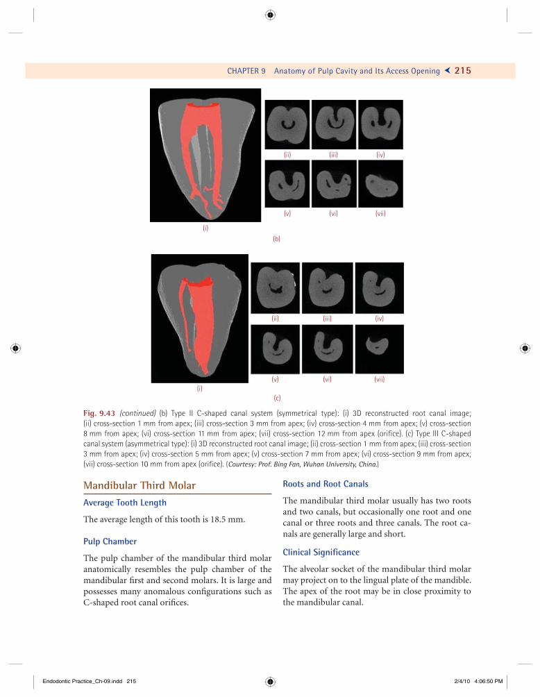
Fig. 9.43 (continued) (b) Type II C-shaped canal system (symmetrical type): (i) 3D reconstructed root canal image; (ii) cross-section 1 mm from apex; (iii) cross-section 3 mm from apex; (iv) cross-section 4 mm from apex; (v) cross-section 8 mm from apex; (vi) cross-section 11 mm from apex; (vii) cross-section 12 mm from apex (orifi ce). (c) Type III C-shaped canal system (asymmetrical type): (i) 3D reconstructed root canal image; (ii) cross-section 1 mm from apex; (iii) cross-section3 mm from apex; (iv) cross-section 5 mm from apex; (v) cross-section 7 mm from apex; (vi) cross-section 9 mm from apex; (vii) cross-section 10 mm from apex (orifi ce). (Courtesy: Prof. Bing Fan, Wuhan University, China.)
Mandibular Third MolarAverage Tooth Length
The average length of this tooth is 18.5 mm.
Pulp Chamber
The pulp chamber of the mandibular third molar anatomically resembles the pulp chamber of the mandibular fi rst and second molars. It is large and possesses many anomalous confi gurations such as C-shaped root canal orifi ces.
Roots and Root Canals
The mandibular third molar usually has two roots and two canals, but occasionally one root and one canal or three roots and three canals. The root ca-nals are generally large and short.
Clinical Signifi cance
The alveolar socket of the mandibular third molar may project on to the lingual plate of the mandible. The apex of the root may be in close proximity to the mandibular canal.
Endodontic Practice_Ch-09.indd 215Endodontic Practice_Ch-09.indd 215 2/4/10 4:06:50 PM2/4/10 4:06:50 PM
What we remove from the pulp space, is far more important than what we replace it with...
221
E ndodontic treatment can be divided into three main phases: (i) proper access prepa-ration into the pulp space, (ii) cleaning and
shaping of the root canal, and (iii) obturation. The initial step for cleaning and shaping the root canal is proper access to the chamber that leads to straight-line penetration of the root canal orifi ces. The next step is exploration of the root canal, extirpation of the remaining pulp tissue or gross debridement of the necrotic tissue, and verifi cation of the instru-ment working length. This step is followed by proper instrumentation, irrigation and debridement, and disinfection/sterilization of the root canal. Obtura-tion usually completes the procedure.
The importance of adequate canal cleaning and shaping, rather than reliance on antiseptics, cannot be overemphasized. Histologic examination of pulp-less teeth in which root canal therapy has failed often shows that the canals were not completely cleaned. Obturation of an improperly cleaned canal would still lead to an endodontic failure (Fig. 10.1).
Cleaning and Shaping of Radicular Space
Cleaning and shaping of the root canal comprises the most important phase of endodontic treatment. Other aspects of treatment cannot be neglected, however, because they are all inter-related and con-tribute to the success of endodontic therapy.
The objectives of cleaning and shaping are two-fold:
1. To debride and disinfect/sterilize the root canal system
2. To shape/contour the root canal walls, for the purpose of sealing the root canal completely with a condensed, inert fi lling material
To help achieve these objectives, each individual root canal should be examined radiographically and explored with endodontic instruments. The examination should include an assessment of canal length, shape, size, curvature, entrance orifi ce, loca-tion of foramina, canal ramifi cations, and presence of calcifi cations or obstructions.
According to Schilder, the cleaning and shaping of a root canal should fulfi ll the following mechani-cal objectives:
! Should have a continuous tapering, coni-cal shape, with the narrowest cross-sectional diameter apically and the widest diameter coronally.
! The walls should taper evenly towards the apex and should be confl uent with the access cavity.
! To give the prepared root canal the “quality of fl ow,” i.e, a shape that permits plasticized gutta-percha to fl ow against the walls without impedance
! Should keep the apical foramen as small as practical.
CHAPTER
10Preparation of the Radicular
Space: Instruments and Techniques
Endodontic Practice_Ch-10.indd 221Endodontic Practice_Ch-10.indd 221 2/4/10 4:32:14 PM2/4/10 4:32:14 PM
234 Ü GROSSMAN’S ENDODONTIC PRACTICE
! Pitch. Pitch is the distance from one cutting edge to the next cutting edge. A fi le with short pitch will have more spirals than a fi le with a longer pitch.
! Rake angle. On perpendicular sectioning of a fi le, the angle which the leading edge forms with the radius of the fi le is known as the rake angle. If it forms an obtuse angle then the rake angle is considered to be positive. An acute an-gle is termed negative rake angle.
! Helix angle. It is the angle the cutting edge forms with the long axis of the tooth.
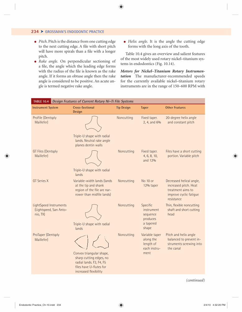
Table 10.4 gives an overview and salient features of the most widely used rotary nickel–titanium sys-tems in endodontics (Fig. 10.14).
Motors for Nickel–Titanium Rotary Instrumen-tation The manufacturer-recommended speeds for the currently available nickel–titanium rotary instruments are in the range of 150–600 RPM with
TABLE 10.4 Design Features of Current Rotary Ni–Ti File Systems
Instrument System Cross-Sectional Design
Tip Design Taper Other Features
ProFile (Dentsply Maillefer)
Triple-U shape with radial lands. Neutral rake angle planes dentin walls
Noncutting Fixed taper. 2, 4, and 6%
20-degree helix angle and constant pitch
GT Files (Dentsply Maillefer)
Triple-U shape with radial lands
Noncutting Fixed taper. 4, 6, 8, 10,
and 12%
Files have a short cutting portion. Variable pitch
GT Series X Variable-width lands (lands at the tip and shank
region of the fi le are nar-rower than midfi le lands)
Noncutting No 10 or 12% taper
Decreased helical angle, increased pitch. Heat
treatment aims to improve cyclic fatigue resistance
LightSpeed Instruments (Lightspeed, San Anto-
nio, TX)
Triple-U shape with radial lands
Noncutting Specifi c instrument
sequenceproduces a tapered shape
Thin, fl exible noncutting shaft and short cutting
head
ProTaper (Dentsply Maillefer)
Convex triangular shape, sharp cutting edges, no
radial lands. F3, F4, F5 fi les have U-fl utes for increased fl exibility
Noncutting Variable taper along the
length of each instru-ment
Pitch and helix angle balanced to prevent in-
struments screwing into the canal
(continued)
Endodontic Practice_Ch-10.indd 234Endodontic Practice_Ch-10.indd 234 2/4/10 4:32:20 PM2/4/10 4:32:20 PM
Medicine heals doubts as well as diseases.
–Karl Marx
263
T he cleaning and shaping of the root canal constitutes one of the most important phases of endodontic therapy. Instrumen-
tation of the canal reduces the microbial content of the root canal to a great extent. However, the root canal anatomy provides areas in which bacteria can persist and thrive (Figs. 11.1 and 11.2).
Individual microorganisms proliferate to form populations which occur as microcolonies. The root canal system with necrotic pulp provides space for microbial colonization and affords the microorganism a moist, warm, nutritious,
and anaerobic environment, protected from host defenses due to lack of microcirculation in the necrotic pulp tissue. A wide variety of microorganisms have been identified to invade depths up to 2000 µm into the dentinal tubules (Table 11.1).
It has become increasingly clear that the largest proportion of endodontic diseases of both pulp and periradicular tissues is due to the presence of microorganisms, though mechanical and manual challenges of root canal debridement remain. Therefore, the success of treatment depends upon
CHAPTER
11Irrigants and Intracanal Medicaments
BA
BABA
DD
BA
DT
BA
DT
RCRCRCRC RCRC
2 µm
10 µm
Fig. 11.1 Bacteria (BA) in dentinal tubules (DT) in the apical part of human root dentin (D). The bacteria invade the tubules from the infected root canal (RC). The presence of dividing forms (inset) is a clear sign of vitality of the microorganisms at the time of fi xation (magnifi cations: !2480; inset !9600). (Courtesy: P.N.R. Nair, Switzerland.)
Endodontic Practice_Ch-11.indd 263Endodontic Practice_Ch-11.indd 263 2/4/10 5:19:53 PM2/4/10 5:19:53 PM
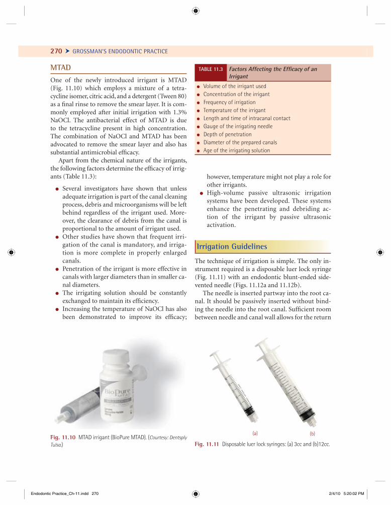
270 Ü GROSSMAN’S ENDODONTIC PRACTICE
MTADOne of the newly introduced irrigant is MTAD (Fig. 11.10) which employs a mixture of a tetra-cycline isomer, citric acid, and a detergent (Tween 80)as a fi nal rinse to remove the smear layer. It is com-monly employed after initial irrigation with 1.3% NaOCl. The antibacterial effect of MTAD is due to the tetracycline present in high concentration. The combination of NaOCl and MTAD has been advocated to remove the smear layer and also has substantial antimicrobial effi cacy.
Apart from the chemical nature of the irrigants, the following factors determine the effi cacy of irrig-ants (Table 11.3):
! Several investigators have shown that unless adequate irrigation is part of the canal cleaning process, debris and microorganisms will be left behind regardless of the irrigant used. More-over, the clearance of debris from the canal is proportional to the amount of irrigant used.
! Other studies have shown that frequent irri-gation of the canal is mandatory, and irriga-tion is more complete in properly enlarged canals.
! Penetration of the irrigant is more effective in canals with larger diameters than in smaller ca-nal diameters.
! The irrigating solution should be constantly exchanged to maintain its effi ciency.
! Increasing the temperature of NaOCl has also been demonstrated to improve its effi cacy;
however, temperature might not play a role for other irrigants.
! High-volume passive ultrasonic irrigation systems have been developed. These systems enhance the penetrating and debriding ac-tion of the irrigant by passive ultrasonic activation.
Irrigation Guidelines
The technique of irrigation is simple. The only in-strument required is a disposable luer lock syringe (Fig. 11.11) with an endodontic blunt-ended side-vented needle (Figs. 11.12a and 11.12b).
The needle is inserted partway into the root ca-nal. It should be passively inserted without bind-ing the needle into the root canal. Suffi cient room between needle and canal wall allows for the return
! Volume of the irrigant used ! Concentration of the irrigant! Frequency of irrigation! Temperature of the irrigant! Length and time of intracanal contact! Gauge of the irrigating needle ! Depth of penetration ! Diameter of the prepared canals! Age of the irrigating solution
TABLE 11.3 Factors Affecting the Effi cacy of an Irrigant
(a) (b)
Fig. 11.11 Disposable luer lock syringes: (a) 3cc and (b)12cc.Fig. 11.10 MTAD irrigant (BioPure MTAD). (Courtesy: Dentsply Tulsa.)
Endodontic Practice_Ch-11.indd 270Endodontic Practice_Ch-11.indd 270 2/4/10 5:20:02 PM2/4/10 5:20:02 PM
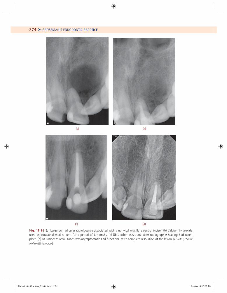
274 Ü GROSSMAN’S ENDODONTIC PRACTICE
(c)
(a) (b)
(d)
Fig. 11.16 (a) Large periradicular radiolucency associated with a nonvital maxillary central incisor. (b) Calcium hydroxide used as intracanal medicament for a period of 6 months. (c) Obturation was done after radiographic healing had taken place. (d) At 6 months recall tooth was asymptomatic and functional with complete resolution of the lesion. (Courtesy: Sashi Nalapatti, Jamaica.)
Endodontic Practice_Ch-11.indd 274Endodontic Practice_Ch-11.indd 274 2/4/10 5:20:05 PM2/4/10 5:20:05 PM
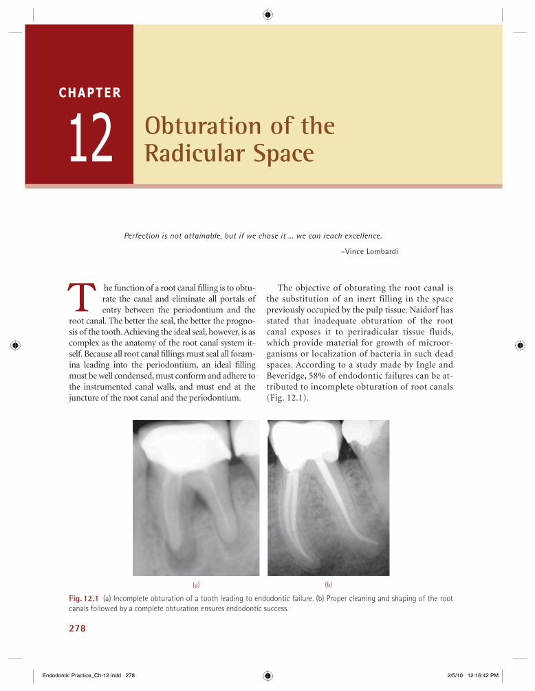
278
Fig. 12.1 (a) Incomplete obturation of a tooth leading to endodontic failure. (b) Proper cleaning and shaping of the root canals followed by a complete obturation ensures endodontic success.
(a) (b)
Obturation of the Radicular Space
Perfection is not attainable, but if we chase it ... we can reach excellence.
–Vince Lombardi
T he function of a root canal fi lling is to obtu-rate the canal and eliminate all portals of entry between the periodontium and the
root canal. The better the seal, the better the progno-sis of the tooth. Achieving the ideal seal, however, is as complex as the anatomy of the root canal system it-self. Because all root canal fi llings must seal all foram-ina leading into the periodontium, an ideal fi lling must be well condensed, must conform and adhere to the instrumented canal walls, and must end at the juncture of the root canal and the periodontium.
The objective of obturating the root canal is the substitution of an inert filling in the space previously occupied by the pulp tissue. Naidorf has stated that inadequate obturation of the root canal exposes it to periradicular tissue fluids, which provide material for growth of microor-ganisms or localization of bacteria in such dead spaces. According to a study made by Ingle and Beveridge, 58% of endodontic failures can be at-tributed to incomplete obturation of root canals (Fig. 12.1).
12CHAPTER
Endodontic Practice_Ch-12.indd 278Endodontic Practice_Ch-12.indd 278 2/5/10 12:16:42 PM2/5/10 12:16:42 PM
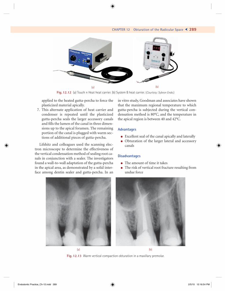
CHAPTER 12 Obturation of the Radicular Space Ö 289
applied to the heated gutta-percha to force the plasticized material apically.
7. This alternate application of heat carrier and condenser is repeated until the plasticized gutta-percha seals the larger accessory canals and fi lls the lumen of the canal in three dimen-sions up to the apical foramen. The remaining portion of the canal is plugged with warm sec-tions of additional pieces of gutta-percha.
Lifshitz and colleagues used the scanning elec-tron microscope to determine the effectiveness of the vertical condensation method of sealing root ca-nals in conjunction with a sealer. The investigators found a wall-to-wall adaptation of the gutta-percha in the apical area, as demonstrated by a solid inter-face among dentin sealer and gutta-percha. In an
in vitro study, Goodman and associates have shown that the maximum regional temperature to which gutta-percha is subjected during the vertical con-densation method is 80°C, and the temperature in the apical region is between 40 and 42°C.
Advantages
! Excellent seal of the canal apically and laterally! Obturation of the larger lateral and accessory
canals
Disadvantages
! The amount of time it takes! The risk of vertical root fracture resulting from
undue force
(a) (b)
Fig. 12.12 (a) Touch n Heat heat carrier. (b) System B heat carrier. (Courtesy: Sybron Endo.)
Fig. 12.13 Warm vertical compaction obturation in a maxillary premolar.
(a) (b)
Endodontic Practice_Ch-12.indd 289Endodontic Practice_Ch-12.indd 289 2/5/10 12:16:54 PM2/5/10 12:16:54 PM
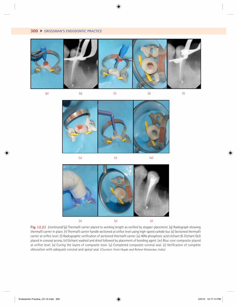
300 Ü GROSSMAN’S ENDODONTIC PRACTICE
Fig. 12.23 (continued) (p) Thermafi l carrier placed to working length as verifi ed by stopper placement. (q) Radiograph showing thermafi l carrier in place. (r) Thermafi l carrier handle sectioned at orifi ce level using high-speed carbide bur. (s) Sectioned thermafi l carrier at orifi ce level. (t) Radiographic verifi cation of sectioned thermafi l carrier. (u) 40% phosphoric acid etchant (K-Etchant Gel) placed in coronal access. (v) Etchant washed and dried followed by placement of bonding agent. (w) Blue core composite placed at orifi ce level. (x) Curing the layers of composite resin. (y) Completed composite coronal seal. (z) Verifi cation of complete obturation with adequate coronal and apical seal. (Courtest: Vivek Hegde and Roheet Khatavkar, India.)
(q) (r) (s) (t)(p)
(v)(u) (w)
(y)(x) (z)
Endodontic Practice_Ch-12.indd 300Endodontic Practice_Ch-12.indd 300 2/5/10 12:17:14 PM2/5/10 12:17:14 PM
310
Vital Pulp Therapy, Pulpotomy, and Apexifi cation
Never must the physician say the disease is incurable. By that admission he denies God, our Creator; He doubts Nature with her profuseness of hidden powers and mysteries.
—Paracelsus
T he unaffected, exposed vital pulp possesses an inherent capacity for healing through cell reorganization and bridge formation
when a proper biological seal is provided and main-tained against leakage of oral contaminations. Throughout the life of a tooth, vital pulp tissue con-tributes to the production of secondary dentin, peritubular dentin, and reparative dentin in re-sponse to biological and pathological stimuli. The pulp tissue with its circulation extending into the tubular dentin keeps the dentin moist, which in turn ensures that the dentin maintains its resilience and toughness (Fig. 13.1). The earliest account of vital pulp therapy was in 1756, when Phillip Pfaff packed a small piece of gold over an exposed vital pulp to promote healing.
By 1922, in the light of his experiences with similar antiseptic treatments, Rebel summarized his thoughts in the expression, “the exposed pulp is a doomed organ.” He concluded that recovery of the vital unaffected pulp when exposed to the oral environment was invariably doomed and that one must consider it as a lost organ. Despite Rebel’s much-quoted statements, the realization gradually evolved that the dental pulp did at times possess defi nite powers of recuperation and repair. Major advances in the practice of vital pulp therapy have
been made and the emphasis has shifted from the “doomed organ” concept of an exposed pulp to one of “predictable repair and recovery.”
Pulpal Infl ammation and Its Sequelae
The tradional theory which explained pulpal in-fl ammation and its sequelae was known as stran-gulation theory. This theory is no longer accepted and a neular current theory explains the sequelae of pulpal infl ammation.
Strangulation TheoryIn the accepted view on pulpal infl ammation, maxi-mum signifi cance was diverted and focused on the infl uence of anatomical environment on the pulp. The assumption was that with local infl ammation of the pulp there is an increased blood fl ow to the infl amed area. Infl ammation induces vasodilatation and increased capillary pressure and permeability. This results in an increased fi ltration from the cap-illaries into the tissues leading to increased tissue pressure. The thin vessel walls are compressed as the increasing pressure outside the vessels leads to a de-crease in blood fl ow as well as an increase in venous pressure. This results in a vicious cycle. The increase
13CHAPTER
Endodontic Practice_Ch-13.indd 310Endodontic Practice_Ch-13.indd 310 2/5/10 6:24:58 PM2/5/10 6:24:58 PM
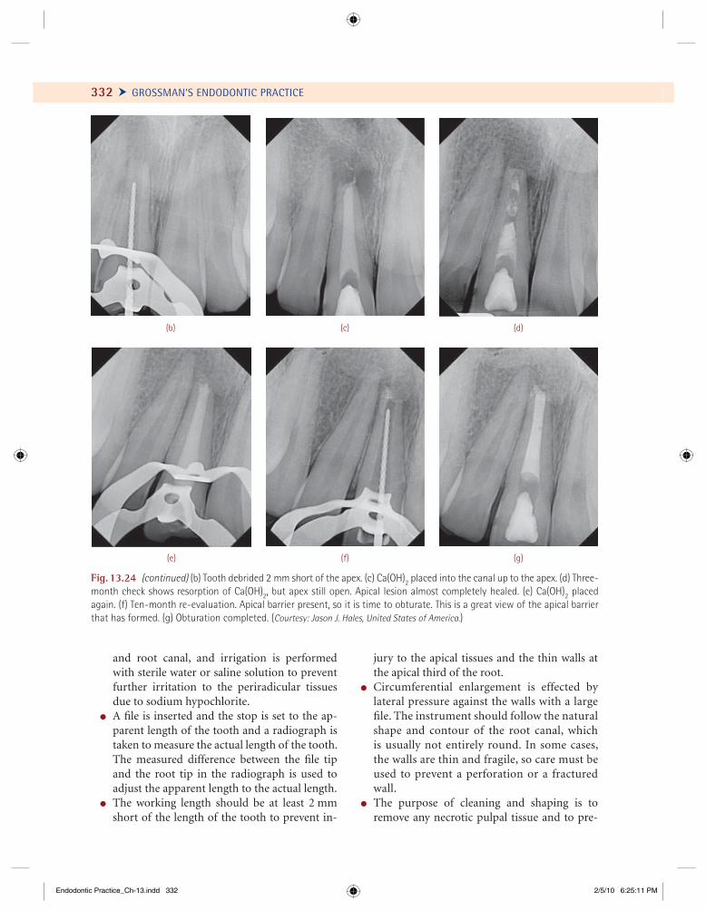
332 Ü GROSSMAN’S ENDODONTIC PRACTICE
and root canal, and irrigation is performed with sterile water or saline solution to prevent further irritation to the periradicular tissues due to sodium hypochlorite.
! A fi le is inserted and the stop is set to the ap-parent length of the tooth and a radiograph is taken to measure the actual length of the tooth. The measured difference between the fi le tip and the root tip in the radiograph is used to adjust the apparent length to the actual length.
! The working length should be at least 2 mm short of the length of the tooth to prevent in-
jury to the apical tissues and the thin walls at the apical third of the root.
! Circumferential enlargement is effected by lateral pressure against the walls with a large fi le. The instrument should follow the natural shape and contour of the root canal, which is usually not entirely round. In some cases, the walls are thin and fragile, so care must be used to prevent a perforation or a fractured wall.
! The purpose of cleaning and shaping is to remove any necrotic pulpal tissue and to pre-
(b)
(g)(e) (f)
(c) (d)
Fig. 13.24 (continued) (b) Tooth debrided 2 mm short of the apex. (c) Ca(OH)2 placed into the canal up to the apex. (d) Three-month check shows resorption of Ca(OH)2, but apex still open. Apical lesion almost completely healed. (e) Ca(OH)2 placed again. (f) Ten-month re-evaluation. Apical barrier present, so it is time to obturate. This is a great view of the apical barrierthat has formed. (g) Obturation completed. (Courtesy: Jason J. Hales, United States of America.)
Endodontic Practice_Ch-13.indd 332Endodontic Practice_Ch-13.indd 332 2/5/10 6:25:11 PM2/5/10 6:25:11 PM

CHAPTER 13 Vital Pulp Therapy, Pulpotomy, and Apexification Ö 333
(a)
Fig. 13.25 (a) Central incisor with open apex. (b) Intracanal Ca(OH)2 placed and GIC entrance fi lling given. (c) Six-month follow-up. (d) Twenty-month follow-up. (e) Calcium hydroxide removed and evidence of calicifi c barrier. (f) Calcifi c barrier seen through the microscope. (g) Thermoplasticized backfi ll obturation done. (h) View through the microscope. (i) Two-year recall shows good healing. (Courtesy: Siju Jacob, India.)
(d)
(g) (h) (i)
(e) (f)
(b) (c)
Endodontic Practice_Ch-13.indd 333Endodontic Practice_Ch-13.indd 333 2/5/10 6:25:12 PM2/5/10 6:25:12 PM
342
Bleaching of Discolored Teeth
Esthetics is an important factor in a patient’s decision to undergo endodontic treatment. A frequent question is, “Will my tooth turn
black?” The usual response is a “qualifi ed no,” with the explanation that modern treatment and proce-dures are designed to avoid crown staining and tooth discoloration. Nevertheless, teeth can and do discolor, sometimes before endodontic treatment and sometimes afterward, in spite of all precautions taken to prevent color changes. When teeth discolor, bleaching should be considered as a means of re-storing tooth esthetics.
The normal color of primary teeth is bluish white. The color of permanent teeth is grayish yellow, grayish white, or yellowish white. The color of teeth is determined by the translucency and thickness of the enamel, the thickness and color of the underlying dentin, and the color of the pulp. Alterations in the color may be physi-ologic or pathologic and endogenous or exog-enous in nature.
With age, the enamel becomes thinner because of abrasion and erosion, and the dentin becomes thicker because of the deposition of secondary and reparative dentin, which produce color changes in teeth during one’s life. Teeth of elderly persons are usually more yellow or grayish yellow than those of younger persons.
We live only to discover beauty. All else is a form of waiting.
—Kahlil Gibran
Classifi cation of Tooth Discoloration
Tooth discoloration can be classifi ed as either extrinsic or intrinsic.
Extrinsic DiscolorationsExtrinsic discolorations are found on the outer surface of teeth and are usually of local origin, such as to-bacco stains. Some extrinsic discoloration, such as the green discoloration associated with the Nasmyth’s membrane in children, and tea and tobacco stains (Fig. 14.1), can be removed by scaling and polishing during tooth prophylaxis. Extrinsic discolorations are stains that get bound to the tooth surface and for this reason can be easily removed. The stains deposited on the tooth surface are a result of attractive forces which are long-range interactions such as van der Waals and electrostatic forces and short-range interactions such as hydration forces, hydrophobic interactions, and hy-drogen bonds. Other types of extrinsic discoloration, such as silver nitrate stains, are almost impossible to eliminate without grinding because the stains pen-etrate the surface of the crowns and are diffi cult to remove by chemical means alone.
In the past, extrinsic discoloration was classi-fi ed as metallic or nonmetallic stains. The problem with this classifi cation is that it does not explain the
14CHAPTER
Endodontic Practice_Ch-14.indd 342Endodontic Practice_Ch-14.indd 342 2/5/10 5:02:51 PM2/5/10 5:02:51 PM
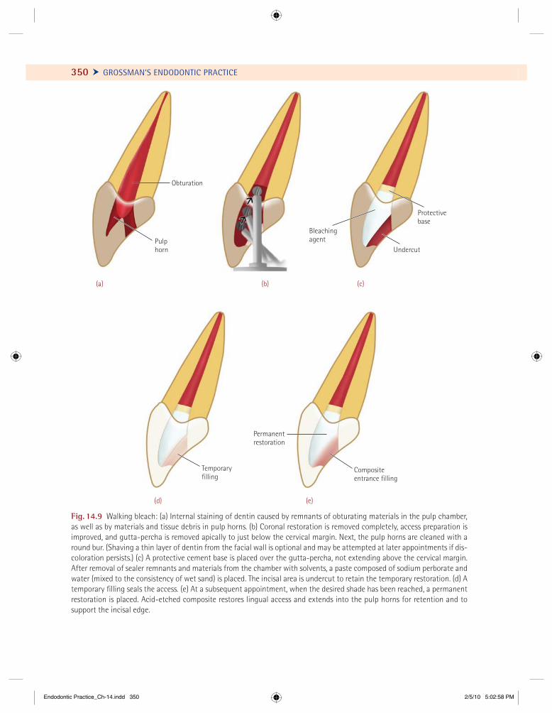
350 Ü GROSSMAN’S ENDODONTIC PRACTICE
Bleachingagent
Undercut
Protectivebase
Temporaryfilling
Compositeentrance filling
Permanentrestoration
Obturation
Pulphorn
(a)
(d) (e)
(b) (c)
Fig. 14.9 Walking bleach: (a) Internal staining of dentin caused by remnants of obturating materials in the pulp chamber, as well as by materials and tissue debris in pulp horns. (b) Coronal restoration is removed completely, access preparation is improved, and gutta-percha is removed apically to just below the cervical margin. Next, the pulp horns are cleaned with a round bur. (Shaving a thin layer of dentin from the facial wall is optional and may be attempted at later appointments if dis-coloration persists.) (c) A protective cement base is placed over the gutta-percha, not extending above the cervical margin. After removal of sealer remnants and materials from the chamber with solvents, a paste composed of sodium perborate and water (mixed to the consistency of wet sand) is placed. The incisal area is undercut to retain the temporary restoration. (d) Atemporary fi lling seals the access. (e) At a subsequent appointment, when the desired shade has been reached, a permanent restoration is placed. Acid-etched composite restores lingual access and extends into the pulp horns for retention and to support the incisal edge.
Endodontic Practice_Ch-14.indd 350Endodontic Practice_Ch-14.indd 350 2/5/10 5:02:58 PM2/5/10 5:02:58 PM

CHAPTER 14 Bleaching of Discolored Teeth Ö 359
Tetracycline Discoloration
Classifi cation (Jordon and Boskman, 1984)
! First degree. Light yellow to light gray staining without banding
! Second degree. Darker and more extensive yel-low or gray staining without banding
(a)
(b)
(c)
Fig. 14.15 (a) Enamel fl uorosis. Isolated dark-brown stains pose a problem for tooth-veneering procedures. (b) Appear-ance following fi rst sitting of microabrasion using modifi ed McInnes solution and fi ne grit abrasive disks. (c) Microabra-sion was repeated after 2 weeks. Note the acceptable light-ening of the stains. Final esthetic appearance can be achieved by composite resin veneering. (Courtesy: Anbu, Inda.)
! Third degree. Severe staining characterized with dark gray or blue discoloration with banding
Minocycline has the ability to affect permanent dentition even in adults due to its ability to form complexes with calcium in dentin. It is usually seen with long-term use for the treatment of acne.
Destaining of the yellow color is most success-ful, whereas brownish teeth are least successfully bleached.
The use of 30% hydrogen peroxide and a ther-mostatically controlled heat source for bleaching tetracycline-stained teeth has been described. Un-fortunately, the decoloration is only superfi cial and does not affect the stained dentin. Another method of bleaching tetracycline stain has been described. In this method, the pulps of the teeth are intention-ally extirpated, the root canals are cleaned, shaped and obturated, and the teeth are internally bleached as previously described. The result in humans, as well as in dogs, has been successful. We believe that labial veneers with composite resins or even por-celain-veneer full-crown restorations are indicated instead of intentional devitalization of a tooth with a normal pulp.
The application of heat to the tooth during bleaching procedures may cause some transitory re-action in the pulp. Although in one study the appli-cation of heat in the range of 115–124°F produced a slight superfi cial infl ammation, in another study, temperatures of 114°F had no effect on the pulp.In this study, the use of hydrogen peroxide caused hemorrhage, infl ammation, and destruction of the odontoblast layer of the pulp, but the reaction was reversible and healing occurred within 60 days.
ManagementTeeth discolored by tetracycline may also be bleached to some extent with hydrogen peroxide, but the bleaching effect leaves something to be desired, with regard to both discoloration and permanency, because the chemical cannot reach the real cause of the discoloration, which is the incorporation of tet-racycline into the dentin. The degree of staining de-pends on the stage of tooth development at the time when medication is begun; the greater the amount of crown developed, the less severe the stain, and vice versa.
Endodontic Practice_Ch-14.indd 359Endodontic Practice_Ch-14.indd 359 2/5/10 5:03:18 PM2/5/10 5:03:18 PM
Healing is a matter of time, but it is sometimes also a matter of opportunity.
—Hippocrates
361
T rauma of the oral and maxillofacial region occurs frequently and comprises 5% of all injuries for which people seek treatment.
Among all facial injuries, dental injuries are the most common, of which crown fractures and luxations occur most frequently. Trauma to the teeth may re-sult either in injury of the pulp, with or without damage to the crown or root, or in displacement of the tooth from its socket. When the crown or root is fractured, the pulp may recover and survive the in-jury, it may succumb immediately, or it may undergo progressive degeneration and ultimately die.
Causes and Incidence of Dental Injuries
Traumatic injuries to the teeth can occur at any age. Young children learning to walk or falling from a chair are subject to anterior tooth injuries. Frequently, child abuse results in facial and dental trauma. Sports ac-cidents and fi ghts affect teenagers and young adults, whereas automobile accidents affect all age groups. As many dental accidents are sports related, every precau-tion should be taken to protect the teeth of children from such accidents by using educational programs in addition to mouth guards. The common causes of traumatic injuries to the teeth include the following:
! Direct and indirect trauma! Sports accidents
! Automobile accidents! Fights and assaults! Domestic violence! Inappropriate use of teeth! Biting hard items
The incidence of fractures is about 5%; Ellis reports an incidence of 4.2%, and Grundy, 5.1%. Boys have about two to three times as many frac-tured teeth as girls. Bakland reported that children 8–12 years of age are most prone to dental accidents. The 2–4 years of age is the other age group which has a high incidence of dental traumatic injuries. The three key predisposing factors for these kinds of injuries are as follows:
! Increased overjet of the teeth! Protrusion of the maxillary anterior teeth! Insuffi cient lip closure
Fractures of Teeth
Fractures of the crowns of teeth are generally di-agonal, involving a corner of the tooth, frequently the mesial. Traumatic injuries that do not cause fracture of the crown or root are just as often re-sponsible for pulp injury as those in which the crown or root is fractured. In injuries that do not fracture the crown or root, the impact of the blow is transmitted head-on to the pulp, which receives
CHAPTER
15Treatment of Traumatized Teeth
Endodontic Practice_Ch-15.indd 361Endodontic Practice_Ch-15.indd 361 2/5/10 7:14:23 PM2/5/10 7:14:23 PM
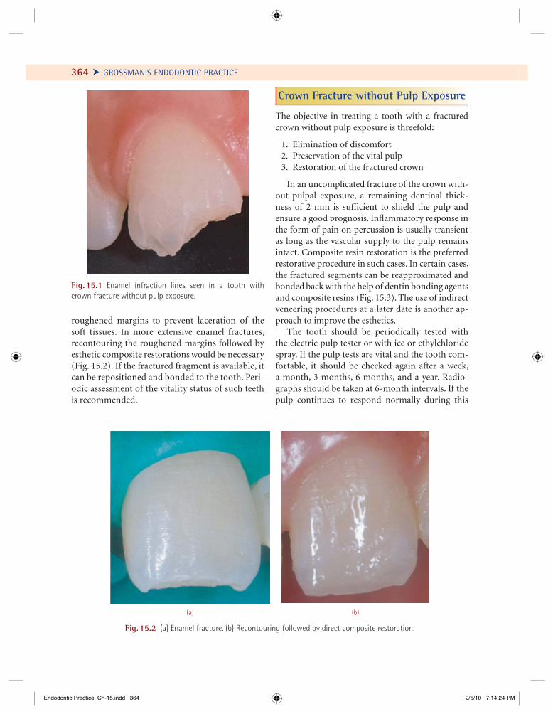
364 Ü GROSSMAN’S ENDODONTIC PRACTICE
Fig. 15.1 Enamel infraction lines seen in a tooth with crown fracture without pulp exposure.
(a) (b)
Fig. 15.2 (a) Enamel fracture. (b) Recontouring followed by direct composite restoration.
roughened margins to prevent laceration of the soft tissues. In more extensive enamel fractures, recontouring the roughened margins followed by esthetic composite restorations would be necessary (Fig. 15.2). If the fractured fragment is available, it can be repositioned and bonded to the tooth. Peri-odic assessment of the vitality status of such teeth is recommended.
Crown Fracture without Pulp Exposure
The objective in treating a tooth with a fractured crown without pulp exposure is threefold:
1. Elimination of discomfort 2. Preservation of the vital pulp 3. Restoration of the fractured crown
In an uncomplicated fracture of the crown with-out pulpal exposure, a remaining dentinal thick-ness of 2 mm is suffi cient to shield the pulp and ensure a good prognosis. Infl ammatory response in the form of pain on percussion is usually transient as long as the vascular supply to the pulp remains intact. Composite resin restoration is the preferred restorative procedure in such cases. In certain cases, the fractured segments can be reapproximated and bonded back with the help of dentin bonding agents and composite resins (Fig. 15.3). The use of indirect veneering procedures at a later date is another ap-proach to improve the esthetics.
The tooth should be periodically tested with the electric pulp tester or with ice or ethylchloride spray. If the pulp tests are vital and the tooth com-fortable, it should be checked again after a week, a month, 3 months, 6 months, and a year. Radio-graphs should be taken at 6-month intervals. If the pulp continues to respond normally during this
Endodontic Practice_Ch-15.indd 364Endodontic Practice_Ch-15.indd 364 2/5/10 7:14:24 PM2/5/10 7:14:24 PM
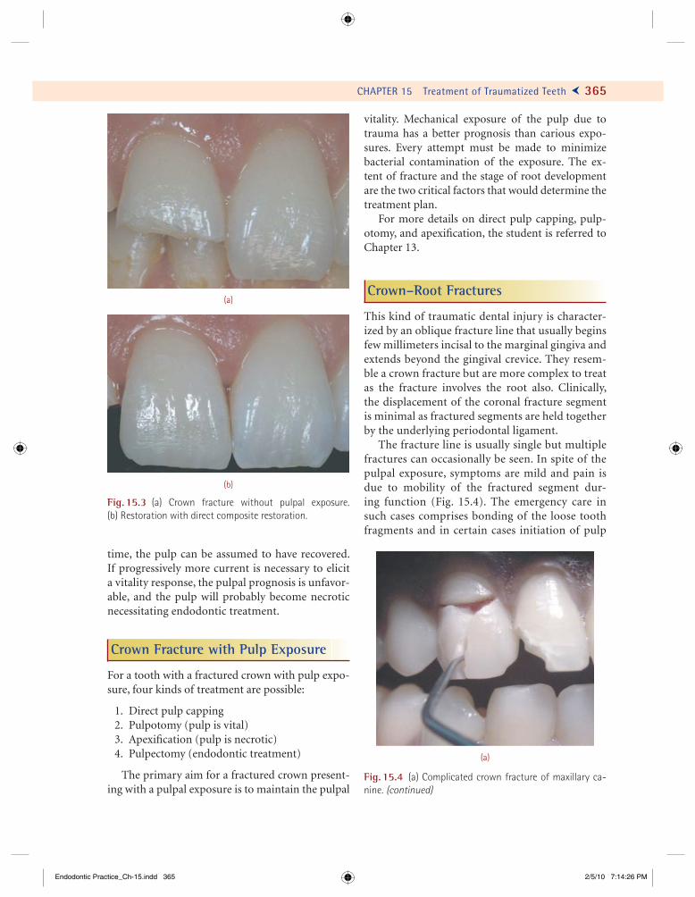
CHAPTER 15 Treatment of Traumatized Teeth Ö 365
time, the pulp can be assumed to have recovered. If progressively more current is necessary to elicit a vitality response, the pulpal prognosis is unfavor-able, and the pulp will probably become necrotic necessitating endodontic treatment.
Crown Fracture with Pulp Exposure
For a tooth with a fractured crown with pulp expo-sure, four kinds of treatment are possible:
1. Direct pulp capping 2. Pulpotomy (pulp is vital) 3. Apexifi cation (pulp is necrotic) 4. Pulpectomy (endodontic treatment)
The primary aim for a fractured crown present-ing with a pulpal exposure is to maintain the pulpal
vitality. Mechanical exposure of the pulp due to trauma has a better prognosis than carious expo-sures. Every attempt must be made to minimize bacterial contamination of the exposure. The ex-tent of fracture and the stage of root development are the two critical factors that would determine the treatment plan.
For more details on direct pulp capping, pulp-otomy, and apexifi cation, the student is referred to Chapter 13.
Crown–Root Fractures
This kind of traumatic dental injury is character-ized by an oblique fracture line that usually begins few millimeters incisal to the marginal gingiva and extends beyond the gingival crevice. They resem-ble a crown fracture but are more complex to treat as the fracture involves the root also. Clinically, the displacement of the coronal fracture segment is minimal as fractured segments are held together by the underlying periodontal ligament.
The fracture line is usually single but multiple fractures can occasionally be seen. In spite of the pulpal exposure, symptoms are mild and pain is due to mobility of the fractured segment dur-ing function (Fig. 15.4). The emergency care in such cases comprises bonding of the loose tooth fragments and in certain cases initiation of pulp
(a)
(b)
Fig. 15.3 (a) Crown fracture without pulpal exposure. (b) Restoration with direct composite restoration.
Fig. 15.4 (a) Complicated crown fracture of maxillary ca-nine. (continued)
(a)
Endodontic Practice_Ch-15.indd 365Endodontic Practice_Ch-15.indd 365 2/5/10 7:14:26 PM2/5/10 7:14:26 PM
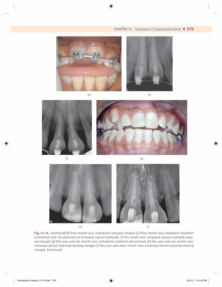
CHAPTER 15 Treatment of Traumatized Teeth Ö 379
(i)
(f)
(e)(d)
(g)
(h)
Fig. 15.16 (continued) (d) Three-month view: orthodontic extrusion initiated. (e) Three-month view: endodontic treatment commenced with the placement of intracanal calcium hydroxide. (f) Ten-month view: intracanal calcium hydroxide dress-ing changed. (g) One-year-and-one-month view: orthodontic treatment discontinued. (h) One-year-and-one-month view: intracanal calcium hydroxide dressing changed. (i) One-year-and-seven-month view: intracanal calcium hydroxide dressing changed. (continued)
Endodontic Practice_Ch-15.indd 379Endodontic Practice_Ch-15.indd 379 2/5/10 7:14:42 PM2/5/10 7:14:42 PM
390
CHAPTER
16 Endodontic Surgery
We are what we repeatedly do .... Excellence, then, is not an act ... but a habit.
—Aristotle
T he scope of endodontic surgery has ex- tended beyond root-end resection to in-clude other forms of periradicular surgery,
fi stulative surgery, corrective surgery, and inten-tional replantation. Root-end resection is still the most common form of periradicular surgery.
In general practice, the number of cases being indicated for root-end surgery has drastically re-duced over a period of time. This may be due to the fact that today the science of endodontics has a better understanding of the biological principles of cleaning and shaping. In the last few decades, endo-dontics is more of a biological science than mere chemomechanical debridement. There has been a tremendous improvement in the available materi-als and instruments for cleaning and shaping. With the present knowledge of internal anatomy of pulp space, microbiology, disinfection of the pulp space, and also with the introduction of rotary and micro-endodontic instrumentation, clinicians are better equipped to produce a more predictable and clean pulp space.
Most of the endodontic periradicular patholo-gies can be managed nonsurgically. There has been a gradual paradigm shift from surgical to nonsurgical treatment over the past few decades. However, the nonsurgical management may not be always successful. Even if nonsurgical treatment is unsuccessful, the current concept is to do an in-trospection of the quality of nonsurgical treatment
before selecting surgical intervention. If the initial endodontic treatment of a tooth is not satisfactory then one should attempt nonsurgical retreatment of that tooth fi rst.
The view that endodontic surgery is the last re-sort is based on past experience with instruments that were unsuitable. Also, the vision available at the surgical site was inadequate and incidence of postoperative complications was high. Fortu-nately, today the endodontist is equipped with bet-ter magnifi cation, illumination, and instruments. The present era of microsurgery is with surgical operating microscopes, ultrasonic tips for retro-preparation, low-speed high-torque motors, and miniaturized surgical instruments for root-end surgery. The introduction of dental surgical micro-scopes has also resulted in better understanding of apical third anatomy, better root-end surgical resection techniques, better patient interaction, and all this has resulted in better success rates. The success rate of surgical endodontics is high, about 73–99%, depending on the criteria used for evalu-ating success.
Objectives and Rationale for Surgery
! Curettage. Effective curettage of the pathologi-cally affected periradicular tissue which cannot be accessed in an orthograde approach. This
Endodontic Practice_Ch-16.indd 390Endodontic Practice_Ch-16.indd 390 2/6/10 2:23:23 PM2/6/10 2:23:23 PM

CHAPTER 16 Endodontic Surgery Ö 393
With DOMs, one can visualize the surgical fi eld and evaluate the surgical technique. The magnifi ca-tion of a microscope is determined by:
! Power of the eyepiece! Focal length of the binoculars! Magnifi cation change factor! Focal length of the objective lens
IlluminationIllumination with DOMs is coaxial with the line of sight. This means the light is focused between the eyes in such a fashion that you can look into the surgical site without seeing any shadow. Elimi-nation of shadow is made possible by using Gali-lean optics, which focuses at infi nity and sends parallel beams of light to each eye. With parallel light, the operator’s eyes are at rest and therefore lengthy operations can be performed without eye fatigue.
Magnifi cation and illumination with the DOMs have helped the endodontists to introduce micro-surgery into surgical endodontics. This, in turn, has helped the endodontists to minimize trauma and enhance the surgical results. The DOMs have benefi tted treatment protocols in periradicular surgery.
They have become a vital armamentarium in not only microsurgical endodontics, but they play a key role in enhancing the quality and precision of routine nonsurgical endodontic therapy (Figs. 16.3 and 16.4). The arrival of DOMs has also brought in revolutionary changes in instru-ments and instrumentation techniques. The in-troduction of ultrasonic instruments has made minimal and precise retropreparation feasible during root-end surgery. The introduction of
Fig. 16.3 Dental operating microscope.
(a)
(b)
Fig. 16.4 (a) A dental operating microscope (DOM)–cen-tered endodontic practice enhances clinical performance and outcomes. (b) Dental operating microscope with beam splitter for documentation. (Courtesy: Global Microscopes.)
Endodontic Practice_Ch-16.indd 393Endodontic Practice_Ch-16.indd 393 2/6/10 2:23:26 PM2/6/10 2:23:26 PM
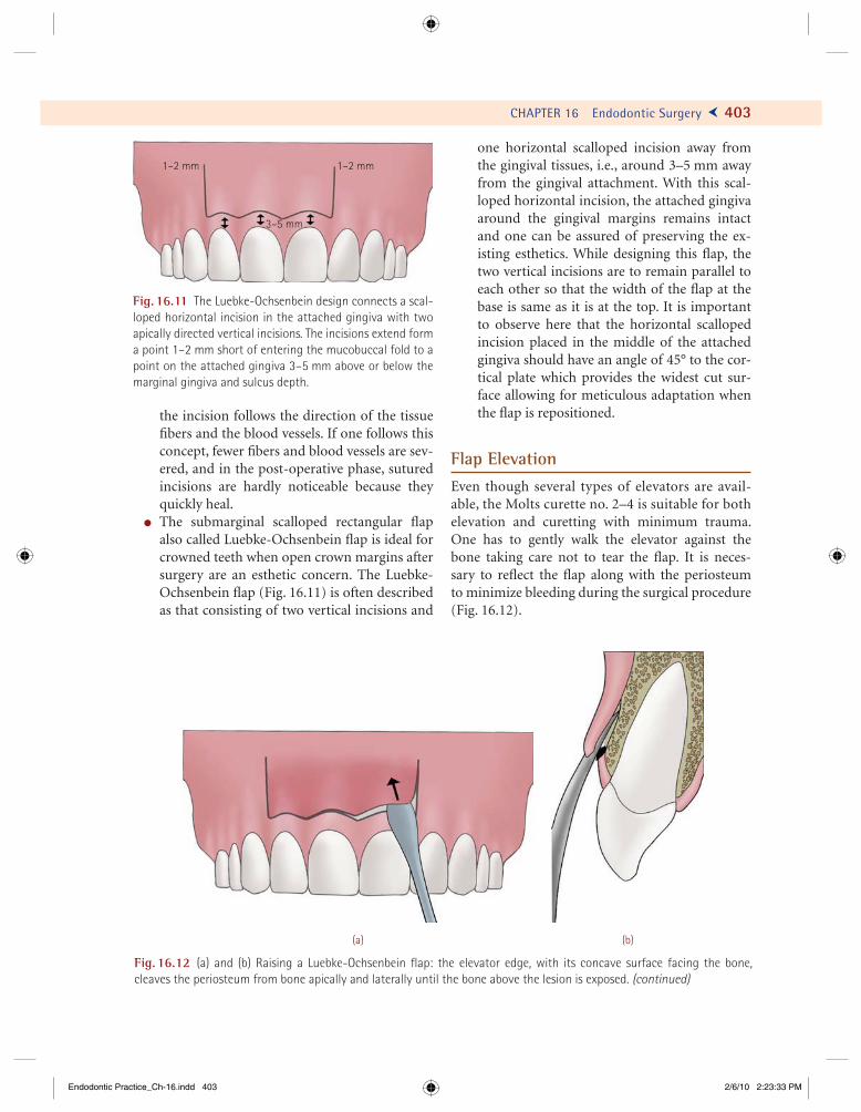
CHAPTER 16 Endodontic Surgery Ö 403
the incision follows the direction of the tissue fi bers and the blood vessels. If one follows this concept, fewer fi bers and blood vessels are sev-ered, and in the post-operative phase, sutured incisions are hardly noticeable because they quickly heal.
! The submarginal scalloped rectangular fl ap also called Luebke-Ochsenbein fl ap is ideal for crowned teeth when open crown margins after surgery are an esthetic concern. The Luebke-Ochsenbein fl ap (Fig. 16.11) is often described as that consisting of two vertical incisions and
one horizontal scalloped incision away from the gingival tissues, i.e., around 3–5 mm away from the gingival attachment. With this scal-loped horizontal incision, the attached gingiva around the gingival margins remains intact and one can be assured of preserving the ex-isting esthetics. While designing this fl ap, the two vertical incisions are to remain parallel to each other so that the width of the fl ap at the base is same as it is at the top. It is important to observe here that the horizontal scalloped incision placed in the middle of the attached gingiva should have an angle of 45° to the cor-tical plate which provides the widest cut sur-face allowing for meticulous adaptation when the fl ap is repositioned.
Flap ElevationEven though several types of elevators are avail-able, the Molts curette no. 2–4 is suitable for both elevation and curetting with minimum trauma. One has to gently walk the elevator against the bone taking care not to tear the fl ap. It is neces-sary to refl ect the fl ap along with the periosteum to minimize bleeding during the surgical procedure (Fig. 16.12).
Fig. 16.11 The Luebke-Ochsenbein design connects a scal-loped horizontal incision in the attached gingiva with two apically directed vertical incisions. The incisions extend form a point 1–2 mm short of entering the mucobuccal fold to a point on the attached gingiva 3–5 mm above or below the marginal gingiva and sulcus depth.
1–2 mm 1–2 mm
3–5 mm
(a) (b)
Fig. 16.12 (a) and (b) Raising a Luebke-Ochsenbein fl ap: the elevator edge, with its concave surface facing the bone, cleaves the periosteum from bone apically and laterally until the bone above the lesion is exposed. (continued)
Endodontic Practice_Ch-16.indd 403Endodontic Practice_Ch-16.indd 403 2/6/10 2:23:33 PM2/6/10 2:23:33 PM

412 Ü GROSSMAN’S ENDODONTIC PRACTICE
Fig. 16.24 (continued) (c) The ultrasonic tip is preparing the lateral cavity for the retrofill. (d) The cavity is now dried with the Stropko Irrigator. (e) The cavity is now ready to be obturated. (f) The retrofilling material is being carried in the cavity. (g) The microplugger is condensing the material. (h) The lateral retrofill has been completed and finished. (continued)
(c) (d)
(e) (f)
(g) (h)
Endodontic Practice_Ch-16.indd 412Endodontic Practice_Ch-16.indd 412 2/6/10 2:23:49 PM2/6/10 2:23:49 PM
Not everything that can be counted counts,and not everything that counts can be counted....
–Albert Einstein
425
T he tooth, its pulp, and its supporting struc-tures must be viewed as a biological unit. Because the vitality of the tooth depends on
its ability to function, and not on the viability of the pulp, the health of these structures is of prime impor-tance. The interrelations among these structures infl uence each other during function and disease.
Until recently, an endodontically involved tooth with an underlying periodontal pathology was considered to have a questionable prognosis. As a result, many teeth were sacrifi ced unnecessarily. Fortunately, the combined endodontic–periodontic lesion that affects a single tooth can now be diag-nosed and treated successfully, with a predictable prognosis in many instances.
Pulpoperiodontal Pathways (Fig. 17.1)
The pulp and the periodontium have intimate em-bryological, anatomical, and vascular pathways of communication. Pathology in either of these two structures invariably leads to an adverse effect on the other. This is evident from the fact that pulpo- periodontal pathoses are responsible for more than 50% of tooth mortality. The following are the means of intercommunication between the pulp and the periodontal tissues:
! Apical foramen! Dentinal tubules! Lateral canals! Periodontal ligament! Alveolar bone! Palatogingival groove! Neural pathways! Vasculolymphatic drainage pathways! Pathological communications due to fractures
and perforations
Etiology of Endo-Perio Lesions
Examination of the etiological factors that cause endoperio lesions indicates that these factors origi-nate from either endodontic or periodontal or both the disease processes. For example, a progressing infrabony pocket exposes the pulp tissue to the oral environment by “uncovering” a lateral canal and resulting in irreversible pulpal infl ammation. Both endodontic and periodontal therapies are required for healing to occur. Similarly, furcation bone loss can lead to pulp exposure by “uncovering” a subpulpal-fl oor accessory canal, with concomitant sequelae. Thus, it is possible for periodontal disease to cause secondary pulpal disease.
Conversely, pulpal disease can cause perio-dontal disease. A pulpal infection can spread
CHAPTER
17Endodontic–Periodontic Interrelationship
Endodontic Practice_Ch-17.indd 425Endodontic Practice_Ch-17.indd 425 2/6/10 10:29:52 AM2/6/10 10:29:52 AM

426 Ü GROSSMAN’S ENDODONTIC PRACTICE
through lateral and accessory canals or apical foramina and may cause furcation breakdown, infrabony pocket formation, and periapical le-sions. Conceivably, a persistent sinus tract drain-ing through the gingival crevice could become an infrabony pocket and could require combined therapy for healing to occur.
The time period of persistence of the etiological factors in a susceptible environment is directly re-lated to the probability that combined therapy will be needed. In other words, duration can be a key factor in evaluating etiological effects.
Classifi cation
Understanding the endodontic–periodontic rela-tionship is essential because it frequently dictates the plan of treatment. Oliet and Pollock suggested a classifi cation for the combined endodontic–periodontic lesion based on treatment procedures.
Oliet and Pollock’s Classifi cationThis classifi cation is based on treatment protocol.
I. Lesions that require endodontic treatment proce-dures only
1. Any tooth with a necrotic pulp and peri-radicular pathosis, with or without a sinus tract (chronic periapical abscess).
2. Chronic periapical abscess with a sinus tract draining through the gingival crevice, thus passing through a section of the attachment apparatus in its entire length alongside the root.
3. Root fractures, longitudinal and horizontal. 4. Root perforations, pathologic and iatrogenic. 5. Teeth with incomplete apical root develop-
ment and infl amed or necrotic pulps, with and without periradicular pathoses.
6. Replants, intentional or traumatic. 7. Transplants, autotransplants or allotransplants. 8. Teeth requiring hemisection or radisectomy. 9. Intentional endodontic therapy for prosth-
odontic consideration.
II. Lesions that require periodontal treatment pro-cedures only
1. Occlusal trauma causing reversible pulpitis. 2. Occlusal trauma plus gingival infl amma-
tion, resulting in pocket formation. (a) Reversible but increased pulpal sensi-
tivity caused by trauma or, possibly, by exposed dentinal tubules.
1° endo2° perio
1° endo, 2° perio
1° endo2° perio
1° perio2° endo
1° endo
1° perio PerioPerio
Endo
True combinedendo–perio
Concomitantendo–perio
endo
(a) (b) (c)
Fig. 17.1 Endodontic and periodontal pathways: (a) Endodontic lesions. The pathway of infl ammation is through the api-cal foramen, furcation canals, and lateral accessory canals to the periodontium. This results in a primary endodontic lesion, sometimes progressing towards secondary periodontal involvement. (b) Periodontal lesions. This is the progression of perio-dontitis by way of lateral canal and apex to induce a secondary endodontic lesion. (c) True combined endodontic and perio-dontal lesion and concomitant endodontic and periodontal lesions.
Endodontic Practice_Ch-17.indd 426Endodontic Practice_Ch-17.indd 426 2/6/10 10:29:53 AM2/6/10 10:29:53 AM

CHAPTER 17 Endodontic–Periodontic Interrelationship Ö 435
(a) (b)
(f)
(d)(c)
(e)
Fig. 17.10 Radisectomy of the mesiobuccal root of a maxillary molar: (a) Lesion in relation to the mesiobuccal root. (b) Osteotomy and radisection of the mesiobuccal root completed. Note the exposed gutta-percha from both the mesiobuc-cal canals. (c) Ultrasonic retropreparation completed in both canals. (d) MTA retrofi lling done. (e) Immediate postoperative radiograph. (f) Three-month follow-up radiograph showing healing. (Courtesy: Jason Hales, United States of America.)
Endodontic Practice_Ch-17.indd 435Endodontic Practice_Ch-17.indd 435 2/6/10 10:29:56 AM2/6/10 10:29:56 AM
439 Ü GROSSMAN’S ENDODONTIC PRACTICE
Our objective should be the perpetual preservation of what remains than the meticulous restoration of what is missing.
–M.M. De Van
439
A successful endodontic treatment has to be complemented with an adequate post-endodontic restoration to make the pulp-
less tooth function indefi nitely as an integral part of the masticatory apparatus. The cumulative loss of tooth structure due to caries, trauma, and endo-dontic procedures combined with the loss of struc-tural integrity contributes to the fracture of the tooth.
Careful postendodontic restoration is required as endodontically treated teeth are mainly lost due to restorative diffi culties and failure of the root ca-nal treatment. Ideally, the fi nal restoration should be planned before root canal treatment is begun, though the restorative plan may be modifi ed as the treatment progresses.
Assessment of Restorability
An endodontically treated tooth must be evaluated before defi nitive restorative procedures are initi-ated. Evaluation factors (Fig. 18.1) are used to de-termine whether the endodontically treated tooth is restorable, unrestorable, or restorable after suc-cessful retreatment. Defi nitive restorative treatment should not be initiated if the treated tooth exhibits any of the following:
1. Poor root canal fi lling 2. Active infl ammation
3. Pressure sensitivity 4. Exudate 5. Fistula (or parulus) 6. Periodontal disease (moderate or severe peri-
odontitis) 7. Severe loss of sound tooth structure (tooth
would not benefi t from crown lengthening or orthodontic extrusion)
In short, seven categories of infection, trauma, infl ammation, unacceptable endodontics, or lack of restorability, as listed, can delay or end up in no de-fi nitive restorative treatment (Figs. 18.2 and 18.3).
The choices of treatment for such cases include the following:
1. Retreatment (a) Endodontic retreatment (can reverse
infl ammation, permitting the tooth to receive restorative treatment)
(i) Nonsurgical endodontic retreatment (ii) Surgical endodontic retreatment
(b) Periodontal retreatment (tooth will require stabilization)
2. Monitoring (time to assess progressive healing) 3. Extraction (unrestorable)
If none of the aforementioned problems exist, a defi nite restorative treatment may be initiated. The treatment guidelines for anterior and posterior
CHAPTER
18Prosthodontic Considerations
in Endodontically Treated Teeth
Endodontic Practice_Ch-18.indd 439Endodontic Practice_Ch-18.indd 439 2/6/10 3:15:23 PM2/6/10 3:15:23 PM
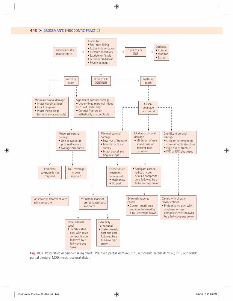
440 Ü GROSSMAN’S ENDODONTIC PRACTICE
Endodonticallytreated tooth
Significant coronal damage• Undermined marginal ridges• Loss of incisal edge• Coronal fracture or
esthetically unacceptable
Minimal coronal damage• Intact marginal ridge• Intact cingulum• Intact incisal ridge
(esthetically acceptable)
Assess for:• Poor root filling• Active inflammation• Pressure sensitivity• Exudate or fistula• Periodontal disease• Severe damage
If yes to any:STOP
Options:• Retreat• Monitor• Extract
Posteriortooth
Moderate coronaldamage• One or two large
proximal lesions• Average-size tooth
Minimal coronaldamage• Low risk of fracture• Minimal occlusal
forces• Intact buccal and
lingual cusps
Significant coronaldamage• Little or no remaining
coronal tooth structure• High risk of fracture• FPD or RPD abutment
Moderate coronaldamage• Minimum of one
sound cusp orextreme rootcurvature
Full-coveragecrown
required
Conservativetreatment(minimum):• MOD onlay• No post
Completecoverage is not
required
Conservative treatment withresin composite
• Custom-made orprefabricated postand cores
Small circularcanal• Prefabricated
post with resincomposite corefollowed by afull-coveragecrown
Extremelyflared canal• Custom-made
post and corefollowed by afull-coveragecrown
Extremely taperedcanals• Custom-made post
and core followed bya full-coverage crown
Canals with circularcross-sections• Prefabricated post with
amalgam or resincomposite core followedby a full-coverage crown
• Amalgam coronal-radicular coreor resin compositecore followed by afull-coverage crown
If no to all:CONTINUE
Anteriortooth
Cuspalcoverage
is required
Fig. 18.1 Restorative decision-making chart. FPD, fi xed partial denture; RPD, removable partial denture; RPD, removable partial denture; MOD, mesio-occlusal-distal.
Endodontic Practice_Ch-18.indd 440Endodontic Practice_Ch-18.indd 440 2/6/10 3:15:23 PM2/6/10 3:15:23 PM
460
We can easily forgive a child who is afraid of the dark.The real tragedy of life is when an adult is afraid of the light.
–Plato
N umerous researchers have investigated la-ser applications in dentistry in the past fi ve decades. Research and development
has lead to a multitude of lasers both in type and means of delivery systems. With better understand-ing of biological tissue responses, the possibilities of a clinician using lasers in a range of endodontic procedures have increased, varying from pulpal di-agnosis, pulpotomy, root canal disinfection, retreat-ment to root resection.
Chronology of Laser Development
1916 Albert Einstein: Theory of spontaneous emission of radiation
1953 Charles Townes: MASER (microwave amplifi cation by the stimulated emission of radiation)
1960 Theodore Maiman: Ruby laser1964 Stern, Sognnaes, and Goldman: Lasers in
dentistry1971 Weichman and Johnson: Lasers in
endodontics1979 Adrian and Gross: Argon laser steriliza-
tion of dental instruments1985 Shoji et al.: Laser-aided pulpotomy1986 Zakariasen et al.: Sterilization of root
canals
1988 Miserendino: Apicectomy with CO2 laser1990 Potts and Petrou: Laser-aided photo-
polymerization of camphoroquinone- activated resins
1993 Paghdiwala: Er:YAG lasers for root resec-tion and retrograde cavity preparation
1994 Morita: Nd:YAG lasers in endodontics1998 Mazeki et al.: Root canal shaping with
Er:YAG laser
Basics of Laser Physics
Light is a form of electromagnetic energy that trav-els at a constant velocity in the form of waves. The basic unit of light is termed photon, or a particle of light. Ordinary light, as produced by the sun or a table lamp, is a sum of many types of photons or light waves that are diffuse and not focused. Photons are released when an atom in its ground state absorbs energy, gets excited, and moves to a higher energy state. The re-lease of photon is spontaneous in nature and hence termed spontaneous emission. This is accompanied by neighboring atoms producing waves that are not in phase with one another, thereby having varying wave-lengths and amplitude. The light waves produced are noncoherent and polychromatic (Fig. 19.1).
Two photons are released if additional quantum of energy is absorbed by an already excited atom. The
CHAPTER
19 Lasers in Endodontics
Endodontic Practice_Ch-19.indd 460Endodontic Practice_Ch-19.indd 460 2/6/10 10:43:18 AM2/6/10 10:43:18 AM
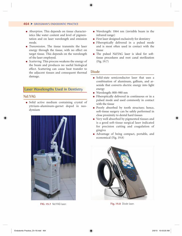
464 Ü GROSSMAN’S ENDODONTIC PRACTICE
! Absorption. This depends on tissue character-istics like water content and level of pigmen-tation and on laser wavelength and emission mode.
! Transmission. The tissue transmits the laser energy through the tissue, with no effect on target tissue. This depends on the wavelength of the laser employed.
! Scattering. This process weakens the energy of the beam and produces no useful biological effect. Scattering can cause heat transfer to the adjacent tissues and consequent thermal damage.
Laser Wavelengths Used in Dentistry
Nd:YAG! Solid active medium containing crystal of
yttrium–aluminum–garnet doped in neo-dymium
FIG. 19.7 Nd:YAG laser. Fig. 19.8 Diode laser.
! Wavelength: 1064 nm (invisible beam in the infrared range)
! First laser designed exclusively for dentistry! Fiberoptically delivered in a pulsed mode
and is most often used in contact with the tissue
! The pulsed Nd:YAG laser is ideal for soft- tissue procedures and root canal sterilization (Fig. 19.7)
Diode! Solid-state semiconductor laser that uses a
combination of aluminum, gallium, and ar-senide that converts electric energy into light energy
! Wavelength: 800–980 nm! Fiberoptically delivered in continuous or in a
pulsed mode and used commonly in contact with the tissue
! Poorly absorbed by tooth structure; hence, soft-tissue surgery can be safely performed in close proximity to dental hard tissues
! Very well absorbed by pigmented tissues and is a good soft-tissue surgical laser indicated for precision cutting and coagulation of gingiva
! Advantage of being compact, portable, and economical (Fig. 19.8)
Endodontic Practice_Ch-19.indd 464Endodontic Practice_Ch-19.indd 464 2/6/10 10:43:20 AM2/6/10 10:43:20 AM
Experience is what we get, when we don’t get what we want ...!
469
P rocedural errors during endodontic therapy occur not only with students and beginners, but also with skilled and experienced clini-
cians in spite of taking all possible precautions. Such iatrogenic complications can be prevented by adhering to the fundamental biological and me-chanical guidelines of endodontics. A clinician should have the ability to:
! Assess and inform the patient about the prog-nosis of a case before initiating the treatment.
! Identify the given clinical problem with diag-nostic acumen.
! Anticipate problems in challenging cases.! Use appropriate materials and modifi cations in
routine techniques in order to prevent proce-dural errors.
! Identify clinical problems the moment they occur during the procedure and manage them positively.
Before commencing a procedure, the endodontist (or a general practitioner), trained in endodontics, must do a sincere introspection of his/her capabili-ties in handling an endodontic situation. If it is not within the purview of the attending clinician, then he/she should either call in a peer consultant or refer the case to one who can handle the situation.
Procedural errors can be avoided to a larger ex-tent by following these guidelines:
! Establish a proper communication and rap-port with the patient. The patient has a moral and legal right to be informed about every step of the treatment procedure.
! Complete a thorough history and meticulous clinical examination of the tooth or teeth in question before commencing the treatment.
! Ascertain the periodontal status, prosthodon-tic restorability, and prognosis of tooth in question.
! One must have a thorough knowledge of in-ternal anatomy of pulp space and variations in the root canal confi guration. Also required is a complete knowledge of apical terminus of root canals especially apical foramen, api-cal constriction, and anatomical apex. In ad-dition, one must know approximately at what age root apex is complete in different teeth in the arch. A beginner can learn to anticipate these variations by observing cross and verti-cal ground sections of all teeth at various levels (see Chapter 9).
! Obtain an intraoral periapical radiograph for every step of endodontic therapy. These include one before commencing the treat-ment, during working length determination, during master apical gutta-percha cone selec-tion, and one following the completion of the obturation.
CHAPTER
20Procedural Errors:
Prevention and Management
Endodontic Practice_Ch-20.indd 469Endodontic Practice_Ch-20.indd 469 2/6/10 5:26:41 PM2/6/10 5:26:41 PM
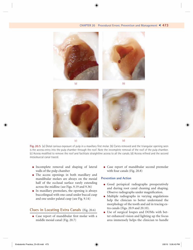
CHAPTER 20 Procedural Errors: Prevention and Management Ö 473
! Incomplete removal and shaping of lateral walls of the pulp chamber
! The access openings in both maxillary and mandibular molars are always on the mesial half of the occlusal surface rarely extending across the midline (see Figs. 9.19 and 9.36)
! In maxillary premolars, the opening is always buccolingual with one canal under buccal cusp and one under palatal cusp (see Fig. 9.14)
Clues in Locating Extra Canals (Fig. 20.6)
! Case report of mandibular fi rst molar with a middle mesial canal (Fig. 20.7)
! Case report of mandibular second premolar with four canals (Fig. 20.8)
Prevention and Action
! Good periapical radiographs preoperatively and during root canal cleaning and shaping. Observe radiographs under magnifi cation.
! Multiple radiographs in varying angulations help the clinician to better understand the morphology of the tooth and aid in tracing ex-tra canals (Figs. 20.9 and 20.10).
! Use of surgical loupes and DOMs with bet-ter enhanced vision and lighting up the focus area immensely helps the clinician to handle
(c) (d)
(a) (b)
Fig. 20.5 (a) Distal carious exposure of pulp in a maxillary fi rst molar. (b) Caries removed and the triangular opening seen is the access entry into the pulp chamber through the roof. Note the incomplete removal of the roof of the pulp chamber. (c) Access modifi ed to remove the roof and facilitate straightline access to all the canals. (d) Access refi ned and the second mesiobuccal canal traced.
Endodontic Practice_Ch-20.indd 473Endodontic Practice_Ch-20.indd 473 2/6/10 5:26:43 PM2/6/10 5:26:43 PM
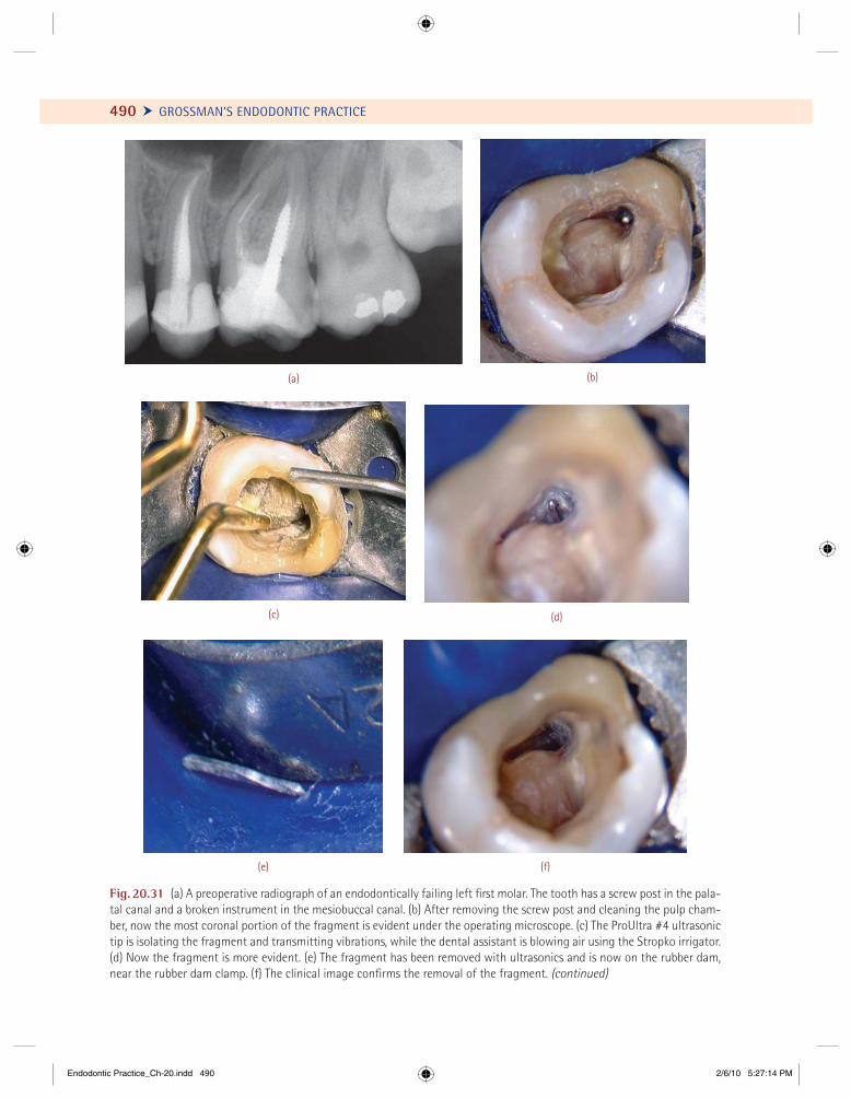
490 Ü GROSSMAN’S ENDODONTIC PRACTICE
Fig. 20.31 (a) A preoperative radiograph of an endodontically failing left fi rst molar. The tooth has a screw post in the pala-tal canal and a broken instrument in the mesiobuccal canal. (b) After removing the screw post and cleaning the pulp cham-ber, now the most coronal portion of the fragment is evident under the operating microscope. (c) The ProUltra #4 ultrasonic tip is isolating the fragment and transmitting vibrations, while the dental assistant is blowing air using the Stropko irrigator. (d) Now the fragment is more evident. (e) The fragment has been removed with ultrasonics and is now on the rubber dam, near the rubber dam clamp. (f) The clinical image confi rms the removal of the fragment. (continued)
(a) (b)
(c) (d)
(e) (f)
Endodontic Practice_Ch-20.indd 490Endodontic Practice_Ch-20.indd 490 2/6/10 5:27:14 PM2/6/10 5:27:14 PM

The most pathetic person in the world is someone who has sight but no vision.
—Helen Keller
497
R adiographs are indispensable diagnostic and prognostic aids in endodontics and are one of the most reliable methods of
monitoring endodontic treatment. Radiographs provide an important visual method of gaining clinical knowledge of teeth and periradicular tissues; therefore, they are essential to the prac-tice of endodontics.
Proper positioning and stabilization of the radiographic fi lm during endodontic procedures becomes diffi cult because of the interference from the protruding rubber dam clamp or root canal in-struments or obturating material protruding from the access cavity. The visualization of the tooth for proper fi lm positioning and cone angulation is impeded by the presence of the rubber dam. This makes the process of taking a radiograph a diffi cult proposition.
Radiographic Technical Requirements
1. The image of the tooth being evaluated or un-dergoing endodontic therapy should be in the center of the radiograph.
2. Radiographs should show at least 5 mm of bone surrounding the apex of the tooth being evaluated or undergoing endodontic therapy.
3. If a periradicular lesion is too large to fi t in one periapical fi lm, supplemental diagnostic radio-graphs must be made.
4. A single radiograph taken from one direction only may not provide suffi cient diagnostic in-formation in multirooted teeth or teeth with curved roots. Under these circumstances, at least two periapical radiographs should be taken to help gain a three-dimensional perspective. One radiograph should be taken at normal vertical and horizontal angulation, while the other at a 20° change in horizontal angle from either me-sial or distal direction (Fig. A.l).
5. If a sinus tract is present, a tracing radiograph should be taken. This procedure is accomplished by carefully threading a gutta-percha cone into the tract and taking a radiograph to identify the origin of the tract (see Figs. 5.16 and 15.12). This technique is also useful for localization and depth marking of certain periodontal defects.
6. Correct processing of the radiographic fi lm is essential to evaluate success or failure of the case at a later date.
In endodontics, the long-cone paralleling tech-nique is preferred over the short-cone bisecting-angle technique because dimensional distortion is lesser, the image is sharper, and the same angulations are easily reproduced. The paralleling technique may
APPENDIX
ARadiographic Technique for Endodontics
Endodontic Practice_Appendix A.i497 497Endodontic Practice_Appendix A.i497 497 2/6/10 6:27:47 PM2/6/10 6:27:47 PM
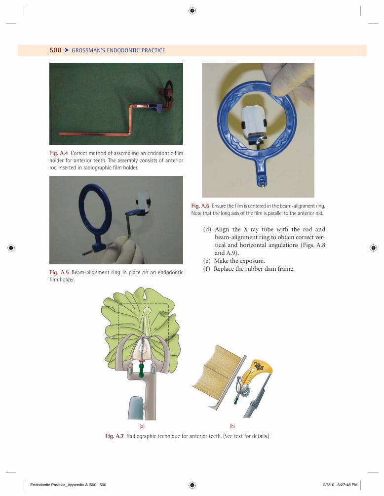
500 Ü GROSSMAN’S ENDODONTIC PRACTICE
Fig. A.4 Correct method of assembling an endodontic fi lm holder for anterior teeth. The assembly consists of anterior rod inserted in radiographic fi lm holder.
Fig. A.5 Beam-alignment ring in place on an endodontic fi lm holder.
Fig. A.6 Ensure the fi lm is centered in the beam-alignment ring. Note that the long axis of the fi lm is parallel to the anterior rod.
(a) (b)
Fig. A.7 Radiographic technique for anterior teeth. (See text for details.)
(d) Align the X-ray tube with the rod and beam-alignment ring to obtain correct ver-tical and horizontal angulations (Figs. A.8 and A.9).
(e) Make the exposure. (f) Replace the rubber dam frame.
Endodontic Practice_Appendix A.i500 500Endodontic Practice_Appendix A.i500 500 2/6/10 6:27:48 PM2/6/10 6:27:48 PM
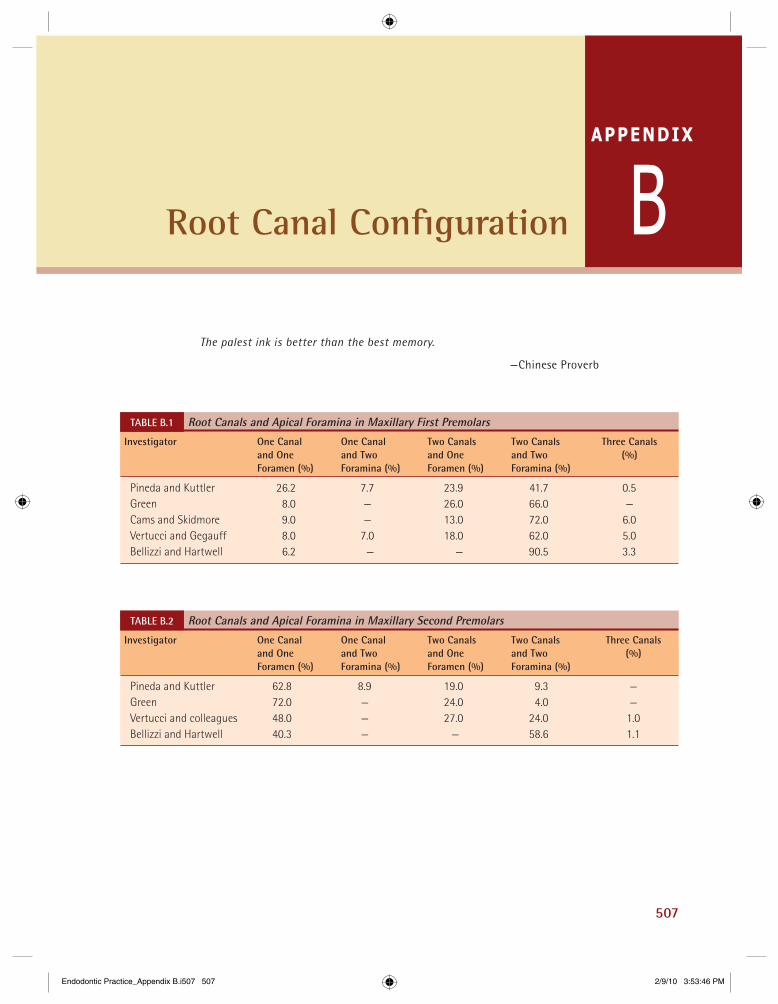
507
APPENDIX
BRoot Canal Confi guration
TABLE B.1 Root Canals and Apical Foramina in Maxillary First PremolarsInvestigator One Canal One Canal Two Canals Two Canals Three Canals and One and Two and One and Two (%) Foramen (%) Foramina (%) Foramen (%) Foramina (%)
Pineda and Kuttler 26.2 7.7 23.9 41.7 0.5 Green 8.0 — 26.0 66.0 — Cams and Skidmore 9.0 — 13.0 72.0 6.0 Vertucci and Gegauff 8.0 7.0 18.0 62.0 5.0 Bellizzi and Hartwell 6.2 — — 90.5 3.3
TABLE B.2 Root Canals and Apical Foramina in Maxillary Second PremolarsInvestigator One Canal One Canal Two Canals Two Canals Three Canals and One and Two and One and Two (%) Foramen (%) Foramina (%) Foramen (%) Foramina (%)
Pineda and Kuttler 62.8 8.9 19.0 9.3 — Green 72.0 — 24.0 4.0 — Vertucci and colleagues 48.0 — 27.0 24.0 1.0 Bellizzi and Hartwell 40.3 — — 58.6 1.1
The palest ink is better than the best memory.
—Chinese Proverb
Endodontic Practice_Appendix B.i507 507Endodontic Practice_Appendix B.i507 507 2/9/10 3:53:46 PM2/9/10 3:53:46 PM
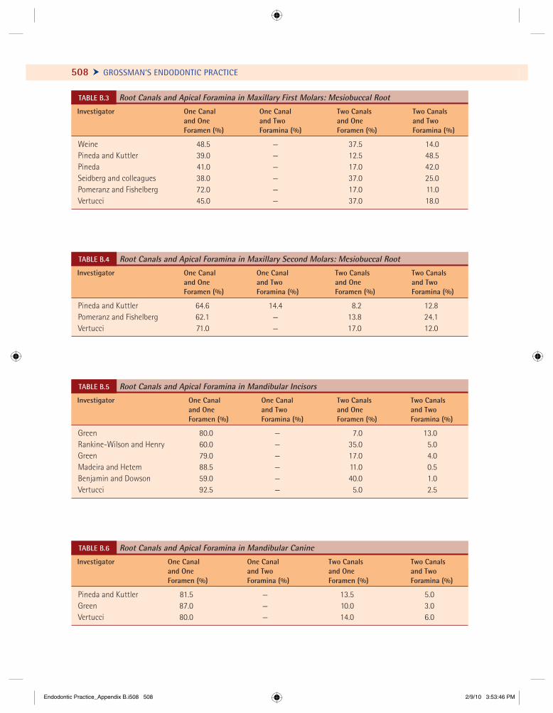
508 Ü GROSSMAN’S ENDODONTIC PRACTICE
TABLE B.4 Root Canals and Apical Foramina in Maxillary Second Molars: Mesiobuccal Root Investigator One Canal One Canal Two Canals Two Canals and One and Two and One and Two Foramen (%) Foramina (%) Foramen (%) Foramina (%)
Pineda and Kuttler 64.6 14.4 8.2 12.8Pomeranz and Fishelberg 62.1 — 13.8 24.1Vertucci 71.0 — 17.0 12.0
TABLE B.5 Root Canals and Apical Foramina in Mandibular Incisors Investigator One Canal One Canal Two Canals Two Canals and One and Two and One and Two Foramen (%) Foramina (%) Foramen (%) Foramina (%)
Green 80.0 — 7.0 13.0Rankine-Wilson and Henry 60.0 — 35.0 5.0Green 79.0 — 17.0 4.0Madeira and Hetem 88.5 — 11.0 0.5Benjamin and Dowson 59.0 — 40.0 1.0Vertucci 92.5 — 5.0 2.5
TABLE B.6 Root Canals and Apical Foramina in Mandibular Canine Investigator One Canal One Canal Two Canals Two Canals and One and Two and One and Two Foramen (%) Foramina (%) Foramen (%) Foramina (%)
Pineda and Kuttler 81.5 — 13.5 5.0Green 87.0 — 10.0 3.0Vertucci 80.0 — 14.0 6.0
TABLE B.3 Root Canals and Apical Foramina in Maxillary First Molars: Mesiobuccal Root Investigator One Canal One Canal Two Canals Two Canals and One and Two and One and Two Foramen (%) Foramina (%) Foramen (%) Foramina (%)
Weine 48.5 — 37.5 14.0Pineda and Kuttler 39.0 — 12.5 48.5Pineda 41.0 — 17.0 42.0Seidberg and colleagues 38.0 — 37.0 25.0Pomeranz and Fishelberg 72.0 — 17.0 11.0Vertucci 45.0 — 37.0 18.0
Endodontic Practice_Appendix B.i508 508Endodontic Practice_Appendix B.i508 508 2/9/10 3:53:46 PM2/9/10 3:53:46 PM
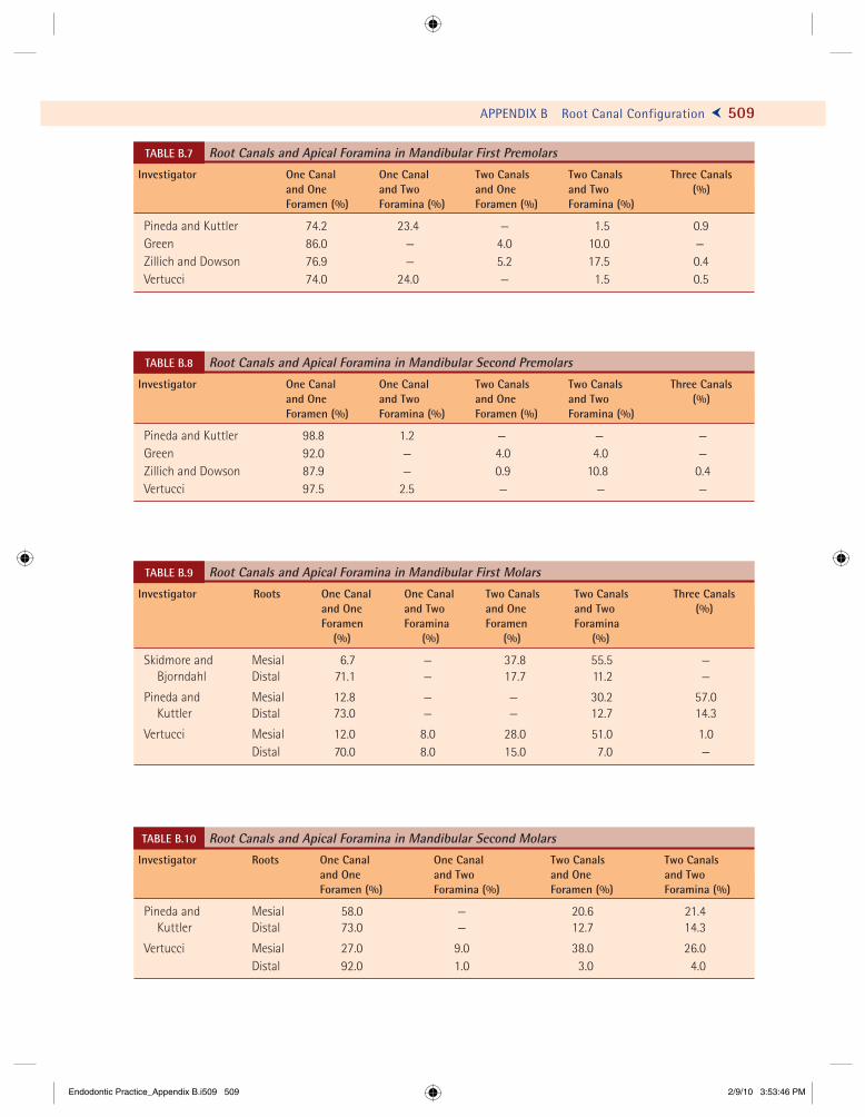
APPENDIX B Root Canal Configuration Ö 509
TABLE B.7 Root Canals and Apical Foramina in Mandibular First PremolarsInvestigator One Canal One Canal Two Canals Two Canals Three Canals and One and Two and One and Two (%) Foramen (%) Foramina (%) Foramen (%) Foramina (%)
Pineda and Kuttler 74.2 23.4 — 1.5 0.9Green 86.0 — 4.0 10.0 —Zillich and Dowson 76.9 — 5.2 17.5 0.4Vertucci 74.0 24.0 — 1.5 0.5
TABLE B.8 Root Canals and Apical Foramina in Mandibular Second PremolarsInvestigator One Canal One Canal Two Canals Two Canals Three Canals and One and Two and One and Two (%) Foramen (%) Foramina (%) Foramen (%) Foramina (%)
Pineda and Kuttler 98.8 1.2 — — —Green 92.0 — 4.0 4.0 —Zillich and Dowson 87.9 — 0.9 10.8 0.4Vertucci 97.5 2.5 — — —
TABLE B.9 Root Canals and Apical Foramina in Mandibular First MolarsInvestigator Roots One Canal One Canal Two Canals Two Canals Three Canals and One and Two and One and Two (%) Foramen Foramina Foramen Foramina (%) (%) (%) (%)
Skidmore and Mesial 6.7 — 37.8 55.5 — Bjorndahl Distal 71.1 — 17.7 11.2 —
Pineda and Mesial 12.8 — — 30.2 57.0 Kuttler Distal 73.0 — — 12.7 14.3
Vertucci Mesial 12.0 8.0 28.0 51.0 1.0 Distal 70.0 8.0 15.0 7.0 —
Root Canals and Apical Foramina in Mandibular Second MolarsTABLE B.10
Investigator Roots One Canal One Canal Two Canals Two Canals and One and Two and One and Two Foramen (%) Foramina (%) Foramen (%) Foramina (%)
Pineda and Mesial 58.0 — 20.6 21.4 Kuttler Distal 73.0 — 12.7 14.3
Vertucci Mesial 27.0 9.0 38.0 26.0 Distal 92.0 1.0 3.0 4.0
Root Canals and Apical Foramina in Mandibular Second Molars
Endodontic Practice_Appendix B.i509 509Endodontic Practice_Appendix B.i509 509 2/9/10 3:53:46 PM2/9/10 3:53:46 PM

Endodontic Practice_Appendix B.i510 510Endodontic Practice_Appendix B.i510 510 2/9/10 3:53:47 PM2/9/10 3:53:47 PM
511
A
Absorbent paper points, 284Accessory canal, 14, 177, 289, 426Accessory cones, 284, 301Active post, 448, 449, 450Acute alveolar abscess, 47, 100–105Acute irreversible pulpitis, 86, 102, 105Acute periodontal abscess, 60, 101Acute pulpitis, 83, 323Acute reversible pulpitis, 56, 83–85Adhesive strength, 281, 303AH26, 303, 304AH plus, 299, 304Alveolar bone, 15, 35, 38, 40, 431Amplitude, 460Anachoresis, 44, 76Analgesics, 35, 103, 240, 494Ankylosis, 122, 123, 384Antibiotics, 53, 103, 142, 144, 272, 398Antioxidant, 352Apexifi cation, 36, 310, 331–36Apexogenesis, 323, 331, 336, 337, 338Apical barrier, 151, 294, 332, 334Apical constriction, 180, 243, 248, 254–55Apical delta, 177Apical foramen, 13, 14, 36, 98, 117, 180,
222, 243Apical stop, 248, 257, 493Argon gas laser, 465Argon laser, 460, 462Asepsis, 170, 272, 305, 419Avulsion, 123, 363, 378–88
B
Bacterial leakage, 80Binoculars, 393, 394Biological width, 441, 442, 443Bleaching, 342–60Bleaching wand, 346–58Blunderbuss canal, 338Bruxism, 76, 77, 344, 372Bupivacaine, 223, 399
C
Calcifi ed canals, 142, 149, 179Calcifi ed orifi ces, 179, 180Calcium hydroxide, 47, 272, 273, 303, 314,
316–36Calcium hydroxide sealer, 303Canal calcifi cation, 64, 324, 381Carbamide peroxide, 347–48, 354Caries excavation, 314Cast post–core, 443, 444, 450–452Cavit, 219, 275, 295Cementum, 14–16, 18, 19, 36–39Cervical pulpotomy, 324Cervical root resorption, 124, 126, 349C-fi bers, 32Chromogen, 343Chromogenic bacteria, 345CO2 gas laser, 462, 465Coconut water, 380Collagen, 39, 313, 400, 413, 418Collagen fi bers, 9, 20, 29, 34, 132, 312Compaction, 281, 282, 290–92
Index
Endodontic Practice_Index.indd 511Endodontic Practice_Index.indd 511 2/10/10 3:00:55 PM2/10/10 3:00:55 PM

512 Ü Index
Compaction cones, 283, 288Concussion, 362, 363, 365Condensation, 282, 283, 287–89Continuous wave laser, 233, 282, 290Core, 279, 445, 450–53Core buildup, 469, 445, 455–56Core buildup materials, 445, 456Core obturating materials, 181, 304Coronal seal, 300, 437Corticosteroids, 303, 306CPP-ACP, 354, 357, 380Cracked tooth syndrome, 58, 62, 76–77Crown fracture, 65, 76, 150, 361–66, 372Crown–root fractures, 65, 363, 365–368C-shaped canal, 241, 256, 267Curettage, 390, 405Curved canals, 141, 178, 483, 485Cyanoacrylate, 375, 413Cytokines, 114
D
Debridement, 239, 242Dental bleaching, 346, 359Dental dam, 159Dental follicle, 4, 7, 13, 16, 63Dental papilla, 1, 4, 7–9, 16, 400Dental sac, 4Denticles, 33, 34Dentinal bridge, 315, 323, 329Dentinal dysplasia, 216Dentinal erosion, 23, 76, 342Dentinal tubules, 12, 15–34, 181, 265Dentinogenesis imperfecta, 216, 343Developmental grooves, 80, 195, 202, 217, 474DIAKET, 409Diode laser light, 462, 464, 466Direct cast post and core technique, 451Direct pulp capping, 294, 312, 315, 321–24Dowel, 451Dye laser, 462
E
Ectodermal derivative, 6Ellis’s classifi cation, 362Enamel infraction, 363, 364Endodontic emergency, 142, 415Epithelial cell rests of Malassez, 14, 119
Er:YAG laser, 460, 465Erbium laser, 465Er,Cr:YSGG, 465Ethylenediamine, 241, 266Eucapercha, 301External surface resorption, 122, 123, 124, 368Extrinsic discoloration, 342–43, 344Extrusive luxation, 363, 376
F
Ferrule, 442–44Fiber posts, 453Flare, 82,105, 136, 494Flare-up, 259Fluid-impervious seal, 467Fluorosis, 344Focal length, 393Focus, 152, 410, 433Formocresol, 272, 324, 327–28Foundation restoration, 447Fracture, 142, 361–75Fracture foramen, 361–362Fracture strength, 445, 453Frequency, 249, 406, 462Frequency-doubled Nd:YAG laser, 462
G
Gas lasers, 465Gates-Glidden drills, 182, 232, 233, 258Glass ionomer cement (GIC), 168, 316, 349, 445Glass ionomer cement sealer, 303Gouging, 475, 481–83Greater taper cone, 280Gutta-percha, 48, 69, 110, 279–99, 491Gutta-percha cones/points, 280, 288, 292Gutta-percha pellets, 292
H
Hank’s balanced salt solution, 380Helium–neon laser, 462Hemostatic agents, 324, 400, 401Hermetically seal, 218, 273Hertwig’s epithelial root sheath, 10, 13, 39, 181Histamine, 134Hollow tube concept, 462Home bleaching, 348, 354
Endodontic Practice_Index.indd 512Endodontic Practice_Index.indd 512 2/10/10 3:18:52 PM2/10/10 3:18:52 PM
Index Ö 513
Hydrochloric acid, 358Hydrogen peroxide, 173, 346–54Hyperocclusion, 99Hypocalcifi cation, 358Hypoplastic, 353, 358
I
Illumination, 390, 392, 393Incision of soft tissue, 103Indirect cast post and core technique, 452Indirect pulp capping, 312, 313, 314In-offi ce bleaching, 354Internal surface resorption, 368Intracoronal bleaching, 348, 352Intraradicular splint, 371Intrinsic discoloration, 343–44Intrusive luxation, 386, 376, 378IRM (intermediate restaurateur material), 219,
275, 314Irrigation, 268, 270–71, 395, 493
K
Kerr sealer, 296Ketac Endo, 303
L
Lamina dura, 16, 40, 41, 66, 144Laser, 48, 72, 324, 354, 460–67Laser-assisted endodontic, 354Laser Doppler fl owmetry, 72, 466Laser media, 354Lateral canal, 177, 181, 182, 406, 425Lateral compaction of cold gutta-percha, 285Lateral luxation, 363, 376Leukotrienes, 135Lidocaine, 223, 399Luting cements, 447, 453Luxations, 361–63, 375–78
M
Macroabrasion, 360Magnifi cation, 2, 6, 11, 121, 392–93, 406, 452Magnifi cation changes, 393Marginal leakage, 65MASER, 460Master cone, 209, 283, 288, 492–93
McSpadden compacter, 291–92Mercury toxicity, 445Metal posts, 152, 346, 449, 452Microabrasion technique, 358–59, 360Microscope, 24, 195, 294, 333, 392Microsurgical approach, 392Microsurgical triad, 392–396Mineral trioxide aggregate (MTA), 317, 480Monoblock concept, 454MTA, 151, 294, 316–31, 410, 411
N
N2, 303, 343Nd:YAG laser, 117, 462, 464, 466Neural crest cells, 1
O
Objective lens, 393Obturation, 49–50, 221, 278–307, 467Orabase, 352, 353Osteodentin, 331Overfi lling, 98, 248, 285, 304, 493Overinstrumentation, 98, 232
P
Palpation, 57, 60, 61Parachlorophenol, 174Passive post, 450Peeso reamers, 232–33Percussion, 57, 60, 384Perforation, 91, 151, 225, 307, 475–84Periodontal ligament, 15, 36, 38, 225Periodontium, 16, 39, 60, 61, 278, 426Photon, 460, 461Physiologic saline, 266Pins, 76, 77, 445Plunger cusp, 271, 292, 492Polyacrylic barrier, 347Post, 369, 447–55Post diameter, 448, 449Post length, 447Post retention, 369Postendodontic restoration, 392, 439Postoperative pain, 105, 303Prechromogen, 343Prefabricated post and core, 440, 452
Endodontic Practice_Index.indd 513Endodontic Practice_Index.indd 513 2/10/10 3:00:56 PM2/10/10 3:00:56 PM

514 Ü Index
Prefabricated post system, 440, 452Primary cones, 283–85Prostaglandin, 132, 135Pulp, 1, 17, 30–34, 43, 67, 76, 82, 92–94,
176, 216, 312, 365Pulp canal obturation, 385Pulp cap, 312, 315, 316, 321, 466Pulp chamber, 176–77, 182, 184–86, 191,
200, 203–04Pulp dentin organ, 1Pulp extirpation, 83, 86, 152, 223, 239, 344, 345Pulp horns, 176–77, 181, 185, 208, 350Pulp stump, 242, 323, 325Pulp testing, 67–69Pulpitis, 56, 82, 83–84, 86–90Pulpotomy, 88, 310, 323–31, 466Pulse oximetry, 71Pulsed mode, 464Pumice, 353, 354
R
Recapitulation, 251, 257–59, 470, 484Referred pain, 53, 92Refl ection, 463Reinforcement, 338, 446, 449, 451Replacement resorption, 122, 124, 384, 387Replantation, 122, 380–84, 419–21Repositioning, 376, 401, 413Revascularization, 336–39, 385Root canal sealer, 279, 284, 301, 303Root-fi lled teeth, 46Root fractures, 36, 151, 362, 363, 369–71Root resorption, 122–27, 145Roth sealer, 48, 301Ruby laser, 460, 462
S
Scanning laser, 48Scatter, 437, 463, 464Sclerotic dentin, 22, 34, 139Sealers, 281, 287, 301–04Secondary caries, 87, 212, 470Semiconductor laser, 464Shaped canal, 222, 451Sharpey’s fi bers, 36, 38, 39, 40, 338Silver points, 281, 282, 492Simplifi l obturation techniques, 299, 301
Single-visit apexifi cation, 335Single-visit endodontics, 90, 335Smear layer, 231, 253, 268–69Sodium perborate, 347, 349, 350, 351Solid-state lasers, 464Splinting, 369, 376, 434Splints, 72, 366, 369, 371, 376, 419Spontaneous emission, 460, 461Stimulated emission, 460, 461Strength, 319, 396, 442, 445Stress concentrations, 449, 450Subluxation, 150, 363, 375Sulfa compounds, 219, 280, 302, 318Superoxol, 352
T
Tetraacetic acid (EDTA), 266Tetracycline staining, 57, 344, 358–59Tetracyclines, 270, 344, 359, 381Thermocatalytic bleaching, 351Thermomechanical compaction of
gutta-percha, 282Thermoplasticized solid-core gutta-percha
obturation, 292, 297, 467Threaded split shank post, 449Three-dimensional obturation, 307Titanium posts, 454, 455Tooth discoloration, 58, 342–60Tooth slooth, 61, 62, 78Toughness, 310, 453Transillumination, 78, 363Transmission, 463, 464Traumatic injury, 57, 76, 306, 345,
361, 363Tubli-Seal, 301, 302, 303Twist drills, 228, 230, 235
U
Undercuts, 350Undifferentiated mesenchymal cells, 27, 29,
39, 336
V
Vertical compaction of gutta-percha, 282, 287Vertical root fracture, 66, 144, 218,
272–275, 372, 375, 450
Endodontic Practice_Index.indd 514Endodontic Practice_Index.indd 514 2/10/10 3:00:56 PM2/10/10 3:00:56 PM
Index Ö 515
Vital pulp therapies, 310, 312–16, 385Vital tooth bleaching, 348Vitality preservation, 323Vitality test, 105, 122, 128, 337
W
Walking bleach, 347, 349–52Wave, 231, 290, 460, 461
Wavelength, 249, 462, 464, 465White spot lesion, 358WHO classifi cation, 97Working length, 199, 201, 243, 247, 493
Z
Zone of coagulation, 317Zone of obliteration, 317
Endodontic Practice_Index.indd 515Endodontic Practice_Index.indd 515 2/10/10 3:00:56 PM2/10/10 3:00:56 PM