growth and development analysis of unilateral cleft palate
TRANSCRIPT

Western University Western University
Scholarship@Western Scholarship@Western
Electronic Thesis and Dissertation Repository
9-23-2020 5:00 PM
Growth and Development Analysis of Unilateral Cleft Palate Growth and Development Analysis of Unilateral Cleft Palate
Patients at One, Five, and Ten Years Patients at One, Five, and Ten Years
Katie Garland, The University of Western Ontario
Supervisor: Matic, Damir, The University of Western Ontario
: Dubois, Luc, The University of Western Ontario
A thesis submitted in partial fulfillment of the requirements for the Master of Science degree in
Surgery
© Katie Garland 2020
Follow this and additional works at: https://ir.lib.uwo.ca/etd
Recommended Citation Recommended Citation Garland, Katie, "Growth and Development Analysis of Unilateral Cleft Palate Patients at One, Five, and Ten Years" (2020). Electronic Thesis and Dissertation Repository. 7358. https://ir.lib.uwo.ca/etd/7358
This Dissertation/Thesis is brought to you for free and open access by Scholarship@Western. It has been accepted for inclusion in Electronic Thesis and Dissertation Repository by an authorized administrator of Scholarship@Western. For more information, please contact [email protected].

ii
Abstract & Key Words
Active and passive pre-surgical orthopedic (PSO) devices are a controversial part
of cleft palate management. There is no consensus as to the effects of these PSO
devices on long-term outcomes and there is limited research comparing different
PSO devices. The first objective of this research was to perform a systematic
review of the literature surrounding the long-term effects of PSO device use. The
second was to analyze and compare 10-year nasolabial aesthetic outcomes
between patients treated with an active PSO device, passive PSO device, or no
device. The final objective was to analyze and compare 10-year dental occlusion
and facial growth in patients who received treatment with an active versus a
passive PSO device. All patient data was assessed at 1, 5, and 10 years.
Nasolabial aesthetics were assessed using patient photographs, dental occlusion
was assessed using dental molds, and facial growth was assessed through
cephalometric analysis. Systematic review identified 41 articles pertaining to long-
term effects of PSO device use. This systematic review didn’t identify a consensus
as to the effects of these devices but did identify that all 41 articles had
methodologic flaws that limit the applicability of their results. Comparison of
nasolabial aesthetics showed that patients treated with a PSO device have
comparable aesthetics at the 10-year mark to patients treated with no device who
have less severe alveolar gaps. Patients treated with active and passive devices
have similar dental occlusion/arch development and facial growth up to 10 years.
Key words: cleft palate, cleft lip, pre-surgical device, pre-surgical orthopedics,
latham device, nasoalveolar molding

iii
Summary for Lay Audience
Patients born with cleft lip and palate are often treated with a pre-surgical device
prior to the surgical repair of their cleft lip. The role of this device is to help decrease
the size of the gap in their palate, which in turn brings the edges of the lip closer
together to facilitate the surgical repair of the lip. These devices can be active or
passive. Active devices drive the edges of the cleft closer together with a pin and
screw device, whereas passive devices gradually mold the cleft with a plate. The
use of these devices is still controversial. The devices have been shown to improve
patient outcomes but have also been shown to limit facial growth in these patients.
In addition, there is very little research that has compared outcomes in patients
depending on which type of device they received. The objectives of this study were
to (1) review the research that has been done on these devices to see their long-
term effects on patient outcomes, and (2) look at the long-term effects of these
devices on facial aesthetics, dental occlusion (how the teeth fit together), and facial
growth in a group of patients that have received treatment with these devices.
The literature review identified that research into the long-term effects of these
devices is lacking. The research that does exist is limited by studies of poor quality.
In addition, very few studies actually compared active and passive devices to see
if one type of device is superior. Aesthetic outcomes for patients that received
active or passive device treatment was similar between groups; aesthetics were
comparable to patients with less severe clefts that did not require a device. Facial
growth and dental occlusion were assessed at 5 and 10 years in patients with an
active or a passive device. Dental occlusion and facial growth were both similar for
patients treated with an active or a passive device up to 10 years of age.

iv
Co-Authorship Statement
Chapter 1: Sole Authorship: Katie Garland Manuscript Review: Damir Matic, Timothy Foley Chapter 2: Study Design: Katie Garland, Luc Dubois, Damir Matic Data Collection: Katie Garland, Brendan McNeely Data Analysis: Katie Garland, Brendan McNeely Statistical Analysis: Katie Garland Manuscript Preparation: Katie Garland Manuscript Review: Damir Matic, Timothy Foley Chapter 3: Study Design: Katie Garland, Damir Matic, Timothy Foley Data Collection: Katie Garland, Michelle Coyle Data Analysis: Katie Garland Statistical Analysis: Katie Garland Manuscript Preparation: Katie Garland Manuscript Review: Luc Dubois, Damir Matic Chapter 4: Study Design: Katie Garland Manuscript Review: Damir Matic, Timothy Foley Statistical Analysis: Katie Garland Manuscript Preparation: Katie Garland Manuscript Review: Damir Matic, Timothy Foley Chapter 5: Sole Authorship: Katie Garland Manuscript Review: Damir Matic, Timothy Foley

v
Dedication
The preparation and completion of this thesis was made possible by the assistance
of many individuals. Firstly, I would like to thank my supervisors Dr. Damir Matic,
Dr. Timothy Foley, and Dr. Luc Dubois. Their support and expertise were essential
for the completion of this work. Without their assistance I would not have
succeeded in completing this project. Specifically, I would like to thank Dr. Matic
for his assistance with the study design and mentorship throughout this entire
project. Thank you for always feeding my passion for pediatric plastic surgery. I
would like to thank Dr. Foley for all of his teaching on dental occlusion and
cephalometric analysis. Finally, I would like to thank Dr. Dubois for his expertise in
both the development of a systematic review and statistical analysis.
I would also like to thank Kristen White for her assistance with organizing patient
follow-up visits, without her help I could not have succeeded in completing this
project. I would also like to thank Dr. Meena Wilde, who generously granted me
the use of her clinic for analyzing cephalograms and patient molds. I would like to
thank Anne Dworschak-Stokan for her assistance with collecting patient photos,
her help was essential to me completing this project.
Last but not least, I would like to thank my partner, my friends, and my family for
all of their support. My success with this project would not have been possible
without their love and support. Majenta, thank you for letting me include your
wonderful nasolabial anatomy in this manuscript. Brendan, you encourage me to
become better every day and are an enormous part of my success in every
endeavor.

vi
Table of Contents
ABSTRACT & KEY WORDS .................................................................................................................... II
SUMMARY FOR LAY AUDIENCE ........................................................................................................... III
CO-AUTHORSHIP STATEMENT ............................................................................................................ IV
DEDICATION .......................................................................................................................................V
TABLE OF CONTENTS .......................................................................................................................... VI
LIST OF TABLES .................................................................................................................................. IX
LIST OF FIGURES .................................................................................................................................. X
LIST OF APPENDICES......................................................................................................................... XIII
LIST OF ABBREVIATIONS ................................................................................................................... XIV
CHAPTER 1: INTRODUCTION ................................................................................................................ 1
EPIDEMIOLOGY OF CLEFT LIP AND PALATE................................................................. 2 EMBRYOLOGY OF CLEFT LIP AND PALATE ................................................................... 3 CLEFT CLASSIFICATION ............................................................................................... 5 SURGICAL ANATOMY OF THE CLEFT LIP ...................................................................... 6 SURGICAL REPAIR OF THE CLEFT LIP ........................................................................... 8 SURGICAL ANATOMY OF THE CLEFT PALATE .............................................................. 9 SURGICAL REPAIR OF CLEFT PALATE ......................................................................... 10 TIMING OF SURGERY ................................................................................................ 10 MANAGEMENT OF CLEFT LIP AND PALATE AT OUR INSTITUTION ............................ 12 PRE-SURGICAL ORTHOPEDIC DEVICES ...................................................................... 14
1.10.1 ACTIVE DEVICES ............................................................................................................ 14 1.10.2 PASSIVE DEVICES ........................................................................................................... 16 1.10.3 COMPARING ACTIVE AND PASSIVE DEVICES ........................................................................ 18
METHODS OF ASSESSING POST-OPERATIVE AESTHETIC OUTCOMES....................... 19 1.11.1 NASOLABIAL AESTHETICS ................................................................................................ 19
METHODS OF ASSESSING POST-OPERATIVE GROWTH OUTCOMES ......................... 21 1.12.1 ANGLE CLASSIFICATION .................................................................................................. 21 1.12.2 MODIFIED HUDDART/BODENHAM CLASSIFICATION ............................................................. 24 1.12.3 DENTAL/OCCLUSAL CHANGES IN CLEFT LIP & PALATE .......................................................... 26 1.12.4 PALATAL MEASUREMENTS .............................................................................................. 26 1.12.5 CEPHALOMETRIC ANALYSIS.............................................................................................. 28 1.12.6 BRIEF OVERVIEW OF IMPORTANT FACIAL RELATIONSHIPS ...................................................... 31 1.12.7 FACIAL GROWTH IN CLEFT LIP & PALATE ........................................................................... 36
THESIS RATIONALE.................................................................................................... 37 RESEARCH OBJECTIVES AND HYPOTHESIS ................................................................ 38
CHAPTER 2: SYSTEMATIC REVIEW OF THE LITERATURE ON THE LONG-TERM PATIENT OUTCOMES ASSOCIATED WITH PRE-SURGICAL ORTHOPEDIC DEVICES ................................................................... 39
INTRODUCTION ........................................................................................................ 40 METHODOLOGY........................................................................................................ 41 RESULTS .................................................................................................................... 43 DISCUSSION .............................................................................................................. 51 CONCLUSION ............................................................................................................ 55
CHAPTER 3: 10-YEAR NASOLABIAL AESTHETIC COMPARISON IN PATIENTS WITH DIFFERENT PRE-SURGICAL ORTHOPEDIC DEVICES ....................................................................................................... 56

vii
INTRODUCTION ........................................................................................................ 57 METHODOLOGY........................................................................................................ 59 NASOLABIAL AESTHETICS ................................................................................................ 61
1.3.1 STATISTICAL ANALYSIS .................................................................................................... 61 RESULTS .................................................................................................................... 63
1.4.1 PATIENT DEMOGRAPHICS ................................................................................................ 63 INTRA-RATER RELIABILITY ................................................................................................ 64
1.5.1 NASOLABIAL AESTHETICS ................................................................................................ 67 1.5.2 SUB-GROUP ANALYSIS .................................................................................................... 67 1.5.3 INTRA-RATER RELIABILITY ................................................................................................ 68
DISCUSSION .............................................................................................................. 72 CONCLUSION ............................................................................................................ 75
CHAPTER 4: 10-YEAR DENTAL OCCLUSION AND FACIAL GROWTH COMPARISON IN PATIENTS TREATED WITH DIFFERENT PRE-SURGICAL ORTHOPEDIC DEVICES ...................................................................... 76
INTRODUCTION ........................................................................................................ 77 METHODOLOGY........................................................................................................ 79
1.2.1 DENTAL OCCLUSION ...................................................................................................... 79 1.2.2 CEPHALOMETRIC ANALYSIS.............................................................................................. 80 1.2.3 STATISTICAL ANALYSIS .................................................................................................... 81
RESULTS .................................................................................................................... 83 1.3.1 DEMOGRAPHIC INFORMATION ......................................................................................... 83 1.3.2 DENTAL ARCH AND OCCLUSION ....................................................................................... 86 1.3.3 CHANGE IN DENTAL ARCH RELATIONSHIPS AND OCCLUSION FROM FIVE TO TEN YEARS .............. 87 1.3.4 INTRA-RATER RELIABILITY ................................................................................................ 88 1.3.5 CEPHALOMETRIC MEASUREMENTS ................................................................................... 94 1.3.6 MAXILLARY GROWTH MEASUREMENTS ............................................................................. 94 1.3.7 MANDIBULAR GROWTH MEASUREMENTS .......................................................................... 96 1.3.8 DENTO-ALVEOLAR RELATIONSHIPS .................................................................................... 97 1.3.9 VERTICAL FACIAL GROWTH ............................................................................................. 98 1.3.10 SOFT TISSUE GROWTH ................................................................................................... 99 1.3.11 GROWTH BETWEEN FIVE AND TEN YEARS ........................................................................ 100 1.3.12 INTRA-RATER RELIABILITY .............................................................................................. 100
DISCUSSION ............................................................................................................ 102 1.4.1 DENTAL ARCH AND OCCLUSION ..................................................................................... 102 1.4.2 FACIAL GROWTH ......................................................................................................... 104
CONCLUSION .......................................................................................................... 106
CHAPTER 5: GENERAL DISCUSSION AND CONCLUSION ...................................................................... 107
SUMMARY .............................................................................................................. 108 SUMMARY OF CHAPTER 2: SYSTEMATIC REVIEW OF THE LITERATURE ON THE LONG-
TERM PATIENT OUTCOMES ASSOCIATED WITH PRE-SURGICAL ORTHOPEDIC DEVICES.............................. 109 SUMMARY OF CHAPTER 3: 10-YEAR NASOLABIAL AESTHETIC COMPARISON IN
PATIENTS WITH DIFFERENT PRE-SURGICAL ORTHOPEDIC DEVICES............................................................. 110 SUMMARY OF CHAPTER 4: 10-YEAR DENTAL OCCLUSION AND FACIAL GROWTH
COMPARISON IN PATIENTS WITH DIFFERENT PRE-SURGICAL ORTHOPEDIC DEVICES ................................. 111 STRENGTHS AND LIMITATIONS .............................................................................. 112 FUTURE DIRECTIONS .............................................................................................. 114 SIGNIFICANCE ......................................................................................................... 115
APPENDICES ................................................................................................................................... 117
APPENDIX 1- GLOSSARY .......................................................................................... 117 APPENDIX 2- NASOLABIAL AESTHETIC RATING SCALE ............................................ 117

viii
ETHICS APPROVAL .......................................................................................................................... 129
IMAGE PERMISSIONS ...................................................................................................................... 130
FIGURE 1 ................................................................................................................... 130 FIGURE 11 ................................................................................................................. 131
REFERENCES ................................................................................................................................... 132
CURRICULUM VITAE: KATIE GARLAND ............................................................................................. 149
ix
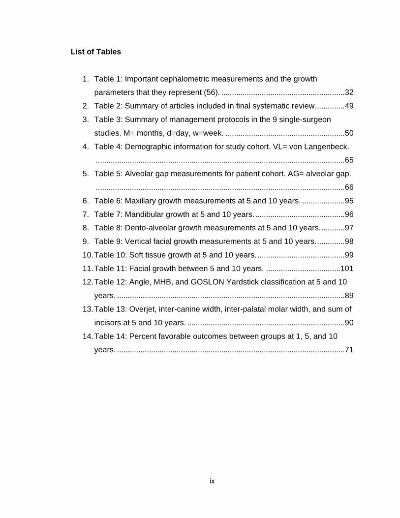
List of Tables
1. Table 1: Important cephalometric measurements and the growth
parameters that they represent (56). .......................................................... 32
2. Table 2: Summary of articles included in final systematic review. ............. 49
3. Table 3: Summary of management protocols in the 9 single-surgeon
studies. M= months, d=day, w=week. ........................................................ 50
4. Table 4: Demographic information for study cohort. VL= von Langenbeck.
..................................................................................................................... 65
5. Table 5: Alveolar gap measurements for patient cohort. AG= alveolar gap.
..................................................................................................................... 66
6. Table 6: Maxillary growth measurements at 5 and 10 years. .................... 95
7. Table 7: Mandibular growth at 5 and 10 years. .......................................... 96
8. Table 8: Dento-alveolar growth measurements at 5 and 10 years. ........... 97
9. Table 9: Vertical facial growth measurements at 5 and 10 years. ............. 98
10. Table 10: Soft tissue growth at 5 and 10 years. ......................................... 99
11. Table 11: Facial growth between 5 and 10 years. ...................................101
12. Table 12: Angle, MHB, and GOSLON Yardstick classification at 5 and 10
years. ........................................................................................................... 89
13. Table 13: Overjet, inter-canine width, inter-palatal molar width, and sum of
incisors at 5 and 10 years. .......................................................................... 90
14. Table 14: Percent favorable outcomes between groups at 1, 5, and 10
years. ........................................................................................................... 71

x
List of Figures
1. Figure 1: Reproduced from Neligan et al. with permission from Elsevier
publishing (4). Embryonic facial prominences and their corresponding
adult facial structures. FNP= frontonasal prominence; MXP= maxillary
prominence; LNP= lateral nasal prominence; MNP= mandibular
prominence. ............................................................................................ 4
2. Figure 2: Copyright Brito et al. (5), open access figure. Normal palate
anatomy (upper middle image). (A) Unilateral complete cleft of primary
palate, (B) Bilateral complete cleft of primary palate, (C) Unilateral
complete cleft of primary and secondary palate, (D) Bilateral complete
cleft of primary and secondary palate, (E) Complete cleft of secondary
palate. ...................................................................................................... 5
3. Figure 3: Original photograph of important nasolabial landmarks. (1)
Philtral column, (2) Philtral dimple, (3) Cupid's bow, (4) Vermillion, (5)
Vermillo-mucosal junction, (6) Alar Base, (7) Columella, (8) Nostril, and
(9) Nasal tip. ............................................................................................ 7
4. Figure 4: Overview of management process for CL/P and CP patients
at our institution. ENT= otolaryngology, SLP= speech language
pathology. .............................................................................................. 13
5. Figure 5: The Latham device is pictured above on a patient mold (left)
and in-situ (right). .................................................................................. 15
6. Figure 6: Original photograph. A custom-made NAM device is pictured
above. .................................................................................................... 17
7. Figure 7: Original photographs. Important landmarks to assess
nasolabial aestehtics include (A) symmetry of Cupid’s Bow and the
vermillo-cutaneous junction, (B) symmetry of the vermillo-mucosal
junction, (C) alar base symmetry, (D) scar quality and position, (E)
nostril symmetry, and (F) nasal tip symmetry. ..................................... 20
8. Figure 8: Original images. Angle classification system of occlusion. .. 21

xi
9. Figure 9: Original photographs. Patient molds representing: (A)
Positive overjet, GOSLON Yardstick Grade 1/2; (B) No overjet,
GOSLON Yardstick Grade 3; (C) Negative overjet, GOSLON Yardstick
Grade 4/5. ............................................................................................. 23
10. Figure 10: Reproduced with permission from Sage Publishing.
Modified Huddart Bodenham classification of molars, canines, and
incisors. ................................................................................................. 25
11. Figure 11: SI= sum of incisors, ICW= inter-canine width, IPMW= inter-
palatal molar width ................................................................................ 27
12. Figure 12: Original photograph. Cephalometric tracing of lateral patient
x-ray. Important boney landmarks identified (in blue). (1) Sella Turcica,
(2) Nasio, (3) A-point, (4, 5) Posterior and Anterior Nasal Spine, (6)
Basion, (7) B-point, (8) Pogonion, (9) Menton. .................................... 29
13. Figure 13: Original photograph. Cephalometric tracing of lateral patient
x-ray. Important soft tissue landmarks identified (in blue). (1) Glabella,
(2) Nasion, (3) Pronasale, (4) Columella, (5) Subnasale, (6) A-point, (7)
Upper lip, (8) B-point, (9) Pogonion. .................................................... 30
14. Figure 14: Maxillary cephalometric measurements. ............................ 33
15. Figure 15: Mandibular cephalometric measurements. ........................ 33
16. Figure 16: Dento-alveolar cephalometric measurements.................... 34
17. Figure 17: Vertical facial growth cephalometric measurements. ........ 34
18. Figure 18: Soft tissue cephalometric measurements. ......................... 35
19. Figure 19: Flowchart of study inclusion. ............................................... 42
20. Figure 20: Distribution of results in all articles. Positive result indicates
that PSO device improved outcomes compared to no device and
negative indicates PSO device worsened outcomes........................... 50
21. Figure 21: Types of variability in the methodology of the reviewed
studies. .................................................................................................. 50
22. Figure 22: Recorded cleft palate landmarks. A/A': antero-medial edge
of hard palate (non-cleft and cleft side); B/B': postero-medial edge of
hard palate (non-cleft and cleft side); C/C': postero-lateral edge of hard

xii
palate (non-cleft and cleft side); V: vertical alveolar gap; H: horizontal
alveolar gap........................................................................................... 60
23. Figure 23: Percent favorable outcomes for each question of the
nasolabial aesthetic scale at the 1-year mark. ..................................... 68
24. Figure 24: Percent favorable outcomes for each question of the
nasolabial aesthetic scale at the 5-year mark. ..................................... 69
25. Figure 25: Percent favorable outcomes for each question of the
nasolabial aesthetic scale at the 10-year mark.................................... 69
26. Figure 26: Percent Overall Favorable Nasolabial Outcomes. ............. 70
27. Figure 27: Number of patients in each Angle classification group at 5
years. ..................................................................................................... 90
28. Figure 28: Number of patients in each GOSLON yardstick classification
group at 5 years. ................................................................................... 91
29. Figure 29: Number of patients in each Angle classification group at 10
years. ..................................................................................................... 91
30. Figure 30: Number of patients in each GOSLON yardstick classification
group at 10 years. ................................................................................. 92
31. Figure 31: Change in occlusion and dental arch relationships from 5 to
10 years................................................................................................. 93

xiii
List of Appendices
1. APPENDIX 1- GLOSSARY 2. APPENDIX 2- NASOLABIAL RATING SCALE
xiv
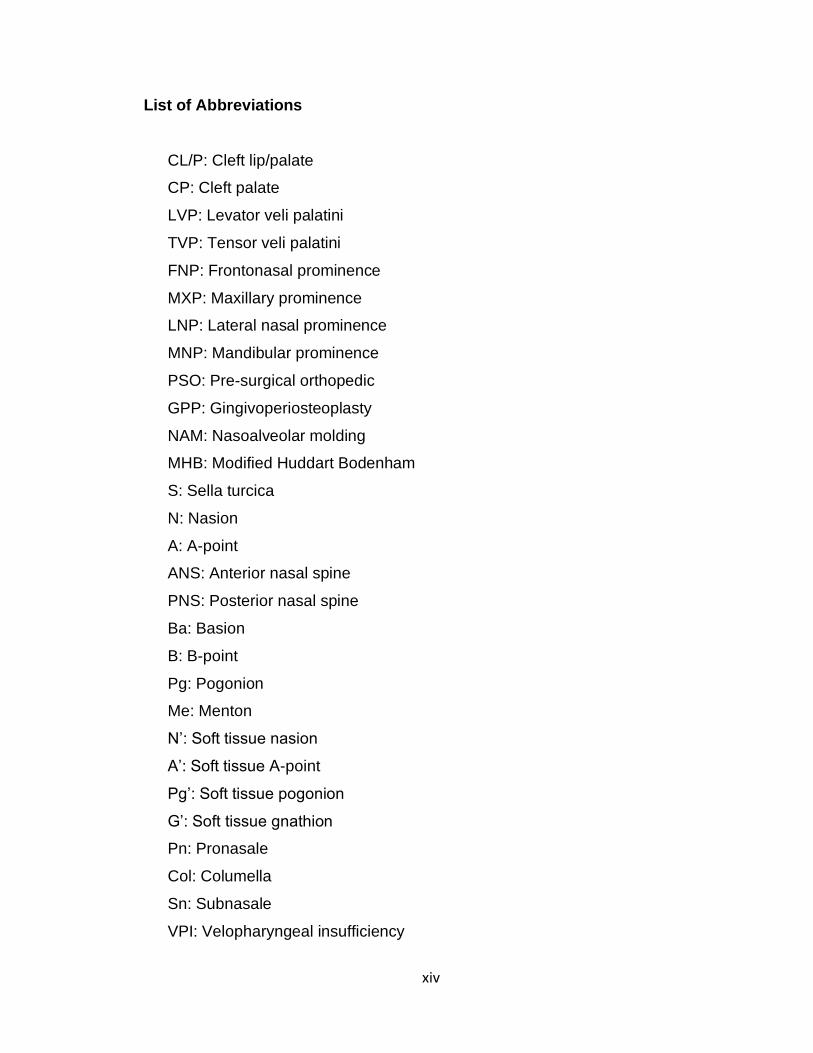
List of Abbreviations
CL/P: Cleft lip/palate
CP: Cleft palate
LVP: Levator veli palatini
TVP: Tensor veli palatini
FNP: Frontonasal prominence
MXP: Maxillary prominence
LNP: Lateral nasal prominence
MNP: Mandibular prominence
PSO: Pre-surgical orthopedic
GPP: Gingivoperiosteoplasty
NAM: Nasoalveolar molding
MHB: Modified Huddart Bodenham
S: Sella turcica
N: Nasion
A: A-point
ANS: Anterior nasal spine
PNS: Posterior nasal spine
Ba: Basion
B: B-point
Pg: Pogonion
Me: Menton
N’: Soft tissue nasion
A’: Soft tissue A-point
Pg’: Soft tissue pogonion
G’: Soft tissue gnathion
Pn: Pronasale
Col: Columella
Sn: Subnasale
VPI: Velopharyngeal insufficiency

xv
AG: Alveolar gap

1
Chapter 1: Introduction
This chapter introduces the current state of knowledge regarding cleft lip and cleft
palate. The embryology, important surgical anatomy, and classification of the cleft
lip and palate are reviewed. Current standards of care for the management of cleft
lip and palate are reviewed including cleft lip repair, cleft palate repair, and pre-
surgical orthopedic devices. Current methods of assessing outcomes in these
patients including nasolabial aesthetic evaluation, dental arch/occlusion analysis,
and cephalometric analysis are reviewed. The objective, purpose, rationale, and
hypothesis of this work are also given.

2
EPIDEMIOLOGY OF CLEFT LIP AND PALATE
Cleft lip and palate are common pediatric craniofacial malformations (1). In
Canada, the incidence of cleft lip and palate in newborns is 0.82:1000 and the
incidence of isolated cleft palate is 0.58:1000 (2). Cleft lip and palate can also be
associated with various syndromes including Stickler syndrome (25% of cases),
velocardiofacial syndrome (15%), and van der Woude’s syndrome (19%).
Approximately 13.8% of cleft lip and palate, and 41.8% of isolated cleft palate, are
associated with syndromes (3). A common triad of presentation in non-syndromic
patients is the Pierre-Robin sequence which includes micrognathia/retrognathia,
glossoptosis, and airway obstruction (3). The genetic association of non-syndromic
cleft lip and palate is estimated at 20 to 50%, with environmental factors likely
contributing to the remainder of cleft development (3). Environmental factors
associated with the development of cleft lip/palate include maternal smoking,
maternal corticosteroid use, folic acid deficiency, high altitude, and increased
parental age (3).

3
EMBRYOLOGY OF CLEFT LIP AND PALATE
The pathogenesis behind the development of cleft lip and palate is complex and
multifactorial. The entire pathophysiology behind the process is not known, and
many studies in this domain are animal studies (4). The following is an overview
of the current state of knowledge of the pathophysiology behind the development
of cleft lip and palate.
Craniofacial tissues originate from neural crest cells and mesoderm. Neural crest
cells migrate to the prominences of the face from the dorsal neural tube. During
development in utero, the lip develops during the first 4 to 10 weeks of gestation
and the palate develops between 4 to 12 weeks of gestation. The lip and palate
are formed by the fusion of 7 facial prominences. Failure of the neural crest cells
in these prominences to migrate to midline and fuse is what leads to cleft lip and
palate deformities. Signaling molecules involved in the migration of neural crest
cells to midline include Wnt, FOXE1, and IRF6 (3,4).
The face is formed by 7 facial prominences (Figure 1): the frontonasal prominence,
bilateral maxillary prominences, lateral nasal prominences, and the bilateral
mandibular prominences. The frontonasal prominence gives rise to the forehead,
central nose, philtrum, middle of the upper lip, and the primary palate. Interruption
in the growth of the frontonasal prominence leads to bilateral cleft lip. The maxillary
prominences give rise to the upper jaw, sides of the face, sides of the upper lip,
and the secondary palate. Disruption of maxillary prominence growth can lead to
clefts of the secondary palate and the lip. The lateral nasal prominences give rise
to the nasal alae. Failure of fusion between the lateral nasal prominences and the
frontonasal/maxillary processes can lead to clefts of the side of the nose. The
mandibular prominences give rise to the lower jaw and lower lip. Disruption in
growth of the mandibular prominences can result in mandibular clefts, although
this is quite rare. Failure of fusion between the frontonasal and maxillary processes
results in clefts of the primary palate (3,4).

4
Figure 1: Reproduced from Neligan et al. with permission from Elsevier publishing (4). Embryonic facial prominences and their corresponding adult facial structures. FNP= frontonasal prominence; MXP= maxillary prominence; LNP= lateral nasal prominence; MNP= mandibular prominence.

5
CLEFT CLASSIFICATION
There are several important definitions used when describing cleft lip and palate.
Clefts can occur in the primary or the secondary palate. The primary palate
extends from the base of the nasal processes to the incisive foramen of the
alveolus. The secondary palate extends from the incisive foramen to the uvula.
Clefts can be classified as unilateral or bilateral. Unilateral clefts are isolated to
one side of the primary/secondary palate (either left or right) and bilateral clefts
involve both sides. Clefts are also described as complete or incomplete. A
complete cleft of the primary or secondary palate involves the entire unit of the
palatal structure, whereas an incomplete cleft palate does not (4). See Figure 2
for examples of cleft classification.
Figure 2: Copyright Brito et al. (5), open access figure. Normal palate anatomy (upper middle image). (A) Unilateral complete cleft of primary palate, (B) Bilateral complete cleft of primary palate, (C) Unilateral complete cleft of primary and secondary palate, (D) Bilateral complete cleft of primary and secondary palate, (E) Complete cleft of secondary palate.

6
SURGICAL ANATOMY OF THE CLEFT LIP
The anatomy of the lip is important for understanding the principles behind surgical
repair and assessing post-operative outcomes. The following is a brief overview of
lip anatomy and changes in the anatomy associated with cleft lip.
The lip itself is composed of several surface landmarks: philtral columns, philtral
dimple, Cupid’s bow, vermillion border, and the vermillo-mucosal junction (Figure
3). The philtral columns are created by the insertion of the orbicularis oris muscle
fibers, and the philtral dimple is the concavity created by the paucity of muscle
fibers between the two columns. The Cupid’s bow is the curve of the upper lip
between the two philtral columns. The vermillion is the border between the red lip
and the adjacent normal skin. The vermillo-mucosal junction is the border between
the dry (keratinized) and the wet (non-keratinized) portions of the lip. Nose
anatomy is also important when discussing clefts of the primary palate. Important
structures of nasal anatomy include the alar base, the nasal tip, the nostrils, and
the columella (Figure 3).
There are 2 principle muscles that are relevant to the surgical repair of the cleft lip,
the orbicularis oris and the levator labii superioris. The orbicularis oris muscle
functions to move the lips for speech, facial expression, and eating. The levator
labii superioris functions to elevate the upper lip. The lips receive their blood supply
from bilateral superior labial arteries, their sensory innervation from the V2 of the
trigeminal nerve, and motor innervation from the facial nerve (3).
In patients with a cleft of the primary palate involving the lip, the orbicularis oris
muscle inserts at the alar wing (at the edge of the cleft) instead of its usual insertion
at the mucous membrane of the lip. There is also hypoplasia of the pars marginalis
(a component of the orbicularis oris). A cleft of the primary palate involving the lip
also causes shortening of the philtrum, diminished vermillion width on the non-cleft
side, and increased vermillion width on the cleft side (3). A complete cleft of the

7
primary palate can cause multiple nasal deformities including nasal tip deviation,
septal deviation (to the non-cleft side), widened and inferiorly positioned alar base
(cleft side), collapsed lateral cartilage (cleft side), columellar shortening (cleft side),
and inferior turbinate hypertrophy (cleft side) (3).
Figure 3: Original photograph of important nasolabial landmarks. (1) Philtral column, (2) Philtral dimple, (3) Cupid's bow, (4) Vermillion, (5) Vermillo-mucosal junction, (6) Alar Base, (7) Columella, (8) Nostril, and (9) Nasal tip.

8
SURGICAL REPAIR OF THE CLEFT LIP
The objective of surgical repair of the cleft lip is anatomic re-creation of normal lip
elements (Figure 3) and maintenance of the vertical height of the lip (6). When
performing cleft lip repair the skin, muscle, and mucosal layers must be re-aligned
(6). The most common repair techniques are the rotation advancement flap by
Millard, triangular flap technique by Tennison, wave line closure by Pfeifer, and
functional repair by Delaire. The techniques vary in their incision lines, but
ultimately aim to restore anatomic alignment of the lip (7,8).
In patients with a complete primary cleft palate, the alveolar gap makes lip repair
difficult. Pre-surgical orthopedics (PSO) are frequently used in patients with large
alveolar gaps prior to lip repair. The purpose of PSOs is to decrease the gap size,
bring the lip elements closer together, re-establish the palatal arch, and aid with
intra-oral feeding (9–11). Pre-surgical orthopedics include lip taping, lip adhesion,
and PSO devices (11,12). On average, the surgical management of cleft lip is
usually undertaken when the child is close to 3 months of age and PSOs are
undertaken soon after birth and used until the time of lip repair (6).
At the time of lip repair, a gingivoperiosteoplasty (GPP) can also be performed to
close the alveolar gap. The purpose of a GPP is to close the alveolar gap, reduce
the necessity for bone grafting, and recreate an anatomic palatal arch (13,14).

9
SURGICAL ANATOMY OF THE CLEFT PALATE
The anatomy of the palate is important for understanding how cleft palate is
managed and for assessing post-operative outcomes. The following is a brief
overview of palate anatomy and changes in the anatomy associated with cleft
palate.
The palate is composed of the hard and the soft palate (Figure 2). The hard palate
is the boney structure that functions as a rigid floor for the nasal cavity. The hard
palate is composed of the premaxillary portion, the palatine processes of the
maxilla, and the palatine processes of the palatine bone. The hard palate receives
its blood supply from the greater palatine, nasopalatine, anterior superior alveolar,
and posterior superior alveolar arteries. The hard palate receives its innervation
from the greater palatine and nasopalatine nerves. The soft palate is the
fibromuscular structure posterior to the hard palate that moves to open and close
the Eustachian canals. The muscles that form the soft palate include the levator
veli palatini, tensor veli palatini (TVP), palatopharyngeus, palatoglossus, the
musculus uvulae, superior pharyngeal constrictor, salpingopharyngeus, and the
stylopharyngeus. Blood supply to the soft palate originates from the ascending
pharyngeal and ascending palatine arteries. Innervation is supplied by the cranial
nerve 5 (TVP only) and pharyngeal plexus (remaining muscles) (3).
The palate can further be divided into the primary palate and secondary palate.
The primary palate lies anterior to the incisive foramen and includes the lip, nostril
sill, alveolus, and the portion of the hard palate that is anterior to the incisive
foramen. The secondary palate lies posterior to the primary palate and includes
the hard palate posterior to the incisive foramen and the soft palate (3).
Changes to palate anatomy vary depending on the extent of the cleft. Typically,
muscle fibers of the involved muscles are hypoplastic and more connective tissue
lies within the muscular bed (3).

10
SURGICAL REPAIR OF CLEFT PALATE
The objective of surgical repair of the cleft palate is to allow for good speech
development, velopharyngeal function, and midface development. Three layers
must be addressed in palate repair: the nasal layer, the muscle layer, and the oral
mucosa layer. Techniques for palate repair include the von Langenbeck
palatoplasty, Veau-Wardill-Kilner palatoplasty, Furlow double opposing Z-
palatoplasty, vomer flaps, and the intra-velar veloplasty (7,8).
Repair of the palate can either be done in 1 stage or 2 stages; 2-stage repair
involves fixing the hard and soft palate at 2 separate times. Two stage repair is
beneficial for midface growth, but does have a detrimental effect on speech
development in addition to the need for an additional operative procedure and
anesthesia (7). It is still unclear from the literature which technique/method results
in the best speech outcomes with the least effect on growth (15–20).
TIMING OF SURGERY
Timing of surgery for cleft lip and palate is crucial for proper growth and speech
development and is still highly debated among cleft palate surgeons. It has been
shown in the literature that patients with untreated cleft lip and palate have normal
growth of the nasomaxillary complex (21,22), whereas patients who have had lip
and palate repairs have diminished growth (23). Ultimately, this indicates that
surgical management may cause limitations in growth. This presents cleft
surgeons with the challenge of balancing the timing of cleft palate repair for
appropriate speech development, while limiting the negative impact on growth.
11
Advocates for early surgical management believe that surgical closure of the
palate allows for improved speech development, while advocates of late palate
closure believe that the closure itself causes a growth deficit (24–26). Previous
research from the Oxford Cleft Palate Study Team shows greater speech
deficiencies (articulation, nasal resonance, intelligibility and substitution pattern) in
patients with delayed hard palate closure (4 years) compared to those with early
palate closure (1 year) (27). No difference in maxillofacial growth was found
between the groups. Conversely, Bardach et al. found improved facial growth in
patients with delayed hard palate closure (up to 12-15 years), but higher incidence
of velopharyngeal insufficiency (25). Rohrich et al. studied outcomes in patients
with early versus delayed closure and found that delayed closure resulted in
significant speech impairments with little difference in overall maxillary growth (27).
Shaffer et al. also found higher rates of language delay and need for speech
therapy in patients with delayed palate repair (over 13 months) (28). Evidence from
the Scandcleft studies have demonstrated no significant difference in dental arch
or maxillary development in patients who had palatal surgery at 12 versus 36
months (29,30).
Overall, the timing for palate surgery is still highly debated. The trend with cleft
palate surgeons in North America is to perform a single stage cleft palate surgery
at 9 to 12 months of age for improved patient speech outcomes and reduced risk
of maxillary growth disturbance (31–34).

12
MANAGEMENT OF CLEFT LIP AND PALATE AT OUR INSTITUTION
At our institution the management of patients with cleft lip and palate is a multi-
staged, multi-disciplinary approach involving plastic surgery, otolaryngology,
speech and language therapy, social work, and dentistry (34). The primary goal of
cleft palate surgery is to achieve satisfactory functional (e.g. eating, speech) and
aesthetic results (1).
At our institution, cleft lip repair is usually performed at 3 months of age and cleft
palate repair is performed at 12 months. All patients with a cleft lip receive a
modified Mohler lip repair. Patients with a cleft of the secondary palate receive a
Furlow palatoplasty, hybrid palatoplasty, or von Langenbeck palatoplasty
depending on whether the cleft is complete or incomplete. Pre-surgical orthopedic
devices are often used to narrow the alveolar gap and facilitate intra-oral feeding
prior to cleft lip repair. At our institution, the decision to use a PSO device is made
at the first clinic visit which is usually 1 to 2 weeks post-birth. Families with a patient
that has an alveolar gap greater than 4mm are offered pre-surgical management
with a PSO device. If the patient is to receive a PSO device, they are sent to an
orthodontic specialist where a plaster cast of their cleft is made. This cast is used
to design either an active or a passive molding device. Passive molding devices
can be inserted in the office, but active PSO devices require insertion in the
operating room under general anesthesia. Active devices require parents to turn a
screw on the device daily to re-approximate the edges of the cleft. For a passive
device, the patient and caregiver are expected to come for regular follow-ups visits
to gradually mold the device. The decision of which PSO device to use is made
based on the severity of the alveolar gap, the caregiver’s capacity to handle care
of the device at home, and caregiver preference. Figure 4 gives a visual
representation of the management protocol that a patient with cleft lip and palate
receives at our institution.

13
Figure 4: Overview of management process for CL/P and CP patients at our institution. ENT= otolaryngology, SLP= speech language pathology.

14
PRE-SURGICAL ORTHOPEDIC DEVICES
1.10.1 Active Devices
Active PSO devices are designed to drive the edges of the alveolar gap closer
together using pins or screws (35). One of the most commonly used active devices
is the Latham appliance. It was first introduced by Latham and Millard in 1990 (36).
The Latham appliance (Figure 5) is a pin-retained intra-oral device that utilizes a
mechanical screw that is turned daily by the parents to approximate the edges of
the alveolar gap by approximately 0.5mm per day (37–39).
Active devices were initially designed to be used in conjunction with GPP to narrow
the alveolar gap prior to lip repair to facilitate the repair and prevent dehiscence
(40,41). Multiple studies have shown that active devices and GPP improved
aesthetic outcomes, allowed for adequate maxillary growth, and decreased the
number of nasoalveolar fistulas (37,41,42). Unfortunately, active devices in
conjunction with GPP have also been identified to cause decreased maxillary
protrusion and height throughout development, reduce successfulness of
secondary bone grafting, and have no effect on the number of patients that require
secondary bone grafting (43,44). Following these more recent studies, the use of
GPP in patients has decreased significantly.
The effect of the active device alone is still debated. Lin et al. have found that the
active device is associated with an increased incidence of ectopic permanent
maxillary first molars compared to patients without a device (45). Chan et al. have
shown that compared to patients treated with no device, there is no difference in
long-term (10-year) dental occlusion or growth in children treated with an active
device (46). Kornbluth et al. have also shown that active devices are associated
with improved nasolabial aesthetic outcomes, but cause a midface growth
disturbance (35). Overall, there is still no conclusion about the effects of the active

15
device and high level evidence is lacking (35). Further research must be done to
establish the effect of the active device without GPP on growth outcomes in
patients with cleft lip and palate.
Figure 5: The Latham device is pictured above on a patient mold (left) and in-situ (right).

16
1.10.2 Passive Devices
Passive PSO devices have the same overall objective as active PSO devices, but
do not actively drive together the edges of the alveolar gap. Instead, passive
devices are intra-oral plates that are custom fit to patients. These devices are
modified weekly to help bring the oronasal structures into a more anatomic
position. Ideally, passive molding is started in the first week of life and used in
patients with a cleft of 3 to 8mm (47). Passive PSO devices work because of the
plasticity of neonate cartilaginous and boney structures shortly after birth (47,48).
An example of a passive PSO device is the nasoalveolar molding (NAM) device
(Figure 6). NAM was first described in 1993 by Barry Grayson and works by
molding the alveolar segments and nose through a custom-fit intra-oral appliance
with nasal stents.
Multiple studies have shown that when compared to the contralateral non-clefted
side, passive devices appear to improve several aesthetic factors including
columella length, columella deviation, nostril width, and nasal height (48–50).
Passive devices have also been shown to improve nasolabial aesthetic outcomes
when compared to patients treated without a PSO device (51,52). In regards to the
effect of these devices on growth, it has been shown that they decrease the size
of the alveolar gap in the short term (less than 2 years of age) (53,54). Previous
studies have compared patients who received a passive device to those who did
not, and passive molding has been shown to improve dental arch symmetry and
maxillary growth until 6 years of age (54).
Contrasting evidence has also been published on passive molding. Prahl et al. as
part of the Dutchcleft study group showed that passive PSO devices do not
improve contact between the alveolar segments of the cleft or prevent long-term
(6-year) alveolar collapse (55,56). Similar results have been found by other groups
(57,58). Studies have also proposed that passive molding provides no
improvement in nasolabial aesthetics compared to patients who received no

17
device (59). With respect to the effect on dental development and facial growth,
several studies have shown no long-term effects of passive molding on maxillary
growth and dental arch relationships when compared to patients treated with no
device (46,55,56),
Overall, the benefits of passive PSO devices are still debated. One of the most
strongly supported benefits is that passive devices can help improve nasal
aesthetics and narrow the cleft (48,49). There is limited research that actually
examines the long-term benefits of passive molding (54,60).
Figure 6: Original photograph. A custom-made NAM device is pictured above.

18
1.10.3 Comparing Active and Passive Devices
In the field of cleft palate research there are very few studies comparing active
versus passive molding devices. The trend in cleft palate management is to either
use active molding or passive molding, and there is very limited data that actually
compares the two methods of molding. Kornbluth et al. have compared patient
outcomes that were treated with active or passive devices compared to those
treated without a device (35). Unfortunately, this study did not report statistics for
the comparison between the active and passive device groups specifically,
although they appeared to have similar results between the two groups for
nasolabial aesthetics, facial growth, and dental occlusion. A major limitation in this
study was that patients in the different device groups were treated by different
surgeons using different management protocols. It is difficult to compare outcomes
in patients treated with active versus passive devices when these confounding
variables could influence results. A comparison of active and passive molding
techniques where confounding variables are eliminated would be beneficial for
determining if certain techniques are superior for patient outcomes including
midface growth, dental occlusion, and nasolabial aesthetics (35).
19
METHODS OF ASSESSING POST-OPERATIVE AESTHETIC OUTCOMES
1.11.1 Nasolabial Aesthetics
Patients with cleft lip and palate undergo multiple surgical interventions in order to
ensure adequate functional and anatomic correction of the cleft deformity (61).
Despite these corrections, patients have been found to have persistent anatomic
deformities including abnormal nasal shape, scarring of the upper lip, and uneven
vermillion border (62,63). While certain studies have reported no psychosocial
concerns for patients with cleft lip and palate compared to patients without, several
systematic reviews have shown that patients with cleft lip/palate are at higher risk
for dissatisfaction with facial appearance, behavioural problems, and impairment
in social functioning (64,65). Therefore, it is important to use an objective
assessment tool for evaluating nasolabial aesthetic outcomes to ensure
satisfactory aesthetic results.
Aesthetic scales are frequently used in plastic surgery to assess post-operative
outcomes and the same tools are used for assessing outcomes in patients with
cleft lip/palate. Scales used to assess nasolabial aesthetics can be divided into
quantitative and qualitative scales. Quantitative scales use specific anthropometric
measurements to evaluate different soft tissue landmarks on patient photographs
for overall symmetry (66,67). Qualitative scales are used to evaluate aesthetics on
patient photographs and include ordinal scales, visual analog scales, and ranking
scales (62). Previous research shows that ranking scales have the greatest intra-
rater reliability, followed by visual analog scales and ordinal scales (63).
Historically, 2-dimensional (2-D) photographs have been used to assess post-
operative outcomes, but studies have begun using 3-dimensional (3-D) images as
well. When comparing 2-D and 3-D images, 3-D images have greater intra-rater
reliability but scoring of patients evaluated with the two modalities is not different
(68).

20
The focus of nasolabial aesthetic assessment in patients with cleft lip and palate
is often on nasal form, nasal symmetry, vermillion border form, and lip symmetry.
These aspects of the nose and lip are disrupted in cleft lip/palate and repair is
meant to bring the nose and lip back into an anatomic position (61,69). Important
components of nasolabial aesthetic outcomes include the scar, alar base
symmetry, columellar deviation, nasal tip recurvatum, nostril symmetry, Cupid’s
Bow symmetry, the vermillo-cutaneous junction, and the vermillo-mucosal junction
(Figure 7) (61,69).
Figure 7: Original photographs. Important landmarks to assess nasolabial aestehtics include (A) symmetry of Cupid’s Bow and the vermillo-cutaneous junction, (B) symmetry of the vermillo-mucosal junction, (C) alar base symmetry, (D) scar quality and position, (E) nostril symmetry, and (F) nasal tip symmetry.

21
METHODS OF ASSESSING POST-OPERATIVE GROWTH OUTCOMES
1.12.1 Angle Classification
Dental occlusion is defined as the way in which the maxillary and mandibular teeth
fit together. Classically, occlusion is described using the Angle classification. The
Angle classification is the gold standard for describing occlusion and was initially
developed in the 1890’s to describe dental occlusion based on the position of the
mesiobuccal cusp of the maxillary first molar to the buccal groove of the
mandibular first molar (70). The Angle classification system is as follows (Figure
8): (1) Normal occlusion, (2) Class I having normal molar relationship despite tooth
rotation or crowding, (3) Class II having lower molars distal to upper molars, and
(4) Class III having lower molars mesial to upper molars (71).
The Angle classification system is widely used for classifying occlusion but has
been criticized for being a 2-D classification of a 3-D problem. The Angle
classification is useful for characterizing sagittal occlusion, but other scoring
systems have been described for assessing occlusion in a 3-D way.
Figure 8: Original images. Angle classification system of occlusion.
1.12.1.1.1 GOSLON Yardstick

22
Several different methods for scoring dental arch relationships have been used in
the CL/P population. The GOSLON Yardstick is the gold standard for measuring
skeletal and dental relationships in patients with CL/P and was first developed in
1987 (35,72). The GOSLON Yardstick classification rates the occlusion into 1 of 5
categories using the anteroposterior, vertical, and sagittal dimensions of 3-D casts
of patients’ mouths. This classification system provides a general prediction about
future requirements for surgery (73). The GOSLON Yardstick is based on the
distance between the incisal edge of the maxillary incisors and the labial surfaces
of the mandibular incisors, termed “overjet”. The five grades are as follow:
Group 1 Positive overjet Minimal or no treatment required
Group 2 Positive overjet Minimal treatment required
Group 3 Edge to edge bite Orthodontic treatment required, but good
outcome anticipated
Group 4 Negative overjet
(1-3mm)
Orthodontic treatment required with
possible future orthognathic surgery
Group 5 Negative overjet (>3mm) Definite orthognathic surgery required
The GOSLON yardstick is a useful tool for analyzing occlusion and predicting the
need for future orthognathic intervention (74). See Figure 9 for photographs of
patient molds with different grades of occlusion.

23
A
B
C
Figure 9: Original photographs. Patient molds representing: (A) Positive overjet, GOSLON Yardstick Grade 1/2; (B) No overjet, GOSLON Yardstick Grade 3; (C) Negative overjet, GOSLON Yardstick Grade 4/5.

24
1.12.2 Modified Huddart/Bodenham Classification
The Modified Huddart/Bodenham (MHB) classification is another classification
system of dental occlusion that was initially developed in 1997 for primary dentition
and modified in 2003 to apply to mixed/permanent dentition (75). The MHB system
classifies the position of each maxillary tooth in relation to its mandibular tooth
partner. Each relationship is given a score from -3 to +1 and the mean score for
each relationship is calculated (Figure 10); a more negative score indicates worse
dental relationships. In primary dentition, the two central incisors, bilateral canines
and bilateral molars are measured to total 8 categories of evaluation. In primary
dentition the total MHB score can range from -24 to +8. In mixed/permanent
dentition the two central incisors, bilateral canines, bilateral pre-molars, and
bilateral first permanent molars are measured to total 10 categories of evaluation.
In mixed/permanent dentition the total MHB score can range from -30 to +10 (75).
In regard to whether the GOSLON yardstick or the MHB classification is superior
for classifying occlusion, this is still highly debated. In the literature, both have been
favored for different reasons. The MHB system is less subjective than the
GOSLON Yardstick and appears to be more versatile for primary versus
permanent dentition (30,76). The GOSLON Yardstick is more user-friendly and
has been used for much longer than the MHB (76). Ultimately, controversy still
remains about which classification system is best. Comparative studies have
shown that MHB has been found to be superior for 5-year (primary) dentition, but
GOSLON Yardstick is preferred for 10-year (permanent) models (72,76).

25
Figure 10: Reproduced with permission from Sage Publishing. Modified Huddart Bodenham classification of molars, canines, and incisors.

26
1.12.3 Dental/Occlusal Changes in Cleft Lip & Palate
Cleft lip and palate can produce a variety of dental problems including
missing/additional teeth and abnormal shape and position of both deciduous and
permanent teeth. Most commonly, teeth in the area of the cleft are affected. The
most common orthodontic problem in patients with CL/P is maxillary asymmetry
causing crossbite formation (7). The overall effect of cleft size on dental occlusion
is still debated. Some authors have found that there is no relationship between the
size of the cleft and occlusion (77,78). Conversely, several studies have shown
that initial cleft size negatively affects maxillary inclination, which in turn affects
occlusion (74,79). The effect of cleft size on dental arch and occlusal relationships
is more likely to be multifactorial, with type and timing of palate repair also affecting
the outcome (74).
1.12.4 Palatal Measurements
The width of the anterior and posterior palate represents the overall width of the
palate and is important for understanding the space that the dental arch has for
the teeth. The inter-canine width (ICW) and sum of the incisors (SI) represent the
width of the anterior dental arch, and the inter-palatal molar width (IPMW)
represents the width of the posterior dental arch (80). Inter-canine width is the
distance between the maxillary canine grooves and IPMW is the distance between
the retromolar points of the maxillary permanent first molars or deciduous second
molars. The sum of the incisors is the maximum mesiodistal size of each of the
four incisors (81). Reduction in maxillary dental arch width is a common concern
with cleft palate and measuring arch width can help monitor growth and
development of the dental arch. See Figure 11 for important dental arch
measurements.

27
Figure 11: SI= sum of incisors, ICW= inter-canine width, IPMW= inter-palatal molar width

28
1.12.5 Cephalometric Analysis
Cephalometric measurements represent the gold standard for measuring facial
growth and development (35). Cephalometric landmarks are plotted on lateral
facial radiographs and used to analyse hard tissue points of interest that affect
facial features. Historically, radiographs have been traced by hand to plot
landmarks. More recently, programs such as Dolphin Imaging™ have been
designed to plot landmarks and calculate measurements electronically (82).
There are two main functions of cephalometric analysis: (1) to compare facial
morphology against a matched norm and (2) to assess facial growth in a patient
(82). Cephalometric measurements can help monitor stability of the facial skeleton
and determine whether surgical intervention is required to help correct deformity.
Traditionally, hard tissue markers have been used for cephalometric analysis.
Boney landmarks do not predict soft tissue changes, so soft tissue traits must also
be taken into consideration when analyzing facial morphology (83). Cephalometric
analysis has now been modified to include soft tissue points of interest to help
better analyze facial growth outcomes. Important boney cephalometric landmarks
include sella turcica (S), nasion (N), A-point (A), posterior nasal spine (PNS),
anterior nasal spine (ANS), basion (Ba), B-point (B), pogonion (Pg), and menton
(Me) (Figure 12) (44,82–84). Important soft tissue landmarks include glabella (G’),
pronasale (Pn’), soft tissue pogonion (Pg’), columella (Col), subnasale (Sn), upper
lip (UL), soft tissue A-point (A’), soft tissue nasion (N’), and soft tissue B-point (B’)
(Figure 13) (44,82–84).
14

29
Figure 12: Original photograph. Cephalometric tracing of lateral patient x-ray. Important boney landmarks identified (in blue). (1) Sella Turcica, (2) Nasio, (3) A-point, (4, 5) Posterior and Anterior Nasal Spine, (6) Basion, (7) B-point, (8) Pogonion, (9) Menton.

30
Figure 13: Original photograph. Cephalometric tracing of lateral patient x-ray. Important soft tissue landmarks identified (in blue). (1) Glabella, (2) Nasion, (3) Pronasale, (4) Columella, (5) Subnasale, (6) A-point, (7) Upper lip, (8) B-point, (9) Pogonion.

31
1.12.6 Brief Overview of Important Facial Relationships
There are five functional components of a cephalogram: cranial base/cranium,
skeletal maxilla, skeletal mandible, maxillary dentition, and mandibular dentition.
There are two ways of standardizing cephalograms with a horizontal reference
plane: the Frankfort plane or the natural head position. The Frankfort plane creates
a horizontal reference plane between the external auditory meatus to the anterior
inferior orbital rim. The natural head position parallels the patient’s visual axis by
having the patient look at an object at the distant horizon (85).
Cephalometric analysis is important for analysis of growth, but these boney
relationships can also help describe occlusion and predict facial attractiveness in
patients. In addition, there exist some soft tissue relationships that are also
predictive of facial attractiveness (85). These relationships can be quantified using
cephalometric analysis. Cephalometric landmarks act as the foundation to create
different cephalometric measurements that reflect important areas of facial growth
and development. See Table 1 for a list of common cephalometric measurements
and their importance for analyzing growth.
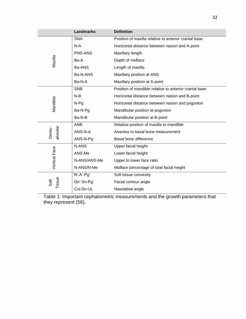
32
Landmarks Definition
Maxill
a
SNA Position of maxilla relative to anterior cranial base
N-A Horizontal distance between nasion and A-point
PNS-ANS Maxillary length
Ba-A Depth of midface
Ba-ANS Length of maxilla
Ba-N-ANS Maxillary position at ANS
Ba-N-A Maxillary position at A-point
Mandib
le
SNB Position of mandible relative to anterior cranial base
N-B Horizontal distance between nasion and B-point
N-Pg Horizontal distance between nasion and pogonion
Ba-N-Pg Mandibular position at pogonion
Ba-N-B Mandibular position at B-point
Dento
-
alv
eola
r ANB Relative position of maxilla to mandible
ANS-N-A Alveolus to basal bone measurement
ANS-N-Pg Basal bone difference
Vert
ical F
ace
N-ANS Upper facial height
ANS-Me Lower facial height
N-ANS/ANS-Me Upper to lower face ratio
N-ANS/N-Me Midface percentage of total facial height
Soft
Tis
sue N’-A’-Pg’ Soft tissue convexity
Gn’-Sn-Pg’ Facial contour angle
Col-Sn-UL Nasolabial angle
Table 1: Important cephalometric measurements and the growth parameters that they represent (56).

33
Figure 14: Maxillary cephalometric measurements.
Figure 15: Mandibular cephalometric measurements.

34
Figure 16: Dento-alveolar cephalometric measurements.
Figure 17: Vertical facial growth cephalometric measurements.

35
Figure 18: Soft tissue cephalometric measurements.

36
1.12.7 Facial Growth in Cleft Lip & Palate
Differences in facial morphology in patients with cleft lip and palate compared to
patients without a cleft include (1) shortened maxilla and mandible, (2) reduced
posterior maxilla height and increased width, (3) reduced palate size, oropharynx
and pharyngeal depth, (4) reduced posterior mandible height, (5) bimaxillary
retrognathia, (6) inferior position of hyoid, (7) reduced tongue height and velar
length, (8) reduced nasolabial angle, (9) reduced percent nose, and (10) reduced
mandibular incisor projection and inclination (84,86–89). There does not appear to
be any difference in skull base anatomy between patients with and without cleft
palate (90,91).
Patients with larger alveolar gaps have been shown to have poorer maxillary
protrusion and length (84,92). It has previously been shown in patients with cleft
lip and palate that overall midface growth in patients that do not undergo lip or
palate repair is similar to patients without any cleft. This finding has led to the
conclusion that surgical intervention does cause a growth disturbance (7). Liao et
al. have shown that when compared to patients who received lip repair only, palate
repair leads to limited anteroposterior development of the maxilla, and decreased
forward displacement (93). Several other studies have shown similar outcomes in
regards to the effect of palate repair on maxillary growth (21,23). Several studies
have shown that pressure from the lip repair can cause the upper anterior teeth to
become retroclined, leading to an anterior crossbite (7,94–97).

37
THESIS RATIONALE
The use of PSO devices in the management of patients with CL/P remains
controversial. In addition, few research studies actually compare patient outcomes
achieved by different types of PSO devices (35). To date, limited research has
compared long-term facial growth, dental occlusion, and nasolabial aesthetics
between patients who received an active PSO device versus a passive PSO
device. The research that does exist on this topic is limited by small patient cohorts
with multiple surgeons and management protocols within the same experimental
group (35,98). At our institution we have a single surgeon that has been using
active and passive PSO devices for over 15 years. All of these patients receive the
same operative and non-operative cleft management, except for the PSO device
used. This presents us with the unique opportunity to examine outcomes in a single
surgeon’s cohort of patients over a long-term period. By using a cohort of patients
operated on by a single surgeon with the same management protocol we can
eliminate some of the unnecessary confounding variables which are present in the
majority of current cleft palate literature (35,98).
Both active and passive PSO devices have their strengths and weaknesses.
Passive devices have an increased burden of care on the patient’s families
because they require weekly device adjustments. Active PSO devices require
fewer follow-up visits, but have the added risk and cost associated with operative
insertion of the device. With steps towards reducing unnecessary procedures,
risks, and costs in the medical system, it is important to determine whether one
type of PSO device produces superior results. The results from this thesis will be
important for determining management of patients with CL/P moving forward.
Overall, this work will give a more conclusive answer about the effects of the PSO
devices on nasolabial aesthetics, dental occlusion, and facial growth. In addition,
this will be the first study of its nature to compare outcomes in patients that
received an active versus a passive device.

38
RESEARCH OBJECTIVES AND HYPOTHESIS
There are 4 principle objectives to this research:
1) Perform a systematic review of current literature describing the long-term
effects of different PSO devices used for the management of cleft palate.
2) Evaluate and compare the nasolabial aesthetic changes in patients who
were treated with an active PSO device, passive PSO device, or no PSO
device at 1 year, 5 years, and 10 years.
3) Evaluate and compare dentoalveolar development/occlusion in patients
who were treated with an active or passive PSO device at 5 years and 10
years.
4) Evaluate and compare the facial growth in patients who were treated with
an active or passive PSO device at 5 years and 10 years.
We hypothesize that nasolabial aesthetics, dentoalveolar development, and facial
growth will be comparable between patients that received active PSO devices and
passive PSO devices. We hypothesize that active and passive PSO devices will
make nasolabial aesthetics in patients with larger alveolar gaps comparable to
patients who had smaller gaps and did not require a PSO.
39
Chapter 2: Systematic Review of the Literature on the Long-term Patient
Outcomes Associated with Pre-Surgical Orthopedic Devices
The use of pre-surgical orthopedic devices in cleft lip/palate patients is
controversial. The literature that exists on this topic is extremely varied. The
purpose of this chapter is to outline the current state of knowledge of the long-term
outcomes of patients with cleft lip/palate treated with pre-surgical devices prior to
lip repair.

40
INTRODUCTION
Clinical use of active and passive presurgical orthopedic (PSO) devices for the
management of patients with cleft lip/palate is a controversial topic. It is widely
accepted that these devices are useful for decreasing alveolar gap size prior to lip
repair (99–102), but their long-term effects on nasolabial aesthetics, dental
occlusion, and facial growth are still debated (35,54,103–108). Consequently, the
use of these devices is dependent on surgeon experience and caregiver
preference (30).
To date, there is a large body of research examining the outcomes of patients
treated with PSO devices, but there is no consensus on their effect on long-term
patient outcomes (35,98). The lack of consensus on the use of these devices is
likely in part due to the quality of the research itself. Research in cleft lip and palate
is often limited by small sample sizes and variable management protocols.
Frequently, patients from numerous sites and surgeons are included in a single
study to try and increase the study cohort. While advantageous for increasing the
power of studies, grouping patients undergoing dissimilar management ultimately
creates confounding factors within the studies (e.g. surgeon experience, surgical
procedure) (35,102,109). The variability that exists within and in between
comparison groups creates bias and confusion when drawing conclusions from the
research performed. In addition, there is also a paucity of research actually
comparing the difference in outcomes between patients treated with different types
of PSO devices (35).
The primary purpose of the research in this chapter was to complete a systematic
review of the literature pertaining to the use of PSO devices in cleft lip/palate and
their effect on long-term patient outcomes.

41
METHODOLOGY
A comprehensive literature review of Embase and Ovid databases was performed
to identify all English-language publications related to unilateral cleft palate,
presurgical devices, and patient outcomes. Selection criteria for included studies
were as follows:
Inclusion criteria:
• Describe the use of PSO for unilateral cleft palate management
• Describe patient outcomes past removal of device
• Include human subjects
• English-language articles
• Published at any date
• Any study design, including case series (>10 cases)
Exclusion criteria:
• Do not describe the use of PSO for cleft management
• Do not describe patient outcomes past removal of device
• Control group is patients without a cleft lip/palate
• Did not separate unilateral and bilateral cleft outcomes
• Study designed to validate a small modification of a previously validated
device
• Case report (<10 cases)
• Non-original studies
Key search terms included: “cleft lip”, “cleft palate”, “preoperative care”,
“orthodontics”, “palatal obturators”, and “infant”. An academic librarian was
involved to help build the literature search. All of the abstracts were reviewed
independently by two reviewers. Any disagreement about the inclusion of a study
was resolved by consensus. In addition, the reference lists of the included articles
were hand-searched for any articles missing from the database search. Following

42
the full-article review, a total of 41 articles were included in this systematic review.
Figure 19 describes the process of inclusion and exclusion of articles in this study.
The following data was collected for each paper: (1) study title, (2) authors, (3)
year of publication, (4) journal of publication, (5) type of publication, (6) number of
patients included in study, (7) type of PSO device used, (8) whether patient
management protocols were standardized within and between groups, (9)
outcomes measured, (10) age of patients at analysis, and (11) conclusions drawn
about the device being evaluated. Meta-analysis was not performed due to the
heterogeneity of reported results.
Figure 19: Flowchart of study inclusion.

43
RESULTS
The literature search yielded a total of 438 studies to review, of which 41 studies
met criteria for inclusion in the systematic review (35,45,46,51,54–
58,101,103,107,108,110–136). Of the 41 studies, 30 were retrospective studies
and 11 were prospective studies. See Table 2 for the list of all included articles,
year of publication, and type of study.
On average, studies had a total of 32.9 patients per intervention group. Sixteen
articles examined outcomes in patients under 5 years of age, 25 articles for
patients 5 to 10 years old, and 4 articles for patients over 10 years old. Of the 41
research articles, 23 focused on passive devices, 8 on active devices, and 10
compared active and passive devices. In total, 13 articles described nasolabial
aesthetics, 9 described facial growth, 23 described dental arch/occlusion
outcomes, 1 described rates of revision surgeries, and 1 described changes in
airway anatomy. For articles reporting on dental arch outcomes, 22% reported that
the PSO device improved dental arch/occlusion, 22% reported that the PSO device
caused worse dental arch/occlusion, and 56% reported no difference in outcomes
between patients that had a PSO device compared to those that did not (Figure
20). For articles reporting on nasolabial aesthetic outcomes, 64% reported
improvement in nasolabial aesthetics with the use of a PSO device and 36%
reported no difference in outcomes between patients that did have a PSO device
compared to those that did not (Figure 20). For articles reporting on facial growth
outcomes, 33% of studies reported that PSO devices worsened facial growth and
67% reported no difference in outcomes between patients that did have a PSO
device compared to those that did not (Figure 20). The sole article reporting on
airway measurements described no difference in outcomes between patients that
did have a PSO device compared to those that did not. The sole article reporting
on rates of revision surgery described a reduced number of revision surgeries in
patients that had a PSO device (Figure 20).

44
A total of 11 articles examined a patient cohort from a single surgeon’s practice,
21 used a cohort from multiple sites and/or surgeons, and 9 examined a cohort
from one site with an unspecified number of surgeons involved in the care of the
patients. Consistency in management protocols was quite variable between these
3 groups (Figure 21). Variability in the protocols included different operating
surgeons (73%), different type and timing of lip and/or palate repair (41%), whether
or not patients received GPP (10%), and whether patients received revision
surgeries (17%). Seven percent of papers did not describe their management
protocols and 9% described similar management for lip repair but did not describe
management following lip repair. In total, 15% of papers had a consistent
management protocol within and in between experimental groups.
With respect to the 11 articles that investigated a single surgeon’s practice, 8
articles examined the effects of a passive device and 3 examined the effects of an
active device. Three articles reported on nasolabial aesthetics, 3 reported on
dental arch/occlusion, 1 reported on facial growth, 1 reported on dental
arch/occlusion and growth, 2 articles reported on dental arch/occlusion and
nasolabial aesthetics, and 1 article compared rates of revision surgeries. Six of the
11 (55%) single-surgeon articles had inconsistent management protocols for all of
their patients or did not specify whether the management between the control and
experimental groups was the same. In the 5 articles that described consistent
management protocols between groups, no study specified whether the
treatment/control groups differed in their management following lip repair (e.g.
palate surgery, revision surgeries etc.). Two of the 5 articles examined outcomes
up to 4 to 6 months of age and the 3 remaining articles measured outcomes each
at one time point between 1 to 10 years of age. On average, these articles had
19.8 (range 13 to 42) patients in the experimental group and 29.5 (range 5 to 61)
patients in the control group. Three articles reported improved nasolabial
aesthetics with the use of a PSO device, 1 article reported no difference in
nasolabial aesthetics whether or not a PSO device was used, and 1 article reported
no difference in dental arch measurements whether or not a PSO device was used.

45

46
Paper Authors Journal Year Type of
Publication
1 Effect of Presurgical Nasoalveolar Molding on Nasal Symmetry in Unilateral Complete Cleft Lip/Palate Patients after Primary Cheiloplasty without Concomitant Nasal Cartilage Dissection: Early Childhood Evaluation.
Liang Z, et al. Cleft Palate-Craniofacial Journal
2018 Retrospective cohort
2 Active presurgical infant orthopedics for unilateral cleft lip and palate: Intercenter outcome comparison of Latham, modified McNeil, and nasoalveolar molding.
Kornbluth M, et al. Cleft Palate-Craniofacial Journal
2018 Retrospective cohort
3 Midface Growth in Patients With Unilateral Cleft Lip and Palate Treated With a Nasoalveolar Molding Protocol.
Rubin M.S, et al. The Journal of Craniofacial Surgery
2019 Retrospective cohort
4 The effect of nasoalveolar molding on nasal airway anatomy: A 9-year follow-up of patients with unilateral cleft lip and palate.
Massie J.P, et al. Cleft Palate-Craniofacial Journal
2018 Retrospective cohort
5 Burden of care of various infant orthopedic protocols for improvement of nasolabial esthetics in patients with CUCLP.
Singer E, et al. Cleft Palate-Craniofacial Journal
2018 Retrospective cohort
6 Long-term effect of presurgical nasoalveolar molding on growth of maxillary arch in unilateral cleft lip and palate: randomized controlled trial.
Shetty V, et al. International Journal of Oral and Maxillofacial Surgery
2017 Randomized controlled trial
7 A Comparison of Mixed Dentition Dental Development in Cleft Patients Treated With and Without the Latham-type Appliance.
Lin J, et al. Pediatric Dentistry 2017 Retrospective cohort
8 Comparison of two treatment protocols in children with unilateral complete cleft lip and palate: Tridimensional evaluation of the maxillary dental arch.
Jorge P.K, et al. Journal of Cranio-Maxillofacial Surgery
2016 Retrospective cohort
9 Comparison of Early Onset Nasoalveolar Molding with Patients Who Presented for Molding Up to 1 Year of Age.
Shetty V, et al. Journal of Oral and Maxillofacial Surgery
2016 Randomized controlled trial
10 Surgeon's and Caregivers' Appraisals of Primary Cleft Lip Treatment with and without Nasoalveolar Molding: A Prospective Multicenter Pilot Study.
Broder H.L, et al. Plastic and reconstructive Surgery
2016 Non-randomized clinical trial
11 Presurgical orthopedics has no effect on archform in unilateral cleft lip and palate.
Adali N, et al. Cleft Palate-Craniofacial Journal
2012 Non-randomized clinical trial
12 Presurgical nasoalveolar molding orthopedic treatment improves the
Sasaki H, et al. Journal of Craniofacial Surgery
2012 Retrospective cohort

47
outcome of primary cheiloplasty of unilateral complete cleft lip and palate, as assessed by naris morphology and cleft gap.
13 Long-term treatment outcome of presurgical nasoalveolar molding in patients with unilateral cleft lip and palate.
Clark S.L, et al. Journal of Craniofacial Surgery
2011 Retrospective cohort
14 Nasoalveolar molding improves long-term nasal symmetry in complete unilateral cleft lip-cleft palate patients.
Barillas I, et al. Plastic and Reconstructive Surgery
2009 Retrospective cohort
15 Infant orthopedics and facial growth in complete unilateral cleft lip and palate until six years of age (Dutchcleft).
Bongaarts C.A.M, et al.
Cleft Palate-Craniofacial Journal
2009 Randomized controlled trial
16 Effect of infant orthopedics on facial appearance of toddlers with complete unilateral cleft lip and palate (Dutchcleft).
Bongaarts C.A.M, et al.
Cleft Palate-Craniofacial Journal
2008 Randomized controlled trial
17 Infant orthopedics has no effect on maxillary arch dimensions in the deciduous dentition of children with complete unilateral cleft lip and palate (Dutchcleft).
Bongaarts C.A.M, et al.
Cleft Palate-Craniofacial Journal
2006 Randomized controlled trial
18 Infant orthopedics and facial appearance: A randomized clinical trial (Dutchcleft).
Prahl C, et al. Cleft Palate-Craniofacial Journal
2006 Randomized controlled trial
19 The effect of infant orthopedics on the occlusion of the deciduous dentition in children with complete unilateral cleft lip and palate (Dutchcleft).
Bongaarts C.A.M, et al.
Cleft Palate-Craniofacial Journal
2004 Randomized controlled trial
20 The effects of active infant orthopedics on occlusal relationships in unilateral complete cleft lip and palate.
Chan K.T, et al. Cleft Palate-Craniofacial Journal
2003 Retrospective cohort
21 A randomized prospective clinical trial of the effect of infant orthopedics in unilateral cleft lip and palate: Prevention of collapse of the alveolar segments (Dutchcleft).
Prahl C, et al. Cleft Palate-Craniofacial Journal
2003 Randomized controlled trial
22 A randomised prospective clinical trial into the effect of infant orthopaedics on maxillary arch dimensions in unilateral cleft lip and palate (Dutchcleft).
Prahl C, et al. European Journal of Oral Sciences
2001 Randomized controlled trial
23 Comparison between palatal configurations in UCLP infants with and without a Hotz plate until four years of age.
Mishima K, et al. Cleft Palate-Craniofacial Journal
2000 Retrospective cohort
24 Quantitative 3D maxillary arch evaluation of two different infant managements for unilateral cleft lip and palate.
Prasad C.N, et al. Cleft Palate-Craniofacial Journal
2000 Retrospective cohort
25 Cleft lip and palate treated by presurgical orthopedics, gingivoperiosteoplasty, and lip adhesion (POPLA) compared with
Millard D.R, et al. Plastic and Reconstructive Surgery
1999 Retrospective cohort

48
previous lip adhesion method: A preliminary study of serial dental casts.
26 Changes produced by presurgical orthopedic treatment before cheiloplasty in cleft lip and palate patients.
Kozelj V Cleft Palate-Craniofacial Journal
1999 Retrospective cohort
27 Analysis of primary gingivoperiosteoplasty in alveolar cleft repair. Part I: Facial growth.
Henkel K.-O, et al. Journal of Cranio-Maxillo-Facial Surgery
1997 Retrospective cohort
28 Morphologic effect of preoperative maxillofacial orthopedics (T-traction) on the maxilla in unilateral cleft lip and palate patients.
Larson M, et al. Cleft Palate-Craniofacial Journal
1993 Retrospective cohort
29 Craniofacial development in children with unilateral cleft of the lip, alveolus, and palate treated according to three different regimes. Assessment of nasolabial appearance.
Brattstrom V, et al. Scandinavian Journal of Plastic and Reconstructive Surgery and Hand Surgery
1992 Retrospective cohort
30 Early bone grafting in complete cleft lip and palate cases following maxillofacial orthopedics. III. A study of the dental occlusion.
Larson O, et al. Scandinavian Journal of Plastic and Reconstructive Surgery
1983 Retrospective cohort
31 An Intercenter Comparison of Dental Arch Relationships and Craniofacial Form Including a Center Using Nasoalveolar Molding.
Peanchitlertkajorn S, et al.
Cleft Palate–Craniofacial Journal
2018 Retrospective cohort
32 Presurgical Nasoalveolar Molding for Correction of Cleft Lip Nasal Deformity: Experience From Northern India
Mishra, et al. Open Access Journal of Plastic Surgery
2010 Retrospective cohort
33 A Six-Center International Study of Treatment Outcome in Patients with Clefts of the Lip and Palate: Part 4. Assessment of Nasolabial Appearance
Asher-McDade, et al.
Cleft Palate–Craniofacial Journal
1992 Retrospective cohort
34 A Six-Center International Study of Treatment Outcome in Patients with Clefts of the Lip and Palate: Part 3. Dental Arch Relationships
Mars, et al. Cleft Palate–Craniofacial Journal
1992 Retrospective cohort
35 Comparative Study of Early Secondary Nasal Revisions and Costs in Patients With Clefts Treated With and Without Nasoalveolar Molding
Patel, et al. Journal of Craniofacial Surgery
2015 Retrospective cohort
36 The Eurocleft Study: Intercenter Study of Treatment Outcome in Patients With Complete Cleft Lip and Palate. Part 2: Craniofacial Form and Nasolabial Appearance
Brattstrom, et al.
Cleft Palate–Craniofacial Journal
2005 Retrospective cohort

49
37 Postoperative Nasal Forms After Presurgical Nasoalveolar Molding Followed by Medial-Upward Advancement of Nasolabial Components With Vestibular Expansion for Children With Unilateral Complete Cleft Lip and Palate
Nakamura, et al. Journal of Oral and Maxillofacial Surgery
2009 Retrospective cohort
38 The Americleft Study: An Inter-Center Study of Treatment Outcomes for Patients with Unilateral Cleft Lip and Palate Part 3. Analysis of Craniofacial Form
Daskaloginannakis, et al.
Cleft Palate–Craniofacial Journal
2011 Retrospective cohort
39 The Americleft Study: An Inter-Center Study of Treatment Outcomes for Patients with Unilateral Cleft Lip and Palate Part 2. Dental Arch Relationships
Hathaway, et al. Cleft Palate–Craniofacial Journal
2011 Retrospective cohort
40 A Six-Center International Study of Treatment Outcome in Patients with Clefts of the Lip and Palate: Part 2. Craniofacial Form and Soft Tissue Profile
Molsted, et al. Cleft Palate–Craniofacial Journal
1992 Retrospective cohort
41 The Eurocleft Study: Intercenter Study of Treatment Outcome in Patients with Complete Cleft Lip and Palate. Part 3: Dental Arch Relationships
Molsted, et a. Cleft Palate–Craniofacial Journal
2005 Retrospective cohort
Table 2: Summary of articles included in final systematic review.

50
Figure 20: Distribution of results in all articles. Positive result indicates that PSO device improved outcomes compared to no device and negative indicates PSO device worsened outcomes.
Figure 21: Types of variability in the methodology of the reviewed studies.
5
9
0 0
113
5
6
1
0
5
0
3
0 0
0%
10%
20%
30%
40%
50%
60%
70%
80%
90%
100%
DentalArch/Dentition
Nasolabialaesthetics
Growth Airway Revisionsurgery
Negative
No difference
Positive
0 2 4 6 8 10 12 14 16
Same lip + palate
Same lip + palate, revisions different
Same lip, not described after
Different lip, not described after
GPP in some
Different lip + palate
Not described
1 surgeon 1 site Multi-site

51
DISCUSSION
Overall, this systematic review identified 41 studies examining the long-term
effects of PSO device use. The primary objective of this systematic review was to
describe the current literature on the long-term outcomes of patients treated with
PSO devices. To this effect, the main clinical outcomes measured in these studies
were nasolabial aesthetics, facial growth, and dental arch/occlusion. Overall, 31%
found improved outcomes with PSO devices, 17% showed worse outcomes with
PSO devices, and 52% found no difference in outcomes in patient treated with a
PSO device versus no device. This large discrepancy between studies highlights
the lack of consensus on the long-term effects of PSO device use in patients with
cleft lip/palate.
A potential reason for the discrepancy found in this systematic review is the large
variability in management of the patients in these studies. This variability in
management within experimental groups creates many confounding factors that
make it difficult to draw meaningful conclusions from the results. Variability in
patient management has previously been identified as weaknesses in cleft
lip/palate literature but have never been quantified as was done in this review
(35,102). In total, this review identified only 7.3% of papers had uniform
management protocols between and within comparison groups.
Identified sources of variability in patient management included different operating
surgeons, different timing/type of lip and/or palate repair, different patients
receiving GPP, and patients receiving different types of revision surgeries. These
differences in management protocols are especially apparent in the Eurocleft
studies, Americleft studies, and the Dutchcleft studies (55,56,117–120,101,110–
116). These large cohort studies were some of the earliest studies describing
nasolabial aesthetic, dental occlusion, and facial growth outcomes in patients with
cleft palate that received PSO device treatment. Each of these studies compared
patients from 4 to 6 centres that all differed in the PSO device used (active versus
52
passive), type/timing of lip and palate surgery, and the surgeon that operated on
the patients. Differing times of lip and palate repair essentially nullifies any
comparison that can be drawn between the PSO groups because the age of the
repair has been previously shown to affect growth (24–26). In addition, in all of
these studies the comparison groups were each operated on by different surgeons.
It cannot be excluded that the reported results may be a consequence of the
operating surgeon and not the PSO device used (121). Despite being large trials,
the variability in patient management makes it difficult to isolate and conclude how
the PSO devices influenced patient outcomes.
In an effort to eliminate some potential bias, this review also focused on studies
from a single surgeon’s practice however, multiple confounding factors and
methodology flaws were still identified. The review identified a total of 11 single
surgeon studies from which 6 studies still had inconsistent management protocols
between groups. In total, 5 single-surgeon studies had consistent management
protocols up to the time of lip repair (57,103,121,125,137). These studies
eliminated many confounding variables, but still had several weaknesses that
decreased the validity of their results. All 5 studies did not report on the number of
patients requiring further surgeries following palate repair. This is an important
distinction that must be made when evaluating patients long-term; changes in
clinical outcomes could be attributed to their revision surgeries instead of their
initial management. These 5 studies were also weakened by short follow-up times
or selection bias as described below.
The 2018 paper by Liang et al. reported no overall difference in nasolabial
aesthetics between patients that received a passive PSO device and no device.
The strengths of this study were the consistent management protocol for each
patient group, and the long-term (5-year) follow-up period. Unfortunately, this study
did not comment on initial alveolar gap size/severity in the patient groups, which
means that selection bias cannot be excluded. It is difficult to draw conclusions
from this study without knowing if these patient cohorts started with similar alveolar
53
gap sizes. The 2011 study by Clark et al. focused on dental occlusion and
nasolabial aesthetic analysis using 3-dimensional molds and photographs. This
study showed no difference in occlusion or nasolabial aesthetic outcomes in
patients treated with a passive PSO versus no PSO. The 2009 study by Barillas et
al. was a single-surgeon study with a 9-year follow-up interval comparing nasal
outcomes in patients treated with a passive device versus no device. This study
found that passive devices improved nasal symmetry. The strengths and
weaknesses of the Clark et al. and Barillas et al. studies were similar to the Liang
et al. paper. The last two single-surgeon studies were by Adali et al. and Sasaki et
al., both of which had follow-up periods that were less than 1 year of age. Adali et
al. looked at the effect of an active PSO device on dental arch up to 6 months of
age and found no difference between patients treated with an active device versus
no device. The 2012 study by Sasaki et al. examined nasolabial aesthetics up to
4 months of age and found improvement with passive device treatment. Overall,
the main weakness of these final 2 single-surgeon studies was the short-term
follow-up. These studies were less informative because they did not examine the
changes that can occur with growth and dental development (98).
This is the second retrospective review to outline long-term outcomes in patients
with cleft lip/palate after the use of PSO devices. The first retrospective review on
this subject examined prospective trials looking at long-term outcomes following
PSO device use (98). This review excluded all retrospective studies and identified
a total of 12 prospective studies. This study concluded that passive devices have
no positive effect on motherhood satisfaction, feeding, speech, facial growth,
dental arch, occlusion, and nasolabial aesthetics and active devices have no
positive effects on feeding (98). Overall, the Uzel et al. review was the first to
summarize long-term patient outcomes from PSO device use but was limited by
the exclusion of retrospective studies. Retrospective studies form a large
proportion of cleft palate literature, our review suggests 73% of cleft palate
literature is retrospective in nature. Excluding retrospective studies in this field
significantly limits the studies that can be examined. With our more recent review

54
(2019 versus 2011) and inclusion of retrospective studies, this second review is a
more thorough and up to date systematic review than the original review done in
2011 (98). Despite differences in the inclusion criteria of both systematic reviews,
the overall results are similar. There is no definite conclusion on the long-term
outcomes in patients that have received PSO treatment. Some studies identified
detriments to growth, nasolabial aesthetics and dental arch/occlusion while others
identified no effect or positive effects.

55
CONCLUSION
Research in the field of cleft lip and palate is greatly limited by small sample sizes
and confounding factors such as multi-surgeon cohorts with multiple different
protocols for patient management. This review has further highlighted the
variability that exists in cleft palate literature. In regard to the long-term effects of
PSO devices, there still remains no consensus on this subject. This lack of
consensus is likely due to the multitude of confounding factors that influence the
reported outcomes. In addition, there are very few studies actually comparing
different types of PSO devices. The studies that do exist are also limited by
methodological flaws. Moving forward, further research comparing active and
passive PSO devices is warranted. In addition, confounding factors need to be
eliminated from the comparison groups so that control and experimental groups
are uniform in the way they are managed. Finally, these studies should match
patients between treatment groups based on cleft severity. This would improve
reliability of the research and resolve the differing conclusions as to how PSO
devices affect long-term facial growth, dental arch development, and nasolabial
aesthetics.

56
Chapter 3: 10-Year Nasolabial Aesthetic Comparison in Patients with
Different Pre-surgical Orthopedic Devices
Long-term outcomes in patients with cleft palate that are treated with different pre-
surgical orthopedic devices are still highly debated. There is no consensus in the
literature about the long-term effects of these devices on nasolabial aesthetics,
and there is minimal research comparing the effects of different types of devices.
The purpose of this chapter was to evaluate the 10-year nasolabial aesthetic
outcomes in patients treated with active or passive pre-surgical orthopedic devices
and patients treated with no device.

57
INTRODUCTION
The management of patients with cleft lip and palate (CL/P) is a multi-staged, multi-
disciplinary process (34). The overall objective of treating patients with CL/P is to
give them a functional lip and palate with good aesthetic results (32,33). Pre-
surgical orthopedic (PSO) devices are a possible management option used to
decrease the size of the alveolar gap, but have also been shown to possibly
improve aesthetic outcomes (52). Pre-surgical orthopedic devices can be active or
passive. Active PSO devices consist of a pin-retained appliance that is adjusted
daily to drive the edges of the alveolar gap closer together, whereas passive
devices are gradually molded to bring the respective nasal and alveolar elements
closer together (35).
A significant reported benefit of PSO device use is improved nasolabial aesthetics
(52). Several studies have shown improved nasolabial aesthetics with active and
passive devices compared to patients who did not receive PSO device treatment
(35,51,52,128). Specifically, passive devices have been shown to improve alar
base symmetry, vermillion symmetry, and columellar lengthening/deviation
(138,139). While studies have shown that active devices have positive effects on
overall nasolabial aesthetics, no specific effects have been described (35).
Conversely, there are also studies that show no improvement in nasolabial
aesthetics with the use of a PSO device (103,116). Ultimately, there is no
consensus in the literature about the effect of PSO devices on nasolabial
aesthetics.
In addition to a lack of consensus on the specific effects of PSO devices on
nasolabial aesthetics, the research that is published on this subject is limited by
small sample sizes and confounding factors that limit the generalizability of the
results (35,52,102,109). Confounding factors include multiple operating surgeons,
different surgical protocols, different rates of revision surgery, and varying degrees
of cleft severity between comparison groups. These confounding factors make it

58
difficult to draw conclusions about the effects of the PSO device on nasolabial
aesthetics. In addition to a lack of consensus on the effect of PSO devices on
nasolabial outcomes, there are few studies comparing the long-term patient
outcomes between patients treated with different types of PSO devices (35). These
studies are limited because centres often do not use both active and passive
devices, so the outcomes are rarely compared.
In conclusion, a study comparing the long-term effects of active and passive PSO
devices on nasolabial aesthetics is required. The objective of this research chapter
was to compare nasolabial aesthetic outcomes in patients from a single surgeon’s
cohort treated with an active PSO device, passive PSO device, or no device over
a 10-year period.

59
METHODOLOGY
All patients in this study were treated by a cleft palate team that has a single cleft
palate surgeon, a single orthodontist, and a single speech language pathologist
involved in the care of every patient. At our institution patients and their families
are offered treatment with a PSO device at approximately 2 weeks of age if their
alveolar gap is greater than 4mm. The decision as to whether an active or a
passive device is used is based on caregiver preference and capacity to manage
the device. Patients undergo a modified Mohler lip repair at approximately 3
months and a palatoplasty at approximately 12 months. Patients and their families
are also offered lip revision surgery if either the surgeon or caregiver are
concerned about the aesthetics or functionality of the lip repair. Lip revision surgery
is offered at any time during management and involves a total takedown and redo
of the lip repair.
A retrospective review of all patients with cleft lip and palate in a single surgeon’s
practice from 2002-2018 was performed. All patients with a unilateral complete or
incomplete cleft of the primary palate were identified. Patients were excluded if
they had a known syndrome, bilateral cleft lip or palate, or were not operated on
by the primary surgeon. Patients were divided into 3 groups based on whether they
received pre-surgical care with an active PSO device, a passive PSO device, or
no PSO device. The following basic patient demographic information was collected
through a chart review: (1) involvement of primary and secondary palate, (2) side
of cleft, (3) smoking mother, (4) family history of cleft lip or palate, (5) use of pre-
surgical taping, (6) age of active device insertion (if applicable), (7) age of lip and
palate repair, (8) type of lip and palate repair, and (9) lip revision surgery.
Initial patient measurements including anterior vertical alveolar gap (AG), anterior
horizontal AG, palate width at the level of the posterior hard palate, cleft width at
the level of the posterior hard palate, and bilateral alar base measurements were
recorded (Figure 22). These measurements were recorded at 3 time points: device

60
insertion, lip repair, and palate repair. Patients with no device did not have
measurements taken until the time of lip repair. Patients with a device had their
initial measurements performed on plaster molds that are taken intra-orally at the
time of their initial visit. These molds were measured by a single blinded reviewer
using electronic calipers to the nearest 1/100 of a millimeter. Measurements were
repeated on 10 patients at 3 separate occasions within 1 month to measure intra-
rater reliability. Alar base measurements were recorded only at the time of lip repair
and posterior palate/cleft width were recorded only at the time of palate repair.
Measurements at the time of lip and palate repair are recorded intra-operatively by
the primary surgeon using calipers (to the nearest 1/10 of a millimeter) and are
recorded in the patient chart. No intra-rater reliability measurements were
performed on the surgeon’s measurements as they were considered to be the
standard for measurements.
Figure 22: Recorded cleft palate landmarks. A/A': antero-medial edge of hard palate (non-cleft and cleft side); B/B': postero-medial edge of hard palate (non-cleft and cleft side); C/C': postero-lateral edge of hard palate (non-cleft and cleft side); V: vertical alveolar gap; H: horizontal alveolar gap.

61
Nasolabial Aesthetics
Photographs are taken at every patient follow-up visit and kept in patient charts.
Patient photographs were digitized and evaluated by a single blinded reviewer.
Worms view and frontal view photographs were used for analysis. All photographs
were cropped to show only the patient’s nose and lips. All other facial features
were excluded. Each photograph was evaluated using a validated 11-point scale
(APPENDIX 2- NASOLABIAL AESTHETIC RATING SCALE). Features
evaluated in the nasolabial scale included: (1) nasal tip symmetry, (2) nostril
circumference, (3) nostril rim symmetry, (4) recurvatum of nostril, (5) columella
angulation, (6) Cupid’s Bow, (7) vermillo-cutaneous junction, (8) vermillo-mucosal
junction, (9) alar base symmetry, (10) scar position, and (11) scar quality (Figure
7). Each specific nasolabial feature was graded as “favorable” or “unfavorable”. If
the rater was unable to determine whether the result was favorable or unfavorable
the section was left blank and excluded from analysis. A subset of 20 randomly
selected photographs were analysed a total of 3 separate times over the course of
1 month to calculate intra-rater reliability.
1.3.1 Statistical Analysis
For comparing demographic information, chi square tests were used. For
comparing palatal/lip measurements one-way ANOVA was used. For both
comparisons a p value of less than 0.05 was considered to be significant. For
statistical analysis of nasolabial aesthetics, a chi square test was done for all 11
questions comparing the number of favorable versus unfavorable outcomes.
Fischer’s exact test was used if the number of cases in a group was less than 5.
Bonferroni correction was used to correct for multiple comparisons for the
nasolabial aesthetics; a p of less than 0.005 was considered significant based on
this correction. Cohen’s kappa coefficient was used to calculate intra-rater

62
reliability for all categorical measurements. All statistical comparisons were
calculated using IBM SPSS ™ software.

63
RESULTS
1.4.1 Patient Demographics
A total of 104 patients were included in this study; 39 patients received treatment
with an active device, 31 patients received treatment with a passive device, and
34 patients did not receive treatment with a PSO device. The active device was
inserted at an average age of 2.2 months. Table 3 outlines the remaining
demographic information for the study population. Age at cheiloplasty was
significantly different between the three groups (p<0.01), but there was no
statistical difference in the age that patients had palatoplasty (p=0.89). The rate of
lip revision in the active, passive, and no device groups was as follows: 7.69%
active, 32.3% passive, 14.7% no device (p=0.03). Otherwise, there was no
statistical difference between the groups for the remaining demographic variables.
Table 4 outlines measurements for vertical AG, horizontal AG, palate width at
posterior hard palate, cleft width at posterior hard palate, and alar base width of
the cleft and non-cleft side for all 3 groups. For initial vertical and horizontal alveolar
gap measurements there was a significant difference between the groups (p<0.01,
p<0.01); patients with no device had smaller gaps than patients with an active
(p<0.01, p<0.01) or a passive device (p=0.01, p<0.01). For initial cleft and non-
cleft side alar base measurements there was a significant difference between the
groups (p<0.01, p<0.01); patients with no device had smaller cleft-side and non-
cleft side alar base measurements than patients with an active (p<0.01, p=0.02) or
a passive device (p<0.01, p=0.01). At the time of lip repair non-cleft side alar base
measurements were similar (p=0.24), but different on the cleft side (p<0.01).
Vertical AG was similar between the 3 groups at lip repair (p=0.11) and palate
repair (p=0.54). Horizontal AG was significantly different at lip repair (p<0.01) but
not significantly different at the time of palate repair (p=0.54). Posterior cleft width
and palate width were not statistically different between the three groups at palate
repair (p=0.15, p=0.29). There was no significant difference between patients who

64
received or did not receive a lip revision surgery based on the size of their
horizontal or vertical alveolar gap at the initial visit (p= 0.17, p= 0.07) or at lip repair
(p= 0.38, p= 0.57).
1.4.2 Intra-rater Reliability
Intra-rater reliability measurements were calculated for all of the initial AG
measurements. Intra-class correlation coefficient values were over 0.90 for all
alveolar gap measurements which indicates excellent intra-rater reliability.
Active PSO
n= 39
Passive PSO
n= 31
No PSO
n= 34
P value

65
Basic
Demographic
Information
M:F
L:R Sided Clefts
Family History
Smoking Mother
27:12
25:14
16/31
5/22
19:12
20:11
7/24
5/20
22:10
17:16
6/18
6/13
0.78
0.47
0.20
0.34
Cheiloplasty Age
Lip Adhesion
4.3 months
0 Lip adhesion
5.2 months
2 Lip adhesion
3.9 months
2 Lip adhesion
<0.01
Palatoplasty Age
Surgery
12.3 months
34 Hybrid
3 Furlow
2 Unknown
12.1 months
18 Hybrid
6 Furlow
7 Unknown
12.2 months
13 Hybrid
4 Furlow
17 Unknown
0.89
0.15
Lip Revision Yes:No
Reason for
revision
1:38
1 Scar >3mm
9:22
2 Hyper. scar
5 Scar >3mm
1 Ala narrow
1 Unknown
5:21
1 Hyper. scar
4 Scar >3mm
0.03
Table 3: Demographic information for study cohort. M=male, F= female, L= left, R=right, Hyper= hypertrophic

66
Active PSO
n= 39
Passive PSO
n= 31
No PSO
n= 24
P
value
Initial (mm) Vertical AG
Horizontal AG
Alar Base (Cleft)
Alar Base (Non-Cleft)
9.44
6.16
15.8
6.10
7.43
6.18
14.1
7.66
3.93
1.89
11.1
5.02
<0.01
<0.01
<0.01
<0.01
Lip Repair
(mm)
Vertical AG
Horizontal AG
Alar Base (Cleft)
Alar Base (Non-Cleft)
5.08
2.70
16.1
4.98
7.23
7.26
13.0
5.40
3.93
1.89
11.1
5.02
0.11
<0.01
<0.01
0.24
Palate
Repair
(mm)
Vertical AG
Horizontal AG
Palate Width
Posterior Cleft Width
2.26
0.59
29.1
11.7
2.90
2.54
29.9
9.57
1.73
0.15
30.8
6.13
0.54
0.09
0.29
0.15
Table 4: Alveolar gap measurements for patient cohort.

67
1.4.3 Nasolabial Aesthetics
The following patients had photographs for nasolabial aesthetic assessment at the
1-year mark: 31 active device, 30 passive device, and 31 patients with no device.
At the 1-year mark there was no significant difference between the 3 groups for
any of the 11 nasolabial outcomes measured (Figure 23). Age that the
photographs were taken was also not significantly different between the 3 groups
at the 1-year mark (p=0.25). At the 5-year mark the following patients had
photographs for analysis: 30 active device, 23 passive device, and 26 patients with
no device. There was no significant difference between the 3 groups for nasolabial
aesthetics or age of the photographs at the 5-year mark (Figure 24). At the 10-year
mark the following patients had photographs for analysis: 18 active device, 24
passive device, and 22 patients with no device. There was no significant difference
between the groups for nasolabial aesthetics at the 10-year mark (Figure 25).
There was a significant difference in the age that photographs were taken; patients
with an active device had photos taken at an average of 8.59 years, patients with
a passive device had photos at 9.77 years, and patients with no device had photos
at 9.75 years (p<0.01). When all 11 variables were combined into an overall score,
there was no significant difference between the 3 groups at the 1-year, 5-year, or
10-year mark (Figure 26, Table 5).
1.4.4 Sub-group Analysis
A separate analysis was also performed to control for the different rates of lip
revision between the 3 groups. A repeat analysis was performed with all patients
who received lip revision surgery excluded. There was no significant difference
between the 3 groups for any nasolabial aesthetic measurements at the 1-year, 5-
year, or 10-year mark.

68
1.4.5 Intra-rater Reliability
Intra-rater reliability was calculated using Cohen’s kappa coefficient. Kappa
coefficient was 0.791 which indicates substantial intra-rater reliability.
Figure 23: Percent favorable outcomes for each question of the nasolabial aesthetic scale at the 1-year mark.
0
0.1
0.2
0.3
0.4
0.5
0.6
0.7
0.8
0.9
1
Q1 Q2 Q3 Q4 Q5 Q6 Q7 Q8 Q9 Q10 Q11
Active Device Passive Device No Device

69
Figure 24: Percent favorable outcomes for each question of the nasolabial aesthetic scale at the 5-year mark.
Figure 25: Percent favorable outcomes for each question of the nasolabial aesthetic scale at the 10-year mark.
0
0.1
0.2
0.3
0.4
0.5
0.6
0.7
0.8
0.9
1
Q1 Q2 Q3 Q4 Q5 Q6 Q7 Q8 Q9 Q10 Q11
Active Device Passive Device No Device
0
0.1
0.2
0.3
0.4
0.5
0.6
0.7
0.8
0.9
1
Q1 Q2 Q3 Q4 Q5 Q6 Q7 Q8 Q9 Q10 Q11
Active Device Passive Device No Device

70
Figure 26: Percent Overall Favorable Nasolabial Outcomes.
69.6
68.5
77.5
70.9
71.1
71.2
72.1
70.2
76.1
60 65 70 75 80
Active Device
Passive Device
No Device
10 Yr
5 Yr
1 Yr

71
Active PSO
n= 39
Passive PSO
n= 31
No PSO
n= 34
P
Value
Subgroup
Analysis
1-Year N
Age (years)
Overall Outcome
31
1.34
69.6%
30
1.08
70.9%
31
1.14
72.1%
0.08 0.25
0.80 0.39
5-Year N
Age (years)
Overall Outcome
Growth 1 to 5 Yr
30
4.23
68.5%
+1.04%
23
5.15
71.1%
+0.84%
26
4.86
70.2%
+0.40%
0.06 0.15
0.82
1.00
0.52
10-Year N
Age (years)
Overall Outcome
Growth 5 to 10 Yr
18
8.59
77.5%
+5.58%
24
9.77
71.2%
+1.86%
22
9.75
76.1%
+4.78%
<0.01 0.03
0.21
0.80
0.62
Table 5: Percent favorable outcomes and growth between groups at 1, 5, and 10 years. Subgroup analysis excluded patients with lip revision surgery.

72
DISCUSSION
The long-term effects of PSO devices on nasolabial aesthetics are still highly
debated. The current literature that exists is limited by patient groups that are
operated on by different surgeons using different protocols. This variability limits
the conclusions that can be drawn from this research. This study is the first single-
surgeon study to examine the long-term effects of PSO devices on nasolabial
aesthetics in patients whose management protocol only differed based on which
PSO device was used.
In this study, nasolabial aesthetics were compared between patients treated with
an active PSO device, a passive PSO device, and patients treated without a PSO
device. All 3 patient groups had similar demographic factors. Patients that were
treated with a passive device had lip repair surgery performed later than the other
2 groups. This is an expected finding; passive devices take longer to re-
approximate the edges of the alveolar gap which does delay lip repair. All 3 groups
had palate repair at the same time (range 12.1 to 12.3 months) and a similar
distribution of types of palate repair. Patients treated with PSO devices had initially
larger vertical and horizontal alveolar gaps as well as cleft and non-cleft side alar
base measurements compared to patients without a device. This is also an
expected finding; patients with more severe alveolar gaps are the ones that are
offered a PSO device. Specifically, patients treated with a passive device had
larger horizontal AG measurements at lip repair. This may be secondary to the fact
that passive devices mold the alveolar elements and do not drive them together
like the active device does. Secondly, patients treated with an active device had
larger cleft-side alar base measurements at lip repair. This difference may be due
to the lack of nasal molding associated with active devices; passive devices have
a nasal molding component while active devices do not. Despite these initial
differences between groups, by the time of palate repair there was no significant
difference in the size of the AG in all 3 groups.

73
Patients with passive devices had higher rates of lip revision surgery compared to
patients with active devices or no device. The majority of patients had a lip revision
over concerns for scarring, specifically a hypertrophic or wide scar. The decision
to undergo revision is usually a joint decision between the surgeon and the
patient’s family. There was no record of whether these patients underwent scar
revision due to suggestion from the family or the surgeon. This makes it difficult to
comment on whether patients and their families were dissatisfied with the scarring
outcomes. One possible explanation for the higher rates of lip revision surgery in
patients treated with a passive device may be that the larger horizontal AG at the
time of lip repair made the repair more challenging. A tighter repair could have led
to more tension on the closure and higher rates of dehiscence. Despite this
possible explanation, a wider AG at lip repair was not associated with increased
risk of lip revision surgery. Since scarring was not found to be significantly different
between the 3 groups and increased AG width was not found to be associated with
higher rates of scar revision, other factors that were not described in this study are
likely the cause of the difference in the rate of scar revision between the groups.
Looking specifically at the nasolabial outcomes, all aspects of the nasolabial
assessment were similar between the 3 groups at the 1-year, 5-year, and 10-year
mark. Sub-group analysis eliminating all patients that received a lip revision
surgery was also performed to ensure that lip revision was not a confounding
factor. There was no change in the significance of the results which indicates that
lip revision did not confound the nasolabial aesthetic findings. Overall, the
similarity in nasolabial aesthetics between these 3 groups indicates that despite
starting with different alveolar gap sizes, patients treated with PSO devices have
comparable nasolabial aesthetic up to 10 years of age.
Looking specifically at the comparison between patients treated with active or
passive PSO devices, it is important to recognize that there was no difference in
nasal outcomes (e.g. columellar deviation, nasal recurvatum) between the 2
groups. Passive molding devices have previously been shown to improve nasal

74
outcomes including columellar deviation/height and alar base symmetry (35,139).
These supposed nasal benefits are a motivating factor for the use of a passive
device over an active device (140). However, the results of this study indicate that
the passive device does not achieve superior nasal outcomes compared to the
active device.
Overall, PSO devices were used in our patient population for patients with more
severe AGs. Based on the results of this current study, active and passive PSO
devices give patients with more severe AGs nasolabial aesthetic outcomes that
are comparable to patients with less severe AGs. Furthermore, this study showed
that passive molding does not provide superior nasal aesthetic outcomes
compared to molding with an active device. Moving forward, a prospective study
comparing these 3 groups matched for AG severity would determine whether PSO
devices help achieve superior nasolabial aesthetics compared to patients with
similar clefts treated without a device.

75
CONCLUSION
Overall, the objective of this study was to compare nasolabial aesthetics between
patients treated with active or passive PSO devices to those treated without a PSO
device up to 10 years of age. The results of this study show that patients treated
with a PSO device start with larger AGs, but the PSO device is able to decrease
the AG to a size that is similar to a less severe or incomplete cleft. In addition,
treatment with PSO devices gives patients starting with more severe AGs
nasolabial aesthetics that are comparable to patients with less severe AGs that do
not require PSO treatment. Finally, this study found that passive PSO devices are
not superior to active devices for nasal aesthetics despite having a nasal molding
component. Moving forward, a prospective study that compares the use of passive
and active PSO devices to patients with no device that are matched for cleft
severity would elucidate whether PSO devices directly improve nasolabial
aesthetics, or whether aesthetics are dependent on other factors.

76
Chapter 4: 10-Year Dental Occlusion and Facial Growth Comparison in
Patients Treated with Different Pre-surgical Orthopedic Devices
Long-term outcomes in patients with cleft palate that are treated with different pre-
surgical orthopedic devices are still highly debated. There is no consensus in the
literature about the long-term effects of these devices on dental occlusion and
facial growth, and there is very little literature comparing different types of these
devices. The purpose of this chapter is to evaluate and compare 10-year dental
occlusion and facial growth in patients treated with active or passive pre-surgical
orthopedic devices.
A portion of this work was presented at the Canadian Society of Plastic Surgeons
(CSPS) 2020 annual meeting.

77
INTRODUCTION
A controversial topic in cleft lip/palate (CL/P) management is the use of pre-
surgical orthopedic (PSO) devices. Broadly, PSO devices can be classified as
either active or passive devices. Passive devices bring the edges of the alveolar
gap closer together through the use of custom palatal molds (with or without nasal
stents), while active devices use pins and screws to reapproximate the edges of
the gap through active movement (35). Pre-surgical orthopedic devices were
initially designed to be used in conjunction with gingivoperiosteoplasty (GPP) to
reduce the size of the alveolar gap and create a more anatomic dental arch (35).
Previous research has shown that active PSO devices in conjunction with GPP
lead to decreased maxillary growth (44). Many surgeons have moved away from
using GPP in conjunction with an active PSO device, but the effect of the active
PSO device alone is still debated (54,123). Unlike research on active devices, to
date there do not appear to be any studies that have looked at facial growth in
patients treated with a passive device and GPP. Furthermore, the studies that have
looked at facial growth outcomes in patients treated with a passive device alone
show that the device has no effect on growth (114,126).
Given these differences reported in the literature, an important comparison that
should be made is between patients receiving different types of PSO devices.
There is currently very minimal research comparing different types of PSO devices
(35). There is no consensus as to the effects of either of these devices on dental
occlusion and growth. In addition, there are very few research publications actually
comparing these two types of devices. Both of these types of devices have
negatives and positives to their use; active devices require an additional anesthetic
for insertion of the device while passive devices take longer to mold the palate
(35). This comparison is important for determining if one device is superior to the
other for reducing negative effects on facial growth and dental occlusion and would
help guide practice moving forward.

78
At our institution, the same surgeon and orthodontist have treated a cohort of
patients with CL/P for over 15 years. Both active and passive molding devices are
offered to patients and their families. This presents the unique opportunity to
compare outcomes in patients whose management only differs in the PSO device
that they received. The objective of this research chapter is to compare the effects
of active PSO devices and passive PSO devices on dental occlusion and facial
growth in a single-surgeon’s cohort of patients with CL/P over a 10-year period.

79
METHODOLOGY
A retrospective review of all patients with cleft lip and palate in a single surgeon’s
practice from 2002-2018 was performed. This cohort of patients was under the
management of a multidisciplinary cleft palate group that consisted of a single
palate surgeon, a single orthodontist, and a single speech language pathologist.
All patients with a unilateral complete cleft of their primary palate treated with an
active or passive pre-surgical device were identified. Patients were excluded if they
had no molds or cephalograms taken, were not treated with a PSO device, had a
known syndrome, had bilateral cleft lip or palate, or were not operated on by the
primary surgeon. Patients were divided into two groups based on whether they
received pre-surgical care with an active PSO device or a passive PSO device.
Basic patient demographic information and initial alveolar gap measurements were
collected as described in CHAPTER 0. Additional demographic patient information
that was recorded for this study included patients who received bone graft surgery,
were diagnosed with velopharyngeal insufficiency (VPI), or had treatment with
braces/elastics and protraction face masks. Patients were excluded from
calculation of the rate of braces application and bone graft surgery if they were
under the age of 8 as these patients are too young for such treatments.
1.2.1 Dental Occlusion
Patient molds are taken at each patient visit; these molds are plaster casts of the
maxillary and mandibular dentition (Figure 9). All patient molds were evaluated and
measured by a single blinded reviewer. A subset of 20 randomly selected molds
were analysed a total of 3 separate times over the course of 1 month to calculate
intra-rater reliability. Dental occlusion was assessed by putting molds in anatomic
alignment and measuring occlusion based on the Angle classification (Figure 8),
GOSLON Yardstick classification (Figure 9), Modified Huddart Bodenham (MHB)
classification (Figure 10), and dental arch measurements (Figure 11).

80
The Angle classification was assessed using the bilateral molars; the second molar
was used to calculate the Angle score for primary dentition and the first molar was
used to calculate the Angle score for mixed/permanent dentition. For classifying
Angle occlusion, half a cusp of anterior or posterior displacement was considered
abnormal and qualified as Grade 2/3. If the Angle scores on the left and right side
differed, the more severe occlusal grade was recorded for each mold.
The GOSLON Yardstick classification was assessed by measuring the overjet
between the upper and lower central incisors with a boley gage to the nearest 1/10
of a millimeter. Occlusion was then graded based on the amount of overjet
measured. Overall GOSLON yardstick grade and overjet (in millimeters) were
recorded for each mold.
Overall grade for the MHB classification was calculated as the sum of each
individual grade. If a maxillary tooth was missing for MHB analysis, the grade was
assessed by using the position of the mandibular tooth compared to the midpoint
of the maxillary arch. If the maxillary and the corresponding mandibular tooth were
both missing, the average MHB grade of the two surrounding teeth was used.
Dental arch measurements were measured as described in CHAPTER 0 using
electronic calipers to the nearest 1/100 of a millimetre. Dental arch measurements
included inter-canine width (ICW), inter-palatal molar width (IPMW), and the sum
of the incisors (SI). If a maxillary tooth required for measurement was missing, the
measurement was taken from the centre of the alveolar arch in line with the
corresponding contralateral tooth.
1.2.2 Cephalometric Analysis
Patient radiographs are taken at 5 and 10 years during their follow-up visits. These
radiographs were digitized and analysed using the Dolphin Imaging™

81
cephalometric software. Each cephalogram was standardized for magnification
using a 40mm ruler included in each radiograph. Standard cephalometric
landmarks were plotted by 2 reviewers who reached a consensus about the
placement of each plotted landmark. Each radiograph was analyzed in a random
order. A subset of 10 randomly selected radiographs were analysed on 3 separate
occasions over the course of 1 month to calculate intra-rater reliability.
Cephalometric measurements were derived from the plotted landmarks; the
Dolphin Imaging™ program calculates cephalometric measurements based on
these plotted landmarks. Table 1 describes each of the cephalometric
measurements that were calculated by Dolphin Imaging™. Overall, maxillary,
mandibular, vertical, dento-alveolar, and soft tissue landmarks were calculated.
1.2.3 Statistical Analysis
For statistical analysis, mean Angle score, GOSLON yardstick score, MHB score,
and dental arch measurements were compared between the 2 groups using an
independent sample t-test. Fischer’s exact test was used to compare the
distribution of patients with each Angle score and GOSLON yardstick score.
Changes in occlusion and dental arch were also calculated for all patients that had
5 and 10-year measurements. This was calculated as the 10-year measurement
minus the 5-year measurement. The means of these growth measurements were
also compared via independent sample t-tests. Bonferroni correction was used to
correct for multiple comparisons of occlusion.
For cephalometric measurements, measurements for patients with an active PSO
versus a passive PSO were calculated using independent sample t-tests. Growth
changes were also calculated for all patients that had 5-year and 10-year
cephalometric measurements. Growth was calculated as the 10-year
cephalometric measurement minus the 5-year cephalometric measurement. The
means of these growth measurements were also compared via independent

82
sample t-tests. Bonferroni correction was used to correct for multiple comparisons
of cephalometric measurements.
Intra-class correlation coefficient (ICC) was calculated for intra-rater reliability of
all measured values. Cohen’s kappa coefficient was used to calculate intra-rater
reliability for all categorical measurements. All statistics were calculated using IBM
SPSS ™ software.
.

83
RESULTS
1.3.1 Demographic Information
Twenty patients with an active device and 23 patients with a passive device were
included in this study. No differences were observed in the male/female
distribution, left/right-sided cleft distribution, risk factors for cleft development, age
and type of lip repair, age and type of palate repair, use of face masks, use of
braces, rate of lip revision surgery, rate of bone grafting, and rate of VPI
development between experimental groups (Table 6). Cleft measurements, alar
base measurements, and palatal width measurements were statistically
insignificant between the two groups at their initial visits. At the time of lip repair,
patients with a passive device had significantly larger horizontal alveolar gaps but
smaller cleft-sided alar base measurements (p<0.01, p<0.01). At the time of palate
repair, the difference in anatomic measurements between the two groups was
insignificant (Table 7).

84
Active PSO
n= 20
Passive PSO
n= 22
P value
Basic
Demographic
Information
M:F
L:R Sided Clefts
Family History of Cleft
Smoking Mother
14:6
13:7
41.2%
33.3%
14:8
13:9
33.3%
18.8%
0.75
0.69
0.63
0.66
Cheiloplasty Age
Surgery
Lip Adhesion
4.5 months
20 Mohler
0 Millard
0 Unknown
0 Lip adhesion
5.1 months
18 Mohler
2 Millard
1 Unknown
1 Lip adhesion
0.29
0.49
Palatoplasty Age
Surgery
12.3 months
19 Hybrid
1 Furlow
0 Unknown
12.0 months
12 Hybrid
5 Furlow
5 Unknown
0.59
0.08
Braces/Elastics Yes:No:N/A 6:5:9 14:8 0.46
Face Masks Yes:No 1:19 6:16 0.10
Lip Revision Yes:No 2:18 7:15 0.07
Bone Graft Yes:No:N/A 7:4:9 19:2:1 0.15
VPI Yes:No:N/A 4:7:9 5:16:1 0.68
Table 6: Basic demographic information for experimental groups. VPI= velopharyngeal insufficiency. N/A indicates patient too young to assess.

85
Active PSO
n= 20
Passive PSO
n= 22
P value
Initial
Measurements
(mm)
Vertical AG
Horizontal AG
Palate Width
Posterior Cleft Width
Alar Base (Cleft)
Alar Base (NC)
9.37
6.28
29.8
15.6
15.6
6.22
7.25
6.46
28.9
16.2
14.0
8.04
0.16
0.87
0.48
0.66
0.21
0.05
Lip Repair
Measurements
(mm)
Vertical AG
Horizontal AG
Alar Base (Cleft)
Alar Base (NC)
5.10
3.15
16.5
5.24
8.19
9.97
11.9
5.56
0.06
<0.01
<0.01
0.36
Palate Repair
Measurements
(mm)
Vertical AG
Horizontal AG
Palate Width
Posterior Cleft Width
1.75
0.66
30.8
14.4
4.00
1.00
29.9
10.5
0.41
0.94
0.38
0.58
Table 7: Anatomic measurements for assessing cleft severity at initial visit, lip repair, and palate repair. NC= non-cleft side

86
1.3.2 Dental Arch and Occlusion
For the 5-year dental analysis, 20 patients with an active device and 20 patients
with a passive device had molds for analysis. There was no significant difference
in the average Angle classification, MHB classification, and GOSLON yardstick
classifications between the three groups (p=0.86, p=0.74, p=0.73) (Table 8).
Average Angle classification was: 2.15 for the active group and 2.20 for the passive
group. All 3 patient groups showed no statistical difference in the number of
patients with Angle class 1, 2, or 3 classification (p=1.00) (Figure 27). Average
MHB classification was -7.50 for the active group and -6.85 for the passive group
(p=0.74). Average GOSLON yardstick classification was: 3.20 for the active group
and 3.15 for the passive group (p=0.73). Both patient groups showed no statistical
difference in the number of patients with GOSLON yardstick class 1, 2, 3, 4, and
5 occlusion (p=0.79) (Figure 28). Average amount of central incisor overjet was
-0.07mm for the active group and 0.12mm for the passive group (p=0.82). Average
inter-canine and inter-palatal molar width for each group was: 25.7mm and
35.0mm for the active group, and 25.9mm and 33.8mm for the passive group
(p=0.81, p=0.45). Average sum of incisor measurements was: 21.7mm and
21.9mm for the active and passive groups respectively (p=0.79) (Table 9). Average
age of device measurement was 5.60 years in the active group and 6.24 in the
passive group (p=0.05).
For the 10-year dental analysis 15 patients with an active device and 22 patients
with a passive device had molds for analysis. There was no significant difference
in the Angle classification, MHB classification, and GOSLON yardstick
classifications between the two groups (p=0.93, p=0.58, p=0.13) (Table 8).
Average Angle classification was: 2.07 for the active group and 2.05 for the passive
group (p=0.93). Patients with an active PSO device and a passive PSO device had
no statistical difference in the distribution of patients with Angle class 1, 2, and 3
classification (p=0.35) (Figure 29). Average MHB classification was -6.43 for the
active group and -5.41 for the passive group (p=0.58). Average GOSLON yardstick

87
classification was: 3.00 for the active group and 2.59 for the passive group
(p=0.13). There was no significant difference in the distribution of patients with
GOSLON yardstick class 1, 2, 3, 4, and 5 occlusion (p=0.14) (Figure 30). Average
amount of central incisor overjet was: 0.70mm in active patients and 0.40mm in
passive patients (p=0.73). Average inter-canine and inter-palatal molar width for
each group was: 28.5 mm and 42.7mm for the active group, and 29.8mm and
40.3mm for the passive group (p=0.41, p=0.10). Average sum of incisor
measurements was: 24.0mm and 26.2mm for the active and passive groups
respectively (p=0.14) (Table 9). Average age of device measurement was 9.37
years in the active group and 9.95 in the passive group (p=0.18).
1.3.3 Change in Dental Arch Relationships and Occlusion from Five to Ten
Years
Fifteen patients from the active device group and 19 patients from the passive
device group had 5-year and 10-year molds for evaluation of changes over the 5-
year period (Figure 31). Average change in Angle classification from 5 to 10 years
was -0.33 for active device patients and -0.26 for passive device patients (p=0.87).
Change in MHB classification was 0.80 for active device patients and 2.11 for
patients with a passive device (p=0.57). The change in GOSLON yardstick scoring
for the groups was -0.27 for the active group and -0.79 for the passive device
(p=0.31). Change in overjet was 0.85mm for active device patients and 0.48mm
for passive device (p=0.77). Change in inter-canine width for patients was not
significant (p=0.12). Change was 2.15mm for patients with an active device and
4.58mm for patients with a passive device. Change in inter-palatal molar width for
active and passive device patients was 7.06mm and 6.72mm respectively
(p=0.82). Finally, change in sum of incisors for active and passive device patients
was 1.83mm and 4.70mm respectively (p=0.07).

88
1.3.4 Intra-rater Reliability
Intra-rater reliability was calculated using Cohen’s kappa coefficient for Angle
classification and GOSLON yardstick classification. The kappa value for intra-rater
reliability of the Angle classification and GOSLON yardstick was 0.53 and 0.80
which represent moderate and substantial intra-rater reliability. Intra-class
correlation coefficient was used to calculate intra-rater reliability for MHB
classification, overjet, ICW, IPMW, and SI. The ICC was 0.88 for MHB which
indicates good intra-rater reliability. The ICC was 0.86 for overjet measurements
which indicates good intra-rater reliability. The ICC for ICW measurements was
0.67 which indicates good intra-rater reliability. The ICC for IPMW measurements
was 0.80 which indicates good intra-rater reliability. The ICC for SI measurements
was 0.85 which indicates good intra-rater reliability.

89
N for 5 years, 10 years
Active PSO n= 20, n= 15
Passive PSO n= 20, n= 22
P value
5 y
ears
Mean age (years)
Range Standard Deviation
5.60 (4.92-8.83)
SD 0.92
6.24 (4.92-8.33)
SD 1.04
0.05
Angle Classification Standard Deviation
2.15 SD 0.94
2.20 SD 0.89
0.86
MHB Classification Range Standard Deviation
-7.50 (-17.0-3.00)
SD 6.53
-6.85 (-19.0-5.00)
SD 5.78
0.74
GOSLON Yardstick Standard Deviation
3.20 SD 1.37
3.15 SD 1.27
0.73
10 y
ears
Mean age (years) Range Standard Deviation
9.37 (7.00-12.3)
SD 1.62
9.95 (7.67-11.4)
SD 0.95
0.18
Angle Classification Standard Deviation
2.07 SD 0.71
2.05 SD 0.79
0.93
MHB Classification Range Standard Deviation
-6.43 (-22.0-9.00)
SD 9.85
-5.41 (-24.0-5.00)
SD 7.99
0.58
GOSLON Yardstick Standard Deviation
3.00 SD 1.72
2.59 SD 1.22
0.13
Table 8: Angle, MHB, and GOSLON Yardstick classification at 5 and 10 years.
N for 5 years, 10 years
Active PSO n= 20, n= 15
Passive PSO n= 20, n= 22
P value

90
5 y
ears
Overjet (mm) Range Standard Deviation
-0.07 (-6.50-4.10)
SD 3.03
0.12 (-4.10-4.10)
SD 2.21
0.82
Inter-canine width (mm) Range Standard Deviation
25.7 (18.2-28.3)
SD 2.36
25.9 (19.0-33.1)
SD 3.92
0.81
Inter-palatal molar width (mm) Range Standard Deviation
35.0 (28.2-44.0)
3.48
33.8 (24.1-48.4)
5.64
0.45
Sum of incisors (mm) Range Standard Deviation
21.7 (18.6-28.7)
SD 2.41
21.9 (15.1-27.0)
SD 3.22
0.79
10 y
ears
Overjet (mm) Range Standard Deviation
0.77 (-7.30-5.00)
SD 3.87
0.40 (-5.00-5.00)
SD 2.96
0.73
Inter-canine width (mm) Range Standard Deviation
28.5 (20.9-36.8)
SD 5.20
29.8 (19.7-39.0)
SD 5.05
0.41
Inter-palatal molar width (mm) Range Standard Deviation
42.7 (35.6-47.1)
SD 3.77
40.3 (32.1-50.4)
SD 3.79
0.10
Sum of incisors (mm) Range Standard Deviation
24.0 (18.9-32.2)
SD 4.43
26.2 (19.1-33.0)
SD 3.88
0.14
Table 9: Overjet, inter-canine width, inter-palatal molar width, and sum of incisors at 5 and 10 years.
Figure 27: Number of patients in each Angle classification group at 5 years.
7
6
3
4
10
10
0 5 10 15 20 25
Active Device
Passive Device
Angle 1
Angle 2
Angle 3
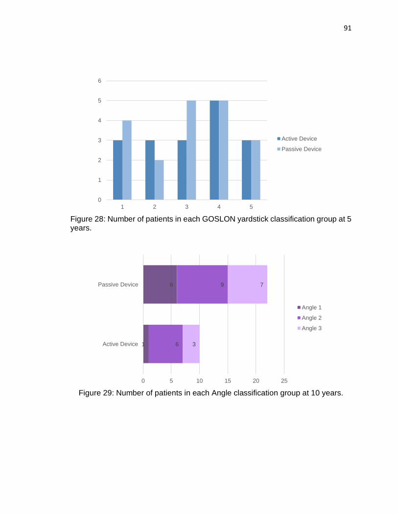
91
Figure 28: Number of patients in each GOSLON yardstick classification group at 5 years.
Figure 29: Number of patients in each Angle classification group at 10 years.
0
1
2
3
4
5
6
1 2 3 4 5
Active Device
Passive Device
1
6
6
9
3
7
0 5 10 15 20 25
Active Device
Passive Device
Angle 1
Angle 2
Angle 3
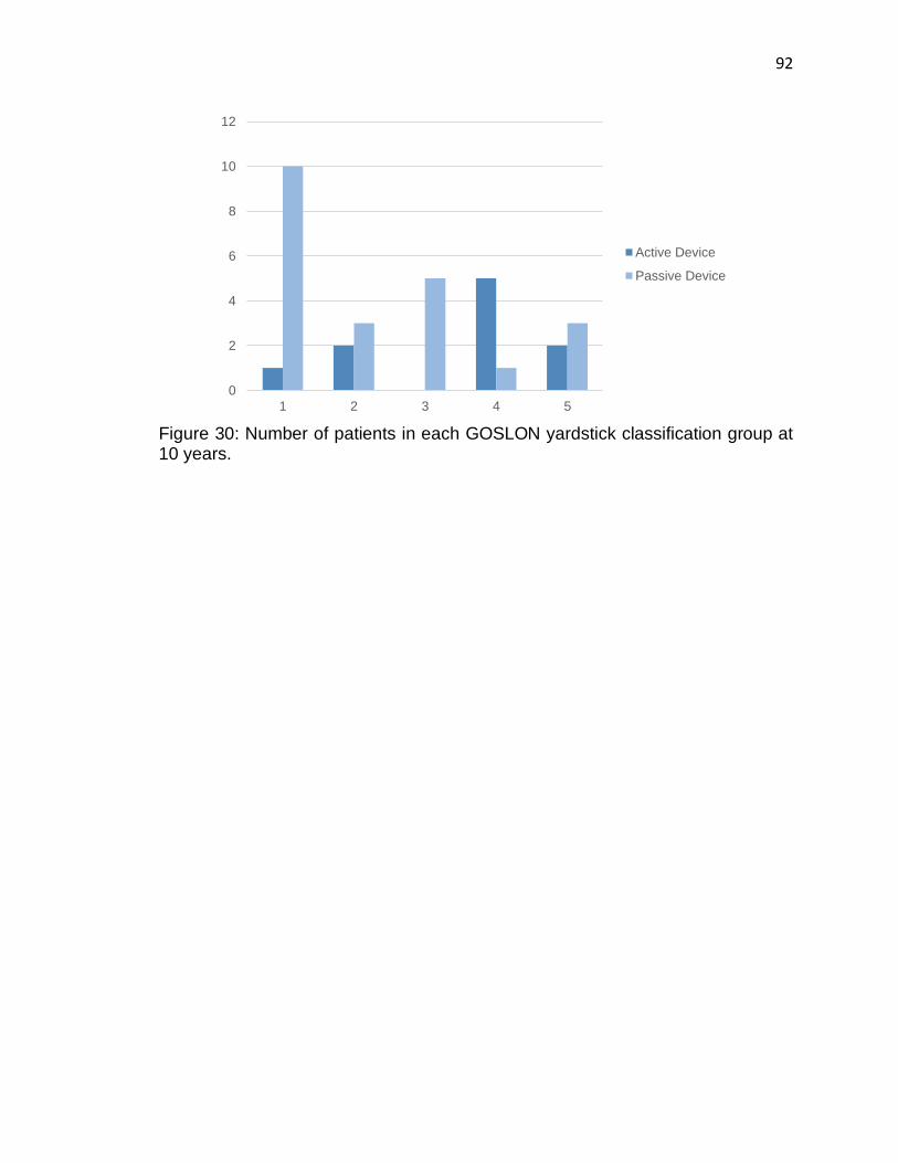
92
Figure 30: Number of patients in each GOSLON yardstick classification group at 10 years.
0
2
4
6
8
10
12
1 2 3 4 5
Active Device
Passive Device

93
Figure 31: Change in occlusion and dental arch relationships from 5 to 10 years.
-2
-1
0
1
2
3
4
5
6
7
8
Angle MHB GOSLON Overjet ICC IPMW SI
Active Device
Passive Device

94
1.3.5 Cephalometric Measurements
For 5-year cephalometric analysis, mean age of measurements was 5.93 years
(n=18) for patients in the active device group and 6.47 years (n=20) for patients in
the passive group (p=0.17). For 10-year cephalometric analysis, mean age of
measurements was 9.62 years (n=16) for patients with an active device and 9.97
years (n=23) for patients with a passive device (p=0.37).
1.3.6 Maxillary Growth Measurements
At 5 years, there was no significant difference between the two groups for the
length of the maxillary palate (PNS-ANS), depth of the midface (Ba-A), length of
the maxilla (Ba-ANS), maxillary protrusion (SNA), horizontal distance between
nasion and A-point (N-A), maxillary position at A-point (Ba-N-A) and the maxillary
position at ANS (Ba-N-ANS) (p=0.03, p=0.03, p=0.04, p=0.48, p=0.43, p=0.31,
p=0.39). At 10 years, there was also no difference between the active and passive
device patients (Table 10). A p-value of less than 0.01 was considered significant
using the Bonferroni correction.

95
N for 5 years, 10 years
Active PSO n= 18, n= 16
Passive PSO n= 20, n= 23
P value (<0.01)
5 y
ears
Mean age (years)
SNA (o)
N-A (mm)
PNS-ANS (mm)
Ba-A (mm)
Ba-ANS (mm)
Ba-N-ANS (o)
Ba-N-A (o)
5.93
77.7
-3.99
41.8
77.2
80.7
63.1
58.5
6.47
78.8
-3.01
45.1
82.4
85.9
64.6
60.0
0.17
0.48
0.43
0.03
0.03
0.04
0.39
0.31
10 y
ears
Mean age (years)
SNA (o)
N-A (mm)
PNS-ANS (mm)
Ba-A (mm)
Ba-ANS (mm)
Ba-N-ANS (o)
Ba-N-A (o)
9.62
75.0
-7.10
44.6
79.7
83.9
61.5
56.6
9.97
76.1
-6.10
43.3
77.0
80.9
61.5
56.8
0.37
0.44
0.44
0.51
0.40
0.37
1.00
0.83
Table 10: Maxillary growth measurements at 5 and 10 years. With Bonferroni correction p-value <0.01 was considered significant.

96
1.3.7 Mandibular Growth Measurements
Mandibular growth measurements were similar between both groups at the 5 and
10-year mark (Table 11). Mandibular growth measurements included mandibular
protrusion (SNB), horizontal distance between nasion and B-point (N-B), horizontal
distance between nasion and pogonion (N-Pg), mandibular position at the
pogonion (Ba-N-Pg), and mandibular position at B-point (Ba-N-B). A p-value of
less than 0.01 was considered significant using the Bonferroni correction.
N for 5 years, 10 years
Active PSO n= 18, n= 16
Passive PSO n= 20, n= 23
P value (<0.01)
5 y
ears
Mean age (years)
SNB (o)
N-B
N-Pg
Ba-N-Pg (o)
Ba-N-B (o)
5.93
74.0
-11.9
-13.0
55.1
54.8
6.47
75.1
-10.3
-11.1
56.6
56.3
0.17
0.42
0.40
0.36
0.14
0.18
10 y
ears
Mean age (years)
SNB (o)
N-B
N-Pg
Ba-N-Pg (o)
Ba-N-B (o)
9.62
74.1
-13.2
-13.8
56.4
55.6
9.97
74.5
-12.4
-12.5
56.2
55.3
0.37
0.74
0.72
0.60
0.88
0.83
Table 11: Mandibular growth at 5 and 10 years. With Bonferroni correction p-value <0.01 was considered significant.

97
1.3.8 Dento-alveolar Relationships
Dento-alveolar measurements were similar between groups at 5 and 10 years.
Dentoalveolar measurements included position of maxilla relative to mandible
(ANB), alveolus to basal bone measurement (ANS-N-A), and basal bone
difference (ANS-N-Pg). There were no differences between the two groups at 5
and 10 years (Table 12). A p-value of less than 0.02 was considered significant
using the Bonferroni correction.
N for 5 years, 10 years
Active PSO n= 18, n= 16
Passive PSO n= 20, n= 23
P value (<0.02)
5 y
ears
Mean age (years)
ANB (o)
ANS-N-A (o)
ANS-N-Pg (o)
5.93
3.68
4.66
165.3
6.47
3.69
4.59
165.5
0.84
1.00
0.91
0.93
10 y
ears
Mean age (years)
ANB (o)
ANS-N-A (o)
ANS-N-Pg (o)
9.62
0.99
4.98
147.9
9.97
1.58
4.66
154.4
0.37
0.57
0.50
0.80
Table 12: Dento-alveolar growth measurements at 5 and 10 years. With Bonferroni correction p-value <0.02 was considered significant.

98
1.3.9 Vertical Facial Growth
At 5 and 10 years there was no significant difference between the groups for any
vertical facial growth factors (Table 13). Vertical facial growth factors included
upper facial height (N-ANS), lower facial height (ANS-Me), upper to lower facial
height ratio (N-ANS/ANS-Me), and midface percent of total facial height (N-ANS/N-
Me) were not significantly different between the two groups. A p-value of less than
0.01 was considered significant using the Bonferroni correction.
N for 5 years, 10 years
Active PSO n= 18, n= 13
Passive PSO n= 20, n= 23
P value (<0.01)
5 y
ears
Mean age (years)
N-ANS (ratio)
ANS-Me (ratio)
N-ANS/ANS-Me (ratio)
N-ANS/N-Me (ratio)
5.93
39.0
58.2
0.74
0.43
6.47
43.0
56.9
0.76
0.44
0.17
0.01
0.03
0.35
0.42
10 y
ears
Mean age (years)
N-ANS (ratio)
ANS-Me (ratio)
N-ANS/ANS-Me (ratio)
N-ANS/N-Me (ratio)
9.62
44.3
57.4
0.77
0.44
9.97
44.3
56.3
0.79
0.44
0.37
0.98
0.65
0.46
0.49
Table 13: Vertical facial growth measurements at 5 and 10 years. With Bonferroni correction p-value <0.01 was considered significant.

99
1.3.10 Soft Tissue Growth
Soft tissue growth measurements included soft tissue convexity, soft tissue profile,
facial contour angle, and nasolabial angle (Table 14). There was no significant
difference between groups at 5 and 10 years. A p-value of less than 0.02 was
considered significant using the Bonferroni correction.
N for 5 years, 10 years
Active PSO n= 18, n= 13
Passive PSO n= 20, n= 23
P value (<0.02)
5 y
ears
Mean age (years)
Soft tissue convexity (o)
Facial contour angle (o)
Nasolabial angle (o)
5.93
141.9
-5.54
110.7
6.47
140.0
-6.36
113.1
0.17
0.34
0.63
0.45
10 y
ears
Mean age (years)
Soft tissue convexity (o)
Facial contour angle (o)
Nasolabial angle (o)
9.62
143.7
-1.41
99.8
9.97
140.8
-5.11
106.8
0.37
0.12
0.06
0.02
Table 14: Soft tissue growth at 5 and 10 years. With Bonferroni correction p-value <0.02 was considered significant.

100
1.3.11 Growth Between Five and Ten Years
A comparison of growth between the 5-year and 10-year measurements was also
performed (Table 15). A total of 16 patients in the active device group and 18
patients in the passive device group had records for this comparison. Growth in
the maxilla, mandible, dento-alveolar relationships, vertical facial height, and soft
tissue values was not significantly different between the 2 groups.
1.3.12 Intra-rater Reliability
Intra-class correlation coefficient was calculated for cephalometric measurements.
The ICC value was 0.81 which represents good intra-rater reliability.

101
N for 5 years, 10 years
Active PSO n= 16
Passive PSO n= 18
P value
Maxill
a
SNA
N-A
PNS-ANS
Ba-A
Ba-ANS
Ba-N-ANS
Ba-N-A
-2.64
-3.08
2.53
2.01
2.63
-1.51
-1.81
-2.93
-3.23
-2.58
-7.06
-6.69
-3.41
-3.43
0.80
0.89
0.06
0.02
0.03
0.21
0.26
Mandib
le SNB
N-B
Pg-N
Ba-N-Pg
Ba-N-B
0.33
-0.89
-0.28
1.74
1.17
-0.36
-1.45
-0.63
-0.21
-0.83
0.40
0.64
0.82
0.04
0.04
Dento
-alv
eola
r
ANB
A-N-ANS
N-ANS-Pg
-2.96
0.31
-16.8
-2.61
0.04
-14.5
0.70
0.73
0.94
Vert
ical
Face
N-ANS
ANS-Me
N-ANS/ANS-Me
N-ANS/N-Me
5.14
3.88
0.04
0.01
0.77
-2.47
0.45
0.11
0.08
0.03
0.79
0.78
Soft
Tis
sue Soft tissue convexity
Facial contour angle
Nasolabial angle
1.39
4.49
-11.7
0.70
1.32
-6.93
0.70
0.02
0.18
Table 15: Facial growth between 5 and 10 years. Significant p-values were considered <0.01 for maxilla growth, mandible growth, and vertical facial growth and <0.02 for dento-alveolar growth and soft tissue growth.

102
DISCUSSION
In the present study, patients being treated with a passive PSO device had
significantly larger horizontal alveolar gaps at the time of lip repair. This
discrepancy indicates that the passive device is not as effective at narrowing the
alveolar gap compared to the active device. The patients treated with an active
PSO device had larger cleft-side alar base measurements compared to the
passive device patients at the time of lip repair. This could be explained by the fact
that the passive device employs a nasal stent which also serves to mold the alar
base, while patients with an active device had an intra-oral device that provides no
nasal molding. By the time of palate repair patients in both device groups had
similar measurements for alar base, palate width, and alveolar gap width. Patients
in the active and passive device groups had similar rates of face mask application,
braces application, lip revision, bone grafting, and VPI, all of which suggests that
the patient group did not influence the number of additional procedures required
following palate repair. In summary, patients in the active and passive device
groups had comparable severity of their alveolar gap by the time of palate repair
and similar management protocols. This is the first study comparing dental
occlusion and facial growth where patients were matched for cleft severity and
management protocol.
1.4.1 Dental Arch and Occlusion
The present study is the largest long-term study comparing dental arch
relationships in patients treated with an active versus a passive PSO device.
Overall, this study showed no difference in dental arch relationships between
patients treated with an active or a passive device. All groups had an Angle
classification grade that indicated a degree of overjet/overbite, a negative MHB
score indicative of a degree of maxillary arch growth restriction (141), and a
GOSLON yardstick score that indicates positive/minimal overjet and predicts good

103
outcomes following orthognathic surgery. When comparing changes from 5 to 10
years within patients, there were no differences between the groups. Angle
classification, MHB score, GOSLON score, and overjet improved in both patient
groups from 5 to 10 years although the changes did not reach significance.
Improved occlusal scores from 5 to 10 years could be secondary to the use of
braces and targeted dental extraction to allow for teeth to fit in anatomic alignment.
Assessing the width of the dental arch is important for determining whether
patients have deficient palatal growth/expansion. While it has been shown that
palatal surgery does cause some dental arch restriction, the effect of PSO devices
on dental arch growth is unknown (142). To our knowledge, this is the first study
to make the comparison in patients treated with active versus passive devices.
When comparing the development of the dental arch in patients treated with a PSO
device, there have been a handful of studies that have shown decreased maxillary
arch collapse in patients treated with a passive device compared to no device
(54,123,124). Unfortunately, these studies showing improved arch collapse only
examined patients up to 18 months of age and so they cannot be used to
extrapolate how the arch behaves as the patient grows older. A previous study by
Banker et al. has reported that an IPMW to ICW ratio of 1:1 +/- 0.5 is indicative of
normal arch development and a ratio of greater than 1.15:1 indicates deficient arch
development (143). This ratio was 1.50 in patients with an active device and 1.35
in patients with a palate device, which indicates both patient groups had a degree
of dental arch deficiency. This same study by Banker et al. also used an IPMW of
under 34.92mm as an indicator for arch restriction. Using this criterion our patients
would not be considered to have a restricted arch; at 10 years patients with an
active device had an IPMW of 42.7mm and patients with a passive device had an
IPMW of 40.3mm. Furthermore, both groups showed an overall expansion of the
IPMW and the ICW from 5 to 10 years which is consistent with no restriction of the
maxillary dental arch.

104
In summary, patients treated with an active device had comparable dental
occlusion and dental arch width compared to patients treated with a passive
device. The reported dental arch measurements indicate that there may be a small
degree of maxillary arch restriction in both groups, although both groups still had
growth in the arch from 5 to 10 years.
1.4.2 Facial Growth
Overall, this study found comparable facial growth outcomes between patients
treated with an active versus a passive PSO device at 5 and 10 years. Before
Bonferroni correction anterior/posterior and vertical maxillary growth was
significantly smaller in patients treated with an active device at 5 years. Loss of
significance following Bonferroni correction indicates that these differences can be
explained by chance from repeated measurements and not by the effects of the
device itself. The similarity in dentoalveolar relationships measured via
cephalometric analysis reinforces the validity of the similar occlusal relationships
that were found using the patient molds. Regarding the changes in growth from 5
to 10 years, patients had similar growth of the maxilla, mandible, dento-alveolar
relationships, vertical facial height, and soft tissue values.
Previous research has shown that PSO devices, specifically active devices, cause
a maxillary growth restriction (35,44). To our knowledge, there are few studies that
have reported negative maxillary growth outcomes with the use of a passive
device. The results from this study indicate that active devices do not restrict
maxillary growth compared to passive devices. Furthermore, the reported average
measurements for SNA angle (maxillary length) and SNB angle (mandibular
length) in this study are similar to previously reported outcomes for patients with
CL/P treated without a PSO device. Those studies reported an average SNA angle
of 76.4 to 80.5 compared to our reported 75.0 to 78.9 and an SNB of 74.4 to 75.4
compared to our reported 74.0 to 75.1(144,145). While these groups cannot be

105
compared statistically because they are from different studies, this finding further
supports our conclusion that the active and passive devices do not appear to
negatively affect maxillary growth.
In summary, patients treated with an active device have similar facial growth to
patients treated with a passive device up 10 years of age.

106
CONCLUSION
Overall, the results of this study indicate that dental occlusion/dental arch
development and facial growth are similar between patients treated with an active
or a passive PSO device. Patients treated with either device have a degree of
overbite/overjet at the level of the molars (Angle 2) with good expected outcomes
following orthognathic surgery (GOSLON 3). Neither device causes a significant
amount of dental arch restriction. Overall, at 10 years patients in the active and
passive device groups also have comparable facial growth. Moving forward, a
prospective study that compares the use of passive and active PSO devices to
patients with no device (with matched cleft severity) would elucidate whether
patients with these devices develop occlusion and facial growth different to
patients treated without a PSO device.

107
Chapter 5: General Discussion and Conclusion
This chapter will briefly outline the hypothesis and objectives of this thesis. This
chapter will summarize the results of Chapter 2 through 4 and discuss weaknesses
and strengths of each study. Finally, future directions of this study will be
discussed.

108
SUMMARY
The purpose of this work was to review the current literature pertaining to long-
term outcomes associated with pre-surgical orthopedic (PSO) devices used in the
management of cleft lip and palate and to study the long-term effects of these
devices.
The objectives of this thesis were to:
1. Perform a systematic review of current literature describing the long-term
effects of different PSO devices used in the management of cleft palate.
(CHAPTER 0)
2. To evaluate and compare 1-year, 5-year, and 10-year nasolabial aesthetic
changes in patients who received treatment with an active PSO device,
passive PSO device, or no PSO device. (CHAPTER 0)
3. To evaluate and compare 5-year and 10-year dental occlusion/dental arch
development in patients who had treatment with an active PSO or passive
PSO device. (CHAPTER 0)
4. To evaluate and compare 5-year and 10-year facial growth in patients who
had treatment with an active PSO device or passive PSO device at 5 years
and 10 years in order to determine whether there is a difference in facial
development between these groups. (CHAPTER 0)
The findings of Chapters 2, 3, and 4 are reviewed below.

109
SUMMARY OF CHAPTER 2: SYSTEMATIC REVIEW OF THE LITERATURE ON
THE LONG-TERM PATIENT OUTCOMES ASSOCIATED WITH PRE-SURGICAL
ORTHOPEDIC DEVICES
The primary objective of this chapter was to perform a systematic review of the
literature pertaining to long-term outcomes in patients who were treated with PSO
devices. The search criteria were developed with assistance from a librarian and
the results of the search were reviewed by 2 independent reviewers using strict
inclusion and exclusion criteria. The reference lists of the included studies were
also screened for inclusion.
The literature review produced a total of 41 articles for inclusion. These studies
highlighted the existence of multiple confounding factors that often occur between
comparison groups. These factors make it difficult to determine whether the
reported outcomes are due to the devices or the confounding factors, and
ultimately limit the applicability of the study results. Described confounding factors
include the severity of the initial alveolar gap, different operating surgeons, and
different management protocols. Secondly, the review highlighted that there is no
consensus in the literature as to the long-term effects of PSO devices on patient
outcomes. Thirdly, this systematic review highlighted the lack of studies comparing
long-term patient outcomes between patients that are treated with active versus
passive PSO devices. In summary, the results from this systematic review
confirmed the initial study hypothesis that there is no consensus in the literature
regarding the effects of these devices and that the literature that does exist is
weakened by the heterogeneity that exists in between and within the different
comparison groups.

110
SUMMARY OF CHAPTER 3: 10-YEAR NASOLABIAL AESTHETIC
COMPARISON IN PATIENTS WITH DIFFERENT PRE-SURGICAL
ORTHOPEDIC DEVICES
The primary objective of this chapter was to assess the 10-year nasolabial
aesthetic outcomes of patients treated with an active PSO device, passive PSO
device, or no device. Patients were included if they had a unilateral complete or
incomplete primary cleft palate and were operated on by the principle surgeon.
Nasolabial aesthetics were assessed using 2-dimensional facial photographs for
patients taken at 3 time points (1 year, 5 years, 10 years). A total of 11 nasolabial
variables were assessed for each photograph.
There were two principle findings in this study. The first finding was that patients
treated with PSO devices had larger alveolar gaps compared to patients treated
without a PSO device. Despite having worse alveolar gaps, patients treated with
either PSO device had comparable 10-year nasolabial aesthetic outcomes to
patients with less severe gaps that did not require device treatment. The second
finding of this study was that despite passive devices providing a nasal molding
component, there was no difference in nasal outcomes between patients treated
with an active or a passive PSO device. Overall, PSO device treatment helps
patients with more severe alveolar gaps achieve nasolabial aesthetic results
similar to patients with less severe gaps.

111
SUMMARY OF CHAPTER 4: 10-YEAR DENTAL OCCLUSION AND FACIAL
GROWTH COMPARISON IN PATIENTS WITH DIFFERENT PRE-SURGICAL
ORTHOPEDIC DEVICES
The primary objective of this chapter was to compare the 10-year dental occlusion
and facial growth outcomes in patients treated with an active PSO device versus
a passive PSO device over a 10-year period. Dental occlusion was assessed by
evaluating patients’ dental models at 5 and 10 years. Facial growth was assessed
through cephalometric analysis of lateral patient radiographs at the same time
points.
There were 3 principle results found in this study. Firstly, patients treated with
active or passive PSO devices had similar dental occlusion and dental arch
development over a 10-year period. Secondly, neither PSO device cause a
restriction in the growth of the maxillary arch. Thirdly, patients treated with an
active PSO device had similar facial growth to patients treated with a passive
device.

112
STRENGTHS AND LIMITATIONS
There are multiple strengths and weaknesses associated with this work. The
strength of Chapter 2 lies in the fact that it provides a thorough summary of the
current state of knowledge in this field. This systematic review was an exhaustive
search of all literature pertaining to this subject. A meta-analysis would be a more
powerful summary of results, but unfortunately the described results were too
heterogenous for this type of analysis.
Chapters 3 and 4 share some similar strengths and weaknesses. In terms of
weaknesses, both studies are retrospective. Retrospective studies may be
influenced by selection bias. Strengths of these studies include that all groups were
operated on by the same surgeon and had the same management protocol. These
similarities help eliminate many confounding factors that could influence study
results. Thirdly, these are 2 of the largest studies comparing these 3 groups over
a 10-year period which makes this a very valuable study for understanding long-
term patient outcomes. Finally, the 10-year follow-up period is another strength of
the aforementioned studies. Frequently, cleft palate research is limited to follow-
up at 1 to 5 years. A significant proportion of growth and development occurs
between 5 and 10 years of age; an important factor to acknowledge when studying
these populations. The 10-year follow-up period in this work accounts for important
facial changes that can influence patient outcomes. Conversely, the follow-up
period is also a weakness; patients still grow up to 18 to 20 years so growth
changes following the 10-year follow-up period are not addressed.
Specific to Chapter 3, a limitation of this study was that patients in the PSO and no
PSO device groups were not matched for alveolar gap severity; patients treated
with no device had small complete or incomplete alveolar gaps and were often not
treated with palate surgery. Palate surgery is a variable that is known to affect
maxillary growth, which by extension could affect nasolabial aesthetics by
changing lip protrusion. Patients in the no device group also had significantly less

113
severe alveolar gaps compared to patients treated with PSO devices. These
differences are potential confounding factors and therefore, the patients treated
with PSO devices versus no device cannot be directly compared in order to
determine if using a PSO device is superior to not using a device. Despite this, it
is still possible to judge whether the PSO devices make nasolabial aesthetics
similar to nasolabial aesthetics in patients treated without a device who start with
more severe alveolar gaps. Secondly, patients did differ in their rates of lip revision,
but this was accounted for through analysis of patients excluding those who
received lip revision surgery.
Specific to Chapter 4, the main limitation is also that it is a retrospective study.
Therefore, some selection bias may exist between the two groups. A strength of
this study was that the 2 comparison groups in this chapter were matched for
alveolar gap severity. Very few studies in cleft palate research have groups
matched for alveolar gap severity, which is a factor that could confound results.
Secondly, to our knowledge this is the largest study comparing outcomes between
patients treated with an active or passive PSO device with all possible surgical
confounding factors eliminated.

114
FUTURE DIRECTIONS
Chapter 2 highlighted the necessity for a single-surgeon study examining the long-
term nasolabial aesthetic, dental occlusion, and facial growth outcomes in patients
treated with both active and passive PSO devices. In addition, this chapter also
highlighted the need for studies actually comparing the different types of PSO
devices. This is exactly what was performed in Chapters 3 and 4. In regard to
future directions for this study, this study will be repeated in the next 3 to 4 years
to examine 15-year outcomes in these patient groups. There are few 15-year
follow-up studies in cleft palate literature, and currently no studies comparing
active and passive PSO device patients who are matched for cleft severity.
Unfortunately, the majority of the study patients are too young for 15-year follow-
up analysis at this time, but this will be repeated when all of these study patients
have reached 15 years old.
A question that this work does not answer is whether PSO devices are truly
required to have adequate growth and aesthetic outcomes. To answer this
question, patients treated with no device with alveolar gaps of equal severity would
also need to be compared. A population like this does not currently exist in our
patient cohort. A second future direction for this research could be a prospective
study that directly compares nasolabial aesthetics, dental occlusion, and facial
growth in patients treated with an active, passive, or no PSO device. A prospective
study of this nature with groups that are matched for the severity of their alveolar
gap would be an important way of assessing whether PSO devices are truly
required to have improved facial growth and dental occlusion.

115
SIGNIFICANCE
The use of PSO devices for patients with cleft palate is still a highly debated topic.
Surgeons base their decision to use a certain PSO device on experience, surgeon
preference, and family preference. A lot of limitations exist in research on this
subject, and Chapter 2 highlights a number of the limitations that exist in this area
of research. This is the first systematic review to highlight these limitations in a
quantitative manner. The significance of this systematic review is that it has
highlighted the need for research with more strict methodology. This systematic
review can be used to guide future research in this field.
Chapters 3 and 4 outlined the long-term effects of using passive and active PSO
devices in patients with cleft lip/palate. These two studies are the first single-
surgeon studies to examine the impact of both active and passive devices on
nasolabial aesthetics, dental occlusion, and facial growth. In addition, these are
the first single-surgeon studies to control for cleft severity and surgical
management between the different patient groups. Overall, this is the largest study
examining the long-term effects of PSO devices between patients treated with an
active or a passive device where confounding factors including the operating
surgeon, surgical management, and cleft severity are all controlled.
There is concern in the cleft palate community about how PSO devices affect
maxillary growth, dental occlusion, and nasolabial aesthetics. Specifically, a large
amount of concern lies in whether active devices reduce maxillary growth. The
results from Chapter 4 allow us to conclude that overall, patients treated with an
active device have comparable growth to patients treated with a passive device.
These results may encourage surgeons who were hesitant to use active devices
for this reason. Secondly, passive devices are often used in patients with cleft
lip/palate for the nasal molding component. This work showed that passive devices
do not produce superior nasal aesthetics compared to active PSO devices. Thirdly,
this work showed that using a PSO device in larger clefts can help achieve

116
nasolabial aesthetic outcomes similar to patients with less severe clefts. Overall,
the results of this work have disproven several current theories about PSO
devices. This work can help guide cleft surgeons with their PSO device selection
and may encourage the use of different PSO devices for different patients.

117
Appendices
APPENDIX 1- GLOSSARY
NAM device: passive PSO device
Latham device: active PSO device
Sella turcica (S): center of the sella turcica
Nasion (N): most superior point of the frontonasal suture
A point (A): most posterior point on the curve between the ANS and the PR
Anterior nasal spine (ANS): most anterior point on the maxilla at the level of the
palate
Posterior nasal spine (PNS): most posterior point on the boney hard palate
Basion (Ba): most inferior posterior point on anterior rim of foramen magnum
(tip of posterior cranial base)
B point (B): most posterior point of bony curvature of the mandible
Pogonion (Pg): most anterior point on mandibular symphysis
Menton (Me): lowest point on chin
Soft tissue nasion (N’): soft tissue marker of most superior point of the
frontonasal suture
Soft tissue A-point (A’): soft tissue marker of most posterior point on the curve
between the ANS and the PR
Soft tissue pogonion (Pg’): most anterior point on chin
Soft tissue gnathion (G’): most anterior inferior point
Pronasale (Pn): most anterior tip of nose
Columella (Col): midpoint on the lower surface of the nose
Subnasale (Sn): junction of where the base of the columella meets the upper
lip
APPENDIX 2- NASOLABIAL AESTHETIC RATING SCALE

118

119
120

121

122
123

124

125

126

127

128

129
Ethics Approval

130
Image Permissions
Figure 1

131
Figure 11

132
References
1. Dissaux C, Bodin F, Grollemund B, Picard A, Vazquez M-P, Morand B, et
al. Evaluation of 5-year-old children with complete cleft lip and palate:
Multicenter study. Part 1: Lip and nose aesthetic results. J Craniomaxillofac
Surg. 2015 Dec;43(10):2085–92.
2. Matthews JLK, Oddone-Paolucci E, Harrop RA. The Epidemiology of Cleft
Lip and Palate in Canada, 1998 to 2007. Cleft Palate Craniofac J. 2015
Jul;52(4):417–24.
3. Janis J. Essentials of Plastic Surgery. 2nd ed. Debrecht A, editor.
Colombus: Thieme Medical Publishers; 2017. 264–285 p.
4. Rodriguez E, Losee J, Neligan P. Plastic Surgery Volume 3: Craniofacial,
Head and Neck Surgery and Pediatric Surgery. Third. Saunders; 2012.
519–520 p.
5. Brito LA, Meira JGC, Kobayashi GS, Passos-Bueno MR. Genetics and
management of the patient with orofacial cleft. Plast Surg Int.
2012;2012:782821.
6. Taub PJ, Piccolo P. Cleft Lip Repair: Through the Looking Glass. J
Craniofac Surg. 2016 Nov;27(8):2031–5.
7. Farronato G, Kairyte L, Giannini L, Galbiati G, Maspero C. How various
surgical protocols of the unilateral cleft lip and palate influence the facial
growth and possible orthodontic problems? Which is the best timing of lip,
palate and alveolus repair? literature review. Stomatologija. 2014;16(2):53–
60.
8. Garland K, Matic D. Current approaches to cleft lip revision. Curr Opin
Otolaryngol Head Neck Surg. 2019 Aug;27(4):287–93.
9. Hosseini HR, Kaklamanos EG, Athanasiou AE. Treatment outcomes of
pre-surgical infant orthopedics in patients with non-syndromic cleft lip
and/or palate: A systematic review and meta-analysis of randomized
controlled trials. PLoS One. 2017;12(7):e0181768.
10. Gatti GL, Freda N, Giacomina A, Montemagni M, Sisti A. Cleft Lip and
Palate Repair. J Craniofac Surg. 2017 Nov;28(8):1918–24.

133
11. Alzain I, Batwa W, Cash A, Murshid ZA. Presurgical cleft lip and palate
orthopedics: an overview. Clin Cosmet Investig Dent. 2017;9:53–9.
12. Papadopoulos MA, Koumpridou EN, Vakalis ML, Papageorgiou SN.
Effectiveness of pre-surgical infant orthopedic treatment for cleft lip and
palate patients: a systematic review and meta-analysis. Orthod Craniofac
Res. 2012 Nov;15(4):207–36.
13. Dao AM, Goudy SL. Cleft Palate Repair, Gingivoperiosteoplasty, and
Alveolar Bone Grafting. Facial Plast Surg Clin North Am. 2016
Nov;24(4):467–76.
14. Hopper RA, Al-Mufarrej F. Gingivoperiosteoplasty. Clin Plast Surg. 2014
Apr;41(2):233–40.
15. Liao Y-F, Yang I-Y, Wang R, Yun C, Huang C-S. Two-stage palate repair
with delayed hard palate closure is related to favorable maxillary growth in
unilateral cleft lip and palate. Plast Reconstr Surg. 2010 May;125(5):1503–
10.
16. Stein S, Dunsche A, Gellrich N-C, Harle F, Jonas I. One- or two-stage
palate closure in patients with unilateral cleft lip and palate: comparing
cephalometric and occlusal outcomes. Cleft Palate Craniofac J. 2007
Jan;44(1):13–22.
17. Pradel W, Senf D, Mai R, Ludicke G, Eckelt U, Lauer G. One-stage palate
repair improves speech outcome and early maxillary growth in patients with
cleft lip and palate. J Physiol Pharmacol. 2009 Dec;60 Suppl 8:37–41.
18. Gaggl A, Feichtinger M, Schultes G, Santler G, Pichlmaier M, Mossbock R,
et al. Cephalometric and occlusal outcome in adults with unilateral cleft lip,
palate, and alveolus after two different surgical techniques. Cleft Palate
Craniofac J. 2003 May;40(3):249–55.
19. Corbo M, Dujardin T, de Maertelaer V, Malevez C, Glineur R.
Dentocraniofacial morphology of 21 patients with unilateral cleft lip and
palate: a cephalometric study. Cleft Palate Craniofac J. 2005
Nov;42(6):618–24.
20. Zemann W, Mossbock R, Karcher H, Kozelj V. Sagittal growth of the facial

134
skeleton of 6-year-old children with a complete unilateral cleft of lip,
alveolus and palate treated with two different protocols. J Craniomaxillofac
Surg. 2007 Dec;35(8):343–9.
21. Mars M, Houston WJ. A preliminary study of facial growth and morphology
in unoperated male unilateral cleft lip and palate subjects over 13 years of
age. Cleft Palate J. 1990 Jan;27(1):7–10.
22. Capelozza Junior L, Taniguchi SM, da Silva Junior OG. Craniofacial
morphology of adult unoperated complete unilateral cleft lip and palate
patients. Cleft Palate Craniofac J. 1993 Jul;30(4):376–81.
23. Shi B, Losee JE. The impact of cleft lip and palate repair on maxillofacial
growth. Int J Oral Sci. 2015 Mar;7(1):14–7.
24. Friede H, Enemark H. Long-term evidence for favorable midfacial growth
after delayed hard palate repair in UCLP patients. Cleft Palate Craniofac J.
2001 Jul;38(4):323–9.
25. Bardach J, Morris HL, Olin WH. Late results of primary veloplasty: the
Marburg Project. Plast Reconstr Surg. 1984 Feb;73(2):207–18.
26. Mylin WK, Hagerty RF. Midfacial skeletal profile in early and late closure of
the hard palate. South Med J. 1983 May;76(5):610–2.
27. Rohrich RJ, Rowsell AR, Johns DF, Drury MA, Grieg G, Watson DJ, et al.
Timing of hard palatal closure: a critical long-term analysis. Plast Reconstr
Surg. 1996 Aug;98(2):236–46.
28. Shaffer AD, Ford MD, Losee JE, Goldstein J, Costello BJ, Grunwaldt LJ, et
al. The Association Between Age at Palatoplasty and Speech and
Language Outcomes in Children With Cleft Palate: An Observational Chart
Review Study. Cleft Palate Craniofac J. 2020 Feb;57(2):148–60.
29. Kuseler A, Molsted K, Marcusson A, Heliovaara A, Karsten A, Bellardie H,
et al. Scandcleft randomized trials of primary surgery for unilateral cleft lip
and palate: maxillary growth at eight years of age. Eur J Orthod. 2020
Jan;42(1):24–9.
30. Heliovaara A, Skaare P, Kuseler A, Shaw W, Molsted K, Karsten A, et al.
Scandcleft randomized trials of primary surgery for unilateral cleft lip and
135
palate. Dental arch relationships in 8 year-olds. Eur J Orthod. 2020
Jan;42(1):1–7.
31. Shaye D. Update on outcomes research for cleft lip and palate. Curr Opin
Otolaryngol Head Neck Surg. 2014 Aug;22(4):255–9.
32. Efunkoya AA, Omeje KU, Amole IO, Osunde OD, Akpasa IO. A review of
cleft lip and palate management: Experience of a Nigerian Teaching
Hospital. Afr J Paediatr Surg. 2015;12(4):257–60.
33. Zhang Z, Stein M, Mercer N, Malic C. Post-operative outcomes after cleft
palate repair in syndromic and non-syndromic children: a systematic
review protocol. Syst Rev. 2017 Mar;6(1):52.
34. Shaye D, Liu CC, Tollefson TT. Cleft Lip and Palate: An Evidence-Based
Review. Facial Plast Surg Clin North Am. 2015 Aug;23(3):357–72.
35. Kornbluth M, Campbell RE, Daskalogiannakis J, Ross EJ, Glick PH,
Russell KA, et al. Active Presurgical Infant Orthopedics for Unilateral Cleft
Lip and Palate: Intercenter Outcome Comparison of Latham, Modified
McNeil, and Nasoalveolar Molding. Cleft Palate Craniofac J. 2018
May;55(5):639–48.
36. Millard DRJ, Latham RA. Improved primary surgical and dental treatment
of clefts. Plast Reconstr Surg. 1990 Nov;86(5):856–71.
37. Lukash FN, Schwartz M, Grauer S, Tuminelli F. Dynamic cleft maxillary
orthopedics and periosteoplasty: benefit or detriment? Ann Plast Surg.
1998 Apr;40(4):321–7.
38. Berkowitz S. A comparison of treatment results in complete bilateral cleft
lip and palate using a conservative approach versus Millard-Latham PSOT
procedure. Semin Orthod. 1996 Sep;2(3):169–84.
39. Cruz C. Presurgical Orthopedics Appliance: The Latham Technique. Oral
Maxillofac Surg Clin North Am. 2016 May;28(2):161–8.
40. Power SM, Matic DB. The Effects of Secondary Cleft Procedures on Alar
Base Position and Nostril Morphology in Patients with Unilateral Clefts.
Cleft Palate Craniofac J. 2017 Jul;54(4):431–5.
41. Jodeh DS, Ruso S, Feldman R, Ruas E, Rottgers SA. Clinical Outcomes

136
Utilizing a “Modified Latham” Appliance for Presurgical Infant Orthopedics
in Patients With Unilateral Complete Cleft Lip and Palate. Cleft Palate
Craniofac J. 2018 Dec;1055665618816892.
42. Lukash FN, Shikowitz-Behr LB, Schwartz M, Tuminelli F. Dynamic Cleft
Maxillary Orthopedics and Periosteoplasty. Ann Plast Surg. 2018
Jan;80(1):40–4.
43. Matic DB, Power SM. Correction of the cleft lip lateral bulge deformity
using anatomic muscle repair. J Craniofac Surg. 2011 Mar;22(2):514–9.
44. Matic DB, Power SM. The effects of gingivoperiosteoplasty following
alveolar molding with a pin-retained Latham appliance versus secondary
bone grafting on midfacial growth in patients with unilateral clefts. Plast
Reconstr Surg. 2008 Sep;122(3):863.
45. Lin J, Allareddy V, Ross E, Shusterman S. A Comparison of Mixed
Dentition Dental Development in Cleft Patients Treated With and Without
the Latham-type Appliance. Pediatr Dent. 2017 Jan;39(1):53–8.
46. Chan KT, Hayes C, Shusterman S, Mulliken JB, Will LA. The effects of
active infant orthopedics on occlusal relationships in unilateral complete
cleft lip and palate. Cleft Palate Craniofac J. 2003 Sep;40(5):511–7.
47. Mariqueo GEC, Arriagada EEA, Bustos TDPI, Navarro SM, Espinoza GEE.
Effectiveness of Nasoalveolar Molding in the Unilateral Cleft Lip and Cleft
Palate. J Craniofac Surg. 2018 Sep;29(6):1522–5.
48. Gomez DF, Donohue ST, Figueroa AA, Polley JW. Nasal changes after
presurgical nasoalveolar molding (PNAM) in the unilateral cleft lip nose.
Cleft Palate Craniofac J. 2012 Nov;49(6):689–700.
49. Ruiz-Escolano MG, Martinez-Plaza A, Fernandez-Valades R, Cortes-
Sanchez R, Munoz-Miguelsanz MA, Velasco-Ortega E, et al. Nasoalveolar
Molding Therapy for the Treatment of Unilateral Cleft Lip and Palate
Improves Nasal Symmetry and Maxillary Alveolar Dimensions. J Craniofac
Surg. 2016 Nov;27(8):1978–82.
50. Nur Yilmaz RB, Germec Cakan D, Nalbantgil D. Aesthetic Assessment of
Infants With Different Cleft Types Before, During, and After Orthopedic

137
Treatment. J Craniofac Surg. 2018 Nov;29(8):2081–7.
51. Broder HL, Flores RL, Clouston S, Kirschner RE, Garfinkle JS, Sischo L, et
al. Surgeon’s and Caregivers’ Appraisals of Primary Cleft Lip Treatment
with and without Nasoalveolar Molding: A Prospective Multicenter Pilot
Study. Plast Reconstr Surg. 2016 Mar;137(3):938–45.
52. Peanchitlertkajorn S, Mercado A, Daskalogiannakis J, Hathaway R,
Russell K, Semb G, et al. An Intercenter Comparison of Nasolabial
Appearance Including a Center Using Nasoalveolar Molding. Cleft Palate
Craniofac J. 2018 May;55(5):655–63.
53. Huddart AG. Presurgical changes in unilateral cleft palate subjects. Cleft
Palate J. 1979 Apr;16(2):147–57.
54. Shetty V, Agrawal RK, Sailer HF. Long-term effect of presurgical
nasoalveolar molding on growth of maxillary arch in unilateral cleft lip and
palate: randomized controlled trial. Int J Oral Maxillofac Surg. 2017
Aug;46(8):977–87.
55. Prahl C, Kuijpers-Jagtman AM, Van ’t Hof MA, Prahl-Andersen B. A
randomized prospective clinical trial of the effect of infant orthopedics in
unilateral cleft lip and palate: prevention of collapse of the alveolar
segments (Dutchcleft). Cleft Palate Craniofac J. 2003 Jul;40(4):337–42.
56. Bongaarts CAM, van ’t Hof MA, Prahl-Andersen B, Dirks I V, Kuijpers-
Jagtman AM. Infant orthopedics has no effect on maxillary arch dimensions
in the deciduous dentition of children with complete unilateral cleft lip and
palate (Dutchcleft). Cleft Palate Craniofac J. 2006 Nov;43(6):665–72.
57. Clark SL, Teichgraeber JF, Fleshman RG, Shaw JD, Chavarria C, Kau C-
H, et al. Long-term treatment outcome of presurgical nasoalveolar molding
in patients with unilateral cleft lip and palate. J Craniofac Surg. 2011
Jan;22(1):333–6.
58. Peanchitlertkajorn S, Mercado A, Daskalogiannakis J, Hathaway R,
Russell K, Semb G, et al. An Intercenter Comparison of Dental Arch
Relationships and Craniofacial Form Including a Center Using
Nasoalveolar Molding. Cleft Palate Craniofac J. 2018 Jul;55(6):821–9.

138
59. Chang S-Y, Lonic D, Pai BC-J, Lo L-J. Primary Repair in Patients With
Unilateral Complete Cleft of Lip and Primary Palate: Assessment of
Outcomes. Ann Plast Surg. 2018 Feb;80(2S Suppl 1):S2–6.
60. Abbott MM, Meara JG. Nasoalveolar molding in cleft care: is it efficacious?
Plast Reconstr Surg. 2012 Sep;130(3):659–66.
61. Kantar RS, Maliha SG, Alfonso AR, Wang MM, Ramly EP, Eisemann BS,
et al. Nasolabial Aesthetics Following Cleft Repair: An Objective Evaluation
of Subjective Assessment. Cleft Palate Craniofac J. 2019 Oct;56(9):1157–
63.
62. Fudalej SA, Desmedt D, Bronkhorst E, Fudalej PS. Comparison of Three
Methods of Rating Nasolabial Appearance in Cleft Lip and Palate. Cleft
Palate Craniofac J. 2017 Jul;54(4):400–7.
63. Nguyen VT, Nguyen T, Jagomagi T. Nasolabial aesthetics of patients with
repaired unilateral cleft lip and palate: A comparison of three rating
methods in two countries. J Craniomaxillofac Surg. 2018 Aug;46(8):1385–
9.
64. Mosmuller DGM, Mennes LM, Prahl C, Kramer GJC, Disse MA, van
Couwelaar GM, et al. The Development of the Cleft Aesthetic Rating Scale:
A New Rating Scale for the Assessment of Nasolabial Appearance in
Complete Unilateral Cleft Lip and Palate Patients. Cleft Palate Craniofac J.
2017 Sep;54(5):555–61.
65. Hunt O, Burden D, Hepper P, Johnston C. The psychosocial effects of cleft
lip and palate: a systematic review. Eur J Orthod. 2005 Jun;27(3):274–85.
66. He X, Li H, Shao Y, Shi B. Objective measurements for grading the nasal
esthetics on Basal view in individuals with secondary cleft nasal deformity.
Cleft Palate Craniofac J. 2015 Jan;52(1):66–9.
67. Fudalej P, Katsaros C, Hozyasz K, Borstlap WA, Kuijpers-Jagtman AM.
Nasolabial symmetry and aesthetics in children with complete unilateral
cleft lip and palate. Br J Oral Maxillofac Surg. 2012 Oct;50(7):621–5.
68. Stebel A, Desmedt D, Bronkhorst E, Kuijpers MA, Fudalej PS. Rating
nasolabial appearance on three-dimensional images in cleft lip and palate:

139
a comparison with standard photographs. Eur J Orthod. 2016
Apr;38(2):197–201.
69. Dogan E, Tiftikcioglu Y, Dogan S. Evaluation of nasolabial aesthetics by
different groups in operated unilateral cleft lip and palate. J Stomatol oral
Maxillofac Surg. 2020 Jan;
70. Andrews LF. The six keys to normal occlusion. Am J Orthod. 1972
Sep;62(3):296–309.
71. Proffit W, Fields H. Contemporary Orthodontics. Second. Reinhardt R,
Steube M, editors. St. Louis: Library of Congress; 1993. 175–176 p.
72. Jones T, Leary S, Atack N, Ireland T, Sandy J. Which index should be
used to measure primary surgical outcome for unilateral cleft lip and palate
patients? Eur J Orthod. 2016 Aug;38(4):345–52.
73. Dogan S, Olmez S, Semb G. Comparative assessment of dental arch
relationships using Goslon Yardstick in patients with unilateral complete
cleft lip and palate using dental casts, two-dimensional photos, and three-
dimensional images. Cleft Palate Craniofac J. 2012 May;49(3):347–51.
74. Botticelli S, Kuseler A, Marcusson A, Molsted K, Norholt SE, Cattaneo PM,
et al. Do Infant Cleft Dimensions Have an Influence on Occlusal Relations?
A Subgroup Analysis Within an RCT of Primary Surgery in Patients With
Unilateral Cleft Lip and Palate. Cleft Palate Craniofac J. 2019
Oct;1055665619875320.
75. Mossey PA, Clark JD, Gray D. Preliminary investigation of a modified
Huddart/Bodenham scoring system for assessment of maxillary arch
constriction in unilateral cleft lip and palate subjects. Eur J Orthod. 2003
Jun;25(3):251–7.
76. Altalibi M, Saltaji H, Edwards R, Major PW, Flores-Mir C. Indices to assess
malocclusions in patients with cleft lip and palate. Eur J Orthod. 2013
Dec;35(6):772–82.
77. Johnson N, Williams A, Singer S, Southall P, Sandy J. Initial cleft size does
not correlate with outcome in unilateral cleft lip and palate. Eur J Orthod.
2000 Feb;22(1):93–100.

140
78. Russell LM, Long REJ, Romberg E. The Effect of Cleft Size in Infants With
Unilateral Cleft Lip and Palate on Mixed Dentition Dental Arch
Relationship. Cleft Palate Craniofac J. 2015 Sep;52(5):605–13.
79. Wiggman K, Larson M, Larson O, Semb G, Brattstrom V. The influence of
the initial width of the cleft in patients with unilateral cleft lip and palate
related to final treatment outcome in the maxilla at 17 years of age. Eur J
Orthod. 2013 Jun;35(3):335–40.
80. Kumar D, Nigam AG, Marwah N, Goenka P, Sharma A. Morphological and
dimensional characteristics of dental arch in children with beta thalassemia
major. J Indian Soc Pedod Prev Dent. 2018;36(1):9–14.
81. Thu KM, Winn T, Abdullah N, Jayasinghe JAP, Chandima GL. The
maxillary arch and its relationship to cephalometric landmarks of selected
malay ethnic group. Malays J Med Sci. 2005 Jan;12(1):29–38.
82. Moyers R. Handbook of Orthodontics. Fourth. Marshall D, Mitera M-C,
editors. Chicago: Library of Congress; 1988. 67, 256–258 p.
83. Bergman RT. Cephalometric soft tissue facial analysis. Am J Orthod
Dentofacial Orthop. 1999 Oct;116(4):373–89.
84. Antonarakis GS, Tompson BD, Fisher DM. Preoperative Cleft Lip
Measurements and Maxillary Growth in Patients With Unilateral Cleft Lip
and Palate. Cleft Palate Craniofac J. 2016 Nov;53(6):e198–207.
85. Graber TM, Vanarsdall RL. Orthodontics: Current Principles and
Techniques. Second. Duncan L, editor. St. Louis: Mosby; 1994. 63–78 p.
86. Kilpelainen P V, Laine-Alava MT, Lammi S. Palatal morphology and type of
clefting. Cleft Palate Craniofac J. 1996 Nov;33(6):477–82.
87. Aras I, Dogan S. Comparative Evaluation of the Pharyngeal Airways and
Related Soft Tissues of Unilateral and Bilateral Cleft Lip and Palate
Patients With the Noncleft Individuals. Cleft Palate Craniofac J. 2017
Jan;54(1):53–9.
88. Aras I, Baykal D, Bulut M, Dogan S. Evaluation of mandibular hard and soft
tissues in cleft patients. J Istanbul Univ Fac Dent. 2017;51(2):14–22.
89. Paradowska-Stolarz AM, Kawala B. The Nasolabial Angle Among Patients

141
with Total Cleft Lip and Palate. Adv Clin Exp Med. 2015;24(3):481–5.
90. Ye B, Wu Y, Zhou Y, Jing H, Hu J, Zhang G. A comparative cephalometric
study for adult operated cleft palate and unoperated cleft palate patients. J
Craniomaxillofac Surg. 2015 Sep;43(7):1218–23.
91. Tinano MM, Martins MATS, Bendo CB, Mazzieiro E. Base of the skull
morphology and Class III malocclusion in patients with unilateral cleft lip
and palate. Dental Press J Orthod. 2015;20(1):79–84.
92. Liao Y-F, Prasad NKK, Chiu Y-T, Yun C, Chen PK-T. Cleft size at the time
of palate repair in complete unilateral cleft lip and palate as an indicator of
maxillary growth. Int J Oral Maxillofac Surg. 2010 Oct;39(10):956–61.
93. Liao Y-F, Mars M. Long-term effects of palate repair on craniofacial
morphology in patients with unilateral cleft lip and palate. Cleft Palate
Craniofac J. 2005 Nov;42(6):594–600.
94. Li Y, Shi B, Song Q-G, Zuo H, Zheng Q. Effects of lip repair on maxillary
growth and facial soft tissue development in patients with a complete
unilateral cleft of lip, alveolus and palate. J Craniomaxillofac Surg. 2006
Sep;34(6):355–61.
95. Huang C-S, Wang W-I, Liou EJ-W, Chen Y-R, Chen PK-T, Noordhoff MS.
Effects of cheiloplasty on maxillary dental arch development in infants with
unilateral complete cleft lip and palate. Cleft Palate Craniofac J. 2002
Sep;39(5):513–6.
96. Rullo R, Laino G, Cataneo M, Mazzarella N, Festa VM, Gombos F. The
effect of Delaire cheilorhinoplasty on midfacial growth in patients with
unilateral cleft lip and palate. Eur J Orthod. 2009 Feb;31(1):64–7.
97. Farmand M. Lip repair techniques and their influence on the nose. Facial
Plast Surg. 2002 Aug;18(3):155–64.
98. Uzel A, Alparslan ZN. Long-term effects of presurgical infant orthopedics in
patients with cleft lip and palate: a systematic review. Cleft Palate
Craniofac J. 2011 Sep;48(5):587–95.
99. Grayson BH, Santiago PE, Brecht LE, Cutting CB. Presurgical
nasoalveolar molding in infants with cleft lip and palate. Cleft Palate
142
Craniofac J. 1999 Nov;36(6):486–98.
100. Winters JC, Hurwitz DJ. Presurgical orthopedics in the surgical
management of unilateral cleft lip and palate. Plast Reconstr Surg. 1995
Apr;95(4):755–64.
101. Prahl C, Kuijpers-Jagtman AM, van’t Hof MA, Prahl-Andersen B. A
randomised prospective clinical trial into the effect of infant orthopaedics on
maxillary arch dimensions in unilateral cleft lip and palate (Dutchcleft). Eur
J Oral Sci. 2001 Oct;109(5):297–305.
102. Isik Aslan B, Gulsen A, Findikcioglu K, Uzuner D, Ucuncu N. Effects of
Nasoalveolar Molding Therapy on Alveolar and Palatal Cleft Deformities in
Unilateral and Bilateral Cleft Lip and Palate. J Craniofac Surg. 2018
Mar;29(2):e179–84.
103. Liang Z, Yao J, Chen PKT, Zheng C, Yang J. Effect of Presurgical
Nasoalveolar Molding on Nasal Symmetry in Unilateral Complete Cleft
Lip/Palate Patients after Primary Cheiloplasty without Concomitant Nasal
Cartilage Dissection: Early Childhood Evaluation. Cleft Palate Craniofac J.
2018 Aug;55(7):935–40.
104. Hay N, Patel B, Haria P, Sommerlad B. Maxillary Growth in Cleft Lip and
Palate Patients, With and Without Vomerine Flap Closure of the Hard
Palate at the Time of Lip Repair: A Retrospective Analysis of Prospectively
Collected Nonrandomized Data, With 10-Year Cephalometric Outcomes.
Cleft Palate Craniofac J. 2018 Oct;55(9):1205–10.
105. Monasterio L, Ford A, Gutierrez C, Tastets ME, Garcia J. Comparative
study of nasoalveolar molding methods: nasal elevator plus DynaCleft(R)
versus NAM-Grayson in patients with complete unilateral cleft lip and
palate. Cleft Palate Craniofac J. 2013 Sep;50(5):548–54.
106. Bajaj A, Rao KS, Sharma SM, Shetty V. Modified presurgical nasoalveolar
molding in the infants with complete unilateral cleft lip and palate: a
stepwise approach. J Maxillofac Oral Surg. 2011 Sep;10(3):275–80.
107. Larson M, Sallstrom KO, Larson O, McWilliam J, Ideberg M. Morphologic
effect of preoperative maxillofacial orthopedics (T-traction) on the maxilla

143
in unilateral cleft lip and palate patients. Cleft Palate Craniofac J. 1993
Jan;30(1):29–34.
108. Henkel KO, Gundlach KK. Analysis of primary gingivoperiosteoplasty in
alveolar cleft repair. Part I: Facial growth. J Craniomaxillofac Surg. 1997
Oct;25(5):266–9.
109. Hegde RJ, Kharkar VR, Kamath S. Presurgical nasoalveolar molding in
unilateral cleft lip and palate. Vol. 6, Contemporary clinical dentistry. India;
2015. p. 567–9.
110. Daskalogiannakis J, Mercado A, Russell K, Hathaway R, Dugas G, Long
REJ, et al. The Americleft study: an inter-center study of treatment
outcomes for patients with unilateral cleft lip and palate part 3. Analysis of
craniofacial form. Cleft palate-craniofacial J Off Publ Am Cleft Palate-
Craniofacial Assoc. 2011 May;48(3):252–8.
111. Hathaway R, Daskalogiannakis J, Mercado A, Russell K, Long REJ, Cohen
M, et al. The Americleft study: an inter-center study of treatment outcomes
for patients with unilateral cleft lip and palate part 2. Dental arch
relationships. Cleft palate-craniofacial J Off Publ Am Cleft Palate-
Craniofacial Assoc. 2011 May;48(3):244–51.
112. Mølsted K, Asher-McDade C, Brattström V, Dahl E, Mars M, McWilliam J,
et al. A six-center international study of treatment outcome in patients with
clefts of the lip and palate: Part 2. Craniofacial form and soft tissue profile.
Cleft palate-craniofacial J Off Publ Am Cleft Palate-Craniofacial Assoc.
1992 Sep;29(5):398–404.
113. Mølsted K, Brattström V, Prahl-Andersen B, Shaw WC, Semb G. The
Eurocleft study: intercenter study of treatment outcome in patients with
complete cleft lip and palate. Part 3: dental arch relationships. Cleft palate-
craniofacial J Off Publ Am Cleft Palate-Craniofacial Assoc. 2005
Jan;42(1):78–82.
114. Bongaarts CAM, Prahl-Andersen B, Bronkhorst EM, Prahl C, Ongkosuwito
EM, Borstlap WA, et al. Infant orthopedics and facial growth in complete
unilateral cleft lip and palate until six years of age (Dutchcleft). Cleft Palate

144
Craniofac J. 2009 Nov;46(6):654–63.
115. Bongaarts CAM, Prahl-Andersen B, Bronkhorst EM, Spauwen PHM,
Mulder JW, Vaandrager JM, et al. Effect of infant orthopedics on facial
appearance of toddlers with complete unilateral cleft lip and palate
(Dutchcleft). Cleft Palate Craniofac J. 2008 Jul;45(4):407–13.
116. Prahl C, Prahl-Andersen B, van ’t Hof MA, Kuijpers-Jagtman AM. Infant
orthopedics and facial appearance: a randomized clinical trial (Dutchcleft).
Cleft Palate Craniofac J. 2006 Nov;43(6):659–64.
117. Bongaarts CAM, Kuijpers-Jagtman AM, van ’t Hof MA, Prahl-Andersen B.
The effect of infant orthopedics on the occlusion of the deciduous dentition
in children with complete unilateral cleft lip and palate (Dutchcleft). Cleft
Palate Craniofac J. 2004 Nov;41(6):633–41.
118. Asher-McDade C, Brattström V, Dahl E, McWilliam J, Mølsted K, Plint DA,
et al. A six-center international study of treatment outcome in patients with
clefts of the lip and palate: Part 4. Assessment of nasolabial appearance.
Cleft palate-craniofacial J Off Publ Am Cleft Palate-Craniofacial Assoc.
1992 Sep;29(5):409–12.
119. Mars M, Asher-McDade C, Brattström V, Dahl E, McWilliam J, Mølsted K,
et al. A six-center international study of treatment outcome in patients with
clefts of the lip and palate: Part 3. Dental arch relationships. Cleft palate-
craniofacial J Off Publ Am Cleft Palate-Craniofacial Assoc. 1992
Sep;29(5):405–8.
120. Brattström V, Mølsted K, Prahl-Andersen B, Semb G, Shaw WC. The
Eurocleft study: intercenter study of treatment outcome in patients with
complete cleft lip and palate. Part 2: craniofacial form and nasolabial
appearance. Cleft palate-craniofacial J Off Publ Am Cleft Palate-
Craniofacial Assoc. 2005 Jan;42(1):69–77.
121. Adali N, Mars M, Petrie A, Noar J, Sommerlad B. Presurgical orthopedics
has no effect on archform in unilateral cleft lip and palate. Cleft Palate
Craniofac J. 2012 Jan;49(1):5–13.
122. Patel PA, Rubin MS, Clouston S, Lalezaradeh F, Brecht LE, Cutting CB, et

145
al. Comparative Study of Early Secondary Nasal Revisions and Costs in
Patients With Clefts Treated With and Without Nasoalveolar Molding. J
Craniofac Surg. 2015 Jun;26(4):1229–33.
123. Jorge PK, Gnoinski W, Vaz Laskos K, Felicio Carvalho Carrara C, Gamba
Garib D, Okada Ozawa T, et al. Comparison of two treatment protocols in
children with unilateral complete cleft lip and palate: Tridimensional
evaluation of the maxillary dental arch. J Craniomaxillofac Surg. 2016
Sep;44(9):1117–22.
124. Shetty V, Thakral A, Sreekumar C. Comparison of Early Onset
Nasoalveolar Molding With Patients Who Presented for Molding Up to 1
Year of Age. J Oral Maxillofac Surg. 2016 Apr;74(4):811–27.
125. Barillas I, Dec W, Warren SM, Cutting CB, Grayson BH. Nasoalveolar
molding improves long-term nasal symmetry in complete unilateral cleft lip-
cleft palate patients. Plast Reconstr Surg. 2009 Mar;123(3):1002–6.
126. Rubin MS, Clouston SAP, Esenlik E, Shetye PR, Flores RL, Grayson BH.
Midface Growth in Patients With Unilateral Cleft Lip and Palate Treated
With a Nasoalveolar Molding Protocol. J Craniofac Surg. 2019 Apr;
127. Massie JP, Bruckman K, Rifkin WJ, Runyan CM, Shetye PR, Grayson B, et
al. The Effect of Nasoalveolar Molding on Nasal Airway Anatomy: A 9-Year
Follow-up of Patients With Unilateral Cleft Lip and Palate. Cleft Palate
Craniofac J. 2018 Apr;55(4):596–601.
128. Singer E, Daskalogiannakis J, Russell KA, Mercado AM, Hathaway RR,
Stoutland A, et al. Burden of Care of Various Infant Orthopedic Protocols
for Improvement of Nasolabial Esthetics in Patients With CUCLP. Cleft
Palate Craniofac J. 2018 Oct;55(9):1236–43.
129. Mishima K, Mori Y, Sugahara T, Minami K, Sakuda M. Comparison
between palatal configurations in UCLP infants with and without a Hotz
plate until four years of age. Cleft Palate Craniofac J. 2000 Mar;37(2):185–
90.
130. Prasad CN, Marsh JL, Long REJ, Galic M, Huebener D V, Bresina SJ, et
al. Quantitative 3D maxillary arch evaluation of two different infant

146
managements for unilateral cleft lip and palate. Cleft Palate Craniofac J.
2000 Nov;37(6):562–70.
131. Millard DR, Latham R, Huifen X, Spiro S, Morovic C. Cleft lip and palate
treated by presurgical orthopedics, gingivoperiosteoplasty, and lip
adhesion (POPLA) compared with previous lip adhesion method: a
preliminary study of serial dental casts. Plast Reconstr Surg. 1999
May;103(6):1630–44.
132. Kozelj V. Changes produced by presurgical orthopedic treatment before
cheiloplasty in cleft lip and palate patients. Cleft Palate Craniofac J. 1999
Nov;36(6):515–21.
133. Brattstrom V, McWilliam J, Larson O, Semb G. Craniofacial development in
children with unilateral clefts of the lip, alveolus, and palate treated
according to three different regimes. Assessment of nasolabial
appearance. Scand J Plast Reconstr Surg hand Surg. 1992;26(3):313–9.
134. Larson O, Ideberg M, Nordin KE. Early bone grafting in complete cleft lip
and palate cases following maxillofacial orthopedics. III. A study of the
dental occlusion. Scand J Plast Reconstr Surg. 1983;17(2):81–92.
135. Mishra B, Singh AK, Zaidi J, Singh GK, Agrawal R, Kumar V. Presurgical
nasoalveolar molding for correction of cleft lip nasal deformity: experience
from northern India. Eplasty. 2010 Jul;10.
136. Nakamura N, Sasaguri M, Nozoe E, Nishihara K, Hasegawa H, Nakamura
S. Postoperative nasal forms after presurgical nasoalveolar molding
followed by medial-upward advancement of nasolabial components with
vestibular expansion for children with unilateral complete cleft lip and
palate. J Oral Maxillofac Surg. 2009 Oct;67(10):2222–31.
137. Sasaki H, Togashi S, Karube R, Yanagawa T, Nakane S, Tabuchi K, et al.
Presurgical nasoalveolar molding orthopedic treatment improves the
outcome of primary cheiloplasty of unilateral complete cleft lip and palate,
as assessed by naris morphology and cleft gap. J Craniofac Surg. 2012
Nov;23(6):1596–601.
138. Bonanthaya K, Nayak T, Bitra S, Rachwalski M, Shetty PN. An assessment

147
and comparison of nasolabial aesthetics in bilateral clefts using the
anatomical subunit-based scale: a nasoalveolar moulding versus non-
nasoalveolar moulding study. Int J Oral Maxillofac Surg. 2019
Mar;48(3):298–301.
139. Liu Y, Hua F, He H. Nasoalveolar Molding Therapy may Offer Positive
Effects on Unilateral Clefts of Lip and/or Palate. J Evid Based Dent Pract.
2018 Sep;18(3):252–4.
140. Grayson BH, Maull D. Nasoalveolar molding for infants born with clefts of
the lip, alveolus, and palate. Clin Plast Surg. 2004 Apr;31(2):149–58, vii.
141. Almuhizi Y, Leser A, Pegelow M. Correlation between the modified Huddart
and Bodenham index and the GOSLON yardstick for assessing occlusal
characteristics at 5 and 10 years of age in individuals born with unilateral
cleft lip and palate. Eur J Orthod. 2016 Aug;38(4):359–65.
142. Dogan E, Işik A, Dogan S. Evaluation of Maxillary Dental Arch Widths in
Individuals Having Unilateral and Bilateral Cleft Lip and Palate Using
Three-Dimensional Digital Models [Internet]. Vol. 9, Open journal of
stomatology . 2019. p. 159–67. Available from:
http://ubc.summon.serialssolutions.com/2.0.0/link/0/eLvHCXMwjV1La8Mw
DBajh7HL2JO90XE7pHUS5jrHrWnpYIXBCjsGLYm3jJCUJoXth-
z_znK6UEoPPfhgEwyWcCRL-j4B-
F5XOGv_BJeUF7vUJ5KecrVWQkszjH2iRBPFa6U6dxsT-
sbyy175VXEJpBt0g75xVyxsXXHrgv79axtcMW6vbPKUniuFufEiaGh9Nm2x
YpFWTMv
143. Banker AM, Pillai JP, Patel KD. Determination of normal maxillary
transverse dimension by using intercanine width and interpalatal first
molar width. Indian J Dent Res Off Publ Indian Soc Dent Res.
2016;27(5):468–72.
144. Meazzini MC, Giussani G, Morabito A, Semb G, Garattini G, Brusati R. A
cephalometric intercenter comparison of patients with unilateral cleft lip and
palate: analysis at 5 and 10 years of age and long term. Cleft palate-
craniofacial J Off Publ Am Cleft Palate-Craniofacial Assoc. 2008

148
Nov;45(6):654–60.
145. Fowler P V, Wholley C, Perry JG, Thompson JMD. Craniofacial
morphology and soft tissue profile outcomes for complete unilateral and
complete bilateral cleft lip and palate in New Zealand. Orthod Craniofac
Res. 2019 Aug;22(3):139–46.

149
Curriculum Vitae: Katie Garland
EDUCATION
PGY3 Plastic and Reconstructive Surgery University of Western Ontario, London, ON Masters of Surgery University of Western Ontario, London, ON Doctor of Medicine University of Ottawa, Ottawa, ON
2018-2023
2019-2020
2014-2018
Bachelor of Science, Physiology (Honors)
McGill University, Montreal, QC
2010-2014
ACADEMIC HIGHLIGHTS & AWARDS
Bootcamp for Resident Teachers Bootcamp for residents nominated for their interest in teaching Schulich School of Medicine and Dentistry, London, ON Global Health International Elective Bursary ($1000) Awarded to select students for participation in global medical electives Faculty of Medicine, University of Ottawa, ON
2019
2018
Dean’s Honor List Top 10% of graduating students in the Faculty of Science Faculty of Science, McGill University, QC
2010-2014
McGill Physiology Honors Program Selected based on academic excellence (10 of 150 students selected) Faculty of Science, McGill University, QC
2012-2014
Respiratory Epidemiology and Clinical Research Unit Research Award ($3000) Awarded in support of student research project (2 awards given) Respiratory Epidemiology and Clinical Research Unit, McGill University, QC
2013

150
RESEARCH PUBLICATIONS
1. Garland K, Chen BP, Roffey DM, Poitras S, et al. “How accurate is administrative coding in detecting adverse events after elective orthopaedic surgery?” Canadian Journal of Surgery. 2020; 63(1): E35-37.
2. Garland K, Matic D. “Current Approaches to cleft lip revision.” Current Opinion Otolaryngology Head and Neck Surgery. 2019; 27(4):287-293.
3. Gawad N, Head L, McGuire C, Gangemi A, Garland K, et al. “Branches From the “SEAD”: Sustained and Feasible Expansion of the Surgical Exploration and Discovery (SEAD) Program.” Journal of Surgical Research. 2018; 235:315-321.
4. Garland K, Nahiddi N, Trull B, Malic C. “Pediatric Burns: Clinico-epidemiological Evaluation via an Outpatient Database.” Burns Journal. 2018; 204-207.
5. Andrioli V, Guerra L, Keays M, Keefe D, Tang K, Sullivan K, Garland K, et al. “Active surveillance for antenatally detected ureteroceles: Predictors of success.” Journal of Pediatric Urology. 2018; 14(3) : 243.e1-243.e6.
6. Chen BP, Garland K, Roffey DM, Poitras S, Dervin G, Lapner P, et al. “Can Surgeons Adequately Capture Adverse Events Using the Spinal Adverse Events Severity System (SAVES) and OrthoSAVES?” Clinical orthopaedics and related research. 2017;475(1):253-60.
7. Labonte LE, Bourbeau J, Daskalopoulou SS, Zhang M, Coulombe P, Garland K, et al. “Club Cell-16 and RelB as Novel Determinants of Arterial Stiffness in Exacerbating COPD Patients.” PloS one. 2016;11(2):e0149974.
SELECT RESEARCH ABSTRACTS AND PRESENTATIONS
1. Garland K, Matic D. “Comparing dental occlusion outcomes in cleft lip and palate patients with different presurgical orthopedic devices.”
• Oral presentation: Canadian Society of Plastic Surgeons; Jun. 2020; online due to Covid-19
2. Chambers S, Garland K, Delyzer T. “Adherence of clinical referrals to ABA criteria in a tertiary care burn centre: A retrospective review.”
• Poster presentation: Canadian Society of Plastic Surgeons; Jun. 2020; online due to Covid-19
• Awarded runner-up for best poster in resident competition
3. Garland K, Matic D. “Anatomic and surgical predictors of velopharyngeal insufficiency following primary cleft palate repair.”
• Accepted for oral presentation (canceled due to Covid-19): American Cleft Palate Association; Apr. 3rd, 2020; Portland, OR

151
4. Garland K, Truong J, Matic D. “Safety and outcomes of Latham appliance in a
consecutive series of unilateral cleft lip in a single surgeon’s practice.”
• Oral presentation: Canadian Society of Plastic Surgeons; Jun. 28th, 2019; St. John’s, NL
5. Garland K, Chen B, Roffey DM, Poitras S,Wai E, Kingwell S, Beaule PB. “How Accurate is Administrative Coding in Detecting Adverse Events After Elective Orthopaedic Surgery?”
• Oral presentation: American Association of Orthopaedic Surgery; Mar. 6th, 2018; New Orleans, LA, USA
6. Malic C, Garland K, Nahiddi N, Trull B. “Pediatric Burns: Clinico-epidemiological evaluation via an outpatient database”
• Oral presentation: Canadian Burn Symposium; Sept. 25th, 2017; Toronto, ON
7. Garland K, Trull B, Nahiddi N, Malic C. “Pediatric hand burns: does dressing choice impact outcomes?”
• Poster presentation: American Society for Surgery of the Hand; Sept. 8th, 2017; San Francisco, CA, USA
8. Garland K, Trull B, Nahiddi N, Malic C. “Pediatric burns: does dressing choice impact outcomes?”
• Poster presentation: The 8th World Congress on Paediatric Burns; June 22nd, 2017; Birmingham, UK
9. Garland K, Roffey D, Phan P, Wai E, Kingwell S. “Comparison of adverse
event recording tools during a ten-week prospective pilot study in elective spine surgery patients at a tertiary spine centre.”
• Oral presentation: Canadian Orthopaedic Association; June 17th, 2016; Quebec City, QC
10. Chen B, Garland K, Roffey D, Poitras S, Lapner P, Dervin G, Phan P, Wai E,
Kingwell S, Beaulé P. “A Pilot Study of the Usability and Viability of Adverse Event Recording Tools in Elective Spine and Orthopaedic Surgery Patients.”
• Oral presentation: Canadian Orthopaedic Association; June 21st, 2017; Quebec City, Quebec