growth and development of the normal canine pelvis, hip ... · femur at intervals from birth to one...
TRANSCRIPT

Growth and Development of the Normal Canine Pelvis,
Hip Joints and Femurs from Birth to Maturity:
A Radiographic Study'
WAYNE H. RISER^
INTRODUCTION
This is a radiographic, gross and micro- scopic study of normal hip development from birth to maturity in the Greyhound. A clear understanding of the normal de- velopment pattern is imperative as a rational basis for comparison in evaluating radiographic changes associated with dis- ease and injury of the hip.
LITERATURE REVIEW
There is a scarcity of reports dealing with the detailed prenatal development of the hip. The most thorough investiga- tion describes how the limb buds are com- posed of mesoderm and appear as small protrusions on the lateral sides of the newly formed spine (12). This work stresses that it is possible for all parts of the hip to form within the joint enclosure from the designated primordia. Develop- ment of the hip proceeds normally as long as the structures differentiate within the range of balanced neutral forces and allow the acetabulum and femoral head to re- main in full congruity (12).
More attention has been given to the development of joints and limbs after birth (14). In discussions of the canine skeleton, the Greyhound was the breed studied most frequently (2, 10, 11).
The anatomy of the adult dog has been described. The most extensive details are available in a recent American text (5).
MATERIALS AND METHODS
Four female purebred Greyhounds were studied. A 1-year-old male Greyhound was also included to complete the data on growth and development from birth to maturity.3 The parent stock was retired from racing. The bitch whelped in our kennel. The pups were weaned at 30 days of age.
Four females from one litter were chosen for serial radiographs of the pelvis. Fe- males were selected rather than males to avoid having the 0s penis interfere with the radiographic image of the pelvis. The pups were subjected to short caudectomies at birth for the same reason. One was designated a t random for serial radio- graphic study. The other three served as controls for comparing weight, size and growth, and for replacement if the one being radiographed became ill or her development varied from the three controls. They were kept with the mother until weaning. The four females were then penned together away from the remainder of the litter.
Starting at the day of birth, radiographs were taken of the pelvis and femurs of the
This paper is based on a thesis written in partial fulfillment of the requirements for the degree of Doctor rnedicinae ueterinaria, Institut Tierpathologie, Universitat Bern. This study was supported by grants NIH A3515 and HD0042, John M. Olin Foundation, Allen Products Co., and donations of funds and specimens from a number of breed and kennel clubs and individuals.
Research Assistant Professor, Department of Pathobiology, University of Pennsylvania, School of Veterinary Medicine, Philadelphia, Pa.
None of the Greyhounds used in this study received training for racing, but all Greyhounds were from racing stock.
24

VOL. XIV GROWTH AND DEVELOPMENT OF THE NORMAL CANINE PELVIS 25
Fig. 1 . Drawing of a dog secured in the extended ventro- dorsal position for radiography of the pelvis. (This figure published with permission of The American Animal Hospital Association).
selected dog twice daily (morning and evening) for the first 4 weeks of life, then daily for the next 26 weeks until the dog was 30 weeks old (seven months). At seven months, 95% of the growth in bone length was completed. A l-year-old male Greyhound was included for comparison and to complete the data on the growth pattern of the pelvis and femurs.
Radiographs of the pelvis and femurs
were taken with the hind legs extended to the maximum as described previously (4). In this position the pelvis and femurs were parallel to the long axis of the body and relatively close to the cassette when radio- graphed, thus minimizing bone magnifica- tion and distortion (Fig. 1).
The dogs were weighed at weekly inter- vals from birth to maturity at 40 weeks, after which they were euthanized.* The l-year-old male Greyhound wasalso weighed and radiographed.
There were no significant differences in the growth rate, size, shape and weight of the four females (the one radiographed and the three controls).5 At 10 weeks of age, however, the dog being radiographed developed an acute respiratory illness and was euthanized. She was replaced by a sibling. When the illness was noticed, the dog and her replacement were the same size and the pelvic radiographs were indis- tinguishable. The sibling was in good health and grew normally during the entire experiment. It is believed that the data obtained from these dogs represents an average normal growth, weight and size pattern for the Greyhound breed, and that the bone and body size of the male and female of this breed varied insignificantly (males 26 to 30 kg, females 23 to 27 kg). References to mean measurements for size and weight of this breed were not found.
Tracings of serial radiographs of the pelvis, femurs and hip joints were made to determine the changes in size and form during the growth period.
Weight Gain The dogs made a steady weight gain
from birth through the growth period (Fig. 2). A standard dry kennel food8 was fed ad lib from the time the dogs began eating
4 One dog died a t 10 weeks of age. The Section of Radiobiology, Armed Forces
Institute of Pathology, reported the amount of X-radiation exposure was well below the dose required to retard the rate of bone growth in the young dog.
GRalston Purina Dog Meal, Ralston Purina Company, St. Louis, Mo.
26 W. H. RISER 1973
Uelght i n Kilograms
30
25
20
15
10
5
1 5 10 15 20 25 90 35 4 0 Age in Weeks
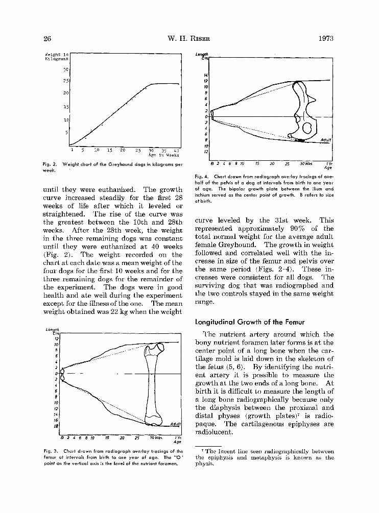
Fig. 2. week.
Weight chart of the Greyhound dogs in kilograms per
until they were euthanized. The growth curve increased steadily for the first 28 weeks of life after which it leveled or straightened. The rise of the curve was the greatest between the 10th and 28th weeks. After the 28th week, the weight in the three remaining dogs was constant until they were euthanized at 40 weeks (Fig. 2). The weight recorded on the chart at each date was a mean weight of the four dogs for the first 10 weeks and for the three remaining dogs for the remainder of the experiment. The dogs were in good health and ate well during the experiment except for the illness of the one. The mean weight obtained was 22 kg when the weight
Len th e m ,
?2 10 8 6 4 2 0 - 2 4 6 8 10 12 14 16 18
I 1 8 2 1 6 8 10 15 20 25 3OWks. 1 Yr
Age
1 1 €3 2 4 6 8 10 15 20 25 3OWks 1Yr
Age
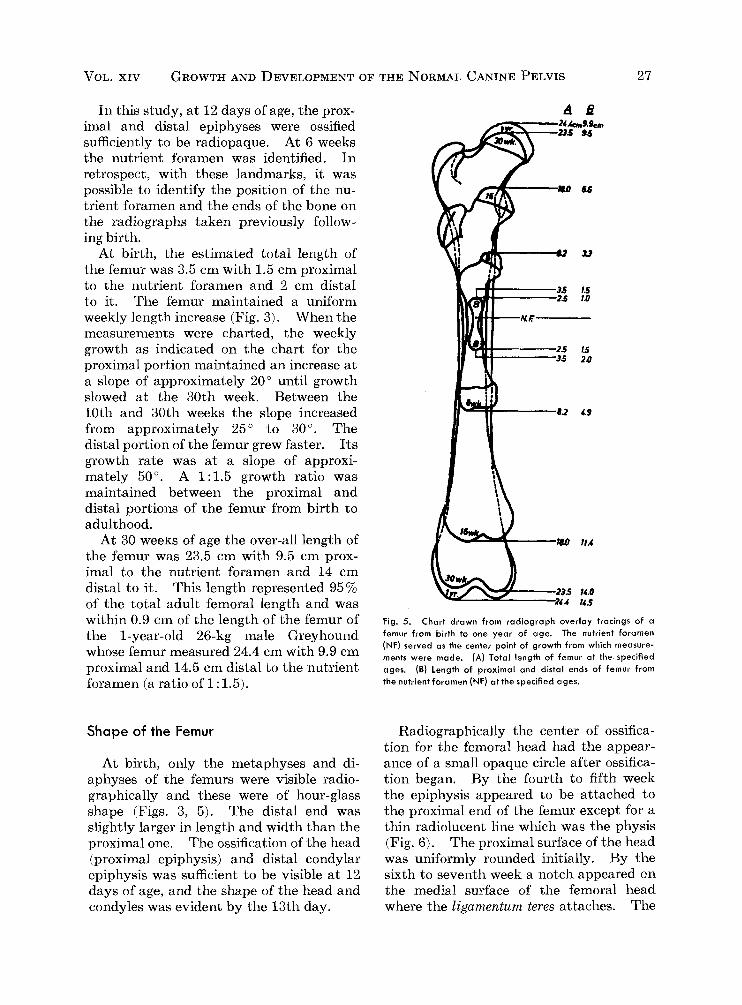
Fig. 4. Chart drawn from radiograph overlay tracings of one- half of the pelvis of a dog at intervals from birth to one year of age. The bipolar growth plate between the ilium and ischium served as the center point of growth. B refers to size at birth.
curve leveled by the 31st week. This represented approximately 90% of the total normal weight for the average adult female Greyhound. The growth in weight followed and correlated well with the in- crease in size of the femur and pelvis over the same period (Figs. 2--4). These in- creases were consistent for all dogs. The surviving dog that was radiographed and the two controls stayed in the same weight range.
Longitudinal Growth of the Femur
The nutrient artery around which the bony nutrient foramen later forms is a t the center point of a long bone when the car- tilage mold is laid down in the skeleton of the fetus (5, 6). By identifying the nutri- ent artery it is possible to measure the growth a t the two ends of a long bone. At birth it is difficult to measure the length of a long bone radiographically because only the diaphysis between the proximal and distal physes (growth plates)? is radio- paque. The cartilagenous epiphyses are radiolucent .
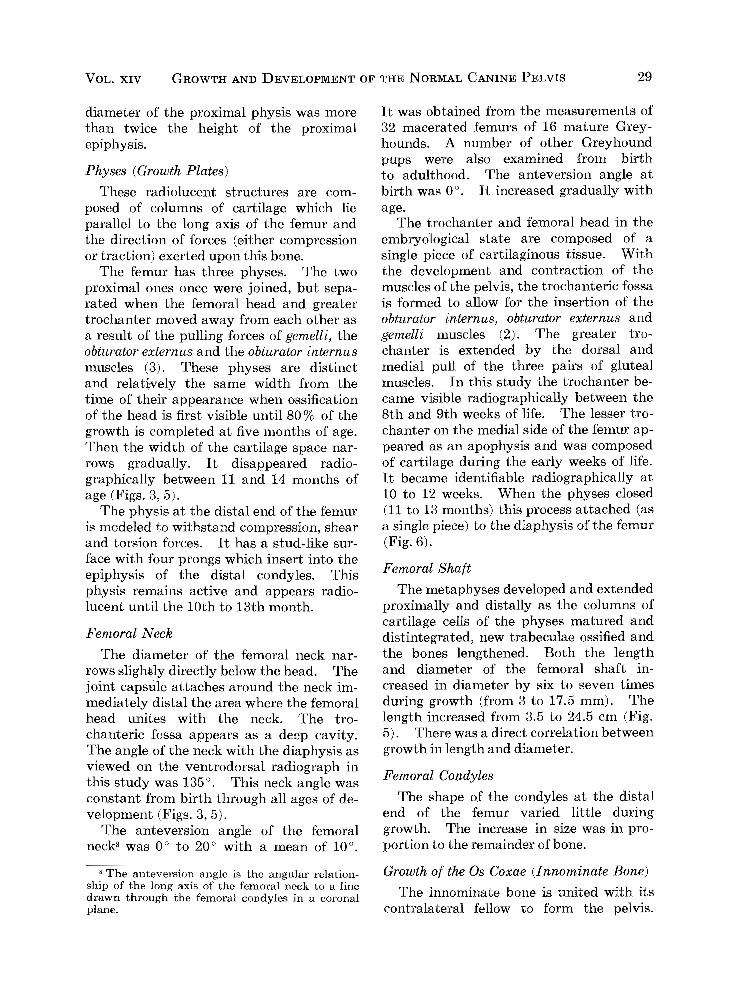
Fig. 3. 7 The lucent line seen radiographically between femur at intervals from birth to one year of age. The "0" the epiphysis and metaphysis is known as the point on the vertical axis is the level of the nutrient foramen.
Chart drawn from radiograph overlay tracings of the
physis.
VOL. XIV GROWTH AND DEVELOPMENT OF THE NORMAL CANINE PELVIS 27
In this study, at 12 days of age, the prox- imal and distal epiphyses were ossified sufficiently to be radiopaque. At 6 weeks the nutrient foramen was identified. In retrospect, with these landmarks, it was possible to identify the position of the nu- trient foramen and the ends of the bone on the radiographs taken previously follow- ing birth.
At birth, the estimated total length of the femur was 3.5 cm with 1.5 cm proximal to the nutrient foramen and 2 cm distal to it. The femur maintained a uniform weekly length increase (Fig. 3 ) . When the measurements were charted, the weekly growth as indicated on the chart for the proximal portion maintained an increase at a slope of approximately 20" until growth slowed a t the 30th week. Between the 10th and 30th weeks the slope increased from approximately 25" to 30". The distal portion of the femur grew faster. Its growth rate was a t a slope of approxi- mately 50". A 1:1.5 growth ratio was maintained between the proximal and distal portions of the femur from birth to adulthood.
At 30 weeks of age the over-all length of the femur was 23.5 cm with 9.5 cm prox- imal to the nutrient foramen and 14 cm distal to it. This length represented 95% of the total adult femoral length and was within 0.9 cm of the length of the femur of the 1-year-old 26-kg male Greyhound whose femur measured 24.4 cm with 9.9 cm proximal and 14.5 cm distal to the nutrient foramen (a ratio of 1 : 1.5).
Shape of the Femur
At birth, only the metaphyses and di- aphyses of the femurs were visible radio- graphically and these were of hour-glass shape (Figs. 3, 5). The distal end was slightly larger in length and width than the proximal one. The ossification of the head (proximal epiphysis) and distal condylar epiphysis was sufficient to be visible at 12 days of age, and the shape of the head and condyles was evident by the 13th day.
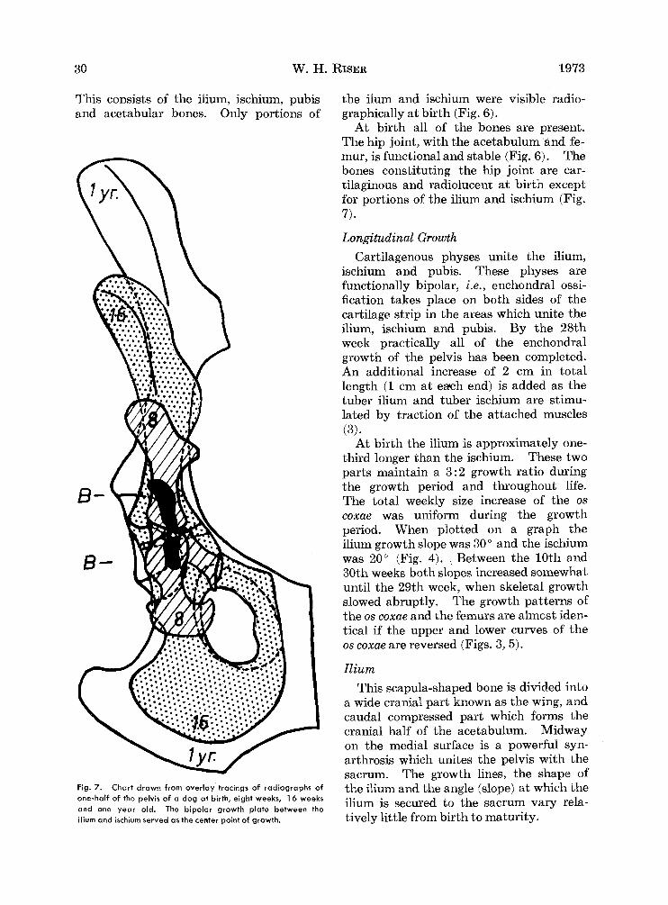
Fig. 5. Chart drawn from radiograph overlay tracings of a femur from birth to one year of age. The nutrient foramen (NF) served as the center point of growth from which measure- ments were made. (A) Total length of femur a t the specified ages. (B) length of proximal and distal ends of femur from the nutrient foramen (NF) at the specified ages.
Radiographically the center of ossifica- tion for the femoral head had the appear- ance of a small opaque circle after ossifica- tion began. By the fourth to fifth week the epiphysis appeared to be attached to the proximal end of the femur except for a thin radiolucent line which was the physis (Fig. 6). The proximal surface of the head was uniformly rounded initially. By the sixth to seventh week a notch appeared on the medial surface of the femoral head where the Zigumentum teres attaches. The

28 W. H. RISER 1973
Fig. 6. Drawings copied from radiograph overlay trucings of normal hip joint growth und development from birth to one. year-old. The number with each druwing indicutes the age of the dog in weeks.
VOL. XIV GROWTH AND DEVELOPMENT OF THE NORMAL CANINE PELVIS 29
diameter of the proximal physis was more than twice the height of the proximal epiphysis.
Physes (Growth Plates) These radiolucent structures are com-
posed of columns of cartilage which lie parallel to the long axis of the femur and the direction of forces (either compression or traction) exerted upon this bone.
The femur has three physes. The two proximal ones once were joined, but sepa- rated when the femoral head and greater trochanter moved away from each other as a result of the pulling forces of gemelli, the obturator externus and the obturator internus muscles (3). These physes are distinct and relatively the same width from the time of their appearance when ossification of the head is first visible until 80% of the growth is completed a t five months of age. Then the width of the cartilage space nar- rows gradually. It disappeared radio- graphically between 11 and 14 months of age (Figs. 3,5).
The physis a t the distal end of the femur is modeled to withstand compression, shear and torsion forces. It has a stud-like sur- face with four prongs which insert into the epiphysis of the distal condyles. This physis remains active and appears radio- lucent until the 10th to 13th month.
Femoral Neck The diameter of the femoral neck nar-
rows slighbly directly below the head. The joint capsule attaches around the neck im- mediately distal the area where the femoral head unites with the neck. The tro- chanteric fossa appears as a deep cavity. The angle of the neck with the diaphysis as viewed on the ventrodorsal radiograph in this study was 135". This neck angle was constant from birth through all ages of de- velopment (Figs. 3,5).
The anteversion angle of the femoral neck8 was 0" to 20" with a mean of 10".
* The anteversion angle is the angular relation- ship of the long axis of the femoral neck to a line drawn through the femoral condyles in a coronal plane.
It was obtained from the measurements of 32 macerated femurs of 16 mature Grey- hounds. A number of other Greyhound pups were also examined from birth to adulthood. The anteversion angle a t birth was 0". It increased gradually with age.
The trochanter and femoral head in the embryological state are composed of a single piece of cartilaginous tissue. With the development and contraction of the muscles of the pelvis, the trochanteric fossa is formed to allow for the insertion of the obturator internus, obturator externus and gemelli muscles (2). The greater tro- chanter is extended by the dorsal and medial pull of the three pairs of gluteal muscles. In this study the trochanter be- came visible radiographically between the 8th and 9th weeks of life. The lesser tro- chanter on the medial side of the femur ap- peared as an apophysis and was composed of cartilage during the early weeks of life. It became identifiable radiographically at 10 to 12 weeks. When the physes closed (11 to 13 months) this process attached (as a single piece) to the diaphysis of the femur (Fig. 6).
Femoral Shaf t The metaphyses developed and extended
proximally and distally as the columns of cartilage cells of the physes matured and distintegrated, new trabeculae ossified and the bones lengthened. Both the length and diameter of the femoral shaft in- creased in diameter by six to seven times during growth (from 3 to 17.5 mm). The length increased from 3.5 to 24.5 cm (Fig. 5). There was a direct correlation between growth in length and diameter.
Femoral Condyles The shape of the condyles at the distal
end of the femur varied little during growth. The increase in size was in pro- portion to the remainder of bone.
Growth of the 0s Coxae (Innominate Bone) The innominate bone is united with its
contralateral fellow to form the pelvis.
30 W. H. RISER 1973
This consists of the ilium, ischium, pubis and acetabular bones. Only portions of
the ilum and ischium were visible radio- graphically a t birth (Fig. 6).
At birth all of the bones are present. The hip joint, with the acetabulum and fe- mur, is functional and stable (Fig. 6). The bones constituting the hip joint are car- tilaginous and radiolucent a t birth except for portions of the ilium and ischium (Fig. 7).
Longitudinal Growth Cartilagenous physes unite the ilium,
ischium and pubis. These physes are functionally bipolar, i.e., enchondral ossi- fication takes place on both sides of the cartilage strip in the areas which unite the ilium, ischium and pubis. By the 28th week practically all of the enchondral growth of the pelvis has been completed. An additional increase of 2 cm in total length (1 cm a t each end) is added as the tuber ilium and tuber ischium are stimu- lated by traction of the attached muscles ( 3 ) .
At birth the ilium is approximately one- third longer than the ischium. These two parts maintain a 3:2 growth ratio during the growth period and throughout life. The total weekly size increase of the 0s coxae was uniform during the growth period. When plotted on a graph the ilium growth slope was 30" and the ischium was 20" (Fig. 4). Between the 10th and 30th weeks both slopes increased somewhat until the 29th week, when skeletal growth slowed abruptly. The growth patterns of the 0s coxae and the femurs are almost iden- tical if the upper and lower curves of the 0s coxae are reversed (Figs. 3,5).
Ilium This scapula-shaped bone is divided into
a wide cranial part known as the wing, and caudal compressed part which forms the cranial half of the acetabulum. Midway on the medial surface is a powerful syn- arthrosis which unites the pelvis with the sacrum. The growth lines, the shape of
ilium is secured to the sacrum vary rela- tively little from birth t o maturity.
Fig. 7. Chart drawn from overlay tracings of radiographs of one-half of the pelvis of a dog at birth, eight weeks, 16 weeks and one year old. The bipolar growth plate between the
the ilium and the angle (slope) a t which the
ilium and ischium served as the center point of growth.
VOL. XIV GROWTH AND DEVELOPMENT OF THE NORMAL CANINE PELVIS 31
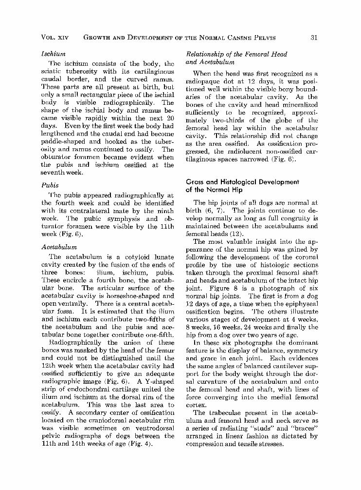
Ischium The ischium consists of the body, the
sciatic tuberosity with its cartilaginous caudal border, and the curved ramus. These parts are all present at birth, but only a small rectangular piece of the ischial body is visible radiographically. The shape of the ischial body and ramus be- came visible rapidly within the next 20 days. Even by the first week the body had lengthened and the caudal end had become paddle-shaped and hooked as the tuber- osity and ramus continued to ossify. The obturator foramen became evident when the pubis and ischium ossified at the seventh week.
Pubis The pubis appeared radiographically at
the fourth week and could be identified with its contralateral mate by the ninth week. The pubic symphysis and ob- turator foramen were visible by the 11th week (Fig. 6).
Acetabulum The acetabulum is a cotyloid lunate
cavity created by the fusion of the ends of three bones: ilium, ischium, pubis. These encircle a fourth bone, the acetab- ular bone. The articular surface of the acetabular cavity is horseshoe-shaped and open ventrally. There is a central acetab- ular fossa. It is estimated that the ilium and ischium each contribute two-fifths of the acetabulum and the pubis and ace- tabular bone together contribute one-fifth.
Radiographically the union of these bones was masked by the head of the femur and could not be distinguished until the 12th week when the acetabular cavity had ossified sufficiently to give an adequate radiographic image (Fig. 6). A Y-shaped strip of endochondral cartilage united the ilium and ischium at the dorsal rim of the acetabulum. This was the last area to ossify. A secondary center of ossification located on the craniodorsal acetabular rim was visible sometimes on ventrodorsal pelvic radiographs of dogs between the 11th and 14th weeks of age (Fig. 4).
Relationship of the Femoral Head and Acetabulum
When the head was first recognized as a radiopaque dot a t 12 days, it was posi- tioned well within the visible bony bound- aries of the acetabular cavity. As the bones of the cavity and head mineralized sufficiently to be recognized, approxi- mately two-thirds of the globe of the femoral head lay within the acetabular cavity. This relationship did not change as the area ossified. As ossification pro- gressed, the radiolucent non-ossified car- tilaginous spaces narrowed (Fig. 6).
Gross and Histological Development of the Normal Hip
The hip joints of all dogs are normal at birth (6, 7). The joints continue to de- velop normally as long as full congruity is maintained between the acetabulums and femoral heads (12).
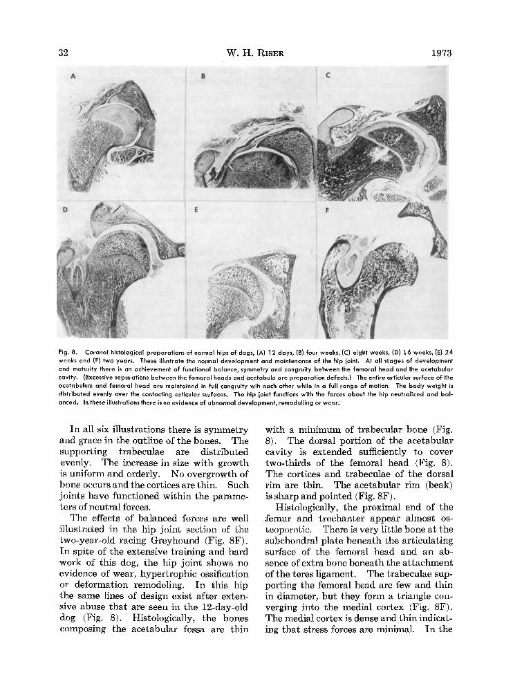
The most valuable insight into the ap- pearance of the normal hip was gained by following the development of the coronal profile by the use of histologic sections taken through the proximal femoral shaft and heads and acetabulum of the intact hip joint. Figure 8 is a photograph of six normal hip joints. The first is from a dog 12 days of age, a time when the epiphyseal ossification begins. The others illustrate various stages of development at 4 weeks, 8 weeks, 16 weeks, 24 weeks and finally the hip from a dog over two years of age.
In these six photographs the dominant feature is the display of balance, symmetry and grace in each joint. Each evidences the same angles of balanced cantilever sup- port for the body weight through the dor- sal curvature of the acetabulum and onto the femoral head and shaft, with lines of force converging into the medial femoral cortex.
The trabeculae present in the acetab- ulum and femoral head and neck serve as a series of radiating “studsf7 and “braces” arranged in linear fashion as dictated by compression and tensile stresses.
32 W. H. RISER 1973
Fig. 8. Coronal histological preparations of normal hips of dogs, (A) 12 days, (B) four weeks, ( C ) eight weeks, (D) 16 weeks, (El 24 weeks and (F) two years. These illustrate the normal development and mointenance of the hip joint. At al l stages of development and maturity there i s an achievement of function01 balance, symmetry and congruity between the femoral head and the acetabulor cavity. (Excessive separotions between the femoral heads and acetabula are preparation defects.) The entire articular surface of the acetabulum and femoral head are maintained in full congruity wih each other while in a full range of motion. The body weight is distributed evenly over the contacting articular surfaces. The hip joint functions with the forces about the hip neutralized and bal- anced. In these illustrations there is no evidence of abnormal development, remodelling or wear.
In all six illustrations there is symmetry and grace in the outline of the bones. The supporting trabeculae are distributed evenly. The increase in size with growth is uniform and orderly. No overgrowth of bone occurs and the cortices are thin. Such joints have functioned within the parame- ters of neutral forces.
The effects of balanced forces are well illustrated in the hip joint section of the two-year-old racing Greyhound (Fig. 8F). In spite of the extensive traiping and hard work of this dog, the hip joint shows no evidence of wear, hypertrophic ossification or deformation remodeling. In this hip the same lines of design exist after exten- sive abuse that are seen in the 12-day-old dog (Fig. 8). Histologically, the bones composing the acetabular fossa are thin
with a minimum of trabecular bone (Fig. 8). The dorsal portion of the acetabular cavity is extended sufficiently to cover two-thirds of the femoral head (Fig. 8). The cortices and trabeculae of the dorsal rim are thin. The acetabular rim (beak) is sharp and pointed (Fig. 8F).
Histologically, the proximal end of the .femur and trochanter appear almost os- teoporotic. There is very little bone at the subchondral plate beneath the articulating surface of the femoral head and an ab- sence of extra bone beneath the attachment of the teres ligament. The trabeculae sup- porting the femoral head are few and thin in diameter, but they form a triangle con- verging into the medial cortex (Fig. 8F). The medial cortex is dense and thin indicat- ing that stress forces are minimal. In the
VOL. XIV GROWTH AND DEVELOPMENT OF THE NORMAL CANINE PELVIS 33
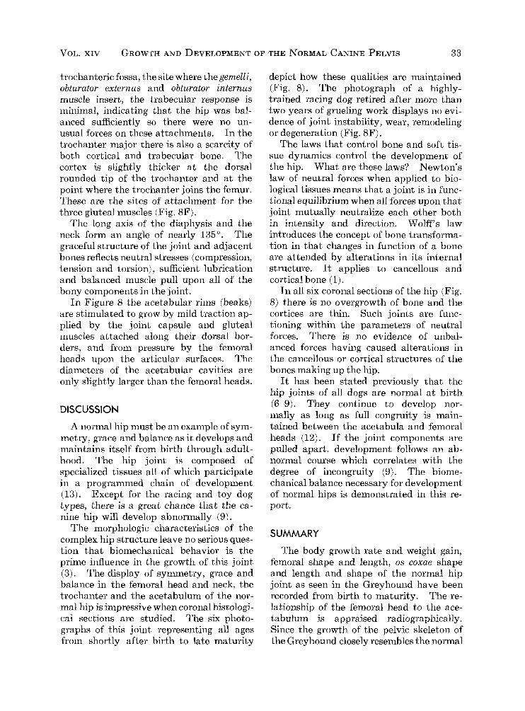
trochanteric fossa, the site where thegemell i, obturator externus and obturator internus muscle insert, the trabecular response is minimal, indicating that the hip was bal- anced sufficiently so there were no un- usual forces on these attachments. In the trochanter major there is also a scarcity of' both cortical and trabecular bone. The cortex is slightly thicker a t the dorsal rounded tip of the trochanter and at the point where the trochanter joins the femur. These are the sites of attachment for the three gluteal muscles (Fig. 8F).
The long axis of the diaphysis and the neck form an angle of nearly 135". The graceful structure of the joint and adjacent bones reflects neutral stresses (compression, tension and torsion) , sufficient lubrication and balanced muscle pull upon all of the bony components in the joint.
In Figure 8 the acetabular rims (beaks) are stimulated to grow by mild traction ap- plied by the joint capsule and gluteal muscles attached along their dorsal bor- ders, and from pressure by the femoral heads upon the articular surfaces. The diameters of the acetabular cavities are only slightly larger than the femoral heads.
DISCUSSION
A normal hip must be an example of sym- metry, grace and balance as it develops and maintains itself from birth through adult- hood. The hip joint is composed of specialized tissues all of which participate in a programmed chain of development (13). Except for the racing and toy dog types, there is a great chance that the ca- nine hip will develop abnormally (9).
The morphologic characteristics of the complex hip structure leave no serious ques- tion that biomechanical behavior is the prime influence in the growth of this joint ( 3 ) . The display of symmetry, grace and balance in the femoral head and neck, the trochanter and the acetabulum of the nor- mal hip is impressive when coronal histologi- cal sections are studied. The six photo- graphs of this joint representing all ages from shortly after birth to late maturity
depict how these qualities are maintained (Fig. 8). The photograph of a highly- trained racing dog retired after more than two years of grueling work displays no evi- dence of joint instability, wear, remodeling or degeneration (Fig. 8F).
The laws that control bone and soft tis- sue dynamics control the development of the hip. What are these laws? Newton's law of neutral forces when applied to bio- logical tissues means that a joint is in func- tional equilibrium when all forces upon that joint mutually neutralize each other both in intensity and direction. Wolff ' S law introduces the concept of bone transforma- tion in that changes in function of a bone are attended by alterations in its internal structure. It applies to cancellous and cortical bone (l),
In all six coronal sections of the hip (Fig. 8) there js no overgrowth of bone and the cortices are thin. Such joints are func- tioning within the parameters of neutral forces. There is no evidence of unbal- anced forces having caused alterations in the cancellous or cortical structures of the bones making up the hip.
It has been stated previously that the hip joints of all dogs are normal at birth (6-9). They continue to develop nor- mally as long as full congruity is main- tained between the acetabula and femoral heads (12). If the joint components are pulled apart, development follows an ab- normal course which correlates with the degree of incongruity (9). The biome- chanical balance necessary for development of normal hips is demonstrated in this re- port.
SUMMARY
The body growth rate and weight gain, femoral shape and length, 0s coxae shape and length and shape of the normal hip joint as seen in the Greyhound have been recorded from birth to maturity. The re- lationship of the femoral head to the ace- tabulum is appraised radiographically. Since the growth of the pelvic skeleton of the Greyhound closely resembles the normal
34 W. H. RISER 1973
radiographic appearance of the hip joints of many large and giant breeds, it provides a basis for comparison with abnormal hips.
The development of the normal hip is followed from birth to two years in histo- logical coronal profile sections made through the proximal femur and acetabulum. The sections were taken from dogs at 12 days, 4 weeks, 8 weeks, 16 weeks, 24 weeks and 2 years of age. In all stages of develop- ment and maturity there is an achievement of functional biomechanical balance, sym- metry and congruity between the femoral head and acetabulum.
School of Veterinary Medicine University of Pennsylvania Philadelphia, Pa.
REFERENCES 1. Enlow, D. H.: Principles of Bone Re-
modeling. Charles C Thomas, Springfield, Ill. (1963).
2. Hare, W. C. D.: Canine Pelvic Limb.
3. Johnson, L. C.: Morphologic Analysis in Pathology, Bone Biodynamics. Little, Brown. Boston, Mass. (1964), 559.
4. Mansson, J. and Norberg, I.: Dysplasia of the Hip in Dogs. Medlemsblad Sveriges Veterinaer Forbund, 13: 330-339, 1961.
5. Miller, M. I., Christensen, G. C., and Evans, H. E.: Anatomy of the Dog. W. B. Saunders Co. Phila., Pa. (1964).
6. Norberg, I.: Hoftledsdysplasi Hos Hund. Hundsport, 69: 13-15, 1961.
7. Ponseti, I. V., Pedrini-Mille, A., and’Ped- rini, V.: Histological Analysis of Human Iliac Crest Cartilage. I. Observations on Trunk Growth.
8. Riser, W. H. and Rhodes, W. H.: Pro- ducing Diagnostic Pelvic Radiographs for Canine Hip Dysplasia Examination. Anim. Hosp., 2: 167- 171, 1966.
9. Riser, W. H.: The Dog as a Model for Hip Dysplasia. Libr. Fakultat Veterinarmedi- zenenischen der Universitat Bern (1973).
10. Smith, R. N.: Epiphyseal Fusion in the Greyhound. Vet, Rec., 72: 75-79, 1960.
11. Smith, R. N.: The Pelvis of the Young Dog.
12. Strayer, L. M.: The Embryology of the Human Hip Joint. Clin. Orthop. Rel. Res., 74:
J.A.V.M.A. 136: 603-611, 1960.
Calcif. Tissue Res., 2: 197-213, 1968.
Vet. Rec., 76: 975-979, 1964.
221-240, 1971.
13. Trueta, J.: Studies of the Development and W. B. Saunders Co.,
14. Wright, V.: Lubrication and Wear in
Decay of the Human Frame. Phila., Pa. (1968).
Joints. J. B. Lippincott Co., Phila., Pa. (1972).
ZUSAMMENFASSUNG Die Korperwachstumsrate und Gewichtzu-
nahme, die femorale Form und Lange, die oscoxae Form, un Lange und Form des normalen Huft- gelenkes, wie sie beim Windhund gesehen werden, sind von Geburt bis zur Reife aufgezeichnet worden. Das Verhaltnis des femoralen Kopfes zur Gelenkpfanne wird radiographisch ausge- wertet. Da das Wachstum des Beckenskelettes des Windhundes sehr stark der normalen radio- graphischen Erscheinung der Huftgelenke vieler grosser und Riesenzuchtungen ahnelt, ergibt es eine Basis fur einen Vergleich mit abnormalen Huften. Die Entwicklung der normalen Hufte wird verfolgt von der Geburt bis zu zwei Jahren in histologischen Kronenprofilschnitten, die durch den obersten Teil des Femurs und der Gelenk- pfanne gemacht wurden. Die Schnitte wurden entnommen von Hunden von 12 Tagen, vier Wochen, acht Wochen, 16 Wochen, 24 Wochen und zwei Jahren Alter. Bei allen Stadien von Entwicklung und Reife gibt es die Erreichung von einem funktionellen, bio-mechanischen Gleichge- wicht, Symmetrie und Kongruitat zwischen dem femoralen Kopf und der Gelenkpfanne.
RESUME On a enregistrk chez le Ihvrier, de sa naissance 2I
sa maturitk le taux de croissance du corps et le gain de poids, la forme et la taille du f6mur, la forme et la taille de Z’os coxa ainsi que la forme et la taille de l’articulation normale de la hanche. On a 6valuk radiographiquement la relation de la t8te f6morale par raport B l’acktabule. Etant donnk que la croissance de l’ossature pelvienne du levrier ressemble particulierement ’a l’aspect normal radiographique des articulations de hanche de maintes races d’animaux de grande taille ou gkants, cela nous fournit une base pour proceder $I la comparaison avec des articulations anormales.
On a suivi de prits, de la naissance 21 l’ige de deux ans le dkveloppement de l’articulation normale dans des sections de profil coronal his- tologique faites d’un bout B l’autre du fkmur et de l’acktabule. Les sections ont Qt6 prises sur des chiens B l’gge de douze jours, quatre semaines, huit semaines, seize semaines, vingtquatre se- maines et deux ans. A tous les stades de developpe- ment et de maturitk il se produit un Qquilibre bio- mkcanique fonctionnel, une symbtrie et ude congruence entre la t8te fkmorale et hcktabule.