growth inhibition of human breast cancer cells and down ... · e-mail: [email protected]...
TRANSCRIPT

O
Go
AD
a
ARAA
KAACO
I
mScbMiopsiEcdi
0c
Revista Brasileira de Farmacognosia 27 (2017) 84–90
ww w . elsev ier .com/ locate /b jp
riginal Article
rowth inhibition of human breast cancer cells and down-regulationf ODC1 and ADA genes by Nepeta binaloudensis
kbar Safipour Afshar ∗, Fatemeh Saeid Nematpour, Mahshid Meshkani, Arezosadat Khafiepartment of Biology, Neyshabur Branch, Islamic Azad University, Neyshabur, Iran
r t i c l e i n f o
rticle history:eceived 10 February 2016ccepted 8 July 2016vailable online 15 September 2016
eywords:denosine deaminasenticancerytotoxicityrnithine decarboxylase
a b s t r a c t
Nepeta binaloudensis Jamzad, Lamiaceae, is a rare medicinal plant endemic to Iran. In spite of many studiesabout the chemical constituents and antibacterial effects of this species, no report has been provided aboutits cytotoxic and anticancer activities. In this study we have evaluated the effects of EtOH 70%, hexaneand aqueous extracts of N. binaloudensis on the cell proliferation and n-hexane extract on the expressionof adenosine deaminase and ornithine decarboxylase 1 genes in breast cancer cell lines (MCF-7, MDA-MB-231) compared to non-cancer line (MCF-10A). The cell lines were subjected to increasing dosesof the extracts ranging from 10 to 320 �g/ml. Cell viability was quantified by MTS assay. Expression ofadenosine deaminase and ornithine decarboxylase 1 genes was analyzed by real time PCR. N. binaloudensisinhibited the growth of malignant cells in a time and dose-dependent manner. Among extracts of N.binaloudensis, the hexane extract was found to be more toxic compared to other extracts. Results showeda marked decrease in the expression of ornithine decarboxylase 1 and adenosine deaminase genes incancer cell lines. At 60 �g/ml concentration of N. binaloudensis hexane extract ornithine decarboxylase1 and adenosine deaminase mRNA expression were reduced 4.9 fold and 3.5 fold in MCF-7 cell line and
3.6 fold and 2.6 fold in MDA-MB-231 cell line compared to control, respectively. The result of our studyhighlights the potential influences of N. binaloudensis hexane extract on ornithine decarboxylase 1 andadenosine deaminase genes expression in breast cancer cells and its relation to inhibition of cancer cellgrowth.© 2016 Sociedade Brasileira de Farmacognosia. Published by Elsevier Editora Ltda. This is an openhe CC
access article under tntroduction
Cancer is the most common cause of death in the world. Theajority of deaths from cancer occur in women with breast cancer.
tatistics indicate that in 2014 about 29% of cancers were breastancers among American women (Siegel et al., 2014). In Iran alsoreast cancer rates have increased in females (Mousavi et al., 2009).any investigations are performed to detect the genes which are
nvolved in breast cancer. One of the most important genes isrnithine decarboxylase (ODC) which encodes a key enzyme inolyamines synthesis (Zhu et al., 2012; Xu et al., 2015). Severaltudies indicate that there are close correlation between ODC activ-ty in the cells and regulation of cell proliferation (Deng et al., 2008;lmets and Athar, 2010; Hayes et al., 2011). Consequently, any
hanges in ODC activity and polyamines level have effects on cellifferentiation and proliferation. In breast cancer cells, estrogensnduce ODC activity. So, the level of ODC and estrogen is increased
∗ Corresponding author.E-mail: [email protected] (A.S. Afshar).
http://dx.doi.org/10.1016/j.bjp.2016.07.005102-695X/© 2016 Sociedade Brasileira de Farmacognosia. Published by Elsevier Editreativecommons.org/licenses/by-nc-nd/4.0/).
BY-NC-ND license (http://creativecommons.org/licenses/by-nc-nd/4.0/).
in these cells (Zhu et al., 2012). In addition, Adenosine Deaminasegene (ADA) encodes the enzyme that catalyzes hydrolyzation ofadenosine to inosin and participates in purines metabolism, whichhas a crucial role in development of immune system and matura-tion of mammalian cells (Ri et al., 2010; Roberts and Roberts, 2012).Recent studies suggest that the level of ADA enzyme is increased inbreast cancer cells in contrast with normal cells (Aghaei et al., 2010;Mahajan et al., 2013). Therefore, decreasing of ODC and ADA geneexpression and respective enzyme activities can be a good strategyfor breast cancer therapy.
Using medicinal plant extracts as anticancer drugs is a moreeffective way for cancer therapy. About 50% of prescribed drugsin the world originate from plants (Van Slambrouck et al., 2007).Nepeta binaloudensis Jamzad, Lamiaceae, is an endemic and raremedicinal plant which grows exclusively in the Binalud Mountainsin Khorasan Razavi province in Northeast of Iran (Tundis et al.,2013). This plant has been applied widely in traditional medicine
for the treatment of respiratory disease, digestive problems andosteoarthritis (Koocheki and Rezvani Moghaddam, 2012). Previouschemical investigation of N. binaloudensis has shown the presenceof nepetalactone and 1,8 cineole (Rustaiyan and Nadji, 1999). Someora Ltda. This is an open access article under the CC BY-NC-ND license (http://
ra de F
sbyclAa
M
P
lpJVIcwHwDs
C
oc(cppCt
C
adftiedtmCat
C
wBdhws
A.S. Afshar et al. / Revista Brasilei
tudies were done about antibacterial activities of N. binaloudensisut its cytotoxicity and anticancer effects have not been studiedet. Therefore, the purpose of the study was to determine the anti-ancer effects of N. binaloudensis extract on two breast cancer cellines (MCF-7, MDA-MB-231) and normal breast cell line (MCF-10A).lso, the present study is focused on evaluating the level of ODC1nd ADA gene expression in the normal and cancerous breast cells.
aterial and methods
lant collection and extraction
Shoots of Nepeta binaloudensis Jamzad, Lamiaceae, were col-ected from Binalud Mountains in Neyshabur, Khorasan Razavirovince, northeast of Iran. The plant was identified by Mr. M.R.
oharchi, from Ferdowsi University of Mashhad Herbarium (FUMH).oucher specimen (No. 1025) was deposited with herbarium of
slamic Azad University, Neyshabur Branch. The extraction pro-ess was performed from dried shoots. The dried shoots (100 g)ere percolated with 100 ml of each solvent (EtOH 70%, hexane and2O) for 4 h separately. The extracts were filtered and the solventsere evaporated under vacuum at 45 ◦C to afford crude extracts.ry extracts were dissolved in dimethyl sulfoxide (DMSO) and then
ubjected to cytotoxic and gene expression assays.
ell lines and culture medium
Two human breast cancer cell lines (MCF-7, MDA-MB-231) andne normal breast cell line (MCF-10A) were obtained from cellulture laboratory of the Faculty of Sciences, Ferdowsi UniversityMashhad, Iran) and Pasteur Institute (Tehran, Iran). The cells wereultured in 25 cm2 flasks with RPMI-1640 (RPMI Sigma) and sup-lemented with 10% fetal bovine serum (FBS, Gibco) and 100 U/mlenicillin and 100 �g/ml streptomycin solutions, at 37 ◦C with 5%O2 and 95% humidity. The medium was exchanged every day andhe cells were passaged every 3–4 days.
ytotoxicity assay
The cytotoxic activity of the extracts was determined using MTTssay (2, 3, 5 diphenil tetrazolium bromide). Cells were plated withensity of 3 × 10⁴ cells/dish in 96 well plates and were incubatedor 24 h at 37 ◦C. Then, the cells were treated with different concen-rations of extracts (0, 10, 20, 40, 80, 160 and 320 �g/ml) and werencubated for 24, 48 and 72 h. A 2 mM MTT solution was added toach well and plate was incubated for 4 h. The medium was theniscarded and 100 ml DMSO (dimethyl sulfoxide) was added andhe plate was shaken for 10 min. Finally, optical density was deter-
ined in 540 nm using ELISA microplate reader (Awareness, Palmity, FL, USA). Each experiment was performed in triplicate. Resultsre expressed as the percentage growth inhibition with respect tohe untreated cells.
olony formation assay
The effect of hexane extract on MCF7 and MDA-MB-231 cellsas investigated by colony formation assay (Franken et al., 2006).riefly, the cells (500 cells/ml) were allowed to grow in 60 mm Petri
ishes for 12 h. Subsequently, the cells were treated for 48 h withexane extract (10, 30 and 60 �g/ml), or 0.1% DMSO. The coloniesere fixed and stained with 0.2% crystal violet and counted undertereomicroscope.
armacognosia 27 (2017) 84–90 85
Real-time PCR
The expression levels of ODC1 and ADA genes, key genes inbreast cancer cells, were analyzed using Real time PCR assay. Thecells were cultured in three groups and treated with increasingconcentrations (0–60 �g/ml) of the hexane extract dissolved inDMSO for 48 h. After 48 h total cellular RNA was extracted fromthe treated and untreated (control) cells using Easy BLUE totalRNA extraction kit (iNtRON). The treatments were repeated forthree times. The quality of RNA was determined by absorption at260 nm using T80 UV-Visible Spectrophotometer (PG Instruments).For each specimen 2 �g of RNA was reverse transcribed into cDNAwith reverse transcription reaction mixture of Power cDNA Syn-thesis Kit (iNtRON). Primers of ODC1, ADA and �ACT were designedusing Gene Runner software (version 4).
Primer sequences for ODC1 were: ODC1-FW: 5′-GTGGGTGATTGGATGCTCTTTG-3′ ODC1-RV: 5′-ACCAGGCTAACTACTCGCTCAA-3′.
Primer sequences for ADA were: ADA-FW: 5′-ACCAGGCTAACTACTCGCTCAA-3′ ADA-RV: 5′-TCAGTAAAGCCCATGTCCCGTT-3′.
Primer sequences for �ACT were: ACT-FW: 5′-TCCATCATGAAGTGTGACGT-3′ ACT-RV: 5′-GAGCAATGATCTTGATCTTCAT-3′.
Real-time PCR was carried out using SYBR Green method on anABI Step-One instrument (Applied Biosystems, USA). The cDNA wastaken as a template for PCR amplification of ADA and ODC1 genesand �ACT gene (control gene). Each reaction was repeated for threetimes. A PCR reaction mixture of 20 �l contained 10 �l SYBR Greenmaster mix, 0.4 �l Reverse primer and 0.4 �l Forward primer, 6.2 �ldH2O (RNase free) and 3 �l cDNA (4 ng). Three pairs of primers wereused separately. The thermal cycling conditions were as follow: 1denaturation cycle of 95 ◦C for 10 min and 40 cycles of 95 ◦C for15 s, 60 ◦C for 60 s (annealing and extension temperature). A neg-ative control was used in each run to assess specificity of primersand possible contamination. The realtime PCR data were analyzedusing the relative gene expression (��Ct) method, as describedin Applied Biosystems User Bulletin No. 2 (Livak and Schmittgen,2001). Briefly, the data are presented as the fold change in geneexpression normalized to the reference gene (�-actin) and weredetermined using the equation fold change = 2−��Ct.
Statistical analysis
One-way analysis of variance and Fisher’s LSD were used for dataanalysis. All results were expressed as mean ±SEM, and p valueslower than 0.05 were judged as significant.
Results
Cytotoxicity of various extracts of Nepeta binaloudensis
The cytotoxic activity of N. binaloudensis extracts were stud-ied against cultured MCF-10A (Normal), MBA-MD-231 and MCF-7(cancer) breast cell lines using MTT assay. The cell lines were sub-jected to increasing doses of EtOH 70%, hexane and H2O extractsranging from 10 to 320 �g/ml for 24, 48 and 72 h. The resultsshowed EtOH 70% and hexane extracts decreased viability of malig-nant cells in a concentration and time-dependent manner. WhereasH2O extract showed no marked cytotoxicity on the cell lines tested.EtOH and hexane extracts in the same concentrations caused less
toxicity to MCF-10A than tumor cells indicating a degree of speci-ficity for malignant cell lines (Fig. 1). Concentrations inducing 50%cell growth inhibition (IC50) against MBA-MD-231 and MCF-7 cellsare presented in Table 1. MCF-7 cells were found to be more
86 A.S. Afshar et al. / Revista Brasileira de Farmacognosia 27 (2017) 84–90
120MCF-10A (H2O)
24 h48 h72 h
10 20 40 80 160 3200
100
80
60
40
Cel
l via
bilit
y, %
20
0
120 MCF-10A (EtOH)
10 20 40 80 160 3200
100
80
60
40
Cel
l via
bilit
y, %
Cel
l via
bilit
y, %
20
0
120MDA-MB-231 (H2O)
MDA-MB-231 (EtOH)
MDA-MB-231 (n-hexane)
MCF7 (EtOH)
MCF7 (n-hexane)
10 20 40 80 160 3200
100
80
60
40
20
0
120
100
80
60
40
20
0
120
100
80
60
40
20
0
120
100
80
60
40
20
0
120
100
80
60
40
20
0
120MCF7 (H2O)
10 20 40 80 160 3200
10 20 40 80 160 3200 10 20 40 80 160 3200
10 20 40 80 160 3200 10 20 40 80 160 3200
100
80
60
40
20
0
120 MCF 10A (n-hexane)
Extract concentration (μg/ml) Extract concentration (μg/ml) Extract concentration (μg/ml)10 20 40 80 160 3200
100
80
60
40
20
0
24 h48 h72 h
24 h48 h72 h
Fig. 1. Cytotoxic effects of Nepeta binaloudensis various extracts on MCF-10A (Normal), MBA-MD-231 and MCF-7 (cancer) breast cell lines. Cells were treated for 24, 48 and72 h with different concentrations of extracts. Cytotoxicity was determined by MTT assay. Results are the mean ± SEM of three independent experiments.
Table 1IC50 values (�g/ml) for different extracts of Nepeta binaloudensis in cell lines.
Plant extract Cell line
MBA-MD-231 MCF-7 MCF-10A
24 h 48 h 72 h 24 h 48 h 72 h 24 h 48 h 72 h
sM
MCi
t2cn
I
tef
EtOH 70% >300 138.4 97.42 249.5Hexane 234.17 92.18 61.29 132
H2O >300 >300 >300 >300
ensitive to the cytotoxic effects of the extracts compared to MBA-D-231cells.Fig. 2 shows the photomicrographic images of MCF-7 and MBA-
D-231cell lines treated by hexane extract of N. binaloudensis.ytotoxicity effects were coupled with morphological changes
ncluding decrease in cell volume and shrinking of the cells.Paclitaxel (700 nM) was used as a positive control. Paclitaxel at
his concentration decreased the viability of MCF-7 and MBA-MD-31 cells to 6.3% ± 0.6 and 12.2 ± 0.3 in comparison with untreatedontrol received an equal volume of the solvent respectively (dataot shown).
n vitro clonogenic survival assay
As given in Fig. 3 there is a significant dose dependent inhibi-ion in colony formation in MCF-7 and MBA-MD-231 after hexanextract treatments. The results were showed a decrease in colonyormation by 30–50% in MBA-MD-231 while a 45–70% decrease
101.9 65.96 >300 >300 >30060.89 39.24 >300 >300 147.8
281.7 229.5 >300 >300 >300
was observed in MCF-7 when treated with 30 �g/ml and 60 �g/mln-hexane extract respectively.
Changes in the expression of ODC1 and ADA
Our results showed that exposure of cells to increasing con-centrations of N. binaloudensis hexane extract for 48 h causedsignificant decrease in the mRNA levels of ODC1 and ADA in cancercell lines but not non-cancer cells. The effect of N. binaloudensison selected genes expression was shown to be concentration-dependent. N. binaloudensis hexane extract at concentrations of 10,30 and 60 �g/ml significantly decreased the ODC1 expression by1.4, 2.3 and 4.97 fold, respectively in MCF-7 cell line. Moreover,this extract significantly reduced mRNA levels of ODC1 in MDA-MB-231 cell line by 1.2, 1.7 and 3.6 fold with10, 30 and 60 �g/ml,
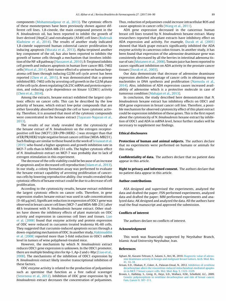
respectively (Fig. 4).Similar to ODC1, N. binaloudensis extract significantly decreasedthe expression of ADA at all the concentrations. In MCF-7 cellline N. binaloudensis extract decreased the expression of ADA in a

A.S. Afshar et al. / Revista Brasileira de Farmacognosia 27 (2017) 84–90 87
ControlM
CF
-7M
DA
-MB
-231
20 μg/ml 80 μg/ml 320 μg/ml
Fig. 2. Morphological changes of MCF-7 and MDA-MB-231 cells treated with different concentrations of the hexane extract of Nepeta binaloudensis for 48 h viewed underinverted phase-contrast microscope (200× magnification). Reduce in cell population was noted with the increase in the concentration of the treatment as compared to thecontrol (untreated cells).
MCF7 MDA-MB-231
Num
ber
of c
olon
ies,
%
120
100
80
60
40
20
00 10
Extract concentration (μg/ml)
∗∗
∗∗
30 60
120
100
80
60
40
20
00 10
Extract concentration (μg/ml)
∗∗
∗∗
30 60
F MCF-7o ).
d6cwrw
D
taceipnaap
ig. 3. Colony formation assay shows a significant reduction in colony formation in
f Nepeta binaloudensis. The results were presented as mean ± SEM, n = 3 (**p < 0.01
ose-dependent manner by 1.32, 1.83 and 3.49 fold with 10, 30 and0 �g/ml, respectively. As shown in Fig. 5, at 10, 30 and 60 �g/mloncentration of N. binaloudensis extract ADA mRNA expressionas reduced 1.21, 1.56 and 2.56 fold in MDA-MB-231 cell line,
espectively (Fig. 5). In cancer cell lines, mRNA expression of MCF-7as reduced more than that of MDA-MB-231.
iscussion
Several studies suggest an increased polyamine concentra-ion in breast cancer tissue compared to normal breast tissuend polyamines biosynthesis inhibitors decrease the growth ofancerous tumors (Brown et al., 2009; Arisan et al., 2012; Zhut al., 2012). Ornithine decarboxylase (ODC1), is a key enzymen polyamine biosynthesis which decarboxylates Ornithine toutrescine (Wang and Jiang, 2014). Furthermore, adenosine deami-
ase (ADA) enzyme, which catalyzes the hydrolytic deamination ofdenosine to inosine, plays an important role in purine metabolismnd Adenosine homeostasis (Garcia-Gil et al., 2015). It has beenroved to be a direct correlation between breast cancer andand MDA-MB-231 cell lines treated with different concentration of hexane extract
increased ADA enzyme activity in cancer tissue of patients (Gocmenet al., 2009; Ri et al., 2010; Ogata et al., 2011). Therefore, ODC1 andADA genes are the appropriate target in anticancer research.
According to previous studies and the importance of recogniz-ing anti-cancer mechanisms of plant extracts, in present study theeffects of various extracts of N. binaloudensis on the cell prolifer-ation and the expression of ADA and ODC genes in breast cancercell lines (MCF-7, MDA-MB-231) with respect to non-cancer lines(MCF-10A) were evaluated.
The results showed that after treatment of cells (at 24, 48 and72 h) with extracts of hexane and EtOH 70%, excluding H2O extract,a linear relationship between the doses with the percentage of cellviability was observed. According to the results of this study hexaneand EtOH extract of this plant has anti-cancer effects.
Different studies have shown the antiproliferative activity ofNepeta species including N. ucrainica (Khakdan and Rassam, 2014),
N. cataria (Formisano et al., 2011) and N. glomerata (Riganoet al., 2011). Chemical composition analyses of N. binaloudensisoil revealed that 1,8-cineol (68.31%), �-terpineol (5.24%), �-pinene (4.74%), �-terpineol (2.57%), �-pinene (1.54%) were main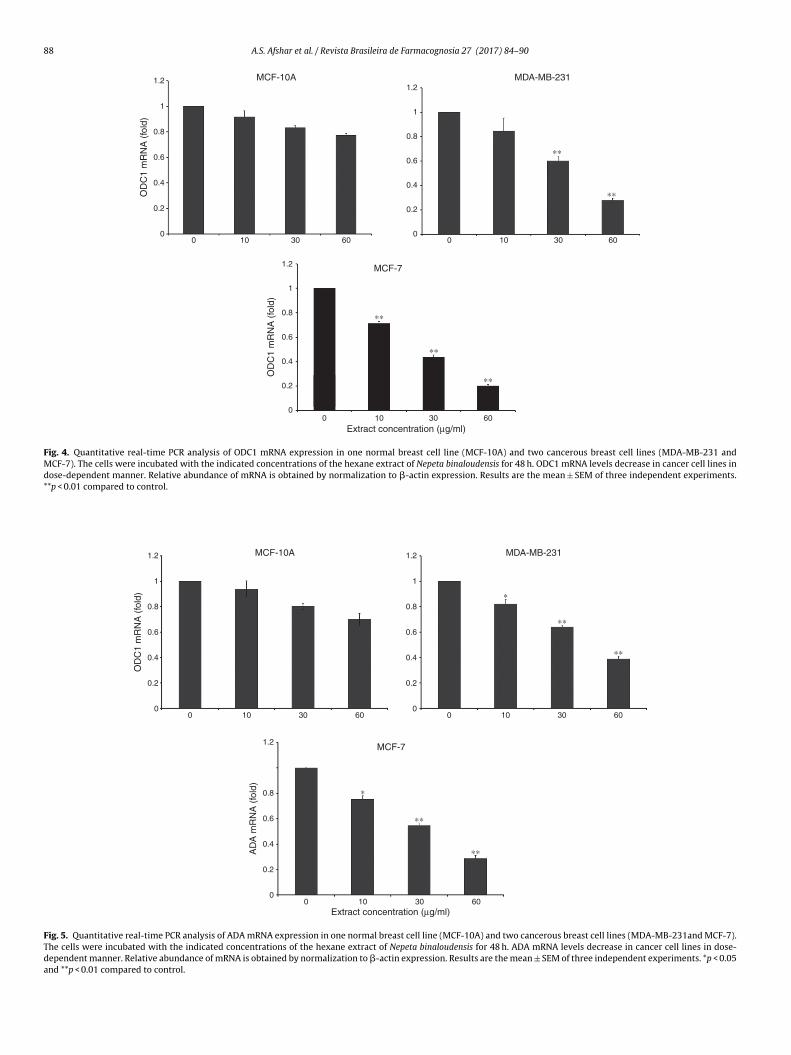
88 A.S. Afshar et al. / Revista Brasileira de Farmacognosia 27 (2017) 84–90
1.2
00 603010 0 603010
0 603010
0.2
0.4
0.6
OD
C1
mR
NA
(fo
ld)
MCF-10A
MCF-7
Extract concentration (μg/ml)
∗∗
∗∗
∗∗
∗∗
∗∗
MDA-MB-231
0.8
1
1.2
0
0.2
0.4
0.6
OD
C1
mR
NA
(fo
ld)
0.8
1
1.2
0
0.2
0.4
0.6
0.8
1
Fig. 4. Quantitative real-time PCR analysis of ODC1 mRNA expression in one normal breast cell line (MCF-10A) and two cancerous breast cell lines (MDA-MB-231 andMCF-7). The cells were incubated with the indicated concentrations of the hexane extract of Nepeta binaloudensis for 48 h. ODC1 mRNA levels decrease in cancer cell lines indose-dependent manner. Relative abundance of mRNA is obtained by normalization to �-actin expression. Results are the mean ± SEM of three independent experiments.**p < 0.01 compared to control.
1.2
0
0.2
0.4
0.6
OD
C1
mR
NA
(fo
ld)
0.8
1.2
0
0.2
0.4
0.6
AD
A m
RN
A (
fold
)
0.8
1
1.2
0
0.2
0.4
0.6
0.8
1
MCF-10A
0 603010
0 603010
0 603010
MDA-MB-231
∗
∗∗
∗
∗∗
∗∗
∗∗
MCF-7
Extract concentration (μg/ml)
Fig. 5. Quantitative real-time PCR analysis of ADA mRNA expression in one normal breast cell line (MCF-10A) and two cancerous breast cell lines (MDA-MB-231and MCF-7).The cells were incubated with the indicated concentrations of the hexane extract of Nepeta binaloudensis for 48 h. ADA mRNA levels decrease in cancer cell lines in dose-dependent manner. Relative abundance of mRNA is obtained by normalization to �-actin expression. Results are the mean ± SEM of three independent experiments. *p < 0.05and **p < 0.01 compared to control.
ra de F
cofNlK1ikgtccarios(
tpeiw2
tpoS(Moe
oItocp
te(o4iaeoTdel
rt2Nt
s(b
A.S. Afshar et al. / Revista Brasilei
omponents (Mohammadpour et al., 2013). The cytotoxic effectsf these monoterpenes have been previously shown against dif-erent cell lines. 1,8-cineole, the main compound present in the. binaloudensis oil, has been reported to inhibit the growth of
iver-derived (HepG2) and extrahepatic (A549) cell lines (Rodenakladniew et al., 2014). The results of another study indicated,8-cineole suppressed human colorectal cancer proliferation by
nducing apoptosis (Murata et al., 2013). Alpha terpineol anotherey component of the oil, has also been reported to inhibits therowth of tumor cells through a mechanism that involves inhibi-ion of the NF-�B pathway (Hassan et al., 2010). �-Terpineol inhibitsell growth and induces apoptosis in human liver cancer BEL-7402ells (Wu et al., 2014). Anti-tumor effect of �-pinene on human hep-toma cell lines through inducing G2/M cell cycle arrest has beeneported (Chen et al., 2015). It was demonstrated that �-pinenenhibited BEL-7402 cells by arresting cell growth in the G2/M phasef the cell cycle, down regulating Cdc25C mRNA and protein expres-ion, and reducing cycle dependence on kinase 1(CDK1) activityChen et al., 2014).
Among the extracts, hexane extract exhibited the largest cyto-oxic effects on cancer cells. This can be described by the lowolarity of hexane, which extract low-polar compounds that areither favorably absorbed through the cell or have cytotoxic activ-ty. Therefore, it is assumed that most of the cytotoxic compounds
ere concentrated in the hexane extract (Tayarani-Najaran et al.,013).
The results of our study revealed that the cytotoxicity ofhe hexane extract of N. binaloudensis on the estrogen receptor-ositive cell line (MCF7) (ER+/PR+/HER2−) was stronger than thatf ER/PR/HER2 triple negative breast cancer cell line (MDA-MB231).uch findings are similar to those found in the work of Srisawat et al.2015) who found a higher apoptosis and growth inhibition rate in
CF-7 cells than in MDA-MB-231 cells. The higher cytotoxic effectf N. binaloudensis extract on MCF-7 was probably due to lack ofstrogen stimulation in this experiment.
The decrease of the cells viability could be because of an increasef apoptosis and/or decreased cell reproduction (Islam et al., 2013).n our study, a colony formation assay was performed to evaluatehe hexane extract capability of arresting proliferation of cancer-us cells by lowering reproductive ability. Our results revealed thatytotoxic effects of hexane extract could be due to a decrease of cellroliferation.
According to the cytotoxicity results, hexane extract exhibitedhe largest cytotoxic effects on cancer cells. Therefore, in genexpression studies hexane extract was used in safe concentrations0–60 �g/ml). Significant reduction in expression of ODC1 gene wasbserved in breast cancer cell lines (MCF-7 and MDA-MB-231) after8 h treatment with N. binaloudensis hexane extract. Other stud-
es have shown the inhibitory effects of plant materials on ODCctivity and expression in cancerous cell lines and tissues. Liaot al. (2008) found that enzyme activity and protein expressionf ODC were reduced in curcumin treated leukemia HL-60 cells.hey suggested that curcumin-induced apoptosis occurs through aown-regulating mechanism of ODC. In another study, Hakimuddint al. (2008) reported more than 3-fold reduction in ODC1 mRNAevel in tumors of wine polyphenol-treated mice.
However, the mechanism by which N. binaloudensis extracteduces ODC1 gene expression is unknown. In the ODC1 promoter,here are multiple binding sites for Ap-1, Ap-2 and c-Myc (Liao et al.,008). The mechanisms of the inhibition of ODC1 expression by. binaloudensis extract likely involve transcriptional inhibition of
hese factors.
ODC enzyme activity is related to the production of polyaminesuch as spermine that function as a free radical scavengerSmirnova et al., 2012). Inhibition of ODC gene expression by N.inaloudensis extract decreases the concentration of polyamines.
armacognosia 27 (2017) 84–90 89
Thus, reduction of polyamines could increase intracellular ROS andcause apoptosis in cancer cells (Wang et al., 2011).
ADA gene expression decreased in the cancerous humanbreast cell lines treated by N. binaloudensis hexane extract. Manyresearchers reported that plant extracts have inhibitory effect onADA expression and activity. For example, Durak et al. (2005)showed that black grape extracts significantly inhibited the ADAenzyme activity in cancerous colon tissues. In another study, it hasbeen found that expression of the adenosine deaminase gene wasconsistently decreased by all the berries in intestinal adenoma tis-sue of rats (Mutanen et al., 2008). Tomato juice has been reported tocauses significant inhibition on ADA activity in the prostate cancertissues (Durak et al., 2003).
Our data demonstrate that decrease of adenosine deaminaseexpression abolishes advantage of cancer cells in obtaining morenucleotides in DNA synthesis and proliferation (Namuslu et al.,2014). Also, inhibition of ADA expression causes increased avail-ability of adenosine which is a protective molecule in case oftumorous condition (Mahajan et al., 2013).
In conclusion, the study described here demonstrates that N.binaloudensis hexane extract has inhibitory effects on ODC1 andADA gene expression in breast cancer cell line. Therefore, a possi-ble mechanism for observed cytotoxicity effect of this plant extractcould be expression inhibition of these genes. This is the first reportabout the cytotoxicity of N. binaloudensis hexane extract by inhibi-tion of ODC1 and ADA in mRNA level, hence further studies will benecessary to supplement our findings.
Ethical disclosures
Protection of human and animal subjects. The authors declarethat no experiments were performed on humans or animals forthis study.
Confidentiality of data. The authors declare that no patient dataappear in this article.
Right to privacy and informed consent. The authors declare thatno patient data appear in this article.
Author contributions
ASA designed and supervised the experiments, analyzed thedata and drafted the paper; FSN performed experiments, analyzeddata and drafted the paper; MM performed experiments and ana-lyzed data; AK designed and analyzed the data. All the authors haveread the final manuscript and approved the submission.
Conflicts of interest
The authors declare no conflicts of interest.
Acknowledgment
This work was financially supported by Neyshabur Branch,Islamic Azad University Neyshabur, Iran.
References
Aghaei, M., Karami-Tehrani, F., Salami, S., Atri, M., 2010. Diagnostic value of adeno-sine deaminase activity in benign and malignant breast tumors. Arch. Med. Res.41, 14–18.
Arisan, E.D., Obakan, P., Coker, A., Palavan-Unsal, N., 2012. Inhibition of ornithine
decarboxylase alters the roscovitine-induced mitochondrial-mediated apopto-sis in MCF-7 breast cancer cells. Mol. Med. Rep. 5, 1323–1329.Brown, I., Halliday, S., Greig, H., Heys, S.D., Wallace, H.M., Schofield, A.C., 2009.Genetic polymorphism in ornithine decarboxylase and risk of breast cancer.Fam. Cancer 8, 307–311.
9 ra de F
C
C
D
D
D
E
F
F
G
G
H
H
H
I
K
K
L
M
M
M
M
M
the ornithine decarboxylase G316A polymorphism and breast cancer survival.
0 A.S. Afshar et al. / Revista Brasilei
hen, W., Liu, Y., Li, M., Mao, J., Zhang, L., Huang, R., Ye, L., 2015. Anti-tumor effect ofalpha-pinene on human hepatoma cell lines through inducing G2/M cell cyclearrest. J. Pharmacol. Sci. 127, 332–338.
hen, W.Q., Xu, B., Mao, J.W., Wei, F.X., Li, M., Liu, T., Zhang, L.R., 2014. Inhibitoryeffects of alpha-pinene on hepatoma carcinoma cell proliferation. Asian Pac. J.Cancer. Prev. 15, 3293–3297.
eng, W., Jiang, X., Mei, Y., Sun, J., Ma, R., Liu, X., Sun, X., 2008. Role of ornithinedecarboxylase in breast cancer. Acta Biochim. Biophys. Sin. 40, 235–243.
urak, I., Biri, H., Avcı, A., Sözen, S., Devrim, E., 2003. Tomato juice inhibits adenosinedeaminase activity in human prostate tissue from patient with prostate cancer.Nutr. Res. 23, 1183–1188.
urak, I., Cetin, R., Devrim, E., Erguder, I.B., 2005. Effects of black grape extract onactivities of DNA turn-over enzymes in cancerous and non cancerous humancolon tissues. Life Sci. 76, 2995–3000.
lmets, C.A., Athar, M., 2010. Targeting ornithine decarboxylase for the preventionof nonmelanoma skin cancer in humans. Cancer Prev. Res. 3, 8–11.
ormisano, C., Rigano, D., Senatore, F., 2011. Chemical constituents and biologicalactivities of Nepeta species. Chem. Biodivers. 8, 1783–1818.
ranken, N.A., Rodermond, H.M., Stap, J., Haveman, J., van Bree, C., 2006. Clonogenicassay of cells in vitro. Nat. Protoc. 1, 2315–2319.
arcia-Gil, M., Tozzi, M.G., Varani, S., Della Verde, L., Petrotto, E., Balestri, F., Camici,M., 2015. The combination of adenosine deaminase inhibition and deoxyadeno-sine induces apoptosis in a human astrocytoma cell line. Neurochem. Int. 80,14–22.
ocmen, E., Tez, M., Ozturk, S., Koc, M., Demirci, S., 2009. Activities of adenosinedeaminase and 5′-nucleotidase in cancereous and non-cancereous human gas-tric tissues. Bratisl. Lek. Listy 110, 416–418.
akimuddin, F., Tiwari, K., Paliyath, G., Meckling, K., 2008. Grape and wine polyphe-nols down-regulate the expression of signal transduction genes and inhibit thegrowth of estrogen receptor–negative MDA-MB231 tumors in nu/nu mousexenografts. Nutr. Res. 28, 702–713.
assan, S.B., Gali-Muhtasib, H., Goransson, H., Larsson, R., 2010. Alpha terpi-neol: a potential anticancer agent which acts through suppressing NF-kappaBsignalling. Anticancer Res. 30, 1911–1919.
ayes, C.S., Defeo, K., Dang, H., Trempus, C.S., Morris, R.J., Gilmour, S.K., 2011. Aprolonged and exaggerated wound response with elevated ODC activity mimicsearly tumor development. Carcinogenesis 32, 1340–1348.
slam, M., Sharma, S., Kumar, B., Teknos, T.N., 2013. Atorvastatin inhibits RhoC func-tion and limits head and neck cancer metastasis. Oral Oncol. 49, 778–786.
hakdan, F., Rassam, G.A., 2014. Chemical composition, antibacterial activity, andcytotoxicity of essential oil from Nepeta ucrainica L. spp. kopetdaghensis. Ind.Crop. Prod. 58, 315–321.
oocheki, A., Rezvani Moghaddam, P., 2012. First experiments on cultivation ofNepeta binaloudensis Jamzad – an example of domestication of a highly endan-gered medicinal plant of Iran. Z. Arznei- Gewurzpfla 17, 64–71.
iao, Y.F., Hung, H.C., Hour, T.C., Hsu, P.C., Kao, M.C., Tsay, G.J., Liu, G.Y., 2008.Curcumin induces apoptosis through an ornithine decarboxylase-dependentpathway in human promyelocytic leukemia HL-60 cells. Life Sci. 82, 367–375.
ahajan, M., Tiwari, N., Sharma, R., Kaur, S., Singh, N., 2013. Oxidative stress andits relationship with adenosine deaminase activity in various stages of breastcancer. Indian J. Clin. Biochem. 28, 51–54.
ohammadpour, N., Emami, S.A., Asili, J., 2013. Identification of volatile oil com-ponents of Nepeta binaloudensis Jamzad by GC-MS and 13C-NMR methods andevaluation of its antimicrobial activity. J. Essent. Oil Bear. Plants 16, 102–107.
ousavi, S.M., Gouya, M.M., Ramazani, R., Davanlou, M., Hajsadeghi, N., Seddighi, Z.,2009. Cancer incidence and mortality in Iran. Ann. Oncol. 20, 556–563.
urata, S., Shiragami, R., Kosugi, C., Tezuka, T., Yamazaki, M., Hirano, A., Koda,K., 2013. Antitumor effect of 1,8-cineole against colon cancer. Oncol. Rep. 30,2647–2652.
utanen, M., Pajari, A.M., Paivarinta, E., Misikangas, M., Rajakangas, J., Martti-nen, M., Oikarinen, S., 2008. Berries as chemopreventive dietary constituents a
armacognosia 27 (2017) 84–90
mechanistic approach with the ApcMin/+ mouse. Asia Pac. J. Clin. Nutr. 17 (Suppl.1), 123–125.
Namuslu, M., Kocaoglu, H., Celik, H.T., Avci, A., Devrim, E., Genc, Y., Durak, I., 2014.Effects of aqueous soybean, mistletoe and red clover extracts on activities ofadenosine deaminase and xanthine oxidase enzyme. Bratisl. Lek. Listy 115,367–371.
Ogata, Y., Aoe, K., Hiraki, A., Murakami, K., Kishino, D., Chikamori, K., Tanimoto, M.,2011. Is adenosine deaminase in pleural fluid a useful marker for differenti-ating tuberculosis from lung cancer or mesothelioma in Japan, a country withintermediate incidence of tuberculosis? Acta Med. Okayama 65, 259–263.
Ri, G., Ohno, S., Furutani, M., Furutani, Y., Tsukahara, T., Hagita, N., Matsuoka, R., 2010.An indication for correlation between the serum ADA level and gastric cancerrisk. Anticancer Res. 30, 2347–2349.
Rigano, D., Arnold, N.A., Conforti, F., Menichini, F., Formisano, C., Piozzi, F., Senatore,F., 2011. Characterisation of the essential oil of Nepeta glomerata Montbret etAucher ex Bentham from Lebanon and its biological activities. Nat. Prod. Res.25, 614–626.
Roberts, E.L., Roberts, O.T., 2012. Plasma adenosine deaminase isoform 2 in cancerpatients undergoing chemotherapy. Br. J. Biomed. Sci. 69, 11–13.
Rodenak Kladniew, B., Polo, M., Montero Villegas, S., Galle, M., Crespo, R., Garcia deBravo, M., 2014. Synergistic antiproliferative and anticholesterogenic effects oflinalool, 1,8-cineole, and simvastatin on human cell lines. Chem. Biol. Interact.214, 57–68.
Rustaiyan, A., Nadji, K., 1999. Composition of the essential oils of Nepeta ispahanicaBoiss, and Nepeta binaloudensis Jamzad from Iran. Flavour Frag. J. 14, 35–37.
Siegel, R., Ma, J., Zou, Z., Jemal, A., 2014. Cancer statistics. CA Cancer J. Clin. 64, 9–29.Smirnova, O.A., Isaguliants, M.G., Hyvonen, M.T., Keinanen, T.A., Tunitskaya, V.L.,
Vepsalainen, J., Ivanov, A.V., 2012. Chemically induced oxidative stress increasespolyamine levels by activating the transcription of ornithine decarboxylase andspermidine/spermine-N1-acetyltransferase in human hepatoma HUH7 cells.Biochimie 94, 1876–1883.
Srisawat, T., Sukpondma, Y., Graidist, P., Chimplee, S., Kanokwiroon, K., 2015. Thedose dependent in vitro responses of MCF-7 and MDA-MB-231 cell lines toextracts of Vatica diospyroides Symington type SS fruit include effects on modeof cell death. Pharmacogn. Mag. 11 (Suppl. 1), S148.
Tayarani-Najaran, Z., Asili, J., Aioubi, E., Emami, S.A., 2013. Growth inhibition andapoptosis induction of Salvia chloroleuca on MCF-7 breast cancer cell line. Iran.J. Pharm. Res. 12, 789–799.
Tundis, R., Nadjafi, F., Menichini, F., 2013. Angiotensin-converting enzyme inhibitoryactivity and antioxidant properties of Nepeta crassifolia Boiss & Buhse and Nepetabinaloudensis Jamzad. Phytother. Res. 27, 572–580.
Van Slambrouck, S., Daniels, A.L., Hooten, C.J., Brock, S.L., Jenkins, A.R., Ogasawara,M.A., Steelant, W.F., 2007. Effects of crude aqueous medicinal plant extracts ongrowth and invasion of breast cancer cells. Oncol. Rep. 17, 1487–1492.
Wang, M.F., Liao, Y.F., Hung, Y.C., Lin, C.L., Hour, T.C., Lue, K.H., Liu, G.Y., 2011.Hydroxydibenzoylmethane induces apoptosis through repressing ornithinedecarboxylase in human promyelocytic leukemia HL-60 cells. Exp. Mol. Med.43, 189–196.
Wang, X., Jiang, L., 2014. Effects of ornithine decarboxylase antizyme 1 on the pro-liferation and differentiation of human oral cancer cells. Int. J. Mol. Med. 34,1606–1612.
Wu, Z.L., Yin, Z.Q., Du, Y.H., Feng, R.Z., Ye, K.C., Wei, Q., Wang, Y., 2014. gamma-Terpineol inhibits cell growth and induces apoptosis in human liver cancer BEL-7402 cells in vitro. Int. J. Clin. Exp. Pathol. 7, 6524–6533.
Xu, L., Long, J., Wang, P., Liu, K., Mai, L., Guo, Y., 2015. Association between
Oncol. Lett. 10, 485–491.Zhu, Q., Jin, L., Casero, R.A., Davidson, N.E., Huang, Y., 2012. Role of ornithine decar-
boxylase in regulation of estrogen receptor alpha expression and growth inhuman breast cancer cells. Breast Cancer Res. Treat. 136, 57–66.