guia ulcera venosa 2014
DESCRIPTION
La ultima guia de ulcera venosa actualizadaTRANSCRIPT

AuthJ Va0741Cophttp
Management of venous leg ulcers: Clinical practiceguidelines of the Society for Vascular Surgery� andthe American Venous Forum
Endorsed by the American College of Phlebology and the Union Internationale de PhlébologieThomas F. O’Donnell Jr, MD, Marc A. Passman, MD, William A. Marston, MD, William J. Ennis, DO,Michael Dalsing, MD, Robert L. Kistner, MD, Fedor Lurie, MD, PhD, Peter K. Henke, MD,Monika L. Gloviczki, MD, PhD, Bo G. Eklöf, MD, PhD, Julianne Stoughton, MD, Sesadri Raju, MD,Cynthia K. Shortell, MD, Joseph D. Raffetto, MD, Hugo Partsch, MD, Lori C. Pounds, MD,Mary E. Cummings, MD, David L. Gillespie, MD, Robert B. McLafferty, MD,Mohammad Hassan Murad, MD, Thomas W. Wakefield, MD, and Peter Gloviczki, MD
SVS/AVF Joint Clinical Practice Guidelines CommitteedVenous Leg Ulcer
Chair:Thomas F. O’Donnell Jr, MD (Cardiovascular Center, Tufts Medical Center, Boston, Mass)
Vice Chair:Marc A. Passman, MD (Division of Vascular Surgery and Endovascular Therapy. University of Alabama atBirmingham, Birmingham, Ala; Birmingham Veterans Administration Medical Center, Birmingham, Ala)
Committee Members:Mary E. Cummings, MD (University of Michigan, Ann Arbor, Mich)Michael C. Dalsing, MD (Indiana University School of Medicine, IU Health Care System, Indianapolis, Ind)Bo G. Eklöf, MD, PhD (Lund University, Sweden)William J. Ennis, DO (University of Illinois Hospital and Health Science, Chicago, Ill)David L. Gillespie, MD (Department of Vascular Surgery, Cardiovascular Care Center, Southcoast HealthcareSystems, Fall River, Mass; Uniformed Services University of the Health Sciences F. Edward Hebert School ofMedicine, Bethesda, Md)
Monika L.Gloviczki, MD, PhD (Gonda Vascular Center, Mayo Clinic, Rochester, Minn)Peter Gloviczki, MD (Division of Vascular and Endovascular Surgery, Mayo Clinic, Rochester, Minn)Peter K. Henke, MD (University of Michigan, Ann Arbor, Mich)Robert L. Kistner, MD (Honolulu, Hawaii)Fedor Lurie, MD, PhD (Jobst Vascular Institute, Toledo, Ohio)William A. Marston, MD (University of North Carolina, Chapel Hill, NC)Robert B. McLafferty, MD (Portland Veterans Administration Medical Center, Portland, Ore; Oregon HealthSciences University, Portland, Ore)
M. Hassan Murad, MD (Division of Preventive Medicine, Mayo Clinic, Rochester, Minn)Hugo Partsch, MD (Medical University of Vienna, Austria)Lori C. Pounds, MD (University of Texas Health Science Center, San Antonio, Tex)Joseph D. Raffetto, MD (Harvard Medical School, Boston Mass; Veterans Administration Boston HealthcareSystem, Boston Mass; Brigham and Women’s Hospital, Boston, Mass)
Sesadri Raju, MD (The Rane Center, Jackson, Miss)Cynthia K. Shortell, MD (Division of Vascular Surgery, Duke University Medical Center, Durham, NC)Julianne Stoughton, MD (Division of Vascular and Endovascular Surgery, Massachusetts General Hospital,Boston, Mass)
Thomas W. Wakefield, MD (University of Michigan, Ann Arbor, Mich)
or conflict of interest: none.sc Surg 2014;60:3S-59S-5214/$36.00yright � 2014 by the Society for Vascular Surgery.://dx.doi.org/10.1016/j.jvs.2014.04.049
3S

JOURNAL OF VASCULAR SURGERY4S O’Donnell et al August Supplement 2014
Sub-CommitteedClinical Evaluation:Marc A. Passman, MD (chair); Peter K. Henke, MD; William A. Marston, MD; Robert B. McLafferty, MD; LoriPounds, MD
Sub-CommitteedWound Care:William A. Marston, MD (co-Chair); William J. Ennis, DO (co-Chair); Emily Cummings, MD; Thomas F.O’Donnell Jr, MD; Lori C. Pounds, MD
Sub-CommitteedCompression:Fedor Lurie, MD, PhD (co-Chair); Thomas W. Wakefield, MD (co-Chair); MD; Monika L. Gloviczki, MD, PhD;Hugo Partsch, MD; Cynthia Shortell, MD
Sub-CommitteedSurgery:Michael C. Dalsing, MD (co-Chair); Robert Kistner, MD (co-Chair); Bo G. Eklöf, MD, PhD; David Gillespie, MD;Peter Gloviczki, MD; Julianne Stoughton, MD; Sesadri Raju, MD
Sub-CommitteedAncillary:Monika L. Gloviczki, MD, PhD (Chair); Cynthia K. Shortell, MD; Julianne Stoughton, MD; William J. Ennis, DO
Sub-CommitteedPrimary Prevention:Peter K. Henke, MD (Chair); Emily Cummings, MD; Michael C. Dalsing, MD; Fedor Lurie, MD, PhD
Section Contributors
Editors: Thomas F. O’Donnell Jr, MD; Marc A. Passman, MDSummary of Guidelines: SVS/AVF CommitteeNeed for Intersociety Consensus Guidelines: Thomas F. O’Donnell Jr, MDMethodology of Guidelines: Thomas F. O’Donnell Jr, MD; Mohammad Hassan Murad, MDDefinitiondVenous Leg Ulcer: Thomas F. O’Donnell Jr, MD; Robert Kistner, MDVenous Anatomy and Pathophysiology: Marc A. Passman, MD (Anatomy); Joseph D. Raffetto, MD (Pathophysiology)Clinical Evaluation: Marc A. Passman, MD; William A. Marston, MD; Peter Henke, MD; Robert B. McLafferty, MD;Lori C. Pounds, MD; William J. Ennis, MD
Wound Care: William A. Marston, MD; William J. Ennis, DO; Emily Cummings, MD; Lori Pounds, MD; Thomas F.O’Donnell Jr, MD
Compression: Fedor Lurie, MD, PhD; Thomas W. Wakefield, MD; Monika L. Gloviczki, MD, PhD; Hugo Partsch,MD; Cynthia Shortell, MD; Andrea Obi, MD (University of Michigan, Ann Arbor, Mich)
Surgery: Michael C. Dalsing, MD; Robert Kistner, MD; Bo G. Eklöf, MD, PhD; Peter Gloviczki, MD; Sesadri Raju,MD; Julianne Stoughton, MD; David Gillespie, MD
Ancillary: Monika L. Gloviczki, MD, PhD; Cynthia K. Shortell, MD; Julianne Stoughton, MD; William J. Ennis, MD;William A. Marston, MD
Primary Prevention: Peter Henke, MD; Fedor Lurie, MD, PhD; Emily Cummings, MD; Michael C. Dalsing, MD
SUMMARY OF GUIDELINES FOR MANAGEMEN
DEFINITION VENOUS LEG ULGuideline 1.1: Venous Leg Ulcer DWe suggest use of a standard defini
area affected by venous hypertension.
VENOUS ANATOMY AND PATGuideline 2.1: Venous Anatomy NWe recommend use of the Internat
dardized venous anatomy nomenclatur
Guideline 2.2: Venous Leg Ulcer PWe recommend a basic practical kn
practitioners caring for venous leg ulce
T OF VENOUS ULCER
CERefinition
tion of venous ulcer as an open skin lesion of the leg or foot that occurs in an[BEST PRACTICE]
HOPHYSIOLOGYomenclatureional Consensus Committee on Venous Anatomical Terminology for stan-e. [BEST PRACTICE]
athophysiologyowledge of venous physiology and venous leg ulcer pathophysiology for allrs. [BEST PRACTICE]

CLINICAL EVALUATIONGuideline 3.1: Clinical EvaluationWe recommend that for all patients with suspected leg ulcers fitting the definition of venous leg ulcer, clinical
evaluation for evidence of chronic venous disease be performed. [BEST PRACTICE]
Guideline 3.2: Nonvenous Causes of Leg UlcersWe recommend identification of medical conditions that affect ulcer healing and other nonvenous causes of
ulcers. [BEST PRACTICE]
Guideline 3.3: Wound DocumentationWe recommend serial venous leg ulcer wound measurement and documentation. [BEST PRACTICE]
Guideline 3.4: Wound CultureWe suggest against routine culture of venous leg ulcers and only to obtain wound culture specimens when clin-
ical evidence of infection is present. [GRADE - 2; LEVEL OF EVIDENCE - C]
Guideline 3.5: Wound BiopsyWe recommend wound biopsy for leg ulcers that do not improve with standard wound and compression
therapy after 4 to 6 weeks of treatment and for all ulcers with atypical features. [GRADE - 1; LEVEL OFEVIDENCE - C]
Guideline 3.6: Laboratory EvaluationWe suggest laboratory evaluation for thrombophilia for patients with a history of recurrent venous thrombosis
and chronic recurrent venous leg ulcers. [GRADE - 2; LEVEL OF EVIDENCE - C]
Guideline 3.7: Arterial TestingWe recommend arterial pulse examination and measurement of ankle-brachial index on all patients with venous
leg ulcer. [GRADE - 1; LEVEL OF EVIDENCE - B]
Guideline 3.8: Microcirculation AssessmentWe suggest against routine microcirculation assessment of venous leg ulcers but suggest selective consideration
as an adjunctive assessment for monitoring of advanced wound therapy. [GRADE - 2; LEVEL OF EVIDENCE - C]
Guideline 3.9: Venous Duplex UltrasoundWe recommend comprehensive venous duplex ultrasound examination of the lower extremity in all patients
with suspected venous leg ulcer. [GRADE - 1; LEVEL OF EVIDENCE - B]
Guideline 3.10: Venous PlethysmographyWe suggest selective use of venous plethysmography in the evaluation of patients with suspected venous leg
ulcer if venous duplex ultrasound does not provide definitive diagnostic information. [GRADE - 2; LEVEL OFEVIDENCE - B]
Guideline 3.11: Venous ImagingWe suggest selective computed tomography venography, magnetic resonance venography, contrast venography,
and/or intravascular ultrasound in patients with suspected venous leg ulceration if additional advanced venousdiagnosis is required for thrombotic or nonthrombotic iliac vein obstruction or for operative planning beforeopen or endovenous interventions. [GRADE - 2; LEVEL OF EVIDENCE - C]
Guideline 3.12: Venous Disease ClassificationWe recommend that all patients with venous leg ulcer be classified on the basis of venous disease classification
assessment, including clinical CEAP, revised Venous Clinical Severity Score, and venous diseaseespecific quality oflife assessment. [BEST PRACTICE]
Guideline 3.13: Venous Procedural Outcome AssessmentWe recommend venous procedural outcome assessment including reporting of anatomic success, venous hemo-
dynamic success, procedure-related minor and major complications, and impact on venous leg ulcer healing. [BESTPRACTICE]
WOUND CAREGuideline 4.1: Wound CleansersWe suggest that venous leg ulcers be cleansed initially and at each dressing change with a neutral, nonirritating,
nontoxic solution, performed with a minimum of chemical or mechanical trauma. [GRADE - 2; LEVEL OFEVIDENCE - C]
JOURNAL OF VASCULAR SURGERYVolume 60, Number 2S O’Donnell et al 5S

Guideline 4.2: DébridementWe recommend that venous leg ulcers receive thorough débridement at their initial evaluation to remove obvious
necrotic tissue, excessive bacterial burden, and cellular burden of dead and senescent cells. [GRADE - 1; LEVEL OFEVIDENCE - B] We suggest that additional maintenance débridement be performed to maintain the appearanceand readiness of the wound bed for healing. [GRADE - 2; LEVEL OF EVIDENCE - B] We suggest that the healthcare provider choose from a number of débridement methods, including sharp, enzymatic, mechanical, biologic, andautolytic. More than one débridement method may be appropriate. [GRADE - 2; LEVEL OF EVIDENCE - B]
Guideline 4.3: Anesthesia for Surgical DébridementWe recommend that local anesthesia (topical or local injection) be administered to minimize discomfort asso-
ciated with surgical venous leg ulcer débridement. In selected cases, regional block or general anesthesia may berequired. [GRADE - 1; LEVEL OF EVIDENCE - B]
Guideline 4.4: Surgical DébridementWe recommend that surgical débridement be performed for venous leg ulcers with slough, nonviable tissue, or
eschar. Serial wound assessment is important in determining the need for repeated débridement. [GRADE - 1;LEVEL OF EVIDENCE - B]
Guideline 4.5 Hydrosurgical DébridementWe suggest hydrosurgical débridement as an alternative to standard surgical débridement of venous leg ulcers.
[GRADE - 2; LEVEL OF EVIDENCE - B]
Guideline 4.6: Ultrasonic DébridementWe suggest against ultrasonic débridement over surgical débridement in the treatment of venous leg ulcers.
[GRADE - 2; LEVEL OF EVIDENCE - C]
Guideline 4.7: Enzymatic DébridementWe suggest enzymatic débridement of venous leg ulcers when no clinician trained in surgical débridement is
available to débride the wound. [GRADE - 2; LEVEL OF EVIDENCE - C] We do not suggest enzymatic débride-ment over surgical débridement. [GRADE - 2; LEVEL OF EVIDENCE - C]
Guideline 4.8: Biologic DébridementWe suggest that larval therapy for venous leg ulcers can be used as an alternative to surgical débridement.
[GRADE - 2; LEVEL OF EVIDENCE - B]
Guideline 4.9: Management of Limb CellulitisWe recommend that cellulitis (inflammation and infection of the skin and subcutaneous tissue) surrounding the
venous leg ulcer be treated with systemic gram-positive antibiotics. [GRADE - 1; LEVEL OF EVIDENCE - B]
Guideline 4.10: Wound Colonization and Bacterial BiofilmsWe suggest against systemic antimicrobial treatment of venous leg ulcer colonization or biofilm without clinical
evidence of infection. [GRADE - 2; LEVEL OF EVIDENCE - C]
Guideline 4.11: Treatment of Wound InfectionWe suggest that venous leg ulcers with >1 3 106 CFU/g of tissue and clinical evidence of infection be treated
with antimicrobial therapy. [GRADE - 2; LEVEL OF EVIDENCE - C] We suggest antimicrobial therapy forvirulent or difficult to eradicate bacteria (such as beta-hemolytic streptococci, pseudomonas, and resistantstaphylococcal species) at lower levels of colony-forming units per gram of tissue. [GRADE - 2; LEVEL OFEVIDENCE - C] We suggest a combination of mechanical disruption and antibiotic therapy as most likely to besuccessful in eradicating venous leg ulcer infection. [GRADE - 2; LEVEL OF EVIDENCE - C]
Guideline 4.12: Systemic AntibioticsWe recommend that venous leg ulcers with clinical evidence of infection be treated with systemic antibiotics
guided by sensitivities performed on wound culture. [GRADE - 1; LEVEL OF EVIDENCE - C] Oral antibioticsare preferred initially, and the duration of antibiotic therapy should be limited to 2 weeks unless persistent evidenceof wound infection is present. [GRADE - 1; LEVEL OF EVIDENCE - C]
Guideline 4.13: Topical Antibiotics for Infected WoundsWe suggest against use of topical antimicrobial agents for the treatment of infected venous leg ulcers.
[GRADE - 2; LEVEL OF EVIDENCE - C]
Guideline 4.14: Topical Dressing SelectionWe suggest applying a topical dressing that will manage venous leg ulcer exudate and maintain a moist, warm
wound bed. [GRADE - 2; LEVEL OF EVIDENCE - C] We suggest selection of a primary wound dressing that willabsorb wound exudate produced by the ulcer (alginates, foams) and protect the periulcer skin. [GRADE - 2;LEVEL OF EVIDENCE - B]
JOURNAL OF VASCULAR SURGERY6S O’Donnell et al August Supplement 2014

Guideline 4.15: Topical Dressings Containing AntimicrobialsWe recommend against the routine use of topical antimicrobial-containing dressings in the treatment of nonin-
fected venous leg ulcers. [GRADE - 2; LEVEL OF EVIDENCE - A]
Guideline 4.16: Periulcer Skin ManagementWe suggest application of skin lubricants underneath compression to reduce dermatitis that commonly affects
periulcer skin. [GRADE - 2; LEVEL OF EVIDENCE -C] In severe cases of dermatitis associated with venous legulcers, we suggest topical steroids to reduce the development of secondary ulcerations and to reduce the symptomsof dermatitis. [GRADE - 2; LEVEL OF EVIDENCE - C]
Guideline 4.17: Anti-inflammatory TherapiesWe suggest against use of anti-inflammatory therapies for the treatment of venous leg ulcers. [GRADE - 2;
LEVEL OF EVIDENCE - C]
Guideline 4.18: Indications for Adjuvant TherapiesWe recommend adjuvant wound therapy options for venous leg ulcers that fail to demonstrate improvement
after a minimum of 4 to 6 weeks of standard wound therapy. [GRADE - 1; LEVEL OF EVIDENCE - B]
Guideline 4.19: Split-thickness Skin GraftingWe suggest against split-thickness skin grafting as primary therapy in treatment of venous leg ulcers. [GRADE -
2; LEVEL OF EVIDENCE - B] We suggest split-thickness skin grafting with continued compression for selectedlarge venous leg ulcers that have failed to show signs of healing with standard care for 4 to 6 weeks. [GRADE - 2;LEVEL OF EVIDENCE - B]
Guideline 4.20: Cellular TherapyWe suggest the use of cultured allogeneic bilayer skin replacements (with both epidermal and dermal layers) to
increase the chances for healing in patients with difficult to heal venous leg ulcers in addition to compression ther-apy in patients who have failed to show signs of healing after standard therapy for 4 to 6 weeks. [GRADE - 2;LEVEL OF EVIDENCE - A]
Guideline 4.21: Preparation for Cellular TherapyWe suggest a therapeutic trial of appropriate compression and wound bed moisture control before application
of cellular therapy. [GRADE - 2; LEVEL OF EVIDENCE - C] We recommend that adequate wound bed prepa-ration, including complete removal of slough, debris, and any necrotic tissue, be completed before the applicationof a bilayered cellular graft. [GRADE - 1; LEVEL OF EVIDENCE - C] We recommend additional evaluation andmanagement of increased bioburden levels before the application of cellular therapy. [GRADE - 1; LEVEL OFEVIDENCE - C]
Guideline 4.22: Frequency of Cellular Therapy ApplicationWe suggest reapplication of cellular therapy as long as the venous leg ulcer continues to respond on the basis of
wound documentation. [GRADE - 2; LEVEL OF EVIDENCE - C]
Guideline 4.23: Tissue Matrices, Human Tissues, or Other Skin SubstitutesWe suggest the use of a porcine small intestinal submucosal tissue construct in addition to compression therapy
for the treatment of venous leg ulcers that have failed to show signs of healing after standard therapy for 4 to 6weeks. [GRADE - 2; LEVEL OF EVIDENCE - B]
Guideline 4.24: Negative Pressure TherapyWe suggest against routine primary use of negative pressure wound therapy for venous leg ulcers. [GRADE - 2;
LEVEL OF EVIDENCE - C]
Guideline 4.25: Electrical StimulationWe suggest against electrical stimulation therapy for venous leg ulcers. [GRADE - 2; LEVELOF EVIDENCE - C]
Guideline 4.26: Ultrasound TherapyWe suggest against routine ultrasound therapy for venous leg ulcers. [GRADE - 2; LEVEL OF EVIDENCE - B]
COMPRESSIONGuideline 5.1: CompressiondUlcer HealingIn a patient with a venous leg ulcer, we recommend compression therapy over no compression therapy to in-
crease venous leg ulcer healing rate. [GRADE - 1; LEVEL OF EVIDENCE - A]
Guideline 5.2: CompressiondUlcer RecurrenceIn a patient with a healed venous leg ulcer, we suggest compression therapy to decrease the risk of ulcer recur-
rence. [GRADE - 2; LEVEL OF EVIDENCE - B]
JOURNAL OF VASCULAR SURGERYVolume 60, Number 2S O’Donnell et al 7S

Guideline 5.3: Multicomponent Compression BandageWe suggest the use of multicomponent compression bandage over single-component bandages for the treat-
ment of venous leg ulcers. [GRADE - 2; LEVEL OF EVIDENCE - B]
Guideline 5.4: CompressiondArterial InsufficiencyIn a patient with a venous leg ulcer and underlying arterial disease, we do not suggest compression bandages or
stockings if the ankle-brachial index is 0.5 or less or if absolute ankle pressure is less than 60 mm Hg. [GRADE - 2;LEVEL OF EVIDENCE - C]
Guideline 5.5: Intermittent Pneumatic CompressionWe suggest use of intermittent pneumatic compression when other compression options are not available,
cannot be used, or have failed to aid in venous leg ulcer healing after prolonged compression therapy. [GRADE - 2;LEVEL OF EVIDENCE - C]
OPERATIVE/ENDOVASCULAR MANAGEMENTGuideline 6.1: Superficial Venous Reflux and Active Venous Leg UlcerdUlcer HealingIn a patient with a venous leg ulcer (C6) and incompetent superficial veins that have axial reflux directed to the
bed of the ulcer, we suggest ablation of the incompetent veins in addition to standard compressive therapy toimprove ulcer healing. [GRADE - 2; LEVEL OF EVIDENCE - C]
Guideline 6.2: Superficial Venous Reflux and Active Venous Leg UlcerdPrevent RecurrenceIn a patient with a venous leg ulcer (C6) and incompetent superficial veins that have axial reflux directed to the
bed of the ulcer, we recommend ablation of the incompetent veins in addition to standard compressive therapy toprevent recurrence. [GRADE - 1; LEVEL OF EVIDENCE - B]
Guideline 6.3: Superficial Venous Reflux and Healed Venous Leg UlcerIn a patient with a healed venous leg ulcer (C5) and incompetent superficial veins that have axial reflux directed
to the bed of the ulcer, we recommend ablation of the incompetent veins in addition to standard compressive ther-apy to prevent recurrence. [GRADE - 1; LEVEL OF EVIDENCE - C]
Guideline 6.4: Superficial Venous Reflux With Skin Changes at Risk for Venous Leg Ulcer (C4b)In a patient with skin changes at risk for venous leg ulcer (C4b) and incompetent superficial veins that have axial
reflux directed to the bed of the affected skin, we suggest ablation of the incompetent superficial veins in addition tostandard compressive therapy to prevent ulceration. [GRADE - 2; LEVEL OF EVIDENCE - C]
Guideline 6.5: Combined Superficial and Perforator Venous Reflux With or Without Deep Venous Reflux andActive Venous Leg Ulcer
In a patient with a venous leg ulcer (C6) and incompetent superficial veins that have reflux to the ulcer bed inaddition to pathologic perforating veins (outward flow of >500 ms duration, with a diameter of >3.5 mm) locatedbeneath or associated with the ulcer bed, we suggest ablation of both the incompetent superficial veins andperforator veins in addition to standard compressive therapy to aid in ulcer healing and to prevent recurrence.[GRADE - 2; LEVEL OF EVIDENCE - C]
Guideline 6.6: Combined Superficial and Perforator Venous Reflux With or Without Deep Venous Disease andSkin Changes at Risk for Venous Leg Ulcer (C4b) or Healed Venous Ulcer (C5)
In a patient with skin changes at risk for venous leg ulcer (C4b) or healed venous ulcer (C5) and incompetentsuperficial veins that have reflux to the ulcer bed in addition to pathologic perforating veins (outward flow of >500ms duration, with a diameter of >3.5 mm) located beneath or associated with the healed ulcer bed, we suggest abla-tion of the incompetent superficial veins to prevent the development or recurrence of a venous leg ulcer. [GRADE -2; LEVEL OF EVIDENCE - C] Treatment of the incompetent perforating veins can be performed simultaneouslywith correction of axial reflux or can be staged with re-evaluation of perforator veins for persistent incompetenceafter correction of axial reflux. [GRADE - 2; LEVEL OF EVIDENCE - C]
Guideline 6.7: Pathologic Perforator Venous Reflux in the Absence of Superficial Venous Disease, With orWithout Deep Venous Reflux, and a Healed or Active Venous Ulcer
In a patient with isolated pathologic perforator veins (outward flow of >500 ms duration, with a diameter of>3.5 mm) located beneath or associated with the healed (C5) or active ulcer (C6) bed regardless of the status of thedeep veins, we suggest ablation of the “pathologic” perforating veins in addition to standard compression therapyto aid in venous ulcer healing and to prevent recurrence. [GRADE - 2; LEVEL OF EVIDENCE - C]
JOURNAL OF VASCULAR SURGERY8S O’Donnell et al August Supplement 2014

Guideline 6.8: Treatment Alternatives for Pathologic Perforator VeinsFor those patients who would benefit from pathologic perforator vein ablation, we recommend treatment by
percutaneous techniques that include ultrasound-guided sclerotherapy or endovenous thermal ablation (radiofre-quency or laser) over open venous perforator surgery to eliminate the need for incisions in areas of compromisedskin. [GRADE - 1; LEVEL OF EVIDENCE - C]
Guideline 6.9: Infrainguinal Deep Venous Obstruction and Skin Changes at Risk for Venous Leg Ulcer (C4b),Healed (C5) or Active (C6) Venous Leg Ulcer
In a patient with infrainguinal deep venous obstruction and skin changes at risk for venous leg ulcer (C4b),healed venous leg ulcer (C5), or active venous leg ulcer (C6), we suggest autogenous venous bypass or endophle-bectomy in addition to standard compression therapy to aid in venous ulcer healing and to prevent recurrence.[GRADE - 2; LEVEL OF EVIDENCE - C]
Guideline 6.10: Deep Venous Reflux With Skin Changes at Risk for Venous Leg Ulcer (C4b), Healed (C5) orActive (C6) Venous Leg UlcerdLigation
In a patient with infrainguinal deep venous reflux and skin changes at risk for venous leg ulcer (C4b), healedvenous leg ulcer (C5), or active venous leg ulcer (C6), we suggest against deep vein ligation of the femoral or popli-teal veins as a routine treatment. [GRADE - 2; LEVEL OF EVIDENCE - C]
Guideline 6.11: Deep Venous Reflux With Skin Changes at Risk for Venous Leg Ulcer (C4b), Healed (C5) orActive (C6) Venous Leg UlcerdPrimary Valve Repair
In a patient with infrainguinal deep venous reflux and skin changes at risk for venous leg ulcer (C4b), healedvenous leg ulcer (C5), or active venous leg ulcer (C6), we suggest individual valve repair for those who have axialreflux with structurally preserved deep venous valves in addition to standard compression therapy to aid in venousulcer healing and to prevent recurrence. [GRADE - 2; LEVEL OF EVIDENCE - C]
Guideline 6.12: Deep Venous Reflux With Skin Changes at Risk for Venous Leg Ulcer (C4b), Healed (C5) orActive (C6) Venous Leg UlcerdValve Transposition or Transplantation
In a patient with infrainguinal deep venous reflux and skin changes at risk for venous leg ulcer (C4b), healedvenous leg ulcer (C5), or active venous leg ulcer (C6), we suggest valve transposition or transplantation for thosewith absence of structurally preserved axial deep venous valves when competent outflow venous pathways areanatomically appropriate for surgical anastomosis in addition to standard compression therapy to aid in venousleg ulcer healing and to prevent recurrence. [GRADE - 2; LEVEL OF EVIDENCE - C]
Guideline 6.13: Deep Venous Reflux With Skin Changes at Risk for Venous Leg Ulcer (C4b), Healed (C5) orActive (C6) Venous Leg UlcerdAutogenous Valve Substitute
In a patient with infrainguinal deep venous reflux and skin changes at risk for venous leg ulcer (C4b), healedvenous leg ulcer (C5), or active venous leg ulcer (C6), we suggest consideration of autogenous valve substitutesby surgeons experienced in these techniques to facilitate ulcer healing and to prevent recurrence in those withno other option available in addition to standard compression therapy to aid in venous ulcer healing and to preventrecurrence. [GRADE - 2; LEVEL OF EVIDENCE - C]
Guideline 6.14: Proximal Chronic Total Venous Occlusion/Severe Stenosis With Skin Changes at Risk forVenous Leg Ulcer (C4b), Healed (C5) or Active (C6) Venous Leg UlcerdEndovascular Repair
In a patient with inferior vena cava or iliac vein chronic total occlusion or severe stenosis, with or without lowerextremity deep venous reflux disease, that is associated with skin changes at risk for venous leg ulcer (C4b), healedvenous leg ulcer (C5), or active venous leg ulcer (C6), we recommend venous angioplasty and stent recanalization inaddition to standard compression therapy to aid in venous ulcer healing and to prevent recurrence. [GRADE - 1;LEVEL OF EVIDENCE - C]
Guideline 6.15: Proximal Chronic Venous Occlusion/Severe Stenosis (Bilateral) With Recalcitrant VenousUlcerdOpen Repair
In a patient with inferior vena cava or iliac vein chronic occlusion or severe stenosis, with or without lower ex-tremity deep venous reflux disease, that is associated with a recalcitrant venous leg ulcer and failed endovasculartreatment, we suggest open surgical bypass with use of an externally supported expanded polytetrafluoroethylenegraft in addition to standard compression therapy to aid in venous leg ulcer healing and to prevent recurrence.[GRADE - 2; LEVEL OF EVIDENCE - C]
Guideline 6.16: Unilateral Iliofemoral Venous Occlusion/Severe Stenosis With Recalcitrant Venous UlcerdOpen Repair
In a patient with unilateral iliofemoral venous occlusion/severe stenosis with recalcitrant venous leg ulcer forwhom attempts at endovascular reconstruction have failed, we suggest open surgical bypass with use of saphenousvein as a cross-pubic bypass (Palma procedure) to aid in venous ulcer healing and to prevent recurrence. A syntheticgraft is an alternative in the absence of autogenous tissue. [GRADE - 2; LEVEL OF EVIDENCE - C]
JOURNAL OF VASCULAR SURGERYVolume 60, Number 2S O’Donnell et al 9S

Guideline 6.17: Proximal Chronic Total Venous Occlusion/Severe Stenosis (Bilateral or Unilateral) WithRecalcitrant Venous UlcerdAdjunctive Arteriovenous Fistula
For those patients who would benefit from an open venous bypass, we suggest the addition of an adjunctivearteriovenous fistula (4-6 mm in size) as an adjunct to improve inflow into autologous or prosthetic crossover by-passes when the inflow is judged to be poor to aid in venous leg ulcer healing and to prevent recurrence. [GRADE -2; LEVEL OF EVIDENCE - C]
ANCILLARY MEASURESGuideline 7.1: Nutrition Assessment and ManagementWe recommend that nutrition assessment be performed in any patient with a venous leg ulcer who has evidence
of malnutrition and that nutritional supplementation be provided if malnutrition is identified. [BEST PRACTICE]
Guideline 7.2: Systemic Drug TherapyFor long-standing or large venous leg ulcer, we recommend treatment with either pentoxifylline or micronized pu-
rified flavonoid fraction used in combination with compression therapy. [GRADE - 1; LEVEL OF EVIDENCE - B]
Guideline 7.3: PhysiotherapyWe suggest supervised active exercise to improve muscle pump function and to reduce pain and edema in
patients with venous leg ulcers. [GRADE - 2; LEVEL OF EVIDENCE - B]
Guideline 7.4: Manual Lymphatic DrainageWe suggest against adjunctive lymphatic drainage for healing of the chronic venous leg ulcers. [GRADE - 2;
LEVEL OF EVIDENCE - C]
Guideline 7.5: BalneotherapyWe suggest balneotherapy to improve skin trophic changes and quality of life in patients with advanced venous
disease. [GRADE - 2; LEVEL OF EVIDENCE - B]
Guideline 7.6: Ultraviolet lightWe suggest against use of ultraviolet light for the treatment of venous leg ulcers. [GRADE - 2; LEVEL OF
EVIDENCE - C]
PRIMARY PREVENTIONGuideline 8.1: Primary PreventiondClinical CEAP C3-4 Primary Venous DiseaseIn patients with clinical CEAP C3-4 disease due to primary valvular reflux, we recommend compression, 20 to
30 mm Hg, knee or thigh high. [GRADE - 2; LEVEL OF EVIDENCE - C]
Guideline 8.2: Primary PreventiondClinical CEAP C1-4 Post-thrombotic Venous DiseaseIn patients with clinical CEAP C1-4 disease related to prior deep venous thrombosis (DVT), we recommend
compression, 30 to 40 mm Hg, knee or thigh high. [GRADE - 1; LEVEL OF EVIDENCE - B]
Guideline 8.3. Primary PreventiondAcute DVT TreatmentAs post-thrombotic syndrome is a common preceding event for venous leg ulcers, we recommend current
evidence-based therapies for acute DVT treatment. [GRADE - 1; LEVEL OF EVIDENCE - B] We suggest use oflow-molecular-weight heparin over vitamin K antagonist therapy of 3-month duration to decrease post-thrombotic syndrome. [GRADE - 2; LEVEL OF EVIDENCE - B] We suggest catheter-directed thrombolysis in pa-tients with low bleeding risk with iliofemoral DVT of duration <14 days. [GRADE - 2; LEVELOF EVIDENCE - B]
Guideline 8.4: Primary PreventiondEducation MeasuresIn patients with C1-4 disease, we suggest patient and family education, regular exercise, leg elevation when at
rest, careful skin care, weight control, and appropriately fitting foot wear. [BEST PRACTICE]
Guideline 8.5: Primary PreventiondOperative TherapyIn patients with asymptomatic C1-2 disease from either primary or secondary causes, we suggest against pro-
phylactic interventional therapies to prevent venous leg ulcer. [GRADE - 2; LEVEL OF EVIDENCE - C]
JOURNAL OF VASCULAR SURGERY10S O’Donnell et al August Supplement 2014
RATIONALE FOR VENOUS LEG ULCERGUIDELINES
Guidelines present a synthesis of evidence-based rec-ommendations for the diagnosis and treatment of a specificmedical condition.1 The value of a guideline is that it pro-vides consistency among treatment protocols given to pa-tients, resulting in improved efficacy and the quality of
care and reduced cost. The need for cost containment ofhealth care has placed the development and implementa-tion of clinical care guidelines as a high priority for healthcare systems Both the prevalence of venous leg ulcers(VLUs)d1% to 1.5% of the populationdand the eco-nomic impact of VLUs are two compelling reasons todevelop a specific guideline for this problem.2 The care

JOURNAL OF VASCULAR SURGERYVolume 60, Number 2S O’Donnell et al 11S
of VLUs can consume a significant amount of resources, sothat an agreed on “best practice” algorithm can maximizethe quality and effectiveness of care while minimizing costand resource use.3 Moreover, VLUs are associated withprolonged disability, important socioeconomic impact,and significant psychosocial morbidity. Because approxi-mately 50% of VLUs may recur within 10 years, they aremarked by a significant component of chronicity, whichcompounds their economic impact and need for repetitivecare. VLUs also can be painful, so that a patient’s abilityto work may be compromised, and can also affect theretired segments of the population, thereby compoundingboth the indirect and direct costs of treating VLU.
Epidemiology of Venous Leg Ulcers. VLUs are themost common ulceration on the lower extremity and ac-count for 70% of all leg ulcers.4 Various estimates havebeen made from observational studies on the prevalence ofVLU, ranging between 0.06% and 2%. The Edinburghstudy, which was a cross-sectional study of a randomsample of more than 1500 people between the ages of 18and 64 years, provided an estimate of VLU prevalence of1%.5 This lower prevalence contrasts with the higherprevalence of an earlier study (2.7% clinical CEAP C5/C6), which was based on a questionnaire and photographsof the legs of the participants.6 As in the Edinburgh study,VLU prevalence increased with age. Two other studiesexamined large populations. Approximately 40,000 Polishpatients underwent clinical evaluation and interviews by avariety of health care professionals in a multicenter studyshowing a 1.5% prevalence of C5/C6 disease.7 Anothercross-sectional study carried out in France addressed asubset of patients from a larger study of Raynaud patients.All of the 400 patients were evaluated by vascular medicinespecialists. C5 disease was higher in men (5.4%) than inwomen (2.7%), but the investigators observed no openulcers.8 The most detailed information comes from theBonn Vein Longitudinal Study initiated by the GermanMinistry of Health.9 More than 3000 participants wererandomly identified between the ages of 18 and 79 years.Advanced chronic venous reflux (chronic venous insuffi-ciency [CVI]) was found in 0.6% with a healed VLU (C5)and in 0.1% with an open VLU for a total of 0.7%. Thislongitudinal study documented a decrease in advancedCVI (C4-C6), which may be related to a more compre-hensive use of diagnostic and therapeutic modalities forCVI. Because of the high prevalence of venous disease, theAVF National Venous Screening Program was establishedin the United States to increase awareness. The programscreened 2234 individuals and identified varicose veins inmore than 30% of participants and more advanced venousdisease (C4-C6) in more than 10%.10 It has been estimatedthat approximately 2.5 million people suffer from CVI inthe United States, and of those, about 20% develop venousulcers.11
Economic Impact. The overall cost of treating VLUapproaches 1% of the health care budget of some westernEuropean countries; in the United Kingdom, the annualcost is estimated to range from ₤300 million to ₤600
million per year.12,13 In the United States, $2.5 billionwas expended for the treatment of VLUs in more than 6million patients.14 A recent study on the cost of treatingVLUs in Germany demonstrated that mean cost averagedV9569 per patient per year, and 92% of this expenditurewas related to direct costs.15 Although the majority ofpatients with VLUs can be treated on an outpatient basisand are infrequently hospitalized except for complications,the direct cost of treating VLUs in the United States hasbeen estimated to be $2500 per month per patient.16 Inthe ambulatory setting, the direct cost of this care is relatedto (1) technical (facility) costs and professional reim-bursement (physicians); (2) labor costs (nurses and para-medical personnel) for wound care treatments, which arethe major driver of costs; and (3) medications as well asspecialized wound dressings and compression garments. Akey determinant of the costs of treating VLU is the effec-tiveness of treatmentdnot only how rapidly the ulcer heals,but also whether the ulcer recurs. For example, a moreexpensive wound dressing may have an economic advan-tage for overall care because of more rapid healing of theVLU with less duration of product use and attendant laborcosts.
To determine the actual costs of treating VLUs duringa 1-year period, a recent study examined a cohort of 84 pa-tients with nonhealing VLUs (CEAP C6) who presentedto a wound clinic. All patients were treated in a woundcenter by five vascular surgeons with a minimum follow-up of 6 months (median, 368 days; 336-483).17 Actualcosts (not charges) were obtained for outpatient and inpa-tient facility, visiting nurse services, and physician practicegroup to yield true cost. The proportion and time to com-plete healing of VLU were determined to calculate time tohealing as well as ulcer-free intervals. Cost/ulcer-free daysand cost to complete healing for the entire follow-upperiod were carried out with univariate analysis of factorsaffecting cost. The mean total cost of treating VLU duringthis follow-up period was $15,732. A total of 50 patients(60%) healed their VLU without recurrence in a meantime of 122 days (6-379 days) at a mean cost of$10,563 ($430-$50,967). Significant contributing factorswere outpatient facility fees ($10,332) and visiting nurseservices ($11,365) related to extended treatment of theopen VLU. Patients who failed to heal their ulcer duringthe duration of this study (20%) had a threefold increasein their costs ($33,907). Those patients who had recur-rence of their VLU (N ¼ 17; 20%) during the follow-upperiod had a total mean cost of $12,760. Inpatient admis-sion, of which nearly two thirds was for treatment of infec-tions that were resistant to therapy in an outpatient setting,markedly increased costs ($33,629). By contrast, VLUtreated with surgical intervention of the superficial venoussystem did not significantly increase total cost over that ofpatients receiving best medical therapy ($11,960 vs$12,304) but significantly reduced recurrence rates (34%vs 5%). By, contrast, patients treated for outflow obstruc-tion had a twofold increase in total costs ($24,241 vs$11,960).

JOURNAL OF VASCULAR SURGERY12S O’Donnell et al August Supplement 2014
Value of Clinical Practice Guidelines for VenousLeg Ulcer Care. The value of developing and implement-ing clinical practice guidelines should be to provide qualityof care without a dramatic increase in cost. This focus on“best outcomes for the most reasonable health care dollar”has stimulated many organizations, such as the SVS andAVF, to develop and to promote a unified set of guidelinesfor treatment of chronic diseases, such as VLUs. Anevidence-based analysis of treatment options shoulddecrease the variations in care while at the same timeensuring that resources are used in an optimal manner.Specialty societies, as in the current SVS and AVF guideline,develop clinical practice guidelines either by independentreview of available studies or by employing evidence fromtechnology assessments and other published guidelines. Forthe development of comprehensive clinical practice guide-lines on VLUs that would be of value to providers and thehealth care system in general, an expert panel needs toevaluate the efficacy of individual wound care dressings,surgery, or compression products, as demonstrated in ran-domized controlled trials (RCTs). In addition, large caseseries and expert opinion are also used as evidence, althoughof lower quality. The value of these clinical practice guide-lines on VLUs is that they sift through the massive amountof data surrounding the management of VLU and provide aconsensus on the evidence, thereby streamlining resources.18
Survey of the Use of Venous Leg Ulcer Guidelinesby Various Health Care Systems. Whereas there areobvious advantages to the development and use of guide-lines on VLU, implementation can be a challenge. Such“best practices” can be construed as antithetical to the au-tonomy of clinical practitioners, who make decisions onthe basis of individual patients. These factors may prevent to-tal “buy in” by physicians. Moreover, a set of guidelines canbe interpreted as instituting the rigid perspectives of policymakers on health care delivery for a specific disease process.To determine the current status of implementation of guide-lines or a specific best practices protocol for the managementof VLUs, several large health care systems in the UnitedStates as well as in Canada and Europe were surveyed.18
Vascular units in these facilities were queried on whether aspecific set of guidelines or protocols was uniformly appliedfor two conditions: (1) the treatment of VLU and (2) theprevention of a post-thrombotic limb. This survey demon-strated that guidelines for VLU care are infrequently used bya select group of academic health care systems in the UnitedStates (20%), but the majority of single-payer systems inCanada and Europe (82%) employed a VLU guideline. Bycontrast, the use of guidelines for the prevention of the post-thrombotic limb after deep venous thrombosis (DVT) wascomparable between the United States and other countries.This low use of VLU guidelines in the United Statesoccurred despite the availability of guidelines developed byvarious societies. This finding emphasizes the need for botha comprehensive and a well-promoted guideline for VLUs.
Efficacy of Current Guidelines for Venous LegUlcer. Whereas there are various treatment protocols forVLUs, they differ in efficacy, quality, and cost. A more
uniform method of treatment that has been agreed on by apanel of experts and based on clinical evidence for efficacymay improve therapeutic effectiveness and possibly reducecost. Several studies have demonstrated that after the institu-tion of a VLU guideline, there were improvements in boththe ulcer healing and recurrence rate and a subsequentreduced resource use with lowering of treatment costs,thereby supporting adoption of VLU guidelines.18 Olsonet al demonstrated that ulcer healing rate was markedlyimproved in a Veterans Hospital population of 155 patientswith VLU if guideline recommendations were followed.During a 5-year period, patients who received dressings thatprovided a moist wound healing environment and compres-sion for 80% of their visits were more likely to heal than thosewho did not comply (<80%; relative risk [RR], 2.52; 95%confidence interval [CI], 1.53-4.16).19 In a combined studypopulation of patients from the United Kingdom and theUnited States, patients who were treated for VLUs by atreatment protocol had a 2 times and a 6.5 times greaterlikelihood of ulcer healing if the guidelines were followed(P < .01). Moreover, the median costs were reduced withadherence to VLU guidelines (P < .01).20 Finally, in a lon-gitudinal cross-sectional Swedish study of patients in Skar-aborg County (population 254,111), the epidemiology of legulcers and of VLUs in particular was defined in 1988 beforethe implementation of a VLU treatment protocol. 21 Theinitial point prevalence (proportion with an ulcer at any pointof time, time period usually shorter than 3 months) for openVLUs was 0.16% and overall (C5 and C6) was 0.5%. Theirprotocol for VLU management emphasized early Doppler-aided diagnosis, organized care pathways, and especiallysurgery for superficial venous incompetence. The effective-ness of the VLU guidelines was judged by the reduction inthe point prevalence of VLUs from 1990 to 2005. Theinstitution of guidelines and concomitant implementation ofcounty-wide care plans, based on these guidelines, resulted ina relative risk for VLU of 46%.22
Objective of SVS and AVF Clinical PracticeGuidelines for Venous Leg Ulcer Management. In2011, SVS and AVF produced clinical practice guidelinesfor the care of patients with varicose veins and associatedvenous disease.23 Although there were some elements ofVLU care within this document, most of the emphasis ofthis guideline was on varicose vein management. In 2012,SVS and AVF clinical practice guidelines addressing earlythrombus removal strategies for acute venous thrombosiswere published.24 Again, whereas there is some crossoverregarding the impact of early venous thrombosis onrestored venous patency, potential preserved valvularfunction, and decreased post-thrombotic morbidity, therewas little direct attention on VLUs. The objective of thecurrent SVS and AVF clinical practice guidelines is to focuson complete management of VLUs at all levels of carebased on strength and quality of supporting evidence toguide specific recommendations. Combined with theseother SVS and AVF clinical practice guidelines, a morecomplete evidence-based strategy is available for manage-ment of patients with chronic venous disease.

JOURNAL OF VASCULAR SURGERYVolume 60, Number 2S O’Donnell et al 13S
METHODOLOGY OF VENOUS LEG ULCERGUIDELINES
The Institute of Medicine, in Clinical Practice Guide-lines We Can Trust, has defined clinical practice guidelinesas “systematically developed statements to assist the practi-tioner and patient decisions about appropriate healthcarefor specific clinical circumstances.”25 The underlying prin-ciple is to use evidence-based medicine to the greatestextent to develop these guidelines so that the assessmentof the optimal treatment plan is based on the best currentavailable knowledge. Evidence-based medicine allows oneto assess the statistical strength of a treatment or interven-tion and, most important, reduces bias in evaluating aparticular therapy.26 Bias is defined as the predispositionof prejudice toward either the experimental or controlgroup, which can lead to either an overestimation or an un-derestimation of the true benefits and harm of the interven-tion.27 This method reduces uncertainty, which is thelargest single cause of misinterpretation of data.28 Thisprocess has been further stated in the recent Institute ofMedicine document: “Hence, critically appraised and syn-thesized scientific evidence has become fundamental toclinical practice. At the same time, and particularly underconditions of uncertainty regarding optimal decisions,clinician experiential knowledge and skill (the “art of med-icine”) and patient values and preferences remain essentialcontributors to quality healthcare practice, in a complexinterplay with science.”25
The Venous Ulcer Guidelines Committee was orga-nized through cooperation between the SVS and AVF.The Venous Ulcer Guidelines Committee was dividedinto six sub-committee sections each headed by a chair:diagnosis; compression; surgery/endovascular; woundcare; ancillary; and prevention. The overall committeethen developed a series of key clinical questions to guidethe overall approach for the guideline document: (1)What is the best treatment for active (CEAP C6) venous ul-cer? (2) What is the best treatment for healed venous ulcer(CEAP C5)? (3) What is the best method for preventingrecurrence of venous ulcers? and (4) Can progressionfrom CEAP C4 to CEAP C6/C5 be prevented? TheVenous Ulcer Guidelines Committee addressed the optimalapproach to be used for their specific section from four gen-eral approaches: (1) de novo development, in which acompletely novel recommendation is developed from a sys-tematic and meta-analysis review of the literature; (2) buildon existing guidelines with a complementary full-literaturesearch update; (3) adapt guidelines from existing guidelines;and (4) total adoption of existing guidelines.18 The need fora systematic and meta-analysis review was determined byeach section team and then agreed on by the entire commit-tee. Each section was categorized by the type of guidelinedevelopment required. All guidelines were developed bybuilding on existing guidelines with a complementary liter-ature search by the section sub-committee.
In this process of guideline development for VLUs, theVenous Ulcer Guidelines Committee also reviewed prior
published consensus documents and the AVF report fromthe 2006 Venous Summit and the 2009 Pacific VascularSymposium, whose purpose was to reduce the incidenceof venous ulcer during the next decade by 50%.29 In a pre-vious systematic review of recently published venous ulcerguidelines, 14 venous ulcer guidelines were identifiedworldwide.18 This review showed that there was a high de-gree of agreement among the 14 VLU guidelines on rec-ommendations for compression (72%), dressings (72%),pentoxifylline (73%), prevention of recurrence by below-knee stockings (70%), and surgery (82%). There was alow proportion of agreement in the areas of diagnosis, clin-ical evaluation, and venous Doppler and duplex ultrasound;in elements of wound care: measurement of the wound,washing of the wound, débridement, and specific type ofwound dressing; and finally in adjunctive measures: theuse of skin grafts and physical therapy to promote anklemobility with enhancement of the calf muscle pump.Several areas of “controversy” were identified that hadnot been particularly addressed in previous guidelines:new innovative, less invasive VLU therapies; the role of iliacobstruction and occlusion with the need for diagnosis byintravascular ultrasound and subsequent monitoring ofstenting by this technique; when and by what methodsto treat perforators; the need for physical therapy to pro-mote ankle mobility and function of the calf musclepump; the role of advanced dressings; and preventing pro-gression to VLU. These areas received special attention inreview for the current guidelines.
The surgery/endovascular and compression sectionswere selected for a de novo development of specific guide-line recommendations based on several of these key ques-tions. For additional systematic review involving thesecritical areas, the Venous Ulcer Guidelines Committeecommissioned an independent group of researchers toconduct two systematic reviews to evaluate the effective-ness of different compression strategies and endovascularand open surgical approaches. The Committee helpeddevelop a priori the protocols of these reviews in terms ofoutcome selection and criteria for including studies withadditional analysis by the Knowledge and EvaluationsResearch Unit at the Mayo Clinic (Rochester, Minn).30,31
In the literature review, several processes were used tominimize heterogeneity:
- Languagedall would be included.- Type of Studies Revieweddrandomized controlledtrials, controlled clinical trials with cohort, and retro-spective large observational case series. The studiesshould be published in peer-reviewed journals.
- Target Audiencedthis Guideline document is in-tended for specialists who treat vascular disease andwounds.
Through an iterative process, the committee developedguidelines based on the grading of recommendation assess-ment, development, and evaluation (GRADE) system(Table I).32,33 The strength of the recommendations is

Table I. GRADE recommendations based on level of evidence
Grade Description of recommendation Benefit vs riskMethodologic quality ofsupporting evidence Implications
1A Strong recommendation,high-quality evidence
Benefits clearly outweigh riskand burdens, or vice versa
RCTs without importantlimitations or overwhelmingevidence from observationalstudies
Strong recommendation, canapply to most patients inmost circumstances withoutreservation
1B Strong recommendation,moderate-quality evidence
Benefits clearly outweigh riskand burdens, or vice versa
RCTs with importantlimitations (inconsistentresults, methodologic flaws,indirect, or imprecise) orexceptionally strongevidence from observationalstudies
Strong recommendation, canapply to most patients inmost circumstances withoutreservation
1C Strong recommendation, low-quality or very-low-qualityevidence
Benefits clearly outweigh riskand burdens, or vice versa
Observational studies or caseseries
Strong recommendation butmay change when higherquality evidence becomesavailable
2A Weak recommendation, high-quality evidence
Benefits closely balanced withrisks and burdens
RCTs without importantlimitations or overwhelmingevidence from observationalstudies
Weak recommendation, bestaction may differ dependingon circumstances orpatients’ or societal values
2B Weak recommendation,moderate-quality evidence
Benefits closely balanced withrisks and burdens
RCTs with importantlimitations (inconsistentresults, methodologic flaws,indirect, or imprecise) orexceptionally strongevidence from observationalstudies
Weak recommendation, bestaction may differ dependingon circumstances orpatients’ or societal values
2C Weak recommendation, low-quality or very-low-qualityevidence
Uncertainty in the estimates ofbenefits and risk, andburdens; Risk, benefit, andburdens may be closelybalanced
Observational studies or caseseries
Very weak recommendations;Other alternatives may bereasonable
RCTs, Randomized controlled trials.Modified from Guyatt G, Gutterman D, Baumann MH, Addrizzo-Harris D, Hylek EM, Phillips B, et al. Grading strength of recommendations and quality ofevidence in clinical guidelines: Report from an American College of Chest Physicians task force. Chest 2006;129:174-81.
JOURNAL OF VASCULAR SURGERY14S O’Donnell et al August Supplement 2014
related to (1) the quality of evidence around that recom-mendation, (2) harm/benefit ratio of the therapy (eg,minimally invasive intervention with few morbid eventsand a possible effect), and (3) patient preference. Everyeffort has been made by the committee to make the processof assigning the strength of the particular recommendationas transparent as possible. By the GRADE system, thestrength of the recommendation or the extent to whichone can be confident that adherence to the recommenda-tion will do more good than harm was divided into [1]strong (we recommend) and [2] weak (we suggest), with[1] favoring benefit over harm and [2] with benefitsclosely balanced by the risk. The “quality of evidence” orthe extent to which confidence in an estimate of effect issufficient to support a particular recommendation wasgraded [A], [B], or [C] by standard evidence-based meth-odologic criteria. It is well recognized that there may notbe studies of the highest evidentiary value for the diagnosisand management of VLUs. When there are no comparablealternatives to a recommendation or evidence is lacking,the Venous Ulcer Guidelines Committee has relied oncase series supplemented by the best opinion of a panelof experts, and the recommendation was labeled [BEST
PRACTICE]. Such recommendations are not gradedbut deemed by the guideline developers to be necessaryto provide a comprehensive guideline that encompassesall the details needed for providing care for patients withvenous ulcers.34 In each section and in the comments toeach specific recommendation, we have attempted toclearly link the recommendation to the evidence and itsquality and to point out where consensus techniqueshave been used. Independent review of GRADE assign-ments made by the Venous Ulcer Guidelines Committeewas also performed by the Knowledge and EvaluationsResearch Unit at the Mayo Clinic (Rochester, Minn) tocorroborate proper strength of evidence and quality of ev-idence for each guideline.
The final document was reviewed by the chairman andvice chairman of the Venous Ulcer Guidelines Committeeand remitted to the entire committee for concurrence.Additional independent review was obtained from selectedreviewers representing multiple medical specialties vested invenous ulcer management. The final document was thenreviewed and approved by the SVS Document OversightCommittee and approved by the Executive Committeesof the SVS and AVF.

JOURNAL OF VASCULAR SURGERYVolume 60, Number 2S O’Donnell et al 15S
DEFINITION—VENOUS LEG ULCER
Guideline 1.1: Venous Leg Ulcer DefinitionWe suggest use of a standard definition of venous
ulcer as an open skin lesion of the leg or foot thatoccurs in an area affected by venous hypertension.[BEST PRACTICE]
Guideline 2.1: Venous Anatomy NomenclatureWe recommend use of the International Consensus
Committee on Venous Anatomical Terminology forstandardized venous anatomy nomenclature. [BESTPRACTICE]
Guideline 2.2: Venous Leg Ulcer PathophysiologyWe recommend a basic practical knowledge of
venous physiology and venous leg ulcer pathophysi-ology for all practitioners caring for venous leg ulcers.[BEST PRACTICE]
Inherent in composing a set of guidelines for VLU isagreeing on a common definition of VLU. Current defini-tions for VLU vary, as exemplified by the following:
d THE AVF CONSENSUS STATEMENT: Venous ul-cer is defined as a full-thickness defect of skin, most frequentlyin ankle region, that fails to heal spontaneously and is sus-tained by CVD (duplex studies).35
d SCOTTISH GUIDELINE DEFINITION: Chronicvenous leg ulcer is defined as an open lesion between theknee and the ankle joint that remains unhealed for at leastfour weeks and occurs in the presence of venous disease.Studies reviewed in this guideline included patients withvenous leg ulcers, irrespective of the method of diagnosis ofvenous reflux.36
d FRENCH HEALTHCARE SYSTEM GUIDE-LINES: A pure venous ulcer is defined, by professionalagreement, as a leg lesion, which has not healed withina month (except in cases of recurrent ulcers when a diag-nosis can be made in less than a month); with a pathophys-iology due to ambulatory venous hypertension, which maybe secondary to: reflux in superficial, perforating or deepveins, and/or obstruction of the deep veins, and/or calfmuscle pump dysfunction; where there is no arterialinvolvement.37
For the purpose of this Guideline document, werecommend the following definition of VLU: A venousulcer is an open skin lesion of the leg or foot that occursin an area affected by venous hypertension. Venous hy-pertension is the result of reflux or obstruction. Thismay occur as a focal phenomenon in the distal extremityor as a central mechanism, as in iliocaval obstruction orelevated inferior vena cava pressure from advanced truncalobesity. The mere existence of either reflux or obstructiondoes not appear to be the full cause of venous ulcers, withadditional biochemical factors due to activation of the in-flammatory cascade, which may act as the inciting factorfor appearance of the actual ulcer. Identification of theseadditional factors awaits further advances in the basicsciences.
At the clinical level, venous ulcers occur as “pure”venous causes, when there is directed axial great saphe-nous vein reflux or incompetent perforator reflux directlyinto the ulcer bed, or as “mixed” venous plus othercauses, as in cases in which arterial ischemia, scarred tis-sue of the gaiter area, hypersensitive skin, lymphedema,autoimmune disease, local trauma, infection, and otherprocesses coexist with the venous hypertension. The
mixed ulcers often have a different rate of healing andrequire additional treatment beyond the appropriatevenous measures for healing to occur and recurrence tobe prevented. The importance of these diagnoses is thatanalysis of healing times and effectiveness of surgicaltreatment of the venous component requires separationof pure venous from mixed ulcers to learn whether treat-ment of the venous component contributes to faster ulcerhealing and early intervention is favored over delayedtreatment.
VENOUS ANATOMY AND PATHOPHYSIOLOGY
The deep veins of the lower limbs are located in thedeep compartment of the leg bound by the muscle fasciaand accompany the main arteries of the leg and pelvis. Su-perficial veins of the lower limbs are those located betweenthe deep fascia covering the muscles of the limb and theskin and primarily included the saphenous system. Perfo-rating veins pass through the deep muscle fascia and con-nect the superficial to the deep venous system.Communicating veins connect veins within the samesystem.
Whereas there is general agreement that veins of thelower extremities be divided into superficial, deep, andperforating venous systems, there have been deficienciesin the past in nomenclature for specific veins in the legwithin each system. In response to variability inanatomic terminology used for venous anatomy, stan-dard anatomic venous terminology has been developedand adopted. For practitioners caring for patients withVLUs, correct standardized venous nomenclatureshould be used as defined by the 2002 InternationalConsensus Committee on Venous Anatomical Termi-nology and updated in 2005 as shown in Table II andTable III.38,39
Chronic venous disease (CVD) is a debilitating condi-tion that affects millions of individuals worldwide. Thecondition can result in varicose veins or advance to severeskin changes and VLU. Both reflux and obstruction ac-count for the pathophysiologic mechanism of CVD;

Table II. The International Consensus Committee on Venous Anatomical Terminology for standardized venousanatomy nomenclature of the leg
Superficial venous system Deep venous system Perforating venous system
Superficial inguinal veinsExternal pudendal veinSuperficial circumflex iliac veinSuperficial epigastric veinSuperficial dorsal vein (clitoris/penis)Anterior labial veinsAnterior scrotal veinsGreat saphenous veinSaphenofemoral junctionTerminal valvePreterminal valveAnterior accessory great saphenous veinPosterior accessory great saphenous veinSuperficial accessory great saphenous veinAnterior thigh circumflex veinPosterior thigh circumflex veinSmall saphenous veinSaphenopopliteal junctionTerminal valvePreterminal valveCranial extension of small saphenous veinSuperficial accessory small saphenous veinIntersaphenous veinsLateral venous systemDorsal venous network of the footDorsal venous arch of the footSuperficial metatarsal veins (dorsal/plantar)Plantar venous subcutaneous networkSuperficial digital veins (dorsal/plantar)Lateral marginal veinMedial marginal vein
Common femoral veinFemoral veinDeep femoral veinMedial circumflex femoral veinLateral circumflex femoral veinDeep femoral communicating veinsSciatic veinPopliteal veinGenicular venous plexusAnterior tibial veinsPosterior tibial veinsFibular or peroneal veinsSural veins
Soleal veinsGastrocnemius veinsMedialLateralIntergemellar
Medial plantar veinsLateral plantar veinsDeep plantar venous archDeep metatarsal veins (plantar/dorsal)Deep digital veins (plantar/dorsal)Pedal vein
Gluteal perforatorsSuperior glutealMidglutealLower gluteal
Thigh perforatorsMedial thighFemoral canalInguinal
Anterior thighLateral thighPosterior thighPosteromedialSciaticPosterolateral
PudendalKnee perforators
Medial kneeSuprapatellarLateral kneeInfrapatellarPopliteal fossa
Leg (calf) perforatorsMedial legParatibialPosterior tibial
Anterior legLateral legPosterior legMedial gastrocnemiusLateral gastrocnemiusIntergemellarPara-achillean
Ankle perforatorsMedial ankleAnterior ankleLateral ankle
Foot perforatorsDorsal footMedial footLateral footPlantar foot
Modified from Caggiati A, Bergan JJ, Gloviczki P, Eklof B, Allegra C, Partsch H. Nomenclature of the veins of the lower limb: Extensions, refinements, andclinical application J Vasc Surg 2005;41:719-24.
JOURNAL OF VASCULAR SURGERY16S O’Donnell et al August Supplement 2014
however, reflux from primary venous disease has a muchhigher prevalence in patients presenting with the differentstages of CVD, which might also include venous ulcers.However, obstruction from secondary venous diseaseand valvular reflux are associated with a much more rapidprogression of disease and a higher rate of progression tovenous ulceration.40-44 Whether reflux or obstruction isthe cause of the patient’s clinical presentation and symp-toms is unclear; both conditions lead to increased ambu-latory venous pressure. The fundamental basis for CVDand venous ulceration is inflammation within the venouscirculation that is subjected to increased hydrostatic pres-sure resulting in increased ambulatory venous pressure.45
The inflammation involves leukocytes (in particular
macrophages and monocytes), inflammatory modulatorsand chemokines, cytokine expression, growth factors,metalloproteinase activity, and many regulatory pathwaysthat perpetuate inflammation.46-48
The pathophysiologic process of primary venousdisease is a complex entity with multifaceted stagesleading to the dilated tortuous, valve-insufficient vari-cose veins, venous hypertension, and the associatedclinical manifestations seen with CVD. The pathophys-iologic mechanism of secondary venous disease involvesinflammation, thrombosis, recanalization resulting invenous wall damage and dilation, and valve insuffi-ciency. The clinical picture is that of the post-thrombotic syndrome (PTS) and can comprise pain,

Table III. The International Consensus Committee onVenous Anatomical Terminology for standardized venousanatomy nomenclature of the pelvis
Main collector Draining veinsPlexus and peripheral
veins
Inferior venacava
Ovarian/testicular veins Pampiniform plexus
Common iliacvein
Median sacral veinIliolumbar veinInternal iliac (hypogastric)External iliac
Sacral venous plexus
Inferiormesentericvein
Superior rectal veinMiddle rectal veinInferior rectal veinSuperior gluteal veinInferior gluteal veinLateral sacral vein
External rectal plexusInternal rectal plexus
Internal iliacvein
Internal pudendal veinObturator veinsVesical veinsUterine veinsVaginal veins
Deep perineal veinsSuperficial perineal
veinsDeep dorsal veins of
clitoris/penisDeep veins of clitoris/
penisUrethral bulb veinsPudendal plexusVesical/prostatic
plexusUterine plexusVein of the broad
ligamentVaginal plexus
External iliacvein
Pubic veins (accessoryobturator veins)
Sovrapubic veinsInferior epigastric veinDeep circumflex iliac
vein
Modified from Caggiati A, Bergan JJ, Gloviczki P, Eklof B, Allegra C,Partsch H. Nomenclature of the veins of the lower limb: Extensions, re-finements, and clinical application J Vasc Surg 2005;41:719-24.
JOURNAL OF VASCULAR SURGERYVolume 60, Number 2S O’Donnell et al 17S
edema, skin changes, and VLUs. Failure for recanaliza-tion is a risk for development of PTS. When the meanlysis time was reduced, the risk of reflux was signifi-cantly reduced, especially in the femoral, popliteal,and deep femoral veins, a critical parameter for preser-ving valve function.49 From a basic science perspective,venous thrombosis and resolution involve importantfactors including microparticles, P-selectin, coagulationcascade and fibrin, macrophages, adhesion molecules,vascular endothelial growth factor, matrix metallopro-teinases (MMPs), inflammatory cytokines and interleu-kins, chemokines, plasmin, and plasminogen activatorsand inhibitors, all of which affect the vein wall and itscompliance, including valvular function.50,51
Several epidemiologic studies have assessed the associ-ated risk factors. Certainly, genetic and environmentalfactors influence the predisposition to and perpetuationof primary venous disease. Some important observations
are a family history, female gender, pregnancy, estrogen,prolonged standing, sitting postures, and obesity. Geneticconditions, such as Klippel-Trénaunay syndrome, cerebralautosomal dominant arteriopathy with subcortical infarctsand leukoencephalopathy (CADASIL) and FOXC2 genemutations, desmulin dysregulation, and Ehlers-Danlossyndrome, display early onset of varicose veins.46,52 How-ever, most individuals with primary venous disease do nothave these genetic conditions, and a specific gene leadingto primary venous disease and varicose veins has not beenidentified, although it appears that the trait is autosomaldominant with variable penetrance.52,53 Further studieshave focused on genetic polymorphism in populationswith CVD in the development and healing potential ofvenous ulcers.54 Hemochromatosis C282Y (HFE) genemutation and certain factor XIII V34L gene variantshave been demonstrated in patients with CVD and vari-cose veins and may have long-term implications forincreased risk of the more severe forms of CVD as wellas the size of venous ulcers.55,56 Factor XIII is an impor-tant cross-linking protein that plays a key role during ul-cer healing.57 HFE and factor XIII genes were alsoevaluated in predicting VLU healing after superficialvenous surgery in patients with CVD. It was demon-strated that specific factor XIII genotypes had favorableulcer healing rates, whereas the HFE gene mutation,despite its importance in venous ulcer risk, had no influ-ence on healing time.58
It is clear from biochemical, immunohistochemical,and functional studies that both the vein wall and thevalve are involved in the primary events leading to venousdisease. Whether the vein wall changes precede valveinsufficiency or the valve insufficiency causes wall disten-tion and wall changes is less clear.46 More important, ev-idence demonstrates that both vein wall and vein valveare pathologically altered to cause primary venous dis-ease. The perturbation in the microcirculation is a criticalcomponent in the pathophysiologic process of CVD. Theendothelium is a key regulator of vascular tone, hemosta-sis, and coagulation. Injury, infection, immune diseases,diabetes, genetic predisposition, environmental factors,smoking, and atherosclerosis all have an adverse effecton the endothelium, which in turn must compensate toprevent further injury and maintain integrity of thevascular wall. In CVD, the sine qua non is persistentelevated ambulatory venous pressure. The effect on themicrocirculation begins with altered shear stress on theendothelial cells, causing endothelial cells to release vaso-active agents and to express E-selectin, inflammatorymolecules, chemokines, and prothrombotic precur-sors.59,60 Mechanical forces, low shear stress, and stretchare sensed by the endothelial cells through intercellularadhesion molecule 1 (ICAM-1, CD54) and the mecha-nosensitive transient receptor potential vanilloid channelsthat are present in the endothelium.60,61 It is well knownthat patients with CVD have increased expression ofICAM-1, which is expressed on endothelial cells and

JOURNAL OF VASCULAR SURGERY18S O’Donnell et al August Supplement 2014
activates the recruitment of leukocytes and initiatesendothelial transmigration, setting up an inflammatorycascade.62,63 Initiating events likely involve altered shearstress and mechanical stress forces on the endotheliumand its glycocalyx (a glycosaminoglycan on the surfaceof endothelial cells); perturbations in nitric oxide produc-tion, in vasoactive substance release, and in the ex-pression of monocyte chemoattractant protein 1,macrophage inflammatory protein 1b, vascular cell adhe-sion molecule 1 (VCAM-1, CD106), L-selectins and E-selectins, and ICAM-1, with recruitment of leukocytesleading to leukocyte transmigration into the vein walland valve, set up an inflammatory cascade and produc-tion of several cytokines (transforming growth factor[TGF]-b1, tumor necrosis factor [TNF]-a, interleukin-1) and increased expression of MMPs.48,60 In addition,the endothelial glycocalyx is an important structure thatprevents leukocyte adhesion, inflammation, and throm-bosis. However, altered shear stress and mechanicalforces on the vein wall cause leukocyte adhesion, andinflammation leads to injury and loss of the glycoca-lyx.64,65 A key component of inflammation and in theVLU is the expression of MMP and cytokines, whichhave significant effects on the vein wall and venous valve,the endothelium, and probably the glycocalyx and sur-rounding tissues, including destruction of the dermiswith eventual skin changes and ulcer formation.48
MMPs can be released as a result of mechanical stretch(and hence during conditions of venous hypertension)and have significant effects on the endothelium, venoussmooth muscle, and adventitia.66,67 MMPs have beendemonstrated in rat venous tissue to cause venous dila-tion by several mechanisms, including hyperpolarizationand inhibition of extracellular calcium mobilization,which are tightly regulated by hypoxia-inducible factor(HIF).68-70 In a study evaluating varicose veins in CVDpatients, it was determined that HIF-1a and HIF-2atranscriptional factors were overexpressed comparedwith control nonvaricose veins, suggesting that the HIFpathway may be associated with several pathophysiologicchanges in the venous wall leading to venous hyperten-sion and that hypoxia may be a feature contributing tothe pathogenesis of varicose veins.70
MMPs have been found to be present in high quan-tities in VLUs and the associated wound fluid, and thereis a correlation of increased expression of proteinase ac-tivity with poor healing. The regulation of MMP produc-tion and function, although not fully understood, is likelyto be linked to cytokines, urokinase plasminogen acti-vator, EMMPRIN (extracellular MMP inducerCD147), platelet-derived growth factor AA, andmitogen-activated protein kinase pathways.71-82 Impor-tantly, the mitogen-activated protein kinase pathways invenous ulcer fibroblast have been demonstrated to over-express P38, a negative modulator of cell proliferationcausing growth arrest, which can also be upregulatedwith the cytokines TNF-a and interleukin-1. The expres-sion of P38 can be suppressed, leading to fibroblast
growth, by treatment with fibroblast growth factor.48,62
Cytokines have important roles at different stages ofCVD, beginning with inflammation, with expression inthe interstitial space and in the venous ulcer woundbed.48 In a nonrandomized evaluation of various cyto-kine levels and venous ulcer healing in patients undergo-ing compression therapy, it was determined thatuntreated ulcers typically display high levels of proinflam-matory cytokines, including several interleukins, TNF-a,and interferon-g. After 4 weeks of compression therapy,the levels of proinflammatory cytokines decreased signif-icantly and the wounds began to heal. After compression,levels of TGF-b1 increased significantly as the ulcersimproved. When specific cytokine levels were related tothe percentage of healing, it was found that those withhigher levels of proinflammatory cytokines includinginterleukin-1 and interferon-g healed significantly betterthan those with lower levels of these cytokines beforecompression.83 As a result of venous hypertension,inflammation, and remodeling, a key feature takes placein the postcapillary venule where the fibrin cuff, a com-plex process involving fibrin and collagen deposition, isformed. The result is a major abnormality in dermalmicrocirculation with many components forming thepostcapillary cuff, including collagen I and III, fibro-nectin, vitronectin, laminin, tenascin, fibrin, TGF-b1,and a2-macroglobulin.48,84 Although the exact mecha-nisms and reasons for development of the fibrin cuff areunknown, the chemical and structural changes in thepostcapillary venule are thought to be in response tochanges in shear stress and elevated venous pressures,which probably lead to progressive dermal changes andulceration. Structural proteins have been analyzed in vari-cose veins from patients with CVD, with the importantfinding that overall collagen is increased and both elastinand laminin are decreased.85 When the collagen wasexamined in smooth muscle from varicose veins, theconsistent finding was that there was a significantdecrease of collagen type III and increased type I, andthe same biochemical profile was also observed in thedermal fibroblasts from varicose vein patients.85,86 Thesefindings indicate a systemic condition with strong geneticinfluences, and because collagen I confers rigiditywhereas collagen III is involved in the extensibility of atissue, modification of the collagen I/III ratio mightcontribute to the weakness and the decreased elasticityof varicose veins. Interestingly, the transcription ofcollagen III is normal in smooth muscle cells from vari-cose veins and the activity of MMP-3 is increased, leadingto post-translational modification of collagen type III;these events are reversible when MMP-3 is inhibitedin vitro.87 MMPs are an important step in the develop-ment of primary venous disease; their implications arein early events affecting endotheliumesmooth muscle in-teractions and venodilation as well as in late events, withextracellular matrix degradation, structural vein wallchanges and fibrosis, and tissue damage leading to venousulceration.

Guideline 3.1: Clinical EvaluationWe recommend that for all patients with suspected
leg ulcers fitting the definition of venous leg ulcer, clin-ical evaluation for evidence of chronic venous disease beperformed. [BEST PRACTICE]
Guideline 3.2: Nonvenous Causes of Leg UlcersWe recommend identification of medical conditions
that affect ulcer healing and other nonvenous causes ofulcers. [BEST PRACTICE]
JOURNAL OF VASCULAR SURGERYVolume 60, Number 2S O’Donnell et al 19S
Venous microvalves have recently been identifiedboth in control specimens and in patients withCVD.88 The interesting aspect of that study is a systemof sequentially smaller generations of tributaries leadingto small venous networks, with competent and incom-petent microvalves. The regions are divided into six gen-erations before reaching the small venous network. Inregions where incompetence existed in microvalves outto the third-generation tributary (the boundary), theresin (resin injected by retrograde corrosion casting)was able to penetrate deeper into the microvenous net-works of the dermis. In limbs with varicose veins andvenous ulcers, reflux into the small venous networksand capillary loops was more extensive with more densenetworks and greater tortuosity. In addition to superfi-cial axial saphenous vein insufficiency, microvalve insuf-ficiency also existed, and once it compromised thethird-generation set of microvalves, there was a greaterrisks for the development of dermal venous ulceration.These findings may help explain why some patientswith long-standing varicose veins do not develop venousulcers because the microvalves may be intact at thethird-generation network, preventing clinical deteriora-tion. In addition, these findings may explain why skinchanges consistent with venous disease (hyperpigmenta-tion and even small skin ulceration) are seen clinically inpatients with normal findings on duplex ultrasound ofthe superficial, deep, and perforator venous systems.Further research on the factors responsible for initiatingthe altered shear stress and stretch on vein walls willmake it possible to have specific pharmaceutical targetsto restore the integrity of the microcirculation, to treatthe spectrum of CVD, and to prevent progression ofVLUs.
Metabolic abnormalities may be critical to venousdysfunction and lead to disease. Metabonomics is thestudy of metabolism in biologic systems in response topathophysiologic stimuli and genetic modifications. Inpatients with varicose veins, there are significant differ-ences in three important metabolic products; creatine,lactate, and myoinositol metabolites are increased andamounts of lipid metabolites are lower compared withnonvaricose control veins.89 Analysis of the cellularmetabolism in varicose veins, with signature end prod-ucts, reflects the metabolism of the tissue and provideskey information to the disease processes. Furtherresearch in this exciting field is necessary to have a bet-ter understanding of the processes leading to CVD andto determine whether patients with venous ulcers havecertain characteristic metabolic profiles that will allowimproved targeted therapy in both the prevention andactive treatment of CVD and VLUs. Another potentialmechanism for the development of CVD and VLUs ishypoxia in the dermis and apoptosis of the vein wall;however, studies evaluating these pathophysiologic al-terations are more likely to be simple associations,with significant variability in results and thereforeinconclusive.90,91
CLINICAL EVALUATION
For a leg ulcer to be classified as a VLU, there needs tobe clinical manifestations consistent with CVD. Clinicalevaluation should differentiate primary, secondary, orcongenital venous problems and establish presence orabsence of venous reflux, obstruction, or both. A thoroughmedical history should be performed to identify symptomspotentially related to venous disease, including extremitypain, burning aching, throbbing, cramps, heaviness, itch-ing, tiredness, fatigue, and restless legs. Venous symptomsare usually exacerbated by limb dependency and relieved byrest or elevation. Medical history should also include riskfactor assessment for venous disease, including age, bodymass index, prior venous thromboembolism, family historyof venous thromboembolism, family history of varicoseveins, episodes of superficial thrombophlebitis, sponta-neous venous rupture, prior use of compression therapy,prior venous operative interventions, use of venotonicmedications, presence of other systemic diseases associatedwith leg wounds, and other possible associated medical fac-tors that may contribute to nonhealing leg wounds. Phys-ical examination for signs of venous disease should includeinspection for telangiectasia, varicose veins, edema, chronicvenous skin changes (skin discoloration, inflammation,eczema, hyperpigmentation, malleolar flair, corona phle-bectatica, atrophie blanche, lipodermatosclerosis), healedulcer, and active ulcer; palpation for varicosity, palpablevenous cord, tenderness, induration, edema, and pulses;auscultation for bruit and reflux; and evaluation of anklemobility.92-96
Differentiation of venous from nonvenous leg ulcersis important before initiation of therapy. In a definedSwedish population, prevalence of active leg ulcers was0.31%, with 54% classified as venous and 46% nonve-nous.97 In a population-based study from Germanyincluding 31,619 patients, venous reflux was the causativefactor in 47.6%, arterial insufficiency in 14.5%, and com-bined arterial and venous reflux in 17.6%. Rarer causesincluded vasculitis (5.1%), exogenous factors (3.8%), pyo-derma gangrenosum (3.0%), infection (1.4%), neoplasia

Table IV. Differential diagnosis for leg ulcers
Vascular disease- Venous: post-thrombotic syndrome, varicose veins, chronic venous reflux- Arterial: peripheral arterial occlusive disease, hypertension, arteriovenous fistulas, arterial thrombosis, embolism, dysplasia,thromboangiitis obliterans, aneurysm
- Lymphatic: lymphedema- Microangiopathy: diabetes mellitus, livedoid vasculopathy- Vasculitis- Hypertensive arteriolopathyNeuropathic- Peripheral neuropathy: diabetes mellitus, alcohol, medication, hereditary- Central neuropathy: tabes dorsalis, myelodysplasia, syringomyelia, spina bifida, poliomyelitis, multiple sclerosisMetabolic- Diabetes mellitus, gout, prolidase deficiency, Gaucher disease, amyloidosis, calciphylaxis, porphyria, hyperhomocysteinemiaHematologic- Sickle cell anemia, thalassemia, polycythemia vera, leukemia, thrombocythemia, lymphoma, myeloplastic disorders, disorders of coagu-lation factors (factors I-XIII), coagulation inhibitors (antithrombin III, activated protein C resistance, protein C and S), or fibrinolysisfactors (tissue plasminogen activator, plasminogen activator inhibitor, plasmin)
Autoimmune- Rheumatoid arthritis, leukocytoclastic vasculitis, polyarteritis nodosa, Wegener granulomatosis, Churg-Strauss syndrome, systemic lupuserythematosus, Sjögren syndrome, scleroderma, Behçet disease, cryoglobulinemia
Exogenous- Heat, cold, pressure, ionizing radiation, chemical, allergens, traumaNeoplasia- Basal cell carcinoma, squamous cell carcinoma (Marjolin ulcer), malignant melanoma, angiosarcoma, cutaneous lymphoma, papilloma-tosis cutis carcinoides, keratoacanthoma
Infection- Bacterial: furuncles, ecthyma, mycobacterioses, syphilis, erysipelas, anthrax, diphtheria, chronic vegetative pyodermia, tropical ulcer- Viral: herpes, variola virus, cytomegaly- Fungal: sporotrichosis, histoplasmosis, blastomycosis, coccidioidomycosis- Protozoal: leishmaniasisMedication- Hydroxyurea, leflunomide, methotrexate, halogens, coumarin, vaccinations, ergotamine, infiltration cytostatic agentsGenetic defect- Klinefelter syndrome, Felty syndrome, TAP1 mutation, leukocyte adhesion deficiency, inherited hypercoagulable factorsSkin disorder- Pyoderma gangrenosum, necrobiosis lipoidica, sarcoidosis, perforating dermatosis, Langerhans cell histiocytosis, papulosis malignaatrophicans, bullous skin diseases
Modified from Dissemond J, Korber A, Grabbe S. Differential diagnosis of leg ulcers. J Dtsch Dermatol Ges 2006;4:627-34.
JOURNAL OF VASCULAR SURGERY20S O’Donnell et al August Supplement 2014
(1.1%), calciphylaxis (1.1%), and drug induced (1.1%).98
Other medical conditions that can cause leg ulcers andaffect ulcer healing should also be identified. Differentialdiagnosis of possible causes for leg ulcers is shown inTable IV.99-101
Guideline 3.4: Wound CultureWe suggest against routine culture of venous leg ul-
cers and only to obtain wound culture specimens whenclinical evidence of infection is present. [GRADE - 2;LEVEL OF EVIDENCE - C]
Guideline 3.3: Wound DocumentationWe recommend serial venous leg ulcer wound mea-
surement and documentation. [BEST PRACTICE]
Serial VLU wound measurement and documentationare important to determine baseline markers and effect ofsubsequent treatment measures on healing parameters.Documentation should include number and position ofulcers on the leg. Wound measurements should bemade for each VLU, including area, perimeter, anddepth, with additional descriptors of wound edge param-eters, wound base quality, drainage, and infection.
Adjuncts for standardization of ulcer documentation,including wound planimetry, digital photography, anddigital planimetry software, are recommended.102-113
There is no evidence to support routine microbiologysurface cultures of VLU in the absence of clinical signs ofinfection as these wounds are usually colonized by multiplemicro-organisms. If there are no clinical signs of infectionand the wound is responding to treatment, there is no indi-cation to culture the wound.114-118
For patients with VLUs who develop associated clinicalsigns of infection, such as fever, leukocytosis, worsening

Guideline 3.7: Arterial TestingWe recommend arterial pulse examination and
measurement of ankle-brachial index on all patientswith venous leg ulcer. [GRADE - 1; LEVEL OFEVIDENCE - B]
JOURNAL OF VASCULAR SURGERYVolume 60, Number 2S O’Donnell et al 21S
pain, cellulitis, purulence, increased exudate, malodor, dis-colored friable granulation tissue, biofilm, tissue necrosis,or ulcer progression, microbiologic cultures are recom-mended. Microbiology cultures can be obtained fromwound surface or wound drainage by validated quantita-tive bacteriology swab methods with reproducible reli-ability compared with deep wound tissue biopsy andculture. Molecular techniques using polymerase chain re-action analysis may improve pathogen identification overstandard swab culture methods.119,120 Deep wound tissuecultures should be reserved for wounds colonized withmultiple micro-organisms when the bacterial pathogen isnot clear from surface cultures, for biofilm infections, orfor recurrent or persistent infection despite appropriateantimicrobial therapy.121-125
Guideline 3.5: Wound BiopsyWe recommend wound biopsy for leg ulcers that do
not improve with standard wound and compressiontherapy after 4 to 6 weeks of treatment and for all ul-cers with atypical features. [GRADE - 1; LEVEL OFEVIDENCE - C]
When VLUs do not respond to standard wound andcompression therapy, contributing factors should be consid-ered, including infection and possible misdiagnosis. Whereasevidence does not support an absolute time frame, most ev-idence and clinical practice guidelines suggest tissue biopsyconsiderations within 4 to 6 weeks of nonresponsivenessto standard treatment. The biopsy specimen should be ob-tained from several sites, including the wound edge and cen-tral provisional matrix. Another consideration for tissuebiopsy independent of time frame is differentiation of otherpossible nonvenous causes for leg ulcer. Although malig-nancy is rare, for any VLU with atypical features, wound tis-sue biopsy should also be performed.126-128 Althoughwound tissue biopsy provides valuable histologic differentia-tion, additional tissue analysis to include inflammatory cyto-kines and MMPs is still under investigation and does nothave current clinical applicability.83,129,130
Guideline 3.6: Laboratory EvaluationWe suggest laboratory evaluation for thrombo-
philia for patients with a history of recurrent venousthrombosis and chronic recurrent venous leg ulcers.[GRADE - 2; LEVEL OF EVIDENCE - C]
Several studies have shown that patients with VLUshave an increased prevalence of thrombophilia.131-133 Thisassociation is increased in patients with documented throm-botic events, family history of thrombotic events, early-onsetVLU before the age of 50 years, and recurrent or recalcitrantVLUs.134-136 Evidence supports selective laboratory evalua-tion for thrombophilia in all patients with VLU and a history
of recurrent venous thrombosis and chronic recurrentVLUs. Laboratory evaluation for thrombophilia shouldinclude testing for inherited hypercoagulable factors (anti-thrombin deficiency, protein C and protein S deficiencies),factor V Leiden (resulting in activated protein C resistance),prothrombin G20210A, plasminogen activator inhibitortype 1 mutations, hyperhomocysteinemia, antiphospholipidantibodies (anticardiolipin and lupus anticoagulant), andcryoglobulins and cryoagglutinins.
Because adequate arterial perfusion is needed forimproved healing, it is important to identify the presenceof underlying peripheral arterial disease (PAD).137,138
Approximately 15% to 25% of patients with VLUs willhave a concomitant PAD component.97,98,139,140 Evalua-tion of possible risk factors for PAD includes male gender,age, smoking, hypertension, dyslipidemia, diabetes,obesity, coexisting cardiovascular disease, stroke, renalinsufficiency, hyperhomocysteinemia, and family historyof cardiovascular disease. Symptoms and signs of PADinclude claudication, rest pain, and ischemic ulceration ornecrosis. Key components of the physical examinationinclude measurement of blood pressure in both arms, car-diac auscultation, leg examination (changes in color, skintemperature, muscle atrophy, decreased hair growth, hy-pertrophied nails), and complete pulse examination (palpa-tion for aneurysms; auscultation for bruits in carotid, aorta,or femoral region; palpation of the radial, ulnar, brachial,carotid, femoral, popliteal, dorsalis pedis, and posteriortibial artery pulse). Lower extremity Doppler examinationis standard for patients with suspected PAD. Measurementof ankle-brachial index (ABI) is performed in the supineposition with a sphygmomanometer cuff placed just abovethe ankle and a continuous-wave Doppler probe used tomeasure the systolic pressure of the posterior tibial and dor-salis pedis arteries of each leg, which is then normalized tothe higher brachial pressure of either to calculate the ABI foreach leg. ABI is calculated by dividing the systolic ankle pres-sure by systolic arm pressure. The reproducibility of the ABIvaries, but it is significant enough that reporting standardsrequire a change of 0.15 in an isolated measurement for itto be considered clinically relevant or>0.10 if it is associatedwith a change in clinical status. The typical cutoff point fordiagnosis of PAD is ABI#0.90 at rest, with ABI#0.50 usu-ally corresponding to critical limb ischemia. In patients withdiabetes, renal insufficiency, or other diseases that causevascular calcification, tibial vessels at the ankle becomenoncompressible, leading to a false elevation of the anklepressure and ABI. In these patients, additional noninvasivediagnostic testing, such as pulse volume recordings or toepressure measurement, should be performed to evaluatefor PAD. For ABI #0.90, referral should be made to a

JOURNAL OF VASCULAR SURGERY22S O’Donnell et al August Supplement 2014
vascular specialist for further arterial evaluation includingcomprehensive lower extremity arterial Doppler study, arte-rial imaging, and possible revascularization considerationbefore VLU compression or operative therapy.141
Guideline 3.8: Microcirculation AssessmentWe suggest against routine microcirculation assess-
ment of venous leg ulcers but suggest selective consid-eration as an adjunctive assessment for monitoring ofadvanced wound therapy. [GRADE - 2; LEVEL OFEVIDENCE - C]
Whereas objective microcirculation assessment bytranscutaneous oxygen, laser Doppler perfusion, and capil-lary microscopy has shown correlation with healing ofother wounds, there is insufficient evidence showingbenefit for VLUs. Microcirculation assessment may be use-ful as an adjunct for mixed arterial and VLUs, when there isconcern for adequate microcirculation related to othermedical conditions involving capillary perfusion, or formonitoring of advanced wound therapy.142-144
Guideline 3.9: Venous Duplex UltrasoundWe recommend comprehensive venous duplex ul-
trasound examination of the lower extremity in all pa-tients with suspected venous leg ulcer. [GRADE - 1 ;LEVEL OF EVIDENCE - B]
Guideline 3.10: Venous PlethysmographyWe suggest selective use of venous plethysmog-
raphy in the evaluation of patients with suspectedvenous leg ulcer if venous duplex ultrasound does notprovide definitive diagnostic information. [GRADE -2; LEVEL OF EVIDENCE - B]
Guideline 3.11: Venous ImagingWe suggest selective computed tomography venog-
raphy, magnetic resonance venography, contrastvenography, and/or intravascular ultrasound in pa-tients with suspected venous leg ulceration if additionaladvanced venous diagnosis is required for thromboticor nonthrombotic iliac vein obstruction or for opera-tive planning before open or endovenous interventions.[GRADE - 2; LEVEL OF EVIDENCE - C]
For a leg ulcer to be classified as a VLU, there needs tobe objective documented evidence of venous disease. Ul-trasound assessment needs to include evaluation for bothobstructive and reflux patterns of venous disease. Whereascontinuous-wave Doppler ultrasonography can be used,comprehensive color flow venous duplex ultrasoundincluding B-mode gray-scale imaging, pulsed Doppler sam-pling, and color flow analysis has improved diagnostic accu-racy and reproducibility and is the preferred first-linediagnostic test for all patients with suspected VLU.145-148
Comprehensive venous duplex ultrasound evaluates forboth venous obstruction/DVT and venous reflux and in-cludes the following components: direct visualization ofdeep, superficial, and perforator venous anatomic segments;compressibility; phasic venous flow with and withoutaugmentation maneuvers; and documentation of venousreflux with measurement of valve closure time.149,150
Venous obstruction is present when thrombosis is directlyvisualized, noncompressibility of vein segment is present,phasic venous flow is absent with and without augmentationmaneuvers, and extensive collateral vessels are presentaround the segment. To identify valvular incompetence, aportion of the venous duplex ultrasound examination shouldbe performed in the upright or standing position. Augmen-tation maneuvers include Valsalva maneuver for commonfemoral vein and saphenofemoral junction and use ofmanual or cuff compression/release placed distal to thepoint of examination for lower extremity veins below thefemoral junction.151,152 Most studies suggest criteria forvalvular incompetence when valve closure time exceeds
$1 second for femoral and popliteal veins and $0.5 secondfor great saphenous vein, small saphenous vein, tibial veins,deep femoral vein, and perforating veins.153 However, useof $0.5 second for all superficial and deep vein segmentscan significantly improve the reliability of reflux detec-tion.154 Perforator vein is defined as pathologic when direc-tional flow is from deep to superficial, valve closure time is$0.5 second, vein diameter exceeds 3.5 mm, and locationof perforator is beneath healed or active VLU.155,156
Venous duplex ultrasound not only provides diagnosticutility but also will identify patterns of venous disease thatmay have therapeutic implications.157,158 Because VLUsare usually associated with multilevel disease affecting the su-perficial, deep, and perforating veins, better defining thesevariable patterns of venous disease is critically importantbefore any treatment. Evidence supports that 74% to 93%of all patients with VLUs will have superficial vein involve-ment, but superficial venous reflux will be isolated in only17% to 54% of the limbs, the remainder of which will haveeither isolated or combined associated deep vein incompe-tence or obstruction or perforator incompetence.159-161
Additional anatomic patterns of axial reflux directed to thebed of the VLU are important to be defined.162
Venous plethysmography (strain-gauge, air, or photople-thysmography) provides additional venous limb physiologicparameters regarding global venous reflux, outflow obstruc-tion, and calf muscle pump function and has shown good cor-relation with venous duplex ultrasound.163-168 Some studieshave shown utility for monitoring of venous functionalchanges and assessment of physiologic outcome of surgicaltreatments.169-172 However, current evidence does not sup-port a primary role for venous plethysmography as a diagnostictest for venous disease above venous duplex ultrasound.Venous plethysmography should be reserved for equivocalfindings of venous duplex ultrasound examination, for recalci-trant or recurrent VLU, or if additional venous physiologictesting is required for diagnostic or therapeutic reasons.
Venous outflow obstruction has been underappreci-ated as a cause of venous hypertension and associatedvenous disease because of lack of a noninvasive screening

Table V. Basic CEAP classification system
Clinical classificationC0 No visible or palpable signs of venous diseaseC1 Telangiectases or reticular veinsC2 Varicose veinsC3 EdemaC4a Pigmentation and/or eczemaC4b Lipodermatosclerosis and/or atrophie blancheC5 Healed venous ulcerC6 Active venous ulcerCS Symptoms, including ache, pain, tightness, skin irritation,
heaviness, muscle cramps, as well as othercomplaints attributable to venous dysfunctionCA Asymptomatic
Etiologic classificationEc CongenitalEp PrimaryEs Secondary (post-thrombotic)En No venous etiology identified
Anatomic classificationAs Superficial veinsAp Perforator veinsAd Deep veinsAn No venous location identified
Pathophysiologic classificationPr RefluxPo ObstructionPr,o Reflux and obstructionPn No venous pathophysiology identifiable
Modified from Eklöf B, Rutherford RB, Bergan JJ, Carpentier PH, Glo-viczki P, Kistner RL, et al. Revision of the CEAP classification for chronicvenous disorders: Consensus statement. J Vasc Surg 2004;40:1248-52.
JOURNAL OF VASCULAR SURGERYVolume 60, Number 2S O’Donnell et al 23S
study that reliably identifies the problem.157,173 Venousduplex ultrasound of the femoral vein may provide indirectevidence of outflow obstruction with monophasic wave-forms, loss of respiratory variation in the femoral tracing,or poor augmentation of the signal with distal limbcompression.174 Although the diagnostic accuracy ofduplex ultrasound may be improved by performing directduplex ultrasound examination of iliocaval veins, reliableand reproducible imaging may be limited by body habitus,intestinal gas, and operator variability.175 Use of venousplethysmography for identification of venous obstructionhas also been described, but venous plethysmography isnot widely available, limiting its usefulness as a screeningmodality.176
For improved diagnostic accuracy, patients with VLUand suspected thrombotic or nonthrombotic venousobstruction should undergo additional contrast imagingwith computed tomography venography (CTV) or mag-netic resonance venography (MRV).177-179 In one study,37% of patients with prior healed or active VLUs had evi-dence of iliocaval venous obstruction >50% and 23%>80% on CTV or MRV, with a higher propensity inwomen, with prior history of DVT, and with evidence ofdeep venous reflux on duplex ultrasound.180
Whereas screening CTV and MRV may provide addi-tional information about potential venous outflow obstruc-tion, diagnosis should be confirmed by contrastvenography and intravascular ultrasound. Imaging withintravascular ultrasound with cross-sectional views of thevein and adjacent structures has high diagnostic accuracyin determining presence of iliocaval thrombus burden oriliocaval compression due to May-Thurner syndrome orother adjacent structures, which may have bearing on addi-tional therapeutic options.181-183
Guideline 3.12: Venous Disease ClassificationWe recommend that all patients with venous leg ul-
cer be classified on the basis of venous disease classifica-tion assessment, including clinical CEAP, revisedVenous Clinical Severity Score, and venous diseaseespecific quality of life assessment. [BEST PRACTICE]
Accurate classification of venous disease is criticallyimportant for standardization of venous disease severityand assessment of treatment effectiveness. Classificationsystems for reporting of venous disease severity have beenin existence for many years, with recent emphasis onboth patient- and physician-generated assessment toolsthat can be used to follow clinically defined end pointsand changes over time.
The Clinical class, Etiology, Anatomy, and Pathophysi-ology (CEAP) classification, introduced in 1994 and revisedin 2004, forms the basis for CVD documentation, and shouldbe used for all patients with VLUs.184-186 The basic CEAPclassification (Table V) is a simplified version of the morecomprehensive CEAP and is recommended for clinical prac-tice, with the more comprehensive CEAP reserved forresearch purposes. Whereas the CEAP classification system
is useful to classify stages of venous disease, its componentshave been recognized to be relatively static and insufficientfor determining changes in venous disease severity. Forexample, a patient presenting with an active VLU will be clin-ical CEAPe6 but when the ulcer heals can improve at bestonly to clinical CEAPe5. Despite this shortcoming, therehas been general acceptance and wide dissemination ofCEAP for both clinical and research purposes, makingCEAP an essential component of venous disease classification.
In an effort to improve standardized outcome assess-ment of venous disease with gradable elements that canchange in response to treatment, the Venous ClinicalSeverity Score (VCSS) was introduced in 2000 and revisedin 2010 (Table VI). The VCSS system includes 10 clinicaldescriptors (pain, varicose veins, venous edema, skin pigmen-tation, inflammation, induration, number of active ulcers,duration of active ulceration, size of ulcer, and compressivetherapy use), scored from 0 to 3 (total possible score, 30),that may be used to assess changes in response to therapy.VCSS has been shown to have minimal intraobserver andinterobserver variability, and validation with objective venousparameters has been shown in a few studies.187-192 The morerecent revised VCSS (rVCSS) should be determined oninitial evaluation and then at serial intervals for assessmentof clinical outcome after nonoperative and operative treat-ment of VLUs and has been shown to be a better tool forassessing response to treatement.192,193 VCSS $8 indicatesa patient with severe disease at risk for progression and war-rants additional diagnostics or treatment.194

Table VI. Revised Venous Clinical Severity Scoring (VCSS) system
None: 0 Mild: 1 Moderate: 2 Severe: 3
Pain or other discomfort (ie,aching, heaviness, fatigue,soreness, burning)
Occasional pain or otherdiscomfort (ie, notrestricting regular dailyactivities)
Daily pain or other discomfort(ie, interfering with but notpreventing regular dailyactivities)
Daily pain or discomfort(ie, limits most regulardaily activities)
Presumes venous originVaricose veins
“Varicose” veins must be$3 mm in diameter toqualify in the standingposition
Few: scattered (ie, isolatedbranch varicosities orclusters)
Also includes coronaphlebectatica (ankle flare)
Confined to calf or thigh Involves calf and thigh
Venous edemaPresumes venous origin Limited to foot and ankle
areaExtends above ankle but
below kneeExtends to knee and above
Skin pigmentationPresumes venous origin None
or focalLimited to perimalleolar area Diffuse over lower third
of calfWider distribution above
lower third of calfDoes not include focalpigmentation overvaricose veins orpigmentation due toother chronic diseases
InflammationMore than just recentpigmentation (ie,erythema, cellulitis,venous eczema,dermatitis)
Limited to perimalleolar area Diffuse over lower thirdof calf
Wider distribution abovelower third of calf
IndurationPresumes venous origin ofsecondary skin andsubcutaneous changes(ie, chronic edema withfibrosis, hypodermitis).Includes white atrophyand lipodermatoselerosis
Limited to perimalleolar area Diffuse over lower thirdof calf
Wider distribution abovelower third of calf
Active ulcer number 0 1 2 $3Active ulcer duration (longest
active)N/A <3 mo >3 mo but <1 y Not healed for >1 y
Active ulcer size (largestactive)
N/A Diameter <2 cm Diameter 2-6 cm Diameter >6 cm
Use of compression therapy 0 1 2 3Not used Intermittent use of stockings Wears stockings most days Full compliance: stockings
Modified from Vasquez MA, Rabe E, McLafferty RB, Shortell CK, Marston WA, Gillespie D, et al. Revision of the venous clinical severity score:Venous outcomes consensus statement: Special communication of the American Venous Forum Ad Hoc Outcomes Working Group. J Vasc Surg2010;52:1387-96.
JOURNAL OF VASCULAR SURGERY24S O’Donnell et al August Supplement 2014
On the basis of the association of chronic PTS withVLUs, additional classification of PTS should be performedfor all patients with prior DVT. Several PTS scoring sys-tems have been used both to diagnose and to classifyPTS severity, including those described by Villalta,195 Gins-berg,196 Brandjes,197 and Widmer.198 Of these PTSscoring systems, the Villalta score (Table VII) has the high-est degree of validation and has been shown to have excel-lent interobserver reliability, association with ambulatoryvenous pressures, correlation with severity of PTS, and abil-ity to assess change in condition over time.199-205 Evidencesupports use of Villalta scoring in all patients with VLU andprior documented DVT to determine diagnosis, severity,
and treatment outcomes of PTS, with consensus recom-mendation to combine Villalta with CEAP for more accu-rate diagnosis of PTS.206
Additional venous disease evaluation should includepatient-oriented quality of life (QOL) assessment. Bothgeneric (36-Item Short Form Health Survey [SF-36])and venous diseaseespecific (Venous Insufficiency Epide-miologic and Economic Study of Quality of Life/Symp-toms (VEINES-QOL/Sym) questionnaires, the ChronicVenous Insufficiency Questionnaire (CIVIQ), the Aber-deen Varicose Vein Questionnaire (AVVQ), and the Char-ing Cross Venous Ulceration Questionnaire (CXVUQ))have been used and validated for patients with venous
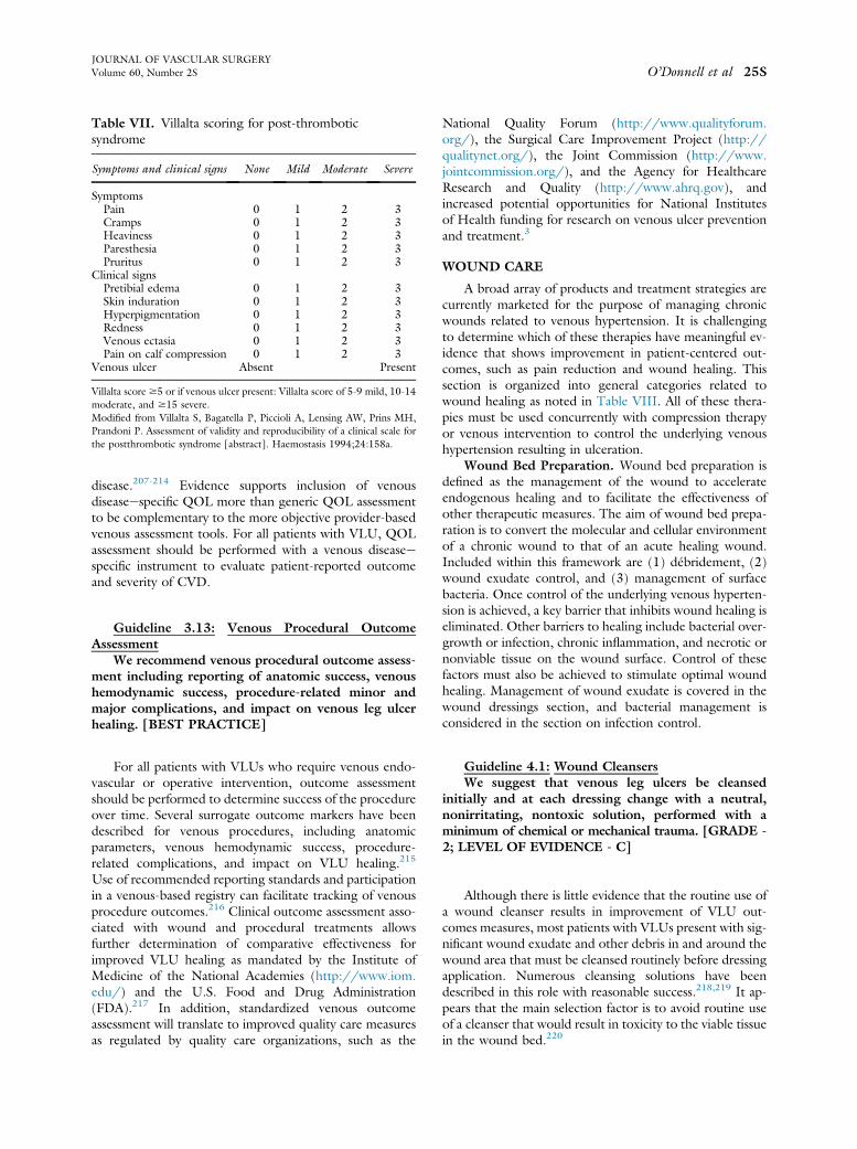
Table VII. Villalta scoring for post-thromboticsyndrome
Symptoms and clinical signs None Mild Moderate Severe
SymptomsPain 0 1 2 3Cramps 0 1 2 3Heaviness 0 1 2 3Paresthesia 0 1 2 3Pruritus 0 1 2 3
Clinical signsPretibial edema 0 1 2 3Skin induration 0 1 2 3Hyperpigmentation 0 1 2 3Redness 0 1 2 3Venous ectasia 0 1 2 3Pain on calf compression 0 1 2 3
Venous ulcer Absent Present
Villalta score $5 or if venous ulcer present: Villalta score of 5-9 mild, 10-14moderate, and $15 severe.Modified from Villalta S, Bagatella P, Piccioli A, Lensing AW, Prins MH,Prandoni P. Assessment of validity and reproducibility of a clinical scale forthe postthrombotic syndrome [abstract]. Haemostasis 1994;24:158a.
JOURNAL OF VASCULAR SURGERYVolume 60, Number 2S O’Donnell et al 25S
disease.207-214 Evidence supports inclusion of venousdiseaseespecific QOL more than generic QOL assessmentto be complementary to the more objective provider-basedvenous assessment tools. For all patients with VLU, QOLassessment should be performed with a venous diseaseespecific instrument to evaluate patient-reported outcomeand severity of CVD.
Guideline 3.13: Venous Procedural OutcomeAssessment
We recommend venous procedural outcome assess-ment including reporting of anatomic success, venoushemodynamic success, procedure-related minor andmajor complications, and impact on venous leg ulcerhealing. [BEST PRACTICE]
Guideline 4.1: Wound CleansersWe suggest that venous leg ulcers be cleansed
initially and at each dressing change with a neutral,nonirritating, nontoxic solution, performed with aminimum of chemical or mechanical trauma. [GRADE -2; LEVEL OF EVIDENCE - C]
For all patients with VLUs who require venous endo-vascular or operative intervention, outcome assessmentshould be performed to determine success of the procedureover time. Several surrogate outcome markers have beendescribed for venous procedures, including anatomicparameters, venous hemodynamic success, procedure-related complications, and impact on VLU healing.215
Use of recommended reporting standards and participationin a venous-based registry can facilitate tracking of venousprocedure outcomes.216 Clinical outcome assessment asso-ciated with wound and procedural treatments allowsfurther determination of comparative effectiveness forimproved VLU healing as mandated by the Institute ofMedicine of the National Academies (http://www.iom.edu/) and the U.S. Food and Drug Administration(FDA).217 In addition, standardized venous outcomeassessment will translate to improved quality care measuresas regulated by quality care organizations, such as the
National Quality Forum (http://www.qualityforum.org/), the Surgical Care Improvement Project (http://qualitynet.org/), the Joint Commission (http://www.jointcommission.org/), and the Agency for HealthcareResearch and Quality (http://www.ahrq.gov), andincreased potential opportunities for National Institutesof Health funding for research on venous ulcer preventionand treatment.3
WOUND CARE
A broad array of products and treatment strategies arecurrently marketed for the purpose of managing chronicwounds related to venous hypertension. It is challengingto determine which of these therapies have meaningful ev-idence that shows improvement in patient-centered out-comes, such as pain reduction and wound healing. Thissection is organized into general categories related towound healing as noted in Table VIII. All of these thera-pies must be used concurrently with compression therapyor venous intervention to control the underlying venoushypertension resulting in ulceration.
Wound Bed Preparation. Wound bed preparation isdefined as the management of the wound to accelerateendogenous healing and to facilitate the effectiveness ofother therapeutic measures. The aim of wound bed prepa-ration is to convert the molecular and cellular environmentof a chronic wound to that of an acute healing wound.Included within this framework are (1) débridement, (2)wound exudate control, and (3) management of surfacebacteria. Once control of the underlying venous hyperten-sion is achieved, a key barrier that inhibits wound healing iseliminated. Other barriers to healing include bacterial over-growth or infection, chronic inflammation, and necrotic ornonviable tissue on the wound surface. Control of thesefactors must also be achieved to stimulate optimal woundhealing. Management of wound exudate is covered in thewound dressings section, and bacterial management isconsidered in the section on infection control.
Although there is little evidence that the routine use ofa wound cleanser results in improvement of VLU out-comes measures, most patients with VLUs present with sig-nificant wound exudate and other debris in and around thewound area that must be cleansed routinely before dressingapplication. Numerous cleansing solutions have beendescribed in this role with reasonable success.218,219 It ap-pears that the main selection factor is to avoid routine useof a cleanser that would result in toxicity to the viable tissuein the wound bed.220

Table VIII. General categories of wound therapy forvenous leg ulcers
Wound bed preparationWound cleansersDébridementSurgical débridementAnesthesia for débridementHydrosurgical débridementUltrasonic débridementEnzymatic débridementBiologic débridement
Nutritional assessment and managementMeasurement of wound progress
Wound infection and bacterial controlWound cultureIndications for cultureMethod of wound culture
Management of limb cellulitisWound colonization and bacterial biofilmsTreatment of wound infectionTopical antibioticsSystemic antibiotics
Primary wound dressingsTopical dressing selectionAntimicrobials in topical dressingsPeriulcer skin managementAnti-inflammatory dressings
Adjunctive wound therapiesIndications for adjunctive therapiesSplit-thickness skin graftingCellular therapyUse of cellular therapyPreparation for cellular therapyFrequency of cellular therapy application
Negative pressure therapyElectrical stimulationUltrasound therapy
Guideline 4.2: DébridementWe recommend that venous leg ulcers receive thor-
ough débridement at their initial evaluation to removeobvious necrotic tissue, excessive bacterial burden, andcellular burden of dead and senescent cells. [GRADE -1; LEVEL OF EVIDENCE - B] We suggest that addi-tional maintenance débridement be performed tomaintain the appearance and readiness of the woundbed for healing. [GRADE - 2; LEVEL OF EVIDENCE -B] We suggest that the health care provider choosefrom a number of débridement methods, includingsharp, enzymatic, mechanical, biologic, and autolytic.More than one débridement method may be appro-priate. [GRADE - 2; LEVEL OF EVIDENCE - B]
Guideline 4.3: Anesthesia for Surgical DébridementWe recommend that local anesthesia (topical or local
injection) be administered to minimize discomfort asso-ciated with surgical venous leg ulcer débridement. Inselected cases, regional block or general anesthesia maybe required. [GRADE - 1; LEVELOF EVIDENCE - B]
Guideline 4.4: Surgical DébridementWe recommend that surgical débridement be per-
formed for venous leg ulcers with slough, nonviable tis-sue, or eschar. Serial wound assessment is important indetermining the need for repeated débridement.[GRADE - 1; LEVEL OF EVIDENCE - B]
Guideline 4.5 Hydrosurgical DébridementWe suggest hydrosurgical débridement as an alter-
native to standard surgical débridement of venous legulcers. [GRADE - 2; LEVEL OF EVIDENCE - B]
Guideline 4.6: Ultrasonic DébridementWe suggest against ultrasonic débridement over
surgical débridement in the treatment of venous leg ul-cers. [GRADE - 2; LEVEL OF EVIDENCE - C]
Guideline 4.7: Enzymatic DébridementWe suggest enzymatic débridement of venous leg
ulcers when no clinician trained in surgical débride-ment is available to débride the wound. [GRADE - 2;LEVEL OF EVIDENCE - C] We do not suggest enzy-matic débridement over surgical débridement.[GRADE - 2; LEVEL OF EVIDENCE - C]
JOURNAL OF VASCULAR SURGERY26S O’Donnell et al August Supplement 2014
Although protocols for management of VLUs usuallyrecommend wound débridement to remove nonviable tissueand to reduce bacterial burden, there is a lack of robust evi-dence that routine wound débridement accelerates woundhealing. Williams et al prospectively studied 28 patientswho underwent débridement for the presence of sloughand nonviable tissue in the VLU bed compared with 27patients who also had little granulation tissue but no sloughand did not receive débridement.221 Patients treated withdébridement had a significantly higher wound size reduction
4 weeks after débridement than those who did not. This dif-ference was maintained 20 weeks after débridement. Cardinalet al reviewed the relationship of débridement to ulcer heal-ing in two prospective RCTs of topical wound treatments on366 VLUs and 310 diabetic foot ulcers.222 VLUs treated at aclinic visit with débridement had a significantly higher me-dian wound surface area reduction in the week after débride-ment than did those for which débridement was notperformed. However, débridement frequency per patientdid not statistically correlate to rates of wound closure.
Recommendations to débride nonviable or necrotic tis-sue from the wound bed are supported by established prin-ciples of management of all wounds.223,224 However, thefrequency and method of débridement are not well studiedand have not been well established as related to the inci-dence of wound closure.225
The efficacy of topical anesthesia for débridement ofvenous leg ulcers was recently reviewed by the CochraneDatabase.226 They identified six trials evaluating eutecticmixture of local anesthetics (EMLA) lidocaine-prilocainecream compared with control.227,228 Patients treated withEMLA had significantly lower pain scores than thosetreated without topical anesthetic, leading to their conclu-sion that EMLA is an effective option for this purpose. Formore extensive débridement, local infiltrative anesthetic,regional block, or general anesthesia may be required foradequate periprocedural pain control.

Guideline 4.8: Biologic DébridementWe suggest that larval therapy for venous leg ulcers
can be used as an alternative to surgical débridement.[GRADE - 2; LEVEL OF EVIDENCE - B]
Guideline 4.9: Management of Limb CellulitisWe recommend that cellulitis (inflammation and
infection of the skin and subcutaneous tissue) sur-rounding the venous leg ulcer be treated with systemicgram-positive antibiotics. [GRADE - 1; LEVEL OFEVIDENCE - B]
Guideline 4.10: Wound Colonization and BacterialBiofilms
We suggest against systemic antimicrobial treat-ment of venous leg ulcer colonization or biofilmwithout clinical evidence of infection. [GRADE - 2;LEVEL OF EVIDENCE - C]
JOURNAL OF VASCULAR SURGERYVolume 60, Number 2S O’Donnell et al 27S
No method of débridement has been proved to be su-perior to surgical methods. However, in some cases, pa-tients may not have ready access to a clinician trained toperform surgical débridement, or this method may beless desirable because of comorbid patient conditions orpain considerations. Hydrosurgical débridement wasfound in some studies to shorten the procedural time ofdébridement but may be associated with significant addi-tional cost.229,230 Hydrosurgical débridement also re-quires a trained clinician and may be associated withsignificant periprocedural pain. No measurable benefit ofthe use of ultrasound methods to débride wounds hasbeen reliably demonstrated. Enzymatic débridement,which does not require a trained clinician for application,has been found in several studies to remove nonviable tis-sue from VLU wound beds, but there is no evidence thatthis method provides a benefit over surgical débride-ment.231-233 In clinical trials, larval therapy along withcompression has been shown to be an effective methodof débridement. However, the use of larval therapy didnot increase the rate of healing for necrotic tissue orslough in leg ulcers compared with ulcers treated withhydrogel and compression.234,235 Barriers to the use oflarval therapy also include personal hygiene concernsand personal bias. Recent adaptations have included con-taining the larvae in a bio-bag, which makes adoptionmore feasible to those with these concerns.
Wound Infection and Bacterial Control. The man-agement of bacterial involvement of chronic VLUs iscontroversial, with limited high-quality research and con-tradictory results in the studies available for review. Morerobust information is available for the treatment of otherinfected chronic wounds, in particular diabetic foot ulcers.This information may not translate to the treatment ofinfected VLUs.
For indications and methods of wound culture, refer toGuideline 3.4. Although there is general agreementamong experts that wounds with obvious signs of clinicalinfection should be treated with systemic antibiotics, thereis no consensus on the management of wounds colonizedwith bacteria or bacterial biofilms without signs of systemicinfection. The definition of a critically colonized wound isnot universally standardized and may vary by the virulenceof the colonizing bacteria. The more recent description ofbacterial biofilms and the multiple species that are typicallycultured from these wounds add another layer ofcomplexity. Finally, the changing nature of bacterialbehavior over time may render studies performed asrecently as 5 to 10 years ago irrelevant.
Developing technology, which has allowed moredetailed descriptions of the bacteria found existing in the
wound bed, employs methodology such as polymerasechain reaction analysis. These techniques may allow morespecific tailored therapy for individual wounds. Thesemethods, however, have not yet been established toimprove outcomes in the care of VLUs. In this setting,definitive guidelines are difficult to establish, and treatmentmust be individualized on the basis of local bacterial epide-miology and patient conditions.
Limb cellulitis is characterized by inflammation andinfection of the skin, subcutaneous tissue, and lymphaticfluid and is most commonly due to streptococci orstaphylococci, but it will occasionally include other spe-cies. There are numerous studies in the medical litera-ture evaluating various systemic antibiotic regimens forthe treatment of lower extremity cellulitis,236-238 alsosupported by a Cochrane review,239 with recommenda-tion for initial treatment with antibiotics directed atgram-positive bacteria, reserving broader coverage forunresponsiveness.
The data available concerning the treatment of bacte-rial colonization of VLUs are insufficient and contradic-tory. Sibbald et al found that high bacterial counts inVLUs were associated with delayed healing.240 Improvedhealing was associated with reduction in bacterial counts.Davies et al suggested that bacterial density was predictiveof nonhealing of VLUs.122 However, Lantis and Gendicsfound that when they treated VLUs with high bacterialcounts with topical antibiotics, bacterial counts decreasedbut there was no correlation with improvement in woundsize.241 Finally, Moore et al reported that the specific bac-terial species present or the number of species of bacteriapresent did not relate to healing outcomes.118 Theyconcluded that their data supported no specific treatmentof wound bacteria regardless of the type or quantity. Onthe basis of these contradictory findings, we are unable togenerate useful supporting clinical guidelines for treatmentof wound colonization or biofilm without clinical evidenceof infection.

Guideline 4.11: Treatment of Wound Infection:We suggest that venous leg ulcers with >1 3 106
CFU/g of tissue and clinical evidence of infection betreated with antimicrobial therapy. [GRADE - 2;LEVEL OF EVIDENCE - C] We suggest antimicrobialtherapy for virulent or difficult to eradicate bacteria(such as beta-hemolytic streptococci, pseudomonas,and resistant staphylococcal species) at lower levelsof colony-forming units per gram of tissue. [GRADE -2; LEVEL OF EVIDENCE - C] We suggest a com-bination of mechanical disruption and antibiotic ther-apy as most likely to be successful in eradicatingvenous leg ulcer infection. [GRADE - 2; LEVEL OFEVIDENCE - C]
Guideline 4.12: Systemic AntibioticsWe recommend that venous leg ulcers with clinical
evidence of infection be treated with systemic antibi-otics guided by sensitivities performed on wound cul-ture. [GRADE - 1; LEVEL OF EVIDENCE - C]Oral antibiotics are preferred initially, and the durationof antibiotic therapy should be limited to 2 weeks un-less persistent evidence of wound infection is present.[GRADE - 1; LEVEL OF EVIDENCE - C]
Guideline 4.14: Topical Dressing SelectionWe suggest applying a topical dressing that will
manage venous leg ulcer exudate and maintain a moist,warm wound bed. [GRADE - 2; LEVEL OF EVI-DENCE - C] We suggest selection of a primary wounddressing that will absorb wound exudate produced bythe ulcer (alginates, foams) and protect the periulcer
Guideline 4.15: Topical Dressings ContainingAntimicrobials
We recommend against the routine use of topicalantimicrobial-containing dressings in the treatment ofnoninfected venous leg ulcers. [GRADE - 2; LEVELOF EVIDENCE - A]
JOURNAL OF VASCULAR SURGERY28S O’Donnell et al August Supplement 2014
Although there is a lack of clinical studies evaluatingthe specific treatment of infected VLUs, available evi-dence supports systemic antibiotic treatment of patientswith clinical evidence of infection and ulcers containing> 1 � 106 CFU/g of bacteria on quantitative culture.242
Aggressive mechanical débridement of infected VLUswhenever possible is also suggested, although there is alack of high-quality evidence indicating that débride-ment improves results in the treatment of infectedulcers.
Guideline 4.13: Topical Antibiotics for InfectedWounds
We suggest against use of topical antimicrobialagents for the treatment of infected venous leg ulcers.[GRADE - 2; LEVEL OF EVIDENCE - C]
Guideline 4.16: Periulcer Skin ManagementWe suggest application of skin lubricants under-
neath compression to reduce dermatitis that commonlyaffects periulcer skin. [GRADE - 2; LEVEL OF EVI-DENCE - C] In severe cases of dermatitis associatedwith venous leg ulcers, we suggest topical steroids toreduce the development of secondary ulcerations andto reduce the symptoms of dermatitis. [GRADE - 2;LEVEL OF EVIDENCE - C]
There is a lack of evidence suggesting that topical anti-biotics have a positive effect on the treatment of infectedVLUs or on the healing of noninfected VLUs. Manizgateet al reported that in a prospective study of two silver-releasing dressings, neither dressing had an impact on thelevel of bioburden identified in treated VLUs.243
Conversely, in another study, Lantis and Gendics foundthat a sustained-release silver sulfadiazine foam reducedthe level of bacteria cultured from infected VLUs, but therewas no correlation with an improvement in wound size.241
A Cochrane review on the use of topical silver for infectedVLUs concluded that there is insufficient evidence torecommend the use of silver-containing dressings or topicalagents for treatment of infected or contaminated chronicwounds.244
Primary Wound Dressings
There is insufficient evidence suggesting that a specificprimary dressing that contacts the wound can result in ahigher rate of wound closure. Most VLUs produce largeamounts of exudate. This fluid contains high concentra-tions of proteases and inflammatory cytokines that maydamage surrounding healthy skin.245 Removal of wounddrainage from the wound bed will reduce the inflammatoryenvironment that prohibits wound healing. We recom-mend use of dressings that will manage wound exudateand maintain a moist wound bed. Primary dressings withhigh absorptive capabilities, including foams, alginates,and other specialty dressings, are often selected for the pri-mary coverage layer for heavily exudative VLUs.246
skin. [GRADE - 2; LEVEL OF EVIDENCE - B]
In the only prospective RCT of topical antibiotics, whichexamined the treatment of noninfected VLUs, Michaels et alrandomized 213 patients with VLUs to silver-releasing dress-ings compared with non-silver-releasing dressings.247 No sig-nificant difference in ulcer healing or other quality of lifemeasures was identified in the silver dressing group. Giventhe increased cost of silver-containing dressings, routine useof topical antimicrobial dressings for the treatment of VLUsis not supported.248,249
Management of dermatitis and other abnormalities ofthe periulcer skin is an important adjunct to other therapiesin the treatment of VLUs.250 Venous hypertension maycause significant dermatitis in the calf and ankle that maybe ameliorated with a variety of skin lubricants and other

JOURNAL OF VASCULAR SURGERYVolume 60, Number 2S O’Donnell et al 29S
agents. Contact dermatitis related to the dressings andother products used to treat VLUs is also frequent. Recog-nizing this problem and treating it expeditiously will pre-vent secondary skin ulceration complicating the primaryleg ulcer.251,252 Consultation from dermatology may beuseful in long-standing or recurrent cases experiencingsensitivity to wound treatment products.
Guideline 4.17: Anti-inflammatory TherapiesWe suggest against use of anti-inflammatory thera-
pies for the treatment of venous leg ulcers. [GRADE -2; LEVEL OF EVIDENCE - C]
Although a significant body of research has identifiedthat VLUs are associated with significant upregulation ofinflammatory cytokines and proteases,71,83,253 there is alack of data identifying that any anti-inflammatory dressingother than compression is associated with accelerated heal-ing of VLUs.254
Adjunctive Wound Therapies
Guideline 4.18: Indications for Adjuvant TherapiesWe recommend adjuvant wound therapy options for
venous leg ulcers that fail to demonstrate improvementafter a minimum of 4 to 6 weeks of standard wound Guideline 4.20: Cellular Therapy
We suggest the use of cultured allogeneic bilayerskin replacements (with both epidermal and dermallayers) to increase the chances for healing in patientswith difficult to heal venous leg ulcers in addition tocompression therapy in patients who have failed toshow signs of healing after standard therapy for 4 to6 weeks. [GRADE - 2; LEVEL OF EVIDENCE - A]
Guideline 4.21: Preparation for Cellular TherapyWe suggest a therapeutic trial of appropriate
compression and wound bed moisture control before
Adjunctive therapies for the healing of VLUs should beconsidered after all standard of care measures have beenimplemented. Comprehensive care should include compres-sion therapy, local wound débridement, control of bio-burden, wound moisture balance with appropriatedressings, and consideration of the use of pentoxifylline. Af-ter a minimum of 4 to 6 weeks of appropriate care, adecrease in wound dimensions should be noted. Standardof care for VLUs will lead to improvement in the majorityof cases, and only in those cases without improvementshould adjunctive modalities be used. The cost of these ther-apies can be high, and the evidence supporting their use isnot sufficiently strong to justify their use as primary therapywithout an attempt at lower cost, evidence-based methods.However, failure to demonstrate improvement after 4 weeksof treatment with a decrease in wound size of at least 30% to40% should lead the clinician to consider adjunctive treat-ment options.109 Re-evaluation of the patient and woundshould be performed before the use of adjuvant therapiesto ensure that compression has achieved edema control, bio-burden is well controlled, and exudate is not excessive.
therapy. [GRADE - 1; LEVEL OF EVIDENCE - B]
Guideline 4.19: Split-thickness Skin GraftingWe suggest against split-thickness skin grafting as
primary therapy in treatment of venous leg ulcers.[GRADE - 2; LEVEL OF EVIDENCE - B] We suggestsplit-thickness skin grafting with continued compres-sion for selected large venous leg ulcers that have failedto show signs of healing with standard care for 4 to 6weeks. [GRADE - 2; LEVEL OF EVIDENCE - B]
There is currently insufficient information to recom-mend use of autologous skin grafting as a primary therapyfor VLUs. Studies have bias, small sample size, and indirectcomparators. A Cochrane review on skin grafting for VLUsalso failed to reach a definitive recommendation.255 Arecent RCT found that use of low-molecular-weight hepa-rin resulted in decreased recurrence compared with skingrafting without anticoagulation.256,257 After skin grafting,patients should continue to use therapeutic compression.
Individual situations and patient preferences mightresult in the use of skin grafting for VLUs. Some cliniciansconsider skin grafting after standard of care has failed, forlarge soft tissue defects, by the patient’s or clinician’s pref-erence, and when there are specific reasons that requiremore expeditious closure. In a nonrandomized study of111 patients, Jankunas demonstrated improved healingand durability with skin grafting compared with conserva-tive therapy for large venous wounds that were presentlonger than 6 months,, but only 65% of cases were judgedto have good take of the split-thickness skin graft.258 Skingrafting should be considered as primary therapy only forlarge wounds (ie, >25 cm2) in which healing is unlikelywithout grafting or as secondary therapy after there is a fail-ure to achieve healing with standard of care.
Apligraf, an allogeneic bilayer cellular therapy, has beenapproved by the FDA for treatment of VLUs. The efficacyof Apligraf was studied in an RCT of 245 patients withVLUs treated with standard of care compared with standardof care plus the application of Apligraf.259,260 VLUs had beenpresent for at least 6 weeks and had not responded well to theinitial use of compression and the other aspects of standardcare. Subsequent research has identified improved outcomesfor recalcitrant VLUs (>1 year duration or large surfacearea).262 No other living cellular therapies have demonstratedbenefit for the treatment of VLUs in prospective RCTs.
application of cellular therapy. [GRADE - 2; LEVELOF EVIDENCE - C] We recommend that adequatewound bed preparation, including complete removal ofslough, debris, and any necrotic tissue, be completedbefore the application of a bilayered cellular graft.[GRADE - 1; LEVEL OF EVIDENCE - C] We recom-mend additional evaluation and management of increasedbioburden levels before the application of cellular ther-apy. [GRADE - 1; LEVEL OF EVIDENCE - C]

Guideline 4.22: Frequency of Cellular TherapyApplication
We suggest reapplication of cellular therapy as longas the venous leg ulcer continues to respond on the ba-sis of wound documentation. [GRADE - 2; LEVEL OFEVIDENCE - C]
Guideline 4.25: Electrical StimulationWe suggest against electrical stimulation therapy
for venous leg ulcers. [GRADE - 2; LEVEL OF EVI-DENCE - C]
Guideline 4.26: Ultrasound TherapyWe suggest against routine ultrasound therapy for
venous leg ulceration. [GRADE -2; LEVEL OF EVI-DENCE - B]
JOURNAL OF VASCULAR SURGERY30S O’Donnell et al August Supplement 2014
The optimal frequency and timing of reapplication ofbiologic skin substitutes to VLUs remain controversialwith little consensus in published studies. The initial pivotaltrial for Apligraf allowed up to five applications of the prod-uct during the first 3 weeks of the study.259 Reapplicationwas performed if the investigator thought that there wasless than 50% “take” of the skin equivalent on inspectionof the wound. Subsequent to this clinical trial, it hasbeen determined that living skin equivalents do not persistin the wound for more than a few weeks, so “no take” oc-curs as is seen with autologous grafts. Other studies havereported application at variable times, including weekly,every few weeks, or once only.261 With no comparativedosing studies published to determine clinical or economicoutcomes, the frequency of application remains at thediscretion of the clinician. Current clinical practice hasincluded application of grafts followed by a period of 1to 3 weeks of observation to determine effectiveness beforereapplication is considered.
Guideline 4.23: Tissue Matrices, Human Tissues,or Other Skin Substitutes
We suggest the use of a porcine small intestinal sub-mucosa tissue construct in addition to compressiontherapy for the treatment of venous leg ulcers thathave failed to show signs of healing after standard ther-apy for 4 to 6 weeks. [GRADE - 2; LEVEL OF EVI-DENCE - B]
Numerous tissue constructs are available for use inchronic wounds that employ either human tissue (amnioticmembrane, cryopreserved skin) or animal tissue (bladder,fetal bovine skin, others). Some are reported to containactive growth factors or other attributes that might bebeneficial to healing of VLUs.262 Of the multitude ofsuch products currently marketed, only porcine small intes-tinal submucosa has prospective RCT data supporting itsuse to accelerate VLU closure.263
Guideline 4.24: Negative Pressure TherapyWe suggest against routine primary use of negative
pressure wound therapy for venous leg ulcers.[GRADE - 2; LEVEL OF EVIDENCE - C]
There is currently not enough information to supportthe primary use of negative pressure wound therapy forVLUs. Evidence supports positive effects with the use ofnegative pressure therapy for wound healing in general.Tissue granulation, area and volume reduction, and reduc-tions in bioburden have all been reported. There have been
few studies specifically studying negative pressure therapyfor VLUs, with most studies reporting on mixed woundcauses.264 There has been an increase in the use of negativepressure wound therapy for wound bed preparation toaugment skin graft healing.265
There is theoretical, preclinical, and limited clinical ev-idence to support the use of electrical stimulation for thetreatment of VLUs.266 However, few studies of electricalstimulation focus solely on VLUs. Also, there is little con-sistency in the parameters of treatment, including vari-ability in type of electrical current, settings, treatmenttimes, and preferred waveforms, making comparisonsimpossible.267,268 For these reasons, electrical stimulationis not recommended for the treatment of VLUs untilfurther evidence supports its use.
Therapeutic ultrasound has been found to stimulate anumber of cellular effects associated with the accelerationof wound healing. These cellular effects have been wellstudied in vitro and include improvement of microcircula-tion, reduction of edema, and increases in cytokine andother protein levels that are active in the healingcascade.269 Clinical trials studying the effects of ultrasoundhave reported positive results in the healing of a variety ofchronic nonhealing wounds.270 Clinical trials of therapeu-tic ultrasound for the healing of VLUs lack standardizedprotocols, and relatively few studies focus on VLUs alone.In a pilot study of VLUs refractory to compression therapy,ultrasound treatment was associated with a decrease inwound size, inflammatory cytokines, bacterial counts, andpain.271 In an RCT of 337 VLU patients, weekly treatmentwith high-frequency ultrasound did not result in an in-crease in healing rates or improved quality of life comparedwith standard VLU care.272 Unfortunately, most therapeu-tic ultrasound protocols have recommended treatment ses-sions at higher frequency, such is three times weekly oronce daily. This has led to criticism of this trial as sufferingfrom inadequate ultrasound dosing to determine whetherit provides any benefit to patients with VLUs. Additionalstudies employing a standardized protocol of therapeuticultrasound in patients with VLUs are required for furtherrecommendations on the use of this modality to bemade.273 On the basis of current literature, there is insuffi-cient evidence to recommend the routine use of therapeu-tic ultrasound for the treatment of VLUs as a primarytherapy.

Guideline 5.1: CompressiondUlcer HealingIn a patient with a venous leg ulcer, we recommend
compression therapy over no compression therapy toincrease venous leg ulcer healing rate. [GRADE - 1;LEVEL OF EVIDENCE - A]
JOURNAL OF VASCULAR SURGERYVolume 60, Number 2S O’Donnell et al 31S
On the basis of the literature review, there are threeadjuvant treatments with evidence suggestive of benefitfor patients with VLUs that do not respond to an initialtreatment course of standard therapy. Split-thickness skingrafting may provide benefit, particularly for large, recalci-trant VLUs. Cellular therapy with Apligraf was found to bebeneficial in a multicenter randomized clinical trial, as wasporcine small intestinal submucosa. There have been nocomparative trials of these therapies published with suffi-cient evidence to recommend one therapy over the othersfor the treatment of recalcitrant VLUs.
COMPRESSION
Compression therapy is based on a mechanical conceptof applying pressure to the limb. This simple action causesmultiple biologic processes proved to be beneficial for man-agement of conditions ranging from occupational swellingto venous ulcers. A variety of devices have been used forcompression therapy. Different types of bandages, bandagesystems, and ready-to-use garments can provide sustainedcompression, whereas several pneumatic devices applyintermittent compression.
For the treatment of VLU, compression systems, whichexert hemodynamic improvement on the dysfunctionalvenous pumping function, are preferred. Such systemsthat do not give way to the expanding muscle duringwalking are either short-stretch textiles or multicomponentbandages containing elastic material in combination withan adhesive or cohesive covering layer. Such bandagesneed to be applied by trained staff. Self-applicable andadjustable short-stretch Velcro band devices may be analternative. Elastic bandages or compression stockings areeffective concerning edema reduction but are hemodynam-ically less effective in improving the venous pumpingfunction.274
Bandage layers should be differentiated from compo-nents. The term multilayer describes one bandage that al-ways covers the leg with more than one layer because ofoverlapping during application. Every multicomponentbandage is a multilayer bandage, whereas graduatedcompression stockings have a single compression layer.The four-layer bandage (4LB) is a multicomponentbandage consisting of four components: orthopedic wool,crepe bandage, elastic bandage, and elastic cohesive (outer)bandage. Applying several components and layers overeach other not only increases the pressure but also changesthe elastic property of the final bandage because of the fric-tion between the surfaces of each bandage and a cohesiveouter layer, producing a final multilayer bandage that isless elastic and more stiff. Short-stretch bandages (SSB)are also often used as multicomponent bandages in combi-nation with different materials. Unfortunately, the differentcombinations of materials are often poorly described intrials.
Coexistence of multiple devices that are designed toperform essentially the same function demonstrates thatalthough compression therapy is generally effective, theoptimization of compression has been a challenging task
because of limitations in current evidence. Use of confusingterminology and not reporting the dose (pressure) of acompression device and physical properties of the compres-sion product are just a few of many deficiencies in availableevidence related to compression therapy.275 The use ofpressure-measuring devices at the skin-bandage interfaceto ensure adequate pressure applied during the bandagingis not routine; however, in specific circumstances (such asstudy protocol and quality assurance), such instrumentscan be applied.276,277
A total of nine RCTs compare compression therapy tono compression therapy, and these trials provide objectiveinformation on initial ulcer size as well as the outcomesof time to complete healing or proportion of ulcers healed(FDA-recommended end points of wound therapy trials).The preponderance of evidence suggests that VLUs healmore quickly with compression therapy vs no compressionas confirmed by our commissioned comparative systematicreview and meta-analysis of compression modalities andvenous ulcer healing.30
One RCT of 36 patients directly compared compres-sive (Unna boot) with noncompressive bandages (polyure-thane foam dressing). Wound healing rates weresignificantly improved by 0.07 cm2/day (P ¼ .004), andmore patients completely healed in the compression group(RR, 2.30; 95% CI, 1.29-4.10).278,279
A recent Cochrane review identified three RCTsencompassing a total of 177 patients comparing compres-sion dressings with primary dressing alone (never withcompression).279 One study (44 patients) showed a differ-ence with improved ulcer area and volume at 8 weeks.280
Two of the studies (49 and 84 patients) failed to show adifference in healing rates between treatments at 7 weeksand 6 months of follow-up, respectively.281,282
Another RCT of 321 patients compared SSB or 4LBwith moist wound dressings without compression. Thehealing rate at 24 weeks was superior in both compressiongroups (72% SSB and 67% 4LB) compared with the non-compression group (29%; P < .0001).283
Three of four additional RCTs that comparedcompression dressings with standard wound care foundimprovement in ulcer healing with compression therapy.One study (53 patients) compared SSB with usual careand found complete ulcer healing at 3 months to be 71%in the SSB group compared with 25% in the usual caregroup (P value not reported).284 The remaining studiescompared 4LB dressing with primary care management.A trial of 36 patients found more complete healing at 3months in the compression group compared with the usualcare group (RR, 4.0; 95% CI, 1.35-11.82).285 Similar

Guideline 5.3: Multicomponent CompressionBandage
We suggest the use of multicomponent compres-
JOURNAL OF VASCULAR SURGERY32S O’Donnell et al August Supplement 2014
results were found in a larger trial encompassing 200 par-ticipants, with 54% of patients healed in the 4LB groupcompared with 34% in the control group (P < .001).286
Another RCT of 233 patients failed to find a difference be-tween two groups with regard to complete healing at 1year but did find improved rate of healing (median timeof 20 vs 43 weeks; P ¼ .03) favoring the compressiongroup.287
Guideline 5.2: CompressiondUlcer RecurrenceIn a patient with a healed venous leg ulcer, we sug-
gest compression therapy to decrease the risk of ulcerrecurrence. [GRADE - 2; LEVEL OF EVIDENCE - B]
sion bandage over single-component bandages for thetreatment of venous leg ulcers. [GRADE - 2; LEVELOF EVIDENCE - B]
Guideline 5.4: CompressiondArterial InsufficiencyIn a patient with a venous leg ulcer and underlying
arterial disease, we do not suggest compression ban-dages or stockings if the ankle-brachial index is 0.5 orless or if absolute ankle pressure is less than 60 mmHg. [GRADE - 2; LEVEL OF EVIDENCE - C]
The potential for recurrence of VLUs is high asdemonstrated by a 67% recurrence rate in one study, inwhich 35% of patients exhibited a remarkable four ormore episodes of ulceration.140 The reported recurrencerate for ulcers in one study in the setting of compressiontherapy alone is 28% and in combination with superficialvenous surgery is 12%, highlighting the recent improve-ments in clinical practice.288
As more success is achieved with healing of primaryVLUs, the number of patients at risk for recurrencerises.289 However, few RCTs have specifically evaluatedthe impact of compression therapy on the risk of ulcerrecurrence. One trial randomized 153 patients withrecently healed (2 weeks) VLU to compression or nocompression therapy. Compression hosiery with a highpressure (34-46 mm Hg) was associated with significantreduction in ulcer recurrence at 6 months compared withno compression (21% vs 46%; RR, 0.46; 95% CI, 0.27-0.76; P ¼ .003).289,290 A second RCT of 188 patients ran-domized 166 into two treatment groups, each receivingtherapy with 23 mm Hg compression at the ankle withtwo brands of stockings. Compliance data reported fromthis trial as summarized in Nelson and Bell-Syer demon-strated that inability to tolerate compression therapy wassignificantly associated with recurrent ulceration (RR,2.58; 95% CI, 1.33-5.01; P ¼ .010), although these pa-tients were not included in randomization.289,291
These RCT data are congruent with 15 years of obser-vational data demonstrating that lack of compliance withcompression therapy was significantly associated with ulcerrecurrence (16% recurrence with compliance, 100%without; P < .0001).292 Two studies to date have evalu-ated the use of moderate (UK class II, 18-24 mm Hg atthe ankle) to high (UK class III, 25-35 mm Hg) compres-sion and compliance with these stockings on prevention ofulcer recurrence, with differing results. One study found nodifference in recurrence between the two groups but signif-icantly lower compliance in the high-compressiongroup.293 Because time to recurrence was not reported,the effect of compression pressures on delay of recurrenceremains unknown.
On the basis of our commissioned comparative system-atic review and meta-analysis of compression modalitiesand venous ulcer healing, low-quality evidence supportsthe effect of compression on ulcer recurrence.30
The possible difference in healing outcomes betweensingle-layer and multilayer compression bandages hasbeen evaluated in four trials, three of which have been pub-lished. In two trials, the number of participants was small(n ¼ 27 and 34) and showed no difference between treat-ment groups.279,294,295 However, a large RCT with 245patients demonstrated improved healing at 24 weeks(67% vs 49%; P ¼ .009) in the multilayer (4LB) bandagegroup compared with the single-layer group.296 In addi-tion, 4LB were associated with faster wound closure (78vs 168 days; P ¼ .001) compared with the single-layergroup.
A recent meta-analysis identified six trials directlycomparing multicomponent bandages containing elasticwith those not containing elastic. Data from 797 patients(five trials with independent patient data available) demon-strated 30% improved healing with 4LB compared withthose with SSB (odds ratio, 1.31; 95% CI, 1.09-1.58;P ¼ .0005).297 These data might in part be explained bythe skill of the health care professional in achieving optimalsub-bandage pressures and graduated compression. Incomparing 4LB with SSB in countries where the medicalstaff is familiar with applying SSB (eg, Netherlands andAustria), there was no significant difference between the ul-cer healing rates after 3 months (33 of 53 ¼ 62% healedwith 4LB vs 43 of 59 ¼ 73% with SSB).297-299 Studiescomparing different versions of the 4LB (proprietary andnonproprietary) were similar in outcomes, and thus a spe-cific 4LB is not endorsed.300-302
On the basis of our commissioned comparative system-atic review and meta-analysis of compression modalitiesand venous ulcer healing, moderate-quality evidence sup-ports multicomponent compression over single componentcompression.30
An ABI of less than 0.5 is considered to be indicativeof severe arterial disease.303 Compression of a limb with

JOURNAL OF VASCULAR SURGERYVolume 60, Number 2S O’Donnell et al 33S
significant arterial disease could compromise arterialperfusion and potentially result in adverse ischemicsequelae. In a survey study, nearly one third of generalsurgery consultants surveyed reported that they hadencountered ulcers or necrosis as the direct result ofcompression treatment.304 Supporting evidence is limitedto a small number of case reports and a dated physiologystudy that examined the effects of compression on the up-per extremity in healthy subjects.305-307 However, onestudy using magnetic resonance flowmetry showed thatsustained compression with a pressure around 40 mmHg increases the arterial flow under the bandage innormal volunteers.308 Similarly, a recent study performedin patients with VLUs and concomitant arterial occlusivedisease (ABI of 0.5 to 0.85) demonstrated by multiplemetrics that arterial blood flow is even augmented by in-elastic bandages.309
For patients with VLU and concomitant PAD, use ofstandard compression has been shown to be safe if ABI$0.80.303,310,311 Modified compression bandages orcompression stockings with lower pressure ratings can beused for ankle systolic pressure $60 mm Hg, digital pres-sure $30 mm Hg or ABI $0.50 with close monitoring,but only after consultation with a vascular specialist.309,312
Use of ankle perfusion pressure of 60 mm Hg or greaterrather than ABI #0.5 as a cutoff for compression ispreferred because this correlates better with tissue perfu-sion pressure, and any sustained external compression pres-sure should never exceed this cutoff perfusion pressure.One study showed that modified compression multilayeredbandages with reduced compression pressure can be bene-ficial for healing of mixed ulcers.311
Guideline 5.5: Intermittent Pneumatic CompressionWe suggest use of intermittent pneumatic compres-
sion when other compression options are not available,cannot be used, or have failed to aid in venous legulcer healing after prolonged compression therapy.[GRADE - 2; LEVEL OF EVIDENCE - C]
Intermittent pneumatic compression (IPC) wasinitially proposed as a method to promote VLU healingby activation of the fibrinolytic system and showed prom-ise in decreasing VLU healing time in patients who hadpreviously failed to respond to conservative treat-ment.313,314 However, this theory remains to be rigor-ously tested. To date, only 387 patients have beenenrolled in RCTs comparing IPC with other treatmentmodalities.36 Whereas it is advantageous to use IPCcompared with no compression therapy, currently thereis limited evidence to suggest that the addition of IPCto compression therapy offers benefit.315 Thus, the pa-tients likely to benefit most from IPC are those whocannot tolerate or do not have access to compression
therapy or who have failed to respond to prolongedcompression therapy alone. Further studies need to be un-dertaken to evaluate the benefit of IPC in this particularcohort. In circumstances in which IPC is used, there issome evidence to suggest that proportion of ulcers healed(86% vs 61% at 6 months; P ¼ .003) is improved withrapid compared with slow cycling.316
OPERATIVE/ENDOVASCULAR MANAGEMENT
Guidelines for operative and endovascular manage-ment are categorized anatomically as superficial venous dis-ease, perforator venous disease, deep infrainguinal venousdisease, and iliocaval disease. Each category can bemanaged with open surgical or endovascular techniques.The rare occurrence of congenital venous disease leadingto VLU and its treatment lacks definitive reporting.
These guidelines for operative and endovascular man-agement also address the twofold goal of VLU treatment,which includes ulcer healing and prevention of ulcer recur-rence. The separate pathologic processes of primary degen-erative reflux disease and inflammatory thrombotic diseaseprovide different challenges for the treating physicianbecause the surgeon or interventionalist must provide aspecific procedure to correct the pathologic state.
In general, quality of the evidence available to supportrecommendations for operative and endovascular manage-ment is mostly limited to level C evidence because of anabsence of comparative prospective RCTs of treatmenttechniques, with the exception of superficial venous treat-ments. The strength of the clinical recommendations foroperative and endovascular management can be increasedby concurrence of results from individual case seriesand expert opinion of the Venous Ulcer GuidelinesCommittee.
This section is predicated on the principle that deepvein reconstruction is reserved for cases in which therehas been failure to control the ulcer diathesis by dedi-cated nonsurgical measures and by simpler conventionalinterventions in the superficial and perforator veins ofthe extremity. In general, less invasive procedures arepreferred when they are available and equally effective.Concurrence has been negatively affected because ofthe evolving developmental status of open surgery forcomplicated pathologic states in the deep veins of theleg, pelvis, and abdomen and the ongoing search forminimally invasive procedures. Within these limitations,there is growing consensus that the endovascular stent-ing techniques are the most appropriate surgical treat-ment for iliac occlusive disease, whereas openautogenous repair is best therapy for deep vein reflux inthe infrainguinal lower extremity or from failed endove-nous stent procedures of the iliocaval and iliofemoralvenous system. A practical algorithm for operative andendovascular management of patients with VLUs is pro-posed in the Fig.

Fig. Proposed algorithm for operative and endovascular treatment of patients with venous leg ulcer (VLU) based oninvolved anatomic venous system and presence of venous reflux or obstruction. The risk-to-benefit ratio is weighed forthose procedures with more risk (lower, moderate, higher) considered later in the treatment when the benefit is similar.
JOURNAL OF VASCULAR SURGERY34S O’Donnell et al August Supplement 2014
Superficial Venous Reflux and Venous Leg Ulcer
Guideline 6.1 Superficial Venous Reflux and ActiveVenous Leg UlcerdUlcer Healing
In a patient with a venous leg ulcer (C6) andincompetent superficial veins that have axial refluxdirected to the bed of the ulcer, we suggest ablationof the incompetent veins in addition to standardcompressive therapy to improve ulcer healing.[GRADE - 2; LEVEL OF EVIDENCE - C]
Guideline 6.2 Superficial Venous Reflux and ActiveVenous Leg UlcerdPrevent Recurrence
In a patient with a venous leg ulcer (C6) andincompetent superficial veins that have axial refluxdirected to the bed of the ulcer, we recommend abla-tion of the incompetent veins in addition to standardcompressive therapy to prevent recurrence. [GRADE -
Our commissioned comparative systematic review andmeta-analysis of surgical interventions vs conservativetherapy including 11 studies (seven RCTs and four obser-vational studies) favored open surgery vs compression forulcer healing, although the quality of the evidence waslow.31 In addition, other individual RCTs have shown a
1; LEVEL OF EVIDENCE - B]
difference that was not statistically significant favoring sur-gical treatment of the superficial venous system andcompression over compression alone.31,288,317 On the ba-sis of these observations, the strength and grade of evi-dence favoring surgery for ulcer healing are limited to aGrade 2C classification. Regarding recurrence, ourcommissioned comparative systematic review and meta-analysis included two RCTs, and each study favoredsurgery plus compression over compression alone to astatistically significant degree for prevention of ulcerrecurrence.288,317,318 Because each study had some chal-lenges in design and baseline imbalance, the strengthand grade of evidence is 1B.
Regarding the choice of method of saphenous abla-tion, there are multiple RCTs showing strong and consis-tent evidence that modern open surgery, radiofrequency,and laser ablation are equivalent in effect and safety(Table IX).319-330 Endovenous ablation can be extrapo-lated to be as effective as open ligation and strippingused in these earlier trials. Moreover, endovenous ablationcan be carried out under local/tumescent anesthesia as anoffice-based procedure, which shifts the risk/benefit ratiosignificantly. Foam sclerotherapy has been demonstratedto rival these modalities and has significantly more recan-alizations and refluxing treated segments on follow-up

Table IX. Randomized controlled trials of endovenous laser or radiofrequency ablation vs open surgery for the treatmentof great saphenous vein incompetence (at least 1 year of follow-up) comparing recurrence of varicose veins andneovascularization
Year No. of patients Follow-up, years
Results
Recurrence Neovascularization
Endovenous laser ablation vs open surgeryLin et al319 2007 80 1 No difference No differenceTheivacumar et al320 2009 120 2 No difference þ LaserChristenson et al321 2010 200 2 No difference No differencePronk et al322 2010 130 1 No difference No differenceCarradice et al323 2011 280 1 þ Laser No differenceRass et al324 2012 346 2 No difference No differenceRasmussen et al325 2013 137 5 No difference No difference
Endovenous radiofrequency ablation vs open surgeryLurie et al326 2005 65 2 No difference No differencePerala et al327 2005 28 3 No difference No differenceKianifard et al328 2006 55 1 No difference þ RadiofrequencyStötter et al329 2006 60 1 No difference No differenceHelmy ElKaffas et al330 2010 180 2 No difference No difference
þ Laser, Favored in comparison of laser vs open surgery; þ Radiofrequency, favored in comparison of radiofrequency vs open surgery.
Guideline 6.5: Combined Superficial and Perfo-rator Venous Reflux With or Without Deep VenousReflux and Active Venous Leg Ulcer
In a patient with a venous leg ulcer (C6) andincompetent superficial veins that have reflux to the ul-
JOURNAL OF VASCULAR SURGERYVolume 60, Number 2S O’Donnell et al 35S
evaluation.331,332 Several ablation techniques remain un-der investigation, including steam ablation and pharmaco-mechanical and cyanoacrylate ablation as well as CHIVA(ambulatory conservative hemodynamic management ofvaricose veins; one small RCT showed that CHIVA hada higher proportion of ulcers heal than with compressionalone) and ASVAL (ambulatory selective varicose veinablation under local anesthesia).20,257,333-338
Guideline 6.3: Superficial Venous Reflux andHealed Venous Leg Ulcer
In a patient with a healed venous leg ulcer (C5) andincompetent superficial veins that have axial refluxdirected to the bed of the ulcer, we recommend abla-tion of the incompetent veins in addition to standardcompressive therapy to prevent recurrence. [GRADE -1; LEVEL OF EVIDENCE - C]
Guideline 6.4: Superficial Venous Reflux With SkinChanges at Risk for Venous Leg Ulcer (C4b)
In a patient with skin changes at risk for venous legulcer (C4b) and incompetent superficial veins that haveaxial reflux directed to the bed of the affected skin, wesuggest ablation of the incompetent superficial veins inaddition to standard compressive therapy to prevent ul-ceration. [GRADE - 2; LEVEL OF EVIDENCE - C]
cer bed in addition to pathologic perforating veins(outward flow of >500 ms duration, with a diameterof >3.5 mm) located beneath or associated with the ul-cer bed, we suggest ablation of both the incompetentsuperficial veins and perforator veins in addition tostandard compressive therapy to aid in ulcer healingand to prevent recurrence. [GRADE - 2; LEVEL OFEVIDENCE - C]
The evidence supporting this guideline is an extrapola-tion of the data on ulcer healing and prevention of ulcerrecurrence stated under Guidelines 6.1 and 6.2. In thisgroup of patients, VLUs had healed, and so interventionis expected to provide protection from recurrence as notedin our commissioned comparative systematic review andmeta-analysis and the two RCTs.31,288,317,318
Perforator Venous Reflux and Venous Leg Ulcer
The evidence regarding treatment of perforator venousreflux associated with a VLU is complicated by several fac-tors but most notably by combining its treatment with thatof superficial venous reflux.339-352 The Dutch RCTcomparing subfascial endoscopic perforator vein surgery(SEPS) with or without superficial reflux ablation to med-ical treatment in patients with venous ulcers found no dif-ference in healing rate or recurrence in the two treatmentgroups.317 However, patients with recurrent ulceration ormedially located ulcers in the surgical group had a longerulcer-free period than did those treated in the conservativegroup (P ¼ .02). A secondary analysis of the Dutch SEPStrial on 94 ulcerated legs was reported with a mean follow-up of 29 months.353 Ulcer recurrence was significantlyhigher in patients who had an incomplete SEPS procedure(P ¼ .007), indicating the clinical significance of incompe-tent perforating veins in patients with venous ulcers.

JOURNAL OF VASCULAR SURGERY36S O’Donnell et al August Supplement 2014
Ameta-analysis that included this RCT and 19 case seriesinvolving 1140 treated limbs found that surgical treatmentby SEPS, with or without superficial venous ablation, resultedin ulcer healing in 88% and a low ulcer recurrence of 13% at21 months.340 Another meta-analysis concluded that perfo-rator interruption used as a part of a treatment regimen forsevere CVI benefits most patients in the short term regardingulcer healing and the prevention of ulcer.345
Guideline 6.6: Combined Superficial and Perfo-rator Venous Reflux With or Without Deep VenousDisease and Skin Changes at Risk for Venous Leg Ulcer(C4b) or Healed Venous Ulcer (C5)
In a patient with skin changes at risk for venous legulcer (C4b) or healed venous ulcer (C5) and incompe-tent superficial veins that have reflux to the ulcer bed inaddition to pathologic perforating veins (outward flowof >500 ms duration, with a diameter of >3.5 mm)located beneath or associated with the healed ulcerbed, we suggest ablation of the incompetent superficialveins to prevent the development or recurrence of avenous leg ulcer. [GRADE - 2; LEVEL OF EVI-DENCE - C] Treatment of the incompetent perfo-rating veins can be performed simultaneously withcorrection of axial reflux or can be staged with re-evaluation of perforator veins for persistent incompe-tence after correction of axial reflux. [GRADE - 2;LEVEL OF EVIDENCE - C]
Guideline 6.8: Treatment Alternatives for Patho-logic Perforator Veins
For those patients who would benefit from patho-logic perforator vein ablation, we recommend treat-ment by percutaneous techniques that includeultrasound-guided sclerotherapy or endovenous ther-mal ablation (radiofrequency or laser) over openvenous perforator surgery to eliminate the need for in-cisions in areas of compromised skin. [GRADE - 1;LEVEL OF EVIDENCE - C]
A recent systematic review that included analysis of 12practice guidelines and four RCTs concluded that perforatingvein interruption should be reserved for patients with largepathologic perforators with advanced CVI, and evidence sug-gests no added benefit in patients with less severe disease (C2,C3).340 The procedure can be performed either combinedwith or staged to follow superficial vein ablations, and thosewith long-standing and large medial ulcers or recurrent ulcer-ations have the chance to benefit most. In the North Amer-ican SEPS registry, factors associated with ulcer healingincluded SEPS with concomitant ablation of superficial refluxand lack of deep venous obstruction (P < .05).354,355
Guideline 6.7: Pathologic Perforator VenousReflux in the Absence of Superficial Venous Disease,With or Without Deep Venous Reflux, and a Healedor Active Venous Ulcer
In a patient with isolated pathologic perforator veins(outward flow of >500 ms duration, with a diameter of>3.5 mm) located beneath or associated with the healed(C5) or active ulcer (C6) bed regardless of the status ofthe deep veins, we suggest ablation of the “pathologic”perforating veins in addition to standard compressiontherapy to aid in venous ulcer healing and to preventrecurrence. [GRADE - 2; LEVEL OF EVIDENCE - C]
There is no RCT that evaluated the effect of isolatedperforator interruption on ulcer healing or recurrence.
Results of the SEPS registry, however, found that ulcerrecurrence in 57 patients who underwent SEPS withoutsaphenous ablation was not significantly higher than recur-rence after combined venous surgery.354 Endovenous inter-ventions, as discussed in Guideline 6.8, will undoubtedlybring new evidence to corroborate the efficacy of perforatorablations in the treatment of venous ulcers.
Endovenous techniques, including radiofrequencyand laser ablations as well as foam sclerotherapy of perfo-rating veins, have gained increasing acceptance becauseof the minimally invasive nature of these interven-tions.23,342-344,346-348,350 Therapy can be repeated withease, risk of complications is minimal, and early tomidterm results in case series are equivalent to or betterthan those reported after conventional perforating veininterruptions.356-358 The risks of open procedures, whichspecifically include infection, difficulty in healing of inci-sions required for treatment, increased local trauma, andneed for general anesthesia, set open operations apartfrom these less invasive percutaneous methods. In oneof the larger series of 140 consecutive endovenous abla-tion procedures (74 superficial and 66 perforator) on110 venous ulcers in 88 limbs, there was measurableand significant reduction in ulcer size and ultimate heal-ing after ablation of incompetent superficial and perfo-rator veins in patients for whom conventionalcompression therapy had failed.337,348
Deep Venous Obstruction/Reflux and Venous LegUlcer. For the following recommendations involving deepvenous obstruction or reflux, patients with VLUs beingevaluated for deep venous intervention should have alreadybeen evaluated and treated for any superficial or perforatorreflux per Guidelines 6.1 to 6.8. For the remainingGuidelines 6.9 to 6.17, the status of the deep venous sys-tem is the only pathologic process requiring treatment. Therisks of open procedures, which specifically include infection,difficulty in healing of incisions required for treatment,increased local trauma, hematoma formation, and need forgeneral anesthesia, set open operations apart from the lessinvasive percutaneous methods. As a result, in patientswith clinically significant iliac and caval obstruction com-bined with infrainguinal reflux or obstruction, percutaneousballoon angioplasty with stenting is suggested as the initialprocedure over deep venous valvular reconstructions oropen operative bypass procedures because it is much lessinvasive and presents less risk to the patient.

Guideline 6.9: Infrainguinal Deep VenousObstruction and Skin Changes at Risk for VenousLeg Ulcer (C4b), Healed (C5) or Active (C6) VenousLeg Ulcer
In a patient with infrainguinal deep venous obstruc-tion and skin changes at risk for venous leg ulcer (C4b),healed venous leg ulcer (C5), or active venous leg ulcer(C6), we suggest autogenous venous bypass or endo-phlebectomy in addition to standard compression ther-apy to aid in venous ulcer healing and to preventrecurrence. [GRADE - 2; LEVEL OF EVIDENCE - C]
Guideline 6.11: Deep Venous Reflux With SkinChanges at Risk for Venous Leg Ulcer (C4b), Healed(C5) or Active (C6) Venous Leg UlcerdPrimary ValveRepair
In a patient with infrainguinal deep venous refluxand skin changes at risk for venous leg ulcer (C4b),healed venous leg ulcer (C5), or active venous leg ulcer(C6), we suggest individual valve repair for those whohave axial reflux with structurally preserved deepvenous valves in addition to standard compressiontherapy to aid in venous ulcer healing and to preventrecurrence. [GRADE - 2; LEVEL OF EVIDENCE - C]
JOURNAL OF VASCULAR SURGERYVolume 60, Number 2S O’Donnell et al 37S
Evidence is limited to case series and is further compli-cated by the variable combinations of reflux and obstructionthat may be encountered from the calf to the inferior venacava. There will frequently be a mixture of primary valvereflux and post-thrombotic secondary venous occlusion atdifferent levels of the venous tree, and decisions as to themost critical site of disease have to be made from detailedpreoperative anatomic analysis of the extremity veins. Theprinciples that guide the choice of an individual procedureinclude the need for adequate inflow pressure and volumeinto and out of the repaired segments and preferential useof autogenous tissue for each element of the repair. Often,there is need to combine various techniques, such as endo-phlebectomy with bypass or with valve repair or venoustransplantation. All of the techniques described in this andthe following Guideline sections are potential choices thatmight be used in combination with endophlebectomy toprovide the inflow and outflow needed for success. Evidenceshows a preponderance of successful results for all tech-niques in 40% to 60% of cases with follow-up of 2 to 4years.359-371 Variations occur according to the status ofthe inflow vessels in the extremity and the outflow vesselsfrom the common femoral vein.
On the basis of evidence for patients with lower ex-tremity infrainguinal deep venous obstruction, autogenousvenous bypass or endophlebectomy is suggested, depend-ing on the status of inflow and outflow vessels.367-371 Chal-lenges exist with identification of physiologic degrees ofobstruction and the quality of the obstructed vein thatwill determine the need for autogenous vein bypassconduit. Future expansion of endovascular techniques incombination with endophlebectomy can be predicted inthis area.372,373
Guideline 6.10: Deep Venous Reflux With SkinChanges at Risk for Venous Leg Ulcer (C4b), Healed(C5) or Active (C6) Venous Leg UlcerdLigation
In a patient with infrainguinal deep venous refluxand skin changes at risk for venous leg ulcer (C4b),healed venous leg ulcer (C5), or active venous leg ulcer(C6), we suggest against deep vein ligation of thefemoral or popliteal veins as a routine treatment.[GRADE - 2; LEVEL OF EVIDENCE - C]
Ligation of the femoral and popliteal veins without well-demonstrated collateral pathways risks an increase of outflow
obstruction and is not recommended unless effective collat-erals can be identified. Several case reports support ligationof the femoral vein as a method of eliminating physiologi-cally significant reflux of the femoral vein when the collateralpathways of both great saphenous and deep femoral veinsare patent and competent.374-377
Several techniques are used to fashion an incompetentvalve into a competent valve. External banding refers to asynthetic sleeve placed around the circumference of a valveto reduce the vein circumference and to produce a compe-tent valve. External valvuloplasty is the technique of placingsutures along the valve attachment line of the intact vein todecrease the vein diameter with or without actually engagingthe valve leaflets. The commissural angle is decreased with orwithout actual leaflet tightening to allow the valve cusps toproperly appose and to prevent valve prolapse. Internal val-vuloplasty requires open venotomy to allow direct visualiza-tion of the valve leaflets so that sutures can be accuratelyplaced to reef or pull the cusps into alignment and to reduceredundancy until a competent valve has been restored.
External banding can achieve acceptable results in well-selected patients with early vein dilation. Long-term ulcerhealing has been reported in about 50% of patients withone valve repair, and improved long-term results havebeen reported with multiple reconstructions.378-381
External valvuloplasty results vary by technique, withthe advocates of the “commissural repair” reporting a com-petency rate of w60% and freedom from ulcer recurrencerate of w70% at 3 years.382 Others have reported lowercompetency (down to 32%) and ulcer-free rates (down to50%) at 2 years. Proper patient selection is a critical variablein the results obtained. Overall, this technique appearsapplicable and effective in select patients, with expectationsof a 50% ulcer-free rate at 2 years.377-380,383-390
Internal valvuloplasty is an anatomically precise tech-nique that has been modified over time. Although internalvalvuloplasty provides the longest durability, it is technicallymore demanding and highly operator dependent. This tech-nique is subject to higher complication rates than external re-pairs. Serial case series have demonstrated valve competencyrates of 60% to 70% at 5 to 8 years and similar ulcer-free rates(Table X).368,377,391-396 Representative series of internal val-vuloplasty interventions in patients with advanced diseaseshow that most patients included in these studies had healed

Table X. Evidence summary for internal valvuloplasty
Year No. of limbs Follow up, range (average), months Valve Competent Symptom resolved/healed
Ferris and Kistner391 1982 32 12-156 (72) 72.7% 81.2%Ericksson392 1990 19 44 68.4% 73.7%Cheatle and Perrin393 1994 52 3-54 85.2% 86.3%Masuda et al376 1994 32 48-252 (127) 77.5% 71.9%Raju et al378 1996 81 12-144 42.3% 76.5%Sottiurai368 1997 143 9-168 75% 75%Perrin395 2000 85 12-96 (58) 61.5% 71.4%Tripathi and Ktenidis396 2001 25 1-12 (6) 85.4% 84.0%Tripathi et al377 2004 90 (24) 79.9% 67.7%
Table XI. Evidence summary for venous valvetransposition
YearNo.
of limbsFollow-up,months
Valvecompetence
Ulcerrecurrence
Johnson et al400 1981 12 18 d 33%Masuda et al376 1992 14 120 40% d
JOURNAL OF VASCULAR SURGERY38S O’Donnell et al August Supplement 2014
or active VLUs. Essentially all studies demonstrate durableresults in patient with VLUs that have proved recalcitrantto healing with other forms of treatment.363,391-393,397-399
Among these are comparable studies performed indepen-dently by experienced venous reconstructive surgeons whoreport a consistent range of successful results with long-term follow-up at 1, 5, and 7 years.363,376,378
Guideline 6.12: Deep Venous Reflux With SkinChanges at Risk for Venous Leg Ulcer (C4b), Healed(C5) or Active (C6) Venous Leg UlcerdValve Trans-position or Transplantation
In a patient with infrainguinal deep venous refluxand skin changes at risk for venous leg ulcer (C4b),healed venous leg ulcer (C5), or active venous leg ulcer(C6), we suggest valve transposition or transplantationfor those with absence of structurally preserved axialdeep venous valves when competent outflow venouspathways are anatomically appropriate for surgicalanastomosis in addition to standard compression ther-apy to aid in venous leg ulcer healing and to preventrecurrence. [GRADE - 2; LEVEL OF EVIDENCE - C]
Sottiurai401 1996 20 9-149 40% 56%Cardon et al402 1999 16 24-120 75% 44%Perrin395 2000 17 12-168 53% 25%Lehtola et al403 2008 14 24-78 43% d
Guideline 6.13: Deep Venous Reflux With SkinChanges at Risk for Venous Leg Ulcer (C4b), Healed(C5) or Active (C6) Venous Leg UlcerdAutogenousValve Substitute
In a patient with infrainguinal deep venous refluxand skin changes at risk for venous leg ulcer (C4b),healed venous leg ulcer (C5), or active venous leg ulcer(C6), we suggest consideration of autogenous valvesubstitutes by surgeons experienced in these techniquesto facilitate ulcer healing and to prevent recurrence inthose with no other option available in addition tostandard compression therapy to aid in venous ulcerhealing and to prevent recurrence. [GRADE - 2;LEVEL OF EVIDENCE - C]
Venous valve transposition requires that a competentvalve, or one that can be made competent, be placed abovethe site where the incompetent venous systems areimplanted into the axial vein. One example would be thepresence of a competent valve in the proximal deep femoralvein, below which the incompetent femoral vein can beimplanted to result in a then competent femoral venoussystem. In the select cohort of patients who are amenableto this intervention, a 40% to 50% competency rate withsimilar freedom from ulcer recurrence has been reportedin a number of case series reports at 5 years of follow-up(Table XI).376,391,395,399-405
Venous valve transplantation involves harvesting of acompetent segment of vein that contains a competent valvefrom another extremity and implanting it into the femoral orpopliteal position to provide a functioning valve in theaffected extremity. Adjunctive use of endophlebectomyand the need for an arteriovenous fistula to increase flowthrough the reconstruction sites are additional consider-ations. The long-term competency rate at 2 to 5 years isw50%, with a slightly higher freedom from ulcer recurrencerate.359-368,377,405,406 Table XII includes representative
series of venous valve transplantation conducted in patientswith C3-6 disease, with most conducted in patients withC5,6 disease. Multiple small case series are reported withsimilar results for both techniques, with restored compe-tence in the range of 40% to 60% for more than 2 years.Anatomic variations determine which procedure can beused in a given case. Both procedures can be effective forshort-term ulcer healing, but both are subject to late failureof valve competence. A late failure of competence does notnecessarily result in recurrence of ulceration.
Autogenous venous valve substitutes use venous tissue tomake a functioning venous valve with pieces of vein tissue orendoluminal scar to fashion competent valves by various tech-niques. These include suturing of tissue in place within theopened vein; invagination of the stump of the great saphenousvein into the femoral vein with appropriate sutures placed toact as a valve; use of tacking sutures to secure the anterior

Table XII. Evidence summary for venous valve transplantation
Year No. of limbs Follow-up, range (average), months Valve competent Symptom resolved/healed
Taheri et al365 1986 46 8-36 91.7% 89.1%Nash366 1988 25 12-18 80% 92.0%Iafrati and O’Donnell405 1997 15 (64) 92% 82%Perrin363 1997 30 12-120 (58) 30% 60%Sottiurai368 1997 33 8-169 (89) 38.7% 45.1%Raju et al406 1999 83 12-180 83% 60%Tripathi et al377 2004a 38 (24) 47.5% 55.3%
aImproved results with multiple valves repaired.
Guideline 6.15: Proximal Chronic Venous Occlu-sion/Severe Stenosis (Bilateral) With Recalcitrant
JOURNAL OF VASCULAR SURGERYVolume 60, Number 2S O’Donnell et al 39S
vein wall inside the vein lumen to act as valve, with a syntheticpatch used to replace the anterior wall; and dissection of theintimal-medial wall of the often postphlebitic vein to form amonocuspid or bicuspid valve, with proper sutures placed toform a functioning valve. Regarding the first three techniques,small case series have been reported, but they lack corrobora-tive series from other surgeons. Raju and Hardy reported fiveof seven patients to have healed recalcitrant VLUs within 4months without recurrence during a 15- to 24-month obser-vation period by use of the first technique.383 Great saphenousvein invagination has been reported in a single series, with 19of 20 repairs remaining patent and competent at 10 months offollow-up, with all VLUs healing within 6 months and no re-currences observed.407,408 The valve made of the anterior veinwall with synthetic patch repair has reported excellent clinicalresults (80% or better) for the first year, but the number ofcases reported is small.
Creation of monocuspid or bicuspid valves from theendoluminal lining of the post-thrombotic or even the pri-mary diseased vein wall (neovalve) has been reported froma single site. It has undergone modifications over the years,and recent reports show 85% patency/competency ratewith about 85% healed and ulcer-free recurrence rate at amean of 28 months. All valves were patent. There was a95% ulcer healing rate and two recurrent ulcers.409,410
These techniques are performed in cases that are highlyselected and have no other known valvular restorative po-tential. The procedures must be considered technicallychallenging and still in need of corroborative data beforewider dissemination. Their use should be limited to experi-enced venous reconstructive surgeons in patients withoutother viable options.
Guideline 6.14: Proximal Chronic Total VenousOcclusion/Severe Stenosis With Skin Changes at Riskfor Venous Leg Ulcer (C4b), Healed (C5) or Active(C6) Venous Leg UlcerdEndovascular Repair
In a patient with inferior vena cava or iliac veinchronic total occlusion or severe stenosis, with orwithout lower extremity deep venous reflux disease,that is associated with skin changes at risk for venousleg ulcer (C4b), healed venous leg ulcer (C5), or activevenous leg ulcer (C6), we recommend venous angioplastyand stent recanalization in addition to standard compres-sion therapy to aid in venous ulcer healing and to preventrecurrence. [GRADE - 1; LEVEL OF EVIDENCE - C]
There is recognition of the importance of iliac veinobstruction in both primary and post-thromboticCVD.175,411,412 Profound proximal obstruction in the ilio-caval veins appears to be the predominant hemodynamicpathophysiologic process compared with peripheral refluxwhen both exist, although definitive proof of this has notbeen tested. As a result, treatment priority is generallyaccorded to the proximal occlusive disease when one isfaced with combined proximal obstruction and distalvalvular reflux. Percutaneous iliac and caval vein stentinghas been proved safe, with low morbidity, high patency,and satisfactory efficacy. Current results demonstrate a cu-mulative patency rate of 90% with ulcer healing rates of60% to 80%, depending on the cohort being treated(Table XIII).173,413-418 Percutaneous iliac and caval veinstenting has become the preferred technique of many cen-ters for iliocaval and some common femoral vein occlusive/stenotic conditions, such as the post-thrombotic conditionor that of the May-Thurner primary disorder when it istechnically achievable.419-427 Extension of the iliac veinstent below the inguinal ligament did not result in stentfracture, erosion, arteriovenous fistula, or higher incidenceof stent thrombosis than is inherent to post-thromboticdisease.428 In the presence of combined iliac obstructionand infrainguinal reflux, treatment of the iliac obstructionis recommended as the first procedure, with repair of refluxbelow the iliac level reserved for persistently symptomaticcases.
Venous UlcerdOpen RepairIn a patient with inferior vena cava or iliac vein
chronic occlusion or severe stenosis, with or withoutlower extremity deep venous reflux disease, that is asso-ciated with a recalcitrant venous leg ulcer and failedendovascular treatment, we suggest open surgical bypasswith use of an externally supported expanded polytetra-fluoroethylene graft in addition to standard compressiontherapy to aid in venous leg ulcer healing and to preventrecurrence. [GRADE - 2; LEVEL OF EVIDENCE - C]
Most series suggest the need for large conduits fromnormal distal to normal proximal vein with supportive arte-riovenous fistula to increase flow during early healing. No

Table XIII. Evidence summary for endovascular iliocaval stents placed for total venous occlusive disease
Year No. of limbs/limbs with ulcers Follow-up, years Cumulative patency % Ulcers healed
Venous stenosisHartung et al413 2009 89/6 7 93% 83%Meng et al414 2011 272/78 5 94% 85%Raju and Neglen173 2006 99/19 (inferior vena cava only) 4 82% 62%Neglen et al415 2007 982/167 6 Nonthrombotic: 100%
Thrombotic: 86%58%
Ye et al416 2012 224/63 4 99% 82%Chronic total occlusions
Raju and Neglen417 2009 139/32 4 66% 58%Rosales et al418 2010 34/7 7 90% 57%
JOURNAL OF VASCULAR SURGERY40S O’Donnell et al August Supplement 2014
RCTs exist in this area, but credible series from specializedcenters report a benefit and have been summarized inrecent reviews.371,373,429-433
Guideline 6.16: Unilateral Iliofemoral VenousOcclusion/Severe Stenosis With Recalcitrant VenousUlcerdOpen Repair
In a patient with unilateral iliofemoral venous oc-clusion/severe stenosis with recalcitrant venous leg ul-cer for whom attempts at endovascular reconstructionhave failed, we suggest open surgical bypass with useof saphenous vein as a cross-pubic bypass (Palma pro-cedure) to aid in venous ulcer healing and to preventrecurrence. A synthetic graft is an alternative in theabsence of autogenous tissue. [GRADE - 2; LEVELOF EVIDENCE - C]
Guideline 7.1: Nutrition Assessment andManagement
We recommend that nutrition assessment be per-formed in any patient with a venous leg ulcer whohas evidence of malnutrition and that nutritional sup-plementation be provided if malnutrition is identified.[BEST PRACTICE]
Anumber of case series report experiences with the cross-pubic bypass, a femoral vein to femoral vein bypass with use ofautogenous vein. Patency has been based on clinical outcomeand not objective assessment in earlier series, whereas morerecent case series using venography or duplex ultrasonogra-phy to assess patency report 5-year secondary patency ratesof w75%. Risk factors for suboptimal outcomes include thelow pressure in the venous system, a small saphenous vein(<5 mm), and technical imperfections such as kinking ofthe graft. A prosthetic bypass for venous obstruction is asalvage procedure when endovenous stenting or venousbypass is not feasible. Lower overall patency rates than witha Palma procedure can be expected.371,431,434-436
Guideline 6.17: Proximal Chronic Total VenousOcclusion/Severe Stenosis (Bilateral or Unilateral)With Recalcitrant Venous UlcerdAdjunctive Arterio-venous Fistula
For those patients who would benefit from an openvenous bypass, we suggest the addition of an adjunctivearteriovenous fistula (4-6 mm in size) as an adjunct toimprove inflow into autologous or prosthetic crossoverbypasses when the inflow is judged to be poor to aid invenous leg ulcer healing and to prevent recurrence.[GRADE - 2; LEVEL OF EVIDENCE - C]
An adjunctive arteriovenous fistula has been used toenhance patency after venous procedures. An arteriove-nous fistula can produce distal venous hypertension ifthe fistulas are too large. Because this pressure differencewill negate the purpose of a venous bypass performedfor relief of venous hypertension, use of an arteriove-nous fistula should be temporary and its size shouldbe controlled to avoid excessive flow. A suitably sizedprosthetic sleeve around the fistula is one method toachieve this effect and allows later percutaneous closurewith coil.437-440
ANCILLARY MEASURES
In addition to specific treatments for VLU, such aswound care, compression therapy, and operative/endovas-cular interventions described in prior guidelines, someancillary complementary measures have been used toimprove ulcer healing.441-449 This section reviews nutri-tion, systemic drug therapy, physiotherapy, lymphatic mas-sage, and balneotherapy options.
Nutrition must be adequate to provide sufficient pro-tein to support the growth of granulation tissue. Althoughmost VLU patients are ambulatory and not as nutritionallydepleted as patients who require frequent or chronic hospi-talization, nutritional support is required if an individual isundernourished.450,451 Deficiency in vitamins A and E,carotene, proteins, and zinc was found in patients withVLUs.452 In addition, elderly individuals susceptible todevelopment of ulcers may have insufficient intake of pro-teins, vitamins, and minerals.453-455 These patients demon-strate improved ulcer healing rates when these deficienciesare corrected. However, no beneficial effect was demon-strated in supplementing patients without deficiencies.450

JOURNAL OF VASCULAR SURGERYVolume 60, Number 2S O’Donnell et al 41S
Whereas one small study456 reported healing of 13 of 18patients with VLUs refractory to treatment after 4 monthsof oral administration of zinc sulfate (220 mg three timesdaily), five other trials failed to confirm these results.457
Guideline 7.2: Systemic Drug TherapyFor long-standing or large venous leg ulcer, we
recommend treatment with either pentoxifylline ormicronized purified flavonoid fraction used in combi-nation with compression therapy. [GRADE - 1;LEVEL OF EVIDENCE - B]
Guideline 7.3: PhysiotherapyWe suggest supervised active exercise to improve
muscle pump function and to reduce pain and edemain patients with venous leg ulcers. [GRADE - 2;LEVEL OF EVIDENCE - B]
Guideline 7.4: Manual Lymphatic DrainageWe suggest against adjunctive lymphatic drainage for
healing of the chronic venous leg ulcers. [GRADE - 2;LEVEL OF EVIDENCE - C]
Several systemic drugs have been evaluated for theirpotential role in improving VLU healing. Noting the limita-tion of oxygen diffusion in ulcerations by a pericapillaryfibrin cuff,458,459 an RCT comparing stanozolol (anabolicsteroid with profibrinolytic effect) with placebo was per-formed, but despite previously demonstrated activity on lip-odermatosclerosis in CVD, the drug failed to showsuperiority over placebo.460 A randomized multicenterplacebo-controlled trial of another profibrinolytic andantithrombotic drug, sulodexide (purified glycosamino-glycan), was encouraging and showed improved VLU heal-ing in 235 patients treated for 3 months.461 Plateletinhibitors have also been studied in RCTs of VLUs, butaspirin and ifetroban (oral thromboxane A2 receptor antag-onist) have failed to show beneficial effect on healing.462,463
Prostaglandin E1 is known for properties that reduce whitecell activation and platelet aggregation and increase vasodi-lation. An intravenous treatment with prostaglandin E1 wastested in VLUs and demonstrated reduced time to healing,improved complete healing rate, and improvement in otheroutcomes pertinent to chronic venous disorders, such asedema and venous symptoms.464-466 The constraint ofintravenous infusion (3 hours daily for 6 weeks), however,makes this therapy difficult to recommend. The discoveryof the involvement of leukocytes in the pathophysiologicmechanism of VLUs has opened another pathway for suc-cessful investigations.467
Two systemic drugs interacting with leukocyte activa-tion have demonstrated an effect on VLU healing, micron-ized purified flavonoid fraction (MPFF or Daflon)14,468-476
and pentoxifylline.286,474,477-483 MPFF protects the micro-circulation from the damage induced by venous ambulatoryhypertension. Clinical outcome measures in five RCTs ofVLUs and a meta-analysis (723 patients involved) showedthat adjunctive to compression therapy and local woundcare, MPFF increased the healing rate (at 6 months, relativerisk reduction was 32%; 95% CI, 3%-70%) and shortenedtime to ulcer healing,29-31 with a favorable cost-effective-ness31,32 and improved venous symptoms.28,29 MPFF isused worldwide but is not FDA approved in the UnitedStates. Pentoxifylline was initially developed to treat PADand has been used off label in VLUs. It has a powerfulinhibitory effect on cytokine-mediated neutrophil activa-tion, white cell adhesion to endothelium, and oxidativestress.484 In nine RCTs (eight vs placebo) including a total
of 572 patients, pentoxifylline combined with compressionimproved ulcer healing with a relative risk ratio comparedwith placebo of 1.4 [95% CI, 1.19-1.66].477
The goal of physiotherapy in treating VLUs is todecrease venous pressure and edema. This can be achievedthrough activation of the calf muscle pump by specific ex-ercises of the ankle or by biomechanical stimulation of thecalf muscle pump. Lifestyle changes and a medical programof specific exercises (individual or in group) can improveboth calf muscle function and ankle mobility. Theimproved function of the calf muscle pump results inboth improved ejection fraction and residual volume ofthe venous reservoir. VLU patients showed significantlyreduced values of ankle range of motion, walking speedand endurance, self-perceived exertion, mobility, and phys-ical activity level.485-487 A recent literature review that stud-ied the importance of lifestyle changes and self-careactivities in preventing recurrence of VLUs identified 16studies. This review concluded that there is some evidenceto support a beneficial impact of increased physical activity,improved mobility, and foot exercises on lowering recur-rence.488 A Dutch RCT investigated the effectiveness ofthe Lively Leg self-management program and showed adecrease in the duration of VLUs and increased use ofleg exercises in patients who received the additional lifestylecounseling.461 The time to recurrence and compliance tocompression, however, were not affected. Other onlinecoaching and home-based exercises programs were success-fully developed.489-491 Two prospective studies focused onthe impact of supervised exercise. An RCT in patients withskin changes or ulceration demonstrated a significantimprovement of calf pump function after a 6-month pro-gram of structured exercise.492 Another study concludedthat even a short 7-day period of supervised isotonic calfmuscle exercises in patients with VLUs improves global he-modynamics in limbs with ulceration.493 A program ofvenous rehabilitation that includes four phases of training(warming, exercises on a specific bike, walking, and relaxa-tion) showed some benefit in a recent study.494 Becausemobilization of the foot and ankle joint increases venousrefilling and reduces venous pressure, passive manipulationof the ankle has been also proposed, but there is no evi-dence of the beneficial effect for VLUs. Orthopedic treat-ment might be indicated for foot anomalies, but there isno evidence supporting improvement of venous plantarpump function.

Guideline 8.1: Primary PreventiondClinical CEAPC3-4 Primary Venous Disease
In patients with clinical CEAP C3-4 disease due toprimary valvular reflux, we recommend compression,
JOURNAL OF VASCULAR SURGERY42S O’Donnell et al August Supplement 2014
Massage of the leg to improve lymphatic drainage iswell established as an effective therapy in patients with lym-phedema. Manual lymphatic drainage increases the trans-port capacity of the lymphatics, stimulates developmentof lymphatic collaterals, and softens fibrotic tissue. This issuggested as a good adjunctive treatment in some resistantulcers associated with venolymphatic edema, but there isno formal supporting evidence.495 One RCT showed thatlymph drainage improved the mobility of the ankle jointaffected bydermatofibrosis in patientswith chronic ulceratedlesions but did not show benefit in ulcer healing.496
Guideline 7.5: BalneotherapyWe suggest balneotherapy to improve skin tro-
phic changes and quality of life in patients withadvanced venous disease. [GRADE - 2; LEVELOF EVIDENCE - B]
20 to 30 mm Hg, knee or thigh high. [GRADE - 2;LEVEL OF EVIDENCE - C]
Balneotherapy, spa therapy, or thermalism was studiedin some European centers that specialize in the treatmentof CVD.497 This complex therapy might benefit patientswith VLUs because of mobilization of fluids and decreaseof edema; improvement of venous hemodynamic, microcir-culation, and skin oxygenation; and increase in circulatorytreatment velocity. The efficacy of hydromassage therapywith thermal water has been demonstrated in patientswith varicose veins. The beneficial results were attributedto the favorable action of hydrostatic pressure, which stim-ulated venous flow, as well as to the characteristics of thethermal water used in this study.498 Another trial alsoconcluded that hydrotherapy may help patients sufferingfrom primary varicose veins.499 An RCT showed that spatherapy, associated with balneotherapy and patient educa-tion, significantly and durably improved trophic skinchanges, quality of life, and symptoms in patients withCVD but did not find a difference in VLUrecurrences.497
Guideline 7.6: Ultraviolet lightWe suggest against use of ultraviolet light for the
treatment of venous leg ulcers. [GRADE - 2; LEVELOF EVIDENCE - C]
Ultraviolet light has been proposed for the treatmentof VLUs to increase skin blood flow and to reduce hypoxia.However, the results of one clinical prospective studyshowed that ultraviolet irradiation improves skin oxygena-tion for only 48 hours, and therefore it cannot be recom-mended until there is further supporting evidence.500
PRIMARY PREVENTION
VLUs develop because of ambulatory venous hyper-tension, which is related primarily to venous valvular reflux.Meaningful ways to prevent VLUs in patients at risk areeffective but limited by lack of provider knowledge of diag-nosis and treatment and patient compliance with the
primary means of prevention, which is compression.Equally important for prevention of VLUs in patientswith DVT is adherence to evidence-based guidelinesfor prevention of DVT recurrence. In this section, currentevidence for therapies related to primary prevention ofVLUs is reviewed.
The concept behind compression is to reduce edemaand inflammation related to venous hypertension in thelower limbs, particularly in the gaiter region, whereVLUs are most likely to develop. Patients may progressfrom C2 to C3-C6 at 2% per year.501 Patients with C3-4
disease have a moderate risk of progressing to venous stasisulceration, depending on their comorbidities and lifestylehabits.502 One study has shown that approximately onethird of patients with significant reflux progress in CEAPclass during 19 months.503 Several risk factors for progres-sion have been identified. Obese patients who stand forprolonged periods may be at higher risk than active nonob-ese patients, whereas patients with altered iron metabolismhave been shown to have an increased risk for VLU.55
Unfortunately, there are no prospective studies of largepopulations that define the risk of progression to C6disease.504
Typically, limbs that go on to ulcerate have significantlyworse ejection fraction and increased residual volume frac-tion by air plethysmography.505 Improvement in calf mus-cle function (ejection fraction) and reduced residualvolume fraction are the primary mechanisms of compres-sion.506,507 A recent meta-analysis showed that leg symp-toms, primarily pain and edema, are also significantlyimproved with use of compression.508 The types ofcompression as well as the length and grade of the garmentvary among the different manufacturers. Overall, no evi-dence exists that one type of compression, duration, orstocking length is more efficacious than any other. Nega-tive gradient compression hose with up to 60 mm Hgcalf and 50 mm Hg foot are associated with improvementin calf muscle pump function compared with graduatedcompression.509,510
We recommend 20 to 30 mm Hg compression withthe length (knee high or thigh high) determined by patientpreference. Compliance with compression is important forits beneficial effect yet may not always be well documentedor emphasized. Difficulty in application of compressiongarments may play a role in noncompliance, which may in-crease with higher compression levels.508 Other reasonsexist for noncompliance, including transient conditionssuch as pregnancy or recent venous surgery.501,511 Goodcompliance produces effective results. For example, in alarge population-based study, most patients with C1-4

Guideline 8.3: Primary PreventiondAcute DVTTreatment
As post-thrombotic syndrome is a common preced-ing event for venous leg ulcers, we recommend current
JOURNAL OF VASCULAR SURGERYVolume 60, Number 2S O’Donnell et al 43S
disease found that compression therapy significantlyimproved swelling, heaviness, and pain with overall >70%compliance during 6-year follow-up.501 Unlike treatmentfor VLU recurrence prevention, when compression isused for prophylaxis against VLU in those at risk, there isno evidence that high levels of compression are more effec-tive than lesser. A meta-analysis of 11 RCTs of compressionfor CVI suggested that the level of compression needed tobe at least 10 to 15 mm Hg, but higher levels were notnecessarily beneficial.508 Thus, as a balance between effec-tiveness and compliance, the more moderate compressionlevel is recommended. Last, follow-up after prescriptionof compression is important to address the patient’s needsrelated to compression fitting and may be a way to enhancecompliance. Although weak evidence exists, compressionmay decrease long-term costs associated with CVI. In aMarkov decision analysis, use of compression was associ-ated with an estimated savings of $17,000 during a pa-tient’s lifetime.512 However, this study used ahypothetical patient with C5 disease and did not directlyassess lower clinical classes of venous reflux. Regardless,this study provides support for use of compression in thoseat risk for VLU without significant downside to use ofcompression.
Guideline 8.2: Primary PreventiondClinical CEAPC1-4 Post-thrombotic Venous Disease
In patients with clinical CEAP C1-4 disease relatedto prior deep venous thrombosis (DVT), we recom-mend compression, 30 to 40 mm Hg, knee or thighhigh. [GRADE - 1; LEVEL OF EVIDENCE - B]
evidence-based therapies for acute DVT treatment.[GRADE - 1; LEVEL OF EVIDENCE - B] We suggestuse of low-molecular-weight heparin over vitamin Kantagonist therapy of 3-month duration to decreasepost-thrombotic syndrome. [GRADE - 2; LEVEL OFEVIDENCE - B] We suggest catheter-directed throm-bolysis in patients with low bleeding risk with iliofe-moral DVT of duration <14 days. [GRADE - 2;LEVEL OF EVIDENCE - B]
Venous reflux related to PTS typically is associated withmore severe symptoms than is primary valvular reflux, pro-gresses to a more severe CEAP class, and may conferincreased risk for VLU.513,514 Venous occlusion does in-crease the risk of severe CVI, as the thrombus becomes athick and collagen-dense occlusive lesion.503 Combineddeep and superficial disease is likely after DVT and is asso-ciated with higher CEAP class.515 The entire vein wall isoften damaged by the thrombotic process, even with recan-alization, as demonstrated by duplex ultrasound. Whethera segment of the deep vein system remains occluded or isrecanalized, normal venous function is lost. After recanali-zation, the valves are incompetent, with significantreflux.516
Given the more aggressive long-term course of PTS,we recommend 30 to 40 mm Hg compression hose, eitherknee or thigh high. Two large RCTs support the use ofcompression to decrease the occurrence of PTS.197,517,518
Overall, a reduction of w50% was observed. Lessercompression strength, such as 20 to 30 mm Hg, is not aseffective.519 Multilayer compression is effective only forsymptoms and not for development of PTS.520 The dura-tion of compression for efficacy has not been rigorouslydetermined by prospective studies.521 However, per thecurrent American College of Chest Physicians 2012 guide-lines, use of compression stockings immediately after
diagnosis of DVT and continued compression for at least6 months to 2 years are supported.522 A large recentRCT calls this into question, but for now, we believe theharms for this therapy are low.523
Use of IPC may be helpful to relieve PTS symptoms,but its role in preventing VLU is unknown.524,525 Use ofIPC should be based on individual assessments as little ev-idence exists. A small randomized crossover trial in patientswith PTS found that compared with placebo-level pressure,IPC with a level of compression of 50 mm Hg was associ-ated with a significant improvement in symptoms.524 How-ever, the hard end point of VLU prevention was notassessed. Two other groups of patients who may benefitfrom IPC are those with severe difficulty in donning stan-dard compression stockings and those who are immo-bile.526,527 Here, studies focusing on the physiology ofdecreased venous pressure and reduced interstitial edemasuggest benefit for the latter group.
The goal of DVT treatment is twofold: first, to pre-vent venous embolization to the pulmonary circulationfrom the thrombus; and second, to prevent propagationand recurrence of DVT, a direct factor for PTS. Venousstasis ulceration is the end-stage manifestation of PTSand is disabling and even limb-threatening.528 PTS treat-ment is primarily palliative, and thus prevention of DVTis paramount. There are numerous risk factors for PTSdevelopment,529 and recurrent DVT in the same segmentof a prior DVT contributes most significantly to thisprocess.530
Current well-established evidence-based guidelinesare available from the 2012 American College of ChestPhysicians guidelines.522 Referral to this document fordetails and references, as the evidence-based recommenda-tions are thorough, is recommended. There are severalsalient points for the treatment of acute DVT to summa-rize. First, rapid parenteral anticoagulation with eitherlow-molecular-weight heparin or fondaparinux is indicatedover intravenous unfractionated heparin (Grade 1A), as thismay increase DVT recanalization.531 Second, early use ofcompression is recommended, generally within 24 hoursof diagnosis (Grade 2C). Third, early ambulation andlimiting of bed rest are recommended.532 Fourth,

Guideline 8.5: Primary PreventiondOperativeTherapy
In patients with asymptomatic C1-2 disease fromeither primary or secondary causes, we suggest againstprophylactic interventional therapies to prevent venousleg ulcer. [GRADE - 2; LEVEL OF EVIDENCE - C]
JOURNAL OF VASCULAR SURGERY44S O’Donnell et al August Supplement 2014
transition from a parenteral agent to vitamin K antagonistwith at least 3-month treatment is the minimum duration.In certain cases, consideration of the new oral anticoagu-lants, such as factor IIa or Xa oral inhibitors, may be indi-cated if the patient is intolerant of warfarin, although theevidence for these agents is less well documented.533
Last, a recent RCT suggests that low-dose aspirin maydecrease recurrent DVT in those treated initially with 6to 12 months of parenteral anticoagulation.534
Recent data, primarily from one study, suggest thatlow-molecular-weight heparin for 3 months may be moreeffective than a vitamin K antagonist for preventing PTS,with significantly fewer ulcers observed during 1 year offollow-up.535 A systemic review also suggested that low-molecular-weight heparin may decrease VLUs at 3 monthsby w87%.536 More evidence is needed to confirm these re-sults, and whether the new oral anticoagulants offer antifi-brotic benefits remains unproven.
Long-term therapeutic anticoagulation is associatedwith lower DVT recurrence rates and, depending on theclinical circumstances of recurrence risk vs bleeding risk,should direct the duration of therapy.522 An adjunctivemeasure to determine duration of anticoagulation is themeasurement of D-dimer 1 month after cessation of anti-coagulation. An RCT showed that if a patient’s D-dimerlevel remains elevated, venous thromboembolism recur-rence risk is significantly elevated (w2.5-fold), and contin-uation or reinstitution of anticoagulation is recommended,usually for life.537 If the D-dimer level is normal, cessationof anticoagulation is usually safe. Second, use of duplex ul-trasonography for detection of residual vein wall thickeningor obstruction (scar tissue, or “residual thrombus”) may bebeneficial, although the evidence is more variable in theliterature, varying from 2.5- to 9.7-fold increasedrisk.538,539 If a patient has an extensively irregular oroccluded long segment of vein, this may be a marker forincreased risk of recurrent DVT, and thus continued anti-coagulation may be beneficial.
Last, the use of catheter-directed thrombolysiscompared with best medical therapy may significantlydecrease PTS. The recent CaVenT trial compared bestmedical therapy with adjunctive catheter-directed throm-bolysis in an RCT of patients with iliofemoral DVT.540
In 209 patients, the absolute risk reduction at 24 monthswas 14.4% (0.2%-28%) in PTS reduction, as assessed by Vil-lalta score. However, several occurrences of major bleedingdid occur, offsetting some of the benefit. In properlyselected patients, the benefit of catheter-directed throm-bolysis outweighs the risks, particularly with modern cath-eter thrombus removal techniques and experiencedpractitioners.541
Guideline 8.4: Primary PreventiondEducationMeasures
In patients with C1-4 disease, we suggest patientand family education, regular exercise, leg elevationwhen at rest, careful skin care, weight control, andappropriately fitting foot wear. [BEST PRACTICE]
In patients with C3 and C4 disease of primary valvularreflux or C1-C4 disease related to DVT, we recommendregular ambulation or equivalent exercise, leg elevationwhen in bed or recumbent, good skin care to maintainskin moisture and to prevent drying and cracking, and foot-wear that is well fitted. Leg elevation is effective in patientswith C4 disease by increasing microcirculatory flow.542
Although employing only a surrogate measure, a pro-spective study of exercise in 20 patients showed similar re-sults in C5 patients, with an improvement in calf musclepump function.543 A structured review also suggested apositive effect of exercise (ambulation) on calf musclepump function, although not directly assessed for VLUoccurrence.443 An RCT of structured exercise with 31 pa-tients with C3 or higher clinical class venous diseaseshowed improved calf muscle pump function comparedwith no specific exercise.492 It is not uncommon for pa-tients to have both venous reflux and diabetes, and stan-dard guidelines in foot care for diabetics have beenproduced.544
Evidence for all these measures in preventing VLU isweak. In consideration of shoe types, one study suggestedthat high heels may decrease hydrostatic pressure at the an-kles.545 A separate study using air plethysmography showedthat high heels were associated with decreased ejectionfraction and increased residual volume fraction.546 Howev-er, specific footwear and risk of VLU has not been tested,but caution should be exercised in those with C3 or higherclinical class venous disease. One study also suggested thatcorrection of pedal podalic angle with special insoles mayincrease the calf muscle pump function.547
No data exist for educational interventions in thosewith CVI to prevent VLU. However, the cost of and riskto patients and family education are likely to be reasonable,with little potential for harm. Knowledge of family historyof maternal venous reflux is important as a prognostic factorfor development of VLU.513 Moreover, education of care-givers who interact with patients with C1 to C4 diseaseis equally important. It is recommended that all vascularspecialists make the effort to educate primary caregivers.
In patients with C1-2 disease from either primary orsecondary causes, it is unclear if interventional therapiesprevent long-term VLU occurrence, in contrast to goodsupporting evidence for surgery and VLU recurrence. Adirect link between early treatment of superficial venousreflux and long-term VLU incidence is only circumstantial.In a large study from Nelzen, a 50% reduction in VLU wasfound to correlate with a better diagnosis and treatment ofsuperficial disease.21 Whereas there have been no trialsassessing primary VLU prevention in those with C1-2

JOURNAL OF VASCULAR SURGERYVolume 60, Number 2S O’Donnell et al 45S
disease, superficial venous interventions for secondary pre-vention decrease recurrent VLU.288 Similarly, there is noevidence that prophylactic repair of the deep venousvalvular system can decrease long-term VLUs in those pa-tients who have not manifested any such entity.
The goal of surgical treatments should be to improvethe patient’s symptoms, including leg pain, swelling, anddiscomfort. To achieve this goal, numerous interventionsexist for treatment of superficial venous disease as well asdeep system disease for secondary prevention of VLU,specified in the section on operative/endovascular manage-ment and in other guidelines for comprehensive manage-ment of superficial venous disease.23
FUTURE DIRECTIONS/CONCLUSIONS
The SVS/AVF Evidence-Based Clinical PracticeGuidelines for Management of Venous Leg Ulcers is theend result of a programmatic initiative by the AVF “toreduce the incidence of venous ulcer 50% over the nextdecade”29 combined with the need for clinical practiceguidelines for VLUs, as recognized by the SVS DocumentOversight Committee. With the primary target audiencefor these guidelines, specialists who treat VLUs, the currentguidelines attempt to update as well as to address gaps inprior VLU guidelines. A recent systematic analysis of exist-ing VLU guidelines demonstrated concordance in certainareas but significant management gaps in others, such asadvanced wound therapies, role of thermal ablation of su-perficial venous disease, valve reconstruction, and treat-ment of outflow obstruction by stenting, all of whichwere not addressed in previous guideline documents.18 To-ward that end, formal new systematic review and meta-analyses were performed for two areas, compression andsurgical/endovascular treatments.30,31
The current SVS/AVF clinical practice guidelines arebased on the best level of evidence currently available fora recommendation. The strength of a recommendationfor a specific guideline is tempered by the quality of evi-dence supporting that recommendation. The SVS/AVFJoint Clinical Practice Guidelines Committee has reviewedthe grading of recommendations within each sub-committee and as an overall group with an eye toward acritical assessment. Overall, the evidence level for venousulcer management is mostly of moderate strength, and asa consequence, most recommendations as a whole achievelower grades, Grade 2B (18.6%) and Grade 2C (53.3%); insome instances, the recommendations may not be gradedand are designated Best Practice (10.7%). Parentheticallyit should be noted that the current American College ofChest Physicians guidelines for antithrombotic therapyand prevention of thrombosis contain no Grade 1A recom-mendations.522 This observation points to the need formore well-done randomized controlled studies on themanagement of VLUs. Whereas the current VLU guide-lines attempt to cover the state of evidence as it existstoday, as more evidence comes to fruition, updates andmodifications will be required on an ongoing basis, sothat these VLU guidelines become “living documents.”
The treatment of VLU currently is hampered by a siloedapproach, that is, just focusing on ulcer healing or ulcerrecurrence rather than on the continuum of care. Forexpressing and assessing cost/resource utilization as wellas quality of outcomes, how long the skin remains in anulcer-free state (ulcer-free interval), as expressed by thepercentage of the follow-up period that the skin is ulcerfree, is a more valuable metric.
Key points from the current guidelines and areas ofevidence needed to assist with future directions for VLUmanagement are summarized as follows.
Venous Ulcer Definition and PathophysiologyA classification system of VLU into meaningful groups
in which comparative studies with different treatments canbe carried out is needed. This is necessary and cogentbecause there is such diversity of disease entities underthe broad term venous ulcer. Pure venous ulcer: Refluxinto ulcer region by any route superficial or perforator ordeep without identifiable additional disease. Mixed venousulcer: Reflux or obstruction creating venous hypertensionin region of venous ulcer in presence of identifiable addi-tional disease.
Additional pathologic processes that are risk factors forvenous disease need to be categorized, such as arterialocclusive disease, infection, inflammatory (dermatitis,allergic, autoimmune), lymphedema, and morbid obesity.Each of these disease processes presents additional barriersthat may require separate adjunctive treatment for the ulcerto heal. If untreated, they can prolong healing even aftercorrection of venous reflux.
The study of the inflammatory cascade that allowsbenign reflux to cause tissue changes leading to andbecoming VLU needs to be expanded. Understanding ofwhy some patients who have varicosities down to the ankleassociated with long-standing venous reflux can convert toCEAP C4b-C6 severity is missing. If the cause is inflamma-tion, then specific treatment of the inflammatory problemwith steroids or other measures may need to be includedin addition to correction of the reflux or obstruction.
Clinical EvaluationThe current management of VLU depends on accurate
identification and assessment of the presence of venous dis-ease underlying wound behavior. Accurate anatomic andfunctional venous and wound classification is required.Whereas duplex ultrasound is an essential tool in achievingvenous disease diagnosis, additional imaging modalities thataid in assessing wound biology and the physiologic impactof wound and venous disease therapy are needed. The roleof outflow imaging for patients with suspected iliocaval ste-nosis/occlusion requires further development. Additionalvalidation of venous disease and wound classification systemsand quality outcome measures is needed to allow bettercomparative analysis evidence of therapeutic modalities.
Wound Care. The wound care field continues toevolve and expand. Drivers include an aging society,increasing prevalence of obesity and other comorbid

JOURNAL OF VASCULAR SURGERY46S O’Donnell et al August Supplement 2014
conditions, and an ever-expanding therapeutic arsenal ofnew treatment technologies. The current VLU guidelinesaddress the most important clinical issues surroundingadvanced wound care, including wound bed preparation,control of bacterial colonization and infection, anddébridement techniques and frequency, which are all crit-ical to achieving healing outcomes. Novel diagnosticmethods under development for the rapid identificationof bacteria and inflammatory cytokine levels will likelyresult in the need for additional comparative effectivenessresearch in the near future for clinicians to decide on bestpractice options. Use of these diagnostic modalities mayallow patient-specific therapeutics to enhance the potentialfor rapid wound healing.
A broad range of therapeutic modalities employingvarious forms of electromagnetic energy transfer may beused adjunctively for the healing of chronic wounds.Currently, there is a lack of standardization concerning en-ergy settings, treatment frequency, and methods of energydelivery, making firm recommendations difficult. There aremore treatments under evaluation presently, with somemethods combining energy sources. These data will needto be evaluated in future updates. Last, there has beenrecent expansion of cellular and acellular biologic dressings.These products can be human or animal derived and comein myriad formats. Currently, there are no comparativeeffectiveness studies available to help guide the clinicianto make appropriate decisions on use of these products. Ev-idence in this expanding field will need to be evaluated sothat populations of patients who would benefit the mostfrom specific tissue products can be identified. A significantrole for patient-centered outcomes measures also needs tobe incorporated into trial design in the future. The pa-tient’s preferences will need to be balanced with clinical ef-ficacy and economic factors in the face of health carereform.
CompressionThe use of compression has been a fundamental treat-
ment component for VLU, but certain knowledge gaps stillexist. Objective measures of the “dose” of compressionshould be used in future studies. These measures, whichinclude resting interface pressure at a standard point andstiffness (change in this pressure during standing andwalking), need to be factored into compression effective-ness. These objective measures should replace such currentclassifications as “elastic” and “inelastic” bandages, espe-cially for multibandage systems. The real behavior of abandage and bandage system is not the elastic propertytested in the laboratory but its clinical behavior on the leg.Even a single bandage applied with overlaps becomes amultilayer wrap with different mechanical properties. Thefriction between multiple layers increases stiffness of thewraps, especiallywhen the surface of a component is adhesiveor cohesive and when substances like zinc paste are used.
In assessing the role of compression, there are otherneeds. Standard training with objective quality control
should be used in studies of bandages and bandage sys-tems; most of the comparative studies to date were per-formed in centers inexperienced with short-stretchmaterial. A large-scale RCT is needed to study whethercompression prevents the recurrence of venous ulcers.The role of IPC in treatment of venous ulcer patients needsto be defined, especially in patients who cannot toleratecompression therapy or who have failed to respond to pro-longed compression therapy alone. Finally, methods forassessing the patient’s compliance with wearing of thesegarments/devices that are better than “patient diaries”should be developed. These monitoring devices are beingemployed in the treatment of other diseases, like the dia-betic foot.
Operative/Endovascular. Unfortunately, the qualityof evidence to support recommendations for operativeand endovascular management is limited to level C evi-dence. This lower level of evidence is due to the absenceof comparative prospective RCTs, with the exception ofa few studies that address treatment of superficial venousdisease. Even the data in the treatment of superficialinsufficiency, as suggested by the commissioned meta-analysis, are limited in scope. The data derived fromopen procedures (ligation and stripping RCTs) have beenextrapolated to results that might be achieved withthermal ablation. Future investigations need to addressthis deficiency in a meaningful way to push the fieldforward.
Many recommendations regarding healed patients orpatients with potential VLU are based on extrapolationsfrom the data obtained from patients with active VLUs.Each specific patient condition needs focused investigationto determine what “best care” is. Several methods of greatand small saphenous ablation are under investigation andmay provide even less risk for the benefits provided.
Many in the venous community have reserved deepvenous reconstruction for those cases in which nothingelse has been successful. This treatment option has beena forgotten therapeutic alternative for a growing popula-tion of patients with deep venous insufficiency in whomnonsurgical and superficial venous intervention has beenunsuccessful. Because of the ease of other interventionsor a misguided assumption, there is room to reinvestigatethis assumption by comparing repair of deep venous insuf-ficiency vs outflow obstruction interventions in patientswho have both conditions.
Ancillary. Ancillary measures to improve ulcer healinginclude adequate nutrition to provide sufficient vitamins,minerals, and protein to support the growth of granulationtissue. Whereas pentoxifylline and micronized purifiedflavonoid fraction, both in combination with compressiontherapy, have demonstrated a beneficial effect on ulcerhealing, these medications are not widely used. Better un-derstanding of the pathophysiologic mechanism of theVLU may help develop other novel systemic or local treat-ment. Physiotherapy is a useful ancillary measure to VLUhealing as it improves calf muscle pump function and

JOURNAL OF VASCULAR SURGERYVolume 60, Number 2S O’Donnell et al 47S
reduces edema, but it is also not readily available. There ismoderate-quality evidence that increased physical activity,improved mobility, and foot exercises decrease VLUrecurrence, but more standardization is required.
Primary Prevention. Cost-effectiveness of VLU pre-vention is difficult to rigorously prove owing to therequirement for large patient numbers in these trials andthe heterogeneous nature of CVI. Adequate compressionin a compliant patient with C1-C4 disease and the use ofevidence-based therapies for treatment with prevention ofDVT recurrence should go a long way toward decreasingthe incidence of VLU. Although much information isavailable to patients online and by other sources, dissemi-nation of preventive measures to front-line physicians andadvanced practice providers is critical for reaching the mostpatients possible at risk of VLU.
As for all clinical classes of CVI, there is a knowledgegap as to whether adequate compression and skin careor, alternatively, surgical intervention for superficial venousincompetence prevents progression to another clinical dis-ease level, specifically from C4 to C6.
REFERENCES
1. Committee on Clinical Practice Guidelines, Institute of Medicine.Guidelines for clinical practice: From development to use. Washing-ton, DC: National Academy Press; 1992.
2. Ruckley CV. Socio-economic impact of chronic venous insufficiencyand leg ulcers. Angiology 1997;48:67-9.
3. Passman MA. Non-medical initiatives to decrease venous ulcer prev-alence. J Vasc Surg 2010;52:29S-36S.
4. Tatsioni A, Balk E, O’Donnell TF Jr, Lau J. Usual care in the man-agement of chronic wounds: A review of the recent literature. J AmColl Surg 2007;205:617-24.
5. Ruckley CV, Evans CJ, Allan P, Lee AJ, Fowkes GR. Chronic venousinsufficiency: Clinical and duplex correlations. The Edinburgh VeinStudy of venous disorders in the general population. J Vasc Surg2002;36:520-5.
6. Fischer H. Venenleidendeine repräsentative Untersuchung in derBundesrepublik Deutschland (Tübinger Studie). München, Germany:Urban & Schwarzenberg; 1981.
7. Jawien A, Grzela T, Ochwat A. Prevalence of chronic venous insuf-ficiency in men and women in Poland: Multicenter cross-sectionalstudy in 40,095 patients. Phlebology 2003;18:110.
8. Carpentier PH, Maricq HR, Biro C, Poncot-Makinen CO, Franco A.Prevalence, risk factors and clinical patterns of chronic venous disor-ders of lower limbs. A population-based study in France. J Vasc Surg2004;40:650-9.
9. Rabe E, Pannier-Fischer F, Bromen K, Schuldt K, Stang A, Poncar C,et al. Bonner Venenstudie der Deutschen Gesellschaft für Phlebolo-gieepidemiologische Untersuchung zur Frage der Häufigkeit undAusprägung von chronischen Venenkrankheiten in der städtischenund ländlichen Wohnbevölkerung. Phlebologie 2003;32:1-14.
10. McLafferty RB, Passman MA, Caprini JA, Rooke TW, Markwell SA,Lohr JM, et al. Increasing awareness about venous disease: TheAmerican Venous Forum expands the National Venous ScreeningProgram. J Vasc Surg 2008;48:394-9.
11. Rhodes JM, Gloviczki P, Canton LG, Rooke T, Lewis BD,Lindsey JR. Factors affecting clinical outcome following endoscopicperforator vein ablation. Am J Surg 1998;176:162-7.
12. Nelzen O. Leq ulcers: Economic Aspects. Cost effectiveness analysisof leg ulcers. Phlebology 2000;15:110-4.
13. Rippon M, Davies P, White R, Bosanquet N. The economic impact ofhard-to-heal leg ulcers. Wounds UK 2007;3:58-69.
14. Simka M, Majewski E. The social and economic burden of venous legulcers: Focus on the role of micronized purified flavonoid fractionadjuvant therapy. Am J Clin Dermatol 2003;4:573-81.
15. Van Gent BW, Wilschut ED, Wittens CW. Management of venousulcer disease. BMJ 2010;341:c6045.
16. Olin JW, Beusterien KM, Childs MB, Seavey C, McHugh L,Griffiths RI. Medical costs of treating venous stasis ulcers; evidencefrom a retrospective cohort study. Vasc Med 1999;4:1-7.
17. Ma H, O’Donnell TF Jr, Rosen NA, Iafrati MD. The real costs oftreating venous ulcers in a contemporary vascular practice [publishedonline ahead of print June 24, 2014]. J Vasc Surg: Venous and LymDis doi: 10.1016/j.jvsv.2014.04.006.
18. O’Donnell TF, Balk EM. The need for an Intersociety ConsensusGuideline for venous ulcer. J Vasc Surg 2011;54:83S-90S.
19. Olson JM, Raugi GJ, Nguyen VQ, et al. Guideline concordant venousulcer care predicts healing in a tertiary care Veterans Affairs MedicalCenter. Wound Repair Regen 2009;17:666-70.
20. McGuckin M, Waterman R, Brooks J, Cherry G, Porten L, Hurley S,et al. Validation of venous leg ulcer guidelines in the United Statesand United Kingdom. Am J Surg 2002;183:132-7.
21. Nelzen O. Fifty percent reduction in venous ulcer prevalence isachievabledSwedish experience. J Vasc Surg 2010;52(Suppl):39S-44S.
22. Forssgren A, Nelzén O. Changes in the aetiological spectrum of legulcers after a broad-scale intervention in a defined geographicalpopulation in Sweden. Eur J Vasc Endovasc Surg 2012;44:498-503.
23. Gloviczki P, Comerota AJ, Dalsing MC, Eklof BG, Gillespie DL,Gloviczki ML, et al. The care of patients with varicose veins andassociated chronic venous diseases: Clinical practice guidelines of theSociety for Vascular Surgery and the American Venous Forum. J VascSurg 2011;53:2S-48S.
24. Meissner MH, Gloviczki P, Comerota AJ, Dalsing MC, Eklof BG,Gillespie DL, et al. Early thrombus removal strategies for acute deepvenous thrombosis:ClinicalPracticeGuidelinesof theSociety forVascularSurgery and theAmericanVenousForum. JVasc Surg 2012;55:1449-62.
25. Institute of Medicine. Clinical practice guidelines we can trust.Washington, DC: The National Academies Press; 2011.
26. Sackett DL, Rosenburg WM, Gray JA, et al. Evidence based medi-cine: What it is and what it isn’t. BMJ 1996;312:71-2.
27. Meissner MH. “I enjoyed your talk, but.”: Evidence-based medicineand the scientific foundation of the American Venous Forum. J VascSurg 2009;49:244-8.
28. O’Donnell TF Jr, Lau J. A systematic review of randomizedcontrolled trials of wound dressings for chronic venous ulcer. J VascSurg 2006;44:118-25.
29. Lurie F, Kistner RL, Eklof B, et al. The structure and processes of thePacific Vascular Symposium. J Vasc Surg 2010;52(Suppl):3S-7S.
30. Mauck KF, Asi N, Elraiyah TA, Undavalli C, Nabhan M, Altayar O,et al. Comparative systematic review and meta-analysis of compressionmodalities for the promotion of venous ulcer healing and reducingulcer recurrence. J Vasc Surg 2014;60:73S-92S.
31. Mauck KF, Asi N, Undavalli C, Elraiyah TA, Nabhan M, Altavar O,et al. Systematic review and meta-analysis of surgical interventions versusconservative therapy from venous ulcers. J Vasc Surg 2014;60:60S-72S.
32. Guyatt G, Gutterman D, Baumann MH, Addrizzo-Harris D,Hylek EM, Phillips B, et al. Grading strength of recommendationsand quality of evidence in clinical guidelines: Report from an Amer-ican College of Chest Physicians task force. Chest 2006;129:174-81.
33. Murad MH, Montori VM, Sidawy AN, Ascher E, Meissner MH,Chaikof EL, et al. Guideline methodology of the Society for VascularSurgery including the experience with the GRADE framework. J VascSurg 2011;53:1375-80.
34. Guyatt G, Oxman AD, Akl EA, Kunz R, Vist G, Brozek J, et al.GRADE guidelines: 1. IntroductiondGRADE evidence profiles andsummary of findings tables. J Clin Epidemiol 2011;64:383-94.
35. Gloviczki P. Handbook of venous disorders: Guidelines of theAmerican Venous Forum. 3rd ed. London: Hodder Arnold; 2009.
36. Scottish Intercollegiate Guidelines Networkdupdate of SIGN 26.The care of patients with chronic (venous) leg ulcers. August 2012.Available at: www.sign.ac.uk/pdf/SIGN120.pdf. Accessed October30, 2013.

JOURNAL OF VASCULAR SURGERY48S O’Donnell et al August Supplement 2014
37. Haute Autorité de Santé (HAS). Clinical practice guidelines. Man-aging venous leg ulcers excluding dressings. June 2006. Available at:www.has-sante.fr. Accessed October 30, 2013.
38. Caggiati A, Bergan JJ, Gloviczki P, Jantet G, Wendell-Smith CP,Partsch H, International Interdisciplinary Consensus Committee onVenous Anatomical Terminology. Nomenclature of the veins of thelower limbs: An international interdisciplinary consensus statement.J Vasc Surg 2002;36:416-22.
39. Caggiati A, Bergan JJ, Gloviczki P, Eklof B, Allegra C, Partsch H.Nomenclature of the veins of the lower limb: Extensions, refinements,and clinical application J. Vasc Surg 2005;41:719-24.
40. Labropoulos N, Leon M, Nicolaides AN, Giannoukas AD, Volteas N,Chan P. Superficial venous insufficiency: Correlation of anatomicextent of reflux with clinical symptoms and signs. J Vasc Surg1994;20:953-8.
41. Meissner MH, Moneta G, Burnand K, Gloviczki P, Lohr JM, Lurie F,et al. The hemodynamics and diagnosis of venous disease. J Vasc Surg2007;46(Suppl S):4S-24S.
42. Labropoulos N, Leon M, Nicolaides AN, Sowade O, Volteas N,Ortega F, et al. Venous reflux in patients with previous deep venousthrombosis: Correlation with ulceration and other symptoms. J VascSurg 1994;20:20-6.
43. Labropoulos N, Gasparis AP, Tassiopoulos AK. Prospective evalua-tion of the clinical deterioration in post-thrombotic limbs. J Vasc Surg2009;50:826-30.
44. Labropoulos N, Gasparis AP, Pefanis D, Leon LR Jr,Tassiopoulos AK. Secondary chronic venous disease progresses fasterthan primary. J Vasc Surg 2009;49:704-10.
45. Eberhardt RT, Raffetto JD. Chronic venous insufficiency. Circulation2005;111:2398-409.
46. Raffetto JD, Khalil RA. Mechanisms of varicose vein formation: Valvedysfunction and wall dilation. Phlebology 2008;23:85-98.
47. Deroo S, Deatrick KB, Henke PK. The vessel wall: A forgotten playerin post thrombotic syndrome. Thromb Haemost 2010;104:681-92.
48. Raffetto JD. Inflammation in chronic venous ulcers. Phlebology2013;28(Suppl 1):61-7.
49. Meissner MH, Manzo RA, Bergelin RO, Markel A, Strandness DE Jr.Deep venous insufficiency: The relationship between lysis and subse-quent reflux. J Vasc Surg 1993;18:596-605.
50. Henke PK, Pearce CG, Moaveni DM, Moore AJ, Lynch EM,Longo C, et al. Targeted deletion of CCR2 impairs deep veinthombosis resolution in a mouse model. J Immunol 2006;177:3388-97.
51. Lester PA, Diaz JA, Shuster KA, Henke PK, Wakefield TW,Myers DD. Inflammation and thrombosis: New insights. Front Biosci(Schol Ed) 2012;4:620-38.
52. Anwar MA, Georgiadis KA, Shalhoub J, Lim CS, Gohel MS,Davies AH. A review of familial, genetic, and congenital aspects ofprimary varicose vein disease. Circ Cardiovasc Genet 2012;5:460-6.
53. Cornu-Thenard A, Boivin P, Baud JM, De Vincenzi I,Carpentier PH. Importance of the familial factor in varicose disease.Clinical study of 134 families. J Dermatol Surg Oncol 1994;20:318-26.
54. Zamboni P, Gemmati D. Clinical implications of gene polymorphismsin venous leg ulcer: A model in tissue injury and reparative process.Thromb Haemost 2007;98:131-7.
55. Zamboni P, Tognazzo S, Izzo M, Pancaldi F, Scapoli GL, Liboni A,et al. Hemochromatosis C282Y gene mutation increases the risk ofvenous leg ulceration. J Vasc Surg 2005;42:309-14.
56. Tognazzo S, Gemmati D, Palazzo A, Catozzi L, Carandina S,Legnaro A, et al. Prognostic role of factor XIII gene variants innonhealing venous leg ulcers. J Vasc Surg 2006;44:815-9.
57. Zamboni P, De Mattei M, Ongaro A, Fogato L, Carandina S, DePalma M, et al. Factor XIII contrasts the effects of metalloproteinasesin human dermal fibroblast cultured cells. Vasc Endovasc Surg2004;38:431-8.
58. Gemmati D, Tognazzo S, Catozzi L, Federici F, De Palma M,Gianesini S, et al. Influence of gene polymorphisms in ulcer healingprocess after superficial venous surgery. J Vasc Surg 2006;44:554-62.
59. Schmid-Shonbein GW, Takase S, Bergan JJ. New advances in theunderstanding of the pathophysiology of chronic venous insufficiency.Angiology 2001;52(Suppl 1):S27-34.
60. Bergan JJ, Schmid-Shonbein GW, Coleridge Smith PD,Nicolaides AN, Boisseau MR, Eklof B. Chronic venous disease.N Engl J Med 2006;355:488-98.
61. Chen YS, Lu MJ, Huang HS, Ma MC. Mechanosensitive transientreceptor potential vanilloid type 1 channels contribute to vascularremodeling of rat fistula veins. J Vasc Surg 2010;52:1310-20.
62. Ono T, Bergan JJ, Schmid-Schönbein GW, Takase S. Monocyteinfiltration into venous valves. J Vasc Surg 1998;27:158-66.
63. Takase S, Pascarella L, Lerond L, Bergan JJ, Schmid-Schönbein GW.Venous hypertension, inflammation and valve remodeling. Eur J VascEndovasc Surg 2004;28:484-93.
64. Mannello F, Raffetto JD. Matrix metalloproteinase activity and gly-cosaminoglycans in chronic venous disease: The linkage among cellbiology, pathology and translational research. Am J Transl Res2011;3:149-58.
65. Mannello F, Medda V, Ligi D, Raffetto JD. Glycosaminoglycansulodexide inhibition of MMP-9 gelatinase secretion and activity:Possible pharmacological role against collagen degradation in vascularchronic diseases. Curr Vasc Pharmacol 2013;11:354-65.
66. Raffetto JD, Qiao X, Koledova VV, Khalil RA. Prolonged increases invein wall tension increase matrix metalloproteinases and decreaseconstriction in rat vena cava: Potential implications in varicose veins.J Vasc Surg 2008;48:447-56.
67. Raffetto JD, Ross RL, Khalil RA. Matrix metalloproteinase 2einduced venous dilation via hyperpolarization and activation of Kþ
channels: Relevance to varicose vein formation. J Vasc Surg 2007;45:373-80.
68. Raffetto JD, Barros YV, Wells AK, Khalil RA. MMP-2 induced veinrelaxation via inhibition of [Ca2þ]e-dependent mechanisms of venoussmooth muscle contraction. Role of RGD peptides. J Surg Res2010;159:755-64.
69. Lim CS, Qiao X, Reslan OM, Xia Y, Raffetto JD, Paleolog E, et al.Prolonged mechanical stretch is associated with upregulation ofhypoxia-inducible factors and reduced contraction in rat inferior venacava. J Vasc Surg 2011;53:764-73.
70. Lim CS, Kiriakidis S, Paleolog EM, Davies AH. Increased activationof the hypoxia-inducible factor pathway in varicose veins. J Vasc Surg2012;55:1427-39.
71. Trengove NJ, Bielefeldt-Ohmann H, Stacey MC. Mitogenic activityand cytokine levels in non-healing and healing chronic leg ulcers.Wound Repair Regen 2000;8:13-25.
72. Tian YW, Stacey MC. Cytokines and growth factors in keratinocytesand sweat glands in chronic venous leg ulcers. An immunohisto-chemical study. Wound Repair Regen 2003;11:316-25.
73. Gohel MS, Windhaber RA, Tarlton JF, Whyman MR, Poskitt KR.The relationship between cytokine concentrations and wound healingin chronic venous ulceration. J Vasc Surg 2008;48:1272-7.
74. Wysocki AB, Staiano-Coico L, Grinnell F. Wound fluid from chronicleg ulcers contains elevated levels of metalloproteinases MMP-2 andMMP-9. J Invest Dermatol 1993;101:64-8.
75. Weckroth M, Vaheri A, Lauharanta J, Sorsa T, Konttinen YT. Matrixmetalloproteinases, gelatinase and collagenase in chronic leg ulcers.J Invest Dermatol 1996;106:1119-24.
76. Herouy Y, May AE, Pornschlegel G, Stetter C, Grenz H,Preissner KT, et al. Lipodermatosclerosis is characterized by elevatedexpression and activation of matrix metalloproteinases. Implicationsfor venous ulcer formation. J Invest Dermatol 1998;111:822-7.
77. Herouy Y, Trefzer D, Hellstern MO, Stark GB, Vanscheidt W,Schöpf E, et al. Plasminogen activation in venous leg ulcers. Br JDermatol 2000;143:930-6.
78. Norgauer J, Hildenbrand T, Idzko M, Panther E, Bandemir E,Hartmann M, et al. Elevated expression of extracellular matrixmetalloproteinase inducer (CD147) and membrane-type matrixmetalloproteinases in venous leg ulcers. Br J Dermatol 2002;147:1180-6.
79. Mwaura B, Mahendran B, Hynes N, Defreitas D, Avalos G,Adegbola T, et al. The impact of differential expression of extracellular

JOURNAL OF VASCULAR SURGERYVolume 60, Number 2S O’Donnell et al 49S
matrix metalloproteinase inducer, matrix metalloproteinase-2, tissueinhibitor of matrix metalloproteinase-2 and PDGF-AA on the chro-nicity of venous leg ulcers. Eur J Vasc Endovasc Surg 2006;31:306-10.
80. Meyer FJ, Burnand KG, Abisi S, Tekoppele JM, van Els B, Smith A.Effect of collagen turnover and matrix metalloproteinase activity onhealing of venous leg ulcers. Br J Surg 2008;95:319-25.
81. Raffetto JD, Vasquez R, Goodwin DG, Menzoian JO. Mitogen-activated protein kinase pathway regulates cell proliferation in venousulcer fibroblasts. Vasc Endovascular Surg 2006;40:59-66.
82. Raffetto JD, Gram CH, Overman KC, Menzoian JO. Mitogen-acti-vated protein kinase p38 pathway in venous ulcer fibroblasts. VascEndovascular Surg 2008;42:367-74.
83. Beidler SK, Douillet CD, Berndt DF, Keagy BA, Rich PB,Marston WA. Inflammatory cytokine levels in chronic venous insuf-ficiency ulcer tissue before and after compression therapy. J Vasc Surg2009;49:1013-20.
84. Pappas PJ, DeFouw DO, Venezio LM, Gorti R, Padberg FT Jr,Silva MB Jr, et al. Morphometric assessment of the dermal microcir-culation in patients with chronic venous insufficiency. J Vasc Surg1997;26:784-95.
85. Sansilvestri-Morel P, Rupin A, Badier-Commander C, Kern P,Fabiani JN, Verbeuren TJ, et al. Imbalance in the synthesis of collagentype I and collagen type III in smooth muscle cells derived fromhuman varicose veins. J Vasc Res 2001;38:560-8.
86. Sansilvestri-Morel P, Rupin A, Jaisson S, Fabiani JN, Verbeuren TJ,Vanhoutte PM. Synthesis of collagen is dysregulated in cultured fi-broblasts derived from skin of subjects with varicose veins as it is invenous smooth muscle cells. Circulation 2002;106:479-83.
87. Sansilvestri-Morel P, Rupin A, Jullien ND, Lembrez N, Mestries-Dubois P, Fabiani JN, et al. Decreased production of collagen type IIIin cultured smooth muscle cells from varicose vein patients is due to adegradation by MMPs: Possible implication of MMP-3. J Vasc Res2005;42:388-98.
88. Vincent JR, Jones GT, Hill GB, van Rij AM. Failure of microvenousvalves in small superficial veins is a key to the skin changes of venousinsufficiency. J Vasc Surg 2011;54(Suppl):62S-9S.
89. Anwar MA, Shalhoub J, Vorkas PA, Lim CS, Want EJ, Nicholson JK,et al. In-vitro identification of distinctive metabolic signatures of intactvaricose vein tissue via magic angle spinning nuclear magnetic reso-nance spectroscopy. Eur J Vasc Endovasc Surg 2012;44:442-50.
90. Lim CS, Davies AH. Pathogenesis of primary varicose veins. Br J Surg2009;96:1231-42.
91. Lim CS, Gohel MS, Shepherd AC, Paleolog E, Davies AH. Venoushypoxia: A poorly studied etiological factor of varicose veins. J VascRes 2011;48:185-94.
92. Bradbury A, Ruckley CV. Clinical presentation and assessment ofpatients with venous disease. In: Gloviczki P, editor. Handbook ofvenous disorders: Guidelines of the American Venous Forum. 3rd ed.London: Hodder Arnold; 2009. p. 331-41.
93. Bradbury AW, Evans CJ, Allan PL, Lee A, Vaughan Ruckley C,Fowkes FGR. What are the symptoms of varicose veins? Edinburghvein study cross sectional population survey. BMJ 1999;318:353-6.
94. Abbade LP, Lastória S. Rollo Hde A. Venous ulcer: Clinical charac-teristics and risk factors. Int J Dermatol 2011;50:405-11.
95. Langer RD, Ho E, Denenberg JO, Fronek A, Allison M, Criqui MH,et al. Relationships between symptoms and venous disease: The SanDiego population study. Arch Intern Med 2005;165:1420-4.
96. Jiang P, van Rij AM, Christie R, Hill G, Solomon C, Thomson I.Recurrent varicose veins: Patterns of reflux and clinical severity. Car-diovasc Surg 1999;7:332-9.
97. Nelzén O, Bergqvist D, Lindhagen A. Venous and non-venous legulcers: Clinical history and appearance in a population study. Br J Surg1994;81:182-7.
98. Korber A, Klode J, Al-Benna S, Wax C, Schadendorf D,Steinstraesser L, et al. Etiology of chronic leg ulcers in 31,619 patientsin Germany analyzed by an expert survey. J Dtsch Dermatol Ges2011;9:116-21.
99. Pannier F, Rabe E. Differential diagnosis of leg ulcers. Phlebology2013;28(Suppl 1):55-60.
100. Labropoulos N, Manalo D, Patel NP, Tiongson J, Pryor L,Giannoukas AD, et al. Uncommon leg ulcers in the lower extremity.J Vasc Surg 2007;45:568-73.
101. Dissemond J, Korber A, Grabbe S. Differential diagnosis of leg ulcers.J Dtsch Dermatol Ges 2006;4:627-34.
102. Stacey MC, Burnand KG, Layer GT, Pattison M, Browse NL. Mea-surement of the healing of venous ulcers. Aust N Z J Surg 1991;61:844-8.
103. Mayrovitz HN, Soontupe LB. Wound areas by computerizedplanimetry of digital images: Accuracy and reliability. Adv SkinWound Care 2009;22:222-9.
104. Kecelj Leskovec N, Perme MP, Jezersek M, Mozina J, Pavlovi�c MD,et al. Initial healing rates as predictive factors of venous ulcer healing:The use of a laser-based three-dimensional ulcer measurement.Wound Repair Regen 2008;16:507-12.
105. Kantor J, Margolis DJ. A multicentre study of percentage change invenous leg ulcer area as a prognostic index of healing at 24 weeks. Br JDermatol 2000;142:960-4.
106. Jessup RL. What is the best method for assessing the rate of woundhealing? A comparison of 3 mathematical formulas. Adv Skin WoundCare 2006;19:138-47.
107. Gorin DR, Cordts PR, LaMorte WW, Manzoian JO. The influence ofwound geometry on the measurement of wound healing rates inclinical trials. J Vasc Surg 1996;23:524-8.
108. Lazarus GS, Cooper DM, Knighton DR, Margolis DJ, Pecoraro RE,Rodeheaver G, et al. Definitions and guidelines for assessmentof wounds and evaluation of healing. Arch Dermatol 1994;130:489-93.
109. Cardinal M, Eisenbud DE, Phillips T, Harding K. Early healing ratesand wound area measurements are reliable predictors of later com-plete wound closure. Wound Repair Regen 2008;16:19-22.
110. Hill DP, Poore S, Wilson J, Robson MC, Cherry GW. Initial healingrates of venous ulcers: Are they useful as predictors of healing? Am JSurg 2004;188:22-5.
111. Keast DH, Bowering K, Evans AW, Mackean GL, Burrows C,D’Souza L. MEASURE: A proposed assessment framework fordeveloping best practice recommendations for wound assessment.Wound Rep Regen 2004;12:S1-17.
112. Ratliff CR, Rodeheaver GT. Use of the PUSH tool to measure venousulcer healing. Ostomy Wound Manage 2005;51:58-60, 62-3.
113. Steed DL, Hill DP, Woodske ME, Payne WG, Robson MC. Wound-healing trajectories as outcome measures of venous stasis ulcer treat-ment. Int Wound J 2006;3:40-7.
114. Hansson C, Hoborn J, Moller A, Swanbeck G. The microbial flora invenous leg ulcers without clinical signs of infection. Repeated cultureusing a validated standardised microbiological technique. Acta DermVenereol 1995;75:24-30.
115. Dowd SE, Sun Y, Secor PR, Rhoads DD, Wolcott BM, James GA,et al. Survey of bacterial diversity in chronic wounds using pyrose-quencing, DGGE, and full ribosome shotgun sequencing. BMCMicribiol 2008;8:43.
116. Hill KE, Davies CE, Wilson MJ, Stephens P, Harding KG,Thomas DW. Molecular analysis of the microflora in chronic venousleg ulceration. J Med Microbiol 2003;52:365-9.
117. Gjodsbol K, Christensen JJ, Karlsmark T, Jorgensen B, Klein BM,Krogfelt KA. Multiple bacterial species reside in chronic wounds: Alongitudinal study. Int Wound J 2006;3:225-31.
118. Moore K, Hall V, Paull A, Morris T, Brown S, McCulloch D, et al.Surface bacteriology of venous leg ulcers and healing outcome. J ClinPathol 2010;63:830-4.
119. Rhoads DD, Wolcott RD, Sun Y, Dowd SE. Comparison of cultureand molecular identification of bacteria in chronic wounds. Int J MolSci 2012;13:2535-50.
120. Wolcott RD, Cox SB, Dowd SE. Healing and healing rates of chronicwounds in the age of molecular pathogen diagnostics. J Wound Care2010;19:272-8.
121. Gjodsbol K, Skindersoe ME, Christensen JJ, Karlsmark T,Jørgensen B, Jensen AM, et al. No need for biopsies: Comparison ofthree sample techniques for wound microbiota determination. IntWound J 2012;9:295-302.

JOURNAL OF VASCULAR SURGERY50S O’Donnell et al August Supplement 2014
122. Davies CE, Hill KE, Newcombe RG, Stephens P, Wilson MJ,Harding KG, et al. A prospective study of the microbiology of chronicvenous leg ulcers to reevaluate predictive value of tissue biopsies andswabs. Wound Repair Regen 2007;15:17-22.
123. Cooper RA, Ameen H, Price P, McCulloch DA, Harding KG.A clinical investigation into the microbiological status of ‘locallyinfected’ leg ulcers. Int Wound J 2009;6:453-62.
124. Angel DE, Lloyd P, Carville K, Santamaria N. The clinical efficacy oftwo semi-quantitative wound-swabbing techniques in identifying thecausative organisms in infected cutaneous wounds. Int Wound J2011;8:176-85.
125. Levine NS, Lindberg RB, Mason AD Jr, Pruitt BA Jr. The quanti-tative swab culture and smear: A quick method for determining thenumber of viable aerobic bacteria in open wounds. J Trauma1976;16:89-94.
126. Yang D, Morrison BD, Vandongen YK, Singh A, Stacey MC. Ma-lignancy in chronic leg ulcers. Med J Aust 1996;164:718-20.
127. Senet P, Combemale P, Debure C, Baudot N, Machet L, Aout M,et al. Angio-Dermatology Group of the French Society of Derma-tology. Malignancy and chronic leg ulcers: The value of systematicwound biopsies: A prospective, multicenter, cross-sectional study.Arch Dermatol 2012;148:704-8.
128. Stanford R, Lowell D, Raju R, Arya A. Marjolin’s ulcer of the legsecondary to nonhealing chronic venous stasis ulcer. J Foot AnkleSurg 2012;51:475-8.
129. Beidler SK, Douillet CD, Berndt DF, Keagy BA, Rich PB,Marston WA. Multiplexed analysis of matrix metalloproteinases in legulcer tissue of patients with chronic venous insufficiency before andafter compression therapy. Wound Repair Regen 2008;16:642-8.
130. Panuncialman J, Hammerman S, Carson P, Falanga V. Wound edgebiopsy sites in chronic wounds heal rapidly and do not result indelayed overall healing of the wounds. Wound Repair Regen2010;18:21-5.
131. Mackenzie RK, Ludlam CA, Ruckley CV, Allan PL, Burns P,Bradbury AW. The prevalence of thrombophilia in patients withchronic venous leg ulceration. J Vasc Surg 2002;35:718-22.
132. Darvall KA, Sam RC, Adam DJ, Silverman SH, Fegan CD,Bradbury AW. Higher prevalence of thrombophilia in patients withvaricose veins and venous ulcers than controls. J Vasc Surg 2009;49:1235-41.
133. Brandt HR, de Lorenzo Messina MC, Hirayama JT, Belda W Jr,Benabou JE, Criado PR. Prevalence of thrombophilia associated withleg ulcers. Br J Dermatol 2009;160:202-3.
134. Calistru AM, Baudrier T, Gonvalves L, Azevedo F. Thrombophilia invenous leg ulcers: A comparative study in early and later onset. IndianJ Dermatol Venereol Leprol 2012;78:406.
135. Wiszniewski A, Bykowska K, Bilski R, Ja�skowiak W, Proniewski J.Prevalence rate for inherited thrombophilia in patients with chronic andrecurrent venous leg ulceration. Wound Repair Regen 2011;19:552-8.
136. Bradbury AW, MacKenzie RK, Burns P, Fegan C. Thrombophilia andchronic venous ulceration. Eur J Vasc Endovasc Surg 2002;24:97-104.
137. Spentzouris G, Labropoulos N. The evaluation of lower-extremityulcers. Semin Intervent Radiol 2009;26:286-95.
138. Norgren L, Hiatt WR, Dormandy JA, Nehler MR, Harris KA,Fowkes FG; TASC II Working Group. Inter-Society Consensus forthe Management of Peripheral Arterial Disease (TASC II). J VascSurg 2007;45(Suppl S):S5-67.
139. Andersson E, Hansson C, Swanbeck G. Leg and foot ulcer prevalenceand investigation of the peripheral arterial and venous circulation in arandomised elderly population. An epidemiological survey and clinicalinvestigation. Acta Derm Venereol 1993;73:57-61.
140. Callam MJ, Harper DR, Dale JJ, Ruckley CV. Chronic ulcer of theleg: Clinical history. Br Med J Clin Res Ed 1987;294:1389-91.
141. Georgopoulos S, Kouvelos GN, Koutsoumpelis A, Bakoyiannis C,Lymperi M, Klonaris C, et al. The effect of revascularization pro-cedures on healing of mixed arterial and venous leg ulcers. Int Angiol2013;32:368-74.
142. Iabichella ML, Melillo E, Mosti G. A review of microvascular mea-surements in wound healing. Int J Low Extrem Wounds 2006;5:181-99.
143. Gschwandtner ME, Ehringer H. Microcirculation in chronic venousinsufficiency. Vasc Med 2001;6:169-79.
144. Ambrozy E, Waczulikova I, Willfort-Ehringer A, Ehringer H,Koppensteiner R, Gschwandtner ME. Microcirculation in mixedarterial/venous ulcers and the surrounding skin: Clinical study using alaser Doppler perfusion imager and capillary microscopy. WoundRepair Regen 2009;17:19-24.
145. McMullin GM, Smith C. An evaluation of Doppler ultrasound andphotoplethysmography in the investigation of venous insufficiency.Aust N Z J Surg 1992;62:270-5.
146. Rautio T, Perala J, Biancari F, Wiik H, Ohtonen P, Haukipuro K,et al. Accuracy of hand-held Doppler in planning the operation forprimary varicose veins. Eur J Vasc Endovasc Surg 2002;24:450-5.
147. Haenen JH, van Langen H, Janssen MC, Wollersheim H, van’thof MA, van Asten WN, et al. Venous duplex scanning of the leg:Range, variability and reproducibility. Clin Sci 1999;96:271-7.
148. Abai B, Labropoulos N. Duplex ultrasound scanning for chronicvenous obstruction and valvular incompetence. In: Gloviczki P, edi-tor. Handbook of venous disorders: Guidelines of the AmericanVenous Forum. 3rd ed. London: Hodder Arnold; 2009. p. 142-55.
149. Sarin S, Sommerville K, Farrah J, Scurr JH, Coleridge Smith PD.Duplex ultrasonography for assessment of venous valvular function ofthe lower limb. Br J Surg 1994;81:1591-5.
150. Intersocietal Commission for the Accreditation of Vascular Labora-tories. The complete ICAVL standards for accreditation in noninva-sive vascular testing. Available at: http://www.icavl.org/icavl/standards/2010_ICAVL_Standards.pdf2010.
151. Masuda EM, Kistner RL, Eklof B. Prospective study of duplex scan-ning for venous reflux: Comparison of Valsalva and pneumatic cufftechniques in the reverse Trendelenburg and standing positions.J Vasc Surg 1994;20:711-20.
152. Markel A, Meissner MH, Manzo RA, Bergelin RO, Strandness DE.A comparison of the cuff deflation method with Valsalva’s maneuverand limb compression in detecting venous valvular reflux. Arch Surg1994;129:701-5.
153. Labropoulos N, Tiongson J, Pryor L, Tassiopoulos AK, Kang SS,Ashraf Mansour M, et al. Definition of venous reflux in lower-extremity veins. J Vasc Surg 2003;38:793-8.
154. Lurie F, Comerota A, Eklof B, Kistner RL, Labropoulos N, Lohr J,et al. Multicenter assessment of venous reflux by duplex ultrasound.J Vasc Surg 2012;55:437-45.
155. Gloviczki P, Baker WH. New insights into perforator vein incompe-tence. Eur J Vasc Endovasc Surg 1999;18:228-34.
156. Sandri JL, Barros FS, Pontes S, Jacques C, Salles-Cunha SX. Diameterreflux relationship in perforating veins of patients with varicose veins.J Vasc Surg 1999;30:867-74.
157. Labropoulos N, Patel PJ, Tiongson JE, Pryor L, Leon LR Jr,Tassiopoulos AK. Patterns of venous reflux and obstruction in patientswith skin damage due to chronic venous disease. Vasc Endovasc Surg2007;41:33-40.
158. Yamaki T, Nozaki M, Sasaki K. Color duplex ultrasound in assessmentof primary venous leg ulceration. Dermatol Surg 1998;24:1124-8.
159. Labropoulos N, Leon M, Geroulakos G, Volteas N, Chan P,Nicolaides AN. Venous hemodynamic abnormalities in patients withleg ulceration. Am J Surg 1995;169:572-4.
160. Labropoulos N, Giannoukas AD, Nicolaides AN, Ramaswami G,Leon M, Burke P. New insights into the pathophysiologic conditionof venous ulceration with color-flow duplex imaging: Implications fortreatment. J Vasc Surg 1995;22:45-50.
161. Hanrahan LM, Araki CT, Rodriguez AA, Kechejian GJ, LaMorteWW,Menzoian JO, et al. Distribution of valvular incompetence in patientswith venous stasis ulceration. J Vasc Surg 1991;13:805-11.
162. Obermayer A, Garzon K. Identifying the source of superficial reflux invenous leg ulcers using duplex ultrasound. J Vasc Surg 2010;52:1255-61.
163. Labropoulos N, Giannoukas AD, Nicolaides AN, Veller M, Leon M,Volteas N, et al. The role of venous reflux and calf musclepump function in nonthrombotic chronic venous insufficiency. Cor-relation with severity of signs and symptoms. Arch Surg 1996;131:403-6.

JOURNAL OF VASCULAR SURGERYVolume 60, Number 2S O’Donnell et al 51S
164. Struckmann JR. Assessment of the venous muscle pump function byambulatory strain gauge plethysmography. Methodological and clin-ical aspects. Dan Med Bull 1993;40:460-77.
165. Criado E, Farber MA, Marston WA, Daniel PF, Burnham CB,Keagy BA, et al. The role of air plethysmography in the diagnosis ofchronic venous insufficiency. J Vasc Surg 1998;27:660-70.
166. Green S, Thorp R, Reeder EJ. Venous occlusion plethysmographyversus Doppler ultrasound in the assessment of leg blood flow duringcalf exercise. Eur J Appl Physiol 2011;111:1889-900.
167. Lurie F, Rooke TW. Evaluation of venous function by indirectnoninvasive testing (plethysmography). In: Gloviczki P, editor.Handbook of venous disorders: Guidelines of the American VenousForum. 3rd ed. London: Hodder Arnold; 2009. p. 156-9.
168. Weingarten MS, Czeredarczuk M, Scovell S, Branas CC,Mignogna GM, Wolferth CC Jr. A correlation of air plethysmographyand color flow-assisted duplex scanning in the quantification ofchronic venous insufficiency. J Vasc Surg 1996;24:750-4.
169. Rhodes JM, Gloviczki P, Canton L, Heaser TV, Rooke TW. Endo-scopic perforator vein division with ablation of superficial reflux im-proves venous hemodynamics. J Vasc Surg 1998;28:839-47.
170. Park UJ, Yun WS, Lee KB, Rho YN, Kim YW, Joh JH, et al. Analysisof the postoperative hemodynamic changes in varicose vein surgeryusing air plethysmography. J Vasc Surg 2010;51:634-8.
171. Padberg FT, Pappas PJ, Araki CT, Back TL, Hobson RW 2nd. He-modynamic and clinical improvement after superficial ablation inprimary combined venous insufficiency with ulceration. J Vasc Surg1996;24:711-8.
172. Owens LV, Farber MA, Young ML, Carlin RE, Criado-Pallares E,Passman MA, et al. The value of air plethysmography in predictingclinical outcome after surgical treatment of chronic venous insuffi-ciency. J Vasc Surg 2000;32:961-8.
173. Raju S, Neglen P. High prevalence of nonthrombotic iliac vein lesionsin chronic venous disease: A permissive role in pathogenicity. J VascSurg 2006;44:136-44.
174. Lin EP, Bhatt S, Rubens D, Dogra VS. The importance of mono-phasic Doppler waveforms in the common femoral vein: A retro-spective study. J Ultrasound Med 2007;26:885-91.
175. Labropoulos N, Borge M, Pieerce K, Pappas PJ. Criteria for definingsignificant central venous stenosis with duplex ultrasound. J Vasc Surg2007;46:101-7.
176. Kalodiki E, Calahoras LS, Delis KT, Zouzias CP, Nicolaides AN. Airplethysmography: The answer in detecting past deep venous throm-bosis. J Vasc Surg 2001;22:715-20.
177. Chung JW, Yoon CJ, Jung HC, Kim HC, Lee W, Kim YI, et al. Acuteiliofemoral deep vein thrombosis: Evaluation of underlying anatomicabnormality by spiral CT venography. J Vasc Interv Radiol 2004;15:249-56.
178. Oguzkurt L, Ozkan U, Ulusan S, Koc Z, Tercan F. Compression of theleft common iliac vein in asymptomatic subjects and patients with leftiliofemoral deep vein thrombosis. J Vasc Interv Radiol 2008;19:366-70.
179. Wolpert LM, Rahmani O, Stein B, Gallagher JJ, Drezner AD. Mag-netic resonance venography in the diagnosis and management ofMay-Thurner syndrome. Vasc Endovascular Surg 2002;36:51-7.
180. Marston W, Fish D, Unger J, Keagy B. Incidence of and risk factorsfor iliocaval venous obstruction in patients with active or healedvenous leg ulcers. J Vasc Surg 2011;53:1303-8.
181. Neglen P, Raju S. Intravascular ultrasound scan evaluation of theobstructed vein. J Vasc Surg 2002;35:694-700.
182. Forauer AR, Gemmete JJ, Dasika NL, Cho KJ, Williams DM.Intravascular ultrasound in the diagnosis and treatment of iliac veincompression (May-Thurner) syndrome. J Vasc Interv Radiol2002;13:523-7.
183. Raju S, Hollis K, Neglen P. Obstructive lesions of the inferior venacava: Clinical features and endovenous treatment. J Vasc Surg2006;44:820-7.
184. Porter JM, Moneta GL. International Consensus Committee onChronic Venous Disease. Reporting standards in venous disease: Anupdate. J Vasc Surg 1995;21:635-45.
185. Nicolaides A, Bergan JJ, Eklof B, Kistner RL, Moneta G. Ad HocCommittee of the American Venous Forum. Classification and
grading of chronic venous disease in the lower limbs: A consensusstatement. In: Gloviczki P, Yao JST, editors. Handbook of venousdisorders: Guidelines of the American Venous Forum. London:Chapman & Hall Medical; 1996. p. 652-60.
186. Eklöf B, Rutherford RB, Bergan JJ, Carpentier PH, Gloviczki P,Kistner RL, et al. Revision of the CEAP classification for chronicvenous disorders: Consensus statement. J Vasc Surg 2004;40:1248-52.
187. Meissner MH, Natiello C, Nicholls SC. Performance characteristics ofthe venous clinical severity score. J Vasc Surg 2002;36:889-95.
188. Ricci MA, Emmerich J, Callas PW, Rosendaal FR, Stanley AC,Naud S, et al. Evaluating chronic venous disease with a new venousseverity scoring system. J Vasc Surg 2003;38:909-15.
189. Gillet JL, Perrin MR, Allaert FA. Clinical presentation and venousseverity scoring of patients with extended deep axial venous reflux.J Vasc Surg 2006;44:588-94.
190. Kakkos SK, Rivera MA, Matsagas MI, Lazarides MK, Robless P,Belcaro G, et al. Validation of the new venous severity scoring systemin varicose vein surgery. J Vasc Surg 2003;38:224-8.
191. Passman MA, McLafferty RB, Lentz MF, Nagre SB, Iafrati MD,Bohannon WT, et al. Validation of Venous Clinical Severity Score(VCSS) with other venous severity assessment tools from the Amer-ican Venous Forum, National Venous Screening Program. J VascSurg 2011;54(Suppl):2S-9S.
192. Marston WA, Vasquez MA, Lurie F, Wakefield TW, Rabe E,Shortell CK, et al. Multicenter assessment of the repeatability andreproducibility of the revised Venous Clinical Severity Score (rVCSS).J Vasc Surg Venous Lymphat Disord 2013;1:219-24.
193. Vasquez MA, Wang J, Mahathanaruk M, Buczkowski G, Sprehe E,Dosluoglu HH, et al. The utility of the Venous Clinical Severity Scorein 682 limbs treated by radiofrequency saphenous vein ablation.J Vasc Surg 2007;45:1008-14.
194. Jayaraj A, Meissner MH. A comparison of Villalta-Prandoni scale andvenous clinical severity score in the assessment of post thromboticsyndrome. Ann Vasc Surg 2014;28:313-7.
195. Villalta S, Bagatella P, Piccioli A, Lensing AW, Prins MH, Prandoni P.Assessment of validity and reproducibility of a clinical scale for thepostthrombotic syndrome. [abstract]. Haemostasis 1994;24:158a.
196. Ginsberg JS, Turkstra F, Buller HR, MacKinnon B, Magier D,Hirsh J. Postthrombotic syndrome after hip or knee arthroplasty: Across-sectional study. Arch Intern Med 2000;160:669-72.
197. Brandjes DP, Buller HR, Heijboer H, Huisman MV, de Rijk M,Jagt H, et al. Randomised trial of effect of compression stockings inpatients with symptomatic proximal-vein thrombosis. Lancet1997;349:759-62.
198. Widmer L, Stathelin H, Nissen C. Venen-, Arterien-, Krankheiten,koronare Herzkrankheit bei Berufstätigen. Bern: Verlag Hans Huber;1981.
199. Galanaud JP, Holcroft CA, Rodger MA, Kovacs MJ, Betancourt MT,Wells PS, et al. Comparison of the Villalta post-thrombotic syndromescore in the ipsilateral vs. contralateral leg after a first unprovokeddeep vein thrombosis. J Thromb Haemost 2012;10:1036-42.
200. Kahn SR, Hirsch A, Shrier I. Effect of postthrombotic syndrome onhealth-related quality of life after deep venous thrombosis. ArchIntern Med 2002;162:1144-8.
201. Rodger MA, Kahn SR, Le Gal G, Solymoss S, Chagnon I,Anderson DR, et al. Inter-observer reliability of measures to assess thepost-thrombotic syndrome. Thromb Haemost 2008;100:164-6.
202. Kolbach DN, Neumann HA, Prins MH. Definition of the post-thrombotic syndrome, differences between existing classifications. EurJ Vasc Endovasc Surg 2005;30:404-14.
203. Kahn SR, Desmarais S, Ducruet T, Arsenault L, Ginsberg JS.Comparison of the Villalta and Ginsberg clinical scales to diagnosethe post-thrombotic syndrome: Correlation with patient-reporteddisease burden and venous valvular reflux. J Thromb Haemost2006;4:907-8.
204. Lamping DL, Schroter S, Kurz X, Kahn SR, Abenhaim L. Evaluatingoutcomes in chronic venous disorders of the leg: Development of ascientifically rigorous patient-reported measure of symptoms andquality of life. J Vasc Surg 2003;37:410-9.

JOURNAL OF VASCULAR SURGERY52S O’Donnell et al August Supplement 2014
205. Soosainathan A, Moore HM, Gohel MS, Davies AH. Scoring systemsfor the post-thrombotic syndrome. J Vasc Surg 2013;57:254-61.
206. Kahn SR, Partsch H, Vedantham S, Prandoni P, Kearon C. Sub-committee on Control of Anticoagulation of the Scientific and Stan-dardization Committee of the International Society on Thrombosisand Haemostasis. Definition of post-thrombotic syndrome of the legfor use in clinical investigations: A recommendation for standardiza-tion. J Thromb Haemost 2009;7:879-83.
207. Baker DM, Turnbull NB, Pearson JC, Makin GS. How successful isvaricose vein surgery? A patient outcome study following varicose veinsurgery using the SF-36 Health Assessment Questionnaire. Eur J VascEndovasc Surg 1995;9:299-304.
208. Garratt AM, Ruta DA, Abdalla MI, Russell IT. SF 36 health surveyquestionnaire: II. Responsiveness to changes in health status in fourcommon clinical conditions. Qual Health Care 1994;3:186-92.
209. Davies AH, Rudarakanchana N. Qualify of life and outcome assess-ment in patients with varicose veins. In: Davies AH, Lees TA,Lane IF, editors. Venous disease simplified. Shropshire, UK: TFMPublishing Ltd; 2006.
210. Vasquez MA, Munschauer CE. Venous Clinical Severity Score andquality-of-life assessment tools: Application to vein practice. Phle-bology 2008;23:259-75.
211. Franks PJ, Moffatt CJ. Health related quality of life in patients withvenous ulceration: Use of the Nottingham health profile. Qual LifeRes 2001;10:693-700.
212. Wiebe S, Guyatt G, Weaver B, Matijevic S, Sidwell C. Comparativeresponsiveness of generic and specific quality-of-life instruments.J Clin Epidemiol 2003;56:52-60.
213. Launois R, Mansilha A, Jantet G. International psychometric valida-tion of the Chronic Venous Disease quality of life Questionnaire(CIVIQ-20). Eur J Vasc Endovasc Surg 2010;40:783-9.
214. Kahn SR, M’Lan CE, Lamping DL, Kurz X, Berard A, Abenhaim LA,et al. Relationship between clinical classification of chronic venousdisease and patient-reported quality of life: Results from an interna-tional cohort study. J Vasc Surg 2004;39:823-8.
215. Rutherford RB, Moneta GL, Padberg FT Jr, Meissner MH. Outcomeassessment in chronic venous disease. In: Gloviczki P, editor. Hand-book of venous disorders: Guidelines of the American Venous Forum.3rd ed. London: Hodder Arnold; 2009. p. 684-93.
216. Kundu S, Lurie F, Millward SF, Padberg F Jr, Vedantham S, Elias S,et al. Recommended reporting standards for endovenous ablation forthe treatment of venous insufficiency: Joint statement of the AmericanVenous Forum and the Society of Interventional Radiology. J VascSurg 2007;46:582-9.
217. United States Department of Health and Human Services, Foodand Drug Administration. Guidance for Industry: Chronic cuta-neous ulcer and burn woundsddeveloping products for treat-ment. June 2006. Available at: http://www.fda.gov/downloads/Drugs/GuidanceComplianceRegulatoryInformation/Guidances/UCM071324.pdf
218. Romanelli M, Dini V, Barbanera S, Bertone MS. Evaluation of the ef-ficacy and tolerability of a solution containing propyl betaine and poli-hexanide for wound irrigation. Skin Pharmacol Physiol 2010;23:41-4.
219. Rodeheaver GT, Kurtz L, Kircher BJ, Edlich RF. Pluronic F-68: Apromising new skin wound cleanser. Ann Emerg Med 1980;9:572-6.
220. Rodeheaver GT. Wound cleansing, wound irrigation, wound disin-fection. In: Krasner D, Kane D, editors. Chronic wound care: Aclinical source book for healthcare professionals. Wayne, Pa: HealthManagement Publications; 1997. p. 97-108.
221. Williams D, Enoch S, Miller D, Harris K, Price P, Harding KG. Effectof sharp debridement using curette on recalcitrant nonhealing venousleg ulcers: A concurrently controlled, prospective cohort study.Wound Repair Regen 2005;13:131-7.
222. Cardinal M, Armstrong DG, Zelen C, Driver V, Attinger C,Phillips T, et al. Serial surgical debridement: A retrospective study onclinical outcomes in chronic lower extremity wounds. Wound RepairRegen 2009;17:306-11.
223. Blumberg SN, Maggi J, Melamed J, Golinko M, Ross F, Chen W.Histopathologic basis for surgical debridement to promote healing ofvenous leg ulcers. J Am Coll Surg 2012;215:751-7.
224. Sibbald RG, Williamson D, Orsted HL. Preparing the wound bed:Debridement. bacterial balance, and moisture balance. OstomyWound Manage 2000;46:14-35.
225. BradleyM,CullumN, SheldonT.The debridement of chronicwounds:A systematic review. Health Technol Assess 1999;3(Pt 1):1-78.
226. Briggs M, Nelson EA, Martyn-St James M. Topical agents or dress-ings for pain in venous leg ulcers. Cochrane Database Syst Rev2012;11:CD001177.
227. Lok C, Paul C, Amblard P, Bessis D, Debure C, Faivre B, et al. EMLAcream as a topical anesthetic for the repeated mechanical debridementof venous leg ulcers: A double-blind, placebo-conrolled study. J AmAcad Dermatol 1999;40:208-13.
228. Hansson C, Holm J, Lillieborg S, Syren A. Repeated treatment withlidocaine/prilocaine cream (EMLA) as a topical anesthetic for thecleansing of venous leg ulcers. A controlled study. Acta DermVenereol 1993;73:231-3.
229. Caputo WJ, Beggs DJ, DeFede JL, Simm L, Dharma H.A prospective randomized controlled clinical trial comparing hydro-surgery debridement with conventional surgical debridement in lowerextremity ulcers. Int Wound J 2008;5:288-94.
230. Mosti G, Labichella ML, Picemi P, Magliaro A, Mattaliano V. Thedebridement of hard to heal leg ulcers by means of a new device basedon Fluidjet technology. Int Wound J 2005;2:307-14.
231. Falanga V. Wound bed preparation and the role of enzymes: A case formultiple actions of the therapeutic agents. Wounds 2002;14:47-57.
232. Rao DB, Sane PG, Georgiev EL. Collagenase in the treatment ofdermal and decubitus ulcers. J Am Geriatr Soc 1975;23:22-30.
233. Mosher BA, Cuddigan J, Thomas DR, Boudreau DM. Outcomes of 4methods of debridement using a decision analysis methodology. AdvWound Care 1999;12:81-8.
234. Dumville JC, Worthy G, Bland JM, Cullum N, Dowson C, Iglesias C,et al. Larval therapy for leg ulcers (VenUS II): Randomized controlledtrial. BMJ 2009;338:773.
235. Soares MO, Iglesias CP, Bland JM, Cullum N, Dumville JC,Nelson EA, et al; VenUS II team. Cost-effectiveness analysis of larvaltherapy for leg ulcers. BMJ 2009;338:b825.
236. Dall L, Peterson S, Simmons T, Dall A. Rapid resolution of cellulitis inpatients managed with combination antibiotic and anti-inflammatorytherapy. Cutis 2005;75:177-80.
237. Khawcharoenporn T, Tice A. Empiric outpatient therapy withtrimethoprim-sulfamethoxazole, cephalexin, or clindamycin forcellulitis. Am J Med 2010;123:942-50.
238. Madaras-Kelly KJ, Remington RE, Oliphant CM, Sloan KL,Bearden DT. Efficacy of oral beta-lactam versus non-beta-lactamtreatment of uncomplicated cellulitis. Am J Med 2008;121:419-25.
239. Kilburn SA, Featherstone P, Higgins B, Brindle R. Interventions forcellulitis and erysipelas. Cochrane Database Syst Rev 2010;6:CD004299.
240. Sibbald RG, Contreras-Ruiz J, Coutts P, Fierheller M, Rothman A,Woo K. Bacteriology, inflammation, and healing: A study of nano-crystalline silver dressings in chronic venous leg ulcers. Adv SkinWound Care 2007;20:549-58.
241. Lantis JC 2nd, Gendics C. In vivo effect of sustained-release silversulphadiazine foam on bioburden and wound closure in infectedvenous leg ulcers. J Wound Care 2011;20:90-6.
242. Schraibman IG. The significance of beta-haemolytic streptococci inchronic leg ulcers. Ann R Coll Surg Engl 1990;72:123-4.
243. Manizgate F, Fuller A, Gendics C, Lantis JC 2nd. A prospectivesingle-center, nonblinded, comparative, postmarket clinical evaluationof a bovine-derived collagen with ionic silver dressing versus acarboxymethylcellulose and ionic silver dressing for the reduction ofbioburden in variable-etiology bilateral lower-extremity wounds. AdvSkin Wound Care 2012;25:220-5.
244. Vermeulen H, van Hattem JM, Storm-Versloot MN, Ubbink DT,Westerbos SJ. Topical silver for treating infected wounds. CochraneDatabase Syst Rev 2007;1:CD005486.
245. Trengove NJ, Stacey MC, MacAuley S, Bennett N, Gibson J,Burslem F, et al. Analysis of the acute and chronic wound environ-ments: The role of proteases and their inhibitors. Wound Rep Regen1999;7:442-52.

JOURNAL OF VASCULAR SURGERYVolume 60, Number 2S O’Donnell et al 53S
246. Yager DR, Zhang LY, Liang HX, Diegelmann RF, Cohen IK. Woundfluids from human pressure ulcers contain elevated matrix metal-loproteinase levels and activity compared to surgical wound fluids.J Invest Dermatol 1996;107:743-8.
247. Michaels JA, Campbell B, King B, Palfreyman SJ, Shackley P,Stevenson M. Randomized controlled trial and cost-effectivenessanalysis of silver-donating antimicrobial dressings for venous leg ul-cers (VULCAN trial). Br J Surg 2009;96:1147-56.
248. Palfreyman S, Nelson EA, Michaels JA. Dressings for venous leg ul-cers: Systematic review and meta-analysis. BMJ 2007;335:244.
249. O’Meara S, Al-Kurdi D, Ologun Y, Ovington LG. Antibiotics andantiseptics for venous leg ulcers. Cochrane Database Syst Rev 2010;1:CD003557.
250. Hess CT. Identifying and managing venous dermatitis. Adv SkinWound Care 2005;18:242-3.
251. Kulozik M, Powell SM, Cherry G, Ryan TJ. Contact sensitivity incommunity-based leg ulcer patients. Clin ExpDermatol 1988;13:82-4.
252. Tavadia S, Bianchi J, Dawe RS, McEvoy M, Wiggins E, Hamill E,et al. Allergic contact dermatitis in venous leg ulcer patients. ContactDermatitis 2003;48:261-5.
253. Lal BK, Saito S, Pappas PJ, Padberg FT Jr, Cerveira JJ,Hobson RW 2nd, et al. Altered proliferative responses of dermal fi-broblasts to TGF-beta1 may contribute to chronic venous stasis ulcer.J Vasc Surg 2003;37:1285-93.
254. Sadler GM, Wallace HJ, Stacey MC. Oral doxycycline for the treat-ment of chronic leg ulceration. Arch Dermatol Res 2012;304:487-93.
255. Jones JE, Nelson EA. Skin grafting for venous leg ulcers. CochraneDatabase Syst Rev 2007;2:CD001737.
256. Serra R, Buffone G, de Franciscis A, Mastrangelo D, Vitagliano T,Greco M, et al. Skin grafting followed by low-molecular-weightheparin long-term therapy in chronic venous leg ulcers. Ann VascSurg 2012;26:190-7.
257. Warburg FE, Danielsen L, Madsen SM, Raaschou HO, Munkvad S,Jensen R, et al. Vein surgery with or without skin grafting versusconservative treatment for leg ulcers. A randomized prospective study.Acta Derm Venereol 1994;74:307-9.
258. Jankunas V. An analysis of the effectiveness of skin grafting to treatchronic venous leg ulcers. Wounds 2007;19:128-37.
259. Falanga V, Sabolinski M. A bilayered living skin construct (APLI-GRAF) accelerates complete closure of hard-to-heal venous ulcers.Wound Repair Regen 1999;7:201-7.
260. Falanga V, Margolis D, Alvarez O, Auletta M, Maggiacomo F,Altman M, et al. Rapid healing of venous ulcers and lack of clinicalrejection with an allogeneic cultured human skin equivalent. HumanSkin Equivalent Investigators Group. Arch Dermatol 1998;134:293-300.
261. Zaulyanov L, Kirsner RS. A review of a bi-layered living cell treatment(Apligraf) in the treatment of venous leg ulcers and diabetic foot ul-cers. Clin Interv Aging 2007;2:93-8.
262. Hankin CS, Knispel J, Lopes M, Bronstone A, Maus E. Clinical andcost efficacy of advanced wound care matrices for venous ulcers.J Manag Care Pharm 2012;18:375-84.
263. Mostow EN, Araway GD, Dalsing M, Hodde JP, King D. Effec-tiveness of an extracellular matrix graft (OASIS Wound Matrix) in thetreatment of chronic leg ulcers: A randomized clinical trial. J VascSurg 2005;41:837-43.
264. Vuerstaek JD, Vainas T, Wuite J, Nelemans P, Neumann MH,Veraart JC. State-of-the-art treatment of chronic leg ulcers: A ran-domized controlled trial comparing vacuum-assisted closure (V.A.C.)with modern wound dressings. J Vasc Surg 2006;44:1029-37; dis-cussion: 1038.
265. Ubbink DT, Westerbos SJ, Evans D, Land L, Vermeulen H. Topicalnegative pressure for treating chronic wounds. Cochrane DatabaseSyst Rev 2008;3:CD001898.
266. Houghton PE, Kincaid CB, Lovell M, Campbell KE, Keast DH,Woodbury MG, et al. Effect of electrical stimulation on chronic legulcer size and appearance. Phys Ther 2003;83:17-28.
267. Ennis WJ, Lee C, Plummer M, Meneses P. Current status of the useof modalities in wound care: Electrical stimulation and ultrasoundtherapy. Plast Reconstr Surg 2011;127(Suppl 1):93S-102S.
268. Gardner SE, Frantz RA, Schmidt FL. Effect of electrical stimulationon chronic wound healing: A meta-analysis. Wound Repair Regen1999;7:495-503.
269. Driver VR, Fabbi M. Recent advances in the use of ultrasound inwound care. Adv Wound Care 2010;1:550-5.
270. Chuang LH, Soares MO, Watson JM, Bland JM, Cullum N,Iglesias C, et al. Economic evaluation of a randomized controlled trialof ultrasound therapy for hard-to-heal venous leg ulcers. Br J Surg2011;98:1099-106.
271. Escandon J, Vivas AC, Perez R, Kirsner R, Davis S. A prospective pilotstudy of ultrasound therapy effectiveness in refractory venous leg ul-cers. Int Wound J 2012;9:570-8.
272. Watson JM, Kang’ombe AR, Soares MO, Chuang LH, Worthy G,Bland JM, et al. Use of weekly, low dose, high frequency ultrasoundfor hard to heal venous leg ulcers: The VenUS III randomisedcontrolled trial. BMJ 2011;342:d1092.
273. Cullum NA, Al-Kurdi D, Bell-Syer SE. Therapeutic ultrasound forvenous leg ulcers. Cochrane Database Syst Rev 2010;6:CD001180.
274. Mosti G, Mattaliano V, Partsch H. Inelastic compression increasesvenous ejection fraction more than elastic bandages in patients withsuperficial venous reflux. Phlebology 2008;23:287-94.
275. Partsch H, Clark M, Mosti G, Steinlechner E, Schuren J, Abel M,et al. Classification of compression bandages: Practical aspects. Der-matol Surg 2008;34:600-9.
276. Partsch B, Partsch H. Calf compression pressure required to achievevenous closure from supine to standing positions. J Vasc Surg2005;42:734-8.
277. Partsch H, Clark M, Bassez S, Benigni JP, Becker F, Blazek V, et al.Measurement of lower leg compression in vivo: Recommendations forthe performance of measurements of interface pressure and stiffness:Consensus statement. Dermatol Surg 2006;32:224-32.
278. Rubin JR, Alexander J, Plecha EJ, Marman C. Unna’s boot vs poly-urethane foam dressings for the treatment of venous ulceration. Arandomized prospective study. Arch Surg 1990;125:489-90.
279. O’Meara S, Cullum N, Nelson EA, Dumville JC. Compressionfor venous leg ulcers. Cochrane Database Syst Rev 2012;11:CD000265.
280. Eriksson G, Eklund A-E, Linden S, Zetterquist S. Comparison ofdifferent treatments of venous leg ulcers: A controlled study usingstereophotogrammetry. Curr Ther Res 1984;35:678-84.
281. Taradaj J, Franek A, Dolibog P, Cierpka L, Blaszczak E. [The impactof the sonotherapy and compression therapy on enhancement ofhealing venous leg ulcers after surgical treatment]. Pol MerkurLekarski 2007;23:426-9.
282. Kikta MJ, Schuler JJ, Meyer JP, Durham JR, Eldrup-Jorgensen J,Schwarcz TH, et al. A prospective, randomized trial of Unna’s bootsversus hydroactive dressing in the treatment of venous stasis ulcers.J Vasc Surg 1988;7:478-83.
283. Wong IK, Andriessen A, Charles HE, Thompson D, Lee DT, So WK,et al. Randomized controlled trial comparing treatment outcome oftwo compression bandaging systems and standard care withoutcompression in patients with venous leg ulcers. J Eur Acad DermatolVenereol 2012;26:102-10.
284. Charles H. Compression healing of ulcers. J District Nurs 1991;10:4-8.
285. Taylor A, Taylor RF, Marcuson RW. Prospective comparison ofhealing rates and therapy costs for conventional and four layer highcompression bandaging treatments of venous leg ulcers. Phlebology1998;13:20-4.
286. O’Brien JF, Grace PA, Perry IJ, Hannigan A, Clarke Moloney M,Burke PE. Randomized clinical trial and economic analysis of four-layer compression bandaging for venous ulcers. Br J Surg 2003;90:794-8.
287. Morrell CJ, Walters SJ, Dixon S, Collins KA, Brereton LM, Peters J,et al. Cost effectiveness of community leg ulcer clinics: Randomisedcontrolled trial. BMJ 1998;316:1487-91.
288. Barwell JR, Davies CE, Deacon J, Harvey K, Minor J, Sassano A, et al.Comparison of surgery and compression alone in chronic venous ul-ceration (ESCHAR study): Randomised controlled trial. Lancet2004;363:1854-9.

JOURNAL OF VASCULAR SURGERY54S O’Donnell et al August Supplement 2014
289. Nelson EA, Bell-Syer SE. Compression for preventing recurrence ofvenous ulcers. Cochrane Database Syst Rev 2012;8:CD002303.
290. Vandongen YK, Stacey MC. Graduated compression elastic stockingsreduce lipodermatosclerosis and ulcer recurrence. Phlebology2000;15:33-7.
291. Franks PJ, Oldroyd MI, Dickson D, Sharp EJ, Moffatt CJ. Risk fac-tors for leg ulcer recurrence: A randomized trial of two types ofcompression stocking. Age Ageing 1995;24:490-4.
292. Mayberry JC, Moneta GL, Taylor LM Jr, Porter JM. Fifteen-yearresults of ambulatory compression therapy for chronic venous ulcers.Surgery 1991;109:575-81.
293. Nelson EA, Harper DR, Prescott RJ, Gibson B, Brown D,Ruckley CV. Prevention of recurrence of venous ulceration: Ran-domized controlled trial of class 2 and class 3 elastic compression.J Vasc Surg 2006;44:803-8.
294. Eriksson G. Comparison of two occlusive bandages in the treatmentof venous leg ulcers. Br J Dermatol 1986;114:227-30.
295. Travers JP, Dalziel KL, Makin GS. Assessment of a new one-layeradhesive bandaging method in maintaining prolonged limb compres-sion and effects on venous ulcer healing. Phlebology 1992;7:59-63.
296. Nelson EA, Prescott RJ, Harper DR, Gibson B, Brown D,Ruckley CV. A factorial, randomized trial of pentoxifylline or placebo,four-layer or single-layer compression, and knitted viscose or hydro-colloid dressings for venous ulcers. J Vasc Surg 2007;45:134-41.
297. O’Meara S, Tierney J, Cullum N, Bland JM, Franks PJ, Mole T, et al.Four layer bandage comparedwith short stretch bandage for venous legulcers: Systematic review and meta-analysis of randomised controlledtrials with data from individual patients. BMJ 2009;338:b1344.
298. Partsch H, Damstra RJ, Tazelaar DJ, Schuller-Petrovic S, Velders AJ,de Rooij MJ, et al. Multicentre, randomised controlled trial of four-layer bandaging versus short-stretch bandaging in the treatment ofvenous leg ulcers. Vasa 2001;30:108-13.
299. Feben K. How effective is training in compression bandaging tech-niques? Br J Community Nurs 2003;8:80-4.
300. Wilkinson E, Buttfield S, Cooper S, Young E. Trial of two bandagingsystems for chronic venous leg ulcers. J Wound Care 1997;6:339-40.
301. Vowden KR, Mason A, Wilkinson D, Vowden P. Comparison of thehealing rates and complications of three four-layer bandage regimens.J Wound Care 2000;9:269-72.
302. Moffatt CJ, Simon DA, Franks PJ, Connolly M, Fielden S, Groarke L,et al. Randomised trial comparing two four-layer bandage systems in themanagement of chronic leg ulceration. Phlebology 1999;14:139-42.
303. Humphreys ML, Stewart AH, Gohel MS, Taylor M, Whyman MR,Poskitt KR. Management of mixed arterial and venous leg ulcers. Br JSurg 2007;94:1104-7.
304. Callam MJ, Ruckley CV, Dale JJ, Harper DR. Hazards of compres-sion treatment of the leg: An estimate from Scottish surgeons. Br MedJ (Clin Res Ed) 1987;295:1382.
305. Merrett ND, Hanel KC. Ischaemic complications of graduatedcompression stockings in the treatment of deep venous thrombosis.Postgrad Med J 1993;69:232-4.
306. Kay TW, Martin FI. Heel ulcers in patients with long-standing dia-betes who wear antiembolism stockings. Med J Aust 1986;145:290-1.
307. Halperin MH, Friedland CK, Wilkins RW. The effect of localcompression upon blood flow in the extremities of man. Am Heart J1948;35:221-37.
308. Mayrovitz HN, Macdonald JM. Medical compression: Effects onpulsatile leg blood flow. Int Angiol 2010;29:436-41.
309. Mosti G, Iabichella ML, Partsch H. Compression therapy in mixedulcers increases venous output and arterial perfusion. J Vasc Surg2012;55:122-8.
310. Moffatt CJ, Oldroyd MI, Greenhalgh RM, Franks PJ. Palpating anklepulses is insufficient in detecting arterial insufficiency in patients withleg ulceration. Phlebology 1994;9:170-2.
311. Marston WA, Carlin RE, Passman MA, Farber MA, Keagy BA.Healing rates and cost efficacy of outpatient compression treatmentfor leg ulcers associated with venous insufficiency. J Vasc Surg1999;30:491-8.
312. Schuren J, Vos A, Allen JO. Venous leg ulcer patients with low ABPIs:How much pressure is safe and tolerable? EWMA J 2010;10:29-34.
313. McCulloch JM. Intermittent compression for the treatment of achronic stasis ulceration: A case report. Phys Ther 1981;61:1452-3.
314. Pekanmaki K, Kolari PJ, Kiistala U. Intermittent pneumaticcompression treatment for post-thrombotic leg ulcers. Clin ExpDermatol 1987;12:350-3.
315. Nelson EA, Mani R, Thomas K, Vowden K. Intermittent pneumaticcompression for treating venous leg ulcers. Cochrane Database SystRev 2011;2:CD001899.
316. Nikolovska S, Arsovski A, Damevska K, Gocev G, Pavlova L. Evalu-ation of two different intermittent pneumatic compression cycle set-tings in the healing of venous ulcers: A randomized trial. Med SciMonit 2005;11:CR337-43.
317. van Gent WB, Hop WC, van Praag MC, Mackaay AJ, de Boer EM,Wittens CH. Conservative versus surgical treatment of venous legulcers: A prospective, randomized, multicenter trial. J Vasc Surg2006;44:563-71.
318. Gohel MS, Barwell JR, Taylor M, Chant T, Foy C, Earnshaw JJ, et al.Long term results of compression therapy alone versus compressionplus surgery in chronic venous ulceration (ESCHAR): Randomisedcontrolled trial. BMJ 2007;335:83.
319. Lin Y, Ye CS, Huang XL, Ye JL, Yin HH, Wang SM. A randomcomparative study on endovenous laser therapy and saphenous veinsstripping for the treatment of great saphenous vein incompetence.Zhonghua Yi Xue Za Zhi 2007;87:3043-6.
320. Theivacumar NS, Darwood R, Gough MJ. Neovascularization andrecurrence 2 years after treatment for sapheno-femoral and greatsaphenous reflux: A comparison of surgery and endovenous laser. EurJ Vasc Endovasc Surg 2009;38:203-7.
321. Christenson JT, Gueddi S, Gemayel G, Bounameaux H. Prospectiverandomized trial comparing endovenous laser ablation and surgery fortreatment of primary great saphenous varicose veins with a 2-yearfollow-up. J Vasc Surg 2010;52:1234-41.
322. Pronk P, Gauw SA, Mooij, Gaastra MT, Lawson JA,van Goethem AR, et al. Randomised controlled trial comparingsapheno-femoral ligation and stripping of the great saphenous veinwith endovenous laser ablation (980 nm) using local tumescentanaesthesia: One year results. Eur J Vasc Endovasc Surg 2010;40:649-56.
323. Carradice D, Mekako AI, Mazari FA, Samuel N, Hatfield J,Chetter IC. Clinical and technical outcomes from a randomisedclinical trial of endovenous laser ablation with conventional surgeryfor great saphenous varicose veins. Br J Surg 2011;98:1117-23.
324. Rass K, Frings N, Glowacki P, Hamsch C, Gräber S, Vogt T, et al.Comparable effectiveness of endovenous laser ablation and highligation with stripping of the great saphenous vein: Two-year resultsof a randomized clinical trial (RELACS study). Arch Dermatol2012;148:49-58.
325. Rasmussen L, Lawaetz M, Bjoern L, Blemings A, Eklof B. Ran-domized trial comparing endovenous laser ablation and stripping ofthe great saphenous vein with duplex outcome after 5 years. J VascSurg 2013;58:421-6.
326. Lurie F, Creton D, Eklof B, Kabnick LS, Kistner RL, Pichot O, et al.Prospective randomized study of endovenous radiofrequency oblit-eration (Closure) versus ligation and vein stripping (EVOLVeS). Twoyear follow-up. Eur J Vasc Endovasc Surg 2005;29:67-73.
327. Perala J, Rautio T, Biancari F, Ohtonen P, Wiik H, Heikkinen T, et al.Radiofrequency endovenous obliteration versus stripping of the longsaphenous vein in the management of primary varicose veins: 3-yearoutcome of a randomized study. Ann Vasc Surg 2005;19:1-4.
328. Kianifard B, Holdstock JM, Whiteley MS. Radiofrequency ablation(VNUS Closure) does not cause neo-vascularization at the groin atone year: Results of a case controlled study. Surgeon 2006;4:71-4.
329. Stötter L, Schaaf I, Bockelbrink A. Comparative outcomes of radio-frequency endoluminal ablation, invagination stripping and cry-ostripping in the treatment of great saphenous vein. Phlebology2006;21:60-4.
330. Helmy ElKaffas K, ElKashef O, ElBaz W. Great saphenous veinradiofrequency ablation versus standard stripping in the managementof primary varicose veinsda randomized clinical trial. Angiology2010;62:49-54.

JOURNAL OF VASCULAR SURGERYVolume 60, Number 2S O’Donnell et al 55S
331. O’Hare JL, Earnshaw JJ. Randomised clinical trial of foam sclero-therapy for patients with a venous leg ulcer. Eur J Vasc Endovasc Surg2010;39:495-9.
332. Eklöf B, Perrin M. Randomised controlled trials comparing endove-nous thermal and chemical ablation. In: Greenhalgh R, editor.Vascular and endovascular challenges update. London: BIBA Pub-lishing; 2013. p. 402-8.
333. Lakhwani MN, Dadlani NI, Wong YC. 980-nm laser therapy versusvaricose vein surgery in racially diverse Penang, Malaysia. ANZ J Surg2009;79:352-7.
334. Zamboni P, Cisno C, Marchetti F, Mazza P, Fogato L, Carandina S,et al. Minimally invasive surgical management of primary venous ul-cers vs. compression treatment: A randomized clinical trial. Eur J VascEndovasc Surg 2003:25313-8.
335. Howard DP, Howard A, Kothari A, Wales L, Guest M, Davies AH.The role of superficial venous surgery in the management of venousulcers: A systematic review. Eur J Vasc Endovasc Surg 2008;36:458-65.
336. Neglén P. Prevention and treatment of venous ulcers in primarychronic venous insufficiency. J Vasc Surg 2010;52(Suppl):15S-20S.
337. Harlander-Locke M, Lawrence PF, Alktaifi A, Jimenez JC,Rigberg D, DeRubertis B. The impact of ablation of incompetentsuperficial and perforator veins on ulcer healing rates. J Vasc Surg2012;55:458-64.
338. Viarengo LMA, Poterio-Filho J, Poterio GMB, Menezes FH,Meirelles GV. Endovenous laser treatment for varicose veins inpatients with active ulcers: Measurement of intravenous and peri-venous temperatures during the procedure. Dermatol Surg 2007;33:1234-41.
339. Gloviczki P, Bergan JJ, Menawat SS, Hobson RW 2nd, Kistner RL,Lawrence PF, et al. Safety, feasibility, and early efficacy of subfascialendoscopic perforator surgery: A preliminary report from the NorthAmerican registry. J Vasc Surg 1997;25:94-105.
340. Tenbrook JA Jr, Iafrati MD, O’Donnell TF Jr, Wolf MP,Hoffman SN, Pauker SG, et al. Systematic review of outcomes aftersurgical management of venous disease incorporating subfascialendoscopic perforator surgery. J Vasc Surg 2004;39:583-93.
341. Masuda FM, Kessler DM, Lurie F, Puggioni A, Kistner RL, Eklof B.The effect of ultrasound-guided sclerotherapy of incompetent perfo-rating veins on venous clinical severity and disability scores. J VascSurg 2006;43:551-7.
342. Kinanifard B, Holdstock J, Allen C, Smith C, Price B, Whiteley MS.Randomized clinical trial of the effect of adding subfascial endoscopicperforator surgery to standard great saphenous vein stripping. Br JSurg 2007;94:1075-80.
343. Proebstle TM, Herdemann S. Early results and feasibility of incom-petent perforator vein ablation by endovenous laser treatment. Der-matol Surg 2007;33:162-8.
344. Elias S, Peden E. Ultrasound-guided percutaneous ablation for thetreatment of perforating vein incompetence. Vascular 2007;15:281-9.
345. Luebke T, Brunkwall J. Meta-analysis of subfascial endoscopicperforator vein surgery (SEPS) for chronic venous insufficiency.Phlebology 2009;24:8-16.
346. Bacon JL, Dinneen AJ, Marsh P, Holdstock JM, Price BA,Whiteley MS. Five-year results of incompetent perforator vein closureusing TRans-Luminal Occlusion of Perforator. Phlebology 2009;24:74-8.
347. O’Donnell TF. The role of perforators in chronic venous insufficiency.Phlebology 2010;25:3-10.
348. Harlander-Locke M, Lawrence P, Jimenez JC, Rigberg D,DeRubertis B, Gelabert H. Combined treatment with compressiontherapy and ablation of incompetent superficial and perforating veinsreduces ulcer recurrence in patients with CEAP 5 venous disease.J Vasc Surg 2012;55:446-50.
349. Lawrence PF, Alktaifi A, Rigberg D, DeRubertis B, Gelabert H,Jimenez JC. Endovenous ablation of incompetent perforating veins iseffective treatment for recalcitrant venous ulcers. J Vasc Surg2011;54:737-42.
350. Pierik EG, van Urk H, Hop WC, Wittens CH. Endoscopic versusopen subfascial division of incompetent perforating veins in the
treatment of venous leg ulceration: A randomized trial. J Vasc Surg1997;26:1049-54.
351. Nelzen O. Prospective study of safety, patient satisfaction and legulcer healing following saphenous and subfascial endoscopic perfo-rator surgery. Br J Surg 2000;87:86-91.
352. Iafrati MD, Pare GJ, O’Donnell TF, Estes J. Is the nihilistic approachto surgical reduction of superficial and perforator vein incompetencefor venous ulcer justified? J Vasc Surg 2002;36:1167-74.
353. Van Gent W, Wittens C. Influence of perforating vein surgery inpatients with venous ulceration. Phlebology 2013. Dec 19 [Epubahead of print].
354. Gloviczki P, Bergan JJ, Rhodes JM, Canton LG, Harmsen S,Ilstrup DM. Mid-term results of endoscopic perforator vein inter-ruption for chronic venous insufficiency: Lessons learned from theNorth American Subfascial Endoscopic Perforator Surgery registry.J Vasc Surg 1999;29:489-502.
355. Vranic H, Hadzimehmeddagic A, Mirsad KM, Mirza D, Adnana T.Combined compressive-surgical treatment of chronic venous ulcer ofthe leg. HealthMed 2010;4:890-5.
356. Bush R, Bush P. Percutaneous foam sclerotherapy for venous legulcers. J Wound Care 2013;22(Suppl):S20-2.
357. Rueda CA, Bittenbinder EN, Buckley CJ, Bohannon WT,Atkins MD, Bush RL. The management of chronic venous insuffi-ciency with ulceration: The role of minimally invasive perforatorinterruption. Ann Vasc Surg 2013;27:89-95.
358. Bush RG. New technique to heal venous ulcers: Terminal interruptionof the reflux source (TIRS). Perspect Vasc Surg Endovasc Ther2010;22:194-9.
359. O’Donnell TF, Mackey WC, Shepard AD, Callow AD. Clinical, he-modynamic, and anatomic follow-up of direct venous reconstruction.Arch Surg 1987;122:474-82.
360. Bry JD, Muto PA, O’Donnell TF, Isaacson LA. The clinical andhemodynamic results after axillary-to-popliteal vein valve trans-plantation. J Vasc Surg 1995;21:110-9.
361. Raju S. Venous insufficiency of the lower limb and stasis ulcera-tion: Changing concepts and management. Ann Surg 1983;197:688-97.
362. Raju S, Fredericks R. Valve reconstruction procedures for non-obstructive venous insufficiency: Rationale, techniques, and results in107 procedures with two- to eight-year follow-up. J Vasc Surg1988;7:301-10.
363. Perrin MR. Results of deep-vein reconstruction. Vasc Surg 1997;31:273-5.
364. Taheri SA, Lazar L, Elias SM, Marchand P. Vein valve transplant.Surgery 1982;91:28-33.
365. Taheri SA, Elias SM, Yacobucci GN, Heffner R, Lazar L. Indicationsand results of vein valve transplant. J Cardiovasc Surg (Torino)1986;27:163-8.
366. Nash T. Long-term results of vein valve transplants placed in thepopliteal vein for intractable post-phlebitic venous ulcers and pre-ulcerskin changes. J Cardiovasc Surg (Torino) 1988;29:712-6.
367. Rai DB, Lerner R. Chronic venous insufficiency disease: Its etiology:A new technique for vein valve transplantation. Int Surg 1991;76:174-8.
368. Sottiurai VS. Results of deep-vein reconstruction. Vasc Surg 1997;31:276-8.
369. Eklof BG, Kistner RL, Masuda EM. Venous bypass and valvereconstruction: Long-term efficacy. Vasc Med 1998;3:157-64.
370. Puggioni A, Kistner RL, Eklof B, Lurie F. Surgical disobliteration ofpostthrombotic deep veinsdendophlebectomydis feasible. J VascSurg 2004;39:1048-52.
371. Gloviczki P, Cho JS. Surgical treatment of chronic occlusions of the iliacveins and the inferior vena cava. In: Rutherford RB, editor. Vascularsurgery. 6th ed. Philadelphia: Saunders Elsevier; 2005. p. 2303-20.
372. Vogel D, Comerota AJ, Al Jabouri M, Assi Z. Common femoralendovenectomy with iliocaval endoluminal recanalization improvessymptoms and quality of life in patients with postthrombotic iliofe-moral obstruction. J Vasc Surg 2012;55:129-35.
373. Garg N, Gloviczki P, Karimi KM, Duncan AA, Bjarnason H, Kalra M,et al. Factors affecting outcome of open and hybrid reconstructions

JOURNAL OF VASCULAR SURGERY56S O’Donnell et al August Supplement 2014
for nonmalignant obstruction of iliofemoral veins and inferior venacava. J Vasc Surg 2011;53:383-93.
374. Linton RR, Hardy JB. Postthrombotic syndrome of the lower ex-tremity. Treatment by interruption of the superficial femoral vein andligation and stripping of the long and short saphenous veins. Surgery1948;24:452-68.
375. Eriksson I, Almgren MD. Influence of the profunda femoris vein onvenous hemodynamics of the limb. Experience from thirty-one deepvein valve reconstructions. J Vasc Surg 1986;4:390-5.
376. Masuda EM, Kistner RL, Ferris EB. Long-term effects of superficialfemoral vein ligation: Thirteen-year follow-up. J Vasc Surg 1992;16:741-9.
377. Tripathi R, Sieunarine K, Abbas M, Durrani N. Deep venous recon-struction for non-healing leg ulcers: Techniques and results. ANZ JSurg 2004;74:34-9.
378. Raju S, Fredericks RK, Neglen PN, Bass JD. Durability of venousvalve reconstruction techniques for “primary” and post-thromboticreflux. J Vasc Surg 1996;23:357-67.
379. Lane JL, Cuzzilla ML, McMahon DG. Intermediate to long-termresults of repairing incompetent multiple deep venous valves usingexternal valvular stenting. ANZ J Surg 2003;73:267-74.
380. Guarnera G, Furgiuele S, Mascellari L, Bianchini G, Camilli S.External banding of the superficial femoral vein in the treatment ofrecurrent varicose veins. Int Angiol 1998;17:268-71.
381. Makhatilov G, Askerkhanov G, Kazakmurzaevv MA, Ismailov I.Endoscopically directed external support of femoral vein valves. J VascSurg 2009;49:676-80.
382. Raju S, Berry MA, Neglen P. Transcommissural valvuloplasty: Tech-nique and results. J Vasc Surg 2000;32:969-76.
383. Raju S, Hardy JD. Technical options in venous valve reconstruction.Am J Surg 1997;173:301-7.
384. Nishibe T, Kudo F, Flores J, Miyazaki K, Yasuda K. Femoral veinvalve repair with angioscopy-assisted anterior valve sinus plication.Early results. J Cardiovasc Surg (Torino) 2001;42:529-35.
385. Kistner RL. Surgical technique of external venous valve repair. StraubFound Proc 1990;55:15.
386. Nishibe T, Kudo F, Miyazaki K, Kondo Y, Koizumi J, Dardik A, et al.Intermediate-term results of angioscopy-assisted anterior valve sinusplication for primary deep venous insufficiency. J Cardiovasc Surg(Torino) 2007;48:21-5.
387. Gloviczki P, Merrell SW, Bower TC. Femoral vein valve repair underdirect vision without venotomy: A modified technique with use ofangioscopy. J Vasc Surg 1991;14:645-8.
388. Belcaro G, Nicolaides AN, Ricci A, Laurora G, Errichi BM,Christopoulos D, et al. External femoral vein valvuloplasty withlimited anterior plication (LAP): A 10-year randomized, follow-upstudy. Angiology 1999;50:531-6.
389. Wang S, Hu Z, Li S, Huang XL, Ye CS. Effect of external valvulo-plasty of the deep vein in the treatment of chronic venous insufficiencyof the lower extremity. J Vasc Surg 2006;44:1296-300.
390. Us M, Basaran M, Sanioglu S, Ogus NT, Ozbek C, Yildirim T, et al.The use of external banding increases the durability of trans-commissural external deep venous valve repair. Eur J Vasc EndovascSurg 2007;33:494-550.
391. Ferris EB, Kistner RL. Femoral vein reconstruction in the manage-ment of chronic venous insufficiency: A 14-year experience. Arch Surg1982;117:1571-9.
392. Eriksson I. Reconstructive surgery for deep vein valve incompetencein the lower limb. Eur J Vasc Surg 1990;4:211-8.
393. Cheatle TR, Perrin M. Venous valve repair: Early results in fifty-twocases. J Vasc Surg 1994;19:404-13.
394. Masuda EM, Kistner RL. Long-term results of venous valve recon-struction: A four to twenty-one year follow-up. J Vasc Surg 1994;19:391-403.
395. Perrin M. Reconstructive surgery for deep venous reflux: A report on144 cases. Cardiovasc Surg 2000;8:246-55.
396. Tripathi R, Ktenidis KD. Trapdoor internal valvuloplasty: A newtechnique for primary deep vein valvular incompetence. Eur J VascEndovasc Surg 2001;22:86-9.
397. Perrin M, Hiltbrand B, Bayon JM. Results of valvuloplasty in patientspresenting deep venous insufficiency and recurring ulceration. AnnVasc Surg 1999;13:524-32.
398. Kistner RL. Surgical repair of the incompetent femoral vein valve.Arch Surg 1975;110:1336-42.
399. Lurie F, Makarova NP, Hmelniker SM. Results of deep-vein recon-struction. Vasc Surg 1997;31:275-6.
400. Johnson ND, Queral LA, Flinn WR, Yao JS, Bergan JJ. Late objectiveassessment of venous valve surgery. Arch Surg 1981;116:146-66.
401. Sottiurai VS. Current surgical approaches to venous hypertension andvalvular reflux. Int J Angiol 1996;5:49-54.
402. Cardon JM, Cardon A, Joyeux A, Mangialardi N, Noblet D,Nguyen T, et al. Use of ipsilateral greater saphenous vein as a valvedtransplant in management of post-thrombotic deep venous insuffi-ciency: Long-term results. Ann Vasc Surg 1999;13:284-9.
403. Lehtola A, Oinonen A, Sugano N, Albäck A, Lepäntalo M. Deepvenous reconstructions: Long-term outcome in patients with primaryor post-thrombotic deep venous incompetence. Eur J Vasc EndovascSurg 2008;35:487-93.
404. Queral LA, Whitehouse WM Jr, Flinn WR, Neiman HL, Yao JS,Bergan JJ. Surgical correction of chronic deep venous insufficiency byvalvular transposition. Surgery 1980;87:688-95.
405. Iafrati M, O’Donnell TF. Surgical reconstruction for deep venousinsufficiency. J Mal Vasc 1997;22:193-7.
406. Raju S, Neglen P, Doolittle J, Meydrech EF. Axillary vein transfer intrabeculated postthrombotic veins. J Vasc Surg 1999;29:1050-64.
407. Opie JC, Payne DN, Opie SR. Monocuspdnovel common femoralvein monocusp surgery uncorrectable chronic venous insufficiencywith aplastic/dysplastic valves. Phlebology 2008;23:158-71.
408. Ignat’ev IM, Akhmetzianov RV, Bredikhin RA. First experience informing a multi-flap valve of the common femoral vein in avalvulationof the deep veins of lower extremities. Angiol Sosud Khir 2010;16:77-9.
409. Lugli M, Guerzoni S, Garofalo M, Smedile G, Maleti O. Neovalveconstruction in deep venous incompetence. J Vasc Surg 2009;49:156-63.
410. Maleti O, Perrin M. Reconstructive surgery for deep vein reflux in thelower limbs: Techniques, results and indications. Eur J Vasc Surg2011;41:837-48.
411. Neglen P, Thrasher TL, Raju S. Venous outflow obstruction: Anunderestimated contributor to chronic venous disease. J Vasc Surg2003;38:879-85.
412. Labropoulos N, Volteas N, Leon M, Sowade O, Rulo A,Giannoukas AD, et al. The role of venous outflow obstruction inpatients with chronic venous dysfunction. Arch Surg 1997;132:46-51.
413. Hartung O, Loundou AD, Barthelemy P, Amoux D, Boufi M,Alimi YS. Endovascular management of chronic disabling ilio-cavalobstructive lesions: Long-term results. Eur J Vasc Endovasc Surg2009;38:118-24.
414. Meng QY, Li XQ, Qian AM, Sang HF, Rong JJ, Zhu LW. Endo-vascular treatment of iliac vein compression syndrome. Chin Med J(Engl) 2011;124:3281-4.
415. Neglen P, Hollis KC, Olivier J, Raju S. Stenting of the venous outflowin chronic venous disease: Long-term stent-related outcome, clinical,and hemodynamic result. J Vasc Surg 2007;46:979-90.
416. Ye K, Lu X, Li W, Huang Y, Huang X, Lu M, et al. Long-termoutcomes of stent placement for symptomatic nonthrombotic iliacvein compression lesions in chronic venous disease. J Vasc IntervRadiol 2012;23:497-502.
417. Raju S, Neglen P. Percutaneous recanalization of total occlusions ofthe iliac vein. J Vasc Surg 2009;50:360-8.
418. Rosales A, Sandbaek G, Jorgensen JJ. Stenting for chronic post-thrombotic vena cava and iliofemoral venous occlusions: mid-termpatency and clinical outcome. Eur J Vasc Endovasc 2010;40:234-40.
419. Raju S. Best management options for chronic iliac vein stenosis andocclusion. J Vasc Surg 2013;57:1163-9.
420. Kolbel T, Lindh M, Akesson M, Wasselius J, Gottsater A, Ivancev K.Chronic iliac vein occlusion: Midterm results of endovascular recan-alization. J Endovasc Therapy 2009;16:483-91.

JOURNAL OF VASCULAR SURGERYVolume 60, Number 2S O’Donnell et al 57S
421. Alhalbouni S, Hingorani A, Shiferson A, Gopal K, Jung D, Novak D,et al. Iliac-femoral venous stenting for lower extremity venous stasissymptoms. Ann Vasc Surg 2012;26:185-9.
422. Gutzeit A, ChL Zollikofer, Dettling-Pizzolato M, Graf N,Largiader J, Binkert CA. Endovascular stent treatment for symp-tomatic benign iliofemoral venous occlusive disease: Long-term re-sults 1987-2009. Cardiovasc Intervent Radiol 2011;34:542-9.
423. Hurst DR, Forauer AR, Bloom JR, Greenfield LJ, Wakefield TW,Williams DM. Diagnosis and endovascular treatment of iliocavalcompression syndrome. J Vasc Surg 2001;34:106-13.
424. Juhan C, Hartung O, Alimi Y, Barthelemy P, Valerio N, Portier F.Treatment of nonmalignant obstructive iliocaval lesions by stentplacement: Mid-term results. Ann Vasc Surg 2001;15:227-32.
425. Kurklinsky AK, Bjarnason H, Friese JL, Wysokinski WE, McBane RD,Misselt A, et al. Outcomes of venoplasty with stent placement forchronic thrombosis of the iliac and femoral veins: Single-centerexperience. J Vasc Interv Radiol 2012;23:1009-15.
426. Oguzkurt L, Tercan F, Ozkan U, Gulcan O. Iliac vein compressionsyndrome: Outcome of endovascular treatment with long-termfollow-up. Eur J Radiol 2008;68:487-92.
427. O’Sullivan GJ, Semba CP, Bittner CA, Kee ST, Razavi MK, Sze DY,et al. Endovascular management of iliac vein compression (May-Thurner) syndrome. J Vasc Interv Radiol 2000;11:823-36.
428. Neglen P, Tackett TP Jr, Raju S. Venous stenting across the inguinalligament. J Vasc Surg 2008;48:1255-61.
429. Danza R, Navarro T, Baldizan J. Reconstructive surgery in chronicvenous obstruction of the lower limbs. J Cardiovasc Surg (Torino)1991;32:98-103.
430. Gloviczki P, Pairolero PC, Toomey BJ, Bower TC, Rooke TW,Stanson AW, et al. Reconstruction of large veins for nonmalignantvenous occlusive disease. J Vasc Surg 1992;16:750-61.
431. Jost CJ, Gloviczki P, Cherry KJ Jr, McKusick MA, Harmsen WS,Jenkins GD, et al. Surgical reconstruction of iliofemoral veins and theinferior vena cava for nonmalignant occlusive disease. J Vasc Surg2001;33:320-7; discussion: 327-8.
432. Lalka SG, Unthank JL, Last HM, McGue JF, Cikrit DF, Sawchuk AP,et al. Hemodynamic effects of varied graft diameters in the venoussystem. Surgery 1991;110:73-9.
433. Lalka SG, Lash JM, Unthank JL, Lalka VK, Cikrit DF, Sawchuk AP,et al. Inadequacy of saphenous vein grafts for cross-femoral venousbypass. J Vasc Surg 1991;13:622-30.
434. Halliday P, Harris J, May JJE. Femoro-femoral crossover grafts(Palma operation): A long-term follow-up study. In: Bergan JJ,Yao JST, editors. Surgery of the veins. Orlando, Fla: Grune &Stratton; 1985. p. 241-54.
435. Harris JP, Kidd J, Burnett A, Halliday P, May J. Patency of femo-rofemoral venous crossover grafts assessed by duplex scanning andphlebography. J Vasc Surg 1988;8:679-82.
436. Danza R, Nav Raju S. New approaches to the diagnosis and treatmentof venous obstruction. J Vasc Surg 1986;4:42-54.
437. Dalsing MC, Lalka SG, Zukowski AJ, Unthank JL, Sawchuk AP,Cikrit DF. Valve transplantation to the canine popliteal vein: Theutility of a distal arteriovenous fistula and the hemodynamic result of asingle functional valve. J Vasc Surg 1994;20:736-43.
438. Edwards WS. A-V fistula after venous reconstruction. A simplifiedmethod of producing and obliterating the shunt. Ann Surg 1982;196:669-71.
439. Eklof B. Arteriovenous fistulas as an adjunct to venous surgery. SeminVasc Surg 2000;13:20-6.
440. Plate G, Akesson H, Einarsson E, Ohlin P, Eklof B. Long-term resultsof venous thrombectomy combined with a temporary arterio-venousfistula. Eur J Vasc Surg 1990;4:483-9.
441. Gwilt M, Athis J. Treating venous leg ulceration with complementarytherapy. Community Nurse 2000;6:51-3.
442. Clarke-Moloney M, Lyons GM, Burke PE, O’Keeffe D, Grace PA.A review of technological approaches to venous ulceration. Crit RevBiomed Eng 2005;33:511-56.
443. HeinenMM, van Achterberg T, op ReimerWS, van de Kerkhof PC, deLaat E. Venous leg ulcer patients: A review of the literature on lifestyleand pain-related interventions. J Clin Nurs 2004;13:355-66.
444. Robson MC, Cooper DM, Aslam R, Gould LJ, Harding KG,Margolis DJ, et al. Guidelines for the treatment of venous ulcers.Wound Repair Regen 2006;14:649-62.
445. Cullum N, Nelson EA, Flemming K, Sheldon T. Systematic reviews ofwound care management: (5) beds; (6) compression; (7) laser ther-apy, therapeutic ultrasound, electrotherapy and electromagnetictherapy. Health Technol Assess 2001;5:1-221.
446. Ramelet AA. Controversies on emerging and obsolete therapies invenous leg ulcers. Curr Probl Dermatol 1999;27:161-4.
447. Phillips TJ. Successful methods of treating leg ulcers. The tried andtrue, plus the novel and new. Postgrad Med 1999;105:159-61, 165-6, 173-4 passim.
448. Bello YM, Phillips TJ. Management of venous ulcers. J Cutan MedSurg 1998;3(Suppl 1):S1-6-12.
449. Marston WA, Farber MA, Bryant J, Burns J, Keagy BA. Managementof lower extremity venous stasis ulcers in a comprehensive wound careclinic. N C Med J 1998;59:128-31.
450. Wissing UE, Wengstrom Y, Skold G, Unosson M. Can individualizednutritional support improve healing in therapy-resistant leg ulcers?J Wound Care 2002;11:15-20.
451. Pompeo M. Misconceptions about protein requirements for woundhealing: Results of a prospective study. Ostomy Wound Manage2007;53:30.
452. Rojas AI, Phillips TJ. Patients with chronic leg ulcers show diminishedlevels of vitamins A and E, carotenes, and zinc. Dermatol Surg1999;25:601-4.
453. Legendre C, Debure C, Meaume S, Lok C, Golmard JL, Senet P.Impact of protein deficiency on venous ulcer healing. J Vasc Surg2008;48:688-93.
454. Wipke-Tevis DD, Stotts NA. Nutrition, tissue oxygenation, andhealing of venous leg ulcers. J Vasc Nurs 1998;16:48-56.
455. Coleridge Smith PD, editor. Drug treatment of varicose veins, venousedema, and ulcers. London: Hodder Arnold; 2009.
456. Greaves MW, Skillen AW. Effects of long-continued ingestion of zincsulphate in patients with venous leg ulceration. Lancet 1970;2:889-91.
457. Wilkinson EA, Hawke CI. Does oral zinc aid the healing of chronic legulcers? A systematic literature review.ArchDermatol 1998;134:1556-60.
458. Browse NL, Burnand KG. The cause of venous ulceration. Lancet1982;2:243-5.
459. Layer GT, Stacey MC, Burnand KG. Stanozolol and the treatment ofvenous ulceration: An interim report. Phlebology 1986;1:197-203.
460. Browse NL, Jarrett PE, Morland M, Burnand K. Treatment of lip-osclerosis of the leg by fibrinolytic enhancement: A preliminary report.Br Med J 1977;2:434-5.
461. Coccheri S, Scondotto G, Agnelli G, Aloisi D, Palazzini E,Zamboni V. Randomised, double blind, multicentre, placebocontrolled study of sulodexide in the treatment of venous leg ulcers.Thromb Haemost 2002;87:947-52.
462. Layton AM, Ibbotson SH, Davies JA, Goodfield MJ. Randomisedtrial of oral aspirin for chronic venous leg ulcers. Lancet 1994;344:164-5.
463. Lyon RT, Veith FJ, Bolton L, Machado F. Clinical benchmark forhealing of chronic venous ulcers. Venous Ulcer Study Collaborators.Am J Surg 1998;176:172-5.
464. Beitner H, Hammar H, Olsson AG, Thyresson N. Prostaglandin E1
treatment of leg ulcers caused by venous or arterial incompetence.Acta Derm Venereol 1980;60:425-30.
465. Rudofsky G. Intravenous prostaglandin E1 in the treatment of venousulcersda double-blind, placebo-controlled trial. Vasa Suppl 1989;28:39-43.
466. Milio G, Mina C, Cospite V, Almasio PL, Novo S. Efficacy of thetreatment with prostaglandin E-1 in venous ulcers of the lower limbs.J Vasc Surg 2005;42:304-8.
467. Coleridge Smith PD, Thomas P, Scurr JH, Dormandy JA. Causes ofvenous ulceration: A new hypothesis. Br Med J (Clin Res Ed)1988;296:1726-7.
468. Lyseng-Williamson KA, Perry CM. Micronised purified flavonoidfraction: A review of its use in chronic venous insufficiency, venousulcers and haemorrhoids. Drugs 2003;63:71-100.
469. Guilhou JJ, Dereure O, Marzin L, Ouvry P, Zuccarelli F,Debure C, et al. Efficacy of Daflon 500 mg in venous leg ulcer

JOURNAL OF VASCULAR SURGERY58S O’Donnell et al August Supplement 2014
healing: A double-blind, randomized, controlled versus placebotrial in 107 patients. Angiology 1997;48:77-85.
470. Coleridge-Smith P, Lok C, Ramelet AA. Venous leg ulcer: A meta-analysis of adjunctive therapy with micronized purified flavonoidfraction. Eur J Vasc Endovasc Surg 2005;30:198-208.
471. Guilhou JJ, Fevrier F, Debure C, Dubeaux D, Gillet-Terver MN,Guillot B, et al. Benefit of a 2-month treatment with a micronized,purified flavonoidic fraction on venous ulcer healing. A randomized,double-blind, controlled versus placebo trial. Int J Microcirc Clin Exp1997;17(Suppl 1):21-6.
472. Glinski W, Chodynicka B, Roszkiewicz J, et al. [Effectiveness of amicronized purified flavonoid fraction (MPFF) in the healing processof lower limb ulcers. An open multicentre study, controlled andrandomized]. Minerva Cardioangiol 2001;49:107-14.
473. Smith PC. Daflon 500 mg and venous leg ulcer: New results from ameta-analysis. Angiology 2005;56(Suppl 1):S33-9.
474. Gohel MS, Davies AH. Pharmacological treatment in patients withC4, C5 and C6 venous disease. Phlebology 2010;25(Suppl 1):35-41.
475. Katsenis K. Micronized purified flavonoid fraction (MPFF): A reviewof its pharmacological effects, therapeutic efficacy and benefits in themanagement of chronic venous insufficiency. Curr Vasc Pharmacol2005;3:1-9.
476. Coleridge Smith PD. From skin disorders to venous leg ulcers:Pathophysiology and efficacy of Daflon 500 mg in ulcer healing.Angiology 2003;54(Suppl 1):S45-50.
477. Jull A, Arroll B, Parag V, Waters J. Pentoxifylline for treating venousleg ulcers. Cochrane Database Syst Rev 2007;3:CD001733.
478. Jull A, Waters J, Arroll B. Pentoxifylline for treatment of venous legulcers: A systematic review. Lancet 2002;359:1550-4.
479. De Sanctis MT, Belcaro G, Cesarone MR, Ippolito E, Nicolaides AN,Incandela L, et al. Treatment of venous ulcers with pentoxifylline: A12-month, double-blind, placebo controlled trial. Microcirculationand healing. Angiology 2002;53(Suppl 1):S49-51.
480. Falanga V, Fujitani RM, Diaz C, Hunter G, Jorizzo J, Lawrence PF,et al. Systemic treatment of venous leg ulcers with high doses ofpentoxifylline: Efficacy in a randomized, placebo-controlled trial.Wound Repair Regen 1999;7:208-13.
481. Dale JJ, Ruckley CV, Harper DR, Gibson B, Nelson EA, Prescott RJ.Randomised, double blind placebo controlled trial of pentoxifylline inthe treatment of venous leg ulcers. BMJ 1999;319:875-8.
482. Mirshahi S, Soria J, Mirshahi M, et al. Expression of elastase and fibrinin venous leg ulcer biopsies: A pilot study of pentoxifylline versusplacebo. J Cardiovasc Pharmacol 1995;25(Suppl 2):S101-5.
483. Iglesias CP, Claxton K. Comprehensive decision-analytic modeland Bayesian value-of-information analysis: Pentoxifylline in thetreatment of chronic venous leg ulcers. Pharmacoeconomics 2006;24:465-78.
484. Sullivan GW, Carper HT, Novick WJ Jr, Mandell GL. Inhibition ofthe inflammatory action of interleukin-1 and tumor necrosis factor(alpha) on neutrophil function by pentoxifylline. Infect Immun1988;56:1722-9.
485. Heinen MM, Evers AW, Van Uden CJ, Van der Vleuten CJ, van deKerkhof PC, Van Achterberg T. Sedentary patients with venous ormixed leg ulcers: Determinants of physical activity. J Adv Nurs2007;60:50-7.
486. HeinenMM, van der Vleuten C, de RooijMJ, UdenCJ, Evers AW, vanAchterbergT.Physical activity and adherence to compression therapy inpatients with venous leg ulcers. Arch Dermatol 2007;143:1283-8.
487. Roaldsen KS, Rollman O, Torebjork E, Olsson E, Stanghelle JK.Functional ability in female leg ulcer patientsda challenge for phys-iotherapy. Physiother Res Int 2006;11:191-203.
488. Brown A. Life-style advice and self-care strategies for venous leg ulcerpatients: What is the evidence? J Wound Care 2012;21:342-4, 346,348-50.
489. Kelechi TJ, Green A, Dumas B, Brotherton SS. Online coaching for alower limb physical activity program for individuals at home with ahistory of venous ulcers. Home Healthc Nurse 2010;28:596-605.
490. Jull A, Parag V, Walker N, Maddison R, Kerse N, Johns T. Theprepare pilot RCT of home-based progressive resistance exercises forvenous leg ulcers. J Wound Care 2009;18:497-503.
491. Davies JA, Bull RH, Farrelly IJ, Wakelin MJ. A home-based exerciseprogramme improves ankle range of motion in long-term venousulcer patients. Phlebology 2007;22:86-9.
492. Padberg FT Jr, Johnston MV, Sisto SA. Structured exercise improvescalf muscle pump function in chronic venous insufficiency: A ran-domized trial. J Vasc Surg 2004;39:79-87.
493. Kan YM, Delis KT. Hemodynamic effects of supervised calf muscleexercise in patients with venous leg ulceration: A prospectivecontrolled study. Arch Surg 2001;136:1364-9.
494. Klyscz T, Junger M, Rassner G. Physical therapy of the ankle joint inpatients with chronic venous incompetence and arthrogenic conges-tive syndrome. Curr Probl Dermatol 1999;27:141-7.
495. Stahel HU. Manual lymph drainage. Curr Probl Dermatol 1999;27:148-52.
496. Pereira de Godoy JM, Braile DM, de Fatima Guerreiro Godoy M.Lymph drainage in patients with joint immobility due to chronic ul-cerated lesions. Phlebology 2008;23:32-4.
497. Carpentier PH, Blaise S, Satger B. A multicenter randomizedcontrolled trial evaluating balneotherapy in patients with advancedchronic venous insufficiency. J Vasc Surg 2014;59:447-54.
498. Ippolito E, De Luca S, Sommaruga S, Grassellino V, Nappi G.Experimental-clinical study on the effects of hydromassage withThermae Oasis’s salsobromoiodine water in chronic venous stasisdisease of the lower extremities. Minerva Cardioangiol 2008;56:401-8.
499. Ernst E, Saradeth T, Resch KL. A single blind randomized, controlledtrial of hydrotherapy for varicose veins. Vasa 1991;20:147-52.
500. Dodd HJ, Sarkany I, Gaylarde PM. The short-term benefit and long-term failure of ultraviolet light in the treatment of venous leg ulcers.Br J Dermatol 1989;120:809-18.
501. Rabe E, Hertel S, Bock E, Hoffmann B, Jockel KH, Pannier F.Therapy with compression stockings in Germanydresults from theBonn Vein Studies. J Dtsch Dermatol Ges 2013;11:257-61.
502. Criqui MH, Jamosmos M, Fronek A, Denenberg JO, Langer RD,Bergan J, et al.Chronic venous disease in anethnicallydiverse population:The San Diego Population Study. Am J Epidemiol 2003;158:448-56.
503. Labropoulos N, Leon L, Kwon S, Tassiopoulos A, Gonzalez-Fajardo JA, Kang SS, et al. Study of the venous reflux progression.J Vasc Surg 2005;41:291-5.
504. Vogeley CL, Coeling H. Prevention of venous ulceration by use ofcompression after deep vein thrombosis. J Vasc Nurs 2000;18:123-7.
505. Araki CT, Back TL, Padberg FT, Thompson PN, Jamil Z, Lee BC,et al. The significance of calf muscle pump function in venous ulcer-ation. J Vasc Surg 1994;20:872-7; discussion: 878-9.
506. Zajkowski PJ, Proctor MC, Wakefield TW, Bloom J, Blessing B,Greenfield LJ. Compression stockings and venous function. ArchSurg 2002;137:1064-8.
507. Ibegbuna V, Delis KT, Nicolaides AN, Aina O. Effect of elasticcompression stockings on venous hemodynamics during walking.J Vasc Surg 2003;37:420-5.
508. Amsler F, Blattler W. Compression therapy for occupational legsymptoms and chronic venous disordersda meta-analysis of rando-mised controlled trials. Eur J Vasc Endovasc Surg 2008;35:366-72.
509. Mosti G, Partsch H. Compression stockings with a negative pressuregradient have a more pronounced effect on venous pumping functionthan graduated elastic compression stockings. Eur J Vasc EndovascSurg 2011;42:261-6.
510. Mosti G, Partsch H. High compression pressure over the calf is moreeffective than graduated compression in enhancing venous pumpfunction. Eur J Vasc Endovasc Surg 2012;44:332-6.
511. Motykie GD, Caprini JA, Arcelus JI, Reyna JJ, Overom E,Mokhtee D.Evaluation of therapeutic compression stockings in the treatment ofchronic venous insufficiency. Dermatol Surg 1999;25:116-20.
512. Korn P, Patel ST, Heller JA, Deitch JS, Krishnasastry KV, Bush HL,et al. Why insurers should reimburse for compression stockings inpatients with chronic venous stasis. J Vasc Surg 2002;35:950-7.
513. Berard A, Abenhaim L, Platt R, Kahn SR, Steinmetz O. Risk factorsfor the first-time development of venous ulcers of the lower limbs:The influence of heredity and physical activity. Angiology 2002;53:647-57.

JOURNAL OF VASCULAR SURGERYVolume 60, Number 2S O’Donnell et al 59S
514. Welkie JF, Comerota AJ, Katz ML, Aldridge SC, Kerr RP, White JV.Hemodynamic deterioration in chronic venous disease. J Vasc Surg1992;16:733-40.
515. Maurins U, Hoffmann BH, Losch C, Jockel KH, Rabe E, Pannier F.Distribution and prevalence of reflux in the superficial and deepvenous system in the general populationdresults from the Bonn VeinStudy, Germany. J Vasc Surg 2008;48:680-7.
516. Haenen JH, Janssen MC, Wollersheim H, Van’t Hof MA, deRooij MJ, van Langen H, et al. The development of postthromboticsyndrome in relationship to venous reflux and calf muscle pumpdysfunction at 2 years after the onset of deep venous thrombosis.J Vasc Surg 2002;35:1184-9.
517. Aschwanden M, Jeanneret C, Koller MT, Thalhammer C,Bucher HC, Jaeger KA. Effect of prolonged treatment withcompression stockings to prevent post-thrombotic sequelae: A ran-domized controlled trial. J Vasc Surg 2008;47:1015-21.
518. Prandoni P, Lensing AW, Prins MH, Frulla M, Marchiori A,Bernardi E, et al. Below-knee elastic compression stockings to preventthe post-thrombotic syndrome: A randomized, controlled trial. AnnIntern Med 2004;141:249-56.
519. Ginsberg JS, Hirsh J, Julian J, Vander LaandeVries M, Magier D,MacKinnon B, et al. Prevention and treatment of postphlebiticsyndrome: Results of a 3-part study. Arch InternMed 2001;161:2105-9.
520. Roumen-Klappe EM, den Heijer M, van Rossum J, Wollersheim H,van der Vleuten C, Thien T, et al. Multilayer compression bandagingin the acute phase of deep-vein thrombosis has no effect on thedevelopment of the post-thrombotic syndrome. J Thromb Throm-bolysis 2009;27:400-5.
521. Kakkos SK, Daskalopoulou SS, Daskalopoulos ME, Nicolaides AN,Geroulakos G. Review on the value of graduated elastic compressionstockings after deep vein thrombosis. ThrombHaemost 2006;96:441-5.
522. Kearon C, Akl EA, Comerota AJ, Prandoni P, Bounameaux H,Goldhaber SZ, et al. Antithrombotic therapy for VTE disease:Antithrombotic Therapy and Prevention of Thrombosis, 9th ed:American College of Chest Physicians Evidence-Based Clinical Prac-tice Guidelines. Chest 2012;141:e419S-94S.
523. Kahn SR, Shapiro S, Wells PS, Rodger MA, Kovacs MJ,Anderson DR. SOX trial investigators. Compression stockings toprevent post-thrombotic syndrome: A randomised placebo-controlledtrial. Lancet 2014;383:880-8.
524. Ginsberg JS, Magier D, Mackinnon B, Gent M, Hirsh J. Intermittentcompression units for severe post-phlebitic syndrome: A randomizedcrossover study. CMAJ 1999;160:1303-6.
525. O’Donnell MJ, McRae S, Kahn SR, Julian JA, Kearon C,Mackinnon B, et al. Evaluation of a venous-return assist device totreat severe post-thrombotic syndrome (VENOPTS). A randomizedcontrolled trial. Thromb Haemost 2008;99:623-9.
526. Comerota AJ. Intermittent pneumatic compression: Physiologic andclinical basis to improve management of venous leg ulcers. J Vasc Surg2011;53:1121-9.
527. Berliner E, Ozbilgin B, Zarin DA. A systematic review of pneumaticcompression for treatment of chronic venous insufficiency and venousulcers. J Vasc Surg 2003;37:539-44.
528. Tsai S, Dubovoy A, Wainess R, Upchurch GR Jr, Wakefield TW,Henke PK. Severe chronic venous insufficiency: Magnitude of theproblem and consequences. Ann Vasc Surg 2005;19:705-11.
529. Kahn SR, Ginsberg JS. Relationship between deep venous thrombosisand the postthrombotic syndrome. Arch Intern Med 2004;164:17-26.
530. Kahn SR, Shrier I, Julian JA, Ducruet T, Arsenault L, Miron MJ, et al.Determinants and time course of the postthrombotic syndrome
after acute deep venous thrombosis. Ann Intern Med 2008;149:698-707.
531. Arpaia G, Cimminiello C, Mastrogiacomo O, de Gaudenzi E. Efficacyof elastic compression stockings used early or after resolution of theedema on recanalization after deep venous thrombosis: The COM.PRE trial. Blood Coagul Fibrinolysis 2007;18:131-7.
532. Partsch H. Ambulation and compression after deep vein thrombosis:Dispelling myths. Semin Vasc Surg 2005;18:148-52.
533. Weitz JI, Eikelboom JW, Samama MM. New antithrombotic drugs:Antithrombotic Therapy and Prevention of Thrombosis, 9th ed:American College of Chest Physicians Evidence-Based Clinical Prac-tice Guidelines. Chest 2012;141:e120S-51S.
534. Becattini C, Agnelli G, Schenone A, Eichinger S, Bucherini E,Silingardi M. WARFASA Investigators. Aspirin for preventing therecurrence of venous thromboembolism. N Engl J Med 2012;366:1959-67.
535. Hull RD, Pineo GF, Brant R, Liang J, Cook R, Solymoss S, et al.Home therapy of venous thrombosis with long-term LMWH versususual care: Patient satisfaction and post-thrombotic syndrome. Am JMed 2009;122:762-9.e3.
536. Hull RD, Liang J, Townshend G. Long-term low-molecular-weightheparin and the post-thrombotic syndrome: A systematic review.Am J Med 2011;124:756-65.
537. Palareti G, Cosmi B, Legnani C, Tosetto A, Brusi C, Iorio A, et al. D-dimer testing to determine the duration of anticoagulation therapy.N Engl J Med 2006;355:1780-9.
538. Siragusa S, Malato A, Saccullo G, Iorio A, Di Ianni M, Caracciolo C,et al. Residual vein thrombosis for assessing duration of anti-coagulation after unprovoked deep vein thrombosis of the lowerlimbs: The extended DACUS study. Am J Hematol 2011;86:914-7.
539. Carrier M, Rodger MA, Wells PS, Righini M, Le Gal G. Residual veinobstruction to predict the risk of recurrent venous thromboembolismin patients with deep vein thrombosis: A systematic review and meta-analysis. J Thromb Haemost 2011;9:1119-25.
540. Enden T, Haig Y, Klow NE, Slagsvold CE, Sandvik L, Ghanima W,et al. Long-term outcome after additional catheter-directed throm-bolysis versus standard treatment for acute iliofemoral deep veinthrombosis (the CaVenT study): A randomised controlled trial.Lancet 2012;379:31-8.
541. Popuri RK, Vedantham S. The role of thrombolysis in the clinicalmanagement of deep vein thrombosis. Arterioscler Thromb Vasc Biol2011;31:479-84.
542. Abu-Own A, Scurr JH, Coleridge Smith PD. Effect of leg elevationon the skin microcirculation in chronic venous insufficiency. J VascSurg 1994;20:705-10.
543. Yang D, Vandongen YK, Stacey MC. Effect of exercise on calf musclepump function in patients with chronic venous disease. Br J Surg1999;86:338-41.
544. Frykberg RG, Zgonis T, Armstrong DG, Driver VR, Giurini JM,Kravitz SR, et al. Diabetic foot disorders. A clinical practice guideline(2006 revision). J Foot Ankle Surg 2006;45:S1-66.
545. Poterio-Filho J, Silveira SA, Poterio GM, Fecuri RJ, de Almeida FH,Menezes FH. The effect of walking with high-heeled shoes on the legvenous pressure. Angiology 2006;57:424-30.
546. Tedeschi Filho W, Dezzotti NR, Joviliano EE, Moriya T,Piccinato CE. Influence of high-heeled shoes on venous function inyoung women. J Vasc Surg 2012;56:1039-44.
547. Saggini R, Bellomo RG, Iodice P, Lessiani G. Venous insufficiencyand foot dysmorphism: Effectiveness of visco-elastic rehabilitationsystems on veno-muscle system of the foot and of the calf. Int JImmunopathol Pharmacol 2009;22:1-8.