guideline for isolation precautions: preventing ...guideline for isolation precautions: preventing...
TRANSCRIPT
Page 1 of 206
Accessable version: https://www.cdc.gov/infectioncontrol/guidelines/isolation/index.html
2007 Guideline for Isolation Precautions: Preventing Transmission of Infectious Agents in Healthcare Settings Last update: July 2019
Jane D. Siegel, MD; Emily Rhinehart, RN MPH CIC; Marguerite Jackson, PhD; Linda Chiarello, RN MS; the Healthcare Infection Control Practices Advisory Committee Acknowledgement: The authors and HICPAC gratefully acknowledge Dr. Larry Strausbaugh for his many contributions and valued guidance in the preparation of this guideline. Suggested citation: Siegel JD, Rhinehart E, Jackson M, Chiarello L, and the Healthcare Infection Control Practices Advisory Committee, 2007 Guideline for Isolation Precautions: Preventing Transmission of Infectious Agents in Healthcare Settings https://www.cdc.gov/infectioncontrol/guidelines/isolation/index.html
Guideline for Isolation Precautions: Preventing Transmission of Infectious Agents in Healthcare Settings (2007)
Last update: July 2019 Page 2 of 206
Healthcare Infection Control Practices Advisory Committee (HICPAC):
Chair Patrick J. Brennan, MD Professor of Medicine Division of Infectious Diseases University of Pennsylvania Medical School
Executive Secretary Michael Bell, MD Division of Healthcare Quality Promotion National Center for Infectious Diseases Centers for Disease Control and Prevention
Members BRINSKO, Vicki L., RN, BA Infection Control Coordinator Vanderbilt University Medical Center
DELLINGER, E. Patchen., MD Professor of Surgery University of Washington School of Medicine
ENGEL, Jeffrey, MD Head General Communicable Disease Control Branch North Carolina State Epidemiologist
GORDON, Steven M., MD Chairman, Department of Infections Diseases Hospital Epidemiologist Cleveland Clinic Foundation Department of Infectious Disease
HARRELL, Lizzie J., PhD, D(ABMM) Research Professor of Molecular Genetics, Microbiology and Pathology Associate Director, Clinical Microbiology Duke University Medical Center
O’BOYLE, Carol, PhD, RN Assistant Professor, School of Nursing University of Minnesota
PEGUES, David Alexander, MD Division of Infectious Diseases David Geffen School of Medicine at UCLA
PERROTTA, Dennis M. PhD., CIC Adjunct Associate Professor of Epidemiology University of Texas School of Public Health Texas A&M University School of Rural Public Health
PITT, Harriett M., MS, CIC, RN Director, Epidemiology Long Beach Memorial Medical Center
RAMSEY, Keith M., MD Professor of Medicine Medical Director of Infection Control The Brody School of Medicine at East Carolina University
SINGH, Nalini, MD, MPH Professor of Pediatrics Epidemiology and International Health The George Washington University Children’s National Medical Center
STEVENSON, Kurt Brown, MD, MPH Division of Infectious Diseases Department of Internal Medicine The Ohio State University Medical Center
SMITH, Philip W., MD Chief, Section of Infectious Diseases Department of Internal Medicine University of Nebraska Medical Center
HICPAC membership (past)
Robert A. Weinstein, MD (Chair) Cook County Hospital Chicago, IL
Jane D. Siegel, MD (Co-Chair) University of Texas Southwestern Medical Center Dallas, TX
Michele L. Pearson, MD (Executive Secretary) Centers for Disease Control and Prevention Atlanta, GA
Guideline for Isolation Precautions: Preventing Transmission of Infectious Agents in Healthcare Settings (2007)
Last update: July 2019 Page 3 of 206
Raymond Y.W. Chinn, MD Sharp Memorial Hospital San Diego, CA
Alfred DeMaria, Jr, MD Massachusetts Department of Public Health Jamaica Plain, MA
James T. Lee, MD, PhD University of Minnesota Minneapolis, MN
William A. Rutala, PhD, MPH University of North Carolina Health Care System Chapel Hill, NC
William E. Scheckler, MD University of Wisconsin Madison, WI
Beth H. Stover, RN Kosair Children’s Hospital Louisville, KY
Marjorie A. Underwood, RN, BSN CIC Mt. Diablo Medical Center Concord, CA
HICPAC Liaisons William B. Baine, MD Liaison to Agency for Healthcare Quality Research
Joan Blanchard, RN, MSN, CNOR Liaison to Association of periOperative Registered Nurses
Patrick J. Brennan, MD Liaison to Board of Scientific Counselors
Nancy Bjerke, RN, MPH, CIC Liaison to Association of Professionals in Infection Prevention and Control
Jeffrey P. Engel, MD Liaison to Advisory Committee on Elimination of Tuberculosis
David Henderson, MD Liaison to National Institutes of Health
Lorine J. Jay MPH, RN, CPHQ Liaison to Healthcare Resources Services Administration
Stephen F. Jencks, MD, MPH Liaison to Center for Medicare and Medicaid Services
Sheila A. Murphey, MD Liaison to Food and Drug Administration
Mark Russi, MD, MPH Liaison to American College of Occupational and Environmental Medicine
Rachel L. Stricof, MPH Liaison to Advisory Committee on Elimination of Tuberculosis
Michael L. Tapper, MD Liaison to Society for Healthcare Epidemiology of America
Robert A. Wise, MD Liaison to Joint Commission on the Accreditation of Healthcare Organizations
Authors’ Associations Jane D. Siegel, MD Professor of Pediatrics Department of Pediatrics University of Texas Southwestern Medical Center
Emily Rhinehart RN MPH CIC CPHQ Vice President AIG Consultants, Inc.
Marguerite Jackson, RN PhD CIC Director, Administrative Unit, National Tuberculosis Curriculum Consortium, Department of Medicine University of California San Diego
Linda Chiarello, RN MS Division of Healthcare Quality Promotion National Center for Infectious Diseases, CDC
Guideline for Isolation Precautions: Preventing Transmission of Infectious Agents in Healthcare Settings (2007)
Last update: July 2019 Page 4 of 206
TABLE OF CONTENTS
Updates ................................................................................................................................................................ 7
Executive Summary .............................................................................................................................................. 8
Parts I - III: Review of the Scientific Data Regarding Transmission of Infectious Agents in Healthcare Settings .......................................................................................................................................... 9
Tables, Appendices, and Other Information ................................................................................................... 10 Appendix A: Type and Duration of Precautions Recommended for Selected Infections and Conditions . 10
Pre- Publication of the Guideline on Preventing Transmission of MDROs ..................................................... 11
Summary ........................................................................................................................................................ 11
Part I: Review of Scientific Data Regarding Transmission of Infectious Agents in Healthcare Settings.......... 13
I.A. Evolution of the 2007 Document .............................................................................................................. 13 Changes or clarifications in terminology. .................................................................................................. 14 Scope. ........................................................................................................................................................ 14
I.B. Rationale for Standard and Transmission-Based Precautions in healthcare settings .............................. 15 I.B.1. Sources of infectious agents. ............................................................................................................ 15 I.B.2. Susceptible hosts. ............................................................................................................................. 15 I.B.3. Modes of transmission. .................................................................................................................... 16
I.B.3.a. Contact transmission. ................................................................................................................ 16 I.B.3.a.i. Direct contact transmission. ............................................................................................... 16 I.B.3.a.ii. Indirect contact transmission. ........................................................................................... 17
I.B.3.b. Droplet transmission. ................................................................................................................ 18 I.B.3.c. Airborne transmission. .............................................................................................................. 19 I.B.3.d. Emerging issues concerning airborne transmission of infectious agents. ................................ 20
I.B.3.d.i. Transmission from patients. ............................................................................................... 20 I.B.3.d.ii. Transmission from the environment. ................................................................................ 21
I.B.3.e. Other sources of infection. ....................................................................................................... 21
I.C. Infectious Agents of Special Infection Control Interest for Healthcare Settings ....................................... 21 I.C.1. Epidemiologically important organisms. .......................................................................................... 22
I.C.1.a. C. difficile. .................................................................................................................................. 22 I.C.1. b. Multidrug-resistant organisms (MDROs). ................................................................................ 23
I.C.2. Agents of bioterrorism. ..................................................................................................................... 24 I.C.2.a. Pre-event administration of smallpox (vaccinia) vaccine to healthcare personnel. ...................... 25 I.C.3. Prions. ............................................................................................................................................... 25 I.C.4. Severe Acute Respiratory Syndrome (SARS). .................................................................................... 27 I.C.5. Monkeypox. ...................................................................................................................................... 29 I.C.6. Noroviruses. ...................................................................................................................................... 30 I.C.7. Hemorrhagic fever viruses (HFV). ..................................................................................................... 31
I.D. Transmission Risks Associated with Specific Types of Healthcare Settings ............................................. 32 I.D.1. Hospitals. .......................................................................................................................................... 33
I.D.1.a. Intensive care units. .................................................................................................................. 33 I.D.1.b. Burn units.................................................................................................................................. 33 I.D.1.c. Pediatrics. .................................................................................................................................. 34
I.D.2. Non-acute healthcare settings. ........................................................................................................ 35 I.D.2.a. Long-term care. ......................................................................................................................... 35 I.D.2.b. Ambulatory care. ...................................................................................................................... 37
Guideline for Isolation Precautions: Preventing Transmission of Infectious Agents in Healthcare Settings (2007)
Last update: July 2019 Page 5 of 206
I.D.2.c. Home care. ................................................................................................................................ 38 I.D.2.d. Other sites of healthcare delivery. ........................................................................................... 39
I.E. Transmission Risks Associated with Special Patient Populations ............................................................. 39 I.E.1. Immunocompromised patients. ....................................................................................................... 40 I.E.2. Cystic fibrosis patients. ..................................................................................................................... 40
I.F. New Therapies Associated with Potentially Transmissible Infectious Agents .......................................... 41 I.F.1. Gene therapy. ................................................................................................................................... 41 I.F.2. Infections transmitted through blood, organs and other tissues. .................................................... 41 I.F.3. Xenotransplantation. ........................................................................................................................ 41
Part II: Fundamental Elements Needed to Prevent Transmission of Infectious Agents in Healthcare Settings ............................................................................................................................................ 43
II.A. Healthcare System Components that Influence the Effectiveness of Precautions to Prevent Transmission ................................................................................................................................................... 43
II.A.1. Administrative measures. ................................................................................................................ 43 II.A.1.a.Scope of work and staffing needs for infection control professionals. .................................... 43
II.A.1.a.i. Infection control nurse liaison. .......................................................................................... 45 II.A.1.b. Bedside nurse staffing.............................................................................................................. 45 II.A.1.c. Clinical microbiology laboratory support. ................................................................................ 45
II.A.2. Institutional safety culture and organizational characteristics. ...................................................... 46 II.A.3. Adherence of healthcare personnel to recommended guidelines. ................................................. 47
II.B. Surveillance for Healthcare-Associated Infections (HAIs) ....................................................................... 48
II.C. Education of HCWs, Patients, and Families ............................................................................................. 49
II.D. Hand Hygiene .......................................................................................................................................... 50
II.E. Personal Protective Equipment (PPE) for Healthcare Personnel ............................................................. 51 II.E.1. Gloves. ............................................................................................................................................. 51 II.E.2. Isolation gowns. ............................................................................................................................... 52 II.E.3. Face protection: masks, goggles, face shields. ................................................................................ 53
II.E.3.a. Masks. ....................................................................................................................................... 53 II.E.3.b. Goggles, face shields. ............................................................................................................... 54
II.E.4. Respiratory protection. .................................................................................................................... 55
II.F. Safe Work Practices to Prevent HCW Exposure to Bloodborne Pathogens ............................................. 57 II.F.1. Prevention of needlesticks and other sharps-related injuries. ........................................................ 57 II.F.2. Prevention of mucous membrane contact. ..................................................................................... 57
II.F.2.a. Precautions during aerosol-generating procedures. ................................................................ 57
II.G. Patient Placement ................................................................................................................................... 58 II.G.1. Hospitals and long-term care settings. ............................................................................................ 58 II.G.2. Ambulatory settings. ....................................................................................................................... 60 II.G.3. Home care. ...................................................................................................................................... 61
II.H. Transport of Patients .............................................................................................................................. 61
II.I. Environmental Measures .......................................................................................................................... 61
II.J. Patient Care Equipment and Instruments/Devices .................................................................................. 62
II.K. Textiles and Laundry ................................................................................................................................ 63
II.L. Solid Waste .............................................................................................................................................. 64
II.M. Dishware and Eating Utensils ................................................................................................................ 64
Guideline for Isolation Precautions: Preventing Transmission of Infectious Agents in Healthcare Settings (2007)
Last update: July 2019 Page 6 of 206
II.N. Adjunctive Measures ............................................................................................................................... 64 II.N.1. Chemoprophylaxis. .......................................................................................................................... 65 II.N.2. Immunoprophylaxis. ....................................................................................................................... 65 II.N. 3. Management of visitors. ................................................................................................................ 66
II.N.3.a. Visitors as sources of infection. ............................................................................................... 66 II.N.3.b. Use of barrier precautions by visitors...................................................................................... 67
Part III: Precautions to Prevent Transmission of Infectious Agents ................................................................. 68
III.A. Standard Precautions ............................................................................................................................. 68 III.A.1. New elements of standard precautions. ........................................................................................ 69
III.A.1.a. Respiratory hygiene/cough etiquette. .................................................................................... 69 III.A.1.b. Safe injection practices. .......................................................................................................... 70 III.A.1.c. Infection Control Practices for Special Lumbar Puncture Procedures. ................................... 71
III.B. Transmission-Based Precautions ............................................................................................................ 71 III.B.1. Contact precautions. ...................................................................................................................... 72 III.B.2. Droplet precautions. ....................................................................................................................... 72 III.B.3. Airborne precautions. ..................................................................................................................... 73
III.C. Syndromic and Empiric Applications of Transmission-Based Precautions ............................................. 73
III.D. Discontinuation of Transmission-Based Precautions ............................................................................. 74
III.E. Application of Transmission-Based Precautions in Ambulatory and Home Care Settings ..................... 75
III.F. Protective Environment .......................................................................................................................... 75
Part IV: Recommendations ................................................................................................................................ 76
Appendix A: ........................................................................................................................................................ 96
Type and Duration of Precautions Recommended for Selected Infections and Conditions1 .......................... 96
Table 1. History of Guidelines for Isolation Precautions in Hospitals* ......................................................... 117
Table 2. Clinical Syndromes or Conditions Warranting Empiric Transmission-Based Precautions in Addition to Standard Precautions. .............................................................................................................................. 118
Table 3. Infection Control Considerations for High-Priority (CDC Category A) Diseases that May Result from Bioterrorist Attacks or are Considered to be Bioterrorist Threats ................................................................ 120
Table 4. Recommendations for Application of Standard Precautions for the Care of All Patients in All Healthcare Settings ...................................................................................................................................... 124
Table 5. Components of a Protective Environment ...................................................................................... 125 I. Patients: allogeneic hematopoeitic stem cell transplant (HSCT) only .................................................. 125 II. Standard and Expanded Precautions ................................................................................................... 125 III. Engineering ......................................................................................................................................... 125 IV. Surfaces .............................................................................................................................................. 126 V. Other .................................................................................................................................................... 126
Figure. Example of Safe Donning and Removal of Personal Protective Equipment (PPE) ............................ 127
Glossary ............................................................................................................................................................ 129
References ........................................................................................................................................................ 136
Guideline for Isolation Precautions: Preventing Transmission of Infectious Agents in Healthcare Settings (2007)
Last update: July 2019 Page 7 of 206
Updates Ebola Virus Disease Update [August 2014]: The recommendations in this guideline for
Ebola has been superseded by these CDC documents: • Infection Prevention and Control Recommendations for Hospitalized Patients with Known
or Suspected Ebola Virus Disease in U.S. Hospitals (https://www.cdc.gov/vhf/ebola/clinicians/evd/infection-control.html accessed September 2018)
• Interim Guidance for Environmental Infection Control in Hospitals for Ebola Virus (https://www.cdc.gov/vhf/ebola/clinicians/cleaning/hospitals.html accessed September 2018)
See CDC’s Ebola Virus Disease website (https://www.cdc.gov/vhf/ebola/ accessed September 2018) for current information on how Ebola virus is transmitted.
Ebola Virus Disease for Healthcare Workers [2014]: Updated recommendations for healthcare workers can be found at Ebola: for Clinicians (https://www.cdc.gov/vhf/ebola/clinicians/index.html accessed September 2018).
Mumps Update [October 2017]: The Healthcare Infection Control Practices Advisory Committee (HICPAC) voted to change the recommendation of isolation for persons with mumps from 9 days to 5 days based on a 2008 MMWR report: Updated Recommendations for Isolation of Persons with Mumps (https://www.cdc.gov/mmwr/preview/mmwrhtml/mm5740a3.htm accessed September 2018)
Tdap Vaccine Recommendations Update [2018]: Current recommendations can be found at Tdap / Td ACIP Vaccine Recommendations (https://www.cdc.gov/vaccines/hcp/acip-recs/vacc-specific/dtap.html accessed September 2018).
Environmental Control Recommendation Correction [April 2019]: For recommendation VI.C.1.c., the pressure differential changed from ≥ 12.5 to ≥ 2.5.
Varicella Post-exposure Prophylaxis Update [May 2019]: This update aligns with and clarifies the 2013 Updated Recommendations for use of varicella zoster immune globulin. For susceptible exposed persons for whom vaccine is contraindicated, provide varicella zoster immune globulin as soon as possible after exposure and within 10 days. See Updated Recommendations for Use of VariZIG — United States, 2013 (https://www.cdc.gov/mmwr/preview/mmwrhtml/mm6228a4.htm accessed September 2018).
Gastroenteritis, Noroviruses Precaution Update [May 2019]: The Type of Precaution for Gastroenteritis, Noroviruses, in Appendix A: Type and Duration of Precautions Recommended for Selected Infections and Conditions was updated from “Standard” to “Contact + Standard” to align with Guideline for the Prevention and Control of Norovirus Gastroenteritis Outbreaks in Healthcare Settings (2011) (https://www.cdc.gov/infectioncontrol/guidelines/norovirus/ accessed May 2019).
Interim Measles Infection Control [July 2019] See Interim Infection Prevention and Control Recommendations for Measles in Healthcare Settings (https://wwwdev.cdc.gov/infectioncontrol/guidelines/measles/ accessed July 2019)
Guideline for Isolation Precautions: Preventing Transmission of Infectious Agents in Healthcare Settings (2007)
Last update: July 2019 Page 8 of 206
Executive Summary
The Guideline for Isolation Precautions: Preventing Transmission of Infectious Agents in Healthcare Settings 2007 updates and expands the 1996 Guideline for Isolation Precautions in Hospitals. The following developments led to revision of the 1996 guideline: 1. The transition of healthcare delivery from primarily acute care hospitals to other
healthcare settings (e.g., home care, ambulatory care, free-standing specialty care sites, long-term care) created a need for recommendations that can be applied in all healthcare settings using common principles of infection control practice, yet can be modified to reflect setting-specific needs. Accordingly, the revised guideline addresses the spectrum of healthcare delivery settings. Furthermore, the term “nosocomial infections” is replaced by “healthcare-associated infections” (HAIs) to reflect the changing patterns in healthcare delivery and difficulty in determining the geographic site of exposure to an infectious agent and/or acquisition of infection.
2. The emergence of new pathogens (e.g., SARS-CoV associated with the severe acute respiratory syndrome [SARS], Avian influenza in humans), renewed concern for evolving known pathogens (e.g., C. difficile, noroviruses, community-associated MRSA [CA-MRSA]), development of new therapies (e.g., gene therapy), and increasing concern for the threat of bioweapons attacks, established a need to address a broader scope of issues than in previous isolation guidelines.
3. The successful experience with Standard Precautions, first recommended in the 1996 guideline, has led to a reaffirmation of this approach as the foundation for preventing transmission of infectious agents in all healthcare settings. New additions to the recommendations for Standard Precautions are Respiratory Hygiene/Cough Etiquette and safe injection practices, including the use of a mask when performing certain high-risk, prolonged procedures involving spinal canal punctures (e.g., myelography, epidural anesthesia). The need for a recommendation for Respiratory Hygiene/Cough Etiquette grew out of observations during the SARS outbreaks where failure to implement simple source control measures with patients, visitors, and healthcare personnel with respiratory symptoms may have contributed to SARS coronavirus (SARS-CoV) transmission. The recommended practices have a strong evidence base. The continued occurrence of outbreaks of hepatitis B and hepatitis C viruses in ambulatory settings indicated a need to re-iterate safe injection practice recommendations as part of Standard Precautions. The addition of a mask for certain spinal injections grew from recent evidence of an associated risk for developing meningitis caused by respiratory flora.
4. The accumulated evidence that environmental controls decrease the risk of life-threatening fungal infections in the most severely immunocompromised patients (allogeneic hematopoietic stem-cell transplant patients) led to the update on the components of the Protective Environment (PE).
5. Evidence that organizational characteristics (e.g., nurse staffing levels and composition, establishment of a safety culture) influence healthcare personnel adherence to recommended infection control practices, and therefore are important factors in preventing transmission of infectious agents, led to a new emphasis and recommendations for administrative involvement in the development and support of infection control programs.
Guideline for Isolation Precautions: Preventing Transmission of Infectious Agents in Healthcare Settings (2007)
Last update: July 2019 Page 9 of 206
6. Continued increase in the incidence of HAIs caused by multidrug-resistant organisms (MDROs) in all healthcare settings and the expanded body of knowledge concerning prevention of transmission of MDROs created a need for more specific recommendations for surveillance and control of these pathogens that would be practical and effective in various types of healthcare settings.
This document is intended for use by infection control staff, healthcare epidemiologists, healthcare administrators, nurses, other healthcare providers, and persons responsible for developing, implementing, and evaluating infection control programs for healthcare settings across the continuum of care. The reader is referred to other guidelines and websites for more detailed information and for recommendations concerning specialized infection control problems.
Parts I - III: Review of the Scientific Data Regarding Transmission of Infectious Agents in Healthcare Settings Part I reviews the relevant scientific literature that supports the recommended prevention and control practices. As with the 1996 guideline, the modes and factors that influence transmission risks are described in detail. New to the section on transmission are discussions of bioaerosols and of how droplet and airborne transmission may contribute to infection transmission. This became a concern during the SARS outbreaks of 2003, when transmission associated with aerosol-generating procedures was observed. Also new is a definition of “epidemiologically important organisms” that was developed to assist in the identification of clusters of infections that require investigation (i.e. multidrug-resistant organisms, C. difficile). Several other pathogens that hold special infection control interest (i.e., norovirus, SARS, Category A bioterrorist agents, prions, monkeypox, and the hemorrhagic fever viruses) also are discussed to present new information and infection control lessons learned from experience with these agents. This section of the guideline also presents information on infection risks associated with specific healthcare settings and patient populations.
Part II updates information on the basic principles of hand hygiene, barrier precautions, safe work practices and isolation practices that were included in previous guidelines. However, new to this guideline, is important information on healthcare system components that influence transmission risks, including those under the influence of healthcare administrators. An important administrative priority that is described is the need for appropriate infection control staffing to meet the ever-expanding role of infection control professionals in the modern, complex healthcare system. Evidence presented also demonstrates another administrative concern, the importance of nurse staffing levels, including numbers of appropriately trained nurses in ICUs for preventing HAIs. The role of the clinical microbiology laboratory in supporting infection control is described to emphasize the need for this service in healthcare facilites. Other factors that influence transmission risks are discussed i.e., healthcare worker adherence to recommended infection control practices, organizational safety culture or climate, education and training.
Guideline for Isolation Precautions: Preventing Transmission of Infectious Agents in Healthcare Settings (2007)
Last update: July 2019 Page 10 of 206
Discussed for the first time in an isolation guideline is surveillance of healthcare-associated infections. The information presented will be useful to new infection control professionals as well as persons involved in designing or responding to state programs for public reporting of HAI rates.
Part III describes each of the categories of precautions developed by the Healthcare Infection Control Practices Advisory Committee (HICPAC) and the Centers for Disease Control and Prevention (CDC) and provides guidance for their application in various healthcare settings. The categories of Transmission-Based Precautions are unchanged from those in the 1996 guideline: Contact, Droplet, and Airborne. One important change is the recommendation to don the indicated personal protective equipment (gowns, gloves, mask) upon entry into the patient’s room for patients who are on Contact and/or Droplet Precautions since the nature of the interaction with the patient cannot be predicted with certainty and contaminated environmental surfaces are important sources for transmission of pathogens.
In addition, the Protective Environment (PE) for allogeneic hematopoietic stem cell transplant patients, described in previous guidelines, has been updated.
Tables, Appendices, and Other Information There are several tables that summarize important information: 1. a summary of the evolution of this document; 2. guidance on using empiric isolation precautions according to a clinical syndrome; 3. a summary of infection control recommendations for category A agents of
bioterrorism; 4. components of Standard Precautions and recommendations for their application; 5. components of the Protective Environment; and 6. a glossary of definitions used in this guideline.
New in this guideline is a figure that shows a recommended sequence for donning and removing personal protective equipment used for isolation precautions to optimize safety and prevent self-contamination during removal.
Appendix A: Type and Duration of Precautions Recommended for Selected Infections and Conditions
Appendix A consists of an updated alphabetical list of most infectious agents and clinical conditions for which isolation precautions are recommended. A preamble to the Appendix provides a rationale for recommending the use of one or more Transmission-Based Precautions, in addition to Standard Precautions, based on a review of the literature and evidence demonstrating a real or potential risk for person-to-person transmission in healthcare settings.The type and duration of recommended precautions are presented with additional comments concerning the use of adjunctive measures or other relevant considerations to prevent transmission of the specific agent. Relevant citations are included.
Guideline for Isolation Precautions: Preventing Transmission of Infectious Agents in Healthcare Settings (2007)
Last update: July 2019 Page 11 of 206
Pre- Publication of the Guideline on Preventing Transmission of MDROs New to this guideline is a comprehensive review and detailed recommendations for prevention of transmission of MDROs. This portion of the guideline was published electronically in October 2006 and updated in November, 2006 (Siegel JD, Rhinehart E, Jackson M, Chiarello L and HICPAC. Management of Multidrug-Resistant Organisms in Healthcare Settings (2006) (https://www.cdc.gov/infectioncontrol/guidelines/mdro/ accessed May 2016)), and is considered a part of the Guideline for Isolation Precautions. This section provides a detailed review of the complex topic of MDRO control in healthcare settings and is intended to provide a context for evaluation of MDRO at individual healthcare settings. A rationale and institutional requirements for developing an effective MDRO control program are summarized. Although the focus of this guideline is on measures to prevent transmission of MDROs in healthcare settings, information concerning the judicious use of antimicrobial agents is presented since such practices are intricately related to the size of the reservoir of MDROs which in turn influences transmission (e.g., colonization pressure). There are two tables that summarize recommended prevention and control practices using the following seven categories of interventions to control MDROs: administrative measures, education of healthcare personnel, judicious antimicrobial use, surveillance, infection control precautions, environmental measures, and decolonization. Recommendations for each category apply to and are adapted for the various healthcare settings. With the increasing incidence and prevalence of MDROs, all healthcare facilities must prioritize effective control of MDRO transmission. Facilities should identify prevalent MDROs at the facility, implement control measures, assess the effectiveness of control programs, and demonstrate decreasing MDRO rates. A set of intensified MDRO prevention interventions is presented to be added 1. if the incidence of transmission of a target MDRO is NOT decreasing despite
implementation of basic MDRO infection control measures, and 2. when the first case(s) of an epidemiologically important MDRO is identified within a
healthcare facility.
Summary This updated guideline responds to changes in healthcare delivery and addresses new concerns about transmission of infectious agents to patients and healthcare workers in the United States and infection control. The primary objective of the guideline is to improve the safety of the nation’s healthcare delivery system by reducing the rates of HAIs.
Guideline for Isolation Precautions: Preventing Transmission of Infectious Agents in Healthcare Settings (2007)
Last update: July 2019 Page 12 of 206
Abbreviations Used in the Guideline
Acronym Definition AIIR Airborne infection isolation room CDC Centers for Disease Control and Prevention CF Cystic fibrosis CJD Creutzfeld-Jakob Disease CLSI Clinical Laboratory Standards Institute ESBL Extended spectrum beta-lactamases FDA Food and Drug Administration HAI Healthcare-associated infections HBV Hepatitis B virus HCV Hepatitis C virus HEPA High efficiency particulate air [filtration] HICPAC Healthcare Infection Control Practices Advisory Committee HIV Human immunodeficiency virus HCW Healthcare worker HSCT Hematopoetic stem-cell transplant ICU Intensive care unit LTCF Long-term care facility MDRO Multidrug-resistant organism MDR-GNB Multidrug-resistant gram-negative bacilli MRSA Methicillin-resistant Staphylococcus aureus NCCLS National Committee for Clinical Laboratory Standards NICU Neonatal intensive care unit NIOSH National Institute for Occupational Safety and Health, CDC NNIS National Nosocomial Infection Surveillance NSSP Nonsusceptible Streptococcus pneumoniae OSHA Occupational Safety and Health Administration PICU Pediatric intensive care unit PPE Personal protective equipment RSV Respiratory syncytial virus SARS Severe acquired respiratory syndrome vCJD variant Creutzfeld-Jakob Disease VRE Vancomycin-resistant enterococci WHO World Health Organization
Guideline for Isolation Precautions: Preventing Transmission of Infectious Agents in Healthcare Settings (2007)
Last update: July 2019 Page 13 of 206
Part I: Review of Scientific Data Regarding Transmission of Infectious Agents in Healthcare Settings I.A. Evolution of the 2007 Document The Guideline for Isolation Precautions: Preventing Transmission of Infectious Agents in Healthcare Settings 2007 builds upon a series of isolation and infection prevention documents promulgated since 1970. These previous documents are summarized and referenced in Table 1 and in Part I of the 1996 Guideline for Isolation Precautions in Hospitals 1.
Objectives and methods The objectives of this guideline are to 1. provide infection control recommendations for all components of the healthcare
delivery system, including hospitals, long-term care facilities, ambulatory care, home care and hospice;
2. reaffirm Standard Precautions as the foundation for preventing transmission during patient care in all healthcare settings;
3. reaffirm the importance of implementing Transmission-Based Precautions based on the clinical presentation or syndrome and likely pathogens until the infectious etiology has been determined (Table 2); and
4. provide epidemiologically sound and, whenever possible, evidence-based recommendations.
This guideline is designed for use by individuals who are charged with administering infection control programs in hospitals and other healthcare settings. The information also will be useful for other healthcare personnel, healthcare administrators, and anyone needing information about infection control measures to prevent transmission of infectious agents. Commonly used abbreviations are provided on page 11 and terms used in the guideline are defined in the Glossary (page 126).
Med-line and Pub Med were used to search for relevant studies published in English, focusing on those published since 1996. Much of the evidence cited for preventing transmission of infectious agents in healthcare settings is derived from studies that used “quasi-experimental designs”, also referred to as nonrandomized, pre- post-intervention study designs 2. Although these types of studies can provide valuable information regarding the effectiveness of various interventions, several factors decrease the certainty of attributing improved outcome to a specific intervention. These include: difficulties in controlling for important confounding variables; the use of multiple interventions during an outbreak; and results that are explained by the statistical principle of regression to the mean, (e.g., improvement over time without any intervention)3. Observational studies remain relevant and have been used to evaluate infection control interventions4, 5. The quality of studies, consistency of results and correlation with results from randomized, controlled trials when available were considered during the literature review and assignment of evidence-based categories (See Part IV: Recommendations) to the recommendations in this guideline. Several authors have summarized properties to consider when evaluating studies for the purpose of determining if the results should change practice or in designing new studies2, 6, 7.
Guideline for Isolation Precautions: Preventing Transmission of Infectious Agents in Healthcare Settings (2007)
Last update: July 2019 Page 14 of 206
Changes or clarifications in terminology. This guideline contains four changes in terminology from the 1996 guideline: • The term nosocomial infection is retained to refer only to infections acquired in
hospitals. The term healthcare-associated infection (HAI) is used to refer to infections associated with healthcare delivery in any setting (e.g., hospitals, long-term care facilities, ambulatory settings, home care). This term reflects the inability to determine with certainty where the pathogen is acquired since patients may be colonized with or exposed to potential pathogens outside of the healthcare setting, before receiving health care, or may develop infections caused by those pathogens when exposed to the conditions associated with delivery of healthcare. Additionally, patients frequently move among the various settings within a healthcare system8.
• A new addition to the practice recommendations for Standard Precautions is Respiratory Hygiene/Cough Etiquette. While Standard Precautions generally apply to the recommended practices of healthcare personnel during patient care, Respiratory Hygiene/Cough Etiquette applies broadly to all persons who enter a healthcare setting, including healthcare personnel, patients and visitors. These recommendations evolved from observations during the SARS epidemic that failure to implement basic source control measures with patients, visitors, and healthcare personnel with signs and symptoms of respiratory tract infection may have contributed to SARS coronavirus (SARS-CoV) transmission. This concept has been incorporated into CDC planning documents for SARS and pandemic influenza9, 10.
• The term “Airborne Precautions” has been supplemented with the term “Airborne Infection Isolation Room (AIIR)” for consistency with the Guidelines for Environmental Infection Control in Healthcare Facilities11, the Guidelines for Preventing the Transmission of Mycobacterium tuberculosis in Health-Care Settings 200512 and the American Institute of Architects (AIA) guidelines for design and construction of hospitals, 200613
• A set of prevention measures termed Protective Environment has been added to the precautions used to prevent HAIs. These measures, which have been defined in other guidelines , consist of engineering and design interventions that decrease the risk of exposure to environmental fungi for severely immunocompromised allogeneic hematiopoietic stem cell transplant (HSCT) patients during their highest risk phase, usually the first 100 days post transplant, or longer in the presence of graft-versus-host disease11, 13-15. Recommendations for a Protective Environment apply only to acute care hospitals that provide care to HSCT patients.
Scope. This guideline, like its predecessors, focuses primarily on interactions between patients and healthcare providers. The Guidelines for the Prevention of MDRO Infection were published separately in November 2006, and are available online at Management of Multidrug-Resistant Organisms in Healthcare Settings (https://www.cdc.gov/infectioncontrol/guidelines/mdro/ accessed May 2016). Several other HICPAC guidelines to prevent transmission of infectious agents associated with
Guideline for Isolation Precautions: Preventing Transmission of Infectious Agents in Healthcare Settings (2007)
Last update: July 2019 Page 15 of 206
healthcare delivery are cited; e.g., Guideline for Hand Hygiene, Guideline for Environmental Infection Control, Guideline for Prevention of Healthcare-Associated Pneumonia, and Guideline for Infection Control in Healthcare Personnel11, 14, 16, 17. In combination, these provide comprehensive guidance on the primary infection control measures for ensuring a safe environment for patients and healthcare personnel.
This guideline does not discuss in detail specialized infection control issues in defined populations that are addressed elsewhere, (e.g., Recommendations for Preventing Transmission of Infections among Chronic Hemodialysis Patients , Guidelines for Preventing the Transmission of Mycobacterium tuberculosis in Health-Care Facilities 2005, Guidelines for Infection Control in Dental Health-Care Settings and Infection Control Recommendations for Patients with Cystic Fibrosis12, 18-20. An exception has been made by including abbreviated guidance for a Protective Environment used for allogeneic HSCT recipients because components of the Protective Environment have been more completely defined since publication of the Guidelines for Preventing Opportunistic Infections Among HSCT Recipients in 2000 and the Guideline for Environmental Infection Control in Healthcare Facilities11, 15.
I.B. Rationale for Standard and Transmission-Based Precautions in healthcare settings
Transmission of infectious agents within a healthcare setting requires three elements: a source (or reservoir) of infectious agents, a susceptible host with a portal of entry receptive to the agent, and a mode of transmission for the agent. This section describes the interrelationship of these elements in the epidemiology of HAIs.
I.B.1. Sources of infectious agents. Infectious agents transmitted during healthcare derive primarily from human sources but inanimate environmental sources also are implicated in transmission. Human reservoirs include patients20-28, healthcare personnel29-35 17, 36-39, and household members and other visitors40-45. Such source individuals may have active infections, may be in the asymptomatic and/or incubation period of an infectious disease, or may be transiently or chronically colonized with pathogenic microorganisms, particularly in the respiratory and gastrointestinal tracts. The endogenous flora of patients (e.g., bacteria residing in the respiratory or gastrointestinal tract) also are the source of HAIs46-54.
I.B.2. Susceptible hosts. Infection is the result of a complex interrelationship between a potential host and an infectious agent. Most of the factors that influence infection and the occurrence and severity of disease are related to the host. However, characteristics of the host-agent interaction as it relates to pathogenicity, virulence and antigenicity are also important, as are the infectious dose, mechanisms of disease production and route of exposure55. There is a spectrum of possible outcomes following exposure to an
Guideline for Isolation Precautions: Preventing Transmission of Infectious Agents in Healthcare Settings (2007)
Last update: July 2019 Page 16 of 206
infectious agent. Some persons exposed to pathogenic microorganisms never develop symptomatic disease while others become severely ill and even die. Some individuals are prone to becoming transiently or permanently colonized but remain asymptomatic. Still others progress from colonization to symptomatic disease either immediately following exposure, or after a period of asymptomatic colonization. The immune state at the time of exposure to an infectious agent, interaction between pathogens, and virulence factors intrinsic to the agent are important predictors of an individuals’ outcome. Host factors such as extremes of age and underlying disease (e.g., diabetes56, 57), human immunodeficiency virus/acquired immune deficiency syndrome [HIV/AIDS]58, 59, malignancy, and transplants18, 60, 61 can increase susceptibility to infection as do a variety of medications that alter the normal flora (e.g., antimicrobial agents, gastric acid suppressants, corticosteroids, antirejection drugs, antineoplastic agents, and immunosuppressive drugs). Surgical procedures and radiation therapy impair defenses of the skin and other involved organ systems. Indwelling devices such as urinary catheters, endotracheal tubes, central venous and arterial catheters62-64 and synthetic implants facilitate development of HAIs by allowing potential pathogens to bypass local defenses that would ordinarily impede their invasion and by providing surfaces for development of bioflms that may facilitate adherence of microorganisms and protect from antimicrobial activity65. Some infections associated with invasive procedures result from transmission within the healthcare facility; others arise from the patient’s endogenous flora46-50. High-risk patient populations with noteworthy risk factors for infection are discussed further in Sections I.D, I.E., and I.F.
I.B.3. Modes of transmission. Several classes of pathogens can cause infection, including bacteria, viruses, fungi, parasites, and prions. The modes of transmission vary by type of organism and some infectious agents may be transmitted by more than one route: some are transmitted primarily by direct or indirect contact, (e.g., Herpes simplex virus [HSV], respiratory syncytial virus, Staphylococcus aureus), others by the droplet, (e.g., influenza virus, B. pertussis) or airborne routes (e.g., M. tuberculosis). Other infectious agents, such as bloodborne viruses (e.g., hepatitis B and C viruses [HBV, HCV] and HIV are transmitted rarely in healthcare settings, via percutaneous or mucous membrane exposure. Importantly, not all infectious agents are transmitted from person to person. These are distinguished in Appendix A. The three principal routes of transmission are summarized below.
I.B.3.a. Contact transmission. The most common mode of transmission, contact transmission is divided into two subgroups: direct contact and indirect contact.
I.B.3.a.i. Direct contact transmission. Direct transmission occurs when microorganisms are transferred from one infected person to another person without a
Guideline for Isolation Precautions: Preventing Transmission of Infectious Agents in Healthcare Settings (2007)
Last update: July 2019 Page 17 of 206
contaminated intermediate object or person. Opportunities for direct contact transmission between patients and healthcare personnel have been summarized in the Guideline for Infection Control in Healthcare Personnel, 1998 17 and include:
• blood or other blood-containing body fluids from a patient directly enters a caregiver’s body through contact with a mucous membrane66 or breaks (i.e., cuts, abrasions) in the skin67.
• mites from a scabies-infested patient are transferred to the skin of a caregiver while he/she is having direct ungloved contact with the patient’s skin68, 69.
• a healthcare provider develops herpetic whitlow on a finger after contact with HSV when providing oral care to a patient without using gloves or HSV is transmitted to a patient from a herpetic whitlow on an ungloved hand of a healthcare worker (HCW)70, 71.
I.B.3.a.ii. Indirect contact transmission. Indirect transmission involves the transfer of an infectious agent through a contaminated intermediate object or person. In the absence of a point-source outbreak, it is difficult to determine how indirect transmission occurs. However, extensive evidence cited in the Guideline for Hand Hygiene in Health-Care Settings suggests that the contaminated hands of healthcare personnel are important contributors to indirect contact transmission16. Examples of opportunities for indirect contact transmission include: • Hands of healthcare personnel may transmit pathogens after touching an infected or
colonized body site on one patient or a contaminated inanimate object, if hand hygiene is not performed before touching another patient.72, 73.
• Patient-care devices (e.g., electronic thermometers, glucose monitoring devices) may transmit pathogens if devices contaminated with blood or body fluids are shared between patients without cleaning and disinfecting between patients74 75-77.
• Shared toys may become a vehicle for transmitting respiratory viruses (e.g., respiratory syncytial virus24, 78, 79 or pathogenic bacteria (e.g., Pseudomonas aeruginosa80) among pediatric patients.
• Instruments that are inadequately cleaned between patients before disinfection or sterilization (e.g., endoscopes or surgical instruments)81-85 or that have manufacturing defects that interfere with the effectiveness of reprocessing 86, 87 may transmit bacterial and viral pathogens.
Clothing, uniforms, laboratory coats, or isolation gowns used as personal protective equipment (PPE), may become contaminated with potential pathogens after care of a patient colonized or infected with an infectious agent, (e.g., MRSA88, VRE89, and C. difficile90. Although contaminated clothing has not been implicated directly in
Guideline for Isolation Precautions: Preventing Transmission of Infectious Agents in Healthcare Settings (2007)
Last update: July 2019 Page 18 of 206
transmission, the potential exists for soiled garments to transfer infectious agents to successive patients.
I.B.3.b. Droplet transmission. Droplet transmission is, technically, a form of contact transmission, and some infectious agents transmitted by the droplet route also may be transmitted by the direct and indirect contact routes. However, in contrast to contact transmission, respiratory droplets carrying infectious pathogens transmit infection when they travel directly from the respiratory tract of the infectious individual to susceptible mucosal surfaces of the recipient, generally over short distances, necessitating facial protection. Respiratory droplets are generated when an infected person coughs, sneezes, or talks91, 92 or during procedures such as suctioning, endotracheal intubation93-
96, cough induction by chest physiotherapy97 and cardiopulmonary resuscitation98, 99. Evidence for droplet transmission comes from epidemiological studies of disease outbreaks100-103, experimental studies104 and from information on aerosol dynamics91, 105. Studies have shown that the nasal mucosa, conjunctivae and less frequently the mouth, are susceptible portals of entry for respiratory viruses106. The maximum distance for droplet transmission is currently unresolved, although pathogens transmitted by the droplet route have not been transmitted through the air over long distances, in contrast to the airborne pathogens discussed below. Historically, the area of defined risk has been a distance of ≤3 feet around the patient and is based on epidemiologic and simulated studies of selected infections103, 104. Using this distance for donning masks has been effective in preventing transmission of infectious agents via the droplet route. However, experimental studies with smallpox107, 108 and investigations during the global SARS outbreaks of 2003101 suggest that droplets from patients with these two infections could reach persons located 6 feet or more from their source. It is likely that the distance droplets travel depends on the velocity and mechanism by which respiratory droplets are propelled from the source, the density of respiratory secretions, environmental factors such as temperature and humidity, and the ability of the pathogen to maintain infectivity over that distance105. Thus, a distance of ≤3 feet around the patient is best viewed as an example of what is meant by “a short distance from a patient” and should not be used as the sole criterion for deciding when a mask should be donned to protect from droplet exposure. Based on these considerations, it may be prudent to don a mask when within 6 to 10 feet of the patient or upon entry into the patient’s room, especially when exposure to emerging or highly virulent pathogens is likely. More studies are needed to improve understanding of droplet transmission under various circumstances.
Droplet size is another variable under discussion. Droplets traditionally have been defined as being >5 µm in size. Droplet nuclei, particles arising from desiccation of suspended droplets, have been associated with airborne transmission and defined as ≤5 µm in size105 , a reflection of the pathogenesis of pulmonary tuberculosis which is not generalizeable to other organisms. Observations of particle dynamics have demonstrated that a range of droplet sizes, including those with diameters of 30µm or greater, can remain suspended in the air109. The behavior of droplets and droplet nuclei affect recommendations for preventing transmission. Whereas fine airborne particles containing pathogens that are able to remain infective may transmit infections over long
Guideline for Isolation Precautions: Preventing Transmission of Infectious Agents in Healthcare Settings (2007)
Last update: July 2019 Page 19 of 206
distances, requiring AIIR to prevent its dissemination within a facility; organisms transmitted by the droplet route do not remain infective over long distances, and therefore do not require special air handling and ventilation. Examples of infectious agents that are transmitted via the droplet route include Bordetella pertussis110, influenza virus23, adenovirus111 , rhinovirus104, Mycoplasma pneumoniae112, SARS-associated coronavirus (SARS-CoV)21, 96, 113, group A streptococcus114, and Neisseria meningitidis95, 103, 115. Although respiratory syncytial virus may be transmitted by the droplet route, direct contact with infected respiratory secretions is the most important determinant of transmission and consistent adherence to Standard plus Contact Precautions prevents transmission in healthcare settings24, 116, 117.
Rarely, pathogens that are not transmitted routinely by the droplet route are dispersed into the air over short distances. For example, although S. aureus is transmitted most frequently by the contact route, viral upper respiratory tract infection has been associated with increased dispersal of S. aureus from the nose into the air for a distance of 4 feet under both outbreak and experimental conditions and is known as the “cloud baby” and “cloud adult” phenomenon118-120.
I.B.3.c. Airborne transmission. Airborne transmission occurs by dissemination of either airborne droplet nuclei or small particles in the respirable size range containing infectious agents that remain infective over time and distance (e.g., spores of Aspergillus spp, and Mycobacterium tuberculosis). Microorganisms carried in this manner may be dispersed over long distances by air currents and may be inhaled by susceptible individuals who have not had face-to-face contact with (or been in the same room with) the infectious individual121-124. Preventing the spread of pathogens that are transmitted by the airborne route requires the use of special air handling and ventilation systems (e.g., AIIRs) to contain and then safely remove the infectious agent11, 12. Infectious agents to which this applies include Mycobacterium tuberculosis124-127, rubeola virus (measles)122, and varicella-zoster virus (chickenpox)123. In addition, published data suggest the possibility that variola virus (smallpox) may be transmitted over long distances through the air under unusual circumstances and AIIRs are recommended for this agent as well; however, droplet and contact routes are the more frequent routes of transmission for smallpox108, 128, 129. In addition to AIIRs, respiratory protection with NIOSH certified N95 or higher level respirator is recommended for healthcare personnel entering the AIIR to prevent acquisition of airborne infectious agents such as M. tuberculosis12.
For certain other respiratory infectious agents, such as influenza130, 131 and rhinovirus104, and even some gastrointestinal viruses (e.g., norovirus132 and rotavirus133 ) there is some evidence that the pathogen may be transmitted via small-particle aerosols, under natural and experimental conditions. Such transmission has occurred over distances longer than 3 feet but within a defined airspace (e.g., patient room), suggesting that it is unlikely that these agents remain viable on air currents that travel long distances. AIIRs are not required routinely to prevent transmission of these agents. Additional issues concerning examples of small particle aerosol transmission of agents that are most
Guideline for Isolation Precautions: Preventing Transmission of Infectious Agents in Healthcare Settings (2007)
Last update: July 2019 Page 20 of 206
frequently transmitted by the droplet route are discussed below.
I.B.3.d. Emerging issues concerning airborne transmission of infectious agents.
I.B.3.d.i. Transmission from patients. The emergence of SARS in 2002, the importation of monkeypox into the United States in 2003, and the emergence of avian influenza present challenges to the assignment of isolation categories because of conflicting information and uncertainty about possible routes of transmission. Although SARS-CoV is transmitted primarily by contact and/or droplet routes, airborne transmission over a limited distance (e.g., within a room), has been suggested, though not proven134-141. This is true of other infectious agents such as influenza virus130 and noroviruses132, 142, 143. Influenza viruses are transmitted primarily by close contact with respiratory droplets23, 102 and acquisition by healthcare personnel has been prevented by Droplet Precautions, even when positive pressure rooms were used in one center144 However, inhalational transmission could not be excluded in an outbreak of influenza in the passengers and crew of a single aircraft130. Observations of a protective effect of UV lights in preventing influenza among patients with tuberculosis during the influenza pandemic of 1957-’58 have been used to suggest airborne transmission145, 146.
In contrast to the strict interpretation of an airborne route for transmission (i.e., long distances beyond the patient room environment), short distance transmission by small particle aerosols generated under specific circumstances (e.g., during endotracheal intubation) to persons in the immediate area near the patient has been demonstrated. Also, aerosolized particles <100 µm can remain suspended in air when room air current velocities exceed the terminal settling velocities of the particles109. SARS-CoV transmission has been associated with endotracheal intubation, noninvasive positive pressure ventilation, and cardio-pulmonary resuscitation93, 94, 96, 98, 141. Although the most frequent routes of transmission of noroviruses are contact and food and waterborne routes, several reports suggest that noroviruses may be transmitted through aerosolization of infectious particles from vomitus or fecal material142, 143, 147, 148. It is hypothesized that the aerosolized particles are inhaled and subsequently swallowed.
Roy and Milton proposed a new classification for aerosol transmission when evaluating routes of SARS transmission: 1. obligate: under natural conditions, disease occurs following transmission of the
agent only through inhalation of small particle aerosols (e.g., tuberculosis); 2. preferential: natural infection results from transmission through multiple routes, but
small particle aerosols are the predominant route (e.g., measles, varicella); and 3. opportunistic: agents that naturally cause disease through other routes, but under
special circumstances may be transmitted via fine particle aerosols149.
This conceptual framework can explain rare occurrences of airborne transmission of
Guideline for Isolation Precautions: Preventing Transmission of Infectious Agents in Healthcare Settings (2007)
Last update: July 2019 Page 21 of 206
agents that are transmitted most frequently by other routes (e.g., smallpox, SARS, influenza, noroviruses). Concerns about unknown or possible routes of transmission of agents associated with severe disease and no known treatment often result in more extreme prevention strategies than may be necessary; therefore, recommended precautions could change as the epidemiology of an emerging infection is defined and controversial issues are resolved.
I.B.3.d.ii. Transmission from the environment. Some airborne infectious agents are derived from the environment and do not usually involve person-to-person transmission. For example, anthrax spores present in a finely milled powdered preparation can be aerosolized from contaminated environmental surfaces and inhaled into the respiratory tract150, 151. Spores of environmental fungi (e.g., Aspergillus spp.) are ubiquitous in the environment and may cause disease in immunocompromised patients who inhale aerosolized (e.g., via construction dust) spores152, 153. As a rule, neither of these organisms is subsequently transmitted from infected patients. However, there is one well-documented report of person-to-person transmission of Aspergillus sp. in the ICU setting that was most likey due to the aerosolization of spores during wound debridement154. A Protective Environment refers to isolation practices designed to decrease the risk of exposure to environmental fungal agents in allogeneic HSCT patients11, 14, 15, 155-158.
Environmental sources of respiratory pathogens (eg. Legionella) transmitted to humans through a common aerosol source is distinct from direct patient-to-patient transmission.
I.B.3.e. Other sources of infection. Transmission of infection from sources other than infectious individuals include those associated with common environmental sources or vehicles (e.g., contaminated food, water, or medications (e.g., intravenous fluids). Although Aspergillus spp. have been recovered from hospital water systems159, the role of water as a reservoir for immunosuppressed patients remains uncertain. Vectorborne transmission of infectious agents from mosquitoes, flies, rats, and other vermin also can occur in healthcare settings. Prevention of vector borne transmission is not addressed in this document.
I.C. Infectious Agents of Special Infection Control Interest for Healthcare Settings Several infectious agents with important infection control implications that either were not discussed extensively in previous isolation guidelines or have emerged recently are discussed below. These are epidemiologically important organisms (e.g., C. difficile), agents of bioterrorism, prions, SARS-CoV, monkeypox, noroviruses, and the hemorrhagic fever viruses. Experience with these agents has broadened the understanding of modes of transmission and effective preventive measures. These agents are included for purposes of information and, for some (i.e., SARS-CoV, monkeypox), because of the lessons that have been learned about preparedness planning and responding effectively to new infectious agents.
Guideline for Isolation Precautions: Preventing Transmission of Infectious Agents in Healthcare Settings (2007)
Last update: July 2019 Page 22 of 206
I.C.1. Epidemiologically important organisms. Any infectious agents transmitted in healthcare settings may, under defined conditions, become targeted for control because they are epidemiologically important. C. difficile is specifically discussed below because of wide recognition of its current importance in U.S. healthcare facilities. In determining what constitutes an “epidemiologically important organism”, the following characteristics apply: • A propensity for transmission within healthcare facilities based on published reports
and the occurrence of temporal or geographic clusters of > 2 patients, (e.g., C..difficile, norovirus, respiratory syncytial virus (RSV), influenza, rotavirus, Enterobacter spp; Serratia spp., group A streptococcus). A single case of healthcare-associated invasive disease caused by certain pathogens (e.g., group A streptococcus post-operatively160, in burn units161, or in a LTCF162; Legionella sp. 14,
163, Aspergillus sp.164 ) is generally considered a trigger for investigation and enhanced control measures because of the risk of additional cases and severity of illness associated with these infections. Antimicrobial resistance
• Resistance to first-line therapies (e.g., MRSA, VISA, VRSA, VRE, ESBL-producing organisms).
• Common and uncommon microorganisms with unusual patterns of resistance within a facility (e.g., the first isolate of Burkholderia cepacia complex or Ralstonia spp. in non-CF patients or a quinolone-resistant strain of Pseudomonas aeruginosa in a facility).
• Difficult to treat because of innate or acquired resistance to multiple classes of antimicrobial agents (e.g., Stenotrophomonas maltophilia, Acinetobacter spp.).
• Association with serious clinical disease, increased morbidity and mortality (e.g., MRSA and MSSA, group A streptococcus)
• A newly discovered or reemerging pathogen
I.C.1.a. C. difficile. C. difficile is a spore-forming gram positive anaerobic bacillus that was first isolated from stools of neonates in 1935 165 and identified as the most commonly identified causative agent of antibiotic-associated diarrhea and pseudomembranous colitis in 1977 166. This pathogen is a major cause of healthcare-associated diarrhea and has been responsible for many large outbreaks in healthcare settings that were extremely difficult to control. Important factors that contribute to healthcare-associated outbreaks include environmental contamination, persistence of spores for prolonged periods of time, resistance of spores to routinely used disinfectants and antiseptics, hand carriage by healthcare personnel to other patients, and exposure of patients to frequent courses of antimicrobial agents167 . Antimicrobials most frequently associated with increased risk of C. difficile include third generation cephalosporins, clindamycin, vancomycin, and fluoroquinolones.
Since 2001, outbreaks and sporadic cases of C. difficile with increased morbidity and mortality have been observed in several U.S. states, Canada, England and the Netherlands168-172. The same strain of C. difficile has been implicated in these outbreaks173. This strain, toxinotype III, North American PFGE type 1, and PCR-ribotype 027 (NAP1/027) has been found to hyperproduce toxin A (16 fold increase) and toxin B (23 fold increase) compared with isolates from 12 different pulsed-field gel
Guideline for Isolation Precautions: Preventing Transmission of Infectious Agents in Healthcare Settings (2007)
Last update: July 2019 Page 23 of 206
electrophoresisPFGE types. A recent survey of U.S. infectious disease physicians found that 40% perceived recent increases in the incidence and severity of C. difficile disease174. Standardization of testing methodology and surveillance definitions is needed for accurate comparisons of trends in rates among hospitals175. It is hypothesized that the incidence of disease and apparent heightened transmissibility of this new strain may be due, at least in part, to the greater production of toxins A and B, increasing the severity of diarrhea and resulting in more environmental contamination. Considering the greater morbidity, mortality, length of stay, and costs associated with C. difficile disease in both acute care and long term care facilities, control of this pathogen is now even more important than previously. Prevention of transmission focuses on syndromic application of Contact Precautions for patients with diarrhea, accurate identification of patients, environmental measures (e.g., rigorous cleaning of patient rooms) and consistent hand hygiene. Use of soap and water, rather than alcohol based handrubs, for mechanical removal of spores from hands, and a bleach-containing disinfectant (5000 ppm) for environmental disinfection, may be valuable when there is transmission in a healthcare facility. See Appendix A for specific recommendations.
I.C.1. b. Multidrug-resistant organisms (MDROs). In general, MDROs are defined as microorganisms – predominantly bacteria – that are resistant to one or more classes of antimicrobial agents176. Although the names of certain MDROs suggest resistance to only one agent (e.g., methicillin-resistant Staphylococcus aureus [MRSA], vancomycin resistant enterococcus [VRE]), these pathogens are usually resistant to all but a few commercially available antimicrobial agents. This latter feature defines MDROs that are considered to be epidemiologically important and deserve special attention in healthcare facilities177. Other MDROs of current concern include multidrug-resistant Streptococcus pneumoniae (MDRSP) which is resistant to penicillin and other broad-spectrum agents such as macrolides and fluroquinolones, multidrug-resistant gram-negative bacilli (MDR- GNB), especially those producing extended spectrum beta-lactamases (ESBLs); and strains of S. aureus that are intermediate or resistant to vancomycin (i.e., VISA and VRSA)178-197 198.
MDROs are transmitted by the same routes as antimicrobial susceptible infectious agents. Patient-to-patient transmission in healthcare settings, usually via hands of HCWs, has been a major factor accounting for the increase in MDRO incidence and prevalence, especially for MRSA and VRE in acute care facilities199-201. Preventing the emergence and transmission of these pathogens requires a comprehensive approach that includes administrative involvement and measures (e.g., nurse staffing, communication systems, performance improvement processes to ensure adherence to recommended infection control measures), education and training of medical and other healthcare personnel, judicious antibiotic use, comprehensive surveillance for targeted MDROs, application of infection control precautions during patient care, environmental measures (e.g., cleaning and disinfection of the patient care environment and equipment, dedicated single-patient-use of non-critical equipment), and decolonization therapy when appropriate.
The prevention and control of MDROs is a national priority - one that requires that all
Guideline for Isolation Precautions: Preventing Transmission of Infectious Agents in Healthcare Settings (2007)
Last update: July 2019 Page 24 of 206
healthcare facilities and agencies assume responsibility and participate in community-wide control programs176, 177. A detailed discussion of this topic and recommendations for prevention was published in 2006 may be found at Management of Multidrug-Resistant Organisms in Healthcare Settings (2006) (https://www.cdc.gov/infectioncontrol/guidelines/mdro/ accessed May 2016).
I.C.2. Agents of bioterrorism. CDC has designated the agents that cause anthrax, smallpox, plague, tularemia, viral hemorrhagic fevers, and botulism as Category A (high priority) because these agents can be easily disseminated environmentally and/or transmitted from person to person; can cause high mortality and have the potential for major public health impact; might cause public panic and social disruption; and require special action for public health preparedness202. General information relevant to infection control in healthcare settings for Category A agents of bioterrorism is summarized in Table 3. Consult [This link is no longer active: www.bt.cdc.gov. Similar information may be found at CDC Bioterrorism Agents/Diseases (https://emergency.cdc.gov/agent/agentlist.asp accessed May 2016.)] for additional, updated Category A agent information as well as information concerning Category B and C agents of bioterrorism and updates. Category B and C agents are important but are not as readily disseminated and cause less morbidity and mortality than Category A agents.
Healthcare facilities confront a different set of issues when dealing with a suspected bioterrorism event as compared with other communicable diseases. An understanding of the epidemiology, modes of transmission, and clinical course of each disease, as well as carefully drafted plans that provide an approach and relevant websites and other resources for disease-specific guidance to healthcare, administrative, and support personnel, are essential for responding to and managing a bioterrorism event. Infection control issues to be addressed include: 1. identifying persons who may be exposed or infected; 2. preventing transmission among patients, healthcare personnel, and visitors; 3. providing treatment, chemoprophylaxis or vaccine to potentially large numbers of
people; 4. protecting the environment including the logistical aspects of securing sufficient
numbers of AIIRs or designating areas for patient cohorts when there are an insufficient number of AIIRs available;
5. providing adequate quantities of appropriate personal protective equipment; and 6. identifying appropriate staff to care for potentially infectious patients (e.g., vaccinated
healthcare personnel for care of patients with smallpox).
The response is likely to differ for exposures resulting from an intentional release compared with naturally occurring disease because of the large number persons that can be exposed at the same time and possible differences in pathogenicity.
A variety of sources offer guidance for the management of persons exposed to the most likely agents of bioterrorism. Federal agency websites (e.g., [This link is no longer active: www.usamriid.army.mil/publications/index.html. Similar information may be
Guideline for Isolation Precautions: Preventing Transmission of Infectious Agents in Healthcare Settings (2007)
Last update: July 2019 Page 25 of 206
found at USAMRIID: Biodefense Solutions to Protect our Nation (http://www.usamriid.army.mil/ accessed May 2016).], [This link is no longer active: www.bt.cdc.gov. Similar information may be found at CDC Bioterrorism Agents/Diseases (https://emergency.cdc.gov/agent/agentlist.asp accessed May 2016.)] and state and county health department web sites should be consulted for the most up-to-date information. Sources of information on specific agents include: anthrax203; smallpox204-206; plague207, 208; botulinum toxin209; tularemia210; and hemorrhagic fever viruses.211, 212.
I.C.2.a. Pre-event administration of smallpox (vaccinia) vaccine to healthcare personnel. Vaccination of personnel in preparation for a possible smallpox exposure has important infection control implications213-215. These include the need for meticulous screening for vaccine contraindications in persons who are at increased risk for adverse vaccinia events; containment and monitoring of the vaccination site to prevent transmission in the healthcare setting and at home; and the management of patients with vaccinia-related adverse events216, 217. The pre-event U.S. smallpox vaccination program of 2003 is an example of the effectiveness of carefully developed recommendations for both screening potential vaccinees for contraindications and vaccination site care and monitoring. Approximately 760,000 individuals were vaccinated in the Department of Defense and 40,000 in the civilian or public health populations from December 2002 to February 2005, including approximately 70,000 who worked in healthcare settings. There were no cases of eczema vaccinatum, progressive vaccinia, fetal vaccinia, or contact transfer of vaccinia in healthcare settings or in military workplaces218, 219. Outside the healthcare setting, there were 53 cases of contact transfer from military vaccinees to close personal contacts (e.g., bed partners or contacts during participation in sports such as wrestling220). All contact transfers were from individuals who were not following recommendations to cover their vaccination sites. Vaccinia virus was confirmed by culture or PCR in 30 cases, and two of the confirmed cases resulted from tertiary transfer. All recipients, including one breast-fed infant, recovered without complication. Subsequent studies using viral culture and PCR techniques have confirmed the effectiveness of semipermeable dressings to contain vaccinia221-224. This experience emphasizes the importance of ensuring that newly vaccinated healthcare personnel adhere to recommended vaccination-site care, especially if they are to care for high-risk patients. Recommendations for pre-event smallpox vaccination of healthcare personnel and vaccinia-related infection control recommendations are published in the MMWR216, 225 with updates posted on the CDC bioterrorism web site205.
I.C.3. Prions. Creutzfeldt-Jakob disease (CJD) is a rapidly progressive, degenerative, neurologic disorder of humans with an incidence in the United States of approximately 1 person/million population/year226, 227 (Creutzfeldt-Jakob Disease, Classic (CJD) (https://www.cdc.gov/prions/cjd/index.html accessed May 2016) [Current version of this document may differ from original.]). CJD is believed to be caused by a transmissible proteinaceous infectious agent termed a prion. Infectious prions are isoforms of a host-encoded glycoprotein known as the prion protein. The incubation period (i.e., time between exposure and and onset of symptoms) varies from two years to many decades. However, death typically occurs within 1 year of the onset of symptoms.
Guideline for Isolation Precautions: Preventing Transmission of Infectious Agents in Healthcare Settings (2007)
Last update: July 2019 Page 26 of 206
Approximately 85% of CJD cases occur sporadically with no known environmental source of infection and 10% are familial. Iatrogenic transmission has occurred with most resulting from treatment with human cadaveric pituitary-derived growth hormone or gonadotropin228, 229, from implantation of contaminated human dura mater grafts230 or from corneal transplants231). Transmission has been linked to the use of contaminated neurosurgical instruments or stereotactic electroencephalogram electrodes232, 233, 234, 235.
Prion diseases in animals include scrapie in sheep and goats, bovine spongiform encephalopathy (BSE, or “mad cow disease”) in cattle, and chronic wasting disease in deer and elk236. BSE, first recognized in the United Kingdom (UK) in 1986, was associated with a major epidemic among cattle that had consumed contaminated meat and bone meal.
The possible transmission of BSE to humans causing variant CJD (vCJD) was first described in 1996 and subsequently found to be associated with consumption of BSE-contaminated cattle products primarily in the United Kingdom. There is strong epidemiologic and laboratory evidence for a causal association between the causative agent of BSE and vCJD237. Although most cases of vCJD have been reported from the UK, a few cases also have been reported from Europe, Japan, Canada, and the United States. Most vCJD cases worldwide lived in or visited the UK during the years of a large outbreak of BSE (1980-96) and may have consumed contaminated cattle products during that time (Creutzfeldt-Jakob Disease, Classic (CJD) (https://www.cdc.gov/prions/cjd/index.html accessed May 2016) [Current version of this document may differ from original.]). Although there has been no indigenously acquired vCJD in the United States, the sporadic occurrence of BSE in cattle in North America has heightened awareness of the possibility that such infections could occur and have led to increased surveillance activities. Updated information may be found on the following website: Creutzfeldt-Jakob Disease, Classic (CJD) (https://www.cdc.gov/prions/cjd/index.html accessed May 2016) [Current version of this document may differ from original.]. The public health impact of prion diseases has been reviewed 238.
vCJD in humans has different clinical and pathologic characteristics from sporadic or classic CJD239, including the following: 1. younger median age at death: 28 (range 16-48) vs. 68 years; 2. longer duration of illness: median 14 months vs. 4-6 months; 3. increased frequency of sensory symptoms and early psychiatric symptoms with
delayed onset of frank neurologic signs; and 4. detection of prions in tonsillar and other lymphoid tissues from vCJD patients but not
from sporadic CJD patients240.
Similar to sporadic CJD, there have been no reported cases of direct human-to-human transmission of vCJD by casual or environmental contact, droplet, or airborne routes. Ongoing blood safety surveillance in the U.S. has not detected sporadic CJD transmission through blood transfusion241-243. However, bloodborne transmission of vCJD is believed to have occurred in two UK patients244, 245. The following FDA
Guideline for Isolation Precautions: Preventing Transmission of Infectious Agents in Healthcare Settings (2007)
Last update: July 2019 Page 27 of 206
websites provide information on steps that are being taken in the US to protect the blood supply from CJD and vCJD: [This link is no longer active: http://www.fda.gov/cber/gdlns/cjdvcjd.htm. Similar information may be found at Guidance for Industry: Revised Preventive Measures (http://www.fda.gov/BiologicsBloodVaccines/GuidanceComplianceRegulatoryInformation/Guidances/Blood/ucm074089.htm accessed May 2016).]; [This link is no longer active: http://www.fda.gov/cber/gdlns/cjdvcjdq&a.htm. Similar information may be found at Questions and Answers on Guidance for Industry: Revised Preventive Measures (http://www.fda.gov/BiologicsBloodVaccines/GuidanceComplianceRegulatoryInformation/Guidances/Blood/ucm074100.htm accessed May 2016.)].
Standard Precautions are used when caring for patients with suspected or confirmed CJD or vCJD. However, special precautions are recommended for tissue handling in the histology laboratory and for conducting an autopsy, embalming, and for contact with a body that has undergone autopsy246. Recommendations for reprocessing surgical instruments to prevent transmission of CJD in healthcare settings have been published by the World Health Organization (WHO) and are currently under review at CDC.
Questions concerning notification of patients potentially exposed to CJD or vCJD through contaminated instruments and blood products from patients with CJD or vCJD or at risk of having vCJD may arise. The risk of transmission associated with such exposures is believed to be extremely low but may vary based on the specific circumstance. Therefore consultation on appropriate options is advised. The United Kingdom has developed several documents that clinicians and patients in the US may find useful ([This link is no longer active: http://www.hpa.org.uk/ infections/topics_az/cjd/information_documents.htm. Similar information may be found at Health Protection Agency: Creutzfeldt-Jakob Disease (CJD) (http://webarchive.nationalarchives.gov.uk/20100121072521/hpa.org.uk/hpa/topics/infectiousdiseases/infectionsaz/1191942152861/), accessed May 2016.]).
I.C.4. Severe Acute Respiratory Syndrome (SARS). SARS is a newly discovered respiratory disease that emerged in China late in 2002 and spread to several countries135, 140; Mainland China, Hong Kong, Hanoi, Singapore, and Toronto were affected significantly. SARS is caused by SARS CoV, a previously unrecognized member of the coronavirus family247, 248. The incubation period from exposure to the onset of symptoms is 2 to 7 days but can be as long as 10 days and uncommonly even longer249. The illness is initially difficult to distinguish from other common respiratory infections. Signs and symptoms usually include fever >38.0°C and chills and rigors, sometimes accompanied by headache, myalgia, and mild to severe respiratory symptoms. Radiographic finding of atypical pneumonia is an important clinical indicator of possible SARS. Compared with adults, children have been affected less frequently, have milder disease, and are less likely to transmit SARS-CoV135, 249-251. The overall case fatality rate is approximately 6.0%; underlying disease and advanced age increase
Guideline for Isolation Precautions: Preventing Transmission of Infectious Agents in Healthcare Settings (2007)
Last update: July 2019 Page 28 of 206
the risk of mortality (WHO Update 49 - SARS case fatality ratio, incubation period (http://www.who.int/csr/sarsarchive/2003_05_07a/en/ accessed May 2016)).
Outbreaks in healthcare settings, with transmission to large numbers of healthcare personnel and patients have been a striking feature of SARS; undiagnosed, infectious patients and visitors were important initiators of these outbreaks21, 252-254. The relative contribution of potential modes of transmission is not precisely known. There is ample evidence for droplet and contact transmission96, 101, 113; however, opportunistic airborne transmission cannot be excluded101, 135-139, 149, 255. For example, exposure to aerosol-generating procedures (e.g., endotracheal intubation, suctioning) was associated with transmission of infection to large numbers of healthcare personnel outside of the United States93, 94, 96, 98, 253.Therefore, aerosolization of small infectious particles generated during these and other similar procedures could be a risk factor for transmission to others within a multi-bed room or shared airspace. A review of the infection control literature generated from the SARS outbreaks of 2003 concluded that the greatest risk of transmission is to those who have close contact, are not properly trained in use of protective infection control procedures, do not consistently use PPE; and that N95 or higher respirators may offer additional protection to those exposed to aerosol- generating procedures and high risk activities256, 257. Organizational and individual factors that affected adherence to infection control practices for SARS also were identified257.
Control of SARS requires a coordinated, dynamic response by multiple disciplines in a healthcare setting9. Early detection of cases is accomplished by screening persons with symptoms of a respiratory infection for history of travel to areas experiencing community transmission or contact with SARS patients, followed by implementation of Respiratory Hygiene/Cough Etiquette (i.e., placing a mask over the patient’s nose and mouth) and physical separation from other patients in common waiting areas.The precise combination of precautions to protect healthcare personnel has not been determined. At the time of this publication, CDC recommends Standard Precautions, with emphasis on the use of hand hygiene, Contact Precautions with emphasis on environmental cleaning due to the detection of SARS CoV RNA by PCR on surfaces in rooms occupied by SARS patients138, 254, 258, Airborne Precautions, including use of fit-tested NIOSH-approved N95 or higher level respirators, and eye protection259. In Hong Kong, the use of Droplet and Contact Precautions, which included use of a mask but not a respirator, was effective in protecting healthcare personnel113. However, in Toronto, consistent use of an N95 respirator was slightly more protective than a mask93. It is noteworthy that there was no transmission of SARS-CoV to public hospital workers in Vietnam despite inconsistent use of infection control measures, including use of PPE, which suggests other factors (e.g., severity of disease, frequency of high risk procedures or events, environmental features) may influence opportunities for transmission260.
SARS-CoV also has been transmitted in the laboratory setting through breaches in recommended laboratory practices. Research laboratories where SARS-CoV was under
Guideline for Isolation Precautions: Preventing Transmission of Infectious Agents in Healthcare Settings (2007)
Last update: July 2019 Page 29 of 206
investigation were the source of most cases reported after the first series of outbreaks in the winter and spring of 2003261, 262. Studies of the SARS outbreaks of 2003 and transmissions that occurred in the laboratory re-affirm the effectiveness of recommended infection control precautions and highlight the importance of consistent adherence to these measures.
Lessons from the SARS outbreaks are useful for planning to respond to future public health crises, such as pandemic influenza and bioterrorism events. Surveillance for cases among patients and healthcare personnel, ensuring availability of adequate supplies and staffing, and limiting access to healthcare facilities were important factors in the response to SARS that have been summarized9. Guidance for infection control precautions in various settings is available at [This link is no longer active: www.cdc.gov/ncidod/sars. Similar information may be found at CDC Severe Acute Respiratory Syndrome (SARS), (https://www.cdc.gov/sars/index.html accessed September 2018.)].
I.C.5. Monkeypox. Monkeypox is a rare viral disease found mostly in the rain forest countries of Central and West Africa. The disease is caused by an orthopoxvirus that is similar in appearance to smallpox but causes a milder disease. The only recognized outbreak of human monkeypox in the United States was detected in June 2003 after several people became ill following contact with sick pet prairie dogs. Infection in the prairie dogs was subsequently traced to their contact with a shipment of animals from Africa, including giant Gambian rats263. This outbreak demonstrates the importance of recognition and prompt reporting of unusual disease presentations by clinicians to enable prompt identification of the etiology; and the potential of epizootic diseases to spread from animal reservoirs to humans through personal and occupational exposure264.
Limited data on transmission of monkeypox are available. Transmission from infected animals and humans is believed to occur primarily through direct contact with lesions and respiratory secretions; airborne transmission from animals to humans is unlikely but cannot be excluded, and may have occurred in veterinary practices (e.g., during administration of nebulized medications to ill prairie dogs265). Among humans, four instances of monkeypox transmission within hospitals have been reported in Africa among children, usually related to sharing the same ward or bed266, 267. Additional recent literature documents transmission of Congo Basin monkeypox in a hospital compound for an extended number of generations268.
There has been no evidence of airborne or any other person-to-person transmission of monkeypox in the United States, and no new cases of monkeypox have been identified since the outbreak in June 2003 269. The outbreak strain is a clade of monkeypox distinct from the Congo Basin clade and may have different epidemiologic properties (including human-to-human transmission potential) from monkeypox strains of the
Guideline for Isolation Precautions: Preventing Transmission of Infectious Agents in Healthcare Settings (2007)
Last update: July 2019 Page 30 of 206
Congo Basin270; this awaits further study. Smallpox vaccine is 85% protective against Congo Basin monkeypox271. Since there is an associated case fatality rate of ≤10%, administration of smallpox vaccine within 4 days to individuals who have had direct exposure to patients or animals with monkeypox is a reasonable consideration272. For the most current information, see CDC Monkeypox (https://www.cdc.gov/poxvirus/monkeypox/index.html accessed September 2018). [Current version of this document may differ from original.].
I.C.6. Noroviruses. Noroviruses, formerly referred to as Norwalk-like viruses, are members of the Caliciviridae family. These agents are transmitted via contaminated food or water and from person-to-person, causing explosive outbreaks of gastrointestinal disease273. Environmental contamination also has been documented as a contributing factor in ongoing transmission during outbreaks274, 275. Although noroviruses cannot be propagated in cell culture, DNA detection by molecular diagnostic techniques has facilitated a greater appreciation of their role in outbreaks of gastrointestinal disease276. Reported outbreaks in hospitals132, 142, 277, nursing homes275,
278-283, cruise ships284, 285, hotels143, 147, schools148, and large crowded shelters established for hurricane evacuees286, demonstrate their highly contagious nature, the disruptive impact they have in healthcare facilities and the community, and the difficulty of controlling outbreaks in settings where people share common facilites and space. Of note, there is nearly a 5 fold increase in the risk to patients in outbreaks where a patient is the index case compared with exposure of patients during outbreaks where a staff member is the index case287.
The average incubation period for gastroenteritis caused by noroviruses is 12-48 hours and the clinical course lasts 12-60 hours273. Illness is characterized by acute onset of nausea, vomiting, abdominal cramps, and/or diarrhea. The disease is largely self-limited; rarely, death caused by severe dehydration can occur, particularly among the elderly with debilitating health conditions.
The epidemiology of norovirus outbreaks shows that even though primary cases may result from exposure to a fecally-contaminated food or water, secondary and tertiary cases often result from person-to-person transmission that is facilitated by contamination of fomites273, 288 and dissemination of infectious particles, especially during the process of vomiting132, 142, 143, 147, 148, 273, 279, 280. Widespread, persistent and inapparent contamination of the environment and fomites can make outbreaks extremely difficult to control147, 275, 284.These clinical observations and the detection of norovirus DNA on horizontal surfaces 5 feet above the level that might be touched normally suggest that, under certain circumstances, aerosolized particles may travel distances beyond 3 feet147. It is hypothesized that infectious particles may be aerosolized from vomitus, inhaled, and swallowed. In addition, individuals who are responsible for cleaning the environment may be at increased risk of infection. Development of disease and transmission may be facilitated by the low infectious dose
Guideline for Isolation Precautions: Preventing Transmission of Infectious Agents in Healthcare Settings (2007)
Last update: July 2019 Page 31 of 206
(i.e., <100 viral particles)289 and the resistance of these viruses to the usual cleaning and disinfection agents (i.e., may survive ≤10 ppm chlorine)290-292. An alternate phenolic agent that was shown to be effective against feline calicivirus was used for environmental cleaning in one outbreak275, 293. There are insufficient data to determine the efficacy of alcohol-based hand rubs against noroviruses when the hands are not visibly soiled294. Absence of disease in certain individuals during an outbreak may be explained by protection from infection conferred by the B histo-blood group antigen295. Consultation on outbreaks of gastroenteritis is available through CDC’s Division of Viral and Rickettsial Diseases296.
I.C.7. Hemorrhagic fever viruses (HFV). The hemorrhagic fever viruses are a mixed group of viruses that cause serious disease with high fever, skin rash, bleeding diathesis, and in some cases, high mortality; the disease caused is referred to as viral hemorrhagic fever (VHF). Among the more commonly known HFVs are Ebola and Marburg viruses (Filoviridae), Lassa virus (Arenaviridae), Crimean-Congo hemorrhagic fever and Rift Valley Fever virus (Bunyaviridae), and Dengue and Yellow fever viruses (Flaviviridae)212, 297. These viruses are transmitted to humans via contact with infected animals or via arthropod vectors. While none of these viruses is endemic in the United States, outbreaks in affected countries provide potential opportunities for importation by infected humans and animals. Furthermore, there are concerns that some of these agents could be used as bioweapons212. Person-to-person transmission is documented for Ebola, Marburg, Lassa and Crimean-Congo hemorrhagic fever viruses. In resource-limited healthcare settings, transmission of these agents to healthcare personnel, patients and visitors has been described and in some outbreaks has accounted for a large proportion of cases298-300. Transmissions within households also have occurred among individuals who had direct contact with ill persons or their body fluids, but not to those who did not have such contact301.
Evidence concerning the transmission of HFVs has been summarized212, 302. Person-to-person transmission is associated primarily with direct blood and body fluid contact. Percutaneous exposure to contaminated blood carries a particularly high risk for transmission and increased mortality303, 304. The finding of large numbers of Ebola viral particles in the skin and the lumina of sweat glands has raised concern that transmission could occur from direct contact with intact skin though epidemiologic evidence to support this is lacking305. Postmortem handling of infected bodies is an important risk for transmission301, 306, 307. In rare situations, cases in which the mode of transmission was unexplained among individuals with no known direct contact , have led to speculation that airborne transmission could have occurred 298. However, airborne transmission of naturally occurring HFVs in humans has not been seen. In one study of airplane passengers exposed to an in-flight index case of Lassa fever, there was no transmission to any passengers308.
In the laboratory setting, animals have been infected experimentally with Marburg or
Guideline for Isolation Precautions: Preventing Transmission of Infectious Agents in Healthcare Settings (2007)
Last update: July 2019 Page 32 of 206
Ebola viruses via direct inoculation of the nose, mouth and/or conjunctiva309, 310 and by using mechanically generated virus-containing aerosols311, 312. Transmission of Ebola virus among laboratory primates in an animal facility has been described313. Secondarily infected animals were in individual cages and separated by approximately 3 meters. Although the possibility of airborne transmission was suggested, the authors were not able to exclude droplet or indirect contact transmission in this incidental observation.
Guidance on infection control precautions for HVFs that are transmitted person-to-person have been published by CDC1, 211 and by the Johns Hopkins Center for Civilian Biodefense Strategies212. The most recent recommendations at the time of publication of this document were posted on the CDC website on 5/19/05 314. Inconsistencies among the various recommendations have raised questions about the appropriate precautions to use in U.S. hospitals. In less developed countries, outbreaks of HFVs have been controlled with basic hygiene, barrier precautions, safe injection practices, and safe burial practices299, 306. The preponderance of evidence on HFV transmission indicates that Standard, Contact and Droplet Precautions with eye protection are effective in protecting healthcare personnel and visitors who may attend an infected patient. Single gloves are adequate for routine patient care; double-gloving is advised during invasive procedures (e.g., surgery) that pose an increased risk for blood exposure. Routine eye protection (i.e. goggles or face shield) is particularly important. Fluid-resistant gowns should be worn for all patient contact. Airborne Precautions are not required for routine patient care; however, use of AIIRs is prudent when procedures that could generate infectious aerosols are performed (e.g., endotracheal intubation, bronchoscopy, suctioning, autopsy procedures involving oscillating saws). N95 or higher level respirators may provide added protection for individuals in a room during aerosol-generating procedures (Table 3, Appendix A). When a patient with a syndrome consistent with hemorrhagic fever also has a history of travel to an endemic area, precautions are initiated upon presentation and then modified as more information is obtained (Table 2). Patients with hemorrhagic fever syndrome in the setting of a suspected bioweapon attack should be managed using Airborne Precautions, including AIIRs, since the epidemiology of a potentially weaponized hemorrhagic fever virus is unpredictable.
I.D. Transmission Risks Associated with Specific Types of Healthcare Settings
Numerous factors influence differences in transmission risks among the various healthcare settings. These include the population characteristics (e.g., increased susceptibility to infections, type and prevalence of indwelling devices), intensity of care, exposure to environmental sources, length of stay, and frequency of interaction between patients/residents with each other and with HCWs. These factors, as well as organizational priorities, goals, and resources, influence how different healthcare settings adapt transmission prevention guidelines to meet their specific needs315, 316.
Guideline for Isolation Precautions: Preventing Transmission of Infectious Agents in Healthcare Settings (2007)
Last update: July 2019 Page 33 of 206
Infection control management decisions are informed by data regarding institutional experience/epidemiology, trends in community and institutional HAIs, local, regional, and national epidemiology, and emerging infectious disease threats.
I.D.1. Hospitals. Infection transmission risks are present in all hospital settings. However, certain hospital settings and patient populations have unique conditions that predispose patients to infection and merit special mention. These are often sentinel sites for the emergence of new transmission risks that may be unique to that setting or present opportunities for transmission to other settings in the hospital.
I.D.1.a. Intensive care units. Intensive care units (ICUs) serve patients who are immunocompromised by disease state and/or by treatment modalities, as well as patients with major trauma, respiratory failure and other life-threatening conditions (e.g., myocardial infarction, congestive heart failure, overdoses, strokes, gastrointestinal bleeding, renal failure, hepatic failure, multi-organ system failure, and the extremes of age). Although ICUs account for a relatively small proportion of hospitalized patients, infections acquired in these units accounted for >20% of all HAIs317. In the National Nosocomial Infection Surveillance (NNIS) system, 26.6% of HAIs were reported from ICU and high risk nursery (NICU) patients in 2002 (NNIS, unpublished data). This patient population has increased susceptibility to colonization and infection, especially with MDROs and Candida sp.318, 319, because of underlying diseases and conditions, the invasive medical devices and technology used in their care (e.g., central venous catheters and other intravascular devices, mechanical ventilators, extracorporeal membrane oxygenation (ECMO), hemodialysis/-filtration, pacemakers, implantable left ventricular assist devices), the frequency of contact with healthcare personnel, prolonged length of stay, and prolonged exposure to antimicrobial agents320-331. Furthermore, adverse patient outcomes in this setting are more severe and are associated with a higher mortality332. Outbreaks associated with a variety of bacterial, fungal and viral pathogens due to common-source and person-to-person transmissions are frequent in adult and pediatric ICUs31, 333-336, 337, 338.
I.D.1.b. Burn units. Burn wounds can provide optimal conditions for colonization, infection, and transmission of pathogens; infection acquired by burn patients is a frequent cause of morbidity and mortality320, 339, 340. In patients with a burn injury involving ≥30% of the total body surface area (TBSA), the risk of invasive burn wound infection is particularly high341, 342. Infections that occur in patients with burn injury involving <30% TBSA are usually associated with the use of invasive devices. Methicillin-susceptible Staphylococcus aureus, MRSA, enterococci, including VRE, gram-negative bacteria, and candida are prevalent pathogens in burn infections53, 340,
343-350 and outbreaks of these organisms have been reported351-354. Shifts over time in the predominance of pathogens causing infections among burn patients often lead to changes in burn care practices343, 355-358. Burn wound infections caused by Aspergillus sp. or other environmental molds may result from exposure to supplies contaminated
Guideline for Isolation Precautions: Preventing Transmission of Infectious Agents in Healthcare Settings (2007)
Last update: July 2019 Page 34 of 206
during construction359 or to dust generated during construction or other environmental disruption360.
Hydrotherapy equipment is an important environmental reservoir of gram-negative organisms. Its use for burn care is discouraged based on demonstrated associations between use of contaminated hydrotherapy equipment and infections. Burn wound infections and colonization, as well as bloodstream infections, caused by multidrug-resistant P. aeruginosa361, A. baumannii362, and MRSA352 have been associated with hydrotherapy; excision of burn wounds in operating rooms is preferred.
Advances in burn care, specifically early excision and grafting of the burn wound, use of topical antimicrobial agents, and institution of early enteral feeding, have led to decreased infectious complications. Other advances have included prophylactic antimicrobial usage, selective digestive decontamination (SDD), and use of antimicrobial-coated catheters (ACC), but few epidemiologic studies and no efficacy studies have been performed to show the relative benefit of these measures 357.
There is no consensus on the most effective infection control practices to prevent transmission of infections to and from patients with serious burns (e.g., single-bed rooms358, laminar flow363 and high efficiency particulate air filtration [HEPA]360 or maintaining burn patients in a separate unit without exposure to patients or equipment from other units364). There also is controversy regarding the need for and type of barrier precautions for routine care of burn patients. One retrospective study demonstrated efficacy and cost effectiveness of a simplified barrier isolation protocol for wound colonization, emphasizing handwashing and use of gloves, caps, masks and plastic impermeable aprons (rather than isolation gowns) for direct patient contact365. However, there have been no studies that define the most effective combination of infection control precautions for use in burn settings. Prospective studies in this area are needed.
I.D.1.c. Pediatrics. Studies of the epidemiology of HAIs in children have identified unique infection control issues in this population63, 64, 366-370. Pediatric intensive care unit (PICU) patients and the lowest birthweight babies in the high-risk nursery (HRN) monitored in the NNIS system have had high rates of central venous catheter-associated bloodstream infections64, 320, 369-372. Additionally, there is a high prevalence of community-acquired infections among hospitalized infants and young children who have not yet become immune either by vaccination or by natural infection. The result is more patients and their sibling visitors with transmissible infections present in pediatric healthcare settings, especially during seasonal epidemics (e.g., pertussis36, 40, 41, respiratory viral infections including those caused by RSV24, influenza viruses373, parainfluenza virus374, human metapneumovirus375, and adenoviruses376; rubeola [measles]34, varicella [chickenpox]377, and rotavirus38, 378).
Close physical contact between healthcare personnel and infants and young children
Guideline for Isolation Precautions: Preventing Transmission of Infectious Agents in Healthcare Settings (2007)
Last update: July 2019 Page 35 of 206
(eg. cuddling, feeding, playing, changing soiled diapers, and cleaning copious uncontrolled respiratory secretions) provides abundant opportunities for transmission of infectious material. Practices and behaviors such as congregation of children in play areas where toys and bodily secretions are easily shared and family members rooming-in with pediatric patients can further increase the risk of transmission. Pathogenic bacteria have been recovered from toys used by hospitalized patients379; contaminated bath toys were implicated in an outbreak of multidrug-resistant P. aeruginosa on a pediatric oncology unit80. In addition, several patient factors increase the likelihood that infection will result from exposure to pathogens in healthcare settings (e.g., immaturity of the neonatal immune system, lack of previous natural infection and resulting immunity, prevalence of patients with congenital or acquired immune deficiencies, congenital anatomic anomalies, and use of life-saving invasive devices in neontal and pediatric intensive care units)63. There are theoretical concerns that infection risk will increase in association with innovative practices used in the NICU for the purpose of improving developmental outcomes, Such factors include co-bedding380 and kangaroo care381 that may increase opportunity for skin-to-skin exposure of multiple gestation infants to each other and to their mothers, respectively; although infection risk smay actually be reduced among infants receiving kangaroo care382. Children who attend child care centers383, 384 and pediatric rehabilitation units385 may increase the overall burden of antimicrobial resistance (eg. by contributing to the reservoir of community-associated MRSA [CA-MRSA])386-391. Patients in chronic care facilities may have increased rates of colonization with resistant GNBs and may be sources of introduction of resistant organisms to acute care settings50.
I.D.2. Non-acute healthcare settings. Healthcare is provided in various settings outside of hospitals including facilities, such as long-term care facilities (LTCF) (e.g., nursing homes), homes for the developmentally disabled, settings where behavioral health services are provided, rehabilitation centers and hospices392. In addition, healthcare may be provided in nonhealthcare settings such as workplaces with occupational health clinics, adult day care centers, assisted living facilities, homeless shelters, jails and prisons, school clinics and infirmaries. Each of these settings has unique circumstances and population risks to consider when designing and implementing an infection control program. Several of the most common settings and their particular challenges are discussed below. While this Guideline does not address each setting, the principles and strategies provided may be adapted and applied as appropriate.
I.D.2.a. Long-term care. The designation LTCF applies to a diverse group of residential settings, ranging from institutions for the developmentally disabled to nursing homes for the elderly and pediatric chronic-care facilities393-395. Nursing homes for the elderly predominate numerically and frequently represent long-term care as a group of facilities. Approximately 1.8 million Americans reside in the nation’s 16,500 nursing homes.396 Estimates of HAI rates of 1.8 to 13.5 per 1000 resident-care days have been reported with a range of 3 to 7 per 1000 resident-care days in the more rigorous studies397-401.
Guideline for Isolation Precautions: Preventing Transmission of Infectious Agents in Healthcare Settings (2007)
Last update: July 2019 Page 36 of 206
The infrastructure described in the Department of Veterans Affairs nursing home care units is a promising example for the development of a nationwide HAI surveillance system for LTCFs402.
LCTFs are different from other healthcare settings in that elderly patients at increased risk for infection are brought together in one setting and remain in the facility for extended periods of time; for most residents, it is their home. An atmosphere of community is fostered and residents share common eating and living areas, and participate in various facility-sponsored activities403, 404. Since able residents interact freely with each other, controlling transmission of infection in this setting is challenging405. Residents who are colonized or infected with certain microorganisms are, in some cases, restricted to their room. However, because of the psychosocial risks associated with such restriction, it has been recommended that psychosocial needs be balanced with infection control needs in the LTCF setting406-409. Documented LTCF outbreaks have been caused by various viruses (e.g., influenza virus35, 410-412, rhinovirus413, adenovirus [conjunctivitis]414, norovirus278, 279 275, 281) and bacteria (e.g., group A streptococcus162, B. pertussis415, non-susceptible S. pneumoniae197, 198, other MDROs, and Clostridium difficile416) These pathogens can lead to substantial morbidity and mortality, and increased medical costs; prompt detection and implementation of effective control measures are required.
Risk factors for infection are prevalent among LTCF residents395, 417, 418. Age-related declines in immunity may affect responses to immunizations for influenza and other infectious agents, and increase susceptibility to tuberculosis. Immobility, incontinence, dysphagia, underlying chronic diseases, poor functional status, and age-related skin changes increase susceptibility to urinary, respiratory and cutaneous and soft tissue infections, while malnutrition can impair wound healing419-423. Medications (e.g., drugs that affect level of consciousness, immune function, gastric acid secretions, and normal flora, including antimicrobial therapy) and invasive devices (e.g., urinary catheters and feeding tubes) heighten susceptibility to infection and colonization in LTCF residents424-
426. Finally, limited functional status and total dependence on healthcare personnel for activities of daily living have been identified as independent risk factors for infection401,
417, 427 and for colonization with MRSA428, 429 and ESBL-producing K. pneumoniae430. Several position papers and review articles have been published that provide guidance on various aspects of infection control and antimicrobial resistance in LTCFs406-408, 431-
436. The Centers for Medicare and Medicaid Services (CMS) have established regulations for the prevention of infection in LTCFs437.
Because residents of LTCFs are hospitalized frequently, they can transfer pathogens between LTCFs and healthcare facilities in which they receive care8, 438-441. This is also true for pediatric long-term care populations. Pediatric chronic care facilities have been associated with importing extended-spectrum cephalosporin-resistant, gram-negative bacilli into one PICU50. Children from pediatric rehabilitation units may contribute to the
Guideline for Isolation Precautions: Preventing Transmission of Infectious Agents in Healthcare Settings (2007)
Last update: July 2019 Page 37 of 206
reservoir of community-associated MRSA385, 389-391.
I.D.2.b. Ambulatory care. In the past decade, healthcare delivery in the United States has shifted from the acute, inpatient hospital to a variety of ambulatory and community-based settings, including the home. Ambulatory care is provided in hospital-based outpatient clinics, nonhospital-based clinics and physician offices, public health clinics, free-standing dialysis centers, ambulatory surgical centers, urgent care centers, and many others. In 2000, there were 83 million visits to hospital outpatient clinics and more than 823 million visits to physician offices442; ambulatory care now accounts for most patient encounters with the health care system443. In these settings, adapting transmission prevention guidelines is challenging because patients remain in common areas for prolonged periods waiting to be seen by a healthcare provider or awaiting admission to the hospital, examination or treatment rooms are turned around quickly with limited cleaning, and infectious patients may not be recognized immediately. Furthermore, immunocompromised patients often receive chemotherapy in infusion rooms where they stay for extended periods of time along with other types of patients.
There are few data on the risk of HAIs in ambulatory care settings, with the exception of hemodialysis centers18, 444, 445. Transmission of infections in outpatient settings has been reviewed in three publications446-448. Goodman and Solomon summarized 53 clusters of infections associated with the outpatient setting from 1961-1990 446. Overall, 29 clusters were associated with common source transmission from contaminated solutions or equipment, 14 with person-to-person transmission from or involving healthcare personnel and ten associated with airborne or droplet transmission among patients and healthcare workers. Transmission of bloodborne pathogens (i.e., hepatitis B and C viruses and, rarely, HIV) in outbreaks, sometimes involving hundreds of patients, continues to occur in ambulatory settings. These outbreaks often are related to common source exposures, usually a contaminated medical device, multi-dose vial, or intravenous solution82, 449-453. In all cases, transmission has been attributed to failure to adhere to fundamental infection control principles, including safe injection practices and aseptic technique.This subject has been reviewed and recommended infection control and safe injection practices summarized454.
Airborne transmission of M.tuberculosis and measles in ambulatory settings, most frequently emergency departments, has been reported34, 127, 446, 448, 455-457. Measles virus was transmitted in physician offices and other outpatient settings during an era when immunization rates were low and measles outbreaks in the community were occurring regularly34, 122, 458. Rubella has been transmitted in the outpatient obstetric setting33; there are no published reports of varicella transmission in the outpatient setting. In the ophthalmology setting, adenovirus type 8 epidemic keratoconjunctivitis has been transmitted via incompletely disinfected ophthalmology equipment and/or from healthcare workers to patients, presumably by contaminated hands17, 446, 448, 459-462.
Guideline for Isolation Precautions: Preventing Transmission of Infectious Agents in Healthcare Settings (2007)
Last update: July 2019 Page 38 of 206
If transmission in outpatient settings is to be prevented, screening for potentially infectious symptomatic and asymptomatic individuals, especially those who may be at risk for transmitting airborne infectious agents (e.g., M. tuberculosis, varicella-zoster virus, rubeola [measles]), is necessary at the start of the initial patient encounter.
Interim Measles Infection Control [July 2019] See Interim Infection Prevention and Control Recommendations for Measles in Healthcare Settings (https://www.cdc.gov/infectioncontrol/guidelines/measles)
Upon identification of a potentially infectious patient, implementation of prevention measures, including prompt separation of potentially infectious patients and implementation of appropriate control measures (e.g., Respiratory Hygiene/Cough Etiquette and Transmission-Based Precautions) can decrease transmission risks9, 12. Transmission of MRSA and VRE in outpatient settings has not been reported, but the association of CA-MRSA in healthcare personnel working in an outpatient HIV clinic with environmental CA-MRSA contamination in that clinic, suggests the possibility of transmission in that setting463. Patient-to-patient transmission of Burkholderia species and Pseudomonas aeruginosa in outpatient clinics for adults and children with cystic fibrosis has been confirmed464, 465.
I.D.2.c. Home care. Home care in the United States is delivered by over 20,000 provider agencies that include home health agencies, hospices, durable medical equipment providers, home infusion therapy services, and personal care and support services providers. Home care is provided to patients of all ages with both acute and chronic conditions. The scope of services ranges from assistance with activities of daily living and physical and occupational therapy to the care of wounds, infusion therapy, and chronic ambulatory peritoneal dialysis (CAPD).
The incidence of infection in home care patients, other than those associated with infusion therapy is not well studied466-471. However, data collection and calculation of infection rates have been accomplished for central venous catheter-associated bloodstream infections in patients receiving home infusion therapy 470-474 and for the risk of blood contact through percutaneous or mucosal exposures, demonstrating that surveillance can be performed in this setting475. Draft definitions for home care associated infections have been developed476.
Transmission risks during home care are presumed to be minimal. The main transmission risks to home care patients are from an infectious healthcare provider or contaminated equipment; providers also can be exposed to an infectious patient during home visits. Since home care involves patient care by a limited number of personnel in settings without multiple patients or shared equipment, the potential reservoir of pathogens is reduced. Infections of home care providers, that could pose a risk to home care patients include infections transmitted by the airborne or droplet routes (e.g., chickenpox, tuberculosis, influenza), and skin infestations (e.g., scabies69 and lice) and
Guideline for Isolation Precautions: Preventing Transmission of Infectious Agents in Healthcare Settings (2007)
Last update: July 2019 Page 39 of 206
infections (e.g.,impetigo) transmitted by direct or indirect contact. There are no published data on indirect transmission of MDROs from one home care patient to another, although this is theoretically possible if contaminated equipment is transported from an infected or colonized patient and used on another patient. Of note, investigation of the first case of VISA in homecare186 and the first 2 reported cases of VRSA178, 180, 181,
183 found no evidence of transmission of VISA or VRSA to other home care recipients. Home health care also may contribute to antimicrobial resistance; a review of outpatient vancomycin use found 39% of recipients did not receive the antibiotic according to recommended guidelines477.
Although most home care agencies implement policies and procedures to prevent transmission of organisms, the current approach is based on the adaptation of the 1996 Guideline for Isolation Precautions in Hospitals 1 as well as other professional guidance478, 479. This issue has been very challenging in the home care industry and practice has been inconsistent and frequently not evidence-based. For example, many home health agencies continue to observe “nursing bag technique,” a practice that prescribes the use of barriers between the nursing bag and environmental surfaces in the home480. While the home environment may not always appear clean, the use of barriers between two non-critical surfaces has been questioned481, 482. Opportunites exist to conduct research in home care related to infection transmission risks483.
I.D.2.d. Other sites of healthcare delivery. Facilities that are not primarily healthcare settings but in which healthcare is delivered include clinics in correctional facilities and shelters. Both settings can have suboptimal features, such as crowded conditions and poor ventilation. Economically disadvantaged individuals who may have chronic illnesses and healthcare problems related to alcoholism, injection drug use, poor nutrition, and/or inadequate shelter often receive their primary healthcare at sites such as these484. Infectious diseases of special concern for transmission include tuberculosis, scabies, respiratory infections (e.g., N. meningitides, S. pneumoniae), sexually transmitted and bloodborne diseases (e.g.,HIV, HBV, HCV, syphilis, gonorrhea), hepatitis A virus (HAV), diarrheal agents such as norovirus, and foodborne diseases286,
485-488. A high index of suspicion for tuberculosis and CA-MRSA in these populations is needed as outbreaks in these settings or among the populations they serve have been reported489-497.
Patient encounters in these types of facilities provide an opportunity to deliver recommended immunizations and screen for M. tuberculosis infection in addition to diagnosing and treating acute illnesses498. Recommended infection control measures in these non-traditional areas designated for healthcare delivery are the same as for other ambulatory care settings. Therefore, these settings must be equipped to observe Standard Precautions and, when indicated, Transmission-based Precautions.
I.E. Transmission Risks Associated with Special Patient Populations

Guideline for Isolation Precautions: Preventing Transmission of Infectious Agents in Healthcare Settings (2007)
Last update: July 2019 Page 40 of 206
As new treatments emerge for complex diseases, unique infection control challenges associated with special patient populations need to be addressed.
I.E.1. Immunocompromised patients. Patients who have congenital primary immune deficiencies or acquired disease (eg. treatment-induced immune deficiencies) are at increased risk for numerous types of infections while receiving healthcare and may be located throughout the healthcare facility. The specific defects of the immune system determine the types of infections that are most likely to be acquired (e.g., viral infections are associated with T-cell defects and fungal and bacterial infections occur in patients who are neutropenic). As a general group, immunocompromised patients can be cared for in the same environment as other patients; however, it is always advisable to minimize exposure to other patients with transmissible infections such as influenza and other respiratory viruses499, 500. The use of more intense chemotherapy regimens for treatment of childhood leukemia may be associated with prolonged periods of neutropenia and suppression of other components of the immune system, extending the period of infection risk and raising the concern that additional precautions may be indicated for select groups501, 502. With the application of newer and more intense immunosuppressive therapies for a variety of medical conditions (e.g., rheumatologic disease503, 504, inflammatory bowel disease505), immunosuppressed patients are likely to be more widely distributed throughout a healthcare facility rather than localized to single patient units (e.g., hematology-oncology). Guidelines for preventing infections in certain groups of immunocompromised patients have been published15, 506, 507.
Published data provide evidence to support placing allogeneic HSCT patients in a Protective Environment15, 157, 158. Also, three guidelines have been developed that address the special requirements of these immunocompromised patients, including use of antimicrobial prophylaxis and engineering controls to create a Protective Environment for the prevention of infections caused by Aspergillus spp. and other environmental fungi11, 14, 15. As more intense chemotherapy regimens associated with prolonged periods of neutropenia or graft-versus-host disease are implemented, the period of risk and duration of environmental protection may need to be prolonged beyond the traditional 100 days508.
I.E.2. Cystic fibrosis patients. Patients with cystic fibrosis (CF) require special consideration when developing infection control guidelines. Compared to other patients, CF patients require additional protection to prevent transmission from contaminated respiratory therapy equipment509-513. Infectious agents such as Burkholderia cepacia complex and P. aeruginosa464, 465, 514, 515 have unique clinical and prognostic significance. In CF patients, B. cepacia infection has been associated with increased morbidity and mortality 516-518, while delayed acquisition of chronic P.aeruginosa infection may be associated with an improved long-term clinical outcome519, 520.
Person-to-person transmission of B. cepacia complex has been demonstrated among
Guideline for Isolation Precautions: Preventing Transmission of Infectious Agents in Healthcare Settings (2007)
Last update: July 2019 Page 41 of 206
children517 and adults521 with CF in healthcare settings464, 522, during various social contacts523, most notably attendance at camps for patients with CF524, and among siblings with CF525. Successful infection control measures used to prevent transmission of respiratory secretions include segregation of CF patients from each other in ambulatory and hospital settings (including use of private rooms with separate showers), environmental decontamination of surfaces and equipment contaminated with respiratory secretions, elimination of group chest physiotherapy sessions, and disbanding of CF camps97, 526. The Cystic Fibrosis Foundation published a consensus document with evidence-based recommendations for infection control practices for CF patients20.
I.F. New Therapies Associated with Potentially Transmissible Infectious Agents
I.F.1. Gene therapy. Gene therapy has has been attempted using a number of different viral vectors, including nonreplicating retroviruses, adenoviruses, adeno-associated viruses, and replication-competent strains of poxviruses. Unexpected adverse events have restricted the prevalence of gene therapy protocols.
The infectious hazards of gene therapy are theoretical at this time, but require meticulous surveillance due to the possible occurrence of in vivo recombination and the subsequent emergence of a transmissible genetically altered pathogen. Greatest concern attends the use of replication-competent viruses, especially vaccinia. As of the time of publication, no reports have described transmission of a vector virus from a gene therapy recipient to another individual, but surveillance is ongoing. Recommendations for monitoring infection control issues throughout the course of gene therapy trials have been published527-529.
I.F.2. Infections transmitted through blood, organs and other tissues. The potential hazard of transmitting infectious pathogens through biologic products is a small but ever present risk, despite donor screening. Reported infections transmitted by transfusion or transplantation include West Nile Virus infection530 cytomegalovirus infection531, Creutzfeldt-Jacob disease230, hepatitis C 532, infections with Clostridium spp. 533 and group A streptococcus534, malaria535, babesiosis536, Chagas disease537, lymphocytic choriomeningitis538, and rabies539, 540. Therefore, it is important to consider receipt of biologic products when evaluating patients for potential sources of infection.
I.F.3. Xenotransplantation. The transplantation of nonhuman cells, tissues, and organs into humans potentially exposes patients to zoonotic pathogens. Transmission of known zoonotic infections (e.g., trichinosis from porcine tissue), constitutes one concern, but also of concern is the possibility that transplantation of nonhuman cells, tissues, or organs may transmit previously unknown zoonotic infections (xenozoonoses) to immunosuppressed human recipients. Potential infections that might accompany
Guideline for Isolation Precautions: Preventing Transmission of Infectious Agents in Healthcare Settings (2007)
Last update: July 2019 Page 42 of 206
transplantation of porcine organs have been described541. Guidelines from the U.S. Public Health Service address many infectious diseases and infection control issues that surround the developing field of xenotransplantation,542 work in this area is ongoing.
Guideline for Isolation Precautions: Preventing Transmission of Infectious Agents in Healthcare Settings (2007)
Last update: July 2019 Page 43 of 206
Part II: Fundamental Elements Needed to Prevent Transmission of Infectious Agents in Healthcare Settings
II.A. Healthcare System Components that Influence the Effectiveness of Precautions to Prevent Transmission II.A.1. Administrative measures. Healthcare organizations can demonstrate a commitment to preventing transmission of infectious agents by incorporating infection control into the objectives of the organization’s patient and occupational safety programs543-547. An infrastructure to guide, support, and monitor adherence to Standard and Transmission-Based Precautions434, 548, 549 will facilitate fulfillment of the organization’s mission and achievement of the Joint Commission on Accreditation of Healthcare Organization’s patient safety goal to decrease HAIs550. Policies and procedures that explain how Standard and Transmission-Based Precautions are applied, including systems used to identify and communicate information about patients with potentially transmissible infectious agents, are essential to ensure the success of these measures and may vary according to the characteristics of the organization.
A key administrative measure is provision of fiscal and human resources for maintaining infection control and occupational health programs that are responsive to emerging needs. Specific components include bedside nurse551 and infection prevention and control professional (ICP) staffing levels552, inclusion of ICPs in facility construction and design decisions11, clinical microbiology laboratory support553, 554, adequate supplies and equipment including facility ventilation systems11, adherence monitoring555, assessment and correction of system failures that contribute to transmission556, 557, and provision of feedback to healthcare personnel and senior administrators434, 548, 549, 558. The positive influence of institutional leadership has been demonstrated repeatedly in studies of HCW adherence to recommended hand hygiene practices176, 177, 434, 548, 549,
559-564. Healthcare administrator involvement in infection control processes can improve administrators’ awareness of the rationale and resource requirements for following recommended infection control practices.
Several administrative factors may affect the transmission of infectious agents in healthcare settings: institutional culture, individual worker behavior, and the work environment. Each of these areas is suitable for performance improvement monitoring and incorporation into the organization’s patient safety goals543, 544, 546, 565.
II.A.1.a.Scope of work and staffing needs for infection control professionals. The effectiveness of infection surveillance and control programs in preventing nosocomial infections in United States hospitals was assessed by the CDC through the Study on the Efficacy of Nosocomial Infection Control (SENIC Project) conducted 1970-76 566. In a representative sample of US general hospitals, those with a trained infection control physician or microbiologist involved in an infection control program, and at least one infection control nurse per 250 beds, were associated with a 32% lower rate of four infections studied (CVC-associated bloodstream infections, ventilator-associated pneumonias, catheter-related urinary tract infections, and surgical site infections).
Guideline for Isolation Precautions: Preventing Transmission of Infectious Agents in Healthcare Settings (2007)
Last update: July 2019 Page 44 of 206
Since that landmark study was published, responsibilities of ICPs have expanded commensurate with the growing complexity of the healthcare system, the patient populations served, and the increasing numbers of medical procedures and devices used in all types of healthcare settings. The scope of work of ICPs was first assessed in 1982567-569 by the Certification Board of Infection Control (CBIC), and has been re-assessed every five years since that time558, 570-572. The findings of these task analyses have been used to develop and update the Infection Control Certification Examination, offered for the first time in 1983. With each survey, it is apparent that the role of the ICP is growing in complexity and scope, beyond traditional infection control activities in acute care hospitals. Activities currently assigned to ICPs in response to emerging challenges include: 1. surveillance and infection prevention at facilities other than acute care hospitals e.g.,
ambulatory clinics, day surgery centers, long term care facilities, rehabilitation centers, home care;
2. oversight of employee health services related to infection prevention, e.g., assessment of risk and administration of recommended treatment following exposure to infectious agents, tuberculosis screening, influenza vaccination, respiratory protection fit testing, and administration of other vaccines as indicated, such as smallpox vaccine in 2003;
3. preparedness planning for annual influenza outbreaks, pandemic influenza, SARS, bioweapons attacks;
4. adherence monitoring for selected infection control practices; 5. oversight of risk assessment and implementation of prevention measures associated
with construction and renovation; 6. prevention of transmission of MDROs; 7. evaluation of new medical products that could be associated with increased infection
risk. e.g.,intravenous infusion materials; 8. communication with the public, facility staff, and state and local health departments
concerning infection control-related issues; and 9. participation in local and multi-center research projects434, 549, 552, 558, 573, 574.
None of the CBIC job analyses addressed specific staffing requirements for the identified tasks, although the surveys did include information about hours worked; the 2001 survey included the number of ICPs assigned to the responding facilities 558. There is agreement in the literature that 1 ICP per 250 acute care beds is no longer adequate to meet current infection control needs; a Delphi project that assessed staffing needs of infection control programs in the 21st century concluded that a ratio of 0.8 to 1.0 ICP per 100 occupied acute care beds is an appropriate level of staffing 552. A survey of participants in the National Nosocomial Infections Surveillance (NNIS) system found the average daily census per ICP was 115 316. Results of other studies have been similar: 3 per 500 beds for large acute care hospitals, 1 per 150-250 beds in long term care facilities, and 1.56 per 250 in small rural hospitals573, 575. The foregoing demonstrates that infection control staffing can no longer be based on patient census alone, but rather must be determined by the scope of the program, characteristics of the patient population, complexity of the healthcare system, tools available to assist personnel to perform essential tasks (e.g., electronic tracking and laboratory support for
Guideline for Isolation Precautions: Preventing Transmission of Infectious Agents in Healthcare Settings (2007)
Last update: July 2019 Page 45 of 206
surveillance), and unique or urgent needs of the institution and community552. Furthermore, appropriate training is required to optimize the quality of work performed558, 572, 576.
II.A.1.a.i. Infection control nurse liaison. Designating a bedside nurse on a patient care unit as an infection control liaison or “link nurse” is reported to be an effective adjunct to enhance infection control at the unit level577-582. Such individuals receive training in basic infection control and have frequent communication with the ICPs, but maintain their primary role as bedside caregiver on their units. The infection control nurse liaison increases the awareness of infection control at the unit level. He or she is especially effective in implementation of new policies or control interventions because of the rapport with individuals on the unit, an understanding of unit-specific challenges, and ability to promote strategies that are most likely to be successful in that unit. This position is an adjunct to, not a replacement for, fully trained ICPs. Furthermore, the infection control liaison nurses should not be counted when considering ICP staffing.
II.A.1.b. Bedside nurse staffing. There is increasing evidence that the level of bedside nurse-staffing influences the quality of patient care583, 584. If there are adequate nursing staff, it is more likely that infection control practices, including hand hygiene and Standard and Transmission-Based Precautions, will be given appropriate attention and applied correctly and consistently552. A national multicenter study reported strong and consistent inverse relationships between nurse staffing and five adverse outcomes in medical patients, two of which were HAIs: urinary tract infections and pneumonia583. The association of nursing staff shortages with increased rates of HAIs has been demonstrated in several outbreaks in hospitals and long term care settings, and with increased transmission of hepatitis C virus in dialysis units22, 418, 551, 585-597. In most cases, when staffing improved as part of a comprehensive control intervention, the outbreak ended or the HAI rate declined. In two studies590, 596, the composition of the nursing staff (“pool” or “float” vs. regular staff nurses) influenced the rate of primary bloodstream infections, with an increased infection rate occurring when the proportion of regular nurses decreased and pool nurses increased.
II.A.1.c. Clinical microbiology laboratory support. The critical role of the clinical microbiology laboratory in infection control and healthcare epidemiology is described well553, 554, 598-600 and is supported by the Infectious Disease Society of America policy statement on consolidation of clinical microbiology laboratories published in 2001 553. The clinical microbiology laboratory contributes to preventing transmission of infectious diseases in healthcare settings by promptly detecting and reporting epidemiologically important organisms, identifying emerging patterns of antimicrobial resistance, and assisting in assessment of the effectiveness of recommended precautions to limit transmission during outbreaks598. Outbreaks of infections may be recognized first by laboratorians162. Healthcare organizations need to ensure the availability of the recommended scope and quality of laboratory services, a sufficient number of appropriately trained laboratory staff members, and systems to promptly communicate epidemiologically important results to those who will take action (e.g., providers of clinical care, infection control staff, healthcare epidemiologists, and infectious disease
Guideline for Isolation Precautions: Preventing Transmission of Infectious Agents in Healthcare Settings (2007)
Last update: July 2019 Page 46 of 206
consultants)601. As concerns about emerging pathogens and bioterrorism grow, the role of the clinical microbiology laboratory takes on even greater importance. For healthcare organizations that outsource microbiology laboratory services (e.g., ambulatory care, home care, LTCFs, smaller acute care hospitals), it is important to specify by contract the types of services (e.g., periodic institution-specific aggregate susceptibility reports) required to support infection control.
Several key functions of the clinical microbiology laboratory are relevant to this guideline: • Antimicrobial susceptibility by testing and interpretation in accordance with current
guidelines developed by the National Committee for Clinical Laboratory Standards (NCCLS), known as the Clinical and Laboratory Standards Institute (CLSI) since 2005 602, for the detection of emerging resistance patterns603, 604, and for the preparation, analysis, and distribution of periodic cumulative antimicrobial susceptibility summary reports605-607. While not required, clinical laboratories ideally should have access to rapid genotypic identification of bacteria and their antibiotic resistance genes608.
• Performance of surveillance cultures when appropriate (including retention of isolates for analysis) to assess patterns of infection transmission and effectiveness of infection control interventions at the facility or organization. Microbiologists assist in decisions concerning the indications for initiating and discontinuing active surveillance programs and optimize the use of laboratory resources.
• Molecular typing, on-site or outsourced, in order to investigate and control healthcare-associated outbreaks609.
• Application of rapid diagnostic tests to support clinical decisions involving patient treatment, room selection, and implementation of control measures including barrier precautions and use of vaccine or chemoprophylaxis agents (e.g., influenza610-612, B. pertussis613, RSV614, 615, and enteroviruses616). The microbiologist provides guidance to limit rapid testing to clinical situations in which rapid results influence patient management decisions, as well as providing oversight of point-of-care testing performed by non-laboratory healthcare workers617.
• Detection and rapid reporting of epidemiologically important organisms, including those that are reportable to public health agencies.
• Implementation of a quality control program that ensures testing services are appropriate for the population served, and stringently evaluated for sensitivity, specificity, applicability, and feasibility.
• Participation in a multidisciplinary team to develop and maintain an effective institutional program for the judicious use of antimicrobial agents618, 619.
II.A.2. Institutional safety culture and organizational characteristics. Safety culture (or safety climate) refers to a work environment where a shared commitment to safety on the part of management and the workforce is understood and followed557, 620, 621. The authors of the Institute of Medicine Report, To Err is Human543, acknowledge that causes of medical error are multifaceted but emphasize repeatedly the pivotal role of system failures and the benefits of a safety culture. A safety culture is created through 1. the actions management takes to improve patient and worker safety;
Guideline for Isolation Precautions: Preventing Transmission of Infectious Agents in Healthcare Settings (2007)
Last update: July 2019 Page 47 of 206
2. worker participation in safety planning; 3. the availability of appropriate protective equipment; 4. influence of group norms regarding acceptable safety practices; and 5. the organization’s socialization process for new personnel.
Safety and patient outcomes can be enhanced by improving or creating organizational characteristics within patient care units as demonstrated by studies of surgical ICUs622,
623. Each of these factors has a direct bearing on adherence to transmission prevention recommendations257. Measurement of an institutional culture of safety is useful for designing improvements in healthcare624, 625. Several hospital-based studies have linked measures of safety culture with both employee adherence to safe practices and reduced exposures to blood and body fluids626-632. One study of hand hygiene practices concluded that improved adherence requires integration of infection control into the organization’s safety culture561. Several hospitals that are part of the Veterans Administration Healthcare System have taken specific steps toward improving the safety culture, including error reporting mechanisms, performing root cause analysis on problems identified, providing safety incentives, and employee education.633-635.
II.A.3. Adherence of healthcare personnel to recommended guidelines. Adherence to recommended infection control practices decreases transmission of infectious agents in healthcare settings116, 562, 636-640. However, several observational studies have shown limited adherence to recommended practices by healthcare personnel559, 640-657. Observed adherence to universal precautions ranged from 43% to 89% 641, 642, 649, 651, 652. However, the degree of adherence depended frequently on the practice that was assessed and, for glove use, the circumstance in which they were used. Appropriate glove use has ranged from a low of 15% 645 to a high of 82% 650. However, 92% and 98% adherence with glove use have been reported during arterial blood gas collection and resuscitation, respectively, procedures where there may be considerable blood contact643, 656. Differences in observed adherence have been reported among occupational groups in the same healthcare facility641 and between experienced and nonexperienced professionals645. In surveys of healthcare personnel, self-reported adherence was generally higher than that reported in observational studies. Furthermore, where an observational component was included with a self-reported survey, self-perceived adherence was often greater than observed adherence657. Among nurses and physicians, increasing years of experience is a negative predictor of adherence645, 651. Education to improve adherence is the primary intervention that has been studied. While positive changes in knowledge and attitude have been demonstrated640, 658, there often has been limited or no accompanying change in behavior642, 644. Self-reported adherence is higher in groups that have received an educational intervention630, 659. Educational interventions that incorporated videotaping and performance feedback were successful in improving adherence during the period of study; the long-term effect of these interventions is not known654.The use of videotape also served to identify system problems (e.g., communication and access to personal protective equipment) that otherwise may not have been recognized.
Use of engineering controls and facility design concepts for improving adherence is gaining interest. While introduction of automated sinks had a negative impact on
Guideline for Isolation Precautions: Preventing Transmission of Infectious Agents in Healthcare Settings (2007)
Last update: July 2019 Page 48 of 206
consistent adherence to hand washing660, use of electronic monitoring and voice prompts to remind healthcare workers to perform hand hygiene, and improving accessibility to hand hygiene products, increased adherence and contributed to a decrease in HAIs in one study661. More information is needed regarding how technology might improve adherence.
Improving adherence to infection control practices requires a multifaceted approach that incorporates continuous assessment of both the individual and the work environment559,
561. Using several behavioral theories, Kretzer and Larson concluded that a single intervention (e.g., a handwashing campaign or putting up new posters about transmission precautions) would likely be ineffective in improving healthcare personnel adherence662. Improvement requires that the organizational leadership make prevention an institutional priority and integrate infection control practices into the organization’s safety culture561. A recent review of the literature concluded that variations in organizational factors (e.g., safety climate, policies and procedures, education and training) and individual factors (e.g., knowledge, perceptions of risk, past experience) were determinants of adherence to infection control guidelines for protection against SARS and other respiratory pathogens257.
II.B. Surveillance for Healthcare-Associated Infections (HAIs) Surveillance is an essential tool for case-finding of single patients or clusters of patients who are infected or colonized with epidemiologically important organisms (e.g., susceptible bacteria such as S. aureus, S. pyogenes [Group A streptococcus] or Enterobacter-Klebsiella spp; MRSA, VRE, and other MDROs; C. difficile; RSV; influenza virus) for which transmission-based precautions may be required. Surveillance is defined as the ongoing, systematic collection, analysis, interpretation, and dissemination of data regarding a health-related event for use in public health action to reduce morbidity and mortality and to improve health663. The work of Ignaz Semmelweis that described the role of person-to-person transmission in puerperal sepsis is the earliest example of the use of surveillance data to reduce transmission of infectious agents664. Surveillance of both process measures and the infection rates to which they are linked are important for evaluating the effectiveness of infection prevention efforts and identifying indications for change555, 665-668.
The Study on the Efficacy of Nosocomial Infection Control (SENIC) found that different combinations of infection control practices resulted in reduced rates of nosocomial surgical site infections, pneumonia, urinary tract infections, and bacteremia in acute care hospitals566; however, surveillance was the only component essential for reducing all four types of HAIs. Although a similar study has not been conducted in other healthcare settings, a role for surveillance and the need for novel strategies have been described in LTCFs398, 434, 669, 670 and in home care470-473. The essential elements of a surveillance system are: 1. standardized definitions; 2. identification of patient populations at risk for infection;
Guideline for Isolation Precautions: Preventing Transmission of Infectious Agents in Healthcare Settings (2007)
Last update: July 2019 Page 49 of 206
3. statistical analysis (e.g., risk-adjustment, calculation of rates using appropriate denominators, trend analysis using methods such as statistical process control charts); and
4. feedback of results to the primary caregivers671-676.
Data gathered through surveillance of high-risk populations, device use, procedures, and/or facility locations (e.g., ICUs) are useful for detecting transmission trends671-673. Identification of clusters of infections should be followed by a systematic epidemiologic investigation to determine commonalities in persons, places, and time; and guide implementation of interventions and evaluation of the effectiveness of those interventions.
Targeted surveillance based on the highest risk areas or patients has been preferred over facility-wide surveillance for the most effective use of resources 673, 676. However, surveillance for certain epidemiologically important organisms may need to be facility-wide. Surveillance methods will continue to evolve as healthcare delivery systems change392, 677 and user-friendly electronic tools become more widely available for electronic tracking and trend analysis674, 678, 679. Individuals with experience in healthcare epidemiology and infection control should be involved in selecting software packages for data aggregation and analysis to assure that the need for efficient and accurate HAI surveillance will be met. Effective surveillance is increasingly important as legislation requiring public reporting of HAI rates is passed and states work to develop effective systems to support such legislation680.
II.C. Education of HCWs, Patients, and Families Education and training of healthcare personnel are a prerequisite for ensuring that policies and procedures for Standard and Transmission-Based Precautions are understood and practiced. Understanding the scientific rationale for the precautions will allow HCWs to apply procedures correctly, as well as safely modify precautions based on changing requirements, resources, or healthcare settings14, 655, 681-688. In one study, the likelihood of HCWs developing SARS was strongly associated with less than 2 hours of infection control training and lack of understanding of infection control procedures689. Education about the important role of vaccines (e.g., influenza, measles, varicella, pertussis, pneumococcal) in protecting healthcare personnel, their patients, and family members can help improve vaccination rates 690-693.
Interim Measles Infection Control [July 2019] See Interim Infection Prevention and Control Recommendations for Measles in Healthcare Settings (https://www.cdc.gov/infectioncontrol/guidelines/measles)
Education on the principles and practices for preventing transmission of infectious agents should begin during training in the health professions and be provided to anyone who has an opportunity for contact with patients or medical equipment (e.g., nursing and medical staff; therapists and technicians, including respiratory, physical, occupational, radiology, and cardiology personnel; phlebotomists; housekeeping and maintenance staff; and students). In healthcare facilities, education and training on
Guideline for Isolation Precautions: Preventing Transmission of Infectious Agents in Healthcare Settings (2007)
Last update: July 2019 Page 50 of 206
Standard and Transmission-Based Precautions are typically provided at the time of orientation and should be repeated as necessary to maintain competency; updated education and training are necessary when policies and procedures are revised or when there is a special circumstance, such as an outbreak that requires modification of current practice or adoption of new recommendations. Education and training materials and methods appropriate to the HCW’s level of responsibility, individual learning habits, and language needs, can improve the learning experience658, 694-702.
Education programs for healthcare personnel have been associated with sustained improvement in adherence to best practices and a related decrease in device-associated HAIs in teaching and non-teaching settings639, 703 and in medical and surgical ICUs {Coopersmith, 2002 #2149; Babcock, 2004 #2126; Berenholtz, 2004 #2289; [This link is no longer active: www.ihi.org/IHI/Programs/Campaign], #2563} Several studies have shown that, in addition to targeted education to improve specific practices, periodic assessment and feedback of the HCWs knowledge,and adherence to recommended practices are necessary to achieve the desired changes and to identify continuing education needs562, 704-708. Effectiveness of this approach for isolation practices has been demonstrated for control of RSV116, 684.
Patients, family members, and visitors can be partners in preventing transmission of infections in healthcare settings9, 42, 709-711. Information about Standard Precautions, especially hand hygiene, Respiratory Hygiene/Cough Etiquette, vaccination (especially against influenza) and other routine infection prevention strategies may be incorporated into patient information materials that are provided upon admission to the healthcare facility. Additional information about Transmission-Based Precautions is best provided at the time they are initiated. Fact sheets, pamphlets, and other printed material may include information on the rationale for the additional precautions, risks to household members, room assignment for Transmission-Based Precautions purposes, explanation about the use of personal protective equipment by HCWs, and directions for use of such equipment by family members and visitors. Such information may be particularly helpful in the home environment where household members often have primary responsibility for adherence to recommended infection control practices. Healthcare personnel must be available and prepared to explain this material and answer questions as needed.
II.D. Hand Hygiene Hand hygiene has been cited frequently as the single most important practice to reduce the transmission of infectious agents in healthcare settings559, 712, 713 and is an essential element of Standard Precautions. The term “hand hygiene” includes both handwashing with either plain or antiseptic-containing soap and water, and use of alcohol-based products (gels, rinses, foams) that do not require the use of water. In the absence of visible soiling of hands, approved alcohol-based products for hand disinfection are preferred over antimicrobial or plain soap and water because of their superior microbiocidal activity, reduced drying of the skin, and convenience559. Improved hand hygiene practices have been associated with a sustained decrease in the incidence of MRSA and VRE infections primarily in the ICU561, 562, 714-717. The scientific rationale,
Guideline for Isolation Precautions: Preventing Transmission of Infectious Agents in Healthcare Settings (2007)
Last update: July 2019 Page 51 of 206
indications, methods, and products for hand hygiene are summarized in other publications559, 717.
The effectiveness of hand hygiene can be reduced by the type and length of fingernails559, 718, 719. Individuals wearing artifical nails have been shown to harbor more pathogenic organisms, especially gram negative bacilli and yeasts, on the nails and in the subungual area than those with native nails720, 721. In 2002, CDC/HICPAC recommended (Category IA) that artificial fingernails and extenders not be worn by healthcare personnel who have contact with high-risk patients (e.g., those in ICUs, ORs) due to the association with outbreaks of gram-negative bacillus and candidal infections as confirmed by molecular typing of isolates30, 31, 559, 722-725.The need to restrict the wearing of artificial fingernails by all healthcare personnel who provide direct patient care or by healthcare personnel who have contact with other high risk groups (e.g., oncology, cystic fibrosis patients), has not been studied, but has been recommended by some experts20. At this time such decisions are at the discretion of an individual facility’s infection control program. There is less evidence that jewelry affects the quality of hand hygiene. Although hand contamination with potential pathogens is increased with ring-wearing559, 726, no studies have related this practice to HCW-to-patient transmission of pathogens.
II.E. Personal Protective Equipment (PPE) for Healthcare Personnel PPE refers to a variety of barriers and respirators used alone or in combination to protect mucous membranes, airways, skin, and clothing from contact with infectious agents. The selection of PPE is based on the nature of the patient interaction and/or the likely mode(s) of transmission. Guidance on the use of PPE is discussed in Part III. A suggested procedure for donning and removing PPE that will prevent skin or clothing contamination is presented in the Figure. Designated containers for used disposable or reusable PPE should be placed in a location that is convenient to the site of removal to facilitate disposal and containment of contaminated materials. Hand hygiene is always the final step after removing and disposing of PPE. The following sections highlight the primary uses and methods for selecting this equipment.
II.E.1. Gloves. Gloves are used to prevent contamination of healthcare personnel hands when 1. anticipating direct contact with blood or body fluids, mucous membranes, nonintact
skin and other potentially infectious material; 2. having direct contact with patients who are colonized or infected with pathogens
transmitted by the contact route e.g., VRE, MRSA, RSV559, 727, 728; or 3. handling or touching visibly or potentially contaminated patient care equipment and
environmental surfaces72, 73, 559.
Gloves can protect both patients and healthcare personnel from exposure to infectious material that may be carried on hands73. The extent to which gloves will protect healthcare personnel from transmission of bloodborne pathogens (e.g., HIV, HBV, HCV) following a needlestick or other pucture that penetrates the glove barrier has not been
Guideline for Isolation Precautions: Preventing Transmission of Infectious Agents in Healthcare Settings (2007)
Last update: July 2019 Page 52 of 206
determined. Although gloves may reduce the volume of blood on the external surface of a sharp by 46-86% 729, the residual blood in the lumen of a hollowbore needle would not be affected; therefore, the effect on transmission risk is unknown.
Gloves manufactured for healthcare purposes are subject to FDA evaluation and clearance730. Nonsterile disposable medical gloves made of a variety of materials (e.g., latex, vinyl, nitrile) are available for routine patient care731. The selection of glove type for non-surgical use is based on a number of factors, including the task that is to be performed, anticipated contact with chemicals and chemotherapeutic agents, latex sensitivity, sizing, and facility policies for creating a latex-free environment17, 732-734. For contact with blood and body fluids during non-surgical patient care, a single pair of gloves generally provides adequate barrier protection734. However, there is considerable variability among gloves; both the quality of the manufacturing process and type of material influence their barrier effectiveness735. While there is little difference in the barrier properties of unused intact gloves736, studies have shown repeatedly that vinyl gloves have higher failure rates than latex or nitrile gloves when tested under simulated and actual clinical conditions731, 735-738. For this reason either latex or nitrile gloves are preferable for clinical procedures that require manual dexterity and/or will involve more than brief patient contact. It may be necessary to stock gloves in several sizes. Heavier, reusable utility gloves are indicated for non-patient care activities, such as handling or cleaning contaminated equipment or surfaces11, 14, 739.
During patient care, transmission of infectious organisms can be reduced by adhering to the principles of working from “clean” to “dirty”, and confining or limiting contamination to surfaces that are directly needed for patient care. It may be necessary to change gloves during the care of a single patient to prevent cross-contamination of body sites559, 740. It also may be necessary to change gloves if the patient interaction also involves touching portable computer keyboards or other mobile equipment that is transported from room to room. Discarding gloves between patients is necessary to prevent transmission of infectious material. Gloves must not be washed for subsequent reuse because microorganisms cannot be removed reliably from glove surfaces and continued glove integrity cannot be ensured. Furthermore, glove reuse has been associated with transmission of MRSA and gram-negative bacilli741-743.
When gloves are worn in combination with other PPE, they are put on last. Gloves that fit snugly around the wrist are preferred for use with an isolation gown because they will cover the gown cuff and provide a more reliable continuous barrier for the arms, wrists, and hands. Gloves that are removed properly will prevent hand contamination (Figure). Hand hygiene following glove removal further ensures that the hands will not carry potentially infectious material that might have penetrated through unrecognized tears or that could contaminate the hands during glove removal559, 728, 741.
II.E.2. Isolation gowns. Isolation gowns are used as specified by Standard and Transmission-Based Precautions, to protect the HCW’s arms and exposed body areas and prevent contamination of clothing with blood, body fluids, and other potentially infectious material24, 88, 262, 744-746. The need for and type of isolation gown selected is
Guideline for Isolation Precautions: Preventing Transmission of Infectious Agents in Healthcare Settings (2007)
Last update: July 2019 Page 53 of 206
based on the nature of the patient interaction, including the anticipated degree of contact with infectious material and potential for blood and body fluid penetration of the barrier. The wearing of isolation gowns and other protective apparel is mandated by the OSHA Bloodborne Pathogens Standard739. Clinical and laboratory coats or jackets worn over personal clothing for comfort and/or purposes of identity are not considered PPE.
When applying Standard Precautions, an isolation gown is worn only if contact with blood or body fluid is anticipated. However, when Contact Precautions are used (i.e., to prevent transmission of an infectious agent that is not interrupted by Standard Precautions alone and that is associated with environmental contamination), donning of both gown and gloves upon room entry is indicated to address unintentional contact with contaminated environmental surfaces54, 72, 73, 88. The routine donning of isolation gowns upon entry into an intensive care unit or other high-risk area does not prevent or influence potential colonization or infection of patients in those areas365, 747-750.
Isolation gowns are always worn in combination with gloves, and with other PPE when indicated. Gowns are usually the first piece of PPE to be donned. Full coverage of the arms and body front, from neck to the mid-thigh or below will ensure that clothing and exposed upper body areas are protected. Several gown sizes should be available in a healthcare facility to ensure appropriate coverage for staff members. Isolation gowns should be removed before leaving the patient care area to prevent possible contamination of the environment outside the patient’s room. Isolation gowns should be removed in a manner that prevents contamination of clothing or skin (Figure). The outer, “contaminated”, side of the gown is turned inward and rolled into a bundle, and then discarded into a designated container for waste or linen to contain contamination.
II.E.3. Face protection: masks, goggles, face shields.
II.E.3.a. Masks. Masks are used for three primary purposes in healthcare settings:
1. placed on healthcare personnel to protect them from contact with infectious material from patients e.g., respiratory secretions and sprays of blood or body fluids, consistent with Standard Precautions and Droplet Precautions;
2. placed on healthcare personnel when engaged in procedures requiring sterile technique to protect patients from exposure to infectious agents carried in a healthcare worker’s mouth or nose, and
3. placed on coughing patients to limit potential dissemination of infectious respiratory secretions from the patient to others (i.e., Respiratory Hygiene/Cough Etiquette).
Masks may be used in combination with goggles to protect the mouth, nose and eyes, or a face shield may be used instead of a mask and goggles, to provide more complete protection for the face, as discussed below. Masks should not be confused with particulate respirators that are used to prevent inhalation of small particles that may contain infectious agents transmitted via the airborne route as described below.
Guideline for Isolation Precautions: Preventing Transmission of Infectious Agents in Healthcare Settings (2007)
Last update: July 2019 Page 54 of 206
The mucous membranes of the mouth, nose, and eyes are susceptible portals of entry for infectious agents, as can be other skin surfaces if skin integrity is compromised (e.g., by acne, dermatitis)66, 751-754. Therefore, use of PPE to protect these body sites is an important component of Standard Precautions. The protective effect of masks for exposed healthcare personnel has been demonstrated93, 113, 755, 756. Procedures that generate splashes or sprays of blood, body fluids, secretions, or excretions (e.g., endotracheal suctioning, bronchoscopy, invasive vascular procedures) require either a face shield (disposable or reusable) or mask and goggles93-95, 96, 113, 115, 262, 739, 757.The wearing of masks, eye protection, and face shields in specified circumstances when blood or body fluid exposures are likely to occur is mandated by the OSHA Bloodborne Pathogens Standard739. Appropriate PPE should be selected based on the anticipated level of exposure.
Two mask types are available for use in healthcare settings: surgical masks that are cleared by the FDA and required to have fluid-resistant properties, and procedure or isolation masks758 #2688. No studies have been published that compare mask types to determine whether one mask type provides better protection than another. Since procedure/isolation masks are not regulated by the FDA, there may be more variability in quality and performance than with surgical masks. Masks come in various shapes (e.g., molded and non-molded), sizes, filtration efficiency, and method of attachment (e.g., ties, elastic, ear loops). Healthcare facilities may find that different types of masks are needed to meet individual healthcare personnel needs.
II.E.3.b. Goggles, face shields. Guidance on eye protection for infection control has been published759. The eye protection chosen for specific work situations (e.g., goggles or face shield) depends upon the circumstances of exposure, other PPE used, and personal vision needs. Personal eyeglasses and contact lenses are NOT considered adequate eye protection (NIOSH Eye Protection for Infection Control (https://www.cdc.gov/niosh/topics/eye/eye-infectious.html accessed May 2016) [Current version of this document may differ from original.]). NIOSH states that, eye protection must be comfortable, allow for sufficient peripheral vision, and must be adjustable to ensure a secure fit. It may be necessary to provide several different types, styles, and sizes of protective equipment. Indirectly-vented goggles with a manufacturer’s anti-fog coating may provide the most reliable practical eye protection from splashes, sprays, and respiratory droplets from multiple angles. Newer styles of goggles may provide better indirect airflow properties to reduce fogging, as well as better peripheral vision and more size options for fitting goggles to different workers. Many styles of goggles fit adequately over prescription glasses with minimal gaps. While effective as eye protection, goggles do not provide splash or spray protection to other parts of the face.
The role of goggles, in addition to a mask, in preventing exposure to infectious agents transmitted via respiratory droplets has been studied only for RSV. Reports published in the mid-1980s demonstrated that eye protection reduced occupational transmission of RSV760, 761. Whether this was due to preventing hand-eye contact or respiratory droplet-eye contact has not been determined. However, subsequent studies demonstrated that RSV transmission is effectively prevented by adherence to Standard plus Contact
Guideline for Isolation Precautions: Preventing Transmission of Infectious Agents in Healthcare Settings (2007)
Last update: July 2019 Page 55 of 206
Precations and that for this virus routine use of goggles is not necessary24, 116, 117, 684, 762. It is important to remind healthcare personnel that even if Droplet Precautions are not recommended for a specific respiratory tract pathogen, protection for the eyes, nose and mouth by using a mask and goggles, or face shield alone, is necessary when it is likely that there will be a splash or spray of any respiratory secretions or other body fluids as defined in Standard Precautions.
Disposable or non-disposable face shields may be used as an alternative to goggles759. As compared with goggles, a face shield can provide protection to other facial areas in addition to the eyes. Face shields extending from chin to crown provide better face and eye protection from splashes and sprays; face shields that wrap around the sides may reduce splashes around the edge of the shield.
Removal of a face shield, goggles and mask can be performed safely after gloves have been removed, and hand hygiene performed. The ties, ear pieces and/or headband used to secure the equipment to the head are considered “clean” and therefore safe to touch with bare hands. The front of a mask, goggles and face shield are considered contaminated (Figure).
II.E.4. Respiratory protection. The subject of respiratory protection as it applies to preventing transmission of airborne infectious agents, including the need for and frequency of fit-testing is under scientific review and was the subject of a CDC workshop in 2004763. Respiratory protection currently requires the use of a respirator with N95 or higher filtration to prevent inhalation of infectious particles. Information about respirators and respiratory protection programs is summarized in the Guideline for Preventing Transmission of Mycobacterium tuberculosis in Health-care Settings, 2005 (CDC.MMWR 2005; 54: RR-1712).
Respiratory protection is broadly regulated by OSHA under the general industry standard for respiratory protection (29CFR1910.134)764 which requires that U.S. employers in all employment settings implement a program to protect employees from inhalation of toxic materials. OSHA program components include medical clearance to wear a respirator; provision and use of appropriate respirators, including fit-tested NIOSH-certified N95 and higher particulate filtering respirators; education on respirator use and periodic re-evaluation of the respiratory protection program. When selecting particulate respirators, models with inherently good fit characteristics (i.e., those expected to provide protection factors of 10 or more to 95% of wearers) are preferred and could theoretically relieve the need for fit testing765, 766. Issues pertaining to respiratory protection remain the subject of ongoing debate. Information on various types of respirators may be found at [This link is no longer active: www.cdc.gov/niosh/npptl/respirators/disp_part/particlist.html. Similar information may be found at NIOSH Respirators (https://www.cdc.gov/niosh/topics/respirators accessed May 2016).] and in published studies765, 767, 768. A user-seal check (formerly called a “fit check”) should be performed by the wearer of a respirator each time a respirator is donned to minimize air leakage around the facepiece769. The optimal frequency of fit-testng has not been determined; re-testing may be indicated if there is a change in
Guideline for Isolation Precautions: Preventing Transmission of Infectious Agents in Healthcare Settings (2007)
Last update: July 2019 Page 56 of 206
facial features of the wearer, onset of a medical condition that would affect respiratory function in the wearer, or a change in the model or size of the initially assigned respirator12.
Respiratory protection was first recommended for protection of preventing U.S. healthcare personnel from exposure to M. tuberculosis in 1989. That recommendation has been maintained in two successive revisions of the Guidelines for Prevention of Transmission of Tuberculosis in Hospitals and other Healthcare Settings12, 126. The incremental benefit from respirator use, in addition to administrative and engineering controls (i.e., AIIRs, early recognition of patients likely to have tuberculosis and prompt placement in an AIIR, and maintenance of a patient with suspected tuberculosis in an AIIR until no longer infectious), for preventing transmission of airborne infectious agents (e.g., M. tuberculosis) is undetermined. Although some studies have demonstrated effective prevention of M. tuberculosis transmission in hospitals where surgical masks, instead of respirators, were used in conjunction with other administrative and engineering controls637, 770, 771, CDC currently recommends N95 or higher level respirators for personnel exposed to patients with suspected or confirmed tuberculosis. Currently this is also true for other diseases that could be transmitted through the airborne route, including SARS262 and smallpox108, 129, 772, until inhalational transmission is better defined or healthcare-specific protective equipment more suitable for for preventing infection are developed. Respirators are also currently recommended to be worn during the performance of aerosol-generating procedures (e.g., intubation, bronchoscopy, suctioning) on patients withSARS Co-V infection, avian influenza and pandemic influenza (See Appendix A).
Although Airborne Precautions are recommended for preventing airborne transmission of measles and varicella-zoster viruses, there are no data upon which to base a recommendation for respiratory protection to protect susceptible personnel against these two infections; transmission of varicella-zoster virus has been prevented among pediatric patients using negative pressure isolation alone773. Whether respiratory protection (i.e., wearing a particulate respirator) would enhance protection from these viruses has not been studied. Since the majority of healthcare personnel have natural or acquired immunity to these viruses, only immune personnel generally care for patients with these infections774-777. Although there is no evidence to suggest that masks are not adequate to protect healthcare personnel in these settings, for purposes of consistency and simplicity, or because of difficulties in ascertaining immunity, some facilities may require the use of respirators for entry into all AIIRs, regardless of the specific infectious agent.
Procedures for safe removal of respirators are provided (Figure). In some healthcare settings, particulate respirators used to provide care for patients with M. tuberculosis are reused by the same HCW. This is an acceptable practice providing the respirator is not damaged or soiled, the fit is not compromised by change in shape, and the respirator has not been contaminated with blood or body fluids. There are no data on which to base a recommendation for the length of time a respirator may be reused.
Guideline for Isolation Precautions: Preventing Transmission of Infectious Agents in Healthcare Settings (2007)
Last update: July 2019 Page 57 of 206
II.F. Safe Work Practices to Prevent HCW Exposure to Bloodborne Pathogens II.F.1. Prevention of needlesticks and other sharps-related injuries. Injuries due to needles and other sharps have been associated with transmission of HBV, HCV and HIV to healthcare personnel778, 779. The prevention of sharps injuries has always been an essential element of Universal and now Standard Precautions1, 780. These include measures to handle needles and other sharp devices in a manner that will prevent injury to the user and to others who may encounter the device during or after a procedure. These measures apply to routine patient care and do not address the prevention of sharps injuries and other blood exposures during surgical and other invasive procedures that are addressed elsewhere781-785.
Since 1991, when OSHA first issued its Bloodborne Pathogens Standard to protect healthcare personnel from blood exposure, the focus of regulatory and legislative activity has been on implementing a hierarchy of control measures. This has included focusing attention on removing sharps hazards through the development and use of engineering controls. The federal Needlestick Safety and Prevention Act signed into law in November, 2000 authorized OSHA's revision of its Bloodborne Pathogens Standard to more explicitly require the use of safety-engineered sharp devices786. CDC has provided guidance on sharps injury prevention787, 788, including for the design, implementation and evaluation of a comprehensive sharps injury prevention program789.
II.F.2. Prevention of mucous membrane contact. Exposure of mucous membranes of the eyes, nose and mouth to blood and body fluids has been associated with the transmission of bloodborne viruses and other infectious agents to healthcare personnel66, 752, 754, 779. The prevention of mucous membrane exposures has always been an element of Universal and now Standard Precautions for routine patient care1,
753 and is subject to OSHA bloodborne pathogen regulations. Safe work practices, in addition to wearing PPE, are used to protect mucous membranes and non-intact skin from contact with potentially infectious material. These include keeping gloved and ungloved hands that are contaminated from touching the mouth, nose, eyes, or face; and positioning patients to direct sprays and splatter away from the face of the caregiver. Careful placement of PPE before patient contact will help avoid the need to make PPE adjustments and possible face or mucous membrane contamination during use.
In areas where the need for resuscitation is unpredictable, mouthpieces, pocket resuscitation masks with one-way valves, and other ventilation devices provide an alternative to mouth-to-mouth resuscitation, preventing exposure of the caregiver’s nose and mouth to oral and respiratory fluids during the procedure.
II.F.2.a. Precautions during aerosol-generating procedures. The performance of procedures that can generate small particle aerosols (aerosol-generating procedures), such as bronchoscopy, endotracheal intubation, and open suctioning of the respiratory tract, have been associated with transmission of infectious agents to healthcare
Guideline for Isolation Precautions: Preventing Transmission of Infectious Agents in Healthcare Settings (2007)
Last update: July 2019 Page 58 of 206
personnel, including M. tuberculosis790, SARS-CoV93, 94, 98 and N. meningitidis95. Protection of the eyes, nose and mouth, in addition to gown and gloves, is recommended during performance of these procedures in accordance with Standard Precautions. Use of a particulate respirator is recommended during aerosol-generating procedures when the aerosol is likely to contain M. tuberculosis, SARS-CoV, or avian or pandemic influenza viruses.
II.G. Patient Placement II.G.1. Hospitals and long-term care settings. Options for patient placement include single patient rooms, two patient rooms, and multi-bed wards. Of these, single patient rooms are prefered when there is a concern about transmission of an infectious agent. Although some studies have failed to demonstrate the efficacy of single patient rooms to prevent HAIs791, other published studies, including one commissioned by the American Institute of Architects and the Facility Guidelines Institute, have documented a beneficial relationship between private rooms and reduction in infectious and noninfectious adverse patient outcomes792, 793. The AIA notes that private rooms are the trend in hospital planning and design. However, most hospitals and long-term care facilities have multi-bed rooms and must consider many competing priorities when determining the appropriate room placement for patients (e.g., reason for admission; patient characteristics, such as age, gender, mental status; staffing needs; family requests; psychosocial factors; reimbursement concerns). In the absence of obvious infectious diseases that require specified airborne infection isolation rooms (e.g., tuberculosis, SARS, chickenpox), the risk of transmission of infectious agents is not always considered when making placement decisions.
When there are only a limited number of single-patient rooms, it is prudent to prioritize them for those patients who have conditions that facilitate transmission of infectious material to other patients (e.g., draining wounds, stool incontinence, uncontained secretions) and for those who are at increased risk of acquisition and adverse outcomes resulting from HAI (e.g., immunosuppression, open wounds, indwelling catheters, anticipated prolonged length of stay, total dependence on HCWs for activities of daily living)15, 24, 43, 430, 794, 795.
Single-patient rooms are always indicated for patients placed on Airborne Precautions and in a Protective Environment and are preferred for patients who require Contact or Droplet Precautions23, 24, 410, 435, 796, 797. During a suspected or proven outbreak caused by a pathogen whose reservoir is the gastrointestinal tract, use of single patient rooms with private bathrooms limits opportunities for transmission, especially when the colonized or infected patient has poor personal hygiene habits, fecal incontinence, or cannot be expected to assist in maintaining procedures that prevent transmission of microorganisms (e.g., infants, children, and patients with altered mental status or developmental delay). In the absence of continued transmission, it is not necessary to provide a private bathroom for patients colonized or infected with enteric pathogens as long as personal hygiene practices and Standard Precautions, especially hand hygiene and appropriate environmental cleaning, are maintained. Assignment of a dedicated
Guideline for Isolation Precautions: Preventing Transmission of Infectious Agents in Healthcare Settings (2007)
Last update: July 2019 Page 59 of 206
commode to a patient,and cleaning and disinfecting fixtures and equipment that may have fecal contamination (e.g., bathrooms, commodes798, scales used for weighing diapers) and the adjacent surfaces with appropriate agents may be especially important when a single-patient room can not be used since environmental contamination with intestinal tract pathogens is likely from both continent and incontinent patients54, 799. Results of several studies to determine the benefit of a single-patient room to prevent transmission of Clostridium difficile are inconclusive 167, 800-802. Some studies have shown that being in the same room with a colonized or infected patient is not necessarily a risk factor for transmission791, 803-805. However, for children, the risk of healthcare-associated diarrhea is increased with the increased number of patients per room806. Thus, patient factors are important determinants of infection transmission risks, and the need for a single-patient room and/or private bathroom for any patient is best determined on a case-by-case basis.
Cohorting is the practice of grouping together patients who are colonized or infected with the same organism to confine their care to one area and prevent contact with other patients. Cohorts are created based on clinical diagnosis, microbiologic confirmation when available, epidemiology, and mode of transmission of the infectious agent. It is generally preferred not to place severely immunosuppressed patients in rooms with other patients. Cohorting has been used extensively for managing outbreaks of MDROs including MRSA22, 807, VRE638, 808, 809, MDR-ESBLs810; Pseudomonas aeruginosa29; methicillin-susceptible Staphylococcus aureus811; RSV812, 813; adenovirus keratoconjunctivitis814; rotavirus815; and SARS816. Modeling studies provide additional support for cohorting patients to control outbreaks Talon817-819. However, cohorting often is implemented only after routine infection control measures have failed to control an outbreak.
Assigning or cohorting healthcare personnel to care only for patients infected or colonized with a single target pathogen limits further transmission of the target pathogen to uninfected patients740, 819 but is difficult to achieve in the face of current staffing shortages in hospitals583 and residential healthcare sites820-822. However, when continued transmission is occurring after implementing routine infection control measures and creating patient cohorts, cohorting of healthcare personnel may be beneficial.
During the seasons when RSV, human metapneumovirus823, parainfluenza, influenza, other respiratory viruses824, and rotavirus are circulating in the community, cohorting based on the presenting clinical syndrome is often a priority in facilities that care for infants and young children825. For example, during the respiratory virus season, infants may be cohorted based soley on the clinical diagnosis of bronchiolitis due to the logistical difficulties and costs associated with requiring microbiologic confirmation prior to room placement, and the predominance of RSV during most of the season. However, when available, single patient rooms are always preferred since a common clinical presentation (e.g., bronchiolitis), can be caused by more than one infectious agent823,
824, 826. Furthermore, the inability of infants and children to contain body fluids, and the
Guideline for Isolation Precautions: Preventing Transmission of Infectious Agents in Healthcare Settings (2007)
Last update: July 2019 Page 60 of 206
close physical contact that occurs during their care, increases infection transmission risks for patients and personnel in this setting24, 795.
II.G.2. Ambulatory settings. Patients actively infected with or incubating transmissible infectious diseases are seen frequently in ambulatory settings (e.g., outpatient clinics, physicians’ offices, emergency departments) and potentially expose healthcare personnel and other patients, family members and visitors21, 34, 127, 135, 142, 827. In response to the global outbreak of SARS in 2003 and in preparation for pandemic influenza, healthcare providers working in outpatient settings are urged to implement source containment measures (e.g., asking couging patients to wear a surgical mask or cover their coughs with tissues) to prevent transmission of respiratory infections, beginning at the point of initial patient encounter9, 262, 828 as described below in section III.A.1.a. Signs can be posted at the entrance to facilities or at the reception or registration desk requesting that the patient or individuals accompanying the patient promptly inform the receptionist if there are symptoms of a respiratory infection (e.g., cough, flu-like illness, increased production of respiratory secretions). The presence of diarrhea, skin rash, or known or suspected exposure to a transmissible disease (e.g., measles, pertussis, chickenpox, tuberculosis) also could be added. Placement of potentially infectious patients without delay in an examination room limits the number of exposed individuals, e.g., in the common waiting area.
Interim Measles Infection Control [July 2019] See Interim Infection Prevention and Control Recommendations for Measles in Healthcare Settings (https://www.cdc.gov/infectioncontrol/guidelines/measles)
In waiting areas, maintaining a distance between symptomatic and non-symptomatic patients (e.g., >3 feet), in addition to source control measures, may limit exposures. However, infections transmitted via the airborne route (e.g., M tuberculosis, measles, chickenpox) require additional precautions12, 125, 829. Patients suspected of having such an infection can wear a surgical mask for source containment, if tolerated, and should be placed in an examination room, preferably an AIIR, as soon as possible. If this is not possible, having the patient wear a mask and segregate him/herself from other patients in the waiting area will reduce opportunities to expose others. Since the person(s) accompanying the patient also may be infectious, application of the same infection control precautions may need to be extended to these persons if they are symptomatic21, 252, 830. For example, family members accompanying children admitted with suspected M. tuberculosis have been found to have unsuspected pulmonary tuberculosis with cavitary lesions, even when asymptomatic42, 831.
Patients with underlying conditions that increase their susceptibility to infection (e.g., those who are immunocompromised43, 44 or have cystic fibrosis20) require special efforts to protect them from exposures to infected patients in common waiting areas. By informing the receptionist of their infection risk upon arrival, appropriate steps may be taken to further protect them from infection. In some cystic fibrosis clinics, in order to avoid exposure to other patients who could be colonized with B. cepacia, patients have
Guideline for Isolation Precautions: Preventing Transmission of Infectious Agents in Healthcare Settings (2007)
Last update: July 2019 Page 61 of 206
been given beepers upon registration so that they may leave the area and receive notification to return when an examination room becomes available832.
II.G.3. Home care. In home care, the patient placement concerns focus on protecting others in the home from exposure to an infectious household member. For individuals who are especially vulnerable to adverse outcomes associated with certain infections, it may be beneficial to either remove them from the home or segregate them within the home. Persons who are not part of the household may need to be prohibited from visiting during the period of infectivity. For example, if a patient with pulmonary tuberculosis is contagious and being cared for at home, very young children (<4 years of age)833 and immunocompromised persons who have not yet been infected should be removed or excluded from the household. During the SARS outbreak of 2003, segregation of infected persons during the communicable phase of the illness was beneficial in preventing household transmission249, 834.
II.H. Transport of Patients Several principles are used to guide transport of patients requiring Transmission-Based Precautions. In the inpatient and residential settings these include 1. limiting transport of such patients to essential purposes, such as diagnostic and
therapeutic procedures that cannot be performed in the patient’s room; 2. when transport is necessary, using appropriate barriers on the patient (e.g., mask,
gown, wrapping in sheets or use of impervious dressings to cover the affected area(s) when infectious skin lesions or drainage are present, consistent with the route and risk of transmission;
3. notifying healthcare personnel in the receiving area of the impending arrival of the patient and of the precautions necessary to prevent transmission; and
4. for patients being transported outside the facility, informing the receiving facility and the medi-van or emergency vehicle personnel in advance about the type of Transmission-Based Precautions being used.
For tuberculosis, additional precautions may be needed in a small shared air space such as in an ambulance12.
II.I. Environmental Measures Cleaning and disinfecting non-critical surfaces in patient-care areas are part of Standard Precautions. In general, these procedures do not need to be changed for patients on Transmission-Based Precautions. The cleaning and disinfection of all patient-care areas is important for frequently touched surfaces, especially those closest to the patient, that are most likely to be contaminated (e.g., bedrails, bedside tables, commodes, doorknobs, sinks, surfaces and equipment in close proximity to the patient) 11, 72, 73, 835. The frequency or intensity of cleaning may need to change based on the patient’s level of hygiene and the degree of environmental contamination and for certain for infectious agents whose reservoir is the intestinal tract 54. This may be especially true in LTCFs and pediatric facilities where patients with stool and urine incontinence are encountered more frequently. Also, increased frequency of cleaning may be needed in a Protective
Guideline for Isolation Precautions: Preventing Transmission of Infectious Agents in Healthcare Settings (2007)
Last update: July 2019 Page 62 of 206
Environment to minimize dust accumulation 11. Special recommendations for cleaning and disinfecting environmental surfaces in dialysis centers have been published 18. In all healthcare settings, administrative, staffing and scheduling activities should prioritize the proper cleaning and disinfection of surfaces that could be implicated in transmission. During a suspected or proven outbreak where an environmental reservoir is suspected, routine cleaning procedures should be reviewed, and the need for additional trained cleaning staff should be assessed. Adherence should be monitored and reinforced to promote consistent and correct cleaning is performed.
EPA-registered disinfectants or detergents/disinfectants that best meet the overall needs of the healthcare facility for routine cleaning and disinfection should be selected11, 836. In general, use of the existing facility detergent/disinfectant according to the manufacturer’s recommendations for amount, dilution, and contact time is sufficient to remove pathogens from surfaces of rooms where colonized or infected individuals were housed. This includes those pathogens that are resistant to multiple classes of antimicrobial agents (e.g., C. difficile, VRE, MRSA, MDR-GNB11, 24, 88, 435, 746, 796, 837). Most often, environmental reservoirs of pathogens during outbreaks are related to a failure to follow recommended procedures for cleaning and disinfection rather than the specific cleaning and disinfectant agents used838-841.
Certain pathogens (e.g., rotavirus, noroviruses, C. difficile) may be resistant to some routinely used hospital disinfectants275, 292, 842-847.The role of specific disinfectants in limiting transmission of rotavirus has been demonstrated experimentally842. Also, since C. difficile may display increased levels of spore production when exposed to non-chlorine-based cleaning agents, and the spores are more resistant than vegetative cells to commonly used surface disinfectants, some investigators have recommended the use of a 1:10 dilution of 5.25% sodium hypochlorite (household bleach) and water for routine environmental disinfection of rooms of patients with C. difficile when there is continued transmission844, 848. In one study, the use of a hypochlorite solution was associated with a decrease in rates of C. difficile infections847. The need to change disinfectants based on the presence of these organisms can be determined in consultation with the infection control committee11, 847, 848.
Detailed recommendations for disinfection and sterilization of surfaces and medical equipment that have been in contact with prion-containing tissue or high risk body fluids, and for cleaning of blood and body substance spills, are available in the Guidelines for Environmental Infection Control in Health-Care Facilities11 and in the Guideline for Disinfection and Sterilization848.
II.J. Patient Care Equipment and Instruments/Devices Medical equipment and instruments/devices must be cleaned and maintained according to the manufacturers’ instructions to prevent patient-to-patient transmission of infectious agents86, 87, 325, 849. Cleaning to remove organic material must always precede high level disinfection and sterilization of critical and semi-critical instruments and devices because residual proteinacous material reduces the effectiveness of the disinfection
Guideline for Isolation Precautions: Preventing Transmission of Infectious Agents in Healthcare Settings (2007)
Last update: July 2019 Page 63 of 206
and sterilization processes836, 848. Noncritical equipment, such as commodes, intravenous pumps, and ventilators, must be thoroughly cleaned and disinfected before use on another patient. All such equipment and devices should be handled in a manner that will prevent HCW and environmental contact with potentially infectious material. It is important to include computers and personal digital assistants (PDAs) used in patient care in policies for cleaning and disinfection of non-critical items. The literature on contamination of computers with pathogens has been summarized850 and two reports have linked computer contamination to colonization and infections in patients851, 852. Although keyboard covers and washable keyboards that can be easily disinfected are in use, the infection control benefit of those items and optimal management have not been determined.
In all healthcare settings, providing patients who are on Transmission-Based Precautions with dedicated noncritical medical equipment (e.g., stethoscope, blood pressure cuff, electronic thermometer) has been beneficial for preventing transmission74, 89, 740, 853, 854. When this is not possible, disinfection after use is recommended. Consult other guidelines for detailed guidance in developing specific protocols for cleaning and reprocessing medical equipment and patient care items in both routine and special circumstances11, 14, 18, 20, 740, 836, 848.
In home care, it is preferable to remove visible blood or body fluids from durable medical equipment before it leaves the home. Equipment can be cleaned on-site using a detergent/disinfectant and, when possible, should be placed in a single plastic bag for transport to the reprocessing location20, 739.
II.K. Textiles and Laundry Soiled textiles, including bedding, towels, and patient or resident clothing may be contaminated with pathogenic microorganisms. However, the risk of disease transmission is negligible if they are handled, transported, and laundered in a safe manner11, 855, 856. Key principles for handling soiled laundry are
1. not shaking the items or handling them in any way that may aerosolize infectious agents;
2. avoiding contact of one’s body and personal clothing with the soiled items being handled; and
3. containing soiled items in a laundry bag or designated bin. When laundry chutes are used, they must be maintained to minimize dispersion of aerosols from contaminated items11.
The methods for handling, transporting, and laundering soiled textiles are determined by organizational policy and any applicable regulations739; guidance is provided in the Guidelines for Environmental Infection Control11. Rather than rigid rules and regulations, hygienic and common sense storage and processing of clean textiles is recommended11, 857. When laundering occurs outside of a healthcare facility, the clean items must be packaged or completely covered and placed in an enclosed space during
Guideline for Isolation Precautions: Preventing Transmission of Infectious Agents in Healthcare Settings (2007)
Last update: July 2019 Page 64 of 206
transport to prevent contamination with outside air or construction dust that could contain infectious fungal spores that are a risk for immunocompromised patients11.
Institutions are required to launder garments used as personal protective equipment and uniforms visibly soiled with blood or infective material739. There are few data to determine the safety of home laundering of HCW uniforms, but no increase in infection rates was observed in the one published study858 and no pathogens were recovered from home- or hospital-laundered scrubs in another study859. In the home, textiles and laundry from patients with potentially transmissible infectious pathogens do not require special handling or separate laundering, and may be washed with warm water and detergent11, 858, 859.
II.L. Solid Waste The management of solid waste emanating from the healthcare environment is subject to federal and state regulations for medical and non-medical waste860, 861. No additional precautions are needed for non-medical solid waste that is being removed from rooms of patients on Transmission-Based Precautions. Solid waste may be contained in a single bag (as compared to using two bags) of sufficient strength862.
II.M. Dishware and Eating Utensils The combination of hot water and detergents used in dishwashers is sufficient to decontaminate dishware and eating utensils. Therefore, no special precautions are needed for dishware (e.g., dishes, glasses, cups) or eating utensils; reusable dishware and utensils may be used for patients requiring Transmission-Based Precautions. In the home and other communal settings, eating utensils and drinking vessels that are being used should not be shared, consistent with principles of good personal hygiene and for the purpose of preventing transmission of respiratory viruses, Herpes simplex virus, and infectious agents that infect the gastrointestinal tract and are transmitted by the fecal/oral route (e.g., hepatitis A virus, noroviruses). If adequate resources for cleaning utensils and dishes are not available, disposable products may be used.
II.N. Adjunctive Measures
Important adjunctive measures that are not considered primary components of programs to prevent transmission of infectious agents, but improve the effectiveness of such programs, include 1. antimicrobial management programs; 2. postexposure chemoprophylaxis with antiviral or antibacterial agents; 3. vaccines used both for pre and postexposure prevention; and 4. screening and restricting visitors with signs of transmissible infections.
Detailed discussion of judicious use of antimicrobial agents is beyond the scope of this document; however the topic is addressed in the Management of Multidrug- Resistant
Guideline for Isolation Precautions: Preventing Transmission of Infectious Agents in Healthcare Settings (2007)
Last update: July 2019 Page 65 of 206
Organisms in Healthcare Settings 2006 (https://www.cdc.gov/infectioncontrol/guidelines/mdro/ accessed May 2016).
II.N.1. Chemoprophylaxis. Antimicrobial agents and topical antiseptics may be used to prevent infection and potential outbreaks of selected agents. Infections for which postexposure chemoprophylaxis is recommended under defined conditions include B. pertussis17, 863, N. meningitidi 864, B. anthracis after environmental exposure to aeosolizable material865, influenza virus611, HIV866, and group A streptococcus160. Orally administered antimicrobials may also be used under defined circumstances for MRSA decolonization of patients or healthcare personnel867.
Another form of chemoprophylaxis is the use of topical antiseptic agents. For example, triple dye is used routinely on the umbilical cords of term newborns to reduce the risk of colonization, skin infections, and omphalitis caused by S. aureus, including MRSA, and group A streptococcus868, 869. Extension of the use of triple dye to low birth weight infants in the NICU was one component of a program that controlled one longstanding MRSA outbreak22. Topical antiseptics are also used for decolonization of healthcare personnel or selected patients colonized with MRSA, using mupirocin as discussed in the MDRO guideline870 867, 871-873.
II.N.2. Immunoprophylaxis. Certain immunizations recommended for susceptible healthcare personnel have decreased the risk of infection and the potential for transmission in healthcare facilities17, 874. The OSHA mandate that requires employers to offer hepatitis B vaccination to HCWs played a substantial role in the sharp decline in incidence of occupational HBV infection778, 875. The use of varicella vaccine in healthcare personnel has decreased the need to place susceptible HCWs on administrative leave following exposure to patients with varicella775. Also, reports of healthcare-associated transmission of rubella in obstetrical clinics33, 876 and measles in acute care settings34 demonstrate the importance of immunization of susceptible healthcare personnel against childhood diseases. Many states have requirements for HCW vaccination for measles and rubella in the absence of evidence of immunity. Annual influenza vaccine campaigns targeted to patients and healthcare personnel in LTCFs and acute-care settings have been instrumental in preventing or limiting institutional outbreaks and increasing attention is being directed toward improving influenza vaccination rates in healthcare personnel35, 611, 690, 877, 878, 879.
Transmission of B. pertussis in healthcare facilities has been associated with large and costly outbreaks that include both healthcare personnel and patients17, 36, 41, 100, 683, 827,
880, 881. HCWs who have close contact with infants with pertussis are at particularly high risk because of waning immunity and, until 2005, the absence of a vaccine that could be used in adults. However, two acellular pertussis vaccines were licensed in the United States in 2005, one for use in individuals aged 11-18 and one for use in ages 10-64 years882. Provisional ACIP recommendations at the time of publication of this document
Guideline for Isolation Precautions: Preventing Transmission of Infectious Agents in Healthcare Settings (2007)
Last update: July 2019 Page 66 of 206
include adolescents and adults, especially those with contact with infants < 12 months of age and healthcare personnel with direct patient contact883 884.
Immunization of children and adults will help prevent the introduction of vaccine-preventable diseases into healthcare settings. The recommended immunization schedule for children is published annually in the January issues of the Morbidity Mortality Weekly Report with interim updates as needed885, 886. An adult immunization schedule also is available for healthy adults and those with special immunization needs due to high risk medical conditions887.
Some vaccines are also used for postexposure prophylaxis of susceptible individuals, including varicella888, influenza611, hepatitis B778, and smallpox225 vaccines17, 874. In the future, administration of a newly developed S. aureus conjugate vaccine (still under investigation) to selected patients may provide a novel method of preventing healthcare-associated S. aureus, including MRSA, infections in high-risk groups (e.g., hemodialysis patients and candidates for selected surgical procedures)889, 890.
Varicella Post-exposure Prophylaxis Update [May 2019]: Immune globulin preparations also are used for postexposure prophylaxis of certain infectious agents under specified circumstances (e.g., varicella-zoster virus [varicella zoster immune globulin], hepatitis B virus [HBIG], rabies [RIG], measles and hepatitis A virus [IG]17, 833,
874). The RSV monoclonal antibody preparation, Palivizumab, may have contributed to controlling a nosocomial outbreak of RSV in one NICU , but there is insufficient evidence to support a routine recommendation for its use in this setting891.
II.N. 3. Management of visitors.
II.N.3.a. Visitors as sources of infection. Visitors have been identified as the source of several types of HAIs (e.g., pertussis40, 41, M. tuberculosis42, 892, influenza, and other respiratory viruses24, 43, 44, 373 and SARS 21, 252-254). However, effective methods for visitor screening in healthcare settings have not been studied. Visitor screening is especially important during community outbreaks of infectious diseases and for high risk patient units. Sibling visits are often encouraged in birthing centers, post partum rooms and in pediatric inpatient units, ICUs, and in residential settings for children; in hospital settings, a child visitor should visit only his or her own sibling. Screening of visiting siblings and other children before they are allowed into clinical areas is necessary to prevent the introduction of childhood illnesses and common respiratory infections. Screening may be passive through the use of signs to alert family members and visitors with signs and symptoms of communicable diseases not to enter clinical areas. More active screening may include the completion of a screening tool or questionnaire which elicits information related to recent exposures or current symptoms. That information is reviewed by the facility staff and the visitor is either permitted to visit or is excluded833.
Guideline for Isolation Precautions: Preventing Transmission of Infectious Agents in Healthcare Settings (2007)
Last update: July 2019 Page 67 of 206
Family and household members visiting pediatric patients with pertussis and tuberculosis may need to be screened for a history of exposure as well as signs and symptoms of current infection. Potentially infectious visitors are excluded until they receive appropriate medical screening, diagnosis, or treatment. If exclusion is not considered to be in the best interest of the patient or family (i.e., primary family members of critically or terminally ill patients), then the symptomatic visitor must wear a mask while in the healthcare facility and remain in the patient’s room, avoiding exposure to others, especially in public waiting areas and the cafeteria.
Visitor screening is used consistently on HSCT units15, 43. However, considering the experience during the 2003 SARS outbreaks and the potential for pandemic influenza, developing effective visitor screening systems will be beneficial9. Education concerning Respiratory Hygiene/Cough Etiquette is a useful adjunct to visitor screening.
II.N.3.b. Use of barrier precautions by visitors. The use of gowns, gloves, or masks by visitors in healthcare settings has not been addressed specifically in the scientific literature. Some studies included the use of gowns and gloves by visitors in the control of MDRO’s, but did not perform a separate analysis to determine whether their use by visitors had a measurable impact893-895. Family members or visitors who are providing care or having very close patient contact (e.g., feeding, holding) may have contact with other patients and could contribute to transmission if barrier precautions are not used correctly. Specific recommendations may vary by facility or by unit and should be determined by the level of interaction.
Guideline for Isolation Precautions: Preventing Transmission of Infectious Agents in Healthcare Settings (2007)
Last update: July 2019 Page 68 of 206
Part III: Precautions to Prevent Transmission of Infectious Agents
There are two tiers of HICPAC/CDC precautions to prevent transmission of infectious agents, Standard Precautions and Transmission-Based Precautions. Standard Precautions are intended to be applied to the care of all patients in all healthcare settings, regardless of the suspected or confirmed presence of an infectious agent. Implementation of Standard Precautions constitutes the primary strategy for the prevention of healthcare-associated transmission of infectious agents among patients and healthcare personnel. Transmission-Based Precautions are for patients who are known or suspected to be infected or colonized with infectious agents, including certain epidemiologically important pathogens, which require additional control measures to effectively prevent transmission. Since the infecting agent often is not known at the time of admission to a healthcare facility, Transmission-Based Precautions are used empirically, according to the clinical syndrome and the likely etiologic agents at the time, and then modified when the pathogen is identified or a transmissible infectious etiology is ruled out. Examples of this syndromic approach are presented in Table 2. The HICPAC/CDC Guidelines also include recommendations for creating a Protective Environment for allogeneic HSCT patients.
The specific elements of Standard and Transmission-Based Precautions are discussed in Part II of this guideline. In Part III, the circumstances in which Standard Precautions, Transmission-Based Precautions, and a Protective Environment are applied are discussed. See Tables 4 and 5 for summaries of the key elements of these sets of precautions.
III.A. Standard Precautions
Standard Precautions combine the major features of Universal Precautions (UP)780, 896 and Body Substance Isolation (BSI)640 and are based on the principle that all blood, body fluids, secretions, excretions except sweat, nonintact skin, and mucous membranes may contain transmissible infectious agents. Standard Precautions include a group of infection prevention practices that apply to all patients, regardless of suspected or confirmed infection status, in any setting in which healthcare is delivered (Table 4). These include: hand hygiene; use of gloves, gown, mask, eye protection, or face shield, depending on the anticipated exposure; and safe injection practices. Also, equipment or items in the patient environment likely to have been contaminated with infectious body fluids must be handled in a manner to prevent transmission of infectious agents (e.g., wear gloves for direct contact, contain heavily soiled equipment, properly clean and disinfect or sterilize reusable equipment before use on another patient).
The application of Standard Precautions during patient care is determined by the nature of the HCW-patient interaction and the extent of anticipated blood, body fluid, or pathogen exposure. For some interactions (e.g., performing venipuncture), only gloves may be needed; during other interactions (e.g., intubation), use of gloves, gown, and face shield or mask and goggles is necessary. Education and training on the principles and rationale for recommended practices are critical elements of Standard Precautions
Guideline for Isolation Precautions: Preventing Transmission of Infectious Agents in Healthcare Settings (2007)
Last update: July 2019 Page 69 of 206
because they facilitate appropriate decision-making and promote adherence when HCWs are faced with new circumstances655, 681-686. An example of the importance of the use of Standard Precautions is intubation, especially under emergency circumstances when infectious agents may not be suspected, but later are identified (e.g., SARS-CoV, N. meningitides). The application of Standard Precautions is described below and summarized in Table 4. Guidance on donning and removing gloves, gowns and other PPE is presented in the Figure.
Standard Precautions are also intended to protect patients by ensuring that healthcare personnel do not carry infectious agents to patients on their hands or via equipment used during patient care.
III.A.1. New elements of standard precautions. Infection control problems that are identified in the course of outbreak investigations often indicate the need for new recommendations or reinforcement of existing infection control recommendations to protect patients. Because such recommendations are considered a standard of care and may not be included in other guidelines, they are added here to Standard Precautions. Three such areas of practice that have been added are: Respiratory Hygiene/Cough Etiquette, safe injection practices, and use of masks for insertion of catheters or injection of material into spinal or epidural spaces via lumbar puncture procedures (e.g., myelogram, spinal or epidural anesthesia). While most elements of Standard Precautions evolved from Universal Precautions that were developed for protection of healthcare personnel, these new elements of Standard Precautions focus on protection of patients.
III.A.1.a. Respiratory hygiene/cough etiquette. The transmission of SARS-CoV in emergency departments by patients and their family members during the widespread SARS outbreaks in 2003 highlighted the need for vigilance and prompt implementation of infection control measures at the first point of encounter within a healthcare setting (e.g., reception and triage areas in emergency departments, outpatient clinics, and physician offices)21, 254, 897. The strategy proposed has been termed Respiratory Hygiene/Cough Etiquette9, 828 and is intended to be incorporated into infection control practices as a new component of Standard Precautions. The strategy is targeted at patients and accompanying family members and friends with undiagnosed transmissible respiratory infections, and applies to any person with signs of illness including cough, congestion, rhinorrhea, or increased production of respiratory secretions when entering a healthcare facility40, 41, 43. The term cough etiquette is derived from recommended source control measures for M. tuberculosis12, 126.
The elements of Respiratory Hygiene/Cough Etiquette include 1. education of healthcare facility staff, patients, and visitors; 2. posted signs, in language(s) appropriate to the population served, with instructions
to patients and accompanying family members or friends; 3. source control measures (e.g., covering the mouth/nose with a tissue when
coughing and prompt disposal of used tissues, using surgical masks on the coughing person when tolerated and appropriate);
Guideline for Isolation Precautions: Preventing Transmission of Infectious Agents in Healthcare Settings (2007)
Last update: July 2019 Page 70 of 206
4. hand hygiene after contact with respiratory secretions; and 5. spatial separation, ideally >3 feet, of persons with respiratory infections in common
waiting areas when possible.
Covering sneezes and coughs and placing masks on coughing patients are proven means of source containment that prevent infected persons from dispersing respiratory secretions into the air107, 145, 898, 899. Masking may be difficult in some settings, (e.g., pediatrics, in which case, the emphasis by necessity may be on cough etiquette900. Physical proximity of <3 feet has been associated with an increased risk for transmission of infections via the droplet route (e.g., N. meningitidis103 and group A streptococcus114 and therefore supports the practice of distancing infected persons from others who are not infected. The effectiveness of good hygiene practices, especially hand hygiene, in preventing transmission of viruses and reducing the incidence of respiratory infections both within and outside901-903 healthcare settings is summarized in several reviews559, 717, 904.
These measures should be effective in decreasing the risk of transmission of pathogens contained in large respiratory droplets (e.g., influenza virus23, adenovirus111, B. pertussis827 and Mycoplasma pneumoniae112. Although fever will be present in many respiratory infections, patients with pertussis and mild upper respiratory tract infections are often afebrile. Therefore, the absence of fever does not always exclude a respiratory infection. Patients who have asthma, allergic rhinitis, or chronic obstructive lung disease also may be coughing and sneezing. While these patients often are not infectious, cough etiquette measures are prudent.
Healthcare personnel are advised to observe Droplet Precautions (i.e., wear a mask) and hand hygiene when examining and caring for patients with signs and symptoms of a respiratory infection. Healthcare personnel who have a respiratory infection are advised to avoid direct patient contact, especially with high risk patients. If this is not possible, then a mask should be worn while providing patient care.
III.A.1.b. Safe injection practices. The investigation of four large outbreaks of HBV and HCV among patients in ambulatory care facilities in the United States identified a need to define and reinforce safe injection practices453. The four outbreaks occurred in a private medical practice, a pain clinic, an endoscopy clinic, and a hematology/oncology clinic. The primary breaches in infection control practice that contributed to these outbreaks were 1. reinsertion of used needles into a multiple-dose vial or solution container (e.g., saline
bag) and 2. use of a single needle/syringe to administer intravenous medication to multiple
patients.
In one of these outbreaks, preparation of medications in the same workspace where used needle/syringes were dismantled also may have been a contributing factor. These and other outbreaks of viral hepatitis could have been prevented by adherence to basic principles of aseptic technique for the preparation and administration of parenteral medications 453, 454. These include the use of a sterile, single-use, disposable needle and syringe for each injection given and prevention of contamination of injection equipment and medication. Whenever possible, use of single-dose vials is preferred
Guideline for Isolation Precautions: Preventing Transmission of Infectious Agents in Healthcare Settings (2007)
Last update: July 2019 Page 71 of 206
over multiple-dose vials, especially when medications will be administered to multiple patients.
Outbreaks related to unsafe injection practices indicate that some healthcare personnel are unaware of, do not understand, or do not adhere to basic principles of infection control and aseptic technique. A survey of US healthcare workers who provide medication through injection found that 1% to 3% reused the same needle and/or syringe on multiple patients905. Among the deficiencies identified in recent outbreaks were a lack of oversight of personnel and failure to follow-up on reported breaches in infection control practices in ambulatory settings. Therefore, to ensure that all healthcare workers understand and adhere to recommended practices, principles of infection control and aseptic technique need to be reinforced in training programs and incorporated into institutional polices that are monitored for adherence454.
III.A.1.c. Infection Control Practices for Special Lumbar Puncture Procedures. In 2004, CDC investigated eight cases of post-myelography meningitis that either were reported to CDC or identified through a survey of the Emerging Infections Network of the Infectious Disease Society of America. Blood and/or cerebrospinal fluid of all eight cases yielded streptococcal species consistent with oropharyngeal flora and there were changes in the CSF indices and clinical status indicative of bacterial meningitis. Equipment and products used during these procedures (e.g., contrast media) were excluded as probable sources of contamination. Procedural details available for seven cases determined that antiseptic skin preparations and sterile gloves had been used. However, none of the clinicians wore a face mask, giving rise to the speculation that droplet transmission of oralpharyngeal flora was the most likely explanation for these infections. Bacterial meningitis following myelogram and other spinal procedures (e.g., lumbar puncture, spinal and epidural anesthesia, intrathecal chemotherapy) has been reported previously906-915. As a result, the question of whether face masks should be worn to prevent droplet spread of oral flora during spinal procedures (e.g., myelogram, lumbar puncture, spinal anesthesia) has been debated916, 917. Face masks are effective in limiting the dispersal of oropharyngeal droplets918 and are recommended for the placement of central venous catheters919. In October 2005, the Healthcare Infection Control Practices Advisory Committee (HICPAC) reviewed the evidence and concluded that there is sufficient experience to warrant the additional protection of a face mask for the individual placing a catheter or injecting material into the spinal or epidural space.
III.B. Transmission-Based Precautions
There are three categories of Transmission-Based Precautions: Contact Precautions, Droplet Precautions, and Airborne Precautions. Transmission-Based Precautions are used when the route(s) of transmission is (are) not completely interrupted using Standard Precautions alone. For some diseases that have multiple routes of
Guideline for Isolation Precautions: Preventing Transmission of Infectious Agents in Healthcare Settings (2007)
Last update: July 2019 Page 72 of 206
transmission (e.g., SARS), more than one Transmission-Based Precautions category may be used. When used either singly or in combination, they are always used in addition to Standard Precautions. See Appendix A for recommended precautions for specific infections. When Transmission-Based Precautions are indicated, efforts must be made to counteract possible adverse effects on patients (i.e., anxiety, depression and other mood disturbances920-922, perceptions of stigma923, reduced contact with clinical staff924-926, and increases in preventable adverse events565 in order to improve acceptance by the patients and adherence by HCWs.
III.B.1. Contact precautions. Contact Precautions are intended to prevent transmission of infectious agents, including epidemiologically important microorganisms, which are spread by direct or indirect contact with the patient or the patient’s environment as described in I.B.3.a. The specific agents and circumstance for which Contact Precautions are indicated are found in Appendix A. The application of Contact Precautions for patients infected or colonized with MDROs is described in the 2006 HICPAC/CDC MDRO guideline927. Contact Precautions also apply where the presence of excessive wound drainage, fecal incontinence, or other discharges from the body suggest an increased potential for extensive environmental contamination and risk of transmission. A single-patient room is preferred for patients who require Contact Precautions. When a single-patient room is not available, consultation with infection control personnel is recommended to assess the various risks associated with other patient placement options (e.g., cohorting, keeping the patient with an existing roommate). In multi-patient rooms, ≥3 feet spatial separation between beds is advised to reduce the opportunities for inadvertent sharing of items between the infected/colonized patient and other patients. Healthcare personnel caring for patients on Contact Precautions wear a gown and gloves for all interactions that may involve contact with the patient or potentially contaminated areas in the patient’s environment. Donning PPE upon room entry and discarding before exiting the patient room is done to contain pathogens, especially those that have been implicated in transmission through environmental contamination (e.g., VRE, C. difficile, noroviruses and other intestinal tract pathogens; RSV)54, 72, 73, 78, 274, 275, 740.
III.B.2. Droplet precautions. Droplet Precautions are intended to prevent transmission of pathogens spread through close respiratory or mucous membrane contact with respiratory secretions as described in I.B.3.b. Because these pathogens do not remain infectious over long distances in a healthcare facility, special air handling and ventilation are not required to prevent droplet transmission. Infectious agents for which Droplet Precautions are indicated are found in Appendix A and include B. pertussis, influenza virus, adenovirus, rhinovirus, N. meningitides, and group A streptococcus (for the first 24 hours of antimicrobial therapy). A single patient room is preferred for patients who require Droplet Precautions. When a single-patient room is not available, consultation
Guideline for Isolation Precautions: Preventing Transmission of Infectious Agents in Healthcare Settings (2007)
Last update: July 2019 Page 73 of 206
with infection control personnel is recommended to assess the various risks associated with other patient placement options (e.g., cohorting, keeping the patient with an existing roommate). Spatial separation of ≥3 feet and drawing the curtain between patient beds is especially important for patients in multi-bed rooms with infections transmitted by the droplet route. Healthcare personnel wear a mask (a respirator is not necessary) for close contact with infectious patient; the mask is generally donned upon room entry. Patients on Droplet Precautions who must be transported outside of the room should wear a mask if tolerated and follow Respiratory Hygiene/Cough Etiquette.
III.B.3. Airborne precautions. Airborne Precautions prevent transmission of infectious agents that remain infectious over long distances when suspended in the air (e.g., rubeola virus [measles], varicella virus [chickenpox], M. tuberculosis, and possibly SARS-CoV) as described in I.B.3.c and Appendix A.
Interim Measles Infection Control [July 2019] See Interim Infection Prevention and Control Recommendations for Measles in Healthcare Settings (https://www.cdc.gov/infectioncontrol/guidelines/measles)
The preferred placement for patients who require Airborne Precautions is in an airborne infection isolation room (AIIR). An AIIR is a single-patient room that is equipped with special air handling and ventilation capacity that meet the American Institute of Architects/Facility Guidelines Institute (AIA/FGI) standards for AIIRs (i.e., monitored negative pressure relative to the surrounding area, 12 air exchanges per hour for new construction and renovation and 6 air exchanges per hour for existing facilities, air exhausted directly to the outside or recirculated through HEPA filtration before return)12,
13. Some states require the availability of such rooms in hospitals, emergency departments, and nursing homes that care for patients with M. tuberculosis. A respiratory protection program that includes education about use of respirators, fit-testing, and user seal checks is required in any facility with AIIRs. In settings where Airborne Precautions cannot be implemented due to limited engineering resources (e.g., physician offices), masking the patient, placing the patient in a private room (e.g., office examination room) with the door closed, and providing N95 or higher level respirators or masks if respirators are not available for healthcare personnel will reduce the likelihood of airborne transmission until the patient is either transferred to a facility with an AIIR or returned to the home environment, as deemed medically appropriate. Healthcare personnel caring for patients on Airborne Precautions wear a mask or respirator, depending on the disease-specific recommendations (Respiratory Protection II.E.4, Table 2, and Appendix A), that is donned prior to room entry. Whenever possible, non-immune HCWs should not care for patients with vaccine-preventable airborne diseases (e.g., measles, chickenpox, and smallpox).
III.C. Syndromic and Empiric Applications of Transmission-Based Precautions Diagnosis of many infections requires laboratory confirmation. Since laboratory tests,
Guideline for Isolation Precautions: Preventing Transmission of Infectious Agents in Healthcare Settings (2007)
Last update: July 2019 Page 74 of 206
especially those that depend on culture techniques, often require two or more days for completion, Transmission-Based Precautions must be implemented while test results are pending based on the clinical presentation and likely pathogens. Use of appropriate Transmission-Based Precautions at the time a patient develops symptoms or signs of transmissible infection, or arrives at a healthcare facility for care, reduces transmission opportunities. While it is not possible to identify prospectively all patients needing Transmission-Based Precautions, certain clinical syndromes and conditions carry a sufficiently high risk to warrant their use empirically while confirmatory tests are pending (Table 2). Infection control professionals are encouraged to modify or adapt this table according to local conditions.
III.D. Discontinuation of Transmission-Based Precautions Transmission-Based Precautions remain in effect for limited periods of time (i.e., while the risk for transmission of the infectious agent persists or for the duration of the illness (Appendix A). For most infectious diseases, this duration reflects known patterns of persistence and shedding of infectious agents associated with the natural history of the infectious process and its treatment. For some diseases (e.g., pharyngeal or cutaneous diphtheria, RSV), Transmission-Based Precautions remain in effect until culture or antigen-detection test results document eradication of the pathogen and, for RSV, symptomatic disease is resolved. For other diseases, (e.g., M. tuberculosis) state laws and regulations, and healthcare facility policies, may dictate the duration of precautions12). In immunocompromised patients, viral shedding can persist for prolonged periods of time (many weeks to months) and transmission to others may occur during that time; therefore, the duration of contact and/or droplet precautions may be prolonged for many weeks500, 928-933.
The duration of Contact Precautions for patients who are colonized or infected with MDROs remains undefined. MRSA is the only MDRO for which effective decolonization regimens are available867. However, carriers of MRSA who have negative nasal cultures after a course of systemic or topical therapy may resume shedding MRSA in the weeks that follow therapy934, 935. Although early guidelines for VRE suggested discontinuation of Contact Precautions after three stool cultures obtained at weekly intervals proved negative740, subsequent experiences have indicated that such screening may fail to detect colonization that can persist for >1 year27, 936-938. Likewise, available data indicate that colonization with VRE, MRSA939, and possibly MDR-GNB, can persist for many months, especially in the presence of severe underlying disease, invasive devices, and recurrent courses of antimicrobial agents.
It may be prudent to assume that MDRO carriers are colonized permanently and manage them accordingly. Alternatively, an interval free of hospitalizations, antimicrobial therapy, and invasive devices (e.g., 6 or 12 months) before reculturing patients to document clearance of carriage may be used. Determination of the best strategy awaits the results of additional studies. See the 2006 HICPAC/CDC MDRO guideline 927 for discussion of possible criteria to discontinue Contact Precautions for patients colonized or infected with MDROs.
Guideline for Isolation Precautions: Preventing Transmission of Infectious Agents in Healthcare Settings (2007)
Last update: July 2019 Page 75 of 206
III.E. Application of Transmission-Based Precautions in Ambulatory and Home Care Settings Although Transmission-Based Precautions generally apply in all healthcare settings, exceptions exist. For example, in home care, AIIRs are not available. Furthermore, family members already exposed to diseases such as varicella and tuberculosis would not use masks or respiratory protection, but visiting HCWs would need to use such protection. Similarly, management of patients colonized or infected with MDROs may necessitate Contact Precautions in acute care hospitals and in some LTCFs when there is continued transmission, but the risk of transmission in ambulatory care and home care, has not been defined. Consistent use of Standard Precautions may suffice in these settings, but more information is needed.
III.F. Protective Environment A Protective Environment is designed for allogeneic HSCT patients to minimize fungal spore counts in the air and reduce the risk of invasive environmental fungal infections (see Table 5 for specifications)11, 13-15. The need for such controls has been demonstrated in studies of aspergillus outbreaks associated with construction11, 14, 15, 157, 158. As defined by the American Insitute of Architecture13 and presented in detail in the Guideline for Environmental Infection Control 200311, 861, air quality for HSCT patients is improved through a combination of environmental controls that include 1. HEPA filtration of incoming air; 2. directed room air flow; 3. positive room air pressure relative to the corridor; 4. well-sealed rooms (including sealed walls, floors, ceilings, windows, electrical
outlets) to prevent flow of air from the outside; 5. ventilation to provide >12 air changes per hour; 6. strategies to minimize dust (e.g., scrubbable surfaces rather than upholstery940 and
carpet941, and routinely cleaning crevices and sprinkler heads); and 7. prohibiting dried and fresh flowers and potted plants in the rooms of HSCT patients.
The latter is based on molecular typing studies that have found indistinguishable strains of Aspergillus terreus in patients with hematologic malignancies and in potted plants in the vicinity of the patients942-944. The desired quality of air may be achieved without incurring the inconvenience or expense of laminar airflow15, 157. To prevent inhalation of fungal spores during periods when construction, renovation, or other dust-generating activities that may be ongoing in and around the health-care facility, it has been advised that severely immunocompromised patients wear a high-efficiency respiratory-protection device (e.g., an N95 respirator) when they leave the Protective Environment11, 14, 945). The use of masks or respirators by HSCT patients when they are outside of the Protective Environment for prevention of environmental fungal infections in the absence of construction has not been evaluated. A Protective Environment does not include the use of barrier precautions beyond those indicated for Standard and Transmission-Based Precautions. No published reports support the benefit of placing solid organ transplants or other immunocompromised patients in a Protective Environment.
Guideline for Isolation Precautions: Preventing Transmission of Infectious Agents in Healthcare Settings (2007)
Last update: July 2019 Page 76 of 206
Part IV: Recommendations
These recommendations are designed to prevent transmission of infectious agents among patients and healthcare personnel in all settings where healthcare is delivered. As in other CDC/HICPAC guidelines, each recommendation is categorized on the basis of existing scientific data, theoretical rationale, applicability, and when possible, economic impact. The CDC/HICPAC system for categorizing recommendations is as follows:
Category IA Strongly recommended for implementation and strongly supported by well-designed experimental, clinical, or epidemiologic studies.
Category IB Strongly recommended for implementation and supported by some experimental, clinical, or epidemiologic studies and a strong theoretical rationale.
Category IC Required for implementation, as mandated by federal and/or state regulation or standard.
Category II Suggested for implementation and supported by suggestive clinical or epidemiologic studies or a theoretical rationale.
No recommendation; unresolved issue. Practices for which insufficient evidence or no consensus regarding efficacy exists.
I. Administrative Responsibilities Healthcare organization administrators should ensure the implementation of recommendations in this section. I.A. Incorporate preventing transmission of infectious agents into the objectives of the
organization’s patient and occupational safety programs543-546, 561, 620, 626, 946. Category IB/IC
I.B. Make preventing transmission of infectious agents a priority for the healthcare organization. Provide administrative support, including fiscal and human resources for maintaining infection control programs434, 548, 549, 559, 561, 566, 662, 552,
562-564, 946. Category IB/IC I.B.1. Assure that individuals with training in infection control are employed by or
are available by contract to all healthcare facilities so that the infection control program is managed by one or more qualified individuals552, 566 316, 575,
947 573, 576, 946. Category IB/IC I.B.1.a. Determine the specific infection control full-time equivalents (FTEs)
according to the scope of the infection control program, the complexity of the healthcare facility or system, the characteristics of the patient population, the unique or urgent needs of the facility and community, and proposed staffing levels based on survey results and recommendations from professional organizations 434, 549, 552, 566 316, 569, 573, 575, 948, 949. Category IB
Guideline for Isolation Precautions: Preventing Transmission of Infectious Agents in Healthcare Settings (2007)
Last update: July 2019 Page 77 of 206
I.B.2. Include prevention of healthcare-associated infections (HAI) as one determinant of bedside nurse staffing levels and composition, especially in high-risk units585-589, 590, 592, 593, 551, 594, 595, 418, 596, 597, 583. Category IB
I.B.3. Delegate authority to infection control personnel or their designees (e.g., patient care unit charge nurses) for making infection control decisions concerning patient placement and assignment of Transmission-Based Precautions549, 434, 857, 946. Category IC
I.B.4. Involve infection control personnel in decisions on facility construction and design, determination of AIIR and Protective Environment capacity needs and environmental assessments11, 13, 950, 951, 12. Category IB/IC I.B.4.a. Provide ventilation systems required for a sufficient number of
AIIRs (as determined by a risk assessment) and Protective Environments in healthcare facilities that provide care to patients for whom such rooms are indicated, according to published recommendations11-13, 15. Category IB/IC
I.B.5. Involve infection control personnel in the selection and post-implementation evaluation of medical equipment and supplies and changes in practice that could affect the risk of HAI952, 953. Category IC
I.B.6. Ensure availability of human and fiscal resources to provide clinical microbiology laboratory support, including a sufficient number of medical technologists trained in microbiology, appropriate to the healthcare setting, for monitoring transmission of microorganisms, planning and conducting epidemiologic investigations, and detecting emerging pathogens. Identify resources for performing surveillance cultures, rapid diagnostic testing for viral and other selected pathogens, preparation of antimicrobial susceptibility summary reports, trend analysis, and molecular typing of clustered isolates (performed either on-site or in a reference laboratory) and use these resources according to facility-specific epidemiologic needs, in consultation with clinical microbiologists 553, 609, 610, 612, 617, 954 614 603, 615, 616 605 599 554 598, 606,
607. Category IB I.B.7. Provide human and fiscal resources to meet occupational health needs
related to infection control (e.g., healthcare personnel immunization, post-exposure evaluation and care, evaluation and management of healthcare personnel with communicable infections739 12, 17, 879-881, 955, 134, 690. Category IB/IC
I.B.8. In all areas where healthcare is delivered, provide supplies and equipment necessary for the consistent observance of Standard Precautions, including hand hygiene products and personal protective equipment (e.g., gloves, gowns, face and eye protection)739, 559, 946. Category IB/IC
I.B.9. Develop and implement policies and procedures to ensure that reusable patient care equipment is cleaned and reprocessed appropriately before use on another patient11, 956, 957, 958, 959, 836, 87, 11, 960, 961. Category IA/IC
I.C. Develop and implement processes to ensure oversight of infection control activities appropriate to the healthcare setting and assign responsibility for oversight of infection control activities to an individual or group within the
Guideline for Isolation Precautions: Preventing Transmission of Infectious Agents in Healthcare Settings (2007)
Last update: July 2019 Page 78 of 206
healthcare organization that is knowledgeable about infection control 434, 549, 566. Category II
I.D. Develop and implement systems for early detection and management (e.g., use of appropriate infection control measures, including isolation precautions, PPE) of potentially infectious persons at initial points of patient encounter in outpatient settings (e.g., triage areas, emergency departments, outpatient clinics, physician offices) and at the time of admission to hospitals and long-term care facilities (LTCF)9, 122, 134, 253, 827. Category IB
I.E. Develop and implement policies and procedures to limit patient visitation by persons with signs or symptoms of a communicable infection. Screen visitors to high-risk patient care areas (e.g., oncology units, hematopoietic stem cell transplant [HSCT] units, intensive care units, other severely immunocompromised patients) for possible infection43 24, 41, 962, 963.Category IB
I.F. Identify performance indicators of the effectiveness of organization-specific measures to prevent transmission of infectious agents (Standard and Transmission-Based Precautions), establish processes to monitor adherence to those performance measures and provide feedback to staff members704 739 705 708 666, 964 667 668 555. Category IB
II. Education and Training II.A. Provide job- or task-specific education and training on preventing transmission of
infectious agents associated with healthcare during orientation to the healthcare facility; update information periodically during ongoing education programs. Target all healthcare personnel for education and training, including but not limited to medical, nursing, clinical technicians, laboratory staff; property service (housekeeping), laundry, maintenance and dietary workers; students, contract staff and volunteers. Document competency initially and repeatedly, as appropriate, for the specific staff positions. Develop a system to ensure that healthcare personnel employed by outside agencies meet these education and training requirements through programs offered by the agencies or by participation in the healthcare facility’s program designed for full-time personnel126, 559, 561, 562, 655, 681-684, 686, 688, 689, 702, 893, 919, 965. Category IB
II.A.1. Include in education and training programs, information concerning use of vaccines as an adjunctive infection control measure 17, 611, 690, 874. Category IB
II.A.2. Enhance education and training by applying principles of adult learning, using reading level and language appropriate material for the target audience, and using online educational tools available to the institution658, 694,
695, 697, 698, 700, 966. Category IB II.B. Provide instructional materials for patients and visitors on recommended hand
hygiene and Respiratory Hygiene/Cough Etiquette practices and the application of Transmission-Based Precautions9, 709, 710, 963. Category II
III. Surveillance III.A. Monitor the incidence of epidemiologically-important organisms and targeted
Guideline for Isolation Precautions: Preventing Transmission of Infectious Agents in Healthcare Settings (2007)
Last update: July 2019 Page 79 of 206
HAIs that have substantial impact on outcome and for which effective preventive interventions are available; use information collected through surveillance of high-risk populations, procedures, devices and highly transmissible infectious agents to detect transmission of infectious agents in the healthcare facility566, 671,
672, 675, 687, 919, 967, 968 673 969 970. Category IA III.B. Apply the following epidemiologic principles of infection surveillance671, 967 673 969
663 664. Category IB Use standardized definitions of infection Use laboratory-based data (when available) Collect epidemiologically-important variables (e.g., patient locations and/or
clinical service in hospitals and other large multi-unit facilities, population-specific risk factors [e.g., low birth-weight neonates], underlying conditions that predispose to serious adverse outcomes)
Analyze data to identify trends that may indicated increased rates of transmission
Feedback information on trends in the incidence and prevalence of HAIs, probable risk factors, and prevention strategies and their impact to the appropriate healthcare providers, organization administrators, and as required by local and state health authorities
III.C. Develop and implement strategies to reduce risks for transmission and evaluate effectiveness566, 673, 684, 970 963 971. Category IB
III.D. When transmission of epidemiologically-important organisms continues despite implementation and documented adherence to infection prevention and control strategies, obtain consultation from persons knowledgeable in infection control and healthcare epidemiology to review the situation and recommend additional measures for control566 247 687. Category IB
III.E. Review periodically information on community or regional trends in the incidence and prevalence of epidemiologically-important organisms (e.g., influenza, RSV, pertussis, invasive group A streptococcal disease, MRSA, VRE) (including in other healthcare facilities) that may impact transmission of organisms within the facility398, 687, 972, 973 974. Category II
IV. Standard Precautions Assume that every person is potentially infected or colonized with an organism that could be transmitted in the healthcare setting and apply the following infection control practices during the delivery of health care.
IV.A. Hand Hygiene IV.A.1. During the delivery of healthcare, avoid unnecessary touching of surfaces in
close proximity to the patient to prevent both contamination of clean hands from environmental surfaces and transmission of pathogens from contaminated hands to surfaces72, 73 739, 800, 975{CDC, 2001 #970. Category IB/IC
IV.A.2. When hands are visibly dirty, contaminated with proteinaceous material, or visibly soiled with blood or body fluids, wash hands with either a
Guideline for Isolation Precautions: Preventing Transmission of Infectious Agents in Healthcare Settings (2007)
Last update: July 2019 Page 80 of 206
nonantimicrobial soap and water or an antimicrobial soap and water 559. Category IA
IV.A.3. If hands are not visibly soiled, or after removing visible material with nonantimicrobial soap and water, decontaminate hands in the clinical situations described in IV.A.3.a-f. The preferred method of hand decontamination is with an alcohol-based hand rub562, 978. Alternatively, hands may be washed with an antimicrobial soap and water. Frequent use of alcohol-based hand rub immediately following handwashing with nonantimicrobial soap may increase the frequency of dermatitis559. Category IB
Edits [February 2017]: An § indicates text that was edited for clarity. The edit does not constitute change to the intent of the recommendations.
Perform hand hygiene § in the following clinical situations: IV.A.3.a. Before having direct contact with patients 664, 979. Category IB IV.A.3.b. After contact with blood, body fluids or excretions, mucous
membranes, nonintact skin, or wound dressings 664. Category IA IV.A.3.c. After contact with a patient’s intact skin (e.g., when taking a pulse
or blood pressure or lifting a patient) 167, 976, 979, 980. Category IB IV.A.3.d. If hands will be moving from a contaminated-body site to a clean-
body site during patient care. Category II IV.A.3.e. After contact with inanimate objects (including medical equipment)
in the immediate vicinity of the patient 72, 73, 88, 800, 981 982. Category II IV.A.3.f. After removing gloves 728, 741, 742. Category IB
IV.A.4. Wash hands with non-antimicrobial soap and water or with antimicrobial soap and water if contact with spores (e.g., C. difficile or Bacillus anthracis) is likely to have occurred. The physical action of washing and rinsing hands under such circumstances is recommended because alcohols, chlorhexidine, iodophors, and other antiseptic agents have poor activity against spores 559,
956, 983. Category II IV.A.5. Do not wear artificial fingernails or extenders if duties include direct contact
with patients at high risk for infection and associated adverse outcomes (e.g., those in ICUs or operating rooms) 30, 31, 559, 722-724. Category IA IV.A.5.a. Develop an organizational policy on the wearing of non-natural
nails by healthcare personnel who have direct contact with patients outside of the groups specified above 984. Category II
IV.B. Personal protective equipment (PPE) (see Figure) IV.B.1. Observe the following principles of use:
IV.B.1.a. Wear PPE, as described in IV.B.2-4,when the nature of the anticipated patient interaction indicates that contact with blood or body fluids may occur 739, 780, 896. Category IB/IC
IV.B.1.b. Prevent contamination of clothing and skin during the process of removing PPE (see Figure). Category II
Guideline for Isolation Precautions: Preventing Transmission of Infectious Agents in Healthcare Settings (2007)
Last update: July 2019 Page 81 of 206
IV.B.1.c. Before leaving the patient’s room or cubicle, remove and discard PPE 18, 739. Category IB/IC
IV.B.2. Gloves IV.B.2.a. Wear gloves when it can be reasonably anticipated that contact
with blood or other potentially infectious materials, mucous membranes, nonintact skin, or potentially contaminated intact skin (e.g., of a patient incontinent of stool or urine) could occur 18, 728, 739,
741, 780, 985. Category IB/IC IV.B.2.b. Wear gloves with fit and durability appropriate to the task 559, 731, 732,
739, 986, 987. Category IB IV.B.2.b.i. Wear disposable medical examination gloves for
providing direct patient care. IV.B.2.b.ii. Wear disposable medical examination gloves or
reusable utility gloves for cleaning the environment or medical equipment.
IV.B.2.c. Remove gloves after contact with a patient and/or the surrounding environment (including medical equipment) using proper technique to prevent hand contamination (see Figure). Do not wear the same pair of gloves for the care of more than one patient. Do not wash gloves for the purpose of reuse since this practice has been associated with transmission of pathogens 559, 728, 741-743, 988. Category IB
IV.B.2.d. Change gloves during patient care if the hands will move from a contaminated body-site (e.g., perineal area) to a clean body-site (e.g., face). Category II
IV.B.3. Gowns IV.B.3.a. Wear a gown, that is appropriate to the task, to protect skin and
prevent soiling or contamination of clothing during procedures and patient-care activities when contact with blood, body fluids, secretions, or excretions is anticipated 739, 780, 896. Category IB/IC IV.B.3.a.i. Wear a gown for direct patient contact if the patient has
uncontained secretions or excretions 24, 88, 89, 739, 744 Category IB/IC
IV.B.3.a.ii. Remove gown and perform hand hygiene before leaving the patient’s environment 24, 88, 89, 739, 744 Category IB/IC
IV.B.3.b. Do not reuse gowns, even for repeated contacts with the same patient. Category II
IV.B.3.c. Routine donning of gowns upon entrance into a high risk unit (e.g., ICU, NICU, HSCT unit) is not indicated 365, 747-750. Category IB
IV.B.4. Mouth, nose, eye protection IV.B.4.a. Use PPE to protect the mucous membranes of the eyes, nose and
mouth during procedures and patient-care activities that are likely to generate splashes or sprays of blood, body fluids, secretions and
Guideline for Isolation Precautions: Preventing Transmission of Infectious Agents in Healthcare Settings (2007)
Last update: July 2019 Page 82 of 206
excretions. Select masks, goggles, face shields, and combinations of each according to the need anticipated by the task performed 113, 739,
780, 896. Category IB/IC IV.B.5. During aerosol-generating procedures (e.g., bronchoscopy, suctioning of the
respiratory tract [if not using in-line suction catheters], endotracheal intubation) in patients who are not suspected of being infected with an agent for which respiratory protection is otherwise recommended (e.g., M. tuberculosis, SARS or hemorrhagic fever viruses), wear one of the following: a face shield that fully covers the front and sides of the face, a mask with attached shield, or a mask and goggles (in addition to gloves and gown) 95, 96, 113, 126 93 94, 134. Category IB
Ebola Virus Disease for Healthcare Workers [2014]: Updated recommendations for healthcare workers can be found at Ebola: for Clinicians (https://www.cdc.gov/vhf/ebola/clinicians/index.html accessed September 2018).
IV.C. Respiratory Hygiene/Cough Etiquette IV.C.1. Educate healthcare personnel on the importance of source control measures
to contain respiratory secretions to prevent droplet and fomite transmission of respiratory pathogens, especially during seasonal outbreaks of viral respiratory tract infections (e.g., influenza, RSV, adenovirus, parainfluenza virus) in communities 14, 24, 684 10, 262. Category IB
IV.C.2. Implement the following measures to contain respiratory secretions in patients and accompanying individuals who have signs and symptoms of a respiratory infection, beginning at the point of initial encounter in a healthcare setting (e.g., triage, reception and waiting areas in emergency departments, outpatient clinics and physician offices) 20, 24, 145, 902, 989. IV.C.2.a. Post signs at entrances and in strategic places (e.g., elevators,
cafeterias) within ambulatory and inpatient settings with instructions to patients and other persons with symptoms of a respiratory infection to cover their mouths/noses when coughing or sneezing, use and dispose of tissues, and perform hand hygiene after hands have been in contact with respiratory secretions. Category II
IV.C.2.b. Provide tissues and no-touch receptacles (e.g.,foot-pedal-operated lid or open, plastic-lined waste basket) for disposal of tissues 20. Category II
IV.C.2.c. Provide resources and instructions for performing hand hygiene in or near waiting areas in ambulatory and inpatient settings; provide conveniently-located dispensers of alcohol-based hand rubs and, where sinks are available, supplies for handwashing 559, 903. Category IB
IV.C.2.d. During periods of increased prevalence of respiratory infections in the community (e.g., as indicated by increased school absenteeism, increased number of patients seeking care for a respiratory infection), offer masks to coughing patients and other

Guideline for Isolation Precautions: Preventing Transmission of Infectious Agents in Healthcare Settings (2007)
Last update: July 2019 Page 83 of 206
symptomatic persons (e.g., persons who accompany ill patients) upon entry into the facility or medical office 126, 899 898 and encourage them to maintain special separation, ideally a distance of at least 3 feet, from others in common waiting areas 23, 103, 111, 114 20, 134. Category IB IV.C.2.d.i. Some facilities may find it logistically easier to institute
this recommendation year-round as a standard of practice. Category II
IV.D. Patient Placement IV.D.1. Include the potential for transmission of infectious agents in patient-
placement decisions. Place patients who pose a risk for transmission to others (e.g., uncontained secretions, excretions or wound drainage; infants with suspected viral respiratory or gastrointestinal infections) in a single-patient room when available 24, 430, 435, 796, 797, 806, 990 410, 793. Category IB
IV.D.2. Determine patient placement based on the following principles: Route(s) of transmission of the known or suspected infectious agent Risk factors for transmission in the infected patient Risk factors for adverse outcomes resulting from an HAI in other patients
in the area or room being considered for patient-placement Availability of single-patient rooms Patient options for room-sharing (e.g., cohorting patients with the same
infection) Category II
IV.E. Patient-care Equipment and Instruments/devices956 IV.E.1. Establish policies and procedures for containing, transporting, and handling
patient-care equipment and instruments/devices that may be contaminated with blood or body fluids18, 739, 975. Category IB/IC
IV.E.2. Remove organic material from critical and semi-critical instrument/devices, using recommended cleaning agents before high level disinfection and sterilization to enable effective disinfection and sterilization processes836 991,
992. Category IA IV.E.3. Wear PPE (e.g., gloves, gown), according to the level of anticipated
contamination, when handling patient-care equipment and instruments/devices that is visibly soiled or may have been in contact with blood or body fluids18, 739, 975. Category IB/IC
IV.F. Care of the Environment11 Edit [February 2017]: An * indicates recommendations that were renumbered for clarity. The renumbering does not constitute change to the intent of the recommendations.
IV.F.1. Establish policies and procedures for routine and targeted cleaning of environmental surfaces as indicated by the level of patient contact and degree of soiling11. Category II
IV.F.2. Clean and disinfect surfaces that are likely to be contaminated with
Guideline for Isolation Precautions: Preventing Transmission of Infectious Agents in Healthcare Settings (2007)
Last update: July 2019 Page 84 of 206
pathogens, including those that are in close proximity to the patient (e.g., bed rails, over bed tables) and frequently-touched surfaces in the patient care environment (e.g., door knobs, surfaces in and surrounding toilets in patients’ rooms) on a more frequent schedule compared to that for other surfaces (e.g., horizontal surfaces in waiting rooms)11 73, 740, 746, 993, 994 72, 800,
835 995. Category IB IV.F.3. Use EPA-registered disinfectants that have microbiocidal (i.e., killing) activity
against the pathogens most likely to contaminate the patient-care environment. Use in accordance with manufacturer’s instructions842-844, 956,
996. Category IB/IC IV.F.3.a. Review the efficacy of in-use disinfectants when evidence of
continuing transmission of an infectious agent (e.g., rotavirus, C. difficile, norovirus) may indicate resistance to the in-use product and change to a more effective disinfectant as indicated275, 842, 847. Category II
Edit [February 2017]: An * indicates recommendations that were renumbered for clarity. The renumbering does not constitute change to the intent of the recommendations.
IV.F.4. In facilities that provide health care to pediatric patients or have waiting areas with child play toys (e.g., obstetric/gynecology offices and clinics), establish policies and procedures for cleaning and disinfecting toys at regular intervals379, 80. Category IB IV.F.4.a. * Use the following principles in developing this policy and
procedures: Category II • Select play toys that can be easily cleaned and disinfected • Do not permit use of stuffed furry toys if they will be shared • Clean and disinfect large stationary toys (e.g., climbing
quipment) at least weekly and whenever visibly soiled • If toys are likely to be mouthed, rinse with water after
disinfection; alternatively wash in a dishwasher • When a toy requires cleaning and disinfection, do so
immediately or store in a designated labeled container separate from toys that are clean and ready for use
IV.F.5. Include multi-use electronic equipment in policies and procedures for preventing contamination and for cleaning and disinfection, especially those items that are used by patients, those used during delivery of patient care, and mobile devices that are moved in and out of patient rooms frequently (e.g., daily)850 851, 852, 997. Category IB IV.F.5.a. No recommendation for use of removable protective covers or
washable keyboards. Unresolved issue
IV.G. Textiles and Laundry IV.G.1. Handle used textiles and fabrics with minimum agitation to avoid
Guideline for Isolation Precautions: Preventing Transmission of Infectious Agents in Healthcare Settings (2007)
Last update: July 2019 Page 85 of 206
contamination of air, surfaces and persons739, 998, 999. Category IB/IC IV.G.2. If laundry chutes are used, ensure that they are properly designed,
maintained, and used in a manner to minimize dispersion of aerosols from contaminated laundry11, 13, 1000, 1001. Category IB/IC
IV.H. Safe Injection Practices The following recommendations apply to the use of needles, cannulas that replace needles, and, where applicable, intravenous delivery systems 454
IV.H.1. Use aseptic technique to avoid contamination of sterile injection equipment1002, 1003. Category IA
IV.H.2. Do not administer medications from a syringe to multiple patients, even if the needle or cannula on the syringe is changed. Needles, cannulae and syringes are sterile, single-use items; they should not be reused for another patient nor to access a medication or solution that might be used for a subsequent patient453, 919, 1004, 1005. Category IA
IV.H.3. Use fluid infusion and administration sets (i.e., intravenous bags, tubing and connectors) for one patient only and dispose appropriately after use. Consider a syringe or needle/cannula contaminated once it has been used to enter or connect to a patient’s intravenous infusion bag or administration set453. Category IB
IV.H.4. Use single-dose vials for parenteral medications whenever possible453. Category IA
IV.H.5. Do not administer medications from single-dose vials or ampules to multiple patients or combine leftover contents for later use369 453, 1005. Category IA
IV.H.6. If multidose vials must be used, both the needle or cannula and syringe used to access the multidose vial must be sterile453, 1002. Category IA
IV.H.7. Do not keep multidose vials in the immediate patient treatment area and store in accordance with the manufacturer’s recommendations; discard if sterility is compromised or questionable453, 1003. Category IA
IV.H.8. Do not use bags or bottles of intravenous solution as a common source of supply for multiple patients453, 1006. Category IB
IV.I. Infection control practices for special lumbar puncture procedures Wear a surgical mask when placing a catheter or injecting material into the spinal canal or subdural space (i.e., during myelograms, lumbar puncture and spinal or epidural anesthesia).906 907-909 910, 911 912-914, 918 1007 Category IB
IV.J. Worker safety Adhere to federal and state requirements for protection of healthcare personnel from exposure to bloodborne pathogens739. Category IC
V. Transmission-Based Precautions V.A. General Principles
V.A.1. In addition to Standard Precautions, use Transmission-Based Precautions for patients with documented or suspected infection or colonization with highly transmissible or epidemiologically-important pathogens for which
Guideline for Isolation Precautions: Preventing Transmission of Infectious Agents in Healthcare Settings (2007)
Last update: July 2019 Page 86 of 206
additional precautions are needed to prevent transmission (see Appendix A)24, 93, 126, 141, 306, 806, 1008. Category IA
V.A.2. Extend duration of Transmission-Based Precautions, (e.g., Droplet, Contact) for immunosuppressed patients with viral infections due to prolonged shedding of viral agents that may be transmitted to others 928, 931-933, 1009-1011. Category IA
V.B. Contact Precautions V.B.1. Use Contact Precautions as recommended in Appendix A for patients with
known or suspected infections or evidence of syndromes that represent an increased risk for contact transmission. For specific recommendations for use of Contact Precautions for colonization or infection with MDROs, go to Management of Multidrug- Resistant Organisms in Healthcare Settings 2006 (https://www.cdc.gov/infectioncontrol/guidelines/mdro/ accessed May 2016)870.
Edit [February 2017]: An * indicates recommendations that were renumbered for clarity. The renumbering does not constitute change to the intent of the recommendations.
V.B.2. Patient placement V.B.2.a. In acute care hospitals, place patients who require Contact
Precautions in a single-patient room when available24, 687, 793, 796, 797,
806, 837, 893, 1012, 1013 Category IB When single-patient rooms are in short supply, apply the following principles for making decisions on patient placement: * V.B.2.a.i. Prioritize patients with conditions that may facilitate
transmission (e.g., uncontained drainage, stool incontinence) for single-patient room placement. Category II
* V.B.2.a.ii.Place together in the same room (cohort) patients who are infected or colonized with the same pathogen and are suitable roommates29, 638, 808, 811-813, 815, 818, 819 Category IB
If it becomes necessary to place a patient who requires Contact Precautions in a room with a patient who is not infected or colonized with the same infectious agent:
* V.B.2.a.iii. Avoid placing patients on Contact Precautions in the same room with patients who have conditions that may increase the risk of adverse outcome from infection or that may facilitate transmission (e.g., those who are immunocompromised, have open wounds, or have anticipated prolonged lengths of stay). Category II
* V.B.2.a.iv. Ensure that patients are physically separated (i.e., >3 feet apart) from each other. Draw the privacy curtain between beds to minimize opportunities for direct contact.) Category II
* V.B.2.a.v. Change protective attire and perform hand hygiene between contact with patients in the same room, regardless of
Guideline for Isolation Precautions: Preventing Transmission of Infectious Agents in Healthcare Settings (2007)
Last update: July 2019 Page 87 of 206
whether one or both patients are on Contact Precautions728, 741,
742, 988, 1014, 1015. Category IB V.B.2.b. In long-term care and other residential settings, make decisions
regarding patient placement on a case-by-case basis, balancing infection risks to other patients in the room, the presence of risk factors that increase the likelihood of transmission, and the potential adverse psychological impact on the infected or colonized patient920,
921. Category II V.B.2.c. In ambulatory settings, place patients who require Contact
Precautions in an examination room or cubicle as soon as possible20. Category II
V.B.3. Use of personal protective equipment V.B.3.a. Gloves
Wear gloves whenever touching the patient’s intact skin24, 89, 134, 559,
746, 837 or surfaces and articles in close proximity to the patient (e.g., medical equipment, bed rails)72, 73, 88, 837. Don gloves upon entry into the room or cubicle. Category IB
V.B.3.b. Gowns V.B.3.b.i. Wear a gown whenever anticipating that clothing will
have direct contact with the patient or potentially contaminated environmental surfaces or equipment in close proximity to the patient. Don gown upon entry into the room or cubicle. Remove gown and observe hand hygiene before leaving the patient-care environment24, 88,
134, 745, 837. Category IB V.B.3.b.ii. After gown removal, ensure that clothing and skin do not
contact potentially contaminated environmental surfaces that could result in possible transfer of microorganism to other patients or environmental surfaces72, 73. Category II
V.B.4. Patient transport V.B.4.a. In acute care hospitals and long-term care and other residential
settings, limit transport and movement of patients outside of the room to medically-necessary purposes. Category II
V.B.4.b. When transport or movement in any healthcare setting is necessary, ensure that infected or colonized areas of the patient’s body are contained and covered. Category II
V.B.4.c. Remove and dispose of contaminated PPE and perform hand hygiene prior to transporting patients on Contact Precautions. Category II
V.B.4.d. Don clean PPE to handle the patient at the transport destination. Category II
V.B.5. Patient-care equipment and instruments/devices V.B.5.a. Handle patient-care equipment and instruments/devices according
to Standard Precautions739, 836. Category IB/IC
Guideline for Isolation Precautions: Preventing Transmission of Infectious Agents in Healthcare Settings (2007)
Last update: July 2019 Page 88 of 206
V.B.5.b. In acute care hospitals and long-term care and other residential settings, use disposable noncritical patient-care equipment (e.g., blood pressure cuffs) or implement patient-dedicated use of such equipment. If common use of equipment for multiple patients is unavoidable, clean and disinfect such equipment before use on another patient 24, 88, 796, 836, 837, 854, 1016. Category IB
V.B.5.c. In home care settings V.B.5.c.i. Limit the amount of non-disposable patient-care
equipment brought into the home of patients on Contact Precautions. Whenever possible, leave patient-care equipment in the home until discharge from home care services. Category II
V.B.5.c.ii. If noncritical patient-care equipment (e.g., stethoscope) cannot remain in the home, clean and disinfect items before taking them from the home using a low- to intermediate-level disinfectant. Alternatively, place contaminated reusable items in a plastic bag for transport and subsequent cleaning and disinfection. Category II
V.B.5.d. In ambulatory settings, place contaminated reusable noncritical patient-care equipment in a plastic bag for transport to a soiled utility area for reprocessing. Category II
V.B.6. Environmental measures Ensure that rooms of patients on Contact Precautions are prioritized for frequent cleaning and disinfection (e.g., at least daily) with a focus on frequently-touched surfaces (e.g., bed rails, overbed table, bedside commode, lavatory surfaces in patient bathrooms, doorknobs) and equipment in the immediate vicinity of the patient11, 24, 88, 746, 837. Category IB
V.B.7. Discontinue Contact Precautions after signs and symptoms of the infection have resolved or according to pathogen-specific recommendations in Appendix A. Category IB
V.C. Droplet Precautions Use Droplet Precautions as recommended in Appendix A for patients known or suspected to be infected with pathogens transmitted by respiratory droplets (i.e., large-particle droplets >5µ in size) that are generated by a patient who is coughing, sneezing or talking14, 23, Steinberg, 1969 #1708, 41, 95, 103, 111, 112, 755, 756, 989, 1017. Category IB
Edit [February 2017]: An * indicates recommendations that were renumbered for clarity. The renumbering does not constitute change to the intent of the recommendations.
V.C.1. Patient placement V.C.1.a. In acute care hospitals, place patients who require Droplet
Precautions in a single-patient room when available Category II When single-patient rooms are in short supply, apply the following
Guideline for Isolation Precautions: Preventing Transmission of Infectious Agents in Healthcare Settings (2007)
Last update: July 2019 Page 89 of 206
principles for making decisions on patient placement: * V.C.2.a.i. Prioritize patients who have excessive cough and
sputum production for single-patient room placement Category II
* V.C.2.a.ii. Place together in the same room (cohort) patients who are infected the same pathogen and are suitable roommates814,816. Category IB
If it becomes necessary to place patients who require Droplet Precautions in a room with a patient who does not have the same infection: * V.C.2.a.iii. Avoid placing patients on Droplet Precautions in the
same room with patients who have conditions that may increase the risk of adverse outcome from infection or that may facilitate transmission (e.g., those who are immunocompromised, have or have anticipated prolonged lengths of stay). Category II
* V.C.2.a.iv. Ensure that patients are physically separated (i.e., >3 feet apart) from each other. Draw the privacy curtain between beds to minimize opportunities for close contact103, 104 410. Category IB
* V.C.2.a.v. Change protective attire and perform hand hygiene between contact with patients in the same room, regardless of whether one patient or both patients are on Droplet Precautions741-743, 988, 1014, 1015. Category IB
V.C.1.b. In long-term care and other residential settings, make decisions regarding patient placement on a case-by-case basis after considering infection risks to other patients in the room and available alternatives410. Category II
V.C.1.c. In ambulatory settings, place patients who require Droplet Precautions in an examination room or cubicle as soon as possible. Instruct patients to follow recommendations for Respiratory Hygiene/Cough Etiquette447, 448 9, 828. Category II
V.C.2. Use of personal protective equipment V.C.2.a. Don a mask upon entry into the patient room or cubicle14, 23, 41, 103,
111, 113, 115, 827. Category IB V.C.2.b. No recommendation for routinely wearing eye protection (e.g.,
goggle or face shield), in addition to a mask, for close contact with patients who require Droplet Precautions. Unresolved issue
V.C.2.c. For patients with suspected or proven SARS, avian influenza or pandemic influenza, refer to the following websites for the most recommendations ([This link is no longer active: www.cdc.gov/ncidod/sars. Similar information may be found at CDC Severe Acute Respiratory Syndrome (SARS) (https://www.cdc.gov/sars/index.html accessed September 2018); CDC Information on Avian Influenza
Guideline for Isolation Precautions: Preventing Transmission of Infectious Agents in Healthcare Settings (2007)
Last update: July 2019 Page 90 of 206
(https://www.cdc.gov/flu/avianflu/ accessed May 2016) Current version of this document may differ from original; Flu.gov Pandemic Awareness (https://www.cdc.gov/flu/ accessed May 2016) Current version of this document may differ from original.])134, 1018, 1019
V.C.3. Patient transport V.C.3.a. In acute care hospitals and long-term care and other residential
settings, limit transport and movement of patients outside of the room to medically-necessary purposes. Category II
V.C.3.b. If transport or movement in any healthcare setting is necessary, instruct patient to wear a mask and follow CDC’s Respiratory Hygiene/Cough Etiquette in Healthcare Settings (https://www.cdc.gov/flu/professionals/infectioncontrol/resphygiene.htm accessed May 2016) [Current version of this document may differ from original.]. Category IB
V.C.3.c. No mask is required for persons transporting patients on Droplet Precautions. Category II
V.C.3.d. Discontinue Droplet Precautions after signs and symptoms have resolved or according to pathogen-specific recommendations in Appendix A. Category IB
V.D. Airborne Precautions V.D.1. Use Airborne Precautions as recommended in Appendix A for patients
known or suspected to be infected with infectious agents transmitted person-to-person by the airborne route (e.g., M tuberculosis 12, measles 34, 122, 1020, chickenpox 123, 773, 1021, disseminated herpes zoster 1022. Category IA/IC
V.D.2. Patient placement V.D.2.a. In acute care hospitals and long-term care settings, place patients
who require Airborne Precautions in an AIIR that has been constructed in accordance with current guidelines11-13. Category IA/IC V.D.2.a.i. Provide at least six (existing facility) or 12 (new
construction/renovation) air changes per hour. V.D.2.a.ii. Direct exhaust of air to the outside. If it is not possible to
exhaust air from an AIIR directly to the outside, the air may be returned to the air-handling system or adjacent spaces if all air is directed through HEPA filters.
V.D.2.a.iii. Whenever an AIIR is in use for a patient on Airborne Precautions, monitor air pressure daily with visual indicators (e.g., smoke tubes, flutter strips), regardless of the presence of differential pressure sensing devices (e.g., manometers)11, 12, 1023, 1024.
V.D.2.a.iv. Keep the AIIR door closed when not required for entry and exit.
V.D.2.b. When an AIIR is not available, transfer the patient to a facility that
Guideline for Isolation Precautions: Preventing Transmission of Infectious Agents in Healthcare Settings (2007)
Last update: July 2019 Page 91 of 206
has an available AIIR12. Category II V.D.2.c. In the event of an outbreak or exposure involving large numbers of
patients who require Airborne Precautions: Consult infection control professionals before patient
placement to determine the safety of alternative room that do not meet engineering requirements for an AIIR.
Place together (cohort) patients who are presumed to have the same infection( based on clinical presentation and diagnosis when known) in areas of the facility that are away from other patients, especially patients who are at increased risk for infection (e.g., immunocompromised patients).
Use temporary portable solutions (e.g., exhaust fan) to create a negative pressure environment in the converted area of the facility. Discharge air directly to the outside,away from people and air intakes, or direct all the air through HEPA filters before it is introduced to other air spaces12 Category II
V.D.2.d. In ambulatory settings: V.D.2.d.i. Develop systems (e.g., triage, signage) to identify
patients with known or suspected infections that require Airborne Precautions upon entry into ambulatory settings9, 12, 34, 127, 134. Category IA
V.D.2.d.ii. Place the patient in an AIIR as soon as possible. If an AIIR is not available, place a surgical mask on the patient and place him/her in an examination room. Once the patient leaves, the room should remain vacant for the appropriate time, generally one hour, to allow for a full exchange of air11, 12, 122. Category IB/IC
V.D.2.d.iii. Instruct patients with a known or suspected airborne infection to wear a surgical mask and observe Respiratory Hygiene/Cough Etiquette. Once in an AIIR, the mask may be removed; the mask should remain on if the patient is not in an AIIR12, 107, 145, 899. Category IB/IC
V.D.3. Personnel restrictions Restrict susceptible healthcare personnel from entering the rooms of patients known or suspected to have measles (rubeola), varicella (chickenpox), disseminated zoster, or smallpox if other immune healthcare personnel are available17, 775. Category IB
V.D.4. Use of PPE
Edit [February 2017]: These recommendations contain minor edits in order to clarify the meaning. The edits do not constitute any change to the intent of the recommendations. * Indicates a change to the numbering system. § Indicates a text change.
Guideline for Isolation Precautions: Preventing Transmission of Infectious Agents in Healthcare Settings (2007)
Last update: July 2019 Page 92 of 206
V.D.4.a. Wear a fit-tested NIOSH-approved N95 or higher level respirator for respiratory protection when entering the room or home of a patient when the following diseases are suspected or confirmed: * V.D.4.a.i. Infectious pulmonary or laryngeal tuberculosis or
when infectious tuberculosis skin lesions are present and procedures that would aerosolize viable organisms (e.g., irrigation, incision and drainage, whirlpool treatments) are performed12, 1025, 1026. Category IB
* V.D.4.a.ii. Smallpox (vaccinated and unvaccinated). Respiratory protection is recommended for all healthcare personnel, including those with a documented “take” after smallpox vaccination due to the risk of a genetically engineered virus against which the vaccine may not provide protection, or of exposure to a very large viral load (e.g., from high-risk aerosol-generating procedures, immunocompromised patients, hemorrhagic or flat smallpox108, 129. Category II
V.D.4.b. § Suspected measles, chickenpox or disseminated zoster. Interim Measles Infection Control [July 2019]
For current recommendations on face protection for measles, see Interim Infection Prevention and Control Recommendations for Measles in Healthcare Settings (https://www.cdc.gov/infectioncontrol/guidelines/measles/)
No recommendation is made regarding the use of PPE by healthcare personnel who are presumed to be immune to measles (rubeola) or varicella-zoster based on history of disease, vaccine, or serologic testing when caring for an individual with known or suspected measles, chickenpox or disseminated zoster, due to difficulties in establishing definite immunity1027, 1028. Unresolved issue
V.D.4.c. § Suspected measles, chickenpox or disseminated zoster. Interim Measles Infection Control [July 2019]
For current recommendations on face protection for measles, see Interim Infection Prevention and Control Recommendations for Measles in Healthcare Settings (https://www.cdc.gov/infectioncontrol/guidelines/measles/)
No recommendation is made regarding the type of personal protective equipment (i.e., surgical mask or respiratory protection with a N95 or higher respirator) to be worn by susceptible healthcare personnel who must have contact with patients with known or suspected measles, chickenpox or disseminated herpes zoster. Unresolved issue
V.D.5. Patient transport V.D.5.a. In acute care hospitals and long-term care and other residential
Guideline for Isolation Precautions: Preventing Transmission of Infectious Agents in Healthcare Settings (2007)
Last update: July 2019 Page 93 of 206
settings, limit transport and movement of patients outside of the room to medically-necessary purposes. Category II
V.D.5.b. If transport or movement outside an AIIR is necessary, instruct patients to wear a surgical mask, if possible, and observe Respiratory Hygiene/Cough Etiquette12. Category II
V.D.5.c. For patients with skin lesions associated with varicella or smallpox or draining skin lesions caused by M. tuberculosis, cover the affected areas to prevent aerosolization or contact with the infectious agent in skin lesions108, 1025, 1026, 1029-1031. Category IB
V.D.5.d. Healthcare personnel transporting patients who are on Airborne Precautions do not need to wear a mask or respirator during transport if the patient is wearing a mask and infectious skin lesions are covered. Category II
V.D.6. Exposure management For current recommendations on face protection for measles, see the
Interim Measles Infection Control [July 2019] See Interim Infection Prevention and Control Recommendations for Measles in Healthcare Settings (https://www.cdc.gov/infectioncontrol/guidelines/measles/) Immunize or provide the appropriate immune globulin to susceptible persons as soon as possible following unprotected contact (i.e., exposed) to a patient with measles, varicella or smallpox: Category IA Administer measles vaccine to exposed susceptible persons within 72
hours after the exposure or administer immune globulin within six days of the exposure event for high-risk persons in whom vaccine is contraindicated17, 1032-1035. Varicella Exposure Management Update [May 2019]: Administer
varicella vaccine to exposed susceptible persons within 120 hours after the exposure or administer varicella immune globulin (varicella zoster immune globulin or alternative product), when available, within 96 hours for high-risk persons in whom vaccine is contraindicated (e.g., immunocompromised patients, pregnant women, newborns whose mother’s varicella onset was <5 days before or within 48 hours after delivery888, 1035-1037).
Administer smallpox vaccine to exposed susceptible persons within 4 days after exposure108, 1038-1040.
V.D.7. Discontinue Airborne Precautions according to pathogen-specific recommendations in Appendix A. Category IB
V.D.8. Consult CDC’s “Guidelines for Preventing the Transmission of Mycobacterium tuberculosis in Health-Care Settings, 2005” (https://www.cdc.gov/mmwr/preview/mmwrhtml/rr5417a1.htm accessed September 2018)12 and the “Guideline for Environmental Infection Control in Health-Care Facilities” (https://www.cdc.gov/infectioncontrol/guidelines/environmental/index.html
Guideline for Isolation Precautions: Preventing Transmission of Infectious Agents in Healthcare Settings (2007)
Last update: July 2019 Page 94 of 206
accessed May 2016)11 for additional guidance on environment strategies for preventing transmission of tuberculosis in healthcare settings. The environmental recommendations in these guidelines may be applied to patients with other infections that require Airborne Precautions.
VI. Protective Environment (Table 4) VI.A. Place allogeneic hematopoietic stem cell transplant (HSCT) patients in a Protective
Environment as described in the “Guideline to Prevent Opportunistic Infections in HSCT Patients” (https://www.cdc.gov/mmwr/preview/mmwrhtml/rr4910a1.htm accessed May 2016)15, the “Guideline for Environmental Infection Control in Health-Care Facilities” (https://www.cdc.gov/infectioncontrol/guidelines/environmental/index.html accessed May 2016)11 and the “Guidelines for Preventing Health-Care-Associated Pneumonia, 2003” (https://www.cdc.gov/infectioncontrol/guidelines/pneumonia/index.html accessed May 2016)14 to reduce exposure to environmental fungi (e.g., Aspergillus sp)157, 158. Category IB
VI.B. No recommendation for placing patients with other medical conditions that are associated with increased risk for environmental fungal infections (e.g., aspergillosis) in a Protective Environment11. Unresolved issue
VI.C. For patients who require a Protective Environment, implement the following (see Table 5)11, 15
Edit [February 2017]: An § indicates text that was edited for clarity. The edit does not constitute change to the intent of the recommendations.
VI.C.1. Environmental controls VI.C.1.a. § Filter incoming air using central or point-of-use high efficiency
particulate (HEPA) filters capable of removing 99.97% of particles ≥0.3 µm in diameter13. Category IB
VI.C.1.b. § Direct room airflow with the air supply on one side of the room that moves air across the patient bed and out through an exhaust on the opposite side of the room13. Category IB
VI.C.1.c. § Ensure positive air pressure in room relative to the corridor (pressure differential of ≥ 2.5 Pa [0.01-in water gauge])13. Category IB
Correction [April 2019] Pressure differential changed from ≥ 12.5 to ≥ 2.5. VI.C.1.c.i. Monitor air pressure daily with visual indicators (e.g., smoke
tubes, flutter strips)11, 1024. Category IA VI.C.1.d. § Ensure well-sealed rooms that prevent infiltration of outside air13.
Category IB VI.C.1.e. § Ensure at least 12 air changes per hour13. Category IB
VI.C.2. Lower dust levels by using smooth, nonporous surfaces and finishes that can be scrubbed, rather than textured material (e.g., upholstery). Wet dust horizontal surfaces whenever dust detected and routinely clean crevices and

Guideline for Isolation Precautions: Preventing Transmission of Infectious Agents in Healthcare Settings (2007)
Last update: July 2019 Page 95 of 206
sprinkler heads where dust may accumulate940, 941. Category II VI.C.3. Avoid carpeting in hallways and patient rooms in areas941. Category IB VI.C.4. Prohibit dried and fresh flowers and potted plants942-944. Category II
VI.D. Minimize the length of time that patients who require a Protective Environment are outside their rooms for diagnostic procedures and other activities11, 158, 945. Category IB
VI.E. During periods of construction, to prevent inhalation of respirable particles that could contain infectious spores, provide respiratory protection (e.g., N95 respirator) to patients who are medically fit to tolerate a respirator when they are required to leave the Protective Environment945, 158. Category II VI.E.1.a. No recommendation for fit-testing of patients who are using respirators.
Unresolved issue VI.E.1.b. No recommendation for use of particulate respirators when leaving the
Protective Environment in the absence of construction. Unresolved issue
VI.F. Use of Standard and Transmission-Based Precautions in a Protective Environment.
VI.F.1. Use Standard Precautions as recommended for all patient interactions. Category IA
VI.F.2. Implement Droplet and Contact Precautions as recommended for diseases listed in Appendix A. Transmission-Based precautions for viral infections may need to be prolonged because of the patient’s immunocompromised state and prolonged shedding of viruses930, 1010, 928, 932, 1011. Category IB
VI.F.3. Barrier precautions, (e.g., masks, gowns, gloves) are not required for healthcare personnel in the absence of suspected or confirmed infection in the patient or if they are not indicated according to Standard Precautions 15. Category II
VI.F.4. Implement Airborne Precautions for patients who require a Protective Environment room and who also have an airborne infectious disease (e.g., pulmonary or laryngeal tuberculosis, acute varicella-zoster). Category IA VI.F.4.a. Ensure that the Protective Environment is designed to maintain
positive pressure13. Category IB VI.F.4.b. Use an anteroom to further support the appropriate air-balance
relative to the corridor and the Protective Environment; provide independent exhaust of contaminated air to the outside or place a HEPA filter in the exhaust duct if the return air must be recirculated13,
1041. Category IB VI.F.4.c. If an anteroom is not available, place the patient in an AIIR and use
portable, industrial-grade HEPA filters in the room to enhance filtration of spores1042. Category II
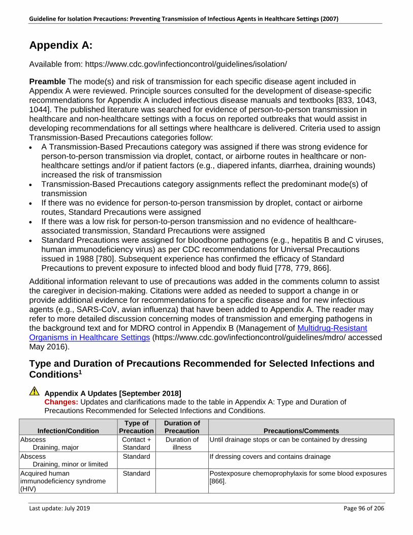
Guideline for Isolation Precautions: Preventing Transmission of Infectious Agents in Healthcare Settings (2007)
Last update: July 2019 Page 96 of 206
Appendix A: Available from: https://www.cdc.gov/infectioncontrol/guidelines/isolation/
Preamble The mode(s) and risk of transmission for each specific disease agent included in Appendix A were reviewed. Principle sources consulted for the development of disease-specific recommendations for Appendix A included infectious disease manuals and textbooks [833, 1043, 1044]. The published literature was searched for evidence of person-to-person transmission in healthcare and non-healthcare settings with a focus on reported outbreaks that would assist in developing recommendations for all settings where healthcare is delivered. Criteria used to assign Transmission-Based Precautions categories follow: • A Transmission-Based Precautions category was assigned if there was strong evidence for
person-to-person transmission via droplet, contact, or airborne routes in healthcare or non-healthcare settings and/or if patient factors (e.g., diapered infants, diarrhea, draining wounds) increased the risk of transmission
• Transmission-Based Precautions category assignments reflect the predominant mode(s) of transmission
• If there was no evidence for person-to-person transmission by droplet, contact or airborne routes, Standard Precautions were assigned
• If there was a low risk for person-to-person transmission and no evidence of healthcare-associated transmission, Standard Precautions were assigned
• Standard Precautions were assigned for bloodborne pathogens (e.g., hepatitis B and C viruses, human immunodeficiency virus) as per CDC recommendations for Universal Precautions issued in 1988 [780]. Subsequent experience has confirmed the efficacy of Standard Precautions to prevent exposure to infected blood and body fluid [778, 779, 866].
Additional information relevant to use of precautions was added in the comments column to assist the caregiver in decision-making. Citations were added as needed to support a change in or provide additional evidence for recommendations for a specific disease and for new infectious agents (e.g., SARS-CoV, avian influenza) that have been added to Appendix A. The reader may refer to more detailed discussion concerning modes of transmission and emerging pathogens in the background text and for MDRO control in Appendix B (Management of Multidrug-Resistant Organisms in Healthcare Settings (https://www.cdc.gov/infectioncontrol/guidelines/mdro/ accessed May 2016).
Type and Duration of Precautions Recommended for Selected Infections and Conditions1
Appendix A Updates [September 2018] Changes: Updates and clarifications made to the table in Appendix A: Type and Duration of Precautions Recommended for Selected Infections and Conditions.
Infection/Condition Type of
Precaution Duration of Precaution Precautions/Comments
Abscess Draining, major
Contact + Standard
Duration of illness
Until drainage stops or can be contained by dressing
Abscess Draining, minor or limited
Standard n/a If dressing covers and contains drainage
Acquired human immunodeficiency syndrome (HIV)
Standard n/a Postexposure chemoprophylaxis for some blood exposures [866].
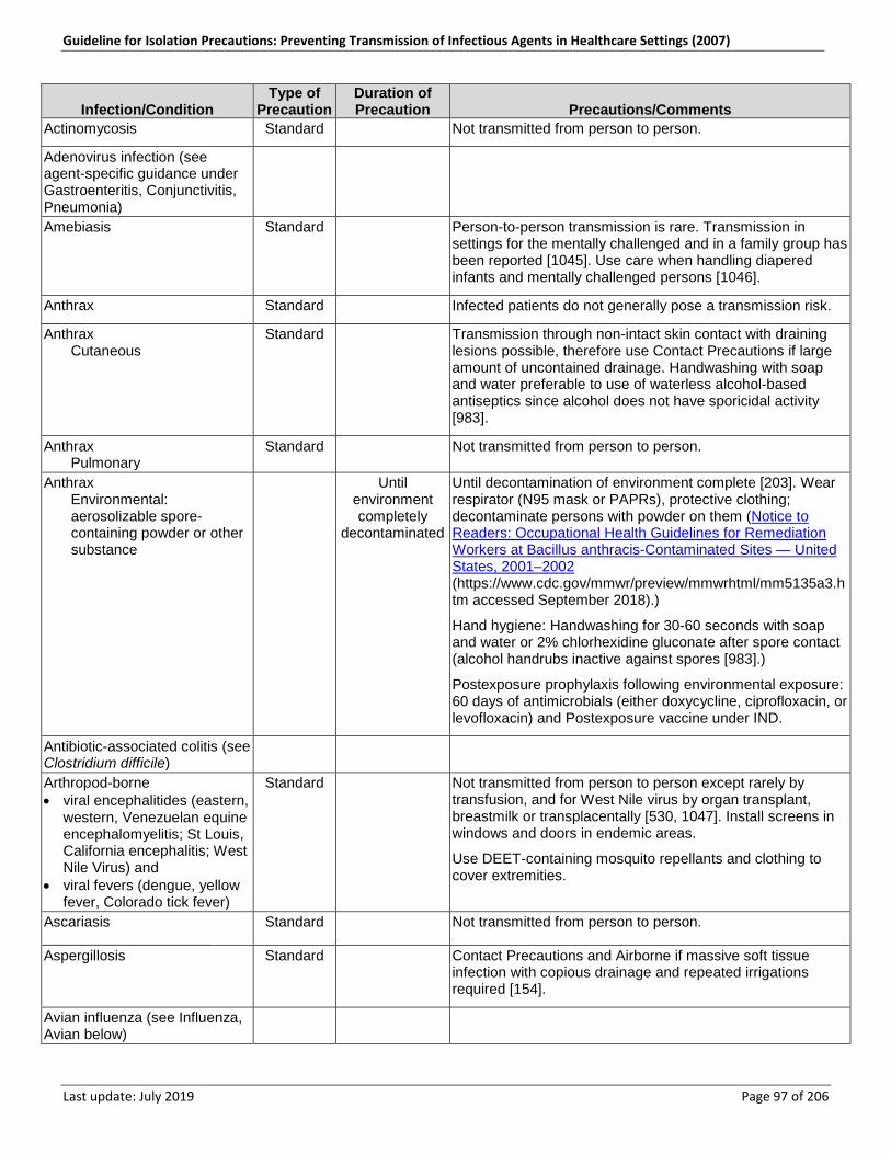
Guideline for Isolation Precautions: Preventing Transmission of Infectious Agents in Healthcare Settings (2007)
Last update: July 2019 Page 97 of 206
Infection/Condition Type of
Precaution Duration of Precaution Precautions/Comments
Actinomycosis Standard n/a Not transmitted from person to person.
Adenovirus infection (see agent-specific guidance under Gastroenteritis, Conjunctivitis, Pneumonia)
n/a n/a n/a
Amebiasis Standard n/a Person-to-person transmission is rare. Transmission in settings for the mentally challenged and in a family group has been reported [1045]. Use care when handling diapered infants and mentally challenged persons [1046].
Anthrax Standard n/a Infected patients do not generally pose a transmission risk.
Anthrax Cutaneous
Standard n/a Transmission through non-intact skin contact with draining lesions possible, therefore use Contact Precautions if large amount of uncontained drainage. Handwashing with soap and water preferable to use of waterless alcohol-based antiseptics since alcohol does not have sporicidal activity [983].
Anthrax Pulmonary
Standard n/a Not transmitted from person to person.
Anthrax Environmental:
aerosolizable spore-containing powder or other substance
n/a Until environment completely
decontaminated
Until decontamination of environment complete [203]. Wear respirator (N95 mask or PAPRs), protective clothing; decontaminate persons with powder on them (Notice to Readers: Occupational Health Guidelines for Remediation Workers at Bacillus anthracis-Contaminated Sites — United States, 2001–2002 (https://www.cdc.gov/mmwr/preview/mmwrhtml/mm5135a3.htm accessed September 2018).)
Hand hygiene: Handwashing for 30-60 seconds with soap and water or 2% chlorhexidine gluconate after spore contact (alcohol handrubs inactive against spores [983].)
Postexposure prophylaxis following environmental exposure: 60 days of antimicrobials (either doxycycline, ciprofloxacin, or levofloxacin) and Postexposure vaccine under IND.
Antibiotic-associated colitis (see Clostridium difficile)
n/a n/a n/a
Arthropod-borne • viral encephalitides (eastern,
western, Venezuelan equine encephalomyelitis; St Louis, California encephalitis; West Nile Virus) and
• viral fevers (dengue, yellow fever, Colorado tick fever)
Standard n/a Not transmitted from person to person except rarely by transfusion, and for West Nile virus by organ transplant, breastmilk or transplacentally [530, 1047]. Install screens in windows and doors in endemic areas.
Use DEET-containing mosquito repellants and clothing to cover extremities.
Ascariasis •••••••••••••••••••
Standard n/a Not transmitted from person to person.
Aspergillosis Standard n/a Contact Precautions and Airborne if massive soft tissue infection with copious drainage and repeated irrigations required [154].
Avian influenza (see Influenza, Avian below)
n/a n/a n/a
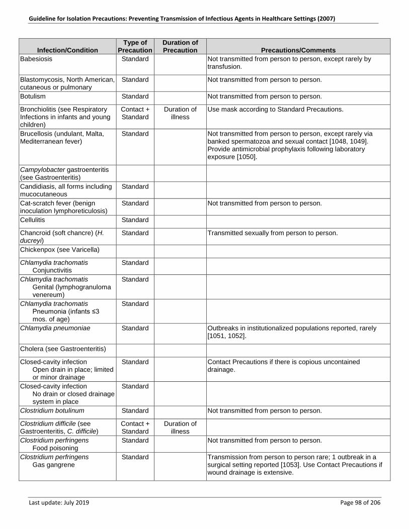
Guideline for Isolation Precautions: Preventing Transmission of Infectious Agents in Healthcare Settings (2007)
Last update: July 2019 Page 98 of 206
Infection/Condition Type of
Precaution Duration of Precaution Precautions/Comments
Babesiosis Standard n/a Not transmitted from person to person, except rarely by transfusion.
Blastomycosis, North American, cutaneous or pulmonary
Standard n/a Not transmitted from person to person.
Botulism Standard n/a Not transmitted from person to person.
Bronchiolitis (see Respiratory Infections in infants and young children)
Contact + Standard
Duration of illness
Use mask according to Standard Precautions.
Brucellosis (undulant, Malta, Mediterranean fever)
Standard n/a Not transmitted from person to person, except rarely via banked spermatozoa and sexual contact [1048, 1049]. Provide antimicrobial prophylaxis following laboratory exposure [1050].
Campylobacter gastroenteritis (see Gastroenteritis)
n/a n/a n/a
Candidiasis, all forms including mucocutaneous
Standard n/a n/a
Cat-scratch fever (benign inoculation lymphoreticulosis)
Standard n/a Not transmitted from person to person.
Cellulitis Standard n/a n/a
Chancroid (soft chancre) (H. ducreyi)
Standard n/a Transmitted sexually from person to person.
Chickenpox (see Varicella) n/a n/a n/a
Chlamydia trachomatis Conjunctivitis
Standard n/a n/a
Chlamydia trachomatis Genital (lymphogranuloma
venereum)
Standard n/a n/a
Chlamydia trachomatis Pneumonia (infants ≤3
mos. of age)
Standard n/a n/a
Chlamydia pneumoniae Standard n/a Outbreaks in institutionalized populations reported, rarely [1051, 1052].
Cholera (see Gastroenteritis) n/a n/a n/a
Closed-cavity infection Open drain in place; limited
or minor drainage
Standard n/a Contact Precautions if there is copious uncontained drainage.
Closed-cavity infection No drain or closed drainage
system in place
Standard n/a n/a
Clostridium botulinum Standard n/a Not transmitted from person to person.
Clostridium difficile (see Gastroenteritis, C. difficile)
Contact + Standard
Duration of illness
n/a
Clostridium perfringens Food poisoning
Standard n/a Not transmitted from person to person.
Clostridium perfringens Gas gangrene
Standard n/a Transmission from person to person rare; 1 outbreak in a surgical setting reported [1053]. Use Contact Precautions if wound drainage is extensive.
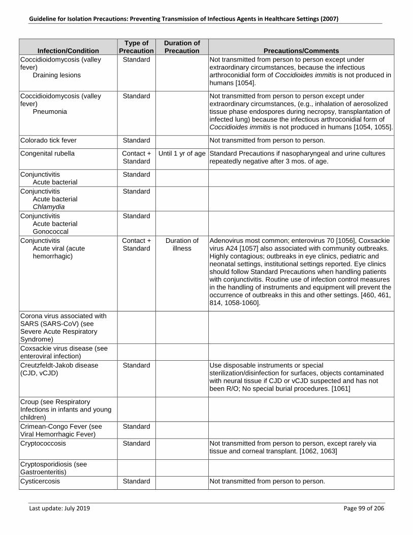
Guideline for Isolation Precautions: Preventing Transmission of Infectious Agents in Healthcare Settings (2007)
Last update: July 2019 Page 99 of 206
Infection/Condition Type of
Precaution Duration of Precaution Precautions/Comments
Coccidioidomycosis (valley fever) Draining lesions
Standard n/a Not transmitted from person to person except under extraordinary circumstances, because the infectious arthroconidial form of Coccidioides immitis is not produced in humans [1054].
Coccidioidomycosis (valley fever) Pneumonia
Standard n/a Not transmitted from person to person except under extraordinary circumstances, (e.g., inhalation of aerosolized tissue phase endospores during necropsy, transplantation of infected lung) because the infectious arthroconidial form of Coccidioides immitis is not produced in humans [1054, 1055].
Colorado tick fever Standard n/a Not transmitted from person to person.
Congenital rubella Contact + Standard
Until 1 yr of age Standard Precautions if nasopharyngeal and urine cultures repeatedly negative after 3 mos. of age.
Conjunctivitis Acute bacterial
Standard n/a n/a
Conjunctivitis Acute bacterial Chlamydia
Standard n/a n/a
Conjunctivitis Acute bacterial Gonococcal
Standard n/a n/a
Conjunctivitis Acute viral (acute
hemorrhagic)
Contact + Standard
Duration of illness
Adenovirus most common; enterovirus 70 [1056], Coxsackie virus A24 [1057] also associated with community outbreaks. Highly contagious; outbreaks in eye clinics, pediatric and neonatal settings, institutional settings reported. Eye clinics should follow Standard Precautions when handling patients with conjunctivitis. Routine use of infection control measures in the handling of instruments and equipment will prevent the occurrence of outbreaks in this and other settings. [460, 461, 814, 1058-1060].
Corona virus associated with SARS (SARS-CoV) (see Severe Acute Respiratory Syndrome)
n/a n/a n/a
Coxsackie virus disease (see enteroviral infection)
n/a n/a n/a
Creutzfeldt-Jakob disease (CJD, vCJD)
Standard n/a Use disposable instruments or special sterilization/disinfection for surfaces, objects contaminated with neural tissue if CJD or vCJD suspected and has not been R/O; No special burial procedures. [1061]
Croup (see Respiratory Infections in infants and young children)
n/a n/a n/a
Crimean-Congo Fever (see Viral Hemorrhagic Fever)
Standard n/a n/a
Cryptococcosis Standard n/a Not transmitted from person to person, except rarely via tissue and corneal transplant. [1062, 1063]
Cryptosporidiosis (see Gastroenteritis)
n/a n/a n/a
Cysticercosis Standard n/a Not transmitted from person to person.
Guideline for Isolation Precautions: Preventing Transmission of Infectious Agents in Healthcare Settings (2007)
Last update: July 2019 Page 100 of 206
Infection/Condition Type of
Precaution Duration of Precaution Precautions/Comments
Cytomegalovirus infection, including in neonates and immunosuppressed patients
Standard n/a No additional precautions for pregnant HCWs.
Decubitus ulcer (see Pressure Ulcer)
n/a n/a n/a
Dengue fever Standard n/a Not transmitted from person to person.
Diarrhea, acute-infective etiology suspected (see Gastroenteritis)
n/a n/a n/a
Diphtheria Cutaneous
Contact + Standard
Until off antimicrobial treatment and
culture-negative
Until 2 cultures taken 24 hours apart negative.
Diphtheria Pharyngeal
Droplet + Standard
Until off antimicrobial treatment and
culture-negative
Until 2 cultures taken 24 hours apart negative.
Ebola virus (see Viral Hemorrhagic Fevers)
n/a n/a Ebola Virus Disease for Healthcare Workers [2014]: Updated recommendations for healthcare workers can be found at Ebola: for Clinicians (https://www.cdc.gov/vhf/ebola/clinicians/index.html accessed September 2018).
Echinococcosis (hydatidosis) Standard n/a Not transmitted from person to person.
Echovirus (see Enteroviral Infection)
n/a n/a n/a
Encephalitis or encephalomyelitis (see specific etiologic agents)
n/a n/a n/a
Endometritis (endomyometritis) Standard n/a n/a
Enterobiasis (pinworm disease, oxyuriasis)
Standard n/a n/a
Enterococcus species (see Multidrug-Resistant Organisms if epidemiologically significant or vancomycin-resistant)
n/a n/a n/a
Enterocolitis, C. difficile (see Gastroenteritis, C. difficile)
n/a n/a n/a
Enteroviral infections (i.e., Group A and B Coxsackie viruses and Echo viruses) (excludes polio virus)
Standard n/a Use Contact Precautions for diapered or incontinent children for duration of illness and to control institutional outbreaks.
Epiglottitis, due to Haemophilus influenzae type b
Droplet + Standard
Until 24 hours after initiation of effective therapy
See specific disease agents for epiglottitis due to other etiologies.
Epstein-Barr virus infection, including infectious mononucleosis
Standard n/a n/a
Erythema infectiosum (also see Parvovirus B19)
n/a n/a n/a

Guideline for Isolation Precautions: Preventing Transmission of Infectious Agents in Healthcare Settings (2007)
Last update: July 2019 Page 101 of 206
Infection/Condition Type of
Precaution Duration of Precaution Precautions/Comments
Escherichia coli gastroenteritis (see gastroenteritis)
n/a n/a n/a
Food poisoning Botulism
Standard n/a Not transmitted from person to person.
Food poisoning C. perfringens or welchii
Standard n/a Not transmitted from person to person.
Food poisoning Staphylococcal
Standard n/a Not transmitted from person to person.
Furunculosis, staphylococcal Standard n/a Contact if drainage not controlled. Follow institutional policies if MRSA.
Furunculosis, staphylococcal Infants and young children
Contact + Standard
Duration of illness (with
wound lesions, until wounds stop draining)
n/a
Gangrene (gas gangrene) Standard n/a Not transmitted from person to person.
Gastroenteritis Standard n/a Use Contact Precautions for diapered or incontinent persons for the duration of illness or to control institutional outbreaks for gastroenteritis caused by all of the agents below.
Gastroenteritis Adenovirus
Standard n/a Use Contact Precautions for diapered or incontinent persons for the duration of illness or to control institutional outbreaks.
Gastroenteritis Campylobacter species
Standard n/a Use Contact Precautions for diapered or incontinent persons for the duration of illness or to control institutional outbreaks.
Gastroenteritis Cholera (Vibrio cholerae)
Standard n/a Use Contact Precautions for diapered or incontinent persons for the duration of illness or to control institutional outbreaks.
Gastroenteritis C. difficile
Contact + Standard
Duration of illness
Discontinue antibiotics if appropriate. Do not share electronic thermometers; [853, 854] ensure consistent environmental cleaning and disinfection. Hypochlorite solutions may be required for cleaning if transmission continues [847]. Handwashing with soap and water preferred because of the absence of sporicidal activity of alcohol in waterless antiseptic handrubs [983].
Gastroenteritis Cryptosporidium species
Standard n/a Use Contact Precautions for diapered or incontinent persons for the duration of illness or to control institutional outbreaks.
Gastroenteritis E. coli Enteropathogenic O157:H7 and other Shiga toxin-producing strains
Standard n/a Use Contact Precautions for diapered or incontinent persons for the duration of illness or to control institutional outbreaks.
Gastroenteritis E. coli Other species
Standard n/a Use Contact Precautions for diapered or incontinent persons for the duration of illness or to control institutional outbreaks.
Gastroenteritis Giardia lamblia
Standard n/a Use Contact Precautions for diapered or incontinent persons for the duration of illness or to control institutional outbreaks.
Guideline for Isolation Precautions: Preventing Transmission of Infectious Agents in Healthcare Settings (2007)
Last update: July 2019 Page 102 of 206
Infection/Condition Type of
Precaution Duration of Precaution Precautions/Comments
Gastroenteritis Noroviruses
Update
Contact + Standard
Use Contact Precautions for a minimum of 48 hours after the resolution of symptoms or to control institutional outbreaks.
Persons who clean areas heavily contaminated with feces or vomitus may benefit from wearing masks since virus can be aerosolized from these body substances [142, 147 148]; ensure consistent environmental cleaning and disinfection with focus on restrooms even when apparently unsoiled [273, 1064]. Hypochlorite solutions may be required when there is continued transmission [290-292]. Alcohol is less active, but there is no evidence that alcohol antiseptic handrubs are not effective for hand decontamination [294].
Cohorting of affected patients to separate airspaces and toilet facilities may help interrupt transmission during outbreaks.
Gastroenteritis, Noroviruses Precaution Update [May 2019]: The Type of Precaution was updated from “Standard” to “Contact + Standard” to align with Guideline for the Prevention and Control of Norovirus Gastroenteritis Outbreaks in Healthcare Settings (2011)
Gastroenteritis Rotavirus
Contact + Standard
Duration of illness
Ensure consistent environmental cleaning and disinfection and frequent removal of soiled diapers. Prolonged shedding may occur in both immunocompetent and immunocompromised children and the elderly [932, 933].
Gastroenteritis Salmonella species
(including S. typhi)
Standard n/a Use Contact Precautions for diapered or incontinent persons for the duration of illness or to control institutional outbreaks.
Gastroenteritis Shigella species (Bacillary
dysentery)
Standard n/a Use Contact Precautions for diapered or incontinent persons for the duration of illness or to control institutional outbreaks.
Gastroenteritis Vibrio parahaemolyticus
Standard n/a Use Contact Precautions for diapered or incontinent persons for the duration of illness or to control institutional outbreaks.
Gastroenteritis Viral (if not covered
elsewhere)
Standard n/a Use Contact Precautions for diapered or incontinent persons for the duration of illness or to control institutional outbreaks.
Gastroenteritis Yersinia enterocolitica
Standard n/a Use Contact Precautions for diapered or incontinent persons for the duration of illness or to control institutional outbreaks.
German measles (see Rubella; see Congenital Rubella)
n/a n/a n/a
Giardiasis (see Gastroenteritis) n/a n/a n/a Gonococcal ophthalmia neonatorum (gonorrheal ophthalmia, acute conjunctivitis of newborn)
Standard n/a n/a
Gonorrhea Standard n/a n/a Granuloma inguinale (Donovanosis, granuloma venereum)
Standard n/a n/a
Guillain-Barré syndrome Standard n/a Not an infectious condition. Haemophilus influenzae (see disease-specific recommendations)
n/a n/a n/a
Guideline for Isolation Precautions: Preventing Transmission of Infectious Agents in Healthcare Settings (2007)
Last update: July 2019 Page 103 of 206
Infection/Condition Type of
Precaution Duration of Precaution Precautions/Comments
Hand, foot, and mouth disease (see Enteroviral Infection)
n/a n/a n/a
Hansen’s Disease (see Leprosy)
n/a n/a n/a
Hantavirus pulmonary syndrome
Standard n/a Not transmitted from person to person.
Helicobacter pylori Standard n/a n/a Hepatitis, viral Type A
Standard n/a Provide hepatitis A vaccine postexposure as recommended. [1065]
Hepatitis, viral Type A-Diapered or
incontinent patients
Contact + Standard
n/a Maintain Contact Precautions in infants and children <3 years of age for duration of hospitalization; for children 3-14 yrs. of age for 2 weeks after onset of symptoms; >14 yrs. of age for 1 week after onset of symptoms [833, 1066, 1067].
Hepatitis, viral Type B-HBsAg positive;
acute or chronic
Standard n/a See specific recommendations for care of patients in hemodialysis centers. [778]
Hepatitis, viral Type C and other
unspecified non-A, non-B
Standard n/a See specific recommendations for care of patients in hemodialysis centers. [778]
Hepatitis, viral Type D (seen only with
hepatitis B)
Standard n/a n/a
Hepatitis, viral Type E
Standard n/a Use Contact Precautions for diapered or incontinent individuals for the duration of illness. [1068]
Hepatitis, viral Type G
Standard n/a n/a
Herpangina (see Enteroviral Infection)
n/a n/a n/a
Hookworm Standard n/a n/a Herpes simplex (Herpesvirus hominis) Encephalitis
Standard n/a n/a
Herpes simplex (Herpesvirus hominis) Mucocutaneous,
disseminated or primary, severe
Contact + Standard
Until lesions dry and crusted
n/a
Herpes simplex (Herpesvirus hominis) Mucocutaneous, recurrent
(skin, oral, genital)
Standard n/a n/a
Herpes simplex (Herpesvirus hominis) Neonatal
Contact + Standard
Until lesions dry and crusted
Also, for asymptomatic, exposed infants delivered vaginally or by C-section and if mother has active infection and membranes have been ruptured for more than 4 to 6 hours until infant surface cultures obtained at 24-36 hours of age negative after 48 hours incubation. [1069, 1070]
Guideline for Isolation Precautions: Preventing Transmission of Infectious Agents in Healthcare Settings (2007)
Last update: July 2019 Page 104 of 206
Infection/Condition Type of
Precaution Duration of Precaution Precautions/Comments
Herpes zoster (varicella-zoster) (shingles) Disseminated disease in
any patient Localized disease in
immunocompromised patient until disseminated infection ruled out
Airborne + Contact + Standard
Duration of illness
Susceptible HCWs should not enter room if immune caregivers are available; no recommendation for protection of immune HCWs; no recommendation for type of protection (i.e. surgical mask or respirator) for susceptible HCWs.
Herpes zoster (varicella-zoster) (shingles) Localized in patient with
intact immune system with lesions that can be contained/covered
Standard Until lesions dry and crusted
Susceptible HCWs should not provide direct patient care when other immune caregivers are available.
Histoplasmosis Standard n/a Not transmitted from person to person. Human immunodeficiency virus (HIV)
Standard n/a Postexposure chemoprophylaxis for some blood exposures [866].
Human metapneumovirus Contact + Standard
Duration of illness
HAI reported [1071], but route of transmission not established [823]. Assumed to be Contact transmission as for RSV since the viruses are closely related and have similar clinical manifestations and epidemiology. Wear masks according to Standard Precautions.
Impetigo Contact + Standard
Until 24 hours after initiation of effective therapy
n/a
Infectious mononucleosis Standard n/a n/a Influenza Human (seasonal
influenza)
n/a n/a See Prevention Strategies for Seasonal Influenza in Healthcare Settings (https://www.cdc.gov/flu/professionals/infectioncontrol/healthcaresettings.htm accessed September 2018). [Current version of this document may differ from original.] for current seasonal influenza guidance.
Influenza Avian (e.g., H5N1, H7, H9
strains)
n/a n/a See [This link is no longer active: www.cdc.gov/flu/avian/professional/infect-control.htm. Similar information may be found at Interim Guidance for Infection Control Within Healthcare Settings When Caring for Confirmed Cases, Probable Cases, and Cases Under Investigation for Infection with Novel Influenza A Viruses Associated with Severe Disease (https://www.cdc.gov/flu/avianflu/novel-flu-infection-control.htm accessed September 2018)] for current avian influenza guidance.
Influenza Pandemic Influenza (also a
human influenza virus)
Droplet + Standard
n/a See [This link is no longer active: http://www.pandemicflu.gov. Similar information may be found at Interim Guidance for Infection Control Within Healthcare Settings When Caring for Confirmed Cases, Probable Cases, and Cases Under Investigation for Infection with Novel Influenza A Viruses Associated with Severe Disease (https://www.cdc.gov/flu/avianflu/novel-flu-infection-control.htm accessed September 2018)] for current pandemic influenza guidance.
Kawasaki syndrome Standard n/a Not an infectious condition. Lassa fever (see Viral Hemorrhagic Fevers)
n/a n/a n/a
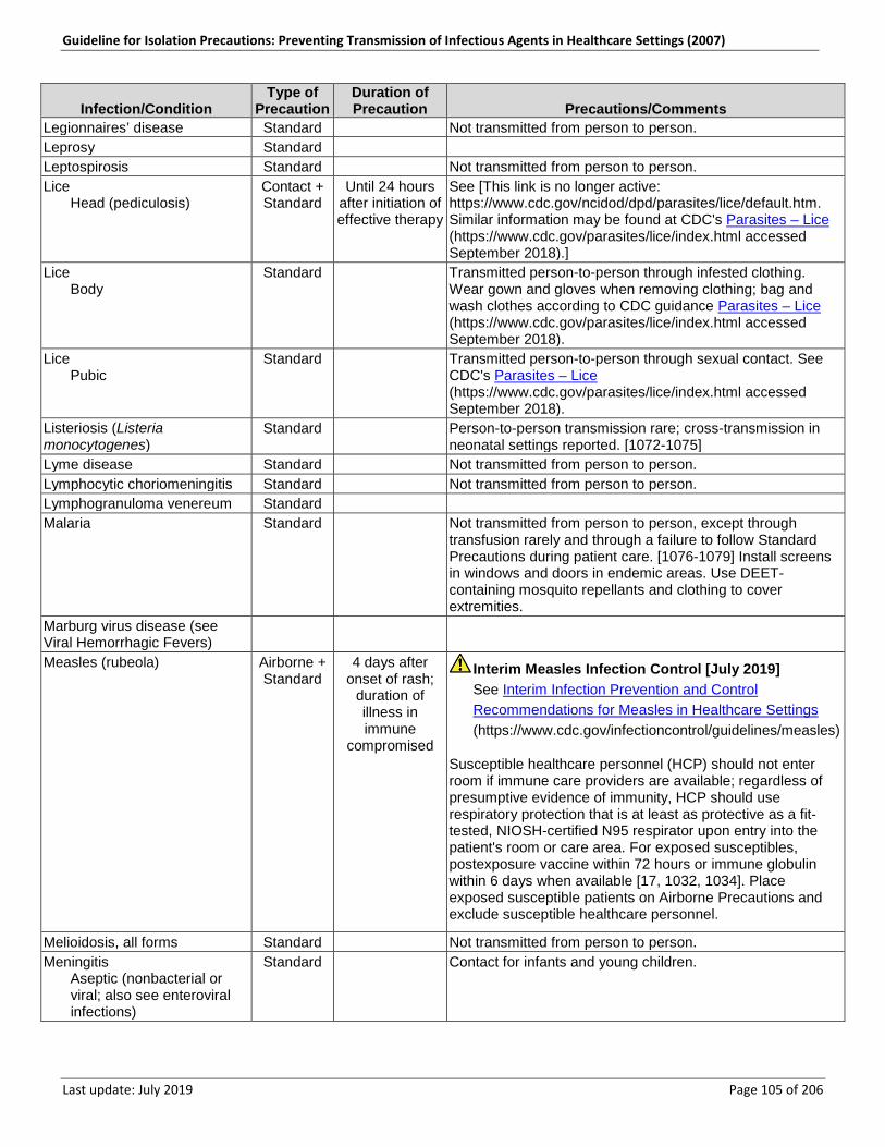
Guideline for Isolation Precautions: Preventing Transmission of Infectious Agents in Healthcare Settings (2007)
Last update: July 2019 Page 105 of 206
Infection/Condition Type of
Precaution Duration of Precaution Precautions/Comments
Legionnaires’ disease Standard n/a Not transmitted from person to person. Leprosy Standard n/a n/a Leptospirosis Standard n/a Not transmitted from person to person. Lice Head (pediculosis)
Contact + Standard
Until 24 hours after initiation of effective therapy
See [This link is no longer active: https://www.cdc.gov/ncidod/dpd/parasites/lice/default.htm. Similar information may be found at CDC's Parasites – Lice (https://www.cdc.gov/parasites/lice/index.html accessed September 2018).]
Lice Body
Standard n/a Transmitted person-to-person through infested clothing. Wear gown and gloves when removing clothing; bag and wash clothes according to CDC guidance Parasites – Lice (https://www.cdc.gov/parasites/lice/index.html accessed September 2018).
Lice Pubic
Standard n/a Transmitted person-to-person through sexual contact. See CDC's Parasites – Lice (https://www.cdc.gov/parasites/lice/index.html accessed September 2018).
Listeriosis (Listeria monocytogenes)
Standard n/a Person-to-person transmission rare; cross-transmission in neonatal settings reported. [1072-1075]
Lyme disease Standard n/a Not transmitted from person to person. Lymphocytic choriomeningitis Standard n/a Not transmitted from person to person. Lymphogranuloma venereum Standard n/a n/a Malaria Standard n/a Not transmitted from person to person, except through
transfusion rarely and through a failure to follow Standard Precautions during patient care. [1076-1079] Install screens in windows and doors in endemic areas. Use DEET- containing mosquito repellants and clothing to cover extremities.
Marburg virus disease (see Viral Hemorrhagic Fevers)
n/a n/a n/a
Measles (rubeola) Airborne + Standard
4 days after onset of rash;
duration of illness in immune
compromised
Interim Measles Infection Control [July 2019] See Interim Infection Prevention and Control Recommendations for Measles in Healthcare Settings (https://www.cdc.gov/infectioncontrol/guidelines/measles)
Susceptible healthcare personnel (HCP) should not enter room if immune care providers are available; regardless of presumptive evidence of immunity, HCP should use respiratory protection that is at least as protective as a fit-tested, NIOSH-certified N95 respirator upon entry into the patient's room or care area. For exposed susceptibles, postexposure vaccine within 72 hours or immune globulin within 6 days when available [17, 1032, 1034]. Place exposed susceptible patients on Airborne Precautions and exclude susceptible healthcare personnel.
Melioidosis, all forms Standard n/a Not transmitted from person to person. Meningitis Aseptic (nonbacterial or
viral; also see enteroviral infections)
Standard n/a Contact for infants and young children.
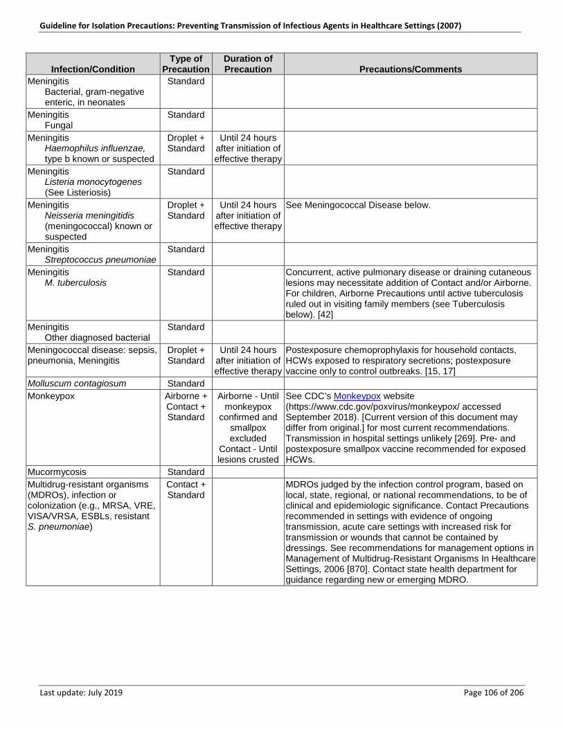
Guideline for Isolation Precautions: Preventing Transmission of Infectious Agents in Healthcare Settings (2007)
Last update: July 2019 Page 106 of 206
Infection/Condition Type of
Precaution Duration of Precaution Precautions/Comments
Meningitis Bacterial, gram-negative
enteric, in neonates
Standard n/a n/a
Meningitis Fungal
Standard n/a n/a
Meningitis Haemophilus influenzae,
type b known or suspected
Droplet + Standard
Until 24 hours after initiation of effective therapy
n/a
Meningitis Listeria monocytogenes
(See Listeriosis)
Standard n/a n/a
Meningitis Neisseria meningitidis
(meningococcal) known or suspected
Droplet + Standard
Until 24 hours after initiation of effective therapy
See Meningococcal Disease below.
Meningitis Streptococcus pneumoniae
Standard n/a n/a
Meningitis M. tuberculosis
Standard n/a Concurrent, active pulmonary disease or draining cutaneous lesions may necessitate addition of Contact and/or Airborne. For children, Airborne Precautions until active tuberculosis ruled out in visiting family members (see Tuberculosis below). [42]
Meningitis Other diagnosed bacterial
Standard n/a n/a
Meningococcal disease: sepsis, pneumonia, Meningitis
Droplet + Standard
Until 24 hours after initiation of effective therapy
Postexposure chemoprophylaxis for household contacts, HCWs exposed to respiratory secretions; postexposure vaccine only to control outbreaks. [15, 17]
Molluscum contagiosum Standard n/a n/a Monkeypox Airborne +
Contact + Standard
Airborne - Until monkeypox
confirmed and smallpox excluded
Contact - Until lesions crusted
See CDC’s Monkeypox website (https://www.cdc.gov/poxvirus/monkeypox/ accessed September 2018). [Current version of this document may differ from original.] for most current recommendations. Transmission in hospital settings unlikely [269]. Pre- and postexposure smallpox vaccine recommended for exposed HCWs.
Mucormycosis Standard n/a n/a Multidrug-resistant organisms (MDROs), infection or colonization (e.g., MRSA, VRE, VISA/VRSA, ESBLs, resistant S. pneumoniae)
Contact + Standard
n/a MDROs judged by the infection control program, based on local, state, regional, or national recommendations, to be of clinical and epidemiologic significance. Contact Precautions recommended in settings with evidence of ongoing transmission, acute care settings with increased risk for transmission or wounds that cannot be contained by dressings. See recommendations for management options in Management of Multidrug-Resistant Organisms In Healthcare Settings, 2006 [870]. Contact state health department for guidance regarding new or emerging MDRO.
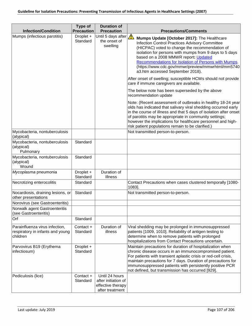
Guideline for Isolation Precautions: Preventing Transmission of Infectious Agents in Healthcare Settings (2007)
Last update: July 2019 Page 107 of 206
Infection/Condition Type of
Precaution Duration of Precaution Precautions/Comments
Mumps (infectious parotitis) Droplet + Standard
Until 5 days after the onset of
swelling
Mumps Update [October 2017]: The Healthcare Infection Control Practices Advisory Committee (HICPAC) voted to change the recommendation of isolation for persons with mumps from 9 days to 5 days based on a 2008 MMWR report: Updated Recommendations for Isolation of Persons with Mumps. (https://www.cdc.gov/mmwr/preview/mmwrhtml/mm5740a3.htm accessed September 2018).
After onset of swelling; susceptible HCWs should not provide care if immune caregivers are available.
The below note has been superseded by the above recommendation update
Note: (Recent assessment of outbreaks in healthy 18-24 year olds has indicated that salivary viral shedding occurred early in the course of illness and that 5 days of isolation after onset of parotitis may be appropriate in community settings; however the implications for healthcare personnel and high-risk patient populations remain to be clarified.)
Mycobacteria, nontuberculosis (atypical)
n/a n/a Not transmitted person-to-person.
Mycobacteria, nontuberculosis (atypical) Pulmonary
Standard n/a
Mycobacteria, nontuberculosis (atypical) Wound
Standard n/a n/a
Mycoplasma pneumonia Droplet + Standard
Duration of Illness
n/a
Necrotizing enterocolitis Standard n/a Contact Precautions when cases clustered temporally [1080-1083].
Nocardiosis, draining lesions, or other presentations
Standard n/a Not transmitted person-to-person.
Norovirus (see Gastroenteritis) n/a n/a n/a Norwalk agent Gastroenteritis (see Gastroenteritis)
n/a n/a n/a
Orf Standard n/a n/a
Parainfluenza virus infection, respiratory in infants and young children
Contact + Standard
Duration of illness
Viral shedding may be prolonged in immunosuppressed patients [1009, 1010]. Reliability of antigen testing to determine when to remove patients with prolonged hospitalizations from Contact Precautions uncertain.
Parvovirus B19 (Erythema infectiosum)
Droplet + Standard
n/a Maintain precautions for duration of hospitalization when chronic disease occurs in an immunocompromised patient. For patients with transient aplastic crisis or red-cell crisis, maintain precautions for 7 days. Duration of precautions for immunosuppressed patients with persistently positive PCR not defined, but transmission has occurred [929].
Pediculosis (lice) Contact + Standard
Until 24 hours after initiation of effective therapy after treatment
n/a
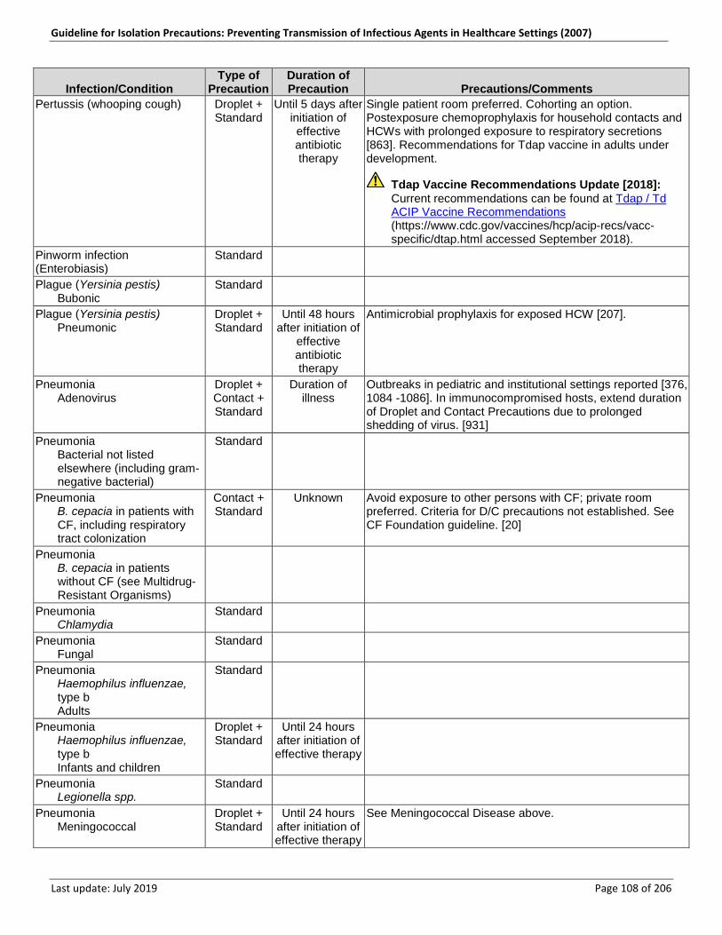
Guideline for Isolation Precautions: Preventing Transmission of Infectious Agents in Healthcare Settings (2007)
Last update: July 2019 Page 108 of 206
Infection/Condition Type of
Precaution Duration of Precaution Precautions/Comments
Pertussis (whooping cough) Droplet + Standard
Until 5 days after initiation of effective antibiotic therapy
Single patient room preferred. Cohorting an option. Postexposure chemoprophylaxis for household contacts and HCWs with prolonged exposure to respiratory secretions [863]. Recommendations for Tdap vaccine in adults under development.
Tdap Vaccine Recommendations Update [2018]: Current recommendations can be found at Tdap / Td ACIP Vaccine Recommendations (https://www.cdc.gov/vaccines/hcp/acip-recs/vacc-specific/dtap.html accessed September 2018).
Pinworm infection (Enterobiasis)
Standard n/a n/a
Plague (Yersinia pestis) Bubonic
Standard n/a n/a
Plague (Yersinia pestis) Pneumonic
Droplet + Standard
Until 48 hours after initiation of
effective antibiotic therapy
Antimicrobial prophylaxis for exposed HCW [207].
Pneumonia Adenovirus
Droplet + Contact + Standard
Duration of illness
Outbreaks in pediatric and institutional settings reported [376, 1084 -1086]. In immunocompromised hosts, extend duration of Droplet and Contact Precautions due to prolonged shedding of virus. [931]
Pneumonia Bacterial not listed
elsewhere (including gram-negative bacterial)
Standard n/a n/a
Pneumonia B. cepacia in patients with
CF, including respiratory tract colonization
Contact + Standard
Unknown Avoid exposure to other persons with CF; private room preferred. Criteria for D/C precautions not established. See CF Foundation guideline. [20]
Pneumonia B. cepacia in patients
without CF (see Multidrug-Resistant Organisms)
n/a n/a n/a
Pneumonia Chlamydia
Standard n/a n/a
Pneumonia Fungal
Standard n/a n/a
Pneumonia Haemophilus influenzae,
type b Adults
Standard n/a n/a
Pneumonia Haemophilus influenzae,
type b Infants and children
Droplet + Standard
Until 24 hours after initiation of effective therapy
n/a
Pneumonia Legionella spp.
Standard n/a n/a
Pneumonia Meningococcal
Droplet + Standard
Until 24 hours after initiation of effective therapy
See Meningococcal Disease above.
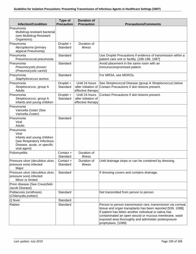
Guideline for Isolation Precautions: Preventing Transmission of Infectious Agents in Healthcare Settings (2007)
Last update: July 2019 Page 109 of 206
Infection/Condition Type of
Precaution Duration of Precaution Precautions/Comments
Pneumonia Multidrug-resistant bacterial
(see Multidrug-Resistant Organisms)
n/a n/a n/a
Pneumonia Mycoplasma (primary
atypical Pneumonia)
Droplet + Standard
Duration of illness
n/a
Pneumonia Pneumococcal pneumonia
Standard n/a Use Droplet Precautions if evidence of transmission within a patient care unit or facility. [196-198, 1087]
Pneumonia Pneumocystis jiroveci
(Pneumocystis carinii)
Standard n/a Avoid placement in the same room with an immunocompromised patient.
Pneumonia Staphylococcus aureus
Standard n/a For MRSA, see MDROs.
Pneumonia Streptococcus, group A Adults
Droplet + Standard
Until 24 hours after initiation of effective therapy
See Streptococcal Disease (group A Streptococcus) below Contact Precautions if skin lesions present.
Pneumonia Streptococcus, group A Infants and young children
Droplet + Standard
Until 24 hours after initiation of effective therapy
Contact Precautions if skin lesions present.
Pneumonia Varicella-Zoster (See
Varicella-Zoster)
n/a n/a n/a
Pneumonia Viral Adults
Standard n/a n/a
Pneumonia Viral Infants and young children
(see Respiratory Infectious Disease, acute, or specific viral agent)
n/a n/a n/a
Poliomyelitis Contact + Standard
Duration of illness
n/a
Pressure ulcer (decubitus ulcer, pressure sore) infected Major
Contact + Standard
Duration of illness
Until drainage stops or can be contained by dressing.
Pressure ulcer (decubitus ulcer, pressure sore) infected Minor or limited
Standard n/a If dressing covers and contains drainage.
Prion disease (See Creutzfeld-Jacob Disease)
n/a n/a n/a
Psittacosis (ornithosis) (Chlamydia psittaci)
Standard n/a Not transmitted from person to person.
Q fever Standard n/a n/a Rabies Standard n/a Person to person transmission rare; transmission via corneal,
tissue and organ transplants has been reported [539, 1088]. If patient has bitten another individual or saliva has contaminated an open wound or mucous membrane, wash exposed area thoroughly and administer postexposure prophylaxis. [1089]
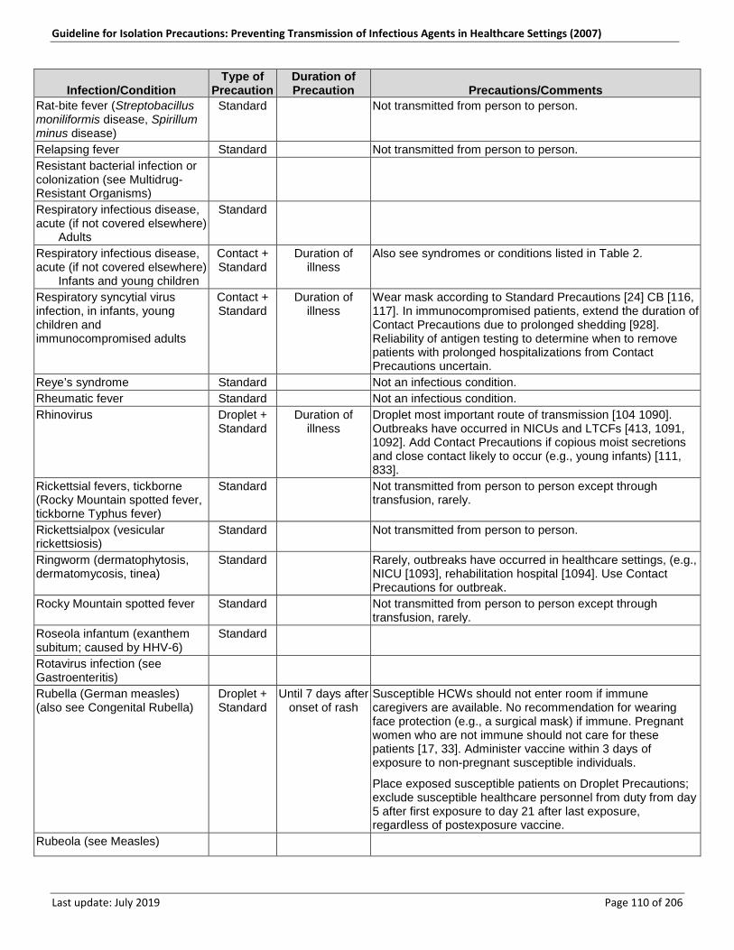
Guideline for Isolation Precautions: Preventing Transmission of Infectious Agents in Healthcare Settings (2007)
Last update: July 2019 Page 110 of 206
Infection/Condition Type of
Precaution Duration of Precaution Precautions/Comments
Rat-bite fever (Streptobacillus moniliformis disease, Spirillum minus disease)
Standard n/a Not transmitted from person to person.
Relapsing fever Standard n/a Not transmitted from person to person. Resistant bacterial infection or colonization (see Multidrug-Resistant Organisms)
n/a n/a n/a
Respiratory infectious disease, acute (if not covered elsewhere) Adults
Standard n/a n/a
Respiratory infectious disease, acute (if not covered elsewhere) Infants and young children
Contact + Standard
Duration of illness
Also see syndromes or conditions listed in Table 2.
Respiratory syncytial virus infection, in infants, young children and immunocompromised adults
Contact + Standard
Duration of illness
Wear mask according to Standard Precautions [24] CB [116, 117]. In immunocompromised patients, extend the duration of Contact Precautions due to prolonged shedding [928]. Reliability of antigen testing to determine when to remove patients with prolonged hospitalizations from Contact Precautions uncertain.
Reye’s syndrome Standard n/a Not an infectious condition. Rheumatic fever Standard n/a Not an infectious condition. Rhinovirus Droplet +
Standard Duration of
illness Droplet most important route of transmission [104 1090]. Outbreaks have occurred in NICUs and LTCFs [413, 1091, 1092]. Add Contact Precautions if copious moist secretions and close contact likely to occur (e.g., young infants) [111, 833].
Rickettsial fevers, tickborne (Rocky Mountain spotted fever, tickborne Typhus fever)
Standard n/a Not transmitted from person to person except through transfusion, rarely.
Rickettsialpox (vesicular rickettsiosis)
Standard n/a Not transmitted from person to person.
Ringworm (dermatophytosis, dermatomycosis, tinea)
Standard n/a Rarely, outbreaks have occurred in healthcare settings, (e.g., NICU [1093], rehabilitation hospital [1094]. Use Contact Precautions for outbreak.
Rocky Mountain spotted fever Standard n/a Not transmitted from person to person except through transfusion, rarely.
Roseola infantum (exanthem subitum; caused by HHV-6)
Standard n/a n/a
Rotavirus infection (see Gastroenteritis)
n/a n/a n/a
Rubella (German measles) (also see Congenital Rubella)
Droplet + Standard
Until 7 days after onset of rash
Susceptible HCWs should not enter room if immune caregivers are available. No recommendation for wearing face protection (e.g., a surgical mask) if immune. Pregnant women who are not immune should not care for these patients [17, 33]. Administer vaccine within 3 days of exposure to non-pregnant susceptible individuals.
Place exposed susceptible patients on Droplet Precautions; exclude susceptible healthcare personnel from duty from day 5 after first exposure to day 21 after last exposure, regardless of postexposure vaccine.
Rubeola (see Measles) n/a n/a n/a
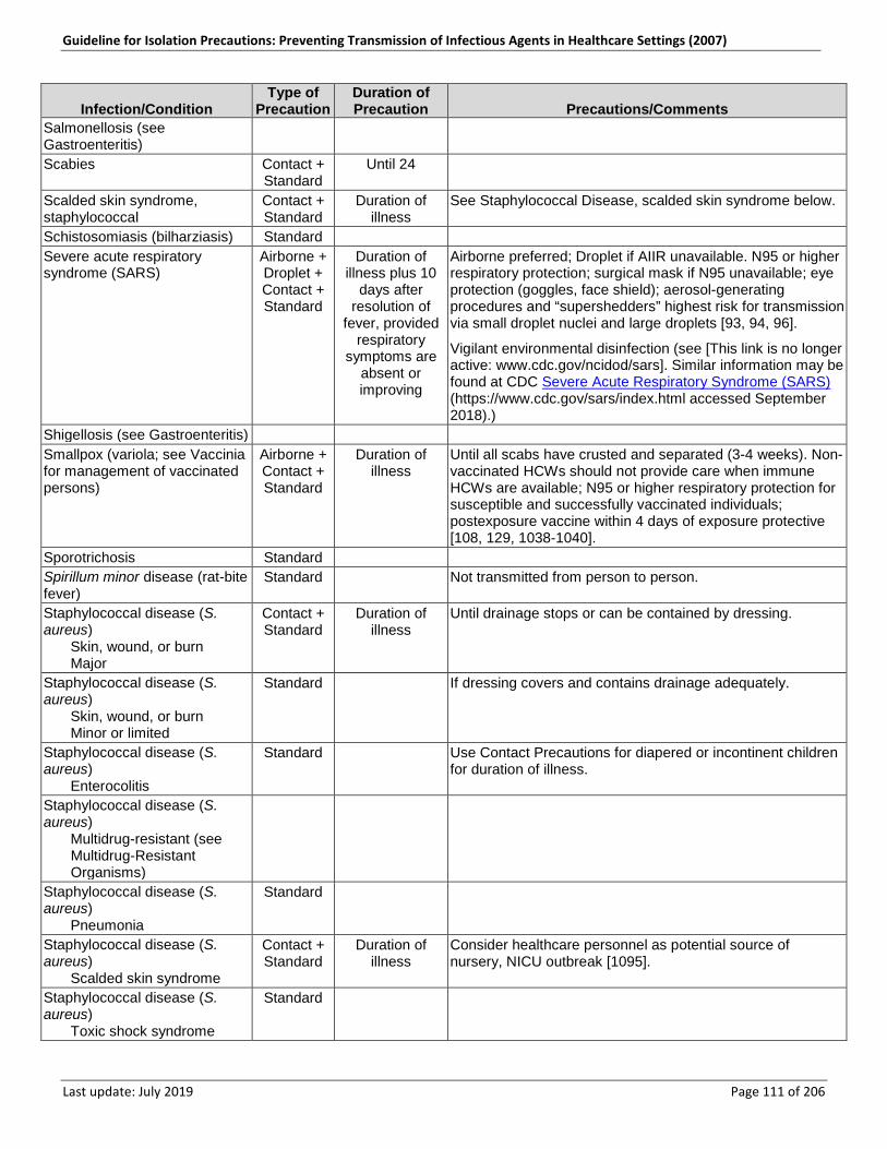
Guideline for Isolation Precautions: Preventing Transmission of Infectious Agents in Healthcare Settings (2007)
Last update: July 2019 Page 111 of 206
Infection/Condition Type of
Precaution Duration of Precaution Precautions/Comments
Salmonellosis (see Gastroenteritis)
n/a n/a n/a
Scabies Contact + Standard
Until 24 n/a
Scalded skin syndrome, staphylococcal
Contact + Standard
Duration of illness
See Staphylococcal Disease, scalded skin syndrome below.
Schistosomiasis (bilharziasis) Standard n/a n/a Severe acute respiratory syndrome (SARS)
Airborne + Droplet + Contact + Standard
Duration of illness plus 10
days after resolution of
fever, provided respiratory
symptoms are absent or improving
Airborne preferred; Droplet if AIIR unavailable. N95 or higher respiratory protection; surgical mask if N95 unavailable; eye protection (goggles, face shield); aerosol-generating procedures and “supershedders” highest risk for transmission via small droplet nuclei and large droplets [93, 94, 96].
Vigilant environmental disinfection (see [This link is no longer active: www.cdc.gov/ncidod/sars]. Similar information may be found at CDC Severe Acute Respiratory Syndrome (SARS) (https://www.cdc.gov/sars/index.html accessed September 2018).)
Shigellosis (see Gastroenteritis) n/a n/a n/a Smallpox (variola; see Vaccinia for management of vaccinated persons)
Airborne + Contact + Standard
Duration of illness
Until all scabs have crusted and separated (3-4 weeks). Non-vaccinated HCWs should not provide care when immune HCWs are available; N95 or higher respiratory protection for susceptible and successfully vaccinated individuals; postexposure vaccine within 4 days of exposure protective [108, 129, 1038-1040].
Sporotrichosis Standard n/a n/a Spirillum minor disease (rat-bite fever)
Standard n/a Not transmitted from person to person.
Staphylococcal disease (S. aureus) Skin, wound, or burn Major
Contact + Standard
Duration of illness
Until drainage stops or can be contained by dressing.
Staphylococcal disease (S. aureus) Skin, wound, or burn Minor or limited
Standard If dressing covers and contains drainage adequately.
Staphylococcal disease (S. aureus) Enterocolitis
Standard n/a Use Contact Precautions for diapered or incontinent children for duration of illness.
Staphylococcal disease (S. aureus) Multidrug-resistant (see
Multidrug-Resistant Organisms)
n/a n/a n/a
Staphylococcal disease (S. aureus) Pneumonia
Standard n/a n/a
Staphylococcal disease (S. aureus) Scalded skin syndrome
Contact + Standard
Duration of illness
Consider healthcare personnel as potential source of nursery, NICU outbreak [1095].
Staphylococcal disease (S. aureus) Toxic shock syndrome
Standard n/a n/a

Guideline for Isolation Precautions: Preventing Transmission of Infectious Agents in Healthcare Settings (2007)
Last update: July 2019 Page 112 of 206
Infection/Condition Type of
Precaution Duration of Precaution Precautions/Comments
Streptobacillus moniliformis disease (rat-bite fever)
Standard n/a Not transmitted from person to person.
Streptococcal disease (group A Streptococcus) Skin, wound, or burn Major
Contact + Droplet + Standard
Until 24 hours after initiation of effective therapy
Until drainage stops or can be contained by dressing.
Streptococcal disease (group A Streptococcus) Skin, wound, or burn Minor or limited
Standard n/a If dressing covers and contains drainage.
Streptococcal disease (group A Streptococcus) Endometritis (puerperal
sepsis)
Standard n/a n/a
Streptococcal disease (group A Streptococcus) Pharyngitis in infants and
young children
Droplet + Standard
Until 24 hours after initiation of effective therapy
n/a
Streptococcal disease (group A Streptococcus) Pneumonia
Droplet + Standard
Until 24 hours after initiation of effective therapy
n/a
Streptococcal disease (group A Streptococcus) Scarlet fever in infants and
young children
Droplet + Standard
Until 24 hours after initiation of effective therapy
n/a
Streptococcal disease (group A Streptococcus) Serious invasive disease
Droplet + Standard
Until 24 hours after initiation of effective therapy
Outbreaks of serious invasive disease have occurred secondary to transmission among patients and healthcare personnel [162, 972, 1096-1098].
Contact Precautions for draining wound as above; follow recommendations for antimicrobial prophylaxis in selected conditions [160].
Streptococcal disease (group B Streptococcus), neonatal
Standard n/a n/a
Streptococcal disease (not group A or B) unless covered elsewhere Multidrug-resistant (see
Multidrug-Resistant Organisms)
n/a n/a n/a
Strongyloidiasis Standard n/a n/a
Syphilis Latent (tertiary) and
seropositivity without lesions
Standard n/a n/a
Syphilis Skin and mucous
membrane, including congenital, primary, Secondary
Standard n/a n/a
Tapeworm disease Hymenolepis nana
Standard n/a Not transmitted from person to person.
Tapeworm disease Taenia solium (pork)
Standard n/a n/a
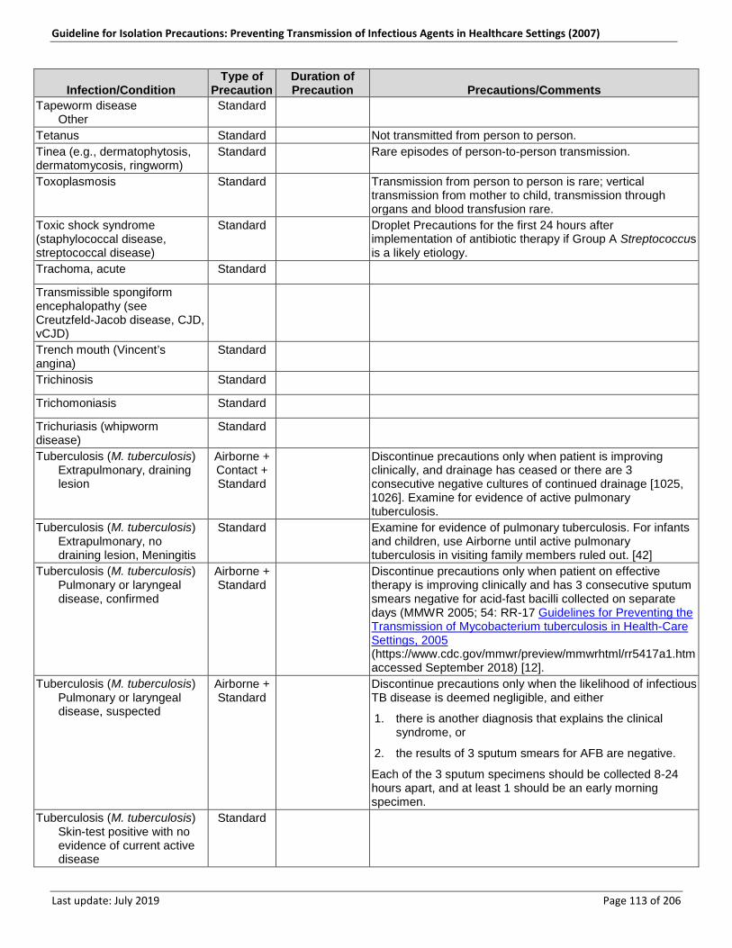
Guideline for Isolation Precautions: Preventing Transmission of Infectious Agents in Healthcare Settings (2007)
Last update: July 2019 Page 113 of 206
Infection/Condition Type of
Precaution Duration of Precaution Precautions/Comments
Tapeworm disease Other
Standard n/a n/a
Tetanus Standard n/a Not transmitted from person to person. Tinea (e.g., dermatophytosis, dermatomycosis, ringworm)
Standard n/a Rare episodes of person-to-person transmission.
Toxoplasmosis Standard n/a Transmission from person to person is rare; vertical transmission from mother to child, transmission through organs and blood transfusion rare.
Toxic shock syndrome (staphylococcal disease, streptococcal disease)
Standard n/a Droplet Precautions for the first 24 hours after implementation of antibiotic therapy if Group A Streptococcus is a likely etiology.
Trachoma, acute Standard n/a n/a
Transmissible spongiform encephalopathy (see Creutzfeld-Jacob disease, CJD, vCJD)
n/a n/a n/a
Trench mouth (Vincent’s angina)
Standard n/a n/a
Trichinosis Standard n/a n/a
Trichomoniasis Standard n/a n/a
Trichuriasis (whipworm disease)
Standard n/a n/a
Tuberculosis (M. tuberculosis) Extrapulmonary, draining
lesion
Airborne + Contact + Standard
n/a Discontinue precautions only when patient is improving clinically, and drainage has ceased or there are 3 consecutive negative cultures of continued drainage [1025, 1026]. Examine for evidence of active pulmonary tuberculosis.
Tuberculosis (M. tuberculosis) Extrapulmonary, no
draining lesion, Meningitis
Standard n/a Examine for evidence of pulmonary tuberculosis. For infants and children, use Airborne until active pulmonary tuberculosis in visiting family members ruled out. [42]
Tuberculosis (M. tuberculosis) Pulmonary or laryngeal
disease, confirmed
Airborne + Standard
n/a Discontinue precautions only when patient on effective therapy is improving clinically and has 3 consecutive sputum smears negative for acid-fast bacilli collected on separate days (MMWR 2005; 54: RR-17 Guidelines for Preventing the Transmission of Mycobacterium tuberculosis in Health-Care Settings, 2005 (https://www.cdc.gov/mmwr/preview/mmwrhtml/rr5417a1.htm accessed September 2018) [12].
Tuberculosis (M. tuberculosis) Pulmonary or laryngeal
disease, suspected
Airborne + Standard
n/a Discontinue precautions only when the likelihood of infectious TB disease is deemed negligible, and either
1. there is another diagnosis that explains the clinical syndrome, or
2. the results of 3 sputum smears for AFB are negative.
Each of the 3 sputum specimens should be collected 8-24 hours apart, and at least 1 should be an early morning specimen.
Tuberculosis (M. tuberculosis) Skin-test positive with no
evidence of current active disease
Standard n/a n/a
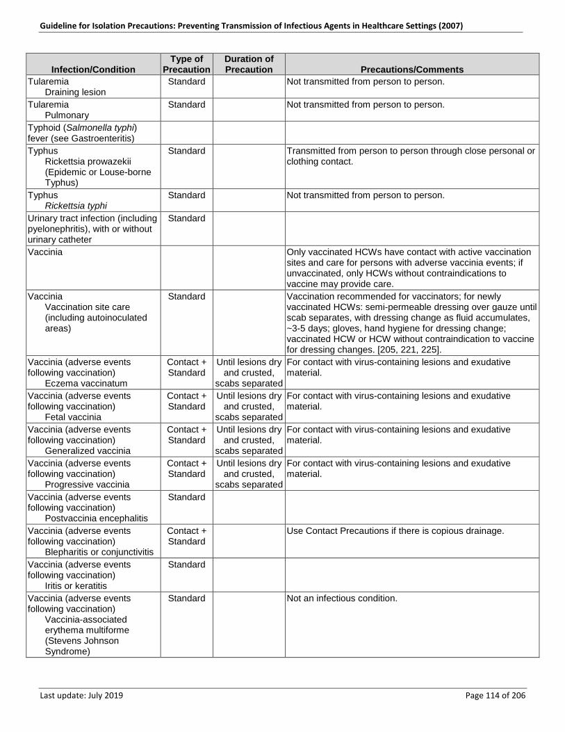
Guideline for Isolation Precautions: Preventing Transmission of Infectious Agents in Healthcare Settings (2007)
Last update: July 2019 Page 114 of 206
Infection/Condition Type of
Precaution Duration of Precaution Precautions/Comments
Tularemia Draining lesion
Standard n/a Not transmitted from person to person.
Tularemia Pulmonary
Standard n/a Not transmitted from person to person.
Typhoid (Salmonella typhi) fever (see Gastroenteritis)
n/a n/a n/a
Typhus Rickettsia prowazekii
(Epidemic or Louse-borne Typhus)
Standard n/a Transmitted from person to person through close personal or clothing contact.
Typhus Rickettsia typhi
Standard n/a Not transmitted from person to person.
Urinary tract infection (including pyelonephritis), with or without urinary catheter
Standard n/a n/a
Vaccinia n/a n/a Only vaccinated HCWs have contact with active vaccination sites and care for persons with adverse vaccinia events; if unvaccinated, only HCWs without contraindications to vaccine may provide care.
Vaccinia Vaccination site care
(including autoinoculated areas)
Standard n/a Vaccination recommended for vaccinators; for newly vaccinated HCWs: semi-permeable dressing over gauze until scab separates, with dressing change as fluid accumulates, ~3-5 days; gloves, hand hygiene for dressing change; vaccinated HCW or HCW without contraindication to vaccine for dressing changes. [205, 221, 225].
Vaccinia (adverse events following vaccination) Eczema vaccinatum
Contact + Standard
Until lesions dry and crusted,
scabs separated
For contact with virus-containing lesions and exudative material.
Vaccinia (adverse events following vaccination) Fetal vaccinia
Contact + Standard
Until lesions dry and crusted,
scabs separated
For contact with virus-containing lesions and exudative material.
Vaccinia (adverse events following vaccination) Generalized vaccinia
Contact + Standard
Until lesions dry and crusted,
scabs separated
For contact with virus-containing lesions and exudative material.
Vaccinia (adverse events following vaccination) Progressive vaccinia
Contact + Standard
Until lesions dry and crusted,
scabs separated
For contact with virus-containing lesions and exudative material.
Vaccinia (adverse events following vaccination) Postvaccinia encephalitis
Standard n/a n/a
Vaccinia (adverse events following vaccination) Blepharitis or conjunctivitis
Contact + Standard
n/a Use Contact Precautions if there is copious drainage.
Vaccinia (adverse events following vaccination) Iritis or keratitis
Standard n/a n/a
Vaccinia (adverse events following vaccination) Vaccinia-associated
erythema multiforme (Stevens Johnson Syndrome)
Standard n/a Not an infectious condition.
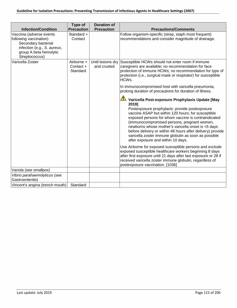
Guideline for Isolation Precautions: Preventing Transmission of Infectious Agents in Healthcare Settings (2007)
Last update: July 2019 Page 115 of 206
Infection/Condition Type of
Precaution Duration of Precaution Precautions/Comments
Vaccinia (adverse events following vaccination) Secondary bacterial
infection (e.g., S. aureus, group A beta hemolytic Streptococcus)
Standard + Contact
n/a Follow organism-specific (strep, staph most frequent) recommendations and consider magnitude of drainage.
Varicella Zoster Airborne + Contact + Standard
Until lesions dry and crusted
Susceptible HCWs should not enter room if immune caregivers are available; no recommendation for face protection of immune HCWs; no recommendation for type of protection (i.e., surgical mask or respirator) for susceptible HCWs.
In immunocompromised host with varicella pneumonia, prolong duration of precautions for duration of illness.
Varicella Post-exposure Prophylaxis Update [May 2019] Postexposure prophylaxis: provide postexposure vaccine ASAP but within 120 hours; for susceptible exposed persons for whom vaccine is contraindicated (immunocompromised persons, pregnant women, newborns whose mother’s varicella onset is <5 days before delivery or within 48 hours after delivery) provide varicella zoster immune globulin as soon as possible after exposure and within 10 days.
Use Airborne for exposed susceptible persons and exclude exposed susceptible healthcare workers beginning 8 days after first exposure until 21 days after last exposure or 28 if received varicella zoster immune globulin, regardless of postexposure vaccination. [1036]
Variola (see smallpox) n/a n/a n/a Vibrio parahaemolyticus (see Gastroenteritis)
n/a n/a n/a
Vincent’s angina (trench mouth) Standard n/a n/a
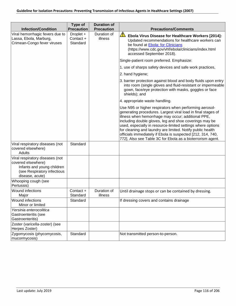
Guideline for Isolation Precautions: Preventing Transmission of Infectious Agents in Healthcare Settings (2007)
Last update: July 2019 Page 116 of 206
Infection/Condition Type of
Precaution Duration of Precaution Precautions/Comments
Viral hemorrhagic fevers due to Lassa, Ebola, Marburg, Crimean-Congo fever viruses
Droplet + Contact + Standard
Duration of illness
Ebola Virus Disease for Healthcare Workers [2014]: Updated recommendations for healthcare workers can be found at Ebola: for Clinicians (https://www.cdc.gov/vhf/ebola/clinicians/index.html accessed September 2018).
Single-patient room preferred. Emphasize:
1. use of sharps safety devices and safe work practices,
2. hand hygiene;
3. barrier protection against blood and body fluids upon entry into room (single gloves and fluid-resistant or impermeable gown, face/eye protection with masks, goggles or face shields); and
4. appropriate waste handling.
Use N95 or higher respirators when performing aerosol-generating procedures. Largest viral load in final stages of illness when hemorrhage may occur; additional PPE, including double gloves, leg and shoe coverings may be used, especially in resource-limited settings where options for cleaning and laundry are limited. Notify public health officials immediately if Ebola is suspected [212, 314, 740, 772]. Also see Table 3C for Ebola as a bioterrorism agent.
Viral respiratory diseases (not covered elsewhere) Adults
Standard
Viral respiratory diseases (not covered elsewhere) Infants and young children
(see Respiratory infectious disease, acute)
Whooping cough (see Pertussis)
n/a n/a
Wound infections Major
Contact + Standard
Duration of illness
Until drainage stops or can be contained by dressing.
Wound infections Minor or limited
Standard n/a If dressing covers and contains drainage
Yersinia enterocolitica Gastroenteritis (see Gastroenteritis)
n/a n/a n/a
Zoster (varicella-zoster) (see Herpes Zoster)
n/a n/a n/a
Zygomycosis (phycomycosis, mucormycosis)
Standard n/a Not transmitted person-to-person.
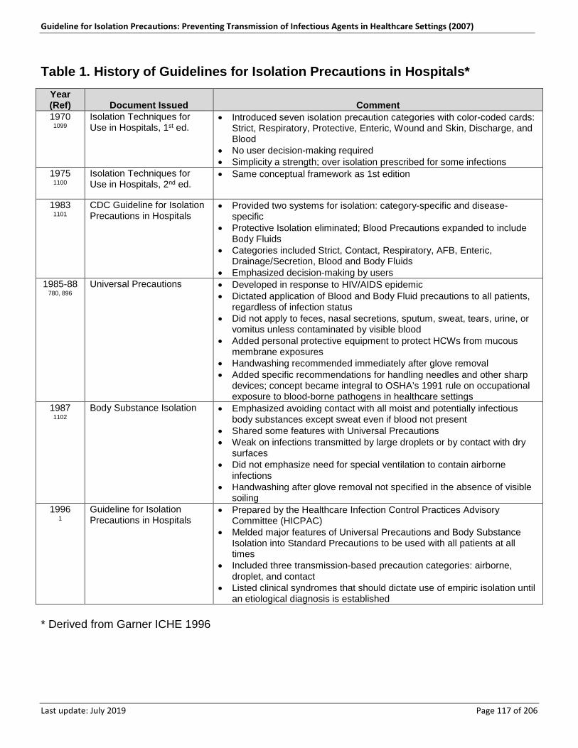
Guideline for Isolation Precautions: Preventing Transmission of Infectious Agents in Healthcare Settings (2007)
Last update: July 2019 Page 117 of 206
Table 1. History of Guidelines for Isolation Precautions in Hospitals* Year (Ref) Document Issued Comment 1970
1099 Isolation Techniques for Use in Hospitals, 1st ed.
• Introduced seven isolation precaution categories with color-coded cards: Strict, Respiratory, Protective, Enteric, Wound and Skin, Discharge, and Blood
• No user decision-making required • Simplicity a strength; over isolation prescribed for some infections
1975 1100
Isolation Techniques for Use in Hospitals, 2nd ed.
• Same conceptual framework as 1st edition
1983 1101
CDC Guideline for Isolation Precautions in Hospitals
• Provided two systems for isolation: category-specific and disease- specific
• Protective Isolation eliminated; Blood Precautions expanded to include Body Fluids
• Categories included Strict, Contact, Respiratory, AFB, Enteric, Drainage/Secretion, Blood and Body Fluids
• Emphasized decision-making by users 1985-88
780, 896 Universal Precautions • Developed in response to HIV/AIDS epidemic
• Dictated application of Blood and Body Fluid precautions to all patients, regardless of infection status
• Did not apply to feces, nasal secretions, sputum, sweat, tears, urine, or vomitus unless contaminated by visible blood
• Added personal protective equipment to protect HCWs from mucous membrane exposures
• Handwashing recommended immediately after glove removal • Added specific recommendations for handling needles and other sharp
devices; concept became integral to OSHA’s 1991 rule on occupational exposure to blood-borne pathogens in healthcare settings
1987 1102
Body Substance Isolation • Emphasized avoiding contact with all moist and potentially infectious body substances except sweat even if blood not present
• Shared some features with Universal Precautions • Weak on infections transmitted by large droplets or by contact with dry
surfaces • Did not emphasize need for special ventilation to contain airborne
infections • Handwashing after glove removal not specified in the absence of visible
soiling 1996
1 Guideline for Isolation Precautions in Hospitals
• Prepared by the Healthcare Infection Control Practices Advisory Committee (HICPAC)
• Melded major features of Universal Precautions and Body Substance Isolation into Standard Precautions to be used with all patients at all times
• Included three transmission-based precaution categories: airborne, droplet, and contact
• Listed clinical syndromes that should dictate use of empiric isolation until an etiological diagnosis is established
* Derived from Garner ICHE 1996
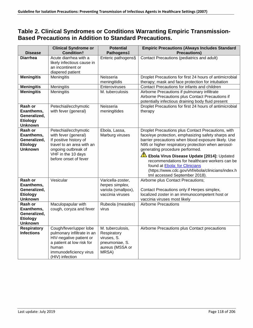
Guideline for Isolation Precautions: Preventing Transmission of Infectious Agents in Healthcare Settings (2007)
Last update: July 2019 Page 118 of 206
Table 2. Clinical Syndromes or Conditions Warranting Empiric Transmission-Based Precautions in Addition to Standard Precautions.
Disease Clinical Syndrome or
Condition† Potential
Pathogens‡ Empiric Precautions (Always Includes Standard
Precautions) Diarrhea Acute diarrhea with a
likely infectious cause in an incontinent or diapered patient
Enteric pathogens§ Contact Precautions (pediatrics and adult)
Meningitis Meningitis Neisseria meningitidis
Droplet Precautions for first 24 hours of antimicrobial therapy; mask and face protection for intubation
Meningitis Meningitis Enteroviruses Contact Precautions for infants and children Meningitis Meningitis M. tuberculosis Airborne Precautions if pulmonary infiltrate
Airborne Precautions plus Contact Precautions if potentially infectious draining body fluid present
Rash or Exanthems, Generalized, Etiology Unknown
Petechial/ecchymotic with fever (general)
Neisseria meningitides
Droplet Precautions for first 24 hours of antimicrobial therapy
Rash or Exanthems, Generalized, Etiology Unknown
Petechial/ecchymotic with fever (general) If positive history of travel to an area with an ongoing outbreak of VHF in the 10 days before onset of fever
Ebola, Lassa, Marburg viruses
Droplet Precautions plus Contact Precautions, with face/eye protection, emphasizing safety sharps and barrier precautions when blood exposure likely. Use N95 or higher respiratory protection when aerosol-generating procedure performed.
Ebola Virus Disease Update [2014]: Updated recommendations for healthcare workers can be found at Ebola: for Clinicians (https://www.cdc.gov/vhf/ebola/clinicians/index.html accessed September 2018).
Rash or Exanthems, Generalized, Etiology Unknown
Vesicular Varicella-zoster, herpes simplex, variola (smallpox), vaccinia viruses
Airborne plus Contact Precautions; Contact Precautions only if Herpes simplex, localized zoster in an immunocompetent host or vaccinia viruses most likely
Rash or Exanthems, Generalized, Etiology Unknown
Maculopapular with cough, coryza and fever
Rubeola (measles) virus
Airborne Precautions
Respiratory Infections
Cough/fever/upper lobe pulmonary infiltrate in an HIV-negative patient or a patient at low risk for human immunodeficiency virus (HIV) infection
M. tuberculosis, Respiratory viruses, S. pneumoniae, S. aureus (MSSA or MRSA)
Airborne Precautions plus Contact precautions
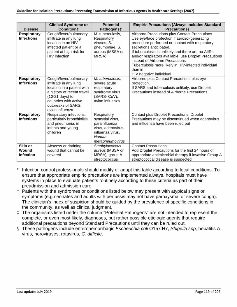
Guideline for Isolation Precautions: Preventing Transmission of Infectious Agents in Healthcare Settings (2007)
Last update: July 2019 Page 119 of 206
Disease Clinical Syndrome or
Condition† Potential
Pathogens‡ Empiric Precautions (Always Includes Standard
Precautions) Respiratory Infections
Cough/fever/pulmonary infiltrate in any lung location in an HIV-infected patient or a patient at high risk for HIV infection
M. tuberculosis, Respiratory viruses, S. pneumoniae, S. aureus (MSSA or MRSA)
Airborne Precautions plus Contact Precautions Use eye/face protection if aerosol-generating procedure performed or contact with respiratory secretions anticipated. If tuberculosis is unlikely and there are no AIIRs and/or respirators available, use Droplet Precautions instead of Airborne Precautions Tuberculosis more likely in HIV-infected individual than in HIV negative individual
Respiratory Infections
Cough/fever/pulmonary infiltrate in any lung location in a patient with a history of recent travel (10-21 days) to countries with active outbreaks of SARS, avian influenza
M. tuberculosis, severe acute respiratory syndrome virus (SARS- CoV), avian influenza
Airborne plus Contact Precautions plus eye protection. If SARS and tuberculosis unlikely, use Droplet Precautions instead of Airborne Precautions.
Respiratory Infections
Respiratory infections, particularly bronchiolitis and pneumonia, in infants and young children
Respiratory syncytial virus, parainfluenza virus, adenovirus, influenza virus, Human metapneumovirus
Contact plus Droplet Precautions; Droplet Precautions may be discontinued when adenovirus and influenza have been ruled out
Skin or Wound Infection
Abscess or draining wound that cannot be covered
Staphylococcus aureus (MSSA or MRSA), group A streptococcus
Contact Precautions Add Droplet Precautions for the first 24 hours of appropriate antimicrobial therapy if invasive Group A streptococcal disease is suspected
* Infection control professionals should modify or adapt this table according to local conditions. To
ensure that appropriate empiric precautions are implemented always, hospitals must have systems in place to evaluate patients routinely according to these criteria as part of their preadmission and admission care.
† Patients with the syndromes or conditions listed below may present with atypical signs or symptoms (e.g.neonates and adults with pertussis may not have paroxysmal or severe cough). The clinician's index of suspicion should be guided by the prevalence of specific conditions in the community, as well as clinical judgment.
‡ The organisms listed under the column "Potential Pathogens" are not intended to represent the complete, or even most likely, diagnoses, but rather possible etiologic agents that require additional precautions beyond Standard Precautions until they can be ruled out.
§ These pathogens include enterohemorrhagic Escherichia coli O157:H7, Shigella spp, hepatitis A virus, noroviruses, rotavirus, C. difficile.
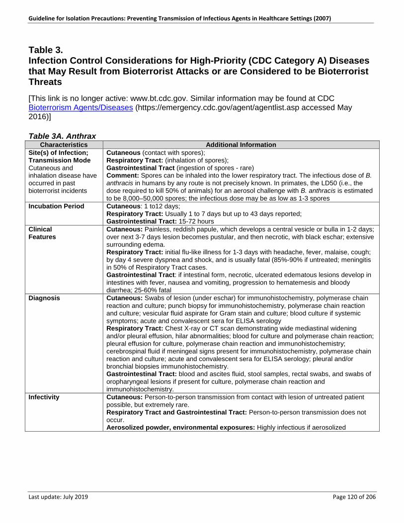
Guideline for Isolation Precautions: Preventing Transmission of Infectious Agents in Healthcare Settings (2007)
Last update: July 2019 Page 120 of 206
Table 3. Infection Control Considerations for High-Priority (CDC Category A) Diseases that May Result from Bioterrorist Attacks or are Considered to be Bioterrorist Threats [This link is no longer active: www.bt.cdc.gov. Similar information may be found at CDC Bioterrorism Agents/Diseases (https://emergency.cdc.gov/agent/agentlist.asp accessed May 2016)] Table 3A. Anthrax
Characteristics Additional Information Site(s) of Infection; Transmission Mode Cutaneous and inhalation disease have occurred in past bioterrorist incidents
Cutaneous (contact with spores); Respiratory Tract: (inhalation of spores); Gastrointestinal Tract (ingestion of spores - rare) Comment: Spores can be inhaled into the lower respiratory tract. The infectious dose of B. anthracis in humans by any route is not precisely known. In primates, the LD50 (i.e., the dose required to kill 50% of animals) for an aerosol challenge with B. anthracis is estimated to be 8,000–50,000 spores; the infectious dose may be as low as 1-3 spores
Incubation Period Cutaneous: 1 to12 days; Respiratory Tract: Usually 1 to 7 days but up to 43 days reported; Gastrointestinal Tract: 15-72 hours
Clinical Features
Cutaneous: Painless, reddish papule, which develops a central vesicle or bulla in 1-2 days; over next 3-7 days lesion becomes pustular, and then necrotic, with black eschar; extensive surrounding edema. Respiratory Tract: initial flu-like illness for 1-3 days with headache, fever, malaise, cough; by day 4 severe dyspnea and shock, and is usually fatal (85%-90% if untreated; meningitis in 50% of Respiratory Tract cases. Gastrointestinal Tract: if intestinal form, necrotic, ulcerated edematous lesions develop in intestines with fever, nausea and vomiting, progression to hematemesis and bloody diarrhea; 25-60% fatal
Diagnosis Cutaneous: Swabs of lesion (under eschar) for immunohistochemistry, polymerase chain reaction and culture; punch biopsy for immunohistochemistry, polymerase chain reaction and culture; vesicular fluid aspirate for Gram stain and culture; blood culture if systemic symptoms; acute and convalescent sera for ELISA serology Respiratory Tract: Chest X-ray or CT scan demonstrating wide mediastinal widening and/or pleural effusion, hilar abnormalities; blood for culture and polymerase chain reaction; pleural effusion for culture, polymerase chain reaction and immunohistochemistry; cerebrospinal fluid if meningeal signs present for immunohistochemistry, polymerase chain reaction and culture; acute and convalescent sera for ELISA serology; pleural and/or bronchial biopsies immunohistochemistry. Gastrointestinal Tract: blood and ascites fluid, stool samples, rectal swabs, and swabs of oropharyngeal lesions if present for culture, polymerase chain reaction and immunohistochemistry.
Infectivity Cutaneous: Person-to-person transmission from contact with lesion of untreated patient possible, but extremely rare. Respiratory Tract and Gastrointestinal Tract: Person-to-person transmission does not occur. Aerosolized powder, environmental exposures: Highly infectious if aerosolized
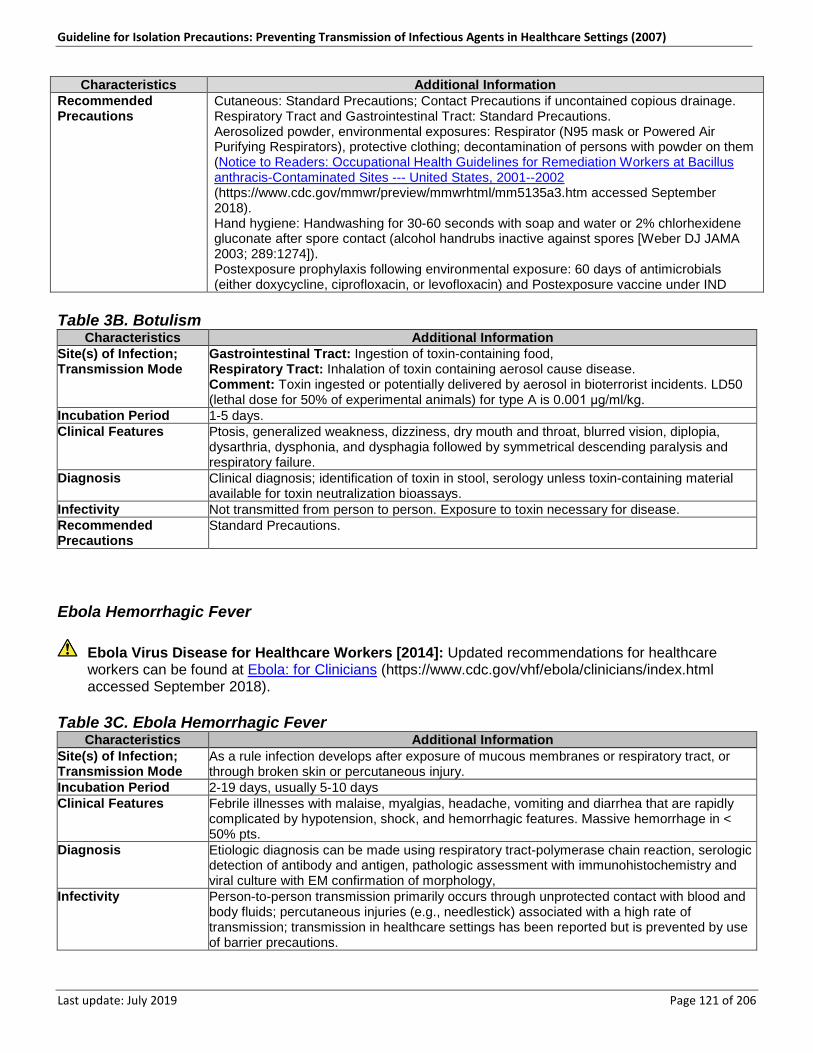
Guideline for Isolation Precautions: Preventing Transmission of Infectious Agents in Healthcare Settings (2007)
Last update: July 2019 Page 121 of 206
Characteristics Additional Information Recommended Precautions
Cutaneous: Standard Precautions; Contact Precautions if uncontained copious drainage. Respiratory Tract and Gastrointestinal Tract: Standard Precautions. Aerosolized powder, environmental exposures: Respirator (N95 mask or Powered Air Purifying Respirators), protective clothing; decontamination of persons with powder on them (Notice to Readers: Occupational Health Guidelines for Remediation Workers at Bacillus anthracis-Contaminated Sites --- United States, 2001--2002 (https://www.cdc.gov/mmwr/preview/mmwrhtml/mm5135a3.htm accessed September 2018). Hand hygiene: Handwashing for 30-60 seconds with soap and water or 2% chlorhexidene gluconate after spore contact (alcohol handrubs inactive against spores [Weber DJ JAMA 2003; 289:1274]). Postexposure prophylaxis following environmental exposure: 60 days of antimicrobials (either doxycycline, ciprofloxacin, or levofloxacin) and Postexposure vaccine under IND
Table 3B. Botulism
Characteristics Additional Information Site(s) of Infection; Transmission Mode
Gastrointestinal Tract: Ingestion of toxin-containing food, Respiratory Tract: Inhalation of toxin containing aerosol cause disease. Comment: Toxin ingested or potentially delivered by aerosol in bioterrorist incidents. LD50 (lethal dose for 50% of experimental animals) for type A is 0.001 μg/ml/kg.
Incubation Period 1-5 days. Clinical Features Ptosis, generalized weakness, dizziness, dry mouth and throat, blurred vision, diplopia,
dysarthria, dysphonia, and dysphagia followed by symmetrical descending paralysis and respiratory failure.
Diagnosis Clinical diagnosis; identification of toxin in stool, serology unless toxin-containing material available for toxin neutralization bioassays.
Infectivity Not transmitted from person to person. Exposure to toxin necessary for disease. Recommended Precautions
Standard Precautions.
Ebola Hemorrhagic Fever
Ebola Virus Disease for Healthcare Workers [2014]: Updated recommendations for healthcare workers can be found at Ebola: for Clinicians (https://www.cdc.gov/vhf/ebola/clinicians/index.html accessed September 2018).
Table 3C. Ebola Hemorrhagic Fever
Characteristics Additional Information Site(s) of Infection; Transmission Mode
As a rule infection develops after exposure of mucous membranes or respiratory tract, or through broken skin or percutaneous injury.
Incubation Period 2-19 days, usually 5-10 days Clinical Features Febrile illnesses with malaise, myalgias, headache, vomiting and diarrhea that are rapidly
complicated by hypotension, shock, and hemorrhagic features. Massive hemorrhage in < 50% pts.
Diagnosis Etiologic diagnosis can be made using respiratory tract-polymerase chain reaction, serologic detection of antibody and antigen, pathologic assessment with immunohistochemistry and viral culture with EM confirmation of morphology,
Infectivity Person-to-person transmission primarily occurs through unprotected contact with blood and body fluids; percutaneous injuries (e.g., needlestick) associated with a high rate of transmission; transmission in healthcare settings has been reported but is prevented by use of barrier precautions.
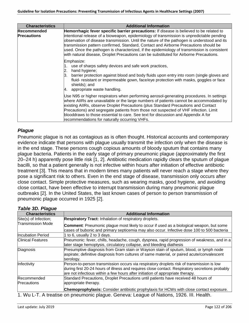
Guideline for Isolation Precautions: Preventing Transmission of Infectious Agents in Healthcare Settings (2007)
Last update: July 2019 Page 122 of 206
Characteristics Additional Information Recommended Precautions
Hemorrhagic fever specific barrier precautions: If disease is believed to be related to intentional release of a bioweapon, epidemiology of transmission is unpredictable pending observation of disease transmission. Until the nature of the pathogen is understood and its transmission pattern confirmed, Standard, Contact and Airborne Precautions should be used. Once the pathogen is characterized, if the epidemiology of transmission is consistent with natural disease, Droplet Precautions can be substituted for Airborne Precautions.
Emphasize: 1. use of sharps safety devices and safe work practices, 2. hand hygiene; 3. barrier protection against blood and body fluids upon entry into room (single gloves and
fluid- resistant or impermeable gown, face/eye protection with masks, goggles or face shields); and
4. appropriate waste handling.
Use N95 or higher respirators when performing aerosol-generating procedures. In settings where AIIRs are unavailable or the large numbers of patients cannot be accommodated by existing AIIRs, observe Droplet Precautions (plus Standard Precautions and Contact Precautions) and segregate patients from those not suspected of VHF infection. Limit blooddraws to those essential to care. See text for discussion and Appendix A for recommendations for naturally occurring VHFs.
Plague Pneumonic plague is not as contagious as is often thought. Historical accounts and contemporary evidence indicate that persons with plague usually transmit the infection only when the disease is in the end stage. These persons cough copious amounts of bloody sputum that contains many plague bacteria. Patients in the early stage of primary pneumonic plague (approximately the first 20–24 h) apparently pose little risk [1, 2]. Antibiotic medication rapidly clears the sputum of plague bacilli, so that a patient generally is not infective within hours after initiation of effective antibiotic treatment [3]. This means that in modern times many patients will never reach a stage where they pose a significant risk to others. Even in the end stage of disease, transmission only occurs after close contact. Simple protective measures, such as wearing masks, good hygiene, and avoiding close contact, have been effective to interrupt transmission during many pneumonic plague outbreaks [2]. In the United States, the last known cases of person to person transmission of pneumonic plague occurred in 1925 [2].
Table 3D. Plague Characteristics Additional Information
Site(s) of Infection; Transmission Mode
Respiratory Tract: Inhalation of respiratory droplets.
Comment: Pneumonic plague most likely to occur if used as a biological weapon, but some cases of bubonic and primary septicemia may also occur. Infective dose 100 to 500 bacteria
Incubation Period 1 to 6, usually 2 to 3 days. Clinical Features Pneumonic: fever, chills, headache, cough, dyspnea, rapid progression of weakness, and in a
later stage hemoptysis, circulatory collapse, and bleeding diathesis Diagnosis Presumptive diagnosis from Gram stain or Wayson stain of sputum, blood, or lymph node
aspirate; definitive diagnosis from cultures of same material, or paired acute/convalescent serology.
Infectivity Person-to-person transmission occurs via respiratory droplets risk of transmission is low during first 20-24 hours of illness and requires close contact. Respiratory secretions probably are not infectious within a few hours after initiation of appropriate therapy.
Recommended Precautions
Standard Precautions, Droplet Precautions until patients have received 48 hours of appropriate therapy.
Chemoprophylaxis: Consider antibiotic prophylaxis for HCWs with close contact exposure. 1. Wu L-T. A treatise on pneumonic plague. Geneva: League of Nations, 1926. III. Health.
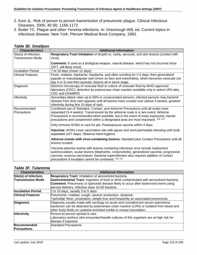
Guideline for Isolation Precautions: Preventing Transmission of Infectious Agents in Healthcare Settings (2007)
Last update: July 2019 Page 123 of 206
2. Kool JL. Risk of person to person transmission of pneumonic plague. Clinical Infectious Diseases, 2005; 40 (8): 1166-1172
3. Butler TC. Plague and other Yersinia infections. In: Greenough WB, ed. Current topics in infectious disease. New York: Plenum Medical Book Company, 1983.
Table 3E. Smallpox Characteristics Additional Information
Site(s) of Infection; Transmission Mode
Respiratory Tract Inhalation of droplet or, rarely, aerosols; and skin lesions (contact with virus).
Comment: If used as a biological weapon, natural disease, which has not occurred since 1977, will likely result.
Incubation Period 7 to 19 days (mean 12 days) Clinical Features Fever, malaise, backache, headache, and often vomiting for 2-3 days; then generalized
papular or maculopapular rash (more on face and extremities), which becomes vesicular (on day 4 or 5) and then pustular; lesions all in same stage.
Diagnosis Electron microscopy of vesicular fluid or culture of vesicular fluid by WHO approved laboratory (CDC); detection by polymerase chain reaction available only in select LRN labs, CDC and USAMRID
Infectivity Secondary attack rates up to 50% in unvaccinated persons; infected persons may transmit disease from time rash appears until all lesions have crusted over (about 3 weeks); greatest infectivity during first 10 days of rash.
Recommended Precautions
Combined use of Standard, Contact, and Airborne Precautions until all scabs have separated (3-4 weeks). Transmission by the airborne route is a rare event; Airborne Precautions is recommended when possible, but in the event of mass exposures, barrier precautions and containment within a designated area are most important. 204, 212
Only immune HCWs to care for pts; Postexposure vaccine within 4 days.
Vaccinia: HCWs cover vaccination site with gauze and semi-permeable dressing until scab separates (≥21 days). Observe hand hygiene.
Adverse events with virus-containing lesions: Standard plus Contact Precautions until all lesions crusted.
Vaccinia adverse events with lesions containing infectious virus include inadvertent autoinoculation, ocular lesions (blepharitis, conjunctivitis), generalized vaccinia, progressive vaccinia, eczema vaccinatum; bacterial superinfection also requires addition of contact precautions if exudates cannot be contained. 216, 217
Table 3F. Tularemia
Characteristics Additional Information Site(s) of Infection; Transmission Mode
Respiratory Tract: Inhalation of aerosolized bacteria. Gastrointestinal Tract: Ingestion of food or drink contaminated with aerosolized bacteria. Comment: Pneumonic or typhoidal disease likely to occur after bioterrorist event using aerosol delivery. Infective dose 10-50 bacteria
Incubation Period 2 to 10 days, usually 3 to 5 days Clinical Features Pneumonic: malaise, cough, sputum production, dyspnea;
Typhoidal: fever, prostration, weight loss and frequently an associated pneumonia. Diagnosis Diagnosis usually made with serology on acute and convalescent serum specimens;
bacterium can be detected by polymerase chain reaction (LRN) or isolated from blood and other body fluids on cysteine-enriched media or mouse inoculation.
Infectivity Person-to-person spread is rare. Laboratory workers who encounter/handle cultures of this organism are at high risk for disease if exposed.
Recommended Precautions
Standard Precautions
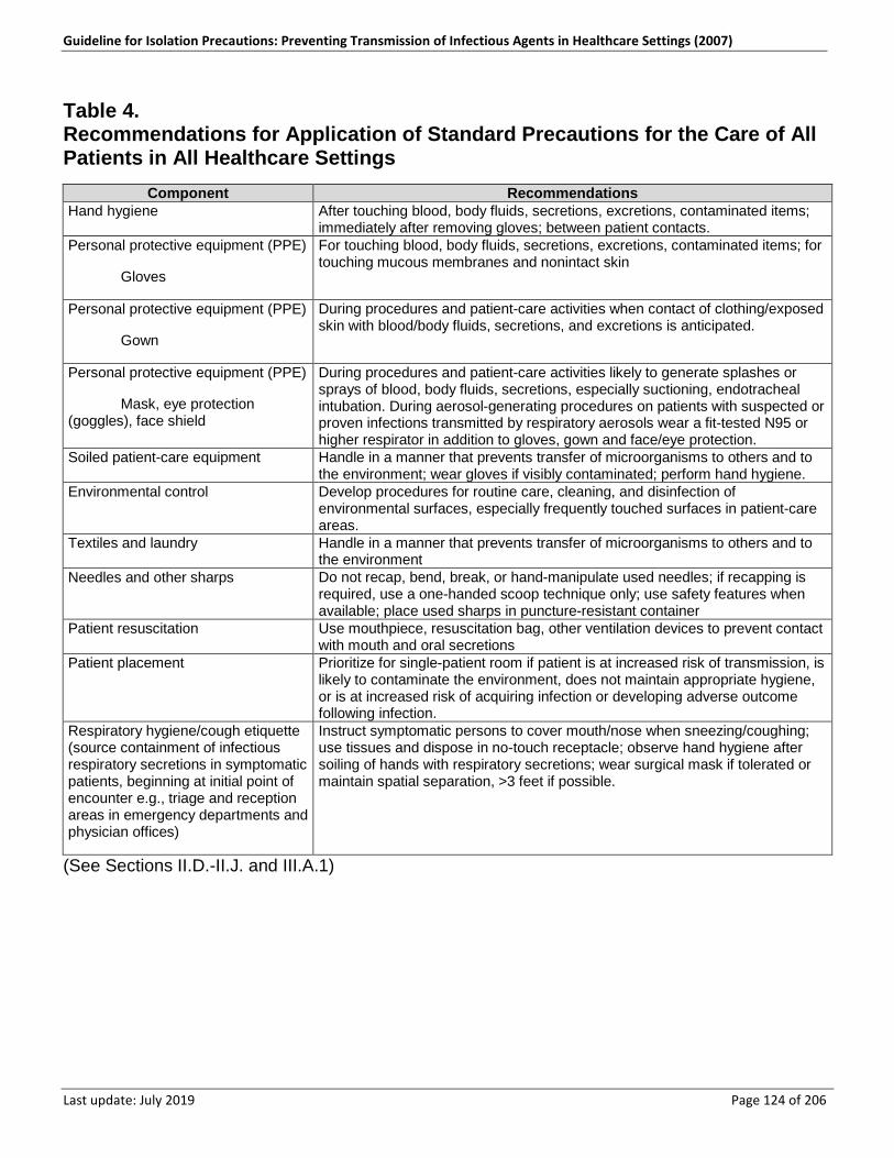
Guideline for Isolation Precautions: Preventing Transmission of Infectious Agents in Healthcare Settings (2007)
Last update: July 2019 Page 124 of 206
Table 4. Recommendations for Application of Standard Precautions for the Care of All Patients in All Healthcare Settings
Component Recommendations Hand hygiene After touching blood, body fluids, secretions, excretions, contaminated items;
immediately after removing gloves; between patient contacts. Personal protective equipment (PPE)
Gloves
For touching blood, body fluids, secretions, excretions, contaminated items; for touching mucous membranes and nonintact skin
Personal protective equipment (PPE)
Gown
During procedures and patient-care activities when contact of clothing/exposed skin with blood/body fluids, secretions, and excretions is anticipated.
Personal protective equipment (PPE)
Mask, eye protection (goggles), face shield
During procedures and patient-care activities likely to generate splashes or sprays of blood, body fluids, secretions, especially suctioning, endotracheal intubation. During aerosol-generating procedures on patients with suspected or proven infections transmitted by respiratory aerosols wear a fit-tested N95 or higher respirator in addition to gloves, gown and face/eye protection.
Soiled patient-care equipment Handle in a manner that prevents transfer of microorganisms to others and to the environment; wear gloves if visibly contaminated; perform hand hygiene.
Environmental control Develop procedures for routine care, cleaning, and disinfection of environmental surfaces, especially frequently touched surfaces in patient-care areas.
Textiles and laundry Handle in a manner that prevents transfer of microorganisms to others and to the environment
Needles and other sharps Do not recap, bend, break, or hand-manipulate used needles; if recapping is required, use a one-handed scoop technique only; use safety features when available; place used sharps in puncture-resistant container
Patient resuscitation Use mouthpiece, resuscitation bag, other ventilation devices to prevent contact with mouth and oral secretions
Patient placement Prioritize for single-patient room if patient is at increased risk of transmission, is likely to contaminate the environment, does not maintain appropriate hygiene, or is at increased risk of acquiring infection or developing adverse outcome following infection.
Respiratory hygiene/cough etiquette (source containment of infectious respiratory secretions in symptomatic patients, beginning at initial point of encounter e.g., triage and reception areas in emergency departments and physician offices)
Instruct symptomatic persons to cover mouth/nose when sneezing/coughing; use tissues and dispose in no-touch receptacle; observe hand hygiene after soiling of hands with respiratory secretions; wear surgical mask if tolerated or maintain spatial separation, >3 feet if possible.
(See Sections II.D.-II.J. and III.A.1)
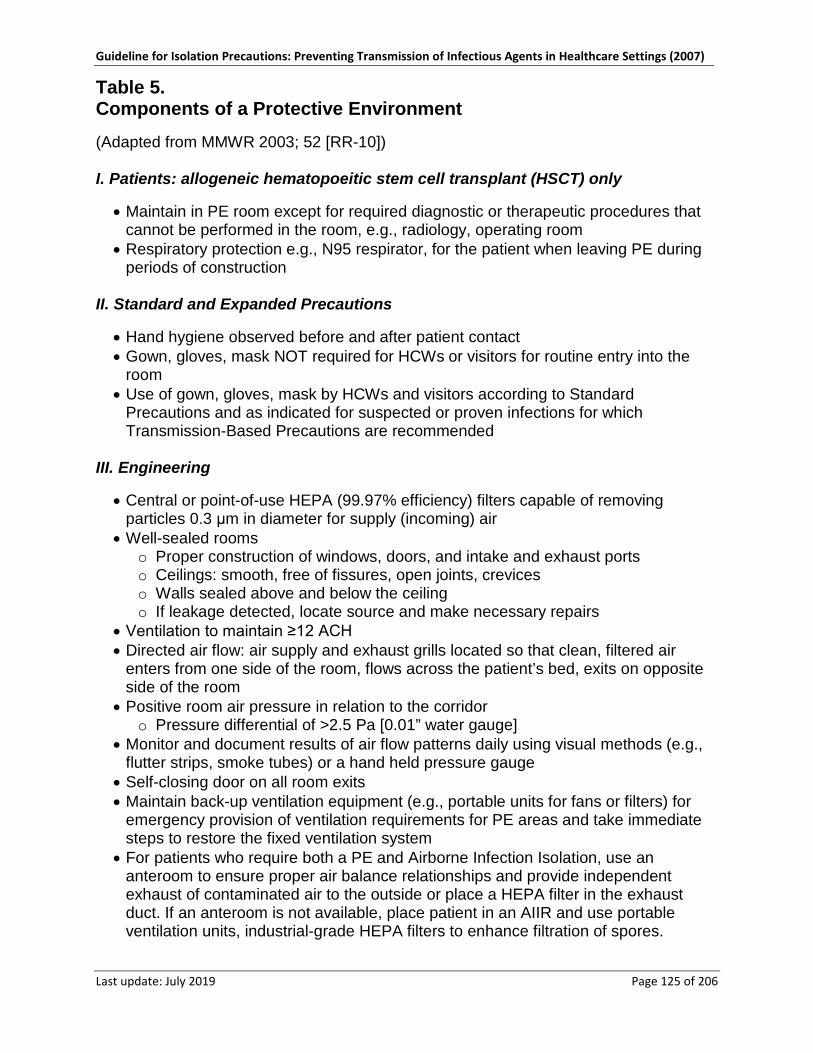
Guideline for Isolation Precautions: Preventing Transmission of Infectious Agents in Healthcare Settings (2007)
Last update: July 2019 Page 125 of 206
Table 5. Components of a Protective Environment (Adapted from MMWR 2003; 52 [RR-10]) I. Patients: allogeneic hematopoeitic stem cell transplant (HSCT) only
• Maintain in PE room except for required diagnostic or therapeutic procedures that cannot be performed in the room, e.g., radiology, operating room
• Respiratory protection e.g., N95 respirator, for the patient when leaving PE during periods of construction
II. Standard and Expanded Precautions
• Hand hygiene observed before and after patient contact • Gown, gloves, mask NOT required for HCWs or visitors for routine entry into the
room • Use of gown, gloves, mask by HCWs and visitors according to Standard
Precautions and as indicated for suspected or proven infections for which Transmission-Based Precautions are recommended
III. Engineering
• Central or point-of-use HEPA (99.97% efficiency) filters capable of removing particles 0.3 μm in diameter for supply (incoming) air
• Well-sealed rooms o Proper construction of windows, doors, and intake and exhaust ports o Ceilings: smooth, free of fissures, open joints, crevices o Walls sealed above and below the ceiling o If leakage detected, locate source and make necessary repairs
• Ventilation to maintain ≥12 ACH • Directed air flow: air supply and exhaust grills located so that clean, filtered air
enters from one side of the room, flows across the patient’s bed, exits on opposite side of the room
• Positive room air pressure in relation to the corridor o Pressure differential of >2.5 Pa [0.01” water gauge]
• Monitor and document results of air flow patterns daily using visual methods (e.g., flutter strips, smoke tubes) or a hand held pressure gauge
• Self-closing door on all room exits • Maintain back-up ventilation equipment (e.g., portable units for fans or filters) for
emergency provision of ventilation requirements for PE areas and take immediate steps to restore the fixed ventilation system
• For patients who require both a PE and Airborne Infection Isolation, use an anteroom to ensure proper air balance relationships and provide independent exhaust of contaminated air to the outside or place a HEPA filter in the exhaust duct. If an anteroom is not available, place patient in an AIIR and use portable ventilation units, industrial-grade HEPA filters to enhance filtration of spores.
Guideline for Isolation Precautions: Preventing Transmission of Infectious Agents in Healthcare Settings (2007)
Last update: July 2019 Page 126 of 206
IV. Surfaces
• Daily wet-dusting of horizontal surfaces using cloths moistened with EPA-registered hospital disinfectant/detergent
• Avoid dusting methods that disperse dust • No carpeting in patient rooms or hallways • No upholstered furniture and furnishings
V. Other
• No flowers (fresh or dried) or potted plants in PE rooms or areas • Use vacuum cleaner equipped with HEPA filters when vacuum cleaning is
necessary
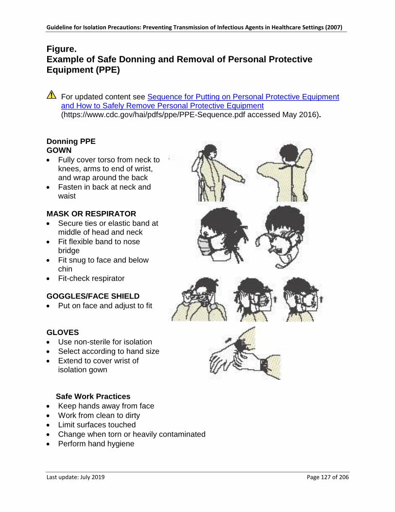
Guideline for Isolation Precautions: Preventing Transmission of Infectious Agents in Healthcare Settings (2007)
Last update: July 2019 Page 127 of 206
Figure. Example of Safe Donning and Removal of Personal Protective Equipment (PPE)
For updated content see Sequence for Putting on Personal Protective Equipment and How to Safely Remove Personal Protective Equipment (https://www.cdc.gov/hai/pdfs/ppe/PPE-Sequence.pdf accessed May 2016).
Donning PPE GOWN • Fully cover torso from neck to
knees, arms to end of wrist, and wrap around the back
• Fasten in back at neck and waist
MASK OR RESPIRATOR • Secure ties or elastic band at
middle of head and neck • Fit flexible band to nose
bridge • Fit snug to face and below
chin • Fit-check respirator GOGGLES/FACE SHIELD • Put on face and adjust to fit GLOVES • Use non-sterile for isolation • Select according to hand size • Extend to cover wrist of
isolation gown
Safe Work Practices
• Keep hands away from face • Work from clean to dirty • Limit surfaces touched • Change when torn or heavily contaminated • Perform hand hygiene
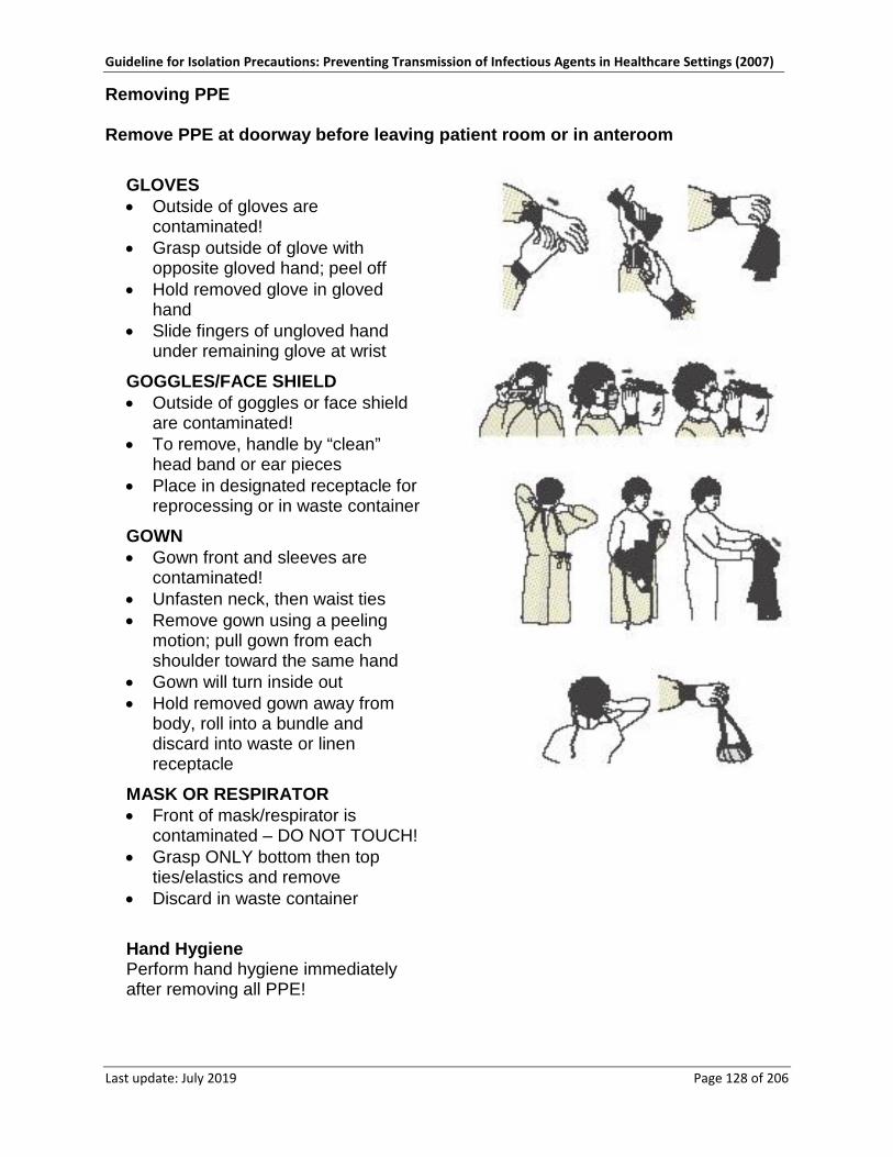
Guideline for Isolation Precautions: Preventing Transmission of Infectious Agents in Healthcare Settings (2007)
Last update: July 2019 Page 128 of 206
Removing PPE Remove PPE at doorway before leaving patient room or in anteroom
GLOVES • Outside of gloves are
contaminated! • Grasp outside of glove with
opposite gloved hand; peel off • Hold removed glove in gloved
hand • Slide fingers of ungloved hand
under remaining glove at wrist
GOGGLES/FACE SHIELD • Outside of goggles or face shield
are contaminated! • To remove, handle by “clean”
head band or ear pieces • Place in designated receptacle for
reprocessing or in waste container
GOWN • Gown front and sleeves are
contaminated! • Unfasten neck, then waist ties • Remove gown using a peeling
motion; pull gown from each shoulder toward the same hand
• Gown will turn inside out • Hold removed gown away from
body, roll into a bundle and discard into waste or linen receptacle
MASK OR RESPIRATOR • Front of mask/respirator is
contaminated – DO NOT TOUCH! • Grasp ONLY bottom then top
ties/elastics and remove • Discard in waste container
Hand Hygiene Perform hand hygiene immediately after removing all PPE!
Guideline for Isolation Precautions: Preventing Transmission of Infectious Agents in Healthcare Settings (2007)
Last update: July 2019 Page 129 of 206
Glossary Airborne infection isolation room (AIIR). Formerly, negative pressure isolation room, an AIIR is a single-occupancy patient-care room used to isolate persons with a suspected or confirmed airborne infectious disease. Environmental factors are controlled in AIIRs to minimize the transmission of infectious agents that are usually transmitted from person to person by droplet nuclei associated with coughing or aerosolization of contaminated fluids. AIIRs should provide negative pressure in the room (so that air flows under the door gap into the room); and an air flow rate of 6-12 ACH (6 ACH for existing structures, 12 ACH for new construction or renovation); and direct exhaust of air from the room to the outside of the building or recirculation of air through a HEPA filter before retruning to circulation (MMWR 2003; 52 [RR-10]; MMWR 1994; 43 [RR-13]).
American Institute of Architects (AIA). A professional organization that develops standards for building ventilation, The “2001Guidelines for Design and Construction of Hospital and Health Care Facilities”, the development of which was supported by the AIA, Academy of Architecture for Health, Facilities Guideline Institute, with assistance from the U.S. Department of Health and Human Services and the National Institutes of Health, is the primary source of guidance for creating airborne infection isolation rooms (AIIRs) and protective environments (American Institute of Architects – Academy of Architecture for Health (https://network.aia.org/academyofarchitectureforhealth/home accessed May 2016) [Current version of this document may differ from original.])
Ambulatory care settings. Facilities that provide health care to patients who do not remain overnight (e.g., hospital-based outpatient clinics, nonhospital-based clinics and physician offices, urgent care centers, surgicenters, free-standing dialysis centers, public health clinics, imaging centers, ambulatory behavioral health and substance abuse clinics, physical therapy and rehabilitation centers, and dental practices.
Bioaerosols. An airborne dispersion of particles containing whole or parts of biological entities, such as bacteria, viruses, dust mites, fungal hyphae, or fungal spores. Such aerosols usually consist of a mixture of mono-dispersed and aggregate cells, spores or viruses, carried by other materials, such as respiratory secretions and/or inert particles. Infectious bioaerosols (i.e., those that contain biological agents capable of causing an infectious disease) can be generated from human sources (e.g., expulsion from the respiratory tract during coughing, sneezing, talking or singing; during suctioning or wound irrigation), wet environmental sources (e.g., HVAC and cooling tower water with Legionella) or dry sources (e.g., construction dust with spores produced by Aspergillus spp.). Bioaerosols include large respiratory droplets and small droplet nuclei (Cole EC. AJIC 1998;26: 453-64).
Caregivers. All persons who are not employees of an organization, are not paid, and provide or assist in providing healthcare to a patient (e.g., family member, friend) and acquire technical training as needed based on the tasks that must be performed.
Cohorting. In the context of this guideline, this term applies to the practice of grouping patients infected or colonized with the same infectious agent together to confine their care to one area and prevent contact with susceptible patients (cohorting patients).
Guideline for Isolation Precautions: Preventing Transmission of Infectious Agents in Healthcare Settings (2007)
Last update: July 2019 Page 130 of 206
During outbreaks, healthcare personnel may be assigned to a cohort of patients to further limit opportunities for transmission (cohorting staff).
Colonization. Proliferation of microorganisms on or within body sites without detectable host immune response, cellular damage, or clinical expression. The presence of a microorganism within a host may occur with varying duration, but may become a source of potential transmission. In many instances, colonization and carriage are synonymous.
Droplet nuclei. Microscopic particles < 5 µm in size that are the residue of evaporated droplets and are produced when a person coughs, sneezes, shouts, or sings. These particles can remain suspended in the air for prolonged periods of time and can be carried on normal air currents in a room or beyond, to adjacent spaces or areas receiving exhaust air.
Engineering controls. Removal or isolation of a workplace hazard through technology. AIIRs, a Protective Environment, engineered sharps injury prevention devices and sharps containers are examples of engineering controls.
Epidemiologically important pathogens. Infectious agents that have one or more of the following characteristics: 1. are readily transmissible; 2. have a proclivity toward causing outbreaks; 3. may be associated with a severe outcome; or 4. are difficult to treat.
Examples include Acinetobacter sp., Aspergillus sp., Burkholderia cepacia, Clostridium difficile, Klebsiella or Enterobacter sp., Extended spectrum beta lactamase producing gram negative bacilli [ESBLs], methicillin-resistant Staphylococcus aureus [MRSA], Pseudomonas aeruginosa, vancomycin-resistant enterococci [VRE], methicillin resistant Staphylococcus aureus [MRSA], vancomycin resistant Staphylococcus aureus [VRSA] influenza virus, respiratory syncytial virus [RSV], rotavirus, SARS-CoV, noroviruses and the hemorrhagic fever viruses).
Hand hygiene. A general term that applies to any one of the following: 1. handwashing with plain (nonantimicrobial) soap and water); 2. antiseptic handwash (soap containing antiseptic agents and water); 3. antiseptic handrub (waterless antiseptic product, most often alcohol-based, rubbed
on all surfaces of hands); or 4. surgical hand antisepsis (antiseptic handwash or antiseptic handrub performed
preoperatively by surgical personnel to eliminate transient hand flora and reduce resident hand flora) 559.
Healthcare-associated infection (HAI). An infection that develops in a patient who is cared for in any setting where healthcare is delivered (e.g., acute care hospital, chronic care facility, ambulatory clinic, dialysis center, surgicenter, home) and is related to receiving health care (i.e., was not incubating or present at the time healthcare was provided). In ambulatory and home settings, HAI would apply to any infection that is associated with a medical or surgical intervention. Since the geographic location of infection acquisition is often uncertain, the preferred term is considered to be healthcare-associated rather than healthcare-acquired.
Guideline for Isolation Precautions: Preventing Transmission of Infectious Agents in Healthcare Settings (2007)
Last update: July 2019 Page 131 of 206
Healthcare epidemiologist. A person whose primary training is medical (M.D., D.O.) and/or masters or doctorate-level epidemiology who has received advanced training in healthcare epidemiology. Typically these professionals direct or provide consultation to an infection control program in a hospital, long term care facility (LTCF), or healthcare delivery system (also see infection control professional).
Healthcare personnel, healthcare worker (HCW). All paid and unpaid persons who work in a healthcare setting (e.g., any person who has professional or technical training in a healthcare-related field and provides patient care in a healthcare setting or any person who provides services that support the delivery of healthcare such as dietary, housekeeping, engineering, maintenance personnel).
Hematopoietic stem cell transplantation (HSCT). Any transplantation of blood- or bone marrow-derived hematopoietic stem cells, regardless of donor type (e.g., allogeneic or autologous) or cell source (e.g., bone marrow, peripheral blood, or placental/umbilical cord blood); associated with periods of severe immunosuppression that vary with the source of the cells, the intensity of chemotherapy required, and the presence of graft versus host disease (MMWR 2000; 49: RR-10).
High-efficiency particulate air (HEPA) filter. An air filter that removes >99.97% of particles ≥ 0.3µm (the most penetrating particle size) at a specified flow rate of air. HEPA filters may be integrated into the central air handling systems, installed at the point of use above the ceiling of a room, or used as portable units (MMWR 2003; 52: RR-10).
Home care. A wide-range of medical, nursing, rehabilitation, hospice and social services delivered to patients in their place of residence (e.g., private residence, senior living center, assisted living facility). Home health-care services include care provided by home health aides and skilled nurses, respiratory therapists, dieticians, physicians, chaplains, and volunteers; provision of durable medical equipment; home infusion therapy; and physical, speech, and occupational therapy.
Immunocompromised patients. Those patients whose immune mechanisms are deficient because of congenital or acquired immunologic disorders (e.g., human immunodeficiency virus [HIV] infection, congenital immune deficiency syndromes), chronic diseases such as diabetes mellitus, cancer, emphysema, or cardiac failure, ICU care, malnutrition, and immunosuppressive therapy of another disease process [e.g., radiation, cytotoxic chemotherapy, anti-graft-rejection medication, corticosteroids, monoclonal antibodies directed against a specific component of the immune system]). The type of infections for which an immunocompromised patient has increased susceptibility is determined by the severity of immunosuppression and the specific component(s) of the immune system that is affected. Patients undergoing allogeneic HSCT and those with chronic graft versus host disease are considered the most vulnerable to HAIs. Immunocompromised states also make it more difficult to diagnose certain infections (e.g., tuberculosis) and are associated with more severe clinical disease states than persons with the same infection and a normal immune system.
Infection. The transmission of microorganisms into a host after evading or overcoming defense mechanisms, resulting in the organism’s proliferation and invasion within host tissue(s). Host responses to infection may include clinical symptoms or may be
Guideline for Isolation Precautions: Preventing Transmission of Infectious Agents in Healthcare Settings (2007)
Last update: July 2019 Page 132 of 206
subclinical, with manifestations of disease mediated by direct organisms pathogenesis and/or a function of cell-mediated or antibody responses that result in the destruction of host tissues.
Infection control and prevention professional (ICP). A person whose primary training is in either nursing, medical technology, microbiology, or epidemiology and who has acquired specialized training in infection control. Responsibilities may include collection, analysis, and feedback of infection data and trends to healthcare providers; consultation on infection risk assessment, prevention and control strategies; performance of education and training activities; implementation of evidence-based infection control practices or those mandated by regulatory and licensing agencies; application of epidemiologic principles to improve patient outcomes; participation in planning renovation and construction projects (e.g., to ensure appropriate containment of construction dust); evaluation of new products or procedures on patient outcomes; oversight of employee health services related to infection prevention; implementation of preparedness plans; communication within the healthcare setting, with local and state health departments, and with the community at large concerning infection control issues; and participation in research. Certification in infection control (CIC) is available through the Certification Board of Infection Control and Epidemiology.
Infection control and prevention program. A multidisciplinary program that includes a group of activities to ensure that recommended practices for the prevention of healthcare-associated infections are implemented and followed by HCWs, making the healthcare setting safe from infection for patients and healthcare personnel. The Joint Commission on Accreditation of Healthcare Organizations (JCAHO) requires the following five components of an infection control program for accreditation: 1. surveillance: monitoring patients and healthcare personnel for acquisition of infection
and/or colonization; 2. investigation: identification and analysis of infection problems or undesirable trends; 3. prevention: implementation of measures to prevent transmission of infectious agents
and to reduce risks for device- and procedure-related infections; 4) control: evaluation and management of outbreaks; and
4. reporting: provision of information to external agencies as required by state and federal law and regulation (The Joint Commission (https://www.jointcommission.org/ accessed May 2016) [Current version of this document may differ from original.]).
The infection control program staff has the ultimate authority to determine infection control policies for a healthcare organization with the approval of the organization’s governing body.
Long-term care facilities (LTCFs). An array of residential and outpatient facilities designed to meet the bio-psychosocial needs of persons with sustained self-care deficits. These include skilled nursing facilities, chronic disease hospitals, nursing homes, foster and group homes, institutions for the developmentally disabled, residential care facilities, assisted living facilities, retirement homes, adult day health care facilities, rehabilitation centers, and long-term psychiatric hospitals.
Mask. A term that applies collectively to items used to cover the nose and mouth and includes both procedure masks and surgical masks ([This link is no longer active: www.fda.gov/cdrh/ode/guidance/094.html#4. Similar information may be found at FDA:
Guideline for Isolation Precautions: Preventing Transmission of Infectious Agents in Healthcare Settings (2007)
Last update: July 2019 Page 133 of 206
Masks and N95 Respirators (http://www.fda.gov/MedicalDevices/ProductsandMedicalProcedures/GeneralHospitalDevicesandSupplies/PersonalProtectiveEquipment/ucm055977.htm accessed May 2016)]).
Multidrug-resistant organisms (MDROs). In general, bacteria (excluding M. tuberculosis) that are resistant to one or more classes of antimicrobial agents and usually are resistant to all but one or two commercially available antimicrobial agents (e.g., MRSA, VRE, extended spectrum beta-lactamase [ESBL]-producing or intrinsically resistant gram-negative bacilli) 176.
Nosocomial infection. Derived from two Greek words “nosos” (disease) and “komeion” (to take care of). Refers to any infection that develops during or as a result of an admission to an acute care facility (hospital) and was not incubating at the time of admission.
Personal protective equipment (PPE). A variety of barriers used alone or in combination to protect mucous membranes, skin, and clothing from contact with infectious agents. PPE includes gloves, masks, respirators, goggles, face shields, and gowns.
Procedure Mask. A covering for the nose and mouth that is intended for use in general patient care situations. These masks generally attach to the face with ear loops rather than ties or elastic. Unlike surgical masks, procedure masks are not regulated by the Food and Drug Administration.
Protective Environment. A specialized patient-care area, usually in a hospital, with a positive air flow relative to the corridor (i.e., air flows from the room to the outside adjacent space). The combination of high-efficiency particulate air (HEPA) filtration, high numbers (≥12) of air changes per hour (ACH), and minimal leakage of air into the room creates an environment that can safely accommodate patients with a severely compromised immune system (e.g., those who have received allogeneic hemopoietic stem-cell transplant [HSCT]) and decrease the risk of exposure to spores produced by environmental fungi. Other components include use of scrubbable surfaces instead of materials such as upholstery or carpeting, cleaning to prevent dust accumulation, and prohibition of fresh flowers or potted plants.
Quasi-experimental studies. Studies to evaluate interventions but do not use randomization as part of the study design. These studies are also referred to as nonrandomized, pre-post-intervention study designs. These studies aim to demonstrate causality between an intervention and an outcome but cannot achieve the level of confidence concerning attributable benefit obtained through a randomized, controlled trial. In hospitals and public health settings, randomized control trials often cannot be implemented due to ethical, practical and urgency reasons; therefore, quasi-experimental design studies are used commonly. However, even if an intervention appears to be effective statistically, the question can be raised as to the possibility of alternative explanations for the result. Such study design is used when it is not logistically feasible or ethically possible to conduct a randomized, controlled trial, (e.g., during outbreaks). Within the classification of quasi-experimental study designs, there is
Guideline for Isolation Precautions: Preventing Transmission of Infectious Agents in Healthcare Settings (2007)
Last update: July 2019 Page 134 of 206
a hierarchy of design features that may contribute to validity of results (Harris et al. CID 2004:38: 1586).
Residential care setting. A facility in which people live, minimal medical care is delivered, and the psychosocial needs of the residents are provided for.
Respirator. A personal protective device worn by healthcare personnel over the nose and mouth to protect them from acquiring airborne infectious diseases due to inhalation of infectious airborne particles that are < 5 μm in size. These include infectious droplet nuclei from patients with M. tuberculosis, variola virus [smallpox], SARS-CoV), and dust particles that contain infectious particles, such as spores of environmental fungi (e.g., Aspergillus sp.). The CDC’s National Institute for Occupational Safety and Health (NIOSH) certifies respirators used in healthcare settings (Personal Protective Equipment for Healthcare Workers (https://www.cdc.gov/NIOSH/docs/2013-138/ accessed May 2016)). [Current version of this document may differ from original.]. The N95 disposable particulate, air purifying, respirator is the type used most commonly by healthcare personnel. Other respirators used include N-99 and N-100 particulate respirators, powered air-purifying respirators (PAPRS) with high efficiency filters; and non-powered full-facepiece elastomeric negative pressure respirators. A listing of NIOSH-approved respirators can be found at [This link is no longer active: www.cdc.gov/niosh/npptl/respirators/disp_part/particlist.html. Similar information may be found at NIOSH Respirator Trusted-Source Information (https://www.cdc.gov/niosh/npptl/topics/respirators/disp_part/respsource.html accessed May 2016).]. Respirators must be used in conjunction with a complete Respiratory Protection Program, as required by the Occupational Safety and Health Administration (OSHA) that includes fit testing, training, proper selection of respirators, medical clearance and respirator maintenance.
Respiratory Hygiene/ Cough Etiquette. A combination of measures designed to minimize the transmission of respiratory pathogens via droplet or airborne routes in healthcare settings. The components of respiratory hygiene/cough etiquette are 1. covering the mouth and nose during coughing and sneezing, 2. using tissues to contain respiratory secretions with prompt disposal into a no-touch
receptacle, 3. offering a surgical mask to persons who are coughing to decrease contamination of
the surrounding environment, and 4. turning the head away from others and maintaining spatial separation, ideally >3
feet, when coughing.
These measures are targeted to all patients with symptoms of respiratory infection and their accompanying family members or friends beginning at the point of initial encounter with a healthcare setting (e.g., reception/triage in emergency departments, ambulatory clinics, healthcare provider offices)126 (Srinivasin A ICHE 2004; 25: 1020; Respiratory Hygiene/Cough Etiquette in Healthcare Settings (https://www.cdc.gov/flu/professionals/infectioncontrol/resphygiene.htm accessed May 2016) [Current version of this document may differ from original.])
Safety culture. Shared perceptions of workers and management regarding the level of safety in the work environment. A hospital safety climate includes the following six organizational components:
Guideline for Isolation Precautions: Preventing Transmission of Infectious Agents in Healthcare Settings (2007)
Last update: July 2019 Page 135 of 206
1. senior management support for safety programs; 2. absence of workplace barriers to safe work practices; 3. cleanliness and orderliness of the worksite; 4. minimal conflict and good communication among staff members; 5. frequent safety-related feedback/training by supervisors; and 6. availability of PPE and engineering controls620.
Source Control. The process of containing an infectious agent either at the portal of exit from the body or within a confined space. The term is applied most frequently to containment of infectious agents transmitted by the respiratory route but could apply to other routes of transmission, (e.g., a draining wound, vesicular or bullous skin lesions). Respiratory Hygiene/Cough Etiquette that encourages individuals to “cover your cough” and/or wear a mask is a source control measure. The use of enclosing devices for local exhaust ventilation (e.g., booths for sputum induction or administration of aerosolized medication) is another example of source control.
Standard Precautions. A group of infection prevention practices that apply to all patients, regardless of suspected or confirmed diagnosis or presumed infection status. Standard Precautions is a combination and expansion of Universal Precautions780 and Body Substance Isolation1102. Standard Precautions is based on the principle that all blood, body fluids, secretions, excretions except sweat, nonintact skin, and mucous membranes may contain transmissible infectious agents. Standard Precautions includes hand hygiene, and depending on the anticipated exposure, use of gloves, gown, mask, eye protection, or face shield. Also, equipment or items in the patient environment likely to have been contaminated with infectious fluids must be handled in a manner to prevent transmission of infectious agents, (e.g., wear gloves for handling, contain heavily soiled equipment, properly clean and disinfect or sterilize reusable equipment before use on another patient).
Surgical mask. A device worn over the mouth and nose by operating room personnel during surgical procedures to protect both surgical patients and operating room personnel from transfer of microorganisms and body fluids. Surgical masks also are used to protect healthcare personnel from contact with large infectious droplets (>5 μm in size). According to draft guidance issued by the Food and Drug Administration on May 15, 2003, surgical masks are evaluated using standardized testing procedures for fluid resistance, bacterial filtration efficiency, differential pressure (air exchange), and flammability in order to mitigate the risks to health associated with the use of surgical masks. These specifications apply to any masks that are labeled surgical, laser, isolation, or dental or medical procedure ([This link is no longer active: www.fda.gov/cdrh/ode/guidance/094.html#4. Similar information may be found at FDA: Masks and N95 Respirators (http://www.fda.gov/MedicalDevices/ProductsandMedicalProcedures/GeneralHospitalDevicesandSupplies/PersonalProtectiveEquipment/ucm055977.htm accessed May 2016.]). Surgical masks do not protect against inhalation of small particles or droplet nuclei and should not be confused with particulate respirators that are recommended for protection against selected airborne infectious agents, (e.g., Mycobacterium tuberculosis).
Guideline for Isolation Precautions: Preventing Transmission of Infectious Agents in Healthcare Settings (2007)
Last update: July 2019 Page 136 of 206
References 1. Garner JS. Guideline for isolation precautions in hospitals. The Hospital Infection
Control Practices Advisory Committee. Infect Control Hosp Epidemiol 1996;17(1):53-80.(s).
2. Harris AD, Bradham DD, Baumgarten M, Zuckerman IH, Fink JC, Perencevich EN. The use and interpretation of quasi-experimental studies in infectious diseases. Clin Infect Dis 2004;38(11):1586-91.
3. Morton V, Torgerson DJ. Effect of regression to the mean on decision making in health care. Bmj 2003;326(7398):1083-4.
4. Pocock SJ, Elbourne DR. Randomized trials or observational tribulations? N Engl J Med 2000;342(25):1907-9.
5. Ioannidis JP, Haidich AB, Pappa M, et al. Comparison of evidence of treatment effects in randomized and nonrandomized studies. Jama 2001;286(7):821-30.
6. Bent S, Shojania KG, Saint S. The use of systematic reviews and meta-analyses in infection control and hospital epidemiology. Am J Infect Control 2004;32(4):246-54.
7. Harris AD, Lautenbach E, Perencevich E. A systematic review of quasi-experimental study designs in the fields of infection control and antibiotic resistance. Clin Infect Dis 2005;41(1):77-82.
8. Evans R, Lloyd JF, Abouzelof RH, Taylor CW, Anderson VR, Samore MH. System-wide Surveillance for Clinical Encounters by Patients Previously Identified with MRSA and VRE. Medinfo 2004;2004:212-6.
9. Srinivasan A, McDonald LC, Jernigan D, et al. Foundations of the severe acute respiratory syndrome preparedness and response plan for healthcare facilities. Infect Control Hosp Epidemiol 2004;25(12):1020-5.
10. [This link is no longer active: www.cdc.gov/flu/avian/professional/infect-control.htm].
11. CDC. Guidelines for Environmental Infection Control in Health-Care Facilities (https://www.cdc.gov/infectioncontrol/guidelines/environmental/index.html accessed May 2016). Recommendations of CDC and the Healthcare Infection Control Practices Advisory Committee (HICPAC). MMWR 2003;52(RR10);1-42.
12. CDC. Guidelines for preventing the transmission of Mycobacterium tuberculosis in health-care settings, 2005. MMWR Recomm Rep 2005;54(17):1-141.
13. AIA. Guidelines for Design and Construction of Hospital and Health Care Facilities. In: American Institute of Architects. Washington, DC: American Institute of Architects Press; 2006.
14. CDC. Guidelines for Preventing Health-Care-Associated Pneumonia, 2003. Recommendations of CDC and the Healthcare Infection Control Practices Advisory Committee. MMWR Recomm Rep 2004;53(RR-3):1-40.
15. CDC. Guidelines for preventing opportunistic infections among hematopoietic stem cell transplant recipients. Recommendations of CDC, the Infectious
Guideline for Isolation Precautions: Preventing Transmission of Infectious Agents in Healthcare Settings (2007)
Last update: July 2019 Page 137 of 206
Disease Society of America, and the American Society of Blood and Marrow Transplantation. MMWR - Morbidity & Mortality Weekly Report 2000;49(RR-10):1-125.
16. Boyce JM, Pittet D. Guideline for Hand Hygiene in Health-Care Settings. Recommendations of the Healthcare Infection Control Practices Advisory Committee and the HICPAC/SHEA/APIC/IDSA Hand Hygiene Task Force. Society for Healthcare Epidemiology of America/Association for Professionals in Infection Control/Infectious Diseases Society of America. MMWR Recomm Rep 2002;51(RR-16):1-45, quiz CE1-4.
17. Bolyard EA, Tablan OC, Williams WW, Pearson ML, Shapiro CN, Deitchmann SD. Guideline for infection control in healthcare personnel, 1998. Hospital Infection Control Practices Advisory Committee. Infect Control Hosp Epidemiol 1998;19(6):407-63.
18. CDC. Recommendations for Preventing Transmission of Infections Among Chronic Hemodialysis Patients. MMWR 2001;50(RR05):1-43.
19. Kohn WG, Collins AS, Cleveland JL, Harte JA, Eklund KJ, Malvitz DM. Guidelines for infection control in dental health-care settings--2003. MMWR Recomm Rep 2003;52(RR-17):1-61.
20. Saiman L, Siegel J. Infection control recommendations for patients with cystic fibrosis: microbiology, important pathogens, and infection control practices to prevent patient-to-patient transmission. Infect Control Hosp Epidemiol 2003;24(5 Suppl):S6-52.
21. Varia M, Wilson S, Sarwal S, et al. Investigation of a nosocomial outbreak of severe acute respiratory syndrome (SARS) in Toronto, Canada. Cmaj 2003;169(4):285-92.
22. Haley RW, Cushion NB, Tenover FC, et al. Eradication of endemic methicillin-resistant Staphylococcus aureus infections from a neonatal intensive care unit. J Infect Dis 1995;171(3):614-24.
23. Bridges CB, Kuehnert MJ, Hall CB. Transmission of influenza: implications for control in health care settings. Clin Infect Dis 2003;37(8):1094-101.
24. Hall CB. Nosocomial respiratory syncytial virus infections: the "Cold War" has not ended. Clin Infect Dis 2000;31(2):590-6.
25. Campbell JR, Zaccaria E, Mason EO, Jr., Baker CJ. Epidemiological analysis defining concurrent outbreaks of Serratia marcescens and methicillin-resistant Staphylococcus aureus in a neonatal intensive-care unit. Infect Control Hosp Epidemiol 1998;19(12):924-8.
26. Pena C, Pujol M, Ardanuy C, et al. Epidemiology and successful control of a large outbreak due to Klebsiella pneumoniae producing extended-spectrum beta-lactamases. Antimicrob Agents Chemother 1998;42(1):53-8.
27. Bonten MJ, Slaughter S, Ambergen AW, et al. The role of "colonization pressure" in the spread of vancomycin-resistant enterococci: an important infection control variable. Arch Intern Med 1998;158(10):1127-32.
28. Jensenius M, Ringertz SH, Berild D, Bell H, Espinoza R, Grinde B. Prolonged
Guideline for Isolation Precautions: Preventing Transmission of Infectious Agents in Healthcare Settings (2007)
Last update: July 2019 Page 138 of 206
nosocomial outbreak of hepatitis A arising from an alcoholic with pneumonia. Scand J Infect Dis 1998;30(2):119-23.
29. Zawacki A, O'Rourke E, Potter-Bynoe G, Macone A, Harbarth S, Goldmann D. An outbreak of Pseudomonas aeruginosa pneumonia and bloodstream infection associated with intermittent otitis externa in a healthcare worker. Infect Control Hosp Epidemiol 2004;25(12):1083-9.
30. Foca M, Jakob K, Whittier S, et al. Endemic Pseudomonas aeruginosa infection in a neonatal intensive care unit. N Engl J Med 2000;343(10):695-700.
31. Gupta A, Della-Latta P, Todd B, et al. Outbreak of extended-spectrum beta-lactamase-producing Klebsiella pneumoniae in a neonatal intensive care unit linked to artificial nails. Infect Control Hosp Epidemiol 2004;25(3):210-5.
32. Boyce JM, Opal SM, Potter-Bynoe G, Medeiros AA. Spread of methicillin-resistant Staphylococcus aureus in a hospital after exposure to a health care worker with chronic sinusitis. Clin Infect Dis 1993;17(3):496-504.
33. Fliegel PE, Weinstein WM. Rubella outbreak in a prenatal clinic: management and prevention. Am J Infect Control 1982;10(1):29-33.
34. Atkinson WL, Markowitz LE, Adams NC, Seastrom GR. Transmission of measles in medical settings--United States, 1985-1989. Am J Med 1991;91(3B):320S-4S.
35. Carman WF, Elder AG, Wallace LA, et al. Effects of influenza vaccination of health-care workers on mortality of elderly people in long-term care: a randomised controlled trial. Lancet 2000;355(9198):93-7.
36. CDC. Outbreaks of pertussis associated with hospitals--Kentucky, Pennsylvania, and Oregon, 2003. MMWR Morb Mortal Wkly Rep 2005;54(3):67-71.
37. Mermel LA, McKay M, Dempsey J, Parenteau S. Pseudomonas surgical-site infections linked to a healthcare worker with onychomycosis. Infect Control Hosp Epidemiol 2003;24(10):749-52.
38. Barnes GL, Callaghan SL, Kirkwood CD, Bogdanovic-Sakran N, Johnston LJ, Bishop RF. Excretion of serotype G1 rotavirus strains by asymptomatic staff: a possible source of nosocomial infection. J Pediatr 2003;142(6):722-5.
39. Wang JT, Chang SC, Ko WJ, et al. A hospital-acquired outbreak of methicillin-resistant Staphylococcus aureus infection initiated by a surgeon carrier. J Hosp Infect 2001;47(2):104-9.
40. Valenti WM, Pincus PH, Messner MK. Nosocomial pertussis: possible spread by a hospital visitor. Am J Dis Child 1980;134(5):520-1.
41. Christie CD, Glover AM, Willke MJ, Marx ML, Reising SF, Hutchinson NM. Containment of pertussis in the regional pediatric hospital during the Greater Cincinnati epidemic of 1993. Infect Control Hosp Epidemiol 1995;16(10):556-63.
42. Munoz FM, Ong LT, Seavy D, Medina D, Correa A, Starke JR. Tuberculosis among adult visitors of children with suspected tuberculosis and employees at a children's hospital. Infect Control Hosp Epidemiol 2002;23(10):568-72.
43. Garcia R, Raad I, Abi-Said D, et al. Nosocomial respiratory syncytial virus infections: prevention and control in bone marrow transplant patients. Infect
Guideline for Isolation Precautions: Preventing Transmission of Infectious Agents in Healthcare Settings (2007)
Last update: July 2019 Page 139 of 206
Control Hosp Epidemiol 1997;18(6):412-6. 44. Whimbey E, Champlin RE, Couch RB, et al. Community respiratory virus
infections among hospitalized adult bone marrow transplant recipients. Clin Infect Dis 1996;22(5):778-82.
45. Saiman L, O'keefe M, Graham PL, et al. Hospital transmission of community-acquired methicillin-resistant Staphylococcus aureus among postpartum women. Clin Infect Dis 2003;37(10):1313-9.
46. Bonten MJ, Slaughter S, Hayden MK, Nathan C, van Voorhis J, Weinstein RA. External sources of vancomycin-resistant enterococci for intensive care units. Crit Care Med 1998;26(12):2001-4.
47. Flynn DM, Weinstein RA, Nathan C, Gaston MA, Kabins SA. Patients' endogenous flora as the source of "nosocomial" Enterobacter in cardiac surgery. J Infect Dis 1987;156(2):363-8.
48. Olson B, Weinstein RA, Nathan C, Chamberlin W, Kabins SA. Epidemiology of endemic Pseudomonas aeruginosa: why infection control efforts have failed. J Infect Dis 1984;150(6):808-16.
49. Perl TM, Cullen JJ, Wenzel RP, et al. Intranasal mupirocin to prevent postoperative Staphylococcus aureus infections. N Engl J Med 2002;346(24):1871-7.
50. Toltzis P, Hoyen C, et al. Factors that predict preexisting colonization with antibiotic-resistant gram-negative bacilli in patients admitted to a pediatric intensive care unit. Pediatrics 1999;103 (4 Pt1):719-23.
51. Sarginson RE, Taylor N, Reilly N, Baines PB, Van Saene HK. Infection in prolonged pediatric critical illness: A prospective four-year study based on knowledge of the carrier state. Crit Care Med 2004;32(3):839-47.
52. Silvestri L, Monti Bragadin C, Milanese M, et al. Are most ICU infections really nosocomial? A prospective observational cohort study in mechanically ventilated patients. J Hosp Infect 1999;42(2):125-33.
53. Heggers JP, Phillips LG, Boertman JA, et al. The epidemiology of methicillin-resistant Staphylococcus aureus in a burn center. J Burn Care Rehabil 1988;9(6):610-2.
54. Donskey CJ. The role of the intestinal tract as a reservoir and source for transmission of nosocomial pathogens. Clin Infect Dis 2004;39(2):219-26.
55. Osterholm MT, Hedberg CW, Moore KA. The epidemiology of infectious diseases. . In: G.L. M, Jr DRG, J.E. B, eds. Principles and Practice of Infectious Diseases. 5th ed ed. Philadelphia: Churchill Livingstone; 2000:161-3.
56. Thomsen RW, Hundborg HH, Lervang HH, Johnsen SP, Schonheyder HC, Sorensen HT. Risk of community-acquired pneumococcal bacteremia in patients with diabetes: a population-based case-control study. Diabetes Care 2004;27(5):1143-7.
57. Carton JA, Maradona JA, Nuno FJ, Fernandez-Alvarez R, Perez-Gonzalez F, Asensi V. Diabetes mellitus and bacteraemia: a comparative study between diabetic and non-diabetic patients. Eur J Med 1992;1(5):281-7.
Guideline for Isolation Precautions: Preventing Transmission of Infectious Agents in Healthcare Settings (2007)
Last update: July 2019 Page 140 of 206
58. Hirschtick RE, Glassroth J, Jordan MC, et al. Bacterial pneumonia in persons infected with the human immunodeficiency virus. Pulmonary Complications of HIV Infection Study Group. N Engl J Med 1995;333(13):845-51.
59. Rosenberg AL, Seneff MG, Atiyeh L, Wagner R, Bojanowski L, Zimmerman JE. The importance of bacterial sepsis in intensive care unit patients with acquired immunodeficiency syndrome: implications for future care in the age of increasing antiretroviral resistance. Crit Care Med 2001;29(3):548-56.
60. Malone JL, Ijaz K, Lambert L, et al. Investigation of healthcare-associated transmission of Mycobacterium tuberculosis among patients with malignancies at three hospitals and at a residential facility. Cancer 2004;101(12):2713-21.
61. Fishman JA, Rubin RH. Infection in organ-transplant recipients. N Engl J Med 1998;338(24):1741-51.
62. Safdar N, Kluger DM, Maki DG. A review of risk factors for catheter-related bloodstream infection caused by percutaneously inserted, noncuffed central venous catheters: implications for preventive strategies. Medicine (Baltimore) 2002;81(6):466-79.
63. Jarvis WR, Robles B. Nosocomial infections in pediatric patients. Adv Pediatr Infect Dis 1996;12:243-959(js).
64. Yogaraj JS, Elward AM, Fraser VJ. Rate, risk factors, and outcomes of nosocomial primary bloodstream infection in pediatric intensive care unit patients. Pediatrics 2002;110(3):481-5.
65. Donlan RM. Biofilms: microbial life on surfaces. Emerg Infect Dis 2002;8(9):881-90.
66. Rosen HR. Acquisition of hepatitis C by a conjunctival splash. Am J Infect Control 1997;25(3):242-7.
67. Beltrami EM, Kozak A, Williams IT, et al. Transmission of HIV and hepatitis C virus from a nursing home patient to a health care worker. Am J Infect Control 2003;31(3):168-75.
68. Obasanjo OO, Wu P, Conlon M, et al. An outbreak of scabies in a teaching hospital: lessons learned. Infect Control Hosp Epidemiol 2001;22(1):13-8.
69. Andersen BM, Haugen H, Rasch M, Heldal Haugen A, Tageson A. Outbreak of scabies in Norwegian nursing homes and home care patients: control and prevention. J Hosp Infect 2000;45(2):160-4.
70. Avitzur Y, Amir J. Herpetic whitlow infection in a general pediatrician--an occupational hazard. Infection 2002;30(4):234-6.
71. Adams G, Stover BH, Keenlyside RA, et al. Nosocomial herpetic infections in a pediatric intensive care unit. Am J Epidemiol 1981;113(2):126-32.
72. Bhalla A, Pultz NJ, Gries DM, et al. Acquisition of nosocomial pathogens on hands after contact with environmental surfaces near hospitalized patients. Infect Control Hosp Epidemiol 2004;25(2):164-7.
73. Duckro AN, Blom DW, Lyle EA, Weinstein RA, Hayden MK. Transfer of vancomycin-resistant enterococci via health care worker hands. Arch Intern Med
Guideline for Isolation Precautions: Preventing Transmission of Infectious Agents in Healthcare Settings (2007)
Last update: July 2019 Page 141 of 206
2005;165(3):302-7. 74. Brooks SE, Veal RO, Kramer M, Dore L, Schupf N, Adachi M. Reduction in the
incidence of Clostridium difficile-associated diarrhea in an acute care hospital and a skilled nursing facility following replacement of electronic thermometers with single-use disposables. Infect Control Hosp Epidemiol 1992;13(2):98-103.
75. CDC. Nosocomial hepatitis B virus infection associated with reusable fingerstick blood sampling devices--Ohio and New York City, 1996. MMWR Morb Mortal Wkly Rep 1997;46 (10):217-21.
76. Desenclos JC, Bourdiol-Razes M, Rolin B, et al. Hepatitis C in a ward for cystic fibrosis and diabetic patients: possible transmission by spring-loaded finger-stick devices for self-monitoring of capillary blood glucose. Infect Control Hosp Epidemiol 2001;22(11):701-7.
77. CDC. Transmission of hepatitis B virus among persons undergoing blood glucose monitoring in long-term-care facilities--Mississippi, North Carolina, and Los Angeles County, California, 2003-2004. MMWR Morb Mortal Wkly Rep 2005;54(9):220-3.
78. Hall CB, Douglas RG, Jr. Modes of transmission of respiratory syncytial virus. J Pediatr 1981;99(1):100-3.
79. Hall CB, Douglas RG, Jr., Geiman JM. Possible transmission by fomites of respiratory syncytial virus. J Infect Dis 1980;141(1):98-102.
80. Buttery JP, Alabaster SJ, Heine RG, et al. Multiresistant Pseudomonas aeruginosa outbreak in a pediatric oncology ward related to bath toys. Pediatr Infect Dis J 1998;17(6):509-13.
81. Agerton T, Valway S, Gore B, et al. Transmission of a highly drug-resistant strain (strain W1) of Mycobacterium tuberculosis. Community outbreak and nosocomial transmission via a contaminated bronchoscope. JAMA 1997;278(13):1073-7.
82. Bronowicki JP, Venard V, Botte C, et al. Patient-to-patient transmission of hepatitis C virus during colonoscopy. N Engl J Med 1997;337(4):237-40.
83. Michele TM, Cronin WA, Graham NM, et al. Transmission of Mycobacterium tuberculosis by a fiberoptic bronchoscope. Identification by DNA fingerprinting. JAMA 1997;278(13):1093-5.
84. Schelenz S, French G. An outbreak of multidrug-resistant Pseudomonas aeruginosa infection associated with contamination of bronchoscopes and an endoscope washer-disinfector. J Hosp Infect 2000;46(1):23-30.
85. Weber DJ, Rutala WA. Lessons from outbreaks associated with bronchoscopy. Infect Control Hosp Epidemiol 2001;22(7):403-8.
86. Kirschke DL, Jones TF, Craig AS, et al. Pseudomonas aeruginosa and Serratia marcescens contamination associated with a manufacturing defect in bronchoscopes. N Engl J Med 2003;348(3):214-20.
87. Srinivasan A, Wolfenden LL, Song X, et al. An outbreak of Pseudomonas aeruginosa infections associated with flexible bronchoscopes. N Engl J Med 2003;348(3):221-7.
Guideline for Isolation Precautions: Preventing Transmission of Infectious Agents in Healthcare Settings (2007)
Last update: July 2019 Page 142 of 206
88. Boyce JM, Potter-Bynoe G, Chenevert C, King T. Environmental contamination due to methicillin-resistant Staphylococcus aureus: possible infection control implications. Infect Control Hosp Epidemiol 1997;18(9):622-7.(mj).
89. Zachary KC, Bayne PS, Morrison VJ, Ford DS, Silver LC, Hooper DC. Contamination of gowns, gloves, and stethoscopes with vancomycin-resistant enterococci. Infect Control Hosp Epidemiol 2001;22(9):560-4.
90. Perry C, Marshall R, Jones E. Bacterial contamination of uniforms. J Hosp Infect 2001;48(3):238-41.
91. Papineni RS, Rosenthal FS. The size distribution of droplets in the exhaled breath of healthy human subjects. J Aerosol Med 1997;10(2):105-16.
92. Wells WF. On airborne infection: Study II. Droplets and droplet nuclei. . Am J Hygiene 1934;20:611-18.
93. Loeb M, McGeer A, Henry B, et al. SARS among critical care nurses, Toronto. Emerg Infect Dis 2004;10(2):251-5.
94. Fowler RA, Guest CB, Lapinsky SE, et al. Transmission of severe acute respiratory syndrome during intubation and mechanical ventilation. Am J Respir Crit Care Med 2004;169(11):1198-202.
95. Gehanno JF, Kohen-Couderc L, Lemeland JF, Leroy J. Nosocomial meningococcemia in a physician. Infect Control Hosp Epidemiol 1999;20(8):564-5.
96. Scales D, et al. Illness in intensive-care staff after brief exposure to severe acute respiratory syndrome. Emerg Infect Dis 2003;9(10):1205-10.
97. Ensor E, Humphreys H, Peckham D, Webster C, Knox AJ. Is Burkholderia (Pseudomonas) cepacia disseminated from cystic fibrosis patients during physiotherapy? J Hosp Infect 1996;32(1):9-15.
98. Christian MD, Loutfy M, McDonald LC, et al. Possible SARS coronavirus transmission during cardiopulmonary resuscitation. Emerg Infect Dis 2004;10(2):287-93.
99. Valenzuela TD, Hooton TM, Kaplan EL, Schlievert P. Transmission of 'toxic strep' syndrome from an infected child to a firefighter during CPR. Ann Emerg Med 1991;20(1):90-2.
100. Bassinet L, Matrat M, Njamkepo E, Aberrane S, Housset B, Guiso N. Nosocomial pertussis outbreak among adult patients and healthcare workers. Infect Control Hosp Epidemiol 2004;25(11):995-7.
101. Wong TW, Lee CK, Tam W, et al. Cluster of SARS among medical students exposed to single patient, Hong Kong. Emerg Infect Dis 2004;10(2):269-76.
102. Pachucki CT, Pappas SA, Fuller GF, Krause SL, Lentino JR, Schaaff DM. Influenza A among hospital personnel and patients. Implications for recognition, prevention, and control. Arch Intern Med 1989;149(1):77-80.
103. Feigin RD, Baker CJ, Herwaldt LA, Lampe RM, Mason EO, Whitney SE. Epidemic meningococcal disease in an elementary-school classroom. N Engl J Med 1982;307(20):1255-7.
Guideline for Isolation Precautions: Preventing Transmission of Infectious Agents in Healthcare Settings (2007)
Last update: July 2019 Page 143 of 206
104. Dick EC, Jennings LC, Mink KA, Wartgow CD, Inhorn SL. Aerosol transmission of rhinovirus colds. J Infect Dis 1987;156(3):442-8.
105. Duguid JP. The size and duration of air-carriage of respiratory droplets and droplet nucleii. J Hyg (Lond) 1946;44:471-9.
106. Hall CB, Douglas RG, Jr., Schnabel KC, Geiman JM. Infectivity of respiratory syncytial virus by various routes of inoculation. Infect Immun 1981;33(3):779-83.
107. Downie AW, Meiklejohn M, St Vincent L, Rao AR, Sundara Babu BV, Kempe CH. The recovery of smallpox virus from patients and their environment in a smallpox hospital. Bull World Health Organ 1965;33(5):615-22.
108. Fenner F, Henderson DA, Arita I, Jezek Z, Ladnyi ID. The epidemiology of smallpox. In: Smallpox and its eradication. Switzerland: World Health Orginization; 1988.
109. Cole EC, Cook CE. Characterization of infectious aerosols in health care facilities: an aid to effective engineering controls and preventive strategies. Am J Infect Control 1998;26(4):453-64.
110. Christie C, Mazon D, Hierholzer W, Jr., Patterson JE. Molecular heterogeneity of Acinetobacter baumanii isolates during seasonal increase in prevalence. Infect Control Hosp Epidemiol 1995;16(10):590-4.
111. Musher DM. How contagious are common respiratory tract infections? N Engl J Med 2003;348(13):1256-66.
112. Steinberg P, White RJ, Fuld SL, Gutekunst RR, Chanock RM, Senterfit LB. Ecology of Mycoplasma pneumoniae infections in marine recruits at Parris Island, South Carolina. Am J Epidemiol 1969;89(1):62-73.
113. Seto WH, Tsang D, Yung RW, et al. Effectiveness of precautions against droplets and contact in prevention of nosocomial transmission of severe acute respiratory syndrome (SARS). Lancet 2003;361(9368):1519-20.
114. Hamburger M, Robertson OH. Expulsion of group A haemolytic streptocicci in droplets and droplet nuclei by sneezing, coughing and talking. Am J Med 1948;4:690.
115. CDC. Nosocomial meningococcemia. MMWR Morb Mortal Wkly Rep 1978;27:358.
116. LeClair JM, Freeman J, Sullivan BF, Crowley CM, Goldmann DA. Prevention of nosocomial respiratory syncytial virus infections through compliance with glove and gown isolation precautions. N Engl J Med 1987;317(6):329-34.
117. Madge P, Paton JY, McColl JH, Mackie PL. Prospective controlled study of four infection-control procedures to prevent nosocomial infection with respiratory syncytial virus. Lancet 1992;340(8827):1079-83.
118. Bassetti S, Bischoff WE, Walter M, et al. Dispersal of Staphylococcus aureus Into the Air Associated With a Rhinovirus Infection. Infect Control Hosp Epidemiol 2005;26(2):196-203.
119. Eichenwald HF, Kotsevalov O, Fasso LA. The "cloud baby": an example of bacterial-viral interaction. Am J Dis Child 1960;100:161-73.
Guideline for Isolation Precautions: Preventing Transmission of Infectious Agents in Healthcare Settings (2007)
Last update: July 2019 Page 144 of 206
120. Sheretz RJ, Reagan DR, Hampton KD, et al. A cloud adult: the Staphylococcus aureus-virus interaction revisited. Ann Intern Med 1996;124(6):539-47.
121. Coronado VG, Beck-Sague CM, Hutton MD, et al. Transmission of multidrug-resistant Mycobacterium tuberculosis among persons with human immunodeficiency virus infection in an urban hospital: epidemiologic and restriction fragment length polymorphism analysis. J Infect Dis 1993;168(4):1052-5.
122. Bloch AB, Orenstein WA, Ewing WM, et al. Measles outbreak in a pediatric practice: airborne transmission in an office setting. Pediatrics 1985;75(4):676-83.
123. LeClair JM, Zaia JA, Levin MJ, Congdon RG, Goldmann DA. Airborne transmission of chickenpox in a hospital. N Engl J Med 1980;302(8):450-3.
124. Riley RL, Mills CC, Nyka W, et al. Aerial dissemination of pulmonary tuberculosis. A two-year study of contagion in a tuberculosis ward. 1959. Am J Hyg 1959;70:185-96.
125. Beck-Sague C, Dooley SW, Hutton MD, et al. Hospital outbreak of multidrug-resistant Mycobacterium tuberculosis infections. Factors in transmission to staff and HIV-infected patients. JAMA 1992;268(10):1280-6.
126. CDC. Guidelines for preventing the transmission of Mycobacterium tuberculosis in health-care facilities, 1994. Centers for Disease Control and Prevention. MMWR Recomm Rep 1994;43(RR-13):1-132.
127. Haley CE, McDonald RC, Rossi L, Jones WD, Jr., Haley RW, Luby JP. Tuberculosis epidemic among hospital personnel. Infect Control Hosp Epidemiol 1989;10(5):204-10.
128. Wehrle PF, Posch J, Richter KH, Henderson DA. An airborne outbreak of smallpox in a German hospital and its significance with respect to other recent outbreaks in Europe. Bull World Health Organ 1970;43(5):669-79.
129. Gelfand HM, Posch J. The recent outbreak of smallpox in Meschede, West Germany. Am J Epidemiol 1971;93(4):234-7.
130. Moser MR, Bender TR, Margolis HS, Noble GR, Kendal AP, Ritter DG. An outbreak of influenza aboard a commercial airliner. Am J Epidemiol 1979;110(1):1-6.
131. Alford RH, Kasel JA, Gerone PJ, Knight V. Human influenza resulting from aerosol inhalation. Proc Soc Exp Biol Med 1966;122(3):800-4.
132. Chadwick PR, McCann R. Transmission of a small round structured virus by vomiting during a hospital outbreak of gastroenteritis. J Hosp Infect 1994;26(4):251-9.
133. Prince DS, Astry C, Vonderfecht S, Jakab G, Shen FM, Yolken RH. Aerosol transmission of experimental rotavirus infection. Pediatr Infect Dis 1986;5(2):218-22.
134. [This link is no longer active: www.cdc.gov/ncidod/sars.] 135. Peiris JS, Yuen KY, Osterhaus AD, Stohr K. The severe acute respiratory
syndrome. N Engl J Med 2003;349(25):2431-41.
Guideline for Isolation Precautions: Preventing Transmission of Infectious Agents in Healthcare Settings (2007)
Last update: July 2019 Page 145 of 206
136. Olsen SJ, Chang HL, Cheung TY, et al. Transmission of the severe acute respiratory syndrome on aircraft. N Engl J Med 2003;349(25):2416-22.
137. Wilder-Smith A, Leong HN, Villacian JS. In-flight transmission of Severe Acute Respiratory Syndrome (SARS): A Case Report. J Travel Med 2003;10(5):299-300.
138. Booth TF, Kournikakis B, Bastien N, et al. Detection of airborne severe acute respiratory syndrome (SARS) coronavirus and environmental contamination in SARS outbreak units. J Infect Dis 2005;191(9):1472-7.
139. Yu IT, Li Y, Wong TW, et al. Evidence of airborne transmission of the severe acute respiratory syndrome virus. N Engl J Med 2004;350(17):1731-9.
140. CDC. Update: Outbreak of severe acute respiratory syndrome--worldwide. MMWR Morb Mortal Wkly Rep 2003;52 (12):241-6, 8.
141. CDC. Cluster of severe acute respiratory syndrome cases among protected health-care workers--Toronto, Canada, April 2003. MMWR Morb Mortal Wkly Rep 2003;52(19):433-6.
142. Sawyer LA, Murphy JJ, Kaplan JE, et al. 25- to 30-nm virus particle associated with a hospital outbreak of acute gastroenteritis with evidence for airborne transmission. Am J Epidemiol 1988;127(6):1261-71.
143. Marks PJ, Vipond IB, Carlisle D, Deakin D, Fey RE, Caul EO. Evidence for airborne transmission of Norwalk-like virus (NLV) in a hotel restaurant. Epidemiol Infect 2000;124(3):481-7.
144. Salgado CD, Farr BM, Hall KK, Hayden FG. Influenza in the acute hospital setting. Lancet Infect Dis 2002;2(3):145-55.
145. Riley RL. Airborne infection. Am J Med 1974;57(3):466-75. 146. McLean R. General discussion. . Am Rev Respir Dis 1961;83 36–8. 147. Cheesbrough JS, Green J, Gallimore CI, Wright PA, Brown DW. Widespread
environmental contamination with Norwalk-like viruses (NLV) detected in a prolonged hotel outbreak of gastroenteritis. Epidemiol Infect 2000;125(1):93-8.
148. Marks PJ, Vipond IB, Regan FM, Wedgwood K, Fey RE, Caul EO. A school outbreak of Norwalk-like virus: evidence for airborne transmission. Epidemiol Infect 2003;131(1):727-36.
149. Roy CJ, Milton DK. Airborne transmission of communicable infection--the elusive pathway. N Engl J Med 2004;350(17):1710-2.
150. Dull PM, Wilson KE, Kournikakis B, et al. Bacillus anthracis aerosolization associated with a contaminated mail sorting machine. Emerg Infect Dis 2002;8(10):1044-7.
151. Weis CP, Intrepido AJ, Miller AK, et al. Secondary aerosolization of viable Bacillus anthracis spores in a contaminated US Senate Office. JAMA 2002;288(22):2853-8.
152. Patterson JE, Zidouh A, Miniter P, Andriole VT, Patterson TF. Hospital epidemiologic surveillance for invasive aspergillosis: patient demographics and the utility of antigen detection. Infect Control Hosp Epidemiol 1997;18(2):104-8.

Guideline for Isolation Precautions: Preventing Transmission of Infectious Agents in Healthcare Settings (2007)
Last update: July 2019 Page 146 of 206
153. Arnow PM, Andersen RL, Mainous PD, Smith EJ. Pumonary aspergillosis during hospital renovation. Am Rev Respir Dis 1978;118(1):49-53.
154. Pegues DA, Lasker BA, McNeil MM, Hamm PM, Lundal JL, Kubak BM. Cluster of cases of invasive aspergillosis in a transplant intensive care unit: evidence of person-to-person airborne transmission. Clin Infect Dis 2002;34(3):412-6.
155. Buffington J, Reporter R, Lasker BA, et al. Investigation of an epidemic of invasive aspergillosis: utility of molecular typing with the use of random amplified polymorphic DNA probes. Pediatr Infect Dis J 1994;13(5):386-93.
156. Krasinski K, Holzman RS, Hanna B, Greco MA, Graff M, Bhogal M. Nosocomial fungal infection during hospital renovation. Infect Control 1985;6(7):278-82.
157. Humphreys H. Positive-pressure isolation and the prevention of invasive aspergillosis. What is the evidence? J Hosp Infect 2004;56(2):93-100.
158. Thio CL, Smith D, Merz WG, et al. Refinements of environmental assessment during an outbreak investigation of invasive aspergillosis in a leukemia and bone marrow transplant unit. Infect Control Hosp Epidemiol 2000;21(1):18-23.
159. Anaissie EJ, Stratton SL, Dignani MC, et al. Pathogenic Aspergillus species recovered from a hospital water system: a 3-year prospective study. Clin Infect Dis 2002;34(6):780-9.
160. CDC. Prevention of invasive group A streptococcal disease among household contacts of case patients and among postpartum and postsurgical patients: recommendations from the Centers for Disease Control and Prevention. Clin Infect Dis 2002;35 (8):950-9.
161. Gruteke P, van Belkum A, Schouls LM, et al. Outbreak of group A streptococci in a burn center: use of pheno- and genotypic procedures for strain tracking. J Clin Microbiol 1996;34(1):114-8.
162. Greene CM, Van Beneden CA, Javadi M, et al. Cluster of deaths from group A streptococcus in a long-term care facility--Georgia, 2001. Am J Infect Control 2005;33(2):108-13.
163. Sabria M, Campins M. Legionnaires' disease: update on epidemiology and management options. Am J Respir Med 2003;2(3):235-43.
164. Bille J, Marchetti O, Calandra T. Changing face of health-care associated fungal infections. Curr Opin Infect Dis 2005;18(4):314-9.
165. Hall IC, O'Toole E. Intestinal flora in newborn infants with a description of a new pathogenic anaerobe, Bacillus difficilis. Am J Dis Child 1935;49:390-402.
166. George WL, Sutter VL, Finegold SM. Antimicrobial agent-induced diarrhea--a bacterial disease. J Infect Dis 1977;136(6):822-8.
167. McFarland LV, Mulligan ME, Kwok RY, Stamm WE. Nosocomial acquisition of Clostridium difficile infection. N Engl J Med 1989;320(4):204-10.
168. Pepin J, Valiquette L, Alary ME, et al. Clostridium difficile-associated diarrhea in a region of Quebec from 1991 to 2003: a changing pattern of disease severity. Cmaj 2004;171(5):466-72.
169. 169. Agency HP. Outbreak of Clostridium difficile infection in a hospital in south
Guideline for Isolation Precautions: Preventing Transmission of Infectious Agents in Healthcare Settings (2007)
Last update: July 2019 Page 147 of 206
east England. Communicable Disease Report Weekly 2005;31(24). 170. Koopmans M, Wilbrink B, Conyn M, et al. Transmission of H7N7 avian influenza
A virus to human beings during a large outbreak in commercial poultry farms in the Netherlands. Lancet 2004;363(9409):587-93.
171. McDonald LC, Killgore GE, Thompson A, et al. An epidemic, toxin gene-variant strain of Clostridium difficile. N Engl J Med 2005;353(23):2433-41.
172. Loo VG, Poirier L, Miller MA, et al. A predominantly clonal multi-institutional outbreak of Clostridium difficile-associated diarrhea with high morbidity and mortality. N Engl J Med 2005;353(23):2442-9.
173. Warny M, Pepin J, Fang A, et al. Toxin production by an emerging strain of Clostridium difficile associated with outbreaks of severe disease in North America and Europe. Lancet 2005;366(9491):1079-84.
174. Layton BA ML, Gerding DN, Liedtke LA, Strausbaugh LJ. Perceived increases in the incidence and severity of Clostridium difficile disease: an emerging threat that continues to unfold. In: 15th Annual Scientific Meeting of the Society for Healthcare Epidemiology of America;. Los Angeles, CA.; 2005.
175. Sohn S, Climo M, Diekema D, et al. Varying rates of Clostridium difficile-associated diarrhea at prevention epicenter hospitals. Infect Control Hosp Epidemiol 2005;26(8):676-9.
176. IOM. Antimicrobial Resistance: Issues and Options. Workshop report. In: Harrison PF, Lederberg J, eds. Washington, DC: National Academy Press; 1998:8-74.
177. Shlaes DM, Gerding DN, John JF, Jr., et al. Society for Healthcare Epidemiology of America and Infectious Diseases Society of America Joint Committee on the Prevention of Antimicrobial Resistance: guidelines for the prevention of antimicrobial resistance in hospitals. Infect Control Hosp Epidemiol 1997;18(4):275-91.
178. Whitener CJ, Park SY, Browne FA, et al. Vancomycin-resistant Staphylococcus aureus in the absence of vancomycin exposure. Clin Infect Dis 2004;38(8):1049-55.
179. CDC. Staphylococcus aureus with reduced susceptibility to vancomycin--United States. MMWR Morb Mortal Wkly Rep 1997;46 (33):765-6.
180. CDC. Staphylococcus aureus resistant to vancomycin--United States, 2002. MMWR Morb Mortal Wkly Rep 2002;51 (26):565-7.
181. CDC. Public Health Dispatch: Vancomycin-Resistant Staphylococcus aureus --- Pennsylvania, 2002. MMWR - Morbidity & Mortality Weekly Report 2002;51(40):902.
182. CDC. Vancomycin-resistant Staphylococcus aureus--New York, 2004. MMWR Morb Mortal Wkly Rep 2004;53(15):322-3.
183. Chang S, Sievert DM, Hageman JC, et al. Infection with vancomycin-resistant Staphylococcus aureus containing the vanA resistance gene. N Engl J Med 2003;348(14):1342-7.
Guideline for Isolation Precautions: Preventing Transmission of Infectious Agents in Healthcare Settings (2007)
Last update: July 2019 Page 148 of 206
184. Fridkin SK, Hageman J, McDougal LK, et al. Epidemiological and microbiological characterization of infections caused by Staphylococcus aureus with reduced susceptibility to vancomycin, United States, 1997-2001. Clin Infect Dis 2003;36(4):429-39.
185. Gold HS, Moellering RC, Jr. Antimicrobial-drug resistance. N Engl J Med 1996;335(19):1445-53.
186. Hageman JC, Pegues DA, Jepson C, et al. Vancomycin-intermediate Staphylococcus aureus in a home health-care patient. Emerg Infect Dis 2001;7(6):1023-5.
187. Harwell JI, Brown RB. The drug-resistant pneumococcus: clinical relevance, therapy, and prevention. Chest 2000;117(2):530-41.
188. Jones RN. Resistance patterns among nosocomial pathogens: trends over the past few years. Chest 2001;119(2 Suppl):397S-404S.
189. Murray BE. Vancomycin-resistant enterococcal infections. N Engl J Med 2000;342(10):710-21.
190. Neuhauser MM, Weinstein RA, Rydman R, Danziger LH, Karam G, Quinn JP. Antibiotic resistance among gram-negative bacilli in US intensive care units: implications for fluoroquinolone use. JAMA 2003;289(7):885-8.
191. Pitout JD, Sanders CC, Sanders WE, Jr. Antimicrobial resistance with focus on beta-lactam resistance in gram- negative bacilli. Am J Med 1997;103(1):51-9.
192. Rotun SS, McMath V, Schoonmaker DJ, et al. Staphylococcus aureus with reduced susceptibility to vancomycin isolated from a patient with fatal bacteremia. Emerg Infect Dis 1999;5(1):147-9.
193. Smith TL, Pearson ML, Wilcox KR, et al. Emergence of vancomycin resistance in Staphylococcus aureus. Glycopeptide-Intermediate Staphylococcus aureus Working Group. N Engl J Med 1999;340(7):493-501.
194. Srinivasan A, Dick JD, Perl TM. Vancomycin resistance in staphylococci. Clin Microbiol Rev 2002;15(3):430-8.
195. Hyle EP, Lipworth AD, Zaoutis TE, et al. Risk Factors for Increasing Multidrug Resistance among Extended-Spectrum ß-lactamase-Lactamase-Producing Escheria coli and Klebsiella species Clin Infect Dis 2005;40(9):1317-24.
196. Gleich S, Morad Y, Echague R, et al. Streptococcus pneumoniae serotype 4 outbreak in a home for the aged: report and review of recent outbreaks. Infect Control Hosp Epidemiol 2000;21:711.
197. Fry AM, Udeagu CC, Soriano-Gabarro M, et al. Persistence of fluoroquinolone-resistant, multidrug-resistant Streptococcus pneumoniae in a long-term-care facility: efforts to reduce intrafacility transmission. Infect Control Hosp Epidemiol 2005;26(3):239-47.
198. Carter RJ, Sorenson G, Heffernan R, et al. Failure to control an outbreak of multidrug-resistant Streptococcus pneumoniae in a long-term-care facility: emergence and ongoing transmission of a fluoroquinolone-resistant strain. Infect Control Hosp Epidemiol 2005;26(3):248-55.
Guideline for Isolation Precautions: Preventing Transmission of Infectious Agents in Healthcare Settings (2007)
Last update: July 2019 Page 149 of 206
199. Blok HE, Troelstra A, Kamp-Hopmans TE, et al. Role of healthcare workers in outbreaks of methicillin-resistant Staphylococcus aureus: a 10-year evaluation from a Dutch university hospital. Infect Control Hosp Epidemiol 2003;24(9):679-85.
200. Muto CA, Jernigan JA, Ostrowsky BE, et al. SHEA guideline for preventing nosocomial transmission of multidrug-resistant strains of Staphylococcus aureus and enterococcus. Infect Control Hosp Epidemiol 2003;24(5):362-86.
201. Tammelin A, Klotz F, Hambraeus A, Stahle E, Ransjo U. Nasal and hand carriage of Staphylococcus aureus in staff at a Department for Thoracic and Cardiovascular Surgery: endogenous or exogenous source? Infect Control Hosp Epidemiol 2003;24(9):686-9.
202. CDC. Biological and chemical terrorism: strategic plan for preparedness and response. Recommendations of the CDC Strategic Planning Workgroup. MMWR Recomm Rep 2000;49(RR-4):1-14.
203. Inglesby TV, O'Toole T, Henderson DA, et al. Anthrax as a biological weapon, 2002: updated recommendations for management. JAMA 2002;287(17):2236-52.
204. Henderson DA, Inglesby TV, Bartlett JG, et al. Smallpox as a biological weapon: medical and public health management. Working Group on Civilian Biodefense. JAMA 1999;281(22):2127-37.
205. CDC. [This link is no longer active:www.bt.cdc.gov/agent/smallpox/] 206. WHO. Emergencies preparedness, response: Smallpox
(http://www.who.int/csr/disease/smallpox/en/)[Current version of this document may differ from original.]
207. Kool JL. Risk of person-to-person transmission of pneumonic plague. Clin Infect Dis 2005;40(8):1166-72.
208. Inglesby TV, Dennis DT, Henderson DA, et al. Plague as a biological weapon: medical and public health management. Working Group on Civilian Biodefense. JAMA 2000;283(17):2281-90.
209. Arnon SS, Schechter R, Inglesby TV, et al. Botulinum toxin as a biological weapon: medical and public health management. JAMA 2001;285(8):1059-70.
210. Dennis DT, Inglesby TV, Henderson DA, et al. Tularemia as a biological weapon: medical and public health management. JAMA 2001;285(21):2763-73.
211. CDC. Notice to Readers Update: Management of Patients with Suspected Viral Hemorrhagic Fever -- United States. MMWR Recomm Rep 1995;44(25):475-9.
212. Borio L, Inglesby T, Peters CJ, et al. Hemorrhagic fever viruses as biological weapons: medical and public health management. JAMA 2002;287(18):2391-405.
213. Neff JM, Lane JM, Fulginiti VA, Henderson DA. Contact vaccinia--transmission of vaccinia from smallpox vaccination. JAMA 2002;288(15):1901-5.
214. Sepkowitz KA. How contagious is vaccinia? N Engl J Med 2003;348(5):439-46. 215. Lane JM, Fulginiti VA. Transmission of vaccinia virus and rationale for measures
for prevention. Clin Infect Dis 2003;37(2):281-4.
Guideline for Isolation Precautions: Preventing Transmission of Infectious Agents in Healthcare Settings (2007)
Last update: July 2019 Page 150 of 206
216. CDC. Smallpox Vaccination and Adverse Reactions: Guidance for Clinicians. MMWR - Morbidity & Mortality Weekly Report 2003;52(RR04):1-28.
217. Fulginiti VA, Papier A, Lane JM, Neff JM, Henderson DA. Smallpox vaccination: a review, part II. Adverse events. Clin Infect Dis 2003;37(2):251-71.
218. [This link is no longer active: www.smallpox.mil./event/SPSafetySum.asp]. 219. CDC. Update: adverse events following civilian smallpox vaccination--United
States, 2003. MMWR Morb Mortal Wkly Rep 2003;52(18):419-20. 220. CDC. Secondary and tertiary transfer of vaccinia virus among U.U. military
personnel--United States and worldwide, 2002-2004. MMWR Morb Mortal Wkly Rep 2004;53(5):103-5.
221. Talbot TR, Ziel E, Doersam JK, LaFleur B, Tollefson S, Edwards KM. Risk of vaccinia transfer to the hands of vaccinated persons after smallpox immunization. Clin Infect Dis 2004;38(4):536-41.
222. Hepburn MJ, Dooley DP, Murray CK, et al. Frequency of vaccinia virus isolation on semipermeable versus nonocclusive dressings covering smallpox vaccination sites in hospital personnel. Am J Infect Control 2004;32(3):126-30.
223. Waibel KH, Ager EP, Topolski RL, Walsh DS. Randomized trial comparing vaccinia on the external surfaces of 3 conventional bandages applied to smallpox vaccination sites in primary vaccinees. Clin Infect Dis 2004;39(7):1004-7.
224. Tenorio AR, Peeples M, Patri M, et al. Quantitative Vaccinia Cultures and Evolution of Vaccinia-Specific CD8+ Cytotoxic T-lymphocytes (CTL) Responses in Revaccinees. Abstract #823, 41st Annual Meeting IDSA, October 2003, San Diego 2003.
225. Wharton M, Strikas RA, Harpaz R, et al. Recommendations for using smallpox vaccine in a pre-event vaccination program. Supplemental recommendations of the Advisory Committee on Immunization Practices (ACIP) and the Healthcare Infection Control Practices Advisory Committee (HICPAC). MMWR Recomm Rep 2003;52(RR-7):1-16.
226. CDC. Surveillance for Creutzfeldt-Jakob Disease -- United States. MMWR 1996;45(31):665-8.
227. Johnson RT, Gibbs CJ, Jr. Creutzfeldt-Jakob disease and related transmissible spongiform encephalopathies. N Engl J Med 1998;339(27):1994-2004.
228. Brown P, Gajdusek DC, Gibbs CJ, Jr., Asher DM. Potential epidemic of Creutzfeldt-Jakob disease from human growth hormone therapy. N Engl J Med 1985;313(12):728-31.
229. Frasier SD, Foley TP, Jr. Clinical review 58: Creutzfeldt-Jakob disease in recipients of pituitary hormones. J Clin Endocrinol Metab 1994;78(6):1277-9.
230. CDC. Update: Creutzfeldt-Jakob disease associated with cadaveric dura mater grafts--Japan, 1979-2003. MMWR Morb Mortal Wkly Rep 2003;52(48):1179-81.
231. Lang CJ, Heckmann JG, Neundorfer B. Creutzfeldt-Jakob disease via dural and corneal transplants. J Neurol Sci 1998;160(2):128-39.
232. el Hachimi KH, Chaunu MP, Cervenakova L, Brown P, Foncin JF. Putative
Guideline for Isolation Precautions: Preventing Transmission of Infectious Agents in Healthcare Settings (2007)
Last update: July 2019 Page 151 of 206
neurosurgical transmission of Creutzfeldt-Jakob disease with analysis of donor and recipient: agent strains. C R Acad Sci III 1997;320(4):319-28.
233. Will RG, Matthews WB. Evidence for case-to-case transmission of Creutzfeldt-Jakob disease. J Neurol Neurosurg Psychiatry 1982;45(3):235-8.
234. Bernoulli C, Siegfried J, Baumgartner G, et al. Danger of accidental person-to-person transmission of Creutzfeldt-Jakob disease by surgery. Lancet 1977;1(8009):478-9.
235. Rutala WA, Weber DJ. Creutzfeldt-Jakob disease: recommendations for disinfection and sterilization. Clin Infect Dis 2001;32(9):1348-56.
236. Belay ED, Maddox RA, Williams ES, Miller MW, Gambetti P, Schonberger LB. Chronic wasting disease and potential transmission to humans. Emerg Infect Dis 2004;10(6):977-84.
237. Collinge J, Sidle KC, Meads J, Ironside J, Hill AF. Molecular analysis of prion strain variation and the aetiology of 'new variant' CJD. Nature 1996;383(6602):685-90.
238. Belay ED, Schonberger LB. The public health impact of prion diseases. Annu Rev Public Health 2005;26:191-212.
239. Belay ED, Schonberger LB. Variant Creutzfeldt-Jakob disease and bovine spongiform encephalopathy. Clin Lab Med 2002;22(4):849-62, v-vi.
240. Hill AF, Butterworth RJ, Joiner S, et al. Investigation of variant Creutzfeldt-Jakob disease and other human prion diseases with tonsil biopsy samples. Lancet 1999;353(9148):183-9.
241. Evatt B. Creutzfeldt-Jakob disease and haemophilia: assessment of risk. Haemophilia 2000;6 Suppl 1:94-9.
242. Chamberland ME. Emerging infectious agents: do they pose a risk to the safety of transfused blood and blood products? Clin Infect Dis 2002;34(6):797-805.
243. [This link is no longer active: www.fda.gov/cber/gdlns/cjdvcjd.htm]. 244. Llewelyn CA, Hewitt PE, Knight RS, et al. Possible transmission of variant
Creutzfeldt-Jakob disease by blood transfusion. Lancet 2004;363(9407):417-21. 245. Peden AH, Head MW, Ritchie DL, Bell JE, Ironside JW. Preclinical vCJD after
blood transfusion in a PRNP codon 129 heterozygous patient. Lancet 2004;364(9433):527-9.
246. Brown P. Guidelines for high risk autopsy cases: special precautions for Creutzfeldt-Jakob Disease. In: Hutchins G, ed. Autopsy Performance and Reporting, Northfield, Ill.:. College of American Pathologists 1990:68-74.
247. Drosten C, Gunther S, Preiser W, et al. Identification of a novel coronavirus in patients with severe acute respiratory syndrome. N Engl J Med 2003;348(20):1967-76.
248. Ksiazek TG, Erdman D, Goldsmith CS, et al. A novel coronavirus associated with severe acute respiratory syndrome. N Engl J Med 2003;348(20):1953-66.
249. Chan WM, Kwan YW, Wan HS, Leung CW, Chiu MC. Epidemiologic linkage and public health implication of a cluster of severe acute respiratory syndrome in an
Guideline for Isolation Precautions: Preventing Transmission of Infectious Agents in Healthcare Settings (2007)
Last update: July 2019 Page 152 of 206
extended family. Pediatr Infect Dis J 2004;23(12):1156-9. 250. Leung CW, Kwan YW, Ko PW, et al. Severe acute respiratory syndrome among
children. Pediatrics 2004;113(6):e535-43. 251. Bitnun A, Allen U, Heurter H, et al. Children hospitalized with severe acute
respiratory syndrome-related illness in Toronto. Pediatrics 2003;112(4):e261. 252. Chow KY, Lee CE, Ling ML, Heng DM, Yap SG. Outbreak of severe acute
respiratory syndrome in a tertiary hospital in Singapore, linked to an index patient with atypical presentation: epidemiological study. Bmj 2004;328(7433):195.
253. Shen Z, Ning F, Zhou W, et al. Superspreading SARS events, Beijing, 2003. Emerg Infect Dis 2004;10(2):256-60.
254. Chen Y-C, Huang L-M, Chan C-C, et al. SARS in Hospital Emergency Room. Emerg Infect Dis 2004;10:782-8.
255. Chung Y-C, Huang L-M, Chan C-C, et al. SARS in Hospital Emergency Room. Emerg Infect Dis 2004;10:782-8.
256. Gamage B, Moore D, Copes R, Yassi A, Bryce E. Protecting health care workers from SARS and other respiratory pathogens: a review of the infection control literature. Am J Infect Control 2005;33(2):114-21.
257. Moore D, Gamage B, Bryce E, Copes R, Yassi A. Protecting health care workers from SARS and other respiratory pathogens: organizational and individual factors that affect adherence to infection control guidelines. Am J Infect Control 2005;33(2):88-96.
258. Dowell SF, Simmerman JM, Erdman DD, et al. Severe acute respiratory syndrome coronavirus on hospital surfaces. Clin Infect Dis 2004;39(5):652-7.
259. CDC. Public Health Guidance for Community-Level Preparedness and Response to Severe Acute Respiratory Syndrome (SARS). 2004. (Accessed at [This link is no longer active: http://www.cdc.gov/ncidod/sars/guidance/I/occupational.htm.].)
260. Le DH, Bloom SA, Nguyen QH, et al. Lack of SARS transmission among public hospital workers, Vietnam. Emerg Infect Dis 2004;10(2):265-8.
261. Lim PL, Kurup A, Gopalakrishna G, et al. Laboratory-acquired severe acute respiratory syndrome. N Engl J Med 2004;350(17):1740-5.
262. CDC. [This link is no longer active: www.cdc.gov/ncidod/sars.] 2003. 263. Reed KD, Melski JW, Graham MB, et al. The detection of monkeypox in humans
in the Western Hemisphere. N Engl J Med 2004;350(4):342-50. 264. Anderson MG, Frenkel LD, Homann S, Guffey J. A case of severe monkeypox
virus disease in an American child: emerging infections and changing professional values. Pediatr Infect Dis J 2003;22:1093-6.
265. Jezek Z, Fenner F. Human monkey pox. In: Melnick JL ed. Monographs in virology. Vol. 17. Basel, Switzerland: S Karger AG. 1988:81-102.
266. Marennikova SS, Jezek Z, Szczeniowski M, Mbudi PM, Vernette M. [Contagiousness of monkey pox for humans: results of an investigation of 2 outbreaks of the infection in Zaire]. Zh Mikrobiol Epidemiol Immunobiol 1985(8):38-43.
Guideline for Isolation Precautions: Preventing Transmission of Infectious Agents in Healthcare Settings (2007)
Last update: July 2019 Page 153 of 206
267. Jezek Z, Arita I, Mutombo M, Dunn C, Nakano JH, Szczeniowski M. Four generations of probable person-to-person transmission of human monkeypox. Am J Epidemiol 1986;123(6):1004-12.
268. Learned LA, Reynolds MG, Wassa DW, et al. Extended interhuman transmission of monkeypox in a hospital community in the Republic of the Congo, 2003. Am J Trop Med Hyg 2005;73(2):428-34.
269. Fleischauer AT, Kile JC, Davidson M, et al. Evaluation of human-to-human transmission of monkeypox from infected patients to health care workers. Clin Infect Dis 2005;40(5):689-94.
270. Likos AM, Sammons SA, Olson VA, et al. A tale of two clades: monkeypox viruses. J Gen Virol 2005;86(Pt 10):2661-72.
271. Fine PE, Jezek Z, Grab B, Dixon H. The transmission potential of monkeypox virus in human populations. Int J Epidemiol 1988;17(3):643-50.
272. Jezek Z, Grab B, Paluku KM, Szczeniowski MV. Human monkeypox: disease pattern, incidence and attack rates in a rural area of northern Zaire. Trop Geogr Med 1988;40(2):73-83.
273. CDC. "Norwalk-Like Viruses":Public Health Consequences and Outbreak Management. MMWR - Morbidity & Mortality Weekly Report 2001;50 (RR-09)(June):1-18.
274. Evans MR, Meldrum R, Lane W, et al. An outbreak of viral gastroenteritis following environmental contamination at a concert hall. Epidemiol Infect 2002;129(2):355-60.
275. Wu HM, Fornek M, Kellogg JS, et al. A Norovirus Outbreak at a Long-Term-Care Facility: The Role of Environmental Surface Contamination. Infect Control Hosp Epidemiol 2005;26(10):802-10.
276. Duizer E, Schwab KJ, Neill FH, Atmar RL, Koopmans MP, Estes MK. Laboratory efforts to cultivate noroviruses. J Gen Virol 2004;85(Pt 1):79-87.
277. Zingg W, Colombo C, Jucker T, Bossart W, Ruef C. Impact of an outbreak of norovirus infection on hospital resources. Infect Control Hosp Epidemiol 2005;26:263-7.
278. Calderon-Margalit R, Sheffer R, Halperin T, Orr N, Cohen D, Shohat T. A large-scale gastroenteritis outbreak associated with Norovirus in nursing homes. Epidemiol Infect 2005;133(1):35-40.
279. Marx A, Shay DK, Noel JS, et al. An outbreak of acute gastroenteritis in a geriatric long-term-care facility: combined application of epidemiological and molecular diagnostic methods. Infect Control Hosp Epidemiol 1999;20(5):306-11.
280. Gellert GA, Waterman SH, Ewert D, et al. An outbreak of acute gastroenteritis caused by a small round structured virus in a geriatric convalescent facility. Infect Control Hosp Epidemiol 1990;11(9):459-64.
281. Cooper E, Blamey S. A Norovirus Gastroenteritis Epidemic in a Long-Term-Care Facility. Infect Control Hosp Epidemiol 2005;26(3):256.
282. Navarro G, Sala RM, Segura F, et al. An Outbreak of Norovirus Infection in a
Guideline for Isolation Precautions: Preventing Transmission of Infectious Agents in Healthcare Settings (2007)
Last update: July 2019 Page 154 of 206
Long-Term-Care. Infect Control Hosp Epidemiol 2005;26(3):259. 283. Green KY, Belliot G, Taylor JL, et al. A predominant role for Norwalk-like viruses
as agents of epidemic gastroenteritis in Maryland nursing homes for the elderly. J Infect Dis 2002;185(2):133-46.
284. Widdowson MA, Cramer EH, Hadley L, et al. Outbreaks of acute gastroenteritis on cruise ships and on land: identification of a predominant circulating strain of norovirus--United States, 2002. J Infect Dis 2004;190(1):27-36.
285. CDC. Outbreaks of gastroenteritis associated with noroviruses on cruise ships--United States, 2002. MMWR Morb Mortal Wkly Rep 2002;51(49):1112-5.
286. CDC. Norovirus outbreak among evacuees from hurricane Katrina--Houston, Texas, September 2005. MMWR Morb Mortal Wkly Rep 2005;54(40):1016-8.
287. Mattner F, Mattner L, Borck HU, Gastmeier P. Evaluation of the Impact of the Source (Patient Versus Staff) on Nosocomial Norovirus Outbreak Severity. Infect Control Hosp Epidemiol 2005;26(3):268-72.
288. Isakbaeva ET, Bulens SN, Beard RS, et al. Norovirus and child care: challenges in outbreak control. Pediatr Infect Dis J 2005;24(6):561-3.
289. Kapikian AZ, Estes MK, Chanock RM. Norwalk group of viruses. In: Fields BN, Knipe DM, Howley PM, eds. Fields virology. 3rd ed. Philadelphia, PA: Lippincott-Raven. 1996:783-810.
290. Duizer E, Bijkerk P, Rockx B, De Groot A, Twisk F, Koopmans M. Inactivation of caliciviruses. Appl Environ Microbiol 2004;70(8):4538-43.
291. Doultree JC, Druce JD, Birch CJ, Bowden DS, Marshall JA. Inactivation of feline calicivirus, a Norwalk virus surrogate. J Hosp Infect 1999;41(1):51-7.
292. Barker J, Vipond IB, Bloomfield SF. Effects of cleaning and disinfection in reducing the spread of Norovirus contamination via environmental surfaces. J Hosp Infect 2004;58(1):42-9.
293. Gulati BR, Allwood PB, Hedberg CW, Goyal SM. Efficacy of commonly used disinfectants for the inactivation of calicivirus on strawberry, lettuce, and a food-contact surface. J Food Prot 2001;64(9):1430-4.
294. Gehrke C, Steinmann J, Goroncy-Bermes P. Inactivation of feline calicivirus, a surrogate of norovirus (formerly Norwalk-like viruses), by different types of alcohol in vitro and in vivo. J Hosp Infect 2004;56(1):49-55.
295. Hutson AM, Atmar RL, Graham DY, Estes MK. Norwalk virus infection and disease is associated with ABO histo-blood group type. J Infect Dis 2002;185(9):1335-7.
296. National Center for Infectious Diseases - Division of Viral and Rickettsial Diseases. [This link is no longer active: www.cdc.gov/ncidod/dvrd/index.htm.]
297. LeDuc JW. Epidemiology of hemorrhagic fever viruses. Rev Infect Dis 1989;11 Suppl 4:S730-5.
298. Roels TH, Bloom AS, Buffington J, et al. Ebola hemorrhagic fever, Kikwit, Democratic Republic of the Congo, 1995: risk factors for patients without a reported exposure. J Infect Dis 1999 Feb;179 Suppl 1:S92-7.
Guideline for Isolation Precautions: Preventing Transmission of Infectious Agents in Healthcare Settings (2007)
Last update: July 2019 Page 155 of 206
299. Suleiman MN, Muscat-Baron JM, Harries JR, et al. Congo/Crimean haemorrhagic fever in Dubai. An outbreak at the Rashid Hospital. Lancet 1980;2(8201):939-41.
300. Monath TP, Mertens PE, Patton R, et al. A hospital epidemic of Lassa fever in Zorzor, Liberia, March-April 1972. Am J Trop Med Hyg 1973;22(6):773-9.
301. Dowell SF, Mukunu R, Ksiazek TG, Khan AS, Rollin PE, Peters CJ. Transmission of Ebola hemorrhagic fever: a study of risk factors in family members, Kikwit, Democratic Republic of the Congo, 1995. J Infect Dis 1999 Feb;179 Suppl 1:S87-91.
302. Peters CJ. Marburg and Ebola--arming ourselves against the deadly filoviruses. N Engl J Med 2005;352(25):2571-3.
303. Ebola haemorrhagic fever in Zaire, 1976. Bull World Health Organ 1978;56(2):271-93.
304. Emond RT, Evans B, Bowen ET, Lloyd G. A case of Ebola virus infection. Br Med J 1977;2(6086):541-4.
305. Zaki SR, Shieh WJ, Greer PW, et al. A novel immunohistochemical assay for the detection of Ebola virus in skin: implications for diagnosis, spread, and surveillance of Ebola hemorrhagic fever. Commission de Lutte contre les Epidemies a Kikwit. J Infect Dis 1999;179 Suppl 1:S36-47.
306. Khan AS, Tshioko FK, Heymann DL, et al. The reemergence of Ebola hemorrhagic fever, Democratic Republic of the Congo, 1995. Commission de Lutte contre les Epidemies a Kikwit. J Infect Dis 1999;179 Suppl 1:S76-86.
307. Muyembe-Tamfum JJ, Kipasa M, Kiyungu C, Colebunders R. Ebola outbreak in Kikwit, Democratic Republic of the Congo: discovery and control measures. J Infect Dis 1999;179 Suppl 1:S259-62.
308. Haas WH, Breuer T, Pfaff G, et al. Imported Lassa fever in Germany: surveillance and management of contact persons. Clin Infect Dis 2003;36(10):1254-8.
309. Simpson DI. Marburg agent disease: in monkeys. Trans R Soc Trop Med Hyg 1969;63(3):303-9.
310. Jaax NK, Davis KJ, Geisbert TJ, et al. Lethal experimental infection of rhesus monkeys with Ebola-Zaire (Mayinga) virus by the oral and conjunctival route of exposure. Arch Pathol Lab Med 1996;120(2):140-55.
311. Stephenson EH, Larson EW, Dominik JW. Effect of environmental factors on aerosol-induced Lassa virus infection. J Med Virol 1984;14(4):295-303.
312. Johnson E, Jaax N, White J, Jahrling P. Lethal experimental infections of rhesus monkeys by aerosolized Ebola virus. Int J Exp Pathol 1995;76(4):227-36.
313. Jaax N, Jahrling P, Geisbert T, et al. Transmission of Ebola virus (Zaire strain) to uninfected control monkeys in a biocontainment laboratory. Lancet 1995;346(8991-8992):1669-71.
314. [This link is no longer active: www.bt.cdc.gov/agent/vhf/.] 315. Nguyen GT, Proctor SE, Sinkowitz-Cochran RL, Garrett DO, Jarvis WR. Status
Guideline for Isolation Precautions: Preventing Transmission of Infectious Agents in Healthcare Settings (2007)
Last update: July 2019 Page 156 of 206
of infection surveillance and control programs in the United States, 1992-1996. Association for Professionals in Infection Control and Epidemiology, Inc. Am J Infect Control 2000;28(6):392-400.
316. Richards C, Emori TG, Edwards J, Fridkin S, Tolson J, Gaynes R. Characteristics of hospitals and infection control professionals participating in the National Nosocomial Infections Surveillance System 1999. Am J Infect Control 2001;29(6):400-3.
317. Wenzel RP, Thompson RL, Landry SM, et al. Hospital-acquired infections in intensive care unit patients: an overview with emphasis on epidemics. Infect Control 1983;4(5):371-5.
318. Wenzel RP, Gennings C. Bloodstream infections due to Candida species in the intensive care unit: identifying especially high-risk patients to determine prevention strategies. Clin Infect Dis 2005;41 Suppl 6:S389-93.
319. San Miguel LG, Cobo J, Otheo E, Sanchez-Sousa A, Abraira V, Moreno S. Secular trends of candidemia in a large tertiary-care hospital from 1988 to 2000: emergence of Candida parapsilosis. Infect Control Hosp Epidemiol 2005;26(6):548-52.
320. NNIS. National Nosocomial Infections Surveillance (NNIS) System Report, data summary from January 1992 through June 2004, issued October 2004. Am J Infect Control 2004;32(8):470-85.
321. Richards MJ, Edwards JR, Culver DH, Gaynes RP. Nosocomial infections in medical intensive care units in the United States. National Nosocomial Infections Surveillance System. Crit Care Med 1999;27(5):887-92.
322. Richards MJ, Edwards JR, Culver DH, Gaynes RP. Nosocomial infections in combined medical-surgical intensive care units in the United States. Infect Control Hosp Epidemiol 2000;21(8):510-5.
323. Hugonnet S, Eggimann P, Borst F, Maricot P, Chevrolet JC, Pittet D. Impact of ventilator-associated pneumonia on resource utilization and patient outcome. Infect Control Hosp Epidemiol 2004;25(12):1090-6.
324. O'Neill JM, Schutze GE, Heulitt MJ, Simpson PM, Taylor BJ. Nosocomial infections during extracorporeal membrane oxygenation. Intensive Care Med 2001;27(8):1247-53.
325. Villegas MV, Hartstein AI. Acinetobacter Outbreaks, 1977–2000. Infect Control Hosp Epidemiol 2003;24(4):284-95.
326. Gordon SM, Schmitt SK, Jacobs M, et al. Nosocomial bloodstream infections in patients with implantable left ventricular assist devices. Ann Thorac Surg 2001;72(3):725-30.
327. Giamarellou H. Nosocomial cardiac infections. J Hosp Infect 2002;50(2):91-105. 328. Fridkin SK. Increasing prevalence of antimicrobial resistance in intensive care
units. Crit Care Med 2001;29(4 Suppl):N64-8. 329. Kollef MH, Fraser VJ. Antibiotic resistance in the intensive care unit. Ann Intern
Med 2001;134(4):298-314.
Guideline for Isolation Precautions: Preventing Transmission of Infectious Agents in Healthcare Settings (2007)
Last update: July 2019 Page 157 of 206
330. Fridkin SK, Edwards JR, Courval JM, et al. The effect of vancomycin and third-generation cephalosporins on prevalence of vancomycin-resistant enterococci in 126 U.S. adult intensive care units. Ann Intern Med 2001;135(3):175-83.
331. Crnich CJ, Safdar N, Maki DG. The role of the intensive care unit environment in the pathogenesis and prevention of ventilator-associated pneumonia. Respir Care 2005;50(6):813-36; discussion 36-8.
332. Knaus WA, Wagner DP, Zimmerman JE, Draper EA. Variations in mortality and length of stay in intensive care units. Ann Intern Med 1993;118(10):753-61.
333. Villarino ME, Stevens LE, Schable B, et al. Risk factors for epidemic Xanthomonas maltophilia infection/colonization in intensive care unit patients. Infect Control Hosp Epidemiol 1992;13(4):201-6.
334. Sanchez V, Vazquez JA, Barth-Jones D, Dembry L, Sobel JD, Zervos MJ. Nosocomial acquisition of Candida parapsilosis: an epidemiologic study. Am J Med 1993;94(6):577-82.
335. Husni RN, Goldstein LS, Arroliga AC, et al. Risk factors for an outbreak of multi-drug-resistant Acinetobacter nosocomial pneumonia among intubated patients. Chest 1999;115(5):1378-82.
336. McDonald LC, Walker M, Carson L, et al. Outbreak of Acinetobacter spp. bloodstream infections in a nursery associated with contaminated aerosols and air conditioners. Pediatr Infect Dis J 1998;17(8):716-22.
337. Trick WE, Kioski CM, Howard KM, et al. Outbreak of Pseudomonas aeruginosa ventriculitis among patients in a neurosurgical intensive care unit. Infect Control Hosp Epidemiol 2000;21(3):204-8.
338. Guidry GG, Black-Payne CA, Payne DK, Jamison RM, George RB, Bocchini JA, Jr. Respiratory syncytial virus infection among intubated adults in a university medical intensive care unit. Chest 1991;100(5):1377-84.
339. Wurtz R, Karajovic M, Dacumos E, Jovanovic B, Hanumadass M. Nosocomial infections in a burn intensive care unit. Burns 1995;21(3):181-4.
340. Rodgers GL, Mortensen J, Fisher MC, Lo A, Cresswell A, Long SS. Predictors of infectious complications after burn injuries in children. Pediatr Infect Dis J 2000;19(10):990-5.
341. Pruitt BA, Jr., McManus AT, Kim SH, Goodwin CW. Burn wound infections: current status. World J Surg 1998;22(2):135-45.
342. Weber J, ed. Epidemiology of Infections and Strategies for Control in Burn Care and Therapy. St. Louis: Mosby, Inc.; 1998.
343. Heggers JP, McCoy L, Reisner B, Smith M, Edgar P, Ramirez RJ. Alternate antimicrobial therapy for vancomycin-resistant enterococci burn wound infections. J Burn Care Rehabil 1998;19(5):399-403.
344. Sheridan RL, Weber J, Benjamin J, Pasternack MS, Tompkins RG. Control of methicillin-resistant Staphylococcus aureus in a pediatric burn unit. Am J Infect Control 1994;22(6):340-5.
345. Matsumura H, Yoshizawa N, Narumi A, Harunari N, Sugamata A, Watanabe K.
Guideline for Isolation Precautions: Preventing Transmission of Infectious Agents in Healthcare Settings (2007)
Last update: July 2019 Page 158 of 206
Effective control of methicillin-resistant Staphylococcus aureus in a burn unit. Burns 1996;22(4):283-6.
346. McGregor JC. Profile of the first four years of the Regional Burn Unit based at St. John's Hospital, West Lothian (1992-1996). J R Coll Surg Edinb 1998;43(1):45-8.
347. Desai MH, Rutan RL, Heggers JP, Herndon DN. Candida infection with and without nystatin prophylaxis. A 11-year experience with patients with burn injury. Arch Surg 1992;127(2):159-62.
348. Ekenna O, Sherertz RJ, Bingham H. Natural history of bloodstream infections in a burn patient population: the importance of candidemia. Am J Infect Control 1993;21(4):189-95.
349. Bowser-Wallace BH, Graves DB, Caldwell FT. An epidemiological profile and trend analysis of wound flora in burned children: 7 years' experience. Burns Incl Therm Inj 1984;11(1):16-25.
350. Tredget EE, Shankowsky HA, Rennie R, Burrell RE, Logsetty S. Pseudomonas infections in the thermally injured patient. Burns 2004;30(1):3-26.
351. Edgar P, Mlcak R, Desai M, Linares HA, Phillips LG, Heggers JP. Containment of a multiresistant Serratia marcescens outbreak. Burns 1997;23(1):15-8.
352. Embil JM, McLeod JA, Al-Barrak AM, et al. An outbreak of methicillin resistant Staphylococcus aureus on a burn unit: potential role of contaminated hydrotherapy equipment. Burns 2001;27(7):681-8.
353. Meier PA, Carter CD, Wallace SE, Hollis RJ, Pfaller MA, Herwaldt LA. A prolonged outbreak of methicillin-resistant Staphylococcus aureus in the burn unit of a tertiary medical center. Infect Control Hosp Epidemiol 1996;17(12):798-802.
354. Snyder LL, Wiebelhaus P, Boon SE, Morin RA, Goering R. Methicillin-resistant Staphylococcus aureus eradication in a burn center. J Burn Care Rehabil 1993;14(2 Pt 1):164-8.
355. May AK, Melton SM, McGwin G, Cross JM, Moser SA, Rue LW. Reduction of vancomycin-resistant enterococcal infections by limitation of broad-spectrum cephalosporin use in a trauma and burn intensive care unit. Shock 2000;14(3):259-64.
356. Sheridan RL, Weber JM, Budkevich LG, Tompkins RG. Candidemia in the pediatric patient with burns. J Burn Care Rehabil 1995;16(4):440-3.
357. Mayhall CG. The epidemiology of burn wound infections: then and now. Clin Infect Dis 2003;37(4):543-50.
358. McManus AT, Mason AD, Jr., McManus WF, Pruitt BA, Jr. A decade of reduced gram-negative infections and mortality associated with improved isolation of burned patients. Arch Surg 1994;129(12):1306-9.
359. Bryce EA, Walker M, Scharf S, et al. An outbreak of cutaneous aspergillosis in a tertiary-care hospital. Infect Control Hosp Epidemiol 1996;17(3):170-2.
360. Levenson C, Wohlford P, Djou J, Evans S, Zawacki B. Preventing postoperative burn wound aspergillosis. J Burn Care Rehabil 1991;12(2):132-5.
Guideline for Isolation Precautions: Preventing Transmission of Infectious Agents in Healthcare Settings (2007)
Last update: July 2019 Page 159 of 206
361. Tredget EE, Shankowsky HA, Joffe AM, et al. Epidemiology of infections with Pseudomonas aeruginosa in burn patients: the role of hydrotherapy. Clin Infect Dis 1992;15(6):941-9.
362. Wisplinghoff H, Perbix W, Seifert H. Risk factors for nosocomial bloodstream infections due to Acinetobacter baumannii: a case-control study of adult burn patients. Clin Infect Dis 1999;28(1):59-66.
363. Weber JM, Sheridan RL, Schulz JT, Tompkins RG, Ryan CM. Effectiveness of bacteria-controlled nursing units in preventing cross-colonization with resistant bacteria in severely burned children. Infect Control Hosp Epidemiol 2002;23(9):549-51.
364. Bayat A, Shaaban H, Dodgson A, Dunn KW. Implications for Burns Unit design following outbreak of multi-resistant Acinetobacter infection in ICU and Burns Unit. Burns 2003;29(4):303-6.
365. Lee JJ, Marvin JA, Heimbach DM, Grube BJ, Engrav LH. Infection control in a burn center. J Burn Care Rehabil 1990;11(6):575-80.
366. Raymond J, Aujard Y. Nosocomial infections in pediatric patients: a European, multicenter prospective study. European Study Group. Infect Control Hosp Epidemiol 2000;21(4):260-3.
367. Campins M, Vaque J, Rossello J, et al. Nosocomial infections in pediatric patients: a prevalence study in Spanish hospitals. EPINE Working Group. Am J Infect Control 1993;21(2):58-63.
368. Allen U, Ford-Jones EL. Nosocomial infections in the pediatric patient: an update. Am J Infect Control 1990;18(3):176-93.
369. Grohskopf LA, Sinkowitz-Cochran RL, Garrett DO, et al. A national point-prevalence survey of pediatric intensive care unit-acquired infections in the United States. J Pediatr 2002;140(4):432-8.
370. Sohn AH, Garrett DO, Sinkowitz-Cochran RL, et al. Prevalence of nosocomial infections in neonatal intensive care unit patients: Results from the first national point-prevalence survey. J Pediatr 2001;139(6):821-7.
371. Gaynes RP, Edwards JR, Jarvis WR, Culver DH, Tolson JS, Martone WJ. Nosocomial infections among neonates in high-risk nurseries in the United States. National Nosocomial Infections Surveillance System. Pediatrics 1996;98(3 Pt 1):357-61.
372. Richards MJ, Edwards JR, Culver DH, Gaynes RP. Nosocomial infections in pediatric intensive care units in the United States. National Nosocomial Infections Surveillance System. Pediatrics 1999;103(4):e39.
373. Maltezou HC, Drancourt M. Nosocomial influenza in children. J Hosp Infect 2003;55(2):83-91.
374. Moisiuk SE, Robson D, Klass L, et al. Outbreak of parainfluenza virus type 3 in an intermediate care neonatal nursery. Pediatr Infect Dis J 1998;17(1):49-53.(mj).
375. Mullins JA, Erdman DD, Weinberg GA, et al. Human metapneumovirus infection among children hospitalized with acute respiratory illness. Emerg Infect Dis
Guideline for Isolation Precautions: Preventing Transmission of Infectious Agents in Healthcare Settings (2007)
Last update: July 2019 Page 160 of 206
2004;10(4):700-5. 376. Hatherill M, Levin M, Lawrenson J, Hsiao NY, Reynolds L, Argent A. Evolution of
an adenovirus outbreak in a multidisciplinary children's hospital. J Paediatr Child Health 2004;40(8):449-54.
377. Langley JM, Hanakowski M. Variation in risk for nosocomial chickenpox after inadvertent exposure. J Hosp Infect 2000;44(3):224-6.
378. Ratner AJ, Neu N, Jakob K, et al. Nosocomial rotavirus in a pediatric hospital. Infect Control Hosp Epidemiol 2001;22(5):299-301.
379. Avila-Aguero ML, German G, Paris MM, Herrera JF. Toys in a pediatric hospital: are they a bacterial source? Am J Infect Control 2004;32(5):287-90.
380. Nyqvist KH, Lutes LM. Co-bedding twins: a developmentally supportive care strategy. J Obstet Gynecol Neonatal Nurs 1998;27(4):450-6.
381. Feldman R, Eidelman AI, Sirota L, Weller A. Comparison of skin-to-skin (kangaroo) and traditional care: parenting outcomes and preterm infant development. Pediatrics 2002;110(1 Pt 1):16-26.
382. Conde-Agudelo A, Diaz-Rossello JL, Belizan JM. Kangaroo mother care to reduce morbidity and mortality in low birthweight infants. Cochrane Database Syst Rev 2003(2):CD002771.
383. Adcock PM, Pastor P, Medley F, Patterson JE, Murphy TV. Methicillin-resistant Staphylococcus aureus in two child care centers. J Infect Dis 1998;178(2):577-80.
384. Shahin R, Johnson IL, Jamieson F, McGeer A, Tolkin J, Ford-Jones EL. Methicillin-resistant Staphylococcus aureus carriage in a child care center following a case of disease. Toronto Child Care Center Study Group. Arch Pediatr Adolesc Med 1999;153(8):864-8.
385. Stover BH, Duff A, Adams G, Buck G, Hancock G, Rabalais G. Emergence and control of methicillin-resistant Staphylococcus aureus in a children's hospital and pediatric long-term care facility. Am J Infect Control 1992;20(5):248-55.
386. Herold BC, Immergluck LC, Maranan MC, et al. Community-acquired methicillin-resistant Staphylococcus aureus in children with no identified predisposing risk. JAMA 1998;279(8):593-8.
387. CDC. Four pediatric deaths from community-acquired methicillin-resistant Staphylococcus aureus--Minnesota and North Dakota, 1997-1999. MMWR - Morbidity & Mortality Weekly Report 1999;48:707-10.
388. Abi-Hanna P, Frank AL, Quinn JP, et al. Clonal features of community-acquired methicillin-resistant Staphylococcus aureus in children. Clin Infect Dis 2000;30(3):630-1.
389. Fergie JE, Purcell K. Community-acquired methicillin-resistant Staphylococcus aureus infections in south Texas children. Pediatr Infect Dis J 2001;20(9):860-3.
390. Sattler CA, Mason EO, Jr., Kaplan SL. Prospective comparison of risk factors and demographic and clinical characteristics of community-acquired, methicillin-resistant versus methicillin-susceptible Staphylococcus aureus infection in
Guideline for Isolation Precautions: Preventing Transmission of Infectious Agents in Healthcare Settings (2007)
Last update: July 2019 Page 161 of 206
children. Pediatr Infect Dis J 2002;21(10):910-7. 391. Kaplan SL, Hulten KG, Gonzalez BE, et al. Three-year surveillance of
community-acquired Staphylococcus aureus infections in children. Clin Infect Dis 2005;40(12):1785-91.
392. Jarvis WR. Infection control and changing health-care delivery systems. Emerg Infect Dis 2001;7(2):170-3.
393. Garibaldi RA. Residential care and the elderly: the burden of infection. J Hosp Infect 1999;43 Suppl:S9-18.
394. 394. Hsu K, Harris JA. Control of infections in nonacute care pediatric settings. Seminars Pedi Infect Dis 2001;12(2):92-9.
395. Strausbaugh LJ, Joseph CL. The burden of infection in long-term care. Infect Control Hosp Epidemiol 2000;21(10):674-9.
396. Centers for Medicare and Medicaid Services. Healthcare Industry Market Update. [This link is no longer active: http://wwwcmshhsgov/reports/hcimu/hcimu_05202003pdf.] May 2003.
397. Lee YL, Thrupp LD, Friis RH, Fine M, Maleki P, Cesario TC. Nosocomial infection and antibiotic utilization in geriatric patients: a pilot prospective surveillance program in skilled nursing facilities. Gerontology 1992;38(4):223-32.
398. Stevenson KB. Regional data set of infection rates for long-term care facilities: description of a valuable benchmarking tool. Am J Infect Control 1999;27(1):20-6.
399. Jackson MM, Fierer J, Barrett-Connor E, et al. Intensive surveillance for infections in a three-year study of nursing home patients. Am J Epidemiol 1992;135(6):685-96.
400. Darnowski SB, Gordon M, Simor AE. Two years of infection surveillance in a geriatric long-term care facility. Am J Infect Control 1991;19(4):185-90.
401. Hoffman N, Jenkins R, Putney K. Nosocomial infection rates during a one-year period in a nursing home care unit of a Veterans Administration hospital. Am J Infect Control 1990;18(2):55-63.
402. Tsan L, Hojlo C, Kearns MA, et al. Infection surveillance and control programs in the Department of Veterans Affairs nursing home care units: a preliminary assessment. Am J Infect Control 2006;34(2):80-3.
403. Kane RA, Caplan AL, Urv-Wong EK, Freeman IC, Aroskar MA, Finch M. Everyday matters in the lives of nursing home residents: wish for and perception of choice and control. J Am Geriatr Soc 1997;45:1086-93.
404. Libow LS, Starer P. Care of the nursing home patient. N Engl J Med 1989;321(2):93-6.
405. Perls TT, Herget M. Higher respiratory infection rates on an Alzheimer's special care unit and successful intervention. J Am Geriatr Soc 1995;43(12):1341-4.
406. Bradley SF. Issues in the management of resistant bacteria in long-term-care facilities. Infect Control Hosp Epidemiol 1999;20(5):362-6.
407. Crossley K. Vancomycin-resistant enterococci in long-term-care facilities. Infect Control Hosp Epidemiol 1998;19(7):521-5.
Guideline for Isolation Precautions: Preventing Transmission of Infectious Agents in Healthcare Settings (2007)
Last update: July 2019 Page 162 of 206
408. Strausbaugh LJ, Crossley KB, Nurse BA, Thrupp LD. Antimicrobial resistance in long-term-care facilities. Infect Control Hosp Epidemiol 1996;17(2):129-40.
409. Richards CL. Infections in Long-Term-Care Facilities: Screen or Clean? Infect Control Hosp Epidemiol 2005;26(10):800-01.
410. Drinka PJ, Krause P, Nest L, Goodman BM, Gravenstein S. Risk of acquiring influenza A in a nursing home from a culture-positive roommate. Infect Control Hosp Epidemiol 2003;24(11):872-4.
411. Falsey AR, Treanor JJ, Betts RF, Walsh EE. Viral respiratory infections in the institutionalized elderly: clinical and epidemiologic findings. J Am Geriatr Soc 1992;40(2):115-9.
412. Ellis SE, Coffey CS, Mitchel EF, Jr., Dittus RS, Griffin MR. Influenza- and respiratory syncytial virus-associated morbidity and mortality in the nursing home population. J Am Geriatr Soc 2003;51(6):761-7.
413. Louie JK, Yagi S, Nelson FA, et al. Rhinovirus outbreak in a long term care facility for elderly persons associated with unusually high mortality. Clin Infect Dis 2005;41(2):262-5.
414. Piednoir E, Bureau-Chalot F, Merle C, Gotzamanis A, Wuibout J, Bajolet O. Direct costs associated with a nosocomial outbreak of adenoviral conjunctivitis infection in a long-term care institution. Am J Infect Control 2002;30(7):407-10.
415. Addiss DG, Davis JP, Meade BD, et al. A pertussis outbreak in a Wisconsin nursing home. J Infect Dis 1991;164(4):704-10.
416. Gaynes R, Rimland D, Killum E, et al. Outbreak of Clostridium difficile infection in a long-term care facility: association with gatifloxacin use. Clin Infect Dis 2004;38(5):640-5.
417. High KP, Bradley S, Loeb M, Palmer R, Quagliarello V, Yoshikawa T. A new paradigm for clinical investigation of infectious syndromes in older adults: assessment of functional status as a risk factor and outcome measure. Clin Infect Dis 2005;40(1):114-22.
418. Loeb MB, Craven S, McGeer AJ, et al. Risk factors for resistance to antimicrobial agents among nursing home residents. Am J Epidemiol 2003;157(1):40-7.
419. Vergis EN, Brennen C, Wagener M, Muder RR. Pneumonia in long-term care: a prospective case-control study of risk factors and impact on survival. Arch Intern Med 2001;161(19):2378-81.
420. Loeb M, McGeer A, McArthur M, Walter S, Simor AE. Risk factors for pneumonia and other lower respiratory tract infections in elderly residents of long-term care facilities. Arch Intern Med 1999;159(17):2058-64.
421. Brandeis GH, Ooi WL, Hossain M, Morris JN, Lipsitz LA. A longitudinal study of risk factors associated with the formation of pressure ulcers in nursing homes. J Am Geriatr Soc 1994;42(4):388-93.
422. Allard JP, Aghdassi E, McArthur M, et al. Nutrition risk factors for survival in the elderly living in Canadian long-term care facilities. J Am Geriatr Soc 2004;52(1):59-65.
Guideline for Isolation Precautions: Preventing Transmission of Infectious Agents in Healthcare Settings (2007)
Last update: July 2019 Page 163 of 206
423. Pick N, McDonald A, Bennett N, et al. Pulmonary aspiration in a long-term care setting: clinical and laboratory observations and an analysis of risk factors. J Am Geriatr Soc 1996;44(7):763-8.
424. Nicolle LE. The chronic indwelling catheter and urinary infection in long-term-care facility residents. Infect Control Hosp Epidemiol 2001;22(5):316-21.
425. Pien EC, Hume KE, Pien FD. Gastrostomy tube infections in a community hospital. Am J Infect Control 1996;24(5):353-8.
426. Gomes GF, Pisani JC, Macedo ED, Campos AC. The nasogastric feeding tube as a risk factor for aspiration and aspiration pneumonia. Curr Opin Clin Nutr Metab Care 2003;6(3):327-33.
427. Bula CJ, Ghilardi G, Wietlisbach V, Petignat C, Francioli P. Infections and functional impairment in nursing home residents: a reciprocal relationship. J Am Geriatr Soc 2004;52(5):700-6.
428. Bradley SF, Terpenning MS, Ramsey MA, et al. Methicillin-resistant Staphylococcus aureus: colonization and infection in a long-term care facility. Ann Intern Med 1991;115(6):417-22.
429. Washio M, Nishisaka S, Kishikawa K, et al. Incidence of methicillin-resistant Staphylococcus aureus (MRSA) isolation in a skilled nursing home: a third report on the risk factors for the occurrence of MRSA infection in the elderly. J Epidemiol 1996;6(2):69-73.
430. Trick WE, Weinstein RA, DeMarais PL, et al. Colonization of skilled-care facility residents with antimicrobial-resistant pathogens. J Am Geriatr Soc 2001;49(3):270-6.
431. Nicolle LE, Garibaldi RA. Infection control in long-term-care facilities. Infect Control Hosp Epidemiol 1995;16(6):348-53.(s).
432. Crossley K. Long-term care facilities as sources of antibiotic-resistant nosocomial pathogens. Curr Opin Infect Dis 2001;14(4):455-9.
433. Smith PW, Rusnak PG. Infection prevention and control in the long-term-care facility. SHEA Long-Term-Care Committee and APIC Guidelines Committee. Infect Control Hosp Epidemiol 1997;18(12):831-49.(s).
434. Friedman C, Barnette M, Buck AS, et al. Requirements for infrastructure and essential activities of infection control and epidemiology in out-of-hospital settings: a consensus panel report. Association for Professionals in Infection Control and Epidemiology and Society for Healthcare Epidemiology of America. Infect Control Hosp Epidemiol 1999;20(10):695-705.
435. Nicolle LE. Infection control in long-term care facilities. Clin Infect Dis 2000;31(3):752-6.
436. Bradley SF. Methicillin-resistant Staphylococcus aureus: long-term care concerns. Am J Med 1999;106(5A):2S-10S; discussion 48S-52S.
437. Centers for Medicare and Medicaid Services. State Operations Manual: Appendix PP - Guidance to Surveyors for Long Term Care Facilities (https://www.cms.gov/Regulations-and-
Guideline for Isolation Precautions: Preventing Transmission of Infectious Agents in Healthcare Settings (2007)
Last update: July 2019 Page 164 of 206
Guidance/Guidance/Manuals/downloads/som107ap_pp_guidelines_ltcf.pdf accessed May 2016) [Current version of this document may differ from original.]
438. Mylotte JM, Goodnough S, Tayara A. Antibiotic-resistant organisms among long-term care facility residents on admission to an inpatient geriatrics unit: Retrospective and prospective surveillance. Am J Infect Control 2001;29(3):139-44.
439. Strausbaugh LJ, Jacobson C, Yost T. Methicillin-resistant Staphylococcus aureus in a nursing home and affiliated hospital: a four-year perspective. Infect Control Hosp Epidemiol 1993;14(6):331-6.
440. Wiener J, Quinn JP, Bradford PA, et al. Multiple antibiotic-resistant Klebsiella and Escherichia coli in nursing homes. Jama 1999;281(6):517-23.
441. Pop-Vicas AE, D'Agata EM. The rising influx of multidrug-resistant gram-negative bacilli into a tertiary care hospital. Clin Infect Dis 2005;40(12):1792-8.
442. Ly N, McCaig LF. National Hospital Ambulatory Medical Care Survey: 2000 outpatient department summary. Adv Data 2002(327):1-27.
443. Cherry DK, Woodwell DA. National Ambulatory Medical Care Survey: 2000 summary. Adv Data 2002(328):1-32.
444. Finelli L, Miller JT, Tokars JI, Alter MJ, Arduino MJ. National surveillance of dialysis-associated diseases in the United States, 2002. Semin Dial 2005;18(1):52-61.
445. D'Agata EM. Antimicrobial-resistant, Gram-positive bacteria among patients undergoing chronic hemodialysis. Clin Infect Dis 2002;35(10):1212-8.
446. Goodman RA, Solomon SL. Transmission of infectious diseases in outpatient health care settings. JAMA 1991;265(18):2377-81.
447. Nafziger DA, Lundstrom T, Chandra S, Massanari RM. Infection control in ambulatory care. Infect Dis Clin North Am 1997;11(2):279-96.
448. Herwaldt LA, Smith SD, Carter CD. Infection control in the outpatient setting. Infect Control Hosp Epidemiol 1998;19(1):41-74.
449. Hlady WG, Hopkins RS, Ogilby TE, Allen ST. Patient-to-patient transmission of hepatitis B in a dermatology practice. Am J Public Health 1993;83(12):1689-93.
450. Birnie GG, Quigley EM, Clements GB, Follet EA, Watkinson G. Endoscopic transmission of hepatitis B virus. Gut 1983;24(2):171-4.
451. Chant K, Lowe D, Rubin G, et al. Patient-to-patient transmission of HIV in private surgical consulting rooms. Lancet 1993;342(8886-8887):1548-9.
452. Chant K, Kociuba K, Munro R, et al. Investigation of Possible Patient-to-Patient Transmission of Hepatitis C in a Hospital. NSW Public Health Bull 1994;5(5):47-51.
453. CDC. Transmission of hepatitis B and C viruses in outpatient settings--New York, Oklahoma, and Nebraska, 2000-2002. MMWR Morb Mortal Wkly Rep 2003;52(38):901-6.
454. Williams IT, Perz JF, Bell BP. Viral hepatitis transmission in ambulatory health care settings. Clin Infect Dis 2004;38(11):1592-8.
Guideline for Isolation Precautions: Preventing Transmission of Infectious Agents in Healthcare Settings (2007)
Last update: July 2019 Page 165 of 206
455. Couldwell DL, Dore GJ, Harkness JL, et al. Nosocomial outbreak of tuberculosis in an outpatient HIV treatment room. Aids 1996;10(5):521-5.
456. CDC. Mycobacterium tuberculosis Transmission in a Health Clinic -- Florida. MMWR 1989;38(15):256-8; 63-64.
457. Calder RA, Duclos P, Wilder MH, Pryor VL, Scheel WJ. Mycobacterium tuberculosis transmission in a health clinic. Bull Int Union Tuberc Lung Dis 1991;66(2-3):103-6.
458. Istre GR, McKee PA, West GR, et al. Measles spread in medical settings: an important focus of disease transmission? Pediatrics 1987;79(3):356-8.
459. Dawson C, Darrell R. Infections due to adenovirus type 8 in the United States. I. An outbreak of epidemic keratoconjunctivitis originating in a physician's office. N Engl J Med 1963;268:1031-4.
460. Montessori V, Scharf S, Holland S, Werker DH, Roberts FJ, Bryce E. Epidemic keratoconjunctivitis outbreak at a tertiary referral eye care clinic. Am J Infect Control 1998;26(4):399-405.
461. Jernigan JA, Lowry BS, Hayden FG, et al. Adenovirus type 8 epidemic keratoconjunctivitis in an eye clinic: risk factors and control. J Infect Dis 1993;167(6):1307-13.
462. Buehler JW, Finton RJ, Goodman RA, et al. Epidemic keratoconjunctivitis: report of an outbreak in an ophthalmology practice and recommendations for prevention. Infect Control 1984;5(8):390-4.
463. Johnston CP, Cooper L, Ruby W, Teeter T, al. E. Community-associated methicillin resistant Staphyloccoccus aureus skin infections among outpatient healthcare workers and its isolation in the clinic environment. Presented at the 15th Annual Scientific Meeting of the Society for Healthcare Epidemiology of America (SHEA), Los Angeles, California, 4/10/05. Abstract #132 2005.
464. Biddick R, Spilker T, Martin A, LiPuma JJ. Evidence of transmission of Burkholderia cepacia, Burkholderia multivorans and Burkholderia dolosa among persons with cystic fibrosis. FEMS Microbiol Lett 2003;228(1):57-62.
465. Griffiths AL, Jamsen K, Carlin JB, et al. Effects of segregation on an epidemic Pseudomonas aeruginosa strain in a cystic fibrosis clinic. Am J Respir Crit Care Med 2005;171(9):1020-5.
466. Danzig LE, Short LJ, Collins K, et al. Bloodstream infections associated with a needleless intravenous infusion system in patients receiving home infusion therapy. JAMA 1995;273(23):1862-4.
467. Kellerman S, Shay DK, Howard J, et al. Bloodstream infections in home infusion patients: the influence of race and needleless intravascular access devices. J Pediatr 1996;129(5):711-7.
468. Do AN, Ray BJ, Banerjee SN, et al. Bloodstream infection associated with needleless device use and the importance of infection-control practices in the home health care setting. J Infect Dis 1999;179(2):442-8.
469. Tokars JI, Cookson ST, McArthur MA, Boyer CL, McGeer AJ, Jarvis WR. Prospective evaluation of risk factors for bloodstream infection in patients
Guideline for Isolation Precautions: Preventing Transmission of Infectious Agents in Healthcare Settings (2007)
Last update: July 2019 Page 166 of 206
receiving home infusion therapy. Ann Intern Med 1999;131(5):340-7. 470. Manangan LP, Pearson ML, Tokars JI, Miller E, Jarvis WR. Feasibility of national
surveillance of health-care-associated infections in home-care settings. Emerg Infect Dis 2002;8(3):233-6.
471. Shah SS, Manning ML, Leahy E, Magnusson M, Rheingold SR, Bell LM. Central venous catheter-associated bloodstream infections in pediatric oncology home care. Infect Control Hosp Epidemiol 2002;23(2):99-101.
472. Gorski LA. Central venous access device outcomes in a homecare agency: a 7-year study. J Infus Nurs 2004;27(2):104-11.
473. Rosenheimer L, Embry FC, Sanford J, Silver SR. Infection surveillance in home care: device-related incidence rates. Am J Infect Control 1998;26(3):359-63.
474. White MC, Ragland KE. Surveillance of intravenous catheter-related infections among home care clients. Am J Infect Control 1994;22(4):231-5.
475. Beltrami EM, McArthur MA, McGeer A, et al. The nature and frequency of blood contacts among home healthcare workers. Infect Control Hosp Epidemiol 2000;21(12):765-70.
476. Embry FC, Chinnes LF. Draft definitions for surveillance of infections in home health care. Am J Infect Control 2000;28(6):449-53.
477. Fraser TG, Stosor V, Wang Q, Allen A, Zembower TR. Vancomycin and home health care. Emerg Infect Dis 2005;11(10):1558-64.
478. Carrico RM, Niner S. Multidrug resistant organisms--VRE and MRSA: practical home care tips. Home Healthc Nurse 2002;20(1):23-8; quiz 8-9.
479. Friedman MM, Rhinehart E. Improving infection control in home care: from ritual to science-based practice. Home Healthc Nurse 2000;18(2):99-105; quiz 6.
480. Friedman MM, Rhinehart E. Putting infection control principles into practice in home care. Nurs Clin North Am 1999;34(2):463-82.
481. Davis PL, Madigan EA. Evidence-based practice and the home care nurse's bag. Home Healthc Nurse 1999;17(5):295-9.
482. Sitzman KL, Pett MA, Bloswick DS. An exploratory study of nurse bag use by home visiting nurses. Home Healthc Nurse 2002;20(4):237-43.
483. Anderson MA, Madigan EA, Helms LB. Nursing research in home health care: endangered species? Home Care Provid 2001;6(6):200-4.
484. White MC. Identifying infectious diseases in prisons: surveillance, protection, and intervention. West J Med 1999;170(3):177.
485. Puisis M. Update on public health in correctional facilities. West J Med 1998;169(6):374.
486. Levy MH, Lerwitworapong J. Issues facing TB control (3.1). Tuberculosis in prisons. Scott Med J 2000;45(5 Suppl):30-2; discussion 3.
487. Parece MS, Herrera GA, Voigt RF, Middlekauff SL, Irwin KL. STD testing policies and practices in U.S. city and county jails. Sex Transm Dis 1999;26(8):431-7.
488. Cieslak PR, Curtis MB, Coulombier DM, Hathcock AL, Bean NH, Tauxe RV.
Guideline for Isolation Precautions: Preventing Transmission of Infectious Agents in Healthcare Settings (2007)
Last update: July 2019 Page 167 of 206
Preventable disease in correctional facilities. Desmoteric foodborne outbreaks in the United States, 1974-1991. Arch Intern Med 1996;156(16):1883-8.
489. CDC. Public health dispatch: tuberculosis outbreak in a homeless population--Portland, Maine, 2002-2003. MMWR Morb Mortal Wkly Rep 2003;52(48):1184.
490. CDC. Public health dispatch: tuberculosis outbreak among homeless persons--King County, Washington, 2002-2003. MMWR Morb Mortal Wkly Rep 2003;52(49):1209-10.
491. CDC. Tuberculosis transmission in a homeless shelter population--New York, 2000-2003. MMWR Morb Mortal Wkly Rep 2005;54(6):149-52.
492. Baillargeon J, Kelley MF, Leach CT, Baillargeon G, Pollock BH. Methicillin-resistant Staphylococcus aureus infection in the Texas prison system. Clin Infect Dis 2004;38(9):e92-5.
493. Young DM, Harris HW, Charlebois ED, et al. An epidemic of methicillin-resistant Staphylococcus aureus soft tissue infections among medically underserved patients. Arch Surg 2004;139(9):947-51; discussion 51-3.
494. Pan ES, Diep BA, Carleton HA, et al. Increasing prevalence of methicillin-resistant Staphylococcus aureus infection in California jails. Clin Infect Dis 2003;37(10):1384-8.
495. CDC. Drug-susceptible tuberculosis outbreak in a state correctional facility housing HIV-infected inmates--South Carolina, 1999-2000. MMWR Morb Mortal Wkly Rep 2000;49(46):1041-4.
496. CDC. Methicillin-resistant Staphylococcus aureus skin or soft tissue infections in a state prison--Mississippi, 2000. MMWR Morb Mortal Wkly Rep 2001;50(42):919-22.
497. Mohle-Boetani JC, Miguelino V, Dewsnup DH, et al. Tuberculosis outbreak in a housing unit for human immunodeficiency virus-infected patients in a correctional facility: transmission risk factors and effective outbreak control. Clin Infect Dis 2002;34(5):668-76.
498. CDC. Prevention and Control of Tuberculosis in Correctional Facilities. Recommendations of the Advisory Council for the Elimination of Tuberculosis. MMWR Recomm Rep 1996;45(RR-8):1-37.
499. Whimbey E, Englund JA, Couch RB. Community respiratory virus infections in immunocompromised patients with cancer. Am J Med 1997;102(3A):10-8; discussion 25-6.
500. Zambon M, Bull T, Sadler CJ, Goldman JM, Ward KN. Molecular epidemiology of two consecutive outbreaks of parainfluenza 3 in a bone marrow transplant unit. J Clin Microbiol 1998;36(8):2289-93.
501. Gamis AS, Howells WB, DeSwarte-Wallace J, Feusner JH, Buckley JD, Woods WG. Alpha hemolytic streptococcal infection during intensive treatment for acute myeloid leukemia: a report from the Children's cancer group study CCG-2891. J Clin Oncol 2000;18(9):1845-55.
502. Ek T, Mellander L, Andersson B, Abrahamsson J. Immune reconstitution after childhood acute lymphoblastic leukemia is most severely affected in the high risk
Guideline for Isolation Precautions: Preventing Transmission of Infectious Agents in Healthcare Settings (2007)
Last update: July 2019 Page 168 of 206
group. Pediatr Blood Cancer 2005;44(5):461-8. 503. Pascual V, Allantaz F, Arce E, Punaro M, Banchereau J. Role of interleukin-1 (IL-
1) in the pathogenesis of systemic onset juvenile idiopathic arthritis and clinical response to IL-1 blockade. J Exp Med 2005;201(9):1479-86.
504. Marchesoni A, Puttini PS, Gorla R, et al. Cyclosporine in addition to infliximab and methotrexate in refractory rheumatoid arthritis. Clin Exp Rheumatol 2005;23(6):916-7.
505. Isaacs KL, Lewis JD, Sandborn WJ, Sands BE, Targan SR. State of the art: IBD therapy and clinical trials in IBD. Inflamm Bowel Dis 2005;11 Suppl 1:S3-12.
506. CDC. Guidelines for preventing opportunitic infections among HIV-infected persons. MMWR Morb Mortal Wkly Rep 2002;51 (RR-8):1-52.
507. Kusne S, and Krystofak S. Infection control issues after solid organ transplantation in transplant infections (Second Edition), ed. Bowden RA, Ljungman P, Paya CV. Lippincott, Williams and Wilkins. Philadelphia; 2003.
508. Anderson D, DeFor T, Burns L, et al. A comparison of related donor peripheral blood and bone marrow transplants: importance of late-onset chronic graft-versus-host disease and infections. Biol Blood Marrow Transplant 2003;9(1):52-9.
509. Pitchford KC, Corey M, Highsmith AK, et al. Pseudomonas species contamination of cystic fibrosis patients' home inhalation equipment. J Pediatr 1987;111(2):212-6.
510. Hamill RJ, Houston ED, Georghiou PR, et al. An outbreak of Burkholderia (formerly Pseudomonas) cepacia respiratory tract colonization and infection associated with nebulized albuterol therapy. Ann Intern Med 1995;122(10):762-6.
511. Hutchinson GR, Parker S, Pryor JA, et al. Home-use nebulizers: a potential primary source of Burkholderia cepacia and other colistin-resistant, gram-negative bacteria in patients with cystic fibrosis. J Clin Microbiol 1996;34(3):584-7.
512. Jakobsson BM, Onnered AB, Hjelte L, Nystrom B. Low bacterial contamination of nebulizers in home treatment of cystic fibrosis patients. J Hosp Infect 1997;36(3):201-7.
513. Rosenfeld M, Joy P, Nguyen CD, Krzewinski J, Burns JL. Cleaning home nebulizers used by patients with cystic fibrosis: is rinsing with tap water enough? J Hosp Infect 2001;49(3):229-30.
514. Govan JR. Infection control in cystic fibrosis: methicillin-resistant Staphylococcus aureus, Pseudomonas aeruginosa and the Burkholderia cepacia complex. J R Soc Med 2000;93(Suppl 38):40-5.
515. Frederiksen B, Koch C, Hoiby N. Changing epidemiology of Pseudomonas aeruginosa infection in Danish cystic fibrosis patients (1974-1995). Pediatr Pulmonol 1999;28(3):159-66.
516. Isles A, Maclusky I, Corey M, et al. Pseudomonas cepacia infection in cystic fibrosis: an emerging problem. J Pediatr 1984;104(2):206-10.

Guideline for Isolation Precautions: Preventing Transmission of Infectious Agents in Healthcare Settings (2007)
Last update: July 2019 Page 169 of 206
517. LiPuma JJ. Burkholderia cepacia. Management issues and new insights. Clin Chest Med 1998;19(3):473-86, vi.
518. Tablan OC, Chorba TL, Schidlow DV, et al. Pseudomonas cepacia colonization in patients with cystic fibrosis: risk factors and clinical outcome. J Pediatr 1985;107(3):382-7.
519. Hudson VL, Wielinski CL, Regelmann WE. Prognostic implications of initial oropharyngeal bacterial flora in patients with cystic fibrosis diagnosed before the age of two years. J Pediatr 1993;122(6):854-60.
520. Farrell PM, Li Z, Kosorok MR, et al. Bronchopulmonary disease in children with cystic fibrosis after early or delayed diagnosis. Am J Respir Crit Care Med 2003;168(9):1100-8.
521. Smith DL, Gumery LB, Smith EG, Stableforth DE, Kaufmann ME, Pitt TL. Epidemic of Pseudomonas cepacia in an adult cystic fibrosis unit: evidence of person-to-person transmission. J Clin Microbiol 1993;31(11):3017-22.js.
522. Pegues DA, Schidlow DV, Tablan OC, Carson LA, Clark NC, Jarvis WR. Possible nosocomial transmission of Pseudomonas cepacia in patients with cystic fibrosis. Arch Pediatr Adolesc Med 1994;148(8):805-12.
523. Govan JR, Brown PH, Maddison J, et al. Evidence for transmission of Pseudomonas cepacia by social contact in cystic fibrosis. Lancet 1993;342(8862):15-9.
524. Pegues DA, Carson LA, Tablan OC, et al. Acquisition of Pseudomonas cepacia at summer camps for patients with cystic fibrosis. Summer Camp Study Group. J Pediatr 1994;124(5 Pt 1):694-702.
525. Tablan OC, Martone WJ, Doershuk CF, et al. Colonization of the respiratory tract with Pseudomonas cepacia in cystic fibrosis. Risk factors and outcomes. Chest 1987;91(4):527-32.
526. Thomassen MJ, Demko CA, Doershuk CF, Stern RC, Klinger JD. Pseudomonas cepacia: decrease in colonization in patients with cystic fibrosis. Am Rev Respir Dis 1986;134(4):669-71.js.
527. Weber DJ, Rutala WA. Gene therapy: a new challenge for infection control. Infect Control Hosp Epidemiol 1999;20(8):530-2.
528. Evans ME, Lesnaw JA. Infection control for gene therapy: a busy physician's primer. Clin Infect Dis 2002;35(5):597-605.
529. Strausbaugh LJ. Gene therapy and infection control: more light on the way. Infect Control Hosp Epidemiol 2000;21(10):630-2.
530. CDC. West Nile virus infections in organ transplant recipients--New York and Pennsylvania, August-September, 2005. MMWR Morb Mortal Wkly Rep 2005;54(40):1021-3.
531. Lawson CA. Cytomegalovirus after kidney transplantation: a case review. Prog Transplant 2005;15(2):157-60.
532. Tugwell BD, Patel PR, Williams IT, et al. Transmission of hepatitis C virus to several organ and tissue recipients from an antibody-negative donor. Ann Intern
Guideline for Isolation Precautions: Preventing Transmission of Infectious Agents in Healthcare Settings (2007)
Last update: July 2019 Page 170 of 206
Med 2005;143(9):648-54. 533. Kainer MA, Linden JV, Whaley DN, et al. Clostridium infections associated with
musculoskeletal-tissue allografts. N Engl J Med 2004;350(25):2564-71. 534. CDC. Invasive Streptococcus pyogenes after allograft implantation--Colorado,
2003. MMWR Morb Mortal Wkly Rep 2003;52(48):1174-6. 535. Mungai M, Tegtmeier G, Chamberland M, Parise M. Transfusion-transmitted
malaria in the United States from 1963 through 1999. N Engl J Med 2001;344(26):1973-8.
536. Lux JZ, Weiss D, Linden JV, et al. Transfusion-associated babesiosis after heart transplant. Emerg Infect Dis 2003;9(1):116-9.
537. CDC. Chagas Disease After Organ Transplantation --- United States, 2001. MMWR 2002;51(10):210-2.
538. CDC. Lymphocytic choriomeningitis virus infection in organ transplant recipients--Massachusetts, Rhode Island, 2005. MMWR Morb Mortal Wkly Rep 2005;54(21):537-9.
539. Srinivasan A, Burton EC, Kuehnert MJ, et al. Transmission of rabies virus from an organ donor to four transplant recipients. N Engl J Med 2005;352(11):1103-11.
540. Gottesdiener KM. Transplanted infections: donor-to-host transmission with the allograft. Ann Intern Med 1989;110(12):1001-16.
541. Borie DC, Cramer DV, Phan-Thanh L, et al. Microbiological hazards related to xenotransplantation of porcine organs into man. Infect Control Hosp Epidemiol 1998;19(5):355-65.
542. CDC. U.S. Public Health Service Guideline on Infectious Disease Issues in Xenotransplantation. Centers for Disease Control and Prevention. MMWR - Morbidity & Mortality Weekly Report 2001;50 (RR-15)(August):1-46.
543. IOM. Institute of Medicine. To err is human: building a safer health system. Washington DC National Academy Press; 1999; [This link is no longer active: http://www.iom.edu/report.asp?id=5575 ].
544. Gerberding JL. Hospital-onset infections: a patient safety issue. Ann Intern Med 2002;137(8):665-70.
545. Leape LL, Berwick DM, Bates DW. What practices will most improve safety? Evidence-based medicine meets patient safety. JAMA 2002;288(4):501-7.
546. Burke JP. Patient safety: infection control - a problem for patient safety. N Engl J Med 2003;348(7):651-6.
547. Shulman L, Ost D. Managing infection in the critical care unit: how can infection control make the ICU safe? Crit Care Clin 2005;21(1):111-28, ix.
548. Goldmann DA, Weinstein RA, Wenzel RP, et al. Strategies to Prevent and Control the Emergence and Spread of Antimicrobial-Resistant Microorganisms in Hospitals. A challenge to hospital leadership. JAMA 1996;275(3):234-40.
549. Scheckler WE, Brimhall D, Buck AS, et al. Requirements for infrastructure and essential activities of infection control and epidemiology in hospitals: a
Guideline for Isolation Precautions: Preventing Transmission of Infectious Agents in Healthcare Settings (2007)
Last update: July 2019 Page 171 of 206
consensus panel report. Society for Healthcare Epidemiology of America. Infect Control Hosp Epidemiol 1998;19(2):114-24.
550. [This link is no longer active: www.jointcommission.org/PatientSafety/NationalPatientSafetyGoals/.].
551. Jackson M, Chiarello LA, Gaynes RP, Gerberding JL. Nurse staffing and health care-associated infections: Proceedings from a working group meeting. Am J Infect Control 2002;30(4):199-206.
552. O'Boyle C, Jackson M, Henly SJ. Staffing requirements for infection control programs in US health care facilities: Delphi project. Am J Infect Control 2002;30(6):321-33.
553. Peterson LR, Hamilton JD, Baron EJ, et al. Role of clinical microbiology laboratories in the management and control of infectious diseases and the delivery of health care. Clin Infect Dis 2001;32(4):605-11.
554. McGowan JE, Jr., Tenover FC. Confronting bacterial resistance in healthcare settings: a crucial role for microbiologists. Nat Rev Microbiol 2004;2(3):251-8.
555. . (Accessed at 556. Curtis JR, Cook DJ, Wall RJ, et al. Intensive care unit quality improvement: a
"how-to" guide for the interdisciplinary team. Crit Care Med 2006;34(1):211-8. 557. Pronovost PJ, Nolan T, Zeger S, Miller M, Rubin H. How can clinicians measure
safety and quality in acute care? Lancet 2004;363(9414):1061-7. 558. Goldrick BA, Dingle DA, Gilmore GK, Curchoe RM, Plackner CL, Fabrey LJ.
Practice analysis for infection control and epidemiology in the new millennium. Am J Infect Control 2002;30(8):437-48.
559. CDC. Guideline for Hand Hygiene in Health-Care Settings: Recommendations of the Healthcare Infection Control Practices Advisory Committee and the HICPAC/SHEA/APIC/IDSA Hand Hygiene Task Force. MMWR 2002;51(16)(RR-16):1-44.
560. Bonomo RA, Rice LB. Emerging issues in antibiotic resistant infections in long-term care facilities. J Gerontol A Biol Sci Med Sci 1999;54(6):B260-7.
561. Larson EL, Early E, Cloonan P, Sugrue S, Parides M. An organizational climate intervention associated with increased handwashing and decreased nosocomial infections. Behav Med 2000;26(1):14-22.
562. Pittet D, Hugonnet S, Harbarth S, et al. Effectiveness of a hospital-wide programme to improve compliance with hand hygiene. Infection Control Programme. Lancet 2000;356(9238):1307-12.
563. Murthy R. Implementation of strategies to control antimicrobial resistance. Chest 2001;119(2 Suppl):405S-11S.
564. Rondeau KV, Wagar TH. Organizational learning and continuous quality improvement: examining the impact on nursing home performance. Healthc Manage Forum 2002;15(2):17-23.
565. Stelfox HT, Bates DW, Redelmeier DA. Safety of patients isolated for infection control. JAMA 2003;290(14):1899-905.
Guideline for Isolation Precautions: Preventing Transmission of Infectious Agents in Healthcare Settings (2007)
Last update: July 2019 Page 172 of 206
566. Haley RW, Culver DH, White JW, et al. The efficacy of infection surveillance and control programs in preventing nosocomial infections in US hospitals. Am J Epidemiol 1985;121(2):182-205.
567. McArthur BJ, Pugliese G, Weinstein S, et al. A national task analysis of infection control practitioners, 1982. Part One: methodology and demography. Am J Infect Control 1984;12(2):88-95.
568. Shannon R, McArthur BJ, Weinstein S, et al. A national task analysis of infection control practitioners, 1982. Part Two: Tasks, knowledge, and abilities for practice. Am J Infect Control 1984;12(3):187-96.
569. Pugliese G, McArthur BJ, Weinstein S, et al. A national task analysis of infection control practitioners, 1982. Part Three: The relationship between hospital size and tasks performed. Am J Infect Control 1984;12(4):221-7.
570. Larson E, Eisenberg R, Soule BM. Validating the certification process for infection control practice. Am J Infect Control 1988;16(5):198-205.
571. Bjerke NB, Fabrey LJ, Johnson CB, et al. Job analysis 1992: infection control practitioner. Am J Infect Control 1993;21(2):51-7.
572. Turner JG, Kolenc KM, Docken L. Job analysis 1996: Infection control professional. Certification Board in Infection Control and Epidemiology, Inc, 1996 Job Analysis Committee. Am J Infect Control 1999;27(2):145-57.
573. Health Canada. Nosocomial and Occupational Infections Section. Development of a resource model for infection prevention and control programs in acute, long term, and home care settings: conference proceedings of the Infection Prevention and Control Alliance. AJIC 2004;32:2-6.
574. Lee TH, Meyer GS, Brennan TA. A middle ground on public accountability. N Engl J Med 2004;350(23):2409-12.
575. Stevenson KB, Murphy CL, Samore MH, et al. Assessing the status of infection control programs in small rural hospitals in the western United States. Am J Infect Control 2004;32(5):255-61.
576. Simonds DN, Horan TC, Kelley R, Jarvis WR. Detecting pediatric nosocomial infections: how do infection control and quality assurance personnel compare? Am J Infect Control 1997;25(3):202-8.
577. Dawson SJ. The role of the infection control link nurse. J Hosp Infect 2003;54(4):251-7; quiz 320.
578. Wright J, Stover BH, Wilkerson S, Bratcher D. Expanding the infection control team: development of the infection control liaison position for the neonatal intensive care unit. Am J Infect Control 2002;30(3):174-8.
579. Teare EL, Peacock A. The development of an infection control link-nurse programme in a district general hospital. J Hosp Infect 1996;34(4):267-78.
580. Ching TY, Seto WH. Evaluating the efficacy of the infection control liaison nurse in the hospital. J Adv Nurs 1990;15(10):1128-31.
581. Amundsen J, Drennan DP. An infection control nurse-advisor program. Am J Infect Control 1983;11(1):20-3.
Guideline for Isolation Precautions: Preventing Transmission of Infectious Agents in Healthcare Settings (2007)
Last update: July 2019 Page 173 of 206
582. Ross KA. A program for infection surveillance utilizing an infection control liaison nurse. Am J Infect Control 1982;10(1):24-8.
583. Needleman J, Buerhaus P, Mattke S, Stewart M, Zelevinsky K. Nurse-staffing levels and the quality of care in hospitals. N Engl J Med 2002;346(22):1715-22.
584. Dimick JB, Swoboda SM, Pronovost PJ, Lipsett PA. Effect of nurse-to-patient ratio in the intensive care unit on pulmonary complications and resource use after hepatectomy. Am J Crit Care 2001;10(6):376-82.
585. Mayhall CG, Lamb VA, Gayle WE, Jr., Haynes BW, Jr. Enterobacter cloacae septicemia in a burn center: epidemiology and control of an outbreak. J Infect Dis 1979;139(2):166-71.
586. Goldmann DA, Durbin WA, Jr., Freeman J. Nosocomial infections in a neonatal intensive care unit. J Infect Dis 1981;144(5):449-59.(mj).
587. Arnow P, Allyn PA, Nichols EM, Hill DL, Pezzlo M, Bartlett RH. Control of methicillin-resistant Staphylococcus aureus in a burn unit: role of nurse staffing. J Trauma 1982;22(11):954-9.
588. Haley RW, Bregman DA. The role of understaffing and overcrowding in recurrent outbreaks of staphylococcal infection in a neonatal special-care unit. J Infect Dis 1982;145(6):875-85.
589. Fridkin SK, Pear SM, Williamson TH, Galgiani JN, Jarvis WR. The role of understaffing in central venous catheter-associated bloodstream infections. Infect Control Hosp Epidemiol 1996;17(3):150-8.
590. Robert J, Fridkin SK, Blumberg HM, et al. The influence of the composition of the nursing staff on primary bloodstream infection rates in a surgical intensive care unit. Infect Control Hosp Epidemiol 2000;21(1):12-7.(mj).
591. Li J, Birkhead GS, Strogatz DS, Coles FB. Impact of institution size, staffing patterns, and infection control practices on communicable disease outbreaks in New York State nursing homes. Am J Epidemiol 1996;143(10):1042-9.
592. Archibald LK, Manning ML, Bell LM, Banerjee S, Jarvis WR. Patient density, nurse-to-patient ratio and nosocomial infection risk in a pediatric cardiac intensive care unit. Pediatr Infect Dis J 1997;16(11):1045-8.
593. Harbarth S, Sudre P, Dharan S, Cadenas M, Pittet D. Outbreak of Enterobacter cloacae related to understaffing, overcrowding, and poor hygiene practices. Infect Control Hosp Epidemiol 1999;20(9):598-603.
594. Vicca AF. Nursing staff workload as a determinant of methicillin-resistant Staphylococcus aureus spread in an adult intensive therapy unit. J Hosp Infect 1999;43(2):109-13.
595. Stegenga J, Bell E, Matlow A. The role of nurse understaffing in nosocomial viral gastrointestinal infections on a general pediatrics ward. Infect Control Hosp Epidemiol 2002;23(3):133-6.
596. Alonso-Echanove J, Edwards JR, Richards MJ, et al. Effect of nurse staffing and antimicrobial-impregnated central venous catheters on the risk for bloodstream infections in intensive care units. Infect Control Hosp Epidemiol 2003;24(12):916-25.
Guideline for Isolation Precautions: Preventing Transmission of Infectious Agents in Healthcare Settings (2007)
Last update: July 2019 Page 174 of 206
597. Petrosillo N, Gilli P, Serraino D, et al. Prevalence of infected patients and understaffing have a role in hepatitis C virus transmission in dialysis. Am J Kidney Dis 2001;37(5):1004-10.
598. Pfaller MA, Herwaldt LA. The clinical microbiology laboratory and infection control: emerging pathogens, antimicrobial resistance, and new technology. Clin Infect Dis 1997;25(4):858-70.
599. Simor AE. The role of the laboratory in infection prevention and control programs in long-term-care facilities for the elderly. Infect Control Hosp Epidemiol 2001;22(7):459-63.
600. Weinstein RA, Mallison GF. The role of the microbiology laboratory in surveillance and control of nosocomial infections. Am J Clin Pathol 1978;69(2):130-6.
601. Kolmos HJ. Interaction between the microbiology laboratory and clinician: what the microbiologist can provide. J Hosp Infect 1999;43 Suppl:S285-91.
602. Clinical and Laboratory Standards Institute (http://clsi.org/ accessed May 2016) [Current version of this document may differ from original.].
603. Ginocchio CC. Role of NCCLS in antimicrobial susceptibility testing and monitoring. Am J Health Syst Pharm 2002;59(8 Suppl 3):S7-11.
604. National Committee for Clinical Laboratory Standards. Performance standards for antimicrobial susceptibility testing; twelfth informational supplement. Document M100-S12. NCCLS, Wayne (PA) 2002.
605. NCCLS. (2002). Analysis and Presentation of Cumulative Antimicrobial Susceptibility Test Data. Approved Guideline. NCCLS document M39-A (ISBN 1-56238-422-9). Wayne: PA, NCCLS. 2002.
606. Halstead DC, Gomez N, McCarter YS. Reality of developing a community-wide antibiogram. J Clin Microbiol 2004;42(1):1-6.
607. Ernst EJ, Diekema DJ, BootsMiller BJ, et al. Are United States hospitals following national guidelines for the analysis and presentation of cumulative antimicrobial susceptibility data? Diagn Microbiol Infect Dis 2004;49(2):141-5.
608. Bergeron MG, Ouellette M. Preventing antibiotic resistance through rapid genotypic identification of bacteria and of their antibiotic resistance genes in the clinical microbiology laboratory. J Clin Microbiol 1998;36(8):2169-72.
609. Hacek DM, Suriano T, Noskin GA, Kruszynski J, Reisberg B, Peterson LR. Medical and economic benefit of a comprehensive infection control program that includes routine determination of microbial clonality. Am J Clin Pathol 1999;111(5):647-54.
610. Rodriguez WJ, Schwartz RH, Thorne MM. Evaluation of diagnostic tests for influenza in a pediatric practice. Pediatr Infect Dis J 2002;21(3):193-6.
611. CDC. Prevention and control of influenza. Recommendations of the Advisory Committee on Immunization Practices (ACIP). MMWR Recomm Rep 2005;54(RR-8):1-40.
612. Uyeki TM. Influenza diagnosis and treatment in children: a review of studies on
Guideline for Isolation Precautions: Preventing Transmission of Infectious Agents in Healthcare Settings (2007)
Last update: July 2019 Page 175 of 206
clinically useful tests and antiviral treatment for influenza. Pediatr Infect Dis J 2003;22(2):164-77.
613. Chan EL, Antonishyn N, McDonald R, et al. The use of TaqMan PCR assay for detection of Bordetella pertussis infection from clinical specimens. Arch Pathol Lab Med 2002;126(2):173-6.
614. Barenfanger J, Drake C, Kacich G. Clinical and financial benefits of rapid bacterial identification and antimicrobial susceptibility testing. J Clin Microbiol 1999;37(5):1415-8.
615. Barenfanger J, Drake C, Leon N, Mueller T, Troutt T. Clinical and financial benefits of rapid detection of respiratory viruses: an outcomes study. J Clin Microbiol 2000;38(8):2824-8.
616. Ramers C, Billman G, Hartin M, Ho S, Sawyer MH. Impact of a diagnostic cerebrospinal fluid enterovirus polymerase chain reaction test on patient management. JAMA 2000;283(20):2680-5.
617. Mackie PL, Joannidis PA, Beattie J. Evaluation of an acute point-of-care system screening for respiratory syncytial virus infection. J Hosp Infect 2001;48(1):66-71.
618. Guillemot D, Courvalin P. Better control of antibiotic resistance. Clin Infect Dis 2001;33(4):542-7.
619. Paterson DL. The role of antimicrobial management programs in optimizing antibiotic prescribing within hospitals. Clin Infect Dis 2006;42 Suppl 2:S90-5.
620. Lundstrom T, Pugliese G, Bartley J, Cox J, Guither C. Organizational and environmental factors that affect worker health and safety and patient outcomes. Am J Infect Control 2002;30(2):93-106.
621. [This link is no longer active: www.patientsafety.com/vision.html]. 622. Pronovost PJ, Jenckes MW, Dorman T, et al. Organizational characteristics of
intensive care units related to outcomes of abdominal aortic surgery. JAMA 1999;281(14):1310-7.
623. Pronovost PJ, Angus DC, Dorman T, Robinson KA, Dremsizov TT, Young TL. Physician staffing patterns and clinical outcomes in critically ill patients: a systematic review. JAMA 2002;288(17):2151-62.
624. Pronovost PJ, Weast B, Holzmueller CG, et al. Evaluation of the culture of safety: survey of clinicians and managers in an academic medical center. Qual Saf Health Care 2003;12(6):405-10.
625. Nieva VF, Sorra J. Safety culture assessment: a tool for improving patient safety in healthcare organizations. Qual Saf Health Care 2003;12 Suppl 2:ii17-23.
626. Clarke SP, Rockett JL, Sloane DM, Aiken LH. Organizational climate, staffing, and safety equipment as predictors of needlestick injuries and near-misses in hospital nurses. Am J Infect Control 2002;30(4):207-16.
627. Rivers DL, Aday LA, Frankowski RF, Felknor S, White D, Nichols B. Predictors of nurses' acceptance of an intravenous catheter safety device. Nurs Res 2003;52(4):249-55.
628. Gershon RR, Karkashian CD, Grosch JW, et al. Hospital safety climate and its
Guideline for Isolation Precautions: Preventing Transmission of Infectious Agents in Healthcare Settings (2007)
Last update: July 2019 Page 176 of 206
relationship with safe work practices and workplace exposure incidents. Am J Infect Control 2000;28(3):211-21.
629. Gershon RR, Vlahov D, Felknor SA, et al. Compliance with universal precautions among health care workers at three regional hospitals. Am J Infect Control 1995;23(4):225-36.
630. Michalsen A, Delclos GL, Felknor SA, et al. Compliance with universal precautions among physicians. J Occup Environ Med 1997;39(2):130-7.
631. Vaughn TE, McCoy KD, Beekmann SE, Woolson RE, Torner JC, Doebbeling BN. Factors promoting consistent adherence to safe needle precautions among hospital workers. Infect Control Hosp Epidemiol 2004;25(7):548-55.
632. Grosch JW, Gershon RR, Murphy LR, DeJoy DM. Safety climate dimensions associated with occupational exposure to blood-borne pathogens in nurses. Am J Ind Med 1999;Suppl 1:122-4.
633. Piotrowski MM, Hinshaw DB. The safety checklist program: creating a culture of safety in intensive care units. Jt Comm J Qual Improv 2002;28(6):306-15.
634. Weeks WB, Bagian JP. Developing a culture of safety in the Veterans Health Administration. Eff Clin Pract 2000;3(6):270-6.
635. Bagian JP, Gosbee JW. Developing a culture of patient safety at the VA. Ambul Outreach 2000:25-9.
636. Tokars JI, McKinley GF, Otten J, et al. Use and efficacy of tuberculosis infection control practices at hospitals with previous outbreaks of multidrug-resistant tuberculosis. Infect Control Hosp Epidemiol 2001;22(7):449-55.
637. Maloney SA, Pearson ML, Gordon MT, Del Castillo R, Boyle JF, Jarvis WR. Efficacy of control measures in preventing nosocomial transmission of multidrug-resistant tuberculosis to patients and health care workers. Ann Intern Med 1995;122(2):90-5.
638. Montecalvo MA, Jarvis WR, Uman J, et al. Infection-control measures reduce transmission of vancomycin-resistant enterococci in an endemic setting. Ann Intern Med 1999;131(4):269-72.
639. Sherertz RJ, Ely EW, Westbrook DM, et al. Education of physicians-in-training can decrease the risk for vascular catheter infection. Ann Intern Med 2000;132(8):641-8.
640. Lynch P, Cummings MJ, Roberts PL, Herriott MJ, Yates B, Stamm WE. Implementing and evaluating a system of generic infection precautions: body substance isolation. Am J Infect Control 1990;18(1):1-12.
641. Kelen GD, DiGiovanna TA, Celentano DD, et al. Adherence to Universal (barrier) Precautions during interventions on critically ill and injured emergency department patients. J Acquir Immune Defic Syndr 1990;3(10):987-94.
642. Courington KR, Patterson SL, Howard RJ. Universal precautions are not universally followed. Arch Surg 1991;126(1):93-6.
643. Kaczmarek RG, Moore RM, Jr., McCrohan J, et al. Glove use by health care workers: results of a tristate investigation. Am J Infect Control 1991;19(5):228-32.
Guideline for Isolation Precautions: Preventing Transmission of Infectious Agents in Healthcare Settings (2007)
Last update: July 2019 Page 177 of 206
644. Freeman SW, Chambers CV. Compliance with universal precautions in a medical practice with a high rate of HIV infection. J Am Board Fam Pract 1992;5(3):313-8.
645. Friedland LR, Joffe M, Wiley JF, 2nd, Schapire A, Moore DF. Effect of educational program on compliance with glove use in a pediatric emergency department. Am J Dis Child 1992;146(11):1355-8.
646. Henry K, Campbell S, Maki M. A comparison of observed and self-reported compliance with universal precautions among emergency department personnel at a Minnesota public teaching hospital: implications for assessing infection control programs. Ann Emerg Med 1992;21(8):940-6.
647. Henry K, Campbell S, Collier P, Williams CO. Compliance with universal precautions and needle handling and disposal practices among emergency department staff at two community hospitals. Am J Infect Control 1994;22(3):129-37.
648. Eustis TC, Wright SW, Wrenn KD, Fowlie EJ, Slovis CM. Compliance with recommendations for universal precautions among prehospital providers. Ann Emerg Med 1995;25(4):512-5.
649. DiGiacomo JC, Hoff WS, Rotondo MF, et al. Barrier precautions in trauma resuscitation: real-time analysis utilizing videotape review. Am J Emerg Med 1997;15(1):34-9.
650. Thompson BL, Dwyer DM, Ussery XT, Denman S, Vacek P, Schwartz B. Handwashing and glove use in a long-term-care facility. Infect Control Hosp Epidemiol 1997;18(2):97-103.(s).
651. Helfgott AW, Taylor-Burton J, Garcini FJ, Eriksen NL, Grimes R. Compliance with universal precautions: knowledge and behavior of residents and students in a department of obstetrics and gynecology. Infect Dis Obstet Gynecol 1998;6(3):123-8.
652. Moore S, Goodwin H, Grossberg R, Toltzis P. Compliance with universal precautions among pediatric residents. Arch Pediatr Adolesc Med 1998;152(6):554-7.
653. Akduman D, Kim LE, Parks RL, et al. Use of personal protective equipment and operating room behaviors in four surgical subspecialties: personal protective equipment and behaviors in surgery. Infect Control Hosp Epidemiol 1999;20(2):110-4.
654. Brooks AJ, Phipson M, Potgieter A, Koertzen H, Boffard KD. Education of the trauma team: video evaluation of the compliance with universal barrier precautions in resuscitation. Eur J Surg 1999;165(12):1125-8.
655. Kidd F, Heitkemper P, Kressel AB. A comprehensive educational approach to improving patient isolation practice. Clin Perform Qual Health Care 1999;7(2):74-6.
656. Madan AK, Rentz DE, Wahle MJ, Flint LM. Noncompliance of health care workers with universal precautions during trauma resuscitations. South Med J 2001;94(3):277-80.
657. Madan AK, Raafat A, Hunt JP, Rentz D, Wahle MJ, Flint LM. Barrier precautions
Guideline for Isolation Precautions: Preventing Transmission of Infectious Agents in Healthcare Settings (2007)
Last update: July 2019 Page 178 of 206
in trauma: is knowledge enough? J Trauma 2002;52(3):540-3. 658. Jeffe DB, Mutha S, Kim LE, Evanoff BA, Fraser VJ. Evaluation of a preclinical,
educational and skills-training program to improve students' use of blood and body fluid precautions: one-year follow-up. Prev Med 1999;29(5):365-73.
659. Williams CO, Campbell S, Henry K, Collier P. Variables influencing worker compliance with universal precautions in the emergency department. Am J Infect Control 1994;22(3):138-48.
660. Larson E, McGeer A, Quraishi ZA, et al. Effect of an automated sink on handwashing practices and attitudes in high-risk units. Infect Control Hosp Epidemiol 1991;12(7):422-8.
661. Swoboda SM, Earsing K, Strauss K, Lane S, Lipsett PA. Electronic monitoring and voice prompts improve hand hygiene and decrease nosocomial infections in an intermediate care unit. Crit Care Med 2004;32(2):358-63.
662. Kretzer EK, Larson EL. Behavioral interventions to improve infection control practices. Am J Infect Control 1998;26(3):245-53.
663. CDC. Updated Guidelines for Evaluating Public Health Surveillance Systems. Recommendations from the Guidelines Working Group. MMWR Recomm Rep 2001;50(RR-13):1-35.
664. Semmelweiss IP. Die aetiologie, der begriff und die prophylaxis des kindbettfiebers. Pest, Wein, und Leipzig:. CA Harleben's Verlags-Expedition 1861.
665. Bratzler DW, Houck PM. Antimicrobial prophylaxis for surgery: an advisory statement from the National Surgical Infection Prevention Project. Clin Infect Dis 2004 Jun 15;38:1706-15.
666. Bloom BT, Craddock A, Delmore PM, et al. Reducing acquired infections in the NICU: observing and implementing meaningful differences in process between high and low acquired infection rate centers. J Perinatol 2003;23:489-92.
667. Braun BI, Kritchevsky SB, Wong ES, et al. Preventing central venous catheter-associated primary bloodstream infections: characteristics of practices among hospitals participating in the Evaluation of Processes and Indicators in Infection Control (EPIC) study. Infect Control Hosp Epidemiol 2003;24(12):926-35.
668. Baker OG. Process surveillance: an epidemiologic challenge for all health care organizations. AJIC 1997;25:96-101.
669. Loeb M, McGeer A, McArthur M, Peeling RW, Petric M, Simor AE. Surveillance for outbreaks of respiratory tract infections in nursing homes. Cmaj 2000;162(8):1133-7.
670. Nicolle LE. Preventing infections in non-hospital settings: long-term care. Emerg Infect Dis 2001;7(2):205-7.
671. Pottinger JM, Herwaldt LA, Perl TM. Basics of surveillance--an overview. Infect Control Hosp Epidemiol 1997;18(7):513-27.
672. Lee TB, Baker OG, Lee JT, Scheckler WE, Steele L, Laxton CE. Recommended practices for surveillance. Association for Professionals in Infection Control and
Guideline for Isolation Precautions: Preventing Transmission of Infectious Agents in Healthcare Settings (2007)
Last update: July 2019 Page 179 of 206
Epidemiology, Inc. Surveillance Initiative working Group. Am J Infect Control 1998;26(3):277-88.
673. Haley RW. The scientific basis for using surveillance and risk factor data to reduce nosocomial infection rates. J Hosp Infect 1995;30 Suppl:3-14.
674. Benneyan JC, Lloyd RC, Plsek PE. Statistical process control as a tool for research and healthcare improvement. Qual Saf Health Care 2003;12(6):458-64.
675. Lemmen SW, Zolldann D, Gastmeier P, Lutticken R. Implementing and evaluating a rotating surveillance system and infection control guidelines in 4 intensive care units. Am J Infect Control 2001;29(2):89-93.
676. Gaynes R, Richards C, Edwards J, et al. Feeding back surveillance data to prevent hospital-acquired infections. Emerg Infect Dis 2001;7(2):295-8.
677. Tokars JI, Richards C, Andrus M, et al. The changing face of surveillance for health care-associated infections. Clin Infect Dis 2004;39:1347-52.
678. Sands KE, Yokoe DS, Hooper DC, et al. Detection of postoperative surgical-site infections: comparison of health plan-based surveillance with hospital-based programs. Infect Control Hosp Epidemiol 2003;24(10):741-3.
679. Jodra VM, Rodela AR, Martinez EM, Fresnena NL. Standardized infection ratios for three general surgery procedures: a comparison between Spanish hospitals and U.S. centers participating in the National Nosocomial Infections Surveillance System. Infect Control Hosp Epidemiol 2003;24(10):744-8.
680. McKibben L, Horan T, Tokars JI, et al. Guidance on public reporting of healthcare-associated infections: recommendations of the Healthcare Infection Control Practices Advisory Committee. Am J Infect Control 2005;33(4):217-26.
681. Gould D, Chamberlain A. The use of a ward-based educational teaching package to enhance nurses' compliance with infection control procedures. J Clin Nurs 1997;6(1):55-67.
682. Calabro K, Weltge A, Parnell S, Kouzekanani K, Ramirez E. Intervention for medical students: effective infection control. Am J Infect Control 1998;26(4):431-6.
683. Haiduven DJ, Hench CP, Simpkins SM, Stevens DA. Standardized management of patients and employees exposed to pertussis. Infect Control Hosp Epidemiol 1998;19(11):861-4.
684. Macartney KK, Gorelick MH, Manning ML, Hodinka RL, Bell LM. Nosocomial respiratory syncytial virus infections: the cost- effectiveness and cost-benefit of infection control. Pediatrics 2000;106(3):520-6.
685. Beekmann SE, Vaughn TE, McCoy KD, et al. Hospital bloodborne pathogens programs: program characteristics and blood and body fluid exposure rates. Infect Control Hosp Epidemiol 2001;22(2):73-82.
686. Sokas RK, Simmens S, Scott J. A training program in universal precautions for second-year medical students. Acad Med 1993;68(5):374-6.
687. Ostrowsky BE, Trick WE, Sohn AH, et al. Control of vancomycin-resistant enterococcus in health care facilities in a region. N Engl J Med
Guideline for Isolation Precautions: Preventing Transmission of Infectious Agents in Healthcare Settings (2007)
Last update: July 2019 Page 180 of 206
2001;344(19):1427-33. 688. Bonten MJ, Kollef MH, Hall JB. Risk factors for ventilator-associated pneumonia:
from epidemiology to patient management. Clin Infect Dis 2004;38(8):1141-9. 689. Lau JT, Fung KS, Wong TW, et al. SARS transmission among hospital workers
in Hong Kong. Emerg Infect Dis 2004;10(2):280-6. 690. Talbot TR, Bradley SE, Cosgrove SE, Ruef C, Siegel JD, Weber DJ. Influenza
vaccination of healthcare workers and vaccine allocation for healthcare workers during vaccine shortages. Infect Control Hosp Epidemiol 2005;26(11):882-90.
691. Harbarth S, Siegrist CA, Schira JC, Wunderli W, Pittet D. Influenza immunization: improving compliance of healthcare workers. Infect Control Hosp Epidemiol 1998;19(5):337-42.
692. Bryant KA, Stover B, Cain L, Levine GL, Siegel J, Jarvis WR. Improving influenza immunization rates among healthcare workers caring for high-risk pediatric patients. Infect Control Hosp Epidemiol 2004;25(11):912-7.
693. Martinello RA, Jones L, Topal JE. Correlation between healthcare workers' knowledge of influenza vaccine and vaccine receipt. Infect Control Hosp Epidemiol 2003;24(11):845-7.
694. Goldrick B, Gruendemann B, Larson E. Learning styles and teaching/learning strategy preferences: implications for educating nurses in critical care, the operating room, and infection control. Heart Lung 1993;22(2):176-82.
695. Davis D, O'Brien MA, Freemantle N, Wolf FM, Mazmanian P, Taylor-Vaisey A. Impact of formal continuing medical education: do conferences, workshops, rounds, and other traditional continuing education activities change physician behavior or health care outcomes? JAMA 1999;282(9):867-74.
696. Carr H, and Hinson P. Education and Training. ed. APIC Text of Infection Control and Epidemiology. 2nd edition. Washington, DC: Association for Professionals in Infection Control and Epidemiology, Inc. (APIC); pp. 11-1; 2005.
697. Caffarella RS. Planning Programs for Adult Learners: A Practical Guide for Educators, Trainers, and Staff Developers, Second Edition. In. San Francisco: Jossey-Bass; 2001.
698. Sargeant J, Curran V, Jarvis-Selinger S, et al. Interactive on-line continuing medical education: physicians' perceptions and experiences. J Contin Educ Health Prof 2004;24(4):227-36.
699. Van Harrison R. Systems-based framework for continuing medical education and improvements in translating new knowledge into physicians' practices. J Contin Educ Health Prof 2004;24 Suppl 1:S50-62.
700. Cole TB, Glass RM. Learning associated with participation in journal-based continuing medical education. J Contin Educ Health Prof 2004;24(4):205-12.
701. Diekema DJ, Albanese MA, Schuldt SS, Doebbeling BN. Blood and body fluid exposures during clinical training: relation to knowledge of universal precautions. J Gen Intern Med 1996;11(2):109-11.
702. Diekema DJ, Schuldt SS, Albanese MA, Doebbeling BN. Universal precautions
Guideline for Isolation Precautions: Preventing Transmission of Infectious Agents in Healthcare Settings (2007)
Last update: July 2019 Page 181 of 206
training of preclinical students: impact on knowledge, attitudes, and compliance. Prev Med 1995;24(6):580-5.
703. Warren DK, Zack JE, Cox MJ, Cohen MM, Fraser VJ. An educational intervention to prevent catheter-associated bloodstream infections in a nonteaching, community medical center. Crit Care Med 2003;31(7):1959-63.
704. Dubbert PM, Dolce J, Richter W, Miller M, Chapman SW. Increasing ICU staff handwashing: effects of education and group feedback. Infect Control Hosp Epidemiol 1990;11(4):191-3.
705. Avila-Aguero ML, Umana MA, Jimenez AL, Faingezicht I, Paris MM. Handwashing practices in a tertiary-care, pediatric hospital and the effect on an educational program. Clin Perform Qual Health Care 1998;6(2):70-2.
706. Lai KK, Fontecchio SA, Kelley AL, Melvin ZS. Knowledge of the transmission of tuberculosis and infection control measures for tuberculosis among healthcare workers. Infect Control Hosp Epidemiol 1996;17(3):168-70.
707. Koenig S, Chu J. Senior medical students' knowledge of universal precautions. Acad Med 1993;68(5):372-4.
708. Babcock HM, Zack JE, Garrison T, et al. An educational intervention to reduce ventilator-associated pneumonia in an integrated health system: a comparison of effects. Chest 2004;125(6):2224-31.
709. McGuckin M, Taylor A, Martin V, Porten L, Salcido R. Evaluation of a patient education model for increasing hand hygiene compliance in an inpatient rehabilitation unit. Am J Infect Control 2004;32(4):235-8.
710. Cirone N. Patient-education handbook. Nursing 1997;27(8):44-5. 711. Chase TM. Learning styles and teaching strategies: enhancing the patient
education experience. SCI Nurse 2001;18:138-41. 712. Jarvis WR. Handwashing--the Semmelweis lesson forgotten? Lancet
1994;344(8933):1311-2. 713. Daniels IR, Rees BI. Handwashing: simple, but effective. Ann R Coll Surg Engl
1999;81:117-8. 714. Webster J, Faoagali JL, Cartwright D. Elimination of methicillin-resistant
Staphylococcus aureus from a neonatal intensive care unit after hand washing with triclosan. J Paediatr Child Health 1994;30(1):59-64.
715. Zafar AB, Butler RC, Reese DJ, Gaydos LA, Mennonna PA. Use of 0.3% triclosan (Bacti-Stat) to eradicate an outbreak of methicillin-resistant Staphylococcus aureus in a neonatal nursery. Am J Infect Control 1995;23(3):200-8.
716. Malik RK, Montecalvo MA, Reale MR, et al. Epidemiology and control of vancomycin-resistant enterococci in a regional neonatal intensive care unit. Pediatr Infect Dis J 1999;18(4):352-6.
717. Pittet D, Boyce JM. Hand hygiene and patient care: pursuing the Semmelweis legacy. Lancet Infect Dis 2001:9-20.
718. Lin CM, Wu FM, Kim HK, Doyle MP, Michael BS, Williams LK. A comparison of
Guideline for Isolation Precautions: Preventing Transmission of Infectious Agents in Healthcare Settings (2007)
Last update: July 2019 Page 182 of 206
hand washing techniques to remove Escherichia coli and caliciviruses under natural or artificial fingernails. J Food Prot 2003;66(12):2296-301.
719. Edel E, Houston S, Kennedy V, LaRocco M. Impact of a 5-minute scrub on the microbial flora found on artificial, polished, or natural fingernails of operating room personnel. Nurs Res 1998;47(1):54-9.
720. Pottinger J, Burns S, Manske C. Bacterial carriage by artificial versus natural nails. Am J Infect Control 1989;17(6):340-4.
721. Hedderwick SA, McNeil SA, Lyons MJ, Kauffman CA. Pathogenic organisms associated with artificial fingernails worn by healthcare workers. Infect Control Hosp Epidemiol 2000;21(8):505-9.
722. Passaro DJ, Waring L, Armstrong R, et al. Postoperative Serratia marcescens wound infections traced to an out-of- hospital source. J Infect Dis 1997;175(4):992-5.
723. Moolenaar RL, Crutcher JM, San Joaquin VH, et al. A prolonged outbreak of Pseudomonas aeruginosa in a neonatal intensive care unit: did staff fingernails play a role in disease transmission? Infect Control Hosp Epidemiol 2000;21(2):80-5.
724. Parry MF, Grant B, Yukna M, et al. Candida osteomyelitis and diskitis after spinal surgery: an outbreak that implicates artificial nail use. Clin Infect Dis 2001;32(3):352-7.
725. Boszczowski I, Nicoletti C, Puccini DM, et al. Outbreak of extended spectrum beta-lactamase-producing Klebsiella pneumoniae infection in a neonatal intensive care unit related to onychomycosis in a health care worker. Pediatr Infect Dis J 2005;24(7):648-50.
726. Trick WE, Vernon MO, Hayes RA, et al. Impact of ring wearing on hand contamination and comparison of hand hygiene agents in a hospital. Clin Infect Dis 2003;36(11):1383-90.
727. Pittet D, Dharan S, Touveneau S, Sauvan V, Perneger TV. Bacterial contamination of the hands of hospital staff during routine patient care. Arch Intern Med 1999;159(8):821-6.
728. Tenorio AR, Badri SM, Sahgal NB, et al. Effectiveness of gloves in the prevention of hand carriage of vancomycin-resistant enterococcus species by health care workers after patient care. Clin Infect Dis 2001;32(5):826-9.(s).
729. Mast ST, Woolwine JD, Gerberding JL. Efficacy of gloves in reducing blood volumes transferred during simulated needlestick injury. J Infect Dis 1993;168(6):1589-92.
730. Medical Glove Guidance Manual. [This link is no longer active: www.fda.gov/cdrh/dsma/gloveman/gloveman99.pdf.]
731. Korniewicz DM, El-Masri M, Broyles JM, Martin CD, O'Connell K P. Performance of latex and nonlatex medical examination gloves during simulated use. Am J Infect Control 2002;30(2):133-8.
732. Korniewicz DM, McLeskey SW. Latex allergy and gloving standards. Semin Perioper Nurs 1998;7(4):216-21.
Guideline for Isolation Precautions: Preventing Transmission of Infectious Agents in Healthcare Settings (2007)
Last update: July 2019 Page 183 of 206
733. Ranta PM, Ownby DR. A review of natural-rubber latex allergy in health care workers. Clin Infect Dis 2004;38(2):252-6.
734. Korniewicz DM, Kirwin M, Cresci K, et al. Barrier protection with examination gloves: double versus single. Am J Infect Control 1994;22(1):12-5.
735. Korniewicz DM, Kirwin M, Cresci K, Larson E. Leakage of latex and vinyl exam gloves in high and low risk clinical settings. Am Ind Hyg Assoc J 1993;54(1):22-6.
736. Rego A, Roley L. In-use barrier integrity of gloves: latex and nitrile superior to vinyl. Am J Infect Control 1999;27(5):405-10.
737. Kotilainen HR, Brinker JP, Avato JL, Gantz NM. Latex and vinyl examination gloves. Quality control procedures and implications for health care workers. Arch Intern Med 1989;149(12):2749-53.
738. Korniewicz DM, Laughon BE, Butz A, Larson E. Integrity of vinyl and latex procedure gloves. Nurs Res 1989;38(3):144-6.
739. OSHA. OSHA. Department of Labor: Occupational Safety and Health Administration. Occupational exposure to bloodborne pathogens: Final rule. 29 CFR Part 1910:1030 Federal Register 1991;56:64003-64182 Revised 2001 CFR 66 2001:5317-25.
740. CDC. Recommendations for preventing the spread of vancomycin resistance. Recommendations of the Hospital Infection Control Practices Advisory Committee (HICPAC). MMWR Recomm Rep 1995;44 (RR-12):1-13.
741. Olsen RJ, Lynch P, Coyle MB, Cummings J, Bokete T, Stamm WE. Examination gloves as barriers to hand contamination in clinical practice. JAMA 1993;270(3):350-3.
742. Doebbeling BN, Pfaller MA, Houston AK, Wenzel RP. Removal of nosocomial pathogens from the contaminated glove. Implications for glove reuse and handwashing. Ann Intern Med 1988;109(5):394-8.
743. Maki DG, McCormick RD, Zilz MA, et al. A MRSA outbreak in an SICU during universial precautions: new epidemiology for nosocomial MRSA. Abstract # 473 Presented at the 30th Annual Meeting of the Interscience Conference on Antimicrobial Agents and Chemotherapy (ICAAC), Chicago, Illinois October 21-24, 1990.
744. Boyce JM, Jackson MM, Pugliese G, et al. Methicillin-resistant Staphylococcus aureus (MRSA): a briefing for acute care hospitals and nursing facilities. The AHA Technical Panel on Infections Within Hospitals. Infect Control Hosp Epidemiol 1994;15(2):105-15.
745. Boyce JM, Mermel LA, Zervos MJ, et al. Controlling vancomycin-resistant enterococci. Infect Control Hosp Epidemiol 1995;16(11):634-7.
746. Gerding DN, Johnson S, Peterson LR, Mulligan ME, Silva J, Jr. Clostridium difficile-associated diarrhea and colitis. Infect Control Hosp Epidemiol 1995;16(8):459-77.
747. Cloney DL, Donowitz LG. Overgrown use for infection control in nurseries and neonatal intensive care units. Am J Dis Child 1986;140(7):680-3.
Guideline for Isolation Precautions: Preventing Transmission of Infectious Agents in Healthcare Settings (2007)
Last update: July 2019 Page 184 of 206
748. Pelke S, Ching D, Easa D, Melish ME. Gowning does not affect colonization or infection rates in a neonatal intensive care unit. Arch Pediatr Adolesc Med 1994;148(10):1016-20.
749. Slaughter S, Hayden MK, Nathan C, et al. A comparison of the effect of universal use of gloves and gowns with that of glove use alone on acquisition of vancomycin-resistant enterococci in a medical intensive care unit. Ann Intern Med 1996;125(6):448-56.
750. Duquette-Petersen L, Francis ME, Dohnalek L, Skinner R, Dudas P. The role of protective clothing in infection prevention in patients undergoing autologous bone marrow transplantation. Oncol Nurs Forum 1999;26(8):1319-24 [This link is no longer active: http://www.ons.org.].
751. Sartori M, La Terra G, Aglietta M, Manzin A, Navino C, Verzetti G. Transmission of hepatitis C via blood splash into conjunctiva. Scand J Infect Dis 1993;25(2):270-1.
752. Hosoglu S, Celen MK, Akalin S, Geyik MF, Soyoral Y, Kara IH. Transmission of hepatitis C by blood splash into conjunctiva in a nurse. Am J Infect Control 2003;31(8):502-4.
753. CDC. Update: human immunodeficiency virus infections in health-care workers exposed to blood of infected patients. MMWR Morb Mortal Wkly Rep 1987;36(19):285-9.
754. Keijman J, Tjhie J, Olde Damink S, Alink M. Unusual nosocomial transmission of Mycobacterium tuberculosis. Eur J Clin Microbiol Infect Dis 2001;20(11):808-9.
755. Weaver GH. Value of the face mask and other measures. JAMA 1918;70:76. 756. Weaver GH. Droplet infection and its prevention by the face mask. J Infect Dis
1919;24:218-30. 757. Davidson IR, Crisp AJ, Hinwood DC, Whitaker SC, Gregson RH. Eye splashes
during invasive vascular procedures. Br J Radiol 1995;68(805):39-41. 758. Guidance for Industry and FDA Staff - Surgical Masks - Premarket Notification
[510(k)] Submissions; Guidance for Industry and FDA. [This link is no longer active: http://www.fda.gov/cdrh/ode/guidance/094.html.].
759. National Institute for Occupational Health and Safety - Eye Protection for Infection Control (https://www.cdc.gov/niosh/topics/eye/eye-infectious.html accessed May 2016) [Current version of this document may differ from original.].
760. Gala CL, Hall CB, Schnabel KC, et al. The use of eye-nose goggles to control nosocomial respiratory syncytial virus infection. Jama 1986;256(19):2706-8.
761. Agah R, Cherry JD, Garakian AJ, Chapin M. Respiratory syncytial virus (RSV) infection rate in personnel caring for children with RSV infections. Routine isolation procedure vs routine procedure supplemented by use of masks and goggles. Am J Dis Child 1987;141(6):695-7.
762. Thorburn K, Kerr S, Taylor N, van Saene HK. RSV outbreak in a paediatric intensive care unit. J Hosp Infect 2004;57(3):194-201.
763. [This link is no longer
Guideline for Isolation Precautions: Preventing Transmission of Infectious Agents in Healthcare Settings (2007)
Last update: July 2019 Page 185 of 206
active:http://a257.g.akamaitech.net/7/257/2422/06jun20041800/edocket.access.gpo.gov/2004/04-25183.htm.]
764. Occupational Safety & Health Administration - Respiratory Protection [This link is no longer active: www.osha.gov/dcsp/ote/trng-materials/respirators/respirators.html].
765. Campbell DL, Coffey CC, Lenhart SW. Respiratory protection as a function of respirator fitting characteristics and fit-test accuracy. Aihaj 2001;62(1):36-44.
766. Lee K, Slavcev A, Nicas M. Respiratory protection against Mycobacterium tuberculosis: quantitative fit test outcomes for five type N95 filtering-facepiece respirators. J Occup Environ Hyg 2004;1(1):22-8.
767. Coffey CC, Campbell DL, Zhuang Z. Simulated workplace performance of N95 respirators. Am Ind Hyg Assoc J 1999;60(5):618-24.
768. Coffey CC, Lawrence RB, Zhuang Z, Campbell DL, Jensen PA, Myers WR. Comparison of five methods for fit-testing N95 filtering-facepiece respirators. Appl Occup Environ Hyg 2002;17(10):723-30.
769. National Personal Protective Technology Laboratory (https://www.cdc.gov/niosh/npptl/ accessed May 2016) [Current version of this document may differ from original.].
770. McGowan JE, Jr. Nosocomial tuberculosis: new progress in control and prevention. Clin Infect Dis 1995;21(3):489-505.
771. Jarvis WR. Nosocomial transmission of multidrug-resistant Mycobacterium tuberculosis. Am J Infect Control 1995;23(2):146-51.
772. CDC. Emergency Preparedness & Response [This link is no longer active: www.bt.cdc.gov] 2003.
773. Anderson JD, Bonner M, Scheifele DW, Schneider BC. Lack of nosocomial spread of Varicella in a pediatric hospital with negative pressure ventilated patient rooms. Infect Control 1985;6(3):120-1.
774. Brunell PA, Wood D. Varicella serological status of healthcare workers as a guide to whom to test or immunize. Infect Control Hosp Epidemiol 1999;20(5):355-7.
775. Saiman L, LaRussa P, Steinberg SP, et al. Persistence of immunity to varicella-zoster virus after vaccination of healthcare workers. Infect Control Hosp Epidemiol 2001;22(5):279-83.
776. Willy ME, Koziol DE, Fleisher T, et al. Measles immunity in a population of healthcare workers. Infect Control Hosp Epidemiol 1994;15(1):12-7.
777. Wright LJ, Carlquist JF. Measles immunity in employees of a multihospital healthcare provider. Infect Control Hosp Epidemiol 1994;15(1):8-11.
778. CDC. Updated U.S. Public Health Service Guidelines for the Management of Occupational Exposures to HBV, HCV, and HIV. MMWR Recomm Rep 2001;50 (RR-11):1-52.
779. Do AN, Ciesielski CA, Metler RP, Hammett TA, Li J, Fleming PL. Occupationally acquired human immunodeficiency virus (HIV) infection: national case
Guideline for Isolation Precautions: Preventing Transmission of Infectious Agents in Healthcare Settings (2007)
Last update: July 2019 Page 186 of 206
surveillance data during 20 years of the HIV epidemic in the United States. Infect Control Hosp Epidemiol 2003;24(2):86-96.
780. CDC. Update: universal precautions for prevention of transmission of human immunodeficiency virus, hepatitis B virus, and other bloodborne pathogens in health-care settings. MMWR Morb Mortal Wkly Rep 1988;37(24):377-82, 87-8.
781. Davis MS. Occupational hazards of operating: opportunities for improvement. Infect Control Hosp Epidemiol 1996;17(10):691-3.
782. Gerberding JL. Procedure-specific infection control for preventing intraoperative blood exposures. Am J Infect Control 1993;21(6):364-7.
783. Fry DE, Telford GL, Fecteau DL, Sperling RS, Meyer AA. Prevention of blood exposure. Body and facial protection. Surg Clin North Am 1995;75(6):1141-57.
784. Hansen ME. Bloodborne pathogens and procedure safety in interventional radiology. Semin Ultrasound CT MR 1998;19(2):209-14.
785. Holodnick CL, Barkauskas V. Reducing percutaneous injuries in the OR by educational methods. Aorn J 2000;72(3):461-4, 8-72, 75-6.
786. Bloodborne Pathogens and Needlestick Prevention. (https://www.osha.gov/SLTC/bloodbornepathogens/index.html accessed May 2016) [Current version of this document may differ from original.]
787. National Institute for Occupational Health and Safety - Preventing Needlestick Injuries in Health Care Settings (https://www.cdc.gov/niosh/docs/2000-108/ accessed May 2016) [Current version of this document may differ from original.].
788. National Institute for Occupational Health and Safety - Safer Medical Device Implementation in Health Care Facilities (https://www.cdc.gov/niosh/topics/bbp/safer/ accessed May 2016) [Current version of this document may differ from original.].
789. About the Workbook for Designing, Implementing & Evaluating a Sharps Injury Prevention Program (https://www.cdc.gov/sharpssafety/resources.html accessed May 2016) [Current version of this document may differ from original.].
790. Catanzaro A. Nosocomial tuberculosis. Am Rev Respir Dis 1982;125(5):559-62. 791. Cepeda JA, Whitehouse T, Cooper B, et al. Isolation of patients in single rooms
or cohorts to reduce spread of MRSA in intensive-care units: prospective two-centre study. Lancet 2005;365(9456):295-304.
792. Mulin B, Rouget C, Clement C, et al. Association of private isolation rooms with ventilator-associated Acinetobacter baumanii pneumonia in a surgical intensive-care unit. Infect Control Hosp Epidemiol 1997;18(7):499-503.
793. [This link is no longer active: www.aia.org/aah_gd_hospcons]. 794. Raad I, Abbas J, Whimbey E. Infection control of nosocomial respiratory viral
disease in the immunocompromised host. Am J Med 1997;102(3A):48-52; discussion 3-4.
795. Isaacs D, Dickson H, O'Callaghan C, Sheaves R, Winter A, Moxon ER. Handwashing and cohorting in prevention of hospital acquired infections with respiratory syncytial virus. Arch Dis Child 1991;66(2):227-31.
Guideline for Isolation Precautions: Preventing Transmission of Infectious Agents in Healthcare Settings (2007)
Last update: July 2019 Page 187 of 206
796. Chang VT, Nelson K. The role of physical proximity in nosocomial diarrhea. Clin Infect Dis 2000;31(3):717-22.
797. Byers KE, Anglim AM, Anneski CJ, et al. A hospital epidemic of vancomycin-resistant Enterococcus: risk factors and control. Infect Control Hosp Epidemiol 2001;22(3):140-7.
798. Dassut B. The implementation of a commode cleaning and identification system. Nurs Times 2004;100(8):47.
799. Mayer RA, Geha RC, Helfand MS, Hoyen CK, Salata RA, Donskey CJ. Role of fecal incontinence in contamination of the environment with vancomycin-resistant enterococci. Am J Infect Control 2003;31(4):221-5.
800. Samore MH, Venkataraman L, DeGirolami PC, Arbeit RD, Karchmer AW. Clinical and molecular epidemiology of sporadic and clustered cases of nosocomial Clostridium difficile diarrhea. Am J Med 1996;100(1):32-40.
801. Clabots CR, Johnson S, Olson MM, Peterson LR, Gerding DN. Acquisition of Clostridium difficile by hospitalized patients: evidence for colonized new admissions as a source of infection. J Infect Dis 1992;166(3):561-7.
802. Samore MH. Epidemiology of nosocomial clostridium difficile diarrhoea. J Hosp Infect 1999;43 Suppl:S183-90.
803. Tokars JI, Satake S, Rimland D, et al. The prevalence of colonization with vancomycin-resistant Enterococcus at a Veterans' Affairs institution. Infect Control Hosp Epidemiol 1999;20(3):171-5.
804. Cone R, Mohan K, Thouless M, Corey L. Nosocomial transmission of rotavirus infection. Pediatr Infect Dis J 1988;7(2):103-9.
805. Bruce BB, Blass MA, Blumberg HM, Lennox JL, del Rio C, Horsburgh CR, Jr. Risk of Cryptosporidium parvum transmission between hospital roommates. Clin Infect Dis 2000;31(4):947-50.
806. Ford-Jones EL, Mindorff CM, Gold R, Petric M. The incidence of viral-associated diarrhea after admission to a pediatric hospital. Am J Epidemiol 1990;131(4):711-8.
807. Murray-Leisure KA, Geib S, Graceley D, et al. Control of epidemic methicillin-resistant Staphylococcus aureus. Infect Control Hosp Epidemiol 1990;11(7):343-50.
808. Jochimsen EM, Fish L, Manning K, et al. Control of vancomycin-resistant enterococci at a community hospital: efficacy of patient and staff cohorting. Infect Control Hosp Epidemiol 1999;20(2):106-9.
809. Sample ML, Gravel D, Oxley C, Toye B, Garber G, Ramotar K. An outbreak of vancomycin-resistant enterococci in a hematology-oncology unit: control by patient cohorting and terminal cleaning of the environment. Infect Control Hosp Epidemiol 2002;23(8):468-70.
810. Podnos YD, Cinat ME, Wilson SE, Cooke J, Gornick W, Thrupp LD. Eradication of multi-drug resistant Acinetobacter from an Intensive Care Unit. Surgical Infections 2001;2(2):297-301.
Guideline for Isolation Precautions: Preventing Transmission of Infectious Agents in Healthcare Settings (2007)
Last update: July 2019 Page 188 of 206
811. Graham PL, 3rd, Morel AS, Zhou J, et al. Epidemiology of methicillin-susceptible Staphylococcus aureus in the neonatal intensive care unit. Infect Control Hosp Epidemiol 2002;23(11):677-82.
812. Doherty JA, Brookfield DS, Gray J, McEwan RA. Cohorting of infants with respiratory syncytial virus. J Hosp Infect 1998;38(3):203-6.
813. Hall CB, Geiman JM, Douglas RG, Jr., Meagher MP. Control of nosocomial respiratory syncytial viral infections. Pediatrics 1978;62(5):728-32.
814. Buffington J, Chapman LE, Stobierski MG, et al. Epidemic keratoconjunctivitis in a chronic care facility: risk factors and measures for control. J Am Geriatr Soc 1993;41(11):1177-81.
815. Grehn M, Kunz J, Sigg P, Slongo R, Zbinden R. Nosocomial rotavirus infections in neonates: means of prevention and control. J Perinat Med 1990;18(5):369-74.
816. Tan YM, Chow PK, Tan BH, et al. Management of inpatients exposed to an outbreak of severe acute respiratory syndrome (SARS). J Hosp Infect 2004;58(3):210-5.
817. Talon D, Vichard P, Muller A, Bertin M, Jeunet L, Bertrand X. Modelling the usefulness of a dedicated cohort facility to prevent the dissemination of MRSA. J Hosp Infect 2003;54(1):57-62.
818. Hotchkiss JR, Strike DG, Simonson DA, Broccard AF, Crooke PS. An agent-based and spatially explicit model of pathogen dissemination in the intensive care unit. Crit Care Med 2005;33(1):168-76; discussion 253-4.
819. Austin DJ, Bonten MJ, Weinstein RA, Slaughter S, Anderson RM. Vancomycin-resistant enterococci in intensive-care hospital settings: transmission dynamics, persistence, and the impact of infection control programs. Proc Natl Acad Sci U S A 1999;96(12):6908-13.
820. Kovner CT, Harrington C. Counting nurses. Data show many nursing homes to be short staffed. Am J Nurs 2000;100(9):53-4.
821. Mueller C. Staffing problems in long-term care. Let's do something about it! J Gerontol Nurs 2003;29(3):3-4.
822. Stats & facts. Nursing staff shortages in long-term care facilities. Manag Care Interface 2000;13(11):46-7.
823. Mejias A, Chavez-Bueno S, Ramilo O. Human metapneumovirus: a not so new virus. Pediatr Infect Dis J 2004;23(1):1-7; quiz 8-10.
824. Iwane MK, Edwards KM, Szilagyi PG, et al. Population-based surveillance for hospitalizations associated with respiratory syncytial virus, influenza virus, and parainfluenza viruses among young children. Pediatrics 2004;113(6):1758-64.
825. Ong GM, Wyatt DE, O'Neill HJ, McCaughey C, Coyle PV. A comparison of nested polymerase chain reaction and immunofluorescence for the diagnosis of respiratory infections in children with bronchiolitis, and the implications for a cohorting strategy. J Hosp Infect 2001;49(2):122-8.
826. von Linstow ML, Larsen HH, Eugen-Olsen J, et al. Human metapneumovirus and respiratory syncytial virus in hospitalized danish children with acute respiratory
Guideline for Isolation Precautions: Preventing Transmission of Infectious Agents in Healthcare Settings (2007)
Last update: July 2019 Page 189 of 206
tract infection. Scan J Infect Dise 2004;36:578-84. 827. Gehanno JF, Pestel-Caron M, Nouvellon M, Caillard JF. Nosocomial pertussis in
healthcare workers from a pediatric emergency unit in France. Infect Control Hosp Epidemiol 1999;20(8):549-52.
828. Respiratory Hygiene/Cough Etiquette in Healthcare Settings (https://www.cdc.gov/flu/professionals/infectioncontrol/resphygiene.htm accessed May 2016) [Current version of this document may differ from original.].
829. Edlin BR, Tokars JI, Grieco MH, et al. An outbreak of multidrug-resistant tuberculosis among hospitalized patients with the acquired immunodeficiency syndrome. N Engl J Med 1992;326(23):1514-21.
830. CDC. Update: Severe Acute Respiratory Syndrome --- Toronto, Canada, 2003. MMWR 2003;52(23):547-50.
831. Starke JR. Transmission of Mycobacterium tuberculosis to and from children and adolescents. Seminars Pedi Infect Dis 2001;12:115-23.
832. Saiman L, Macdonald N, Burns JL, Hoiby N, Speert DP, Weber D. Infection control in cystic fibrosis: practical recommendations for the hospital, clinic, and social settings. Am J Infect Control 2000;28(5):381-5.(s).
833. COID. 2003 Report of the Committee on Infectious Diseases. In: Redbook. Elk Grove Village, IL: American Academy of Pediatrics; 2003.
834. Lau JT, Lau M, Kim JH, Tsui HY, Tsang T, Wong TW. Probable secondary infections in households of SARS patients in Hong Kong. Emerg Infect Dis 2004;10(2):235-43.
835. Hota B. Contamination, disinfection, and cross-colonization: are hospital surfaces reservoirs for nosocomial infection? Clin Infect Dis 2004;39(8):1182-9.
836. Rutala WA, Weber DJ. Disinfection and sterilization in health care facilities: what clinicians need to know. Clin Infect Dis 2004;39(5):702-9.
837. Boyce JM, Opal SM, Chow JW, et al. Outbreak of multidrug-resistant Enterococcus faecium with transferable vanB class vancomycin resistance. J Clin Microbiol 1994;32(5):1148-53.
838. Engelhart S, Krizek L, Glasmacher A, Fischnaller E, Marklein G, Exner M. Pseudomonas aeruginosa outbreak in a haematology-oncology unit associated with contaminated surface cleaning equipment. J Hosp Infect 2002;52(2):93-8.
839. Denton M, Wilcox MH, Parnell P, et al. Role of environmental cleaning in controlling an outbreak of Acinetobacter baumannii on a neurosurgical intensive care unit. J Hosp Infect 2004;56(2):106-10.
840. Hollyoak V, Allison D, Summers J. Pseudomonas aeruginosa wound infection associated with a nursing home's whirlpool bath. Commun Dis Rep CDR Rev 1995;5(7):R100-2.
841. Malik RE, Cooper RA, Griffith CJ. Use of audit tools to evaluate the efficacy of cleaning systems in hospitals. Am J Infect Control 2003;31(3):181-7.
842. Ansari SA, Springthorpe VS, Sattar SA. Survival and vehicular spread of human rotaviruses: possible relation to seasonality of outbreaks. Rev Infect Dis
Guideline for Isolation Precautions: Preventing Transmission of Infectious Agents in Healthcare Settings (2007)
Last update: July 2019 Page 190 of 206
1991;13(3):448-61. 843. Kaatz GW, Gitlin SD, Schaberg DR, et al. Acquisition of Clostridium difficile from
the hospital environment. Am J Epidemiol 1988;127(6):1289-94. 844. Mayfield JL, Leet T, Miller J, Mundy LM. Environmental control to reduce
transmission of Clostridium difficile. Clin Infect Dis 2000;31(4):995-1000. 845. Dennehy PH. Transmission of rotavirus and other enteric pathogens in the home.
Pediatr Infect Dis J 2000;19(10 Suppl):S103-5. 846. Dennehy P. Rotavirus infections in infection control reference service. In: Abrutyn
E, Goldmann D, Scheckler W, eds. Philadelphia: WE Saunders; 2001:821-3. 847. Wilcox MH, Fawley WN, Wigglesworth N, Parnell P, Verity P, Freeman J.
Comparison of the effect of detergent versus hypochlorite cleaning on environmental contamination and incidence of Clostridium difficile infection. J Hosp Infect 2003;54(2):109-14.
848. Rutala WA, Weber DJ, Committee HICPAC. Guideline for Disinfection and Sterilization in Health-Care Facilities 2008 (in press).
849. Bernards AT, Harinck HI, Dijkshoorn L, van der Reijden TJ, van den Broek PJ. Persistent Acinetobacter baumannii? Look inside your medical equipment. Infect Control Hosp Epidemiol 2004;25(11):1002-4.
850. Neely AN, Weber JM, Daviau P, et al. Computer equipment used in patient care within a multihospital system: recommendations for cleaning and disinfection. Am J Infect Control 2005;33(4):233-7.
851. Neely AN, Maley MP, Warden GD. Computer keyboards as reservoirs for Acinetobacter baumannii in a burn hospital. Clin Infect Dis 1999;29(5):1358-60.
852. Bures S, Fishbain JT, Uyehara CF, Parker JM, Berg BW. Computer keyboards and faucet handles as reservoirs of nosocomial pathogens in the intensive care unit. Am J Infect Control 2000;28(6):465-71.
853. Brooks S, Khan A, Stoica D, et al. Reduction in vancomycin-resistant Enterococcus and Clostridium difficile infections following change to tympanic thermometers. Infect Control Hosp Epidemiol 1998;19(5):333-6.
854. Jernigan JA, Siegman-Igra Y, Guerrant RC, Farr BM. A randomized crossover study of disposable thermometers for prevention of Clostridium difficile and other nosocomial infections. Infect Control Hosp Epidemiol 1998;19(7):494-9.
855. Weinstein SA, Gantz NM, Pelletier C, Hibert D. Bacterial surface contamination of patients' linen: isolation precautions versus standard care. Am J Infect Control 1989;17(5):264-7.
856. Pugliese G. Isolating and double-bagging laundry: is it really necessary? Health Facil Manage 1989;2(2):16, 8-21.
857. . (Accessed 2007, at 858. Kiehl E, Wallace R, Warren C. Tracking perinatal infection: is it safe to launder
your scrubs at home? MCN Am J Matern Child Nurs 1997;22(4):195-7. 859. Jurkovich P. Home- versus hospital-laundered scrubs: a pilot study. MCN Am J
Matern Child Nurs 2004;29(2):106-10.
Guideline for Isolation Precautions: Preventing Transmission of Infectious Agents in Healthcare Settings (2007)
Last update: July 2019 Page 191 of 206
860. United States Environmental Protection Agency - Medical Waste. [This link is no longer active: www.epa.gov/epaoswer/other/medical/.].
861. Guidelines for Environmental Infection Control in Health-Care Facilities (https://www.cdc.gov/infectioncontrol/guidelines/environmental/index.html accessed May 2016).
862. Maki DG, Alvarado C, Hassemer C. Double-bagging of items from isolation rooms is unnecessary as an infection control measure: a comparative study of surface contamination with single- and double-bagging. Infect Control 1986;7(11):535-7.
863. CDC. Recommended antimicrobial agents for the treatment and postexposure prophylaxis of pertussis: 2005 CDC Guidelines. MMWR Recomm Rep 2005;54(RR-14):1-16.
864. CDC. Prevention and control of meningococcal disease. Recommendations of the Advisory Committee on Immunization Practices (ACIP). MMWR Recomm Rep 2000;49(RR-7):1-10.
865. CDC. Notice to Readers: Additional options for preventive treatment for persons exposed to inhalational anthrax. MMWR Morb Mortal Wkly Rep 2001;50 (50):1142-51.
866. CDC. Updated U.S. Public Health Service guidelines for the management of occupational exposures to HIV and recommendations for postexposure prophylaxis. MMWR Recomm Rep 2005;54(RR-9):1-17.
867. Boyce JM. MRSA patients: proven methods to treat colonization and infection. J Hosp Infect 2001;48 Suppl A:S9-14.
868. Barrett FF, Mason EO, Jr., Fleming D. Brief clinical and laboratory observations. J Pediatr 1979;94(5):796-800.
869. American Academy of Pediatrics. Guidelines for Perinatal Care. American Academy of Obstetricians and Gynecologists, 2002. Elk Grove Village, Il; 2002.
870. Management of Multidrug-Resistant Organisms in Healthcare Settings (https://www.cdc.gov/infectioncontrol/guidelines/mdro/ accessed May 2016), 2006. CDC, 2006.
871. Kallen AJ, Wilson CT, Larson RJ. Perioperative intranasal mupirocin for the prevention of surgical-site infections: systematic review of the literature and meta-analysis. Infect Control Hosp Epidemiol 2005;26(12):916-22.
872. Carrier M, Marchand R, Auger P, et al. Methicillin-resistant Staphylococcus aureus infection in a cardiac surgical unit. J Thorac Cardiovasc Surg 2002;123(1):40-4.
873. Tacconelli E, Carmeli Y, Aizer A, Ferreira G, Foreman MG, D'Agata EM. Mupirocin prophylaxis to prevent Staphylococcus aureus infection in patients undergoing dialysis: a meta-analysis. Clin Infect Dis 2003;37(12):1629-38.
874. CDC. Immunization of health-care workers: recommendations of the Advisory Committee on Immunization Practices (ACIP) and the Hospital Infection Control Practices Advisory Committee (HICPAC). MMWR Recomm Rep 1997;46(RR18):1-42.
Guideline for Isolation Precautions: Preventing Transmission of Infectious Agents in Healthcare Settings (2007)
Last update: July 2019 Page 192 of 206
875. Mahoney FJ, Stewart K, Hu H, Coleman P, Alter MJ. Progress toward the elimination of hepatitis B virus transmission among health care workers in the United States. Arch Intern Med 1997;157(22):2601-5.
876. Gladstone JL, Millian SJ. Rubella exposure in an obstetric clinic. Obstet Gynecol 1981;57(2):182-6.
877. Wilde JA, McMillan JA, Serwint J, Butta J, O'Riordan MA, Steinhoff MC. Effectiveness of influenza vaccine in health care professionals: a randomized trial. JAMA 1999;281(10):908-13.
878. Potter J, Stott DJ, Roberts MA, et al. Influenza vaccination of health care workers in long-term-care hospitals reduces the mortality of elderly patients. J Infect Dis 1997;175(1):1-6.
879. Pearson ML, Bridges CB, Harper SA. Influenza vaccination of health-care personnel: recommendations of the Healthcare Infection Control Practices Advisory Committee (HICPAC) and the Advisory Committee on Immunization Practices (ACIP). MMWR Recomm Rep 2006;55(RR-2):1-16.
880. Wright SW, Decker MD, Edwards KM. Incidence of pertussis infection in healthcare workers. Infect Control Hosp Epidemiol 1999;20(2):120-3.
881. Calugar A, Ortega-Sanchez IR, Tiwari T, Oakes L, Jahre JA, Murphy TV. Nosocomial pertussis: costs of an outbreak and benefits of vaccinating health care workers. Clin Infect Dis 2006;42(7):981-8.
882. Food and Drug Administration (http://www.fda.gov/ accessed May 2016) [Current version of this document may differ from original.].
883. Campins-Marti M, Cheng HK, Forsyth K, et al. Recommendations are needed for adolescent and adult pertussis immunisation: rationale and strategies for consideration. Vaccine 2001;20(5-6):641-6.
884. [This link is no longer active: www.cdc.gov/nip/recs/provisional_recs/default.htm]. 885. CDC. Recommended childhood and adolescent immunization schedule -- United
States, 2006. MMWR Morb Mortal Wkly Rep 2006;54(Nos. 51 & 52):Q1-Q4. 886. Recommended childhood and adolescent immunization schedule--United States,
2006. Pediatrics 2006;117(1):239-40. 887. CDC. Recommended adult immunization schedule -- United States, October
2005-September 2006. MMWR Morb Mortal Wkly Rep 2005;54(40):Q1-Q4; October 14.
888. CDC. Prevention of Varicella Updated Recommendations of the Advisory Committee on Immunization Practices (ACIP). MMWR Morb Mortal Wkly Rep 1999;48 (RR-6):1-5.
889. McKenney D, Pouliot KL, Wang Y, et al. Broadly protective vaccine for Staphylococcus aureus based on an in vivo-expressed antigen. Science 1999;284(5419):1523-7.
890. Shinefield H, Black S, Fattom A, et al. Use of a Staphylococcus aureus conjugate vaccine in patients receiving hemodialysis. N Engl J Med 2002;346(7):491-6.
891. Abadesso C, Almeida HI, Virella D, Carreiro MH, Machado MC. Use of
Guideline for Isolation Precautions: Preventing Transmission of Infectious Agents in Healthcare Settings (2007)
Last update: July 2019 Page 193 of 206
palivizumab to control an outbreak of syncytial respiratory virus in a neonatal intensive care unit. J Hosp Infect 2004;58(1):38-41.
892. George RH, Gully PR, Gill ON, Innes JA, Bakhshi SS, Connolly M. An outbreak of tuberculosis in a children's hospital. J Hosp Infect 1986;8(2):129-42.
893. Simor AE, Lee M, Vearncombe M, et al. An outbreak due to multiresistant Acinetobacter baumannii in a burn unit: risk factors for acquisition and management. Infect Control Hosp Epidemiol 2002;23(5):261-7.
894. Puzniak LA, Leet T, Mayfield J, Kollef M, Mundy LM. To gown or not to gown: the effect on acquisition of vancomycin-resistant enterococci. Clin Infect Dis 2002;35(1):18-25.
895. Hanna H, Umphrey J, Tarrand J, Mendoza M, Raad I. Management of an outbreak of vancomycin-resistant enterococci in the medical intensive care unit of a cancer center. Infect Control Hosp Epidemiol 2001;22(4):217-9.
896. CDC. Recommendations for preventing transmission of infection with human T- lymphotropic virus type III/lymphadenopathy-associated virus in the workplace. MMWR Morb Mortal Wkly Rep 1985;34(450:681-6, 91-5.
897. CDC. Severe acute respiratory syndrome--Taiwan, 2003. MMWR Morb Mortal Wkly Rep 2003;52(20):461-6.
898. Capps JA. Measures for the prevention and control of respiratory infections in military camps. JAMA 1918;71:448-51.
899. Thomas C. Efficiency of surgical masks in use in hospital wards. Report to the Control of Infection Subcommittee. Guys Hosp Rep 1961;110:157-67.
900. Beck M, Antle BJ, Berlin D, et al. Wearing masks in a pediatric hospital: developing practical guidelines. Can J Public Health 2004;95(4):256-7.
901. Ryan MA, Christian RS, Wohlrabe J. Handwashing and respiratory illness among young adults in military training. Am J Prev Med 2001;21(2):79-83.
902. Roberts L, Smith W, Jorm L, Patel M, Douglas RM, McGilchrist C. Effect of infection control measures on the frequency of upper respiratory infection in child care: a randomized, controlled trial. Pediatrics 2000;105(4 Pt 1):738-42.js.
903. White C, Kolble R, Carlson R, et al. The effect of hand hygiene on illness rate among students in university residence halls. Am J Infect Control 2003;31(6):364-70.
904. Aiello AE, Larson EL. What is the evidence for a causal link between hygiene and infections? Lancet Infect Dis 2002;2(2):103-10.
905. [This link is no longer active: www.aana.com/news.aspx?ucNavMenu_TSMenuTargetID=171&ucNavMenu_TSMenuTargetType=4&ucNavMenu_TSMenuID=6&id=1613].
906. Watanakunakorn C, Stahl C. Streptococcus salivarius meningitis following myelography. Infect Control Hosp Epidemiol 1992;13(8):454.
907. Gelfand MS, Abolnik IZ. Streptococcal meningitis complicating diagnostic myelography: three cases and review. Clin Infect Dis 1995;20(3):582-7.
908. Schlesinger JJ, Salit IE, McCormack G. Streptococcal meningitis after
Guideline for Isolation Precautions: Preventing Transmission of Infectious Agents in Healthcare Settings (2007)
Last update: July 2019 Page 194 of 206
myelography. Arch Neurol 1982;39(9):576-7. 909. Yaniv LG, Potasman I. Iatrogenic meningitis: an increasing role for resistant
viridans streptococci? Case report and review of the last 20 years. Scand J Infect Dis 2000;32(6):693-6.
910. Schlegel L, Merlet C, Laroche JM, Fremaux A, Geslin P. Iatrogenic meningitis due to Abiotrophia defectiva after myelography. Clin Infect Dis 1999;28(1):155-6.
911. Schneeberger PM, Janssen M, Voss A. Alpha-hemolytic streptococci: a major pathogen of iatrogenic meningitis following lumbar puncture. Case reports and a review of the literature. Infection 1996;24(1):29-33.
912. Veringa E, van Belkum A, Schellekens H. Iatrogenic meningitis by Streptococcus salivarius following lumbar puncture. J Hosp Infect 1995;29(4):316-8.
913. Couzigou C, Vuong TK, Botherel AH, Aggoune M, Astagneau P. Iatrogenic Streptococcus salivarius meningitis after spinal anaesthesia: need for strict application of standard precautions. J Hosp Infect 2003;53(4):313-4.
914. Torres E, Alba D, Frank A, Diez-Tejedor E. Iatrogenic meningitis due to Streptococcus salivarius following a spinal tap. Clin Infect Dis 1993;17(3):525-6.
915. Trautmann M, Lepper PM, Schmitz FJ. Three cases of bacterial meningitis after spinal and epidural anesthesia. Eur J Clin Microbiol Infect Dis 2002;21(1):43-5.
916. Baer ET. Iatrogenic meningitis: the case for face masks. Clin Infect Dis 2000;31(2):519-21.
917. Black SR, Weinstein RA. The case for face masks-zorro or zero? Clin Infect Dis 2000;31(2):522-3.
918. Philips BJ, Fergusson S, Armstrong P, Anderson FM, Wildsmith JA. Surgical face masks are effective in reducing bacterial contamination caused by dispersal from the upper airway. Br J Anaesth 1992;69(4):407-8.
919. CDC. Guidelines for the Prevention of Intravascular Catheter-Related Infections. MMWR 2002;51(RR10)(10):1-26.
920. Catalano G, Houston SH, Catalano MC, et al. Anxiety and depression in hospitalized patients in resistant organism isolation. South Med J 2003;96(2):141-5.
921. Tarzi S, Kennedy P, Stone S, Evans M. Methicillin-resistant Staphylococcus aureus: psychological impact of hospitalization and isolation in an older adult population. J Hosp Infect 2001;49(4):250-4.
922. Kelly-Rossini L, Perlman DC, Mason DJ. The experience of respiratory isolation for HIV-infected persons with tuberculosis. J Assoc Nurses AIDS Care 1996;Jan-Feb; 7(1):29-36.
923. Knowles HE. The experience of infectious patients in isolation. Nurs Times 1993;89(30):53-6.
924. Evans HL, Shaffer MM, Hughes MG, et al. Contact isolation in surgical patients: a barrier to care? Surgery 2003;134(2):180-8.
925. Kirkland KB, Weinstein JM. Adverse effects of contact isolation. Lancet 1999;354(9185):1177-8.
Guideline for Isolation Precautions: Preventing Transmission of Infectious Agents in Healthcare Settings (2007)
Last update: July 2019 Page 195 of 206
926. Saint S, Higgins LA, Nallamothu BK, Chenoweth C. Do physicians examine patients in contact isolation less frequently? A brief report. Am J Infect Control 2003;31(6):354-6.
927. Management of Multidrug-Resistant Organisms in Healthcare Settings (https://www.cdc.gov/infectioncontrol/guidelines/mdro/ accessed May 2016) 2006.
928. Hall CB, Powell KR, MacDonald NE, et al. Respiratory syncytial viral infection in children with compromised immune function. N Engl J Med 1986;315(2):77-81.
929. Lui SL, Luk WK, Cheung CY, Chan TM, Lai KN, Peiris JS. Nosocomial outbreak of parvovirus B19 infection in a renal transplant unit. Transplantation 2001;71(1):59-64.
930. Weinstock DM, Gubareva LV, Zuccotti G. Prolonged shedding of multidrug-resistant influenza A virus in an immunocompromised patient. N Engl J Med 2003;348(9):867-8.
931. van Tol MJ, Claas EC, Heemskerk B, et al. Adenovirus infection in children after allogeneic stem cell transplantation: diagnosis, treatment and immunity. Bone Marrow Transplant 2005;35 Suppl 1:S73-6.
932. Wood DJ, David TJ, Chrystie IL, Totterdell B. Chronic enteric virus infection in two T-cell immunodeficient children. J Med Virol 1988;24(4):435-44.
933. 933. Mori I, Matsumoto K, Sugimoto K, et al. Prolonged shedding of rotavirus in a geriatric inpatient. J Med Virol 2002;67(4):613-5.
934. Cederna JE, Terpenning MS, Ensberg M, Bradley SF, Kauffman CA. Staphylococcus aureus nasal colonization in a nursing home: eradication with mupirocin. Infect Control Hosp Epidemiol 1990;11(1):13-6.
935. Kauffman CA, Terpenning MS, He X, et al. Attempts to eradicate methicillin-resistant Staphylococcus aureus from a long-term-care facility with the use of mupirocin ointment. Am J Med 1993;94(4):371-8.
936. Montecalvo MA, de Lencastre H, Carraher M, et al. Natural history of colonization with vancomycin-resistant Enterococcus faecium. Infect Control Hosp Epidemiol 1995;16(12):680-5.
937. D'Agata EM, et al. High rate of false-negative results of the rectal swab culture method in detection of gastrointestinal colonization with vancomycin-resistant enterococci. Clin Infect Dis 2002;34(2):167-72.
938. Donskey CJ, Hoyen CK, Das SM, Helfand MS, Hecker MT. Recurrence of vancomycin-resistant Enterococcus stool colonization during antibiotic therapy. Infect Control Hosp Epidemiol 2002;23(8):436-40.
939. Scanvic A, Denic L, Gaillon S, Giry P, Andremont A, Lucet JC. Duration of colonization by methicillin-resistant Staphylococcus aureus after hospital discharge and risk factors for prolonged carriage. Clin Infect Dis 2001;32(10):1393-8.
940. Noskin GA, Bednarz P, Suriano T, Reiner S, Peterson LR. Persistent contamination of fabric-covered furniture by vancomycin-resistant enterococci: implications for upholstery selection in hospitals. Am J Infect Control
Guideline for Isolation Precautions: Preventing Transmission of Infectious Agents in Healthcare Settings (2007)
Last update: July 2019 Page 196 of 206
2000;28(4):311-3. 941. Gerson SL, Parker P, Jacobs MR, Creger R, Lazarus HM. Aspergillosis due to
carpet contamination. Infect Control Hosp Epidemiol 1994;15(4 Pt 1):221-3. 942. Taplin D, Mertz PM. Flower vases in hospitals as reservoirs of pathogens. Lancet
1973;2(7841):1279-81. 943. Walsh TJ, Dixon DM. Nosocomial aspergillosis: environmental microbiology,
hospital epidemiology, diagnosis and treatment. Eur J Epidemiol 1989;5(2):131-42.
944. Lass-Florl C, Rath P, Niederwieser D, et al. Aspergillus terreus infections in haematological malignancies: molecular epidemiology suggests association with in-hospital plants. J Hosp Infect 2000;46(1):31-5.
945. Raad I, Hanna H, Osting C, et al. Masking of neutropenic patients on transport from hospital rooms is associated with a decrease in nosocomial aspergillosis during construction. Infect Control Hosp Epidemiol 2002;23(1):41-3.
946. [This link is no longer active: www.cms.hhs.gov/CLIA]. 947. Emori TG, Haley RW, Stanley RC. The infection control nurse in US hospitals,
1976-1977. Characteristics of the position and its occupant. Am J Epidemiol 1980;111(5):592-607.
948. Richet HM, Benbachir M, Brown DE, et al. Are there regional variations in the diagnosis, surveillance, and control of methicillin-resistant Staphylococcus aureus? Infect Control Hosp Epidemiol 2003;24(5):334-41.
949. Anderson DJ, Kirkland KB, McDonald JR, et al. Results of a survey of work duties of 56 infection control professionals (ICPs): Are new guidelines needed for the staffing of infection control (IC) programs? Abstract #146. In: 16th Annual Society for Healthcare Epidemiology of America. Chicago, Ill; 2006.
950. Harvey MA. Critical-care-unit bedside design and furnishing: impact on nosocomial infections. Infect Control Hosp Epidemiol 1998;19(8):597-601.
951. Srinivasan A, Beck C, Buckley T, et al. The ability of hospital ventilation systems to filter Aspergillus and other fungi following a building implosion. Infect Control Hosp Epidemiol 2002;23(9):520-4.
952. Maragakis LL, Bradley KL, Song X, et al. Increased catheter-related bloodstream infection rates after the introduction of a new mechanical valve intravenous access port. Infect Control Hosp Epidemiol 2006;27(1):67-70.
953. Organizations JCoAoH. Comprehensive Accredication Manual for Hospitals: The Official Handbook. Oakbrook Terrace: JCAHO; 2007.
954. Peterson LR, Noskin GA. New technology for detecting multidrug-resistant pathogens in the clinical microbiology laboratory. Emerg Infect Dis 2001;7(2):306-11.
955. Diekema DJ, Doebbeling BN. Employee health and infection control. Infect Control Hosp Epidemiol 1995;16(5):292-301.
956. Rutala WA, Weber DJ, Healthcare Infection Control Practices Advisory Committee (HICPAC). Guideline for Disinfection and Sterilization in Health-Care
Guideline for Isolation Precautions: Preventing Transmission of Infectious Agents in Healthcare Settings (2007)
Last update: July 2019 Page 197 of 206
Facilities. In preparation. 957. Weems JJ, Jr. Nosocomial outbreak of Pseudomonas cepacia associated with
contamination of reusable electronic ventilator temperature probes. Infect Control Hosp Epidemiol 1993;14(10):583-6.
958. Berthelot P, Grattard F, Mahul P, et al. Ventilator temperature sensors: an unusual source of Pseudomonas cepacia in nosocomial infection. J Hosp Infect 1993;25(1):33-43.
959. 959. CDC. Bronchoscopy-related infections and pseudoinfections--New York, 1996 and 1998. MMWR Morb Mortal Wkly Rep 1999;48(26):557-60.
960. Heeg P, Roth K, Reichl R, Cogdill CP, Bond WW. Decontaminated single-use devices: an oxymoron that may be placing patients at risk for cross-contamination. Infect Control Hosp Epidemiol 2001;22(9):542-9.
961. [This link is no longer active: www.fda.gov/cdrh/reprocessing/]. 962. CDC. Prevention and Control of Influenza: Recommendations of the Advisory
Committee on Immunization Practices (ACIP). MMWR - Morbidity & Mortality Weekly Report 2003;52(RR08):1-36.
963. Weinstock DM, Eagan J, Malak SA, et al. Control of influenza A on a bone marrow transplant unit. Infect Control Hosp Epidemiol 2000;21(11):730-2.
964. Cromer AL, Hutsell SO, Latham SC, et al. Impact of implementing a method of feedback and accountability related to contact precautions compliance. Am J Infect Control 2004;32(8):451-5.
965. Eveillard M, Eb F, Tramier B, et al. Evaluation of the contribution of isolation precautions in prevention and control of multi-resistant bacteria in a teaching hospital. J Hosp Infect 2001;47(2):116-24.
966. Pfeiffer J, Gilmore G. The Text as an Orientation Tool. In: Pfeiffer J, ed. APIC Text of Infection Control and Epidemiology. Washingto, DC: Association for Professionals in Infection Control and Epidemiology, Inc. (APIC); 2000:7/1 - 7/8.
967. Gaynes RP, Emori TG. Chapter 5: Surveillance for Nosocomial Infections. In: Abrutyn E, Goldmann DA, Scheckler WE, eds. Saunders Infection Control Reference Service. Philadelphia, PA: W.B. Saunders Company; 2001:40-4.
968. CDC. Monitoring hospital-acquired infections to promote patient safety--United States, 1990-1999. MMWR Morb Mortal Wkly Rep 2000;49(8):149-53.
969. 969. Curran ET, Benneyan JC, Hood J. Controlling methicillin-resistant Staphylococcus aureus: a feedback approach using annotated statistical process control charts. Infect Control Hosp Epidemiol 2002;23(1):13-8.
970. Lanotte P, Cantagrel S, Mereghetti L, et al. Spread of Stenotrophomonas maltophilia colonization in a pediatric intensive care unit detected by monitoring tracheal bacterial carriage and molecular typing. Clin Microbiol Infect 2003;9(11):1142-7.
971. Coopersmith CM, Zack JE, Ward MR, et al. The impact of bedside behavior on catheter-related bacteremia in the intensive care unit. Arch Surg 2004;139(2):131-6.
Guideline for Isolation Precautions: Preventing Transmission of Infectious Agents in Healthcare Settings (2007)
Last update: July 2019 Page 198 of 206
972. O'Brien KL, Beall B, Barrett NL, et al. Epidemiology of invasive group a streptococcus disease in the United States, 1995-1999. Clin Infect Dis 2002;35(3):268-76.
973. Nicolle LE, Dyck B, Thompson G, et al. Regional dissemination and control of epidemic methicillin-resistant Staphylococcus aureus. Manitoba Chapter of CHICA-Canada. Infect Control Hosp Epidemiol 1999;20(3):202-5.
974. Seybold U, Kourbatova EV, Johnson JG, et al. Emergence of community-associated methicillin-resistant Staphylococcus aureus USA300 genotype as a major cause of health care-associated blood stream infections. Clin Infect Dis 2006;42(5):647-56.
975. Bond WW, Favero MS, Petersen NJ, Gravelle CR, Ebert JW, Maynard JE. Survival of hepatitis B virus after drying and storage for one week. Lancet 1981;1(8219):550-1.
976. Ehrenkranz NJ, Alfonso BC. Failure of bland soap handwash to prevent hand transfer of patient bacteria to urethral catheters. Infect Control Hosp Epidemiol 1991;12(11):654-62.
977. Winnefeld M, Richard MA, Drancourt M, Grob JJ. Skin tolerance and effectiveness of two hand decontamination procedures in everyday hospital use. Br J Dermatol 2000;143(3):546-50.
978. Widmer AF. Replace hand washing with use of a waterless alcohol hand rub? Clin Infect Dis 2000;31(1):136-43.
979. Mortimer EA, Jr., Lipsitz PJ, Wolinsky E, Gonzaga AJ, Rammelkamp CH, Jr. Transmission of staphylococci between newborns. Importance of the hands to personnel. Am J Dis Child 1962;104:289-95.
980. Casewell M, Phillips I. Hands as route of transmission for Klebsiella species. Br Med J 1977;2(6098):1315-7.
981. Ojajarvi J. Effectiveness of hand washing and disinfection methods in removing transient bacteria after patient nursing. J Hyg (Lond) 1980;85(2):193-203.
982. Otter J, Havill N, Adams N, Joyce J. Extensive environmental contamination associated with patients with loose stools and MRSA colonization of the gastrointestinal tract. Abstract #159. In: the 16th annual scientific meeting of the Society for Healthcare Epidemiology of America. In. Chicago, Illinois; 2006.
983. Weber DJ, Sickbert-Bennett E, Gergen MF, Rutala WA. Efficacy of selected hand hygiene agents used to remove Bacillus atrophaeus (a surrogate of Bacillus anthracis) from contaminated hands. Jama 2003;289(10):1274-7.
984. Saiman L, Lerner A, Saal L, et al. Banning artificial nails from health care settings. Am J Infect Control 2002;30(4):252-4.
985. Johnson S, Gerding DN, Olson MM, et al. Prospective, controlled study of vinyl glove use to interrupt Clostridium difficile nosocomial transmission. Am J Med 1990;88(2):137-40.
986. Neal JG, Jackson EM, Suber F, Edlich RF. Latex glove penetration by pathogens: a review of the literature. J Long Term Eff Med Implants 1998;8(3-4):233-40.
Guideline for Isolation Precautions: Preventing Transmission of Infectious Agents in Healthcare Settings (2007)
Last update: July 2019 Page 199 of 206
987. Broyles JM, O'Connell KP, Korniewicz DM. PCR-based method for detecting viral penetration of medical exam gloves. J Clin Microbiol 2002;40(8):2725-8.
988. Patterson JE, Vecchio J, Pantelick EL, et al. Association of contaminated gloves with transmission of Acinetobacter calcoaceticus var. anitratus in an intensive care unit. Am J Med 1991;91(5):479-83.
989. Goldmann DA. Epidemiology and prevention of pediatric viral respiratory infections in health-care institutions. Emerg Infect Dis 2001;7(2):249-53.
990. Gaggero A, Avendano LF, Fernandez J, Spencer E. Nosocomial transmission of rotavirus from patients admitted with diarrhea. J Clin Microbiol 1992;30(12):3294-7.
991. Merritt K, Hitchins VM, Brown SA. Safety and cleaning of medical materials and devices. J Biomed Mater Res 2000;53(2):131-6.
992. Kampf G, Bloss R, Martiny H. Surface fixation of dried blood by glutaraldehyde and peracetic acid. J Hosp Infect 2004;57(2):139-43.
993. Weber DJ, Rutala WA. Role of environmental contamination in the transmission of vancomycin-resistant enterococci. Infect Control Hosp Epidemiol 1997;18(5):306-9.
994. Byers KE, Durbin LJ, Simonton BM, Anglim AM, Adal KA, Farr BM. Disinfection of hospital rooms contaminated with vancomycin-resistant Enterococcus faecium. Infect Control Hosp Epidemiol 1998;19(4):261-4.
995. Martinez JA, Ruthazer R, Hansjosten K, Barefoot L, Snydman DR. Role of environmental contamination as a risk factor for acquisition of vancomycin-resistant enterococci in patients treated in a medical intensive care unit. Arch Intern Med 2003;163(16):1905-12.
996. EPA. Federal Insecticide, Fungicide, and Rodenticidal Act 7 U.S.C. 136 et seq. In: Agency EP, ed.
997. Devine J, Cooke RP, Wright EP. Is methicillin-resistant Staphylococcus aureus (MRSA) contamination of ward-based computer terminals a surrogate marker for nosocomial MRSA transmission and handwashing compliance? J Hosp Infect 2001;48(1):72-5.
998. Sattar SA, Springthorpe S, Mani S, et al. Transfer of bacteria from fabrics to hands and other fabrics: development and application of a quantitative method using Staphylococcus aureus as a model. J Appl Microbiol 2001;90(6):962-70.
999. Shiomori T, Miyamoto H, Makishima K, et al. Evaluation of bedmaking-related airborne and surface methicillin-resistant Staphylococcus aureus contamination. J Hosp Infect 2002;50(1):30-5.
1000. Whyte W, Baird G, Annand R. Bacterial contamination on the surface of hospital linen chutes. J Hyg (Lond) 1969;67(3):427-35.
1001. Michaelsen GS. Designing Linen Chutes to Reduce Spread of Infectious Organisms. Hospitals 1965;39:116-9.
1002. Plott RT, Wagner RF, Jr., Tyring SK. Iatrogenic contamination of multidose vials in simulated use. A reassessment of current patient injection technique. Arch
Guideline for Isolation Precautions: Preventing Transmission of Infectious Agents in Healthcare Settings (2007)
Last update: July 2019 Page 200 of 206
Dermatol 1990;126(11):1441-4. 1003. Samandari T, Malakmadze N, Balter S, et al. A large outbreak of hepatitis B virus
infections associated with frequent injections at a physician's office. Infect Control Hosp Epidemiol 2005;26(9):745-50.
1004. Comstock RD, Mallonee S, Fox JL, et al. A large nosocomial outbreak of hepatitis C and hepatitis B among patients receiving pain remediation treatments. Infect Control Hosp Epidemiol 2004;25(7):576-83.
1005. Germain JM, Carbonne A, Thiers V, et al. Patient-to-patient transmission of hepatitis C virus through the use of multidose vials during general anesthesia. Infect Control Hosp Epidemiol 2005;26(9):789-92.
1006. Macedo de Oliveira A, White KL, Leschinsky DP, et al. An outbreak of hepatitis C virus infections among outpatients at a hematology/oncology clinic. Ann Intern Med 2005;142(11):898-902.
1007. Hsu J, Jensen B, Arduino M, et al. Streptococcal Meningitis Following Myelogram Procedures. Infect Control Hosp Epidemiol 2007;28(5):614-17.
1008. Srinivasan A, Song X, Ross T, Merz W, Brower R, Perl TM. A prospective study to determine whether cover gowns in addition to gloves decrease nosocomial transmission of vancomycin-resistant enterococci in an intensive care unit. Infect Control Hosp Epidemiol 2002;23(8):424-8.
1009. Nichols WG, Corey L, Gooley T, Davis C, Boeckh M. Parainfluenza virus infections after hematopoietic stem cell transplantation: risk factors, response to antiviral therapy, and effect on transplant outcome. Blood 2001;98(3):573-8.
1010. Elizaga J, Olavarria E, Apperley J, Goldman J, Ward K. Parainfluenza virus 3 infection after stem cell transplant: relevance to outcome of rapid diagnosis and ribavirin treatment. Clin Infect Dis 2001;32(3):413-8.
1011. Oishi I, Kimura T, Murakami T, et al. Serial observations of chronic rotavirus infection in an immunodeficient child. Microbiology and Immunology 1991;35(11):953-61.
1012. Fierobe L, Lucet JC, Decre D, et al. An outbreak of imipenem-resistant Acinetobacter baumannii in critically ill surgical patients. Infect Control Hosp Epidemiol 2001;22(1):35-40.
1013. Montesinos I, Salido E, Delgado T, Lecuona M, Sierra A. Epidemiology of methicillin-resistant Staphylococcus aureus at a university hospital in the Canary Islands. Infect Control Hosp Epidemiol 2003;24(9):667-72.
1014. Poutanen SM, Vearncombe M, McGeer AJ, Gardam M, Large G, Simor AE. Nosocomial acquisition of methicillin-resistant Staphylococcus aureus during an outbreak of severe acute respiratory syndrome. Infect Control Hosp Epidemiol 2005;26(2):134-7.
1015. Yap FH, Gomersall CD, Fung KS, et al. Increase in methicillin-resistant Staphylococcus aureus acquisition rate and change in pathogen pattern associated with an outbreak of severe acute respiratory syndrome. Clin Infect Dis 2004;39(4):511-6.
1016. Layton MC, Perez M, Heald P, Patterson JE. An outbreak of mupirocin-resistant
Guideline for Isolation Precautions: Preventing Transmission of Infectious Agents in Healthcare Settings (2007)
Last update: July 2019 Page 201 of 206
Staphylococcus aureus on a dermatology ward associated with an environmental reservoir. Infect Control Hosp Epidemiol 1993;14(7):369-75.
1017. Gilmore A, Stuart J, Andrews N. Risk of secondary meningococcal disease in health-care workers. Lancet 2000;356(9242):1654-5.
1018. [This link is no longer active: www.cdc.gov/flu/avian/index.htm]. 1019. [This link is no longer active: www.hhs.gov/pandemicflu/plan/pdf/S04.pdf]. 1020. Ehresmann KR, Hedberg CW, Grimm MB, Norton CA, MacDonald KL, Osterholm
MT. An outbreak of measles at an international sporting event with airborne transmission in a domed stadium. J Infect Dis 1995;171(3):679-83.
1021. Gustafson TL, Lavely GB, Brawner ER, Jr., Hutcheson RH, Jr., Wright PF, Schaffner W. An outbreak of airborne nosocomial varicella. Pediatrics 1982;70(4):550-6.
1022. Hyams PJ, Stuewe MC, Heitzer V. Herpes zoster causing varicella (chickenpox) in hospital employees: cost of a casual attitude. Am J Infect Control 1984;12(1):2-5.
1023. Pavelchak N, DePersis RP, London M, et al. Identification of factors that disrupt negative air pressurization of respiratory isolation rooms. Infect Control Hosp Epidemiol 2000;21(3):191-5.
1024. Rice N, Streifel A, Vesley D. An evaluation of hospital special-ventilation-room pressures. Infect Control Hosp Epidemiol 2001;22(1):19-23.
1025. Hutton MD, Stead WW, Cauthen GM, Bloch AB, Ewing WM. Nosocomial transmission of tuberculosis associated with a draining abscess. J Infect Dis 1990;161(2):286-95.
1026. Frampton MW. An outbreak of tuberculosis among hospital personnel caring for a patient with a skin ulcer. Ann Intern Med 1992;117(4):312-3.
1027. Ammari LK, Bell LM, Hodinka RL. Secondary measles vaccine failure in healthcare workers exposed to infected patients. Infect Control Hosp Epidemiol 1993;14(2):81-6.
1028. Behrman A, Schmid DS, Crivaro A, Watson B. A cluster of primary varicella cases among healthcare workers with false-positive varicella zoster virus titers. Infect Control Hosp Epidemiol 2003;24(3):202-6.
1029. Josephson A, Gombert ME. Airborne transmission of nosocomial varicella from localized zoster. J Infect Dis 1988;158(1):238-41.
1030. Brodkin RH. Zoster Causing Varicella. Current Dangers Of Contagion Without Isolation. Arch Dermatol 1963;88:322-4.
1031. Suzuki K, Yoshikawa T, Tomitaka A, Matsunaga K, Asano Y. Detection of aerosolized varicella-zoster virus DNA in patients with localized herpes zoster. J Infect Dis 2004;189(6):1009-12.
1032. Ruuskanen O, Salmi TT, Halonen P. Measles vaccination after exposure to natural measles. J Pediatr 1978;93(1):43-6.
1033. Berkovich S, Starr S. Use of live-measles-virus vaccine to abort an expected outbreak of measles within a closed population. N Engl J Med 1963;269:75-7.
Guideline for Isolation Precautions: Preventing Transmission of Infectious Agents in Healthcare Settings (2007)
Last update: July 2019 Page 202 of 206
1034. CDC. Measles, mumps, and rubella--vaccine use and strategies for elimination of measles, rubella, and congenital rubella syndrome and control of mumps: recommendations of the Advisory Committee on Immunization Practices (ACIP). MMWR Recomm Rep 1998;47(RR-8):1-57.
1035. CDC. General recommendations on immunization: recommendations of the Advisory Committee on Immunization Practices (ACIP). MMWR Morb Mortal Wkly Rep 2006;55(RR-15):1-48.
1036. Watson B, Seward J, Yang A, et al. Postexposure effectiveness of varicella vaccine. Pediatrics 2000;105(1 Pt 1):84-8.
1037. Salzman MB, Garcia C. Postexposure varicella vaccination in siblings of children with active varicella. Pediatr Infect Dis J 1998;17(3):256-7.
1038. CDC. Vaccinia (smallpox) vaccine: recommendations of the Advisory Committee on Immunization Practices (ACIP), 2001. MMWR Recomm Rep 2001;50(RR-10):1-25; quiz CE1-7.
1039. Fulginiti VA, Papier A, Lane JM, Neff JM, Henderson DA. Smallpox vaccination: a review, part I. Background, vaccination technique, normal vaccination and revaccination, and expected normal reactions. Clin Infect Dis 2003;37(2):241-50.
1040. Dixon CW. Smallpox in Tripolitania, 1946: an epidemiological and clinical study of 500 cases, including trials of penicillin treatment. . J Hyg (Lond) 1948;46:351-77.
1041. Murray WA, Streifel AJ, O'Dea TJ, Rhame FS. Ventilation for protection of immune compromised patients. ASHRAE Transactions 1988;94:1185.
1042. Rutala WA, Jones SM, Worthington JM, Reist PC, Weber DJ. Efficacy of portable filtration units in reducing aerosolized particles in the size range of Mycobacterium tuberculosis. Infect Control Hosp Epidemiol 1995;16(7):391-8.
1043. Mandell GL, Bennett JE, Dolin R. Mandell, Douglas and Bennett's Principles and Practice of Infectious Diseases. GL Mandell, JE Bennett, R Dolin, Eds. 5th edition. Churchill Livingstone, Philadelphia, 2000. 2000.
1044. Control of Communicable Diseases Manual. DL Heymann Ed. 18th edition, American Public Health Association, Washington, DC 2005. 2005.
1045. Vreden SG, Visser LG, Verweij JJ, et al. Outbreak of amebiasis in a family in The Netherlands. Clin Infect Dis 2000;31(4):1101-4.
1046. Thacker SB, Kimball AM, Wolfe M, Choi K, Gilmore L. Parasitic disease control in a residential facility for the mentally retarded: failure of selected isolation procedures. Am J Public Health 1981;71(3):303-5.
1047. Sampathkumar P. West Nile virus: epidemiology, clinical presentation, diagnosis, and prevention. Mayo Clin Proc 2003;78(9):1137-43; quiz 44.
1048. Ruben B, Band JD, Wong P, Colville J. Person-to-person transmission of Brucella melitensis. Lancet 1991;337(8732):14-5.
1049. Vandercam B, Zech F, de Cooman S, Bughin C, Gigi J, Wauters G. Isolation of Brucella melitensis from human sperm. Eur J Clin Microbiol Infect Dis 1990;9(4):303-4.
Guideline for Isolation Precautions: Preventing Transmission of Infectious Agents in Healthcare Settings (2007)
Last update: July 2019 Page 203 of 206
1050. Robichaud S, Libman M, Behr M, Rubin E. Prevention of laboratory-acquired brucellosis. Clin Infect Dis 2004;38(12):e119-22.
1051. Troy CJ, Peeling RW, Ellis AG, et al. Chlamydia pneumoniae as a new source of infectious outbreaks in nursing homes. Jama 1997;277(15):1214-8.
1052. Ekman MR, Grayston JT, Visakorpi R, Kleemola M, Kuo CC, Saikku P. An epidemic of infections due to Chlamydia pneumoniae in military conscripts. Clin Infect Dis 1993;17(3):420-5.
1053. Eickhoff TC. An outbreak of surgical wound infections due to Clostridium perfringens. Surg Gynecol Obstet 1962;114:102-8.
1054. Kohn GJ, Linne SR, Smith CM, Hoeprich PD. Acquisition of coccidioidomycosis at necropsy by inhalation of coccidioidal endospores. Diagn Microbiol Infect Dis 1992;15(6):527-30.
1055. Wright PW, Pappagianis D, Wilson M, et al. Donor-related coccidioidomycosis in organ transplant recipients. Clin Infect Dis 2003;37(9):1265-9.
1056. Maitreyi RS, Dar L, Muthukumar A, et al. Acute hemorrhagic conjunctivitis due to enterovirus 70 in India. Emerg Infect Dis 1999;5(2):267-9.
1057. CDC. Acute hemorrhagic conjunctivitis outbreak caused by Coxsackievirus A24--Puerto Rico, 2003. MMWR Morb Mortal Wkly Rep 2004;53(28):632-4.
1058. Faden H, Wynn RJ, Campagna L, Ryan RM. Outbreak of adenovirus type 30 in a neonatal intensive care unit. J Pediatr 2005;146(4):523-7.
1059. Chaberny IE, Schnitzler P, Geiss HK, Wendt C. An outbreak of epidemic keratoconjunctivtis in a pediatric unit due to adenovirus type 8. Infect Control Hosp Epidemiol 2003;24(7):514-9.
1060. Warren D, Nelson KE, Farrar JA, et al. A large outbreak of epidemic keratoconjunctivitis: problems in controlling nosocomial spread. J Infect Dis 1989;160(6):938-43.
1061. [This link is no longer active: www.cdc.gov/ncidod/dvrd/cjd/qa_cjd_infection_control.htm].
1062. Wang CY, Wu HD, Hsueh PR. Nosocomial transmission of cryptococcosis. N Engl J Med 2005;352(12):1271-2.
1063. Beyt BE, Jr., Waltman SR. Cryptococcal endophthalmitis after corneal transplantation. N Engl J Med 1978;298(15):825-6.
1064. Widdowson MA, Glass R, Monroe S, et al. Probable transmission of norovirus on an airplane. Jama 2005;293(15):1859-60.
1065. CDC. Prevention of Hepatitis A Through Active or Passive Immunization: Recommendations of the Advisory Committee on Immunization Practices (ACIP). MMWR Recomm Rep 1999;48(RR-12):1-37.
1066. Rosenblum LS, Villarino ME, Nainan OV, et al. Hepatitis A outbreak in a neonatal intensive care unit: risk factors for transmission and evidence of prolonged viral excretion among preterm infants. J Infect Dis 1991;164(3):476-82.
1067. Carl M, Kantor RJ, Webster HM, Fields HA, Maynard JE. Excretion of hepatitis A virus in the stools of hospitalized hepatitis patients. J Med Virol 1982;9(2):125-9.
Guideline for Isolation Precautions: Preventing Transmission of Infectious Agents in Healthcare Settings (2007)
Last update: July 2019 Page 204 of 206
1068. Robson SC, Adams S, Brink N, Woodruff B, Bradley D. Hospital outbreak of hepatitis E. Lancet 1992;339(8806):1424-5.
1069. Arvin A, Whitley R. Herpes Simplex virus infections in Infectious Diseases of the Fetus and Newborn Infant, ed. Remington JS and Klein JO. Fifth Edition. WB Saunders Co., Philadelphia, PA. 2001.
1070. Enright AM, Prober CG. Neonatal herpes infection: diagnosis, treatment and prevention. Semin Neonatol 2002;7(4):283-91.
1071. Esper F, Boucher D, Weibel C, Martinello RA, Kahn JS. Human metapneumovirus infection in the United States: clinical manifestations associated with a newly emerging respiratory infection in children. Pediatrics 2003;111(6 Pt 1):1407-10.
1072. Colodner R, Sakran W, Miron D, Teitler N, Khavalevsky E, Kopelowitz J. Listeria moncytogenes cross-contamination in a nursery. Am J Infect Control 2003;31(5):322-4.
1073. Farber JM, Peterkin PI, Carter AO, Varughese PV, Ashton FE, Ewan EP. Neonatal listeriosis due to cross-infection confirmed by isoenzyme typing and DNA fingerprinting. J Infect Dis 1991;163(4):927-8.
1074. Schuchat A, Lizano C, Broome CV, Swaminathan B, Kim C, Winn K. Outbreak of neonatal listeriosis associated with mineral oil. Pediatr Infect Dis J 1991;10(3):183-9.
1075. Pejaver RK, Watson AH, Mucklow ES. Neonatal cross-infection with Listeria monocytogenes. J Infect 1993;26(3):301-3.
1076. Jain SK, Persaud D, Perl TM, et al. Nosocomial malaria and saline flush. Emerg Infect Dis 2005;11(7):1097-9.
1077. Abulrahi HA, Bohlega EA, Fontaine RE, al-Seghayer SM, al-Ruwais AA. Plasmodium falciparum malaria transmitted in hospital through heparin locks. Lancet 1997;349(9044):23-5.
1078. Al-Saigul AM, Fontaine RE, Haddad Q. Nosocomial malaria from contamination of a multidose heparin container with blood. Infect Control Hosp Epidemiol 2000;21(5):329-30.
1079. Piro S, Sammud M, Badi S, Al Ssabi L. Hospital-acquired malaria transmitted by contaminated gloves. J Hosp Infect 2001;47(2):156-8.
1080. Book LS, Overall JC, Jr., Herbst JJ, Britt MR, Epstein B, Jung AL. Clustering of necrotizing enterocolitis. Interruption by infection-control measures. N Engl J Med 1977;297(18):984-6.
1081. Rotbart HA, Levin MJ. How contagious is necrotizing enterocolitis? Pediatr Infect Dis 1983;2(5):406-13.
1082. Rotbart HA, Levin MJ, Yolken RH, Manchester DK, Jantzen J. An outbreak of rotavirus-associated neonatal necrotizing enterocolitis. J Pediatr 1983;103(3):454-9.
1083. Gerber AR, Hopkins RS, Lauer BA, Curry-Kane AG, Rotbart HA. Increased risk of illness among nursery staff caring for neonates with necrotizing enterocolitis.
Guideline for Isolation Precautions: Preventing Transmission of Infectious Agents in Healthcare Settings (2007)
Last update: July 2019 Page 205 of 206
Pediatr Infect Dis 1985;4(3):246-9. 1084. Sanchez MP, Erdman DD, Torok TJ, Freeman CJ, Matyas BT. Outbreak of
adenovirus 35 pneumonia among adult residents and staff of a chronic care psychiatric facility. J Infect Dis 1997;176(3):760-3.(s).
1085. Singh-Naz N, Brown M, Ganeshananthan M. Nosocomial adenovirus infection: molecular epidemiology of an outbreak. Pediatr Infect Dis J 1993;12(11):922-5.
1086. Uemura T, Kawashitam T, Ostuka Y, Tanaka Y, Kusubae R, Yoshinaga M. A recent outbreak of adenovirus type 7 infection in a chronic inpatient facility for the severely handicapped. Infect Control Hosp Epidemiol 2000;21(9):559-60.
1087. Nuorti JP, Butler JC, Crutcher JM, et al. An outbreak of multidrug-resistant pneumococcal pneumonia and bacteremia among unvaccinated nursing home residents. N Engl J Med 1998;338(26):1861-8.
1088. Houff SA, Burton RC, Wilson RW, et al. Human-to-human transmission of rabies virus by corneal transplant. N Engl J Med 1979;300(11):603-4.
1089. CDC. Human Rabies Prevention - United States, 1999 Recommendations of the Advisory Committee on Immunization Practices (ACIP). MMWR 1999;48 (RR-1):1-21.
1090. Hayden FG. Rhinovirus and the lower respiratory tract. Rev Med Virol 2004;14(1):17-31.
1091. Valenti WM, Clarke TA, Hall CB, Menegus MA, Shapiro DL. Concurrent outbreaks of rhinovirus and respiratory syncytial virus in an intensive care nursery: epidemiology and associated risk factors. J Pediatr 1982;100(5):722-6.
1092. Chidekel AS, Rosen CL, Bazzy AR. Rhinovirus infection associated with serious lower respiratory illness in patients with bronchopulmonary dysplasia. Pediatr Infect Dis J 1997;16(1):43-7.
1093. Drusin LM, Ross BG, Rhodes KH, Krauss AN, Scott RA. Nosocomial ringworm in a neonatal intensive care unit: a nurse and her cat. Infect Control Hosp Epidemiol 2000;21(9):605-7.
1094. Lewis SM, Lewis BG. Nosocomial transmission of Trichophyton tonsurans tinea corporis in a rehabilitation hospital. Infect Control Hosp Epidemiol 1997;18(5):322-5.
1095. Saiman L, Jakob K, Holmes KW, et al. Molecular epidemiology of staphylococcal scalded skin syndrome in premature infants. Pediatr Infect Dis J 1998;17(4):329-34.
1096. Ramage L, Green K, Pyskir D, Simor AE. An outbreak of fatal nosocomial infections due to group A streptococcus on a medical ward. Infect Control Hosp Epidemiol 1996;17(7):429-31.
1097. Kakis A, Gibbs L, Eguia J, et al. An outbreak of group A Streptococcal infection among health care workers. Clin Infect Dis 2002;35(11):1353-9.
1098. Schwartz B, Elliott JA, Butler JC, et al. Clusters of invasive group A streptococcal infections in family, hospital, and nursing home settings. Clin Infect Dis 1992;15(2):277-84.
Guideline for Isolation Precautions: Preventing Transmission of Infectious Agents in Healthcare Settings (2007)
Last update: July 2019 Page 206 of 206
1099. National Communicable Disease Center. Isolation Techniques for Use in Hospitals. 1st ed. Washington, DC: US Government Printing Office;. PHS publication no 2054 1970.
1100. CDC. Isolation Techniques for Use in Hospitals. 2nd ed. Washington, DC: US Government Printing Office;1975. HHS publication no. (CDC) 80-8314. 1975.
1101. Garner JS, Simmons BP. CDC Guideline for Isolation Precautions in Hospitals. Atlanta, GA: US Department of Health and Human Services, Public Health Service, Centers for Disease Control; 1983. HHS publication no. (CDC) 83-8314. Infect Control 1983;4:245-325.
1102. Lynch P, Jackson MM, Cummings MJ, Stamm WE. Rethinking the role of isolation practices in the prevention of nosocomial infections. Ann Intern Med 1987;107(2):243-6.