guideline: haemodialysis catheters - queensland … · management of infected haemodialysis...
TRANSCRIPT

Version No.: <no> ; Effective From: <date> Page 1 of 33 Version No.: <no> ; Effective From: <date> Page 1 of 33
[Optional heading here. Change font size to suit]
Page 1 of 23
Haemodialysis catheters 1. Purpose This guideline has been developed as part of the I-Care intervention bundle for the management of intravascular devices (IVDs). This guideline provides recommendations regarding best practice for the use and management of invasive devices based on current evidence for the prevention and control of healthcare associated infection (HAI).
2. Scope This guideline provides information for all employees, contractors and consultants within the Hospital and Health Services, divisions and commercialised business units within the Queensland public health system.
3. Related documents Authorising Policy and Standard/s
• NSQHS Standard 3 – Preventing and Controlling Healthcare Associated Infections
Standards, procedures, guidelines
• Australian guidelines for the prevention and control of infection in healthcare
• Guideline for surveillance of healthcare associated infection
• Hand hygiene guideline
Forms, templates
• I-CARE: Haemodialysis catheter: maintenance – Point of care tool
4. Guideline for haemodialysis catheters Contents Key critical points ......................................................................................................................................... 3 General recommendations ........................................................................................................... 3
Education and competency assessment ..................................................................................... 3 Hand hygiene .............................................................................................................................. 4 Surveillance ................................................................................................................................ 4
Insertion & management requirements ....................................................................................... 4 Insertion location ......................................................................................................................... 4 Catheter types and materials ...................................................................................................... 5 Prophylactic antibiotics ................................................................................................................ 5 Catheter site selection: tunnelled cuffed catheters ...................................................................... 5 Catheter site selection: non-tunnelled non-cuffed catheters ........................................................ 6 Maximal barrier precautions ........................................................................................................ 6 Skin preparation: insertion site .................................................................................................... 7 Catheter fixation .......................................................................................................................... 8
Guideline

Haemodialysis catheters
Page 2 of 23
Dressing type and replacement intervals ..................................................................................... 9 Dressings: skin preparation and connection .............................................................................. 10 Antimicrobial ointments ............................................................................................................. 11 Chlorhexidine bathing ............................................................................................................... 12 Catheter exit site review ............................................................................................................ 12 Circulation access ..................................................................................................................... 12 Disconnection ........................................................................................................................... 13 Locking of haemodialysis catheters ........................................................................................... 13 IV admixtures ............................................................................................................................ 14 Administration set changes ....................................................................................................... 15
Disconnection of administration sets ..................................................................................... 15 Medication labelling ................................................................................................................... 15 Needleless access ports ........................................................................................................... 15 Management of infected haemodialysis catheters ..................................................................... 16 Blood culture collection for diagnosis of BSI .............................................................................. 17 Culturing of CVC tips ................................................................................................................. 18 Ethanol lock therapy .................................................................................................................. 19 Catheter duration and replacement ........................................................................................... 20 Guide-wire exchanges .............................................................................................................. 21 Removal of tunnelled catheters ................................................................................................. 21
References .................................................................................................................................. 22 Bibliography ................................................................................................................................ 29
Haemodialysis catheters
Page 3 of 23
Key critical points • Only competent staff (or training staff supervised by competent staff) are to insert
Haemodialysis Catheters.
• Accurate documentation and record keeping should be maintained to ensure patient safety.
• IVD requirements should be constantly reassessed and any non-essential intravenous devices should be promptly removed.
General recommendations • The clinician should choose an appropriate Intravascular Device (IVD) – consider catheter type,
number of lumens, length, type of therapy, site of insertion, risk of complications including infection, and patient factors.(1)
• Only competent staff (or training staff supervised by competent staff) should insert IVDs to minimise infection and other complications.(1, 2)
• The clinician should explain to the patient (if possible) or parent/guardian the procedure and need for catheterisation.
• Environmental control measures (e.g. pulled curtains, closed door) should be taken to eliminate environmental risk factors for all procedures involving CVCs.(2)
• All sterile fields should be set up immediately prior to any procedure by the clinician or suitably trained assistant:
o trolleys/carts that include all necessary supplies should be dedicated for CVC insertion.(3, 4)
• Accurate documentation and record keeping should be maintained by the clinician to ensure patient safety, to allow for audits, and to track outbreaks of infection. The documentation should include the date and time of insertion including type of IVD, gauge, length of line on insertion and removal, anatomical site, skin preparation solution used, name of operator, site observations and device removal/replacement details. (5, 6)
Education and competency assessment All clinicians involved in the insertion and maintenance of IVDs must ensure that this is within their scope of clinical practice, determined by the individual’s credentials, education, training, competence and maintenance of performance at an expected level of safety and quality. The clinician’s scope of practice is also dependent upon the capacity and capability of the service in which they are working.(7, 8)
• All staff involved in the insertion and maintenance of IVDs should complete all competency assessments as required by the healthcare facility. A record of this should be maintained by the facility.(1, 3, 4, 6, 9-14)
• Simulation training of catheter insertion procedures including infection prevention strategies has been shown to be successful in reducing rates of Central Line Associated Bloodstream Infection (CLABSI).(3, 14-16)
• A proportion of patients will be responsible for their own catheter care when discharged from hospital in between treatment regimens. It is recommended that patients be provided with
Haemodialysis catheters
Page 4 of 23
theoretical and practical training by a clinician.(9, 11, 13, 17) This should include step-by-step instructions in text and images, of clinical procedures needed for care, including principles and techniques i.e. hygiene, dressing changes, flushing techniques and manipulation of the catheter.(5, 12) Where possible, controlled testing of the patient’s knowledge as well as their practical execution of the techniques should be undertaken.
Hand hygiene • It is recommended that healthcare workers perform hand hygiene with an antiseptic-containing
soap solution or use an alcohol-based hand rub:
o before and after palpating catheter insertion sites
o before and after accessing, repairing, or dressing an intravascular catheter; this includes associated components such as administration sets and access ports.(1, 3, 4, 11-14, 17-21)
• The use of gloves does not obviate the need for hand hygiene.
• It is recommended that the clinician educate patients and carers about the importance of hand hygiene and ask that they remind all caregivers to clean their hands.(2)
Surveillance It is recommended that surveillance be conducted in high-risk patient populations by a facility appointed person to determine healthcare associated (HCA) IVD-related Bloodstream Infection (BSI) rates, monitor trends in rates and assist in identifying lapses in infection control practices.
• A facility-appointed person should:
o report HCA IVD-related BSIs at least monthly to all stakeholders
o investigate all clusters of HCA IVD-related BSIs for common cause problems
o investigate all episodes of HCA IVD-related Staphylococcus aureus BSI using an Investigation Checklist(1) e.g. The Staphylococcus aureus BSI Checklist available from: https://www.health.qld.gov.au/publications/clinical-practice/guidelines-procedures/diseases-infection/infection-prevention/icare-bsi-checklist.pdf
• The introduction of new products or processes should be monitored to identify any increase or decrease in the occurrence of device associated infection.(2)
Insertion & management requirements Insertion location • Imaging facilities (fluoroscopy, intravenous contrast studies and standard radiography) should
be available for the insertion of tunnelled central venous catheters (CVC).(1, 22, 23)
• Tunnelled CVCs should be inserted by a clinician in an area where sterile conditions can be maintained (e.g. interventional radiology suite, surgical operating room) and where the patient can be monitored (i.e. ECG and pulse oximetry).
o Radiologic insertion, in both adult and paediatric populations, has been found to increase procedure success, decrease acute complications, and result in long-term safety comparable with or better than that achieved by surgical insertion.(1, 22-27)
Haemodialysis catheters
Page 5 of 23
• Non-tunnelled CVCs should be inserted by a clinician in an area where asepsis can be maintained (e.g. Radiology Suite, Operating Theatre or Recovery Unit) and where the patient can be monitored.
o Ultrasound-guided access of short-term catheters also minimises insertion complications.(1, 14,
22, 23, 28, 29)
• A chest x-ray should be performed post-CVC insertion. A further chest x-ray will be required if the patient becomes dyspnoeic or complains of lateral chest wall discomfort.(22, 23)
Catheter types and materials • Catheters capable of a rapid blood flow rate are preferred.(23)
• There is evidence supporting the use of antimicrobial coated or impregnated catheters to reduce catheter colonisation and associated bloodstream infection.(1, 30) Further clinical trials are required to determine the benefits of these catheters in terms of overall patient morbidity and mortality before they can be recommended across all settings.(31-34) The overall benefits of antimicrobial-impregnated CVCs are uncertain and therefore caution should be exercised before considering the use of these catheters across all settings.(3, 35)
• Catheter choice should be based on local experience, goals for use, and cost.(23)
• It is recommended that a CVC with the minimum number of ports or lumens essential for the management of the patient be used.(1, 6, 10, 13, 36-38)
Prophylactic antibiotics • There is currently no data specifically related to the haemodialysis patient population that
recommends routine parenteral antibiotics at the time of insertion of a CVC to prevent catheter colonisation or bloodstream infection.
o The need for antibiotic prophylaxis prior to vascular access should be determined by each facility after review of local factors including infection rates.
o There is evidence of the benefit of eliminating Staphylococcus aureus nasal carriage using intranasal mupirocin ointment during the maintenance phase of dialysis, however wide-spread usage is not recommended due to the potential for emergence of mupirocin-resistant organisms.(23, 26, 39, 40)
• Anti-infective/microbial lock prophylaxis - additional studies are required before antimicrobial lock solutions instilled into the catheter lumen(s) can be recommended for preventing BSIs due to concerns of toxicity and emergence of antimicrobial resistance.(10, 13, 25, 37, 40-43)
Catheter site selection: tunnelled cuffed catheters The 2012 Caring for Australasians with Renal Impairment (CARI) and the 2006 National Kidney Foundation Kidney Disease Outcomes Quality Initiative (NKF/KDOQI) Guidelines provide extensive clinical practice guidelines for vascular access including central venous catheters.(22, 23) The following recommendations reinforce practices for reducing the risk of complication including infection:
• The right internal jugular vein (IJV) is the preferred insertion site for tunnelled cuffed venous dialysis catheters.(19, 22-24, 40)
Haemodialysis catheters
Page 6 of 23
o The right external jugular vein, the left internal and external jugular veins, subclavian veins, femoral veins or translumbar access to the inferior vena cava, are other sites available for insertion of these catheters.(5, 23, 26)
o The subclavian vein should not be used unless other options are unavailable,(23) because subclavian placement invariably leads to stenosis of the vessel and hampers future placement of an AV fistula or graft in that extremity.(22, 44)
o Femoral and translumbar vein placement are associated with the greatest infection rates compared to other sites.(23)
• Optimal blood flow is important during dialysis; this may be achieved by adjusting the catheter tip to the level of the caval-atrial junction or into the midatrium, with the arterial lumen facing the mediastinum.
• The position of the catheter tip should be verified radiologically by an appropriate clinician.(5, 23,
24, 26)
Catheter site selection: non-tunnelled non-cuffed catheters Short-term CVCs are suitable for immediate use, but have a finite use-life and therefore should not be inserted until they are needed.
• Short-term non-cuffed non-tunnelled catheters can be inserted in the internal jugular, subclavian or femoral veins.
o The femoral vein is the preferred site for short-term cannulation.(45)
o Non-cuffed femoral catheters should be used in bed-bound patients only.(23)
o The infraclavicular subclavian approach is not considered a suitable access site due to the higher short- and long-term complication rate, however, in urgent cases or where other access sites are unavailable its use may occasionally be required.(1)
• The position of the catheter tip should be verified radiologically.(5, 23, 24, 26)
• Due to the risk of complications (infection, inadvertent removal, haemorrhage, and air embolism) a patient with a short-term femoral catheter should not be discharged home.
• A short-term catheter can be converted to a long-term catheter if there is no evidence of active infection.(23)
Maximal barrier precautions Before placing a CVC (including guide-wire exchanges), it is recommended that the operator and any person who enters the sterile field to assist in the procedure should use maximal barrier precautions including a cap, mask, sterile gown, sterile gloves, and a sterile full body drape.(1, 3-5, 10,
12-14, 20, 25, 36-38)
• The patient’s hair should be entirely covered with a surgical cap (tunnelled catheters only).
• Place surgical cap on head to cover all hair, then don protective eyewear and surgical mask (the mask should cover the nose and mouth tightly).
• CVC insertion requires surgical aseptic technique(2) and therefore a surgical scrub should be performed prior to the procedure.(46)
• Aseptically don sterile long-sleeved gown.

Haemodialysis catheters
Page 7 of 23
• Aseptically don sterile surgical gloves (ensure gloves cover cuff of gown).
• Prep catheter insertion site, allow to dry (refer: Skin Preparation-Insertion Site).
• Drape the entire body of the patient (while maintaining a sterile field) with a large sterile fenestrated drape leaving only a small opening at the insertion site. The wide arc of the guide-wire and the subsequent need to control its free end require adequate draping well beyond its radius.
• Non-tunnelled CVC - a surgical cap should be used to contain hair that may fall across the operator’s face during the procedure.
Skin preparation: insertion site It is recommended that:
• Hair at the insertion site should only be removed by a clinician (prior to antiseptic application), using clippers (not shaved) to improve adherence of the dressing.(5)
• The skin should be physically cleaned (if necessary) prior to applying the antiseptic solution and inserting the catheter.
• Removal of skin lipids (defatting) by a clinician with alcohol, ether or acetone is not recommended.(36)
• Use alcohol-containing skin preparatory agents if no contraindication exists. The most effective disinfectant (chlorhexidine or povidone iodine) to combine with alcohol has not been established in the literature (be aware that either agent may be contraindicated e.g. sensitivity, allergy)
o A solution containing 2% chlorhexidine gluconate (CHG) in ≥ 70% (ethyl or isopropyl) alcohol (alcoholic chlorhexidine) should be used by clinicians for preparation of the insertion site.(5, 21,
47)
or
o A solution containing povidone-iodine 10% in 70% ethyl alcohol (ethanol)(48) (povidone-iodine should remain on the skin for at least two minutes and until dry before inserting the catheter).
- Non-sterile antiseptic applicators (e.g. swabsticks) should not be placed on the sterile field. Antiseptic liquid solutions are able to be poured into a sterile pot on the sterile field.(36)
- If using non-sterile antiseptic applicators, skin preparation should be undertaken by an alternative staff member who is not gowned and gloved to insert the line.
• If alcohol is contraindicated (e.g. allergy, sensitivity, skin condition) clinicians should use aqueous povidone-iodine(49) 10%* or sterile normal saline 0.9% (*NB: the drying time for aqueous based antiseptics is longer than alcohol based products).
• Note: The same antimicrobial agent shall be used for all phases of the patient’s skin preparation, to ensure full residual benefit and consistent action.(50)
• 70% alcohol solution (including alcohol-impregnated swabs) should not be used by clinicians as it has no residual antimicrobial activity on the skin.
• The solution should be applied vigorously by the clinician to an area of skin approximately 30cm in diameter, in a circular motion beginning in the centre of the proposed site and moving outward, for at least 30 seconds.(5) Do not use a forward and backward movement.

Haemodialysis catheters
Page 8 of 23
o The clinician should repeat this step a total of three times using a new swab for each application.
• The clinician should allow the antiseptic to air dry completely prior to inserting the catheter; do not wipe or blot.(1)
• Clinicians should not palpate the insertion site after the application of antiseptic, unless aseptic technique is maintained.(1, 11)
• The length of the line used should be noted prior to insertion and clearly documented in the patient’s notes.(5)
Catheter fixation • Tunnelled cuffed catheters:
o Suture at the insertion and anterior chest wall puncture sites.
- The suture at the insertion site is usually removed at 7-10 days; the second suture at the exit site should be removed after three weeks or as per local protocol.
o Sutureless securement devices:
- A sutureless securement device has been shown to be superior in reducing infection risk, length of time required to secure the catheter to the skin and avoiding the addition risk of needlestick injury associated with suturing.(1)
- The potential for this device to reduce infection may derive from the elimination of skin suture wounds that are contiguous to the newly inserted catheter and from minimisation of the to-and-fro positioning of the catheter, which may promote invasion of the tract by cutaneous microorganisms through capillary action.(1)
- Sutureless securement devices should be used in accordance with manufacturer’s instructions.
• Non-tunnelled non-cuffed catheters:
o Suture to the skin or utilise a sutureless fixation/securement device.
o A catheter that has migrated externally should not be readvanced prior to restabilisation.(5)

Haemodialysis catheters
Page 9 of 23
Dressing type and replacement intervals • Only trained dialysis staff should change haemodialysis catheter dressings and manipulate
catheters that access the patient’s bloodstream.
Table 1: Dressing types and replacement intervals Dressing type Replacement interval
Transparent, semi-permeable, self-adhesive polyurethane
Weekly*(1, 3, 6, 11, 13, 18, 20, 21, 24, 36-38, 51)
Gauze Each haemodialysis treatment*(3, 6, 14)
Chlorhexidine-impregnated Weekly*(18)
*All dressings should be replaced routinely as well as when the dressing becomes damp, loosened, no longer occlusive or adherent, soiled, if there is evidence of inflammation, or excessive accumulation of fluid. Manufacturer’s recommendations should be followed. (1, 3, 6, 11, 13,
18, 20, 21, 24, 36-38, 51)
• Application of antimicrobial ointments on the exit site (e.g. medical grade honey) may require the dressing to be changed more frequently (Refer: Antimicrobial ointments).(52)
• Patient as well as environmental factors should be considered when selecting the most appropriate dressing for use on a haemodialysis catheter. The following recommendations should be considered:
o Transparent, semi-permeable, self-adhesive, (standard or hyperpermeable), polyurethane dressings:(6, 13, 20, 25, 51, 53, 54)
- Benefits include protecting the site from extrinsic contamination, allowing continuous observation of the insertion site, and helping stabilise and secure the catheter.(6, 36, 54)
- The clinician should inspect the dressing on the exit site at each haemodialysis treatment.
o Sterile gauze dressing secured with adhesive tape or semi-permeable dressing:(20, 51, 53, 54)
- If gauze is used in combination with a semi-permeable dressing, it is considered a gauze dressing and should be changed at each haemodialysis treatment.(1, 18)
- Gauze dressings should only be used by clinicians if there is a true contraindication to polyurethane dressings including diaphoresis or excessive ooze from the insertion site and should be replaced by a transparent dressing as soon as possible.(13, 36)
o Chlorhexidine-impregnated dressings and sponges have been shown to reduce the risk of exit site infection(55) and catheter-related bloodstream infection (CRBSI).(13, 14, 24, 25, 56-58)
• The dressing (including polyurethane types) should not be immersed or submerged in water:(1)
o Showering is preferable to bathing, and swimming or spa bathing should be avoided with any external catheter, in order to prevent colonisation with Gram negative organisms, especially Pseudomonas spp.

Haemodialysis catheters
Page 10 of 23
Dressings: skin preparation and connection It is recommended that each catheter should be dressed as a separate procedure.
Recommendations for CVC dressing:
• 2% alcoholic chlorhexidine should be used by clinicians for skin preparation for dressings however, if contraindicated the same solution utilised for skin preparation prior to CVC insertion should be used (refer: Skin Preparation-Insertion Site).(1, 3, 5, 6, 12, 13, 19, 20, 23, 39, 51, 59, 60)
• Most CVC and other catheter materials are generally alcohol-resistant however, alcohol can damage some types of polyurethane and silicone CVC tubing; clinicians should refer to manufacturer’s instructions.(1, 5, 13)
• The clinician should not remove skin lipids (defatting) with alcohol, ether or acetone.(36)
• The clinician should utilise an aseptic technique(6, 13) including sterile dressing (or dressing change) pack with drape and sterile gloves, when changing the dressing on a haemodialysis catheter.(1)
o If the patient is coughing or cannot turn their head away from the access site, consider having them wear a face mask.(23)
• The clinician should remove blood or ooze from the catheter insertion site with sterile 0.9% sodium chloride.
• The clinician should cleanse the area (the size of the final dressing) around the catheter including under the hub; repeat this step a total of three times.
o Cleansing should be performed using a circular motion moving in concentric circles from the site outward.(1)
o Apply the antiseptic solution using friction, for at least 30 seconds and allow to air dry; do not wipe or blot.
• The clinician should cleanse the section of the catheter that lies adjacent to the skin (which will be covered by the dressing), by gently swabbing the top and undersides of the catheter starting at the exit site and working outwards.
• Apply antimicrobial ointment if required (refer: Antimicrobial Ointments).
• The clinician should apply dressing (refer: Dressing Type and Replacement Interval).(23)
Recommendations for connection – also refer: Circulation access
The clinician should:
1. Perform aseptic/clinical hand hygiene.
2. Don a new pair of sterile gloves.
3. Use two sterile gauze swabs with alcoholic chlorhexidine to meticulously clean the CVC hub and cap; repeat this step at least twice and allow to dry.(3)
4. Do not drop a connection site once it is cleaned.
5. Remove and discard caps, aspirate and/or flush catheter lumens as per hospital procedure.
6. Connect lines and commence dialysis.
Haemodialysis catheters
Page 11 of 23
Antimicrobial ointments • The application of antimicrobial ointments or creams under the dressing at the insertion site is
recommended.(3, 23, 24, 26, 39, 51, 53)
o Whilst there is evidence to support not using antimicrobial ointments or creams on the site of other central venous access devices, there is currently no published literature supporting not using these products for haemodialysis catheters.
o The decision to use no antimicrobial ointment or cream should be determined by each facility based on local factors including infection rates.
Recommendations regarding the use of antimicrobial ointments:
• Single-use or single patient use sachets of ointment or cream should be used.
• The antimicrobial ointment/cream should be compatible with the catheter material (refer manufacturer’s instructions).(1, 3, 13) For example, ointments containing polyethylene glycol (PEG) should not be placed on long-term polyurethane dialysis catheters. The ointment may cause the polyurethane material to become opaque, swell and crack. PEG is a common constituent of most antimicrobial ointments including povidone-iodine and mupirocin.
o Povidone-iodine 10% ointment:
- Povidone-iodine ointment has been shown to be effective in reducing the incidence of exit-site and bloodstream infections.(3, 19, 22, 51, 53)
- Povidone-iodine ointment should be applied to the catheter exit-site after catheter placement and each dialysis treatment.(1, 22, 39)
- Povidone-iodine may adversely affect the integrity of polyurethane catheters.
o Bacterial honey:
- Selected honeys have been found to be highly effective against fungi as well as Gram-negative and Gram-positive bacteria.
- Bacterial honey has been demonstrated to be as effective as mupirocin in reducing catheter infection and has a lower likelihood for selecting out resistant organisms.(23, 39, 40,
51, 61)
- Thrice-weekly application of antibacterial honey to haemodialysis catheter exit sites is required.(52)
o Mupirocin ointment:
- Recommended to reduce skin and catheter colonisation as well as local and systemic infections in patients colonised with S. aureus (nasal carriers) (also refer: Prophylactic antibiotics).(62)
- Mupirocin ointment can be applied to the exit site.(23, 24, 26, 39, 40, 51)
- Regular screening and re-treatment is necessary (refer: Guideline for prevention and control of infections in dialysis settings).
- Wide-spread mupirocin usage amongst dialysis patients is not recommended due to the potential for emergence of mupirocin-resistant organisms.(3, 39)
- Mupirocin may also adversely affect the integrity of polyurethane catheters.

Haemodialysis catheters
Page 12 of 23
o Chlorhexidine gluconate ointment:
- Not recommended due to limited spectrum of activity.
o Neosporin (triple) antibiotic ointment (polymyxin, bacitracin and neomycin):(3)
- Not available on the list of approved medications (LAM)
- Not recommended due to the increase in the incidence of fungal infections with its use and the presence of hypersensitivity.
• Antiseptic-impregnated (chlorhexidine gluconate) dressings/sponges have been shown to be effective in reducing vascular catheter bacterial colonisation(55) and bacteraemia.(24, 25, 56, 57)
Chlorhexidine bathing For information regarding chlorhexidine bathing please refer to appendix two of the Guideline for the management of multi-resistant organisms.
Catheter exit site review It is recommended that:
• CVCs should be reviewed for signs of infection at each haemodialysis treatment or whenever accessed.(23, 40, 51)
• The insertion site should be examined by the clinician for erythema, exudate, tenderness, pain, redness, swelling, suture integrity and catheter position.(6)
• Patients should be encouraged (where possible) by clinicians to report any changes in their catheter site or any new discomfort.
Circulation access It is recommended that:
• All persons handling or entering the system should first perform hand hygiene.
• During catheter connect and disconnect, clinicians should wear personal protective equipment including face protection (eyewear/goggles and mask, or face shield) and sterile gloves.
• Clinicians should maintain an aseptic technique when accessing the catheter.(6, 13)
• When clinicians access the catheter, it should be kept sterile and the lumen should never be left open to the air to prevent complications.(23)
o Needleless access ports/connector should be used according to manufacturer’s recommendations.
o Whilst a needleless connector potentially eliminates ‘opening’ the catheter hub, an aseptic technique should be used when accessing the catheter including vigorously cleaning the hub and access port/connector(13) (refer: Dressings: skin preparation and connection).
• Add-on equipment should be of luer-lock design.(5)
• Any time a cap is removed from a catheter, it should be discarded by the clinician, and a new sterile cap should be attached.
• Connection:
o Refer: Dressings: skin preparation and connection.

Haemodialysis catheters
Page 13 of 23
Disconnection Recommended steps for disconnection:
1. Clamp catheter lumens.
2. The clinician should utilise an aseptic technique(13) including sterile dressing (or dressing change) pack with drape and sterile gloves, for catheter disconnection.
- Consider having the patient wear a surgical mask or turn their head away from the catheter exit site during the procedure.(23)
3. The clinician should use two sterile gauze swabs impregnated with alcoholic chlorhexidine to vigorously clean the CVC hub and connection; repeat this step at least twice and allow to dry.
4. The clinician should disconnect line.
5. The clinician should flush and lock catheter as per hospital procedure (also refer: Locking of haemodialysis Catheters).
6. Clinicians should not use adhesive tape as a means of junction securement between the hub and cap because it can harbour microbes and the adherent glue is difficult to clean effectively.
Locking of haemodialysis catheters • It is recommended that all catheters are locked by the clinician with an anticoagulant.
• The main purpose of the lock is to prevent thrombosis.
• Strong solutions of heparin have traditionally been used to maintain patency of tunnelled and non-tunnelled haemodialysis catheters (e.g. 5,000 units per mL of heparin).(19, 23, 59)
o Heparin has never been shown to prevent infectious complications.
o The efficacy of heparin in preventing clotting may be no better than saline.(63)
o Complications such as heparin-induced thrombocytopenia (HIT), altered coagulation studies and bleeding have been reported, particularly if other anticoagulant therapy is administered.(19, 23, 64, 65)
o Heparin may promote biofilm formation(65-68) and bacterial growth.(69)
Antimicrobial locking solutions
• Antimicrobial (antibiotic and non-antibiotic) locking solutions have been suggested as an alternative to heparin or saline, with the added theoretical benefit of reducing risk of CRBSI. Several examples of these solutions are described below. Factors to consider are the potential for adverse events, ease of use, interactions with catheter material or medications, stability of the product, cost and lock dwell time. Additional well-designed and adequately powered randomised controlled trials are needed to further assess these factors and confirm the efficacy of these locking solutions in the prevention and treatment of CRBSIs. Until such evidence is available, we are currently unable to recommend any of the below options.
Antibiotic-containing lock solutions:
• Concerns include the potential for development of antibiotic resistance(66, 70-73) and toxicity from bloodstream levels of antibiotics (e.g. ototoxicity from gentamicin).(65, 74, 75)
o Therefore, antibiotic lock solutions are not recommended.
Haemodialysis catheters
Page 14 of 23
Non-antibiotic (antiseptic) antimicrobial locking solutions
• Antiseptic locking solutions that do not contain antibiotics are not currently known to lead to antimicrobial resistance.(70, 75, 76)
• Ethanol (refer: Ethanol lock therapy)
o Ethanol lock therapy for prophylaxis and treatment of CRBSIs is associated with reduction in biofilm and decreased rates of infection and catheter removal.(70, 74)
o Ethanol has no anticoagulant properties and may induce coagulation by protein denaturation.(77)
o Interactions with medications instilled through the catheter may occur after use of ethanol lock solutions.(77)
• Trisodium citrate
o Citrate has only been shown to possess antimicrobial properties at minimum concentrations of 30%. Low concentrations of citrate may promote biofilm formation and the growth of Escherichia coli and Klebsiella pneumonia.(67)
o The FDA has released a warning against the use of citrate in concentrations exceeding 4%, as a patient suffered a cardiac arrest after 46.7% trisodium citrate was intravenously injected rapidly at full strength.(71, 75, 78)
• Taurolidine
o Very broad-spectrum antimicrobial activity.(67, 75) Decreases development of biofilm.(75) Associated with a reduced CRBSI rate compared to heparin.(67, 69, 76)
o May be equally effective in preventing catheter occlusion due to thrombosis as heparin.(69)
Locking process
• Flush the catheter with sterile 0.9% sodium chloride for injection (normal saline).
o The volume of the saline flush should equal at least twice the volume of the catheter (plus any add on devices).(5, 18)
• The catheter should be locked by the clinician using the volume of locking solution recommended by the manufacturer (the volume is generally printed on the hub or lumen of the catheter).
o The volume of locking solution should not exceed the recommended amount to avoid systemic administration to the patient.
o Only single-dose solutions should be used.
• Low-dose oral warfarin or other systemic anticoagulants should not be prescribed for prophylaxis of catheter occlusion.(10, 12, 13)
IV admixtures It is recommended that:
• Clinicians should admix all intravenous fluids using an aseptic technique.(13)
• Clinicians should not use containers of intravenous fluid that have visible turbidity, leaks, cracks or particulate matter, or if the manufacturer’s expiration date has passed.

Haemodialysis catheters
Page 15 of 23
• Clinicians should use single-dose vials for parenteral additives or medications when possible.
• Clinicians should use the recommended needle gauge for injecting additives into infusion bags and/or burettes.(5)
Administration set changes • Clinicians should ensure all components of the administration system are compatible (ensure
the administration sets are compatible with the haemodialysis lines).
o Add-on equipment should be of luer-lock design.(5)
Blood components
• Must be transfused using an administration set approved for this purpose, incorporating a standard filter which removes clots and small clumps of debris that may form during collection and storage. The recommended filter pore size is 170-200 micron.(5, 79)
• Any number of red cell units may be transfused during a 12-hour period provided the flow rate remains adequate, however specific manufacturer’s recommendations defining the maximum number of units per blood administration set must not be exceeded.(79) Administration sets should be removed by the clinician immediately after use.(13, 21)
Disconnection of administration sets
• Administration sets should not be intermittently disconnected (including for patient showering/toileting).(2)
• If administration sets are disconnected, the set should be discarded and a new administration set connected using aseptic technique and observing standard precautions.
Medication labelling • It is recommended that clinicians abide by labelling recommendations for ALL injectable
products prepared in the ward or clinical area, including recommendations for labelling containers (bags, bottles and syringes) and conduits (lines and catheters).(5, 80)
• It is recommended that Clinicians ensure labelling complies with the national recommendations for user-applied labelling of injectable medicines, fluids and lines (current edition) as set out by The Australian Commission on Safety and Quality in Healthcare.
Needleless access ports • Clinicians should minimise catheter manipulation.(81)
• Closed catheter access systems are associated with fewer CRBSIs than open systems.(1) Therefore, needleless access ports should be used on all lumens.
o Stopcocks should be end-capped with a needleless access port when not in use.(1, 25)
• All persons handling or accessing the intravascular system should first perform hand hygiene.(21,
36-38, 82)
• Needleless access ports should be used by clinicians according to manufacturer’s recommendations.
• Clinicians should not use adhesive tape as a means of junction securement between the hub and connector or infusion line.

Haemodialysis catheters
Page 16 of 23
• All needleless access ports on haemodialysis CVCs should be meticulously cleaned and accessed as per recommendations for connection and circulation access.
• The catheter should be accessed by the clinician with a sterile single-use device.
• Anytime an access port is removed from a catheter, the clinician should discard it and a new sterile access port should be attached.
• The integrity of the access port should be confirmed by the clinician before and immediately after each use. If the integrity of the access port is compromised or if residual blood remains within the access port, it should be replaced immediately and consideration given to changing the administration set.(5)
• Needleless access ports should be changed as per manufacturer’s instructions, or if the integrity of the port is compromised.(5) In general, a lot of manufacturers recommend that their needleless components be changed weekly or when there are signs of blood, precipitate, leaks or other defects.(82) o CDC guidelines currently recommend that needleless components be changed at least as
frequently as the administration set, but no more frequently than every 72 hours.(1) A recent study has identified an increased CLABSI rate when needleless access ports were changed every 24 hours with lines containing blood products or lipids.(83)
o More frequent changing of connectors may reduce the burden of access port contamination that could lead to bloodstream infection, however more frequent manipulation of the catheter for access port changes could increase the risk of infection.(83)
• If a patient is dialysing, blood can be taken from the arterial port of the extracorporeal circuit. If necessary, clinicians should minimise blood sampling by batching laboratory specimen draws.(81)
Management of infected haemodialysis catheters Definitions - Refer: Definition of terms
General management recommendations:
• Accurate and early diagnosis of infection is essential. Clinicians should monitor for signs of infection including erythema, pain, exudate, heat, tenderness, swelling and systemic signs of sepsis.
• Blood cultures should be promptly collected by clinicians on suspicion of catheter-related BSI.
• In some situations the use of ethanol lock therapy can preserve a CVC. (Refer: Ethanol lock therapy)
• The management of catheter-related infection depends on a number of factors including:(9, 10, 14)
o patient factors
o the type of organism involved
o the requirement for the CVAD
o the type of infection.
• Catheter removal is not always feasible (e.g. absence of other vascular access sites).(14) In such cases, intravenous antimicrobial therapy may be required for longer periods.(10)

Haemodialysis catheters
Page 17 of 23
• Exit site infections without clinical or microbiological evidence of BSI may be able to be treated with catheter retention and local exit site care with or without systemic antibiotic therapy.(19, 84)
• Infections of non-tunnelled catheters usually require catheter removal and systemic antibiotic therapy.(26)
• Tunnel infections usually require catheter removal and systemic antibiotic therapy,(11, 84) and may involve excision of the tunnel site.(23, 40)
• Catheter-related BSIs require prompt initiation of empiric systemic antibiotic therapy, subsequent modification of antibiotic therapy based upon microbiological results, consideration of catheter removal, and investigation for metastatic infective complications (e.g. endocarditis).(40, 55, 84)
• The duration of antibiotic therapy depends upon clinical response, culture results, and the presence of metastatic infective complications.(39)
• In some situations the use of ethanol lock therapy may preserve a CVC. (Refer: Ethanol lock therapy)
Blood culture collection for diagnosis of BSI Refer to local hospital procedure for blood culture collection and Pathology Queensland and CHRISP Recommendations for Blood Culture Collection – Adults (Queensland Health Intranet access only).
It is recommended that:
• Blood cultures should always be collected by a clinician from a peripheral vessel.
o Approximately 20mL is required and 10mL should be placed in each of the anaerobic and aerobic blood culture bottles.(85-87)
o Staff should read the instructions on the blood culture bottle as different blood culture systems have different requirements.
o Each anaerobic and aerobic bottle constitutes a blood culture ‘set’. No more than three sets are required in one episode. Two sets has a sensitivity of >90% while collecting three sets will increase that to >98%.(88)
• The practice of taking blood cultures through a CVC is discouraged due to the risk of catheter lumen colonisation and the interpretation difficulties that arise from this.(87, 88)
• Blood for culture should only be collected from a CVC in addition to peripheral blood where:
o there is no other access available, or
o following placement of a new CVC and only by the operator, or
o attempting to determine if the catheter (lumen) is contaminated.(14, 42, 84, 89)
• 10mL draws are suggested for each bottle. There is no need to collect more than two bottles per lumen.(87)
• If catheter-related bloodstream infection is suspected:
o the clinician should use strict aseptic technique and hand hygiene prior to blood culture collection to reduce the risk of microbial contamination(90)
o the clinician should utilise sterile collection equipment

Haemodialysis catheters
Page 18 of 23
o the clinician should use standard precautions when collecting blood cultures, including eye protection
- Non-sterile gloves can be used in accordance with aseptic technique. If key parts or key sites are touched, sterile gloves should be used.(90) If there is a high rate of contamination, routine sterile gloving and/or sterile blood culture kits have been shown to significantly decrease contamination rates.(91-93) Cost vs benefit should be considered(90, 94)
o the first sample is to be taken peripherally by the clinician; cleanse skin with alcoholic chlorhexidine or ≥70% alcohol(90, 95-98) and allow to dry prior to venepuncture
o additional specimens can be collected by the clinician from each lumen of the catheter as above.(14, 38, 85, 86)
The following recommendations refer specifically to haemodialysis catheters:
• If the patient presents with a suspected BSI when not dialysing, a catheter lumen specimen should only be collected by experienced haemodialysis staff.
o The volume of the locking solution should be removed by the clinician prior to blood culture collection.(82)
o Clinicians should not use the locking specimen for blood cultures. Heparin provides a suitable growth medium for microorganisms and a positive result will likely indicate ‘lock’ colonisation as opposed to ‘catheter’ colonisation.
• The catheter should be re-locked by the clinician as per Locking of Haemodialysis Catheters.
• If the patient is dialysing, a specimen can be collected from the arterial line port by the clinician as above, because this is the easiest site to access.(89)
o Note that this will not sample blood returning from the machine. If the machine is considered to be the source of the patient reaction, an alternative method of culture surveillance is required.
• The collection site as well as the patient’s clinical and demographic data should be recorded on the request form by the clinician.(87)
o The blood culture bottle diaphragm should be swabbed by the clinician with a single-use 70% alcohol-impregnated swab prior to inoculating the bottle.(90)
o There is no need for the clinician to change the blood culture collection needle between venepuncture and bottle inoculation(88) (careful skin preparation is a more important factor than changing needles in reducing contamination during blood culture collection).(85)
• Catheter discard blood, arterial line blood, intravenous catheter blood, “left over” blood from blood gas or other analyses should not be used by the clinician for blood cultures. If further blood tubes are required for testing, they should be collected after the blood cultures are drawn.(88, 90)
Culturing of CVC tips • Routine culturing of catheter tips is not recommended,(38, 86, 89) however periodic sampling could
be considered in the context of measuring the effectiveness of interventions. This should only occur in consultation with Infection Prevention and Control and the Microbiology Laboratory.
• Culture of vascular catheter tips may be useful in confirming the source of line related bacteraemia when performed concurrently with peripheral blood cultures. Depending on local
Haemodialysis catheters
Page 19 of 23
laboratory practice, vascular catheter tips are only processed if there is an associated positive blood culture.(1) Consult with local laboratory.
• If pus is present at the insertion site, it is recommended that the clinician swab the site prior to cleaning and send for culture.
• If catheter-related sepsis is suspected, it is recommended that:
o the clinician should clean the skin at the skin-catheter junction with alcoholic chlorhexidine and allow the solution to dry prior to catheter removal – this will minimise skin contamination of the catheter tip
o the clinician should remove the catheter aseptically
o a segment of the tip of the catheter (optimum length 5cm) should be submitted.(38, 89) The tip should be aseptically cut from the end of the catheter directly into a sterile specimen container.(86) Transport to laboratory as quickly as possible to prevent excessive drying.(99)
• The clinician should ensure the site and type of catheter are noted on the request form as well as the required clinical and demographic data.(99)
Ethanol lock therapy Antibiotics may be ineffective in the treatment of infected central venous catheters. This is due to the formation of a biofilm on the internal lumen of the catheter. Biofilm prevents antibiotic penetration to the surface of the inner lumen of the catheter despite appropriate antibiotic therapy. Ethanol locks have been proven to be effective in treating catheter infections and prolonging the life of the central venous catheter.(39, 43, 70, 100)
Recommendations regarding ethanol lock therapy:
• Commencement of ethanol lock therapy should only occur after the patient has been reviewed by the infectious diseases team and following discussion with the treating consultant.
• Ethanol lock therapy should not be used if:
o the patient is unstable
o the patient has an exit site or tunnel infection
o the patient is pregnant or breast feeding
o the patient has a Staphylococcus aureus bacteraemia, known multi-resistant organism present or fungaemia (including candidaemia).
• Ethanol lock therapy can be used if:
o the patient is stable
o the patient has a catheter-associated bloodstream infection
o there is no evidence of exit site or tunnel infection
o appropriate antibiotic therapy has been initiated
o the infectious diseases team and treating consultant agree to commence treatment.
• Prescribing recommendations:

Haemodialysis catheters
Page 20 of 23
o Ethanol installation volume and withdrawal volumes and sodium chloride 0.9% flushes including the frequency of locks should be ordered by an appropriate clinician on the patient medication chart.
o The dwell time for an ethanol lock is four hours. The ethanol lock should be repeated daily by clinicians for 4-5 days.
o The clinician should aspirate the instilled volume at the conclusion of the dwell time and record this in the patient chart.
o The volume of ethanol to be instilled equals the volume of the lumen plus any connecting tubing. This volume is determined by the CVC type. Refer to the patient chart notes for the manufacturer and serial number of the inserted CVC. Refer to the manufacturer’s reference tables for lumen volume.
• Dilution:
o The clinician should draw up 3.5mL of alcohol 100% (ethanol) and 1.5mL sterile water for injection in a 10mL syringe (makes a total of 5mL of 70%).
o The clinician should discard excess drug to leave the required volume for the catheter lumen volume.
o The clinician should flush the CVC pre and post ethanol lock with sodium chloride 0.9%. Post flushing of the line should only occur after the alcohol volume has been withdrawn from the CVC at the conclusion of the four hour dwell time.
• Refer to: Locking of haemodialysis catheters for correct technique to access line.
Catheter duration and replacement It is recommended that:
• CVCs should not be routinely replaced.(13, 14)
• Patients transferring from other healthcare facilities with a CVC in situ should have this device reviewed upon arrival by a clinician for infectious and mechanical complications.
• Short-term catheters:
o Short-term non-tunnelled non-cuffed catheters should be used for acute dialysis and for a limited duration in hospitalised patients.
o There should be a plan to:
- discontinue, or
- convert any short-term catheter to a long-term catheter as soon as possible as the risk of infection increases exponentially after 5 days.
o Short-term catheters should be removed when infected.(23)
o There is no conclusive evidence to support a rationale for scheduled replacement except for those in the femoral area.(1)
• Long-term catheters:
o Replace tunnelled cuffed CVCs only on clinical indications (refer: Management of infected haemodialysis catheters).
Haemodialysis catheters
Page 21 of 23
Guide-wire exchanges Guide-wire assisted catheter exchange is not advised for haemodialysis catheters when it may be technically easier and safer to insert a new catheter into a clean site (refer: Management of infected haemodialysis catheters).
Removal of tunnelled catheters • Also refer to local hospital procedure for removal of tunnelled CVC.
• Indications for catheter removal include:
o catheter-related infection
o persistent catheter occlusion
o catheter-related thrombus
o damaged catheter
o withdrawal from treatment.
It is recommended that:
• Removal should be undertaken by experienced personnel.
• Removal of a skin-tunnelled catheter requires local anaesthetic and minor surgical cut-down to remove the cuff if the catheter has been in situ for more than approximately three weeks.(101-104)
o The clinician should position the patient supine, if possible.(5, 103)
o The clinician should perform hand hygiene and don non-sterile gloves.
o The clinician should clean the site thoroughly with alcoholic chlorhexidine and allow to dry prior to removal of catheter.
o Simple traction by the clinician can remove the catheter and cuff in catheters which have been in less than three weeks. Digital pressure should be applied by the clinician until haemostasis is achieved.(101, 103, 104)
o Otherwise, a cut-down procedure (small incision) is used by the clinician to release/free the cuff prior to line removal.(101, 103, 104) The incision is sutured and sutures should be removed after one week by a clinician.
o Some target vessels cannot be compressed during CVC removal, therefore special precautions should be taken by the clinician to observe the patient after removal for signs and symptoms of bleeding.
o Cover site with gauze and a transparent dressing; the dressing should be changed and the access site assessed every 24 hours by a clinician until the sutures are removed and the site epithelialized.
o On removal the clinician should visually check the integrity of the line to ensure that the tip is present, the complete line has been removed and no breakage has occurred. The removed line should be measured and its length documented and checked against the length documented on insertion.
Haemodialysis catheters
Page 22 of 23
References 1. O'Grady N, Alexander M, Burns L, Dellinger E, Garland J, Heard S, et al. Healthcare
Infection Control Practices Advisory Committee (HICPAC). Guidelines for the prevention of intravascular catheter-related infections. 2011.
2. ACSQHC. Australian Guidelines for the Prevention and Control of Infection in Healthcare. National Health and Medical Research Council; 2010.
3. Marschall J, Mermel L, Fakih M, Hadaway L, Kallen A, O'Grady N, et al. Strategies to prevent central line-associated bloodstream infections in acute care hospitals: 2014 update. Infection Control and Hospital Epidemiology. 2014;35(7):753-71.
4. Sacks G, Diggs B, Hadjizacharia P, Green D, Salim A, Malinoski D. Reducing the rate of catheter-associated bloodstream infections in a surgical intensive care unit using the Institute for Healthcare Improvement central line bundle. The American Journal of Surgery. 2014;207(6):817-23.
5. Dougherty L, Bravery K, Gabriel J, Kayley J, Malster M, Scales K, et al. Standards for infusion therapy (third edition). Royal College of Nursing; 2010.
6. Ho A, Bravery K. Central venous access devices (long term). London: Great Ormond Street Hospital for Children NHS Foundation Trust; 2013.
7. Queensland Government. Health service directive # QH-HSD-034:2014 Credentialing and defining the scope of clinical practice. 2014.
8. ACSQHC. Standard for credentialling and defining the scope of clinical practice. Australian Commission on Safety and Quality in Health Care. 2004.
9. Adams S, Barrett L, Brooks S, Dahler A, Jansens W, Shaw H. Central venous access devices: principles for nursing practice and education. Cancer Nurses Society of Australia; 2007.
10. Schiffer C, Mangu P, Wade J, Camp-Sorrell D, Cope D, El-Rayes B, et al. Central venous catheter care for the patient with cancer: American Society of Clinical Oncology Clinical Practice Guideline. Journal of Clinical Oncology. 2013;31(10):1357-70.
11. Bowe-Geddes L. Planning for and successfully managing long-term venous access devices. Mosby's Nursing Consult. 2013 Dec 20, 2013.
12. Camp-Sorrell D. State of the science of oncology vascular access devices. Seminars in Oncology Nursing. 2010;26(2):80-7.
13. Loveday H, Wilson J, Pratt R, Golsorkhi M, Tingle A, Bak A, et al. epic3: national evidence-based guidelines for preventing healthcare-associated infections in NHS hospitals in England. Journal of Hospital Infection. 2014(86S1):S1-S70.
14. Hentrich M, Schalk E, Schmidt-Hieber M, Chaberny I, Mousset S, Buchheidt D, et al. Central venous catheter-related infections in hematology and oncology: 2012 updated guidelines on diagnosis, management and prevention by the Infectious Diseases Working Party of the German Society of Hematology and Medical Oncology. Annals of Oncology. 2014;25(5):936-47.
15. Allen G, Miller V, Nicholas C, Hess S, Cordes M, Fortune J, et al. A multitiered strategy of simulation training, kit consolidation, and electronic documentation is associated with a

Haemodialysis catheters
Page 23 of 23
reduction in central line-associated bloodstream infections. American Journal of Infection Control. 2014;42:643-8.
16. Barsuk J, Cohen E, Potts S, Demo H, Gupta S, Feinglass J, et al. Dissemination of a simulation-based mastery learning intervention reduces central-line associated bloodstream infections. BMJ Quality and Safety. 2014;23:749-56.
17. Lok C, Thumma J, McCullough K, Gillepsie B, Fluck R, Marshall M, et al. Catheter-related infection and septicemia: Impact of seasonality and modifiable practices from the DOPPS. Seminars in Dialysis. 2013;27(1):72-7.
18. Nailon R, O'Neill S, Cowdery P, Wardian Hartung S, Tyner K, Tomb P, et al. Standardizing central venous catheter care: hospital to home. Rockville MD: Agency for Healthcare Research and Quality (AHRQ); 2012.
19. Bhola C, Lok C. Central venous catheters: optimizing the suboptimal. Nephrology News and Issues. 2011 April 27, 2011.
20. Zingg W, Cartier-Fassler V, Walder B. Central venous catheter-associated infections. Best Practice and Research: Clinical Anaesthesiology. 2008;22(3):407-21.
21. McIntyre H, Raw A, Stephenson J, Bradley A, Dawson J, Fay M, et al. NICE quality standard 61: Infection prevention and control. National Institute for Health and Care Excellence; 2014.
22. MacGinley R, Owen A. CARI Guideline: Insertion of catheters. Kidney Health Australia. 2012.
23. National Kidney Foundation. KDOQI clinical practice guidelines and clinical practice recommendations for 2006 updates: haemodialysis adequacy, peritoneal dialysis adequacy and vascular access. American Journal of Kidney Disease. 2006;48(S1-S322).
24. Polkinghorne K, Chin G, MacGinley R, Owen A, Russel C, Talaulikar G, et al. KHA-CARI Guideline: Vascular access - central venous catheters, arteriovenous fistulae and arteriovenous grafts. Nephrology. 2013;18:701-5.
25. Rupp S, Apfelbaum J, Blitt C, Caplan R, Connis R, Domino K, et al. Practice guidelines for central venous access: a report by the American Society of Anesthesiologists Task Force on Central Venous Access. Anesthesiology. 2012;116(3):539-73.
26. Tordoir J, Canaud B, Haage P, Konner K, Basci A, Fouque D, et al. EBPG on Vascular Access. Nephrology Dialysis Transplantation. 2007 May 1, 2007;22(2):ii88-ii117.
27. Troianos C, Hartman G, Glas K, Skubas N, Eberhardt R, Walker J, et al. Guidelines for performing ultrasound guided vascular cannulation: recommendations of the American Society of Echocardiography and the Society of Cardiovascular Anesthesiologists. Journal of the American Society of Echocardiography. 2011;24(12):1291-318.
28. Association for Vascular Access. The use of ultrasound guidance by registered nurses for central venous catheter insertion (position statement). 2010 17 June.
29. Cartier V, Haenny A, Inan C, Walder B, Zingg W. No association between ultrasound-guided insertion of central venous catheters and bloodstream infection: a prospective observational study. Journal of Hospital Infection. 2014;87:103-8.

Haemodialysis catheters
Page 24 of 23
30. Novikov A, Lam M, Mermel L, Casey A, Elliott T, Nightingale P. Impact of catheter antimicrobial coating on species-specific risk of catheter colonization: a meta-analysis. Antimicrobial Resistance and Infection Control. 2012;1(40).
31. Raad I, Mohamed J, Reitzel R, Jiang Y, Raad S, Shuaibi M, et al. Improved antibiotic-impregnated cathaters with extended-spectrum activity against resistant bacteria and fungi. Antimicrobial Agents and Chemotherapy. 2012;56(2):935-41.
32. Jamal M, Rosenblatt J, Hachem R, Ying J, Pravinkumar E, Nates J, et al. Prevention of biofilm colonization by gram-negative bacteria on minocycline-rifampin-impregnated catheters sequentially coated with chlorhexidine. Antimicrobial Agents and Chemotherapy. 2014;58(2):1179-82.
33. Wang H, Huang T, Jing J, Jin J, Wang P, Yang M, et al. Effectiveness of different central venous catheters for catheter-related infections: a network meta-analysis. Journal of Hospital Infection. 2010;76:1-11.
34. Ramritu P, Halton K, Caollignon P, Cook D, Fraenkel D, Battistutta D, et al. A systematic review comparing the relative effectiveness of antimicrobial-coated catheters in intensive care units. American Journal of Infection Control. 2008;36(2):104-17.
35. Lai N, Chaiyakunapruk L, Lai N, O'Riordan E, Pau W, Saint S. Catheter impregnation, coating or bonding for reducing central venous catheter-related infections in adults (review). The Cochrane Library. 2013(6).
36. Australian and New Zealand Intensive Care Society (ANZICS). Central line insertion and maintenance guideline. Australian Commission on Safety and Quality in Health Care. 2012 April 2012.
37. Smith R, Nolan J. Central venous catheters. BMJ. 2013;347:28-32.
38. Cameron J, Cameron A. Central line-associated bloodstream infections. In: Cameron C, Cameron A, editors. Current Surgical Therapy, eleventh edition. Philadelphia, PA: Saunders; 2014.
39. Lok C, Mokrzycki M. Prevention and management of catheter-related infection in hemodialysis patients. Kidney International. 2011;79:587-98.
40. Vanholder R, Canaud B, Fluck R, Jadoul M, Labriola L, Marti-Monros A, et al. Diagnosis, prevention and treatment of haemodialysis catheter-related bloodstream infections (CRBSI): a position statement of European Renal Best Practice (ERBP). NDT Plus. 2010 June 1, 2010;3(3):234-46.
41. van de Wetering M, van Woensel J, Lawrie T. Prophylactic antibiotics for preventing gram positive infections associated with long-term central venous catheters in oncology patients (review). The Cochrane Library. 2013(11).
42. Schoot R, Dalen E, Ommen C, van de Wetering M. Antibiotic and other lock treatments for tunnelled central venous catheter-related infections, in children with cancer (review). The Cochrane Library. 2013(6).
43. Chaudhury A, Rangineni J, Venkatramana B. Catheter lock technique: in vitro efficacy of ethanol for eradication of methicillin-resistant staphylococcal biofilm compared with other agents. Federation of European Microbiological Societies: Immunology & Medical Microbiology. 2012;65:305-8.

Haemodialysis catheters
Page 25 of 23
44. Association for Vascular Access. Preservation of peripheral veins in patients with chronic kidney disease (position statement). 2011 14 March.
45. Ge X, Cavallazzi R, Li C, Pan S, Wang Y, Wang F. Central venous access sites for the prevention of venous thrombosis, stenosis and infection (review). The Cochrane Library. 2012(3).
46. Grayson L, Russo P, K R, S H, K H. Five moments for hand hygiene. Hand Hygiene Australia. 2013.
47. NSW Ministry of Health. Guideline for Peripheral Intravenous Cannula (PIVC) insertion and post insertion care in adult patients. 2013.
48. Morris W, Tay M. Strategies for preventing peripheral intravenous cannula infection. British Journal of Nursing. 2008;17(19):S14-S21.
49. American Society of Anesthesiologists Task Force (ASA). Practice guidelines for central venous access. Anesthesiology. 2012;116(3):539-73.
50. Australian College of Operating Room Nurses (ACORN). 2014-2015. Standards for perioperative nursing.
51. Harwood L, Wilson B, Thompson B, Brown E, Young D. Predictors of hemodialysis central venous catheter exit-site infections. Canadian Association of Nephrology Nurses and Technologists Journal. 2008;18(2):26-35.
52. Johnson D, van Eps C, Mudge D, Wiggins K, Armstrong K, Hawley C, et al. Randomized, controlled trial of topical exit-site application of honey (Medihoney) versus Mupirocin for the prevention of catheter-associated infections in hemodialysis patients. Journal of the American Society of Nephrology. 2005;16:1456-62.
53. McCann M, Moore Z. Interventions for preventing infectious complications in haemodialysis patients with central venous catheters (review). The Cochrane Library. 2010(1).
54. Webster J, Gillies D, O'Riordan E, Sherriff K, Rickard C. Gauze and tape and transparent polyurethane dressings for central venous catheters (review). The Cochrane Library. 2011(11).
55. Chin G. CARI Guideline: Prevention of tunnelled dialysis catheter infection. Kidney Health Australia. 2012.
56. Scheithauer S, Lewalter K, Schroder J, Koch A, Hafner H, Krizanovic V, et al. Reduction of central venous line-associated bloodstream infection rates by using a chlorhexidine-containing dressing. Infection. 2014;42:155-9.
57. Timsit J, Mimoz O, Mourvillier B, Souweine B, Garrouste-Orgeas M, Alfandari S, et al. Randomized controlled trial of chlorhexidine dressing and highly adhesive dressing for preventing catheter-related infections in critically ill adults. American Journal of Respiratory and Critical Care Medicine. 2012;186(12):1272-8.
58. Safdar N, O'Horo J, Ghufran A, Bearden A, Didier M, Chateau D, et al. Chlorhexidine-impregnated dressing for prevention of catheter-related bloodstream infection: a meta-analysis. Critical Care Medicine. 2014;42(7):1703-13.
59. Lopez-Vargas P, Polkinghorne K. CARI Guideline: Nursing care of central venous catheters. Kidney Health Australia. 2012.

Haemodialysis catheters
Page 26 of 23
60. Bilir A, Yelken B, Erkan A. Chlorhexidine, octenidine or providone iodine for catheter related infections: a randomized controlled trial. Journal of Research in Medical Sciences. 2013;18(6):510-2.
61. Camins B, Richmond A, Dyer K, Zimmerman H, Coyne D, Rothstein M, et al. A crossover intervention trial evaluating the efficacy of a chlorhexidine-impregnated sponge [BIOPATCH(R)] to reduce catheter-related bloodstream infections in hemodialysis patients. Infection Control and Hospital Epidemiology. 2010;31(11):1118-23.
62. Rijen M, Bonten M, Wenzel R, Kluytmans J. Mupirocin ointment for preventing staphylococus aureus infections in nasal carriers (review). The Cochrane Library. 2008(4).
63. Beigi A, HadiZadeh M, Salimi F, Ghaheri H. Heparin compared with normal saline to maintain patency of permanent double lumen hemodialysis catheters: a randomized controlled trial. Advanced Biomedical Research. 2013;3(121).
64. Sona C, Prentice D, Schallom L. National survey of central venous catheter flushing in the intensive care unit. American Association of Critical-Care Nurses. 2012;32(1):e12-e9.
65. Arechabala M, Catoni M, Claro J, Rojas N, Rubio M, Calvo M, et al. Antimicrobial lock solutions for preventing catheter-related infections in haemodialysis (protocol). The Cochrane Library. 2013(7).
66. Rosenblatt J, Reitzel R, Dvorak T, Jiang Y. Glyceryl trinitrate complements citrate and ethanol in a novel antimicrobial catheter lock solution to eradicate biofilm organisms. Antimicrobial Agents and Chemotherapy. 2013;57(8):3555-60.
67. Olthof E, Nijland R, Gülich A, Wanten G. Microbiocidal effects of various taurolidine containing catheter lock solutions. Clinical Nutrition. 2014.
68. Raad I, Chaftari A. Advances in prevention and management of central line-associated bloodstream infections in patients with cancer. Clinical Infectious Diseases. 2014;59(5):S340-S3.
69. Bisseling T, Willems M, Versleijen M, Hendriks J, Vissers R, Wanten G. Taurolidine lock is highly effective in preventing catheter-related bloodstream infections in patients on home parenteral nutrition: a heparin0controlled prospective trial. Clinical Nutrition. 2010;29:464-8.
70. Tan M, Lau J, Guglielmo B. Ethanol locks in the prevention and treatment of catheter-related bloodstream infections. Annals of Pharmacotherapy. 2014;48(5):607-15.
71. Oliveira C, Nasr A, Brindle M, Wales P. Ethanol locks to prevent catheter-related bloodstream infections in parenteral nutrition: a meta-analysis. Pediatrics. 2012;129(318).
72. Silva T, Marchi D, Medes M, Barretti P, Ponce D. Approach to prophylactic measures for central venous catheter-related infections in hemodialysis: a critical review. Hemodialysis International. 2014;18:15-23.
73. Zacharioudakis I, Zervou F, Arvanitis M, Ziakas P, Mermel L, Mylonakis E. Antimicrobial lock solutions as a method to prevent central line-associated bloodstream infections: a meta-analysis of randomized controlled trials. Clinical Infectious Diseases. 2014.
74. Broom J, Krishnasamy R, Hawley C, Playford G, Johnson D. A randomised controlled trial of heparin versus ethanol lock therapy for the prevention of catheter associated infection in haemodialysis patients - the HEALTHY-CATH trial. BMC Nephrology. 2012;13(146).

Haemodialysis catheters
Page 27 of 23
75. Solomon L, Cheesbrough J, Ebah L, Al-Sayed T, Heap M, Millband N, et al. A randomized double-blind controlled trial of taurolidine-citrate catheter locks for the prevention of bacteremia in patients treated with hemodialysis. American Journal of Kidney Diseases. 2010;55(6):1060-8.
76. Liu Y, Zhang A, Cao L, Xia H, Ma J. Taurolidine lock solutions for the prevention of catheter-related bloodstream infections: a systematic review and meta-analysis of randomized controlled trials. PLoS ONE. 2013;8(11).
77. Mermel L, Alang N. Adverse effects associated with ethanol catheter lock solutions: a systematic review. Journal of Antimicrobial Chemotherapy. 2014.
78. U.S. Food and Drug Administration. 2000. Warning on tricitrasol dialysis catheter anticoagulant. US Department of Health and Human Services.
79. Australian and New Zealand Society of Blood Transfusion Ltd. Guidelines for the administration of blood products, 2nd edition. Royal College of Nursing, Australia; 2011.
80. ACSQHC. National recommendations for user-applied labelling of injectable medicines, fluids and lines. Australian Commission on Safety and Quality in Health Care; 2012.
81. Davis M. Pediatric central venous catheter management: a review of current practice. Journal of the Association for Vascular Access. 2013;18(2):93-8.
82. Centers for Disease Control and Prevention (CDC). Basic infection control and prevention plan for outpatient oncology settings. Atlanda, GA: Division of Healthcare Quality Promotion (DHQP); 2011.
83. Sandora T, Graham D, Conway M, Dodson B, Potter-Bynoe G, Margossian S. Impact of needleless connector change frequency on central line-associated bloodstream infection rate. American Journal of Infection Control. 2014;42:485-9.
84. Collier S. Management of line infections in renal dialysis patients. Journal of Renal Nursing. 2014;6(4):177-80.
85. Pfenninger J, Fowler G. Drawing blood cultures. In: Pfenninger J, Fowler G, editors. Pfenninger and Fowler's Procedures for Primary Care, third edition. Philadelphia, PA: Elsevier Mosby; 2011.
86. Septimus E. Clinician guide for collecting cultures. Centers for Disease Control and Prevention. 2014.
87. Halm M, Hickson T, Stein D, Tanner M, VandeGraaf S. Blood cultures and central catheters: is the "easiest way" best practice? American Journal of Critical Care. 2011;20:335-8.
88. Heney C. Pathology Queensland/CHRISP recommendations for blood culture collections - adults. Queensland Health; 2014.
89. Mermel L, Allon M, Bouza E, Craven D, Flynn P, O'Grady N, et al. Clinical practice guidelines for the diagnosis and management of intravascular catheter-related infection: 2009 update by the Infectious Diseases Society of America. Clinical Infectious Diseases. 2009;49:1-45.
90. Health Protection Scotland. Targeted literature review: what are the key infection prevention and control recommendations to inform a prevention of blood culture contamination quality improvement tool? Version 2.0: September 2014.

Haemodialysis catheters
Page 28 of 23
91. Kim N, Kim M, Lee S, Yun N, Kim K, Park S, et al. Effect of routine sterile gloving on contamination rates in blood culture: a cluster randomized trial. Annals of Internal Medicine. 2011;154(3):145-51.
92. Self W, Speroff T, Grijalva C, McNaughton C, Ashburn J, Liu D, et al. Reducing blood culture contamination in the emergency department: an interrupted time series quality improvement study. Academic Emergency Medicine. 2013;20:89-97.
93. Self W, Mickanin J, Grijalva C, Grant F, Henderson M, Corley G, et al. Reducing blood culture contamination in community hospital emergency departments: a multicenter evaluation of a quality improvement intervention. Academic Emergency Medicine. 2014;21(3):274-82.
94. Dawson S. Blood culture contaminants. Journal of Hospital Infection. 2014;87:1-10.
95. Caldeira D, David C, Sampaio C. Skin antiseptics in venous puncture-site disinfection for prevention of blood culture contamination: systematic review with meta-analysis. Journal of Hospital Infection. 2011;77:223-32.
96. Maiwald M, Chan E. The forgotten role of alcohol: a systematic review and meta-analysis of the clinical efficacy and perceived role of chlorhexidine in skin antisepsis. PLoS One. 2012;7(9).
97. Maiwald M, Chan E. Pitfalls in evidence assessment: the case of chlorhexidine and alcohol in skin antisepsis. Journal of Antimicrobial Chemotherapy. 2014;69(8):2017-21.
98. Washer L, Chenoweth C, Kim H, Rogers M, Malani A, Riddell J, et al. Blood culture contamination: a randomized trial evaluating the comparative effectiveness of 3 skin antiseptic interventions. Infection Control and Hospital Epidemiology. 2013;34(1):15-21.
99. Harper J. Culture of tips and related devices (Pathology Queensland). Queensland Health; 2014.
100. Rajpurkar M, McGrath E, Joyce J, Boldt-MacDonald K, Chitlur M, Lusher J. Therapeutic and prophylactic ethanol lock therapy in patients with bleeding disorders. Haemophilia. 2014;20:52-7.
101. Newsome J, Tisnado J. Chapter 15: Catheter and port removal: techniques and follow-up care. In: Pieters P, Tisnado J, Mauro M, editors. Venous catheters: a practice manual. New York, NY: Thieme; 2011.
102. Huang F, Abrahm J. Chapter 89: Indwelling access devices. In: Hoffman R, Benz E, Silberstein L, Heslop H, Weitz J, Anastasi J, editors. Hematology: Basic principles and practice (6th edition). Philadelphia, PA: Elsevier Saunders; 2013.
103. Kaufman J. Chapter 7: Upper extremity, neck and central thoracic veins. In: Kaufman J, Lee M, editors. Vascular and interventional radiology (2nd edition). Philadelphia, PA: Elsevier Saunders; 2014.
104. Kandarpa K, Machan L. Handbook of interventional radiologic procedures. Philadelphia, PA: Lippincott Williams & Wilkins; 2011.
105. ACSQHC. Core information components, structured microbiology requests and reports for healthcare associated infections. Australian Commission on Safety and Quality in Health Care; 2013.
Haemodialysis catheters
Page 29 of 23
106. FDA Drug Safety communication: FDA requests label changes and single-use packaging for some over-the-counter topical antiseptic products to decrease risk of infection. U.S. Food and Drug Administration; 2013. Available from http://www.fda.gov/downloads/Drugs/DrugSafety/UCM374870.pdf.
Bibliography 1. Australia. Australian Council for Safety and Quality in Health Care. National Strategy to
Address Health Care Associated Infection: 2003.
2. Bernath V. Tourniquets in phlebotomy. Australia: Monash University:2006.
3. Australia. NSW Health. Cancer Institute NSW CI-SCaT. Central Venous Access Devices:2005.
4. Australia. Queensland Health. Queensland Health Pathology and Scientific Services. 2005 Pathology Manual: 2005.
5. Australia. The Canberra Hospital. Lever D, Dahler A, Boland M. Central Venous Catheter Manual: 2005.
6. Australia. The Royal College of Pathologists of Australasia (RCPA). The RCPA Manual Version 4.0: 2004. Available from: http://www.rcpamanual.edu.au
7. Canada. Registered Nurses Association of Ontario. Nursing Best Practice Guideline. Care and Maintenance to Reduce Vascular Access Complications: April 2005.
8. United States. Centers for Disease Control and Prevention. Guidelines for the Prevention of Intravascular Catheter-Related Infections: 2011.
9. Crnich C, Maki D. The Promise of Novel Technology for the Prevention of Intravascular Device-Related Bloodstream Infection. I. Pathogenesis and Short-Term Devices. Healthcare Epidemiology 2002; 34: 1232-1242.
10. Crnich C, Maki D. The Promise of Novel Technology for the Prevention of Intravascular Device-Related Bloodstream Infection. II. Long-Term Devices. Healthcare Epidemiology 2002; 34: 1362-1368.
11. Crowley J. Vascular Access. Techniques in Vascular and Interventional Radiology 2003; 6(4): 176-181.
12. Dudrick S. History of Vascular Access. Journal of Parenteral and Enteral Nutrition 2006; 30(1): S47-S56.
13. Ewenstein B, Valentino L, Journeycake J, Tarantino M, Shapiro A, Blanchette S, Hoots W, Buchanan G, Manco-Johnson M, Rivard G, Miller L, Geraghty S, Maahs J, Stuart R, Dunham T, Navickis R. Consensus recommendations for the use of central venous devices in haemophilia. Haemophilia 2004; 10(5): 629-648.
14. Fellowes C, Kerstein R, Clark J, Azadian B. MRSA on tourniquets and keyboards. Journal of Hospital Infection 2006; 64(1): 87-88.
15. Forseter G, Joline C, Wormser G. Blood contamination of tourniquets used in routine phlebotomy. American Journal of Infection Control 1990; 18(8): 389-390.
Haemodialysis catheters
Page 30 of 23
16. Frey A, Schears G. Why Are We Stuck on Tape and Suture?: A Review of Catheter Securement Devices. Journal of Infusion Nursing 2006; 29(1): 34-38.
17. Galloway S, Bodenham A. Long-term central venous access. British Journal of Anaesthesia 2004; 92(5): 722-734.
18. Ganeshan A, Warakaulle D, Uberoi R. Central Venous Access. Cardio Vascular and Interventional Radiology 2007; 30: 26-33.
19. Gillies D, O’Riordan L, Wallen M, Morrison A, Rankin K, Nagy S. Optimal timing for intravenous administration set replacement. The Cochrane Database of Systematic Reviews 2005, Issue 4. Art. No.: CD003588.pub2. DOI: 10.1002/14651858.CD003588.pub2.
20. Gillies D, O’Riordan L, Wallen M, Rankin K, Morrison A & Nagy S. Timing Of Intravenous Administration Set Changes: A Systematic Review. Infection Control and Hospital Epidemiology 2004; 25(3): 240-250.
21. Grant J. Anatomy and Physiology of Venous System Vascular Access: Implications. Journal of Parenteral and Enteral Nutrition 2006; 30(1): S7-S12.
22. Guidelines on Paediatric Parenteral Nutrition. Chapter 9. Venous Access. Journal of Pediatric Gastroenterology and Nutrition 2005; 41: S54-62.
23. Hadaway L. Prevent occlusions with these flushing pointers. Nursing 2003; 33(1): 28.
24. Infusion Nurses Standards of Practice. Journal of Infusion Nursing 2006; 29(1) (Supplement): S1-S62.
25. Isaacman D, Karasic R. Lack of effect of changing needles on contamination of blood cultures. The Pediatric Infectious Disease Journal 1990; 9(4): 274-278.
26. Lyon S. Vascular Access Devices and the Oncology Patient. Cancer Forum 2005; 29(3): 140-144.
27. Moureau N. Vascular Access Devices. Nursing 2001; 31(7): 52-55.
28. Playford E, Looke D, Whitby M, Stackelroth J, Harrison K, Watts A. Endemic Nosocomial Gram-negative Bacteraemias Resulting from Contamination of Intravenous Heparin Infusions. Journal of Hospital Infection 1999; 42(1): 21-26.
29. Pratt RJ, Pellowe C, Loveday HP, Robinson N, Smith G, Barrett S, Davey P, Loveday C, McDougall C, Mulhall A, Privett S, Smales C, Taylor L, Weller B, Wilcox Ml. The epic Project: Developing National Evidence-based Guidelines for Preventing Healthcare Associated Infections. Guidelines for preventing infections associated with the insertion and maintenance of central venous catheters. Journal of Hospital Infection, 2001; 47 (Supplement): S47-S67.
30. Pratt R, Pellowe C, Wilson J, Loveday H, Harper P, Jones S, McDougall C, Wilcox M. epic2 National Evidence-Based Guidelines for Preventing Healthcare-Associated Infections in NHS Hospitals in England. Journal of Hospital Infection 2007; 65S: S1-S64.
31. Rickard C, Lipman J, Courtney M, Siversen R, Daley P. Routine Changing Of Intravenous Administration Sets Does Not Reduce Colonization Or Infection In Central Venous Catheters. Infection Control and Hospital Epidemiology 2004; 25(8): 650-655.
32. Rosenthal K. Pinpointing intravascular device infections. Nursing Management 2003; 34(6): 35-43.

Haemodialysis catheters
Page 31 of 23
33. Rosenthal K. Optimal infusion therapy? Overcome occlusions. Nursing Management 2002; 33(2): 49-50.
34. Rourke C, Bates C, Read R. Poor hospital infection control practice in venepuncture and the use of tourniquets. Journal of Hospital Infection 2001; 49: 59-61.
35. Rubinson L, Wu A, Haponik E, Diette G. Why Is It That Internists Do Not Follow Guidelines For Preventing Intravascular Catheter Infections? Infection Control and Hospital Epidemiology 2005; 26(6): 525-533.
36. Sacar S, Turgut H, Kaleli I, Cevahir N, Asan, Sacar M, Tekin K. Poor hospital infection control practice in hand hygiene, glove utilization, and usage of tourniquets. American Journal of Infection Control 2006; 34(9): 606-609.
37. Safdar N, Maki D. Inflammation at the insertion site is not predictive of catheter-related bloodstream infection with short-term, noncuffed central venous catheters. Critical Care Medicine 2002; 30(12): 2632-2635.
38. Sansivero G. Venous Anatomy and Physiology: Considerations for Vascular Access Device Placement and Function. Journal of Infusion Nursing 1998; 21(5S): S107-S114.
39. United States. Infection Control Today. Hanchett M. Needleless Connectors and Bacteremia: Is There a Relationship? [online] 1 November 2005. Available from: http://www.infectioncontroltoday.com/articles/410/410_5b1feat2.html (Internet Access Required)
40. United States. Infection Control Today. Schmidt M. Preventing Intravenous Catheter-Associated Infections: An Update: 2001.
41. United Kingdom. National Patient Safety Agency. Learning through action to reduce infection: 2006.
42. United States. eMedicine. Larson S, Hebra A. Vascular Access: A Surgical Perspective: 2006. Available from: http://www.emedicine.com/ped/topic3050.htm (Internet Access Required)
43. United States. Institute for Healthcare Improvement (IHI). Getting Started Kit: Prevent Central Line Infections. How-to Guide: 2006. Available from: http://www.ihi.org/IHI/Programs/Campaign/ (Internet Access Required)
44. Waitt C, Waitt P, Pirmohamed M. Intravenous therapy. Postgrad Med J 2004; 80: 1-6.
45. Astle C, Jensen L. A Trial of ExSept® for Hemodialysis Central Venous Catheters. Nephrology Nursing Journal 2005; 32(4): 517-525.
46. Berns J, Tokars J. Preventing bacterial infections and antimicrobial resistance in dialysis patients. American Journal of Kidney Diseases 2002; 40(5): 886-898.
47. Bleyer A, Mason L, Russell G, Raad I, Sherertz R. A Randomized, Controlled Trial of a New Vascular Catheter Flush Solution (Minocycline-EDTA) in Temporary Hemodialysis Access. Infection Control and Hospital Epidemiology 2005; 26(6): 520-524.
48. Chopra P. The Role of Nursing in the Management of Venous Access for the Patient on Hemodialysis. Journal of Intravenous Nursing 2001; 24(3S): S35-S38.
49. Colville L, Lee A. Retrospective Analysis of Catheter-Related Infections in a Hemodialysis Unit. Infection Control and Hospital Epidemiology 2006; 27(9): 969-973.
Haemodialysis catheters
Page 32 of 23
50. Johnson D, van Eps C, Mudge D, Wiggins K, Armstrong K, Hawley C, Campbell S, Isbel N, Nimmo G, Gibbs H. Randomized, Controlled Trial of Topical Exit-Site Application of Honey (Medihoney) versus Mupirocin for the Prevention of Catheter-Associated Infections in Hemodialysis Patients. Journal of the American Society of Nephrology 2005; 16: 1456-1462.
51. Kovalik E, Schwab S. Treatment approaches for infected hemodialysis catheters. Current Opinion in Nephrology and Hypertension 2002; 11: 593-596.
52. Liangos O, Gul A, Madias N, Jaber B. Long-Term Management of the Tunnelled Venous Catheter. Seminars in Dialysis 2006; 19(2): 158-164.
53. Nassar G, Ayus J. Infectious complications of hemodialysis access. Kidney International, 2001; 60: 1-13.
54. Piraino B. Staphylococcus aureus Infections in Dialysis Patients: Focus on Prevention. ASAIO Journal 2000; 26: S13-S17.
55. Taal M, Chesterton L, McIntyre C. Venography at insertion of tunnelled internal jugular vein dialysis catheters reveals significant occult stenosis. Nephrology Dialysis Transplantation 2004; 19(6): 1542-1545.
56. Saxena A, Panhotra B. Prevention of catheter-related bloodstream infections: An appraisal of developments in designing an infection-resistant ‘dream dialysis-catheter’. Nephrology 2005; 10: 240-248.
57. Stevenson K, Hannah E, Lowder C, Adcox M, Davidson R, Mallea M, Narasimhan N, Wagnild J. Epidemiology of hemodialysis vascular access infections from longitudinal infection surveillance data: Predicting the impact of NKF-DOQI clinical practice guidelines for vascular access. American Journal of Kidney Diseases 2002; 39(3): 549-555.
58. United States. National Kidney Foundation (NKF) Kidney Disease Outcomes Quality Initiative (K/DOQI). Clinical Practice Guidelines and Clinical Practice Recommendations: 2006. Available from: http://www.kidney.org/professionals/kdoqi/guideline_upHD_PD_VA/index.htm
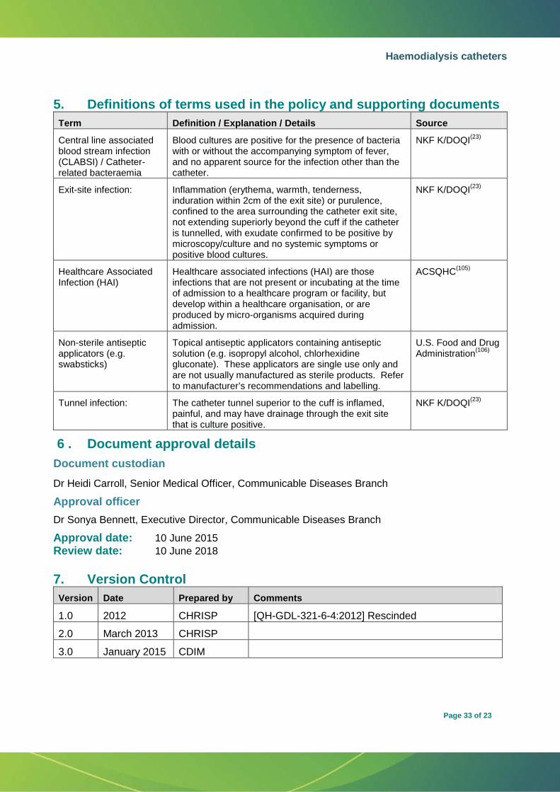
Haemodialysis catheters
Page 33 of 23
5. Definitions of terms used in the policy and supporting documents Term Definition / Explanation / Details Source
Central line associated blood stream infection (CLABSI) / Catheter-related bacteraemia
Blood cultures are positive for the presence of bacteria with or without the accompanying symptom of fever, and no apparent source for the infection other than the catheter.
NKF K/DOQI(23)
Exit-site infection: Inflammation (erythema, warmth, tenderness, induration within 2cm of the exit site) or purulence, confined to the area surrounding the catheter exit site, not extending superiorly beyond the cuff if the catheter is tunnelled, with exudate confirmed to be positive by microscopy/culture and no systemic symptoms or positive blood cultures.
NKF K/DOQI(23)
Healthcare Associated Infection (HAI)
Healthcare associated infections (HAI) are those infections that are not present or incubating at the time of admission to a healthcare program or facility, but develop within a healthcare organisation, or are produced by micro-organisms acquired during admission.
ACSQHC(105)
Non-sterile antiseptic applicators (e.g. swabsticks)
Topical antiseptic applicators containing antiseptic solution (e.g. isopropyl alcohol, chlorhexidine gluconate). These applicators are single use only and are not usually manufactured as sterile products. Refer to manufacturer’s recommendations and labelling.
U.S. Food and Drug Administration(106)
Tunnel infection: The catheter tunnel superior to the cuff is inflamed, painful, and may have drainage through the exit site that is culture positive.
NKF K/DOQI(23)
6 . Document approval details Document custodian Dr Heidi Carroll, Senior Medical Officer, Communicable Diseases Branch
Approval officer Dr Sonya Bennett, Executive Director, Communicable Diseases Branch Approval date: 10 June 2015 Review date: 10 June 2018
7. Version Control Version Date Prepared by Comments
1.0 2012 CHRISP [QH-GDL-321-6-4:2012] Rescinded
2.0 March 2013 CHRISP
3.0 January 2015 CDIM