guidelines for diagnosis and treatment of primary ciliary
TRANSCRIPT

Guidelines for Diagnosis and
Treatment of Primary Ciliary
DyskinesiaAdam J. Shapiro, MD
McGill University Health Centre
Canadian Respiratory Conference
April 2019

I have no conflict of interest.
Disclosure of Conflict of Interest(over the past 2 years)
Adam J. Shapiro, MD
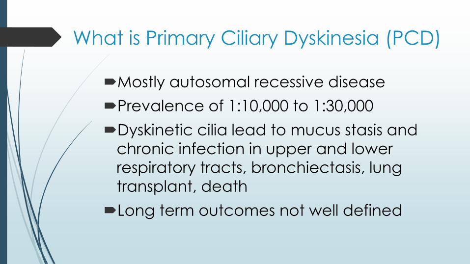
What is Primary Ciliary Dyskinesia (PCD)
Mostly autosomal recessive disease
Prevalence of 1:10,000 to 1:30,000
Dyskinetic cilia lead to mucus stasis and
chronic infection in upper and lower
respiratory tracts, bronchiectasis, lung
transplant, death
Long term outcomes not well defined

Diagnosis of Primary Ciliary Dyskinesia
Delayed diagnosis - symptoms overlap with other
diseases - CF, immune, aspiration, “daycare-itis”
Key clinical PCD symptoms in children1:
1. Year-round, wet cough starting <6 months old
2. Year-round nasal congestion starting <6 months old
3. Unexplained neonatal respiratory distress, >24 hours
of supplemental O2 or pressure support
4. Organ laterality defect (Situs inversus or ambiguus)
1Leigh et al. Annals ATS, 2016

Diagnosis of Primary Ciliary Dyskinesia
Gold-standard diagnosis was
transmission electron
microscopy (TEM) analysis of
ciliary ultrastructure
TEM normal in 30% of PCD1
Diagram of normal cilia
cross-section on electron
micrograph1Kouis et al. Pediatr Res, 2017.
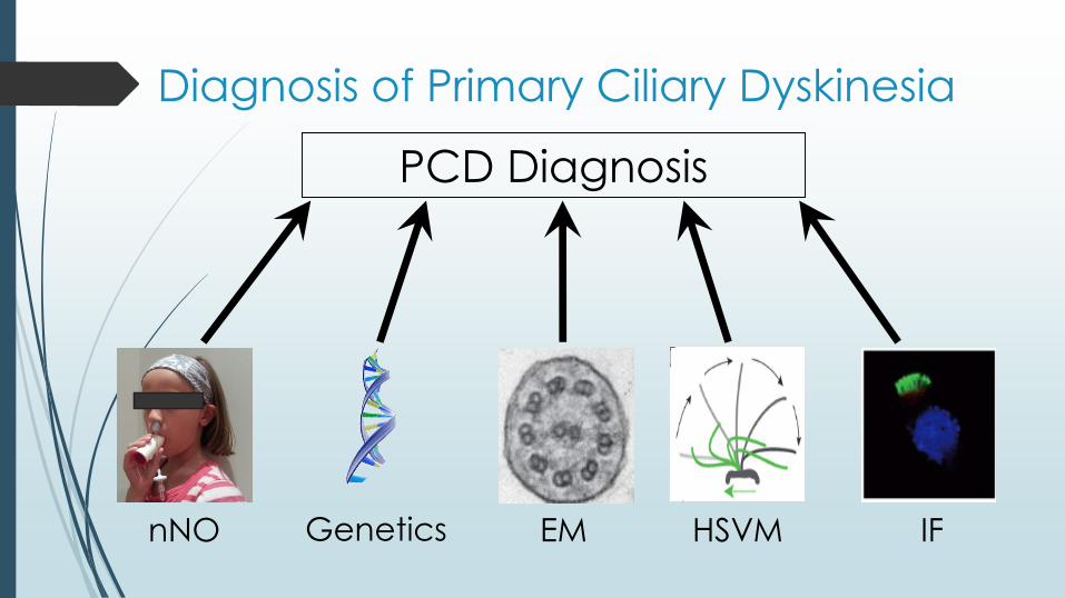
PCD Diagnosis
nNO EM HSVMGenetics IF
Diagnosis of Primary Ciliary Dyskinesia
Diagnosis of Primary Ciliary Dyskinesia
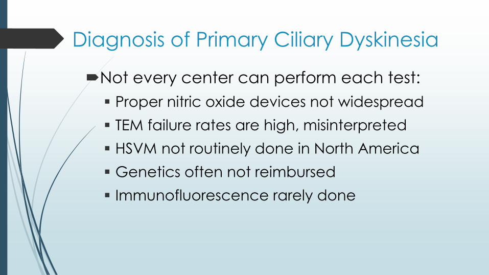
Not every center can perform each test:
▪ Proper nitric oxide devices not widespread
▪ TEM failure rates are high, misinterpreted
▪ HSVM not routinely done in North America
▪ Genetics often not reimbursed
▪ Immunofluorescence rarely done

Guidelines and Consensus
PCD Diagnosis:
▪ Shapiro AJ et al. Diagnosis of Primary Ciliary Dyskinesia. An Official American Thoracic Society Clinical Practice Guideline. Am J Respir Crit Care Med. 2018.
PCD Therapeutics:
▪ Shapiro AJ et al. Diagnosis, monitoring, and treatment of primary ciliary dyskinesia: PCD foundation consensus recommendations based on state of the art review. Pediatr Pulmonol. 2016.

30 international PCD experts - pediatric & adult
pulmonology, ENT, neonatology, radiology,
genetics, cardiology, expert methodologist, PCD
patients/parents
4 PICO questions on PCD diagnostic testing
compared against:
▪ “Reference Standard” of Classic TEM defect
and/or 2 pathogenic mutations in one PCD gene
▪ In those with a high clinical probability of PCD (at
least 2 of 4 key clinical features)
ATS Committee Composition

Should an extended genetic panel (testing
>12 genes) be used as a diagnostic test in
patients with a high probability of PCD?
▪ As replacement of classic TEM ciliary defect
and/or standard genetic panels testing ≤12
PCD genes
PICO1 – PCD Genetic Testing

# of coding exons in each of the genes implicated in Human PCD
Comparison with CFTR
Genes
# o
f co
din
g e
xo
ns
32 genes: 678 coding exons
PICO1 – PCD Genetic Testing
2007 - 9 PCD genes
2019 - 42 PCD genes
2015 - extended gene
panels commercially
available
▪ ≤12 → >34 genes
Courtesy M. Zariwala, UNC.
PICO1 – PCD Genetic Testing
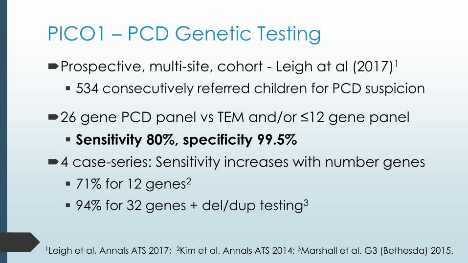
Prospective, multi-site, cohort - Leigh at al (2017)1
▪ 534 consecutively referred children for PCD suspicion
26 gene PCD panel vs TEM and/or ≤12 gene panel
▪ Sensitivity 80%, specificity 99.5%
4 case-series: Sensitivity increases with number genes
▪ 71% for 12 genes2
▪ 94% for 32 genes + del/dup testing3
1Leigh et al, Annals ATS 2017; 2Kim et al. Annals ATS 2014; 3Marshall et al. G3 (Bethesda) 2015.
In patients with high clinical probability of PCD, we suggest using an extended genetic panel as a diagnostic test over TEM &/or standard genetic panels.
Conditional recommendation - Highly feasible, prognosis through genotype, family planning
Limitations of genetic testing:
▪ NOT a rule out test
▪ NEED 2 in same PCD gene, arising in trans
▪ Variants of unknown significance are non-diagnostic, require local genetic specialists for support
PICO1: Should extended gene panel testing be used to replace EM and/or standard gene panels?

PICO2 – nNO Testing
Should a low nasal nitric oxide level be used
as a diagnostic test for PCD, in adult and
pediatric patients ≥5 years old, who are at
high probability of having PCD?
▪ With chemiluminescence technology
▪ After ruling out cystic fibrosis
▪ As a replacement of reference standard of
classic TEM structural ciliary defect and/or
biallelic causative mutations in PCD genes
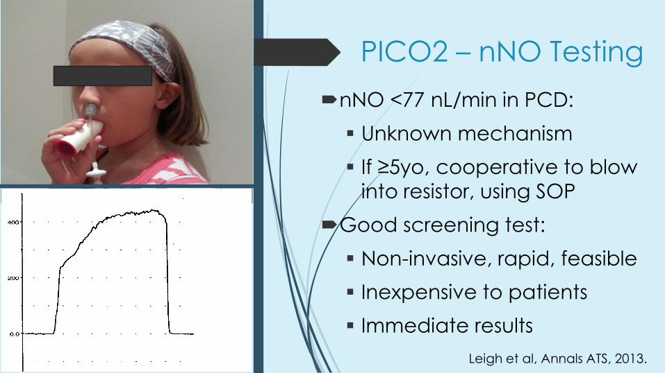
PICO2 – nNO Testing
nNO <77 nL/min in PCD:
▪ Unknown mechanism
▪ If ≥5yo, cooperative to blow
into resistor, using SOP
Good screening test:
▪ Non-invasive, rapid, feasible
▪ Inexpensive to patients
▪ Immediate results
Leigh et al, Annals ATS, 2013.
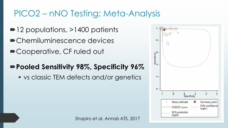
PICO2 – nNO Testing: Meta-Analysis
12 populations, >1400 patients
Chemiluminescence devices
Cooperative, CF ruled out
Pooled Sensitivity 98%, Specificity 96%
▪ vs classic TEM defects and/or genetics
Shapiro et al. Annals ATS, 2017
In cooperative patients ≥5 years old, with high clinical
probability of PCD and with CF excluded, we suggest
using nNO testing as a diagnostic test over TEM and/or
genetics - Conditional recommendation
Comment: nNO values may be transiently decreased
with acute viral respiratory infection:
▪ NEED low nNO on 2 separate occasions
▪ NEED to use a proven SOP
PICO2: Should nNO testing be used to replace TEM and/or standard gene panels?
Limitations to nNO Testing:
Lacking diagnostic nNO cut-offs for <5 years old by tidal breathing, but normal tidal values (>77 nL/min) are very reassuring
Need normal nNO levels in disease controls
Devices not Health Canada approved
Rare examples of normal nNO in PCD (RSPH1)
NOT a screening tool for general populations
PICO2 – nNO Testing

Should digital high speed videomicroscopy
with ciliary beat pattern analysis (HSVM) be
used as a PCD diagnostic test in patients In
patients with high clinical probability of PCD?
▪ As a replacement of reference standards of
classic TEM structural ciliary defect and/or
biallelic causative mutations in PCD genes.
PICO3 – HSVM Testing
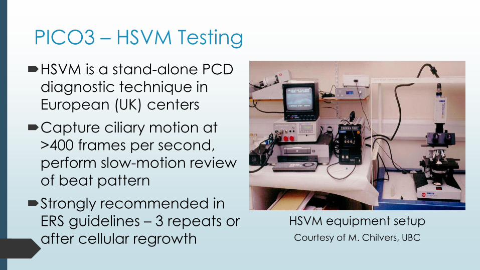
HSVM is a stand-alone PCD
diagnostic technique in
European (UK) centers
Capture ciliary motion at
>400 frames per second,
perform slow-motion review
of beat pattern
Strongly recommended in
ERS guidelines – 3 repeats or
after cellular regrowth
HSVM equipment setup
Courtesy of M. Chilvers, UBC
PICO3 – HSVM Testing

Center outside of UK shows poorly diagnostic accuracy
Non-standard interpretation techniques: Qualitative CBP
description vs semi-quantitative Dyskinesia Score
No data on repeat HSVM results at separate visits
2 studies used HSVM after cell regrowth, 50% success
All compared to TEM diagnoses, no genetic testing
PICO3 – HSVM Testing
1Hirst et al. Chest, 2010. 3Papon et al. Orphanet J Rare Dis, 2012.2Hirst et al. PLoS ONE, 2014. 4Stannard et al. Am J Respir Crit Care Med. 2010.

Pooled Sensitivity 97.3%, Specificity 96.5%, but very large confidence interval:
▪ Great variation in certainty of results
▪ Accuracy likely lower when include PCD
diagnosed by genetics
▪ Poor test feasibility across many centers
▪ Blinded, inter-rater HSVM agreement is
poor in healthy control samples1
PICO3 – HSVM Testing
1Kempeneers et al. Chest, 2017.
We suggest NOT using HSVM as a replacement diagnostic test in patients who are at high probability of having PCD - Conditional recommendation
This is a research-based test that works well in PCD centers that are highly experienced with HSVM.
The lack of feasibility, standardization, and successful cellular regrowth protocols make this test extremely difficult to perform across various clinical centers.
Should HSVM be used to replace EM and/or PCD gene testing?

PICO4 – CBF and Non-HSVM Recording
Should 1) ciliary waveform analysis using light
microscopy without high speed recording or 2)
ciliary beat frequency (CBF), be used as a PCD
diagnostic test, in patients with high clinical
probability of PCD?
▪ As replacement of reference standards of classic
TEM structural ciliary defect and/or biallelic
causative mutations in PCD genes.
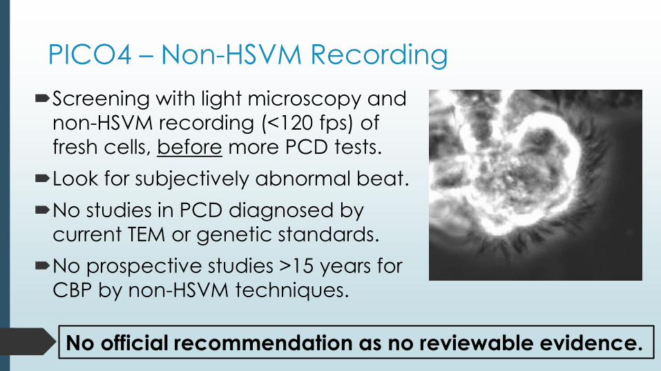
Screening with light microscopy and
non-HSVM recording (<120 fps) of
fresh cells, before more PCD tests.
Look for subjectively abnormal beat.
No studies in PCD diagnosed by
current TEM or genetic standards.
No prospective studies >15 years for
CBP by non-HSVM techniques.
PICO4 – Non-HSVM Recording
No official recommendation as no reviewable evidence.

In 3 studies, 458 patients:
▪ “Normal” CBF varies greatly between labs
▪ No genetic PCD diagnoses, only TEM
▪ Sensitivity 68-100%, specificity 61-78%
1Hirst et al. PLoS ONE, 2014; 2Olm et al. J Appl Physiol, 2011; 3Stannard et al. AJRCCM, 2010.
Should CBF alone replace EM and/or
PCD gene testing?
We suggest NOT using CBF measurement as a
diagnostic test in patients who are at high
probability of having PCD
Conditional recommendation
Should CBF alone replace EM and/or
PCD gene testing?

Summary of Diagnostic Recommendations
No single test will accurately diagnose all forms of PCD
nNO
Rarely normal, ≥5 years old
EM HSVMGenetics
≥70% detection
IF
Limited centers
$$ $$$$$ $$$FEAS+++ FEAS++ FEAS-FEAS++++ FEAS+
30% normal in PCD
False +/-results

Suggested PCD Diagnostic Algorithm

*CF ruled out†Including >12 genes, plus deletion/duplication testing
* †
Suggested PCD Diagnostic Algorithm

Guidelines and Consensus
PCD Diagnosis:
▪ Shapiro AJ et al. Diagnosis of Primary Ciliary Dyskinesia. An Official American Thoracic Society Clinical Practice Guideline. Am J Respir Crit Care Med. 2018.
PCD Therapuetics:
▪ Shapiro AJ et al. Diagnosis, monitoring, and treatment of primary ciliary dyskinesia: PCD foundation consensus recommendations based on state of the art review. Pediatr Pulmonol. 2016.

Completing the Ciliary Phenotype
Retinal examination in RPGR, visual deficits, or family
history of retinal/ciliary blindness
Genetics consultation with syndromic features
Consider abdominal/cardiac ultrasounds for laterality
issues in all PCD patients (200X risk of CHD):
▪ Occult cardiac septal defects, L-TGA
▪ If spleen anomalies: Howell-Jolly Body test
▪ If intestinal malrotation: surgical consult
Shapiro et al. Pediatr Pulmonol. 2016
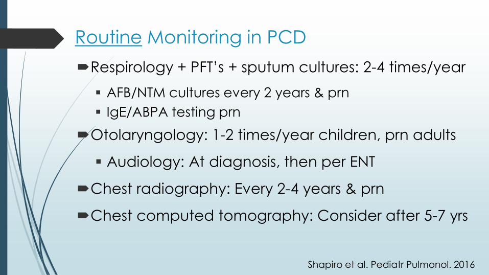
Routine Monitoring in PCD
Respirology + PFT’s + sputum cultures: 2-4 times/year
▪ AFB/NTM cultures every 2 years & prn
▪ IgE/ABPA testing prn
Otolaryngology: 1-2 times/year children, prn adults
▪ Audiology: At diagnosis, then per ENT
Chest radiography: Every 2-4 years & prn
Chest computed tomography: Consider after 5-7 yrs
Shapiro et al. Pediatr Pulmonol. 2016
Prospective Studies in PCD Therapeutics
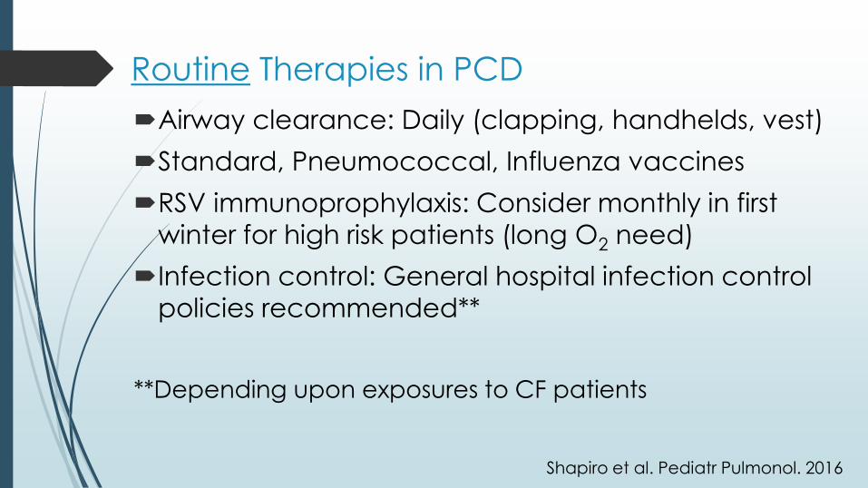
Routine Therapies in PCD
Airway clearance: Daily (clapping, handhelds, vest)
Standard, Pneumococcal, Influenza vaccines
RSV immunoprophylaxis: Consider monthly in first
winter for high risk patients (long O2 need)
Infection control: General hospital infection control
policies recommended**
**Depending upon exposures to CF patients
Shapiro et al. Pediatr Pulmonol. 2016

Antibiotics: As needed for acute exacerbations
▪ Mild – broad spectrum oral agent ≥14 days
▪ Moderate/Severe – IV therapy for ≥14 days
▪ Inhaled antibiotics not studied
Initial P. aeruginosa airway culture eradication
suggested, though not studied
Routine Therapies in PCD
Shapiro et al. Pediatr Pulmonol. 2016

Case by Case Therapies in PCD
Chronic oral suppressive antibiotics
▪ Azithromycin:
• Benefits in CF & Non-CF bronchiectasis1
• Requires NTM surveillance2
• European PCD trial complete, awaiting results3
Chronic inhaled suppressive antibiotics
▪ Chronic P. aeruginosa colonization
1Koppers et al. JAMA 2013; 2Renna et al. J Clin Invest 2011; 3Kobbernagel et al. BMC Pulm Med 2016

Inhaled bronchodilators: unclear benefits in PCD1
Inhaled hypertonic saline - 2 non-CF adult studies:
• No benefit or limited positive benefits vs NS2
• Small PCD study – no benefit in SGRQ3
• International trial with inhaled HS + ENaC inhibitor + ivacaftor complete, awaiting results4
Inhaled Dnase – 2 non-CF adult studies:
• No benefit or more exacerbations, worse PFT’s5
Case by Case Therapies in PCD
1Koh et al. Chest 2000; 2Hart et al. Cochrane Syst Rev 2014; 3Paff et al. ERJ 2017;4https://clinicaltrials.gov/ct2/show/NCT02871778; 5Wilkinson et al. Cochrane Syst Rev 2014

Therapies Not Recommended in PCD
Inhaled corticosteroids
▪ Reserved for PCD with airway reactivity or asthma
IVIG - unless proven humoral dysfunction
▪ Isolated IgA or IgG subclass deficiency does not
justify IVIG therapy in PCD
Lobectomy – Poor prognostic factor, lower PFT’s
in adults with PCD.
Shapiro et al. Pediatr Pulmonol. 2016

Lacking any prospective data on ENT in PCD
▪ Recurrent otitis media + effusion is very common
▪ Monitor regularly for hearing deficits:
• PE tubes with deficits or speech/language delay
• 80-100% have normal hearing post-PE tubes
• Post-operative otorrhea not worse than non-PCD
• Topical therapy for future otitis media
Routine ENT Therapies in PCD
Wolter et al. Int J Pediatr Otorhinolaryngol. 2012

Routine ENT Therapies in PCD
Chronic rhino-sinusitis is
omnipresent in PCD:
Need regular surveillance:
▪ Nasal endoscopy for polyps
▪ Consider chronic nasal steroids
▪ Daily sinus irrigation in PCD after
ESS improves quality of life1
▪ Does sinus therapy help lungs?
1Alanin MC et al. Int Forum Allergy Rhinol. 2017

Conclusion
PCD diagnosis in North America rests primarily
on nasal nitric oxide and genetic testing.
Other PCD diagnostic tests should remain in
expert research settings for now.
PCD monitoring and therapeutics are
borrowed from CF experiences, but PCD
clinical trial results are arriving soon…