guidelines for monitoring autophagy in higher eukaryotes
TRANSCRIPT

1
Guidelines for the Use and Interpretation of Assays for Monitoring Autophagy (2nd
edition)
Daniel J Klionsky,1,2,*
Kotb Abdelmohsen, Akihisa Abe, Md. Joynal Abedin, Hagai
Abeliovich, Abraham Acevedo Arozena, Hiroaki Adachi, Christopher M Adams, Peter D
Adams, Khosrow Adeli, Peter J. Adhihetty, Sharon G Adler, Galila Agam, Rajesh
Agarwal, Manish K Aghi, Maria Agnello, Patrizia Agostinis, Julio Aguirre-Ghiso,
Slimane Ait-Si-Ali, Takahiko Akematsu, Emmanuel T Akporiaye, Mohamed Al-Rubeai,
Guillermo M Albaiceta, Diego Albani, Matthew L Albert, Jesus Aldudo, Hana Algül,
Mehrdad Alirezaei, Iraide Alloza, Alexandru Almasan, Emad S Alnemri, Covadonga
Alonso, Dario C Altieri, Lydia Alvarez-Erviti, Sandro Alves, Giuseppina Amadoro,
Atsuo Amano, Consuelo Amantini, Santiago Ambrosio, Amal O Amer, Mohamed
Amessou, Angelika Amon, Frank A. Anania, Stig U Andersen, Usha P Andley, Catherine
K Andreadi, Nathalie Andrieu-Abadie, Alberto Anel, David K Ann, Shailendra
Anoopkumar-Dukie, Hiroshi Aoki, Nadezda Apostolova, Saveria Aquila, Katia
Aquilano, Koichi Araki, Eli Arama, Jun Araya, Alexandre Arcaro, Esperanza Arias,
Hirokazu Arimoto, Aileen R Ariosa, Thierry Arnould, Ivica Arsov, Valerie Askanas, Eric
Asselin, Ryuichiro Atarashi, Julie D Atkin, Laura D Attardi, Patrick Auberger, Georg
Auburger, Laure Aurelian, Riccardo Autelli, Laura Avagliano, Maria Laura Avantaggiati,
Laura Avagliano, Limor Avrahami, Tiziana Bachetti, Jonathan M Backer, Dong-Hun
Bae, Ok-Nam Bae, Soo Han Bae, Eric H Baehrecke, Seung-Hoon Baek, Stephen
Baghdiguian, Agnieszka Bagniewska-Zadworna, Hua Bai, Xue-Yuan Bai, Kithiganahalli
Narayanaswamy Balaji, Walter Balduini, Andrea Ballabio, Rena Balzan, Rajkumar

2
Banerjee, Gábor Bánhegyi, Haijun Bao, Esther Barreiro, Bonnie Bartel, Alberto
Bartolomé, Diane C Bassham, Maria Teresa Bassi, Robert C Bast, Jr, Henri Batoko,
Maurizio Battino, Kyle Bauckman, K Ulrich Bayer, Jean-François Beaulieu, George R
Beck Jr, J David Beckham, Patrick J Bednarski, Thomas J Begley, Christian Behl, Kevin
E Behrns, Eloy Bejarano, Amine Belaid, Francesca Belleudi, Guy Berchem, Daniele
Bergamaschi, Matteo Bergami, Ben Berkhout, Laura Berliocchi, Amélie Bernard,
Monique Bernard, Amanda S Bess, Sébastien Besteiro, Savita Bhalla, Sujit K Bhutia,
Michele Wolfe Bianchi, Martine Biard-Piechaczyk, Claudia Bincoletto, Sara W Bird,
Marc Bitoun, Guillermo A Blanco, Heidi Kiil Blomhoff, Emilio Boada-Romero, Stefan
Böckler, Marianne Boes, Andrea Boman, Paolo Bonaldo, Jürgen Bosch, Marina Bouché,
Marion Bouchecareilh, Sebastien G Bouret, Patricia Boya, Michaël Boyer-Guittaut, Peter
Bozhkov, Vania MM Braga, Claudio Brancolini, Gerhard H Braus, José M Bravo-San
Pedro, Lisa A Brennan, Emery H Bresnick, Patrick Brest, Marie-Agnès Bringer, Marisa
Brini, Glauber C Brito, Paul S Brookes, Karen Brown, Alain Bruhat, Patricia Chakur
Brum, John H Brumell, Nicola Brunetti-Pierri, Hikmet Budak, Dmitry V Bulavin, Scott J
Bultman, Geert Bultynck, Yan Burelle, Robert E Burke, Laura Caberlotto, Ken Cadwell,
Dongsheng Cai, Nadine Camougrand, Michelangelo Campanella, Grant R Campbell,
Matthew Campbell, Silvia Campello, Isabella Caniggia, Lavinia Cantoni, Lizhi Cao,
Allan B Caplan, Michele Caraglia, Claudio Cardinali, Sandra Morais Cardoso, Laura A
Carleton, Silvia Carloni, Sven R Carlsson, Didac Carmona-Gutierrez, Leticia AM
Carneiro, Serena Carra, Alice Carrier, Bernadette Carroll, Caty Casas, Giuliana
Cassinelli, Perrine Castets, Susana Castro-Obregon, Gabriella Cavallini, Isabella
Ceccherini, Francesco Cecconi, Valentín Ceña, Simone Cenci, Davide Cervia, Silvia

3
Cetrullo, Hassan Chaachouay, Han-Jung Chae, Chee-Yin Chai, Edmond YW Chan,
Dhyan Chandra, Raymond Chuen-Chung Chang, Gopal Chakrabarti, Pallavi Chandra,
Chih-Peng Chang, Saurabh Chatterjee, Yongsheng Che, Michael E Cheetham, Rajkumar
Cheluvappa, Chun-Jung Chen, Gang Chen, Gang Chen, Guang-Chao Chen, Guoqiang
Chen, Hongzhuan Chen, Jeff W Chen, Jian-Kang Chen, Mingzhou Chen, Peiwen Chen,
Qi Chen, Quan Chen, Shang-Der Chen, Si Chen, Steve S-L Chen, Wei Chen, Wenli
Chen, Xiangmei Chen, Ye-Guang Chen, Yingyu Chen, Yongshun Chen, Yu-Jen Chen,
Yujie Chen, Zhen Chen, Zhong Chen, Christopher HK Cheng, Hua Cheng, Heesun
Cheong, Chun Hei Antonio Cheung, Hsiang Cheng Chi, Sung-Gil Chi, Fulvio
Chiacchiera, Hui-Ling Chiang, Roberto Chiarelli, Mario Chiariello, Lih-Shen Chin, Gigi
NC Chiu, Dong-Hyung Cho, Ssang-Goo Cho, William C Cho, Yong-Yeon Cho, Young-
Seok Cho, Augustine MK Choi, Eui-Ju Choi, Eun-Kyoung Choi, Jayoung Choi, Mary E
Choi, Seung-Il Choi, Tsui-Fen Chou, Salem Chouaib, Divaker Choubey, Vinay Choubey,
Kuan-Chih Chow, Kamal Chowdhury, Charleen T Chu, Tsung-Hsien Chuang, Taehoon
Chun, Taijoon Chung, Yong-Joon Chwae, Roberto Ciarcia, Iwona A Ciechomska, Maria
Rosa Ciriolo, Mara Cirone, Joan Clària, Peter GH Clarke, Robert Clarke, Emilio
Clementi, Cédric Cleyrat, Miriam Cnop, Eliana M Coccia, Tiziana Cocco, Jörn Coers,
Ezra EW Cohen, David Colecchia, Núria S Coll, Emma Colucci-Guyon, Sergio
Comincini, Maria Condello, Graham H Coombs, Cynthia D Cooper, J Mark Cooper,
Isabelle Coppens, Marco Corazzari, Ramon Corbalan, Mario D Cordero, Cristina Corral-
Ramos, Olga Corti, Andrea Cossarizza, Paola Costelli, Safia Costes, Ana Coto-Montes,
Sandra Cottet, Eduardo Couve, Lori R Covey, Carolyn B Coyne, Mark S Cragg, Rolf J
Craven, Tiziana Crepaldi, Jose L Crespo, Ana Maria Cuervo, Jose M Cuezva, Pedro R

4
Cutillas, Mark J Czaja, Maria F Czyzyk-Krzeska, Ruben K Dagda, Uta Dahmen,
Chunsun Dai, Yun Dai, Kevin N Dalby, Luisa Dalla Valle, Guillaume Dalmasso,
Marcello D’Amelio, Markus Damme, Arlette Darfeuille-Michaud, Victor M Darley-
Usmar, Srinivasan Dasarathy, Srikanta Dash, Crispin R Dass, Hazel Marie Davey, Lester
M Davids, David Dávila, Roger J Davis, Ted M Dawson, Valina L Dawson, Paula Daza,
Jackie de Belleroche, Paul de Figueiredo, Regina Celia Bressan Queiroz de Figueiredo,
José de la Fuente, Luisa De Martino, Guido RY De Meyer, Angelo De Milito, Mauro De
Santi, Jayanta Debnath, Reinhard Dechant, Jean-Paul Decuypere, Shane Deegan,
Benjamin Dehay, Barbara Del Bello, Régis Delage-Mourroux, Lea MD Delbridge,
Elizabeth Delorme-Axford, Yizhen Deng, Joern Dengjel, Melanie Denizot, Paul Dent,
Vojo Deretic, Eric Deutsch, Timothy P Devarenne, Rodney J Devenish, Abhinav Diwan,
Sabrina Di Bartolomeo, Nicola Di Daniele, Fabio Di Domenico, Alessia Di Nardo,
Antonio Di Pietro, Aaron DiAntonio, Guillermo Díaz-Araya, Ines Díaz-Laviada, Maria T
Diaz-Meco, Javier Diaz-Nido, Chad A Dickey, Robert C Dickson, Marc Diederich, Paul
Digard, Ivan Dikic, Savithrama P Dinesh-Kumar, Chan Ding, Wen-Xing Ding, Luciana
Dini, Jörg HW Distler, Mojgan Djavaheri-Mergny, Kostyantyn Dmytruk, Renwick CJ
Dobson, Karol Dokladny, Kelly S Doran, Gabriella D'Orazi, Isabelle Dugail, Svetlana
Dokudovskaya, Massimo Donadelli, X Charlie Dong, Terrence M Donohue Jr, Gerald W
Dorn II, Victor Dosenko, Liat Drucker, André du Toit, Li-Lin Du, Lihuan Du,
Priyamvada Dua, Lei Duan, Michael R Duchen, Michel A Duchosal, Isabelle Dugail,
Verónica I Dumit, Mara C Duncan, William A Dunn Jr, Thomas M Durcan, Stéphane
Duvezin-Caubet, Umamaheswar Duvvuri, Vinay Eapen, Darius Ebrahimi-Fakhari,
Arnaud Echard, Charles L Edelstein, Aimee L Edinger, Ludwig Eichinger, Tobias

5
Eisenberg, Avital Eisenberg-Lerner, N Tony Eissa, Wafik S El-Deiry, Victoria El-
Khoury, Hagit Eldar-Finkelman, Enzo Emanuele, Nikolai Engedal, Anna-Mart
Engelbrecht, Simone Engelender, Jorrit M Enserink, Ralf Erdmann, Jekaterina
Erenpreisa, Andreja Erman, Ricardo Escalante, Eeva-Liisa Eskelinen, Lucile Espert,
Lorena Esteban-Martínez, Thomas J Evans, Gemma Fabrias, Cinzia Fabrizi, Antonio
Facchiano, Alberto Faggioni, Chunhai Fan, Daping Fan, Shengyun Fang, Manolis Fanto,
Alessandro Fanzani, Mathias Faure, Francois B Favier, Howard Fearnhead, Massimo
Federici, Erkang Fei, Tania C Felizardo, Hua Feng, Yibin Feng, Yuchen Feng, Thomas A
Ferguson, Álvaro F Fernández , Maite G Fernandez-Barrena, Jose C Fernandez-Checa,
Martin E Fernandez-Zapico, Elisabetta Ferraro, Carmen Veríssima Ferreira-Halder,
Laszlo Fesus, Ralph Feuer, Fabienne C Fiesel, Giuseppe Filomeni, Eduardo C Filippi-
Chiela, Gian Maria Fimia, Toren Finkel, Filomena Fiorito, Paul B Fisher, Marc Flajolet,
Flavio Flamigni, Oliver Florey, Salvatore Florio, R Andres Floto, Marco Folini, Carlo
Follo, Edward A Fon, Francesco Fornai, Franco Fortunato, Alessandro Fraldi, Rodrigo
Franco, Arnaud Francois, Aurélie François, Iain DC Fraser, Damien G Freyssenet, Scott
L Friedman, Daniel E Frigo, Dongxu Fu, José M Fuentes, Juan Fueyo, Yuuki Fujiwara,
Mikihiro Fujiya, Mitsunori Fukuda, Simone Fulda, Carmela Fusco, Matthias Gaestel,
Philippe Gailly, Malgorzata Gajewska, Gad Galili, Maria F Galindo, Lorenzo Galluzzi,
Luca Galluzzi, Vincent Galy, Sam Gandy, Anand K Ganesan, Swamynathan Ganesan,
Fen-Biao Gao, Jian-Xin Gao, Eleonora García Véscovi, Marina García-Macia, Lorena
García Nannig, Abhishek D Garg, Pramod Kumar Garg, Ricardo Gargini, Nils Christian
Gassen, Damián Gatica, Evelina Gatti, Julie Gavard, Evripidis Gavathiotis, Liang Ge,
Shengfang Ge, Po-Wu Gean, Armando A Genazzani, Pascal Genschik, David A Gewirtz,

6
Saeid Ghavami, Eric Ghigo, Anna Maria Giammarioli, Francesca Giampieri, Claudia
Giampietri, Alexandra Giatromanolaki, Derrick J Gibbings, Lara Gibellini, Spencer B
Gibson, Vanessa Ginet, Antonio Giordano, Flaviano Giorgini, Elisa Giovannetti, Stephen
E Girardin, Alvaro Glavic, Martin Gleave, Robert M Gogal Jr, Gustavo H Goldman,
Delia Goletti, Michael S Goligorsky, Aldrin V Gomes, Hernando Gomez, Candelaria
Gomez-Manzano, Rubén Gómez-Sánchez, Céline Gongora, Pedro Gonzalez-Alegre,
Pilar Gonzalez-Cabo, Rosa Ana González-Polo, Carlos Gorbea, Nikolai V. Gorbunov,
Daphne R Goring, Adrienne M Gorman, Sharon M Gorski, Sandro Goruppi, Shino
Yamada Goto, Cecilia Gotor, Roberta A Gottlieb, Illana Gozes, Devrim Gozuacik,
Yacine Graba, Martin Graef, Giovanna E Granato, Gary Dean Grant, Steven Grant,
Giovanni Luca Gravina, Douglas R Green, Alexander Greenhough, Michel T
Greenwood, Benedetto Grimaldi, Frédéric Gros, Charles Grose, Jean-Francois Groulx,
Paolo Grumati, Jun-Lin Guan, Kun-Liang Guan, Barbara Guerra, Carlos Guillen, Kailash
Gulshan, Jan Gunst, Chuanyong Guo, Lei Guo, Ming Guo, Wenjie Guo, Xu-Guang Guo,
Andrea A Gust, Åsa B Gustafsson, Ho-Shin Gwak, Albert Haas, James E Haber, Shinji
Hadano, Monica Hagedorn, Andrew J Halayko, Anne Hamacher-Brady, Isabelle Hamer,
Qutayba Hamid, Ester M Hammond, Feng Han, Xiao Han, John A Hanover, Malene
Hansen, Masaru Harada, Ljubica Harhaji-Trajkovic, J Wade Harper, Abdel Halim
Harrath, James Harris, Udo Hasler, Peter Hasselblatt, Kazuhisa Hasui, Robert G Hawley,
Congcong He, Cynthia Y He, Fengtian He, Rong-Rong He, Xian-Hui He, You-Wen He,
Joan K Heath, Marie-Josée Hébert, Gudmundur Vignir Helgason, Elizabeth P Henske,
Paul K Herman, Sonia Hernández-Tiedra, Claudio Hetz, P. Robin Hiesinger, Sabine
Hilfiker, Bradford G Hill, Joseph A Hill, William D Hill, Keisuke Hino, Paul Hofman,

7
Jörg Höhfeld, Marina K Holz, Yonggeun Hong, David A Hood, Daniel Hofius, Günter U
Höglinger, Jincai Hou, Chin Hsu, Li-Chung Hsu, Dong Hu, Hong-Ming Hu, Hongbo Hu,
Ming Chang Hu, Yu-Chen Hu, Zhuo-Wei Hu, Fang Hua, Ya Hua, Canhua Huang, Huey-
Lan Huang, Kuo-How Huang, Kuo-Yang Huang, Shiqian Huang, Wei-Pang Huang,
Yunfei Huang, Tobias B Huber, Patricia Huebbe, Won-Ki Huh, Juha J Hulmi, Gang Min
Hur, James H Hurley, Zvenyslava Husak, Sabah NA Hussain, Salik Hussain, Jung Jin
Hwang, Seungmin Hwang, Thomas IS Hwang, Atsuhiro Ichihara, Yuzuru Imai, Carol
Imbriano, Megumi Inomata, Takeshi Into, Valentina Iovane, Juan L Iovanna, Renato V
Iozzo, Nancy Y Ip, Pablo Iribarren, Yoshitaka Isaka, Aleksandra J Isakovic, Hiroyuki
Ishida, Isao Ishii, Jane E. Ishmael, Ciro Isidoro, Ken-ichi Isobe, Shohreh Issazadeh-
Navikas, Koji Itahana, Anand Krishnan V Iyer, Yotaro Izumi, Marja Jäättelä, Nadia
Jaber, Daniel John Jackson, William T Jackson, Tony George Jacob, Chinnaswamy
Jagannath, Alkesh Jani, Bassam Janji, Paulo Roberto Jannig, Patric J Jansson, Marina
Jendrach, Ju-Hong Jeon, Eui-Bae Jeung, Kailiang Jia, Lijun Jia, Hong Jiang, Liwen
Jiang, Teng Jiang, Xiaoyan Jiang, Xuejun Jiang, Xuejun Jiang, Yongjun Jiang, Alberto
Jiménez, Cheng Jin, Hongchuan Jin, Meiyan Jin, Shengkan Jin, Umesh Kumar Jinwal,
Eun-Kyeong Jo, Terje Johansen, Daniel E Johnson, Gail VW Johnson, James D Johnson,
Eric Jonasch, Chris Jones, Leo AB Joosten, Joaquin Jordan, Anna-Maria Joseph,
Bertrand Joseph, Annie M Joubert, Dianwen Ju, Hsueh-Fen Juan, Katrin Juenemann,
Gábor Juhász, Hye Seung Jung, Jae U Jung, Yong-Keun Jung, Heinz Jungbluth, Matthew
J Justice, Barry Jutten, Nadeem O Kaakoush, Allen Kaasik, Tomohiro Kabuta, Katarina
Kågedal, Alon Kahana, Or Kakhlon, Dhan V Kalvakolanu, Vitaliy O Kaminskyy,
Mustapha Kandouz, Chanhee Kang, Rui Kang, Tae-Cheon Kang, Tomotake Kanki,

8
Thirumala-Devi Kanneganti, Haruo Kanno, Anumantha G Kanthasamy, Marc Kantorow,
Maria Kaparakis-Liaskos, Orsolya Kapuy, Vassiliki Karantza, Md. Razaul Karim,
Parimal Karmakar, Arthur Kaser, A Murat Kaynar, Po-Yuan Ke, John H Kehrl, Kate E
Keller, Oliver Kepp, Andreas Kern, Santosh Kesari, David Kessel, Robin Ketteler,
Muzamil Majid Khan, Juliann G Kiang, Akio Kihara, Arianna L Kim, Cheol Hyeon Kim,
Deok Ryong Kim, Do-Hyung Kim, Eung Kweon Kim, Hye Young Kim, Hyung-Ryong
Kim, Jae-Sung Kim, Jeong Hun Kim, Jin Cheon Kim, Jin Hyoung Kim, Peter K Kim,
Seong Who Kim, Yonghyun Kim, Adi Kimchi, Alec C Kimmelman, Jason S King,
Lorrie A Kirshenbaum, Shuji Kishi, Katsuhiko Kitamoto, Kaio Kitazato, Rudolf A Kley,
Walter T Klimecki, Jochen Klucken, Helene Knævelsrud, Erwin Knecht, Jiunn-Liang
Ko, Satoru Kobayashi, Christelle Koechlin-Ramonatxo, Ulrich Koenig, Young Ho Koh,
Katja Köhler, Sepp D Kohlwein, Masaaki Komatsu, Hee Jeong Kong, Eumorphia G
Konstantakou, Benjamin T Kopp, Tamas Korcsmaros, Laura Korhonen, Viktor I
Korolchuk, Nadezhda V Koshkina, Michael I Koukourakis, Constantinos Koumenis,
Attila L Kovács, Daisuke Koya, Claudine Kraft, Dimitri Krainc, Roswith Krick, Janos
Kriston-Vizi, Guido Kroemer, Rejko Kruger, Nicholas T Ktistakis, Kazuyuki Kuchitsu,
A Pratap Kumar, Ashok Kumar, Deepak Kumar, Dhiraj Kumar, Sharad Kumar, Hsing-
Jien Kung, Atsushi Kuno, Jeff Kuret, Tino Kurz, Taeg Kyu Kwon, Yong Tae Kwon,
Albert R La Spada, Frank Lafont, Tim Lahm, Truong Lam, Terry H Landowski, Jon D
Lane, Cinzia Lanzi, Louis R Lapierre, Jocelyn Laporte, Gordon W Laurie, Sergio
Lavandero, Lena Lavie, Kelsey B Law, Pedro A Lazo, Laurent Le Cam, Karine G Le
Roch, Vijittra Leardkamolkarn, Marc Lecuit, Byung-Hoon Lee, Che-Hsin Lee, Gyun Min
Lee, He-Jin Lee, Hsinyu Lee, Jae Keun Lee, Jongdae Lee, Jun Hee Lee, Michael Lee,

9
Myung-Shik Lee, Sam W Lee, Seung-Jae Lee, Shiow-Ju Lee, Sug Hyung Lee, Sung Sik
Lee, Sung-Joon Lee, Sunhee Lee, Yong J Lee, Young H Lee, Christiaan Leeuwenburgh,
Sylvain Lefort, Renaud Legouis, Jinzhi Lei, Qun-Ying Lei, David A Leib, Gil
Leibowitz, Stéphane D Lemaire, John J Lemasters, Antoinette Lemoine, Guido Lenz,
Lilach O Lerman, Daniele Lettieri Barbato, Julia I-Ju Leu, Hing Y Leung, Beth Levine,
Patrick A Lewis, Chi Li, Dan Li, Faqiang Li, Feng-Jun Li, Jun Li, Ke Li, Lian Li, Min Li,
Min Li, Qiang Li, Rui Li, Sheng Li, Wei Li, Xiaotao Li, Yumin Li, Jiqin Lian, Chengyu
Liang, Yulin Liao, Pawel P Liberski, Andrew P Lieberman, Kyu Lim, Chang-Shen Lin,
Chiou-Feng Lin, Fang Lin, Fangming Lin, Fu-Cheng Lin, Kui Lin, Kwang-Huei Lin,
Tianwei Lin, Wan-Wan Lin, Yee-Shin Lin, Yong Lin, Rafael Linden, Dan Lindholm,
Lisa M Lindqvist, Lance A Liotta, Marta M Lipinski, Vitor A Lira, Michael P Lisanti,
Paloma B Liton, Bo Liu, Chun-Feng Liu, Fei Liu, Hung-Jen Liu, Jianxun Liu, Jing-Jing
Liu, Jing-Lan Liu, Ke Liu, Leyuan Liu, Quentin Liu, Rong-Yu Liu, Shiming Liu, Wei
Liu, Xian-De Liu, Xiangguo Liu, Xiao-Hong Liu, Xinfeng Liu, Xu Liu, Yule Liu, Zexian
Liu, Susan E Logue, Sagar Lonial, Ben Loos, Carlos López-Otín, Cristina López-Vicario,
Mar Lorente, Péter Lőrincz, Marek Los, Michael T Lotze, Penny E Lovat, Binfeng Lu,
Bo Lu, Jiahong Lu, Shemin Lu, Shuyan Lu, Frédéric Luciano, Shirley Luckhart, John
Milton Lucocq, Paula Ludovico, Aurelia Lugea, Nicholas W Lukacs, Julian J Lum,
Anders H Lund, Honglin Luo, Jia Luo, Shouqing Luo, Glaucia M Machado-Santelli,
Gustavo Macintosh, Jeffrey P MacKeigan, Kay F Macleod, John D MacMicking, Frank
Madeo, Muniswamy Madesh, Akiko Maeda, Emilia Maellaro, Hannelore Maes, Marta
Magariños, Kenneth Maiese, Tapas K Maiti, Luigi Maiuri, Maria Chiara Maiuri, Carl G
Maki, Roland Malli, Walter Malorni, Na Man, Eva-Maria Mandelkow, Angelo A

10
Manfredi, Claudia Manzoni, Kai Mao, Zixu Mao, Zong-Wan Mao, Philippe Marambaud,
Anna Maria Marconi, Gabriella Marfe, Marta Margeta, Eva Margittai, Muriel Mari,
Francesca V Mariani, Concepcio Marin, Sara Marinelli, Guillermo Mariño, Ivanka
Markovic, Rebecca Marquez, Alberto M Martelli, Sascha Martens, Katie R Martin,
Seamus J Martin, Miguel A Martin-Acebes, Paloma Martín-Sanz, Wim Martinet, Jennifer
Martinez, Ubaldo Martinez-Outschoorn, Moisés Martínez-Velázquez, Marta Martinez-
Vicente, Hirosato Mashima, James A Mastrianni, Giuseppe Matarese, Paola Matarrese,
Roberto Mateo, Satoaki Matoba, Naomichi Matsumoto, Takehiko Matsushita, Takeshi
Matsuzawa, Mark P Mattson, Soledad Matus, Norma Maugeri, Caroline Mauvezin,
Dusica Maysinger, Kimberly McCall, Craig McCormick, Gerald M McInerney, Skye C
McIver, Sharon McKenna, John J McMahon, Fatima Mechta-Grigoriou, Maryam
Mehrpour, Jawahar L Mehta, Yide Mei, Ute-Christiane Meier, Alfred J Meijer, Alicia
Meléndez, Gerry Melino, Sonia Melino, Maria Á Mena, Marc D Meneghini, Javier A
Menendez, Liesu Meng, Ling-hua Meng, Songshu Meng, Rossella Menghini, Rubem FS
Menna-Barreto, A Sue Menko, Manoj B Menon, Giuseppe Merla, Luciano Merlini,
Angelica M Merlot, Andreas Meryk, Stefania Meschini, Joel N Meyer, Mantian Mi,
Chao-Yu Miao, Simon Michaeli, Anna Rita Migliaccio, Dalibor Mijaljica, Enrico Milan,
Leonor Miller-Fleming, Ian G Mills, Georgia Minakaki, Berge A Minassian, Farida
Minibayeva, Elena A Minina, Justine Mintern, Saverio Minucci, Claire H Mitchell,
Keisuke Miyazawa, Noboru Mizushima, Katarzyna Mnich, Baharia Mograbi, Simin
Mohseni, Luis Ferreira Moita, Marco Molinari, Maurizio Molinari, Bertrand Mollereau,
Faustino Mollinedo, Marco Mongillo, Serena Montagnaro, Craig Montell, Darren J
Moore, Etienne Morel, Maria Beatrice Morelli, Sandra Moreno, Yuji Moriyasu, Janna L

11
Morrison, Lynda A Morrison, Pope L Moseley, Serge Mostowy, Elisa Motori, Charbel E-
H Moussa, Vassiliki E Mpakou, Jean M Mulcahy Levy, Sylviane Muller, Raquel Muñoz-
Moreno, Cristina Muñoz-Pinedo, Christian Münz, Maureen E Murphy, James T Murray,
Indira Mysorekar, Ivan R Nabi, Massimo Nabissi, Gustavo A Nader, Kazuhiro Nagata,
Péter Nagy, Samisubbu R Naidu, Sreejayan Nair, Hiroyasu Nakano, Hitoshi Nakatogawa,
Meera Nanjundan, Naweed I Naqvi, Roberta Nardacci, Masashi Narita, Steffan T
Nawrocki, Thomas Neill, Luca M Neri, Mihai G Netea, Paul A Ney, Ioannis P Nezis,
Hang TT Nguyen, Huu Phuc Nguyen, Anne-Sophie Nicot, Hilde Nilsen, Mikio
Nishimura, Ichizo Nishino, Mireia Niso-Santano, Hua Niu, Ralph Nixon, Vincent CO
Njar, Takeshi Noda, Angelika A Noegel, Koenraad K Norga, Shoji Notomi, Lucia
Notterpek, Karin Nowikovsky, Nobuyuki Nukina, Thorsten Nürnberger, Peter J
O'Dwyer, Ina Oehme, Clara L Oeste, Michinaga Ogawa, Besim Ogretmen, Yuji Ogura,
Young J Oh, Koji Okamoto, Toshiro Okazaki, F Javier Oliver, Stefan Olsson, Daniel P
Orban, Eyleen J O'Rourke, Angel L Ortega, Elena Ortona, Laura D Osellame, Takanobu
Otomo, Jing-hsiung James Ou, Tiago F Outeiro, Dong-yun Ouyang, Hongjiao Ouyang,
Michael Overholtzer, P Hande Ozdinler, Bulent Ozpolat, Consiglia Pacelli, Guylène
Page, Ugo Pagnini, Stephen C Pak, Karolina Pakos-Zebrucka, Nazzy Pakpour, Zdena
Palkova, Kathrin Pallauf, Nicolas Pallet, Marta Palmieri, Søren R Paludan, Camilla
Palumbo, Olatz Pampliega, Wei Pan, Theocharis Panaretakis, Areti Pantazopoulou,
Issidora Papassideri, Alessio Papini, Julian Pardo, Vrajesh V Parekh, Giancarlo Parenti,
Jong-In Park, Junsoo Park, Ohkmae K Park, Roy Parker, Jan B Parys, Katherine R
Parzych, Jean-max Pasquet, Sophie Pattingre, Flaminia Pavone, Zully Pedrozo, Fernando
J Peña, Miguel A Peñalva, Mario Pende, Jianxin Peng, Fabio Penna, Josef M Penninger,

12
Paulo C Pereira, María Esther Pérez-Pérez, Dolores Pérez-Sala, Andras Perl, David H
Perlmutter, Ida Perrotta, Jeffrey E Pessin, Godefridus J Peters, Irina Petrache, Basil J
Petrof, Goran Petrovski, James M Phang, Marina Pierdominici, Philippe Pierre, Valérie
Pierrefite-Carle, Felipe X Pimentel-Muiños, Marcello Pinti, Paolo Pinton, Bilal Piperdi,
James M Piret, Leonidas C Platanias, Harald W Platta, Edward D Plowey, Stefanie
Pöggeler, Marc Poirot, Peter Polčic, Angelo Poletti, Audrey H Poon, Hana Popelka,
Blagovesta Popova, Scott K Powers, Mercedes Pozuelo-Rubio, Krisna Prak, Reinhild
Prange, Mark Prescott, Sharon Prince, Richard L Proia, Tassula Proikas-Cezanne, Vasilis
J Promponas, Karin Przyklenk, Rosa Puertollano, Subbiah Pugazhenthi, Julian Puyal,
Dohun Pyeon, Xin Qi, Zheng-Hong Qin, Yu Qiu, Joe Quadrilatero, Frederick Quinn,
Nina Raben, Hannah Rabinowich, Komal Raina, Rajagopal Ramesh, Abdelhaq Rami,
Felix Randow, Hai Rao, V Ashutosh Rao, Blake B Rasmussen, Tobias M Rasse, Edward
A Ratovitski, Pierre-Emmanuel Rautou, Swapan K Ray, Babak Razani, Bruce H Reed,
Fulvio Reggiori, Markus Rehm, Andreas S Reichert, Theo Rein, Eric Reits, Jun Ren,
Xingcong Ren, Jose L Revuelta, Alireza R Rezaie, Des R Richardson, Michael A Riehle ,
Bertrand H Rihn, Yasuko Rikihisa, Brigit E Riley, Gerald Rimbach, Konstantinos Ritis,
Peter J Roach, Jeffrey Robbins, Michel Roberge, Gabriela Roca, Maria Carmela
Roccheri, Sonia Rocha, Cecilia MP Rodrigues, Clara I Rodríguez, Santiago Rodriguez de
Cordoba, Troy T Rohn, Bärbel Rohrer, Davide Romanelli, Luigina Romani, Patricia
Silvia Romano, M Isabel G Roncero, Jose Luis Rosa, Alicia Rosello, Philip Rosenstiel,
Magdalena Rost-Roszkowska, Kevin A Roth, Gael Roué, Mustapha Rouis, Kasper M
Rouschop, Daniel T Ruan, Diego Ruano, David C Rubinsztein, Edmund B Rucker III,
Emil Rudolf, Carmen Ruiz-Roldán, Ruediger Rudolf, Markus A Ruegg, Giuseppe Russo,
13
Rossella Russo, Victoria Ryabovol, Kevin M Ryan, Stefan W Ryter, David M Sabatini,
Michael Sacher, Carsten Sachse, Junichi Sadoshima, Paul Saftig, Ronit Sagi-Eisenberg,
Sumit Sahni, Koichi Sakakura, María Salazar-Roa, Paolo Salomoni, Paul M Salvaterra,
Rosa Salvioli, Afshin Samali, José A Sánchez-Alcázar, Ricardo Sanchez-Prieto, Marco
Sandri, Miguel A Sanjuan, Stefano Santaguida, Laura Santambrogio, Giorgio Santoni,
Graeme Sargent, Sovan Sarkar, Maria-Rosa Sarrias, Minnie M Sarwal, Chihiro
Sasakawa, Motoko Sasaki, Miklos Sass, Ken Sato, Miyuki Sato, Joseph Satriano,
Svetlana Saveljeva, Liliana Schaefer, Ulrich E Schaible, Michael Scharl, Randy
Schekman, Wiep Scheper, Alfonso Schiavi, Hyman M Schipper, Hana Schmeisser, Jens
Schmidt, Ingo Schmitz, Bianca E Schneider, E Marion Schneider, Jaime L Schneider,
Eric A Schon, Bernd Schröder, Ryan J Schulze, Melanie Schwarten, Thomas L Schwarz,
Sebastiano Sciarretta, A Ivana Scovassi, Robert A Screaton, Nava Segev, Per O Seglen,
Jose M Seguí-Simarro, Juan Segura-Aguilar, Iban Seiliez, Ekihiro Seki, Clay F
Semenkovich, Gregg L Semenza, Utpal Sen, Andreas L Serra, Hiromi Sesaki, Takao
Setoguchi, Carmine Settembre, John J Shacka, Ayesha N Shajahan-Haq, Irving M
Shapiro, C-K James Shen, Chiung-Chyi Shen, Han-Ming Shen, Sanbing Shen, Weili
Shen, Rui Sheng, Zu-Hang Sheng, Trevor G Shepherd, Junyan Shi, Qinghua Shi,
Yuguang Shi, Yoshihiro Shidoji, Jeng-Jer Shieh, Chwen-Ming Shih, Shigeomi Shimizu,
Dong Wook Shin, Michiko Shintani, Takahiro Shintani, Tetsuo Shioi, Ronit Shiri-
Sverdlov, Orian Shirihai, Gordon C Shore, Andriy A Sibirny, Christina J Sigurdson,
Einar M Sigurdsson, Sandrine Silvente-Poirot, Gary A Silverman, Cristiano Simone,
Balindiwe JN Sishi, Anna Katharina Simon, Hans-Uwe Simon, Anne Simonsen, Rajat
Singh, Shivendra V Singh, Debasish Sinha, Frank A Sinicrope, Agnieszka Sirko, Annie

14
Sittler, Parco M Siu, Efthimios Sivridis, Ruth Slack, Iva Slaninová, Soraya S Smaili,
Duncan R Smith, Jin H Son, Avinash Sonawane, Fuyong Song, Hyun Kyu Song, Ju-Xian
Song, Wei Song, Kai Y Soo, Anil K Sood, Tuck Wah Soong, Virawudh
Soontornniyomkij, Maurizio Sorice, Federica Sotgia, Areechun Sotthibundhu, Anne
Spang, Herman P Spaink, Stephen A Spector, Claudia D Spies, Wolfdieter Springer,
Daret St. Clair, Michael T Stang, Daniel T Starczynowski, Petro Starokadomskyy,
Clemens Steegborn, Joan Steffan, Harald Stenmark, Stephan T Stern, Craig Stevens,
Brent R Stockwell, Zuzana Storchova, Björn Stork, Dimitrios J Stravopodis, Pavel
Strnad, Anna-Lena Ström, Per Stromhaug, Jiri Stulik, Carlos S Subauste, Sang Won Suh,
Fang-Lin Sun, Jiaren Sun, Shi-Yong Sun, Yang Sun, Yi Sun, Yingjie Sun, Kuninori
Suzuki, Naoki Suzuki, Tadashi Suzuki, Yuichiro J Suzuki, Michele S Swanson, Charles
Swanton, Karl Swärd, Ghanshyam Swarup, Sean T Sweeney, Zsuzsanna Szatmari, Eva
Szegezdi, Peter W Szlosarek, Emmanuel Taillebourg, Stephen WG Tait, Yoshinori
Takahashi, Szabolcs Takáts, Genzou Takemura, Nicholas J Talbot, Elena Tamagno,
Jerome Tamburini, Cai-Ping Tan, Lan Tan, Mei Lan Tan, Ming Tan, Yee-Joo Tan, Keiji
Tanaka, Masaki Tanaka, Daolin Tang, Dingzhong Tang, Isei Tanida, Kunikazu Tanji,
Bakhos A Tannous, Inmaculada Tasset-Cuevas, Marc Tatar, Nektarios Tavernarakis,
Allen Taylor, Gregory A Taylor, J Paul Taylor, Andrew R Tee, Steven Teitelbaum, Ru-
Jeng Teng, Faraj Terro, Fatima Teixeira-Clerc, Gianluca Tettamanti, Andrew Thorburn,
Marcos P Thomé, Paul G Thomes, Thomas Thum, Michael Thumm, Ling Tian, Andreas
Till, Jenny Pan-yun Ting, Lilach Toker, Sharon A Tooze, Ivan Topisirovic, Maria
Lyngaas Torgersen, Liliana Torosantucci, Alicia Torriglia, Maria Rosaria Torrisi, Cathy
Tournier, Vladimir Trajkovic, Leonardo H Travassos, Daniela Trisciuoglio, Rodrigo

15
Troncoso, Ioannis P Trougakos, Anita C Truttmann, Kuen-Jer Tsai, Mario P Tschan, Yi-
Hsin Tseng, Allan Tsung, Shuiping Tu, Hsing-Yu Tuan, Boris Turk, Robin FB Turner,
Suresh C Tyagi, Andrej Udelnow, Takashi Ueno, Midori Umekawa, Christian
Ungermann, Ryo Ushioda, Vladimir N Uversky, Néstor L Uzcátegui, Thomas Vaccari,
Maria I Vaccaro, Libuse Vachova, Enza Maria Valente, Angela M Valverde, Greet Van
den Berghe, Gijs R van den Brink, F Gisou van der Goot, Ida J van der Klei, Luc JW van
der Laan, Wouter G van Doorn, Marjolein van Egmond, Kenneth L van Golen, Luc Van
Kaer, Peter Vandenabeele, Wim Vandenberghe, Ilse Vanhorebeek, Isabel Varela-Nieto,
Radovan Vasko, Demetrios G Vavvas, Ignacio Vega-Naredo, Guillermo Velasco,
Athanassios D Velentzas, Panagiotis D Velentzas, Edo Vellenga, Kartik Venkatachalam,
Natascia Ventura, Patrícia ST Veras, Beata G Vertessy, Richard D Vierstra, Nadarajah
Vigneswaran, Neeraj Vij, Miquel Vila, Joan Villarroya, Cécile Vindis, Giampietro Viola,
Maria Teresa Viscomi, Giovanni Vitale, Dan T Vogl, Clarissa von Haefen, Karin von
Schwarzenberg, Valérie Vouret-Craviari, Kristiina Vuori, Jatin M Vyas, Mark J Walker,
Jochen Walter, Lei Wan, Xiangbo Wan, Bo Wang, Caihong Wang, Chao-Yung Wang,
Chengshu Wang, Chenran Wang, Chuangui Wang, Dong Wang, Fen Wang, Guanghui
Wang, Hai-jie Wang, Haichao Wang, Hong-Gang Wang, Horng-Dar Wang, Jing Wang,
Junjun Wang, Mei Wang, Pei-Yu Wang, Peng Wang, Richard C Wang, Shuo Wang,
Ting-Fang Wang, Xian Wang, Xiao-jia Wang, Xiao-Wei Wang, Xuejun Wang, Yanming
Wang, Ying Wang, Ying-Jan Wang, Yipeng Wang, Yu Tian Wang, Yuqing Wang,
Zhinong Wang, Pablo Wappner, Carl Ward, Gary Warnes, Hirotaka Watada, Yoshihisa
Watanabe, Timothy E Weaver, Conrad C Weihl, Simone Nardin Weis, Longping Wen,
Xin Wen, Yunfei Wen, Benedikt Westermann, Cornelia M Weyand, Anthony R White,

16
Alexander J Whitworth, Eileen White, J Lindsay Whitton, Franziska Wild, Tom
Wileman, Deepti Srinivas Wilkinson, Simon Wilkinson, Dieter Willbold, Chris Williams,
Peter R Williamson, Steven S Witkin, Stephanie E Wohlgemuth, Thomas Wollert, Ernst J
Wolvetang, Esther Wong, Richard W Wong, Karen L Wright, Chunlai Wu, Defeng Wu,
Jian Wu, Junfang Wu, Mian Wu, William KK Wu, Zhenlong Wu, Ramnik J Xavier, Tian
Xia, Weiliang Xia, Yong Xia, Hengyi Xiao, Shi Xiao, Wuhan Xiao, Chuan-Ming Xie,
Zhiping Xie, Zhonglin Xie, Chuanshan Xu, Congfeng Xu, Feng Xu, Haoxing Xu,
Hongwei Xu, Jian Xu, Jianzhen Xu, Jinxian Xu, Liang Xu, Xiaolei Xu, Yu Xue,
Takahiro Yamada, Ai Yamamoto, Shunhei Yamashina, Bo Yan, Xianghua Yan, Zhen
Yan, Dun-Sheng Yang, Jiin-Ming Yang, Minghua Yang, Pei-Ming Yang, Qian Yang,
Sijun Yang, Wannian Yang, Wei Yuan Yang, Yi Yang, Ying Yang, Meng-Chao Yao,
Zhenyu Yao, Behzad Yeganeh, Long Yi, Xiao-Ming Yin, Calvin K Yip, Yeong-Min
Yoo, Young Hyun Yoo, Seung-Yong Yoon, Kenichi Yoshida, Tamotsu Yoshimori, Ken
H Young, Huixin Yu, Jane J Yu, Jin-Tai Yu, Jun Yu, Li Yu, Zhengping Yu, Junying
Yuan, Zhi-Min Yuan, Beatrice YJT Yue, Zhenyu Yue, David N Zacks, Eldad
Zacksenhaus, Nadia Zaffaroni, Tania Zaglia, Zahra Zakeri, Jinsheng Zeng, Min Zeng, Qi
Zeng, Antonis S Zervos, Donna D Zhang, Fan Zhang, Guo-Chang Zhang, Guo Zhang,
Hao Zhang, Hong Zhang, Hong Zhang, Hongbing Zhang, Jianhua Zhang, Jing-pu Zhang,
Li Zhang, Lin Zhang, Long Zhang, Ming-Yong Zhang, Xiangnan Zhang, Xu Dong
Zhang, Yan Zhang, Yingmei Zhang, Mei Zhao, Wei-Li Zhao, Xiaonan Zhao, Yan G
Zhao, Ying Zhao, Yongchao Zhao, Yu-xia Zhao, Zhendong Zhao, Dexian Zheng, Xi-
Long Zheng, Xiaoxiang Zheng, Boris Zhivotovsky, Qing Zhong, Guofei Zhou, Shufeng

17
Zhou, Xu-jie Zhou, Hongxin Zhu, Wei-Guo Zhu, Wenhua Zhu, Xiao-Feng Zhu, Shi-Mei
Zhuang, Elio Ziparo, Christos E Zois, Teresa Zoladek, Wei-Xing Zong, Antonio Zorzano
1Life Sciences Institute, and
2Department of Molecular, Cellular and Developmental
Biology University of Michigan, Ann Arbor, MI USA;
Running title: Autophagy guidelines
Keywords: autolysosome, autophagosome, flux, LC3, lysosome, phagophore, stress,
vacuole
*Corresponding author: Correspondence: Daniel J. Klionsky, Life Sciences Institute,
University of Michigan, Ann Arbor, MI 48109-2216. Tel. 734-615-6556; Fax. 734-647-
9702; Email: [email protected]

18
Table of Contents
Introduction
A. Methods for monitoring autophagy
1. Transmission electron microscopy……………………………………… XX
2. Atg8/LC3 detection and quantification…………………………………. XX
a. Western blotting and ubiquitin-like protein conjugation systems…… XX
b. Turnover of LC3-II/Atg8–PE………………………………………... XX
c. GFP-Atg8/LC3 lysosomal delivery and partial proteolysis………….. XX
d. GFP-Atg8/LC3 fluorescence microscopy…………………………….. XX
e. Tandem mRFP/mCherry-GFP fluorescence microscopy…………….. XX
f. Autophagic flux determination using flow and multispectral imaging
cytometry…………………………………………………………….. XX
g. Immunohistochemistry…………………………………………….... XX
3. SQSTM1 and related LC3 binding protein turnover assays…………….. XX
4. MTOR, AMPK and Atg1/ULK1……………………………………….. XX
5. Additional autophagy-related protein markers………………………….. XX
a. Atg9……………………………………………………………..….. XX
b. ATG12–ATG5……………………………………………………..….. XX
c. ATG14…………………………………………………………..….. XX
d. ATG16L1………………………………………………………..….. XX
e. Atg18/WIPI family……………………………………………..….. XX
f. BECN1/Vps30/Atg6………………………………………………..….. XX
g. DRAM1…………………………………………………………..….. XX

19
h. ZFYVE1/DFCP1………………………………………………..….. XX
i. STX17, TECPR1…………………………………………………… XX
6. Sphingolipids…………………………………………………………… XX
7. Transcriptional, translational and posttranslational
regulation…………………………….. XX
8. Posttranslational modification of ATG proteins………………………... XX
9. Autophagic protein degradation…………….………………………….. XX
10. Selective types of autophagy……………………….………………….. XX
a. The Cvt pathway, mitophagy, pexophagy, piecemeal microautophagy
of the nucleus and nucleophagy in yeast and filamentous fungi.. XX
b. Reticulophagy and ribophagy…………………………………..….. XX
c. Vacuole import and degradation pathway………………………….. XX
d. Animal mitophagy and pexophagy………………………..……….. XX
e. Aggrephagy……………………………………………………..….. XX
f. Allophagy…………………………………………………..…..….. XX
g. Chlorophagy……………………………………………………….. XX
h. Chromatophagy……………………………………………..…..….. XX
i. Ferritinophagy……………………………………………………… XX
j. Lipophagy ……………………………………………………..….. XX
k. Intraplastidial autophagy…………………………………………. XX
l. RNA-silencing components………………………………………. XX
m. Xenophagy.………………………………………….…………….. XX
n. Zymophagy……………………………………………………..….. XX

20
11. Autophagic sequestration assays……………………………..……….. XX
12. Turnover of autophagic compartments……………………………….. XX
13. Autophagosome-lysosome colocalization and dequenching assay…… XX
14. Tissue fractionation………………………………………………..….. XX
15. Analyses in vivo…………………………………………………..….. XX
16. Clinical setting……………………………………………………….. XX
17. Cell death……………………………………………..…………..….. XX
18. Chaperone-mediated autophagy…………………………………..….. XX
B. Comments on additional methods…………………………………..….. XX
1. Acidotropic dyes……………………………..……………………..….. XX
2. Autophagy inhibitors and inducers…………….…………………..….. XX
3. Basal autophagy………………………………..…………………..….. XX
4. Experimental systems……………………………………………..….. XX
5. Nomenclature……………………………………………………..….. XX
C. Methods and challenges of specialized topics/model systems……..….. XX
1. C. elegans…………………………………………………..………… XX
2. Chicken B-lymphoid DT40 cells, retina and inner ear………………. XX
3. Chlamydomonas…………………………………………………..….. XX
4. Drosophila…………………………………………………..………... XX
5. Erythroid cells………………………………………………………… XX
6. Filamentous fungi…………………………………………………..…. XX
7. Food biotechnology…………………………………………………….. XX
8. Honeybee…………………………………………………..………….. XX

21
9. Human…………………………………………………..….. ………… XX
10. Hydra…………………………………………………..……………... XX
11. Lepidoptera…………………………………………………..……….. XX
12. Mammals………………………………………………………..…….. XX
13. Marine invertebrates………………………………………………….. XX
14. Neotropical teleosts………………………………………………….... XX
15. Odontoblasts………………………………………………………….. XX
16. Planarians………………………………………………………….….. XX
17. Plants…………………………………………………..…………….... XX
18. Protists……………………………………………………………..……. XX
19. Rainbow trout.……………………………………………………..……. XX
20. Sea Urchin………………………………………………………...…….. XX
21. Ticks…………………………………………………..……………........ XX
22. Zebrafish…………………………………………………..……………. XX
D. Noncanonical use of autophagy-related proteins………………………..….. XX
1. LC3-associated phagocytosis…………………………………………. XX
2. LC3-associated apicoplast…………………………………………….. XX
3. LC3 conjugation system for IFNG-mediated pathogen control………. XX
4. Intracellular trafficking of bacterial pathogens………………………. XX
5. Other processes……………………………………………………….. XX
E. Interpretation of in silico assays for monitoring autophagy……………….. XX
1. Sequence comparison and comparative genomics approaches……….. XX
2. Web-based resources related to autophagy…………………………… XX

22
a. The THANATOS database………………………………………. XX
b. The human autophagy database (HADb)………………………… XX
c. The Autophagy Database………………………………………… XX
d. The Autophagy Regulatory Network (ARN)……………………. XX
e. Prediction of Atg8-family interacting proteins………………….. XX
f. The iLIR server…………………………………………………….. XX
g. The Eukaryotic Linear Motif resource (ELM)……………………. XX
Conclusions and future perspectives…………………………………….….. XX
References…………………………………………………..……………..….. XX
Glossary…………………………………………………..…………………... XX
Index…………………………………………………..……………………... XX
Abbreviations: 3-MA, 3-methyladenine; ALIS, aggresome-like induced structures; Ape1,
aminopeptidase I; ARN, Autophagy Regulatory Network; Atg, autophagy-related; AV,
autophagic vacuole; CLEAR, coordinated lysosomal enhancement and regulation;
CLEM, correlative light and electron microscopy; CMA, chaperone-mediated autophagy;
cryo-SXT, cryo-soft X-ray tomography; Cvt, cytoplasm-to-vacuole targeting; DQ-BSA,
dequenched bovine serum albumin; e-MI, endosomal microautophagy; EBSS, Earle’s
balanced salt solution; ER, endoplasmic reticulum; FACS, fluorescence-activated cell
sorter; GBP, guanylate binding protein; GFP, green fluorescent protein; HKP,
housekeeping protein; HSV, herpes simplex virus; ICD, immunogenic cell death; IHC,
immunohistochemistry; IMP, intramembrane particle; LAMP2, lysosome-associated
membrane protein 2; LAP, LC3-associated phagocytosis; LC3, microtubule-associated

23
protein 1 light chain 3 (MAP1LC3); LN, late nucleophagy; MDC,
monodansylcadaverine; mRFP, monomeric red fluorescent protein; mtDNA,
mitochondrial DNA; MTOR, mechanistic target of rapamycin; MVB, multivesicular
body; NETs, neutrophil extracellular traps; NVJ, nucleus-vacuole junction; PAS,
phagophore assembly site; PE, phosphatidylethanolamine; PI3K, phosphoinositide 3-
kinase; PMN, piecemeal microautophagy of the nucleus; PSSM, position-specific scoring
matrix; PtdIns3K, phosphatidylinositol 3-kinase; PtdIns3P, phosphatidylinositol 3-
phosphate; PVM, parasitophorus vacuole membrane; RBC, red blood cell; Rluc, Renilla
reniformis luciferase; ROS, reactive oxygen species; SD, standard deviation; SOD,
superoxide dismutase; TEM, transmission electron microscopy; tfLC3, tandem
fluorescent LC3; TORC1, TOR complex I; TR-FRET, time-resolved fluorescence
resonance energy transfer; TVA, tubulovesicular autophagosome; UPR, unfolded protein
response; UPS, ubiquitin-proteasome system

24
In 2008 we published the first set of guidelines for standardizing research in autophagy. Since
then, research on this topic has continued to accelerate, and many new scientists have entered the
field. Our knowledge base and relevant new technologies have also been expanding.
Accordingly, it is important to update these guidelines for monitoring autophagy in different
organisms. Various reviews have described the range of assays that have been used for this
purpose. Nevertheless, there continues to be confusion regarding acceptable methods to measure
autophagy, especially in multicellular eukaryotes.
For example, a key point that needs to be emphasized is that there is a difference between
measurements that monitor the numbers or volume of autophagic elements (e.g.,
autophagosomes or autolysosomes) at any stage of the autophagic process versus those that
measure flux through the autophagy pathway (i.e., the complete process including the amount
and rate of cargo sequestered and degraded). In particular, a block in macroautophagy that results
in autophagosome accumulation must be differentiated from stimuli that increase autophagic
activity, defined as increased autophagy induction coupled with increased delivery to, and
degradation within, lysosomes (in most higher eukaryotes and some protists such as
Dictyostelium) or the vacuole (in plants and fungi). In other words, it is especially important that
investigators new to the field understand that the appearance of more autophagosomes does not
necessarily equate with more autophagy. In fact, in many cases, autophagosomes accumulate
because of a block in trafficking to lysosomes without a concomitant change in autophagosome
biogenesis, whereas an increase in autolysosomes may reflect a reduction in degradative activity.
Here, we present a set of guidelines for the selection and interpretation of methods for use
by investigators who aim to examine macroautophagy and related processes, as well as for
reviewers who need to provide realistic and reasonable critiques of papers that are focused on

25
these processes. These guidelines are not meant to be a formulaic set of rules, because the
appropriate assays depend in part on the question being asked and the system being used. In
addition, we emphasize that no individual assay is guaranteed to be the most appropriate one in
every situation, and we strongly recommend the use of multiple assays to monitor autophagy.
Along these lines, when attempting to block autophagy through genetic manipulation it is
imperative to delete or knock down more than one autophagy-related gene because individual
Atg proteins, or groups of proteins, are involved in other cellular pathways; not all Atg proteins
can be used as a specific marker for an autophagic process. In these guidelines, we consider these
various methods of assessing autophagy and what information can, or cannot, be obtained from
them. Finally, by discussing the merits and limits of particular autophagy assays, we hope to
encourage technical innovation in the field.
Introduction
Many researchers, especially those new to the field, need to determine which criteria are
essential for demonstrating autophagy, either for the purposes of their own research, or in the
capacity of a manuscript or grant review.1 Acceptable standards are an important issue,
particularly considering that each of us may have his/her own opinion regarding the answer.
Unfortunately, the answer is in part a “moving target” as the field evolves.2 This can be
extremely frustrating for researchers who may think they have met those criteria, only to find out
that the reviewers of their paper have different ideas. Conversely, as a reviewer, it is tiresome to
raise the same objections repeatedly, wondering why researchers have not fulfilled some of the
basic requirements for establishing the occurrence of an autophagic process. In addition, drugs
that potentially modulate autophagy are increasingly being used in clinical trials, and screens are

26
being carried out for new drugs that can modulate autophagy for therapeutic purposes. Clearly it
is important to determine whether these drugs are truly affecting autophagy, and which step(s) of
the process are affected, based on a set of accepted criteria. Accordingly, we describe here a
basic set of contemporary guidelines that can be used by researchers to plan and interpret their
experiments, by clinicians to evaluate the literature with regard to autophagy-modulating
therapies, and by both authors and reviewers to justify or criticize an experimental approach.
Several fundamental points must be kept in mind as we establish guidelines for the
selection of appropriate methods to monitor autophagy.1 Importantly, there are no absolute
criteria for determining autophagic status that are applicable in every biological or experimental
context. This is because some assays are inappropriate, problematic or may not work at all in
particular cells, tissues or organisms.2-6
In addition, these guidelines are likely to evolve as new
methodologies are developed and current assays are superseded. Nonetheless, it is useful to
establish guidelines for acceptable assays that can reliably monitor autophagy in many
experimental systems. It is important to note that in this set of guidelines the term “autophagy”
generally refers to macroautophagy; other autophagy-related processes are specifically
designated when appropriate.
For the purposes of this review, the autophagic compartments (Fig. 1) are referred to as
the sequestering (pre-autophagosomal) phagophore (previously called the isolation or
sequestration membrane3,4
),5 the autophagosome,
6 the amphisome (generated by the fusion of
autophagosomes with endosomes),7 the autolysosome (generated by fusion of autophagosomes
or amphisomes with a lysosome), and the autophagic body (generated by fusion and release of
the internal autophagosomal compartment into the vacuole in fungi and (presumably) plants.
Except for cases of highly stimulated autophagic sequestration (Fig. 2*), autophagic bodies are

27
not visualized in animal cells as lysosomes/autolysosomes are typically smaller than
autophagosomes8).
4,6 One critical point is that autophagy is a highly dynamic, multi-step process.
Like other cellular pathways, it can be modulated at several steps, both positively and negatively.
An accumulation of autophagosomes [measured by transmission electron microscopy (TEM)
image analysis,9 as green fluorescent protein (GFP)-MAP1LC3 (GFP-LC3) dots, or as LC3
lipidation on a western blot], could, for example, reflect reduction in autophagosome turnover,10-
12 or the inability of turnover to keep pace with increased autophagosome formation (Fig. 1).
13
For example, inefficient fusion with endosomes and/or lysosomes, or perturbation of the
transport machinery,14
would inhibit autophagosome maturation to amphisomes or
autolysosomes, whereas decreased flux could also be due to inefficient degradation of the cargo
once fusion has occurred.15
Moreover, GFP-LC3 dots and LC3 lipidation can reflect the
induction of a different/modified pathway such as LC3-associated phagocytosis (LAP),16
and the
noncanonical destruction pathway of the paternal mitochondria after fertilization.17,18
Accordingly, the use of autophagy markers such as LC3-II needs to be complemented by
assays to estimate overall autophagic flux, or flow, to permit a correct interpretation of the
results. That is, autophagic activity includes not just the increased synthesis or lipidation of
Atg8/LC3 (LC3 is the mammalian homolog of yeast Atg8), or an increase in the formation of
autophagosomes, but, most importantly, flux through the entire system, including lysosomes or
the vacuole, and the subsequent release of the breakdown products. Therefore, autophagic
substrates need to be monitored dynamically over time to verify that they have reached the
lysosome/vacuole, and, when appropriate, are degraded. By responding to perturbations in the
extracellular environment, cells tune the autophagic flux to meet intracellular metabolic
demands. The impact of autophagic flux on cell death and human pathologies therefore demands

28
accurate tools to measure not only the current flux of the system, but also its capacity,19
and its
response time, when exposed to a defined insult.20
One approach to evaluate autophagic flux is to measure the rate of general protein
breakdown by autophagy.4,21
Alternatively, it is possible to arrest the autophagic flux at a given
point, and then record the time-dependent accumulation of an organelle, an organelle marker, a
cargo marker or the entire cargo at the point of blockage; however, the latter assumes there is no
feedback of the accumulating structure on its own rate of formation.22
Along the same lines, one
can follow the time-dependent decrease of an autophagy-degradable marker (with the caveat that
the potential contribution of other proteolytic systems and of new protein synthesis need to be
experimentally addressed). In theory, this can be achieved by blocking autophagic sequestration
at specific steps of the pathway (e.g., blocking further induction or nucleation of new
phagophores) and by measuring the decrease of markers distal to the block point.10,12,23
The key
issue is to differentiate between the often transient accumulation of autophagosomes due to
increased induction, and their accumulation due to inefficient completion of autophagy by both
measuring the levels of autophagosomes at static time points and by addressing changes in the
rates of autophagic degradation of cellular components.15
Both processes have been used to
estimate “autophagy,” but unless the experiments can relate changes in autophagosome quantity
to a direct or indirect measurement for autophagic flux, the results may be difficult to interpret.24
A general caution regarding the use of the term “steady state” is warranted at this point. It should
not be assumed that an autophagic system is at steady state in the strict biochemical meaning of
this term, as this implies that the level of autophagosomes does not change with time, and the
flux through the system is constant. In these guidelines, we use steady state to refer to the

29
baseline range of autophagic flux in a system that is not subjected to specific perturbations that
increase or decrease that flux.
Autophagic flux refers to the entire process of autophagy, which encompasses the
inclusion (or exclusion) of cargo within the autophagosome, the delivery of cargo to lysosomes
(via fusion of the latter with autophagosomes or amphisomes) and its subsequent breakdown and
release of the resulting macromolecules back into the cytosol (this may be referred to as
productive or complete autophagy). Thus, increases in the level of phosphatidylethanolamine
(PE)-modified Atg8/LC3 (Atg8–PE/LC3-II), or even the appearance of autophagosomes, are not
measures of autophagic flux per se, but can reflect the induction of autophagic sequestration
and/or inhibition of autophagosome or amphisome clearance. Also, it is important to realize that
while formation of Atg8–PE/LC3-II appears to correlate with the induction of autophagy, we do
not know, at present, the actual mechanistic relationship between Atg8–PE/LC3-II formation and
the rest of the autophagic process; indeed, it may be possible to execute “self-eating” in the
absence of LC3-II.25
As a final note, we also recommend that researchers refrain from the use of the
expression “percent autophagy” when describing experimental results, as in “The cells displayed
a 25% increase in autophagy.” Instead, it is appropriate to indicate that the average number of
GFP-Atg8/LC3 puncta per cell is increased or a certain percentage of cells displayed punctate
GFP-Atg8/LC3 that exceeds a particular threshold (and this threshold should be clearly defined
in the methods), or that there is a particular increase or decrease in the rate of cargo sequestration
or the degradation of long-lived proteins, as these are the actual measurements being quantified.
In the previous version of these guidelines,1 the methods were separated into 2 main
sections—steady state and flux. In some instances, a lack of clear distinction between the actual

30
methodologies and their potential uses made such a separation somewhat artificial. For example,
fluorescence microscopy was initially listed as a steady-state method, although this approach can
clearly be used to monitor flux as described in this article, especially when considering the
increasing availability of new technologies such as microfluidic chambers. Furthermore, the use
of multiple time points and/or lysosomal fusion/degradation inhibitors can turn even a typically
static method such as TEM into one that monitors flux. Therefore, although we maintain the
importance of monitoring autophagic flux and not just induction, this revised set of guidelines
does not separate the methods based on this criterion. Readers should be aware that this article is
not meant to present protocols, but rather guidelines, including information that is typically not
presented in protocol papers. For detailed information on experimental procedures we refer
readers to various protocols that have been published elsewhere.26-41,42
Collectively, we propose the following guidelines for measuring various aspects of
selective and nonselective autophagy in eukaryotes.
A. Methods for Monitoring Autophagy
1. Transmission electron microscopy. Autophagy was first detected by TEM in the 1950s
(reviewed in ref. 4). It was originally observed as focal degradation of cytoplasmic areas
performed by lysosomes, which remains the hallmark of this process. Later analysis revealed that
it starts with the sequestration of portions of the cytoplasm by a special double membrane
structure (now termed the phagophore), which matures into the autophagosome, still bordered by
a double membrane. Subsequent fusion events transport the cargo to the lysosome (or the
vacuole in yeast) for enzymatic breakdown.

31
The importance of TEM in autophagy research lies in several qualities. It is the only tool
that reveals the morphology of autophagic structures at a resolution in the nm range; shows them
in their natural environment and position among all other cellular components; allows their exact
identification; and, in addition, it can support quantitative studies if the rules of proper sampling
are followed.9
Autophagy can be both selective and nonselective, and TEM can be used to monitor both.
In the case of selective autophagy, the cargo is the specific substrate being targeted for
sequestration—bulk cytoplasm is essentially excluded. In contrast, during nonselective
autophagy, the various cytoplasmic constituents are sequestered randomly, resulting in
autophagosomes in the size range of normal mitochondria. Sequestration of larger structures
(such as big lipid droplets, extremely elongated or branching mitochondria or the entire Golgi
complex) is rare, indicating an apparent upper size limit for individual autophagosomes.
However, it has been observed that under special circumstances the potential exists for the
formation of huge autophagosomes, which can even engulf a complete nucleus.23
Cellular
components that form large confluent areas excluding bulk cytoplasm, such as glycogen or
organized, functional myofibrillar structures, do not seem to be sequestered by macroautophagy.
After sequestration, the content of the autophagosome and its bordering double
membrane remain morphologically unchanged, and clearly recognizable for a considerable time,
which can be measured in at least many minutes. The membranes of the sequestered organelles
(for example the ER or mitochondria) remain intact, and the ribosomes maintain their normal
density. Degradation and the corresponding deterioration of the sequestered structures starts and
gets completed in the amphisome and the autolysosome after fusion with a late endosome and
lysosome (the vacuole in yeast and plant cells), respectively (Fig. 1).43
The sequential

32
morphological changes during the autophagic process can be followed by TEM. The maturation
from the phagophore through the autolysosome is a dynamic and continuous process,44
and, thus,
the classification of compartments into discrete morphological subsets can be problematic;
therefore, some basic guidelines are offered below.
In the preceeding sections the autophagosome, the amphisome and the autolysosome
were used as the terms for 3 basic stages and compartments of autophagy. It is important to make
it clear that many times, when we do not want to, or cannot, differentiate among the
autophagosomal, amphisomal and autolysosomal stage we use the term autophagic vacuole.
(Since yeast studies have appeared in the autophagy field the term “autophagic vesicle” is
extensively used; by now the two terms are used in parallel and can be considered synonyms.)
Autophagosomes, also referred to as initial autophagic vacuoles (AVi), typically have a double
membrane. This membrane is usually well visible as 2 parallel membrane layers (bilayers)
separated by a narrower or wider electron-lucent cleft, when the simplest routine EM fixation
procedure is applied (Fig. 2A).45,46
In the case of nonselective autophagy, autophagosomes
contain cytosol and/or organelles appearing morphologically intact as also described above.43,47
Amphisomes48
can sometimes be identified by the presence of small internal vesicles inside the
autophagosome/autophagic vacuole (AV).49
These internal vesicles are delivered into the lumen
by fusion with multivesicular endosomes, and care should therefore be taken in the identification
of the organelles, especially in cells that produce large numbers of MVB-derived exosomes, such
as tumor and stem cells.50
Late/degradative autophagic vacuoles/autolysosomes (AVd or AVl)
typically have only one limiting membrane; frequently they contain electron dense cytoplasmic
material and/or organelles at various stages of degradation (Fig. 2A and B).43,47
33
The presence of lytic enzymes in autolysosomes can be used to identify them. For that
purpose the traditional method is to demonstrate the activity of acid phosphatase by enzyme
cytochemistry51
or to show their presence by immunocytochemistry.52
The sequential deterioration of cytoplasmic structures being digested can be used for
identifying autolysosomes by TEM. Even when the partially digested and destroyed structure
cannot be recognized in itself, it can be traced back to earlier forms by identifying preceeding
stages of sequential morphological deterioration. Degradation usually leads first to increased
density of still recognizable organelles, then to vacuoles with heterogenous density, which are
becoming more and more homogenous, amorphous, mostly dense, but sometimes light.
It must be emphasized that in addition to the autophagic input, other (e.g., endosomal,
phagosomal, chaperone-mediated) processes also carry cargo to the lysosomes.53,54
Therefore,
strictly speaking, we can only have a mixed lytic compartment. However, we still may use the
term “autolysosome” if the content appears to be overwhelmingly autophagic. Note that the
engulfment of apoptotic cells also produces lysosomes that contain cytoplasmic structures, but in
this case it originates from the dying cell.
For many biological and pathological situations, examination of both early and late
autophagic vacuoles yields valuable data regarding the overall autophagy/lysosomal status in the
cells.13
Along these lines, it is possible to use immunocytochemistry to follow particular
cytosolic proteins such as SOD1/CuZn superoxide dismutase and CA/carbonic anhydrase to
determine the stage of autophagy; the former is much more resistant to lysosomal degradation.55
In some autophagy-inducing conditions it is possible to observe multi-lamellar membrane
structures in addition to the conventional double-membrane autophagosomes, although the
nature of these structures is not fully understood. These multi-lamellar structures may indeed be

34
multiple double layers of phagophores56
and positive for LC3,57
they could be autolysosomes,58
or they may form artifactually during fixation.
Special features of the autophagic process may be clarified by immuno-TEM with gold-
labeling,59,60
using antibodies, for example, to cargo proteins of cytoplasmic origin and to LC3 to
verify the autophagic nature of the compartment. LC3 immunogold labeling also makes it
possible to detect novel degradative organelles within autophagy compartments. This is the case
with the autophagoproteasome where costaining for LC3 and ubiquitin-proteasome system
(UPS) antigens occurs. The autophagoproteasome consists of single-, double-, or multiple-
membrane LC3-positive autophagosomes costaining for specific components of the UPS. It may
be that a rich multi-enzymatic (both autophagic and UPS) activity takes place within these
organelles instead of being segregated within different cell domains.
Although labeling of LC3 can be difficult, an increasing number of commercial
antibodies are becoming available, among them good ones to visualize the GFP moiety of GFP-
LC3 reporter constructs.61
It is important to keep in mind that LC3 can be associated with non-
autophagic structures (see Xenophagy and Noncanonical use of autophagy-related proteins).
LC3 is involved in specialized forms of endocytosis like LC3-associated phagocytosis. In
addition, LC3 can decorate vesicles dedicated to exocytosis in non-conventional secretion
systems (reviewed in ref. 62
). Antibodies against an abundant cytosolic protein will result in high
labeling all over the cytoplasm; however, organelle markers work well. Because there are very
few characterized proteins that remain associated with the completed autophagosome, the
choices for confirmation of its autophagic nature are limited. Furthermore, autophagosome-
associated proteins may be cell type-specific. At any rate, the success of this methodology
depends on the quality of the antibodies and also on the TEM preparation and fixation

35
procedures utilized. With immuno-TEM, authors should provide controls showing that labeling
is specific. This may require a quantification of staining over different cellular compartments.
In addition, statistical information should be provided due to the necessity of showing
only a selective number of sections. Again, we note that for quantitative data it is preferable
necessary to use proper volumetric analysis rather than just counting numbers of sectioned
objects. On the one hand, it must be kept in mind that even volumetric morphometry/stereology
only shows either steady state levels, or a snapshot in a changing dynamic process. Such data by
themselves are not informative regarding autophagic flux, unless carried out over multiple time
points. Alternatively, investigation in the presence and absence of flux inhibitors can reveal the
dynamic changes in various stages of the autophagic process (Fig. 20).10,19,63,64,40
On the one
hand, if the turnover of autolysosomes is very rapid, a low number/volume will not necessarily
be an accurate reflection of low autophagic activity. On the other hand, quantitative analyses
indicate that autophagosome volume in many cases does correlate with the rates of protein
degradation.65-67
One potential compromise is to perform whole cell quantification of
autophagosomes using fluorescence methods, with qualitative verification by TEM,68
to show
that the changes in fluorescent puncta reflect corresponding changes in autophagic structures.
One additional caveat with TEM, and to some extent with confocal fluorescence
microscopy, is that the analysis of a single plane within a cell can be misleading and may make
the identification of autophagic structures difficult. Confocal microscopy and fluorescence
microscopy with deconvolution software (or with much more work, 3-dimensional TEM) can be
used to generate multiple/serial sections of the same cell to reduce this concern; however, in
many cases where there is sufficient structural resolution, analysis of a single plane with multiple
cells can suffice given practical limitations. Newer EM technologies, including focused ion beam

36
dual-beam EM, should make it much easier to apply three-dimensional analyses. An additional
methodology to assess autophagosome accumulation is correlative light and electron
microscopy, CLEM, which is helpful in confirming that fluorescent structures are
autophagosomes.69-71
Along these lines, it is important to note that even though GFP
fluorescence will be quenched in the acidic environment of the autolysosome, some of the GFP
puncta detected by light microscopy may correspond to early autolysosomes prior to GFP
quenching. The mini Singlet Oxygen Generator (miniSOG) fluorescent flavoprotein, which is
less than half the size of GFP, provides an additional means to genetically tag proteins for CLEM
analysis under conditions that are particularly suited to subsequent TEM analysis.72
Combinatorial assays using tandem monomeric red fluorescent protein (mRFP)-GFP-LC3 (see
Tandem mRFP/mCherry-GFP fluorescence microscopy) along with static TEM images should
help in the analysis of flux and the visualization of cargo structures.73
Another technique that has proven quite useful for analyzing the complex membrane
structures that participate in autophagy is three-dimensional electron tomography,74,75
and
cryoelectron microscopy (Fig. 4). More sophisticated, cryo-soft X-ray tomography (cryo-SXT) is
an emerging imaging technique used to visualize autophagosomes.76
Cryo-SXT extracts
ultrastructural information from whole, unstained mammalian cells as close to the “near- native”
fully-hydrated (living) state as possible. Correlative studies combining cryo-fluorescence and
cryo-SXT workflow (cryo-CLXM) have been applied to capture early autophagosomes.
Finally, although only as an indirect measurement, the comparison of the ratio of
autophagosomes to autolysosomes by TEM can support alterations in autophagy identified by
other procedures.77
In this case it is important to always compare samples to the control of the
same cell type and in the same growth phase, as the autophagosome/autolysosome ratio varies in

37
a cell context-dependent fashion, depending on their clearance activity. It may also be necessary
to distinguish autolysosomes from telolysosomes/late secondary lysosomes (the former are
actively engaged in degradation, whereas the latter have reached an end point in the breakdown
of lumenal contents and are also referred to as residual bodies) because lysosome numbers
generally increase when autophagy is induced.
TEM observations of platinum-carbon replicas obtained by the freeze fracture technique
can also supply useful ultrastructural information on the autophagic process. In quickly frozen
and fractured cells the fracture runs preferentially along the hydrophobic plane of the
membranes, allowing characterization of the limiting membranes of the different types of
autophagic vacuoles and visualization of their limited protein intramembrane particles (IMPs, or
integral membrane proteins). Several studies have been carried out using this technique on
yeast,78
as well as on mammalian cells or tissue, first on mouse exocrine pancreas,79
then on
mouse and rat liver,80,81
mouse seminal vesicle epithelium,23,56
rat tumor and heart,82
or cancer
cell lines (e.g., breast cancer MDA-MB-231)83
to investigate the various phases of
autophagosome maturation, and to reveal useful details about the origin and evolution of their
limiting membranes.4,84-87
The phagophore and the limiting membranes of autophagosomes contain few, or no
detectable, IMPs (Fig. 3A,B), when compared to other cellular membranes and to the membranes
of lysosomes. In subsequent stages of the autophagic process the fusion of the autophagosome
with an endosome and a lysosome results in increased density of IMPs in the membrane of the
formed autophagic compartments (amphisomes, autolysosomes; Fig. 3C).4,23,78-81,88,89
Autolysosomes are delimited by a single membrane because, in addition to the engulfed material,
the inner membrane is also degraded by the lytic enzymes. Similarly, the limiting membrane of

38
autophagic bodies in yeast (and presumably plants) is also quickly broken down under normal
conditions. Autophagic bodies can be stabilized, however, by the addition of
phenylmethylsulphonylfluoride (PMSF) or genetically by the deletion of the yeast PEP4 gene
(see The Cvt pathway, mitophagy, pexophagy and piecemeal microautophagy of the nucleus and
nucleophagy in yeast and filamentous fungi.). Thus, another method to consider for monitoring
autophagy in yeast (and potentially in plants) is to count autophagic bodies by TEM using at least
2 time points. The advantage of this approach is that it can provide accurate information on flux
even when the autophagosomes are abnormally small.90,91
Thus, although a high frequency of
“abnormal” structures presents a challenge, TEM is still very helpful in analyzing autophagy.
Cautionary notes: Despite the introduction of many new methods TEM maintains its
special role in autophagy research. There are, however, difficulties in utilizing TEM. It is
relatively time consuming, and needs technical expertise to ensure proper handling of samples in
all stages of preparation from fixation to sectioning and staining (contrasting). After all these
criteria are met, we face the most important problem of proper identification of autophagic
structures. This is crucial for both qualitative and quantitative characterization, and needs
considerable experience, even in the case of one cell type. The difficulty lies in the fact that
many subcellular components may be mistaken for autophagic structures. For example,
reviewers of manuscripts assume that almost all cytoplasmic structures that, in the section plane,
are surrounded by two (more or less) parallel membranes are autophagosomes. These include
swollen mitochondria, plastids in plant cells, cellular interdigitations, endocytosed apoptotic
bodies, circular structures of lamellar smooth endoplasmic reticulum (ER), and even areas
surrounded by rough ER. Endosomes, phagosomes and secretory vacuoles may have
heterogenous content that makes it possible to confuse them with autolysosomes. Additional

39
identification problems may arise from damages caused by improper sample taking or fixation
artifacts.45,46,92,93
Whereas fixation of in vitro samples is relatively straightforward, fixation of excised
tissues requires care to avoid sampling a nonrepresentative, uninformative, or damaged part of
the tissue. For instance, if 95% of a tumor is necrotic, TEM analysis of the necrotic core may not
be informative, and if the sampling is from the viable rim, this needs to be specified when
reported. Clearly this introduces the potential for subjectivity because reviewers of a paper
cannot request multiple images with a careful statistical analysis with these types of samples. In
addition, ex vivo samples are not typically randomized during processing, further complicating
the possibility of valid statistical analyses. Ex vivo tissue should be fixed immediately and
systematically across samples to avoid changes in autophagy that may occur simply due to the
elapsed time ex vivo. It is recommended that for tissue samples, perfusion fixation should be
used when possible. For yeast, rapid freezing techniques such as high pressure freezing followed
by freeze substitution (i.e., dehydration at low temperature) may be particularly useful.
Quantification of autophagy by TEM morphometry has been rather controversial, and
unreliable procedures still continue to be used. For the principles of reliable quantification and to
avoid misleading results, excellent reviews are available.9,94-96
In line with the basic principles of
morphometry we find it necessary to emphasize here some common problems with regard to
quantification. Counting autophagic vacuole profiles in sections of cells gives totally unreliable
results, partly because both cell areas and profile areas are variable and also because the
frequency of section profiles depends on the size of the vacuoles. There are morphometric
procedures to measure or estimate the size range and the number of spherical objects by profiles

40
in sections;95
however, such methods have been used in autophagy research only a few
times.30,91,97,98
Proper morphometry described in the cited reviews will give us data expressed in µm3
autophagic vacuole/µm3 cytoplasm for relative volume (also called volume fraction or volume
density), or µm2 autophagic vacuole surface/µm
3 cytoplasm for relative surface (surface density).
Examples of actual morphometric measurements for the characterization of autophagic processes
can be found in several articles.19,92,95,99,100
It is appropriate to note here that a change in the
volume fraction of the autophagic compartment may come from 2 sources; from the real growth
of its size in a given cytoplasmic volume, or from the decrease of the cytoplasmic volume itself.
To avoid this so-called “reference trap,” the reference space volume can be determined by
different methods.96,101
If different magnifications are used for measuring the autophagic
vacuoles and the cytoplasm (which may be practical when autophagy is less intense) correction
factors should always be used.
In some cases, it may be prudent to employ tomographic reconstructions of the TEM
images to confirm that the autophagic compartments are spherical and are not being confused
with interdigitations observed between neighboring cells, endomembrane cisternae or damaged
mitochondria with similar appearance in thin-sections (e.g., see ref. 102
), but this is obviously a
time-consuming approach requiring sophisticated equipment. In addition, interpretation of
tomographic images can be problematic. For example, starvation-induced autophagosomes
should contain cytoplasm (i.e., cytosol and possibly organelles), but autophagosome-related
structures involved in specific types of autophagy should show the selective cytoplasmic target,
but may be relatively devoid of cytoplasm. Such processes include selective peroxisome or
mitochondria degradation (pexophagy or mitophagy, respectively),103,104
targeted degradation of

41
pathogenic microbes (xenophagy),105-110
a combination of xenophagy and stress-induced
mitophagy,111
as well as the yeast biosynthetic cytoplasm-to-vacuole targeting (Cvt) pathway.112
Furthermore, some pathogenic microbes express membrane-disrupting factors during infection
(e.g., phospholipases) that disrupt the normal double-membrane architecture of
autophagosomes.113
It is not even clear if the sequestering compartments used for specific
organelle degradation or xenophagy should be termed autophagosomes or if alternate terms such
as pexophagosome,114
mitophagosome and xenophagosome should be used, even though the
membrane and mechanisms involved in their formation may be identical to those for starvation-
induced autophagosomes; for example, the double-membrane vesicle of the Cvt pathway is
referred to as a Cvt vesicle.
The confusion of heterophagic structures with autophagic ones is a major source of
misinterpretation. A prominent example of this is related to apoptosis. Apoptotic bodies from
neighboring cells are readily phagocytosed by surviving cells of the same tissue.115,116
Immediately after phagocytic uptake of apoptotic bodies, phagosomes may have double limiting
membranes. The inner one is the plasma membrane of the apoptotic body and the outer one is
that of the phagocytizing cell. The early heterophagic vacuole formed in this way may appear
similar to an autophagosome or, in a later stage, an early autolysosome in that it contains
recognizable or identifiable cytoplasmic material. A major difference, however, is that the
surrounding membranes are the thicker plasma membrane type, rather than the thinner
sequestration membrane type (9-10 nm, versus 7-8 nm, respectively).93
A good feature to
distinguish between autophagosomes and double plasma membrane-bound structures is the lack
of the distended empty space (characteristic for the sequestration membranes of
autophagosomes) between the 2 membranes of the phagocytic vacuoles. In addition, engulfed

42
apoptotic bodies usually have a larger average size than autophagosomes.117,118
The problem of
heterophagic elements interfering with the identification of autophagic ones is most prominent in
cell types with particularly intense heterophagic activity (such as macrophages, and amoeboid or
ciliate protists). Special attention has to be paid to this problem in cell cultures or in vivo
treatments (e.g., with toxic or chemotherapeutic agents) causing extensive apoptosis.
The most common organelles confused with autophagic vacuoles are mitochondria, ER,
endosomes, and also (depending on their structure) plastids in plants. Due to the cisternal
structure of the ER, double membrane-like structures surrounding mitochondria or other
organelles are often observed after sectioning,119
but these can also correspond to cisternae of the
ER coming into and out of the section plane.45
If there are ribosomes associated with these
membranes they can help in distinguishing them from the ribosome-free double-membrane of the
phagophore and autophagosome. Observation of a mixture of early and late autophagic vacuoles
that is modulated by the time point of collection and/or brief pulses of bafilomycin A1 (a V-
ATPase inhibitor) to trap the cargo in a recognizable early state40
increases the confidence that
an autophagic process is being observed. In these cases, however, the possibility that feedback
activation of sequestration gets involved in the autophagic process has to be carefully considered.
To minimize the impact of errors, exact categorization of autophagic elements should be applied.
Efforts should be made to clarify the nature of questionable structures by extensive preliminary
comparison in many test areas. Elements that still remain questionable should be categorized into
special groups and measured separately. Should their later identification become possible, they
can be added to the proper category or, if not, kept separate.
For nonspecialists it can be particularly difficult to distinguish among amphisomes,
autolysosomes and lysosomes, which are all single-membrane compartments containing more or
43
less degraded material. Therefore, we suggest in general to measure autophagosomes as a
separate category for a start, and to compile another category of degradative compartments
(including amphisomes, autolysosomes and lysosomes). All of these compartments increase in
quantity upon real autophagy induction.
In yeast, it is convenient to identify autophagic bodies that reside within the vacuole
lumen, and to quantify them as an alternative to the direct examination of autophagosomes.
However, it is important to keep in mind that it may not be possible to distinguish between
autophagic bodies that are derived from the fusion of autophagosomes with the vacuole, and the
single-membrane vesicles that are generated during microautophagy-like processes such as
micropexophagy and micromitophagy.
Conclusion: EM is an extremely informative and powerful method for monitoring
autophagy and remains the only technique that shows autophagy in its complex cellular
environment with subcellular resolution. The cornerstone of successfully using TEM is the
proper identification of autophagic structures, which is also the prerequisite to get reliable
quantitative results by EM morphometry. EM is best used in combination with other methods to
ensure the complex and holistic approach that is becoming increasingly necessary for further
progress in autophagy research.
2. Atg8/LC3 detection and quantification. Atg8/LC3 is the most widely monitored
autophagy-related protein. In this section we describe multiple assays that utilize this protein,
separating the descriptions into several subsections for ease of discussion.
a. Western blotting and ubiquitin-like protein conjugation systems. The Atg8/LC3 protein is a
ubiquitin-like protein that can be conjugated to PE (and possibly to phosphatidylserine120
). In

44
yeast and several other organisms, the conjugated form is referred to as Atg8–PE. The
mammalian homologs of Atg8 constitute a family of proteins subdivided in 2 major subfamilies:
MAP1LC3/LC3 and GABARAP. The former consists of LC3A, B, B2 and C, whereas the latter
family includes GABARAP, GABARAPL1, and GABARAPL2/GATE-16.121
After cleavage of
the precursor protein mostly by ATG4B,[REF] the nonlipidated and lipidated forms are usually
referred to respectively as LC3-I and LC3-II, or GABARAP and GABARAP–PE, etc. The PE-
conjugated form of Atg8/LC3, although larger in mass, shows elevated electrophoretic mobility
in SDS-PAGE gels, probably as a consequence of increased hydrophobicity. The positions of
both Atg8/LC3-I (approximately 16 kDa) and Atg8–PE/LC3-II (approximately 14 kDa) should
be indicated on western blots whenever both are detectable. The differences among the LC3
proteins are not known. Therefore, it is important to indicate the isoform being analyzed just as it
is for the GABARAP subfamily.
The mammalian Atg8 homologs share from 29% to 94% sequence identity with the yeast
protein and have all, apart from GABARAPL3, been demonstrated to be involved in
autophagosome biogenesis.122
The LC3 proteins are involved in phagophore formation, with
participation of GABARAP subfamily members in later stages of autophagosome formation, in
particular phagophore elongation and closure.123
Due to unique features in their molecular
surface charge distribution,124
emerging evidence indicates that LC3 and GABARAP proteins
may be involved in recognizing distinct sets of cargoes for selective autophagy.125-127
Nevertheless, in most published studies, LC3 has been the primary Atg8 homolog examined in
mammalian cells and the one that is typically characterized as an autophagosome marker per se.
Note that although this protein is referred to as “Atg8” in many other systems, we primarily refer
to it here as LC3 to distinguish it from the yeast protein and from the GABARAP subfamily.

45
LC3, like the other Atg8 homologs, is initially synthesized in an unprocessed form, proLC3,
which is converted into a proteolytically processed form lacking amino acids from the C
terminus, LC3-I, and is finally modified into the PE-conjugated form, LC3-II (Fig. 5). Atg8–
PE/LC3-II is the only protein marker that is reliably associated with completed autophagosomes,
but is also localized to phagophores. In yeast, Atg8 amounts increase at least ten-fold when
autophagy is induced.128
In mammalian cells, however, the total levels of LC3 do not necessarily
change in a predictable manner, as there may be increases in the conversion of LC3-I to LC3-II,
or a decrease in LC3-II relative to LC3-I if degradation of LC3-II via lysosomal turnover is
particularly rapid. Both of these events can be seen sequentially in several cell types as a
response to total nutrient and serum starvation. In cells of neuronal origin a high ratio of LC3-I to
LC3-II is a common finding.129
For instance, SH-SY5Y neuroblastoma cell lines display only a
slight increase of LC3-II after nutrient deprivation, whereas LC3-I is clearly reduced. This is
likely related to a high basal autophagic flux, as suggested by the higher increase in LC3-II when
cells are treated with NH4Cl,130,131
although cell-specific differences in transcriptional regulation
of LC3 may also play a role (along these lines, it should be noted that SH-SY5Y cells are not of
neuronal origin, but rather originate from an osteosarcoma that migrated to the brain and
acquired neuronal properties). In fact stimuli or stress that inhibit transcription or translation of
LC3 might actually be misinterpreted as inhibition of autophagy (R. Franco personal
communication). Importantly, in brain tissue, LC3-II is only discernable in enriched fractions of
autophagosomes, autolysosomes and ER, but not in crude homogenate or cytosol.132
Indeed,
when brain crude homogenate (i.e., cytosol) is run in parallel to a crude liver fraction, both LC3-I
and LC3-II are observed in the liver, but only LC3-I is discernible in brain homogenate (L. Toker

46
and G. Agam, personal communication) similar to a previous report,133
and consistent with
morphological examination.
The pattern of LC3-I to LC3-II conversion seems not only to be cell specific, but also
related to the kind of stress to which cells are subjected. For example, the same SH-SY5Y cells
display a strong increase of LC3-II when treated with the mitochondrial uncoupler CCCP, a
well-known inducer of mitophagy. Thus, neither assessment of LC3-I consumption nor the
evaluation of LC3-II levels would necessarily reveal a slight induction of autophagy (e.g., by
rapamycin). Also, there is not always a clear precursor/product relationship between LC3-I and
LC3-II, because the conversion of the former to the latter is cell type-specific and dependent on
the treatment used to induce autophagy. Accumulation of LC3-II can be obtained by interrupting
the autophagosome-lysosome fusion step (e.g., by depolymerizing acetylated microtubules with
vinblastine, by inhibiting the ATP2A/SERCA Ca2+
pump with bafilomycin A1 or by raising the
lysosomal pH by the addition of chloroquine,134
although some of these treatments may increase
autophagosome numbers by disrupting the lysosome-dependent activation of MTORC1, a major
suppressor of autophagy induction),135,136
or by inhibiting lysosome-mediated proteolysis (e.g.,
with the cysteine protease inhibitor E-64d, the aspartic protease inhibitor pepstatin A, or
treatment with bafilomycin A1, NH4Cl or chloroquine134
). Western blotting can be used to
monitor changes in LC3 amounts (Fig. 5);24,137
however, even if the total amount of LC3 does
increase, the magnitude of the response is generally less than that documented in yeast. It is
worth noting that since the conjugated forms of the GABARAP subfamily members are usually
undetectable without induction of autophagy in mammalian cells,138
these proteins might be
more suitable than LC3 to study and quantify subtle changes in autophagy induction.

47
In most organisms, Atg8/LC3 is initially synthesized with a C-terminal extension that is
removed by the Atg4 protease. Accordingly, it is possible to use this processing event to monitor
Atg4 activity. For example, when GFP is fused at the C terminus of Atg8 (Atg8-GFP), the GFP
moiety is removed in the cytosol to generate free Atg8 and GFP. This processing can be easily
monitored by western blot.139
It is also possible to use assays with an artificial fluorogenic
substrate, or a fusion of LC3B to phospholipase A2 that allows the release of the active
phospholipase for a subsequent fluorogenic assay,140
and there is a FRET-based assay utilizing
CFP and YFP tagged versions of LC3B and GABARAPL2/GATE-16 that can be used for high-
throughput screening.141
Another method to monitor ATG4 activity in vivo uses the release of
Gaussia luciferase from the C terminus of LC3 that is tethered to actin.142
Note that there are 4
Atg4 homologs in mammals, and they have different activities with regard to the Atg8
subfamilies of proteins.143
ATG4A is able to cleave the GABARAP subfamily, but has very
limited activity toward the LC3 subfamily, whereas ATG4B is apparently active against most or
all of these proteins. The ATG4C and ATG4D isoforms have minimal activity for any of the
Atg8 homologs. In particular because a C-terminal fusion will be cleaved immediately by Atg4,
researchers should be careful to specify whether they are using GFP-Atg8/LC3 (an N-terminal
fusion, which can be used to monitor various steps of autophagy) or Atg8/LC3-GFP (a C-
terminal fusion, which can only be used to monitor Atg4 activity).144
Cautionary notes: There are several important caveats to using Atg8/LC3-II or
GABARAP-II to visualize fluctuations in autophagy. First, changes in LC3-II amounts are
tissue- and cell context-dependent.133,145
Indeed, in some cases, autophagosome accumulation
detected by TEM does not correlate well with the amount of LC3-II (Tallóczy Z, de Vries RLA,
and Sulzer D, unpublished results; Eskelinen E-L, unpublished results). This is particularly

48
evident in those cells that show low levels of LC3-II (based on western blotting) because of an
intense autophagy flux that consumes this protein,146
or in cell lines having high levels of LC3-II
that are tumor-derived, such as MDA-MB-231.145
Conversely, without careful quantification the
detectable formation of LC3-II is not sufficient evidence for autophagy. For example,
homozygous deletion of Becn1 does not prevent the formation of LC3-II in embryonic stem cells
even though autophagy is substantially reduced, whereas deletion of atg5 results in the complete
absence of LC3-II (see Fig. 5, panel A and supplemental data in ref. 147
). The same is true for the
generation of Atg8–PE in yeast in the absence of VPS30/ATG6 (see Fig. 7 in ref. 148
). Thus, it is
important to remember that not all of the autophagy-related proteins are required for Atg8/LC3
processing, including lipidation.148
Vagaries in the detection and amounts of LC3-I versus LC3-
II present technical problems. For example, LC3-I is very abundant in brain tissue, and the
intensity of the LC3-I band may obscure detection of LC3-II, unless the polyacrylamide
crosslinking density is optimized, or the membrane fraction of LC3 is first separated from the
cytosolic fraction.42
Conversely, certain cell lines have much less visible LC3-I compared to
LC3-II. In addition, tissues may have asynchronous and heterogeneous cell populations, and this
variability may present challenges when analyzing LC3 by western blotting.
Second, LC3-II also associates with the membranes of nonautophagic structures. For
example, some members of the -protocadherin family undergo clustering to form intracellular
tubules that emanate from lysosomes.149
LC3-II is recruited to these tubules, where it appears to
promote or stabilize membrane expansion. Furthermore, LC3 can be recruited directly to
apoptotic cell-containing phagosome membranes,150,151
macropinosomes,150
the parasitophorous
vacuole of Toxoplasma gondii,152
and single-membrane entotic vacuoles,150
as well as to
bacteria-containing phagosome membranes under certain immune activating conditions, for

49
example, TLR-mediated stimulation in LC3-associated phagocytosis.153,154
Importantly, LC3 is
involved in secretory trafficking as it has been associated with secretory granules in mast cells155
and PC12 hormone secreting cells.156
LC3 is also detected on secretory lysosomes in
osteoblasts157
and in amphisome-like structures involved in mucin secretion by goblet cells.158
Therefore, in studies of infection of mammalian cells by bacterial pathogens, the identity of the
LC3-II labelled compartment as an autophagosome should be confirmed by a second method,
such as TEM. It is also worth noting that autophagy induced in response to bacterial infection is
not directed solely against the bacteria but can also be a response to remnants of the phagocytic
membrane.159
Similar cautions apply with regard to viral infection. For example, coronaviruses
induce autophagosomes during infection through the expression of nsp6; however, coronaviruses
also induce the formation of double-membrane vesicles that are coated with LC3-I, a
nonlipidated form of LC3 that plays an autophagy-independent role in viral replication.160,161
Along these lines, during herpes simplex virus (HSV) infection, an LC3+ autophagosome-like
organelle that is derived from nuclear membranes and that contains viral proteins is observed,162
whereas influenza virus A, via a LIR motif in its M2 protein, directs LC3 to the plasma
membrane.163
Moreover, in vivo studies have shown that coxsackievirus (an enterovirus) induces
formation of autophagy-like vesicles in pancreatic acinar cells, together with extremely large
autophagy-related compartments that have been termed megaphagosomes;164
the absence of
Atg5 disrupts viral replication and prevents the formation of these structures.165
Third, caution must be exercised in general when evaluating LC3 by western blotting,
and appropriate standardization controls are necessary. For example, LC3-I may be less sensitive
to detection by certain anti-LC3 antibodies. Moreover, LC3-I is more labile than LC3-II, being
more sensitive to freezing-thawing and to degradation in SDS sample buffer. Therefore, fresh

50
samples should be boiled and assessed as soon as possible and should not be subjected to
repeated freeze-thaw cycles. A general point to consider when examining transfected cells
concerns the efficiency of transfection. A western blot will detect LC3 in the entire cell
population, including those that are not transfected. Thus, if transfection efficiency is too low, it
may be necessary to use methods, such as fluorescence microscopy, that allow autophagy to be
monitored in single cells. The critical point is that the analysis of the gel shift of transfected LC3
or GFP-LC3 can be employed to follow LC3 lipidation only in highly transfectable cells.166
When dealing with animal tissues, western blotting of LC3 should be performed on
frozen biopsy samples homogenized in the presence of general protease inhibitors (C. Isidoro,
personal communication; see also Human).167
Caveats regarding detection of LC3 by western
blotting have been covered in a review.24
For example, PVDF membranes may result in a
stronger LC3-II retention than nitrocellulose membranes, possibly due to a higher affinity for
hydrophobic proteins (Fig. 5B; J. Kovsan and A. Rudich, personal communication), and Triton
X-100 may not efficiently solubilize LC3-II in some systems.168
Heating in the presence of 1%
SDS, or analysis of membrane fractions,42
may assist in the detection of the lipidated form of this
protein. This observation is particularly relevant for cells with a high nucleocytoplasmic ratio,
such as lymphocytes. Under these constraints, direct lysis in Laemmli loading buffer, containing
SDS, just before heating, greatly improves LC3 detection on PVDF membranes, especially when
working with a small number of cells (F. Gros, unpublished observations).169
Perhaps one of the most important issues concerns the quantification of changes in LC3-
II, because this assay is one of the most widely used in the field, but is also prone to
misinterpretation. The first version of these guidelines specifically stated that the levels of LC3-
II should be compared not to LC3-I, but to actin (e.g., ACTB). Here we would modify this to

51
include other appropriate “housekeeping” proteins (HKPs); for example, in some cases (both in
mammals and yeast) actin levels decrease when autophagy is induced, so it would be more
appropriate to determine the ratio of LC3-II (or Atg8 in yeast) to another HKP control. Along
these lines, it is also worth noting the use of Coomassie Brilliant Blue, Ponceau Red or Stain-
Free gels170
to detect total cellular proteins as an alternative to standard loading controls such as
detecting GAPDH, actin or tubulin, which are highly expressed and often overloaded on gels171
.
Depending on the experiment normalizing LC3-II to a housekeeping protein may still be valid,
but there are situations where this type of analysis can be misleading. Either method (i.e.,
comparison of LC3-II to LC3-I versus determining the ratio of LC3-II to a HKP) has its potential
advantages and disadvantages. For example, if the amount of LC3-I is high relative to LC3-II (as
in brain tissues, where the LC3-I signal can be overwhelming), it can be difficult to quantify the
change in LC3-II relative to LC3-I. Under such a scenario, it may be helpful to use gradient gels
to increase the separation of LC3-I from LC3-II and/or cut away the part of the blot with LC3-I
prior to the detection of LC3-II. It is also always critical to ensure that exposures of western blots
are not saturated. Conversely, by ignoring the level of LC3-I in favor of LC3-II normalized to a
HKP, the researcher may miss part of the overall picture of the cellular autophagic response.
Furthermore, since the dynamic range of LC3 immunoblots are generally quite limited, we think
it is imperative that other assays be used in parallel in order to draw valid conclusions about
changes in autophagy activity.
Fourth, in mammalian cells LC3 is expressed as 4 isoforms, LC3A, LC3B, LC3B2 and
LC3C,172,173
which exhibit different tissue distributions and different functions, but these are yet
to be fully elucidated. Another point of caution along these lines is that the increase in LC3A-II
versus LC3B-II levels may not display equivalent changes in all organisms under autophagy-

52
inducing conditions, and it should not be assumed that LC3B is the optimal protein to monitor.174
A key technical consideration is that the isoforms may exhibit different specificities for antisera
or antibodies. Thus, it is recommended that investigators report exactly the source and catalog
number of the antibodies used to detect LC3 as this might help avoid discrepancies between
studies. The commercialized anti-LC3B antibodies also recognize LC3A, but do not recognize
LC3C, which shares less sequence homology. It is important to note that LC3C possesses in its
primary amino acid sequence the DYKD motif that is recognized with a high affinity by anti-
FLAG antibodies. Thus, the standard anti-FLAG M2 antibody can detect and immunoprecipitate
overexpressed LC3C, and caution has to be taken in experiments using FLAG-tagged proteins
(M. Biard-Piechaczyk and L. Espert, personal communication). Note that according to Ensembl
there is no LC3C in mouse or rat.
In addition, it is important to keep in mind the other subfamily of Atg8 proteins, the
GABARAP subfamily (see above).122,175
Certain types of mitophagy induced by BNIP3L/NIX
are highly dependent on GABARAP and less dependent on LC3 proteins.176,177
Furthermore,
commercial antibodies for GABARAPL1 also recognize GABARAP,121
which might lead to
misinterpretation of experiments, in particular those using immunohistochemical techniques.
Sometimes the problem with cross-reactivity of the anti-GABARAPL1 antibody can be
overcome when analyzing these proteins by western blot because the isoforms can be resolved
during SDS-PAGE using high concentration (15%) gels, as GABARAP migrates faster than
GABARAPL1 (M. Boyer-Guittaut, personal communication). Because GABARAP and
GABARAPL1 can both be proteolytically processed and lipidated, generating GABARAP-I or
GABARAPL1-I and GABARAP-II or GABARAPL1-II, respectively, this may lead to a
misassignment of the different bands. As soon as highly specific antibodies that are able to

53
discriminate between GABARAP and GABARAPL1 become available, we strongly advise their
use; until then, we advise caution in interpreting results based on the detection of these proteins
by western blot. In general, we advise caution in choosing antibodies for western blotting and
immunofluorescence experiments and in interpreting results based on stated affinities of
antibodies unless these have been clearly determined. As with any western blot, proper methods
of quantification must be used, which are, unfortunately, often not well disseminated; readers are
referred to an excellent paper on this subject (see ref. 178
). Unlike the other members of the
GABARAP family, almost no information is available on GABARAPL3, perhaps because it is
not yet possible to differentiate between GABARAPL1 and GABARAPL3 proteins, which have
94% identity. As stated by the laboratory that described the cloning of the human GABARAPL1
and GABARAPL3 genes,175
their expression patterns are apparently identical. It is worth noting
that GABARAPL3 is the only gene of the GABARAP subfamily that seems to lack an ortholog in
mice.175
GABARAPL3 might therefore be considered as a pseudogene without an intron that is
derived from GABARAPL1. Hence, until new data are published, GABARAPL3 should not be
considered as the fourth member of the GABARAP family.
Fifth, in non-mammalian species, the discrimination of Atg8–PE from the nonlipidated
form can be complicated by their nearly identical SDS-PAGE mobilities and the presence of
multiple isoforms (e.g., there are 9 in Arabidopsis). In yeast, it is possible to resolve Atg8 (the
nonlipidated form) from Atg8–PE by including 6 M urea in the SDS-PAGE separating gel,179
or
by using a 15% resolving gel without urea (F. Reggiori, personal communication). Similarly,
urea combined with prior treatment of the samples with (or without) phospholipase D (that will
remove the PE moiety) can often resolve the ATG8 species in plants.180,181
It is also possible to
label cells with radioactive ethanolamine, followed by autoradiography to identify Atg8–PE, and

54
a C-terminal peptide can be analyzed by mass spectrometry to identify the lipid modification at
the terminal glycine residue. Special treatments are not needed for the separation of mammalian
LC3-I from LC3-II.
Sixth, it is important to keep in mind that ATG8, and to a lesser extent LC3, undergoes
substantial transcriptional and posttranscriptional regulation. Accordingly, to obtain an accurate
interpretation of Atg8/LC3 protein levels it is also necessary to monitor the mRNA levels.
Without analyzing the corresponding mRNA it is not possible to discriminate between changes
that are strictly reflected in altered amounts of protein versus those that are due to changes in
transcription (e.g., the rate of transcription, or the stability of the message). For example, in cells
treated with the calcium ionophore A23187 or the ER calcium pump blocker thapsigargin, an
obvious correlation is found between the time-dependent increases in LC3-I and LC3-II protein
levels, as well as with the observed increase in LC3B mRNA levels.182
Seventh, LC3-I can be fully degraded by the 20S proteasome or, more problematically,
processed to a form appearing equal in size to LC3-II on a western blot (LC3-T); LC3-T was
identified in HeLa cells and is devoid of the ubiquitin conjugation domain, thus lacking its
adaptor function for autophagy.{[Gao, 2010 #3508}
Finally, we would like to point out that one general issue with regard to any assay is that
experimental manipulation could introduce some type of stress—for example, mechanical stress
due to lysis, temperature stress due to heating or cooling a sample, or oxidative stress on a
microscope slide, which could lead to potential artifacts including the induction of autophagy.183
Special care should be taken with cells in suspension, as the stress resulting from centrifugation
can induce autophagy. This point is not intended to limit the use of any specific methodology,
but rather to note that there are no perfect assays. Therefore, it is important to verify that the

55
positive (e.g., treatment with rapamycin, torin1 or other inducers) and negative (e.g., inhibitor
treatment) controls behave as expected in any assays being utilized. Similarly, plasmid
transfection or nucleofection can result in the potent induction of autophagy (based on increases
in LC3-II or SQSTM1/p62 degradation). In some cell types, the amount of autophagy induced by
transfection of a control empty vector may be so high that it is virtually impossible to examine
the effect of enforced gene expression on autophagy (B. Levine, personal communication). It is
thus advisable to perform time course experiments to determine when the transfection effect
returns to acceptably low levels and to use appropriate time-matched transfection controls (see
also the discussion in GFP-Atg8/LC3 fluorescence microscopy). This effect is generally not
observed with siRNA transfection; however, it is an issue for plasmid expression constructs
including those for shRNA and for viral delivery systems. The use of endotoxin-free DNA
reduces, but does not eliminate, this problem. In many cells the cationic polymers used for DNA
transfection, such as liposomes and polyplex, induce large tubulovesicular autophagosomes
(TVAs) in the absence of DNA.184
These structures accumulate SQSTM1 and fuse slowly with
lysosomes. Interestingly, these TVAs appear to reduce gene delivery, which increases 8-10 fold
in cells that are unable to make TVAs due to the absence of ATG5. Finally, the precise
composition of media components and the density of cells in culture can have profound effects
on basal autophagy levels and may need to be modified empirically depending on the cell lines
being used. Along these lines various types of media, in particular those with different serum
levels (ranging from 0-15%), may have profound effects with regard to how cells (or organs)
perceive a fed versus starved state. For example, normal serum contains significant levels of
cytokines and hormones that likely regulate the basal levels of autophagy and or its modulation
by additional stress or stimuli; thus, the use of dialyzed serum might be an alternative for these

56
studies. For these reasons, the cell culture conditions should be fully described. It is also
important to specify duration of autophagy stimulation, as long-term autophagy can modify
signal transduction pathways of importance in cell survival.185
Conclusion: Atg8/LC3 is often an excellent marker for autophagic structures; however, it
must be kept in mind that there are multiple LC3 isoforms, there is a second family of
mammalian Atg8-like proteins (GABARAPs), and antibody affinity (for LC3-I versus LC3-II)
and specificity (for example, for LC3A versus LC3B) must be considered and/or determined.
Moreover, LC3 levels on their own do not address issues of autophagic flux. Finally, even when
flux assays are carried out, there is a problem with the limited dynamic range of LC3
immunoblots; accordingly, this method should not be used by itself to analyze changes in
autophagy.
b. Turnover of LC3-II/Atg8–PE. Autophagic flux is often inferred on the basis of LC3-II
turnover, measured by western blot (Fig. 5C)145
in the presence and absence of lysosomal, or
vacuolar degradation. However, it should be cautioned that such LC3 assays are merely
indicative of autophagic “carrier flux”, not of actual autophagic cargo/substrate flux. It has, in
fact, been observed that in rat hepatocytes, an autophagic-lysosomal flux of LC3-II can take
place in the absence of an accompanying flux of cytosolic bulk cargo (N Engedal and PO Seglen,
unpublished observations). The relevant parameter in LC3 assays is the difference in the amount
of LC3-II in the presence and absence of saturating levels of inhibitors, which can be used to
examine the transit of LC3-II through the autophagic pathway; if flux is occurring, the amount of
LC3-II will be higher in the presence of the inhibitor.145
Lysosomal degradation can be prevented
through the use of protease inhibitors (e.g., pepstatin A and E-64d), compounds that neutralize

57
the lysosomal pH such as bafilomycin A1, chloroquine or NH4Cl,14,129,186,187
or by treatment with
agents that block fusion of autophagosomes with lysosomes (note that bafilomycin A1 will
ultimately cause a fusion block as well as neutralize the pH,188
but the inhibition of fusion may
be due to a block in ATP2A/SERCA activity189
).190
Alternatively, knocking down or knocking
out LAMP2 represents a genetic approach to block the fusion of autophagosomes and lysosomes
(for example, inhibiting LAMP2 in myeloid leukemic cells results in a marked increase of GFP-
LC3 dots and endogenous LC3-II protein compared to control cells upon autophagy induction
during myeloid differentiation [M.P. Tschan, unpublished data]).191
This approach, however, is
only valid when the knockdown of LAMP2 is directed against the mRNA region specific for the
LAMP2B spliced variant, as targeting the region common to the 3 variants would also inhibit
chaperone-mediated autophagy, which may result in the compensatory upregulation of
macroautophagy.77,
192,193
Increased levels of LC3-II in the presence of lysosomal inhibition or interfering with
autophagosome-lysosome fusion alone (e.g., with bafilomycin A1), may be indicative of
autophagic carrier flux, but to assess whether a particular treatment alters this flux, the treatment
plus bafilomycin A1 must be compared with results obtained with treatment alone as well as with
bafilomycin A1 alone. An additive or supra-additive effect in LC3-II levels may indicate that the
treatment enhances autophagic flux (Fig. 5C). Moreover, higher LC3-II levels with treatment
plus bafilomycin A1 compared to bafilomycin A1 alone may indicate that the treatment increases
the synthesis of autophagy-related membranes. If the treatment by itself increases LC3-II levels,
but the treatment plus bafilomycin A1 does not increase LC3-II levels compared to bafilomycin
A1 alone, this may indicate that the treatment induced a partial block in autophagic flux. A
treatment condition increasing LC3-II on its own that has no difference in LC3-II in the presence

58
of bafilomycin A1 compared to treatment alone may suggest a complete block in autophagy at
the terminal stages.194
This procedure has been validated with several autophagy modulators.195
With each of these techniques, it is essential to avoid assay saturation. The duration of the
bafilomycin A1 treatment (or any other inhibitor of autophagy flux such as chloroquine) needs to
be relatively short (1-4 h)196
to allow comparisons of the amount of LC3 that is lysosomally
degraded over a given time frame under one treatment condition to another treatment condition.
A dose-curve and time-course standardization for the use of autophagy flux inhibitors is required
for the initial optimization of the conditions to detect LC3-II accumulation and avoid nonspecific
or secondary effects. Positive control experiments using treatment with known autophagy
inducers, along with bafilomycin A1 versus vehicle, are important to demonstrate the utility of
this approach in each experimental context. The same type of assay monitoring the turnover of
Atg8–PE can be used to monitor flux in yeast, by comparing the amount of Atg8 present in a
wild-type versus a pep4∆ strain following autophagy induction;197
however, it is important to be
aware that the PEP4 knockout can influence yeast cell physiology.
An additional methodology for monitoring autophagy relies on the observation that in
some cell types a subpopulation of LC3-II exists in a cytosolic form (LC3-IIs).198-200
The amount
of cytosolic LC3-IIs and the ratio between LC3-I and LC3-IIs appears to correlate with changes
in autophagy and may provide a more accurate measure of autophagic flux than ratios based on
the total level of LC3-II.200
The validity of this method has been demonstrated by comparing
autophagic proteolytic flux in rat hepatocytes, hepatoma cells and myoblasts. One advantage of
this approach is that it does not require the presence of autophagic or lysosomal inhibitors to
block the degradation of LC3-II.

59
Due to the advancements in time-lapse fluorescence microscopy and the
development of photoswitchable fluorescent proteins, autophagic flux can also be
monitored by assessing the half-life of LC3 protein201
post-photoactivation or by
quantitatively measuring the autophagosomal pool size and its transition time.202
These
approaches deliver invaluable information on the kinetics of the system and the time
required to clear a complete autophagosomal pool. Nonetheless, care must be taken for
this type of analysis as changes in translational/transcriptional regulation of LC3 might
also affect the readout.
Finally, autophagic flux can be monitored based on the turnover of LC3-II, by utilizing a
fluorescence-based assay. For example, a reporter assay based on the degradation of Renilla
reniformis luciferase (Rluc)-LC3 fusion proteins is well suited for screening compounds
affecting autophagic flux.203
In this assay, Rluc is fused N-terminally to either wild-type LC3
(LC3wt) or a lipidation-deficient mutant of LC3 (G120A). Since Rluc-LC3wt, in contrast to
Rluc-LC3-G120A, specifically associates with the autophagosomal membranes, Rluc-LC3wt is
more sensitive to autophagic degradation. A change in autophagy-dependent LC3-turnover can
thus be estimated by monitoring the change in the ratio of luciferase activities between the 2 cell
populations expressing either Rluc-LC3wt or Rluc-LC3-G120A. In its simplest form, the Rluc-
LC3-assay can be used to estimate autophagic flux at a single time point by defining the
luciferase activities in cell extracts. Moreover, the use of a live cell luciferase substrate makes it
possible to monitor changes in autophagic activity in living cells in real time. This method has
been successfully used to identify positive and negative regulators of autophagy from cells
treated with microRNA, siRNA and small molecule libraries.203-206,207
60
Cautionary notes: The main caveat regarding the measurement of LC3-IIs/LC3-I is that
this method has only been tested in isolated rat hepatocytes and H4-II-E cells. Thus, it is not yet
known whether it is generally applicable to other cell types. Indeed, a soluble form of LC3-II
(i.e., LC3-IIs) is not observed in many standard cell types including HeLa, HEK 293 and PC12.
In addition, the same concerns apply regarding detection of LC3-I by western blotting. It should
be noted that the LC3-IIs/LC3-I ratio must be analyzed using the cytosolic fractions rather than
the total homogenates. Furthermore, the same caveats mentioned above regarding the use of LC3
for qualitatively monitoring autophagy also apply to the use of this marker for evaluating flux.
The use of a radioactive pulse-chase analysis provides an alternative to lysosomal
protease inhibitors,128
although such inhibitors should still be used to verify that degradation is
lysosome-dependent. In addition, drugs must be used at concentrations and for time spans that
are effective in inhibiting fusion or degradation, but that do not provoke cell death. Thus, these
techniques may not be practical in all cell types or in tissues from whole organisms where the
use of protease inhibitors is problematic, and where pulse labeling requires artificial short-term
culture conditions that may induce autophagy. Another concern when monitoring flux via LC3-II
turnover may be seen in the case of a partial autophagy block; in this situation, agents that
disrupt autophagy (e.g., bafilomycin A1) will still result in an increase in LC3-II. Thus, care is
needed in interpretation. For characterizing new autophagy modulators, it is ideal to test
autophagic flux at early (e.g., 4 h) and late (e.g., 24 h) time-points, since in certain instances,
such as with calcium phosphate precipitates, a compound may increase or decrease flux at these
2 time-points, respectively.196
Finally, many of the chemicals used to inhibit autophagy, such as
bafilomycin A1, NH4Cl (see Autophagy inhibitors and inducers below) or chloroquine, also
directly inhibit the endocytosis/uncoating of viruses (D.R. Smith, personal communication), and

61
other endocytic events requiring low pH, as well as exit from the Golgi (S. Tooze, personal
communication), As such, agents that neutralize endosomal compartments should be used only
with extreme caution in studies investigating autophagy-virus interactions.
One additional consideration is that it may not be absolutely necessary to follow LC3-II
turnover if other substrates are being monitored simultaneously. For example, an increase in
LC3-II levels in combination with the lysosomal (or ideally autophagy-specific) removal of an
autophagic substrate (such as an organelle208,209
) that is not a good proteasomal substrate
provides an independent assessment of autophagic flux. However, it is probably prudent to
monitor both turnover of LC3-II and an autophagosome substrate in parallel, due to the fact that
LC3 might be coupled to endosomal membranes and not just autophagosomes, and the levels of
well-characterized autophagosome substrates such as SQSTM1 can also be affected by
proteasome inhibitors.210
Another issue relates to the use of protease inhibitors (see Autophagy inhibitors and
inducers below). When using lysosomal protease inhibitors, it is of fundamental importance to
assess proper conditions of inhibitor concentration and time of pre-incubation to ensure full
inhibition of lysosomal cathepsins. In this respect, 1 h of pre-incubation with 10 µg/ml E-64d is
sufficient in most cases, since this inhibitor is membrane permeable and rapidly accumulates
within lysosomes. On the other hand, pepstatin A is membrane impermeable (ethanol or
preferably DMSO must be employed as a vehicle) and requires a prolonged incubation (> 8 h)
and a relatively high concentration (>50 µg/ml) to fully inhibit lysosomal CTSD/cathepsin D
(Fig. 6). An incubation of this duration, however, can be problematic due to indirect effects (see
GFP-Atg8/LC3 lysosomal delivery and proteolysis). Also, note that the relative amount of
lysosomal CTSB and CTSD is cell-specific and changes with culture conditions. A possible

62
alternative to pepstatin A is the pepstatin A, BODIPY® FL conjugate,211,212
which is transported
to lysosomes via endocytosis. In contrast to the protease inhibitors, chloroquine (10-40 µM) or
bafilomycin A1 (1-100 nM) can be added to cells immediately prior to autophagy induction.
Conclusion: It is important to be aware of the difference between monitoring the steady-
state level of Atg8/LC3 and autophagic flux. The latter may be assessed by following Atg8/LC3
in the absence and presence of autophagy inhibitors, and by examining the autophagy-dependent
degradation of appropriate substrates. In particular, if there is any evidence of an increase in
LC3-II (or autophagosomes), it is essential to determine whether this represents increased flux,
or a block in fusion or degradation through the use of inhibitors such as chloroquine or
bafilomycin A1.
c. GFP-Atg8/LC3 lysosomal delivery and partial proteolysis. GFP-LC3B (hereafter
referred to as GFP-LC3) has also been used to follow flux. It should be cautioned that, as
with endogenous LC3, an assessment of autophagic GFP-LC3 flux is a carrier flux that
cannot be equated with, and is not necessarily representative of, an autophagic cargo flux.
When GFP-Atg8 or GFP-LC3 is delivered to a lysosome/vacuole, the Atg8/LC3 part of
the chimera is sensitive to degradation, whereas the GFP protein is relatively resistant to
hydrolysis (note, however, that GFP fluorescence is quenched by low pH; see GFP-
Atg8/LC3 fluorescence microscopy and Tandem mRFP/mCherry-GFP fluorescence
microscopy). Therefore, the appearance of free GFP on western blots can be used to
monitor lysis of the inner autophagosome membrane and breakdown of the cargo in
metazoans (Fig. 7A),197,213,214
or the delivery of autophagosomes to, and the breakdown
of autophagic bodies within, the yeast and plant vacuole.180,181,197,215
Reports on

63
Dictyostelium and mammalian cells highlight the importance of lysosomal pH as a
critical factor in the detection of free GFP that results from the degradation of fused
proteins. In these cell types, free GFP fragments are only detectable in the presence of
nonsaturating levels of lysosomotropic compounds (NH4Cl or choroquine) or under
conditions that attenuate lysosomal acidity; otherwise, the autophagic/degradative
machinery appears to be too efficient to allow the accumulation of the proteolytic
fragment (Fig. 7B,C).35,216
Hence, a reduction in the intensity of the free GFP band may
indicate reduced flux, but it may also be due to efficient turnover. Using a range of
concentrations and treatment times of compounds that inhibit autophagy can be useful in
distinguishing between these possibilities.217
Since the pH in the yeast vacuole is higher
than that in mammalian or Dictyostelium lysosomes, the levels of free GFP fragments are
detectable in yeast even in the absence of lysosomotropic compounds.28
Additionally, in
yeast the diffuse fluorescent haze from the released GFP moiety within the vacuole
lumen can be observed by fluorescence microscopy.
The dynamic movement to lysosomes of GFP-LC3, or of its associated cargo, also can be
monitored by time-lapse fluorescence microscopy, although, as mentioned above, the GFP
fluorescent signal is more sensitive to acidic pH than other fluorophores (see GFP-Atg8/LC3
fluorescence microscopy). A time-course evaluation of the cell population showing GFP-LC3
puncta can serve to monitor the autophagy flux, since a constant increase in the number of cells
accumulating GFP-LC3 puncta is suggestive of defective fusion of autophagosomes with
lysosomes. Conversely, a decline implies that GFP-LC3 is delivered to properly acidified
lysosomes and consumed within them. In either case, it can be problematic to use GFP
fluorescence to follow flux, as new GFP-LC3 is continuously being synthesized. A potential

64
solution to this problem is to follow the fluorescence of a photoactivatable version of the
fluorescent protein,218
which allows this assay to be performed essentially as a pulse/chase
analysis. Another alternative is to follow flux using GFP-LC3 fluorescence by adding lysosomal
protease or fusion inhibitors to cells expressing GFP-LC3 and monitoring changes in the number
of puncta. In this case, the presence of lysosomal inhibitors should increase the number of GFP-
LC3-positive structures, and the absence of an effect on the total number of GFP-LC3 puncta or
on the percentage of cells displaying numerous puncta is indicative of a defect(s) in autophagic
flux.219
The combination of protease inhibitors (to prevent the degradation of GFP) or
compounds that modify lysosomal pH such as NH4Cl or chloroquine, or compounds that block
fusion of autophagosomes with lysosomes such as bafilomycin A1 or others (e.g., vinblastine)
may be most effective in preventing lysosome-dependent decreases in GFP-LC3 puncta.
However, because the stability of GFP is affected by lysosomal pH, researchers may also
consider the use of protease inhibitors whether or not lysosomotropic compounds or fusion
inhibitors are included. (Although lysosomotropic compounds should help stabilize GFP by
neutralizing the pH in the lysosome they do not have an immediate effect on lysosomal
hydrolase activity.)
Cautionary notes: The GFP-Atg8 processing assay is used routinely to monitor autophagy
in yeast. One caveat, however, is that this assay is not always carried out in a quantitative
manner. For example, western blot exposures need to be in the linear range. Accordingly, an
enzymatic assay such as the Pho8∆60 assay may be preferred (see Autophagic protein
degradation),220,221
especially when the differences in autophagic activity need to be determined
precisely (note that an equivalent assay has not been developed for higher eukaryotic cells);
however, as with any enzyme assay, appropriate caution must be used regarding for example,

65
substrate concentrations and linearity. The Pho8∆60 also requires a control to verify equal
Pho8∆60 expression in the different genetic backgrounds or conditions to be tested;220
differences in Pho8∆60 expression potentially affect its activity and may thus cause
misinterpretation of results. Another issue to keep in mind is that GFP-Atg8 processing
correlates with the surface area of the inner sphere of the autophagosome, and thus provides a
smaller signal than assays that measure the volume of the autophagosome. Therefore, Pgk1-GFP
processing,28
or the Pho8∆60 assay are generally more sensitive assays.
The main limitation of the GFP-LC3 processing assay in mammalian cells is that it seems
to depend on cell type and culture conditions (N. Hosokawa and N. Mizushima, unpublished
data). Apparently, GFP is more sensitive to mammalian lysosomal hydrolases than to the
degradative milieu of the yeast vacuole or the lysosomes in Drosophila. Alternatively, the lower
pH of mammalian lysosomes relative to that of the yeast vacuole may contribute to differences in
detecting free GFP. Under certain conditions [such as Earle’s balanced salt solution (EBSS)-
induced starvation] in some cell lines, when the lysosomal pH becomes particularly low, free
GFP is undetectable because both the LC3-II and free GFP fragments are quickly degraded.216
Therefore, if this method is used it should be accompanied by immunoblotting and include
controls to address the stability of nonlysosomal GFP such as GFP-LC3-I. It should also be noted
that free GFP can be detected when cells are treated with nonsaturating doses of inhibitors such
as chloroquine, E-64d and bafilomycin A1. The saturating concentrations of these lysosomal
inhibitors vary in different cell lines, and it would be better to use a saturating concentration of
lysosomal inhibitors when performing an autophagic flux assay.216
Therefore, caution must be
exercised in interpreting the data using this assay; it would be helpful to combine an analysis of

66
GFP-LC3 processing with other assays, such as the monitoring of endogenous LC3-II by western
blot.
Along these lines, a caution concerning the use of the EGFP fluorescent protein for
microscopy is that, this fluorophore has a relatively neutral pH optimum for fluorescence,222
and
its signal diminishes quickly during live cell imaging due to the acidic environment of the
lysosome. It is possible to circumvent this latter problem by imaging paraformaldehyde-fixed
cultures that are maintained in a neutral pH buffer, which retains EGFP fluorescence (M.
Kleinman and J.J. Reiners, personal communication). Alternatively, it may be preferable to use a
different fluorophore such as monomeric red fluorescent protein (mRFP) or mCherry, which
retain fluorescence even at acidic pH.223
On the one hand, a putative advantage of mCherry over
mRFP is its enhanced photostability and intensity, which are an order of magnitude higher (and
comparable to GFP), enabling acquisition of images at similar exposure settings as are used for
GFP, thus minimizing potential bias in interpretation.224
On the other hand, caution is required
when evaluating the localization of mCherry fusion proteins during autophagy due to the
persistence of the mCherry signal in acidic environments; all tagged proteins are prone to show
enrichment in lysosomes during nonspecific autophagy of the cytoplasm, especially at higher
expression levels. In addition, red fluorescent proteins (even the monomeric forms) can be toxic
due to oligomer formation.225
Dendra2 is an improved version of the green-to-red
photoswitchable fluorescent protein Dendra, which is derived from the octocoral
Dendronephthya sp.226
Dendra2 is capable of irreversible photoconversion from a green to a red
fluorescent form, but can be used also as normal GFP or RFP vector. This modified version of
the fluorophore has certain properties including a monomeric state, low phototoxic activation
and efficient chromophore maturation, which make it suitable for real-time tracking of LC3 and

67
SQSTM1 (Fig. 8; K. Kaarniranta, personal communication). Another alternative to mRFP or
mCherry is to use the Venus variant of YFP, which is brighter than mRFP and less sensitive to
pH than GFP.227
The pH optimum of EGFP is important to consider when using GFP-LC3 constructs, as
the original GFP-LC3 marker228
uses the EGFP variant, which may result in a reduced signal
upon the formation of amphisomes or autolysosomes. An additional caveat when using the
photoactivatable construct PA-GFP222
is that the process of activation by photons may induce
DNA damage, which could, in turn, induce autophagy. Also, GFP is relatively resistant to
denaturation, and boiling for 5 min may be needed to prevent the folded protein from being
trapped in the stacking gel during SDS-PAGE.
As noted above (see Western blotting and ubiquitin-like protein conjugation systems),
Atg4 cleaves the residue(s) that follow the C-terminal glycine of Atg8/LC3 that will be
conjugated to PE. Accordingly, it is critical that any chimeras be constructed with the fluorescent
tag at the amino terminus of Atg8/LC3.
Finally, lysosomal inhibition needs to be carefully controlled. Prolonged inhibition of
lysosomal hydrolases (>6 h) is likely to induce a secondary autophagic response triggered by the
accumulated undigested autophagy cargo. This secondary autophagic response can complicate
the analysis of the autophagy flux, making it appear more vigorous than it would in the absence
of the lysosomal inhibitors.
Conclusion: The GFP-Atg8/LC3 processing assay, which monitors free GFP generated
within the vacuole/lysosome, is a convenient way to follow autophagy, but it does not work in all
cell types, and is not as easy to quantify as enzyme-based assays. Furthermore, the assay

68
measures the flux of an autophagic carrier, which may not necessarily be equivalent to
autophagic cargo flux.
d. GFP-Atg8/LC3 fluorescence microscopy. LC3B, or the protein tagged at its N terminus with
a fluorescent protein such as GFP (GFP-LC3), has been used to monitor autophagy through
indirect immunofluorescence or direct fluorescence microscopy (Fig. 9), measured as an increase
in punctate LC3 or GFP-LC3.228,229
The detection of GFP-LC3/Atg8 is also useful for in vivo
studies using transgenic organisms such as Caenorhabditis elegans,230
Dictyostelium
discoideum,231
filamentous ascomycetes,232-236
Ciona intestinalis,237
Drosophila
melanogaster,238-240
Arabidopsis thaliana,241
Zea mays,242
Leishmania major243-246
and mice.133
It
is also possible to use anti-LC3/Atg8 antibodies for immunocytochemistry or
immunohistochemistry (IHC),167,247-252
procedures that have the advantages of detecting the
endogenous protein, obviating the need for transfection and/or the generation of a transgenic
organism, as well as avoiding potential artifacts resulting from overexpression. For example,
high levels of overexpressed GFP-LC3 can result in its nuclear localization, although the protein
can still relocate to the cytosol upon starvation. The use of imaging cytometry allows rapid and
quantitative measures of the number of LC3 puncta and their relative number in individual or
mixed cell types, using computerized assessment, enumeration, and data display (e.g., see refs.
42,253). In this respect, the alternative use of an automated counting system may be helpful for
obtaining an objective number of puncta per cell. For this purpose, the WatershedCounting3D
plug-in for ImageJ may be useful.254,255
Monitoring the endogenous Atg8/LC3 protein obviously depends on the ability to detect
it in the system of interest, which is not always possible. If the endogenous amount is below the

69
level of detection, the use of an exogenous construct is warranted. In this case, it is important to
consider the use of stable transformants versus transient transfections. On the one hand, stable
transformants may have reduced background resulting from the lower gene expression, and
artifacts resulting from recent exposure to transfection reagents (see below) are eliminated.
Furthermore, with stable transformants more cells can be easily analyzed because nearly 100%
of the population will express tagged LC3. On the other hand, a disadvantage of stable
transfectants is that the integration sites cannot always be predicted, and expression levels may
not be optimal. Therefore, it is worth considering the use of stable episomal plasmids that avoid
the problem of unsuitable integration.223
An important advantage of transient transfection is that
this approach is better for examining the immediate effects of the transfected protein on
autophagy; however, the transient transfection approach restricts the length of time that the
analysis can be performed, and consideration must be given to the induction of autophagy
resulting from exposure to the transfection reagents (see below). One word of caution is that
optimizing the time of transient expression of GFP-LC3 is necessary, as some cell types (e.g.,
HeLa cells) may require 1 day for achieving optimal expression to visualize GFP-LC3 puncta,
whereas neuronal cell lines such as SH-SY5Y cells typically need at least 48 h of expression
prior to performing GFP-LC3 puncta analyses. In addition, a double transfection can be used
(e.g., with GFP-LC3 and the protein of interest) to visually tag the cells that express the protein
being examined.
A disadvantage of transfecting GFP-LC3 with liposomes is that frequently it leads to an
unstable efficiency of transfection, causing a reduction in the number of cells effectively
expressing GFP-LC3, and degradation of the plasmid, thus decreasing the numbers of GFP-LC3
puncta. Stable cells lines expressing GFP-LC3 can be generated using lentiviral systems and

70
efficiently selected through antibiotic resistance leading to uniform and prolonged expression
levels. These stable cell lines are sensitive to autophagy inducers as measured by the LC3-
II/LC3-I ratio by western blot, and also show increased numbers of cytoplasmic GFP-LC3
puncta upon autophagic stimuli (unpublished results R. Muñoz-Moreno, R. I. Galindo, L.
Barrado-Gil and C. Alonso).
In conclusion, there is no simple rule for the use of stable versus transient transfections.
When stable transfections are utilized through a nonlentiviral system it is worthwhile screening
for stable clones that give the best signal to noise ratio; when transient transfections are used, it is
worthwhile optimizing the GFP-LC3 DNA concentration to give the best signal to noise ratio. In
clones, the uniformity of expression of GFP-LC3 facilitates “thresholding” when scoring puncta-
positive cells (see below). However, there is also a need to be aware that a single cell clone may
not be representative of the overall pool. Using a pool of multiple selected clones may reduce
artifacts that can arise from the selection and propagation of individual clones from a single
transfected cell (although the use of a pool is also problematic as its composition will change
over time). Another possibility is using FACS sorting to select a mixed stable population with
uniform GFP-LC3 expression levels.256
Optimization, together with including the appropriate
controls (e.g., transfecting GFP-LC3G120A
as a negative control), will help overcome the effects
of the inherent variability in these analyses. For accurate interpretations, it is also important to
assess the level of overexpression of the GFP-LC3 constructs relative to endogenous LC3 by
western blot.
An additional use of GFP-LC3 is to monitor colocalization with a target during
autophagy-related processes such as organelle degradation or the sequestration of pathogenic
microbes.257-259
Preincubation of cells stably expressing GFP-LC3 with leupeptin can help

71
stabilize the GFP-LC3 signal during fluorescence microscopy, especially under conditions of
induced autophagic flux. Leupeptin is an inhibitor of lysosomal cysteine and serine proteases and
will therefore inhibit degradation of membrane-conjugated GFP-LC3 that is present within
autolysosomes.
Cautionary notes: Quantification of autophagy by measuring GFP-LC3 puncta (or LC3
by immunofluorescence) can, depending on the method used, be more tedious than monitoring
LC3-II by western blot; however, the former may be more sensitive and quantitative. Ideally, it is
preferable to include both assays and to compare the 2 sets of results. In addition, if GFP-LC3 is
being quantified, it is better to determine the number of puncta corresponding to GFP-LC3 on a
per cell basis (or per cell area basis) rather than simply the total number (or percentage) of cells
displaying puncta. This latter point is critical because, even in nutrient-rich conditions, cells
display some basal level of GFP-LC3 puncta. There are, however, practical issues with counting
puncta manually and reliably, especially if there are large numbers per cell. Nevertheless, manual
scoring may be more accurate than relying on a software program, in which case it is important
to ensure that only appropriate dots are being counted (applicable programs include ImageJ,
Imaris, and the open-source software CellProfiler260
). Moreover, when autophagosome-lysosome
fusion is blocked, larger autophagosomes are detected, possibly due to autophagosome-
autophagosome fusion, or to an inability to resolve individual autophagosomes when they are
present in large numbers. Although it is possible to detect changes in the size of GFP-Atg8/LC3
puncta by fluorescence microscopy, it is not possible to correlate size with autophagy activity
without additional assay methods. Size determinations can be problematic by fluorescence
microscopy unless careful standardization is carried out,261
and size estimation on its own
without considering puncta number per cell is not recommended as a method for monitoring

72
autophagy; however, it is possible to quantify the fluorescence intensity of GFP-Atg8/LC3 at
specific puncta, which does provide a valid measure of protein recruitment.262
In addition to autophagosome size, the number of puncta visible to the eye will also be
influenced by both the level of expression of GFP-LC3 in a given cell (an issue that can be
avoided by analyzing endogenous LC3 by immunofluorescence) and by the exposure time of the
microscope, if using widefield microscopy. Another way to account for differential GFP-LC3
expression levels and/or exposure is to normalize the intensity of GFP-LC3 present in the puncta
to the total GFP-LC3 intensity in the cell. This can be done either on the population level263
or
individual cell level.256
In many cell types it may be possible to establish a threshold value for
the number of puncta per cell in conditions of “low” and “high” autophagy.264
This can be tested
empirically by exposing cells to autophagy-inducing and -blocking agents. Thus, cell populations
showing significantly greater proportions of cells with autophagosome numbers higher than the
threshold in perturbation conditions compared to the control cells could provide quantitative
evidence of altered autophagy. It is then possible to score the population as the percentage of
cells displaying numerous autophagosomes. This approach will only be feasible if the
background number of puncta is relatively low. For this method, it is particularly important to
count a large number of cells and multiple representative sections of the sample. Typically, it is
appropriate to score on the order of 50 or more cells, preferably in at least 3 different trials,
depending on the particular system and experiment, but the critical point is that this
determination should be based on statistical power analysis. Accordingly, high-content imaging
analysis methods enable quantification of GFP-LC3 puncta (or overall fluorescence intensity) in
thousands of cells per sample (e.g. see refs. 204,217,265
). When using automated analysis methods,
care must be taken to manually evaluate parameters used to establish background threshold

73
values for different treatment conditions and cell types, particularly as many systems image at
lower magnifications that may be insufficient to resolve individual puncta. Another note of
caution is that treatments affecting cell morphology, leading to the “rounding-up” of cells for
example, can result in apparent changes in the number of GFP-LC3 puncta per cell. To avoid
misinterpretation of results due to such potential artifacts, manual review of cell images is highly
recommended. If cells are rounding up due to apoptosis or mitosis, it is easy to automatically
remove them from analysis based on nuclear morphology (using DAPI or Hoechst staining) or
cell roundness. If levels of autophagy in the rounded up cells are of particular interest, images
can be acquired as z-stacks and either analyzed as a z-series or processed to generate maximum
projection or extended depth of field images and than analyzed.266
To allow comparisons by other researchers attempting to repeat these experiments, it is
critical that the authors also specify the baseline number of puncta that are used to define
“normal” or “low” autophagy. Furthermore, the cells should be counted using unbiased
procedures (e.g., using a random start point followed by inclusion of all cells at regular
intervals), and statistical information should be provided for both baseline and altered conditions,
as these assays can be highly variable. One possible method to obtain unbiased counting of GFP-
LC3 puncta in a large number of cells is to perform multispectral imaging flow cytometry (see
Autophagic flux determination using flow and multispectral imaging cytometry).267
Multispectral
imaging flow cytometry allows characterization of single cells within a population by assessing a
combination of morphology and immunofluorescence patterns, thereby providing statistically
meaningful data.268
This method can also be used for endogenous LC3, and, therefore, is useful
for non-transfected primary cells.269
For adherent cell cultures, one caution for flow cytometry is
that the techniques necessary to produce single cell suspensions can cause significant injury to

74
the cells, leading to secondary changes in autophagy. Therefore, staining for plasma membrane
permeabilization (e.g., cell death) before versus after isolation is an important control, and
allowing a period of recovery between harvesting the culture and staining is also advisable.270
An important caveat in the use of GFP-LC3 is that this chimera can associate with
aggregates, especially when expressed at high levels in the presence of aggregate-prone proteins,
which can lead to a misinterpretation of the results.271
Of note, GFP-LC3 can associate with
ubiquitinated protein aggregates;272
however, this does not occur if the GFP-LC3 is expressed at
low levels (D.C. Rubinsztein, unpublished observations). These aggregates have been described
in many systems and are also referred to as aggresome-like induced structures (ALIS),272-274
dendritic cell ALIS,275
SQSTM1 bodies/sequestosomes276
and inclusions. Indeed, many PAMPs
described to induce the formation of autophagosomes in fact trigger massive formation of
SQSTM1 bodies (LH Travassos, unpublished observations). Inhibition of autophagy in vitro and
in vivo leads to the accumulation of these aggregates, suggesting a role for autophagy in
mediating their clearance.272,273,277-279
One way to control for background levels of puncta is to
determine fluorescence from untagged GFP.
The receptor protein SQSTM1 is required for the formation of ubiquitinated protein
aggregates in vitro (see SQSTM1 and related LC3 binding protein turnover assays).276
In this
case, the interaction of SQSTM1with both ubiquitinated proteins and LC3 is thought to mediate
delivery of these aggregates to the autophagy system.280,281
Many cellular stresses can induce the
formation of aggregates, including transfection reagents,272
or foreign DNA (especially if the
DNA is not extracted endotoxin free). SQSTM1-positive aggregates are also formed by
proteasome inhibition or puromycin treatment. Calcium phosphate transfection of COS7 cells or
lipofectamine transfection of MEFs (R. Pinkas-Kramarski, personal communication), primary

75
neurons (A.R. La Spada, personal communication) or neuronal cells (C.T. Chu, personal
communication) transiently increases basal levels of GFP-LC3 puncta and/or the amount of LC3-
II. One solution to this artifact is to examine GFP-LC3 puncta in cells stably expressing GFP-
LC3; however, as transfection-induced increases in GFP-LC3 puncta and LC3-II are often
transient, another approach is to use cells transfected with GFP, with cells subjected to a mock
time-matched transfection as the background (negative) control. A lipidation-defective LC3
mutant where glycine 120 is mutated to alanine is targeted to these aggregates independently of
autophagy (likely via its interaction with SQSTM1, see above); as a result, this mutant can serve
as another specificity control.272
When carrying out transfections it may be necessary to alter the
protocol depending on the level of background fluorescence. For example, changing the medium
and waiting 24 to 48 h after the transfection can help to reduce the background level of GFP-LC3
puncta that is due to the transfection reagent (M. I. Colombo, personal communication).
Similarly, when using an mCherry-GFP-SQSTM1 double tag (see Tandem mRFP/mCherry-GFP
fluorescence microscopy) in transient transfections it is best to wait 48 h after transfection to
reduce the level of aggregate formation and potential inhibition of autophagy (T. Johansen,
personal communication). An additional consideration is that, in addition to transfection, viral
infection can activate stress pathways in some cells and possibly induce autophagy, again
emphasizing the importance of appropriate controls, such as control viruses expressing GFP.282
Ubiquitinated protein aggregate formation and clearance appear to represent a cellular
recycling process. Aggregate formation can occur when autophagy is either inhibited or when its
capacity for degradation is exceeded by the formation of proteins delivered to the aggregates. In
principle, formation of GFP-LC3-positive aggregates represents a component of the autophagy
process. However, the formation of GFP-LC3-positive ubiquitinated protein aggregates does not

76
directly reflect either the induction of autophagy (or autophagosome formation) or flux through
the system. Indeed, formation of ubiquitinated protein aggregates that are GFP-LC3 positive can
occur in autophagy-deficient cells.272
Therefore, it should be remembered that GFP-LC3 puncta
likely represent a mix of ubiquitinated protein aggregates in the cytosol, ubiquitinated protein
aggregates within autophagosomes and/or more “conventional” phagophores and
autophagosomes bearing other cytoplasmic cargo (this is one example where CLEM could help
in resolving this question69
). In Dictyostelium, inhibition of autophagy leads to huge
ubiquitinated protein aggregates containing SQSTM1 and GFP-Atg8, when the latter is co-
expressed; the large size of the aggregates makes them easily distinguishable from
autophagosomes. Saponin treatment has been used to reduce background fluorescence under
conditions where no aggregation of GFP-LC3 is detected in hepatocytes, GFP-LC3 stably-
transfected HEK 293282
and human osteosarcoma cells, and in nontransfected cells;283
however,
because treatment with saponin and other detergents can provoke artifactual GFP-LC3 puncta
formation,284
specificity controls need to be included in such experiments. In general, it is
preferable to include additional assays that measure autophagy rather than relying solely on
monitoring GFP-LC3. In addition, we recommend that researchers validate their assays by
demonstrating the absence or reversal of GFP-LC3 puncta formation in cells treated with
pharmacological or RNA interference-based autophagy inhibitors (Table 1). For example, 3-MA
is commonly used to inhibit starvation- or rapamycin-induced autophagy,285
but it has no effect
on BECN1-independent forms of autophagy,68,131
and some data indicate that this compound can
also have stimulatory effects (see Autophagy inhibitors and inducers).286
Another general limitation of the GFP-LC3 assay is that it requires a system amenable to
the introduction of an exogenous gene. Accordingly, the use of GFP-LC3 in primary non-

77
transgenic cells is more challenging. Here again, controls need to be included to verify that the
transfection protocol itself does not artifactually induce GFP-LC3 puncta or cause LC3
aggregation. Furthermore, transfection should be performed with low levels of constructs, and
the transfected cells should be followed to determine 1) when sufficient expression for detection
is achieved, and 2) that, during the time frame of the assay, basal GFP-LC3 puncta remain
appropriately low. In addition, the demonstration of a reduction in the number of induced GFP-
LC3 puncta under conditions of autophagy inhibition is helpful. For some primary cells,
delivering GFP-LC3 to precursor cells by infection with recombinant lentivirus, retrovirus or
adenovirus,287
and subsequent differentiation into the cell type of interest, is a powerful
alternative to transfection of the already differentiated cell type.61
To implement the scoring of autophagy via fluorescence microscopy, one option is to
measure pixel intensity. Since the expression of GFP-LC3 may not be the same in all cells—as
discussed above—it is possible to use specific imaging software to calculate the standard
deviation (SD) of pixel intensity within the fluorescence image and divide this by the mean
intensity of the pixels within the area of analysis. This will provide a ratio useful for establishing
differences in the degree of autophagy between cells. Cells with increased levels of autophagic
activity, and hence a greater number of autophagosomes in their cytosol, are associated with a
greater variability in pixel intensity (i.e., a high SD). Conversely, in cells where autophagy is not
occurring, GFP-LC3 is uniformly distributed throughout the cytosol and a variation in pixel
intensity is not observed (i.e., a low SD) (M. Campanella, personal communication).
Although LC3-II is primarily membrane-associated, it is not necessarily associated with
autophagosomes as is often assumed; the protein is also found on phagophores, the precursors to
autophagosomes, as well as on amphisomes and phagosomes (see Western blotting and
78
ubiquitin-like protein conjugation systems).154,288,289
Along these lines, yeast Atg8 can associate
with the vacuole membrane independent of lipidation, so that a punctate pattern does not
necessarily correspond to autophagic compartments.290
Thus, the use of additional markers is
necessary to specify the identity of an LC3-positive structure; for example, ATG12–ATG5-
ATG16L1 would be present on a phagophore, but not an autophagosome. In addition, the site(s)
of LC3 conjugation to PE is not definitively known, and levels of Atg8–PE/LC3-II can increase
even in autophagy mutants that cannot form autophagosomes.291
One method that can be used to
examine LC3-II membrane association is differential extraction in Triton X-114, which can be
used with mammalian cells,287
or western blot analysis of total membrane fractions following
solubilization with Triton X-100, which is helpful in plants.180,181
Another approach is to
examine colocalization of LC3 with ATG5 (or other ATG proteins); the ATG12–ATG5
conjugate does not remain associated with autophagosomes, meaning that colocalized structures
would correspond to phagophores. Importantly, we stress again that numbers of GFP-LC3
puncta, similar to steady state LC3-II levels, reflect only a snapshot of the numbers of
autophagy-related structures (e.g., autophagosomes) in a cell at one time, not autophagic flux.
Finally, we offer a general note of caution with regard to using GFP. First, the GFP tag is
large, in particular relative to the size of LC3; therefore, it is possible that a chimera may behave
differently from the native protein in some respects. Second, GFP is not native to most systems,
and as such it may be recognized as an aberrant protein and targeted for degradation, which has
obvious implications when studying autophagy. Third, some forms of GFP tend to oligomerize,
which may interfere with protein function and/or localization. Fourth, EGFP inhibits
polyubiquitination292
and may cause defects in other cellular processes. Fifth, not all LC3 puncta
represent LC3-II and correspond to autophagosomes.161,293,294
Accordingly it would be prudent to
79
complement any assays that rely on GFP fusions (to Atg8/LC3 or any protein) with additional
methods that avoid the use of this fluorophore. Similarly, with the emergence of “super-
resolution” microscopy methods such as photoactivated localization microscopy (PALM), new
tags are being used (e.g., the EosFP green to red photoconvertible fluorescent protein, or the
Dronpa GFP-like protein) that will need to be tested and validated.295
Conclusion: GFP-LC3 provides a marker that is relatively easy to use for monitoring
autophagy induction (based on the appearance of puncta), or colocalization with cargo; however,
monitoring this chimera does not determine flux unless utilized in conjunction with inhibitors of
lysosomal fusion and/or degradation. In addition, it is recommended that results obtained by
GFP-LC3 fluorescence microscopy be verified by additional assays.
e. Tandem mRFP/mCherry-GFP fluorescence microscopy. A fluorescence assay that is
designed to monitor flux relies on the use of a tandem monomeric RFP-GFP-tagged LC3 (tfLC3;
Fig. 10).223
The GFP signal is sensitive to the acidic and/or proteolytic conditions of the
lysosome lumen, whereas mRFP is more stable. Therefore, colocalization of both GFP and
mRFP fluorescence indicates a compartment that has not fused with a lysosome, such as the
phagophore or an autophagosome. In contrast, a mRFP signal without GFP corresponds to an
amphisome or autolysosome. Other fluorophores such as mCherry are also suitable instead of
mRFP,276
and an image-recognition algorithm has been developed to quantify flux of the reporter
to acidified compartments.296-298
One of the major advantages of the tandem mRFP/mCherry-
GFP reporter method is that it enables simultaneous estimation of both the induction of
autophagy and flux through autophagic compartments without requiring the use of any lysosomal
inhibitors. The use of more than one time point allows visualization of increased early

80
autophagosomes followed by increases in late autophagosomes as an additional assurance that
flux has been maintained.299
In addition, this method can be used to monitor autophagy in high-
throughput drug screening studies.297
The quantification of “yellow only” (where the yellow
signal results from merging the red and green channels) and “red only” dots in a stable tandem-
fluorescent LC3-reporter cell line can be automated by a Cellomics microscope that can be used
to assess a huge population of cells (1,000 or more) over a large number of random fields of
view.196,300
Notably, organelle-specific variations of the tandem mRFP/mCherry-GFP reporter
system have successfully been used to analyze selective types of autophagy, such as
pexophagy301
and mitophagy302
in mammalian cells.
An alternative dual fluorescence assay involves the Rosella pH biosensor. This assay
monitors the uptake of material to the lysosome/vacuole and complements the use of the tandem
mRFP/mCherry-GFP reporter. The assay is based upon the genetically encoded dual color-
emission biosensor Rosella, a fusion between a relatively pH-stable fast-maturing RFP variant,
and a pH-sensitive GFP variant. When targeted to specific cellular compartments or fused to an
individual protein, the Rosella biosensor provides information about the identity of the cellular
component being delivered to the vacuole/lysosome for degradation. Importantly, the pH-
sensitive dual color fluorescence emission provides information about the environment of the
biosensor during autophagy of various cellular components. In yeast, Rosella has been
successfully used to monitor autophagy of cytosol, mitochondria (mitophagy) and the nucleus
(nucleophagy).303-305
Furthermore, the Rosella biosensor can be used as a reporter under various
conditions including nitrogen depletion-dependent induction of autophagy.303,304
The Rosella
biosensor can also be expressed in mammalian cells to follow either nonselective autophagy
(cytoplasmic turnover), or mitophagy.304
81
Cautionary notes: The use of tandem mRFP/mCherry-GFP-LC3/Atg8 reporters in live
imaging experiments can be complicated by the motion of LC3/Atg8 puncta. As a consequence,
conventional confocal microscopy may not allow visualization of colocalized mRFP/mCherry-
GFP puncta. In this case, GFP colocalized puncta represent newly formed autophagic structures
whereas mRFP/mCherry-only puncta are ambiguous. Spinning disk confocal microscopy or
rapid acquisition times may be required for imaging tandem mRFP/mCherry-GFP proteins,
although these techniques require a brighter fluorescent signal associated with what may be
undesirably higher levels of transgene expression. One solution is to use the mTagRFP-
mWasabi-LC3 chimera,306
as mTagRFP is brighter than mRFP1 and mCherry, and mWasabi is
brighter than EGFP (J. Lin, personal communication). Another possibility is to use fixed cells;
however, this presents an additional concern: The use of tandem mRFP/mCherry-GFP relies on
the quenching of the GFP signal in the acidic autolysosome; however, fixation solutions are
often neutral or weak bases, which will increase the pH of the entire cell. Accordingly, the GFP
signal may be restored after fixation (Fig. 11), which would cause an underestimation of the
amount of signal that corresponds only to RFP (i.e., in the autolysosome). Thus, the tissue or cell
samples must be properly processed to avoid losing the acidic environment of the autolysosomes.
In addition, there may be weak fluorescence of EGFP even in an acidic environment (pH
between 4 and 5).222,287
Therefore, it may be desirable to choose a monomeric green fluorescent
protein that is more acid sensitive than EGFP for assaying autophagic flux.
Another caution in the interpretation of the tandem fluorescent marker is that
colocalization of GFP and mRFP/mCherry might also be seen in the case of impaired proteolytic
degradation within autolysosomes or altered lysosomal pH. Finally, expression of tandem
mRFP-GFP-LC3 is toxic to some cancer cell lines relative to GFP-LC3 or RFP-LC3 (K.S. Choi,

82
personal communication). The cytotoxicity of DsRed and its variants such as mRFP1 is
associated with downregulation of BCL2L1 (Bcl-xL).307
In contrast to mRFP-GFP-LC3,
overexpression of mTagRFP-mWasabi-LC3 does not appear to be toxic to HeLa cells (J. Lin,
personal communication).
The Rosella assay has not been tested in a wide range of mammalian cell types.
Accordingly, the sensitivity and the specificity of the assay must be verified independently until
this method has been tested more extensively and used more widely.
Finally, it may be desirable to capture the dynamic behavior of autophagy in real time, to
generate data revealing the rate of formation and clearance of autophagosomes over time, rather
than single data points. For example, by acquiring signals from 2 fluorescent constructs in real
time, the rate of change in colocalization signal as a measure of the fusion rate and recycling rate
between autophagosomes and lysosomes can be assessed.308
Importantly, due to the integral
dynamic relationship of autophagic flux with the onset of apoptosis and necrosis, it is
advantageous to monitor cell death and autophagic flux parameters concomitantly over time,
which FRET-based reporter constructs make possible.309
In addition, as the metabolic control of autophagy is becoming increasingly clear,
highlighting a tight network between the autophagy machinery, energy sensing pathways and the
cell’s metabolic circuits,310,311
mitochondrial parameters such as fission and fusion rate as well as
the cell’s ATP demand should be monitored and correlated with autophagic flux data. This will
provide a better understanding on the variability of autophagy and cell death susceptibility.
Conclusion: The use of tandem fluorescent constructs, which display different emission
signals depending on the environment (in particular, GFP fluorescence is sensitive to an acidic
pH), provides a convenient way to monitor autophagy flux in many cell types.

83
f. Autophagic flux determination using flow and multispectral imaging cytometry. Whereas
fluorescence microscopy, in combination with novel autophagy probes, has permitted single cell
analysis of autophagic flux, automation for allowing medium- to high-throughput analysis has
been challenging. A number of methods has been developed that allow the determination of
autophagic flux using the fluorescence-activated cell sorter (FACS),187,268,283,312-314
and
commercial kits are now available for monitoring autophagy by flow cytometry. These
approaches make it possible to capture data or, in specialized instruments, high-content,
multiparametric images of cells in flow (at rates of up to 1,000 cells/sec for imaging, and higher
in nonimaging flow cytometers), and are particularly useful for cells that grow in suspension.
Optimization of image analysis permits the study of cells with heterogeneous LC3 puncta, thus
making it possible to quantify autophagic flux accurately in situations that might perturb normal
processes (e.g., microbial infection).314,315
Since EGFP-LC3 is a substrate for autophagic
degradation, total fluorescence intensity of EGFP-LC3 can be used to indicate levels of
autophagy in living mammalian cells.312
When autophagy is induced, the decrease in total
cellular fluorescence can be precisely quantified in large numbers of cells to obtain robust data.
In another approach, soluble EGFP-LC3-I can be depleted from the cell by a brief saponin
extraction so that the total fluorescence of EGFP-LC3 then represents that of EGFP-LC3-II alone
(Fig. 12A).282,283
Since EGFP-LC3 transfection typically results in high relative levels of EGFP-
LC3-I, this treatment significantly reduces the background fluorescence due to non-
autophagosome-associated reporter protein. By comparing treatments in the presence or absence
of lysosomal degradation inhibitors, subtle changes in the flux rate of the GFP-LC3 reporter
construct can be detected. If it is not desirable to treat cells with lysosomal inhibitors to

84
determine rates of autophagic flux, a tandem mRFP/mCherry-EGFP-LC3 (or similar) construct
can also be used for autophagic flux measurements in FACS experiments (see Tandem
mRFP/mCherry-GFP fluorescence microscopy).313
These methods, however, require the cells of interest to be transfected with reporter
constructs. Since the saponin extraction method can also be combined with intracellular staining
for endogenous LC3 protein, subtle changes in autophagic flux can be measured without the
need for reporter transfections (Fig. 12B). Thus autophagic flux can be investigated in a wide
variety of cell types and tissues by using either immunofluorescence or flow cytometry methods
for detection of endogenous LC3 due to the availability of commercially available antibodies.
Cautionary notes: Care must be taken when applying flow cytometry measurements to
adherent cells, particularly neurons and other cells with interdigitated processes, as the
preparation of single cell suspensions entails significant levels of plasma membrane disruption
and injury that can secondarily induce autophagy.
Users of the saponin extraction method should carefully titrate saponin concentrations
and times of treatment to ensure specific extraction of LC3-I in their systems. Also, it has been
observed in some cell types that saponin treatment can lead to non-autophagic aggregation of
LC3,284
which should be controlled for in these assays (see GFP-Atg8/LC3 fluorescence
microscopy).
Cell membrane permeabilization with digitonin and extraction of the nonmembrane-
bound form of LC3 allows combined staining of membrane-associated LC3-II protein and any
markers for detection of autophagy in relation to other cellular events/processes. Based on this
approach, a method for monitoring autophagy in different stages of the cell cycle was
developed.316
Thus, the presence of basal or starvation-induced autophagy is detected in G1, S,

85
and G2/M phases of the cell cycle in MEFs with doxycycline-regulated ATG5 expression. In
these experiments cells were gated based on their DNA content and the relative intensity of GFP-
LC3-II and LC3-II expression. This approach might also be used for the detection of autophagic
flux in different stages of the cell cycle or subG1 apoptotic cell population by measuring
accumulation of LC3-II in the presence or absence of lysosomal inhibitors.
Although GFP-LC3 can be used as a reporter for flow cytometry, it is more stable (which
is not necessarily ideal for flux measurements) than GFP-SQSTM1 or GFP-NBR1 (NBR1 is a
selective autophagic substrate with structural similarity to SQSTM1317
). GFP-SQSTM1 displays
the largest magnitude change following the induction of autophagy by amino acid deprivation or
rapamycin treatment, and may thus be a better marker for following autophagic flux by this
method (confirmed in SH-SY5Y neuronal cell lines stably expressing GFP-SQSTM1; E.M.
Valente, personal communication).318
Conclusion: Medium- to high-throughput analysis of autophagy is possible using flow
and multispectral imaging cytometry (Fig. 13). The advantage of this approach is that larger
numbers of cells can be analyzed with regard to GFP-LC3 puncta, cell morphology and/or
autophagic flux, and concomitant detection of surface markers can be included, potentially
providing more robust data than is achieved with other methods. A major disadvantage, however,
is that flow cytometry only measures changes in total GFP-LC3 levels, which can be subject to
modification by changes in transcription/translation or by pH, and this approach cannot
accurately evaluate localization (e.g., to autophagosomes) or lipidation (generation of LC3-II)
without further permeabilization of the cell.

86
g. Immunohistochemistry. Immunodetection of ATG proteins (particularly LC3 and BECN1)
has been reported as a prognostic factor in various human carcinomas, including
lymphoma,167,319
breast carcinoma,320
endometrial adenocarcinoma,321,322
head and neck
squamous cell carcinoma,323-325
hepatocellular carcinoma,326,327
gliomas,328
non-small cell lung
carcinomas,329
pancreatic330
and colon adenocarcinomas,331-333
as well as in cutaneous and uveal
melanomas.334,335
Unfortunately, the reported changes often reflect overall diffuse staining
intensity rather than appropriately compartmentalized puncta. Therefore, the observation of
increased levels of diffuse LC3 staining (which may reflect a decrease in autophagy) should not
be used to draw conclusions that autophagy is increased in cancer or other tissue samples.
Importantly, this kind of assay should be performed as recommended by the Reporting
Recommendations for Tumor Marker Prognostic Studies (REMARK).336
As we identify new
drugs for modulating autophagy in clinical applications, this type of information may prove
useful in the identification of subgroups of patients for targeted therapy.337-339
In mouse and rat tissues, endogenous LC3, ATG4B, and ATG9A have been detected by
immnohistochemical analyses using both paraffin sections and cryosections.251,340-342
When
autophagosomes are absent, the localization pattern of LC3 in the cells of various tissues is
diffuse and cytosolic. Moreover, intense fibrillary staining of LC3 is detectable along dendrites
of intact neurons, whereas granular staining for LC3 appears mainly in the perikarya of neurons
in CTSD- or CTSB- and CTSL-deficient mouse brains.251
LC3 puncta are also observed in mice
in the peripheral nerves, specifically in Schwann cells after neurodegeneration.343
In developing
inner ear and retinal tissue in chicken, BECN1 is detected by immunofluorescence; in chick
retina AMBRA1 is also detected.344-346
Finally, in non-mammalian vertebrates, BECN1 was
detected during follicular atresia in the ovary of 3 fish species using paraffin sections; a punctate

87
immunostaining for BECN1 was scattered throughout the cytoplasm of the follicular cells when
they were in intense phagocytic activity for yolk removal (E. Rizzo, unpublished results).
Cautionary notes: One problem with LC3 IHC is that in some tissues this protein can be
localized in structures other than autophagosomes. For example, in murine hepatocytes and
cardiomyocytes under starved conditions, endogenous LC3 is detected not only in
autophagosomes but also on lipid droplets.347
In neurons in ATG7-deficient mice, LC3
accumulates in ubiquitin- and SQSTM1-positive aggregates.348
Thus, immunodetection of LC3
in cytoplasmic granules is not sufficient to monitor autophagy in vivo.
Conclusion: It has not been clearly demonstrated that IHC of ATG proteins in tissues
corresponds to autophagy activity, and this area of research needs to be further explored before
we can make specific recommendations.
3. SQSTM1 and related LC3 binding protein turnover assays. In addition to LC3,
SQSTM1/p62 or other receptors such as NBR1, can also be used as protein markers, at least in
certain settings.24,349
For example, SQSTM1 can be detected as puncta by IHC in cancer cells,
similar to LC3.325
The SQSTM1 protein serves as a link between LC3 and ubiquitinated
substrates.69
SQSTM1 and SQSTM1-bound polyubiquitinated proteins become incorporated into
the completed autophagosome and are degraded in autolysosomes, thus serving as an index of
autophagic degradation (Fig. 14). Inhibition of autophagy correlates with increased levels of
SQSTM1 in mammals and Drosophila, suggesting that steady state levels of this protein reflect
the autophagic status.342,350-355
Similarly, decreased SQSTM1 levels are associated with
autophagy activation. The phosphorylation of SQSTM1 at Ser403 appears to regulate its role in

88
the autophagic clearance of ubiquitinated proteins, and anti-phospho-SQSTM1 antibodies can be
used to detect the modified form of the protein.281
Cautionary notes: SQSTM1 changes can be cell type and context specific. In some cell
types, there is no change in the overall amount of SQSTM1 despite strong levels of autophagy
induction, verified by the tandem mRFP/mCherry-GFP-LC3 reporter as well as Atg7- and
lysosome-dependent turnover of cargo proteins (C.T. Chu, personal observation). In other
contexts, a robust loss of SQSTM1 does not correlate with increased autophagic flux as assessed
by a luciferase-based measure of flux;206
a decrease of SQSTM1 can even relate to a blockage of
autophagy due to cleavage of the protein, together with other autophagy proteins, by caspases or
calpains.356
SQSTM1 may be transcriptionally upregulated under certain conditions,274,357-360
further complicating the interpretation of results. For example, SQSTM1 upregulation, and at
least transient increases in the amount of SQSTM1, is seen in some situations where there is an
increase in autophagic flux.361-363
For example, during retinoic acid-induced differentiation of
AML cells SQSTM1 is upregulated358
with concomitant increased autophagy flux.364
Activation
of a signaling pathway, e.g. RAF1/Raf-MAP2K/MEK-MAPK/ERK, can also upregulate
SQSTM1 transcription.365
SQSTM1 mRNA is also upregulated following prolonged starvation,
which can restore the SQSTM1 protein level to that before starvation.366,367
Another instance
when both mRNA and protein levels of SQSTM1 are elevated even though autophagy flux is not
impaired is observed in aneuploid human and murine cells that are generated by introduction of 1
or 2 extra chromosomes.368,369
Thus, appropriate positive and negative controls are needed prior
to the use of SQSTM1 as a flux indicator in a particular cellular context, and we recommend
monitoring the SQSTM1 mRNA level as part of a complete analysis.

89
Of interest, SQSTM1 hyperexpression at both gene and protein levels can be observed in
muscle atrophy induced by cancer, though not by glucocorticoids, suggesting that the stimulus
inducing autophagy may also be relevant to the differential regulation of autophagy-related
proteins (F. Penna and P. Costelli, unpublished observations). One solution to problems relating
to variations in SQSTM1 expression levels is to use a HaloTag®
-p62 (SQSTM1) chimera.370
The
chimeric protein can be covalently labeled with HaloTag® ligands, and the loss of signal can then
be monitored without interference by subsequent changes in protein synthesis. Similarly, a stable
cell line expressing EGFP-tagged SQSTM1 under the control of an inducible promoter can be
used to assess the rates of SQSTM1 degradation, taking into account the limitations outlined
above (see Autophagic flux determination using flow and multispectral imaging cytometry).318
A
similar system exists in Drosophila in which a GFP-tagged SQSTM1 can be expressed using the
UAS-GAL4 system.371
It is worth noting that tetracycline can reduce autophagy levels; therefore,
the appropriate control of only tetracycline addition has to be included if using an inducible
promoter that responds to this drug.372
Yet another solution is to employ a radioactive pulse-
chase assay to measure the rates of SQSTM1 degradation.373
SQSTM1 contains an LC3 interacting motif as well as a ubiquitin binding domain, and
appears to act by linking ubiquitinated substrates with the autophagic machinery. Nonetheless, it
would be prudent to keep in mind that SQSTM1 contains domains that interact with several
signaling molecules,374
and SQSTM1 may be part of the mechanistic target of rapamycin
(MTOR) complex 1 (TORC1).375
Thus, it may have additional functions that need to be
considered with regard to its role in autophagy. In the context of autophagy as a stress response,
the complexity of using SQSTM1 as an autophagy marker protein is underscored by its capacity
to modulate the NFE2L2/NRF2 anti-oxidant response pathway through a KEAP1 binding

90
domain.376,377
In fact, SQSTM1 may, itself, be transcriptionally induced by NFE2L2.378
Furthermore, it is preferable to examine endogenous SQSTM1 because overexpression of this
protein leads to the formation of protein inclusions. In fact, even endogenous SQSTM1 becomes
Triton X-100-insoluble in the presence of protein aggregates and when autophagic degradation is
inhibited; thus, results with this protein are often context-dependent. Indeed, there is a reciprocal
cross-talk between the UPS and autophagy, with SQSTM1 being a key link between them.379
First, SQSTM1 participates in proteasomal degradation, and its level may also increase when the
proteasome is inhibited.380
Accordingly, the SQSTM1 degradation rate should be analyzed in the
presence of an inhibitor such as epoxomicin or lactacystin to determine the contribution from the
proteasome (see Autophagy inhibitors and inducers for potential problems with MG132).381
Second, the accumulation of SQSTM1 due to autophagy inhibition can impair UPS function by
competitively binding ubiquitinated proteins, preventing their delivery to and degradation by the
proteasome.382
Accordingly, it may be advisable to measure the UPS flux by using UbG76V
-GFP,
a ubiquitin-proteasome activity reporter, when SQSTM1 accumulation is observed. Thus, it is
very important to determine whether autophagy alone or in conjunction with the UPS accounts
for substrate degradation induced by a particular biological change. A number of stressors that
impair the UPS induce the aggregation/dimerization of SQSTM1, and this can be seen by the
detection of a high molecular mass (~150 kDa) protein complex by western blot, which is
recognized by SQSTM1 antibodies (R. Franco, personal communication).383,384
Although the
accumulation of this protein complex can be related to the accumulation of ubiquitinated
SQSTM1-bound proteins, or the dimerization/inactivation of SQSTM1 (R. Franco, personal
communication),385
evaluation of the ratio between SQSTM1 (aggregates/dimmers) and

91
SQSTM1 monomers is likely a better measurement of changes in SQSTM1 dynamics linked to
autophagy or the UPS.
SQSTM1 is also a substrate for CASP6/caspase 6 and CASP8 (as well as CAPN1/calpain
1), which may confound its use in examining cell death and autophagy.386
This is one reason why
SQSTM1 degradation should also be analyzed in the presence of a pan-caspase inhibitor such as
Q-VD-Oph before concluding that autophagy is activated based on a decrease of this protein.356
Another issue is that some phosphatidylinositol 3-kinase (PtdIns3K) inhibitors such as
LY294002, and to a lesser extent wortmannin (but apparently not 3-MA),285
can inhibit protein
synthesis;387
this might in turn affect the turnover of SQSTM1 and LC3, which could influence
conclusions that are drawn from the status of these proteins regarding autophagy flux or ALIS
formation. Accordingly, it may be advisable to measure protein synthesis and proteasome
activity along with autophagy under inhibitory or activating conditions. With regard to protein
synthesis, it is worth noting that this can be monitored through a nonradioactive method.388
Western blot analysis of cell lysates prepared using NP40- or Triton X-100-containing
lysis buffers in autophagic conditions typically shows a reduction in SQSTM1 levels. However,
this does not necessarily indicate that SQSTM1 is degraded, because SQSTM1 aggregates are
insoluble in these detergent lysis conditions. 274,389
Moreover, in some instances SQSTM1 levels
do not change in the soluble fractions despite autophagic degradation, a finding that might be
explained by simultaneous transcriptional induction of the gene encoding SQSTM1, since the
soluble fraction accounts only for the diffuse or free form of SQSTM1. Accumulation of
SQSTM1 in the Triton X-100-insoluble fraction can be observed when autophagy-mediated
degradation is inhibited. Under conditions of higher autophagic flux, accumulation of SQSTM1
in Triton X-100-insoluble fractions may not be observed and SQSTM1 levels may be reduced or

92
maintained. The simplest approach to circumvent many of these problems is using lysis buffer
that allows identification of the entire cellular pool of SQSTM1 (e.g., containing 1% SDS);
however, additional assessment of both Triton X-100-soluble and -insoluble fractions will
provide further information regarding the extent of SQSTM1 oligomerization.348
Note, when
performing a western blot using an SQSTM1 antibody, it is always a good idea to include a
positive control in which SQSTM1 accumulates, such as atg8 mutant (e.g., see Fig. S3 in ref.
390).
To conclusively establish SQSTM1 degradation by autophagy, SQSTM1 levels in both
Triton X-100-soluble and -insoluble fractions need to be determined upon treatment with
autophagy inducers in combination with autophagy inhibitors, such as those that inhibit the
autolysosomal degradation steps (e.g., protease inhibitors, chloroquine or bafilomycin A1).
Additionally, an alteration in the level of SQSTM1 may not be immediately evident with
changes observed in autophagic flux upon certain chemical perturbations (S. Sarkar, personal
communication). Whereas LC3 changes may be rapid, clearance of autophagy substrates may
require a longer time. Therefore, if LC3 changes are assessed at 6 h or 24 h after a drug
treatment, SQSTM1 levels can be tested not only at the same time points, but also at later time
points (24 h or 48 h) to determine the maximal impact on substrate clearance. An alternative
method is immunostaining, with and without autophagy inhibitors, for SQSTM1, which will
appear as either a diffuse or punctate pattern. Experiments with autophagy inducers and
inhibitors, in combination with western blot and immunostaining analyses, best establish
autophagic degradation based on SQSTM1 turnover. A final point, however, is that empirical
evidence suggests that the species-specificity of antibodies for detecting SQSTM1 must be taken
into account. For example, some commercial antibodies recognize both human and mouse

93
SQSTM1, whereas others detect the human, but not the mouse protein.391
Another issue with
detecting SQSTM1 in the context of human diseases is that it can be mutated (e.g., in Paget
disease of bone).392
Thus, care should be taken to ensure that potential mutations are not
affecting the epitopes that are recognized by anti-SQSTM1 antibodies when using western
blotting to detect this protein.
As an alternative, the SQSTM1:BECN1 protein level ratio can be used as a
readout of autophagy.393
Since both decreased SQSTM1 levels and increased BECN1
levels correlate with enhanced autophagy (as noted in the present review), a decreased
SQSTM1:BECN1 protein level ratio (when derived from the same protein extract) may,
cautiously, be interpreted as augmented autophagy. As a general note, using ratios of the
levels of proteins changing in opposite directions, rather than the protein levels
themselves, could be beneficial since it overcomes the loading normalization issue. The
often-used alternative approach of “housekeeping” proteins to normalize for loading
biases among samples is sometimes problematic as levels of the HKPs change under
various physiological, pathological and pharmacological conditions.394-398
In particular, in
the case of autophagy, some of the widely used normalizing proteins (e.g., actins and
tubulins) are specifically involved in the autophagic process.399-403
Finally, a novel protein family of autophagy receptors, named CUET (from
Cue5/Tollip), was identified, which in contrast to SQSTM1 and NBR1 has members that
are present in all eukaryotes.404
The CUET proteins also possess a ubiquitin-binding
CUE-domain and an AIM/LIR sequence that interacts with Atg8/LC3. In their absence,
cells are more vulnerable to the toxicity resulting from aggregation-prone proteins
showing that CUET proteins, and more generally autophagy, play a critical evolutionarily

94
conserved role in the clearance of cytotoxic protein aggregates.404
Experiments in yeast
have shown that Cue5 and the cytoplasmic proteins that require this autophagy receptor
for rapid degradation under starvation conditions could be potentially good marker
proteins for measuring autophagic flux.
Conclusion: There is not always a clear correlation between increases in LC3-II and
decreases in SQSTM1. Thus, although analysis of SQSTM1 can assist in assessing the
impairment of autophagy or autophagy flux, we recommend using SQSTM1 only in combination
with other methods detailed in these guidelines to monitor flux. See also the discussion in
Autophagic flux determination using flow and multispectral imaging cytometry.
4. TOR, AMPK and Atg1/ULK1. Atg1/ULK1 are central components in autophagy that
likely act at more than one stage of the process. There are multiple ULK isoforms in mammalian
cells including ULK1, ULK2, ULK3, ULK4 and STK36.405
ULK3 is a positive regulator of the
Hedgehog signaling pathway,406
and its overexpression induces both autophagy and
senescence.407
Along these lines, ectopic ULK3 displays a punctate pattern upon starvation-
induced autophagy induction.407
ULK3, ULK4 and STK36, however, lack the domains present
on ULK1 and ULK2 that bind ATG13 and RB1CC1/FIP200.408
Thus, ULK3 may play a role that
is restricted to senescence and that is independent of the core autophagy machinery. ULK2 has a
higher degree of identity with ULK1 than any of the other homologs, and they may have similar
functions that are tissue specific. However, ULK1 may be the predominant isoform involved in
autophagy, as knockdown of ULK2 does not affect movement of ATG9.409
The stability and
activation of ULK1, but not ULK2, is dependent on its interaction with the HSP90-CDC37

95
chaperone complex. Pharmacological or genetic inhibition of the chaperone complex increases
proteasome-mediated turnover of ULK1, impairing its kinase activity and ability to promote both
starvation-induced autophagy and mitophagy.410
AMPK (protein kinase, AMP-activated) is a multimeric serine/threonine protein kinase
comprising PRKAA1/AMPK1 or PRKAA2/AMPK2 (α, catalytic), the PRKAB1/AMPK1 or
PRKAB2/AMPK2 (β, scaffold), and the PRKAG1/AMPK1, PRKAG2/AMPK2 or
PRKAG3/AMPK3 (γ, regulatory) subunits. The enzyme activity of AMPK is dependent on
phosphorylation of the α-subunit on Thr172,411,412
and, therefore, can be conveniently monitored
by western blotting with a phosphospecific antibody against this site. In some cells, Thr172 is
phosphorylated by CAMKK2/CaMKK, whereas in others it is a substrate of the constitutively
active kinase STK11/LKB1. Regulation of AMPK activity is mediated primarily by Thr172-
dephosphorylating protein phosphatases such as PP1 and PP2A.413
Thr172 dephosphorylation is
modulated by adenine nucleotides that bind competitively to regulatory sites in the -subunit.
AMP and ADP inhibit dephosphorylation and promote AMPK activity, whereas Mg2+
-ATP has
the opposite effect.412
Thus, AMPK acts as a fine-tuned sensor of the overall cellular energy
charge that regulates cellular metabolism to maintain energy homeostasis. Overexpression of a
dominant negative mutant (R531G) of PRKAG2, the subunit isoform 2 of AMPK that is unable
to bind AMP, makes it possible to analyze the relationship between AMP modulation (or
alteration of energetic metabolism) and AMPK activity.414,415
Activation of AMPK is also
associated with the phosphorylation of downstream enzymes involved in ATP-consuming
processes, such as fatty acid (acetyl-CoA carboxylase) and cholesterol (hydroxymethylglutaryl-
CoA dehydrogenase) biosynthesis.

96
The role of AMPK in autophagy is complex and highly dependent on both cell type and
metabolic conditions. Furthermore, as noted above, there are 2 isoforms of the catalytic subunit,
PRKAA1/AMPK1 and PRKAA2/AMPK2, and these may have distinct effects with regard to
autophagy (C. Koumenis, personal communication). In liver cells, AMPK suppresses autophagy
at the level of cargo sequestration, as indicated by the rapid sequestration-inhibitory effects of a
variety of AMPK activators, whereas it appears to stimulate autophagy in many other cell types,
including fibroblasts, colon carcinoma cells and skeletal muscle.416-425
Autophagy-promoting
effects of AMPK are most evident in cells cultured in a complete medium with serum and amino
acids, where cargo sequestration is otherwise largely suppressed.422
Presumably, AMPK
antagonizes the autophagy-inhibitory effect of amino acids (at the level of phagophore assembly)
by phosphorylating proteins involved in TORC1 signaling, such as TSC2426
and RPTOR427
as
well the TORC1 target ULK1 (see below).428-430
Compound C is an effective and widely used inhibitor of activated (phosphorylated)
AMPK.431,432
However, being a non-specific inhibitor of oxidative phosphorylation,433,434
this
drug has been observed to inhibit autophagy under conditions where AMPK is already inactive
or knocked out,435
and it has even been shown to stimulate autophagy by an AMP-independent
mechanism.434,436
Compound C thus cannot be used as a stand-alone indicator of AMPK
involvement.
TORC1 is an autophagy-suppressive regulator that integrates growth factor, nutrient and
energy signals. In most systems, inhibition of MTOR leads to induction of autophagy, and
AMPK activity is generally antagonistic toward MTOR function. TORC1 mediates the
autophagy-inhibitory effect of amino acids, which stimulate the MTOR protein kinase through a
Rag GTPase dimer. Insulin and growth factors activate TORC1 through upstream kinases
97
including AKT/protein kinase B and MAPK1/3 (ERK2/1) when the energy supply is sufficient,
whereas energy depletion may induce AMPK-mediated TORC1 inhibition and autophagy
stimulation, for example, during glucose starvation. Amino acid starvation, on the other hand,
can strongly induce autophagy even in cells completely lacking AMPK catalytic activity.437
AMPK and TORC1 regulate autophagy through coordinated phosphorylation of ULK1.
Under glucose starvation, AMPK promotes autophagy by directly activating ULK1 through
phosphorylation, although the exact AMPK-mediated ULK1 phosphorylation site(s) remains
unclear (Table 2).425,428-430
Under conditions of nutrient sufficiency, high TORC1 activity
prevents ULK1 activation by phosphorylating alternate ULK1 residues and disrupting the
interaction between ULK1 and AMPK. There are commercially available phospho-specific
antibodies that recognize different forms of ULK1. For example, phosphorylation at Ser555, an
AMPK site, is indicative of increased autophagy in response to nutrient stress, whereas Ser757 is
targeted by MTOR to inhibit autophagy. Even the autophagy-suppressive effects of AMPK
could, conceivably, be mediated through ULK1 phosphorylation, for example, at the inhibitory
site Ser638.438
AMPK inhibits MTOR by phosphorylating and activating TSC2.439
Therefore,
AMPK is involved in processes that synergize to activate autophagy, by directly activating
ULK1, and indirectly impairing MTOR-dependent inhibition of ULK1. The identification of
ULK1 as a direct target of TORC1 and AMPK represents a significant step toward the definition
of new tools to monitor the induction of autophagy. However, further studies directed at
identifying physiological substrates of ULK1 will be essential to understand how ULK1
activation results in initiation of the autophagy program. Along these lines, ULK1
phosphorylates AMBRA1,440
and the MLCK-like protein Sqa,441
as well as ATG13, ATG9 and
RB1CC1/FIP200.371,442-445
Furthermore, following amino-acid starvation or MTOR inhibition,

98
the activated ULK1 phosphorylates BECN1 on Ser14, enhancing the activity of the complexes
containing ATG14 and PIK3C3/VPS34. This BECN1 phosphorylation by ULK1 is required for
full autophagic induction.446
In addition, ULK1 binds to, and phosphorylates, RPTOR, leading to
inhibition of TORC1.447
Furthermore, ULK1 itself appears to be able to mediate inhibitory
AMPK phosphorylation to generate a negative feedback loop.448
Note that caution should be
taken to use appropriate inhibitors of phosphatases (e.g, sodium fluoride, and beta-
glycerophosphate) in cell lysis buffer before analyzing the phosphorylation of AMPK and ULK1
at serine and threonine sites.
TORC1 activity can be monitored by following the phosphorylation of its substrates,
such as EIF4EBP1/4E-BP1/PHAS-I and RPS6KB/p70S6 kinase or the latter’s downstream
target, RPS6, for which good commercial antibodies are available.449-451
In mammalian cells, the
analysis should focus on the phosphorylation of S6K1 at Thr389, and EIF4EBP1 at Thr37 and
Thr46, which are directly phosphorylated by TORC1.452
The TORC1-dependent phosphorylation
of EIF4EBP1 can be detected as a molecular mass shift by western blot.451
Examining the
phosphorylation status of RPS6KB and EIF4EBP1 may be a better method for monitoring
TORC1 activity than following the phosphorylation of proteins such as RPS6, because the latter
is not a direct substrate of TORC1 (although RPS6 phosphorylation is a good readout for
RPS6KB1/2 activities, which are directly dependent on MTOR), and it can also be
phosphorylated by other kinases such as RPS6KA/RSK. Furthermore, the mechanisms that
determine the selectivity as well as the sensitivity of TORC1 for its substrates seem to be
dependent on the integrity and configuration of TORC1. For example, rapamycin strongly
reduces RPS6KB1 phosphorylation, whereas its effect on EIF4EBP1 is more variable. In the
case of rapamycin treatment, EIF4EBP1 can be phosphorylated by TORC1 until rapamycin

99
disrupts TORC1 dimerization and its integrity, whereas RPS6KB1 phosphorylation is quickly
reduced when rapamycin simply interacts with MTOR in TORC1 (see Autophagy inhibitors and
inducers for information on catalytic MTOR inhibitors such as torin1).452
Since it is likely that
other inhibitors, stress, and stimuli may also affect the integrity of TORC1, a decrease or
increase in the phosphorylation status of one TORC1 substrate does not necessarily correlate
with changes in others, including ULK1. Therefore, reliable antiphospho-ULK1 antibodies
should be used to directly examine the phosphorylation state of ULK1, along with additional
experimental approaches to analyze the role of the MTOR complex in regulating autophagy. The
TORC1-mediated phosphorylation of AMBRA1 on Ser52 has also been described as relevant to
ULK1 regulation and autophagy induction.440,453
In line with what is described for ULK1, the
antiphospho-AMBRA1 antibody, commercially available, could be used to indirectly measure
TORC1 activity.453
Activation/assembly of the Atg1 complex in yeast (composed of at least Atg1-Atg13-
Atg17-Atg31-Atg29) or the ULK1 complex in mammals (ULK1-RB1CC1/FIP200-ATG13-
ATG101) is one of the first steps of autophagy induction. Therefore, activation of this complex
can be assessed to monitor autophagy induction. In yeast, dephosphorylation of Atg13 is
associated with activation/assembly of the core complex, which can be assessed by
immunoprecipitation or western blotting.454-457
In addition, the autophosphorylation of Atg1 at
Thr226 is required for its kinase activity and for autophagy induction; this can be detected using
phospho-specific antibodies, by immunoprecipitation or western blotting (Fig. 15).458,459
In
Drosophila, TORC1-dependent phosphorylation of Atg1 and Atg1-dependent phosphorylation of
Atg13 can be indirectly determined by monitoring phosphorylation-induced electromobility
retardation (gel shift) of protein bands in immunoblot images.371,460,461
Nutritional starvation

100
suppresses TORC1-mediated Atg1 phosphorylation,371,460
while stimulating Atg1-mediated
Atg13 phosphorylation.371,460,461
In mammalian cells, the phosphorylation status of ULK1 at the
activating sites (Ser317, 777, 467, 555, 637, or Thr574) or dephosphorylation at inactivating sites
(Ser638, 757) can be determined using phospho-specific antibodies,429,430
or by western
blotting.432,462
In general, the core complex is stable in mammalian cells, although, as noted
above, upstream inhibitors (MTOR) or activators (AMPK) may interact dynamically with it,
thereby determining the status of autophagy.
One additional topic that bears on ULK1 concerns the process of LC3-associated
phagocytosis (see Noncanonical use of autophagy-related proteins). LAP is a type of
phagocytosis in macrophages that involves the conjugation of LC3 to single-membrane
pathogen-containing phagosomes, a process that promotes phagosome acidification and fusion
with lysosomes.153
Although ULK1 is not required for LAP, in this context it is important to note
that UNC-51 (the Atg1 homolog in C. elegans) is required for apoptotic cell corpse clearance (a
process corresponding to LAP) during embryonic development in worms,463
although this
process is mediated by LAP in mammals,151
and does not require UNC-51 in C. elegans Q cell
neuroblasts.464
In human macrophages infected with Mycobacterium tuberculosis, it has been
shown that MORN2 is recruited at the phagosome membrane containing M. tuberculosis to
induce the recruitment of LC3, and subsequent maturation into phagolysosomes. In addition,
MORN2 drives trafficking of M. tuberculosis to a single-membrane compartment. Thus, in
certain conditions MORN2 can be used to help to make the distinction between autophagy and
LAP.465
Cautionary notes: A decrease in TORC1 activity is a good measure for autophagy
induction; however, TORC1 activity does not necessarily preclude autophagy induction because

101
there are TOR-independent mechanisms that induce autophagy both in mammals and yeast.466-470
Along these lines, whereas in most systems inhibition of MTOR leads to the induction of
autophagy, there are instances in commonly used cancer cell lines in which MTOR appears to be
a positive effector.471
Also, MTOR suppression does not always induce autophagy, such as when
BECN1 undergoes inhibitory phosphorylation by the growth factor signaling molecules EGFR
and AKT.472,473
In adult skeletal muscle, active MTORC1 phosphorylates ULK1 at Ser757 to
inhibit the induction of autophagosome formation. Thus, induction of autophagy requires
inhibition of MTORC1 and not of MTORC2. 474,475
There is also evidence that inhibition of
MTORC1 is not sufficient to maintain autophagy flux, but requires additional activation of
FOXO transcription factors for the upregulation of autophagy gene expression.420
In addition,
TORC1 is downstream of AKT; however, oxidative stress inhibits MTOR, thus allowing
autophagy, despite the concomitant activation of AKT.130
Also, persistent MTORC1 inhibition
can cause downregulation of negative feedback loops on IRS-MTORC2-AKT that results in the
reactivation of MTORC2 under conditions of ongoing starvation.185,366,476
Along these lines, both
TORC1 and autophagy can be active in specific cell subpopulations of yeast colonies.470
Thus, it
is necessary to be cautious in deciding how to monitor the MTOR pathway, and to verify that the
pathway being analyzed displays MTOR-dependent inhibition.
In addition, the regulation of autophagy by MTOR can be ULK1-independent. During
mycobacterial infection of macrophages, MTOR induces the expression of MIR155 and MIR31
to sustain the activation of the WNT5A and SHH/sonic hedgehog pathways. Together, these
pathways contribute to the expression of lipoxygenases and downregulation of IFNG-induced
autophagy.477
Signaling pathways can be monitored by western blotting, and TaqMan miRNA
assays are available to detect these miRNAs.

102
One problem in monitoring assembly of the ULK1 complex is the low abundance of
endogenous ULK1 in many systems, which makes it difficult to detect phospho-ULK1 by
western blot analysis. In addition, Atg1/ULK1 is phosphorylated by multiple kinases, and the
amount of phosphorylation at different sites can increase or decrease during autophagy
induction. Thus, although there is an increase in phosphorylation at the activating sites upon
induction, the overall phosphorylation states of ULK1 and ATG13 are decreased under
conditions that lead to induction of autophagy; therefore, monitoring changes in phosphorylation
by following molecular mass shifts upon SDS-PAGE may not be informative. In addition, such
phosphorylation/dephosphorylation events are expected to occur relatively early (1-2 h) in the
signaling cascade of autophagy. Therefore, it is necessary to optimize treatment time conditions.
Finally, in Arabidopsis and possibly other eukaryotes, the ATG1 and ATG13 proteins are targets
of autophagy, which means that their levels may drop substantially under conditions that induce
autophagic turnover.215
At present, the use of Atg1/ULK1 kinase activity as a tool to monitor autophagy is
limited because only a few physiological substrates have been identified, and the importance of
the Atg1/ULK1-dependent phosphorylation has not been determined. Nonetheless, Atg1/ULK1
kinase activity appears to increase when autophagy is induced, irrespective of the pathway
leading to induction. As additional physiological substrates of Atg1/ULK1 are identified, it will
be possible to follow their phosphorylation in vivo as is done with analyses for MTOR.
Nonetheless, it must be kept in mind that monitoring changes in the activity of Atg1/ULK1 is not
a direct assay for autophagy, although such changes may correlate with autophagy activity.
Furthermore, in some cells ULK1 has functions in addition to autophagy, such as in axonal
transport and outgrowth, and its activity state may thus reflect its role in these processes.478-483

103
Accordingly, other methods as described throughout these guidelines should also be used to
follow autophagy directly.
Finally, there is not a complete consensus on the specific residues of ULK1 that are
targeted by AMPK or MTOR. Similarly, apparently contradictory data have been published
regarding the association of AMPK and MTOR with the ULK1 kinase complex under different
conditions. Therefore, caution should be used in monitoring ULK1 phosphorylation or the status
of ULK1 association with AMPK until these issues are resolved.
Conclusion: Assays for Atg1/ULK1 can provide detailed insight into the induction of
autophagy, but they are not a direct measurement of the process. Similarly, since MTOR
substrates such as RPS6KB1 and EIF4EBP1 are not recommended readouts for autophagy, their
analysis needs to be combined with other assays that directly monitor autophagy activity.
5. Additional autophagy-related protein markers. Although Atg8/LC3 has been the most
extensively used protein for monitoring autophagy, other proteins can also be used for this
purpose. Here, we discuss some of the more commonly used or better-characterized possibilities.
a. Atg9. Atg9 is the only integral membrane Atg protein that is essential for autophagosome
formation in all eukaryotes. Mammalian ATG9 displays partial colocalization with GFP-LC3.484
Perhaps the most unique feature of Atg9, however, is that it localizes to multiple discrete puncta,
whereas most Atg proteins are detected primarily in a single punctum or diffusely within the
cytosol. Yeast Atg9 may cycle between the phagophore assembly site (PAS) and peripheral
reservoirs;485
the latter correspond to tubulovesicular clusters that are precursors to the
phagophore.486
Anterograde movement to the PAS is dependent on Atg11, Atg23, Atg27 and
actin. Retrograde movement requires Atg1-Atg13, Atg2-Atg18 and the PtdIns3K complex I.487

104
Mutants such as atg1∆ accumulate Atg9 exclusively at the PAS, and this phenotype forms the
basis of the transport of Atg9 after knocking out ATG1 (TAKA) assay.90
In brief, this is an
epistasis analysis in which a double-mutant strain is constructed (one of the mutations being
atg1∆) that expresses Atg9-GFP. If the second mutated gene encodes a protein that is needed for
Atg9 anterograde transport, the double mutant will display multiple Atg9-GFP puncta. In
contrast, if the protein acts along with or after Atg1, all of the Atg9-GFP will be confined to the
PAS. Monitoring the localization of ATG9 has not been used extensively in higher eukaryotes,
but this protein displays the same type of dependence on Atg1/ULK1 and PtdIns3P for cycling as
seen in yeast,484,487
suggesting that it is possible to follow this ATG9 as an indication of ULK1
and ATG13 function.444
b. Atg12–Atg5. ATG5, ATG12, and ATG16L1, associate with the phagophore and have been
detected by fluorescence or immunofluorescence (Fig. 16).488,489
Endogenous ATG5, ATG12 or
ATG16L1 puncta formation can be followed to monitor autophagy upregulation. Under
physiological conditions, the endogenous proteins are predominantly diffusely distributed
throughout the cytoplasm. Upon induction of autophagy, for example during starvation, there is a
marked increase in the proportion of cells with punctate ATG5, ATG12 and ATG16L1.
Furthermore, upstream inhibitors of autophagosome formation result in a block in this starvation-
induced puncta formation, and this assay is very robust in some mammalian cells. Conversely,
downstream inhibition of autophagy at the level of autophagosome elongation, such as with
inhibition of LC3/GABARAP expression, results in an accumulation of the phagophore-
associated ATG5, ATG12, and ATG16L1 immunofluorescent puncta.490

105
ATG12–ATG5 conjugation has been used in some studies to measure autophagy. In
Arabidopsis and some mammalian cells it appears that essentially all of the ATG5 and ATG12
proteins exist in the conjugated form and the expression levels do not change, at least during
short-term starvation.180,488,489,491
Therefore, monitoring ATG12–ATG5 conjugation per se may
not be a useful method for following the induction of autophagy. It is worth noting, however,
that in some cell lines free ATG5 can be detected,492
suggesting that the amount of free ATG5
may be cell line-dependent; free ATG5 levels also vary in response to stress such as DNA
damage.493
One final parameter that may be considered is that the total amount of the ATG12–
ATG5 conjugate may increase following prolonged starvation as has been observed in
hepatocytes and both mouse and human fibroblasts (A.M. Cuervo, personal communication; S.
Sarkar, personal communication).
c. ATG14. Yeast Atg14 is the autophagy-specific subunit of the Vps34 complex I,494
and a
human homolog, named ATG14/ATG14L/BARKOR, has been identified.495-498
ATG14 localizes
primarily to phagophores. The C-terminal fragment of the protein, named the BATS domain, is
able to direct GFP and BECN1 to autophagosomes in the context of a chimeric protein.499
ATG14-GFP or BATS-GFP detected by fluorescence microscopy or TEM can be used as a
phagophore marker protein; however, ATG14 is not localized exclusively to phagophores, as it
can also be detected on mature autophagosomes as well as the ER.499,500
Accordingly, detection
of ATG14 should be carried out in combination with other phagophore and autophagosome
markers. A good antibody that can be used to detect endogenous ATG14 is now available
commercially.

106
d. ATG16L1. ATG16L1 has been used to monitor the movement of plasma membrane as a
donor for autophagy, and thus an early step in the process. Indeed, ATG16L1 is located on
phagophores, but not on completed autophagosomes.300,501
ATG16L1 can be detected by
immuno-TEM, by immunostaining of Flag epitope-tagged ATG16L1, and/or by the use of GFP-
tagged ATG16L1.
e. Atg18/WIPI family. Yeast Atg18 502,503
and Atg21291
(or the mammalian WIPI homologs504
)
are required for both macroautophagy (i.e., nonspecific sequestration of cytoplasm) and
autophagy-related processes (e.g., the Cvt pathway,505,506
specific organelle degradation,103
and
autophagic elimination of invasive microbes106,107,109,110,507
).502
These proteins bind
phosphatidylinositol 3-phosphate (PtdIns3P) that is present at the phagophore and
autophagosome508,509
and also PtdIns(3,5)P2. Human WIPI1 and WIPI- function downstream of
the class III phosphatidylinositol 3-kinase complex I [PIK3C3/VPS34, BECN1, PIK3R4/VPS15,
ATG14] and upstream of both the ATG12 and LC3 ubiquitin-like conjugation systems.508,510,511
Upon the initiation of the autophagic pathway, WIPI1 and WIPI2 bind PtdIns3P and accumulate
at limiting membranes, such as those of the ER, where they participate in the formation of
omegasomes and/or autophagosomes. On the basis of quantitative fluorescence microscopy, this
specific WIPI protein localization has been used as an assay to monitor autophagy in human
cells.509
Using either endogenous WIPI1 or WIPI2, detected by indirect fluorescence microscopy
or EM, or transiently or stably expressed tagged fusions of GFP to WIPI1 or WIPI2, basal
autophagy can be detected in cells that display WIPI puncta at autophagosomal membranes. In
circumstances of increased autophagic activity, such as nutrient starvation or rapamycin
administration, the induction of autophagy is reflected by the elevated number of cells that

107
display WIPI puncta when compared to the control setting. Also, in circumstances of reduced
autophagic activity such as wortmannin treatment, the reduced number of WIPI puncta-positive
cells reflects the inhibition of autophagy. Basal, induced and inhibited formation of WIPI puncta
closely correlates with both the protein level of LC3-II and the formation of GFP-LC3
puncta.509,511
Accordingly, WIPI puncta can be assessed as an alternative to LC3. Automated
imaging and analysis of fluorescent WIPI1 (Fig. 17) or WIPI2 puncta represent an efficient and
reliable opportunity to combine the detection of WIPI proteins with other parameters. It should
be noted that there are 2 isoforms of WIPI2 (2B and 2D),511
and in C. elegans WIPI4 (EPG-6)
has been identified as the WIPI homolog required for autophagy.512
Thus, these proteins, along
with the currently uncharacterized WDR45B/WIPI3, provide additional possibilities for
monitoring phagophore and autophagosome formation.
Cautionary notes: With regard to detection of the WIPI proteins, endogenous WIPI1
puncta cannot be detected in many cell types,508
and the level of transiently expressed GFP-
WIPI1 puncta is cell context-dependent508,509
However, this approach has been used in human
and mouse cell systems422,509
and mCherry-Atg18 also works well for monitoring autophagy in
transgenic Drosophila,118
although one caution with regard to the latter is that GFP-Atg18
expression enhances Atg8 lipidation in the fat body of fed larvae. GFP-WIPI1 and GFP-WIPI2
have been detected on the completed (mature) autophagosome by freeze-fracture analysis,87
but
endogenous WIPI2 has not been detected on mRFP-LC3- or LAMP2-positive autophagosomes
or autolysosomes using immunolabeling.508
Accordingly, it may be possible to follow the
formation and subsequent disappearance of WIPI puncta to monitor autophagy induction and
flux using specific techniques. As with GFP-LC3, overexpression of WIPI1 or WIPI2 can lead to
the formation of aggregates, which are stable in the presence of PtdIns3K inhibitors.

108
f. BECN1/Vps30/Atg6. BECN1 (yeast Vps30/Atg6) and PIK3C3/VPS34 are essential partners in
the autophagy interactome that signals the onset of autophagy,494,513,514
and many researchers use
this protein as a way to monitor autophagy. BECN1 is inhibited by its binding to the anti-
apoptotic protein BCL2.515
Autophagy is induced by the release of BECN1 from BCL2 by pro-
apoptotic BH3 proteins, phosphorylation of BECN1 by DAPK1 (at Thr119, located in the BH3
domain),516
or phosphorylation of BCL2 by MAPK8/JNK1 (at Thr69, Ser70 and Ser87).517,518
The relationship between BECN1 and BCL2 is more complex in developing cerebellar neurons,
as it appears that the cellular levels of BCL2 are, in turn, post-translationally regulated by an
autophagic mechanism linked to a switch from immaturity to maturity.519,520
It is important to be
aware, however, that certain forms of macroautophagy are induced in a BECN1-independent
manner and are not blocked by PtdIns3K inhibitors.68,521
Interestingly, caspase-mediated
cleavage of BECN1 inactivates BECN1-induced autophagy and enhances apoptosis in several
cell types,522
emphasizing that the crosstalk between apoptosis and autophagy is complex.
Although a population of BECN1 may localize in proximity to the trans-Golgi
network,523
it is also present at the ER and mitochondria.515
In keeping with these observations,
in cerebellar organotypic cultures BECN1 co-immunoprecipitates with BCL2 that is primarily
localized at the mitochondria and ER; and in a mouse model of neurodegeneration, autophagic
vacuoles in Purkinje neurons contain partially digested organelles that are immunoreactive for
BCL2.520,524
In addition, BECN1 and PIK3C3/VPS34 can be present in multiple complexes, so
caution must be exercised when monitoring localization. On induction of autophagy by various
stimuli the presence of BECN1- and PIK3C3/VPS34-positive macroaggregates can be detected
in the region of the Golgi complex by immunofluorescence.130,525
Thus, BECN1-GFP puncta
109
detected by fluorescence microscopy or TEM may serve as an additional marker for autophagy
induction;526
however, it should be noted that caspase cleavage of BECN1 can be detected in
normal culture conditions (S Luo, personal communication), and cleaved BECN1 is translocated
into the nucleus,527
thus care needs to be taken with these assays under stress conditions in which
more pronounced BECN1 cleavage occurs. In addition, as with any GFP chimeras there is a
concern that the GFP moiety interferes with correct localization of BECN1. To demonstrate that
BECN1 or PtdIns3K macroaggregates are an indirect indication of ongoing autophagy, it is
mandatory to show their specific association with the process by including appropriate controls
with inhibitors (e.g., 3-MA) or autophagy gene silencing. When a BECN1-independent
autophagy pathway is induced, such aggregates are not formed regardless of the fact that the cell
expresses BECN1 (e.g., as assessed by western blotting) (C. Isidoro, personal communication).
As BECN1-associated PtdIns3K activity is crucial in autophagosome formation in BECN1-
dependent autophagy, the measurement of PtdInsk3K in vitro lipid kinase activity in BECN1
immunoprecipitates can be a useful technique to monitor the functional activity of this complex
during autophagy modulation.472,473,528
g. DRAM1. DRAM1 is a gene induced by activated p53 in response to different types of cellular
stress, including DNA damage.529,530
DRAM1 is a small hydrophobic protein with 6
transmembrane domains. It is detected as a subpopulation in the Golgi and cis-Golgi,
colocalizing with giantin and GM130, and also in early and late endosomes and lysosomes,
colocalizing with EEA1 and LAMP2.530
The elimination of DRAM1 by siRNA blocks
autophagy, 530,531
as effectively as elimination of BECN1, indicating it is an essential component
for this process, although its mechanism of action is not known. The time course of autophagy as

110
a consequence of DRAM1 activation can be monitored by immunoblot by following the
disappearance of the VRK1 protein, a direct target of this process.530
Detection of DRAM1 RNA
is very easy by qRT-PCR during autophagy; 529,530
however, detection of the DRAM1 protein is
very difficult because of its small size and hydrophobicity, features that complicate the
generation of specific antibodies, which in general have very low sensitivity.
h. ZFYVE1/DFCP1. ZFYVE1 binds PtdIns3P that localizes to the ER and Golgi. Starvation
induces the translocation of ZFYVE1 to punctate structures on the ER; the ER population of
ZFYVE1 marks the site of omegasome formation.532
ZFYVE1 partially colocalizes with WIPI1
upon nutrient starvation511
and also with WIPI2.508
i. STX17 is a SNARE protein that is recruited to completely sealed autophagosomes, but not to
phagophores.533,534
As little STX17 is present on autolysosomes, STX17 is enriched on
completed autophagosomes among autophagy-related structures. However, STX17 as a
competence factor may be recruited just prior to fusion of autophagosomes with lysosomes, and
not all autophagosomes are positive for this protein. Moreover, it is also present in the ER and
mitochondria.
TECPR1 binds ATG5 through an AFIM (ATG5 [five] interacting motif). TECPR1
competes with ATG16L1 for binding to ATG5, suggesting that there is a transition from the
ATG5-ATG16L1 complex that is involved in phagophore expansion to an ATG5-TECPR1
complex that plays a role in autophagosome-lysosome fusion. TECPR1 thus marks lysosomes
and autolysosomes.535

111
Conclusion: Proteins other than Atg8/LC3 can be monitored to follow autophagy, and
there can be important tools to define specific steps of the process. For example, WIPI puncta
formation can be used to monitor autophagy, but, similar to Atg8/LC3, should be examined in
the presence and absence of lysosomal inhibitors. Analysis of WIPI puncta should be combined
with other assays because individual members of the WIPI family might also participate in
additional, uncharacterized functions apart from their role in autophagy. At present, we caution
against the use of changes in BECN1 localization as a marker of autophagy induction. It is also
worth considering the use of different markers depending on the specific autophagic stimuli.
6. Sphingolipids. Sphingolipids are ubiquitous membrane lipids that participate in the
formation of different membrane structures and subcellular organelles, including mitochondria
and the ER, but they are also involved in the intermixing of cell membranes.536
Along these
lines, gangliosides, a class of sphingolipids, can be involved in autolysosome morphogenesis.537
To analyze the role of gangliosides in autophagy, 2 main technical approaches can be used:
coimmunoprecipitation and fluorescence resonance energy transfer (FRET). For the first, lysates
from untreated or autophagy-induced cells have to be immunoprecipitated with an anti-LC3
polyclonal antibody (a rabbit IgG isotypic control should be used as a negative control). The
obtained immunoprecipitates are subjected to ganglioside extraction, and the extracts run on an
HPTLC aluminum-backed silica gel and analyzed for the presence of specific gangliosides by
using monoclonal antibodies. Alternatively, the use of FRET by flow cytometry appears to be
extremely sensitive to small changes in distance between 2 molecules and is thus suitable to
study molecular interactions, for example, between a ganglioside and LC3. Of note,
immunoprecipitation requires ~10 times as much biological material as FRET.

112
In addition, recent data illustrate that direct association between ceramide, a tumor
suppressor sphingolipid (generated by CERS1/ceramide synthase 1) and LC3-II targets damaged
mitochondria for autophagosomal digestion in response to ceramide stress, leading to tumor
suppression.538-540
Ceramide-LC3-II binding can be detected using anti-ceramide and anti-LC3
antibodies by immunofluorescence and confocal microscopy, co-immunoprecipitation using anti-
LC3 antibody followed by liquid chromatography-mass spectrometry (lipidomics), or labeling
cells with biotin-sphingosine to generate biotin-ceramide, and immunoprecipitation using avidin-
columns followed by western blotting to detect LC3-II. It should be noted that inhibitors of
ceramide generation, mutants of LC3 with altered ceramide binding (F52A or I35A), and/or
G120A should be used as negative controls.
Conclusion: Sphingolipids are bioactive molecules that play key roles in the regulation
of autophagy at various stages, including autolysosome morphogenesis, and/or targeting
phagophores to mitochondria for degradation mainly via sphingolipid-LC3 association. There are
also studies that implicate a role for sphingolipids in the control of upstream signal transduction
pathways to regulate autophagy via transcriptional and/or translational mechanisms.539
7. Transcriptional, translational and posttranslational regulation. The induction of
autophagy in certain scenarios is accompanied by an increase in the mRNA levels of certain
autophagy genes, such as ATG8/Lc3,541,542
Atg12,543
and Atg14,544
and an autophagy-dedicated
microarray was developed as a high-throughput tool to simultaneously monitor the
transcriptional regulation of all genes involved in, and related to, autophagy.545
The gene that
shows the greatest transcriptional regulation in the liver (in response to starvation and circadian
signals) is Ulk1, but others also show more limited changes in mRNA levels including

113
Gabarapl1, Bnip3 and, to a minor extent, Lc3B (J.D. Lin, personal communication). In several
mouse and human cancer cell lines, ER stress and hypoxia increase the transcription of LC3,
ATG5 and ATG12 by a mechanism involving the unfolded protein response (UPR). Similarly, a
stimulus-dependent increase in LC3B expression is detected in neural stem cells undergoing
autophagy induction.546
Increased expression of Atg5 in vivo after optic nerve axotomy in
mice547
and increased expression of Atg7, Becn1 and Lc3a during neurogenesis at different
embryonic stages in the mouse olfactory bulb are also seen.548
LC3 and ATG5 are not required
for the initiation of autophagy, but mediate phagophore expansion and autophagosome
formation. In this regard, the transcriptional induction of LC3 may be necessary to replenish the
LC3 protein that is turned over during extensive ER stress- and hypoxia-induced
autophagy.543,549
Thus, assessing the mRNA levels of LC3 and other autophagy-related genes by
northern blot or qRT-PCR may provide correlative data relating to the induction of autophagy.
Downregulation of autophagy-related mRNAs has been observed in human islets under
conditions of lipotoxicity360
that impair autophagic flux.550
It is not clear if these changes are
sufficient to regulate autophagy, however, and therefore these are not direct measurements.
Of note, large changes in Atg gene transcription just prior to Drosophila salivary gland
cell death (that is accompanied by an increase in autophagy) are detected for Atg2, Atg4, Atg5
and Atg7, whereas there is no significant change in Atg8a or Atg8b mRNA.551,552
Autophagy is
critical for Drosophila midgut cell death, which is accompanied by transcriptional upregulation
of all of the Atg genes tested, including Atg8a (Fig. 18).240,553
Similarly, in the silkworm
(Bombyx mori) larval midgut554
and fat body,555
the occurrence of autophagy is accompanied by
an upregulation of the mRNA levels of several Atg genes. Transcriptional upregulation of
Drosophila Atg8a and Atg8b is also observed in the fat body following induction of autophagy at

114
the end of larval development,556
and these genes as well as Atg2, Atg9 and Atg18 show a more
than 10-fold induction during starvation.557
Atg5, Atg6, Atg8a and Atg18 are upregulated in the
ovary of starved flies,558
and an increase in Drosophila Atg8b is observed in cultured Drosophila
l(2)mbn cells following starvation (S. Gorski, personal communication). An upregulation of plant
ATG8 may be needed during the adaptation to reproductive growth; a T-DNA inserted mutation
of rice ATG8b blocked the change from vegetative growth to reproductive growth in both
homozygous and heterozygous plant lines (Ming-Yong Zhang, unpublished results).
Similarly, the upregulation of autophagy-related genes (Lc3, Gabarapl1, Bnip3, Atg4b,
Atg12l) has been documented at the transcriptional and translational level in several other species
(e.g., mouse, rat, trout, Arabidopsis and maize) under conditions of ER stress,543
and diverse
types of prolonged (several days) catabolic situations including cancer cachexia, diabetes
mellitus, uremia and fasting.181,420,559-561
Along these lines, ATG9 and ATG16L1 are
transcriptionally upregulated upon influenza virus infection (H. Khalil, personal
communication), and in C. elegans, the FOXA transcription factor PHA-4 and the TFEB
ortholog (see Methods and challenges of specialized topics/model systems. C. elegans) HLH-30
regulate the expression of several autophagy-related genes.562,563
Such prolonged induction of the
expression of ATG genes has been thought to allow the replenishment of critical proteins (e.g.,
LC3 and GABARAP) that are destroyed during autophagosome fusion with the lysosome.564
The
polyamine spermidine increases life span and induces autophagy in cultured yeast and
mammalian cells, as well as in nematodes and flies. In aging yeast, spermidine treatment triggers
epigenetic deacetylation of histone H3 through inhibition of histone acetyltransferases, leading to
significant upregulation of various autophagy-related transcripts.565

115
In addition to the ATG genes, transcriptional upregulation of VMP1 (a protein that is
involved in autophagy regulation and that remains associated with the completed
autophagosome) can be detected in mammalian cells subjected to rapamycin treatment or
starvation, and in tissues undergoing disease-induced autophagy such as cancer.566
VMP1 is an
essential autophagy gene that is conserved from Dictyostelium to mammals,279,567
and the VMP1
protein regulates early steps of the autophagic pathway.510
VMP1 is poorly expressed in
mammalian cells under nutrient-normal conditions, but is highly upregulated in cells undergoing
autophagy, and the expression of VMP1 induces autophagosome formation. The GLI3
transcription factor is an effector of KRAS that regulates the expression and promoter activity of
VMP1, using the histone acetyltransferase EP300/p300 as a co-activator.568
A gene regulatory network, named CLEAR (coordinated lysosomal expression and
regulation) that controls both lysosome and autophagosome biogenesis was identified using a
systems-biology approach.569-571
The basic helix-loop-helix transcription factor EB (TFEB) acts
as a master gene of the CLEAR network and positively regulates the expression of both
lysosomal and autophagy genes, thus linking the biogenesis of 2 distinct types of cellular
compartments that cooperate in the autophagic pathway. TFEB activity is regulated by starvation
and is controlled by both MAPK1/ERK2- and MTOR-mediated phosphorylation at specific
serine residues;569,572,573
thus, it can serve as a new tool for monitoring transcriptional regulation
connected with autophagy. TFEB is phosphorylated by MTORC1 on the lysosomal surface,
preventing its nuclear translocation. A lysosome-to-nucleus signaling mechanism
transcriptionally regulates autophagy and lysosomal biogenesis via MTOR and TFEB.573
A very
useful readout of endogenous TFEB activity is the evaluation of TFEB subcellular localization,
as activation of TFEB correlates with its translocation from the cytoplasm to the nucleus. This

116
shift can be monitored by immunofluorescence using antibodies against TFEB. TFEB
localization may also be studied to monitor MTOR activity, as in most cases TFEB nuclear
localization correlates with inhibition of MTOR. However, due to the low expression levels of
TFEB in most cells and tissues, it may be difficult to visualize the endogenous protein. Thus a
TFEB nuclear translocation assay was developed in a HeLa cell line stably transfected with
TFEB-GFP. This fluorescence assay can be used to identify the conditions and factors that
promote TFEB activation.573
Similar to TFEB, the erythroid transcription factor GATA1 and its coregulator
ZFPM1/FOG1 induce the transcription of multiple genes encoding autophagy components. This
developmentally regulated transcriptional response is coupled to increases in autophagosome
number as well as the percent of cells that contain autophagosomes.574
FOXO transcription
factors, especially FOXO1 and FOXO3, also play critical roles in the regulation of autophagy
gene expression.420,544,575
Finally, CEBPB/C/EBP is a transcription factor that regulates
autophagy in response to the circadian cycle.576
Although less work has been done on post-transcriptional regulation, several studies
implicate microRNAs in controlling the expression of proteins associated with autophagy.204,577-
579
Cautionary notes: Most of the ATG genes do not show significant changes in mRNA
levels when autophagy is induced. Even increases in LC3 mRNA can be quite modest and are
cell type- and organism-dependent.580
In addition, it is generally better to follow protein levels,
which, ultimately, are the significant parameter with regard to the initiation and completion of
autophagy. However, ATG protein amounts do not always change significantly and the extent of
increase is again cell type- and tissue-dependent. In some cases (e.g., yeast ATG14), increased

117
transcription is not accompanied by increased protein levels, apparently due to changes in
translation efficiency under starvation conditions (H. Abeliovich, unpublished data). Finally,
changes in autophagy protein levels are not sufficient evidence of autophagy induction and must
be accompanied by additional assays as described herein. Thus, monitoring changes in mRNA
levels for either ATG genes or autophagy regulators may provide some evidence supporting
upregulation of the potential to undergo autophagy, but should be used along with other methods.
Another general caution pertains to the fact that in any cell culture system mixed
populations of cells (for example, those undergoing autophagy or not) exist simultaneously.
Therefore, only an average level of protein or mRNA expression can be evaluated with most
methods. This means that the results regarding specific changes in autophagic cells could be
hidden due to the background of the average data. Along these lines, experiments using single-
cell real-time PCR to examine gene expression in individual cardiomyocytes with and without
signs of autophagy revealed that the transcription of MTOR markedly and significantly increased
in autophagic cells in intact cultures (spontaneously undergoing autophagy) as well as in cultures
treated with proteasome inhibitors to induce autophagy (V. Dosenko, personal communication).
Finally, researchers need to realize that mammalian cell lines may have mutations that alter
autophagy signaling or execution; this problem can be avoided by using primary cells.
Conclusion: Although there are changes in ATG gene expression that coincide with, and
may be needed for, autophagy, this has not been carefully studied experimentally. Therefore, at
the present time we do not recommend the monitoring of ATG gene transcription as a general
readout for autophagy unless there is clear documentation that the change(s) correlates with
autophagy activity.

118
8. Posttranslational modification of ATG proteins. Autophagy is also controlled by
posttranslational modifications of ATG proteins such as phosphorylation, ubiquitination,
acetylation, oxidation and cleavage, which can be monitored to analyze the status of the
process.386,469,473,581,582
[add Tang and Feng papers, and Cherra 2010] The global deacetylation of
proteins, which often accompanies autophagy, can be conveniently measured by quantitative
immunofluorescence with antibodies specific recognizing acetylated lysine residues.583
Indeed,
depletion of the nutrient supply causes autophagy in yeast or mammalian cells by reducing the
nucleo-cytosolic pool of acetyl-coenzyme A, which provides acetyl groups to acetyltransferases,
thus reducing the acetylation level of hundreds of cytoplasmic and nuclear proteins.584
A global
deacetylation of cellular proteins is also observed in response to so-called “caloric restriction
mimetics”, that is, a class of pharmacological agents that deplete the nucleo-cytosolic pool of
acetyl-coenzyme A, inhibit acetyltransferases (such as EP300) or activate deacetylases (such as
SIRT1). All these agents reduce protein acetylation levels in cells as they induce autophagy.585
One prominent ATG protein that is subjected to pro-autophagic deacetylation is LC3.586,587
.
9. Autophagic protein degradation. Protein degradation assays represent a well-established
methodology for measuring autophagic flux, and they allow good quantification. The general
strategy is first to label cellular proteins by incorporation of a radioactive amino acid (e.g., [14
C]-
or [3H]-leucine, [
14C]-valine or [
35S]-methionine; although valine may be preferred over leucine
due to the strong inhibitory effects of the latter on autophagy), preferably for a period sufficient
to achieve labeling of the long-lived proteins that best represent autophagic substrates, and then
to follow this with a long cold-chase so that the assay starts well after labeled short-lived proteins
are degraded (which occurs predominantly via the proteasome). Next, the time-dependent release
of acid-soluble radioactivity from the labeled protein in intact cells or perfused organs is

119
measured.2,588,589
Note that the inclusion of the appropriate unlabeled amino acid (i.e., valine,
leucine or methionine) in the starvation medium at a concentration equivalent to that of other
amino acids in the chase medium is necessary; otherwise, the released [14
C]-amino acid is
effectively re-incorporated into cellular proteins, which results in a significant underestimation of
protein degradation. A newer method of quantifying autophagic protein degradation is based on
L-azidohomoalanine (AHA) labeling.590
When added to cultured cells, L-azidohomoalanine is
incorporated into proteins during active protein synthesis. After a click reaction between an azide
and an alkyne, the azide-containing proteins can be detected with an alkyne-tagged fluorescent
dye, coupled with flow cytometry. The turnover of specific proteins can also be measured in a
pulse-chase regimen using the Tet-ON/OFF system and subsequent western blot analysis.591,592
In this type of assay a considerable fraction of the measured degradation will be non-
autophagic, and thus it is important to also measure, in parallel, cell samples treated with
autophagy-suppressive concentrations of 3-MA or amino acids, or obtained from mutants
missing central ATG components (however, it is important to note that these controls are only
appropriate assuming that nonautophagic proteolytic activity remains unchanged, which is
unlikely); these values are then subtracted from the total readouts. The complementary approach
of using compounds that block other degradative pathways, such as proteasome inhibitors, may
cause unexpected results and should be interpreted with caution due to crosstalk among the
degradative systems. For example, blocking proteasome function may activate autophagy.593-596
Thus, when using inhibitors it is critical to know whether the inhibitors being used alter
autophagy in the particular cell type and context being examined. In addition, because 3-MA
could have some autophagy-independent effects in particular settings it is advisable to verify that

120
the 3-MA-sensitive degradation is also sensitive to general lysosomal inhibitors (such as NH4Cl
or leupeptin).
The use of stable isotopes, such as 13
C and 15
N, in quantitative mass spectrometry-based
proteomics allows the recording of degradation rates of thousands of proteins simultaneously.
These assays may be applied to autophagy-related questions enabling researchers to investigate
differential effects in global protein or even organelle degradation studies.597,598
SILAC (stable
isotope labeling with amino acids in cell culture) can also provide comparative information
between different treatment conditions, or between a wild type and mutant.
Another assay that could be considered relies on the limited proteolysis of a betaine
homocysteine methyltransferase (BHMT) fusion protein. The 44-kDa full-length BHMT protein
is cleaved in hepatocyte amphisomes in the presence of leupeptin to generate 32-kDa and 10-kDa
fragments.599-602
Accumulation of these fragments is time-dependent and is blocked by treatment
with autophagy inhibitors. A modified version of this marker, GST-BHMT, can be expressed in
other cell lines where it behaves similar to the wild-type protein.603
Other substrates may be
considered for similar types of assays. For example, the neomycin phosphotransferase II-GFP
(NeoR-GFP) fusion protein is a target of autophagy.
604 Transfection of lymphoblastoid cells with
a plasmid encoding NeoR-GFP followed by incubation in the presence of 3-MA leads to an
accumulation of the NeoR-GFP protein as measured by flow cytometry.605
A similar western blot assay is based on the degradation of a cytosolic protein fused to
GFP. This method has been used in yeast and Dictyostelium cells using GFP-PgkA and GFP-
Tkt-1 (phosphoglycerate kinase and transketolase, respectively). In this case the relative amount
of the free GFP and the complete fusion protein is the relevant parameter for quantification;
although it may not be possible to detect clear changes in the amount of the full-length chimera,

121
especially under conditions of limited flux.28,35
As described above for the marker GFP-
Atg8/LC3, nonsaturating levels of lysosomal inhibitors are also needed in Dictyostelium cells to
slow down the autophagic degradation, allowing the accumulation and detection of free GFP. It
should be noted that this method monitors bulk autophagy since it relies on the passive transit of
a cytoplasmic marker to the lysosome. Consequently, it is important to determine that the marker
is distributed homogeneously in the cytoplasm.
One of the most useful methods for monitoring autophagy in S. cerevisiae is the Pho8∆60
assay. PHO8 encodes the vacuolar alkaline phosphatase, which is synthesized as a zymogen
before finally being transported to and activated in the vacuole.606
A molecular genetic
modification that eliminates the first 60 amino acids prevents the mutant (Pho8∆60) from
entering the ER, leaving the zymogen in the cytosol. When autophagy is induced, the mutant
zymogen is delivered to the vacuole nonselectively inside autophagosomes along with other
cytoplasmic material. The resulting activation of the zymogen can be easily measured by assays
for alkaline phosphatase.220
To minimize background activity, it is preferable to have the gene
encoding cytosolic alkaline phosphatase (PHO13) additionally deleted (although this is not
necessary when assaying certain substrates).
Cautionary notes: Measuring the degradation of long-lived proteins requires prior
radiolabeling of the cells, and subsequent separation of acid-soluble from acid-insoluble
radioactivity. The labeling can be done with relative ease both in cultured cells and in live
animals.2 In cells, it is also possible to measure the release of an unlabeled amino acid by
chromatographic methods, thereby obviating the need for prelabeling;607
however, it is important
to keep in mind that amino acid release is also regulated by protein synthesis, which in turn is
modulated by many different factors. In either case, one potential problem is that the released

122
amino acid may be further metabolized. For example, branched chain amino acids are good
indicators of proteolysis in hepatocytes, but not in muscle cells where they are further oxidized
(A.J. Meijer, personal communication). In addition, the amino acid can be reincorporated into
protein; for this reason, such experiments can be carried out in the presence of cycloheximide,
but this raises additional concerns (see Turnover of autophagic compartments). In the case of
labeled amino acids, a nonlabeled chase is added where the tracer amino acid is present in excess
(being cautious to avoid using an amino acid that inhibits autophagy), or by use of single pass
perfused organs or superfused cells.608,609
The perfused organ system also allows for testing the
reversibility of effects on proteolysis and the use of autophagy-specific inhibitors in the same
experimental preparation, which are crucial controls for proper assessment.
If the autophagic protein degradation is low (as it will be in cells in replete medium), it
may be difficult to measure it reliably above the relatively high background of non-autophagic
degradation. It should also be noted that the usual practice of incubating the cells under
“degradation conditions,” that is, in a saline buffer, indicates the potential autophagic capacity
(maximal attainable activity) of the cells rather than the autophagic activity that prevails in vivo
or under rich culture conditions. Finally, inhibition of a particular degradative pathway is
typically accompanied by an increase in a separate pathway as the cell attempts to compensate
for the loss of degradative capacity.192,595,610
This compensation might interfere with control
measurements under conditions that attempt to inhibit macroautophagy; however, as the latter is
the major degradative pathway, the contributions of other types of degradation over the course of
this type of experiment are most often negligible. Another issue of concern, however, is that
most pharmacological protease inhibitors have “off target” effects that complicate the
interpretation of the data.

123
The Pho8∆60 assay requires standard positive and negative controls (such as an atg1∆
strain), and care must be taken to ensure the efficiency of cell lysis. Glass beads lysis works well
in general, provided that the agitation speed of the instrument is adequate. Instruments designed
for liquid mixing with lower speeds should be avoided. We also recommend against holding
individual sample tubes on a vortex, as it is difficult to maintain reproducibility; devices or
attachments are available to allow multiple tubes to be agitated simultaneously. Finally, it is also
important to realize that the deletion of PHO8 can affect yeast cell physiology, especially
depending on the growth conditions, and this may in turn have consequences for the cell wall;
cells under starvation stress generate thicker cell walls that can be difficult to degrade
enzymatically.
Conclusion: Measuring the turnover of long-lived proteins is a standard method for
determining autophagic flux. Newer proteomic techniques that compare protein levels in
autophagy-deficient animals relative to wild-type animals are promising,611
but the current
ratiometric methods are affected by both protein synthesis and degradation, and thus analyze
protein turnover, rather than degradation.
10. Selective types of autophagy. Although autophagy can be nonspecific, in particular
during starvation, there are many examples of selective types of autophagy.
a. The Cvt pathway, mitophagy, pexophagy and piecemeal microautophagy of the nucleus and
nucleophagy in yeast and filamentous fungi. The precursor form of aminopeptidase I (prApe1)
is the major cargo of the Cvt pathway in yeast, a biosynthetic autophagy-related pathway.112
The
propeptide of prApe1 is proteolytically cleaved upon vacuolar delivery, and the resulting shift in
molecular mass can be monitored by western blot. Under starvation conditions, prApe1 can enter

124
the vacuole through nonspecific autophagy, and thus has been used as a marker for both the Cvt
pathway and autophagy. The yeast Cvt pathway is unique in that it is a biosynthetic route that
utilizes the autophagy-related protein machinery, whereas other types of selective autophagy are
degradative. The latter include pexophagy, mitophagy, reticulophagy, ribophagy and xenophagy,
and each process has its own marker proteins, although these are typically variations of other
assays used to monitor the Cvt pathway or autophagy. One common type of assay involves the
processing of a GFP chimera similar to the GFP-Atg8/LC3 processing assay (see GFP-Atg8/LC3
lysosomal delivery and proteolysis). For example, yeast pexophagy utilizes the processing of
Pex14-GFP and Pot1 (thiolase)-GFP,612,613
whereas mitophagy can be monitored by the
generation of free GFP from Om45-GFP, Idh1-GFP, Idp1-GFP or mito-DHFR-GFP. 614,615-618
Localization of these mitochondrially-targeted proteins (or specific MitoTracker dyes) or similar
organelle markers such as those for the peroxisome [e.g., GFP-SKL with Ser-Lys-Leu at the C
terminus that acts as a peroxisomal targeting signal, acyl-CoA oxidase 3 (Aox3-EYFP) that
allows simultaneous observation of peroxisome-vacuole dynamics with the single FITC filter set,
or GFP-catalase] can also be followed by fluorescence microscopy.502,613,619-621
In addition, yeast
mitophagy requires both the Slt2 and Hog1 signaling pathways; the activation and
phosphorylation of Slt2 and Hog1 can be monitored with commercially available phospho-
specific antibodies (Fig. 19).459
It is also possible to monitor pexophagy in yeasts by the
disappearance of activities of specific peroxisome markers such as catalase, alcohol oxidase or
amine oxidase in cell-free extracts,622
or permeabilized cell suspensions. Catalase activity,
however, is a useful marker only when peroxisomal catalases are the only such enzymes present
or when activities of different catalases can be distinguished. In S. cerevisiae there are 2 genes,
CTT1 and CTA1, encoding catalase activity, and only one of these gene products, Cta1, is

125
localized in peroxisomes. Activities of both catalases can be distinguished using an in-gel
activity assay after PAGE under nondenaturing conditions by staining with
diaminobenzidine.623,624
Plate assays for monitoring the activity of peroxisomal oxidases in yeast
colonies are also available.619,625
The decrease in the level of endogenous proteins such as
alcohol oxidase, Pex14 or Pot1 can be followed by western blotting,502,626-629
TEM,630
fluorescence microscopy 502,631,632
or laser confocal scanning microscopy of GFP-labeled
peroxisomes.633,634
Bimolecular fluorescence complementation (BiFC) may be useful to study protein-
protein interactions in the autophagic pathway.635-637
In this assay, a protein of interest is cloned
into a vector containing one half of a fluorescent reporter (e.g., YFP), while a second protein is
cloned into a different vector containing the other half of the reporter. Constructs are co-
transfected into cells. If the 2 proteins of interest interact, the 2 halves of the reporter are brought
into close proximity and a fluorescent signal is reconstituted, which can be monitored by
confocal microscopy. This assay can be used to determine protein interactions without prior
knowledge of the location or structural nature of the interaction interface. Moreover, it is
applicable to living cells and relatively low concentrations of recombinant protein are required.
In yeast, nonspecific autophagy can be induced by nitrogen starvation conditions,
whereas degradative types of selective autophagy generally require a carbon source change or
ER stress for efficient induction. For example, in S. cerevisiae, to induce a substantial level of
mitophagy, cells need to be precultured in a nonfermentable carbon source such as lactate or
glycerol to stimulate the proliferation of mitochondria (although this is not the case in Pichia
pastoris). After sufficient mitochondria proliferation, shifting the cells back to a fermentable
carbon source such as glucose will cause the autophagic degradation of superfluous

126
mitochondria.615
It should be noted that in addition to carbon source change, simultaneous
nitrogen starvation is also required for efficient mitophagy induction. This is possibly because
excessive mitochondria can be segregated into daughter cells by cell division if growth
continues.615
A similar carbon source change from oleic acid or methanol to ethanol or glucose
(with or without nitrogen starvation) can be used to assay for pexophagy.638
Mitophagy can also
be induced by treatment with ROS, to induce mitochondria damages.639
In addition, mitophagy
can also be induced by culturing the cells in a nonfermentable carbon source to post-log phase.
In this case, mitophagy may be induced because the energy demand is lower at post-log phase
and the mitochondrial mass exceeds the cell’s needs.104,640,641
It has been suggested by several
workers in the field that this type of mitophagy, also known as “stationary phase mitophagy,”
reflects a quality-control function that culls defective mitochondria that accumulate in
nondividing, respiring cells.642
The recently developed toll PMI that pharmacologically induces
mitophagy without disrupting mitochondrial respiration{[East, 2014 #3500} should provide
further insight as it circumvents the acute, chemically induced, blockade of mitochondrial
respiration hitherto adopted to dissect the process. Similarly, pexophagy can be induced by
culturing the cells in a peroxisome proliferation medium to post-log phase (J.-C. Farré,
unpublished results). Along these lines, it should also be realized that selective types of
autophagy continuously occur at a low level under noninducing conditions. Thus, organelles
such as peroxisomes have a finite life span and are turned over at a slow rate by autophagy-
related pathways.643
Piecemeal microautophagy of the nucleus (PMN, also micronucleophagy) is another
selective autophagic subtype, which targets portions of the nucleus for degradation.644-646
In S.
cerevisiae, the nuclear outer membrane, which is continuous with the nuclear ER, forms contact

127
sites with the vacuolar membrane. These nucleus-vacuole junctions (NVJs) are generated by
interaction of the outer nuclear membrane protein Nvj1 with the vacuolar protein Vac8.647
Nvj1
further recruits the ER-membrane protein Tsc13, which is involved in the synthesis of very-long-
chain fatty acids (VLCFAs) and Osh1, a member of a family of oxysterol-binding proteins. Upon
starvation the NVJs bulge into the vacuole and subsequently a PMN-vesicle pinches off into the
vacuole. PMN vesicles thus contain nuclear material and are limited by 3 membranes with the
outermost derived from the vacuole, and the 2 inner ones from the nuclear ER. It is not clear
which nuclear components are removed by PMN, but since PMN is not a cell death mechanism
per se, most likely superfluous material is recycled. During PMN the NVJs are selectively
incorporated into the PMN vesicles and degraded. Accordingly, PMN can be monitored using
the proteins that are associated with the NVJs as markers. To quantitatively follow PMN, an
assay analogous to the above-described GFP-Atg8/LC3 processing assay has been established
using either GFP-Osh1 or Nvj1-GFP. These GFP chimeras are, together with the PMN-vesicles,
degraded in the vacuole. Thus, the formation of the relatively proteolysis-resistant GFP detected
in western blots correlates with the PMN rate. In fluorescence microscopy, PMN can be
visualized with the same constructs, and a chimera of mCherry fused to a nuclear localization
signal (NLS-mCherry) can also be used. To assure that the measured PMN rate is indeed due to
selective micronucleophagy, appropriate controls such as cells lacking Nvj1 or Vac8 should be
included. Detailed protocols for the described assays are provided in ref. 648
.
Late nucleophagy (LN) is another type of selective degradation of the nucleus, which
specifically targets bulk nucleoplasm for degradation after prolonged periods (20-24 h) of
nitrogen starvation.649
LN induction occurs in the absence of essential PMN proteins, Nvj1 and
Vac8, and therefore, the formation of NVJs. Although, some components of the core Atg

128
machinery are required for LN, the macroautophagy-specific Atg11 and the Vps34-containing
PtdIns3K complex I are not. LN can be monitored by employing a nuclear-targeted version of
the Rosella biosensor (n-Rosella) and following either its accumulation (by confocal
microscopy), or degradation (by immunoblotting), within the vacuole.649
Dual labeling of cells
with Nvj1-EYFP, a nuclear membrane reporter of PMN, and the nucleoplasm-targeted NAB35-
DsRed.T3 (NAB35 is a target sequence for the Nab2 RNA-binding protein, and DsRed.T3 is the
pH-stable, red fluorescent component of n-Rosella) allows detection of PMN soon after the
commencement of nitrogen starvation, whereas delivery to the vacuole of the nucleoplasm
reporter, indicative of LN, is observed only after prolonged periods of nitrogen starvation. Few
cells show simultaneous accumulation of both reporters in the vacuole indicating PMN and LN
are temporally and spatially separated.649
In contrast to unicellular yeasts, filamentous fungi form an interconnected mycelium of
multinucleate hyphae containing up to 100 nuclei in a single hyphal compartment. A mycelial
colony grows by tip extension with actively growing hyphae at the colony margin surrounded by
an older, inner hyphal network that recycles nutrients to fuel the hyphal tips. By labeling
organelle markers with GFP it was possible to show in A. oryzae that macroautophagy mediates
degradation of basal hyphal organelles such as peroxisomes, mitochondria and entire nuclei.650
In
contrast to yeast, PMN has not been observed in filamentous ascomycetes651
. In Magnaporthe
oryzae germination of the condiospore and formation of the appressorium is accompanied by
nuclear degeneration in the spore.234
The degradation of nuclei in spores requires the
nonselective autophagy machinery, whereas conserved components of the PMN pathway such as
Vac8 and Tsc13 are dispensable for nuclear breakdown during plant infection.652
Nuclei are
proposed to function in storage of growth-limiting nutrients such as phosphate and

129
nitrogen.653,654
Similar to nuclei, mitochondria and peroxisomes are also preferentially degraded
in the basal hyphae of filamentous ascomycetes.234,650,652-655
Cautionary notes: The Cvt pathway has been demonstrated to occur only in yeast. In
addition, the sequestration of prApe1 is specific, even under starvation conditions, as it involves
the recognition of the propeptide by a receptor, Atg19, which in turn interacts with the scaffold
protein Atg11.656,657
Thus, unless the propeptide is removed, prApe1 is recognized as a selective
substrate. Overexpression of prApe1 saturates import by the Cvt pathway, and the precursor
form accumulates, but is rapidly matured upon autophagy induction.262
In addition, mutants such
as vac8∆ and tlg2∆ accumulate prApe1 under rich conditions, but not during autophagy.457,658
Accordingly, it is possible to monitor the processing of prApe1 when overexpressed, or in certain
mutant strains to follow autophagy induction. However, even the latter conditions may be
misleading, as they do not indicate the size of the autophagosome. The Cvt complex (prApe1
bound to Atg19) is smaller than typical peroxisomes or mitochondrial fragments that are subject
to autophagic degradation. Accordingly, particular mutants may display complete maturation of
prApe1 under autophagy-inducing conditions, but may still have a defect in other types of
selective autophagy, as well as being unable to induce a normal level of nonspecific autophagy.90
For this reason, it is good practice to evaluate autophagosome size and number by TEM.
Actually, it is much simpler to monitor autophagic bodies (rather than autophagosomes) in yeast.
First, the vacuole is easily identified, making the identification of autophagic bodies much
simpler. Second, autophagic bodies can be accumulated within the vacuole, allowing for an
increased sample size. It is best to use a strain background that is pep4∆ vps4∆ to prevent the
breakdown of the autophagic bodies, and to eliminate confounding vesicles from the

130
multivesicular body pathway. One caveat to the detection of autophagic bodies, however, is that
they may coalesce in the vacuole lumen, making it difficult to obtain an accurate quantification.
In general, when working with yeast it is preferable to use strains that have the marker
proteins integrated into the chromosome rather than relying on plasmid-based expression,
because plasmid numbers can vary from cell to cell. The GFP-Atg8, or similar, processing assay
is easy to perform and is suitable for analysis by microscopy as well as western blotting;
however, particular care is needed to obtain quantitative data for GFP-Atg8, Pex14-GFP or
Om45-GFP, etc. processing assays (see cautionary notes for GFP-Atg8/LC3 lysosomal delivery
and proteolysis). An alternative is an organelle targeted Pho8∆60 assay. For example,
mitoPho8∆60 can be used to quantitatively measure mitophagy.616
In addition, for the GFP-Atg8
processing assay, 2 h of starvation is generally sufficient to detect a significant level of free (i.e.,
vacuolar) GFP by western blotting as a measure of nonselective autophagy. For selective types
of autophagy, the length of induction needed for a clearly detectable free GFP band will vary
depending on the rate of cargo delivery/degradation. Usually 6 h of mitophagy induction is
needed to be able to detect free GFP (e.g., from Om45-GFP) by western blot under starvation
conditions, whereas stationary phase mitophagy typically requires 3 days before a free GFP band
is observed. However, as with animal systems (see Animal mitophagy and pexophagy), it would
be prudent to follow more than one GFP-tagged protein, as the kinetics, and even the occurrence
of mitophagic trafficking, seems to be protein species-dependent, even within the mitochondrial
matrix.659
Care should be taken when choosing antibodies to assess the degree of mitochondrial
protein removal by autophagy; the quality and clarity of the result may vary depending on the
specifics of the antibody. In testing the efficiency of mitophagy clearer results may be obtained

131
by using antibodies against mtDNA-encoded proteins. This experimental precaution may prove
critical to uncover subtle differences that could be missed when evaluating the process with
antibodies against nuclearly encoded, mitochondrially imported, proteins (M. Campanella
personal communication).
b. Reticulophagy and ribophagy. Activation of the UPR in the ER in yeast induces a type of
selective macroautophagy of the ER.660-662
This process is termed reticulophagy to be consistent
with the terms pexophagy and mitophagy.662
Reticulophagy-associated autophagosomes are
lamellar-membraned structures that contain ER proteins. In theory, since reticulophagy is
selective, it should be able to sequester parts of the ER that are damaged, and eliminate protein
aggregates that cannot be removed in other ways. Reticulophagy may also serve to limit the
UPR, by reducing the ER to a normal level after a particular stress condition has ended. Some of
the mutated dysferlin protein, LGMB2B/Miyoshi type muscle dystrophy, which accumulates in
the ER, is degraded by ER-stress mediated reticulophagy.663
In addition to activation of the UPR
(particularly upregulation of HSPA5/GRP78 and phosphorylation of EIF2S1/eIF2359,664-666
),
PtdIns3P and its binding proteins could be good markers for reticulophagy.532
Autophagy is also
used for the selective removal of ribosomes.667
This process can be monitored by western blot,
following the generation of free GFP from Rpl25-GFP or the disappearance of ribosomal
subunits such as Rps3. Vacuolar localization of Rpl25-GFP can also be seen by fluorescence
microscopy.
c. Vacuole import and degradation pathway. In yeast, gluconeogenic enzymes such as fructose-
1,6-bisphosphatase (Fbp1, also called FBPase), malate dehydrogenase (Mdh2), isocitrate lyase
132
(Icl1) and phosphoenolpyruvate carboxykinase (Pck1) constitute the cargo of the vacuole import
and degradation (Vid) pathway.668
These enzymes are induced when yeast cells are glucose
starved (grown in a medium containing 0.5% glucose and potassium acetate). Upon replenishing
these cells with fresh glucose (a medium containing 2% glucose), these enzymes are degraded in
either the proteasome669-671
or the vacuole668,672
depending on the duration of starvation.
Following glucose replenishment after 3 days glucose starvation, the gluconeogenic enzymes are
delivered to the vacuole for degradation.673
These enzymes are sequestered in specialized 30- to
50-nm vesicles called Vid vesicles.674
Vid vesicles can be purified by fractionation and gradient
centrifugation; western blotting analysis using antibodies against organelle markers and Fbp1,
and the subsequent verification of fractions by EM facilitate their identification.674
Furthermore,
the amount of marker proteins in the cytosol compared to the Vid vesicles can be examined by
differential centrifugation. In this case, yeast cells are lysed and subjected to differential
centrifugation. The Vid vesicle-enriched pellet fraction and the cytosolic supernatant fraction are
examined with antibodies against Vid24, Vid30, Sec28 and Fbp1.675-677
Vid/endosomes containing their cargo aggregate around endocytic vesicles forming on
the plasma membrane and are released into the cytoplasm. The Vid/endosomes can be purified
by fractionation and density gradient centrifugation.678
The fractions containing purified
Vid/endosomes can be identified by western blot analysis using antibodies against Vid24, Fbp1
and the endosomal marker Pep12. The distribution of Vid vesicles containing cargo destined for
endosomes, and finally for the vacuole, can be examined using FM 4-64, a lipophilic dye that
stains endocytic compartments and the vacuole limiting membrane.679
In these experiments,
starved yeast cells are replenished with fresh glucose and FM 4-64, and cells are collected at
appropriate time points for examination by fluorescence microscopy.676
The site of degradation

133
of the cargo in the vacuole can be determined by studying the distribution of Fbp1-GFP, or other
Vid cargo markers in wild-type and pep4∆ cells.680
Cells can also be examined for the
distribution of Fbp1 at the ultrastructural level by immuno-TEM.678
As actin patch polymerization is required for the delivery of cargo to the vacuole in the
Vid pathway, distribution of Vid vesicles containing cargo and actin patches can be examined by
actin staining (with phalloidin conjugated to rhodamine) using fluorescence microscopy.678
The
distribution of GFP tagged protein and actin is examined by fluorescent microscopy. GFP-Vid24,
Vid30-GFP and Sec28-GFP colocalize with actin during prolonged glucose starvation and for up
to 30 min following glucose replenishment in wild-type cells; however, colocalization is less
obvious by the 60 min time point.675,678
d. Animal mitophagy and pexophagy. There is no consensus at the present time with regard to
the best method for monitoring mitophagy in animals. As with any organelle-specific form of
autophagy, it is necessary to demonstrate: i) increased levels of autophagosomes containing
mitochondria, ii) maturation of these autophagosomes to culminate with mitochondrial
degradation, which can be blocked by specific inhibitors of autophagy or of lysosomal
degradation, and iii) whether the changes are due to selective mitophagy or increased
mitochondrial degradation during generalized autophagy. Techniques to address each of these
points have been reviewed.40,681
Ultrastructural analysis at early time points can be used to establish selective mitophagy,
although a maturation inhibitor may be needed to trap early autophagosomes with recognizable
cargo (Fig. 20). Depending on the use of specific imaging techniques, dyes for living cells or
antibodies for fixed cells have to be chosen. In any case, transfection of the autophagosomal

134
marker GFP-LC3 to monitor the initiation of mitophagy, or RFP-LC3 to assess mitophagy
progression, and visualization of mitochondria (independent of their mitochondrial membrane
potential) makes it possible to determine the association of these 2 cellular components.
Qualitatively, this may appear as fluorescence colocalization or as rings of GFP-LC3
surrounding mitochondria in higher resolution images.682,683
For live cell imaging microscopy,
mitochondria should be labeled by a matrix-targeted fluorescent protein transfection or by
mitochondria-specific dies. When using matrix-targeted fluorophores for certain cell lines (e.g.,
SH-SY5Y), it is important to allow at least 48 h of transient expression for sufficient
targeting/import of mitochondrial GFP/RFP prior to analyzing mitophagy. Among the
MitoTracker probes are lipophilic cations that include a chloromethyl group and a fluorescent
moiety. They concentrate in mitochondria due to their negative charge and react with the reduced
thiols present in mitochondrial matrix proteins.684-686
After this reaction the probe can be fixed
and remains in the mitochondria independent of altered mitochondrial function or mitochondrial
membrane potential.686-688
This method can thus be used when cells remain healthy as the dye
will remain in the mitochondria and is retained after fixation, although, as stated above,
accumulation is dependent on the membrane potential. Antibodies that specifically recognize
mitochondrial proteins such as VDAC, TOMM20 or COX4I1 (cytochorome c oxidase subunit
IV isoform I) may be used to visualize mitochondria in immunohistochemical experimental
procedures.689,690
In neuronal cells, stabilized PINK1 on the mitochondrial outer membrane that
accumulates in response to certain forms of acute mitochondrial damage is also a useful marker
because it differentiates between healthy mitochondria and those that have lost their membrane
potential. Redistribution of cardiolipin to the outer mitochondrial membrane acts as an
elimination signal for mitophagy in mammalian cells, including primary neurons, and an

135
ANXA5 binding assay for externalized cardiolipin can also be considered a good marker for
damaged mitochondria and early mitophagy.125
Colocalization analyses of mitochondria and
autophagosomes provide an indication of the degree of autophagic sequestration. TEM can be
used to demonstrate the presence of mitochondria within autophagosomes (referred to as
mitophagosomes during mitophagy), and this can be coupled with bafilomycin A1 treatment to
prevent fusion with the lysosome.40
To quantify early mitophagy, the percentage of LC3 puncta
(endogenous, RFP- or GFP-LC3 puncta) that colocalize with mitochondria and the number of
colocalizing LC3 puncta per cell—as assessed by confocal microscopy—in response to
mitophagic stimuli can be employed as well. In addition, the percentage of lysosomes that
colocalize with mitochondria can be used to quantify lysosomal-mediated delivery of
mitochondria. Overall, it is important to quantify mitophagy at various stages (initiation,
progression, and late mitophagy) to identify stimuli that elicit this process.691,692
The fusion process of mitophagosomes with hydrolase-containing lysosomes represents
the next step in the degradation process. To monitor the amount of fused organelles via live cell
imaging microscopy, MitoTracker® Green FM and LysoTracker® Red DND-99 may be used to
visualize the fusion process (Fig. 21). Independent of the cell-type specific concentration used
for both dyes, we recommend exchanging MitoTracker® Green FM with normal medium
(preferably phenol-free and CO2 independent to reduce unwanted autofluorescence) after
incubation with the dye, whereas it is best to maintain the LysoTracker® Red staining in the
incubation medium during the acquisition of images. Given that these fluorescent dyes are
extremely sensitive to photobleaching, it is critical to perform live cell mitophagy experiments
via confocal microscopy, preferably by using a spinning disc confocal microscope for long-term
imaging experiments. For immuncytochemical experiments, antibodies specific for

136
mitochondrial proteins and an antibody against LAMP1 (lysosomal-associated membrane protein
1) can be used. Overlapping signals appear as a merged color and can be used as indicators for
successful fusion of autophagosomes that contain mitochondria with lysosomal structures.693
To
measure the correlation between 2 variables by imaging techniques, such as the colocalization of
2 different stainings, we recommend some form of correlation analysis to assess the value
correlating with the strength of the association. This may use, for example, ImageJ software, or
other colocalization scores that can be derived from consideration not only of pixel
colocalization, but also from a determination that the structures have the appropriate shape.
During live-cell imaging, the 2 structures (autophagosomes and mitochondria) should move
together in more than one frame. Mitophagy can also be quantitatively monitored using a
mitochondria-targeted version of the pH-dependent Keima protein.694
The peak of the excitation
spectrum of the protein shifts from 440 nm to 586 nm when mitochondria are delivered to acidic
lysosomes, which allows easy quantification of mitophagy (Fig. 22). However, it should be
noted that long exposure time of the specimen to intense laser light lead to a similar spectral
change. Finally, a mitochondrially-targeted version of the tandem mCherry-GFP fluorescent
reporter (see Tandem mRFP/mCherry-GFP fluorescence microscopy) using a targeting sequence
from the mitochondrial membrane protein FIS1302,695
can be used to monitor mitophagy flux.695
The third and last step of the degradation process is the monitoring of the amount of
remaining mitochondria by analyzing the mitochondrial mass. This final step provides the
opportunity to determine the efficiency of degradation of dysfunctional, aged or impaired
mitochondria. Mitochondrial mass can either be measured by a FACS technique using
MitoTracker® Green FM or MitoTracker Deep Red FM,686
on a single cell basis, by either live
cell imaging or immuncytochemistry (using antibodies specifically raised against different

137
mitochondrial proteins). Alternatively, mitochondrial content in response to mitophagic stimuli
(in the presence and absence of autophagy inhibitors to assess the contribution of mitophagy) in
live or fixed cells can be quantified at the single cell level as the percentage of cytosol occupied
by mitochondrial-specific fluorescent pixels using NIH ImageJ.692
Immunoblot analysis of the
levels of mitochondrial proteins from different mitochondrial subcompartments is valuable for
validating the data from FACS or microscopy studies, and it should be noted that outer
mitochondrial membrane proteins in particular can be degraded by the proteasome, especially in
the context of mitochondrial depolarization.696,697
EM can also be used to verify loss of entire
mitochondria, and PCR (or fluorescence microscopy) to quantify mitochondrial DNA (mtDNA).
A reliable estimation of mtDNA can be performed by real-time PCR of the MT-ND2
(mitochondrially encoded NADH dehydrogenase 2) gene expressed as a ratio of mtDNA:nuclear
DNA by normalizing to that of TERT genomic DNA.698
The spectrophotometric measurement of
the activity of CS (citrate synthase), a mitochondrial matrix enzyme of the Krebs cycle, which
remains highly constant in these organelles and is considered a reliable marker of their
intracellular content, can also be used to estimate the mitochondrial mass.698
In addition to monitoring the steady state levels of different steps of mitophagy—whether
by single-cell analyses of LC3 mitochondrial colocalization or by immunoblotting for
mitochondrial markers—investigation of the mitophagic flux is needed to determine whether
mitophagy is impaired or activated in response to stimuli, and at which steps. Therefore,
appropriate treatment (pharmacological or siRNA-mediated knockdown of ATG genes) may be
applied to prevent mitochondrial degradation at distinct steps of the process. A recent method
using flow cytometry in combination with autophagy and mitophagy inhibitors has been
developed to determine mitophagy flux using MitoTracker probes. 686

138
Certain cellular models require stress conditions to measure the mitochondrial
degradation capacity, as basal levels are too low to reliably assess organelle clearance. However,
one exception has been identified in Drosophila where large numbers of mitochondria are
cleared by mitophagy during developmentally-triggered autophagy.699
Hence, in many cases, it
may be useful to pretreat the cells with uncoupling agents, such as CCCP that stimulate
mitochondrial degradation and allow measurements of mitophagic activity; however, it should be
kept in mind that, although helpful to stimulate mitochondrial degradation, this treatment is not
physiological and promotes the rapid degradation of outer membrane-localized mitochondrial
proteins. Another method to induce mitophagy is by expressing and activating a mitochondrially-
localized fluorescent protein photosensitizer such as Killer Red.700
The excitation of Killer Red
results in an acute increase of superoxide, due to phototoxicity, that causes mitochondrial
damage resulting in mitophagy.701
The advantage of using a genetically encoded photosensitizer
is that it allows for both spatial and temporal control in inducing mitophagy. Finally, the forced
targeting of AMBRA1 to the external mitochondrial membrane is sufficient to induce massive
mitophagy.702
A new classification suggests that mitophagy can be divided into 3 types.703
Type 1
mitophagy, involves the formation of a phagophore, and typically also requires mitochondrial
fission; the PtdIns3K containing BECN1 mediates this process. In contrast, type 2 mitophagy is
independent of BECN1 and takes place when mitochondria have been damaged, resulting in
depolarization; sequestration involves the coalescence of GFP-LC3 membranes around the
mitochondria rather than through fission and engulfment within a phagophore. In type 3
mitophagy, mitochondrial fragments or vesicles from damaged organelles are sequestered

139
through a microautophagy-like process that is independent of ATG5 and LC3, but requires
PINK1 and PARK2.
Although the process of pexophagy is prominent and well described in yeast cells,626,704
relatively little work has been done in the area of selective mammalian peroxisome degradation
by autophagy (for a review see ref. 705
), and at present it is not known if this is a selective
process. Typically, peroxisomes are induced by treatment with hypolipidemic drugs such as
clofibrate or dioctyl phthalate, which bind to a subfamily of nuclear receptors, referred to as
peroxisome proliferator-activated receptors (PPARs).706
Degradation of excess organelles is
induced by drug withdrawal, although starvation without prior proliferation can also be used.
Loss of peroxisomes can be followed enzymatically or by immunoblot, monitoring enzymes
such as fatty acyl-CoA oxidase (note that this enzyme is sometimes abbreviated “AOX,” but
should not be confused with the enzyme alcohol oxidase that is frequently used in assays for
yeast pexophagy) or catalase, and also by EM, cytochemistry or immunocytochemistry.707-710
Finally, a HaloTag®
-PTS1 marker that is targeted to peroxisomes has been used to fluorescently
label the organelle.711
An alternative approach uses a peroxisome-specific tandem fluorochrome
assay (RFP-EGFP localizing to peroxisomes by the C-terminal addition of the tripeptide SKL),
which has been used to demonstrate the involvement of ACBD5/ATG37 in mammalian
pexophagy.301
Cautionary notes: There are many assays that can be used to monitor specific types of
autophagy, but caution must be used in choosing an appropriate marker(s). To follow mitophagy
it is best to monitor more than one protein and to include an inner membrane or matrix
component in the analysis. In particular, it is not sufficient to follow a single mitochondrial outer
membrane protein because these can be degraded independently of mitophagy. Although the

140
localization of PARK2/PARKIN to mitochondria as monitored by fluorescence microscopy is
associated with the early stages of protonophore uncoupler (CCCP)-driven mitochondria
degradation,209
this by itself cannot be used as a marker for mitophagy, as these events can be
dissociated.712
Moreover, mitophagy elicited in a number of disease models does not involve
mitochondrial PARK2 translocation.125,695,713
Along these lines, recent studies implicate an
essential role for TRAF2, an E3 ubiquitin ligase, as a mitophagy effector in concert with PARK2
in cardiac myocytes; whereby mitochondrial proteins accumulate differentially with deficiency
of either, indicating nonredundant roles for these E3 ubiquitin ligases in mitophagy.714
This
finding necessitates an integrated approach to assess mitophagy based on a broad evaluation of
multiple mitochondrial effectors and proteins.
PARK2 translocates to damaged mitochondria and ubiquitinates a wide range of outer
membrane proteins including VDAC1, MFN1/2 and TOMM20/TOM20.690,696,697,715
This results
in the preferential degradation of mitochondrial outer membrane proteins by the proteasome,
while inner membrane proteins and mitochondrial DNA716
remain intact. Monitoring loss of a
single protein such as TOMM20 by western blot or fluorescence microscopy to follow
mitophagy may thus be misleading.715
MitoTracker dyes are widely used to stain mitochondria
and, when colocalized with GFP-LC3, they can function as a marker for mitophagy. However,
staining with MitoTracker dyes depends on mitochondrial membrane potential (although
MitoTracker Green FM is less sensitive to loss of membrane potential), so that damaged, or
sequestered nonfunctional mitochondria may not be stained. In vitro this can be avoided by
labeling the cells with MitoTracker before the induction by the mitophagic stimuli.686
One
additional point is that MitoTracker dyes might influence mitochondrial protein import (D.
Ebrahimi-Fakhari, personal communication).

141
Although it is widely assumed that macroautophagy is the major mechanism for
degradation of entire organelles, there are multiple mechanisms that may account for the
disappearance of mitochondrial markers. These include proteasomal degradation of outer
membrane proteins and/or proteins that fail to correctly translocate into the mitochondria,
degradation due to proteases within the mitochondria, and reduced biosynthesis or import of
mitochondrial proteins. PINK1 and PARK2 also participate in an ATG gene-independent
pathway for lysosomal degradation of small mitochondria-derived vesicles.717
Furthermore, the
PINK1-PARK2 mitophagy pathway is also transcriptionally upregulated in response to
starvation-triggered generalized autophagy, and is intertwined with the lipogenesis pathway.718-
721 In addition to mitophagy, mitochondria can be eliminated by extrusion from the cell
(mitoptosis).722
Transcellular degradation of mitochondria, or transmitophagy, also occurs in
nervous system when astrocytes degrade axon-derived mitochondria.723
Thus, it is advisable to
use a variety of complementary methods to monitor mitochondria loss including TEM, single
cell analysis of LC3 fluorescent puncta that colocalize with mitochondria, and western blot, in
conjunction with flux inhibitors and specific inhibitors of autophagy induction compared with
inhibitors of the other major degradation systems (see cautions in Autophagy inhibitors and
inducers). To monitor and/or rule out changes in cellular capacity to undergo mitochondrial
biogenesis, a process that is tightly coordinated to opposing mitophagy and can dictate the
outcome following mitophagy-inducing insults especially in primary neurons and other
mitochondria-dependent cells, colocalization analysis after double staining for the mitochondrial
marker TOMM20 and BrdU (for visualization of newly synthesized mtDNA) can be performed
(Fig. 28).

142
Likewise, although the mechanism(s) of peroxisomal protein degradation in mammals
awaits further elucidation, it can occur by both autophagic and proteasome-dependent
mechanisms.724
Thus, controls are needed to determine the extent of degradation that is due to
the proteasome. Moreover, 2 additional degradation mechanisms have been suggested: the action
of the peroxisome-specific Lon protease and the membrane disruption effect of 15-
lipoxygenase.725
e. Aggrephagy. Aggrephagy is the selective removal of aggregates by
macroautophagy.726
This process can be followed in vitro (in cell culture) and in vivo (in
mice) by monitoring the levels of an aggregate-prone protein such as an expanded
polyglutamine (polyQ)-containing protein or mutant SNCA/-synuclein [synuclein,
alpha (non A4 component of amyloid precursor)]. Levels are quantified by
immunofluorescence or traditional immunoblot. In yeast, degradation of SNCA
aggregates can be followed by promoter shut-off assays. Espression of the inducible
GAL1 promoter of GFP- removal of
aggregates is thus monitored with fluorescence microscopy. The contribution of
autophagy to SNCA aggregate clearance can be studied by the use of different autophagy
mutants or by pharmacological treatment with the proteinase B inhibitor PMSF.{Petroi,
2012 #3475;Shahpasandzadeh, 2014 #3474} Similarly, fluorescently tagged aggregated
proteins such as polyQ80-CFP can be monitored via immunoblot and
immunofluorescence. A polyQ80-luciferase reporter, which forms aggregates, can also be
used to follow aggrephagy.727
A nonaggregating polyQ19-luciferase or untagged full-

143
length luciferase serves as a control. The ratio of luciferase activity from these 2
constructs can be calculated to determine autophagic flux.
Autophagic degradation of endogenous aggregates such as lipofuscin can be monitored in
some cell types by fluorescence microscopy, utilizing the autofluorescence of lipofuscin
particles. The amount of lipofuscin in primary human adipocytes can be reduced by activation of
autophagy, and the amount of lipofuscin is dramatically reduced in adipocytes from patients with
type 2 diabetes and chronically enhanced autophagy.252
Cautionary notes: Caution must be used when performing immunoblots of aggregated
proteins, as many protein aggregates fail to enter the resolving gel and are retained in the
stacking gel. In addition, the polyQ80-luciferase in the aggregated state lacks luciferase activity
whereas soluble polyQ80-luciferase retains activity. Therefore, caution must be used when
interpreting results with these vectors, as treatments that increase aggrephagy or enhance protein
aggregation can lead to a decrease in luciferase activity.728
Finally, soluble polyQ reporters can
be degraded by the proteasome; thus, changes in the ratio of polyQ19-luciferase:polyQ80-
luciferase may also reflect proteasomal effects and not just changes in autophagic flux.
f. Allophagy. In C. elegans, mitochondria, and hence mitochondrial DNA, are eliminated from
sperm by an autophagic process. This process of allogeneic (nonself) organelle autophagy is
termed “allophagy.”729,730
During allophagy in C. elegans, both paternal mitochondria and
membranous organelles (a sperm-specific membrane compartment) are eliminated by the 16-cell
stage (100-120 min post-fertilization).731,732
The degradation process can be monitored in living
embryos with GFP::ubiquitin, which appears in the vicinity of the sperm chromatin (labeled for
example with mCherry-histone H2B) on the membranous organelles within 3 min after
fertilization. GFP fusions and antibodies specific for LGG-1 and LGG-2 (Atg8/LC3 homologs),

144
which appear next to the sperm DNA, membranous organelles and mitochondria (labeled with
CMXRos or mitochondria-targeted GFP) within 15 to 30 min post-fertilization, can be used to
verify the autophagic nature of the degradation. TEM can also be utilized to demonstrate the
presence of mitochondria within autophagosomes in the early embryo.
Conclusion: There are many assays that can be used to monitor specific types of
autophagy, but caution must be used in choosing an appropriate marker(s). The potential role of
other degradative pathways for any individual organelle or cargo marker should be considered,
and it is advisable to use more than one marker or technique.
g. Chlorophagy. Besides functioning as the primary energy suppliers for plants, chloroplasts
represent a major source of fixed carbon and nitrogen to be remobilized from senescing leaves to
storage organs and newly developing tissues. As such, the turnover of these organelles has long
been considered to occur via an autophagy-type mechanism. But, while the detection of
chloroplasts within autophagic body-like vesicles or within vacuole-like compartments has been
observed for decades, only recently has a direct connection between chloroplast turnover and
autophagy been made through the analysis of atg mutants combined with the use of fluorescent
ATG8 reporters.733,734
In fact, it is now clear that chlorophagy, the selective degradation of
chloroplasts by macroautophagy, can occur via several routes, including the encapsulation of
whole chloroplasts, or the budding of chloroplast material into small distinct autophagic vesicles
called Rubisco-containing bodies (RCBs) and ATI1 plastid-associated bodes (ATI-PS), which
then transport chloroplast cargo to the vacuole.733,735
Chloroplasts produce long tubes called
stromules that project out from the organelles outer membrane. Recent studies suggest that
stromules are part of the chlorophagy process, by which the stromule tips presumably containing

145
unwanted or damaged chloroplast material are engulfed by autophagic membranes using
ESCRTII endocytotic machinery that depends on ATG8.736
The appearance of RCBs is tightly
linked with leaf carbon status, indicating that chlorophagy through RBCs represents an important
route for recycling plant nutrients provided in plastid stores.
h. Chromatophagy. Autophagy has been known for its pro-survival role in cells under
metabolic stress and other conditions. However, excessively induced autophagy may be
cytotoxic and may lead to cell death. Chromatophagy (chromatin-specific autophagy)
comes into view as one of the autophagic responses that can contribute to cell death.737
Chromatophagy can be seen in cells during nutrient depletion, such as arginine starvation,
and its phenotype consists of giant-autophagosome formation, nucleus membrane rupture
and histone-associated-chromatin/DNA leakage that is captured by autophagosomes.737
Arginine starvation can be achieved by adding PADI (peptidyl arginine deiminase) to
remove arginine from culture medium, or by using arginine-dropout medium. The
degradation of leaked nuclear DNA/chromatin can be observed by fluorescence
microscopy; with GFP-LC3 or anti-LC3 antibody, and LysoTracker Red or anti-LAMP1,
multiple giant autophagosomes or autolysosomes containing leaked nuclear DNA can be
detected. In addition, the chromatophagy-related autophagosomes also contain parts of
the nuclear outer-membrane, including NUP98 (nucleoporin 98kDa), indicating that the
process involves a fusion event.737
i. Ferritinophagy. This is a selective form of autophagy that functions in intracellular iron
processing.738
Iron is recruited to ferritin for storage and to prevent generation of free radical

146
iron.739,740
To release iron from ferritin, the iron bound form is sequestered within an
autophagosome.741
Fusion with a lysosome leads to breakdown of ferritin and release of iron.
Furthermore, iron can be acidified in the lysosome, converting it from an inactive state of Fe+3
to
Fe+2
.742,743
Iron can be detected in the autolysosome via TEM.742
Colocalization of iron with
autolysosomes may also be determined utilizing calcein AM to tag iron.742,744
NCOA4 is a cargo
receptor that recruits ferritin to the autophagosome.738
j. Lipophagy. The specific macroautophagic degradation of lipid droplets represents another type
of selective autophagy.745
Lipophagy requires the core autophagic machinery and can be
monitored by following triglyceride content, or total lipid levels using BODIPY 493/503 or HCS
LipidTOX neutral lipid stains with fluorescence microscopy, cell staining with Oil Red O, the
cholesterol dye filipin III,{[Koenig, 2015 #3509} or ideally label-free techniques such as CARS
or SRS microscopy. BODIPY 493/503 should be used with caution, however, when performing
costains (especially in the green and red spectra) because this commonly used fluorescent marker
of neutral lipids is highly susceptible to bleed-through into the other fluorescence channels
(hence often yielding false positives), unlike the LipidTOX stain that has a narrow emission
spectrum.746
TEM can also be used to monitor lipid droplet size and number, as well as lipid
droplet-associated double-membrane structures, which correspond to autophagosomes.745,747,748
The master transcriptional regulator of autophagy TFEB positively regulates lipophagy and
promotes fatty acid -oxidation, thus providing a regulatory link between different lipid
degradation pathways. Accordingly, TFEB overexpression rescues fat accumulation and
metabolic syndrome in a diet-induced model of obesity.749,750
The regulation of expression of
lipid droplet regulators (such as the perilipin family) and of autophagy adaptors (such as the

147
TBC1D1 family) during starvation and disease is one of several areas in this topic that deserves
further exploration.751-753
Cautionary notes: With regard to changes in the cellular neutral lipid content, the
presence and potential activation of cytoplasmic lipases that are unrelated to lysosomal
degradation must be considered.
k. Intraplastidial autophagy. Intraplastidial autophagy is a process whereby plastids of some cell
types adopt autophagic functions, engulfing and digesting portions of the cytoplasm. These
plastids are characterized by formation of invaginations in their double-membrane envelopes that
eventually generate a cytoplasmic compartment within the plastidial stroma, isolated from the
outer cytoplasm. W. Nagl (1977) coined the term plastolysome to define this special plastid
type.754
Initially, the engulfed cytoplasm is identical to the outer cytoplasm, containing
ribosomes, vesicles and even larger organelles. Lytic activity was demonstrated in these plastids,
in both the cytoplasmic compartment and the stroma. Therefore, it was suggested that
plastolysomes digest themselves together with their cytoplasmic cargo, and transform into lytic
vacuoles. Intaplastidial autophagy has been reported in plastids of suspensor cells of Phaseolus
coccineus754
and Phaseolus vulgaris,755
where plastids transformed into autophagic vacuoles
during the senescence of the suspensor. This process was also demonstrated in petal cells of
Dendrobium756
and in Brassica napus microspores experimentally induced towards
embryogenesis.757
All these reports established a clear link between these plastid transformations
and their engagement in autophagy. At present, descriptions of this process are limited to few,
specialized, plant cell types. However, pictures of cytoplasm-containing plastids in other plant
cell types have been occasionally published, although the authors did not make any mention of

148
this special plastid type. For example, pictures have been published of fertile and Ogu-INRA
male sterile tetrads of Brassica napus where a few plastids contain round compartments filled
with cytoplasm,758
and of Phaseolus vulgaris root cells where some plastids contained cytoplasm
portions.759
Possibly, this process is not as rare as initially thought, but authors have only paid
attention to it in those cell types where it is particularly frequent.
l. RNA-silencing components. Several components of the RNA-silencing machinery are
selectively degraded by autophagy in different organisms. This was first shown for the plant
AGO1/ARGONAUTE1 protein, a key component of the Arabidopsis RNA-induced silencing
complex (RISC) that after ubiquitination by a virus encoded F-box protein is targeted to the
vacuole.760
AGO1 colocalizes with Arabidopsis ATG8a-positive bodies and its degradation is
impaired by various drugs such as 3-MA and E64d, or in Arabidopsis mutants in which
autophagy is compromised such as the TOR-overexpressing mutant line G548 or the atg7-2
mutant allele (P. Genschik, unpublished data). Moreover, this pathway also degrades AGO1 in a
nonviral context, especially when the production of miRNAs is impaired. In mammalian cells,
not only the main miRNA effector AGO2, but also the miRNA-processing enzyme DICER1, is
degraded as a miRNA-free entity by selective autophagy.761
Chemical inhibitors of autophagy
(bafilomycin A1 and chloroquine) and, in HeLa cells, depletion of key autophagy components
ATG5, ATG6 or ATG7 using short interfering RNAs, blocks the degradation of both proteins.
Electron microscopy shows that DICER1 is associated with membrane-bound structures having
the hallmarks of autophagosomes. Moreover, the selectivity of DICER1 and AGO2 degradation
might depend on the autophagy receptor CALCOCO2/NDP52, at least in these cell types.
Finally, in C. elegans, AIN-1, a homolog of mammalian TNRC6A/GW182 that interacts with
149
AGO and mediates silencing, is also degraded by autophagy.762
AIN-1 colocalizes with SQST-1
that acts as a receptor for autophagic degradation of ubiquitinated protein aggregates and also
directly interacts with Atg8/LC3 contributing to cargo specificity.
m. Xenophagy. The macroautophagy pathway has emerged as an important cellular factor in
both innate and adaptive immunity. Many in vitro and in vivo studies have demonstrated that
genes encoding macroautophagy components are required for host defense against infection by
bacteria, parasites and viruses. Xenophagy is often used as a term to describe autophagy of
microbial pathogens, mediating their capture and delivery to lysosomes for degradation. Since
xenophagy presents an immune defense, it is not surprising that microbial pathogens have
evolved strategies to overcome it. The interactions of such pathogens with the autophagy system
of host cells are complex and have been the subject of several excellent reviews. 105-110,763-768
Here we will make note of a few key considerations when studying interactions of microbial
pathogens with the autophagy system. Importantly, autophagy should no longer be considered as
strictly antibacterial, and several studies have described the fact that autophagy may serve to
either restrict or promote bacterial replication both in vivo769
and in vitro (reviewed in refs.
770,771).
LC3 is commonly used as a marker of macroautophagy. However, studies have
established that LC3 can promote phagosome maturation independently of macroautophagy
through LC3-associated phagocytosis (see cautionary notes in Atg8/LC3 detection and
quantification and Noncanonical use of autophagy-related proteins). Other studies show that
macroautophagy of Salmonella Typhimurium is dependent on Atg9, an essential
macroautophagy gene, whereas LC3 recruitment to bacteria does not require Atg9.772
In contrast,

150
macroautophagy of these bacteria requires either glycan-dependent binding of galectin-8 to
damaged membranes and subsequent recruitment of the cargo receptor CALCOCO2/NDP52773
or ubiquitination of target proteins (not yet identified) and recruitment of 3 ubiquitin-binding
receptor proteins, SQSTM1,774
CALCOCO2/NDP52775
and OPTN.776
Therefore, the currently
available criteria to differentiate LAP from macroautophagy include: i) LAP involves LC3
recruitment to bacteria in a manner that requires reactive oxygen species (ROS) production by an
NADPH oxidase. It should be noted that most cells express at least one member of the NADPH
oxidase (also known as NOX) family. Targeting expression of the common p22phox
subunit is an
effective way to disrupt the NOX NADPH oxidases. Scavenging of ROS by antioxidants such as
resveratrol and alpha-tocopherol is also an effective way to inhibit LAP. In contrast, N-
acetylcysteine, which raises cellular glutathione levels, does not inhibit LAP.777
ii)
Macroautophagy of bacteria requires ATG9, while LAP apparently does not.772
iii) LAP involves
single-membrane structures. For LAP, CLEM (with LC3 as a marker) is expected to show
single-membrane structures that are LC3+ with LAP.
153 In contrast, macroautophagy is expected
to generate double-membrane structures surrounding cargo (which may include single membrane
phagosomes, giving rise to triple-membrane structures772
). It is anticipated that more specific
markers of LAP will be identified as these phagosomes are further characterized.
Nonmotile Listeria monocytogenes can be targeted to double-membrane autophagosomes
upon antibiotic treatment,778
which indicates that macroautophagy serves as a cellular defense to
microbes in the cytosol. However, subsequent studies have revealed that macroautophagy can
also target pathogens within phagosomes, damaged phagosomes or the cytosol. Therefore, when
studying microbial interactions by EM, many structures can be visualized, with any number of
membranes encompassing microbes, all of which may be LC3+.779
As discussed above, single-

151
membrane structures that are LC3+ may arise through LAP, and we cannot rule out the
possibility that both LAP and macroautophagy may operate at the same time to target the same
phagosome.
Viruses can also be targeted by autophagy, and in turn can act to inhibit autophagy. For
example, infection of a cell by influenza and dengue viruses780
or enforced expression of the
hepatitis B virus C protein781
have profound consequences for autophagy, as viral proteins such
as NS4A stimulate autophagy and protect the infected cell against apoptosis, thus extending the
time in which the virus can replicate. Conversely, the HSV ICP34.5 protein inhibits autophagy
by targeting BECN1.782
While the impact of ICP34.5’s targeting of BECN1 on virus replication
in cultured permissive cells is minimal, it has a significant impact upon pathogenesis in vivo,
most likely through interfering with activation of CD4+ T cells,
783,784 and through cell-intrinsic
antiviral effects in neurons.785
Also, viral BCL2, encoded by large DNA viruses, are able to
inhibit autophagy interacting with BECN1515
through their BH3 homology domain. An example
of these include murine herpes virus 68,786
Kaposi sarcoma herpes virus and African swine fever
virus (ASFV) vBCL2 homologs.787
ASFV encodes a protein homologous to HSV ICP34.5,
which, similar to its herpes virus counterpart, inhibits the ER stress response activating
PPP1/protein phosphatase1; however, in contrast to HSV ICP34.5 it does not interact with
BECN1. ASFV vBCL2 strongly inhibits both autophagy (reviewed in ref. 788
) and apoptosis.789
HIV utilizes the initial, nondegradative stages of autophagy to promote its replication in
macrophages. In addition, the HIV-1 protein Nef acts as an anti-autophagic maturation factor
protecting the virus from degradation by physically blocking BECN1.790-792
Autophagy
contributes to limiting viral pathogenesis in HIV-1 nonprogressor-infected patients by targeting
viral components for degradation.793

152
Care must be taken in determining the role of autophagy in virus replication, as some
viruses such as vaccinia use double-membrane structures that form independently of the
autophagy machinery.794
Similarly, dengue virus replication, which appears to involve a double-
membrane compartment, requires the ER rather than autophagosomes,795
whereas coronaviruses
use a nonlipidated version of LC3 (see Atg8/LC3 detection and quantification).161
Yet another
type of variation is seen with hepatitis C virus, which requires BECN1, ATG4B, ATG5 and
ATG12 for initiating replication, but does not require these proteins once an infection is
established.796
Finally, it is important to realize that there may be other macroautophagy-like pathways
that have yet to be characterized. For example, in response to cytotoxic stress (treatment with
etoposide), autophagosomes are formed in an ATG5- and ATG7-independent manner.25
While
this does not rule out involvement of other macroautophagy regulators/components in the
formation of these autophagosomes, it does establish that the canonical macroautophagy pathway
involving LC3 conjugation is not involved. In contrast, RAB9 is required for this alternative
pathway, potentially providing a useful marker for analysis of these structures. Returning to
xenophagy, M. tuberculosis can be targeted to autophagosomes in an ATG5-independent
manner.797
Furthermore, up to 25% of intracellular Salmonella typhimurium are observed in
multi-lamellar membrane structures resembling autophagosomes in Atg5-/-
MEFs.774
These
findings indicate that an alternate macroautophagy pathway is relevant to host-pathogen
interactions. Moreover, differences are observed that depend on the cell type being studied.
Yersinia pseudotuberculosis is targeted to autophagosomes where they can replicate in bone
marrow-derived macrophages,798
whereas in RAW264.7 and J774 cells, bacteria are targeted

153
both to autophagosomes, and LC3-negative, single-membrane vacuoles (F. Lafont, personal
communication).
One key consideration has recently emerged in studying xenophagy. Whereas the basal
autophagy flux in most cells is essential for their survival, infecting pathogens can selectively
modulate antibacterial autophagy (i.e., xenophagy) without influencing basal autophagy. This
may help pathogens ensure prolonged cellular survival. Thus, in the case of xenophagy it would
be prudent to monitor substrate (pathogen)-specific autophagy flux to understand the true nature
of the perturbation of infecting pathogens on autophagy (D Kumar, personal communication).
Furthermore, this consideration particularly limits the sensitivity of LC3 western blots for use in
monitoring autophagy regulation.
n. Zymophagy. Zymophagy Zymophagy refers to a specific mechanism of crinophagy (i.e.,
selective degradation of secretory granules by autophagy), which eliminates pancreatitis-
activated zymogen granules in the pancreatic acinar cells and, thus, prevents deleterious effects
of prematurely activated and intracellularly released proteolytic enzymes, when impairment of
secretory function occurs.799
Therefore, zymophagy is considered to be a protective mechanism
implemented to sustain secretory homeostasis and to mitigate pancreatitis. Crinophagy
processing of secretory granules similar to the defined zymophagy was reported in activated
secretory Paneth cells of the crypts of Lieberkuhn in the small intestine.800
Note that one of the
major functions of Paneth cells is to prevent translocation of intestinal bacteria by secreting
hydrolytic enzymes and antibacterial peptides to the crypt lumens. The presence of similarity in
mechanisms of degradation of secretory granules in these 2 different types of secretory cells

154
sustains the concept of protective role of autophagy when “self-inflicted” damage may occur due
to over-reaction and/or secretory malfunction in specialized cells.
Zymophagy can be monitored by TEM, identifying autophagosomes containing secretory
granules, by following SQSTM1 degradation by western blot, and by examining the subcellular
localization of VMP1-EGFP, which relocates to granular areas of the cell upon zymophagy
induction. Colocalization of trypsinogen (which is packaged within zymogen granules) and LC3,
or of GFP-ubiquitin (which is recruited to the activated granules) with RFP-LC3 can also be
observed by indirect or direct immunofluorescence microscopy, respectively. Active trypsin is
also detectable in zymophagosomes and participates in the early onset of acute pancreatitis
(Fortunato et al., unpublished data).
11. Autophagic sequestration assays. Although it is useful to employ autophagic markers such
as LC3 in studies of autophagy, LC3-II levels or LC3 dots cannot quantify actual autophagic
activity, since LC3-II is not involved in all cargo sequestration events, and LC3-II can be found
on phagophores and nonautophagosomal membranes in addition to autophagosomes. Thus,
quantification of autophagic markers such as LC3 does not tell how much cargo material has
acutally been sequestered inside autophagosomes. Moreover, LC3 and several other autophagic
markers cannot be used to monitor noncanonical autophagy. Autophagic sequestration assays
constitute marker-independent methods to measure the sequestration of autophagic cargo into
autophagosomal compartments, and are among the few functional autophagy assays described to
date. Macroautophagic cargo sequestration activity can be monitored using either an
(electro)injected, inert cytosolic marker such as [3H]raffinose
801 or an endogenous cytosolic
protein such as lactate dehydrogenase (LDH),802
in the latter case along with treatment with a

155
protease inhibitor (e.g., leupeptin) or other inhibitors of lysosomal activity (e.g., bafilomycin
A1)182
to prevent intralysosomal degradation of the protein marker. The assay simply measures
the transfer of cargo from the soluble (cytosol) to the insoluble (sedimentable) cell fraction
(which includes autophagic compartments), with no need for a sophisticated subcellular
fractionation. Electrodisruption of the plasma membrane followed by centrifugation through a
density cushion was originally used to separate cytosol from sedimentable cell fractions in
primary hepatocytes.803
This method has also been used in various human cancer cell lines and
mouse embryonic fibroblasts, where the LDH sequestration assay has been validated with
pharmacological agents as well as genetic silencing or knockout of key factors of the autophagic
machinery (N Engedal, unpublished results).182
Homogenization and sonication techniques have
also been successfully used for the LDH sequestration assay.588,804
The endogenous LDH cargo
marker can be quantified by an enzymatic assay, or by western blotting. In principle, any
intracellular component can be used as a cargo marker, but cytosolic enzymes having low
sedimentable backgrounds are preferable. Membrane-associated markers are less suitable, and
proteins such as LC3, which are part of the sequestering system itself, will have a much more
complex relationship to the autophagic flux than a pure cargo marker such as LDH.
In yeast, sequestration assays are typically done by monitoring protease protection of an
autophagosome marker or a cargo protein. For example, prApe1, and GFP-Atg8 have been used
to follow completion of the autophagosome.805
The relative resistance or sensitivity to an
exogenous protease in the absence of detergent is an indication of whether the autophagosome
(or other sequestering vesicle) is complete or incomplete, respectively. Thus, this method also
distinguishes between a block in autophagosome formation versus fusion with the vacuole. The
critical issues to keep in mind involve the use of appropriate control strains and/or proteins, and

156
deciding on the correct reporter protein. In addition to protease protection assays, sequestration
can be monitored by fluorescence microscopy during pexophagy of methanol-induced
peroxisomes, using GFP-Atg8 as a pexophagosome marker and BFP-SKL to label the
peroxisomes. The vacuolar sequestration process during micropexophagy can also be monitored
by formation of the vacuolar sequestration membrane (VSM) stained with FM 4-64.619,627
Sequestration assays can be designed to measure flux through individual steps of the
autophagy pathway. For example, intralysosomally degraded sequestration probes such as [14
C]-
lactate or LDH will mark prelysosomal compartments in the absence of degradation inhibitors.
Hence, their accumulation in such compartments can be observed when fusion with lysosomes is
suppressed, for example, by a microtubule inhibitor such as vinblastine.806
Furthermore, lactate
hydrolysis can be used to monitor the overall autophagic pathway (autophagic lactolysis).807
One
caveat, however, is that inhibitors may affect sequestration indirectly, for example, by modifying
the uptake and metabolism (including protein synthesis) of autophagy-suppressive amino acids
(see Autophagy inhibitors and inducers). Under some conditions, such as amino acid starvation,
sequestered LDH en route through the autophagosomal-lysosomal pathway can also be detected
in the absence of inhibitors.182
A variation of this approach applicable to mammalian cells includes live cell imaging.
Autophagy induction is monitored as the movement of cargo, such as mitochondria, to GFP-
LC3-colocalizing compartments, and then fusion/flux is measured by delivery of cargo to
lysosomal compartments.287,808
In addition, sequestration of fluorescently tagged cytosolic
proteins into membranous compartments can be measured, as fluorescent puncta become
resistant to the detergent digitonin.809
Use of multiple time points and monitoring colocalization

157
of a particular cargo with GFP-LC3 and lysosomes can also be used to assess sequestration of
cargo with autophagosomes as well as delivery to lysosomes.692
In the Drosophila fat body, the localization of free cytosolic mCherry changes from a
diffuse to a punctate pattern in an Atg gene-dependent manner, and these mCherry dots
colocalize with the lysosomal marker Lamp1-GFP during starvation (G Juhasz, unpublished
data). Thus, the redistribution of free cytosolic mCherry may be used to follow bulk,
nonselective autophagy due to its stability and accumulation in autolysosomes.
Cautionary notes: The electro-injection of radiolabeled probes is technically demanding,
but the use of an endogenous cytosolic protein probe is very simple and requires no pretreatment
of the cells other than with a protease inhibitor. Another concern with electro-injection is that it
can affect cellular physiology, so it is necessary to verify that the cells behave properly under
control situations such as amino acid deprivation. An alternate approach for incorporating
exogenous proteins into mammalian cell cytosol is to use “scrape-loading,” a method that works
for cells that are adherent to tissue culture plates.810
Finally, these assays work well with
hepatocytes but may be problematic with other cell types, and it can be difficult to load the cell
while retaining the integrity of the compartments in the post-nuclear supernatant (S. Tooze,
unpublished results). General points of caution to be addressed with regard to live cell imaging
relate to photobleaching of the fluorophore, cell injury due to repetitive imaging,
autofluorescence in tissues containing lipofuscin, and the pH sensitivity of the fluorophore.
There are several issues to keep in mind when monitoring sequestration by the protease
protection assay in yeast.805
First, as discussed in Selective types of autophagy, prApe1 is not an
accurate marker for nonspecific autophagy; import of prApe1 utilizes a receptor (Atg19) and a
scaffold (Atg11) that make the process specific. In addition, vesicles that are substantially

158
smaller than autophagosomes can effectively sequester the Cvt complex. Another problem is that
prApe1 cannot be used as an autophagy reporter for mutants that are not defective in the Cvt
pathway, although this can be bypassed by using a vac8∆ background.811
At present, the prApe1
assay cannot be used in any system other than yeast. The GFP-Atg8 protease protection assay
avoids these problems, but the signal-to-noise ratio is typically substantially lower. In theory, it
should be possible to use this assay in other cell types, and protease protection of GFP-LC3 and
GFP-SQSTM1 was analyzed in HeLa cells.812
As a cautionary note, tendencies of GFP-LC3 and
particularly GFP-SQSTM1 to aggregate may make LC3 and SQSTM1 inaccesible to proteases.
Conclusion: Sequestration assays represent the most direct method for monitoring
autophagy, and in particular for discriminating between conditions where the autophagosome is
complete (but not fused with the lysosome/vacuole) or open (i.e., a phagophore). These assays
can also be modified to measure autophagic flux.
12. Turnover of autophagic compartments. Inhibitors of autophagic sequestration (e.g., amino
acids, 3-MA or wortmannin) can be used to monitor the disappearance of autophagic elements
(phagophores, autophagosomes, autolysosomes) to estimate their half-life by TEM
morphometry/stereology. The turnover of the autophagosome or the autolysosome will be
differentially affected if fusion or intralysosomal degradation is inhibited.10,12,23,813
The duration
of such experiments is usually only a few hours; therefore, long-term side effects or declining
effectiveness of the inhibitors can be avoided. It should be noted that fluorescence microscopy
has also been used to monitor the half-life of autophagosomes, monitoring GFP-LC3 in the
presence and absence of bafilomycin A1 or following GFP-LC3 after starvation and recovery in
amino acid-rich medium (see Atg8/LC3 detection and quantification).14,814

159
Cautionary notes: The inhibitory effect must be strong and the efficiency of the inhibitor
needs to be tested under the experimental conditions to be employed. Cycloheximide is
sometimes used as an autophagy inhibitor, but its use in long-term experiments may be
problematic because of the many potential indirect effects. Cycloheximide inhibits translational
elongation, and therefore protein synthesis. In addition, it decreases the efficiency of protein
degradation in several cell types (AM Cuervo, personal communication) including hematopoietic
cells (A Edinger, personal communication). Treatment with cycloheximide causes a potent
increase in TORC1 activity, which can decrease autophagy in part as a result of the increase in
the amino acid pool resulting from suppressed protein synthesis (H-M Shen, personal
communication; I Topisirovic, personal communication).815,816
In addition, at high concentrations
(in the millimolar range) cycloheximide inhibits complex I of the mitochondrial respiratory
chain,817,818
but this is not a problem, at least in hepatocytes, at low concentrations (10 -20 µM)
that are sufficient to prevent protein synthesis (AJ Meijer, personal communication).
Conclusion: The turnover of autophagic compartments is a valid method for monitoring
autophagic-lysosomal flux, but cycloheximide must be used with caution in long-term
experiments.
13. Autophagosome-lysosome colocalization and dequenching assay. Another method to
demonstrate the convergence of the autophagic pathway with a functional degradative
compartment is to incubate cells with the bovine serum albumin derivative dequenched (DQ)-
BSA that has been labeled with the red-fluorescent BODIPY TR-X dye; this conjugate will
accumulate in lysosomes. The labeling of DQ-BSA is so extensive that the fluorophore is self-
quenched. Proteolysis of this compound results in dequenching and the release of brightly

160
fluorescent fragments. Thus, DQ-BSA is useful for detecting intracellular proteolytic activity as
a measure of a functional lysosome.819
Furthermore, DQ-BSA labeling can be combined with GFP-LC3 to monitor
colocalization, and thus visualize the convergence, of amphisomes with a functional degradative
compartment (DQ-BSA is internalized by endocytosis). This method can also be used to
visualize fusion events in real-time experiments by confocal microscopy (live cell imaging).
Along similar lines, other approaches for monitoring convergence are to follow the
colocalization of RFP-LC3 and LysoSensor Green (M. Bains and K. A. Heidenreich, personal
communication), mCherry-LC3 and LysoSensor Blue,288
or tagged versions of LC3 and LAMP1
(K. Macleod, personal communication) or CD63287
as a measure of the fusion of
autophagosomes with lysosomes. It is also possible to trace autophagic events by visualizing the
pH-dependent excitation changes of the coral protein Keima.694
This quantitative technique is
capable of monitoring the fusion of autophagosomes with lysosomes, that is, the formation of an
autolysosome, and the assay does not depend on the analysis of LC3.
Cautionary notes: Some experiments require the use of inhibitors (e.g., 3-MA or
wortmannin) or overexpression of proteins (e.g., RAB7 dominant negative mutants) that may
also affect the endocytic pathway or the delivery of DQ-BSA to lysosomes (e.g., wortmannin
causes the swelling of late endosomes820
). In this case, the lysosomal compartment can be
labeled with DQ-BSA overnight before treating the cells with the drugs, or prior to the
transfection.
Conclusion: DQ-BSA provides a relatively convenient means for monitoring lysosomal
protease function and can also be used to follow the fusion of amphisomes with the lysosome.

161
Colocalization of autophagosomes (fluorescently tagged LC3) with lysosomal proteins or dyes
can also be monitored.
14. Tissue fractionation. The study of autophagy in the organs of larger animals, in large
numbers of organisms with very similar characteristics, or in tissue culture cells provides an
opportunity to use tissue fractionation techniques as has been possible with autophagy in rat
liver.33,48,821-826
Because of their sizes [smaller than nuclei but larger than membrane fragments
(microsomes)], differential centrifugation can be used to obtain a subcellular fraction enriched in
mitochondria and organelles of the autophagic-lysosomal system, which can then be subjected to
density gradient centrifugation to enrich autophagosomes, amphisomes, autolysosomes and
lysosomes.33,48,826-830
Any part of such a fraction can be considered to be a representative sample
of tissue constituents and used in quantitative biochemical, centrifugational and morphological
studies of autophagic particle populations.
The simplest studies of the autophagic process take advantage of sequestered marker
enzymes, changes in location of these enzymes, differences in particle/compartment size and
differential sensitivity of particles of different sizes to mechanical and osmotic stress (for
example, acid hydrolases are found primarily in membrane-bound compartments and their latent
activities cannot be measured unless these membranes are lysed). Such a change in enzyme
accessibility can be used to follow the time course of an exogenously induced, or naturally
occurring, autophagic process.821,823,825
Quantitative localization of enzymatic activity (or any other marker) to specific
cytoplasmic particle populations and changes in the location of such markers during autophagy
can be assessed by using rate sedimentation ultracentrifugation.827
Similar results can be

162
obtained with isopycnic centrifugation where particles enter a density gradient (sometimes made
with sucrose but iso-osmotic media such as iodixanol, metrizamide and Nycodenz may be
preferred as discussed below under Cautionary notes) and are centrifuged until they reach
locations in the gradient where their densities are equal to those of the gradient.827
The fractionation of organelles can also be evaluated by protein-correlation-profiling
(PCP), a quantitative mass spectrometry-based proteomics approach. Similar to the biochemical
assays described above, gradient profiles of marker proteins can be recorded and compared to
proteins of interest.315
Compared to classical biochemical approaches, PCP allows the proteome-
wide recording of protein gradient profiles.
Particle populations in subcellular fractions evaluated with quantitative biochemical and
centrifugational approaches can also be studied with quantitative morphological methods.
Detailed morphological study of the particle populations involved in the autophagic process
usually requires the use of EM. The thin sections required for such studies pose major sampling
problems in both intact cells831
and subcellular fractions.827
With the latter, 2,000,000 sections
can be obtained from each 0.1 ml of pellet volume, so any practical sample size is an
infinitesimally small subsample of the total sample.827
However, through homogenization and
resuspension, complex and heterogeneous components of subcellular fractions become randomly
distributed throughout the fraction volume. Therefore, any aliquot of that volume can be
considered a random sample of the whole volume. What is necessary is to conserve this property
of subcellular fractions in the generation of a specimen that can be examined with the electron
microscope. This can be done with the use of a pressure filtration procedure.832,827
Because of the
thinness of the sections, multiple sections of individual particles are possible so
morphometric/stereological methods831
must be used to determine the volume occupied by a

163
given class of particles, as well as the size distribution and average size of the particle class.
From this information the number of particles in a specific particle class can be calculated.833
Examination of individual profiles gives information on the contents of different types of
particles and their degree of degradation, as well as their enclosing membranes.821,823
Cautionary notes: When isolating organelles from tissues and cells in culture it is
essential to use disruption methods that do not alter the membrane of lysosomes and
autophagosomes, compartments that are particularly sensitive to some of those procedures. For
example teflon/glass motor homogenization is suitable for tissues with abundant connective
tissue, such as liver, but for circulating cells or cells in culture, disruption by nitrogen cavitation
is a good method to preserve lysosomal membrane stability;834
however, this method is not
suitable for small samples and may not be readily available. Other methods, including “Balch” or
“Dounce” homogenizers also work well.835,836
During the isolation procedure it is essential to
always use iso-osmotic solutions to avoid hypotonic or hypertonic disruption of the organelles.
In that respect, because lysosomes are able to take up sucrose if it is present at high
concentrations, the use of sucrose gradients for the isolation of intact lysosome-related organelles
is strongly discouraged. It should also be noted that several commercially available kits for
subcellular fractionation contain reducing compounds such as dithiothreitol, which may affect
the redox status of any prepared fractions. Since numerous proteins involved in autophagy are
redox sensitive (an area requiring much additional experimentation), there exists the potential for
redox-active compounds in kits to interfere with results. As such, it is suggested to make
solutions for fractionation from scratch, whenever possible.
As with the isolation of any other intracellular organelle, it is essential to assess the purity
of each preparation, as there is often considerable variability from experiment to experiment due

164
to the many steps involved in the process. Correction for purity can be done through calculation
of recovery (percentage of the total activity present in the homogenate) and enrichment (dividing
by the specific activity in the homogenate) of enzymes or protein markers for those
compartments (e.g., -hexosaminidase is routinely used to assess lysosomal purity, but enzymes
such as CTSB may also be used and may provide more accurate readouts).834
Because of the
time-consuming nature of quantitative morphological studies, such studies should not be carried
out until simpler biochemical procedures have established the circumstances most likely to give
meaningful morphometric/stereological results.
Finally, it is worthwhile noting that not all lysosomes are alike. For example, there are
differences among primary lysosomes, autolysosomes and telolysosomes. Furthermore, what we
refer to as “lysosomes” are actually a very heterogeneous pool of organelles that simply fulfill 5
classical criteria, having a pH <5.6, mature cathepsins, the presence of LAMP proteins, a single
membrane, and the absence of endosomal and recycling compartment markers (e.g., the
mannose-6-phosphate receptor or RAB5). But even applying those criteria we can separate
lysosomes with clear differences in their proteome and other properties, and these distinct
populations of lysosomes are likely to participate in different functions in the cell (see
Chaperone-mediated autophagy).837
Conclusion: Considering the limited methods available for in vivo analysis of autophagy,
tissue fractionation is a valid, although relatively laborious, method for monitoring autophagy.
Care must be taken to ensure that sample analysis is representative.
15. Analyses in vivo. Monitoring autophagic flux in vivo or in organs is one of the least
developed areas at present, and ideal methods relative to the techniques possible with cell culture

165
may not exist. Importantly, the level of basal autophagy, time course of autophagic induction,
and the bioavailabilty of autophagy-stimulating and -inhibiting drugs is likely tissue specific.
Moreover, basal autophagy or sensitivity to autophagic induction may vary with animal age, sex
or strain background. Therefore methods may need to be optimized for the tissue of interest. One
method for in vivo studies is the analysis of GFP-LC3/Atg8 (see GFP-Atg8/LC3 fluorescence
microscopy above). Autophagy can be monitored in tissue (e.g., skeletal muscle, liver, brain and
retina) in vivo in transgenic mice systemically expressing GFP-LC3,133,547,838,839
or in other
models by transfection with GFP-LC3 plasmids or in transgenic strains that possess either
mCherry- or GFP-LC3/Atg8 under control of either inducible or LC3/Atg8 promoter
sequences.240,420,699
It should be noted that tissues such as white adipose tissue, ovary, and testes
and some brain regions such as the hypothalamus do not appear to express the Actb promoter-
driven GFP-Lc3 transgene strongly enough to allow detection of the fluorescent protein.133
In
addition, tissue-specific GFP-LC3 mice have been generated for monitoring cardiac
myocytes.840,841
In these settings, GFP fluorescent puncta are indicative of autophagic structures;
however, the use of a lysosomal fusion or protease inhibitor would be needed to assess flux.
Cleavage of GFP-LC3 to generate free GFP can be evaluated as one method to monitor the
completion of autophagy. This has been successfully performed in mouse liver,216,682
suggesting
the GFP-LC3 cleavage assay may also be applied to in vivo studies. Note that the accumulation
of free GFP in the mouse brain is minimal after autophagy is induced with rapamycin (autophagy
induction based on GFP-LC3 imaging and SQSTM1 IHC; M. Lipinski, personal
communication), but significant when autophagy flux is partially blocked after traumatic brain
injury.839
Thus, caution needs to be taken when interpreting results of these assays in different
tissues. We also recommend including a control under conditions known to induce autophagy

166
flux such as starvation. A simple methodology to measure autophagy flux in the brain was
recently described.842
This strategy combines the generation of adeno-associated virus and the
use of the dynamic fluorescent reporter mCherry-GFP-LC3, that allows an extended transduction
and stable expression of mCherry-GFP-LC3 after intracerebroventricular injection in newborn
animals. With this approach, a widespread transduction levels is achieved along neurons at the
central nervous system when newborn pups are injected, including pyramidal cortical and
hippocampal neurons, Purkinje cells, and motor neurons in the spinal cord and also, to a lesser
extent, in oligodendrocytes.842
The use of different serotypes of adeno-associated virus could be
used to transduce other cell types at the CNS.843
This methodology allows a reproducible and
sensitive mCherry-GFP-LC3 detection, and a strong LC3 flux when animals are treated with
autophagy inducers including rapamycin and trehalose.844
Therefore, using these combined
strategies can be applied to follow autophagy activity in mice and can be particulary usefull to
evaluate it in animals models of diseases affecting the nervous system.843
Alternatively, confocal
laser scanning microscopy, which makes it possible to obtain numerous sections and substantial
data about spatial localization features, can be a suitable system for studying autophagic
structures (especially for whole mount embryo in vivo analysis).845
In addition, this method can
be used to obtain quantitative data through densitometric analysis of fluorescent signals.846
Another possibility is immunohistochemical staining, an important procedure that may be
applicable to human studies as well considering the role of autophagy in neurodegeneration,
myopathies and cardiac disease where samples may be limited to biopsy/autopsy tissue.
Immunodetection of LC3 as definite puncta is possible in paraffin-embedded tissue sections and
fresh frozen tissue, by either IHC or immunofluorescence;167,847-853
however, this methodology
has not received extensive evaluation, and does not lend itself well to dynamic assays. Other

167
autophagic substrates can be evaluated via IHC and include SQSTM1, NBR1, ubiquitinated
inclusions and protein aggregates. Similarly, autophagy can be evaluated by measuring levels of
these autophagic substrates via traditional immunoblot; however, their presence or absence needs
to be cautiously interpreted as some of these substrates can accumulate with either an increase or
a decrease in autophagic flux (see SQSTM1 and related LC3 binding protein turnover assays).
Bone marrow transfer has been used to document in vivo the role of autophagy in the reverse
cholesterol transport pathway from peripheral tissues or cells (e.g., macrophages) to the liver for
secretion in bile and for excretion,854
and a study shows that TGM2 (transglutaminase 2) protein
levels decrease in mouse liver in vivo upon starvation in an autophagy-dependent manner (and in
human cell lines in vitro in response to various stimuli; M. Piacentini, personal communication),
presenting additional possible methods for following autophagy activity. In that respect, it is
noteworthy to mention that TGM2 can negatively affect autophagy by modifying ITPR1 (inositol
1,4,5-trisphosphate receptor, type 1) and suppressing its Ca2+
-release activity.855
It is also possible to analyze tissues ex vivo, and these studies can be particularly helpful
in assessing autophagic flux as they avoid the risks of toxicity and bioavailabilty of compounds
such as bafilomycin A1 or other autophagy inhibitors. Along these lines, autophagic flux can be
determined by western blot in retinas placed in culture for 4 h with protease inhibitors.856,857
This
method could be used in tissues that can remain “alive” for several hours in culture such as the
retina,856,857
brain slices,839,858
and spinal cord slices.859
Several studies have demonstrated the feasibility of monitoring autophagic flux in vivo in
skeletal muscle. Starvation is one of the easiest and most rapid methods for stimulating the
autophagic machinery in skeletal muscles. 12 h of fasting in mice may be sufficient to trigger
autophagy in muscle,860,861
but the appropriate time should be determined empirically. Although
168
food deprivation does not induce autophagy in the brain it induces autophagy in the retina, and
by the use of in vivo injection of leupeptin autophagic flux can be evaluated with LC3 lipidation
by western blot.857
Data about the autophagic flux can be obtained by treating mice with, for
example, chloroquine,861
leupeptin857,862
or colchicine186
and then monitoring the change in
accumulation of LC3 (see cautionary notes). This type of analysis can also be done with liver, by
comparing the LC3-II level in untreated liver (obtained by a partial hepatectomy) to that
following subsequent exposure to chloroquine (V. Skop, Z. Papackova and M. Cahová, personal
communication). Additional reporter assays to monitor autophagy flux in vivo need to be
developed, including tandem fluorescent-LC3 transgenic mice, or viral vectors to express this
construct in vivo in localized areas. One of the challenges of studying autophagic flux in intact
animals is the demonstration of cargo clearance, but studies of fly intestines that combine
sophisticated mosaic mutant cell genetics with imaging of mitochondrial clearance reveal that
such analyses are possible.699
Another organ particularly amenable to ex-vivo analysis is the heart, with rodent hearts
easily subjected to perfusion by the methods of Langendorff established in 1895 (for review see
ref. 863
). Autophagy has been monitored in perfused hearts,864
where it is thought to be an
important process in several modes of cardioprotection against ischemic injury.865
It should be
noted that baseline autophagy levels (as indicated by LC3-II) appear relatively high in the
perfused heart, although this may be due to perceived starvation by the ex-vivo organ,
highlighting the need to ensure adequate delivery of metabolic substrates in perfusion media,
which may include the addition of insulin. Another concern is that the high PO2 of the perfusate
(e.g., buffer perfused with 95/5% [O2/CO2] used in the Langendorff method makes this
preparation problematic for the study of autophagy because of the high levels of oxidation (redox

169
disturbances) resulting from the preparation. Therefore, great caution should be exercised in
interpretation of these results.
Human placenta also represents an organ suitable for ex vivo studies, such as to
investigate pregnancy outcome abnormalities. Autophagy has been evaluated in placentas from
normal pregnancies866-868
identifying a baseline autophagy level (as indicated by LC3-II) in
uneventful gestation. In cases with abnormal pregnancy outcome, LC3-II is increased in
placentas complicated by intrauterine growth restriction in cases both from singleton
pregnancies869
and from monochorionic twins pregnancies.870
Moreover, placentas from
pregnancies complicated by preeclampsia show a higher level of LC3-II than normal
pregnancies.871
Finally, placentas from acidotic newborns developing neonatal encephalopathy
exhibit a higher IHC LC3 expression than placentas from newborn without neonatal
encephalopathy.872
For this reported association, further investigations are needed to assess if
autophagy protein expression in placentas with severe neonatal acidosis could be a potential
marker for poor neurological outcome.
The retina is a very suitable organ for ex vivo as well as in vivo autophagy determination.
The retina is a part of the central nervous system, is readily accessible and can be maintained in
organotypic cultures for some time allowing treatment with protease and autophagy inhibitors.
This allows determination of autophagy flux ex vivo in adult and embryonic retinas by western
blot345,856
as well as by flow cytometry and microscopy analysis.857
Moreover, only 4 hours of
leupeptin injection in fasted mice allows for autophagy flux assessment in the retina857
indicating
2 things: first, food deprivation induces autophagy in selected areas of the central nervous
system; and second, leupeptin can cross the blood-retinal barrier.

170
In vivo analysis of the autophagic flux in the brain tissue of neonatal rats can also be
performed. These studies use the intraperitoneal administration of the acidotropic dye
monodansylcadaverine (MDC) to pup rats 1 h before sacrifice, followed by the analysis of tissue
labeling through fluorescence or confocal laser scanning microscope (365/525 nm
excitation/emission filter). This method was adapted to study autophagy in the central nervous
system after its validation in cardiac tissue.873
MDC labels acidic endosomes, lysosomes, and
late-stage autophagosomes, and its labeling is upregulated under conditions that increase
autophagy.874
In a neonatal model of hypoxic-ischemic brain injury, where autophagy activation
is a direct consequence of the insult,875
MDC labeling is detectable only in the ischemic tissue,
and colocalizes with LC3-II.876
The number of MDC/LC3-II-positive structures changes when
autophagy is pharmacologically up- or downregulated.876,877
Whether this method can also be
used in adult animals needs to be determined. Furthermore, it should be kept in mind that
staining with MDC is not, by itself, a sufficient method for monitoring autophagy (see
Acidotropic dyes).
Another approach that can be used in vivo in brain tissue is to stain for lysosomal
enzymes. In situations where an increase in autophagosomes has been shown (e.g., by
immunostaining for LC3 and immunoblotting for LC3-II), it is important to show
whether this is due to a shutdown of the lysosomal system, causing an accumulation of
autophagosomes, or whether this is due to a true increase in autophagic flux. The
standard methods described above for in vitro research, such as the study of clearance of
a substrate, are difficult to use in vivo, but if it can be demonstrated that the increase in
autophagosomes is accompanied by an increase in lysosomes, this makes it very likely
that there has been a true increase in autophagic flux. Lysosomal enzymes can be

171
detected by IHC (e.g., for LAMP1 or CTSD) or by classical histochemistry to reveal their
activity (e.g., ACP/acid phosphatase or HEX/β-hexosaminidase).878-880
Some biochemical assays may be used to at least provide indirect correlative data relating
to autophagy, in particular when examining the role of autophagy in cell death. For example,
cellular viability is related to high CTSB activity and low CTSD activities.881
Therefore, the
appearance of the opposite levels of activities may be one indication of the initiation of
autophagy (lysosome)-dependent cell death. The question of “high” versus “low” activities can
be determined by comparison to the same tissue under control conditions, or to a different tissue
in the same organism, depending on the specific question.
Cautionary notes: The major hurdle with in vivo analyses is the identification of
autophagy-specific substrates and the ability to “block” autophagosome degradation with a
compound such as bafilomycin A1. Regardless, it is still essential to adapt the same rigors for
measuring autophagic flux in vitro to measurements made with in vivo systems. Moreover, as
with cell culture, to substantiate a change in autophagic flux it is not adequate to rely solely on
the analysis of static levels or changes in LC3-II protein levels on western blot using tissue
samples. To truly measure in vivo autophagic flux using LC3-II as a biomarker, it is necessary to
block lysosomal degradation of the protein. Several studies have successfully done this in select
tissues in vivo. Certain general principles need to be kept in mind: a) Any autophagic blocker,
whether leupeptin, bafilomycin A1, chloroquine or microtubule depolarizing agents such as
colchicine or vinblastine, must significantly increase basal LC3-II levels. The turnover of LC3-II
or rate of basal autophagic flux is not known for tissues in vivo, and therefore short treatments
(e.g., 4 h) may not be as effective as blocking for longer times (e.g., 12 to 24 h); b) the toxicity of
the blocking agent needs to be considered (e.g., treating animals with bafilomycin A1 for 2 h can

172
be quite toxic), and food intake must be monitored. If long-term treatment is needed to see a
change in LC3-II levels, then confirmation that the animals have not lost weight may be needed.
Mice may lose a substantial portion of their body weight when deprived of food for 24 h, and
starvation is a potent stimulus for the activation of autophagy. c) The bioavailability of the agent
needs to be considered. For example, many inhibitors such as bafilomycin A1 or chloroquine
have relatively poor bioavailability to the central nervous system. To overcome this problem,
intracerebroventricular injection can be performed.
A dramatic increase of intracellular free poly-unsaturated fatty acid levels can be
observed by proton nuclear magnetic resonance (NMR)-spectroscopy in living pancreatic cancer
cells within 4 h of autophagy inhibition by omeprazole, which interacts with the vacuolar proton
pump and probably inhibits autophagosome-lysosome fusion. Omeprazole is one of the most
frequently prescribed drugs worldwide and shows only minor side effect even in higher doses.
Proton NMR spectroscopy is a noninvasive method that can be also applied as localized
spectroscopy in MR-tomography and therefore opens the possibility of a noninvasive, clinically
applicable autophagy monitoring method, although technical issues still have to be solved.882
When analyzing autophagic flux in vivo, one major limitation is the variability between
animals. Different animals do not always activate autophagy at the same time. To improve the
statistical relevance and avoid unclear results, these experiments should be repeated more than
once, with each experiment including several animals. Induction of autophagy in a time-
dependent manner by fasting mice for different times requires appropriate caution. Mice are
nocturnal animals, so they preferentially move and eat during the night, while they mostly rest
during daylight. Therefore, in such experiments it is better to start food deprivation early in the
morning, to avoid the possibility that the animals have already been fasting for several hours.

173
The use of chloroquine is technically easier, since it only needs one intraperitoneal injection per
day, but the main concern is that chloroquine has some toxicity. Chloroquine suppresses the
immunological response in a manner that is not due to its pH-dependent lysosomotropic
accumulation (chloroquine interferes with lipopolysaccharide-induced TNF/TNF-gene
expression by a nonlysosmotropic mechanism),883
as well as through its pH-dependent inhibition
of antigen presentation.884
Therefore, chloroquine treatment should be used for short times and at
doses that do not induce severe collateral effects, which may invalidate the measurement of the
autophagic flux, and care must be exercised in using chloroquine for studies on autophagy that
involve immunological aspects. It is also important to have time-matched controls for in vivo
analyses. That is, having only a zero hour time point control is not sufficient because there may
be substantial diurnal changes in basal autophagy.576
For example, variations in basal flux in the
liver associated with circadian rhythm may be several fold,885
which can equal or exceed the
changes due to starvation. Along these lines, to allow comparisons of a single time-point it is
important to specify what time of day the measurement is taken and the lighting conditions under
which the animals are housed. It is also important that the replicate experiments are conducted at
the same time of day. Controlling for circadian effects can greatly reduce the mouse-to-mouse
variability in autophagy markers and flux (J.A. Haspel and A.M.K. Choi, personal
communication).
When analyzing basal autophagic level in vivo using GFP-LC3 transgenic mice,133
one
pitfall is that GFP-LC3 expression is driven by the cytomegalovirus enhancer and -actin (CAG)
promoter, so that the intensity of the GFP signal may not always represent the actual autophagic
activity, but rather the CAG promoter activity in individual cells. For example, GFP-LC3
transgenic mice exhibit prominent fluorescence in podocytes, but rarely in tubular epithelial cells

174
in the kidney,133
but a similar GFP pattern is observed in transgenic mice carrying CAG
promoter-driven non-tagged GFP.886
Furthermore, proximal tubule-specific ATG5-deficient
mice887
display a degeneration phenotype earlier than podocyte-specific ATG5-deficient mice,888
suggesting that autophagy, and hence LC3 levels, might actually be more prominent in the
former.
One caution in using approaches that monitor ubiquitinated aggregates is that the
accumulation of ubiquitin may indicate a block in autophagy or inhibition of proteasomal
degradation, or it may correspond to structural changes in the substrate proteins that hinder their
degradation. In addition, only cytosolic and not nuclear ubiquitin is subject to autophagic
degradation. It is helpful to analyze aggregate degradation in an autophagy-deficient control
strain, such as an autophagy mutant mouse, whenever possible to determine whether an
aggregate is being degraded by an autophagic mechanism. This type of control will be
impractical for some tissues such as those of the central nervous system because the absence of
autophagy leads to rapid degeneration. Accordingly, the use of Atg16l1 hypomorphs or Becn1
heterozygotes may help circumvent this problem.
Conclusion: Although the techniques for analyzing autophagy in vivo are not as
advanced as those for cell culture, it is still possible to follow this process (including flux) by
monitoring, for example, GFP-LC3 by fluorescence microscopy, and SQSTM1 and NBR1 by
IHC and/or western blotting.
16. Clinical setting. Altered autophagy is clearly relevant in neurodegenerative disease, as
demonstrated by the accumulation of protein aggregates, for example in Alzheimer disease,889,890
Parkinson disease,891
polyglutamine diseases,892
muscle diseases,893
and amyotrophic lateral

175
sclerosis.894
Further evidence comes from the observations that the crucial mitophagy regulators
PINK1 and PARK2 show loss-of-function mutations in autosomal recessive juvenile
Parkinsonism,895
and that the putative ribophagy regulator VCP/p97 (an ortholog of yeast Cdc48)
as well as the autophagy receptor OPTN1 are mutated in motor neuron disease.896,897
In addition
to neurodegenerative diseases, alterations in autophagy have also been implicated in other
neurological diseases including some epilepsies, neurometabolic and neurodevelopmental
disorders.858,898,899
A very useful nonspecific indicator of deficient aggrephagy in autopsy brain
or biopsy tissue is SQSTM1 IHC.900,901
For clinical attempts to monitor autophagy alterations in
peripheral tissues such as blood, it is important to know that eating behavior may be altered as a
consequence of the disease,902
resulting in a need to control feeding-fasting conditions during the
analyses. Recently, altered autophagy was also implicated in schizophrenia, with BECN1
transcript levels decreasing in the postmortem hippocampus in comparison to appropriate
controls.903
In the same hippocampal postmortem samples, the correlation between the RNA
transcript content for ADNP (activity-dependent neuroprotective homeobox) and its sister
protein ADNP2 is deregulated,904
and ADNP as well as ADNP2 RNA levels increase in
peripheral lymphocytes from schizophrenia patients compared to matched healthy controls,
suggesting a potential biomarker.903
Similarly, autophagy inhibition plays a key role in the pathogenesis of inherited
autophagic vacuolar myopathies (including Danon disease, X-linked myopathy with excessive
autophagy, and infantile autophagic vacuolar myopathy), all of which are characterized by
lysosomal defects and an accumulation of autophagic vacuoles.905
Autophagic vacuolar
myopathies and cardiomyopathies can also be secondary to treatment with autophagy-inhibiting
drugs (chloroquine, hydroxychloroquine and colchicine), which are used experimentally to

176
interrogate autophagic flux and clinically to treat malaria, rheumatological diseases, and gout.853
Autophagy impairment has also been implicated in the pathogenesis of inclusion body myositis,
an age-associated inflammatory myopathy that is currently refractory to any form of
treatment.906,907
[Activated platelets present high mobility group box 1 to
neutrophils, inducing autophagy and promoting the extrusion of
neutrophil extracellular traps.Maugeri N, et al J Thromb Haemost.
2014 Dec;12(12):2074-88] In all these striated muscle disorders, definitive tissue
diagnosis used to require ultrastructural demonstration of accumulated autophagic vacuoles;
more recently, it has been shown that IHC for LC3 and/or SQSTM1 can be used instead.851-853,908
In addition, altered basal autophagy levels are seen in rheumatoid arthritis.909,910
Other
aspects of the immune response associated with dysfunctional autophagy are seen in neutrophils
from patients with familial Mediterranean fever911
and in monocytes from patients with TNF
receptor-associated periodic syndrome,912
2 autoinflammatory disorders. Moreover, autophagy
regulates an important neutrophil function, the generation of neutrophil extracellular traps
(NETs).913,914
The important role of autophagy in the induction of NET formation has been
studied in several neutrophil-associated disorders such as gout,915
sepsis,916
and lung fibrosis.917
Furthermore, there is an intersection between autophagy and the secretory pathway in
mammalian macrophages for the release of IL1B,918
demonstrating a possible alternative role of
autophagy for protein trafficking. This role has also been implied in neutrophils through
exposure of protein epitopes on NETs by acidified LC3-positive vacuoles in sepsis916
and
antineutrophil cytoplasmic antibody associated vasculitis.919
Patients with chronic kidney disease
also have impaired autophagy activation in leukocytes, which is closely related to their cardiac
abnormalities. There is also evidence for altered autophagy in pancreatic beta cells of type 2

177
diabetic patients.920,921
However, autophagy was also shown to play an important role in the
development in vitro of giant phagocytes, a long-lived neutrophil subpopulation, derived from
neutrophils of healthy individuals.922,923
Photodynamic therapy (PDT), an FDA-approved anticancer therapy, has high
selectivity for tumor cell elimination by eliciting efficient apoptosis and autophagy
induction and fulfills the need to merge a direct cytotoxic action on tumor cells with
potent immunostimulatory effects (i.e., immunogenic cell death, ICD).924
A few
photosensitizers, such as Photofrin, Hypericin, Foscan, 5-ALA and Rose Bengal acetate,
are associated with damage-associated molecular patterns exposure and/or release that is
a requisite to elicit ICD. Rose Bengal acetate PDT is the first treatment to induce
autophagic HeLa cells expressing and releasing damage-associated molecular
patterns/DAMPS, thus suggesting a possible role of the autophagic cells in the ICD
induction.925
A crucial role for therapy-induced autophagy in cancer cells has recently
emerged, in modulating the interface of cancer cells and the immune system;926
primarily,
by affecting the nature of danger signaling (i.e., the signaling cascade that facilitates the
exposure and/or release of danger signals) associated with immunogenic cell death
(ICD).924,926-929
This is an important point considering the recent clinical surge in the
success of cancer immunotherapy in patients, and the emerging clinical relevance of ICD
for positive patient prognosis. Several notorious autophagy-inducing anticancer therapies
induce ICD including mitoxantrone, doxorubicin, oxaliplatin, radiotherapy, certain
oncolytic viruses and hypericin-based photodynamic therapy (Hyp-PDT).929-932
In fact, in
the setting of Hyp-PDT, ER stress-induced autophagy in human cancer cells suppresses
178
calreticulin surface exposure (a danger signal crucial for ICD) thereby leading to
suppression of human dendritic cell maturation and human CD4+/CD8
+ T cell
stimulation.931
Conversely, chemotherapy (mitoxantrone or oxaliplatin)-induced
autophagy facilitates ATP secretion (another crucial ICD-associated danger signal)
thereby facilitating ICD and anti-tumor immunity in the murine system, the first
documented instance of autophagy-based ICD modulation.933
In addition, cells lacking
the essential CMA gene LAMP2A fail to expose surface CALR/calreticulin after
treatment with both Hyp-PDT and mitoxantrone.934
These observations have highlighted
the important, context-dependent role of therapy-induced autophagy, in modulating the
cancer cell-immune cell interface by regulating the emission of ICD-associated danger
signals.935
Recent studies also have implicated insufficient autophagy in the pathogenesis
of nonresolving vital organ failure and muscle weakness during critical illness, 2 leading
causes of death in prolonged critically ill patients.936,937
Finally, a block of autophagy
with consequent accumulation of autophagy substrates is detected in liver fibrosis,938,939
and lysosomal storage diseases.940
A set of recommendations regarding the design of clinical trials modulating autophagy
can be found in ref. 941
Cautionary notes: To establish a role for autophagy in modulating the interface with the
immune system, specific tests need to be performed where genes encoding autophagy-relevant
components (e.g., ATG5, ATG7 or BECN1) have been knocked down through RNA silencing or
other protein/gene-specific targeting technologies.931,933,934
Usage of chemical inhibitors such as
bafilomycin A1, 3-MA or chloroquine can create problems owing to their off-target effects,
especially on immune cells, and thus their use should be subjected to due caution and relevant

179
controls are critical to account for any off-target effects. In the context of ICD, consideration
should be given to the observations that autophagy can play a context-dependent role in
modulating danger signaling;931,933,934
and thus, all the relevant danger signals (e.g., surface
exposed CALR or secreted ATP) should be (re-)tested for new agents/therapies in the presence
of targeted ablation of autophagy-relevant proteins/genes, accompanied by relevant
immunological assays (e.g., in vivo rodent vaccination/anti-tumor immunity studies or ex vivo
immune cell stimulation assays), in order to imply a role for autophagy in regulating ICD or
general immune responses.
17. Cell death. In several cases, autophagy has been established as the cause of cell
death;68,240,308,699,942-950
although opposite results have been reported using analogous
experimental settings.951
Furthermore, many of the papers claiming a causative role of autophagy
in cell death fail to provide adequate evidence.952
Other papers suffer from ambiguous use of the
term “autophagic cell death,” which was coined in the 1970s953
in a purely morphological
context to refer to cell death with autophagic features (especially the presence of numerous
secondary lysosomes); this was sometimes taken to suggest a role of autophagy in the cell death
mechanism, but death-mediation was not part of the definition.954
Unfortunately, the term
“autophagic cell death” is now used in at least 3 different ways: a) autophagy-associated cell
death (the original meaning); b) autophagy-mediated cell death (which could involve a standard
mechanism of cell death such as apoptosis, but triggered by autophagy); c) a distinct mechanism
of cell death, independent of apoptosis or necrosis. Clearly claim (b) is stronger than claim (a),
and needs to be justified by proof that inhibiting autophagy, through either genetic or chemical
means, prevents cell death.955
Claim (c) is still stronger, because, even if the cell death is blocked

180
by autophagy inhibition, proof needs to be provided that the cell death mechanism is not
apoptosis or necrosis.956
In view of the current confusion it may be preferable to replace the term
“autophagic cell death” by other terms such as “autophagy-associated cell death” or “autophagy-
mediated cell death,” unless the criteria in claim (c) above have been satisfied. Along these lines,
it is preferable to use the term “autophagy-dependent cell death” instead of “autophagy-mediated
cell death” when it is proven that autophagy is a pre-requisite for the occurrence of cell death,
but it is not proven that autophagy mechanistically mediates the switch to cell death. It is
important to note that a stress/stimulus can in many circumstances induce different cell death
pathways at the same time, which might lead to a “type” of cell death with mixed phenotypes.
Furthermore, inhibition of one cell death pathway (e.g., apoptosis) can actually induce the
compensatory activation of a secondary mechanism (e.g., necrosis).957,958
The role of autophagy in the death of plant cells is less ambiguous, because plants are
devoid of the apoptotic machinery and use lytic vacuoles to disassemble dying cells from
inside.959
This mode of cell death governs many plant developmental processes and was named
“vacuolar cell death”.960
Recent studies have revealed a key role of autophagy in the execution of
vacuolar cell death, where autophagy sustains the growth of lytic vacuoles.961,962
Besides being
an executioner of vacuolar cell death, autophagy can also play an upstream, initiator role in
immunity-associated cell death related to the pathogen-triggered hypersensitive response.959,963
Upon induction by starvation of multicellular development in the protist Dictyostelium,
autophagy (or at least Atg1) is required to protect against starvation-induced cell death, allowing
vacuolar developmental cell death to take place instead.964,965
Autophagy may be involved not
only in allowing this death to occur, but also, as during vacuolar cell death in plants, in the
vacuolization process itself.966

181
Recently, a novel form of autophagy-dependent cell death has been described, autosis,
which not only meets the criteria in claim (c) (i.e., blocked by autophagy inhibition, independent
of apoptosis or necrosis), but also demonstrates unique morphological features and a unique
ability to be suppressed by pharmacological or genetic inhibition of the Na+,K
+-ATPase.
949 In
addition, the demonstration that autophagy is required for cell death during Drosophila
development where caspases and necrosis do not appear to be involved may be the best known
physiologically relevant model of cell death that involves autophagy.240,699
Cautionary notes: In brief, rigorous criteria must be met in order to establish a death-
mediating role of autophagy, as this process typically promotes cell survival. These include a
clear demonstration of autophagic flux as described in this article, as well as verification that
inhibition of autophagy prevents cell death [claim (b) above; if using a knockdown approach, at
least 2 ATG genes should be targeted], and that other mechanisms of cell death are not
responsible [claim (c) above]. As part of this analysis, it is necessary to examine the effect of the
specific treatment, conditions or mutation on cell viability using several methods.957
In the case
of postmitotic cells such as neurons or retinal cells, cell death—and cell rescue by autophagy
inhibition—can usually be established in vivo by morphological analysis,967
and in culture by
cell counts and/or measurement of the release of an enzyme such as LDH into the medium at
early and late time points; however, a substantial amount of neuronal cell death occurs during
neurogenesis, making it problematic to carry out a correct analysis in vivo or ex vivo.968,969
In
populations of rapidly dividing cells, the problems may be greater. A commonly used method is
the 3-(4,5-dimethylthiazol-2-yl)-2,5-diphenyltetrazolium bromide (MTT) assay or a related assay
using a similar, or a water-soluble, tetrazolium salt. The main concern with the MTT assay is that
it measures mitochondrial activity, but does not allow a precise determination of cellular viability

182
or cell death, whereas methods that show cell death directly (e.g., trypan blue exclusion, or LDH
release assay) fail to establish the viability of the remaining cell population.970
Accordingly, a
preferred alternative is to accurately quantify cell death by appropriate cytofluorometric or
microscopy assays.957
Moreover, long-term clonogenic assays should be employed when
possible to measure the effective functional survival of cells.
Conclusion: In most systems, ascribing death to autophagy based solely on
morphological criteria is insufficient; autophagic cell death can only be demonstrated as death
that is suppressed by the inhibition of autophagy, through either genetic or chemical means.955
In
addition, more than one assay should be used to measure cell death. In this regard, it is important
to mention that neither changes in mitochondrial activity/potential, nor caspase activation or
externalization of phosphatidylserine can be accurately used to determine cell death as all these
phenomena have been reported to be reversible. Only determination of celluar viability (ratio
between dead/live cells) can be used to accurately determine cell death progression.
18. Chaperone-mediated autophagy. The primary characteristic that makes chaperone-mediated
autophagy (CMA) different from the other autophagic variants described in these guidelines is
that it does not require formation of intermediate vesicular compartments (autophagosomes or
microvesicles) for the import of cargo into lysosomes.971,972
Instead, the CMA substrates are
translocated across the lysosomal membrane through the action of HSPA8 (HSC70) located in
the cytosol and lysosome lumen, and the lysosome membrane protein LAMP2A. To date, CMA
has only been identified in mammalian cells, and accordingly this section refers only to studies in
mammals.
183
The following section discusses methods commonly utilized to determine if a
protein is a CMA substrate (see ref. 973
for experimental details): a) Analysis of the amino
acid sequence of the protein to identify the presence of a KFERQ-related motif that is an
absolute requirement for all CMA substrates.974
b) Colocalization studies with lysosomal
markers (typically LAMP2A and/or LysoTracker) to identify a fraction of the protein
associated with lysosomes. The increase in association of the putative substrate under
conditions that upregulate CMA (such as prolonged starvation) or upon blockage of
lysosomal proteases (to prevent the degradation of the protein) helps support the
hypothesis that the protein of interest is a CMA substrate. However, association with
lysosomes is necessary but not sufficient to consider a protein an authentic CMA
substrate, because proteins delivered by other pathways to lysosomes will also behave in
a similar manner. A higher degree of confidence can be attained if the association is
preferentially with the subset of lysosomes active for CMA (i.e., those containing HSPA8
in their lumen), which can be separated from other lysosomes following published
procedures.837
c) Co-immunoprecipitation of the protein of interest with cytosolic
HSPA8. Due to the large number of proteins that interact with this chaperone, it is
usually better to perform affinity isolation with the protein of interest and then analyze
the isolated proteins for the presence of HSPA8 rather than vice versa. d) Co-
immunoprecipitation of the protein of interest with LAMP2A.975
Due to the fact that the
only antibodies specific for the LAMP2A variant (the only 1 of the 3 LAMP2 variants
involved in CMA 77,976
) are generated against the cytosolic tail of LAMP2A, where the
substrate also binds, it is necessary to affinity isolate the protein of interest and then
analyze for the presence of LAMP2A. Immunoblot for LAMP2A in the precipitate can

184
only be done with the antibodies specific for LAMP2A and not just those that recognize
the lumenal portion of the protein that is identical in the other LAMP2 variants. If the
protein of interest is abundant inside cells, co-immunoprecipitations with LAMP2A can
be done in total cellular lysates, but for low abundance cellular proteins, preparation of a
membrane fraction (enriched in lysosomes) by differential centrifugation may facilitate
the detection of the population of the protein bound to LAMP2A. e) Selective
upregulation and blockage of CMA to demonstrate that degradation of the protein of
interest changes with these manipulations. Selective chemical inhibitors for CMA are not
currently available. Note that general inhibitors of lysosomal proteases (e.g., bafilomycin
A1, NH4Cl, leupeptin) also block the degradation of proteins delivered to lysosomes by
other autophagic and endosomal pathways. The most selective way to block CMA is by
knockdown of LAMP2A, which causes this protein to become a limiting factor. 77
The
other components involved in CMA, including HSPA8, HSP90, GFAP, and
EEF1A/eF1, are all multifunctional cellular proteins, making it difficult to interpret the
effects of knockdowns. Overexpression of LAMP2A975
is also a better approach to
upregulate CMA than the use of chemical modulators. The 2 compounds demonstrated to
affect degradation of long-lived proteins in lysosomes,977
6-aminonicotinamide and
geldanamycin, lack selectivity, as they affect many other cellular processes. In addition,
in the case of geldanamycin, the effect on CMA can be the opposite (inhibition rather
than stimulation) depending on the cell type (this is due to the fact that the observed
stimulation of CMA is actually a compensatory response to the blockage of HSP90 in
lysosomes, and different cells activate different compensatory responses).978
f) The most
conclusive way to prove that a protein is a CMA substrate is by reconstituting its direct

185
translocation into lysosomes using a cell-free system.973
This method is only possible
when the protein of interest can be purified, and it requires the isolation of the population
of lysosomes active for CMA. Internalization of the protein of interest inside lysosomes
upon incubation with the isolated organelle can be monitored using protease protection
assays (in which addition of an exogenous protease removes the protein bound to the
cytosolic side of lysosomes, whereas it is inaccessible to the protein that has reached the
lysosomal lumen; note that pre-incubation of lysosomes with lysosomal protease
inhibitors before adding the substrate is required to prevent the degradation of the
translocated substrate inside lysosomes).979
The use of exogenous protease requires
numerous controls (see ref. 973
) to guarantee that the amount of protease is sufficient to
remove all the substrate outside lysosomes, but will not penetrate inside the lysosomal
lumen upon breaking the lysosomal membrane. The difficulties in the adjustment of the
amount of protease have led to the development of a second method that is more suitable
for laboratories that have no previous experience with these procedures. In this case, the
substrate is incubated with lysosomes untreated or previously incubated with inhibitors of
lysosomal proteases, and then uptake is determined as the difference of protein associated
with lysosomes not incubated with inhibitors (in which the only remaining protein will be
the one associated with the cytosolic side of the lysosomal membrane) and those
incubated with the protease inhibitors (which contain both the protein bound to the
membrane and that translocated into the lumen).980
Confidence that the lysosomal
internalization is by CMA increases if the uptake of the substrate can be competed with
proteins previously identified as substrates for CMA (e.g., glyceraldehyde-3-phosphate
dehydrogenase or ribonuclease A, both commercially available as purified proteins), but

186
is not affected by the presence of similar amounts of nonsubstrate proteins (such as
ovalbumin or cyclophilin A). Blockage of uptake by pre-incubation of the lysosomes
with antibodies against the cytosolic tail of LAMP2A also reinforces the hypothesis that
the protein is a CMA substrate. It should be noted that several commercially available
kits for lysosome isolation separate a mixture of lysosomal populations and do not enrich
in the subgroup of lysosomes active for CMA, which limits their use for CMA uptake
assays.
In other instances, rather than determining if a particular protein is a CMA
substrate, the interest may be to analyze possible changes in CMA activity under different
conditions or in response to different modifications. We enumerate here the methods,
from lower to higher complexity, that can be utilized to measure CMA in cultured cells
and in tissues (see ref. 973
for detailed experimental procedures). a) Measurement of
changes in the intracellular rates of degradation of long-lived proteins, when combined
with inhibitors of other autophagic pathways, can provide a first demonstration in support
of changes that are due to CMA. For example, CMA is defined as lysosomal degradation
upregulated in response to serum removal but insensitive to PtdIns3K inhibitors. b)
Measurement of levels of CMA components is insufficient to conclude changes in CMA
because this does not provide functional information, and changes in CMA components
can also occur under other conditions. However, analysis of the levels of LAMP2A can
be used to support changes in CMA detected by other procedures. Cytosolic levels of
HSPA8 remain constant and are not limiting for CMA, thus providing no information
about this pathway. Likewise, changes in total cellular levels of LAMP2A do not have an
impact on this pathway unless they also affect their lysosomal levels (i.e., conditions in

187
which LAMP2A is massively overexpressed lead to its targeting to the plasma membrane
where it cannot function in CMA). It is advisable that changes in the levels of these 2
CMA components are confirmed to occur in lysosomes, either by colocalization with
lysosomal markers when using image-based procedures or by performing immunoblot of
a lysosomal enriched fraction (purification of this fraction does not require the large
amounts of cells/tissue necessary for the isolation of the subset of lysosomes active for
CMA). c) Tracking changes in the subset of lysosomes active for CMA. This group of
lysosomes is defined as those containing HSPA8 in their lumen (note that LAMP2A is
present in both lysosomes that are active and inactive for CMA, and it is the presence of
HSPA8 that confers CMA capability). Immunogold or immunofluorescence against these
2 proteins (LAMP2A and HSPA8) makes it possible to quantify changes in the levels of
these lysosomes present at a given time, which correlates well with CMA activity.837
d)
Analysis of lysosomal association of fluorescent artificial CMA substrates. Two different
fluorescent probes have been generated to track changes in CMA activity in cultured cells
using immunofluorescence or FACS analysis.837
These probes contain the KFERQ and
context sequences in frame with photoswitchable or photoactivated fluorescent proteins.
Activation of CMA results in the mobilization of a fraction of the cytosolic probe to
lysosomes and the subsequent change from a diffuse to a punctate pattern. CMA activity
can be quantified as the number of fluorescent puncta per cell or as the decay in
fluorescence activity over time because of degradation of the artificial substrate. Because
the assay does not allow measuring accumulation of the substrate (which must unfold for
translocation), it is advisable to perform a time-course analysis to determine gradual
changes in CMA activity. Antibodies against the fluorescent protein in combination with

188
inhibitors of lysosomal proteases can be used to monitor accumulation of the probe in
lysosomes over a period of time, but both the photoswitchable and the unmodified probe
will be detected by this procedure.981
As for any other fluorescence probe based on
analysis of intracellular “puncta” it is essential to include controls to confirm that the
puncta are indeed lysosomes (colocalization with LysoTracker or LAMPs and lack of
colocalization with markers of cytosolic aggregation such as ubiquitin) and do not reach
the lysosomes through other autophagic pathways (insensitivity to PtdIns3K inhibitors
and sensitivity to LAMP2A knockdown are good controls in this respect). e) Direct
measurement of CMA using in vitro cell free assays. Although the introduction of the
fluorescent probes should facilitate measurement of CMA in many instances, they are not
applicable for tissue samples. In addition, because the probes measure binding of
substrate to lysosomal membranes it is important to confirm that enhanced binding does
not result from defective translocation. Lastly, the in vitro uptake assays are also the most
efficient way to determine primary changes in CMA independently of changes in other
proteolytic systems in the cells. These in vitro assays are the same ones described in the
previous section on the identification of proteins as substrates of CMA, but are performed
in this case with purified proteins previously characterized to be substrates for CMA. In
this case the substrate protein is always the same, and what changes is the source of
lysosomes (from the different tissues or cells that are to be compared). As described in
the previous section, binding and uptake can be analyzed separately using lysosomes
previously treated or not with protease inhibitors. The analysis of the purity of the
lysosomal fractions prior to performing functional analysis is essential to conclude that
changes in the efficiency to take up the substrates results from changes in CMA rather

189
than from different levels of lysosomes in the isolated fractions. Control of the integrity
of the lysosomal membrane and sufficiency of the proteases are also essential to discard
the possibility that degradation is occurring outside lysosomes because of leakage, or that
accumulation of substrates inside lysosomes is due to enhanced uptake rather than to
decreased degradation.
Cautionary notes: The discovery of a new selective form of protein degradation in
mammals named endosomal-microautophagy (e-MI)982
has made it necessary to
reconsider some of the criteria that applied in the past for the definition of a protein as a
CMA substrate. The KFERQ-like motif, previously considered to be exclusive for CMA,
is also used to mediate selective targeting of cytosolic proteins to the surface of late
endosomes. Once there, substrates can be internalized in microvesicles that form from the
surface of these organelles in an ESCRT-dependent manner. HSPA8 has been identified
as the chaperone that binds this subset of substrates and directly interacts with lipids in
the late endosomal membrane, acting thus as a receptor for cytosolic substrates in this
compartment. At a practical level, to determine if a KFERQ-containing protein is being
degraded by CMA or e-MI the following criteria can be applied: a) Inhibition of
lysosomal proteolysis (for example with NH4Cl and leupeptin) blocks degradation by
both pathways. b) Knockdown of LAMP2A inhibits CMA but not e-MI. c) Knockdown
of components of ESCRTI and II (e.g., VPS4A and TSG101) inhibits e-MI but not CMA.
d) Interfering with the capability to unfold the substrate protein blocks its degradation by
CMA, but does not affect e-MI of the protein. In this respect, soluble proteins, oligomers
and protein aggregates can undergo e-MI, but only soluble proteins can be CMA
substrates. e) In vitro uptake of e-MI substrates can be reconstituted using isolated late

190
endosomes whereas in vitro uptake of CMA substrates can only be reconstituted using
lysosomes.
Another pathway that needs to be considered relative to CMA is chaperone-
assisted selective autophagy (CASA).983
CASA is dependent on HSPA8 and LAMP2
(although it is not yet known if it is dependent solely on the LAMP2A isoform). Thus, a
requirement for these 2 proteins is not sufficient to conclude that a protein is degraded by
CMA. It should also be noted that LAMP1 and LAMP2 share common function as
revealed by the embryonic lethal phenotype of lamp1-/-
lamp2y/-
double-deficient
mice.{[Eskelinen, 2004 #3502} In addition to CMA, LAMP2 is involved in the fusion of
late endosomes and autophagosomes/phagosomes.{[Eskelinen, 2002 #3501;Huynh, 2007
#3503} LAMP1/LAMP2 deficiency does not necessarily affect protein degradation under
conditions when CMA is active,{[Eskelinen, 2004 #3502} and the expression levels of
neuronal CMA substrates does not change upon loss of LAMP2.{[Furuta, 2015
#3505;Rothaug, 2015 #3504}
Conclusion: One of the key issues with the analysis of CMA is verifying that the protein
of interest is an authentic susbstrate. Methods for monitoring CMA that utilize fluorescent probes
are available that eliminate the need for the isolation of CMA-competent lysosomes, one of the
most difficult aspects of assaying this process.
B. Comments on Additional Methods
1. Acidotropic dyes. Among the older methods for following autophagy is staining with
acidotropic dyes such as monodansylcadaverine,984
acridine orange,985
Neutral Red,845
LysoSensor Blue986
and LysoTracker Red.239,987

191
Cautionary notes: Although MDC was first described as a specific marker of autophagic
vacuoles988
subsequent studies have suggested that this, and other acidotropic dyes, are not
specific markers for early autophagosomes,287
but rather label later stages in the degradation
process. For example, autophagosomes are not acidic, and MDC staining can be seen in
autophagy-defective mutants489
and in the absence of autophagy activation.989
MDC may also
show confounding levels of background labeling unless narrow bandpass filters are used.
However, in the presence of vinblastine, which blocks fusion with lysosomes, MDC labeling
increases, suggesting that under these conditions MDC can label late-stage autophagosomes.874
Along these lines, cells that overexpress a dominant negative version of RAB7 (the T22N
mutant) show colocalization of this protein with MDC; in this case fusion with lysosomes is also
blocked990
indicating that MDC does not just label lysosomes. Nevertheless, MDC labeling could
be considered to be an indicator of autophagy when the increased labeling of cellular
compartments by this dye is prevented by treatment with specific autophagy inhibitors.
Overall, staining with MDC or its derivative monodansylamylamine (MDH)984
is not, by
itself, a sufficient method for monitoring autophagy. Similarly, LysoTracker Red, Neutral Red
and acridine orange are not ideal markers for autophagy because they primarily detect lysosomes
and an increase in lysosome size or number could reflect an increase in nonprofessional
phagocytosis (often seen in embryonic tissues991
) rather than autophagy. These markers are,
however, useful for monitoring selective autophagy when used in conjunction with protein
markers or other dyes. For example, increased colocalization of mitochondria with both GFP-
LC3 and LysoTracker can be used as evidence of autophagic cargo delivery to lysosomes.
Moreover, LysoTracker Red has been used to provide correlative data on autophagy in
Drosophila melanogaster fat body cells (Fig. 23).238,239
However, additional assays, such as

192
GFP-Atg8/LC3 fluorescence and EM, should be used to substantiate results obtained with
acidotropic dyes whenever possible to rule out the possibility that LAP is involved (see
Noncanonical use of autophagy-related proteins). Finally, one important caution when co-
imaging with LysoTracker Red and a green-fluorescing marker (e.g., GFP-LC3 or MitoTracker
Green) is that it is necessary to control for rapid red-to-green photoconversion of the
LysoTracker, which can otherwise result in an incorrect interpretation of colocalization.992
Some of the confusion regarding the interpretation of results with these dyes stems in part
from the nomenclature in this field. Indeed, the discussion of acidotropic dyes points out why it
is advisable to differentiate between the terms “autophagosome” and “autophagic vacuole,”
although they are occasionally, and incorrectly, used interchangeably. The autophagosome is the
sequestering compartment generated by the phagophore. The fusion of an autophagosome with
an endosome or a lysosome generates an amphisome or an autolysosome, respectively.993
The
early autophagosome is not an acidic compartment, whereas amphisomes and autolysosomes are
acidic. Earlier names for these compartments are “initial autophagic vacuole (AVi),”
“intermediate autophagic vacuole (AVi/d)” and “degradative autophagic vacuole (AVd),”
respectively. Thus, acidotropic dyes can stain late autophagic vacuoles (in particular
autolysosomes), but not the initial autophagic vacuole, the early autophagosome.
A recently developed dye for monitoring autophagy, Cyto-ID, stains vesicular structures
shortly after amino acid deprivation, which extensively colocalize with RFP-LC3-positive
structures, while colocalizing partially with lysosomal probes.994
Moreover, unlike MDC, Cyto-
ID does not show background fluorescence under control conditions and the 2 dyes colocalize
only marginally. Furthermore, the Cyto-ID signal responds to well-known autophagy

193
modulators. Therefore, this amphiphilic dye, which partitions in hydrophobic environments, may
prove more selective for autophagic vacuoles than the previously discussed lysosomotropic dyes.
With the above caveats in mind, the combined use of early and late markers of autophagy
is highly encouraged, and when quantifying mammalian lysosomes, it is important to keep in
mind that increases in both lysosome size and number are frequently observed. Finally, to avoid
confusion with the plant and fungal vacuole, the equivalent organelle to the lysosome, we
recommend the use of the term “autophagosome” instead of “autophagic vacuole,” and the use of
“autophagic compartment” when the specific nature of the structure is not known.
Conclusion: Given the development of better techniques that are indicators of autophagy,
the use of acidotropic dyes to study this process is discouraged, and relying entirely on such dyes
is not acceptable.
2. Autophagy inhibitors and inducers. In many situations it is important to demonstrate an
effect resulting from inhibition or stimulation of autophagy (see ref. 995
for a partial listing of
regulatory compounds), and a few words of caution are worthwhile in this regard. Most chemical
inhibitors of autophagy are not entirely specific, and it is important to consider possible dose-
and time-dependent effects. Accordingly, it is generally preferable to analyze specific loss-of-
function Atg mutants. However, it must be kept in mind that some apparently specific Atg gene
products may have autophagy-independent roles (e.g., ATG5 in cell death, and the
PIK3C3/VPS34-containing complexes—including BECN1—in apoptosis, endosomal function
and protein trafficking), or may be dispensable for autophagy.25,492,522,996-999
Therefore, the
experimental conditions of inhibitor application and their side effects must be carefully
considered. In addition, it must be emphasized once again that autophagy, as a multistep process,

194
can be inhibited at different stages. Sequestration inhibitors, including 3-MA, LY294002 and
wortmannin, inhibit class I phosphoinositide 3-kinases (PI3Ks) as well as class III
PtdIns3Ks.132,286,1000
The class I enzymes generate products [PtdIns(3,4,5)P3] that inhibit
autophagic sequestration, whereas the class III product (PtdIns3P) generally stimulates
autophagic sequestration. The overall effect of these inhibitors is typically to block autophagy
because the class III enzymes that are required to activate autophagy act downstream of the
negative regulatory class I enzymes, although cell death may ensue in cell types that are
dependent upon high levels of AKT for survival. The effect of 3-MA (but not that of
wortmannin) is further complicated by the fact that it has different temporal patterns of
inhibition, causing a long-term suppression of the class I PI3K, but only a transient inhibition of
the class III enzyme. In cells incubated in a complete medium for extended periods of time, 3-
MA may, therefore (particularly at suboptimal concentrations), promote autophagy by inhibition
of the class I enzyme.286
Thus, wortmannin may be considered as an alternative to 3-MA for
autophagy inhibition.286
However, wortmannin can induce the formation of vacuoles that may
have the appearance of autophagosomes, although they are swollen late endocytic
compartments.820
Furthermore, studies have demonstrated that inhibition of autophagy with 3-
MA or wortmannin can have effects on cytokine transcription, processing and secretion,
particularly IL1 family members,1001-1003
but 3-MA also inhibits the secretion of some cytokines
(e.g., TNF, IL6) in an autophagy-independent manner (J. Harris, unpublished observations).
Thus, in studies where the effect of autophagy inhibition on specific cellular processes is being
investigated, it is important to confirm results using other methods, such as RNA silencing. Due
to these issues, it is of great interest that inhibitors with specificity for the class III PtdIns3Ks,
and their consequent effects on autophagy, have been described.205,1004,1005
195
A mutant mouse line carrying a floxed allele of Pik3c3 has been created.1006
This
provides a useful genetic tool that will help in defining the physiological role of the class III
PtdIns3K with bona fide specificity by deleting the class III kinase in a cell type-specific manner
in a whole animal using the Cre-LoxP strategy. For example, the phenotype resulting from a
knockout of Pik3c3 specifically in the kidney glomerular podocytes (Pik3c3pdKO
) indicates that
there is no compensation by other classes of PtdIns3Ks or related Atg genes, thus highlighting
the functional specificity and physiological importance of class III PtdIns3K in these cells.
Cycloheximide, a commonly used protein synthesis inhibitor in mammals, is also an
inhibitor of sequestration in vivo,10-12,63,813,1007-1011
and in various cell types in vitro,418,1012
and it
has been utilized to investigate the dynamic nature of the regression of various autophagic
elements.10-12,23,63,1008,1009
The mechanism of action of cycloheximide in short-term experiments
is not clear, but it has no direct relation to the inhibition of protein synthesis.418
This latter
activity, however, may complicate certain types of analysis when using this drug.
A significant challenge for a more detailed analysis of the dynamic role of autophagy in
physiological and pathophysiological processes, for instance with regard to cancer and cancer
therapy, is to find more specific inhibitors of autophagy signaling which do not affect other
signaling cascades. For example, in the context of cellular radiation responses it is well known
that PtdIns3Ks (e.g., ATM, DNA-PKcs), in addition to signaling through the PtdIns3K-AKT
pathway, have a major role in the regulation of DNA-damage repair.1013
However, 3-MA, which
is a nonspecific inhibitor of class III PtdIns3Ks, can alter the function of other classes of this
enzyme, which are involved in the DNA-damage repair response. This is of particular
importance for investigations into the role of radiation-induced autophagy in cellular radiation
sensitivity or resistance.1014,1015
196
Most other inhibitory drugs act at post-sequestration steps. These types of agents have
been used in many experiments to both inhibit endogenous protein degradation and to increase
the number of autophagic compartments. They cause the accumulation of sequestered material in
either autophagosomes or autolysosomes, or both, because they allow autophagic sequestration
to proceed. The main categories of these types of inhibitors include the vinca alkaloids (e.g.,
vinblastine) and other microtubule poisons that inhibit fusion, inhibitors of lysosomal enzymes
(e.g., leupeptin, pepstatin A and E-64d), and compounds that elevate lysosomal pH [e.g.,
inhibitors of vacuolar-type ATPases such as bafilomycin A1 and concanamycin A (another V-
ATPase inhibitor), and weak base amines including methyl- or propylamine, chloroquine, and
Neutral Red, some of which slow down fusion]. Ammonia is a very useful agent for the elevation
of lysosomal pH in short-term experiments, but it has been reported to cause a stimulation of
autophagy during long-term incubation of cells in a full medium,1016
under which conditions a
good alternative might be methylamine or propylamine.1017
Along these lines, it should be noted
that the half-life of glutamine in cell culture media is approximately 2 weeks due to chemical
decomposition, which results in media with lowered glutamine and elevated ammonia
concentrations that can affect the autophagic flux (either inhibiting or stimulating autophagy,
depending on the concentration1018
). Thus, to help reduce experimental variation, the use of
freshly prepared cell culture media with glutamine is advised. A special note of caution is also
warranted in regard to chloroquine. Although this chemical is commonly used as an autophagy
inhibitor, chloroquine may initially stimulate autophagy (F.C. Dorsey, personal communication;
R. Franco, personal communication). In addition, culture conditions requiring acidic media
preclude the use of chloroquine because intracellular accumulation of the chemical is
dramatically reduced by low pH.1019

197
Some data suggest that particular nanomaterials may also be novel inhibitors of
autophagy, by as yet unidentified mechanisms.1020
It is worth noting that lysosomal proteases fall
into 3 general groups, cysteine, aspartic acid and serine proteases. Therefore, the fact that
leupeptin, a serine and cysteine protease inhibitor, has little or no effect does not necessarily
indicate that lysosomal degradation is not taking place; a combination of leupeptin, pepstatin A
and E-64d may be a more effective treatment. However, it should also be pointed out that these
protease inhibitors can exert inhibitory effects not only on lysosomal proteases, but also on
cytosolic proteases; that is, degradation of proteins might be blocked through inhibition of
cytosolic instead of lysosomal proteases. Conversely, it should be noted that MG132 (Z-leu-leu-
leu-al) and its related peptide aldehydes are commonly used as proteasomal inhibitors, but they
can also inhibit certain lysosomal hydrolases such as cathepsins and calpains.1021
Thus, any
positive results using MG132 do not rule out the possibility of involvement of the autophagy-
lysosome system. Therefore, even if MG132 is effective in inhibiting autophagy, it is important
to confirm the result using more specific proteasomal inhibitors such as lactacystin or
epoxomicin. Finally, there are significant differences in cell permeability among protease
inhibitors. For example, E-64d is membrane permeable, whereas leupeptin and pepstatin A are
not (although there are derivatives that display greater permeability such as pepstatin A methyl
ester).1022
Thus, when analyzing whether a protein is an autophagy substrate, caution should be
taken in utilizing these protease inhibitors to block autophagy.
As with the PtdIns3K inhibitors, many autophagy-suppressive compounds are not
specific. For example, okadaic acid1023
is a powerful general inhibitor of both type 1 (PP1) and
type 2A (PP2A) protein phosphatases.1024
Bafilomycin A1 and other compounds that raise the
lysosomal pH may have indirect effects on any acidified compartments. Moreover, treatment
198
with bafilomycin A1 for extended periods (18 h) can cause significant disruption of the
mitochondrial network in cultured cells (M.E. Gegg, personal communication), and either
bafilomycin A1 or concanamycin A cause swelling of the Golgi in plants,1025
and increase cell
death by apoptosis in cancer cells (V.A. Rao, personal communication). Furthermore,
bafilomycin A1 may have off-target effects on the cell, particularly on MTORC1.439,476,1026
Bafilomycin A1 is often used at a final concentration of 100 nM, but much lower concentrations
such as 1 nM may be sufficient to inhibit autophagic-lysosomal degradation and are less likely to
cause indirect effects.187,1027,1028
For example, in pulmonary A549 epithelial cells bafilomycin A1
exhibits concentration-dependent effects on cellular morphology and on protein expression; at
concentrations of 10 and 100 nM the cells become more rounded accompanied by increased
expression of VIM (vimentin) and a decrease in CDH1/E-cadherin (B. Yeganeh, M. Post and S.
Ghavami, unpublished observations). Thus, appropriate inhibitory concentrations should be
empirically determined for each cell type.194
Although these various agents can inhibit different steps of the autophagic pathway, their
potential side effects must be considered in interpretation of the secondary consequences of
autophagy inhibition, especially in long-term studies. For example, lysosomotropic compounds
can increase the rate of autophagosome formation by inhibiting TORC1, as activation of
lysosomally localized TORC1 depends on an active V-ATPase (as well as RRAG
GTPases136
).439,1029
Along these lines, chloroquine treatment may cause an apparent increase in
the formation of autophagosomes possibly by blocking fusion with the lysosome (F.C. Dorsey
and J.L. Cleveland, personal communication). This conclusion is supported by the finding that
chloroquine reduces the colocalization of LC3 and LysoTracker despite the presence of
autophagosomes and lysosomes (A.K. Simon, personal communication). This mechanism might

199
be cell-type specific, as other studies report that chloroquine prevents autolysosome clearance
and degradation of cargo content, but not autophagosome-lysosome fusion.1030-1033
Concanamycin A blocks sorting of vacuolar proteins in plant cells in addition to inhibiting
vacuolar acidification.1034
Furthermore, in addition to causing the accumulation of autophagic
compartments, many of these drugs seem to stimulate sequestration in many cell types,
especially in vivo.64,282,813,1008,1012,1035-1039
Although it is clear why these drugs cause the
accumulation of autophagic compartments, it is not known why they stimulate sequestration.
One possibility, at least for hepatocytes, is that the inhibition of protein degradation reduces the
intracellular amino acid pool, which in turn upregulates sequestration. A time-course study of the
changes in both the intra- and extracellular fractions may provide accurate information regarding
amino acid metabolism. For these various reasons, it is important to include appropriate controls;
along these lines, MTOR inhibitors such as rapamycin or amino acid deprivation can be utilized
as positive controls for inducing autophagy. In many cell types, however, the induction of
autophagy by rapamycin is relatively slow, or transient, allowing more time for indirect effects.
Several small molecule inhibitors, including torin1, PP242, KU-0063794, PI-103 and
NVP-BEZ235, have been developed that target the catalytic domain of MTOR in an ATP-
competitive manner.187,1040-1044
In comparison to rapamycin, these catalytic MTOR inhibitors are
more potent, and hence are stronger autophagy agonists in most cell lines.297,1042,1045
The use of
these second-generation MTOR inhibitors may reveal that some reports of mTOR-independent
autophagy may actually reflect the use of the relatively weak inhibitor rapamycin. Furthermore,
the use of these compounds has revealed a role for TORC1 and TORC2 as independent
regulators of autophagy.1046

200
Neurons, however, seem to be a particular case in regard to their response to MTOR
inhibitors. Rapamycin may fail to activate autophagy in cultured primary neurons, despite its
potent stimulation of autophagy in some cancer cell lines,62,493,1047
Interestingly, both rapamycin
and catalytic MTOR inhibitors do not induce a robust autophagy in either cultured primary
mouse neurons or human neuroblastoma SH-SY5Y cells, which can differentiate into neuron-
like cells, whereas the drugs do elicit a potent autophagic response in cultured astrocytes (J.
Diaz-Nido and R. Gargini, personal communication). This suggests a differential regulation of
autophagy in neurons. It has been suggested that control of neuronal autophagy may reflect the
particular physiological adaptations and metabolic requirements of neurons, which are very
different from most peripheral cell types.1048
It has been demonstrated that acute starvation in
transgenic mice expressing GFP-LC3 leads to a potent induction of autophagy in the liver,
muscle and heart but not in the brain.133
Along these lines, glucose depletion may be much more
efficient at inducing autophagy than rapamycin or amino acid starvation in neurons in culture
(M. Germain and R. Slack, personal communication). Indeed treatment of cultured primary
mouse neurons and human neuroblastoma SH-SY5Y cells with 2-deoxy-glucose, which hampers
glucose metabolism and leads to activation of AMP kinase, results in robust autophagy induction
(J. Diaz-Nido and R. Gargini, personal communication). Interestingly, a number of compounds
can also be quite efficient autophagy inducers in neurons including the CAPN/calpain inhibitor
calpeptin.1049-1051
Thus, it has been suggested that autophagy induction in neurons may be
achieved by molecular mechanisms relying on AMP kinase or increases in intracellular calcium
concentration.1048
An example where changes in cytosolic calcium levels, due to the incapacity
of the mitochondria to buffer Ca2+
release, result in an increase in autophagy is seen in a cellular

201
model of the neurodegenerative disease Friedreich ataxia, based on FXN/frataxin silencing in
SH-SY5Y human neuroblastoma cells.1052
Finally, a specialized class of compounds with ,-unsaturated ketone structure tends to
induce autophagic cell death, accompanied by changes in mitochondrial morphology. Since the
cytotoxic action of these compounds is efficiently blocked by N-acetyl-L-cysteine, the -position
in the structure may interact with an SH group of the targeted molecules.1053
Due to the potential
pleiotropic effects of various drug treatments, it is incumbent upon the researcher to demonstrate
that autophagy is indeed inhibited, by using the methodologies described herein. Accordingly, it
is critical to verify the effect of a particular biochemical treatment with regard to its effects on
autophagy induction or inhibition when using a cell line that was previously uncharacterized for
the chemical being used. Similarly, cytotoxicity of the relevant chemical should be assessed.
The use of gene deletions/inactivations (e.g., in primary or immortalized atg-/-
MEFs,489
plant T-DNA or transposon insertion mutants,241,1054
or in vivo using transgenic knockout
models1055,1056
including Cre-lox based “conditional” knockouts277,278
) or functional knockdowns
(e.g., with RNAi against ATG genes) is the preferred approach when possible because these
methods allow a more direct assessment of the resulting phenotype; however, different floxed
genes are deleted with varying efficiency, and the proportion deleted must be carefully
quantified.1057
Studies also suggest that microRNAs may be used for blocking gene
expression.204,577,578,1058,207
In most contexts, it is advisable when using a knockout or knockdown
approach to examine multiple autophagy-related genes to exclude the possibility that the
phenotype observed is due to effects on a non-autophagic function(s) of the corresponding
protein, especially when examining the possibility of autophagic cell death (in contrast, if
examining whether perturbation induces clearance of a substrate via autophagy, a single Atg gene

202
knockout is probably sufficient). This is particularly the case in evaluating BECN1, which
interacts with anti-apoptotic BCL2 family proteins,515
or when low levels of a target protein are
sufficient for maintaining autophagy as is the case with ATG5.214
With regard to ATG5, a better
approach may be to use a dominant negative (K130R) version.999,1047,1059
Also noteworthy is the
role of ATG5 in mitotic catastrophe493
and several other nonautophagic roles of ATG proteins.62
Along these lines, and as stated above for the use of inhibitors, when employing a knockout or
especially a knockdown approach, it is again incumbent upon the researcher to demonstrate that
autophagy is actually inhibited, by using the methodologies described herein. Finally, we note
that the long-term secondary consequences of gene knockouts or knockdowns are likely much
more complex than the immediate effects of the actual autophagy inhibition. To overcome this
concern, inducible knockout systems might be useful.214,355
One additional caveat to knockdown
experiments is that pathogen-associated molecular pattern (PAMP) recognition pathways can be
triggered by double-stranded RNAs (dsRNA), like siRNA probes, or the viral vector systems that
deliver shRNA.1060
Some of these, like TLR-mediated RNA recognition,1061
can influence
autophagy by either masking any inhibitory effect or compromising autophagy independent of
the knockdown probe. Therefore, nontargeting (scrambled) siRNA or shRNA controls should be
used with the respective transfection or transduction methods in the experiments that employ
ATG knockdown. Another strategy to specifically interfere with autophagy is to use dominant
negative inhibitors. Delivery of these agents by transient transfection, adenovirus, or TAT-
mediated protein transduction offers the possibility of their use in cell culture or in vivo.1059
However, since autophagy is an essential metabolic process for many cell types and tissues, loss
of viability due to autophagy inhibition always has to be a concern when analyzing cell death-
unrelated questions. In this respect it is noteworthy that some cell-types of the immune system

203
such as dendritic cells289
seem to tolerate loss of autophagy fairly well, whereas others such as T
and B cells are compromised in their development and function after autophagy
inhibition.1062,1063
In addition to pharmacological inhibition, RNA silencing, gene knockout and dominant
negative RAB and ATG protein expression, pathogen-derived autophagy inhibitors can also be
considered to manipulate autophagy. Along these lines ICP34.5, viral BCL2 homologs and viral
FLIP of herpesviruses block autophagosome formation,515,782,1064
whereas M2 of influenza virus
and HIV Nef block autophagosome degradation.315,792
However, as with other tools discussed in
this section, transfection or transduction of viral autophagy inhibitors should be used in parallel
with other means of autophagy manipulation, because these proteins are used for the regulation
of usually more than one cellular pathway by the respective pathogens.
There are fewer compounds that act as inducers of autophagy, but the initial
characterization of this process was due in large part to the inducing effects of glucagon, which
appears to act through indirect inhibition of MTOR via the activation of STK11/LKB1-
AMPK.824,825,1065
Currently, the most commonly used inducer of autophagy is rapamycin, an
allosteric inhibitor of TORC1 (although as mentioned above, catalytic inhibitors such as torin1
are increasingly being used). Nevertheless, one caution is that MTOR is a major regulatory
protein that is part of several signaling pathways, including for example those that respond to
insulin, epidermal growth factor and amino acids, and it thus controls processes other than
autophagy, so rapamycin will ultimately affect many metabolic pathways.456,1066-1068
In
particular, the strong effects of MTOR on protein synthesis may be a confounding factor when
analyzing the effects of rapamycin. MTOR-independent regulation can be achieved through
lithium, sodium valproate and carbamazepine, compounds that lower the myo-inositol 1,4,5-

204
triphosphate levels,1069
as well as FDA-approved compounds such as verapamil, trifluoperazine
and clonidine.1070,1071
In vivo treatment of embryos with cadmium results in an increase in
autophagy, probably to counter the stress, allowing cell survival through the
elimination/recycling of damaged structures.845
Autophagy may also be regulated by the release
of calcium from the ER under stress conditions;255,1023,1072,1073
however, additional calcium
signals from other stores such as the mitochondria and lysosomes could also play an important
role in autophagy induction. The activation of the lysosomal two pore channel (TPC), by
nicotinic acid adenine dinucleotide phosphate (NAADP) induces autophagy, which can
selectively be inhibited by the TPC blocker NED-19, or by pre-incubation with BAPTA,
showing that lysosomal calcium also modulates autophagy.1074
Cell penetrating autophagy-
inducing peptides, such as Tat-vFLIP or Tat-Beclin 1 (Tat-BECN1), are also potent inducers of
autophagy in cultured cells as well as in mice.1064,1075
In contrast to other PI3K inhibitors, caffeine induces autophagy in the food spoilage yeast
Zygosaccharomyeces bailli,1076
mouse embryonic fibroblasts,1077
and Saccharomyces cerevisiae
(V Eapen and J Haber, personal communication) at millimolar concentrations. In higher
eukaroyotes this is accompanied by inhibition of the MTOR pathway. Similarly, in budding
yeast caffeine is a potent TORC1 inhibitor suggesting that this drug induces autophagy via
inhibition of the TORC1 signalling pathway; however, as with other PI3K inhibitors caffeine
targets other proteins, notably Mec1/ATR and Tel1/ATM, and affects the cellular response to
DNA damage.
Another autophagy inducer is the histone deacetylase inhibitor valproic acid.1078,1079
The
mechanism by which valproic acid stimulates autophagy is not entirely clear but may occur due

205
to inhibition of the histone deacetylase Rpd3, which negatively regulates the transcription of
ATG genes (most notably ATG81080
) and via deacetylation of Atg3, controls Atg8 lipidation.1081
It is also possible, depending on the organism or cell system, to modulate autophagy
through transcriptional control. For example, this can be achieved either through overexpression
or post-translational activation of the gene encoding TFEB (see Transcriptional and
translational regulation), a transcriptional regulator of the biogenesis of both lysosomes and
autophagosomes.569,570
Similarly, adenoviral-mediated expression of the transcription factor
CEBPB induces autophagy in hepatocytes.576
Relatively little is known about direct regulation via the ATG proteins, but there is some
indication that tamoxifen acts to induce autophagy by increasing the expression of BECN1 in
MCF7 cells.1082
However, BECN1 does not appear to be upregulated in U87MG cells treated
with tamoxifen, whereas the levels of LC3-II and SQSTM1 are increased, while LAMP2B is
downregulated and CTSD and CTSL activities are almost completely blocked (K.S. Choi,
personal communication). Thus, the effect of tamoxifen may differ depending on the cell type.
Other data suggest that tamoxifen acts by blocking cholesterol biosynthesis, and that the sterol
balance may determine whether autophagy acts in a protective versus cytotoxic manner.1083,1084
Finally, screens have identified small molecules that induce autophagy independently of
rapamycin and allow the removal of misfolded or aggregate-prone proteins,1071,1085
suggesting
that they may prove useful in therapeutic applications. However, caution should be taken
because of the crosstalk between autophagy and the proteasomal system. For example, trehalose,
an MTOR-independent autophagy inducer,1086
can compromise proteasomal activity in cultured
primary neurons.1087

206
Because gangliosides are implicated in autophagosome morphogenesis, pharmacological
or genetic impairment of gangliosidic compartment integrity and function can provide useful
information in the analysis of autophagy. To deplete cells of gangliosides, an inhibitor of
ceramide synthase, such as a fungal metabolite produced by Fusarium moniliforme (fumonisin
B1), or, alternatively, siRNA to ST8SIA1 to knock down the ceramide synthase, can be used.537
Finally, in addition to genetic and chemical compounds, it was recently reported that
electromagnetic fields can induce autophagy in mammalian cells. Studies of biological effects of
novel therapeutic approaches for cancer therapy based on the use of noninvasive radiofrequency
fields reveals that autophagy, but not apoptosis, is induced in cancer cells in response to this
treatment, which leads to cell death.1088
This effect was tumor specific and different from
traditional ionizing radiation therapy that induces apoptosis in cells.
Conclusion: Considering that pharmacological inhibitors or activators of autophagy have
an impact on many other cellular pathways, use of more than one methodology, including
molecular methods, is desirable. Rapamycin is less effective at inhibiting MTOR and inducing
autophagy than catalytic inhibitors; however, it must be kept in mind that catalytic inhibitors also
affect MTORC2. The main concern with pharmacological manipulations is pleiotropic effects of
the compound being used. Accordingly, genetic confirmation is preferred whenever possible.
3. Basal autophagy. Basal levels of LC3-II or GFP-LC3 puncta may change according to
the time after addition of fresh medium to cells, and this can lead to misinterpretations of what
basal autophagy means. This is particularly important when comparing the levels of basal
autophagy between different cell populations (such as knockout versus wild-type clones). If cells
are very sensitive to nutrient supply and display a high variability of basal autophagy, the best

207
experimental condition is to monitor the levels of basal autophagy at different times after the
addition of fresh medium. One example is the chicken lymphoma DT40 cells (see Chicken B-
lymphoid DT40 cells below) and their knockout variant for all 3 ITPR ( inositol 1,4,5-
trisphosphate receptor) isoforms.1089-1091
In these cells, no differences in basal levels of LC3-II
can be observed up to 4 h after addition of fresh medium, but differences can be observed after
longer times (J.M. Vicencio and G. Szabadkai, personal communication). This concept should
also be applied to experiments in which the effect of a drug upon autophagy is the subject of
study. If the drugs are added after a time in which basal autophagy is already high, then the
effects of the drug can be masked by the cell’s basal autophagy, and wrong conclusions may be
drawn. To avoid this, fresh medium should be added first in order to reduce and equilibrate basal
autophagy in cells under all conditions, and then the drugs can be added. The basal autophagy
levels of the cell under study must be identified beforehand to know the time needed to reduce
basal autophagy.
A similar caution must be exercised with regard to cell culture density and hypoxia.
When cells are grown in normoxic conditions at high cell density, HIF-1 is stabilized at levels
similar to that obtained with low-density cultures under hypoxic conditions.1092
This results in
the induction of BNIP3 and BNIP3L and “hypoxia”-induced autophagy, even though the
conditions are theoretically normoxic.1093
Therefore, researchers need to be careful about cell
density to avoid accidental induction of autophagy.
It should be realized that in yeast species, medium changes can trigger a higher “basal”
level of autophagy in the cells. In the methylotrophic yeast species P. pastoris and Hansenula
polymorpha a shift of cells grown in batch from glucose to methanol results in stimulation of
autophagy.1094,1095
A shift to a new medium can be considered a stress situation. Thus, it appears

208
to be essential to cultivate the yeast cells for a number of hours to stabilize the level of basal
autophagy before performing experiments intended to study levels of (selective) autophagy (e.g.,
pexophagy). Finally, plant root tips cultured in nutrient-sufficient medium display constitutive
autophagic flux (i.e., a basal level), which is enhanced in nutrient-deprived medium.987,1096,1097
Conclusion: The levels of basal autophagy can vary substantially and can mask the
effects of the experimental parameters being tested. Changes in media and growth conditions
need to be examined empirically to determine affects on basal autophagy and the appropriate
times for subsequent manipulations.
4. Experimental systems. Throughout these guidelines we have noted that it is not possible
to state explicit rules that can be applied to all experimental systems. For example, some
techniques may not work in particular cell types or organisms. In each case, efficacy of
autophagy promotors, inhibitors and measurement techniques must be empirically determined,
which is why it is important to include appropriate controls. Differences may also be seen
between in vivo or perfused organ studies and cell culture analyses. For example, insulin has no
effect on proteolysis in suspended rat hepatocytes, in contrast to the result with perfused rat liver.
The insulin effect reappears, however, when isolated hepatocytes are incubated in stationary
dishes1098,1099
or are allowed to settle down on the matrix (D. Häussinger, personal
communication). The reason for this might be that autophagy regulation by insulin and some
amino acids requires volume sensing via integrin-matrix interactions and also intact
microtubules.1100-1102
Along these lines, the use of whole embryos makes it possible to
investigate autophagy in multipotent cells, which interact among themselves in their natural
environment, bypassing the disadvantages of isolated cells that are deprived of their normal

209
network of interactions.845
In general, it is important to keep in mind that results from one
particular system may not be generally applicable to others.
Conclusion: Although autophagy is conserved from yeast to human, there may be
tremendous differences in the specific details among systems. Thus, results based on one system
should not be assumed to be applicable to another.
5. Nomenclature. To minimize confusion regarding nomenclature, we make the following
notes: In general, we follow the conventions established by the nomenclature committees for
each model organism whenever appropriate guidelines are available, and briefly summarize the
information here using “ATG1” as an example for yeast and mammals. The standard
nomenclature of autophagy-related genes, mutants and proteins for yeast is ATG1, atg1 (or atg1∆
in the case of deletions) and Atg1, respectively, according to the guidelines adopted by the
Saccharomyces Genome Database (http://www.yeastgenome.org/gene_guidelines.shtml). For
mammals we follow the recommendations of the International Committee on Standardized
Genetic Nomenclature for Mice (http://www.informatics.jax.org/mgihome/nomen/), which
dictates the designations Atg1, atg1 and ATG1 (for all rodents), respectively, and the guidelines
for human genes established by the HUGO Nomenclature Committee
(http://www.genenames.org/guidelines.html), which states that human gene symbols are in the
form ATG1 and recommends that proteins use the same designation without italics, as with
ATG1.1103
C. Methods and challenges of specialized topics/model systems

210
There are now a large number of model systems being used to study autophagy. These
guidelines cannot cover every detail, and this article is not meant to provide detailed
protocols. Nonetheless, we think it is useful to briefly discuss what techniques can be
used in these systems and to highlight some of the specific concerns and/or challenges.
We also refer readers to the 3 volumes of Methods in Enzymology that provide additional
information for “nonstandard” model systems.37-39
1. C. elegans. C. elegans has a single ortholog of most yeast Atg proteins; however,
2 nematode homologs exist for Atg4, Atg8 and Atg16.1104-1106
Multiple studies have
established C. elegans as a useful multicellular genetic model to delineate the autophagy
pathway and associated functions (see for example refs. 230,567,731,732,1107
). The LGG-
1/Atg8/LC3 reporter is the most commonly used tool to detect autophagy in C. elegans.
Similar to Atg8, which is incorporated into the double membrane of autophagic vesicles
during autophagy,128,228,542
the C. elegans LGG-1 localizes into cytoplasmic puncta under
conditions known to induce autophagy. Fluorescent reporter fusions of LGG-1/Atg8 with
GFP, DsRED or mCherry have been used to monitor autophagosome formation in vivo,
in the nematode. These reporters can be expressed either in specific cells and tissues or
throughout the animal.230,732,1108,1109
LGG-2 is the second LC3 homolog and is also a
convenient marker for autophagy either using specific antibodies731
or fused to GFP,1110
especially when expressed from an integrated transgene to prevent its germline
silencing.731
The exact function of LGG-1 versus LGG-2 remains to be addressed.1111
For observing autophagy by GFP-LC3 fluorescence in C. elegans, it is best to use
integrated versions of GFP-LC3731,732,1112
(GFP::LGG-1 and GFP::LGG-2; Fig. 24) rather
than extrachromosomal transgenic strains230,1110
because the latter show variable

211
expression among different animals or mosaic expression (C. Kang, personal
communication; V. Galy, personal communication). It is also possible to carry out
indirect immunofluorescence microscopy using antibodies against endogenous LGG-1,
567,732 or LGG-2.
731 In addition, with the integrated version, or with antibodies directed
against endogenous LGG-1, it is possible to perform a western blot analysis for
lipidation, at least in embryos1112
and in the whole animal.732
Finally, we point out the
increasing availability of instruments that are capable of “super-resolution” fluorescence
microscopy, which will further enhance the value and possibilities afforded by this
technology.1113,1114
LGG-1-I (the nonlipidated form) and LGG-1-II (the lipidated form) can be
detected in a western blot assay using anti-LGG-1 antibody.567
The LGG-1 precursor
accumulates in atg-4.1 mutants, but is undetectable in wild-type animals.1105
In some
autophagy mutants, including epg-3, epg-4, epg-5, and epg-6 mutants, levels of LGG-1-I
and LGG-1-II are elevated.512,567,1115,1116
In an immunostaining assay, endogenous LGG-1
forms distinct punctate structures, mostly at the ~64- to 100-cell embryonic stage. LGG-1
puncta are absent in atg-3, atg-7, atg-5 and atg-10 mutants,567,1106
but dramatically
accumulate in some autophagy mutants.512,567
The widely used GFP::LGG-1 reporter
forms aggregates in atg-3 and atg-7 mutant embryos, in which endogenous LGG-1
puncta are absent, indicating that GFP::LGG-1 could be incorporated into protein
aggregates during embryogenesis. Immunostaining for endogenous VPS-34 is also a
useful marker of autophagy induction in C. elegans embryos.1117
A variety of protein aggregates, including PGL granules (PGL-1-PGL-3-SEPA-1)
and the C. elegans SQSTM1 homolog SQST-1, are selectively degraded by autophagy

212
during embryogenesis; impaired autophagy activity results in their accumulation into
numerous aggregates.567,1107
Thus, degradation of these autophagy substrates can also be
used to monitor autophagy activity, with similar cautionary notes to those described in
section A3 (see SQSTM1 and related LC3 binding protein turnover assays) for the
SQST-1 turnover assay. Similar to mammalian cells, the total amount of LGG-1::GFP
along with SQST-1::GFP transcriptional expression coupled with its posttranscriptional
accumulation can be informative with regard to autophagic flux (again with the same
cautionary notes described in section A3) (N Ventura, personal communication).563
As with its mammalian counterpart, loss of the C. elegans TP53 ortholog, cep-1,
increases autophagosome accumulation1118
and extends the animal’s life span.1119
bec-1-
and cep-1-regualted autophagy is also required for optimal life span extension and to
reduce lipid accumulation in response to silencing FRH-1/frataxin, a protein involved in
mitochondrial respiratory chain functionlity.1120
Again similar to its mammalian
counterpart, the TFEB-ortholog HLH-30 transcriptionally regulates autophagy and
promotes lipid degradation and longevity in C. elegans.563,749,1121
For a more complete review of methods for monitoring autophagy in C. elegans
see ref. XX [Zhang et al.].
2. Chicken B-lymphoid DT40 cells, retina and inner ear. The chicken B-
lymphoid DT40 cell line represents a suitable tool for the analysis of autophagic
processes in a nonmammalian vertebrate system. In DT40 cells, foreign DNA integrates
with a very high frequency by homologous recombination compared to random
integration. This makes the cell line a valuable tool for the generation of cellular gene

213
knockouts. Generally, the complete knockout of autophagy-regulatory proteins is
preferable compared to RNAi-mediated knockdown, since in some cases these proteins
function normally when expressed at reduced levels.214
Different Atg-deficient DT40 cell
lines already exist, including atg13-/-
, ulk1-/-
, ulk2-/-
, ulk1/2-/-
,1122
becn1-/-
, and
rb1cc1/fip200-/-
(B. Stork, personal communication). Many additional non-autophagy-
related gene knockout DT40 cell lines have been generated and are commercially
available.1123
DT40 cells are highly proliferative (the generation time is approximately 10 h),
and knockout cells can be easily reconstituted with cDNAs by retroviral gene transfer for
the mutational analysis of signaling pathways. DT40 cells mount an autophagic response
upon starvation in EBSS,1122
and autophagy can be analyzed by a variety of assays in this
cell line. Steady state methods that can be used include TEM, LC3 western blotting and
fluorescence microscopy; flux measurements include monitoring LC3-II turnover and
tandem mRFP/mCherry-GFP-LC3 fluorescence microscopy. Using atg13-/-
and ulk1/2-/-
DT40 cells, it was shown that ATG13 and its binding capacity for RB1CC1/FIP200 are
mandatory for both basal and starvation-induced autophagy in this cell line, whereas
ULK1/2 and in vitro-mapped ULK1-dependent phosphorylation sites of ATG13 appear
to be dispensable for these processes.1122
Another useful system is chick retina, which can be used for monitoring
autophagy at different stages of development. For example, lipidation of LC3 is observed
during starvation, and can be blocked with a short-term incubation with 3-MA.344,345
LEP-100 antibody is commercially available for the detection of this lysosomal protein.

214
In the developing chicken inner ear, LC3 flux can be detected in otic vesicles cultured in
a serum-free medium exposed to either 3-MA or cloroquine.346
Cautionary notes: Since the DT40 cell line derives from a chicken bursal
lymphoma, not all ATG proteins and autophagy-regulatory proteins are detected by the
commercially available antibodies produced against their mammalian orthologs. The
chicken genome is almost completely assembled, which facilitates the design of targeting
constructs. However, in the May 2006 chicken (Gallus gallus) v2.1 assembly, 5% of the
sequence has not been anchored to specific chromosomes, and this might also include
autophagy regulatory genes. It is possible that there is some divergence within the
signaling pathways between mammalian and nonmammalian model systems. One
example might be the role of ULK1/2 in starvation-induced autophagy described above.
Additionally, neither rapamycin nor torin1 seem to be potent inducers of autophagy in
DT40 cells, although MTOR activity is completely repressed as detected by
phosphorylated RPS6KB western blotting.1122
Finally, DT40 cells represent a
transformed cell line, being derived from an avian leukosis virus (ALV)-induced bursal
lymphoma. Thus, DT40 cells release ALV into the medium, and the 3'-long terminal
repeat has integrated upstream of the c-myc gene, leading to an increased c-myc
expression.1124
Both circumstances might influence basal and starvation-induced
autophagy.
3. Chlamydomonas. The unicellular green alga Chlamydomonas reinhardtii is an
excellent model system to investigate autophagy in photosynthetic eukaryotes. Most of
the ATG genes that constitute the autophagy core machinery including the ATG8 and
ATG12 ubiquitin-like systems are conserved as single-copy genes in the nuclear genome
215
of this model alga. Autophagy can be monitored in Chlamydomonas by western blotting
through the detection of Atg8 lipidation as well as an increase in the abundance of this
protein in response to autophagy activation.250
Localization of Atg8 by
immunofluorescence microscopy can also be used to study autophagy in Chlamydomonas
since the cellular distribution of this protein changes drastically upon autophagy
induction. The Atg8 signal is weak and usually detected as a single spot in nonstressed
cells, whereas autophagy activation results in the localization of Atg8 in multiple spots
with a very intense signal.250,1125,1126
Finally, enhanced expression of ATG8 and other
ATG genes has also been reported in stressed Chlamydomonas cells.1125
These
methodological approaches have been used to investigate the activation of autophagy in
Chlamydomonas under different stress conditions including nutrient (nitrogen or carbon)
limitation, rapamycin treatment, ER stress, oxidative stress, photo-oxidative damage or
high light stress.250,1125,1126
4. Drosophila. Drosophila provides an excellent system for in vivo analysis of autophagy,
partly because the problem of animal-to-animal variability can be circumvented by the use of
clonal mutant cell analysis, a major advantage of this model system. In this scenario, somatic
clones of cells are induced that either overexpress the gene of interest, or silence the gene
through expression of a transgenic RNA interference construct, or homozygous mutant cells are
generated. These gain- or loss-of-function clones are surrounded by wild-type cells, which serve
as an internal control for autophagy induction. In such an analysis, autophagy in these genetically
distinct cells is always compared to neighboring cells of the same tissue, thus eliminating most of
the variability and also ruling out potential non-cell-autonomous effects that may arise in mutant
animals. Along these lines, clonal analysis should be an integral part of in vivo Drosophila

216
studies when possible. Multiple steps of the autophagic pathway can be now monitored in
Drosophila due to the recent development of useful markers, corresponding to every step of the
process. Interested readers may find further information in 2 recent reviews with a detailed
discussion of the currently available assays and reagents for the study of autophagy in
Drosophila.118,1127
LC3-II western blotting using antibodies against mammalian proteins does not work in
Drosophila (E Baehrecke, D Denton, S Kumar and T Neufeld, unpublished results). Western
blotting and fluorescence microscopy have been used successfully in Drosophila by monitoring
flies expressing human GFP-LC3,73,238
GFP-Atg8a1128
or using any of several antibodies directed
against the endogenous Atg8 protein.461,558,1129
In addition, cultured Drosophila (S2) cells can be
stably transfected with GFP fused to Drosophila Atg8a, which generates easily resolvable GFP-
Atg8a and GFP-Atg8a–PE forms that respond to autophagic stimuli (S. Wilkinson, personal
communication); stable S2 cells with GFP-Atg8a under the control of a 2-kb Atg8a 5’ UTR are
also available.1130
Similarly, cultured Drosophila cells (l(2)mbn or S2) stably transfected with
EGFP-humanLC3B respond to autophagy stimuli (nutrient deprivation) and inhibitors (3-MA,
bafilomycin A1) as expected, and can be used to quantify GFP-LC3 puncta, which works best
using fixed cells with the aid of an anti-GFP antibody.1131
However, in the Drosophila eye,
overexpression of GFP-Atg8 results in a significant increase in Atg8–PE by western blot, and
this occurs even in control flies in which punctate GFP-Atg8 is not detected by
immunofluorescence (M. Fanto, unpublished results), and in transfected Drosophila Kc167 cells,
uninducible but persistent GFP-Atg8 puncta are detected (A. Kiger, unpublished results). In
contrast, expression of GFP-LC3 under the control of the rh1 promoter in wild-type flies did not
result in the formation of LC3-II detectable by western blot, nor the formation of punctate

217
staining; however, increased GFP-LC3 puncta by immunofluorescence or LC3-II by western blot
were observed upon activation of autophagy.390
Autophagy can also be monitored with mCherry-
Atg18, which is displayed in punctate patterns that are very similar to mCherry-Atg8a.118
Tandem fluorescence reporters have been established in Drosophila in vivo, where GFP-
mCherry-Atg8a can be expressed in the nurse cells of the developing egg chamber or in other
cell types.118,946
A Drosophila transgenic line [Ref(2)P-GFP] and different specific antibodies
against Ref(2)P, the Drosophila SQSTM1 homolog, are available to follow Ref(2)P expression
and localization.352,371,1132
Finally, it is worth noting that Atg5 antibody can be used in the
Drosophila eye and the staining is similar to GFP-LC3.1133
In addition, Atg5-GFP and Atg6-GFP
constructs are available in Drosophila.1134
5. Erythroid cells. The unique morphology of red blood cells (RBCs) is instrumental to
their function. These cells have a bi-concave shape provided by a highly flexible membrane and
a cytoplasm deficient in organelles. This architecture allows unimpeded circulation of the RBC
even through the thinnest blood vessels, thereby delivering O2 to all the tissues of the body.
Erythroid cells acquire this unique morphology upon terminal erythroid maturation, which
commences in the bone marrow and is completed in the circulation. This process involves
extrusion of the pycnotic nucleus through a specialized form of asymmetric division, and
degradation of the ribosome and mitochondria machinery via a specialized form of autophagy
(Fig. 24*). In the context of RBC biogenesis, autophagy exerts a unique function to sculpt the
cytoplasm, with the mature autophagic vacuoles engulfing and degrading organelles, such as
mitochondria and ribosomes, whose presence would impair the flexibility of the cells.
Another unique feature of erythropoiesis is that expression of genes required for
autophagosome assembly/function, such as LC3B, does not appear to be regulated by nutrient

218
deprivation, but rather is upregulated by the erythroid-specific transcription factor GATA1.574
FOXO3, a transcription factor that modulates RBC production based on the levels of O2 present
in the tissues,1135
amplifies GATA1-mediated activation of autophagy genes574
and additional
genes required for erythroid maturation.1136
Furthermore, lipidation of the cytosolic form of
LC3B into the lipidated LC3-II form is controlled by EPO (erythropoietin), the erythroid-specific
growth factor that ensures survival of the maturing erythroid cells. The fact that the genes
encoding the autophagic machinery are controlled by the same factors that regulate expression of
genes encoding important red cell constituents (such as red blood cell antigens and cytoskeletal
components, globin, and proteins mediating heme biosynthesis),1137-1139
ensures that the process
of terminal maturation progresses in a highly ordered fashion.
The importance of autophagy for RBC production has been established through the use of
mutant mouse strains lacking genes encoding proteins of the autophagy machinery (BNIP3L,
ULK1, ATG7).1140-1143
These mutant mice exhibit erythroid cells blocked at various stages of
terminal erythroid maturation and are anemic. Abnormalities of the autophagic machinery are
also linked to anemia observed in certain human diseases, especially those categorized as
ribosomopathies. As in other cell types, in erythroid cells TP53 activation may influence the
functional consequences of autophagy—to determine cell death rather than maturation. TP53,
through MDM2, is the gatekeeper to ensure normal ribosome biosynthesis by inducing death of
cells lacking sufficient levels of ribosomal proteins. Diseases associated with congenic or
acquired loss-of-function mutations of genes encoding ribosomal proteins, such as Diamond-
Blackfan anemia or myelodysplastic syndrome, are characterized by activated TP53 and
abnormally high levels of autophagic death of erythroid cells and anemia. Conversely, the

219
anemia of at least certain Diamond-Blackfan anemia patients may be treated with glucocorticoids
that inhibit TP53 activity.
6. Filamentous fungi. As in yeast, autophagy is involved in nutrient recycling during
starvation.234,235,1144-1148
In addition, macroautophagy seems to be involved in many normal
developmental processes such as sexual and asexual reproduction, where there is a need for
reallocation of nutrients from one part of the mycelium to another to supply the developing
spores and spore-bearing structures.235,655,1144,1145,1147,1149-1151
Similarly, autophagy also affects
conidial germination under nitrogen-limiting conditions.235
In Podospora anserina, autophagy
has been studied in relation to incompatibility reactions between mating strains where it seems to
play a prosurvival role.233,1149
Of special interest to many researchers of autophagy in filamentous
fungi has been the possible involvement of autophagy in plant and insect pathogen infection and
growth inside the host.234,639,1144,1145,1152-1155
Autophagy also appears to be necessary for the
development of aerial hyphae,235,1144,1149,1153
and for appresorium function in Magnaporthe
oryzae, Colletotrichum orbiculare and Metarhizium robertsii.234,1152,1153,1155
Some of these
effects could be caused by the absence of autophagic processing of storage lipids (lipophagy) to
generate glycerol for increasing turgor and recycling the contents of spores into the incipient
appressorium, as a prerequisite to infection.1144,1153,1154
Methods for functional analysis of autophagy have been covered in a review article.1156
Most studies on autophagy in filamentous fungi have involved deleting some of the key genes
necessary for autophagy, followed by an investigation of what effects this has on the biology of
the fungus. Most commonly, ATG1, ATG4 and/or ATG8 have been deleted.234,1144,1145,1147-
1149,1153,1155,1157,1158 To confirm that the deletion(s) affects autophagy, the formation of autophagic
bodies in the wild type and the mutant can be compared. In filamentous fungi the presence of

220
autophagic bodies can be detected using MDC staining,234,1144
TEM234,1145
or fluorescence
microscopy to monitor Atg8 tagged with a fluorescent protein.235,1147,1149
This type of analysis is
most effective after increasing the number of autophagic bodies by starvation, in combination
with decreasing the degradation of the autophagic bodies through the use of the protease
inhibitor PMSF,234,1145,1147,1149
or alternatively by adding the autophagy-inducing drug
rapamycin.235,1144
In filamentous fungi it might also be possible to detect the accumulation of
autophagic bodies in the vacuoles using differential interference contrast (DIC) microscopy,
especially following PMSF treatment.1147,1149
Additional information regarding the timing of
autophagy induction can be gained by monitoring transcript accumulation of ATG1 and/or ATG8
using qRT-PCR.1145
Autophagy has been investigated intensively in Aspergilli, and in particular in the
genetically amenable species Aspergillus nidulans, which is well suited to investigate
intracellular traffic.1159
In A. oryzae, autophagy has been monitored by the rapamycin-induced
and Atg8-dependent delivery of DsRed2, which is normally cytosolic, to the vacuoles.235
In A.
nidulans, autophagy has been monitored by the more “canonical” GFP-Atg8 proteolysis assays,
by monitoring the delivery of GFP-Atg8 to the vacuole (by time-lapse microscopy), and by
directly following the biogenesis of GFP-Atg8-labeled phagophores and autophagosomes, which
can be tracked in large numbers using kymographs traced across the hyphal axis. In these
kymographs, the autophagosome cycle starting from a PAS “draws” a cone whose apex and base
correspond to the “parental” PAS punctum and to the diameter of the “final” autophagosome,
respectively.1160
Genetic analyses revealed that autophagosomes normally fuse with the vacuole
in a RAB7-dependent manner. However, should RAB7 fusogenic activity be mutationally
inactivated, autophagosomes can traffic to the endosomes in a RAB5- and CORVET-dependent

221
manner.1160
An important finding was that RAB1 plays a key role in A. nidulans autophagy (and
actually can be observed on the phagophore membranes). This finding agrees with previous work
in S. cerevisiae demonstrating that Ypt1 (the homolog of RAB1) is activated by the Trs85-
containing version of TRAPP, TRAPPII, for autophagy.1161,1162
This crucial involvement of
RAB1/Ypt1 points at the ER as one source of membrane for autophagosomes. Reticulophagy,
wich is Ypt1-dependent, takes place in S. cerevisiae.1163
The suitability of A. nidulans to in vivo
microscopy has been exploited to demonstrate that nascent phagophores are cradled by ER-
associated structures resembling mammalian omegasomes.1160
The macroautophagic degradation
of whole nuclei that has been observed in A. oryzae650
might be considered as a specialized
version of reticulophagy. Finally, autophagosome biogenesis has also been observed using a
PtdIns3P-binding GFP-tagged FYVE domain probe in mutant cells lacking RAB5. Under these
genetic conditions Vps34 cannot be recruited to endosomes and is entirely at the disposition of
autophagy,1160
such that PtdIns3P is only present in autophagic membranes.
Mitophagy has been studied in Magnaporthe oryzae, by detecting the endogenous level
of porin (a mitochondrial outer membrane protein) by western blot, and by microscopy
observation of vacuolar accumulation of mito-GFP.639
Mitophagy is involved in regulating the
dynamics of mitochondrial morphology and/or mitochondrial quality control, during asexual
development and invasive growth in Magnaporthe oryzae. Pexophagy has also been studied in
rice-blast fungus and it serves no obvious biological function, but is naturally induced during
appressorial development, likely for clearance of excessive peroxisomes prior to cell death.1164
Methods to monitor pexophagy in M. oryzae include microscopy observation of the vacuolar
accumulation of GFP-SRL (peroxisome localized GFP), and detection of the endogenous
Pot1/-thiolase,1164
or Pex14 levels.

222
7. Food biotechnology. Required for yeast cell survival under a variety of stress
conditions, autophagy has the potential to contribute to the outcome of many food
fermentation processes. For example, autophagy induction is observed during the primary
fermentation of synthetic grape must1165
and during sparkling wine production (secondary
fermentation).1166
A number of genome-wide studies have identified vacuolar functions
and autophagy as relevant processes during primary wine fermentation or for ethanol
tolerance, based on gene expression data or cell viability of knockout yeast
strains.1165,1167-1171
However, determining the relevance of autophagy to yeast-driven food
fermentation processes requires experimentation using some of the methods available for
S. cerevisiae as described in these guidelines.
Autophagy is a target for some widespread food preservatives used to prevent
yeast-dependent spoilage. For example, the effect of benzoic acid is exacerbated when
concurrent with nitrogen starvation.1172
This observation opened the way to devise
strategies to improve the usefulness of sorbic and benzoic acid, taking advantage of their
combination with stress conditions that would require functional autophagy for yeast cell
survival.1076
Practical application of these findings would also require extending this
research to other relevant food spoilage yeast species, which would be of obvious
practical interest.
In the food/health interface, the effect of some food bioactive compounds on
autophagy in different human cell types has already attracted some attention.1173,1174
Interpreting the results of this type of research, however, warrants 2 cautionary notes.1175
First, the relationship between health status and autophagic activity is obviously far from
being direct. Second, experimental design in this field must take into account the actual

223
levels of these molecules in the target organs after ingestion, as well as exposure time and
their transformations in the human body. In addition, attention must be paid to the fact
that several mechanisms might contribute to the observed biological effects. Thus,
relevant conclusions about the actual involvement of autophagy on the health-related
effect of food bioactive compounds would only be possible by assaying the correct
molecules in the appropriate concentrations.
8. Honeybee. The reproductive system of bees, or insects whose ovaries exhibit a meroistic
polytrophic developmental cycle can be a useful tool to analyze and monitor physiological
autophagy. Both queen and worker ovaries of Africanized A. mellifera display time-regulated
features of cell death that are, however, linked to external stimuli.1176
Features of apoptosis and
autophagy are frequently associated with the degeneration process in bee organs, but only more
recently has the role of autophagy been highlighted in degenerating bee tissues. The primary
method currently being used to monitor autophagy is following the formation of autophagosomes
and autolysosomes by TEM. This technique can be combined with cytochemical and
immunohistochemical detection of acid phosphatase as a marker for autolysosomes.1177,1178
Acidotropic dyes can also be used to follow autophagy in bee organs, as long as the cautions
noted in this article are followed. The honeybee genome has been sequenced, and differential
gene expression has been used to monitor Atg18 in bees parasitized by Varroa destructor.1179
9. Human. Considering that much of the research conducted today is directed at
understanding the functioning of the human body, in both normal and disease states, it is
pertinent to include humans and primary human tissues and cells as important models for the
investigation of autophagy. Although clinical studies are not readily amenable to these types of
analyses, it should be kept in mind that the TORC1 inhibitor rapamycin, the lysosomal inhibitors

224
chloroquine and hydroxychloroquine, and the microtubule depolymerizing agent colchicine are
all available as clinically approved drugs. However, these drugs have serious side effects, which
often impede their clinical use to study autophagy (e.g.m severe immunesuppressive effects of
rapamycin; gastrointestinal complaints, bone marrow depression, neuropathy and acute renal
failure induced by colchicine; gastrointestinal complaints, neuropathy and convulsions,
retinopathy induced by [hydroxyl]chloroquine). The potential for serious adverse effects and
toxicity of these drugs warrants caution, especially when studying a role of autophagy in high-
risk patients, such as the critically ill. Furthermore, fresh biopsies of some human tissues are
possible to obtain. Blood, in particular, as well as samples of adipose and muscle tissues, can be
obtained from needle biopsies or from elective surgery. For example, in a large study, adipocytes
were isolated from pieces of adipose tissue (obtained during surgery) and examined for insulin
signaling and autophagy. It was demonstrated that autophagy was strongly upregulated (based on
LC3 flux, EM, and lipofuscin degradation) in adipocytes obtained from obese patients with type
2 diabetes compared with nondiabetic subjects.252
The study of autophagy in the blood has revealed that SNCA may represent a further
marker to evaluate the autophagy level in T lymphocytes isolated from peripheral blood.1180
In
these cells it has been shown that a) knocking down the SNCA gene results in increased
macroautophagy, b) autophagy induction by energy deprivation is associated with a significant
decrease of SNCA levels, c) macroautophagy inhibition (e.g., with 3-MA or knocking down
ATG5) leads to a significant increase of SNCA levels, and d) SNCA levels negatively correlate
with LC3-II levels. Thus, SNCA, and in particular the 14 kDa monomeric form, can be detected
by western blot as a useful tool for the evaluation of macroautophagy in primary T lymphocytes.
In contrast, the analysis of SQSTM1 or NBR1 in freshly isolated T lymphocytes fails to reveal

225
any correlation with either LC3-II or SNCA, suggesting that these markers cannot be used to
evaluate basal macroautophagy in these primary cells. Conversely, LC3-II upregulation is
correlated with SQSTM1 degradation in neutrophils, as demonstrated in a human sepsis
model.916
A major caveat of the work concerning autophagy on human tissue is the problem of
postmortem times and fixation. Postmortem times are typically longer in autopsy material than
when biopsies are obtained. For tumors, careful sampling to avoid necrosis, hemorrhagic areas
and non-neoplastic tissue is required. The problem of fixation is that it can diminish the antibody
binding capability; in addition, especially in autopsies, material is not obtained immediately after
death.1181,1182
The possibilities of postmortem autolysis and fixation artifacts must always be
taken into consideration when interpreting changes attributed to autophagy.1183
Analyses of these
types of samples require not only special antigen retrieval techniques, but also histopathological
experience to interpret autophagy studies by IHC, immunofluorescence or TEM. Nonetheless, at
least one recent study demonstrated that LC3 and SQSTM1 accumulation can be readily detected
in autopsy-derived cardiac tissue from patients with chloroquine- and hydroxychloroquine-
induced autophagic vacuolar cardiomyopathy.851
The situation is even worse with TEM, where postmortem delays can cause
vacuolization. Researchers experienced in the analysis of TEM images corresponding to
autophagy should be able to identify these potential artifacts because autophagic vacuoles should
contain cytoplasm. While brain biopsies may be usable for high quality TEM (Fig. 25, 26), this
depends upon proper handling at the intraoperative consultation stage, and such biopsies are
performed infrequently except for brain tumor diagnostic studies. Conversely, biopsies of organs
such as the digestive tract, the liver, muscle and the skin are routinely performed and thus nearly

226
always yield high-quality TEM images. When possible, nonsurgical biopsies are preferable since
surgery is usually performed in anesthetized and fasting patients, 2 conditions possibly affecting
autophagy. Moreover, certain surgical procedures require tissue ischemia-reperfusion strategies
that can also affect autophagy level.1184
An analysis that examined liver and skeletal muscle from
critically ill patients utilized tissue biopsies that were taken within 30 ± 20 min after death and
were flash-frozen in liquid nitrogen followed by storage at -80°C.937
Samples could subsequently
be used for EM and western blot analysis.
A major limitation of studying patient biopsies is that only static measurements can be
performed. This limitation does not apply, however, for dynamic experiments on tissue biopsies
or cells derived from biopsies, as described above.252
Multiple measurements over time,
especially when deep (vital) organs are involved, are impossible and ethically not justifiable.
Hence, quantitative flux measurements are virtually impossible in patients. To overcome these
problems to the extent possible and to gain a more robust picture of the autophagic status,
observational studies need to include 2 different aspects. First, a static marker for phagophore or
autophagosome formation needs to be measured. This can be done by assessing ultrastructural
changes with TEM and/or on the molecular level by measuring LC3-II protein levels. Second,
accumulation of autophagy substrates, such as SQSTM1 and (poly)ubiquitinated proteins, can
provide information on the overall efficacy of the pathway and can be a surrogate marker of the
consequences of altered autophagic flux, especially when autophagy is insufficient, although
these changes can also be affected by the ubiquitin-proteasome system as mentioned above. In
addition, and even more so when problems with specific pathways are suspected (e.g.,
mitophagy), specific substrates of these pathways should be determined. Again, none of these
measurements on its own provides enough information on (the efficacy of) autophagy, because

227
other processes may confound every single parameter. However, the combination of multiple
analyses should be informative. Of note, there has been recent interest in assessing markers of
autophagy and autophagic flux in right atrial biopsy samples obtained from patients undergoing
cardiac surgery.1185,1186
Evidence to date suggests that cardiac surgery may be associated with an
increase in autophagic flux, and that this response may protect the heart from per-operative
cardiac ischemia-reperfusion injury.1185 Although still in its infancy with regard to autophagy, it
is worth pointing out that mathematical modeling has the power to bridge whole body in vivo
data with in vitro data from tissues and cells. The usefulness of so-called hierarchical or
multilevel modeling has thus been demonstrated when examining the relevance of insulin
signaling to glucose uptake in primary human adipocytes compared with whole-body glucose
homeostasis.1187
Lipophagy is an important pathway of lipid droplet clearance in hepatocytes, and the
extent of lipophagy modulates the lipid content in these cells. Hepatocytes break down lipid
droplets through lipophagy as a pathway of endogenous lipid clearance in response to hormones
or daily rhythms of nutrient supply.938
LC3-II colocalizes with lipid droplets, indicating a role for
autophagy in the mobilization of free fatty acids.745
Little is known regarding the changes of
lipophagy under pathological conditions, such as drug toxicity, alcoholic steatohepatitis or
nonalcoholic steatohepatitis (NASH). The accumulation of lipid droplets in hepatocytes activates
ATG5 in the droplets, and initiates a lipophagy process; in addition, increased influx of fatty
acids in hepatocytes results in oxidant stress, ER stress and autophagy,1188,1189
as indicated by the
fact that there is enhanced staining of LC3-II in NASH tissue.1188,1190
However, autophagy flux is
impaired in liver specimens of NASH patients as indicated by increased levels of SQSTM1.1191

228
Therefore, the value of using LC3-II staining in tissue as an indication of autophagy or lipophagy
is in question.
A stepwise process can be proposed for linking changes in the autophagic pathway to
changes in disease outcome. First, in an observational study, the changes in the autophagic
pathway (see above) should be quantified and linked to changes in disease outcome. To prove
causality, a subsequent autophagy-modifying intervention should be tested in a randomized
study. Before an intervention study is performed in human patients, the phenotype of (in)active
autophagy contributing to poor outcome should be established in a validated animal model of the
disease. For the validation of the hypothesis in an animal model, a similar two-step process is
suggested, with the assessment of the phenotype in a first stage, followed by a proof-of-concept
intervention study (see Large animals).
10. Hydra. Hydra is a freshwater cnidarian animal that provides a unique model
system to test autophagy. The process can be analyzed either in the context of nutrient
deprivation, as these animals easily survive several weeks of starvation,1192,1193
or in the
context of regeneration, because in the absence of protease inhibitors, bisection of the
animals leads to an uncontrolled wave of autophagy. In the latter case, an excess of
autophagy in the regenerating tip immediately after amputation is deleterious.1194-1196
Most components of the autophagy and MTOR pathways are evolutionarily conserved in
Hydra.1193
For steady-state measurements, autophagy can be monitored by western blot
for ATG8/LC3, by immunofluorescence (using antibodies to ATG8/LC3, LBPA or
RSK), or with dyes such as MitoFluor Red 589 and LysoTracker Red. Flux
measurements can be made by following ATG8/LC3 turnover using lysosomal protease
inhibitors (leupeptin and pepstatin A) or in vivo labeling using LysoTracker Red. It is

229
also possible to monitor MTOR activity with phosphospecific antibodies to RPS6KB
kinase and EIF4EBP1 or to examine gene expression by semiquantitative RT-PCR, using
primers that are designed for Hydra. Autophagy can be induced by RNAi-mediated
knockdown of Kazal1,1194,1195
or with rapamycin treatment, and can be inhibited with
wortmannin or bafilomycin A1.1192,1193
11. Lepidoptera. Some of the earliest work in the autophagy field was carried out in
the area of insect metamorphosis.953
Microscopy and biochemical research revealed
autophagy during the metamorphosis of American silkmoths and the tobacco hornworm,
Manduca sexta, and included studies of the intersegmental muscles, but they did not
include molecular analysis of autophagy. Overall, these tissues cannot be easily
maintained in culture, and antibodies against mammalian proteins do not often work.
Accordingly, these studies were confined to biochemical measurements and electron
micrographs. During metamorphosis, the bulk of the larval tissue is removed by
autophagy and other forms of proteolysis.1197
Bombyx mori is now used as a
representative model among Lepidoptera, for studying not only the regulation of
autophagy in a developmental setting, but also the relations between autophagy and
apoptosis. The advantages of this model are the large amount of information gathered on
its developmental biology, physiology and endocrinology, the availability of numerous
genetic and molecular biology tools, and a completely sequenced genome.1198
The basic
studies of B. mori autophagy have been carried out in 4 main larval systems: the silk
gland, the fat body, the midgut and the ovary. The techniques used for these studies are
comparatively similar, starting from EM, which is the most widely used method to follow
the changes of various autophagic structures and other features of the cytosol and

230
organelles that are degraded during autophagy.554,1199-1202
Immuno-TEM also can be used,
when specific antibodies for autophagic markers are available. As in other model systems
the use of Atg8 antibodies has been reported in Lepidoptera. In B. mori midgut554
and fat
body,555
as well as in various larval tissues of Galleria mellonella1203
and Helicoverpa
armigera,1204
the use of custom antibodies makes it possible to monitor Atg8 processing
to Atg8–PE by western blotting. Moreover transfection of GFP-Atg8 or mCherry-GFP-
Atg8 has been used to study autophagy in several lepidopteran cell lines.1204
Activation of
TOR can be monitored with a phosphospecific antibody against EIF4EBP1.555
Acidotropic dyes such as MDC and LysoTracker Red staining have been used as markers
for autophagy in silk moth egg chambers combined always with additional assays.1199,1200
Acidic phosphatase also can be used as a marker for autolysosomal participation in these
tissues.554,1201,1205
Systematic cloning and analysis revealed that homologs of most of the
Atg genes identified in other insect species such as Drosophila are present in B. mori, and
14 Atg genes have now been identified in the silkworm genome, as well as other genes
involved in the TOR signal transduction pathway.1206-1208
Variations in the expression of
several of these genes have been monitored not only in silkworm larval organs, where
autophagy is associated with development,554,1206,1207,1209
but also in the fat body of larvae
undergoing starvation.1206,1210
In the IPLB-LdFB cell line, derived from the fat body of the caterpillar of the
gypsy moth Lymantria dispar, indirect immunofluorescence experiments have
demonstrated an increased number of Atg8-positive dots in cells with increased
autophagic activity; however, western blotting did not reveal the conversion of Atg8 into
Atg8–PE. Instead, a single band with an approximate molecular mass of 42 kDa was

231
observed that was independent of the percentage of cells displaying punctate Atg8 (D.
Malagoli, unpublished results). In contrast, with B. mori midgut, the use of an antibody
specific for BmAtg8 makes it possible to monitor BmAtg8 processing to BmAtg8–PE by
western blotting.554
Thus, the utility of monitoring Atg8 in insects may depend on the
particular organism and antibody.
12. Mammals. This section refers to mammals other than humans. Assessment of
autophagy (and, in particular, autophagic flux) in clinically relevant large animal models
is critical in establishing its (patho)physiological role in multiple disease states. For
example, evidence obtained in swine suggests that upregulation of autophagy may protect
the heart against damage caused by acute myocardial infarction/heart attack.1211
Studies
in rabbits have suggested a protective role of upregulated autophagy against critical
illness-induced multiple organ failure and muscle weakness,1212,1213
which was
corroborated by human studies.936,937
Conversely, autophagy may contribute to the
pathogenesis of some types of tissue injury, at least in the lung.1214,1215
Autophagy also
plays an important role in the development and remodeling of the bovine mammary
gland. In vitro studies with the use of a three-dimensional culture model of bovine
mammary epithelial cells (MECs) have shown that this process is involved in the
formation of fully developed alveoli-like structures.1216
Earlier studies show that
intensified autophagy is observed in bovine MECs at the end of lactation and during the
dry period, when there is a decrease in the levels of lactogenic hormones, increased
expression of auto/paracrine apoptogenic peptides, increased influence of sex steroids
and enhanced competition between the intensively developing fetus and the mother
organism for nutritional and bioactive compounds.1217,1218
These studies were based on

232
some of the methods described elsewhere in these guidelines, including GFP-Atg8/LC3
fluorescence microscopy, TEM, and western blotting of LC3 and BECN1. Creation of a
specific GFP-LC3 construct by insertion of cDNA encoding bovine LC3 into the pEGFP-
C1 vector makes it possible to observe induction of autophagy in bovine MECs in a more
specific manner than can be achieved by immunofluoresce techniques, in which the
antibodies do not show specific reactivity to bovine cells and tissues.1216,1218
However, it
is important to remember that definitive confirmation of cause-and-effect is challenging
for studies on large animals, given the lack or poor availability of specific antibodies and
other molecular tools, the frequent inability to utilize genetic approaches, and the often
prohibitive costs of administering pharmacological inhibitors in these translational
preparations.
In contrast with cell culture experiments, precise monitoring of autophagic flux is
practically impossible in mammals. Theoretically, repetitive analyses of small tissue
biopsies should be performed to study ultrastructural and molecular alterations over time
in the presence or absence of an autophagy inhibitor (e.g., chloroquine). However,
several practical problems impede applicability of this approach. First, repetitive
sampling of small needle biopsies in the same animal (a major challenge by itself) could
be assumed to induce artifacts following repetitive tissue destruction, especially when
deep (vital) organs are involved. In addition, chemical inhibitors of autophagy have
considerable side effects and toxicity, hampering their usage. Also, the general physical
condition of an animal may confound results obtained with administration of a certain
compound, for instance altered uptake of the compound when perfusion is worse.

233
Therefore, in contrast to cells, where it is more practical to accurately document
autophagic flux, we suggest the use of a stepwise approach in animal models to provide a
proof-of-concept with an initial evaluation of sequellae of (in)active autophagy and the
relation to the outcome of interest.
First, prior to an intervention, the static ultrastructural and molecular changes in
the autophagic pathway should be documented and linked to the outcome of interest
(organ function, muscle mass or strength, survival, etc.). These changes can be evaluated
by light microscopy, EM and/or by molecular markers such as LC3-II. In addition, the
cellular content of specific substrates normally cleared by autophagy should be
quantified, as, despite its static nature, such measurement could provide a clue about the
results of altered autophagic flux in vivo. These autophagic substrates can include
SQSTM1 and (poly)ubiquitinated substrates or aggregates, but also specific substrates
such as damaged mitochondria. As noted above, measurement of these autophagic
substrates is mainly informative when autophagic flux is prohibited/insufficient, and,
individually, all have specific limitations for interpretation. As mentioned several times
in these guidelines, no single measurement provides enough information on its own to
reliably assess autophagy, and all measurements should be interpreted in view of the
whole picture. In every case, both static measurements reflecting the number of
autophagosomes (ultrastructural and/or molecular) and measurements of autophagic
substrates as surrogate markers of autophagic flux need to be combined. Depending on
the study hypothesis, essential molecular markers can further be studied to pinpoint at
which stage of the process autophagy may be disrupted.

234
After having identified a potential role of autophagy in mediating an outcome in a
clinically relevant mammalian model, an autophagy-modifying intervention should be
tested. For this purpose, an adequately designed, randomized controlled study of
sufficient size on the effect of a certain intervention on the phenotype and outcome can
be performed in a large animal model. Alternatively, the effect of a genetic intervention
can be studied in a small animal model with clinical relevance to the studied disease.
As mentioned above, exact assessment of autophagic flux requires multiple time
points, which cannot be done in the same animal. Alternatively, different animals can be
studied for different periods of time. Due to the high variability between animals,
however, it is important to include a sufficiently high number of animals per time point as
corroborated by statistical power analyses. This thus limits feasibility and the number of
time points that can be investigated. The right approach to studying autophagy in large
animals likely differs depending on the question that is being addressed. Several
shortcomings regarding the methodology, inherent to working with large animals, can be
overcome by an adequate study design. As for every study question, the use of an
appropriate control group with a sufficient number of animals is crucial in this regard.
13. Marine invertebrates. The invaluable diversity of biological properties in marine
invertebrates offers a unique opportunity to explore the different facets of autophagy at
various levels from cell to tissue, and throughout development and evolution. For
example, work on the tunicate Ciona intestinalis has highlighted the key role of
autophagy during the late phases of development in lecithotrophic organisms (larvae
during metamorphosis feed exclusively from the egg yolk resources).237,1219
This work
has also helped in pinpointing the coexistence of autophagy and apoptosis in cells as well

235
as the beneficial value of combining complementary experimental data such as LC3
immunolabeling and TUNEL detection. This type of approach could shed a new light on
the close relationship between autophagy and apoptosis and provide valuable information
about how molecular mechanisms control the existing continuum between these 2 forms
of programmed cell death. Autophagy plays a key role in the resistance to nutritional
stress as is known to be the case in many Mediterranean bivalve molluscs in the winter.
For example, the European clam Ruditapes decussatus is able to withstand strict fasting
for 2 months, and this resistant characteristic is accompanied by massive macroautophagy
in the digestive gland (Fig. XX). This phenomenon, observed by TEM, demonstrates
once again the advantage of using this classical ultrastructural method to study autophagy
in unconventional biological models for which molecular tools may not be operational.
Finally, autophagy also appears to play a role in the cell renewal process observed during
the regeneration of the carnivorous sponge Asbestopluma hypogea.1220
The presence of
the autophagic machinery in this sister group of Eumetazoans should incite interest into
considering the study of the molecular networks that regulate autophagy within an
evolutionary framework.
14. Neotropical teleosts. In tropical environments, fish have developed different
reproductive strategies, and many species have the potential for use as biological model
in cell and molecular biology, especially for studying the mechanisms that regulate
gametogenesis and embryo development. In these fish, the ovary is a suitable
experimental model system for studying autophagy and its interplay with cell death
programs due to the presence of postovulatory follicles (POFs) and atretic follicles (AFs),
which follow different routes during ovarian remodeling after spawning.1221
In the fish

236
reproductive biology, POFs are excellent morphological indicators of spawning, whereas
AFs are relevant biomarkers of environmental stress. In addition, many freshwater
teleosts of commercial value do not spawn spontaneously in captivity, providing a
suitable model for studying the mechanisms of follicular atresia under controlled
conditions.1222
When these species are subjected to induced spawning, the final oocyte
maturation (resumption of meiosis) occurs, and POFs are formed and quickly reabsorbed
in ovaries after spawning.1223
Assessment of autophagy in fish has been primarily made
using TEM at different times of ovarian regression.1224
Due to the difficulty of obtaining
antibodies specific for each fish species, immunodetection of ATG-proteins (mainly LC3
and BECN1) by IHC associated with analyses by western blotting can be performed
using antibodies that are commercially available for other vertebrates. Such studies
suggest dual roles for autophagy in follicular cells;1221
however, evaluation of the
autophagic flux in different conditions is critical for establishing its physiological role
during follicular regression and ovarian remodeling after spawning. Given the ease of
obtaining samples and monitoring them during development, embryos of these fish are
also suitable models for studying autophagy that is activated in response to different
environmental stressors, particularly in studies in vivo.
15. Odontoblasts. Odontoblasts are long-lived dentin-forming postmitotic cells,
which evolved from neural crest cells early during vertebrate evolution. These cells are
aligned at the periphery of the dental pulp and are maintained during the entire healthy
life of a tooth. As opposed to other permanent postmitotic cells such as cardiac myocytes
or central nervous system neurons, odontoblasts are significantly less protected from
environmental insult, such as dental caries and trauma. Mature odontoblasts develop a

237
well-characterized autophagic-lysosomal system, including a conspicuous autophagic
vacuole that ensures turnover and degradation of cell components. Immunocytochemical
and TEM studies make it possible to monitor age-related changes in autophagic activity
in human odontoblasts.1225
16. Planarians. Because planarians are one of the favorite model systems in which to
study regeneration and stem cell biology, these flatworms represent a unique model
where it is possible to investigate autophagy in the context of regeneration, stem cells and
growth. Currently the method used to detect autophagy is TEM. A detailed protocol
adapted to planarians has been described.1226,1227
However, complementary methods to
detect autophagy are also needed, since TEM cannot easily distinguish between
activation and blockage of autophagy, which would both be observed as an accumulation
of autophagosomes. Other methods to detect autophagy are being developed (C.
González-Estévez, personal communication), including IHC and western blotting
approaches for the planarian homolog of LC3. Several commercial antibodies against
human LC3 have been tried for cross-reactivity without success (see the Autophagy
Forum for details), and 3 planarian-specific antibodies have been generated. Some
preliminary results show that LysoTracker Red can be a useful reagent to analyze whole-
mount planarians. Interestingly, most of the components of the autophagy and MTOR
signaling machinery are evolutionarily conserved in planarians, and an RNA interference
screen is being performed. Whether autophagy genes vary at the mRNA level during
starvation and after depletion of MTOR signaling components is also currently being
investigated (C. González-Estévez, personal communication).

238
17. Plants. As stated above with regard to other organisms, staining with MDC or
derivatives (such as MDH) is not sufficient for detection of autophagy, as these stains
also detect vacuoles. Similarly, the use of LysoTracker Red, Neutral Red or acridine
orange is not proof of autophagy, because these stains also detect vacuoles. The
fluorophore of the red fluorescent protein shows a relatively high stability under acidic
pH conditions. Thus, chimeric RFP fusion proteins that are sequestered within
autophagosomes and delivered to the plant vacuole can be easily detected by fluorescence
microscopy. Furthermore, fusion proteins with some versions of RFP tend to form
intracellular aggregates, allowing the development of a visible autophagic assay for plant
cells.1228
For example, fusion of cytochrome b5 and the original (tetrameric) RFP
generate an aggregated cargo protein that displays cytosolic puncta of red fluorescence
and, following vacuolar delivery, diffuse staining throughout the vacuolar lumen.
However, it is not certain whether these puncta represent autophagosomes or small
vacuoles, and therefore these data should be combined with immuno-TEM or with
conventional TEM using high pressure-frozen and freeze-substituted samples.1229
In plant studies, GFP-Atg8 fluorescence is typically assumed to correspond to
autophagosomes; however, as with other systems, caution needs to be exercised because
it cannot be ruled out that Atg8 is involved in processes other than autophagy.
Immunolabeled GFP-Atg8 can be detected both on the inner and outer membrane of an
autophagosome in an Arabidopsis root cell, using chemical fixation (see Fig. 6b in ref.
1230), suggesting that it will be a useful marker to monitor autophagy. Arabidopsis cells
can be stably transfected with GFP fused to plant ATG8, and the lipidated and
nonlipidated forms can be separated by SDS-PAGE.180
Furthermore, the GFP-ATG8

239
processing assay is particularly robust in Arabidopsis and can be observed by western
blotting.181,215
Two kinds of GFP-ATG8 transgenic seeds are currently available from the
Arabidopsis Biological Resource Center, each expressing similar GFP-ATG8a transgenes
but having different promoter strength. One transgene is under the control of the stronger
Cauliflower mosaic virus 35S promoter,491
while the other uses a promoter of the
Arabidopsis ubiquitin10 gene.1231
In the GFP-ATG8 processing assay, the former has a
higher ratio of GFP-ATG8a band intensity to that of free GFP than does the latter.1231
Since free GFP level reflects vacuolar delivery of GFP-ATG8, the ubiquitin promoter
line may be useful when studying an inhibitory effect of a drug/mutation on autophagic
delivery. Likewise, the 35S promoter line may be used for testing potential autophagy
inducers.
Thus, as with other systems, autophagosome formation in plants can be monitored
through the combined use of fluorescent protein fusions to ATG8, immunolabeling and
TEM (Fig. 27). A tandem fluorescence reporter system is also available in
Arabidopsis.1232
The number of fluorescent Atg8-labeled vesicles can be increased by
pretreatment with concanamycin A, which inhibits vacuolar acidification; 962
,1230
however, this may interfere with the detection of MDC and LysoTracker Red. It is also
possible to use plant homologs of SQSTM1 and NBR1 in Arabidopsis1232
(the NBR1
homolog is called JOKA2 in tobacco1233
) as markers for selective autophagy when
constructed as fluorescent chimeras. In addition, detection of the NBR1 protein level by
western blot, preferably accompanied by qPCR analysis of its transcript level, provides
reliable semi-quantitative data about autophagic flux in plant cells.1234
240
It has been assumed that, just as in yeast, autophagic bodies are found in the
vacuoles of plant cells, since both microautophagy and macroautophagy are detected in
plant cells.1235
The data supporting this conclusion are mainly based on EM studies
showing vesicles filled with material in the vacuole of the epidermis cells of Arabidopsis
roots; these vesicles are absent in ATG4a and ATG4b mutant plants.241
However, it
cannot be excluded that these vacuolar vesicles are in fact cytoplasmic/protoplasmic
strands, or that they arrived at the vacuole independent of macroautophagy; although the
amount of such strands would not be expected to increase following treatment with
concanamycin. Immunolabeling with an antibody to detect ATG8 could clarify this issue.
Other methods described throughout these guidelines can also be used in
plants.1236
For example, in tobacco cells cultured in sucrose starvation medium, the net
degradation of cellular proteins can be measured by a standard protein assay; this
degradation is inhibited by 3-MA and E-64c (an analog of E-64d), and is thus presumed
to be due to autophagy.1237-1239
Cautionary notes: Although the detection of vacuolar RFP can be applied to both
plant cell lines and to intact plants, it is not practical to measure RFP fluorescence in
intact plant leaves, due to the very high red autofluorescence of chlorophyll in the
chloroplasts. Furthermore, different autophagic induction conditions cause differences in
protein synthesis rates; thus, special care should be taken to monitor the efficiency of
autophagy by quantifying the intact and processed cargo proteins.
18. Protists. An essential role of autophagy during the differentiation of parasitic
protists (formerly called protozoa) is clearly emerging. Only a few of the known ATG
genes are present in these organisms, which raises the question about the minimal system

241
that is necessary for the normal functioning of autophagy. The reduced complexity of the
autophagic machinery in many protists provides a simplified model to investigate the
core mechanisms of autophagosome formation necessary for selective proteolysis;
accordingly, protest models have the potential to open a completely new area in
autophagy research. Some of the standard techniques used in other systems can be
applied to protists including indirect immunofluorescence using antibodies generated
against ATG8 and the generation of stable lines expressing mCherry- or GFP-fused
ATG8 for live microscopy and immuno-TEM analyses. Extrachromosomal constructs of
GFP-ATG8 also work well with lower eukaryotes,244-246
as do other fluorescently-tagged
ATG proteins including ATG5 and ATG12.
The unicellular amoeba Dictyostelium discoideum provides another useful system
for monitoring autophagy.1240
The primary advantage of Dictyostelium is that it has a
unique life cycle that involves a transition from a unicellular to a multicellular form.
Upon starvation, up to 100,000 single cells aggregate by chemotaxis and form a
multicellular structure that undergoes morphogenesis and cell-type differentiation.
Development proceeds via the mound stage, the tipped aggregate and a motile slug and
culminates with the formation of a fruiting body that is composed of a ball of spores
supported by a thin, long stalk made of vacuolized dead cells. Development is dependent
on autophagy and, at present, all of the generated mutants in Dictyostelium autophagy
genes display developmental phenotypes of varying severity.1240,1241
D. discoideum is
also a versatile model to study infection with human pathogens and the role of autophagy
in the infection process. The susceptibility of D. discoideum to microbial infection and its
strategies to counteract pathogens are similar to those in higher eukaryotes.1242
Along

242
these lines, Dictyostelium utilizes some of the proteins involved in autophagy that are not
present in S. cerevisiae including ATG101 and VMP1, in addition to the core Atg
proteins. The classical markers GFP-Atg8 and GFP-Atg18 can be used to detect
autophagosomes by fluorescence microscopy. Flux assays based on the proteolytic
cleavage of cytoplasmic substrates are also available.35,279
One cautionary note with regard to the use of GFP-ATG8 in protists is that these
organisms display some “nonclassical” variations in their ATG proteins (see LC3-associated
apicoplast) and possibly a wide phylogenetic variation since they constitute a paraphyletic
taxon.1243
For example, Leishmania contains many apparent ATG8-like proteins (the number
varying per species; e.g., up to 25 in L. major) grouped in 4 families, but only one labels true
autophagosomes even though the others form puncta,244
and ATG12 requires truncation to
provide the C-terminal glycine before it functions in the canonical way. Unusual variants in
protein structures also exist in other protists, including apicomplexan parasites, for example, the
malaria parasite Plasmodium spp. or Toxoplasma gondii, which express ATG8 with a terminal
glycine not requiring cleavage to be membrane associated.1244
Thus, in each case care needs to
be applied and the use of the protein to monitor autophagy validated. In addition, due to possible
divergence in the upstream signaling kinases, classical inhibitors such as 3-MA, or inducers like
rapamycin, which are not as potent for trypanosomes1245
or apicomplexan parasites as in
mammalian cells or yeast, must be used with caution (I. Coppens, personal communication);246
however, RNAi knockdown of TORC1 (e.g., TOR1 or RPTOR) is effective in inducing
autophagy in trypanosomes.
In conventional autophagy, the final destination of autophagosomes is their fusion with
lysosomes for intracellular degradation. However, T. gondii and certain stages of Plasmodium

243
(insect and hepatic) lack degradative lysosomes, which makes questionable the presence of
canonical autophagosomes and a process of autophagy in these parasites. Nevertheless, if
protozoa employ their autophagic machineries in unconventional manners, studies on their core
machinery of autophagy will provide information as to how autophagy has changed and adapted
through evolution.
The scuticociliate Philasterides dicentrarchi has proven to be a good experimental
organism for identifying autophagy-inducing drugs or for autophagy initiation by starvation-like
conditions, since this process can be easily induced and visualized in this ciliate.1246
In
scuticociliates, the presence of autophagic vacuoles can be detected by TEM, fluorescence
microscopy or confocal laser scanning microscopy by using dyes such as MitoTracker Deep Red
FM©
and MDC.
Finally, a novel autophagy event has been found in Tetrahymena thermophila, which is a
free-living ciliated protist. A remarkable, virtually unique feature of the ciliates is that they
maintain spatially differentiated germline and somatic nuclear genomes within a single cell. The
germline genome is housed in the micronucleus, while the somatic genome is housed in the
macronucleus. These nuclei are produced during sexual reproduction (conjugation), which
involves not only meiosis and mitosis of the micronucleus and its products, but also degradation
of some of these nuclei as well as the parental old macronucleus. Hence, there should be a
mechanism governing the degradation of these nuclei. The inhibition of PtdIns3Ks with
wortmannin or LY294002 results in the accumulation of additional nuclei during conjugation.1247
During degradation of the parental old macronucleus, the envelope of the nucleus becomes
MDC- and LysoTracker Red-stainable without sequestration of the nucleus by a double
membrane and with the exposure of certain sugars and phosphatidylserine on the envelope.1248

244
Subsequently, lysosomes fuse only to the old parental macronucleus, but other co-existing nuclei
such as developing new macro- and micronuclei are unaffected.1248
Using gene technology it has
been shown that ATG8 and VPS34 play critical roles in nuclear degradation.1249,1250
Knockout
mutations of the corresponding genes result in a block in nuclear acidification, suggesting that
these proteins function in lysosome-nucleus fusion. In addition, the envelope of the nucleus in
the VPS34 knockout mutant does not become stainable with MDC. This evidence suggests that
selective autophagy may be involved in the degradation of the parental macronucleus and
implies a link between VPS34 and ATG8 in controlling this event.
19. Rainbow trout. Salmonids (e.g., salmon, rainbow trout) experience long periods
of fasting often associated with seasonal reductions in water temperature and prey
availability or spawning migrations. As such, they represent an interesting model system
for studying and monitoring the long-term induction of autophagy. Moreover, the
rainbow trout (Oncorhynchus mykiss) displays unusual metabolic features that may allow
us to gain a better understanding of the nutritional regulation of this degradative system
(i.e., a high dietary protein requirement, an important use of amino acids as energy
sources, and an apparent inability to metabolize dietary carbohydrates). It is also probably
one of the most deeply studied fish species with a long history of research carried out in
physiology, nutrition, ecology, genetics, pathology, carcinogenesis and toxicology. 1251
Its relatively large size compared to model fish, such as zebrafish or medaka, makes
rainbow trout a particularly well-suited alternative model to carry out biochemical and
molecular studies on specific tissues or cells that are impossible to decipher in small fish
models. The genomic resources in rainbow trout are now being extensively developed; a

245
high-throughput DNA sequencing program of EST has been initiated associated with
numerous transcriptomics studies,1252-1255
and the full genome sequence is now available.
Most components of the autophagy and associated signaling pathways (AKT,
TOR, AMPK, FOXO) are evolutionarily conserved in rainbow trout;561,1256-1258
however,
not all ATG proteins and autophagy-regulatory proteins are detected by the commercially
available antibodies produced against their mammalian orthologs. Nonetheless, the
expressed sequence transcript databases facilitate the design of targeting constructs. For
steady-state measurement, autophagy can be monitored by western blot or by
immunofluorescence using antibodies to ATG8/LC3.1258
Flux measurements can be made
in a trout cell culture model (e.g., in primary culture of trout myocytes) by following
ATG8/LC3 turnover in the absence and presence of bafilomycin A1. It is also possible to
monitor the mRNA levels of ATG genes by real-time PCR using primer sequences
chosen from trout sequences
available in the above-mentioned expressed sequence
transcript database. A major challenge in the near future will be to develop for this model
the use of RNAi-mediated gene silencing to analyze the role of some signaling proteins in
the control of autophagy, and also the function of autophagy-related genes in this species.
20. Sea urchin. Sea urchin embryo is an appropriate model system for studying and
monitoring autophagy and other defense mechanisms activated during physiological
development and in response to stress.845
This experimental model offers the possibility
of detecting LC3 through both protein gel blot and immunofluorescence in situ analysis.
Furthermore, in vivo staining of autolysosomes with acidotropic dyes can also be carried
out. Studies on whole embryos make it possible to obtain qualitative and quantitative data
for autophagy and also to get information about spatial localization aspects in cells that

246
interact among themselves in their natural environment. Furthermore, since
embryogenesis of this model system occurs simply in a culture of sea water, it is very
easy to study the effects of inducers or inhibitors of autophagy by adding these
substances directly into the culture. Exploiting this potential, it has recently been possible
to understand the functional relationship between autophagy and apoptosis induced by
cadmium stress during sea urchin development. In fact, inhibition of autophagy by 3-MA
results in a concurrent reduction of apoptosis; however, using a substrate for ATP
production, methyl pyruvate, apoptosis (assessed by TUNEL assay and cleaved CASP3
immunocytochemistry) is substantially induced in cadmium-treated embryos where
autophagy is inhibited. Therefore, autophagy could play a crucial role in the stress
response of this organism since it could energetically contribute to apoptotic execution
through its catabolic role.1259
Cautionary notes include the standard recommendation that
it is always preferable to combine molecular and morphological parameters to validate
the data.
21. Ticks. In the hard tick Haemaphysalis longicornis, endogenous autophagy-related
proteins (Atg6 and Atg12) can be detected by western blotting and/or by
immunohistochemical analysis of midgut sections.1260,1261
It is also possible to detect
endogenous Atg3 and Atg8 by western blotting using antibodies produced against the H.
longicornis proteins (R. Umemiya-Shirafuji, unpublished results). Commercial antibodies
against mammalian ATG orthologs (ATG3, ATG5, and BECN1) can be also used for
western blotting. However, when the tick samples include blood of a host animal, the
animal species immunized with autophagy-related proteins should be checked before use
to avoid nonspecific background cross-reactivity. In addition to these methods, TEM is

247
recommended to detect autophagosomes and autolysosomes. Although acidotropic dyes
can be useful as a marker for autolysosomes in some animals, careful attention should be
taken when using the dyes in ticks. Since the midgut epithelial cells contain acidic
organelles (e.g., lysosomes) that are related to blood digestion during blood feeding, this
method may cause confusion. It is difficult to distinguish between autophagy
(autolysosomes) and blood digestion (lysosomes) with acidotropic dyes. Another
available monitoring method is to assess the mRNA levels of tick ATG genes by real-time
PCR.1262
However, this method should be used along with other approaches such as
western blotting, immunostaining, and TEM as described in this article. Unlike model
insects, such as Drosophila, powerful genetic tools to assess autophagy are still not
established in ticks. However, RNAi-mediated gene silencing is now well established in
ticks,1263
and is currently being developed to analyze the function of autophagy-related
genes in ticks during nonfeeding periods (R. Umemiya-Shirafuji, unpublished results)
and in response to pathogen infection.1264
Recently, “omics” technologies such as
transcriptomics and proteomics have been applied to the study of autophagy-related
pathways in Ixodes scapularis ticks in response to infection with Anaplasma
phagocytophilum.1265
Ixodes scapularis, the vector of Lyme disease and human
granulocytic anaplasmosis, is the only tick species for which genome sequence
information is available (assembly JCVI_ISG_i3_1.0;
http://www.ncbi.nlm.nih.gov/nuccore/NZ_ABJB000000000). For related tick species
such as I. ricinus, mapping to the I. scapularis genome sequence is possible,1266
but for
other tick species more sequence information is needed for these analyses.

248
22. Zebrafish. Zebrafish have many characteristics that make them a valuable
vertebrate model organism for the analysis of autophagy. For example, taking advantage
of the transparency of embryos, autophagosome formation can be visualized in vivo
during development using transgenic GFP-Lc3 and GFP-Gabarap fish.34,1267,1268
Visualization of later-stage embryos is enhanced when medium is supplemented with 1-
phenyl-2-thiourea (PTU), which inhibits melanogenesis. Lysosomes can also be readily
detected in vivo by the addition of LysoTracker Red to fish media prior to visualization.
Additionally, protocols have been developed to monitor Lc3 protein levels and
conjugation to PE by western blot analysis using commercially available Lc3
antibodies.34,1269
Because of their translucent character and external fertilization and development,
zebrafish have proven to be an exceptional choice for developmental research. In situ
hydridization of whole embryos can be performed to determine expression patterns.
Knockdown of gene function is performed by treatment with morpholinos; the core
autophagy machinery protein Gabarap,1270
and regulatory proteins such as the
phosphoinositide phosphatase Mtmr14,1271
Raptor and Mtor,1272
have all been
successfully knocked down by morpholino treatment. The CRISPR/Cas system is now
being used for efficient targeted gene deletions.
Zebrafish are ideal organisms for in vivo drug discovery and/or verification
because of their relatively small size and aqueous habitat, and several chemicals have
been identified that modulate zebrafish autophagy activity.1269
Many chemicals can be
added to the media and are absorbed directly through the skin. Because of simple drug
delivery and the onset of neurodegenerative disease phenotypes at the larval stage,

249
zebrafish are a promising organism for the study of autophagy’s role in
neurodegenerative disease. Along these lines, a zebrafish model of Huntington disease
has been developed.1050
In the case of infection, studies in zebrafish have made important
contributions to understanding the role of bacterial autophagy in vivo.1273,1274
They have
also contributed to understanding the role of autophagy in different aspects of
development, including cardiac morphogenesis, and muscle and brain
development.1267,1275,1276
D. Noncanonical use of autophagy-related proteins
1. LC3-associated phagocytosis. Although the lipidation of LC3 to form LC3-II is a
commonly used marker of macroautophagy, studies have established that LC3-II can also
be targeted to phagosomes to promote maturation independently of traditional autophagy,
in a noncanonical autophagic process termed LC3-associated phagocytosis.24,1277,1278
LAP
occurs upon engulfment of particles that engage a receptor-mediated signaling pathway,
resulting in the recruitment of some but not all of the autophagic machinery to the
phagosome. These autophagic components facilitate rapid phagosome maturation and
degradation of engulfed cargo, and play roles in the generation of signaling molecules
and regulation of immune responses.150,151,1279
LAP thus represents a unique process that
marries the ancient pathways of phagocytosis and autophagy.
Despite overlap in molecular machinery, there currently exist several criteria by
which to differentiate LAP from macroautophagy: A) Whereas LC3-decorated
autophagosomes can take hours to form, LC3 can be detected on LAP-engaged
phagosomes as early as 10 min after phagocytosis, and PtdIns3P can be seen at LAP-

250
engaged phagosomes minutes after phagocytosis.151,153,1279
B) EM analysis reveals that
LAP involves single-membrane structures.153
In contrast, macroautophagy is expected to
generate double-membrane structures surrounding cargo. C) While most of the core
autophagy components are required for LAP, the 2 processes can be distinguished by the
involvement of the pre-initiation complex. RB1CC1, ATG13, and ULK1 are dispensable
for LAP, which provides a convenient means for distinguishing between the 2
processes.151,1279
D) LAP involves LC3 recruitment in a manner that requires ROS
production by the NOX/NADPH oxidase family, notably CYBB/NOX2. It should be
noted that most cells express at least one member of the NOX family. Silencing of the
common subunits, CYBB/gp91phox
or CYBA/p22phox
, is an effective way to disrupt
NOX2 activity and therefore LAP. Scavenging of ROS by antioxidants such as
resveratrol or alpha-tocopherol is also an effective way to inhibit LAP. In contrast, N-
acetylcysteine, which raises cellular glutathione levels, does not inhibit LAP.777
It is
anticipated that more specific markers of LAP will be identified as this process is further
characterized. Finally, an ATG5- and CTSL-dependent cell death process has been
reported that can be activated by the small molecule NID-1; this process depends on
PtdIns3K signaling, generates LC3B puncta and single-membrane vacuoles, and results
in the clearance of SQSTM1. Thus, LAP and related processes can be co-opted to cause
cell death in some cases.1280
2. LC3-associated apicoplast. In the Apicomplexa parasitic protists (e.g., Toxoplasma
gondii and Plasmodium spp.), the single ATG8 homolog localizes to an endosymbiotic
nonphotosynthetic plastid, called the apicoplast.1244,1281-1284
This organelle is the product
of a secondary endosymbiotic event, in which a red alga was endocytosed by an

251
auxotrophic eukaryote (ancestor of an apicomplexan parasite); the apicoplast is the main
remnant of this red alga. This organelle is approximately 300 nm in diameter, and is
composed of 4 membranes that trace their ancestry to 3 different organisms. The
outermost membranes of the apicoplast are derived from the plasma membrane of the
auxotrophic eukaryote and the plasma membrane of the internalized alga. ATG8 is
located in the outermost membranes that are enriched in PtdIns3P, which marks
autophagic structures in mammalian cells. Consequently, caution must be taken when
identifying stress-induced autophagosomes by electron microscopy or by fluorescence
microscopy with ATG8 labeling in these parasites.
3. LC3 conjugation system for IFNG-mediated pathogen control. Similar to LAP,
LC3 localizes on the parasitophorus vacuole membrane (PVM) of Toxoplasma gondii.152
The parasitophorus vacuole is a vesicle-like structure formed from host plasma
membrane during the invasion of T. gondii, and it sequesters and protects the invasive T.
gondii from the hostile host cytoplasm. The cell-autonomous immune system uses IFNG-
induced effectors, such as immunity related GTPases and guanylate binding proteins
(GBPs), to attack and disrupt this type of membrane structure; consequently, naked T.
gondii in the cytoplasm are killed by a currently unknown mechanism. Intriguingly,
proper targeting of these effectors onto the PVM of T. gondii requires the autophagic
ubiquitin-like conjugation system, including ATG7, ATG3, and the ATG12–ATG5-
ATG16L1 complex, although the necessity of LC3-conjugation itself for the targeting is
not yet clear. In contrast, up- or downregulation of canonical autophagy using rapamycin,
wortmannin, or starvation do not significantly affect the IFNG-mediated control of T.
gondii. Furthermore, the degradative function or other components of the autophagy

252
pathway, such as ULK1/2 and ATG14, are dispensable. Many groups have confirmed the
essential nature of the LC3-conjugation system for the control of T. gondii,1285-1287
and
the same or a similar mechanism also functions against other pathogens such as murine
norovirus and Chlamydia trachomatis.1057,1285
Although topologically and
mechanistically similar to LAP, the one notable difference is that the parasitophorous
vacuole of T. gondii is actively made by the pathogen itself using host membrane, and the
LC3-conjugation system-dependent targeting happens even in nonphagocytic cells. GBP-
mediated lysis of pathogen-containing vacuoles is important for the activation of
noncanonical inflammasomes,1288
but the targeting mechanism of GBPs to the vacuoles is
unknown. Considering the necessity of the LC3-conjugation system to target GBPs to the
PVM of T. gondii, this system may play crucial roles in the general guidance of various
effector molecules to target membranes as well as in selective autophagosome-dependent
sequestration, phagophore membrane expansion and autophagosome maturation.
4. Intracellular trafficking of bacterial pathogens. Some ATG proteins are involved in
the intracellular trafficking and cell-to-cell spread of bacterial pathogens by noncanonical
autophagic pathways. For example, ATG9 and WIPI1, but not ULK1, BECN1, ATG5,
ATG7 or LC3B are required for the establishment of endoplasmic reticulum-derived
replicative niche after cell invasion with Brucella abortus.{[Taguchi, 2015 #3506} In
addition, the cell-to-cell transmission of B. abortus seems to be dependent on ULK1,
ATG14 and PIK3C3/Vps34, but independent of ATG5, ATG7, ATG4B and
ATG16L1.{[Starr, 2012 #3507}
5. Other processes. ATG proteins are involved in various other nonautophagic
processes, particularly apoptosis, as discussed in recent reviews.[REF]

253
E. Interpretation of in silico assays for monitoring autophagy
The increasing availability of complete (or near complete) genomes for key
species spanning the eukaryotic domain provides a unique opportunity for delineating the
spread of autophagic machinery components in the eukaryotic world.1289,1290
Fast and
sensitive sequence similarity search procedures are already available; an increasing
number of experimental biologists are now comfortable “BLASTing” their favorite
sequences against the ever-increasing sequence databases for identifying putative
homologs in different species.1291
Nevertheless, several limiting factors and potential
pitfalls need to be taken into account.
In addition to sequence comparison approaches, a number of computational tools
and resources related to autophagy have become available online. All the aforementioned
methods and approaches may be collectively considered as “in silico assays” for
monitoring autophagy, in the sense that they can be used to identify the presence of
autophagy components in different species and provide information on their known or
predicted associations.
In the following sections we briefly present relevant in silico approaches,
highlighting their strengths while underscoring some inherent limitations, with the hope
that this information will provide guidelines for the most appropriate usage of these
resources.
1. Sequence comparison and comparative genomics approaches

254
Apart from the generic shortcomings when performing sequence comparisons
(discussed in ref. 1292
), there are some important issues that need to be taken into account,
especially for autophagy-related proteins. Since autophagy components seem to be
conserved throughout the eukaryotic domain of life, the deep divergent relations of key
subunits may reside in the so called “midnight zone” of sequence similarity: i.e., genuine
orthologs may share even less than 10% sequence identity at the amino acid sequence
level.1293
This has been shown to be the case with autophagy subunits in protists1294,1295
and, more recently, with other universally conserved eukaryotic systems, as for example
the nuclear pore complex.1296
In such cases, sophisticated (manual) iterative database
search protocols, including proper handling of compositionally biased subsequences and
considering domain architecture may assist in eliminating spurious similarities.1295,1296
Genome-aware comparative genomics methods1297
can also provide invaluable
information on yet unidentified components of autophagy. However, care should be taken
to avoid possible Next Generation Sequencing artifacts (usually incorrect genome
assemblies): these may directly (via a similarity to a protein encoded in an incorrectly
assembled genomic region) or indirectly (via propagating erroneous annotations in
databases) give misleading homolog assignments (VJ Promponas, I Iliopoulos and CA
Ouzounis, submitted). In addition, taking into account other types of high-throughput
data available in publicly accessible repositories (e.g., EST/RNAseq data, expression
data) can provide orthogonal evidence for validation purposes when sequence similarities
are marginal.1296
2. Web-based resources related to autophagy

255
A number of autophagy related resources are now available online, providing access to
diverse data types ranging from gene lists and sequences to comprehensive catalogs of
physical and indirect interactions. In the following we do not attempt to review all
functionalities offered by the different servers, but to highlight those that (a) offer
possibilities for identifying novel autophagy-related proteins or (b) characterize features
that may link specific proteins to autophagic processes. Two comments regarding
biological databases in general also apply to autophagy-related resources as well: (a) the
need for regular updates and (b) data and annotation quality. Nevertheless, these issues
are not discussed further herein.
a. The THANATOS database. THANATOS (THe Apoptosis, Necrosis, AuTophagy
OrchestratorS) is a resource being developed by the CUCKOO Workgroup at the
Huazhong University of Science and Technology (China). THANATOS is still under
development (Y Xue, personal communication) and it is focused on the integration of
sequence data related to the main mechanisms leading to programmed cell death in
eukaryotes. A simple web interface assists in data retrieval, using keyword searches,
browsing by species and cell death type, performing BLAST searches with user-defined
sequences, and by requesting the display of orthologs among predefined species. A Java
application is also available to download for standalone usage of the THANATOS
resource. The THANATOS database is publicly available online at the URL
http://thanatos.biocuckoo.org/.

256
b. The human autophagy database (HADb). The human autophagy database, developed
in the Laboratory of Experimental Hemato-Oncology (Luxembourg), lists over 200
human genes/proteins related to autophagy.545
These entries have been manually
collected from the biomedical literature and other online resources545
and there is
currently no information that the initially published list has been further updated. For
each gene there exists information on its sequence, transcripts and isoforms (including
exon boundaries) as well as links to external resources. HADb provides basic search and
browsing functionalities and is publicly available online at the URL http://autophagy.lu/.
c. The Autophagy Database. The Autophagy Database is a multifaceted online resource
providing information for proteins related to autophagy and their homologs across several
eukaryotic species, with a focus on functional and structural data.1298
It is developed by
the National Institute of Genetics (Japan) under the Targeted Proteins Research Program
of the Ministry of Education, Culture, Sports, Science and Technology (MEXT)
(http://www.tanpaku.org/). This resource is regularly updated and as of August 2014
contained information regarding 312 reviewed protein entries; when additional data
regarding orthologous/homologous proteins from more than 50 eukaryotes is considered,
the total number of entries reaches approximately 9,000. In addition to the browse
functionalities offered under the “Protein List” and the “Homologs” menus, an instance
of the NCBI-BLAST software facilitates sequence-based queries against the database
entries. Moreover, interested users may download the gene list or the autophagy dump
files licensed under a Creative Commons Attribution-ShareAlike 2.1 Japan License. The

257
Autophagy Database is publicly available online at the URL
http://www.tanpaku.org/autophagy/index.html.
d. The Autophagy Regulatory Network (ARN). A recent addition to the web-based
resources relevant to autophagy research is the Autophagy Regulatory Network (ARN),
developed at the Eötvös Loránd University and Semmelweis University (Hungary) in
collaboration with the Institute of Food Research and The Genome Analysis Centre
(TGAC, United Kingdom). Maintanence and hosting the ARN resource is secured at
TGAC until at least 2019. ARN is an integrated systems-level resource aiming to collect
and provide an interactive user interface enabling access to validated or predicted protein-
protein, transcription factor-gene and miRNA-mRNA interactions related to autophagy in
human.1299
ARN contains data from 26 resources, including an in-house extensive
manual curation, the dataset of the ChIP-MS study of Behrends et al.,416
ADB and ELM.
As of June 2015, a total of more than 14,000 proteins and 386 miRNAs are included in
ARN, including 38 core autophagy proteins and 113 predicted regulators. Importantly, all
autophagy-related proteins are linked to major signaling pathways. A flexible—in terms
of both content and format—download functionality enables users to locally use the ARN
data under the Creative Commons Attribution-NonCommercial-ShareAlike 3.0 Unported
License. The autophagy regulatory network resource is publicly available online at the
URL http://autophagy-regulation.org.
e. Prediction of Atg8-family interacting proteins. Being central components of the
autophagic core machinery, Atg8-family members (e.g., LC3 and GABARAP in

258
mammals) and their interactome have attracted a lot of interest.416,1300,1301
During the last
decade, a number of proteins have been shown to interact with Atg8 homologs via a short
linear peptide; depending on context, different research groups have described this
peptide as the LC3-interacting region (LIR)276
, the LC3 recognition sequence (LRS)591
,
or the Atg8-family interacting motif (AIM)1302
. Recently, two independent efforts
resulted in the first online available tools for identification of these motifs (LIR-motifs
for brevity) in combination with other sequence features, which may signify interesting
targets for further validation.
f. The iLIR server. The iLIR server is a specialized web server that scans an input
sequence for the presence of a degenerate version of LIR, the extended LIR-motif
(xLIR).1303
Currently, the server also reports additional matches to the “canonical” LIR
motif (WxxL), described by the simple regular expression x(2)-[WFY]-x(2)-[LIV].
Kalvari and colleagues have also compiled a position-specific scoring matrix (PSSM)
based on validated instances of the LIR motif, demonstrating that many of the false
positive hits (i.e., spurious matches to the xLIR motif) are eliminated when a PSSM score
>15 is sought. In addition, iLIR also overlays the aforementioned results to segments that
reside in or are adjacent to disordered regions and are likely to form stabilizing
interactions upon binding to another globular protein as predicted by the ANCHOR
package.1304
A combination of an xLIR match with a high PSSM score (>13) and/or an
overlap with an ANCHOR segment is shown to give reliable predictions.1303
It is worth
mentioning that, intentionally, iLIR does not provide explicit predictions of functional
LIR-motifs but rather displays all the above information accompanied by a graphical

259
depiction of query matches to known protein domains and motifs; it is up to the user to
interpret the iLIR output. As mentioned in the original iLIR publication, a limitation of
this tool is that it does not handle any non-canonical LIR-motifs at present. The iLIR
server is jointly developed by the University of Warwick and University of Cyprus and is
freely available online at the URL http://repeat.biol.ucy.ac.cy/iLIR.
g. The Eukaryotic Linear Motif resource (ELM). The Eukaryotic Linear Motif resource
(ELM)1305
is a generic resource for examining functional sites in proteins in the form of
short linear motifs, which have been manually curated from the literature. Sophisticated
filters based on known (or predicted) query features (such as taxonomy, subcellular
localization, structural context) are used to narrow down the results lists, which can be
very long lists of potential matches due to the short lengths of ELMs. This resource has
incorporated 4 entries related to the LIR-motif (since May 2014;
http://elm.eu.org/infos/news.html), while another 3 are being evaluated as candidate ELM
additions (Table 3). Again, the ELM resource displays matches to any motifs and users
are left with the decision as to which of them are worth studying further. ELM is
developed/maintained by a consortium of European groups coordinated by the European
Molecular Biology Laboratory (EMBL) and is freely available online at the URL
http://elm.eu.org.
Conclusions and future perspectives
There is no question that research on the topic of autophagy has expanded dramatically since the
publication of the first set of guidelines.1 To help keep track of the field we have published a

260
glossary of autophagy-related molecules and processes,1306,1307
and now include the glossary as
part of these guidelines.
With this continued influx of new researchers we think it is critical to try to define
standards for the field. Accordingly, we have highlighted the uses and caveats of an expanding
set of recommended methods for monitoring macroautophagy in a wide range of systems (Table
4). Importantly, investigators need to determine whether they are evaluating levels of early or
late autophagic compartments, or autophagic flux. If the question being asked is whether a
particular condition changes autophagic flux (i.e., the rate of delivery of autophagy substrates to
lysosomes or the vacuole, followed by degradation), then assessment of steady state levels of
autophagosomes (e.g., by counting GFP-LC3 puncta, monitoring the amount of LC3-II without
examining turnover, or by single time point electron micrographs) is not sufficient as an isolated
approach. In this case it is also necessary to directly measure the flux of autophagosomes and/or
autophagy cargo (e.g., in wild-type cells compared to autophagy-deficient cells, the latter
generated by treatment with an autophagy inhibitor or resulting from ATG gene knockdowns).
Collectively, we strongly recommend the use of multiple assays whenever possible, rather than
relying on the results from a single method.
As a final reminder, we stated at the beginning of this article that this set of guidelines is
not meant to be a formulaic compilation of rules, because the appropriate assays depend in part
on the question being asked and the system being used. Rather, these guidelines are presented
primarily to emphasize key issues that need to be addressed such as the difference between
measuring autophagy components, and flux or substrate clearance; they are not meant to
constrain imaginative approaches to monitoring autophagy. Indeed, it is hoped that new methods
for monitoring autophagy will continue to be developed, and new findings may alter our view of

261
the current assays. Similar to the process of autophagy, this is a dynamic field, and we need to
remain flexible in the standards we apply.
Acknowledgments
In a rapidly expanding and highly dynamic field such as autophagy, it is possible that some
authors who should have been included on this manuscript have been missed. D.J.K. extends his
apologies to researchers in the field of autophagy who, due to oversight or any other reason,
could not be included on this manuscript. This work was supported in part by National Institutes
of Health, including Public Health Service grant GM053396 to D.J.K. Due to space and other
limitations, it is not possible to include all other sources of financial support.

262
Table 1. Genetic and pharmacological regulation of autophagy.1
Method Comments
1. 3-methyladenine A PtdIns3K inhibitor that effectively blocks an early stage
of autophagy by inhibiting the class III PtdIns3K, but not a
specific autophagy inhibitor. 3-MA also inhibits the class I
PtdIns3K and can thus, at suboptimal concentrations in
long-term experiments, promote autophagy in some
systems, as well as affect cell survival through AKT and
other kinases. 3-MA does not inhibit BECN1-independent
autophagy.
2. 10-NCP 10-(4′-N-diethylamino)butyl)-2-chlorophenoxazine; an
AKT inhibitor that induces autophagy in neurons.1049
3. 17-AAG An inhibitor of the HSP90-CDC37 chaperone complex;
induces autophagy in certain systems (e.g., neurons), but
impairs starvation-induced autophagy and mitophagy in
others by promoting the turnover of ULK1.410
4. Akti-1/2 An allosteric inhibitor of AKT1 and AKT2 that promotes
autophagy in B-cell lymphoma.1308
5. AR7 AR7 was developed as a highly potent and selective
enhancer of CMA through antagonizing RARA/RARα;
AR7 is the first small molecule developed to selectively
1 This table is not meant to be complete, as there are many compounds and genetic
methods that regulate autophagy, and new ones are being discovered routinely.

263
stimulate CMA without affecting
macroautophagy.1309
6. ARN5187 Lysosomotropic compound with a dual inhibitory activity
against the circadian regulator NR1D2/REV-ERB and
autophagy.1310
7. ATG4C74A
An active site mutant of ATG4 that is defective for
autophagy.1311
8. Bafilomycin A1 A V-ATPase inhibitor that causes an increase in
lysosomal/vacuolar pH, and, ultimately, blocks fusion of
autophagosomes with the vacuole; the latter may result
from inhibition of ATP2A/SERCA. Mauvezin, 2015
#3456}
9. Calcium An autophagy activator that can be released from ER or
lysosomal stores under stress conditions; however, calcium
can also inhibit autophagy.1090
10. Chloroquine, NH4Cl Lysosomotropic compounds that elevate/neutralize the
lysosomal/vacuolar pH.
11. DFMO α-difluoromethylornithine, an irreversible inhibitor of
ODC1 (ornithine decarboxylase 1) that blocks spermidine
synthesis and ATG gene expression.
12. E-64d A membrane-permeable cysteine protease inhibitor that can

264
block the activity of a subset of lysosomal hydrolases;
should be used in combination with pepstatin A to inhibit
lysosomal protein degradation.
13. ESC8 A cationic estradiol derivative that induces autophagy and
apoptosis simultaneously by downregulating the MTOR
kinase pathway in breast cancer cells.
14. Everolimus An inhibitor of MTORC1 that induces both autophagy and
apoptosis in B-cell lymphoma primary cultures.1308
15. Fumonisin B1 An inhibitor of ceramide synthesis that interferes with
macroautophagy.
16. Gene deletion This method provides the most direct evidence for the role
of an autophagic component; however, more than one gene
involved in autophagy should be targeted to avoid indirect
effects.
17. HMOX1 induction Mitophagy and the formation of iron-containing
cytoplasmic inclusions and corpora amylacea are
accelerated in HMOX1-transfected rat astroglia and
astrocytes of GFAP. HMOX1 transgenic mice. Heme-
derived ferrous iron and carbon monoxide, products of the
HMOX1 reaction, promote macroautophagy in these
cells.1312-1314
18. Knockdown This method (including miRNA, RNAi, shRNA and
siRNA) can be used to inhibit gene expression and provides

265
relatively direct evidence for the role of an autophagic
component. However, the efficiency of knockdown varies,
as does the stability of the targeted protein. In addition,
more than one gene involved in autophagy should be
targeted to avoid misinterpreting indirect effects.
19. KU-0063794 An MTOR inhibitor that binds the catalytic site and
activates autophagy.297,1315
20. Leupeptin An inhibitor of cysteine, serine and threonine proteases that
can be used in combination with pepstatin A and/or E-64d
to block lysosomal protein degradation. Leupeptin is not
membrane permeable, so its effect on cathepsins may
depend on endocytic activity.
21. microRNA Can be used to reduce the levels of target mRNA(s) or
block translation.
22. MLN4924 A small molecule inhibitor of NAE (NEDD8 activating
enzyme);1316
induces autophagy by blockage of MTOR
signals via DEPTOR and the HIF1A-DDIT4/REDD1-
TSC1/2 axis as a result of inactivation of cullin-RING
ligases.1317-1319
23. NAADP-AM Activates the lysosomal two pore channel and induces
autophagy.1074
24. NED-19 Inhibits the lysosomal two-pore channel and NAADP-
induced autophagy.1074

266
25. NVP-BEZ235 A dual inhibitor of p110 and the MTOR catalytic
site that activates autophagy.1320,1321
26. Pathogen-derived ICP34.5. vBCL2, vFLIP, influenza M2, and HIV Nef
autophagy inhibitor transfection
27. Pepstatin A An aspartyl protease inhibitor that can be used to partially
block lysosomal degradation; should be used in
combination with other inhibitors such as E-64d. Pepstatin
A is not membrane permeable.
28. Protease inhibitors These chemicals inhibit the degradation of autophagic
substrates within the lysosome/vacuole lumen. A
combination of inhibitors (e.g., leupeptin, pepstatin A and
E-64d) is needed for complete blockage of degradation.
29. PMI p62/SQSTM1-mediated mitophagy inducer is a
pharmacological activator of autophagic selection of
mitochondria that operates without collapsing the
mitochondrial membrane potential (m) and hence by
exploiting the autophagic compoenent of the
process.{[East, 2014 #3500}
30. Rapamycin Binds to FKBP1A/FKBP12 and inhibits MTORC1 by
binding to the FRB domain of MTOR and limiting its
interaction with RPTOR, thus inducing autophagy, but only
providing partial MTORC1 inhibition.

267
31. Resveratrol A natural polyphenol that affects many proteins1322
and
induces autophagy via activation of AMPK.1323,1324
32. RNAi Can be used to inhibit gene expression.
33. RSVAs Synthetic small-molecule analogs of resveratrol that
potently activate AMPK and induce autophagy.1325
34. Thapsigargin An inhibitor of the sarcoplasmic/ER Ca2+
ATPase
(SERCA) that inhibits autophagic sequestration through
the depletion of intracellular Ca2+
stores;182,1326
however,
thapsigargin may also block fusion of autophagosomes
with endosomes by interfering with recruitment of RAB7,
resulting in autophagosome accumulation.1327
35. Torin1 A catalytic MTOR inhibitor that induces autophagy and
provides more complete inhibition than rapamycin (it
inhibits all forms of MTOR).1042
36. Trehalose An inducer of autophagy that may be relevant for the
treatment of different neurodegenerative
diseases.1086,1328,1329
37. Tunicamycin A glycosylation inhibitor that induces autophagy due to ER
stress.1330
38. Vinblastine A depolymerizer of both normal and acetylated
microtubules that interferes with autophagosome-lysosome
fusion.190
39. Wortmannin An inhibitor of PtdIns3K that blocks autophagy, but

268
not a specific inhibitor (see 3-MA above).

269
Table 2. Phosphorylation targets of AKT, AMPK, GSK3B, MTORC1, PKA and Atg1/ULK1.
Protein and
phosphorylation
site
Main kinase Function Ref
AMBRA1 S52 TORC1
Atg1 PKA
Atg9 Atg1 445
BECN1 S91,
S94 (S93, S96 in
human)
AMPK
Required for glucose
starvation-induced
autophagy
1331
BECN1 S234,
S295 AKT Suppresses autophagy
472
LC3 S12 PKA
Inhibits autophagy by
reducing recruitment to
phagophores
299
MTOR S2448 AKT Correlates with the
activity of MTORC1 1332
MTOR S2481 Autophosphorylation
Necessary for MTORC1
formation and kinase
activity
1333
NBR1 T586 GSK3A/B Modulates protein
aggregation 1334
RPS6KB T389
MTORC1 (apparently
indirect, through
reduction of
dephosphorylation)
Necessary for protein
activity 1335
RPS6KB S371 GSK3B
Necessary for T389
phosphorylation and the
activity of RPS6KB
1336

270
RPTOR S792 AMPK Suppresses MTORC1 427
SQSTM1 S403 ULK1 (also TBK1,
CSNK, CDK1)
Promotes autophagic
degradation of SQSTM1
and its substrates
1337
ULK1 S555 AMPK (direct)
Necessary for ATG13-
ULK1 interaction and for
autophagy mediated by
ULK complex
429
ULK1 S317,
S467, S555,
S574, S777
AMPK (direct) Necessary for the kinase
activity of ULK1 429,430
ULK1 S757 MTORC1 Prevents ULK1
interaction with AMPK 430
ULK1 S758 MTORC1 Facilitates ULK1
interaction with AMPK 430,1338
ULK1 S638 MTORC1, AMPK Facilitates ULK1
interaction with AMPK 429,1338
ULK1 (uncertain
site between 278
and 351)
Autophosphorylation
Modulates the
conformation of the C-
terminal tail and prevents
its interaction with
ATG13
444,1339
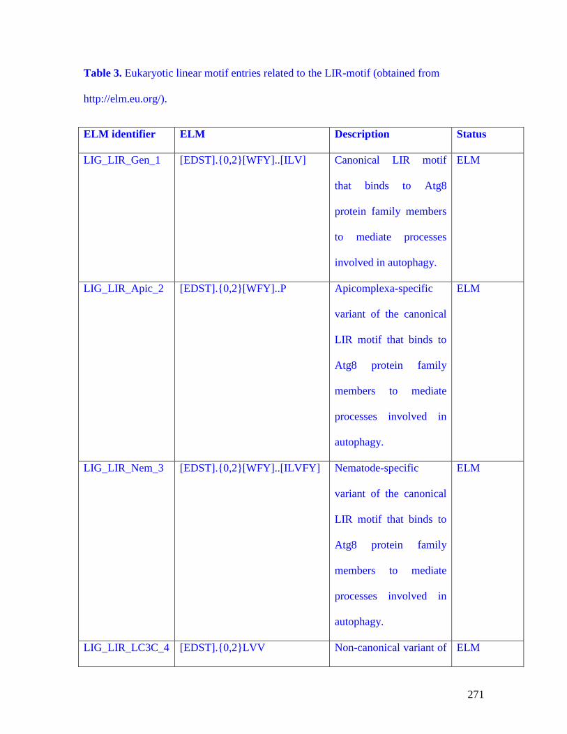
271
Table 3. Eukaryotic linear motif entries related to the LIR-motif (obtained from
http://elm.eu.org/).
ELM identifier ELM Description Status
LIG_LIR_Gen_1 [EDST].{0,2}[WFY]..[ILV] Canonical LIR motif
that binds to Atg8
protein family members
to mediate processes
involved in autophagy.
ELM
LIG_LIR_Apic_2 [EDST].{0,2}[WFY]..P Apicomplexa-specific
variant of the canonical
LIR motif that binds to
Atg8 protein family
members to mediate
processes involved in
autophagy.
ELM
LIG_LIR_Nem_3 [EDST].{0,2}[WFY]..[ILVFY] Nematode-specific
variant of the canonical
LIR motif that binds to
Atg8 protein family
members to mediate
processes involved in
autophagy.
ELM
LIG_LIR_LC3C_4 [EDST].{0,2}LVV Non-canonical variant of ELM

272
the LIR motif that binds
to Atg8 protein family
members to mediate
processes involved in
autophagy.
LIG_AIM [WY]..[ILV] Atg8-family interacting
motif (AIM) found in
Atg19, SQSTM1/p62,
ATG4B and
CALR/calreticulin,
involved in autophagy-
related processes.
Candidate
LIG_LIR WxxL or [WYF]xx[LIV] LC3-interacting region
(LIR) might link
ubiquitinated substrates
that should be degraded
to the autophagy-related
proteins in the
phagophore membrane.
Candidate
LIG_GABARAP W.FL GABAA receptor binding
to clathrin and CALR;
possibly linked to
trafficking.
Candidate

273
Table 4. Recommended methods for monitoring autophagy.
Method Description
1. Electron microscopy Quantitative electron microscopy,
immuno-TEM; monitor autophagosome
number, volume, and content/cargo
2. Atg8/LC3 western blotting Western blot. The analysis is carried out in
the absence and presence of lysosomal
protease or fusion inhibitors to monitor flux;
an increase in the LC3-II amount in the
presence of the inhibitor is usually indicative
of flux
3. GFP-Atg8/LC3 lysosomal delivery and Western blot +/- lysosomal fusion or
proteolysis degradation inhibitors; the generation of free
GFP indicates lysosomal/vacuolar delivery
4. GFP-Atg8/LC3 fluorescence microscopy Fluorescence microscopy, FACS to monitor
vacuolar/lysosomal localization. Also,
increase in punctate GFP-Atg8/LC3 or
Atg18/WIPI, and live time-lapse
fluorescence microscopy to track the
dynamics of GFP-Atg8/LC3-positive
structures

274
5. Tandem mRFP/mCherry-GFP fluorescence Flux can be monitored as a decrease in
microscopy, Rosella green/red (yellow) fluorescence
(phagophores, autophagosomes) and an
increase in red fluorescence (autolysosomes)
6. Autophagosome quantification Flow cytometry
7. SQSTM1 and related LC3 binding protein The amount of SQSTM1increases when
turnover autophagy is inhibited and decreases when
autophagy is induced, but the potential
impact of transcriptional/translational
regulation or the formation of insoluble
aggregates should be addressed in individual
experimental systems
8. MTOR, AMPK and Atg1/ULK1 kinase activity Western blot, immunoprecipitation or kinase
assays
9. WIPI fluorescence microscopy Quantitative fluorescence analysis using
endogenous WIPI proteins, or GFP- or Myc-
tagged versions. Suitable for high-
throughput imaging procedures.
10. Bimolecular fluorescence complementation Can be used to monitor protein-protein
interaction in vivo
11. FRET Interaction of LC3 with gangliosides to
monitor autophagosome formation
12. Transcriptional and translational regulation Northern blot, or qRT-PCR, autophagy-

275
dedicated microarray
13. Autophagic protein degradation Turnover of long-lived proteins to monitor
flux
14. Pex14-GFP, GFP-Atg8, Om45-GFP, A range of assays can be used to monitor
mitoPho8∆60 selective types of autophagy. These typically
involve proteolytic maturation of a resident
enzyme or degradation of a chimera, which
can be followed enzymatically or by western
blot
15. Autophagic sequestration assays Accumulation of cargo in autophagic
compartments in the presence of lysosomal
protease or fusion inhibitors by biochemical
or multilabel fluorescence techniques
16. Turnover of autophagic compartments Electron microscopy with
morphometry/stereology at different time
points
17. Autophagosome-lysosome colocalization Fluorescence microscopy
and dequenching assay
18. Sequestration and processing assays Chimeric RFP fluorescence and processing,
in plants and light and electron microscopy
19. Tissue fractionation Centrifugation, western blot and electron
microscopy
20. Degradation of endogenous lipofuscin Fluorescence microscopy

276

277
References
1. Klionsky DJ, Abeliovich H, Agostinis P, Agrawal DK, Aliev G, Askew DS, Baba
M, Baehrecke EH, Bahr BA, Ballabio A, et al. Guidelines for the use and interpretation
of assays for monitoring autophagy in higher eukaryotes. Autophagy 2008; 4:151-75.
2. Klionsky DJ, Cuervo AM, Seglen PO. Methods for monitoring autophagy from
yeast to human. Autophagy 2007; 3:181-206.
3. Klionsky DJ. The autophagosome is overrated! Autophagy 2011; 7:353-4.
4. Eskelinen E-L, Reggiori F, Baba M, Kovacs AL, Seglen PO. Seeing is believing:
The impact of electron microscopy on autophagy research. Autophagy 2011; 7:935-56.
5. Seglen PO. Regulation of autophagic protein degradation in isolated liver cells.
In: Glaumann H and Ballard FJ, eds. Lysosomes: Their Role in Protein Breakdown.
London: Academic Press, 1987:369-414.
6. de Duve C, Wattiaux R. Functions of lysosomes. Annu Rev Physiol 1966;
28:435-92.
7. Gordon PB, Seglen PO. Prelysosomal convergence of autophagic and endocytic
pathways. Biochemical and biophysical research communications 1988; 151:40-7.
8. Dice JF, Klionsky DJ. Artophagy, the art of autophagy–macroautophagy.
Autophagy 2010; 6.
9. Lucocq JM, Hacker C. Cutting a fine figure: On the use of thin sections in
electron microscopy to quantify autophagy. Autophagy 2013; 9:1443-8.
10. Kovács J, Fellinger E, Karpati AP, Kovács AL, Laszlo L, Réz G. Morphometric
evaluation of the turnover of autophagic vacuoles after treatment with Triton X-100 and
vinblastine in murine pancreatic acinar and seminal vesicle epithelial cells. Virchows
Arch B Cell Pathol Incl Mol Pathol 1987; 53:183-90.
11. Kovács J, Fellinger E, Karpati PA, Kovács AL, Laszlo L. The turnover of
autophagic vacuoles: evaluation by quantitative electron microscopy. Biomed Biochim
Acta 1986; 45:1543-7.
12. Kovács J, Laszlo L, Kovács AL. Regression of autophagic vacuoles in pancreatic
acinar, seminal vesicle epithelial, and liver parenchymal cells: a comparative
morphometric study of the effect of vinblastine and leupeptin followed by cycloheximide
treatment. Experimental cell research 1988; 174:244-51.
13. Chu CT. Autophagic stress in neuronal injury and disease. J Neuropathol Exp
Neurol 2006; 65:423-32.
14. Fass E, Shvets E, Degani I, Hirschberg K, Elazar Z. Microtubules support
production of starvation-induced autophagosomes but not their targeting and fusion with
lysosomes. The Journal of biological chemistry 2006; 281:36303-16.
15. Kovács AL, Reith A, Seglen PO. Accumulation of autophagosomes after
inhibition of hepatocytic protein degradation by vinblastine, leupeptin or a
lysosomotropic amine. Experimental cell research 1982; 137:191-201.
16. Bestebroer J, V'Kovski P, Mauthe M, Reggiori F. Hidden behind autophagy: the
unconventional roles of ATG proteins. Traffic 2013; 14:1029-41.
17. Luo SM, Ge ZJ, Wang ZW, Jiang ZZ, Wang ZB, Ouyang YC, Hou Y, Schatten
H, Sun QY. Unique insights into maternal mitochondrial inheritance in mice. Proceedings
of the National Academy of Sciences of the United States of America 2013; 110:13038-
43.

278
18. Politi Y, Gal L, Kalifa Y, Ravid L, Elazar Z, Arama E. Paternal mitochondrial
destruction after fertilization is mediated by a common endocytic and autophagic
pathway in Drosophila. Developmental cell 2014; 29:305-20.
19. Toth S, Nagy K, Palfia Z, Rez G. Cellular autophagic capacity changes during
azaserine-induced tumour progression in the rat pancreas. Up-regulation in all
premalignant stages and down-regulation with loss of cycloheximide sensitivity of
segregation along with malignant transformation. Cell and tissue research 2002; 309:409-
16.
20. Loos B, Engelbrecht AM. Cell death: a dynamic response concept. Autophagy
2009; 5:590-603.
21. Seglen PO, Gordon PB, Grinde B, Solheim A, Kovacs AL, Poli A. Inhibitors and
pathways of hepatocytic protein degradation. Acta Biol Med Ger 1981; 40:1587-98.
22. Ktistakis NT, Andrews S, Long J. What is the advantage of a transient precursor
in autophagosome biogenesis? Autophagy 2011; 7:118-22.
23. Kovács AL, Réz G, Pálfia Z, Kovács J. Autophagy in the epithelial cells of
murine seminal vesicle in vitro. Formation of large sheets of nascent isolation
membranes, sequestration of the nucleus and inhibition by wortmannin and 3-
ethyladenine. Cell and tissue research 2000; 302:253-61.
24. Mizushima N, Yoshimori T. How to interpret LC3 immunoblotting. Autophagy
2007; 3:542-5.
25. Nishida Y, Arakawa S, Fujitani K, Yamaguchi H, Mizuta T, Kanaseki T,
Komatsu M, Otsu K, Tsujimoto Y, Shimizu S. Discovery of Atg5/Atg7-independent
alternative macroautophagy. Nature 2009; 461:654-8.
26. Kanki T, Kang D, Klionsky DJ. Monitoring mitophagy in yeast: the Om45-GFP
processing assay. Autophagy 2009; 5:1186-9.
27. Grander D, Kharaziha P, Laane E, Pokrovskaja K, Panaretakis T. Autophagy as
the main means of cytotoxicity by glucocorticoids in hematological malignancies.
Autophagy 2009; 5:1198-200.
28. Welter E, Thumm M, Krick R. Quantification of nonselective bulk autophagy in
S. cerevisiae using Pgk1-GFP. Autophagy 2010; 6:794-7.
29. Raju D, Jones NL. Methods to monitor autophagy in H. pylori vacuolating
cytotoxin A (VacA)-treated cells. Autophagy 2010; 6:138-43.
30. Geng J, Klionsky DJ. Determining Atg protein stoichiometry at the phagophore
assembly site by fluorescence microscopy. Autophagy 2010; 6:144-7.
31. Swanlund JM, Kregel KC, Oberley TD. Investigating autophagy: quantitative
morphometric analysis using electron microscopy. Autophagy 2010; 6:270-7.
32. Zhang J, Ney PA. Reticulocyte mitophagy: monitoring mitochondrial clearance in
a mammalian model. Autophagy 2010; 6:405-8.
33. Seglen PO, Brinchmann MF. Purification of autophagosomes from rat
hepatocytes. Autophagy 2010; 6:542-7.
34. He C, Klionsky DJ. Analyzing autophagy in zebrafish. Autophagy 2010; 6.
35. Calvo-Garrido J, Carilla-Latorre S, Mesquita A, Escalante R. A proteolytic
cleavage assay to monitor autophagy in Dictyostelium discoideum. Autophagy 2011;
7:1063-8.
36. Xu F, Liu XH, Zhuang FL, Zhu J, Lin FC. Analyzing autophagy in Magnaporthe
oryzae. Autophagy 2011; 7:525-30.

279
37. Klionsky DJ. Autophagy: Lower Eukaryotes and Non-Mammalian Systems, Part
A. Amsterdam: Academic Press/Elsevier, 2008.
38. Klionsky DJ. Autophagy in Disease and Clinical Applications, Part C.
Amsterdam: Academic Press/Elsevier, 2008.
39. Klionsky DJ. Autophagy in Mammalian Systems, Part B. Amsterdam: Academic
Press/Elsevier, 2008.
40. Zhu J, Dagda RK, Chu CT. Monitoring mitophagy in neuronal cell cultures.
Methods in molecular biology 2011; 793:325-39.
41. Klionsky DJ. Protocol URL.
42. Chu CT, Plowey ED, Dagda RK, Hickey RW, Cherra SJ, 3rd, Clark RS.
Autophagy in neurite injury and neurodegeneration: in vitro and in vivo models. Methods
in enzymology 2009; 453:217-49.
43. Yla-Anttila P, Vihinen H, Jokitalo E, Eskelinen E-L. Monitoring autophagy by
electron microscopy in Mammalian cells. Methods in enzymology 2009; 452:143-64.
44. Eskelinen E-L. Maturation of autophagic vacuoles in mammalian cells.
Autophagy 2005; 1:1-10.
45. Eskelinen E-L. To be or not to be? Examples of incorrect identification of
autophagic compartments in conventional transmission electron microscopy of
mammalian cells. Autophagy 2008; 4:257-60.
46. Eskelinen E-L, Kovacs AL. Double membranes vs. lipid bilayers, and their
significance for correct identification of macroautophagic structures. Autophagy 2011;
7:931-2.
47. Eskelinen E-L. Fine structure of the autophagosome. In: Deretic V, ed.
Autophagosome and Phagosome. Totowa, NJ: Humana Press, 2008:11-28.
48. Berg TO, Fengsrud M, Stromhaug PE, Berg T, Seglen PO. Isolation and
characterization of rat liver amphisomes. Evidence for fusion of autophagosomes with
both early and late endosomes. The Journal of biological chemistry 1998; 273:21883-92.
49. Eskelinen E-L. Macroautophagy in mammalian cells. In: Saftig P, ed. Lysosomes.
Georgetown, TX: LandesBioscience/Eurekah.com, 2005.
50. Turturici G, Tinnirello R, Sconzo G, Geraci F. Extracellular membrane vesicles as
a mechanism of cell-to-cell communication: advantages and disadvantages. American
journal of physiology Cell physiology 2014; 306:C621-33.
51. Pfeifer U. Functional morphology of the lysosomal apparatus. In: Glaumann H
and Ballard FJ, eds. Lysosomes: their role in protein breakdown: Academic Press,
1987:3-59.
52. Yokota S, Himeno M, Kato K. Immunocytochemical localization of acid
phosphatase in rat liver. Cell structure and function 1989; 14:163-71.
53. Luzio JP, Pryor PR, Bright NA. Lysosomes: fusion and function. Nature reviews
Molecular cell biology 2007; 8:622-32.
54. Mizushima N, Yoshimori T, Ohsumi Y. The role of Atg proteins in
autophagosome formation. Annual review of cell and developmental biology 2011;
27:107-32.
55. Rabouille C, Strous GJ, Crapo JD, Geuze HJ, Slot JW. The differential
degradation of two cytosolic proteins as a tool to monitor autophagy in hepatocytes by
immunocytochemistry. The Journal of cell biology 1993; 120:897-908.

280
56. Kovács AL, Pálfia Z, Réz G, Vellai T, Kovács J. Sequestration revisited:
integrating traditional electron microscopy, de novo assembly and new results.
Autophagy 2007; 3:655-62.
57. Gao W, Kang JH, Liao Y, Ding WX, Gambotto AA, Watkins SC, Liu YJ, Stolz
DB, Yin XM. Biochemical isolation and characterization of the tubulovesicular LC3-
positive autophagosomal compartment. The Journal of biological chemistry 2010;
285:1371-83.
58. Lajoie P, Guay G, Dennis JW, Nabi IR. The lipid composition of autophagic
vacuoles regulates expression of multilamellar bodies. Journal of cell science 2005;
118:1991-2003.
59. Mayhew TM. Quantitative immunoelectron microscopy: alternative ways of
assessing subcellular patterns of gold labeling. Methods Mol Biol 2007; 369:309-29.
60. Mayhew TM, Lucocq JM, Griffiths G. Relative labelling index: a novel
stereological approach to test for non-random immunogold labelling of organelles and
membranes on transmission electron microscopy thin sections. J Microsc 2002; 205:153-
64.
61. Schmid D, Pypaert M, Münz C. Antigen-loading compartments for major
histocompatibility complex class II molecules continuously receive input from
autophagosomes. Immunity 2007; 26:79-92.
62. Subramani S, Malhotra V. Non-autophagic roles of autophagy-related proteins.
EMBO reports 2013; 14:143-51.
63. Kovács J. Regression of autophagic vacuoles in seminal vesicle cells following
cycloheximide treatment. Experimental cell research 1983; 144:231-4.
64. Réz G, Csak J, Fellinger E, Laszlo L, Kovács AL, Oliva O, Kovács J. Time
course of vinblastine-induced autophagocytosis and changes in the endoplasmic
reticulum in murine pancreatic acinar cells: a morphometric and biochemical study. Eur J
Cell Biol 1996; 71:341-50.
65. Kovács AL, Grinde B, Seglen PO. Inhibition of autophagic vacuole formation and
protein degradation by amino acids in isolated hepatocytes. Experimental cell research
1981; 133:431-6.
66. Mortimore GE, Hutson NJ, Surmacz CA. Quantitative correlation between
proteolysis and macro- and microautophagy in mouse hepatocytes during starvation and
refeeding. Proceedings of the National Academy of Sciences of the United States of
America 1983; 80:2179-83.
67. Mortimore GE, Lardeux BR, Adams CE. Regulation of microautophagy and basal
protein turnover in rat liver. Effects of short-term starvation. The Journal of biological
chemistry 1988; 263:2506-12.
68. Zhu JH, Horbinski C, Guo F, Watkins S, Uchiyama Y, Chu CT. Regulation of
autophagy by extracellular signal-regulated protein kinases during 1-methyl-4-
phenylpyridinium-induced cell death. The American journal of pathology 2007; 170:75-
86.
69. Bjørkøy G, Lamark T, Brech A, Outzen H, Perander M, Øvervatn A, Stenmark H,
Johansen T. p62/SQSTM1 forms protein aggregates degraded by autophagy and has a
protective effect on huntingtin-induced cell death. The Journal of cell biology 2005;
171:603-14.
281
70. Orvedahl A, Sumpter R, Jr., Xiao G, Ng A, Zou Z, Tang Y, Narimatsu M, Gilpin
C, Sun Q, Roth M, et al. Image-based genome-wide siRNA screen identifies selective
autophagy factors. Nature 2011; 480:113-7.
71. Razi M, Tooze SA. Correlative light and electron microscopy. Methods in
enzymology 2009; 452:261-75.
72. Shu X, Lev-Ram V, Deerinck TJ, Qi Y, Ramko EB, Davidson MW, Jin Y,
Ellisman MH, Tsien RY. A genetically encoded tag for correlated light and electron
microscopy of intact cells, tissues, and organisms. PLoS biology 2011; 9:e1001041.
73. Castillo K, Rojas-Rivera D, Lisbona F, Caballero B, Nassif M, Court F, Schuck S,
Ibar C, Walter P, Sierralta J, et al. BAX inhibitor-1 regulates autophagy by controlling
sponse. EMBO J 2011; in press.
74. Yla-Anttila P, Vihinen H, Jokitalo E, Eskelinen E-L. 3D tomography reveals
connections between the phagophore and endoplasmic reticulum. Autophagy 2009;
5:1180-5.
75. Hayashi-Nishino M, Fujita N, Noda T, Yamaguchi A, Yoshimori T, Yamamoto
A. Electron tomography reveals the endoplasmic reticulum as a membrane source for
autophagosome formation. Autophagy 2010; 6:301-3.
76. Duke EM, Razi M, Weston A, Guttmann P, Werner S, Henzler K, Schneider G,
Tooze SA, Collinson LM. Imaging endosomes and autophagosomes in whole mammalian
cells using correlative cryo-fluorescence and cryo-soft X-ray microscopy (cryo-CLXM).
Ultramicroscopy 2014; 143:77-87.
77. Massey AC, Kaushik S, Sovak G, Kiffin R, Cuervo AM. Consequences of the
selective blockage of chaperone-mediated autophagy. Proceedings of the National
Academy of Sciences of the United States of America 2006; 103:5805-10.
78. Baba M, Osumi M, Ohsumi Y. Analysis of the membrane structures involved in
autophagy in yeast by freeze-replica method. Cell structure and function 1995; 20:465-
71.
79. Rez G, Meldolesi J. Freeze-fracture of drug-induced autophagocytosis in the
mouse exocrine pancreas. Laboratory investigation; a journal of technical methods and
pathology 1980; 43:269-77.
80. Punnonen E-L, Pihakaski K, Mattila K, Lounatmaa K, Hirsimaki P.
Intramembrane particles and filipin labelling on the membranes of autophagic vacuoles
and lysosomes in mouse liver. Cell and tissue research 1989; 258:269-76.
81. Fengsrud M, Erichsen ES, Berg TO, Raiborg C, Seglen PO. Ultrastructural
characterization of the delimiting membranes of isolated autophagosomes and
amphisomes by freeze-fracture electron microscopy. Eur J Cell Biol 2000; 79:871-82.
82. Dickey JS, Gonzalez Y, Aryal B, Mog S, Nakamura AJ, Redon CE, Baxa U,
Rosen E, Cheng G, Zielonka J, et al. Mito-tempol and dexrazoxane exhibit
cardioprotective and chemotherapeutic effects through specific protein oxidation and
autophagy in a syngeneic breast tumor preclinical model. PloS one 2013; 8:e70575.
83. Rao VA, Klein SR, Bonar SJ, Zielonka J, Mizuno N, Dickey JS, Keller PW,
Joseph J, Kalyanaraman B, Shacter E. The antioxidant transcription factor Nrf2
negatively regulates autophagy and growth arrest induced by the anticancer redox agent
mitoquinone. The Journal of biological chemistry 2010; 285:34447-59.
84. Klionsky DJ. Autophagy: from phenomenology to molecular understanding in
less than a decade. Nature reviews Molecular cell biology 2007; 8:931-7.

282
85. Krick R, M(u)he Y, Prick T, Bredschneider M, Bremer S, Wenzel D, Eskelinen
E-L, Thumm M. Piecemeal microautophagy of the nucleus: genetic and morphological
traits. Autophagy 2009; 5:270-2.
86. Meschini S, Condello M, Calcabrini A, Marra M, Formisano G, Lista P, De
Milito A, Federici E, Arancia G. The plant alkaloid voacamine induces apoptosis-
independent autophagic cell death on both sensitive and multidrug resistant human
osteosarcoma cells. Autophagy 2008; 4:1020-33.
87. Proikas-Cezanne T, Robenek H. Freeze-fracture replica immunolabelling reveals
human WIPI-1 and WIPI-2 as membrane proteins of autophagosomes. Journal of cellular
and molecular medicine 2011; 15:2007-10.
88. Kovacs J, Rez G, Kovacs AL, Csak J, Zboray G. Autophagocytosis: freeze-
fracture morphology, effects of vinblastine and influence of transcriptional and
translational inhibitors. Acta biologica et medica Germanica 1982; 41:131-5.
89. Hirsimaki Y, Hirsimaki P, Lounatmaa K. Vinblastine-induced autophagic
vacuoles in mouse liver and Ehrlich ascites tumor cells as assessed by freeze-fracture
electron microscopy. Eur J Cell Biol 1982; 27:298-301.
90. Cheong H, Yorimitsu T, Reggiori F, Legakis JE, Wang C-W, Klionsky DJ. Atg17
regulates the magnitude of the autophagic response. Molecular biology of the cell 2005;
16:3438-53.
91. Xie Z, Nair U, Klionsky DJ. Atg8 controls phagophore expansion during
autophagosome formation. Molecular biology of the cell 2008; 19:3290-8.
92. Sigmond T, Feher J, Baksa A, Pasti G, Palfia Z, Takacs-Vellai K, Kovacs J,
Vellai T, Kovacs AL. Qualitative and quantitative characterization of autophagy in
Caenorhabditis elegans by electron microscopy. Methods in enzymology 2008; 451:467-
91.
93. Kovács AL, Vellai T, Müller F. Autophagy in Caenorhabditis elegans. In:
Klionsky DJ, ed. Autophagy. Georgetown, Texas: Landes Bioscience, 2004:217-23.
94. Weibel ER. Practical Methods for Biological Morphometry. Academic Press,
New York, 1979.
95. Williams MA. Quantitative methods in biology: Practical methods in electron
microscopy. Amsterdam, New York, Oxford: North-Holland Publishing Company, 1977.
96. Howard V, Reed MG. Unbiased stereology; three dimensional measurement in
microscopy. U Bios Scientific Publishers, 1998.
97. Kovacs AL. A simple method to estimate the number of autophagic elements by
electron microscopic morphometry in real cellular dimensions. BioMed research
international 2014; 2014:578698.
98. Xie Z, Nair U, Geng J, Szefler MB, Rothman ED, Klionsky DJ. Indirect
estimation of the area density of Atg8 on the phagophore. Autophagy 2009; 5:217-20.
99. Punnonen EL, Reunanen H. Effects of vinblastine, leucine, and histidine, and 3-
methyladenine on autophagy in Ehrlich ascites cells. Exp Mol Pathol 1990; 52:87-97.
100. Kovacs AL, Laszlo L, Fellinger E, Jakab A, Orosz A, Rez G, Kovacs J.
Combined effects of fasting and vinblastine treatment on serum insulin level, the size of
autophagic-lysosomal compartment, protein content and lysosomal enzyme activities of
liver and exocrine pancreatic cells of the mouse. Comparative biochemistry and
physiology B, Comparative biochemistry 1989; 94:505-10.

283
101. Griffiths G. Fine structure immunocytochemistry Heidelberg, Germany: Springer-
Verlag, 1993.
102. Reyes FC, Chung T, Holding D, Jung R, Vierstra R, Otegui MS. Delivery of
prolamins to the protein storage vacuole in maize aleurone cells. The Plant cell 2011;
23:769-84.
103. Dunn WA, Jr., Cregg JM, Kiel JAKW, van der Klei IJ, Oku M, Sakai Y, Sibirny
AA, Stasyk OV, Veenhuis M. Pexophagy: the selective autophagy of peroxisomes.
Autophagy 2005; 1:75-83.
104. Wang K, Klionsky DJ. Mitochondria removal by autophagy. Autophagy 2011;
7:297-300.
105. Belanger M, Rodrigues PH, Dunn WA, Jr., Progulske-Fox A. Autophagy: a
highway for Porphyromonas gingivalis in endothelial cells. Autophagy 2006; 2:165-70.
106. Birmingham CL, Brumell JH. Autophagy recognizes intracellular Salmonella
enterica serovar Typhimurium in damaged vacuoles. Autophagy 2006; 2:156-8.
107. Colombo MI, Gutierrez MG, Romano PS. The two faces of autophagy: Coxiella
and Mycobacterium. Autophagy 2006; 2:162-4.
108. Ogawa M, Sasakawa C. Shigella and autophagy. Autophagy 2006; 2:171-4.
109. Vergne I, Singh S, Roberts E, Kyei G, Master S, Harris J, de Haro S, Naylor J,
Davis A, Delgado M, et al. Autophagy in immune defense against Mycobacterium
tuberculosis. Autophagy 2006; 2:175-8.
110. Yoshimori T. Autophagy vs. Group A Streptococcus. Autophagy 2006; 2:154-5.
111. Gorbunov NV, McDaniel DP, Zhai M, Liao PJ, Garrison BR, Kiang JG.
Autophagy and mitochondrial remodelling in mouse mesenchymal stromal cells
challenged with Staphylococcus epidermidis. Journal of cellular and molecular medicine
2015; 19:1133-50.
112. Lynch-Day MA, Klionsky DJ. The Cvt pathway as a model for selective
autophagy. FEBS letters 2010; 584:1359-66.
113. Birmingham CL, Canadien V, Gouin E, Troy EB, Yoshimori T, Cossart P,
Higgins DE, Brumell JH. Listeria monocytogenes evades killing by autophagy during
colonization of host cells. Autophagy 2007; 3:442-51.
114. Klionsky DJ. Protein transport from the cytoplasm into the vacuole. J Membr Biol
1997; 157:105-15.
115. Dini L, Pagliara P, Carla EC. Phagocytosis of apoptotic cells by liver: a
morphological study. Microscopy research and technique 2002; 57:530-40.
116. Kroemer G, El-Deiry WS, Golstein P, Peter ME, Vaux D, Vandenabeele P,
Zhivotovsky B, Blagosklonny MV, Malorni W, Knight RA, et al. Classification of cell
death: recommendations of the Nomenclature Committee on Cell Death. Cell death and
differentiation 2005; 12:1463-7.
117. Rez G, Palfia Z, Fellinger E. Occurrence and inhibition by cycloheximide of
apoptosis in vinblastine-treated murine pancreas. A role for autophagy? Acta biologica
Hungarica 1991; 42:133-40.
118. Nagy P, Varga A, Kovács AL, Takáts S, Juhász G. How and why to study
autophagy in Drosophila: It's more than just a garbage chute. Methods 2015.
119. Giammarioli AM, Gambardella L, Barbati C, Pietraforte D, Tinari A, Alberton M,
Gnessi L, Griffin RJ, Minetti M, Malorni W. Differential effects of the glycolysis
inhibitor 2-deoxy-D-glucose on the activity of pro-apoptotic agents in metastatic

284
melanoma cells, and induction of a cytoprotective autophagic response. International
journal of cancer Journal international du cancer 2011; in press.
120. Sou YS, Tanida I, Komatsu M, Ueno T, Kominami E. Phosphatidylserine in
addition to phosphatidylethanolamine is an in vitro target of the mammalian Atg8
modifiers, LC3, GABARAP, and GATE-16. The Journal of biological chemistry 2006;
281:3017-24.
121. Le Grand JN, Chakrama FZ, Seguin-Py S, Fraichard A, Delage-Mourroux R,
Jouvenot M, Boyer-Guittaut M. GABARAPL1 (GEC1): Original or copycat? Autophagy
2011; 7:1098-107.
122. Kabeya Y, Mizushima N, Yamamoto A, Oshitani-Okamoto S, Ohsumi Y,
Yoshimori T. LC3, GABARAP and GATE16 localize to autophagosomal membrane
depending on form-II formation. Journal of cell science 2004; 117:2805-12.
123. Weidberg H, Shvets E, Shpilka T, Shimron F, Shinder V, Elazar Z. LC3 and
GATE-16/GABARAP subfamilies are both essential yet act differently in
autophagosome biogenesis. The EMBO journal 2010; 29:1792-802.
124. Sugawara K, Suzuki NN, Fujioka Y, Mizushima N, Ohsumi Y, Inagaki F. The
crystal structure of microtubule-associated protein light chain 3, a mammalian
homologue of Saccharomyces cerevisiae Atg8. Genes to cells : devoted to molecular &
cellular mechanisms 2004; 9:611-8.
125. Chu CT, Ji J, Dagda RK, Jiang JF, Tyurina YY, Kapralov AA, Tyurin VA,
Yanamala N, Shrivastava IH, Mohammadyani D, et al. Cardiolipin externalization to the
outer mitochondrial membrane acts as an elimination signal for mitophagy in neuronal
cells. Nature cell biology 2013; 15:1197-205.
126. Lystad AH, Ichimura Y, Takagi K, Yang Y, Pankiv S, Kanegae Y, Kageyama S,
Suzuki M, Saito I, Mizushima T, et al. Structural determinants in GABARAP required
for the selective binding and recruitment of ALFY to LC3B-positive structures. EMBO
reports 2014; 15:557-65.
127. von Muhlinen N, Akutsu M, Ravenhill BJ, Foeglein A, Bloor S, Rutherford TJ,
Freund SM, Komander D, Randow F. LC3C, bound selectively by a noncanonical LIR
motif in NDP52, is required for antibacterial autophagy. Molecular cell 2012; 48:329-42.
128. Huang W-P, Scott SV, Kim J, Klionsky DJ. The itinerary of a vesicle component,
Aut7p/Cvt5p, terminates in the yeast vacuole via the autophagy/Cvt pathways. The
Journal of biological chemistry 2000; 275:5845-51.
129. Cai Q, Lu L, Tian J-H, Zhu Y-B, Qiao H, Sheng Z-H. Snapin-regulated late
endosomal transport is critical for efficient autophagy-lysosomal function in neurons.
Neuron 2010; 68:73-86.
130. Castino R, Fiorentino I, Cagnin M, Giovia A, Isidoro C. Chelation of lysosomal
iron protects dopaminergic SH-SY5Y neuroblastoma cells from hydrogen peroxide
toxicity by precluding autophagy and Akt dephosphorylation. Toxicological sciences : an
official journal of the Society of Toxicology 2011:523-41.
131. Michiorri S, Gelmetti V, Giarda E, Lombardi F, Romano F, Marongiu R, Nerini-
Molteni S, Sale P, Vago R, Arena G, et al. The Parkinson-associated protein PINK1
interacts with Beclin1 and promotes autophagy. Cell death and differentiation 2010;
17:962-74.
132. Yang DS, Stavrides P, Mohan PS, Kaushik S, Kumar A, Ohno M, Schmidt SD,
Wesson D, Bandyopadhyay U, Jiang Y, et al. Reversal of autophagy dysfunction in the

285
TgCRND8 mouse model of Alzheimer's disease ameliorates amyloid pathologies and
memory deficits. Brain : a journal of neurology 2011; 134:258-77.
133. Mizushima N, Yamamoto A, Matsui M, Yoshimori T, Ohsumi Y. In vivo analysis
of autophagy in response to nutrient starvation using transgenic mice expressing a
fluorescent autophagosome marker. Molecular biology of the cell 2004; 15:1101-11.
134. Ahlberg J, Berkenstam A, Henell F, Glaumann H. Degradation of short and long
lived proteins in isolated rat liver lysosomes. Effects of pH, temperature, and proteolytic
inhibitors. The Journal of biological chemistry 1985; 260:5847-54.
135. Juhasz G. Interpretation of bafilomycin, pH neutralizing or protease inhibitor
treatments in autophagic flux experiments: novel considerations. Autophagy 2012;
8:1875-6.
136. Li M, Khambu B, Zhang H, Kang JH, Chen X, Chen D, Vollmer L, Liu PQ, Vogt
A, Yin XM. Suppression of lysosome function induces autophagy via a feedback down-
regulation of MTOR complex 1 (MTORC1) activity. The Journal of biological chemistry
2013; 288:35769-80.
137. McLeland CB, Rodriguez J, Stern ST. Autophagy monitoring assay: qualitative
analysis of MAP LC3-I to II conversion by immunoblot. Methods in molecular biology
2011; 697:199-206.
138. Chakrama FZ, Seguin-Py S, Le Grand JN, Fraichard A, Delage-Mourroux R,
Despouy G, Perez V, Jouvenot M, Boyer-Guittaut M. GABARAPL1 (GEC1) associates
with autophagic vesicles. Autophagy 2010; 6:495-505.
139. Kim J, Huang W-P, Klionsky DJ. Membrane recruitment of Aut7p in the
autophagy and cytoplasm to vacuole targeting pathways requires Aut1p, Aut2p, and the
autophagy conjugation complex. The Journal of cell biology 2001; 152:51-64.
140. Shu CW, Drag M, Bekes M, Zhai D, Salvesen GS, Reed JC. Synthetic substrates
for measuring activity of autophagy proteases: autophagins (Atg4). Autophagy 2010;
6:936-47.
141. Li M, Chen X, Ye Q-Z, Vogt A, Yin X-M. A High-throughput FRET-based
Assay for Determination of Atg4 Activity. Autophagy 2012; 8:in press.
142. Ketteler R, Seed B. Quantitation of autophagy by luciferase release assay.
Autophagy 2008; 4:801-6.
143. Li M, Hou Y, Wang J, Chen X, Shao ZM, Yin X-M. Kinetics comparisons of
mammalian Atg4 homologues indicate selective preferences toward diverse Atg8
substrates. The Journal of biological chemistry 2011; 286:7327-38.
144. Klionsky DJ. For the last time, it is GFP-Atg8, not Atg8-GFP (and the same goes
for LC3). Autophagy 2011; 7:1093-4.
145. Tanida I, Minematsu-Ikeguchi N, Ueno T, Kominami E. Lysosomal turnover, but
not a cellular level, of endogenous LC3 is a marker for autophagy. Autophagy 2005;
1:84-91.
146. Castino R, Lazzeri G, Lenzi P, Bellio N, Follo C, Ferrucci M, Fornai F, Isidoro C.
Suppression of autophagy precipitates neuronal cell death following low doses of
methamphetamine. Journal of neurochemistry 2008; 106:1426-39.
147. Matsui Y, Takagi H, Qu X, Abdellatif M, Sakoda H, Asano T, Levine B,
Sadoshima J. Distinct roles of autophagy in the heart during ischemia and reperfusion:
roles of AMP-activated protein kinase and Beclin 1 in mediating autophagy. Circ Res
2007; 100:914-22.

286
148. Suzuki K, Kirisako T, Kamada Y, Mizushima N, Noda T, Ohsumi Y. The pre-
autophagosomal structure organized by concerted functions of APG genes is essential for
autophagosome formation. The EMBO journal 2001; 20:5971-81.
149. Hanson HH, Kang S, Fernandez-Monreal M, Oung T, Yildirim M, Lee R,
Suyama K, Hazan RB, Phillips GR. LC3-dependent intracellular membrane tubules
induced by gamma-protocadherins A3 and B2: a role for intraluminal interactions. The
Journal of biological chemistry 2010; 285:20982-92.
150. Florey O, Kim SE, Sandoval CP, Haynes CM, Overholtzer M. Autophagy
machinery mediates macroendocytic processing and entotic cell death by targeting single
membranes. Nature cell biology 2011; 13:1335-43.
151. Martinez J, Almendinger J, Oberst A, Ness R, Dillon CP, Fitzgerald P,
Hengartner MO, Green DR. Microtubule-associated protein 1 light chain 3 alpha (LC3)-
associated phagocytosis is required for the efficient clearance of dead cells. Proceedings
of the National Academy of Sciences of the United States of America 2011; 108:17396-
401.
152. Choi J, Park S, Biering SB, Selleck E, Liu CY, Zhang X, Fujita N, Saitoh T,
Akira S, Yoshimori T, et al. The parasitophorous vacuole membrane of Toxoplasma
gondii is targeted for disruption by ubiquitin-like conjugation systems of autophagy.
Immunity 2014; 40:924-35.
153. Sanjuan MA, Dillon CP, Tait SW, Moshiach S, Dorsey F, Connell S, Komatsu M,
Tanaka K, Cleveland JL, Withoff S, et al. Toll-like receptor signalling in macrophages
links the autophagy pathway to phagocytosis. Nature 2007; 450:1253-7.
154. Sanjuan MA, Milasta S, Green DR. Toll-like receptor signaling in the lysosomal
pathways. Immunological reviews 2009; 227:203-20.
155. Ushio H, Ueno T, Kojima Y, Komatsu M, Tanaka S, Yamamoto A, Ichimura Y,
Ezaki J, Nishida K, Komazawa-Sakon S, et al. Crucial role for autophagy in
degranulation of mast cells. The Journal of allergy and clinical immunology 2011;
127:1267-76 e6.
156. Ishibashi K, Uemura T, Waguri S, Fukuda M. Atg16L1, an essential factor for
canonical autophagy, participates in hormone secretion from PC12 cells independently of
autophagic activity. Molecular biology of the cell 2012; 23:3193-202.
157. DeSelm CJ, Miller BC, Zou W, Beatty WL, van Meel E, Takahata Y,
Klumperman J, Tooze SA, Teitelbaum SL, Virgin HW. Autophagy proteins regulate the
secretory component of osteoclastic bone resorption. Developmental cell 2011; 21:966-
74.
158. Patel KK, Miyoshi H, Beatty WL, Head RD, Malvin NP, Cadwell K, Guan JL,
Saitoh T, Akira S, Seglen PO, et al. Autophagy proteins control goblet cell function by
potentiating reactive oxygen species production. The EMBO journal 2013; 32:3130-44.
159. Dupont N, Lacas-Gervais S, Bertout J, Paz I, Freche B, Van Nhieu GT, van der
Goot FG, Sansonetti PJ, Lafont F. Shigella phagocytic vacuolar membrane remnants
participate in the cellular response to pathogen invasion and are regulated by autophagy.
Cell host & microbe 2009; 6:137-49.
160. Cottam EM, Maier HJ, Manifava M, Vaux LC, Chandra-Schoenfelder P, Gerner
W, Britton P, Ktistakis NT, Wileman T. Coronavirus nsp6 proteins generate
autophagosomes from the endoplasmic reticulum via an omegasome intermediate.
Autophagy 2011; 7:1335-47.

287
161. Reggiori F, Monastyrska I, Verheije MH, Cali T, Ulasli M, Bianchi S, Bernasconi
R, de Haan CA, Molinari M. Coronaviruses Hijack the LC3-I-positive EDEMosomes,
ER-derived vesicles exporting short-lived ERAD regulators, for replication. Cell host &
microbe 2010; 7:500-8.
162. English L, Chemali M, Duron J, Rondeau C, Laplante A, Gingras D, Alexander
D, Leib D, Norbury C, Lippe R, et al. Autophagy enhances the presentation of
endogenous viral antigens on MHC class I molecules during HSV-1 infection. Nature
immunology 2009; 10:480-7.
163. Beale R, Wise H, Stuart A, Ravenhill BJ, Digard P, Randow F. A LC3-interacting
motif in the influenza A virus M2 protein is required to subvert autophagy and maintain
virion stability. Cell host & microbe 2014; 15:239-47.
164. Kemball CC, Alirezaei M, Flynn CT, Wood MR, Harkins S, Kiosses WB,
Whitton JL. Coxsackievirus infection induces autophagy-like vesicles and
megaphagosomes in pancreatic acinar cells in vivo. Journal of virology 2010; 84:12110-
24.
165. Alirezaei M, Flynn CT, Wood MR, Whitton JL. Pancreatic acinar cell-specific
autophagy disruption reduces coxsackievirus replication and pathogenesis in vivo. Cell
host & microbe 2012; 11:298-305.
166. Plowey ED, Cherra SJ, 3rd, Liu YJ, Chu CT. Role of autophagy in G2019S-
LRRK2-associated neurite shortening in differentiated SH-SY5Y cells. Journal of
neurochemistry 2008; 105:1048-56.
167. Nicotra G, Mercalli F, Peracchio C, Castino R, Follo C, Valente G, Isidoro C.
Autophagy-active beclin-1 correlates with favourable clinical outcome in non-Hodgkin
lymphomas. Modern pathology : an official journal of the United States and Canadian
Academy of Pathology, Inc 2010; 23:937-50.
168. Tanida I, Ueno T, Kominami E. LC3 and autophagy. Methods in Molecular
Biology 2008; 445:77-88.
169. Gros F, Arnold J, Page N, Decossas M, Korganow AS, Martin T, Muller S.
Macroautophagy is deregulated in murine and human lupus T lymphocytes. Autophagy
2012; 8:1113-23.
170. Colella AD, Chegenii N, Tea MN, Gibbins IL, Williams KA, Chataway TK.
Comparison of Stain-Free gels with traditional immunoblot loading control methodology.
Analytical biochemistry 2012; 430:108-10.
171. Welinder C, Ekblad L. Coomassie staining as loading control in Western blot
analysis. Journal of proteome research 2011; 10:1416-9.
172. He H, Dang Y, Dai F, Guo Z, Wu J, She X, Pei Y, Chen Y, Ling W, Wu C, et al.
Post-translational modifications of three members of the human MAP1LC3 family and
detection of a novel type of modification for MAP1LC3B. The Journal of biological
chemistry 2003; 278:29278-87.
173. Shpilka T, Weidberg H, Pietrokovski S, Elazar Z. Atg8: an autophagy-related
ubiquitin-like protein family. Genome biology 2011; 12:226.
174. Zois CE, Koukourakis MI. Radiation-induced autophagy in normal and cancer
cells: towards novel cytoprotection and radio-sensitization policies? Autophagy 2009;
5:442-50.
175. Xin Y, Yu L, Chen Z, Zheng L, Fu Q, Jiang J, Zhang P, Gong R, Zhao S.
Cloning, expression patterns, and chromosome localization of three human and two
288
mouse homologues of GABA(A) receptor-associated protein. Genomics 2001; 74:408-
13.
176. Novak I, Kirkin V, McEwan DG, Zhang J, Wild P, Rozenknop A, Rogov V, Lohr
F, Popovic D, Occhipinti A, et al. Nix is a selective autophagy receptor for mitochondrial
clearance. EMBO reports 2010; 11:45-51.
177. Schwarten M, Mohrluder J, Ma P, Stoldt M, Thielmann Y, Stangler T, Hersch N,
Hoffmann B, Merkel R, Willbold D. Nix directly binds to GABARAP: a possible
crosstalk between apoptosis and autophagy. Autophagy 2009; 5:690-8.
178. Gassmann M, Grenacher B, Rohde B, Vogel J. Quantifying Western blots: pitfalls
of densitometry. Electrophoresis 2009; 30:1845-55.
179. Kirisako T, Ichimura Y, Okada H, Kabeya Y, Mizushima N, Yoshimori T,
Ohsumi M, Takao T, Noda T, Ohsumi Y. The reversible modification regulates the
membrane-binding state of Apg8/Aut7 essential for autophagy and the cytoplasm to
vacuole targeting pathway. The Journal of cell biology 2000; 151:263-76.
180. Chung T, Phillips AR, Vierstra RD. ATG8 lipidation and ATG8-mediated
autophagy in Arabidopsis require ATG12 expressed from the differentially controlled
ATG12A AND ATG12B loci. The Plant journal : for cell and molecular biology 2010;
62:483-93.
181. Chung T, Suttangkakul A, Vierstra RD. The ATG autophagic conjugation system
in maize: ATG transcripts and abundance of the ATG8-lipid adduct are regulated by
development and nutrient availability. Plant physiology 2009; 149:220-34.
182. Engedal N, Torgersen ML, Guldvik IJ, Barfeld SJ, Bakula D, Saetre F, Hagen
LK, Patterson JB, Proikas-Cezanne T, Seglen PO, et al. Modulation of intracellular
calcium homeostasis blocks autophagosome formation. Autophagy 2013; 9:1475-90.
183. King JS, Veltman DM, Insall RH. The induction of autophagy by mechanical
stress. Autophagy 2011; 7:1490-9.
184. Roberts R, Al-Jamal WT, Whelband M, Thomas P, Jefferson M, van den Bossche
J, Powell PP, Kostarelos K, Wileman T. Autophagy and formation of tubulovesicular
autophagosomes provide a barrier against nonviral gene delivery. Autophagy 2013;
9:667-82.
185. Bernard M, Dieude M, Yang B, Hamelin K, Underwood K, Hebert MJ.
Autophagy fosters myofibroblast differentiation through MTORC2 activation and
downstream upregulation of CTGF. Autophagy 2014:0.
186. Ju JS, Varadhachary AS, Miller SE, Weihl CC. Quantitation of "autophagic flux"
in mature skeletal muscle. Autophagy 2010; 6:929-35.
187. Degtyarev M, De Maziere A, Orr C, Lin J, Lee BB, Tien JY, Prior WW, van Dijk
S, Wu H, Gray DC, et al. Akt inhibition promotes autophagy and sensitizes PTEN-null
tumors to lysosomotropic agents. The Journal of cell biology 2008; 183:101-16.
188. Klionsky DJ, Elazar Z, Seglen PO, Rubinsztein DC. Does bafilomycin A1 block
the fusion of autophagosomes with lysosomes? Autophagy 2008; 4:849-950.
189. Mauvezin C, Nagy P, Juhasz G, Neufeld TP. Autophagosome-lysosome fusion is
independent of V-ATPase-mediated acidification. Nature communications 2015; 6:7007.
190. Xie R, Nguyen S, McKeehan WL, Liu L. Acetylated microtubules are required
for fusion of autophagosomes with lysosomes. BMC cell biology 2010; 11:89.
191. Gonzalez-Polo RA, Boya P, Pauleau AL, Jalil A, Larochette N, Souquere S,
Eskelinen EL, Pierron G, Saftig P, Kroemer G. The apoptosis/autophagy paradox:

289
autophagic vacuolization before apoptotic death. Journal of cell science 2005; 118:3091-
102.
192. Cuervo AM, Stefanis L, Fredenburg R, Lansbury PT, Sulzer D. Impaired
degradation of mutant {alpha}-synuclein by chaperone-mediated autophagy. Science
2004; 305:1292-5.
193. Trincheri NF, Follo C, Nicotra G, Peracchio C, Castino R, Isidoro C. Resveratrol-
induced apoptosis depends on the lipid kinase activity of Vps34 and on the formation of
autophagolysosomes. Carcinogenesis 2008; 29:381-9.
194. Rubinsztein DC, Cuervo AM, Ravikumar B, Sarkar S, Korolchuk V, Kaushik S,
Klionsky DJ. In search of an "autophagomometer". Autophagy 2009; 5:585-9.
195. Sarkar S, Ravikumar B, Rubinsztein DC. Autophagic clearance of aggregate-
prone proteins associated with neurodegeneration. Methods in enzymology 2009; 453:83-
110.
196. Sarkar S, Korolchuk V, Renna M, Winslow A, Rubinsztein DC. Methodological
considerations for assessing autophagy modulators: a study with calcium phosphate
precipitates. Autophagy 2009; 5:307-13.
197. Shintani T, Klionsky DJ. Cargo proteins facilitate the formation of transport
vesicles in the cytoplasm to vacuole targeting pathway. The Journal of biological
chemistry 2004; 279:29889-94.
198. Karim MR, Kanazawa T, Daigaku Y, Fujimura S, Miotto G, Kadowaki M.
Cytosolic LC3 ratio as a sensitive index of macroautophagy in isolated rat hepatocytes
and H4-II-E cells. Autophagy 2007; 3:553-60.
199. Kim CH, Kim KH, Yoo YM. Melatonin protects against apoptotic and autophagic
cell death in C2C12 murine myoblast cells. Journal of pineal research 2011; 50:241-9.
200. Karim MR, Kanazawa T, Daigaku Y, Fujimura S, Miotto G, Kadowaki M.
Cytosolic LC3 ratio as a sensitive index of macroautophagy in isolated rat hepatocytes
and H4-II-E cells. Autophagy 2007; 3:in press.
201. Tsvetkov AS, Arrasate M, Barmada S, Ando DM, Sharma P, Shaby BA,
Finkbeiner S. Proteostasis of polyglutamine varies among neurons and predicts
neurodegeneration. Nature chemical biology 2013; 9:586-92.
202. Loos B, du Toit A, Hofmeyr JH. Defining and measuring autophagosome flux-
concept and reality. Autophagy 2014:0.
203. Farkas T, Hoyer-Hansen M, Jaattela M. Identification of novel autophagy
regulators by a luciferase-based assay for the kinetics of autophagic flux. Autophagy
2009; 5:1018-25.
204. Frankel LB, Wen J, Lees M, H(o)yer-Hansen M, Farkas T, Krogh A, Jaattela M,
Lund AH. microRNA-101 is a potent inhibitor of autophagy. The EMBO journal
2011:4628-41.
205. Farkas T, Daugaard M, Jaattela M. Identification of small molecule inhibitors of
phosphatidylinositol 3-kinase and autophagy. The Journal of biological chemistry 2011;
in press:38904-12.
206. Szyniarowski P, Corcelle-Termeau E, Farkas T, Hoyer-Hansen M, Nylandsted J,
Kallunki T, Jaattela M. A comprehensive siRNA screen for kinases that suppress
macroautophagy in optimal growth conditions. Autophagy 2011; 7:892-903.
207. Nguyen HT, Dalmasso G, Muller S, Carriere J, Seibold F, Darfeuille-Michaud A.
Crohn's disease-associated adherent invasive Escherichia coli modulate levels of

290
microRNAs in intestinal epithelial cells to reduce autophagy. Gastroenterology 2014;
146:508-19.
208. Iwata J, Ezaki J, Komatsu M, Yokota S, Ueno T, Tanida I, Chiba T, Tanaka K,
Kominami E. Excess peroxisomes are degraded by autophagic machinery in mammals.
The Journal of biological chemistry 2006; 281:4035-41.
209. Narendra D, Tanaka A, Suen DF, Youle RJ. Parkin is recruited selectively to
impaired mitochondria and promotes their autophagy. The Journal of cell biology 2008;
183:795-803.
210. Nogalska A, Terracciano C, D'Agostino C, King Engel W, Askanas V.
p62/SQSTM1 is overexpressed and prominently accumulated in inclusions of sporadic
inclusion-body myositis muscle fibers, and can help differentiating it from polymyositis
and dermatomyositis. Acta neuropathologica 2009; 118:407-13.
211. Chahory S, Keller N, Martin E, Omri B, Crisanti P, Torriglia A. Light induced
retinal degeneration activates a caspase-independent pathway involving cathepsin D.
Neurochemistry international 2010; 57:278-87.
212. Padron-Barthe L, Courta J, Lepretre C, Nagbou A, Torriglia A. Leukocyte
Elastase Inhibitor, the precursor of L-DNase II, inhibits apoptosis by interfering with
caspase-8 activation. Biochimica et biophysica acta 2008; 1783:1755-66.
213. Gutierrez MG, Saka HA, Chinen I, Zoppino FC, Yoshimori T, Bocco JL,
Colombo MI. Protective role of autophagy against Vibrio cholerae cytolysin, a pore-
forming toxin from V. cholerae. Proceedings of the National Academy of Sciences of the
United States of America 2007; 104:1829-34.
214. Hosokawa N, Hara Y, Mizushima N. Generation of cell lines with tetracycline-
regulated autophagy and a role for autophagy in controlling cell size. FEBS letters 2006;
580:2623-9.
215. Suttangkakul A, Li F, Chung T, Vierstra RD. The ATG1/13 protein kinase
complex is both a regulator and a substrate of autophagic recycling in Arabidopsis. The
Plant cell 2011; 23:3761-79.
216. Ni HM, Bockus A, Wozniak AL, Jones K, Weinman S, Yin XM, Ding WX.
Dissecting the dynamic turnover of GFP-LC3 in the autolysosome. Autophagy 2011;
7:188-204.
217. Balgi AD, Fonseca BD, Donohue E, Tsang TC, Lajoie P, Proud CG, Nabi IR,
Roberge M. Screen for chemical modulators of autophagy reveals novel therapeutic
inhibitors of mTORC1 signaling. PloS one 2009; 4:e7124.
218. Patterson GH, Lippincott-Schwartz J. Selective photolabeling of proteins using
photoactivatable GFP. Methods 2004; 32:445-50.
219. Hamacher-Brady A, Brady NR, Gottlieb RA. Enhancing macroautophagy protects
against ischemia/reperfusion injury in cardiac myocytes. The Journal of biological
chemistry 2006; 281:29776-87.
220. Noda T, Klionsky DJ. The quantitative Pho8Delta60 assay of nonspecific
autophagy. Methods in enzymology 2008; 451:33-42.
221. Klionsky DJ. Monitoring autophagy in yeast: the Pho8Delta60 assay. Methods in
molecular biology 2007; 390:363-71.
222. Patterson GH, Knobel SM, Sharif WD, Kain SR, Piston DW. Use of the green
fluorescent protein and its mutants in quantitative fluorescence microscopy. Biophys J
1997; 73:2782-90.

291
223. Kimura S, Noda T, Yoshimori T. Dissection of the autophagosome maturation
process by a novel reporter protein, tandem fluorescent-tagged LC3. Autophagy 2007;
3:452-60.
224. Shaner NC, Campbell RE, Steinbach PA, Giepmans BN, Palmer AE, Tsien RY.
Improved monomeric red, orange and yellow fluorescent proteins derived from
Discosoma sp. red fluorescent protein. Nature biotechnology 2004; 22:1567-72.
225. Strack RL, Keenan RJ, Glick BS. Noncytotoxic DsRed derivatives for whole-cell
labeling. Methods in molecular biology 2011; 699:355-70.
226. Gurskaya NG, Verkhusha VV, Shcheglov AS, Staroverov DB, Chepurnykh TV,
Fradkov AF, Lukyanov S, Lukyanov KA. Engineering of a monomeric green-to-red
photoactivatable fluorescent protein induced by blue light. Nature biotechnology 2006;
24:461-5.
227. Rekas A, Alattia JR, Nagai T, Miyawaki A, Ikura M. Crystal structure of venus, a
yellow fluorescent protein with improved maturation and reduced environmental
sensitivity. The Journal of biological chemistry 2002; 277:50573-8.
228. Kabeya Y, Mizushima N, Ueno T, Yamamoto A, Kirisako T, Noda T, Kominami
E, Ohsumi Y, Yoshimori T. LC3, a mammalian homologue of yeast Apg8p, is localized
in autophagosome membranes after processing. The EMBO journal 2000; 19:5720-8.
229. Badr CE, Wurdinger T, Nilsson J, Niers JM, Whalen M, Degterev A, Tannous
BA. Lanatoside C sensitizes glioblastoma cells to tumor necrosis factor-related apoptosis-
inducing ligand and induces an alternative cell death pathway. Neuro-oncology 2011;
13:1213-24.
230. Meléndez A, Tallóczy Z, Seaman M, Eskelinen E-L, Hall DH, Levine B.
Autophagy genes are essential for dauer development and life-span extension in C.
elegans. Science 2003; 301:1387-91.
231. Otto GP, Wu MY, Kazgan N, Anderson OR, Kessin RH. Macroautophagy is
required for multicellular development of the social amoeba Dictyostelium discoideum.
The Journal of biological chemistry 2003; 278:17636-45.
232. Liu XH, Liu TB, Lin FC. Monitoring autophagy in Magnaporthe oryzae. Methods
in enzymology 2008; 451:271-94.
233. Pinan-Lucarre B, Paoletti M, Dementhon K, Coulary-Salin B, Clave C.
Autophagy is induced during cell death by incompatibility and is essential for
differentiation in the filamentous fungus Podospora anserina. Molecular microbiology
2003; 47:321-33.
234. Veneault-Fourrey C, Barooah M, Egan M, Wakley G, Talbot NJ. Autophagic
fungal cell death is necessary for infection by the rice blast fungus. Science 2006;
312:580-3.
235. Kikuma T, Ohneda M, Arioka M, Kitamoto K. Functional analysis of the ATG8
homologue Aoatg8 and role of autophagy in differentiation and germination in
Aspergillus oryzae. Eukaryotic cell 2006; 5:1328-36.
236. Nolting N, Bernhards Y, Poggeler S. SmATG7 is required for viability in the
homothallic ascomycete Sordaria macrospora. Fungal genetics and biology : FG & B
2009; 46:531-42.
237. Baghdiguian S, Martinand-Mari C, Mangeat P. Using Ciona to study
developmental programmed cell death. Seminars in cancer biology 2007; 17:147-53.

292
238. Rusten TE, Lindmo K, Juhasz G, Sass M, Seglen PO, Brech A, Stenmark H.
Programmed autophagy in the Drosophila fat body is induced by ecdysone through
regulation of the PI3K pathway. Developmental cell 2004; 7:179-92.
239. Scott RC, Schuldiner O, Neufeld TP. Role and regulation of starvation-induced
autophagy in the Drosophila fat body. Developmental cell 2004; 7:167-78.
240. Denton D, Shravage B, Simin R, Mills K, Berry DL, Baehrecke EH, Kumar S.
Autophagy, not apoptosis, is essential for midgut cell death in Drosophila. Current
biology : CB 2009; 19:1741-6.
241. Yoshimoto K, Hanaoka H, Sato S, Kato T, Tabata S, Noda T, Ohsumi Y.
Processing of ATG8s, ubiquitin-like proteins, and their deconjugation by ATG4s are
essential for plant autophagy. The Plant cell 2004; 16:2967-83.
242. Li F, Chung T, Pennington JG, Federico ML, Kaeppler HF, Kaeppler SM, Otegui
MS, Vierstra RD. Autophagic recycling plays a central role in maize nitrogen
remobilization. The Plant cell 2015; 27:1389-408.
243. Besteiro S, Williams RA, Morrison LS, Coombs GH, Mottram JC. Endosome
sorting and autophagy are essential for differentiation and virulence of Leishmania major.
The Journal of biological chemistry 2006; 281:11384-96.
244. Williams RA, Tetley L, Mottram JC, Coombs GH. Cysteine peptidases CPA and
CPB are vital for autophagy and differentiation in Leishmania mexicana. Molecular
microbiology 2006; 61:655-74.
245. Williams RA, Woods KL, Juliano L, Mottram JC, Coombs GH. Characterization
of unusual families of ATG8-like proteins and ATG12 in the protozoan parasite
Leishmania major. Autophagy 2009; 5:159-72.
246. Besteiro S, Brooks CF, Striepen B, Dubremetz J-F. Autophagy protein Atg3 is
essential for maintaining mitochondrial integrity and for normal intracellular
development of Toxoplasma gondii tachyzoites. 2011; 7:in press.
247. Elsasser A, Vogt AM, Nef H, Kostin S, Mollmann H, Skwara W, Bode C, Hamm
C, Schaper J. Human hibernating myocardium is jeopardized by apoptotic and autophagic
cell death. J Am Coll Cardiol 2004; 43:2191-9.
248. Knaapen MW, Davies MJ, De Bie M, Haven AJ, Martinet W, Kockx MM.
Apoptotic versus autophagic cell death in heart failure. Cardiovasc Res 2001; 51:304-12.
249. Kostin S, Pool L, Elsasser A, Hein S, Drexler HC, Arnon E, Hayakawa Y,
Zimmermann R, Bauer E, Klovekorn WP, et al. Myocytes die by multiple mechanisms in
failing human hearts. Circ Res 2003; 92:715-24.
250. Perez-Perez ME, Florencio FJ, Crespo JL. Inhibition of target of rapamycin
signaling and stress activate autophagy in Chlamydomonas reinhardtii. Plant physiology
2010; 152:1874-88.
251. Koike M, Shibata M, Waguri S, Yoshimura K, Tanida I, Kominami E, Gotow T,
Peters C, von Figura K, Mizushima N, et al. Participation of autophagy in storage of
lysosomes in neurons from mouse models of neuronal ceroid-lipofuscinoses (Batten
disease). The American journal of pathology 2005; 167:1713-28.
252. O]st A, Svensson K, Ruishalme I, Brannmark C, Franck N, Krook H, Sandstrom
P, Kjolhede P, Stralfors P. Attenuated mTOR signaling and enhanced autophagy in
adipocytes from obese patients with type 2 diabetes. Molecular medicine 2010; 16:235-
46.

293
253. Tang D, Kang R, Livesey KM, Cheh CW, Farkas A, Loughran P, Hoppe G,
Bianchi ME, Tracey KJ, Zeh HJ, 3rd, et al. Endogenous HMGB1 regulates autophagy.
The Journal of cell biology 2010; 190:881-92.
254. Gniadek TJ, Warren G. WatershedCounting3D: a new method for segmenting and
counting punctate structures from confocal image data. Traffic 2007; 8:339-46.
255. Decuypere J-P, Welkenhuyzen K, Luyten Y, Ponsaerts R, Dewaele M, Molgó J,
Agostinis P, Missiaen L, De Smedt H, Parys JB, et al. IP3 receptor-mediated Ca2+
signaling and autophagy induction are interrelated. Autophagy 2011; 7:1472-89.
256. Xu Y, Yuan J, Lipinski MM. Live imaging and single-cell analysis reveal
differential dynamics of autophagy and apoptosis. Autophagy 2013; 9:1418-30.
257. Amer AO, Swanson MS. Autophagy is an immediate macrophage response to
Legionella pneumophila. Cell Microbiol 2005; 7:765-78.
258. Gutierrez MG, Master SS, Singh SB, Taylor GA, Colombo MI, Deretic V.
Autophagy is a defense mechanism inhibiting BCG and Mycobacterium tuberculosis
survival in infected macrophages. Cell 2004; 119:753-66.
259. Ogawa M, Sasakawa C. Intracellular survival of Shigella. Cell Microbiol 2006;
8:177-84.
260. Kamentsky L, Jones TR, Fraser A, Bray MA, Logan DJ, Madden KL, Ljosa V,
Rueden C, Eliceiri KW, Carpenter AE. Improved structure, function and compatibility
for CellProfiler: modular high-throughput image analysis software. Bioinformatics 2011;
27:1179-80.
261. Wu JQ, Pollard TD. Counting cytokinesis proteins globally and locally in fission
yeast. Science 2005; 310:310-4.
262. Geng J, Baba M, Nair U, Klionsky DJ. Quantitative analysis of autophagy-related
protein stoichiometry by fluorescence microscopy. The Journal of cell biology 2008;
182:129-40.
263. Lipinski MM, Hoffman G, Ng A, Zhou W, Py BF, Hsu E, Liu X, Eisenberg J, Liu
J, Blenis J, et al. A genome-wide siRNA screen reveals multiple mTORC1 independent
signaling pathways regulating autophagy under normal nutritional conditions.
Developmental cell 2010; 18:1041-52.
264. Brady NR, Hamacher-Brady A, Yuan H, Gottlieb RA. The autophagic response to
nutrient deprivation in the HL-1 cardiac myocyte is modulated by Bcl-2 and
sarco/endoplasmic reticulum calcium stores. The FEBS journal 2007; 274:3184-97.
265. Qadir MA, Kwok B, Dragowska WH, To KH, Le D, Bally MB, Gorski SM.
Macroautophagy inhibition sensitizes tamoxifen-resistant breast cancer cells and
enhances mitochondrial depolarization. Breast cancer research and treatment 2008;
112:389-403.
266. Furuya T, Kim M, Lipinski M, Li J, Kim D, Lu T, Shen Y, Rameh L, Yankner B,
Tsai LH, et al. Negative regulation of Vps34 by Cdk mediated phosphorylation.
Molecular cell 2010; 38:500-11.
267. Dolloff NG, Ma X, Dicker DT, Humphreys RC, Li LZ, El-Deiry WS. Spectral
imaging-based methods for quantifying autophagy and apoptosis. Cancer biology &
therapy 2011; 12:349-56.
268. Lee HK, Lund JM, Ramanathan B, Mizushima N, Iwasaki A. Autophagy-
dependent viral recognition by plasmacytoid dendritic cells. Science 2007; 315:1398-401.

294
269. Phadwal K, Alegre-Abarrategui J, Watson AS, Pike L, Anbalagan S, Hammond
EM, Wade-Martins R, McMichael A, Klenerman P, Simon AK. A novel method for
autophagy detection in primary cells: Impaired levels of macroautophagy in
immunosenescent T cells. Autophagy 2012; 8:in press.
270. Davey HM, Hexley P. Red but not dead? Membranes of stressed Saccharomyces
cerevisiae are permeable to propidium iodide. Environmental microbiology 2011;
13:163-71.
271. Kuma A, Matsui M, Mizushima N. LC3, an autophagosome marker, can be
incorporated into protein aggregates independent of autophagy: caution in the
interpretation of LC3 localization. Autophagy 2007; 3:323-8.
272. Szeto J, Kaniuk NA, Canadien V, Nisman R, Mizushima N, Yoshimori T, Bazett-
Jones DP, Brumell JH. ALIS are stress-induced protein storage compartments for
substrates of the proteasome and autophagy. Autophagy 2006; 2:189-99.
273. Kaniuk NA, Kiraly M, Bates H, Vranic M, Volchuk A, Brumell JH.
Ubiquitinated-protein aggregates form in pancreatic [beta]-cells during diabetes-induced
oxidative stress and are regulated by autophagy. Diabetes 2007; 56:930-9.
274. Fujita K, Maeda D, Xiao Q, Srinivasula SM. Nrf2-mediated induction of p62
controls Toll-like receptor-4-driven aggresome-like induced structure formation and
autophagic degradation. Proceedings of the National Academy of Sciences of the United
States of America 2011; 108:1427-32.
275. Pierre P. Dendritic cells, DRiPs, and DALIS in the control of antigen processing.
Immunological reviews 2005; 207:184-90.
276. Pankiv S, Høyvarde Clausen T, Lamark T, Brech A, Bruun JA, Outzen H,
Øvervatn A, Bjørkøy G, Johansen T. p62/SQSTM1 binds directly to Atg8/LC3 to
facilitate degradation of ubiquitinated protein aggregates by autophagy. The Journal of
biological chemistry 2007; 282:24131-45.
277. Hara T, Nakamura K, Matsui M, Yamamoto A, Nakahara Y, Suzuki-Migishima
R, Yokoyama M, Mishima K, Saito I, Okano H, et al. Suppression of basal autophagy in
neural cells causes neurodegenerative disease in mice. Nature 2006; 441:885-9.
278. Komatsu M, Waguri S, Chiba T, Murata S, Iwata J, Tanida I, Ueno T, Koike M,
Uchiyama Y, Kominami E, et al. Loss of autophagy in the central nervous system causes
neurodegeneration in mice. Nature 2006; 441:880-4.
279. Calvo-Garrido J, Escalante R. Autophagy dysfunction and ubiquitin-positive
protein aggregates in Dictyostelium cells lacking Vmp1. Autophagy 2010; 6:100-9.
280. Bjorkoy G, Lamark T, Johansen T. p62/SQSTM1: a missing link between protein
aggregates and the autophagy machinery. Autophagy 2006; 2:138-9.
281. Matsumoto G, Wada K, Okuno M, Kurosawa M, Nukina N. Serine 403
Phosphorylation of p62/SQSTM1 Regulates Selective Autophagic Clearance of
Ubiquitinated Proteins. Molecular cell 2011; 44:279-89.
282. Köchl R, Hu XW, Chan EYW, Tooze SA. Microtubules facilitate autophagosome
formation and fusion of autophagosomes with endosomes. Traffic 2006; 7:129-45.
283. Eng KE, Panas MD, Karlsson Hedestam GB, McInerney GM. A novel
quantitative flow cytometry-based assay for autophagy. Autophagy 2010; 6:634-41.
284. Ciechomska IA, Tolkovsky AM. Non-autophagic GFP-LC3 puncta induced by
saponin and other detergents. Autophagy 2007; 3:586-90.

295
285. Seglen PO, Gordon PB. 3-Methyladenine: specific inhibitor of
autophagic/lysosomal protein degradation in isolated rat hepatocytes. Proceedings of the
National Academy of Sciences of the United States of America 1982; 79:1889-92.
286. Wu YT, Tan HL, Shui G, Bauvy C, Huang Q, Wenk MR, Ong CN, Codogno P,
Shen H-M. Dual role of 3-methyladenine in modulation of autophagy via different
temporal patterns of inhibition on class I and III phosphoinositide 3-kinase. The Journal
of biological chemistry 2010; 285:10850-61.
287. Bampton ET, Goemans CG, Niranjan D, Mizushima N, Tolkovsky AM. The
dynamics of autophagy visualized in live cells: from autophagosome formation to fusion
with endo/lysosomes. Autophagy 2005; 1:23-36.
288. Tormo D, Checinska A, Alonso-Curbelo D, Perez-Guijarro E, Canon E, Riveiro-
Falkenbach E, Calvo TG, Larribere L, Megias D, Mulero F, et al. Targeted activation of
innate immunity for therapeutic induction of autophagy and apoptosis in melanoma cells.
Cancer cell 2009; 16:103-14.
289. Lee HK, Mattei LM, Steinberg BE, Alberts P, Lee YH, Chervonsky A,
Mizushima N, Grinstein S, Iwasaki A. In vivo requirement for Atg5 in antigen
presentation by dendritic cells. Immunity 2010; 32:227-39.
290. Tamura N, Oku M, Sakai Y. Atg8 regulates vacuolar membrane dynamics in a
lipidation-independent manner in Pichia pastoris. Journal of cell science 2010; 123:4107-
16.
291. Stromhaug PE, Reggiori F, Guan J, Wang C-W, Klionsky DJ. Atg21 is a
phosphoinositide binding protein required for efficient lipidation and localization of Atg8
during uptake of aminopeptidase I by selective autophagy. Molecular biology of the cell
2004; 15:3553-66.
292. Baens M, Noels H, Broeckx V, Hagens S, Fevery S, Billiau AD, Vankelecom H,
Marynen P. The dark side of EGFP: defective polyubiquitination. PloS one 2006; 1:e54.
293. Cali T, Galli C, Olivari S, Molinari M. Segregation and rapid turnover of EDEM1
by an autophagy-like mechanism modulates standard ERAD and folding activities.
Biochemical and biophysical research communications 2008; 371:405-10.
294. Al-Younes HM, Al-Zeer MA, Khalil H, Gussmann J, Karlas A, Machuy N,
Brinkmann V, Braun PR, Meyer TF. Autophagy-independent function of MAP-LC3
during intracellular propagation of Chlamydia trachomatis. Autophagy 2011; 7:814-28.
295. Shroff H, Galbraith CG, Galbraith JA, White H, Gillette J, Olenych S, Davidson
MW, Betzig E. Dual-color superresolution imaging of genetically expressed probes
within individual adhesion complexes. Proceedings of the National Academy of Sciences
of the United States of America 2007; 104:20308-13.
296. Nicklin P, Bergman P, Zhang B, Triantafellow E, Wang H, Nyfeler B, Yang H,
Hild M, Kung C, Wilson C, et al. Bidirectional transport of amino acids regulates mTOR
and autophagy. Cell 2009; 136:521-34.
297. Nyfeler B, Bergman P, Triantafellow E, Wilson CJ, Zhu Y, Radetich B, Finan
PM, Klionsky DJ, Murphy LO. Relieving autophagy and 4EBP1 from rapamycin
resistance. Molecular and cellular biology 2011; 31:2867-76.
298. Singh K, Sharma A, Mir MC, Drazba JA, Heston WD, Magi-Galluzzi C, Hansel
D, Rubin BP, Klein EA, Almasan A. Autophagic flux determines cell death and survival
in response to Apo2L/TRAIL (dulanermin). Molecular cancer 2014; 13:70.

296
299. Cherra SJ, III, Kulich SM, Uechi G, Balasubramani M, Mountzouris J, Day BW,
Chu CT. Regulation of the autophagy protein LC3 by phosphorylation. The Journal of
cell biology 2010; 190:533-9.
300. Sarkar S, Korolchuk VI, Renna M, Imarisio S, Fleming A, Williams A, Garcia-
Arencibia M, Rose C, Luo S, Underwood BR, et al. Complex inhibitory effects of nitric
oxide on autophagy. Molecular cell 2011; 43:19-32.
301. Nazarko TY, Ozeki K, Till A, Ramakrishnan G, Lotfi P, Yan M, Subramani S.
Peroxisomal Atg37 binds Atg30 or palmitoyl-CoA to regulate phagophore formation
during pexophagy. The Journal of cell biology 2014; 204:541-57.
302. Kim SJ, Syed GH, Khan M, Chiu WW, Sohail MA, Gish RG, Siddiqui A.
Hepatitis C virus triggers mitochondrial fission and attenuates apoptosis to promote viral
persistence. Proceedings of the National Academy of Sciences of the United States of
America 2014; 111:6413-8.
303. Rosado CJ, Mijaljica D, Hatzinisiriou I, Prescott M, Devenish RJ. Rosella: a
fluorescent pH-biosensor for reporting vacuolar turnover of cytosol and organelles in
yeast. Autophagy 2008; 4:205-13.
304. Mijaljica D, Rosado CJ, Devenish RJ, Prescott M. Biosensors for monitoring
autophagy In: Serra PA, ed. Biosensors-Emerging Materials and Applications Croatia:
InTech, 2011:383-400.
305. Nowikovsky K, Reipert S, Devenish RJ, Schweyen RJ. Mdm38 protein depletion
causes loss of mitochondrial K+/H+ exchange activity, osmotic swelling and mitophagy.
Cell death and differentiation 2007; 14:1647-56.
306. Chudakov DM, Matz MV, Lukyanov S, Lukyanov KA. Fluorescent proteins and
their applications in imaging living cells and tissues. Physiological reviews 2010;
90:1103-63.
307. Zhou J, Lin J, Zhou C, Deng X, Xia B. Cytotoxicity of red fluorescent protein
DsRed is associated with the suppression of Bcl-xL translation. FEBS letters 2011;
585:821-7.
308. Wen Y, Zand B, Ozpolat B, Szczepanski MJ, Lu C, Yuca E, Carroll AR, Alpay
N, Bartholomeusz C, Tekedereli I, et al. Antagonism of tumoral prolactin receptor
promotes autophagy-related cell death. Cell reports 2014; 7:488-500.
309. Loos B, Genade S, Ellis B, Lochner A, Engelbrecht AM. At the core of survival:
autophagy delays the onset of both apoptotic and necrotic cell death in a model of
ischemic cell injury. Experimental cell research 2011; 317:1437-53.
310. Galluzzi L, Pietrocola F, Levine B, Kroemer G. Metabolic Control of Autophagy.
Cell 2014; 159:1263-76.
311. Loos B, Engelbrecht AM, Lockshin RA, Klionsky DJ, Zakeri Z. The variability of
autophagy and cell death susceptibility: Unanswered questions. Autophagy 2013; 9:1270-
85.
312. Shvets E, Fass E, Elazar Z. Utilizing flow cytometry to monitor autophagy in
living mammalian cells. Autophagy 2008; 4:621-8.
313. Hundeshagen P, Hamacher-Brady A, Eils R, Brady NR. Concurrent detection of
autolysosome formation and lysosomal degradation by flow cytometry in a high-content
screen for inducers of autophagy. BMC biology 2011; 9:38.

297
314. de la Calle C, Joubert PE, Law HK, Hasan M, Albert ML. Simultaneous
assessment of autophagy and apoptosis using multispectral imaging cytometry.
Autophagy 2011; 7:1045-51.
315. Gannage M, Dormann D, Albrecht R, Dengjel J, Torossi T, Ramer PC, Lee M,
Strowig T, Arrey F, Conenello G, et al. Matrix protein 2 of influenza A virus blocks
autophagosome fusion with lysosomes. Cell host & microbe 2009; 6:367-80.
316. Kaminskyy V, Abdi A, Zhivotovsky B. A quantitative assay for the monitoring of
autophagosome accumulation in different phases of the cell cycle. Autophagy 2011; 7:83-
90.
317. Kirkin V, Lamark T, Sou YS, Bjorkoy G, Nunn JL, Bruun JA, Shvets E, McEwan
DG, Clausen TH, Wild P, et al. A role for NBR1 in autophagosomal degradation of
ubiquitinated substrates. Molecular cell 2009; 33:505-16.
318. Larsen KB, Lamark T, Overvatn A, Harneshaug I, Johansen T, Bjorkoy G. A
reporter cell system to monitor autophagy based on p62/SQSTM1. Autophagy 2010;
6:784-93.
319. Huang JJ, Li HR, Huang Y, Jiang WQ, Xu RH, Huang HQ, Lv Y, Xia ZJ, Zhu
XF, Lin TY, et al. Beclin 1 expression: a predictor of prognosis in patients with
extranodal natural killer T-cell lymphoma, nasal type. Autophagy 2010; 6:777-83.
320. Sivridis E, Koukourakis MI, Zois CE, Ledaki I, Ferguson DJ, Harris AL, Gatter
KC, Giatromanolaki A. LC3A-positive light microscopy detected patterns of autophagy
and prognosis in operable breast carcinomas. The American journal of pathology 2010;
176:2477-89.
321. Sivridis E, Giatromanolaki A, Liberis V, Koukourakis MI. Autophagy in
endometrial carcinomas and prognostic relevance of 'stone-like' structures (SLS): what is
destined for the atypical endometrial hyperplasia? Autophagy 2011; 7:74-82.
322. Giatromanolaki A, Koukourakis MI, Koutsopoulos A, Chloropoulou P, Liberis V,
Sivridis E. High Beclin 1 expression defines a poor prognosis in endometrial
adenocarcinomas. Gynecologic oncology 2011; 123:147-51.
323. Chen Y, Lu Y, Lu C, Zhang L. Beclin-1 expression is a predictor of clinical
outcome in patients with esophageal squamous cell carcinoma and correlated to hypoxia-
inducible factor (HIF)-1alpha expression. Pathology oncology research : POR 2009;
15:487-93.
324. Wan XB, Fan XJ, Chen MY, Xiang J, Huang PY, Guo L, Wu XY, Xu J, Long ZJ,
Zhao Y, et al. Elevated Beclin 1 expression is correlated with HIF-1[a] in predicting poor
prognosis of nasopharyngeal carcinoma. Autophagy 2010; 6:395-404.
325. Sakakura K, Takahashi H, Kaira K, Toyoda M, Oyama T, Chikamatsu K.
Immunological significance of the accumulation of autophagy components in oral
squamous cell carcinoma. Cancer science 2015; 106:1-8.
326. Shi YH, Ding ZB, Zhou J, Qiu SJ, Fan J. Prognostic significance of Beclin 1-
dependent apoptotic activity in hepatocellular carcinoma. Autophagy 2009; 5:380-2.
327. Ding ZB, Shi YH, Zhou J, Qiu SJ, Xu Y, Dai Z, Shi GM, Wang XY, Ke AW, Wu
B, et al. Association of autophagy defect with a malignant phenotype and poor prognosis
of hepatocellular carcinoma. Cancer research 2008; 68:9167-75.
328. Pirtoli L, Cevenini G, Tini P, Vannini M, Oliveri G, Marsili S, Mourmouras V,
Rubino G, Miracco C. The prognostic role of Beclin 1 protein expression in high-grade
gliomas. Autophagy 2009; 5:930-6.

298
329. Karpathiou G, Sivridis E, Koukourakis MI, Mikroulis D, Bouros D, Froudarakis
ME, Giatromanolaki A. Light-chain 3A autophagic activity and prognostic significance
in non-small cell lung carcinomas. Chest 2011; 140:127-34.
330. Fujii S, Mitsunaga S, Yamazaki M, Hasebe T, Ishii G, Kojima M, Kinoshita T,
Ueno T, Esumi H, Ochiai A. Autophagy is activated in pancreatic cancer cells and
correlates with poor patient outcome. Cancer science 2008; 99:1813-9.
331. Li BX, Li CY, Peng RQ, Wu XJ, Wang HY, Wan DS, Zhu XF, Zhang XS. The
expression of beclin 1 is associated with favorable prognosis in stage IIIB colon cancers.
Autophagy 2009; 5:303-6.
332. Koukourakis MI, Giatromanolaki A, Sivridis E, Pitiakoudis M, Gatter KC, Harris
AL. Beclin 1 over- and underexpression in colorectal cancer: distinct patterns relate to
prognosis and tumour hypoxia. British journal of cancer 2010; 103:1209-14.
333. Giatromanolaki A, Koukourakis MI, Harris AL, Polychronidis A, Gatter KC,
Sivridis E. Prognostic relevance of light chain 3 (LC3A) autophagy patterns in colorectal
adenocarcinomas. Journal of clinical pathology 2010; 63:867-72.
334. Sivridis E, Koukourakis MI, Mendrinos SE, Karpouzis A, Fiska A, Kouskoukis
C, Giatromanolaki A. Beclin-1 and LC3A expression in cutaneous malignant melanomas:
a biphasic survival pattern for beclin-1. Melanoma research 2011; 21:188-95.
335. Giatromanolaki AN, St Charitoudis G, Bechrakis NE, Kozobolis VP,
Koukourakis MI, Foerster MH, Sivridis EL. Autophagy patterns and prognosis in uveal
melanomas. Modern pathology : an official journal of the United States and Canadian
Academy of Pathology, Inc 2011; 24:1036-45.
336. McShane LM, Altman DG, Sauerbrei W, Taube SE, Gion M, Clark GM.
Reporting recommendations for tumor marker prognostic studies (REMARK). Journal of
the National Cancer Institute 2005; 97:1180-4.
337. Kuwahara Y, Oikawa T, Ochiai Y, Roudkenar MH, Fukumoto M, Shimura T,
Ohtake Y, Ohkubo Y, Mori S, Uchiyama Y. Enhancement of autophagy is a potential
modality for tumors refractory to radiotherapy. Cell death & disease 2011; 2:e177.
338. Hou YJ, Dong LW, Tan YX, Yang GZ, Pan YF, Li Z, Tang L, Wang M, Wang Q,
Wang HY. Inhibition of active autophagy induces apoptosis and increases
chemosensitivity in cholangiocarcinoma. Laboratory investigation; a journal of technical
methods and pathology 2011; 91:1146-57.
339. O'Donovan TR, O'Sullivan GC, McKenna SL. Induction of autophagy by drug-
resistant esophageal cancer cells promotes their survival and recovery following
treatment with chemotherapeutics. Autophagy 2011; 7:509-24.
340. Yoshimura K, Shibata M, Koike M, Gotoh K, Fukaya M, Watanabe M, Uchiyama
Y. Effects of RNA interference of Atg4B on the limited proteolysis of LC3 in PC12 cells
and expression of Atg4B in various rat tissues. Autophagy 2006; 2:200-8.
341. Tamura H, Shibata M, Koike M, Sasaki M, Uchiyama Y. Atg9A protein, an
autophagy-related membrane protein, is localized in the neurons of mouse brains. The
journal of histochemistry and cytochemistry : official journal of the Histochemistry
Society 2010; 58:443-53.
342. Cui J, Bai XY, Shi S, Cui S, Hong Q, Cai G, Chen X. Age-related changes in the
function of autophagy in rat kidneys. Age 2011; 10.1007/s11357-011-9237-1.

299
343. Marinelli S, Nazio F, Tinari A, Ciarlo L, D'Amelio M, Pieroni L, Vacca V,
Urbani A, Cecconi F, Malorni W, et al. Schwann cell autophagy counteracts the onset
and chronification of neuropathic pain. Pain 2014; 155:93-107.
344. Mell[e]n MA, de la Rosa EJ, Boya P. The autophagic machinery is necessary for
removal of cell corpses from the developing retinal neuroepithelium. Cell death and
differentiation 2008; 15:1279-90.
345. Mellen MA, de la Rosa EJ, Boya P. Autophagy is not universally required for
phosphatidyl-serine exposure and apoptotic cell engulfment during neural development.
Autophagy 2009; 5:964-72.
346. Aburto MR, Sanchez-Calderon H, Hurle JM, Varela-Nieto I, Magarinos M. Early
otic development depends on autophagy for apoptotic cell clearance and neural
differentiation. Cell death & disease 2012; 3:e394.
347. Shibata M, Yoshimura K, Furuya N, Koike M, Ueno T, Komatsu M, Arai H,
Tanaka K, Kominami E, Uchiyama Y. The MAP1-LC3 conjugation system is involved in
lipid droplet formation. Biochemical and biophysical research communications 2009;
382:419-23.
348. Komatsu M, Waguri S, Koike M, Sou Y-s, Ueno T, Hara T, Mizushima N, Iwata
J-i, Ezaki J, Murata S, et al. Homeostatic levels of p62 control cytoplasmic inclusion
body formation in autophagy-deficient mice. Cell 2007; 131:1149-63.
349. Germain M, Nguyen AP, Le Grand JN, Arbour N, Vanderluit JL, Park DS,
Opferman JT, Slack RS. MCL-1 is a stress sensor that regulates autophagy in a
developmentally regulated manner. The EMBO journal 2011; 30:395-407.
350. Komatsu M, Wang QJ, Holstein GR, Friedrich VL, Jr., Iwata J, Kominami E,
Chait BT, Tanaka K, Yue Z. Essential role for autophagy protein Atg7 in the
maintenance of axonal homeostasis and the prevention of axonal degeneration.
Proceedings of the National Academy of Sciences of the United States of America 2007;
104:14489-94.
351. Wang QJ, Ding Y, Kohtz DS, Mizushima N, Cristea IM, Rout MP, Chait BT,
Zhong Y, Heintz N, Yue Z. Induction of autophagy in axonal dystrophy and
degeneration. The Journal of neuroscience : the official journal of the Society for
Neuroscience 2006; 26:8057-68.
352. Nezis IP, Simonsen A, Sagona AP, Finley K, Gaumer S, Contamine D, Rusten
TE, Stenmark H, Brech A. Ref(2)P, the Drosophila melanogaster homologue of
mammalian p62, is required for the formation of protein aggregates in adult brain. The
Journal of cell biology 2008; 180:1065-71.
353. Bartlett BJ, Isakson P, Lewerenz J, Sanchez H, Kotzebue RW, Cumming RC,
Harris GL, Nezis IP, Schubert DR, Simonsen A, et al. p62, Ref(2)P and ubiquitinated
proteins are conserved markers of neuronal aging, aggregate formation and progressive
autophagic defects. Autophagy 2011; 7:572-83.
354. Lee JH, Yu WH, Kumar A, Lee S, Mohan PS, Peterhoff CM, Wolfe DM,
Martinez-Vicente M, Massey AC, Sovak G, et al. Lysosomal proteolysis and autophagy
require presenilin 1 and are disrupted by Alzheimer-related PS1 mutations. Cell 2010;
141:1146-58.
355. Masiero E, Agatea L, Mammucari C, Blaauw B, Loro E, Komatsu M, Metzger D,
Reggiani C, Schiaffino S, Sandri M. Autophagy is required to maintain muscle mass. Cell
metabolism 2009; 10:507-15.

300
356. El-Khoury V, Pierson S, Szwarcbart E, Brons NH, Roland O, Cherrier-De Wilde
S, Plawny L, Van Dyck E, Berchem G. Disruption of autophagy by the histone
deacetylase inhibitor MGCD0103 and its therapeutic implication in B-cell chronic
lymphocytic leukemia. Leukemia 2014; 28:1636-46.
357. Nakaso K, Yoshimoto Y, Nakano T, Takeshima T, Fukuhara Y, Yasui K, Araga
S, Yanagawa T, Ishii T, Nakashima K. Transcriptional activation of p62/A170/ZIP
during the formation of the aggregates: possible mechanisms and the role in Lewy body
formation in Parkinson's disease. Brain Res 2004; 1012:42-51.
358. Trocoli A, Bensadoun P, Richard E, Labrunie G, Merhi F, Schlafli AM, Brigger
D, Souquere S, Pierron G, Pasquet JM, et al. p62/SQSTM1 upregulation constitutes a
survival mechanism that occurs during granulocytic differentiation of acute myeloid
leukemia cells. Cell death and differentiation 2014.
359. B'Chir W, Maurin AC, Carraro V, Averous J, Jousse C, Muranishi Y, Parry L,
Stepien G, Fafournoux P, Bruhat A. The eIF2alpha/ATF4 pathway is essential for stress-
induced autophagy gene expression. Nucleic acids research 2013; 41:7683-99.
360. Cnop M, Abdulkarim B, Bottu G, Cunha DA, Igoillo-Esteve M, Masini M,
Turatsinze JV, Griebel T, Villate O, Santin I, et al. RNA sequencing identifies
dysregulation of the human pancreatic islet transcriptome by the saturated fatty acid
palmitate. Diabetes 2014; 63:1978-93.
361. Colosetti P, Puissant A, Robert G, Luciano F, Jacquel A, Gounon P, Cassuto JP,
Auberger P. Autophagy is an important event for megakaryocytic differentiation of the
chronic myelogenous leukemia K562 cell line. Autophagy 2009; 5:1092-8.
362. Toepfer N, Childress C, Parikh A, Rukstalis D, Yang W. Atorvastatin induces
autophagy in prostate cancer PC3 cells through activation of LC3 transcription. Cancer
biology & therapy 2011; 12:691-9.
363. Zheng Q, Su H, Ranek MJ, Wang X. Autophagy and p62 in cardiac
proteinopathy. Circ Res 2011; 109:296-308.
364. Trocoli A, Mathieu J, Priault M, Reiffers J, Souquere S, Pierron G, Besancon F,
Djavaheri-Mergny M. ATRA-induced upregulation of Beclin 1 prolongs the life span of
differentiated acute promyelocytic leukemia cells. Autophagy 2011; 7:1108-14.
365. Kim JH, Hong SK, Wu PK, Richards AL, Jackson WT, Park JI. Raf/MEK/ERK
can regulate cellular levels of LC3B and SQSTM1/p62 at expression levels. Experimental
cell research 2014; 327:340-52.
366. Sahani MH, Itakura E, Mizushima N. Expression of the autophagy substrate
SQSTM1/p62 is restored during prolonged starvation depending on transcriptional
upregulation and autophagy-derived amino acids. Autophagy 2014; 10:431-41.
367. B'Chir W, Chaveroux C, Carraro V, Averous J, Maurin AC, Jousse C, Muranishi
Y, Parry L, Fafournoux P, Bruhat A. Dual role for CHOP in the crosstalk between
autophagy and apoptosis to determine cell fate in response to amino acid deprivation.
Cellular signalling 2014; 26:1385-91.
368. Stingele S, Stoehr G, Peplowska K, Cox J, Mann M, Storchova Z. Global analysis
of genome, transcriptome and proteome reveals the response to aneuploidy in human
cells. Molecular systems biology 2012; 8:608.
369. Tang YC, Williams BR, Siegel JJ, Amon A. Identification of aneuploidy-selective
antiproliferation compounds. Cell 2011; 144:499-512.

301
370. BenYounes A, Tajeddine N, Tailler M, Malik SA, Shen S, Metivier D, Kepp O,
Vitale I, Maiuri MC, Kroemer G. A fluorescence-microscopic and cytofluorometric
system for monitoring the turnover of the autophagic substrate p62/SQSTM1. Autophagy
2011; 7:883-91.
371. Chang Y-Y, Neufeld TP. An Atg1/Atg13 complex with multiple roles in TOR-
mediated autophagy regulation. Molecular biology of the cell 2009; 20:2004-14.
372. Jiang Y, Zhu J, Wu L, Xu G, Dai J, Liu X. Tetracycline inhibits local
inflammation induced by cerebral ischemia via modulating autophagy. PloS one 2012;
7:e48672.
373. Bjorkoy G, Lamark T, Pankiv S, Overvatn A, Brech A, Johansen T. Monitoring
autophagic degradation of p62/SQSTM1. Methods in enzymology 2009; 452:181-97.
374. Moscat J, Diaz-Meco MT. p62 at the crossroads of autophagy, apoptosis, and
cancer. Cell 2009; 137:1001-4.
375. Duran A, Amanchy R, Linares JF, Joshi J, Abu-Baker S, Porollo A, Hansen M,
Moscat J, Diaz-Meco MT. p62 is a key regulator of nutrient sensing in the mTORC1
pathway. Molecular cell 2011; 44:134-46.
376. Komatsu M, Kurokawa H, Waguri S, Taguchi K, Kobayashi A, Ichimura Y, Sou
YS, Ueno I, Sakamoto A, Tong KI, et al. The selective autophagy substrate p62 activates
the stress responsive transcription factor Nrf2 through inactivation of Keap1. Nature cell
biology 2010; 12:213-23.
377. Gonzalez Y, Aryal B, Chehab L, Rao VA. Atg7- and Keap1-dependent autophagy
protects breast cancer cell lines against mitoquinone-induced oxidative stress. Oncotarget
2014; 5:1526-37.
378. Jain A, Lamark T, Sjottem E, Larsen KB, Awuh JA, Overvatn A, McMahon M,
Hayes JD, Johansen T. p62/SQSTM1 is a target gene for transcription factor NRF2 and
creates a positive feedback loop by inducing antioxidant response element-driven gene
transcription. The Journal of biological chemistry 2010; 285:22576-91.
379. Korolchuk VI, Menzies FM, Rubinsztein DC. Mechanisms of cross-talk between
the ubiquitin-proteasome and autophagy-lysosome systems. FEBS letters 2010;
584:1393-8.
380. Bardag-Gorce F, Francis T, Nan L, Li J, He Lue Y, French BA, French SW.
Modifications in p62 occur due to proteasome inhibition in alcoholic liver disease. Life
Sci 2005; 77:2594-602.
381. Myeku N, Figueiredo-Pereira ME. Dynamics of the degradation of ubiquitinated
proteins by proteasomes and autophagy: association with sequestosome 1/p62. The
Journal of biological chemistry 2011; 286:22426-40.
382. Korolchuk VI, Mansilla A, Menzies FM, Rubinsztein DC. Autophagy inhibition
compromises degradation of ubiquitin-proteasome pathway substrates. Molecular cell
2009; 33:517-27.
383. Monick MM, Powers LS, Walters K, Lovan N, Zhang M, Gerke A, Hansdottir S,
Hunninghake GW. Identification of an autophagy defect in smokers' alveolar
macrophages. Journal of immunology 2010; 185:5425-35.
384. Vallelian F, Deuel JW, Opitz L, Schaer CA, Puglia M, Lonn M, Engelsberger W,
Schauer S, Karnaukhova E, Spahn DR, et al. Proteasome inhibition and oxidative
reactions disrupt cellular homeostasis during heme stress. Cell death and differentiation
2015; 22:597-611.
302
385. Long J, Garner TP, Pandya MJ, Craven CJ, Chen P, Shaw B, Williamson MP,
Layfield R, Searle MS. Dimerisation of the UBA domain of p62 inhibits ubiquitin
binding and regulates NF-kappaB signalling. Journal of molecular biology 2010;
396:178-94.
386. Norman JM, Cohen GM, Bampton ET. The in vitro cleavage of the hAtg proteins
by cell death proteases. Autophagy 2010; 6:1042-56.
387. Lelouard H, Schmidt EK, Camosseto V, Clavarino G, Ceppi M, Hsu HT, Pierre P.
Regulation of translation is required for dendritic cell function and survival during
activation. The Journal of cell biology 2007; 179:1427-39.
388. Schmidt EK, Clavarino G, Ceppi M, Pierre P. SUnSET, a nonradioactive method
to monitor protein synthesis. Nat Methods 2009; 6:275-7.
389. Lim J, Kim HW, Youdim MB, Rhyu IJ, Choe KM, Oh YJ. Binding preference of
p62 towards LC3-ll during dopaminergic neurotoxin-induced impairment of autophagic
flux. Autophagy 2011; 7:51-60.
390. Fouillet A, Levet C, Virgone A, Robin M, Dourlen P, Rieusset J, Belaidi E, Ovize
M, Touret M, Nataf S, et al. ER stress inhibits neuronal death by promoting autophagy.
Autophagy 2012; 8:915-26.
391. Waguri S, Komatsu M. Biochemical and morphological detection of inclusion
bodies in autophagy-deficient mice. Methods in enzymology 2009; 453:181-96.
392. Hocking LJ, Lucas GJ, Daroszewska A, Mangion J, Olavesen M, Cundy T,
Nicholson GC, Ward L, Bennett ST, Wuyts W, et al. Domain-specific mutations in
sequestosome 1 (SQSTM1) cause familial and sporadic Paget's disease. Human
molecular genetics 2002; 11:2735-9.
393. Kara NZ, Toker L, Agam G, Anderson GW, Belmaker RH, Einat H. Trehalose
induced antidepressant-like effects and autophagy enhancement in mice.
Psychopharmacology 2013; 229:367-75.
394. Beasley CL, Pennington K, Behan A, Wait R, Dunn MJ, Cotter D. Proteomic
analysis of the anterior cingulate cortex in the major psychiatric disorders: Evidence for
disease-associated changes. Proteomics 2006; 6:3414-25.
395. Behan AT, Byrne C, Dunn MJ, Cagney G, Cotter DR. Proteomic analysis of
membrane microdomain-associated proteins in the dorsolateral prefrontal cortex in
schizophrenia and bipolar disorder reveals alterations in LAMP, STXBP1 and BASP1
protein expression. Molecular psychiatry 2009; 14:601-13.
396. Chetcuti A, Adams LJ, Mitchell PB, Schofield PR. Microarray gene expression
profiling of mouse brain mRNA in a model of lithium treatment. Psychiatric genetics
2008; 18:64-72.
397. Focking M, Dicker P, English JA, Schubert KO, Dunn MJ, Cotter DR. Common
proteomic changes in the hippocampus in schizophrenia and bipolar disorder and
particular evidence for involvement of cornu ammonis regions 2 and 3. Archives of
general psychiatry 2011; 68:477-88.
398. Nielsen J, Hoffert JD, Knepper MA, Agre P, Nielsen S, Fenton RA. Proteomic
analysis of lithium-induced nephrogenic diabetes insipidus: mechanisms for aquaporin 2
down-regulation and cellular proliferation. Proceedings of the National Academy of
Sciences of the United States of America 2008; 105:3634-9.

303
399. Aguilera MO, Beron W, Colombo MI. The actin cytoskeleton participates in the
early events of autophagosome formation upon starvation induced autophagy. Autophagy
2012; 8:1590-603.
400. Geeraert C, Ratier A, Pfisterer SG, Perdiz D, Cantaloube I, Rouault A, Pattingre
S, Proikas-Cezanne T, Codogno P, Pous C. Starvation-induced hyperacetylation of
tubulin is required for the stimulation of autophagy by nutrient deprivation. The Journal
of biological chemistry 2010; 285:24184-94.
401. He C, Klionsky DJ. Regulation mechanisms and signaling pathways of
autophagy. Annual review of genetics 2009; 43:67-93.
402. Mackeh R, Perdiz D, Lorin S, Codogno P, Pous C. Autophagy and microtubules -
new story, old players. Journal of cell science 2013; 126:1071-80.
403. Zhuo C, Ji Y, Chen Z, Kitazato K, Xiang Y, Zhong M, Wang Q, Pei Y, Ju H,
Wang Y. Proteomics analysis of autophagy-deficient Atg7-/- MEFs reveals a close
relationship between F-actin and autophagy. Biochemical and biophysical research
communications 2013; 437:482-8.
404. Lu K, Psakhye I, Jentsch S. Autophagic Clearance of PolyQ Proteins Mediated by
Ubiquitin-Atg8 Adaptors of the Conserved CUET Protein Family. Cell 2014; 158:549-
63.
405. Mizushima N, Levine B. Autophagy in mammalian development and
differentiation. Nature cell biology 2010; 12:823-30.
406. Maloverjan A, Piirsoo M, Michelson P, Kogerman P, Osterlund T. Identification
of a novel serine/threonine kinase ULK3 as a positive regulator of Hedgehog pathway.
Experimental cell research 2010; 316:627-37.
407. Young ARJ, Narita M, Ferreira M, Kirschner K, Sadaie M, Darot JF, Tavar{e} S,
Arakawa S, Shimizu S, Watt FM. Autophagy mediates the mitotic senescence transition.
Genes & development 2009; 23:798-803.
408. Chan EY, Tooze SA. Evolution of Atg1 function and regulation. Autophagy
2009; 5:758-65.
409. Chan EY, Kir S, Tooze SA. siRNA screening of the kinome identifies ULK1 as a
multidomain modulator of autophagy. The Journal of biological chemistry 2007;
282:25464-74.
410. Joo JH, Dorsey FC, Joshi A, Hennessy-Walters KM, Rose KL, McCastlain K,
Zhang J, Iyengar R, Jung CH, Suen DF, et al. Hsp90-Cdc37 chaperone complex regulates
Ulk1- and Atg13-mediated mitophagy. Molecular cell 2011; 43:572-85.
411. Hardie DG. AMP-activated protein kinase: an energy sensor that regulates all
aspects of cell function. Genes & development 2011; 25:1895-908.
412. Carling D, Mayer FV, Sanders MJ, Gamblin SJ. AMP-activated protein kinase:
nature's energy sensor. Nature chemical biology 2011; 7:512-8.
413. Samari HR, Moller MT, Holden L, Asmyhr T, Seglen PO. Stimulation of
hepatocytic AMP-activated protein kinase by okadaic acid and other autophagy-
suppressive toxins. The Biochemical journal 2005; 386:237-44.
414. Dando I, Donadelli M, Costanzo C, Dalla Pozza E, D'Alessandro A, Zolla L,
Palmieri M. Cannabinoids inhibit energetic metabolism and induce AMPK-dependent
autophagy in pancreatic cancer cells. Cell death & disease 2013; 4:e664.
415. Hawley SA, Ross FA, Chevtzoff C, Green KA, Evans A, Fogarty S, Towler MC,
Brown LJ, Ogunbayo OA, Evans AM, et al. Use of cells expressing gamma subunit

304
variants to identify diverse mechanisms of AMPK activation. Cell metabolism 2010;
11:554-65.
416. Behrends C, Sowa ME, Gygi SP, Harper JW. Network organization of the human
autophagy system. Nature 2010; 466:68-76.
417. Chiacchiera F, Matrone A, Ferrari E, Ingravallo G, Lo Sasso G, Murzilli S,
Petruzzelli M, Salvatore L, Moschetta A, Simone C. p38alpha blockade inhibits
colorectal cancer growth in vivo by inducing a switch from HIF1alpha- to FoxO-
dependent transcription. Cell death and differentiation 2009; 16:1203-14.
418. Kovács AL, Seglen PO. Inhibition of hepatocytic protein degradation by
methylaminopurines and inhibitors of protein synthesis. Biochimica et biophysica acta
1981; 676:213-20.
419. Liu HY, Han J, Cao SY, Hong T, Zhuo D, Shi J, Liu Z, Cao W. Hepatic
autophagy is suppressed in the presence of insulin resistance and hyperinsulinemia:
inhibition of FoxO1-dependent expression of key autophagy genes by insulin. The
Journal of biological chemistry 2009; 284:31484-92.
420. Mammucari C, Milan G, Romanello V, Masiero E, Rudolf R, Del Piccolo P,
Burden SJ, Di Lisi R, Sandri C, Zhao J, et al. FoxO3 controls autophagy in skeletal
muscle in vivo. Cell metabolism 2007; 6:458-71.
421. Mihaylova MM, Vasquez DS, Ravnskjaer K, Denechaud PD, Yu RT, Alvarez JG,
Downes M, Evans RM, Montminy M, Shaw RJ. Class IIa histone deacetylases are
hormone-activated regulators of FOXO and mammalian glucose homeostasis. Cell 2011;
145:607-21.
422. Pfisterer SG, Mauthe M, Codogno P, Proikas-Cezanne T. Ca2+/calmodulin-
dependent kinase (CaMK) signaling via CaMKI and AMP-activated protein kinase
contributes to the regulation of WIPI-1 at the onset of autophagy. Molecular
pharmacology 2011; 80:1066-75.
423. Rodgers JT, Lerin C, Gerhart-Hines Z, Puigserver P. Metabolic adaptations
through the PGC-1 alpha and SIRT1 pathways. FEBS letters 2008; 582:46-53.
424. Samari HR, Seglen PO. Inhibition of hepatocytic autophagy by adenosine,
aminoimidazole-4-carboxamide riboside, and N6-mercaptopurine riboside. Evidence for
involvement of amp-activated protein kinase. The Journal of biological chemistry 1998;
273:23758-63.
425. Sanchez AM, Csibi A, Raibon A, Cornille K, Gay S, Bernardi H, Candau R.
AMPK promotes skeletal muscle autophagy through activation of Forkhead FoxO3a and
interaction with Ulk1. Journal of cellular biochemistry 2011.
426. Inoki K, Zhu T, Guan KL. TSC2 mediates cellular energy response to control cell
growth and survival. Cell 2003; 115:577-90.
427. Gwinn DM, Shackelford DB, Egan DF, Mihaylova MM, Mery A, Vasquez DS,
Turk BE, Shaw RJ. AMPK phosphorylation of raptor mediates a metabolic checkpoint.
Molecular cell 2008; 30:214-26.
428. Egan D, Kim J, Shaw RJ, Guan K-L. The autophagy initiating kinase ULK1 is
regulated via opposing phosphorylation by AMPK and mTOR. Autophagy 2011; 7:643-
4.
429. Egan DF, Shackelford DB, Mihaylova MM, Gelino S, Kohnz RA, Mair W,
Vasquez DS, Joshi A, Gwinn DM, Taylor R, et al. Phosphorylation of ULK1 (hATG1)

305
by AMP-activated protein kinase connects energy sensing to mitophagy. Science 2011;
331:456-61.
430. Kim J, Kundu M, Viollet B, Guan K-L. AMPK and mTOR regulate autophagy
through direct phosphorylation of Ulk1. Nature cell biology 2011; 13:132-41.
431. Zhou G, Myers R, Li Y, Chen Y, Shen X, Fenyk-Melody J, Wu M, Ventre J,
Doebber T, Fujii N, et al. Role of AMP-activated protein kinase in mechanism of
metformin action. The Journal of clinical investigation 2001; 108:1167-74.
432. Sharma A, Singh K, Mazumder S, Hill BT, Kalaycio M, Almasan A. BECN1 and
BIM interactions with MCL-1 determine fludarabine resistance in leukemic B cells. Cell
death & disease 2013; 4:e628.
433. Emerling BM, Viollet B, Tormos KV, Chandel NS. Compound C inhibits hypoxic
activation of HIF-1 independent of AMPK. FEBS letters 2007; 581:5727-31.
434. Vucicevic L, Misirkic M, Janjetovic K, Vilimanovich U, Sudar E, Isenovic E,
Prica M, Harhaji-Trajkovic L, Kravic-Stevovic T, Bumbasirevic V, et al. Compound C
induces protective autophagy in cancer cells through AMPK inhibition-independent
blockade of Akt/mTOR pathway. Autophagy 2011; 7:40-50.
435. Meley D, Bauvy C, Houben-Weerts JH, Dubbelhuis PF, Helmond MT, Codogno
P, Meijer AJ. AMP-activated protein kinase and the regulation of autophagic proteolysis.
The Journal of biological chemistry 2006; 281:34870-9.
436. Grotemeier A, Alers S, Pfisterer SG, Paasch F, Daubrawa M, Dieterle A, Viollet
B, Wesselborg S, Proikas-Cezanne T, Stork B. AMPK-independent induction of
autophagy by cytosolic Ca2+ increase. Cellular signalling 2010; 22:914-25.
437. Williams T, Forsberg LJ, Viollet B, Brenman JE. Basal autophagy induction
without AMP-activated protein kinase under low glucose conditions. Autophagy 2009;
5:1155-65.
438. Shang L, Chen S, Du F, Li S, Zhao L, Wang X. Nutrient starvation elicits an acute
autophagic response mediated by Ulk1 dephosphorylation and its subsequent dissociation
from AMPK. Proceedings of the National Academy of Sciences of the United States of
America 2011; 108:4788-93.
439. Zoncu R, Bar-Peled L, Efeyan A, Wang S, Sancak Y, Sabatini DM. mTORC1
senses lysosomal amino acids through an inside-out mechanism that requires the vacuolar
H-ATPase. Science 2011; 334:678-83.
440. Di Bartolomeo S, Corazzari M, Nazio F, Oliverio S, Lisi G, Antonioli M,
Pagliarini V, Matteoni S, Fuoco C, Giunta L, et al. The dynamic interaction of AMBRA1
with the dynein motor complex regulates mammalian autophagy. The Journal of cell
biology 2010; 191:155-68.
441. Tang HW, Wang YB, Wang SL, Wu MH, Lin SY, Chen GC. Atg1-mediated
myosin II activation regulates autophagosome formation during starvation-induced
autophagy. The EMBO journal 2011; 30:636-51.
442. Jung CH, Jun CB, Ro S-H, Kim Y-M, Otto NM, Cao J, Kundu M, Kim D-H.
ULK-Atg13-FIP200 complexes mediate mTOR signaling to the autophagy machinery.
Molecular biology of the cell 2009; 20:1992-2003.
443. Hosokawa N, Hara T, Kaizuka T, Kishi C, Takamura A, Miura Y, Iemura S,
Natsume T, Takehana K, Yamada N, et al. Nutrient-dependent mTORC1 association with
the ULK1-Atg13-FIP200 complex required for autophagy. Molecular biology of the cell
2009; 20:1981-91.

306
444. Chan EYW, Longatti A, McKnight NC, Tooze SA. Kinase-inactivated ULK
proteins inhibit autophagy via their conserved C-terminal domains using an Atg13-
independent mechanism. Molecular and cellular biology 2009; 29:157-71.
445. Papinski D, Schuschnig M, Reiter W, Wilhelm L, Barnes CA, Maiolica A,
Hansmann I, Pfaffenwimmer T, Kijanska M, Stoffel I, et al. Early steps in autophagy
depend on direct phosphorylation of Atg9 by the Atg1 kinase. Molecular cell 2014;
53:471-83.
446. Russell RC, Tian Y, Yuan H, Park HW, Chang YY, Kim J, Kim H, Neufeld TP,
Dillin A, Guan KL. ULK1 induces autophagy by phosphorylating Beclin-1 and activating
VPS34 lipid kinase. Nature cell biology 2013; 15:741-50.
447. Jung CH, Seo M, Otto NM, Kim DH. ULK1 inhibits the kinase activity of
mTORC1 and cell proliferation. Autophagy 2011; 7:1212-21.
448. Loffler AS, Alers S, Dieterle AM, Keppeler H, Franz-Wachtel M, Kundu M,
Campbell DG, Wesselborg S, Alessi DR, Stork B. Ulk1-mediated phosphorylation of
AMPK constitutes a negative regulatory feedback loop. Autophagy 2011; 7:696-706.
449. Erlich S, Alexandrovich A, Shohami E, Pinkas-Kramarski R. Rapamycin is a
neuroprotective treatment for traumatic brain injury. Neurobiology of disease 2007;
26:86-93.
450. Lavieu G, Scarlatti F, Sala G, Carpentier S, Levade T, Ghidoni R, Botti J,
Codogno P. Regulation of autophagy by sphingosine kinase 1 and its role in cell survival
during nutrient starvation. The Journal of biological chemistry 2006; 281:8518-27.
451. Brunn GJ, Hudson CC, Sekulic A, Williams JM, Hosoi H, Houghton PJ,
Lawrence JC, Jr., Abraham RT. Phosphorylation of the translational repressor PHAS-I by
the mammalian target of rapamycin. Science 1997; 277:99-101.
452. Yip CK, Murata K, Walz T, Sabatini DM, Kang SA. Structure of the human
mTOR complex I and its implications for rapamycin inhibition. Molecular cell 2010;
38:768-74.
453. Nazio F, Strappazzon F, Antonioli M, Bielli P, Cianfanelli V, Bordi M,
Gretzmeier C, Dengjel J, Piacentini M, Fimia GM, et al. mTOR inhibits autophagy by
controlling ULK1 ubiquitylation, self-association and function through AMBRA1 and
TRAF6. Nature cell biology 2013; 15:406-16.
454. Cheong H, Nair U, Geng J, Klionsky DJ. The Atg1 kinase complex is involved in
the regulation of protein recruitment to initiate sequestering vesicle formation for
nonspecific autophagy in Saccharomyces cerevisiae. Molecular biology of the cell 2008;
19:668-81.
455. Kabeya Y, Kamada Y, Baba M, Takikawa H, Sasaki M, Ohsumi Y. Atg17
functions in cooperation with Atg1 and Atg13 in yeast autophagy. Molecular biology of
the cell 2005; 16:2544-53.
456. Kamada Y, Funakoshi T, Shintani T, Nagano K, Ohsumi M, Ohsumi Y. Tor-
mediated induction of autophagy via an Apg1 protein kinase complex. The Journal of cell
biology 2000; 150:1507-13.
457. Scott SV, Nice DC, III, Nau JJ, Weisman LS, Kamada Y, Keizer-Gunnink I,
Funakoshi T, Veenhuis M, Ohsumi Y, Klionsky DJ. Apg13p and Vac8p are part of a
complex of phosphoproteins that are required for cytoplasm to vacuole targeting. The
Journal of biological chemistry 2000; 275:25840-9.

307
458. Yeh YY, Wrasman K, Herman PK. Autophosphorylation within the Atg1
activation loop is required for both kinase activity and the induction of autophagy in
Saccharomyces cerevisiae. Genetics 2010; 185:871-82.
459. Mao K, Wang K, Zhao M, Xu T, Klionsky DJ. Two MAPK-signaling pathways
are required for mitophagy in Saccharomyces cerevisiae. The Journal of cell biology
2011; 193:755-67.
460. Kim M, Park HL, Park HW, Ro SH, Nam SG, Reed JM, Guan JL, Lee JH.
Drosophila Fip200 is an essential regulator of autophagy that attenuates both growth and
aging. Autophagy 2013; 9:1201-13.
461. Nagy P, Karpati M, Varga A, Pircs K, Venkei Z, Takats S, Varga K, Erdi B,
Hegedus K, Juhasz G. Atg17/FIP200 localizes to perilysosomal Ref(2)P aggregates and
promotes autophagy by activation of Atg1 in Drosophila. Autophagy 2014; 10:453-67.
462. Singh K, Matsuyama S, Drazba JA, Almasan A. Autophagy-dependent
senescence in response to DNA damage and chronic apoptotic stress. Autophagy 2012;
8:in press.
463. Ruck A, Attonito J, Garces KT, Nunez L, Palmisano NJ, Rubel Z, Bai Z, Nguyen
KC, Sun L, Grant BD, et al. The Atg6/Vps30/Beclin 1 ortholog BEC-1 mediates
endocytic retrograde transport in addition to autophagy in C. elegans. Autophagy 2011;
7:386-400.
464. Li W, Zou W, Yang Y, Chai Y, Chen B, Cheng S, Tian D, Wang X, Vale RD, Ou
G. Autophagy genes function sequentially to promote apoptotic cell corpse degradation in
the engulfing cell. The Journal of cell biology 2012; 197:27-35.
465. Abnave P, Mottola G, Gimenez G, Boucherit N, Trouplin V, Torre C, Conti F,
Ben Amara A, Lepolard C, Djian B, et al. Screening in planarians identifies MORN2 as a
key component in LC3-associated phagocytosis and resistance to bacterial infection. Cell
host & microbe 2014; 16:338-50.
466. Djavaheri-Mergny M, Amelotti M, Mathieu J, Besancon F, Bauvy C, Souquere S,
Pierron G, Codogno P. NF-[kappa]B activation represses tumor necrosis factor-alpha-
induced autophagy. The Journal of biological chemistry 2006; 281:30373-82.
467. Liu Z, Lenardo MJ. Reactive oxygen species regulate autophagy through redox-
sensitive proteases. Developmental cell 2007; 12:484-5.
468. Scarlatti F, Bauvy C, Ventruti A, Sala G, Cluzeaud F, Vandewalle A, Ghidoni R,
Codogno P. Ceramide-mediated macroautophagy involves inhibition of protein kinase B
and up-regulation of beclin 1. The Journal of biological chemistry 2004; 279:18384-91.
469. Scherz-Shouval R, Shvets E, Fass E, Shorer H, Gil L, Elazar Z. Reactive oxygen
species are essential for autophagy and specifically regulate the activity of Atg4. The
EMBO journal 2007; 26:1749-60.
470. Cap M, Stepanek L, Harant K, Vachova L, Palkova Z. Cell differentiation within
a yeast colony: metabolic and regulatory parallels with a tumor-affected organism.
Molecular cell 2012; 46:436-48.
471. Zeng X, Kinsella TJ. Mammalian target of rapamycin and S6 kinase 1 positively
regulate 6-thioguanine-induced autophagy. Cancer research 2008; 68:2384-90.
472. Wang RC, Wei Y, An Z, Zou Z, Xiao G, Bhagat G, White M, Reichelt J, Levine
B. Akt-mediated regulation of autophagy and tumorigenesis through Beclin 1
phosphorylation. Science 2012; 338:956-9.

308
473. Wei Y, Zou Z, Becker N, Anderson M, Sumpter R, Xiao G, Kinch L, Koduru P,
Christudass CS, Veltri RW, et al. EGFR-mediated Beclin 1 phosphorylation in autophagy
suppression, tumor progression, and tumor chemoresistance. Cell 2013; 154:1269-84.
474. Castets P, Lin S, Rion N, Di Fulvio S, Romanino K, Guridi M, Frank S, Tintignac
LA, Sinnreich M, Ruegg MA. Sustained activation of mTORC1 in skeletal muscle
inhibits constitutive and starvation-induced autophagy and causes a severe, late-onset
myopathy. Cell metabolism 2013; 17:731-44.
475. Castets P, Ruegg MA. MTORC1 determines autophagy through ULK1 regulation
in skeletal muscle. Autophagy 2013; 9:1435-7.
476. Yu L, McPhee CK, Zheng L, Mardones GA, Rong Y, Peng J, Mi N, Zhao Y, Liu
Z, Wan F, et al. Termination of autophagy and reformation of lysosomes regulated by
mTOR. Nature 2010; 465:942-6.
477. Holla S, Kurowska-Stolarska M, Bayry J, Balaji KN. Selective inhibition of
IFNG-induced autophagy by Mir155- and Mir31-responsive WNT5A and SHH signaling.
Autophagy 2014; 10:311-30.
478. Mochizuki H, Toda H, Ando M, Kurusu M, Tomoda T, Furukubo-Tokunaga K.
Unc-51/ATG1 controls axonal and dendritic development via kinesin-mediated vesicle
transport in the Drosophila brain. PloS one 2011; 6:e19632.
479. Wairkar YP, Toda H, Mochizuki H, Furukubo-Tokunaga K, Tomoda T,
Diantonio A. Unc-51 controls active zone density and protein composition by
downregulating ERK signaling. The Journal of neuroscience : the official journal of the
Society for Neuroscience 2009; 29:517-28.
480. Loh SH, Francescut L, Lingor P, Bahr M, Nicotera P. Identification of new kinase
clusters required for neurite outgrowth and retraction by a loss-of-function RNA
interference screen. Cell death and differentiation 2008; 15:283-98.
481. Zhou X, Babu JR, da Silva S, Shu Q, Graef IA, Oliver T, Tomoda T, Tani T,
Wooten MW, Wang F. Unc-51-like kinase 1/2-mediated endocytic processes regulate
filopodia extension and branching of sensory axons. Proceedings of the National
Academy of Sciences of the United States of America 2007; 104:5842-7.
482. Tomoda T, Kim JH, Zhan C, Hatten ME. Role of Unc51.1 and its binding
partners in CNS axon outgrowth. Genes & development 2004; 18:541-58.
483. Okazaki N, Yan J, Yuasa S, Ueno T, Kominami E, Masuho Y, Koga H,
Muramatsu M. Interaction of the Unc-51-like kinase and microtubule-associated protein
light chain 3 related proteins in the brain: possible role of vesicular transport in axonal
elongation. Brain research Molecular brain research 2000; 85:1-12.
484. Young ARJ, Chan EYW, Hu XW, Köchl R, Crawshaw SG, High S, Hailey DW,
Lippincott-Schwartz J, Tooze SA. Starvation and ULK1-dependent cycling of
mammalian Atg9 between the TGN and endosomes. Journal of cell science 2006;
119:3888-900.
485. Reggiori F, Shintani T, Nair U, Klionsky DJ. Atg9 cycles between mitochondria
and the pre-autophagosomal structure in yeasts. Autophagy 2005; 1:101-9.
486. Mari M, Griffith J, Rieter E, Krishnappa L, Klionsky DJ, Reggiori F. An Atg9-
containing compartment that functions in the early steps of autophagosome biogenesis.
The Journal of cell biology 2010; 190:1005-22.

309
487. Reggiori F, Tucker KA, Stromhaug PE, Klionsky DJ. The Atg1-Atg13 complex
regulates Atg9 and Atg23 retrieval transport from the pre-autophagosomal structure.
Developmental cell 2004; 6:79-90.
488. Mizushima N, Kuma A, Kobayashi Y, Yamamoto A, Matsubae M, Takao T,
Natsume T, Ohsumi Y, Yoshimori T. Mouse Apg16L, a novel WD-repeat protein, targets
to the autophagic isolation membrane with the Apg12-Apg5 conjugate. Journal of cell
science 2003; 116:1679-88.
489. Mizushima N, Yamamoto A, Hatano M, Kobayashi Y, Kabeya Y, Suzuki K,
Tokuhisa T, Ohsumi Y, Yoshimori T. Dissection of autophagosome formation using
Apg5-deficient mouse embryonic stem cells. The Journal of cell biology 2001; 152:657-
68.
490. Mikhaylova O, Stratton Y, Hall D, Kellner E, Ehmer B, Drew AF, Gallo CA, Plas
DR, Biesiada J, Meller J, et al. VHL-regulated MiR-204 suppresses tumor growth
through inhibition of LC3B-mediated autophagy in renal clear cell carcinoma. Cancer
cell 2012; 21:532-46.
491. Thompson AR, Doelling JH, Suttangkakul A, Vierstra RD. Autophagic nutrient
recycling in Arabidopsis directed by the ATG8 and ATG12 conjugation pathways. Plant
physiology 2005; 138:2097-110.
492. Yousefi S, Perozzo R, Schmid I, Ziemiecki A, Schaffner T, Scapozza L, Brunner
T, Simon HU. Calpain-mediated cleavage of Atg5 switches autophagy to apoptosis.
Nature cell biology 2006; 8:1124-32.
493. Maskey D, Yousefi S, Schmid I, Zlobec I, Perren A, Friis R, Simon HU. ATG5 is
induced by DNA-damaging agents and promotes mitotic catastrophe independent of
autophagy. Nature communications 2013; 4:2130.
494. Kihara A, Noda T, Ishihara N, Ohsumi Y. Two distinct Vps34
phosphatidylinositol 3-kinase complexes function in autophagy and carboxypeptidase Y
sorting in Saccharomyces cerevisiae. The Journal of cell biology 2001; 152:519-30.
495. Matsunaga K, Saitoh T, Tabata K, Omori H, Satoh T, Kurotori N, Maejima I,
Shirahama-Noda K, Ichimura T, Isobe T, et al. Two Beclin 1-binding proteins, Atg14L
and Rubicon, reciprocally regulate autophagy at different stages. Nature cell biology
2009; 11:385-96.
496. Zhong Y, Wang QJ, Li X, Yan Y, Backer JM, Chait BT, Heintz N, Yue Z.
Distinct regulation of autophagic activity by Atg14L and Rubicon associated with Beclin
1-phosphatidylinositol-3-kinase complex. Nature cell biology 2009; 11:468-76.
497. Sun Q, Fan W, Chen K, Ding X, Chen S, Zhong Q. Identification of Barkor as a
mammalian autophagy-specific factor for Beclin 1 and class III phosphatidylinositol 3-
kinase. Proceedings of the National Academy of Sciences of the United States of
America 2008; 105:19211-6.
498. Itakura E, Kishi C, Inoue K, Mizushima N. Beclin 1 forms two distinct
phosphatidylinositol 3-kinase complexes with mammalian Atg14 and UVRAG.
Molecular biology of the cell 2008; 19:5360-72.
499. Fan W, Nassiri A, Zhong Q. Autophagosome targeting and membrane curvature
sensing by Barkor/Atg14(L). Proceedings of the National Academy of Sciences of the
United States of America 2011; 108:7769-74.
310
500. Matsunaga K, Morita E, Saitoh T, Akira S, Ktistakis NT, Izumi T, Noda T,
Yoshimori T. Autophagy requires endoplasmic reticulum targeting of the PI3-kinase
complex via Atg14L. The Journal of cell biology 2010; 190:511-21.
501. Ravikumar B, Moreau K, Jahreiss L, Puri C, Rubinsztein DC. Plasma membrane
contributes to the formation of pre-autophagosomal structures. Nature cell biology 2010;
12:747-57.
502. Guan J, Stromhaug PE, George MD, Habibzadegah-Tari P, Bevan A, Dunn WA,
Jr., Klionsky DJ. Cvt18/Gsa12 is required for cytoplasm-to-vacuole transport,
pexophagy, and autophagy in Saccharomyces cerevisiae and Pichia pastoris. Molecular
biology of the cell 2001; 12:3821-38.
503. Barth H, Meiling-Wesse K, Epple UD, Thumm M. Autophagy and the cytoplasm
to vacuole targeting pathway both require Aut10p. FEBS letters 2001; 508:23-8.
504. Proikas-Cezanne T, Waddell S, Gaugel A, Frickey T, Lupas A, Nordheim A.
WIPI-1alpha (WIPI49), a member of the novel 7-bladed WIPI protein family, is
aberrantly expressed in human cancer and is linked to starvation-induced autophagy.
Oncogene 2004; 23:9314-25.
505. Monastyrska I, Klionsky DJ. Autophagy in organelle homeostasis: peroxisome
turnover. Mol Aspects Med 2006; 27:483-94.
506. Nair U, Klionsky DJ. Molecular mechanisms and regulation of specific and
nonspecific autophagy pathways in yeast. The Journal of biological chemistry 2005;
280:41785-8.
507. Tallóczy Z, Virgin HW, IV, Levine B. PKR-dependent autophagic degradation of
herpes simplex virus type 1. Autophagy 2006; 2:24-9.
508. Polson HE, de Lartigue J, Rigden DJ, Reedijk M, Urbe S, Clague MJ, Tooze SA.
Mammalian Atg18 (WIPI2) localizes to omegasome-anchored phagophores and
positively regulates LC3 lipidation. Autophagy 2010; 6:506-22.
509. Proikas-Cezanne T, Ruckerbauer S, Stierhof YD, Berg C, Nordheim A. Human
WIPI-1 puncta-formation: A novel assay to assess mammalian autophagy. FEBS letters
2007; 581:3396-404.
510. Itakura E, Mizushima N. Characterization of autophagosome formation site by a
hierarchical analysis of mammalian Atg proteins. Autophagy 2010; 6:764-76.
511. Mauthe M, Jacob A, Freiberger S, Hentschel K, Stierhof YD, Codogno P,
Proikas-Cezanne T. Resveratrol-mediated autophagy requires WIPI-1 regulated LC3
lipidation in the absence of induced phagophore formation. Autophagy 2011; 7:1448-61.
512. Lu Q, Yang P, Huang X, Hu W, Guo B, Wu F, Lin L, Kovacs AL, Yu L, Zhang
H. The WD40 repeat PtdIns(3)P-binding protein EPG-6 regulates progression of
omegasomes to autophagosomes. Developmental cell 2011; 21:343-57.
513. Yang Z, Klionsky DJ. Mammalian autophagy: core molecular machinery and
signaling regulation. Current opinion in cell biology 2010; 22:124-31.
514. Cao Y, Klionsky DJ. Physiological functions of Atg6/Beclin 1: a unique
autophagy-related protein. Cell research 2007; 17:839-49.
515. Pattingre S, Tassa A, Qu X, Garuti R, Liang XH, Mizushima N, Packer M,
Schneider MD, Levine B. Bcl-2 antiapoptotic proteins inhibit Beclin 1-dependent
autophagy. Cell 2005; 122:927-39.
516. Zalckvar E, Berissi H, Mizrachy L, Idelchuk Y, Koren I, Eisenstein M, Sabanay
H, Pinkas-Kramarski R, Kimchi A. DAP-kinase-mediated phosphorylation on the BH3

311
domain of beclin 1 promotes dissociation of beclin 1 from Bcl-XL and induction of
autophagy. EMBO reports 2009; 10:285-92.
517. Wei Y, Sinha S, Levine B. Dual role of JNK1-mediated phosphorylation of Bcl-2
in autophagy and apoptosis regulation. Autophagy 2008; 4:949-51.
518. Wei Y, Pattingre S, Sinha S, Bassik M, Levine B. JNK1-mediated
phosphorylation of Bcl-2 regulates starvation-induced autophagy. Molecular cell 2008;
30:678-88.
519. Lossi L, Gambino G, Ferrini F, Alasia S, Merighi A. Posttranslational regulation
of BCL2 levels in cerebellar granule cells: A mechanism of neuronal survival.
Developmental neurobiology 2009; 69:855-70.
520. Lossi L, Gambino G, Salio C, Merighi A. Autophagy regulates the post-
translational cleavage of BCL-2 and promotes neuronal survival.
TheScientificWorldJournal 2010; 10:924-9.
521. Scarlatti F, Maffei R, Beau I, Codogno P, Ghidoni R. Role of non-canonical
Beclin 1-independent autophagy in cell death induced by resveratrol in human breast
cancer cells. Cell death and differentiation 2008; 15:1318-29.
522. Kang R, Zeh HJ, Lotze MT, Tang D. The Beclin 1 network regulates autophagy
and apoptosis. Cell death and differentiation 2011; 18:571-80.
523. Kihara A, Kabeya Y, Ohsumi Y, Yoshimori T. Beclin-phosphatidylinositol 3-
kinase complex functions at the trans-Golgi network. EMBO reports 2001; 2:330-5.
524. Amritraj A, Peake K, Kodam A, Salio C, Merighi A, Vance JE, Kar S. Increased
activity and altered subcellular distribution of lysosomal enzymes determine neuronal
vulnerability in Niemann-Pick type C1-deficient mice. The American journal of
pathology 2009; 175:2540-56.
525. Castino R, Bellio N, Follo C, Murphy D, Isidoro C. Inhibition of PI3k class III-
dependent autophagy prevents apoptosis and necrosis by oxidative stress in dopaminergic
neuroblastoma cells. Toxicological sciences : an official journal of the Society of
Toxicology 2010; 117:152-62.
526. Yue Z, Horton A, Bravin M, DeJager PL, Selimi F, Heintz N. A novel protein
complex linking the delta 2 glutamate receptor and autophagy: implications for
neurodegeneration in lurcher mice. Neuron 2002; 35:921-33.
527. Luo S, Rubinsztein DC. Apoptosis blocks Beclin 1-dependent autophagosome
synthesis: an effect rescued by Bcl-xL. Cell death and differentiation 2010; 17:268-77.
528. Furuya N, Yu J, Byfield M, Pattingre S, Levine B. The evolutionarily conserved
domain of Beclin 1 is required for Vps34 binding, autophagy and tumor suppressor
function. Autophagy 2005; 1:46-52.
529. Crighton D, Wilkinson S, O'Prey J, Syed N, Smith P, Harrison PR, Gasco M,
Garrone O, Crook T, Ryan KM. DRAM, a p53-induced modulator of autophagy, is
critical for apoptosis. Cell 2006; 126:121-34.
530. Valbuena A, Castro-Obregon S, Lazo PA. Downregulation of VRK1 by p53 in
response to DNA damage is mediated by the autophagic pathway. PloS one 2011;
6:e17320.
531. Lorin S, Pierron G, Ryan KM, Codogno P, Djavaheri-Mergny M. Evidence for
the interplay between JNK and p53-DRAM signalling pathways in the regulation of
autophagy. Autophagy 2010; 6:153-4.

312
532. Axe EL, Walker SA, Manifava M, Chandra P, Roderick HL, Habermann A,
Griffiths G, Ktistakis NT. Autophagosome formation from membrane compartments
enriched in phosphatidylinositol 3-phosphate and dynamically connected to the
endoplasmic reticulum. The Journal of cell biology 2008; 182:685-701.
533. Itakura E, Kishi-Itakura C, Mizushima N. The hairpin-type tail-anchored SNARE
syntaxin 17 targets to autophagosomes for fusion with endosomes/lysosomes. Cell 2012;
151:1256-69.
534. Takats S, Nagy P, Varga A, Pircs K, Karpati M, Varga K, Kovacs AL, Hegedus
K, Juhasz G. Autophagosomal Syntaxin17-dependent lysosomal degradation maintains
neuronal function in Drosophila. The Journal of cell biology 2013; 201:531-9.
535. Chen D, Zhong Q. A tethering coherent protein in autophagosome maturation.
Autophagy 2012; 8:985-6.
536. Matarrese P, Manganelli V, Garofalo T, Tinari A, Gambardella L, Ndebele K,
Khosravi-Far R, Sorice M, Esposti MD, Malorni W. Endosomal compartment contributes
to the propagation of CD95/Fas-mediated signals in type II cells. The Biochemical
journal 2008; 413:467-78.
537. Matarrese P, Garofalo T, Manganelli V, Gambardella L, Marconi M, Grasso M,
Tinari A, Misasi R, Malorni W, Sorice M. Evidence for the involvement of GD3
ganglioside in autophagosome formation and maturation. Autophagy 2014; 10:750-65.
538. Jiang W, Ogretmen B. Ceramide stress in survival versus lethal autophagy
paradox: ceramide targets autophagosomes to mitochondria and induces lethal
mitophagy. Autophagy 2013; 9:258-9.
539. Jiang W, Ogretmen B. Autophagy paradox and ceramide. Biochimica et
biophysica acta 2014; 1841:783-92.
540. Sentelle RD, Senkal CE, Jiang W, Ponnusamy S, Gencer S, Selvam SP,
Ramshesh VK, Peterson YK, Lemasters JJ, Szulc ZM, et al. Ceramide targets
autophagosomes to mitochondria and induces lethal mitophagy. Nature chemical biology
2012; 8:831-8.
541. Nara A, Mizushima N, Yamamoto A, Kabeya Y, Ohsumi Y, Yoshimori T. SKD1
AAA ATPase-dependent endosomal transport is involved in autolysosome formation.
Cell structure and function 2002; 27:29-37.
542. Kirisako T, Baba M, Ishihara N, Miyazawa K, Ohsumi M, Yoshimori T, Noda T,
Ohsumi Y. Formation process of autophagosome is traced with Apg8/Aut7p in yeast. The
Journal of cell biology 1999; 147:435-46.
543. Kouroku Y, Fujita E, Tanida I, Ueno T, Isoai A, Kumagai H, Ogawa S, Kaufman
RJ, Kominami E, Momoi T. ER stress (PERK/eIF2[alpha] phosphorylation) mediates the
polyglutamine-induced LC3 conversion, an essential step for autophagy formation. Cell
death and differentiation 2007; 14:230-9.
544. Xiong X, Tao R, DePinho RA, Dong XC. The autophagy-related gene 14 (Atg14)
is regulated by forkhead box O transcription factors and circadian rhythms and plays a
critical role in hepatic autophagy and lipid metabolism. The Journal of biological
chemistry 2012; 287:39107-14.
545. Moussay E, Kaoma T, Baginska J, Muller A, Van Moer K, Nicot N, Nazarov PV,
Vallar L, Chouaib S, Berchem G, et al. The acquisition of resistance to TNFalpha in
breast cancer cells is associated with constitutive activation of autophagy as revealed by a
transcriptome analysis using a custom microarray. Autophagy 2011; 7:760-70.

313
546. Mitroulis I, Kourtzelis I, Kambas K, Rafail S, Chrysanthopoulou A, Speletas M,
Ritis K. Regulation of the autophagic machinery in human neutrophils. Eur J Immunol
2010; 40:1461-72.
547. Rodriguez-Muela N, Germain F, Marino G, Fitze PS, Boya P. Autophagy
promotes survival of retinal ganglion cells after optic nerve axotomy in mice. Cell death
and differentiation 2012; 19:162-9.
548. Vázquez P, Arroba AI, Cecconi F, de la Rosa EJ, Boya P, De Pablo F. Atg5 and
Ambra1 differentially modulate neurogenesis in neural stem cells. 2012; 8:in press.
549. Rouschop KM, van den Beucken T, Dubois L, Niessen H, Bussink J, Savelkouls
K, Keulers T, Mujcic H, Landuyt W, Voncken JW, et al. The unfolded protein response
protects human tumor cells during hypoxia through regulation of the autophagy genes
MAP1LC3B and ATG5. The Journal of clinical investigation 2010; 120:127-41.
550. Las G, Serada SB, Wikstrom JD, Twig G, Shirihai OS. Fatty acids suppress
autophagic turnover in beta-cells. The Journal of biological chemistry 2011; 286:42534-
44.
551. Gorski SM, Chittaranjan S, Pleasance ED, Freeman JD, Anderson CL, Varhol RJ,
Coughlin SM, Zuyderduyn SD, Jones SJ, Marra MA. A SAGE approach to discovery of
genes involved in autophagic cell death. Current biology : CB 2003; 13:358-63.
552. Lee C-Y, Clough EA, Yellon P, Teslovich TM, Stephan DA, Baehrecke EH.
Genome-wide analyses of steroid- and radiation-triggered programmed cell death in
Drosophila. Current biology : CB 2003; 13:350-7.
553. Denton D, Shravage B, Simin R, Baehrecke EH, Kumar S. Larval midgut
destruction in Drosophila: not dependent on caspases but suppressed by the loss of
autophagy. Autophagy 2010; 6:163-5.
554. Franzetti E, Huang ZJ, Shi YX, Xie K, Deng XJ, Li JP, Li QR, Yang WY, Zeng
WN, Casartelli M, et al. Autophagy precedes apoptosis during the remodeling of
silkworm larval midgut. Apoptosis 2012; in press.
555. Tian L, Ma L, Guo E, Deng X, Ma S, Xia Q, Cao Y, Li S. 20-Hydroxyecdysone
upregulates Atg genes to induce autophagy in the Bombyx fat body. Autophagy 2013;
9:1172-87.
556. Juhasz G, Puskas LG, Komonyi O, Erdi B, Maroy P, Neufeld TP, Sass M. Gene
expression profiling identifies FKBP39 as an inhibitor of autophagy in larval Drosophila
fat body. Cell death and differentiation 2007; 14:1181-90.
557. Erdi B, Nagy P, Zvara A, Varga A, Pircs K, Menesi D, Puskas LG, Juhasz G.
Loss of the starvation-induced gene Rack1 leads to glycogen deficiency and impaired
autophagic responses in Drosophila. Autophagy 2012; 8:1124-35.
558. Barth JM, Szabad J, Hafen E, Kohler K. Autophagy in Drosophila ovaries is
induced by starvation and is required for oogenesis. Cell death and differentiation 2011;
18:915-24.
559. Lecker SH, Jagoe RT, Gilbert A, Gomes M, Baracos V, Bailey J, Price SR, Mitch
WE, Goldberg AL. Multiple types of skeletal muscle atrophy involve a common program
of changes in gene expression. The FASEB journal : official publication of the Federation
of American Societies for Experimental Biology 2004; 18:39-51.
560. Phillips AR, Suttangkakul A, Vierstra RD. The ATG12-conjugating enzyme
ATG10 Is essential for autophagic vesicle formation in Arabidopsis thaliana. Genetics
2008; 178:1339-53.
314
561. Seiliez I, Gutierrez J, Salmeron C, Skiba-Cassy S, Chauvin C, Dias K, Kaushik S,
Tesseraud S, Panserat S. An in vivo and in vitro assessment of autophagy-related gene
expression in muscle of rainbow trout (Oncorhynchus mykiss). Comparative
biochemistry and physiology Part B, Biochemistry & molecular biology 2010; 157:258-
66.
562. Lapierre LR, Gelino S, Melendez A, Hansen M. Autophagy and lipid metabolism
coordinately modulate life span in germline-less C. elegans. Current biology : CB 2011;
21:1507-14.
563. Lapierre LR, De Magalhaes Filho CD, McQuary PR, Chu CC, Visvikis O, Chang
JT, Gelino S, Ong B, Davis AE, Irazoqui JE, et al. The TFEB orthologue HLH-30
regulates autophagy and modulates longevity in Caenorhabditis elegans. Nature
communications 2013; 4:2267.
564. Sandri M. Autophagy in health and disease. 3. Involvement of autophagy in
muscle atrophy. American journal of physiology Cell physiology 2010; 298:C1291-7.
565. Eisenberg T, Knauer H, Schauer A, Buttner S, Ruckenstuhl C, Carmona-Gutierrez
D, Ring J, Schroeder S, Magnes C, Antonacci L, et al. Induction of autophagy by
spermidine promotes longevity. Nature cell biology 2009; 11:1305-14.
566. Ropolo A, Grasso D, Pardo R, Sacchetti ML, Archange C, Lo Re A, Seux M,
Nowak J, Gonzalez CD, Iovanna JL, et al. The pancreatitis-induced vacuole membrane
protein 1 triggers autophagy in mammalian cells. The Journal of biological chemistry
2007; 282:37124-33.
567. Tian Y, Li Z, Hu W, Ren H, Tian E, Zhao Y, Lu Q, Huang X, Yang P, Li X, et al.
C. elegans screen identifies autophagy genes specific to multicellular organisms. Cell
2010; 141:1042-55.
568. Lo Re AE, Fernandez-Barrena MG, Almada LL, Mills LD, Elsawa SF, Lund G,
Ropolo A, Molejon MI, Vaccaro MI, Fernandez-Zapico ME. Novel AKT1-GLI3-VMP1
pathway mediates KRAS oncogene-induced autophagy in cancer cells. The Journal of
biological chemistry 2012; 287:25325-34.
569. Settembre C, Di Malta C, Polito VA, Garcia Arencibia M, Vetrini F, Erdin S,
Erdin SU, Huynh T, Medina D, Colella P, et al. TFEB links autophagy to lysosomal
biogenesis. Science 2011; 332:1429-33.
570. Sardiello M, Palmieri M, di Ronza A, Medina DL, Valenza M, Gennarino VA, Di
Malta C, Donaudy F, Embrione V, Polishchuk RS, et al. A gene network regulating
lysosomal biogenesis and function. Science 2009; 325:473-7.
571. Palmieri M, Impey S, Kang H, di Ronza A, Pelz C, Sardiello M, Ballabio A.
Characterization of the CLEAR network reveals an integrated control of cellular
clearance pathways. Human molecular genetics 2011; 20:3852-66.
572. Martina JA, Chen Y, Gucek M, Puertollano R. MTORC1 functions as a
transcriptional regulator of autophagy by preventing nuclear transport of TFEB.
Autophagy 2012; 8:903-14.
573. Settembre C, Zoncu R, Medina DL, Vetrini F, Erdin S, Erdin S, Huynh T, Ferron
M, Karsenty G, Vellard MC, et al. A lysosome-to-nucleus signalling mechanism senses
and regulates the lysosome via mTOR and TFEB. The EMBO journal 2012; 31:1095-
108.

315
574. Kang YA, Sanalkumar R, O'Geen H, Linnemann AK, Chang CJ, Bouhassira EE,
Farnham PJ, Keles S, Bresnick EH. Autophagy driven by a master regulator of
hematopoiesis. Molecular and cellular biology 2012; 32:226-39.
575. Zhao J, Brault JJ, Schild A, Cao P, Sandri M, Schiaffino S, Lecker SH, Goldberg
AL. FoxO3 coordinately activates protein degradation by the autophagic/lysosomal and
proteasomal pathways in atrophying muscle cells. Cell metabolism 2007; 6:472-83.
576. Ma D, Panda S, Lin JD. Temporal orchestration of circadian autophagy rhythm by
C/EBP{beta}. The EMBO journal 2011.
577. Brest P, Lapaquette P, Souidi M, Lebrigand K, Cesaro A, Vouret-Craviari V,
Mari B, Barbry P, Mosnier JF, Hebuterne X, et al. A synonymous variant in IRGM alters
a binding site for miR-196 and causes deregulation of IRGM-dependent xenophagy in
Crohn's disease. Nature genetics 2011; 43:242-5.
578. Meenhuis A, van Veelen PA, de Looper H, van Boxtel N, van den Berge IJ, Sun
SM, Taskesen E, Stern P, de Ru AH, van Adrichem AJ, et al. MiR-17/20/93/106 promote
hematopoietic cell expansion by targeting sequestosome 1-regulated pathways in mice.
Blood 2011; 118:916-25.
579. Roccaro AM, Sacco A, Jia X, Azab AK, Maiso P, Ngo HT, Azab F, Runnels J,
Quang P, Ghobrial IM. microRNA-dependent modulation of histone acetylation in
Waldenstrom macroglobulinemia. Blood 2010; 116:1506-14.
580. Martinet W, De Meyer GR, Andries L, Herman AG, Kockx MM. In situ detection
of starvation-induced autophagy. The journal of histochemistry and cytochemistry :
official journal of the Histochemistry Society 2006; 54:85-96.
581. Banreti A, Sass M, Graba Y. The emerging role of acetylation in the regulation of
autophagy. Autophagy 2013; 9:819-29.
582. Jin M, Klionsky DJ. Regulation of autophagy: Modulation of the size and number
of autophagosomes. FEBS letters 2014; 588:2457-63.
583. Pietrocola F, Marino G, Lissa D, Vacchelli E, Malik SA, Niso-Santano M,
Zamzami N, Galluzzi L, Maiuri MC, Kroemer G. Pro-autophagic polyphenols reduce the
acetylation of cytoplasmic proteins. Cell cycle 2012; 11:3851-60.
584. Marino G, Pietrocola F, Madeo F, Kroemer G. Caloric restriction mimetics:
natural/physiological pharmacological autophagy inducers. Autophagy 2014; 10:1879-82.
585. Madeo F, Pietrocola F, Eisenberg T, Kroemer G. Caloric restriction mimetics:
towards a molecular definition. Nature reviews Drug discovery 2014; 13:727-40.
586. Lee IH, Finkel T. Regulation of autophagy by the p300 acetyltransferase. The
Journal of biological chemistry 2009; 284:6322-8.
587. Huang R, Xu Y, Wan W, Shou X, Qian J, You Z, Liu B, Chang C, Zhou T,
Lippincott-Schwartz J, et al. Deacetylation of Nuclear LC3 Drives Autophagy Initiation
under Starvation. Molecular cell 2015.
588. Pattingre S, Petiot A, Codogno P. Analyses of G[a]-interacting protein and
activator of G-protein-signaling-3 functions in macroautophagy. Methods in enzymology
2004; 390:17-31.
589. Bauvy C, Meijer AJ, Codogno P. Assaying of autophagic protein degradation.
Methods in enzymology 2009; 452:47-61.
590. Zhang J, Wang J, Ng S, Lin Q, Shen HM. Development of a novel method for
quantification of autophagic protein degradation by AHA labeling. Autophagy 2014;
10:901-12.

316
591. Ichimura Y, Kumanomidou T, Sou YS, Mizushima T, Ezaki J, Ueno T,
Kominami E, Yamane T, Tanaka K, Komatsu M. Structural basis for sorting mechanism
of p62 in selective autophagy. The Journal of biological chemistry 2008; 283:22847-57.
592. Kabuta T, Furuta A, Aoki S, Furuta K, Wada K. Aberrant interaction between
Parkinson disease-associated mutant UCH-L1 and the lysosomal receptor for chaperone-
mediated autophagy. The Journal of biological chemistry 2008; 283:23731-8.
593. Ding WX, Ni HM, Gao W, Yoshimori T, Stolz DB, Ron D, Yin XM. Linking of
autophagy to ubiquitin-proteasome system is important for the regulation of endoplasmic
reticulum stress and cell viability. The American journal of pathology 2007; 171:513-24.
594. Iwata A, Riley BE, Johnston JA, Kopito RR. HDAC6 and microtubules are
required for autophagic degradation of aggregated huntingtin. The Journal of biological
chemistry 2005; 280:40282-92.
595. Pandey UB, Nie Z, Batlevi Y, McCray BA, Ritson GP, Nedelsky NB, Schwartz
SL, DiProspero NA, Knight MA, Schuldiner O, et al. HDAC6 rescues neurodegeneration
and provides an essential link between autophagy and the UPS. Nature 2007; 447:859-63.
596. Tomek K, Wagner R, Varga F, Singer CF, Karlic H, Grunt TW. Blockade of fatty
acid synthase induces ubiquitination and degradation of phosphoinositide-3-kinase
signaling proteins in ovarian cancer. Molecular cancer research : MCR 2011:1767-79.
597. Zimmermann AC, Zarei M, Eiselein S, Dengjel J. Quantitative proteomics for the
analysis of spatio-temporal protein dynamics during autophagy. Autophagy 2010;
6:1009-16.
598. Kristensen AR, Schandorff S, Hoyer-Hansen M, Nielsen MO, Jaattela M, Dengjel
J, Andersen JS. Ordered organelle degradation during starvation-induced autophagy.
Molecular & cellular proteomics : MCP 2008; 7:2419-28.
599. Furuya N, Kanazawa T, Fujimura S, Ueno T, Kominami E, Kadowaki M.
Leupeptin-induced appearance of partial fragment of betaine homocysteine
methyltransferase during autophagic maturation in rat hepatocytes. J Biochem (Tokyo)
2001; 129:313-20.
600. Ueno T, Ishidoh K, Mineki R, Tanida I, Murayama K, Kadowaki M, Kominami
E. Autolysosomal membrane-associated betaine homocysteine methyltransferase.
Limited degradation fragment of a sequestered cytosolic enzyme monitoring autophagy.
The Journal of biological chemistry 1999; 274:15222-9.
601. Overbye A, Saetre F, Hagen LK, Johansen HT, Seglen PO. Autophagic activity
measured in whole rat hepatocytes as the accumulation of a novel BHMT fragment (p10),
generated in amphisomes by the asparaginyl proteinase, legumain. Autophagy 2011;
7:1011-27.
602. Seglen PO, Overbye A, Saetre F. Sequestration assays for mammalian autophagy.
Methods in enzymology 2009; 452:63-83.
603. Mercer CA, Kaliappan A, Dennis PB. Macroautophagy-dependent,
intralysosomal cleavage of a betaine homocysteine methyltransferase fusion protein
requires stable multimerization. Autophagy 2008; 4:185-94.
604. Nimmerjahn F, Milosevic S, Behrends U, Jaffee EM, Pardoll DM, Bornkamm
GW, Mautner J. Major histocompatibility complex class II-restricted presentation of a
cytosolic antigen by autophagy. Eur J Immunol 2003; 33:1250-9.

317
605. Taylor GS, Long HM, Haigh TA, Larsen M, Brooks J, Rickinson AB. A role for
intercellular antigen transfer in the recognition of EBV-transformed B cell lines by EBV
nuclear antigen-specific CD4+ T cells. Journal of immunology 2006; 177:3746-56.
606. Klionsky DJ, Emr SD. Membrane protein sorting: biosynthesis, transport and
processing of yeast vacuolar alkaline phosphatase. The EMBO journal 1989; 8:2241-50.
607. Venerando R, Miotto G, Kadowaki M, Siliprandi N, Mortimore GE. Multiphasic
control of proteolysis by leucine and alanine in the isolated rat hepatocyte. Am J Physiol
1994; 266:C455-61.
608. Häussinger D, Hallbrucker C, vom Dahl S, Lang F, Gerok W. Cell swelling
inhibits proteolysis in perfused rat liver. The Biochemical journal 1990; 272:239-42.
609. vom Dahl S, Häussinger D. Cell hydration and proteolysis control in liver. The
Biochemical journal 1995; 312:988-9.
610. Cuervo AM, Stefanis L, Fredenburg R, Lansbury PT, Sulzer D. Impaired
degradation of mutant a-synuclein by chaperone-mediated autophagy. Science (New
York, NY 2004; 305:1292-5.
611. Vincow ES, Merrihew G, Thomas RE, Shulman NJ, Beyer RP, MacCoss MJ,
Pallanck LJ. The PINK1-Parkin pathway promotes both mitophagy and selective
respiratory chain turnover in vivo. Proceedings of the National Academy of Sciences of
the United States of America 2013; 110:6400-5.
612. Reggiori F, Monastyrska I, Shintani T, Klionsky DJ. The actin cytoskeleton is
required for selective types of autophagy, but not nonspecific autophagy, in the yeast
Saccharomyces cerevisiae. Molecular biology of the cell 2005; 16:5843-56.
613. Manjithaya R, Jain S, Farre JC, Subramani S. A yeast MAPK cascade regulates
pexophagy but not other autophagy pathways. The Journal of cell biology 2010; 189:303-
10.
614. Journo D, Mor A, Abeliovich H. Aup1-mediated regulation of Rtg3 during
mitophagy. The Journal of biological chemistry 2009; 284:35885-95.
615. Kanki T, Klionsky DJ. Mitophagy in yeast occurs through a selective mechanism.
The Journal of biological chemistry 2008; 283:32386-93.
616. Kanki T, Wang K, Baba M, Bartholomew CR, Lynch-Day MA, Du Z, Geng J,
Mao K, Yang Z, Yen WL, et al. A genomic screen for yeast mutants defective in
selective mitochondria autophagy. Molecular biology of the cell 2009; 20:4730-8.
617. Kanki T, Wang K, Cao Y, Baba M, Klionsky DJ. Atg32 is a mitochondrial
protein that confers selectivity during mitophagy. Developmental cell 2009; 17:98-109.
618. Okamoto K, Kondo-Okamoto N, Ohsumi Y. Mitochondria-anchored receptor
Atg32 mediates degradation of mitochondria via selective autophagy. Developmental cell
2009; 17:87-97.
619. Sakai Y, Koller A, Rangell LK, Keller GA, Subramani S. Peroxisome degradation
by microautophagy in Pichia pastoris: identification of specific steps and morphological
intermediates. The Journal of cell biology 1998; 141:625-36.
620. Nazarko TY, Nicaud JM, Sibirny AA. Observation of the Yarrowia lipolytica
peroxisome-vacuole dynamics by fluorescence microscopy with a single filter set. Cell
Biol Int 2005; 29:65-70.
621. Roetzer A, Gratz N, Kovarik P, Schuller C. Autophagy supports Candida
glabrata survival during phagocytosis. Cell Microbiol 2010; 12:199-216.

318
622. Bormann C, Sahm H. Degradation of microbodies in relation to activities of
alcohol oxidase and catalase in Candida boidinii. Archives of microbiology 1978;
117:67-72.
623. Clare DA, Duong MN, Darr D, Archibald F, Fridovich I. Effects of molecular
oxygen on detection of superoxide radical with nitroblue tetrazolium and on activity
stains for catalase. Analytical biochemistry 1984; 140:532-7.
624. Vachova L, Kucerova H, Devaux F, Ulehlova M, Palkova Z. Metabolic
diversification of cells during the development of yeast colonies. Environmental
microbiology 2009; 11:494-504.
625. Stasyk OV, Nazarko TY, Sibirny AA. Methods of plate pexophagy monitoring
and positive selection for ATG gene cloning in yeasts. Methods in enzymology 2008;
451:229-39.
626. Hutchins MU, Veenhuis M, Klionsky DJ. Peroxisome degradation in
Saccharomyces cerevisiae is dependent on machinery of macroautophagy and the Cvt
pathway. Journal of cell science 1999; 112:4079-87.
627. Mukaiyama H, Oku M, Baba M, Samizo T, Hammond AT, Glick BS, Kato N,
Sakai Y. Paz2 and 13 other PAZ gene products regulate vacuolar engulfment of
peroxisomes during micropexophagy. Genes to cells : devoted to molecular & cellular
mechanisms 2002; 7:75-90.
628. Tuttle DL, Dunn WA, Jr. Divergent modes of autophagy in the methylotrophic
yeast Pichia pastoris. Journal of cell science 1995; 108 ( Pt 1):25-35.
629. Nazarko TY, Huang J, Nicaud JM, Klionsky DJ, Sibirny AA. Trs85 is required
for macroautophagy, pexophagy and cytoplasm to vacuole targeting in Yarrowia
lipolytica and Saccharomyces cerevisiae. Autophagy 2005; 1:37-45.
630. Veenhuis M, Douma A, Harder W, Osumi M. Degradation and turnover of
peroxisomes in the yeast Hansenula polymorpha induced by selective inactivation of
peroxisomal enzymes. Archives of microbiology 1983; 134:193-203.
631. Monosov EZ, Wenzel TJ, Luers GH, Heyman JA, Subramani S. Labeling of
peroxisomes with green fluorescent protein in living P. pastoris cells. The journal of
histochemistry and cytochemistry : official journal of the Histochemistry Society 1996;
44:581-9.
632. Wiemer EA, Wenzel T, Deerinck TJ, Ellisman MH, Subramani S. Visualization
of the peroxisomal compartment in living mammalian cells: dynamic behavior and
association with microtubules. The Journal of cell biology 1997; 136:71-80.
633. Monastyrska I, van der Heide M, Krikken AM, Kiel JAKW, van der Klei IJ,
Veenhuis M. Atg8 is essential for macropexophagy in Hansenula polymorpha. Traffic
2005; 6:66-74.
634. Devenish RJ, Prescott M, Turcic K, Mijaljica D. Monitoring organelle turnover in
yeast using fluorescent protein tags. Methods in enzymology 2008; 451:109-31.
635. Mao K, Wang K, Liu X, Klionsky DJ. The scaffold protein Atg11 recruits fission
machinery to drive selective mitochondria degradation by autophagy. Developmental cell
2013; 26:9-18.
636. Kerppola TK. Design and implementation of bimolecular fluorescence
complementation (BiFC) assays for the visualization of protein interactions in living
cells. Nature Protocols 2006; 1:1278-86.

319
637. Shyu YJ, Liu H, Deng X, Hu CD. Identification of new fluorescent protein
fragments for bimolecular fluorescence complementation analysis under physiological
conditions. BioTechniques 2006; 40:61-6.
638. Farre JC, Manjithaya R, Mathewson RD, Subramani S. PpAtg30 tags
peroxisomes for turnover by selective autophagy. Developmental cell 2008; 14:365-76.
639. He Y, Deng YZ, Naqvi NI. Atg24-assisted mitophagy in the foot cells is
necessary for proper asexual differentiation in Magnaporthe oryzae. Autophagy 2013;
9:1818-27.
640. Kanki T, Klionsky DJ. The molecular mechanism of mitochondria autophagy in
yeast. Molecular microbiology 2010; 75:795-800.
641. Tal R, Winter G, Ecker N, Klionsky DJ, Abeliovich H. Aup1p, a yeast
mitochondrial protein phosphatase homolog, is required for efficient stationary phase
mitophagy and cell survival. The Journal of biological chemistry 2007; 282:5617-24.
642. Abeliovich H. Stationary-phase mitophagy in respiring Saccharomyces
cerevisiae. Antioxidants & redox signaling 2011; 14:2003-11.
643. Aksam EB, Koek A, Kiel JAKW, Jourdan S, Veenhuis M, van der Klei IJ. A
peroxisomal lon protease and peroxisome degradation by autophagy play key roles in
vitality of Hansenula polymorpha cells. Autophagy 2007; 3:96-105.
644. Roberts P, Moshitch-Moshkovitz S, Kvam E, O'Toole E, Winey M, Goldfarb DS.
Piecemeal microautophagy of nucleus in Saccharomyces cerevisiae. Molecular biology
of the cell 2003; 14:129-41.
645. Krick R, Muehe Y, Prick T, Bremer S, Schlotterhose P, Eskelinen EL, Millen J,
Goldfarb DS, Thumm M. Piecemeal microautophagy of the nucleus requires the core
macroautophagy genes. Molecular biology of the cell 2008; 19:4492-505.
646. Farre JC, Krick R, Subramani S, Thumm M. Turnover of organelles by autophagy
in yeast. Current opinion in cell biology 2009; 21:522-30.
647. Kvam E, Goldfarb DS. Structure and function of nucleus-vacuole junctions:
outer-nuclear-membrane targeting of Nvj1p and a role in tryptophan uptake. Journal of
cell science 2006; 119:3622-33.
648. Millen JI, Krick R, Prick T, Thumm M, Goldfarb DS. Measuring piecemeal
microautophagy of the nucleus in Saccharomyces cerevisiae. Autophagy 2009; 5:75-81.
649. Mijaljica D, Prescott M, Devenish RJ. A late form of nucleophagy in
Saccharomyces cerevisiae. PloS one 2012; 7:e40013.
650. Shoji JY, Kikuma T, Arioka M, Kitamoto K. Macroautophagy-mediated
degradation of whole nuclei in the filamentous fungus Aspergillus oryzae. PloS one
2010; 5:e15650.
651. Shoji J-y, Kikuma T, Arioka M, Kitamoto K. Macroautophagy-mediated
degradation of whole nuclei in the filamentous fungus Aspergillus oryzae. PLoS ONE
2010; 5:e15650.
652. He M, Kershaw MJ, Soanes DM, Xia Y, Talbot NJ. Infection-associated nuclear
degeneration in the rice blast fungus Magnaporthe oryzae requires non-selective macro-
autophagy. PloS one 2012; 7:e33270.
653. Maheshwari R. Nuclear behavior in fungal hyphae. FEMS microbiology letters
2005; 249:7-14.
654. Shoji J-y, Craven KD. Autophagy in basal hyphal compartments: A green strategy
of great recyclers. Fungal Biol Rev 2011; 25:79-83.

320
655. Voigt O, Poggeler S. Autophagy genes Smatg8 and Smatg4 are required for
fruiting-body development, vegetative growth and ascospore germination in the
filamentous ascomycete Sordaria macrospora. Autophagy 2013; 9:33-49.
656. Yorimitsu T, Klionsky DJ. Atg11 links cargo to the vesicle-forming machinery in
the cytoplasm to vacuole targeting pathway. Molecular biology of the cell 2005; 16:1593-
605.
657. Shintani T, Huang W-P, Stromhaug PE, Klionsky DJ. Mechanism of cargo
selection in the cytoplasm to vacuole targeting pathway. Developmental cell 2002; 3:825-
37.
658. Abeliovich H, Darsow T, Emr SD. Cytoplasm to vacuole trafficking of
aminopeptidase I requires a t-SNARE-Sec1p complex composed of Tlg2p and Vps45p.
The EMBO journal 1999; 18:6005-16.
659. Abeliovich H, Zarei M, Rigbolt KT, Youle RJ, Dengjel J. Involvement of
mitochondrial dynamics in the segregation of mitochondrial matrix proteins during
stationary phase mitophagy. Nature communications 2013; 4:2789.
660. Bernales S, McDonald KL, Walter P. Autophagy counterbalances endoplasmic
reticulum expansion during the unfolded protein response. PLoS Biol 2006; 4:2311-24.
661. Yorimitsu T, Nair U, Yang Z, Klionsky DJ. Endoplasmic reticulum stress triggers
autophagy. The Journal of biological chemistry 2006; 281:30299-304.
662. Klionsky DJ, Cuervo AM, Dunn WA, Jr., Levine B, van der Klei I, Seglen PO.
How shall I eat thee? Autophagy 2007; 3:413-6.
663. Fujita E, Kouroku Y, Isoai A, Kumagai H, Misutani A, Matsuda C, Hayashi YK,
Momoi T. Two endoplasmic reticulum-associated degradation (ERAD) systems for the
novel variant of the mutant dysferlin: ubiquitin/proteasome ERAD(I) and
autophagy/lysosome ERAD(II). Human molecular genetics 2007; 16:618-29.
664. Cook KL, Shajahan AN, Warri A, Jin L, Hilakivi-Clarke LA, Clarke R. Glucose-
regulated protein 78 controls cross-talk between apoptosis and autophagy to determine
antiestrogen responsiveness. Cancer research 2012; 72:3337-49.
665. Li J, Ni M, Lee B, Barron E, Hinton DR, Lee AS. The unfolded protein response
regulator GRP78/BiP is required for endoplasmic reticulum integrity and stress-induced
autophagy in mammalian cells. Cell death and differentiation 2008; 15:1460-71.
666. Talloczy Z, Jiang W, Virgin HWt, Leib DA, Scheuner D, Kaufman RJ, Eskelinen
EL, Levine B. Regulation of starvation- and virus-induced autophagy by the eIF2alpha
kinase signaling pathway. Proceedings of the National Academy of Sciences of the
United States of America 2002; 99:190-5.
667. Kraft C, Deplazes A, Sohrmann M, Peter M. Mature ribosomes are selectively
degraded upon starvation by an autophagy pathway requiring the Ubp3p/Bre5p ubiquitin
protease. Nature cell biology 2008; 10:602-10.
668. Brown CR, Chiang H-L. A selective autophagy pathway that degrades
gluconeogenic enzymes during catabolite inactivation. Communicative & integrative
biology 2009; 2:177-83.
669. Schule T, Rose M, Entian KD, Thumm M, Wolf DH. Ubc8p functions in
catabolite degradation of fructose-1, 6-bisphosphatase in yeast. The EMBO journal 2000;
19:2161-7.

321
670. Schork SM, Thumm M, Wolf DH. Catabolite inactivation of fructose-1,6-
bisphosphatase of Saccharomyces cerevisiae. Degradation occurs via the ubiquitin
pathway. The Journal of biological chemistry 1995; 270:26446-50.
671. Regelmann J, Schule T, Josupeit FS, Horak J, Rose M, Entian KD, Thumm M,
Wolf DH. Catabolite degradation of fructose-1,6-bisphosphatase in the yeast
Saccharomyces cerevisiae: a genome-wide screen identifies eight novel GID genes and
indicates the existence of two degradation pathways. Molecular biology of the cell 2003;
14:1652-63.
672. Hung GC, Brown CR, Wolfe AB, Liu J, Chiang HL. Degradation of the
gluconeogenic enzymes fructose-1,6-bisphosphatase and malate dehydrogenase is
mediated by distinct proteolytic pathways and signaling events. The Journal of biological
chemistry 2004; 279:49138-50.
673. Chiang H-L, Schekman R, Hamamoto S. Selective uptake of cytosolic,
peroxisomal, and plasma membrane proteins into the yeast lysosome for degradation. The
Journal of biological chemistry 1996; 271:9934-41.
674. Huang PH, Chiang H-L. Identification of novel vesicles in the cytosol to vacuole
protein degradation pathway. The Journal of cell biology 1997; 136:803-10.
675. Alibhoy AA, Giardina BJ, Dunton DD, Chiang H-L. Vid30 is required for the
association of Vid vesicles and actin patches in the vacuole import and degradation
pathway. Autophagy 2012; 12:in press.
676. Brown CR, Wolfe AB, Cui D, Chiang H-L. The vacuolar import and degradation
pathway merges with the endocytic pathway to deliver fructose-1,6-bisphosphatase to the
vacuole for degradation. The Journal of biological chemistry 2008; 283:26116-27.
677. Chiang MC, Chiang H-L. Vid24p, a novel protein localized to the fructose-1, 6-
bisphosphatase-containing vesicles, regulates targeting of fructose-1,6-bisphosphatase
from the vesicles to the vacuole for degradation. The Journal of cell biology 1998;
140:1347-56.
678. Brown CR, Dunton D, Chiang H-L. The vacuole import and degradation pathway
utilizes early steps of endocytosis and actin polymerization to deliver cargo proteins to
the vacuole for degradation. The Journal of biological chemistry 2010; 285:1516-28.
679. Vida TA, Emr SD. A new vital stain for visualizing vacuolar membrane dynamics
and endocytosis in yeast. The Journal of cell biology 1995; 128:779-92.
680. Brown CR, Hung GC, Dunton D, Chiang H-L. The TOR complex 1 is distributed
in endosomes and in retrograde vesicles that form from the vacuole membrane and plays
an important role in the vacuole import and degradation pathway. The Journal of
biological chemistry 2010; 285:23359-70.
681. Ding WX, Yin XM. Mitophagy: mechanisms, pathophysiological roles, and
analysis. Biological chemistry 2012; 393:547-64.
682. Ding WX, Li M, Chen X, Ni HM, Lin CW, Gao W, Lu B, Stolz DB, Clemens
DL, Yin XM. Autophagy reduces acute ethanol-induced hepatotoxicity and steatosis in
mice. Gastroenterology 2010; 139:1740-52.
683. Kim I, Rodriguez-Enriquez S, Lemasters JJ. Selective degradation of
mitochondria by mitophagy. Arch Biochem Biophys 2007; 462:245-53.
684. Dong H, Cheung SH, Liang Y, Wang B, Ramalingam R, Wang P, Sun H, Cheng
SH, Lam YW. "Stainomics": identification of mitotracker labeled proteins in mammalian
cells. Electrophoresis 2013; 34:1957-64.

322
685. Presley AD, Fuller KM, Arriaga EA. MitoTracker Green labeling of
mitochondrial proteins and their subsequent analysis by capillary electrophoresis with
laser-induced fluorescence detection. Journal of chromatography B, Analytical
technologies in the biomedical and life sciences 2003; 793:141-50.
686. Mauro-Lizcano. New method to assess mitophagy flux by flow cytometry.
Autophagy 2015; 11:in press.
687. Keij JF, Bell-Prince C, Steinkamp JA. Staining of mitochondrial membranes with
10-nonyl acridine orange, MitoFluor Green, and MitoTracker Green is affected by
mitochondrial membrane potential altering drugs. Cytometry 2000; 39:203-10.
688. Poot M, Zhang YZ, Kramer JA, Wells KS, Jones LJ, Hanzel DK, Lugade AG,
Singer VL, Haugland RP. Analysis of mitochondrial morphology and function with novel
fixable fluorescent stains. The journal of histochemistry and cytochemistry : official
journal of the Histochemistry Society 1996; 44:1363-72.
689. Geisler S, Holmstrom KM, Treis A, Skujat D, Weber SS, Fiesel FC, Kahle PJ,
Springer W. The PINK1/Parkin-mediated mitophagy is compromised by PD-associated
mutations. Autophagy 2010; 6:871-8.
690. Geisler S, Holmstrom KM, Skujat D, Fiesel FC, Rothfuss OC, Kahle PJ, Springer
W. PINK1/Parkin-mediated mitophagy is dependent on VDAC1 and p62/SQSTM1.
Nature cell biology 2010; 12:119-31.
691. Dagda RK, Cherra SJ, III, Kulich SM, Tandon A, Park D, Chu CT. Loss of
PINK1 function promotes mitophagy through effects on oxidative stress and
mitochondrial fission. The Journal of biological chemistry 2009; 284:13843-55.
692. Dagda RK, Zhu J, Kulich SM, Chu CT. Mitochondrially localized ERK2
regulates mitophagy and autophagic cell stress: implications for Parkinson's disease.
Autophagy 2008; 4:770-82.
693. Boya P, Gonzalez-Polo RA, Casares N, Perfettini JL, Dessen P, Larochette N,
Metivier D, Meley D, Souquere S, Yoshimori T, et al. Inhibition of macroautophagy
triggers apoptosis. Molecular and cellular biology 2005; 25:1025-40.
694. Katayama H, Kogure T, Mizushima N, Yoshimori T, Miyawaki A. A sensitive
and quantitative technique for detecting autophagic events based on lysosomal delivery.
Chemistry & biology 2011; 18:1042-52.
695. Allen GF, Toth R, James J, Ganley IG. Loss of iron triggers PINK1/Parkin-
independent mitophagy. EMBO reports 2013; 14:1127-35.
696. Tanaka A, Cleland MM, Xu S, Narendra DP, Suen DF, Karbowski M, Youle RJ.
Proteasome and p97 mediate mitophagy and degradation of mitofusins induced by
Parkin. The Journal of cell biology 2010; 191:1367-80.
697. Yoshii SR, Kishi C, Ishihara N, Mizushima N. Parkin mediates proteasome-
dependent protein degradation and rupture of the outer mitochondrial membrane. The
Journal of biological chemistry 2011; 286:19630-40.
698. Amadoro G, Corsetti V, Florenzano F, Atlante A, Ciotti MT, Mongiardi MP,
Bussani R, Nicolin V, Nori SL, Campanella M, et al. AD-linked, toxic NH2 human tau
affects the quality control of mitochondria in neurons. Neurobiology of disease 2014;
62:489-507.
699. Chang TK, Shravage BV, Hayes SD, Powers CM, Simin RT, Wade Harper J,
Baehrecke EH. Uba1 functions in Atg7- and Atg3-independent autophagy. Nature cell
biology 2013; 15:1067-78.

323
700. Yang JY, Yang WY. Spatiotemporally controlled initiation of Parkin-mediated
mitophagy within single cells. Autophagy 2011; 7:1230-8.
701. Wang Y, Nartiss Y, Steipe B, McQuibban GA, Kim PK. ROS-induced
mitochondrial depolarization initiates PARK2/PARKIN-dependent mitochondrial
degradation by autophagy. Autophagy 2012; 8:1462-76.
702. Strappazzon F, Nazio F, Corrado M, Cianfanelli V, Romagnoli A, Fimia GM,
Campello S, Nardacci R, Piacentini M, Campanella M, et al. AMBRA1 is able to induce
mitophagy via LC3 binding, regardless of PARKIN and p62/SQSTM1. Cell death and
differentiation 2014.
703. Lemasters JJ. Variants of mitochondrial autophagy: Types 1 and 2 mitophagy and
micromitophagy (Type 3). Redox biology 2014; 2:749-54.
704. Manjithaya R, Nazarko TY, Farre JC, Subramani S. Molecular mechanism and
physiological role of pexophagy. FEBS letters 2010; 584:1367-73.
705. Till A, Lakhani R, Burnett SF, Subramani S. Pexophagy: the selective
degradation of peroxisomes. International journal of cell biology 2012; 2012:512721.
706. Michalik L, Auwerx J, Berger JP, Chatterjee VK, Glass CK, Gonzalez FJ,
Grimaldi PA, Kadowaki T, Lazar MA, O'Rahilly S, et al. International Union of
Pharmacology. LXI. Peroxisome proliferator-activated receptors. Pharmacological
reviews 2006; 58:726-41.
707. Luiken JJ, van den Berg M, Heikoop JC, Meijer AJ. Autophagic degradation of
peroxisomes in isolated rat hepatocytes. FEBS Lett 1992; 304:93-7.
708. Yokota S. Formation of autophagosomes during degradation of excess
peroxisomes induced by administration of dioctyl phthalate. Eur J Cell Biol 1993; 61:67-
80.
709. D'Eletto M, Farrace MG, Rossin F, Strappazzon F, Giacomo GD, Cecconi F,
Melino G, Sepe S, Moreno S, Fimia GM, et al. Type 2 transglutaminase is involved in the
autophagy-dependent clearance of ubiquitinated proteins. Cell death and differentiation
2012; 19:1228-38.
710. Nardacci R, Sartori C, Stefanini S. Selective autophagy of clofibrate-induced rat
liver peroxisomes. Cytochemistry and immunocytochemistry on tissue specimens and on
fractions obtained by Nycodenz density gradient centrifugation. Cellular and molecular
biology 2000; 46:1277-90.
711. Huybrechts SJ, Van Veldhoven PP, Brees C, Mannaerts GP, Los GV, Fransen M.
Peroxisome dynamics in cultured mammalian cells. Traffic 2009; 10:1722-33.
712. Lee JY, Nagano Y, Taylor JP, Lim KL, Yao TP. Disease-causing mutations in
parkin impair mitochondrial ubiquitination, aggregation, and HDAC6-dependent
mitophagy. The Journal of cell biology 2010; 189:671-9.
713. Kondapalli C, Kazlauskaite A, Zhang N, Woodroof HI, Campbell DG, Gourlay R,
Burchell L, Walden H, Macartney TJ, Deak M, et al. PINK1 is activated by
mitochondrial membrane potential depolarization and stimulates Parkin E3 ligase activity
by phosphorylating Serine 65. Open biology 2012; 2:120080.
714. Yang KC, Ma X, Liu H, Murphy J, Barger PM, Mann DL, Diwan A. TNF-
Receptor Associated Factor-2 Mediates Mitochondrial Autophagy. Circulation Heart
failure 2014.

324
715. Chan NC, Salazar AM, Pham AH, Sweredoski MJ, Kolawa NJ, Graham RL, Hess
S, Chan DC. Broad activation of the ubiquitin-proteasome system by Parkin is critical for
mitophagy. Human molecular genetics 2011; 20:1726-37.
716. Okatsu K, Saisho K, Shimanuki M, Nakada K, Shitara H, Sou YS, Kimura M,
Sato S, Hattori N, Komatsu M, et al. p62/SQSTM1 cooperates with Parkin for
perinuclear clustering of depolarized mitochondria. Genes to cells : devoted to molecular
& cellular mechanisms 2010; 15:887-900.
717. McLelland GL, Soubannier V, Chen CX, McBride HM, Fon EA. Parkin and
PINK1 function in a vesicular trafficking pathway regulating mitochondrial quality
control. The EMBO journal 2014; 33:282-95.
718. Ivatt RM, Sanchez-Martinez A, Godena VK, Brown S, Ziviani E, Whitworth AJ.
Genome-wide RNAi screen identifies the Parkinson disease GWAS risk locus SREBF1
as a regulator of mitophagy. Proceedings of the National Academy of Sciences of the
United States of America 2014; 111:8494-9.
719. Kim KY, Stevens MV, Akter MH, Rusk SE, Huang RJ, Cohen A, Noguchi A,
Springer D, Bocharov AV, Eggerman TL, et al. Parkin is a lipid-responsive regulator of
fat uptake in mice and mutant human cells. The Journal of clinical investigation 2011;
121:3701-12.
720. Klinkenberg M, Gispert S, Dominguez-Bautista JA, Braun I, Auburger G,
Jendrach M. Restriction of trophic factors and nutrients induces PARKIN expression.
Neurogenetics 2012; 13:9-21.
721. Parganlija D, Klinkenberg M, Dominguez-Bautista J, Hetzel M, Gispert S, Chimi
MA, Drose S, Mai S, Brandt U, Auburger G, et al. Loss of PINK1 Impairs Stress-Induced
Autophagy and Cell Survival. PloS one 2014; 9:e95288.
722. Lyamzaev KG, Nepryakhina OK, Saprunova VB, Bakeeva LE, Pletjushkina OY,
Chernyak BV, Skulachev VP. Novel mechanism of elimination of malfunctioning
mitochondria (mitoptosis): formation of mitoptotic bodies and extrusion of mitochondrial
material from the cell. Biochimica et biophysica acta 2008; 1777:817-25.
723. Davis CH, Kim KY, Bushong EA, Mills EA, Boassa D, Shih T, Kinebuchi M,
Phan S, Zhou Y, Bihlmeyer NA, et al. Transcellular degradation of axonal mitochondria.
Proceedings of the National Academy of Sciences of the United States of America 2014;
111:9633-8.
724. Hara-Kuge S, Fujiki Y. The peroxin Pex14p is involved in LC3-dependent
degradation of mammalian peroxisomes. Experimental cell research 2008; 314:3531-41.
725. Ezaki J, Kominami E, Ueno T. Peroxisome degradation in mammals. IUBMB life
2011; 63:1001-8.
726. Overbye A, Fengsrud M, Seglen PO. Proteomic analysis of membrane-associated
proteins from rat liver autophagosomes. Autophagy 2007; 3:300-22.
727. Ju JS, Miller SE, Jackson E, Cadwell K, Piwnica-Worms D, Weihl CC.
Quantitation of selective autophagic protein aggregate degradation in vitro and in vivo
using luciferase reporters. Autophagy 2009; 5:511-9.
728. Fuentealba RA, Marasa J, Diamond MI, Piwnica-Worms D, Weihl CC. An
aggregation sensing reporter identifies leflunomide and teriflunomide as polyglutamine
aggregate inhibitors. Human molecular genetics 2012; in press.

325
729. Al Rawi S, Louvet-Vallée S, Djeddi A, Sachse M, Culetto E, Hajjar C, Boyd L,
Legouis R, Galy V. Allophagy: A macroautophagic process degrading spermatozoid-
inherited organelles. 2012; 8:in press.
730. Sato M, Sato K. Maternal inheritance of mitochondrial DNA: Degradation of
paternal mitochondria by allogeneic organelle autophagy, allophagy. 2012; 8:in press.
731. Al Rawi S, Louvet-Vallee S, Djeddi A, Sachse M, Culetto E, Hajjar C, Boyd L,
Legouis R, Galy V. Postfertilization autophagy of sperm organelles prevents paternal
mitochondrial DNA transmission. Science 2011.
732. Sato M, Sato K. Degradation of paternal mitochondria by fertilization-triggered
autophagy in C. elegans embryos. Science 2011; 334:1141-4.
733. Ishida H, Yoshimoto K, Izumi M, Reisen D, Yano Y, Makino A, Ohsumi Y,
Hanson MR, Mae T. Mobilization of rubisco and stroma-localized fluorescent proteins of
chloroplasts to the vacuole by an ATG gene-dependent autophagic process. Plant
physiology 2008; 148:142-55.
734. Wada S, Ishida H, Izumi M, Yoshimoto K, Ohsumi Y, Mae T, Makino A.
Autophagy plays a role in chloroplast degradation during senescence in individually
darkened leaves. Plant physiology 2009; 149:885-93.
735. Michaeli S, Honig A, Levanony H, Peled-Zehavi H, Galili G. Arabidopsis ATG8-
INTERACTING PROTEIN1 Is Involved in Autophagy-Dependent Vesicular Trafficking
of Plastid Proteins to the Vacuole. The Plant cell 2014; 26:4084-101.
736. Spitzer C, Li F, Buono R, Roschzttardtz H, Chung T, Zhang M, Osteryoung KW,
Vierstra RD, Otegui MS. The Endosomal Protein CHARGED MULTIVESICULAR
BODY PROTEIN1 Regulates the Autophagic Turnover of Plastids in Arabidopsis. The
Plant cell 2015.
737. Changou CA, Chen YR, Xing L, Yen Y, Chuang FY, Cheng RH, Bold RJ, Ann
DK, Kung HJ. Arginine starvation-associated atypical cellular death involves
mitochondrial dysfunction, nuclear DNA leakage, and chromatin autophagy. Proceedings
of the National Academy of Sciences of the United States of America 2014; 111:14147-
52.
738. Mancias JD, Wang X, Gygi SP, Harper JW, Kimmelman AC. Quantitative
proteomics identifies NCOA4 as the cargo receptor mediating ferritinophagy. Nature
2014.
739. Kurz T, Eaton JW, Brunk UT. The role of lysosomes in iron metabolism and
recycling. The international journal of biochemistry & cell biology 2011; 43:1686-97.
740. Terman A, Kurz T. Lysosomal iron, iron chelation, and cell death. Antioxidants &
redox signaling 2013; 18:888-98.
741. Asano T, Komatsu M, Yamaguchi-Iwai Y, Ishikawa F, Mizushima N, Iwai K.
Distinct mechanisms of ferritin delivery to lysosomes in iron-depleted and iron-replete
cells. Molecular and cellular biology 2011; 31:2040-52.
742. Bauckman KA, Haller E, Flores I, Nanjundan M. Iron modulates cell survival in a
Ras- and MAPK-dependent manner in ovarian cells. Cell death & disease 2013; 4:e592.
743. De Domenico I, Ward DM, Kaplan J. Autophagy, ferritin and iron chelation.
Autophagy 2010; 6:157.
744. Sturm B, Goldenberg H, Scheiber-Mojdehkar B. Transient increase of the labile
iron pool in HepG2 cells by intravenous iron preparations. European journal of
biochemistry / FEBS 2003; 270:3731-8.

326
745. Singh R, Kaushik S, Wang Y, Xiang Y, Novak I, Komatsu M, Tanaka K, Cuervo
AM, Czaja MJ. Autophagy regulates lipid metabolism. Nature 2009; 458:1131-5.
746. Shi Y, Han JJ, Tennakoon JB, Mehta FF, Merchant FA, Burns AR, Howe MK,
McDonnell DP, Frigo DE. Androgens promote prostate cancer cell growth through
induction of autophagy. Molecular endocrinology 2013; 27:280-95.
747. Inokuchi-Shimizu S, Park EJ, Roh YS, Yang L, Zhang B, Song J, Liang S,
Pimienta M, Taniguchi K, Wu X, et al. TAK1-mediated autophagy and fatty acid
oxidation prevent hepatosteatosis and tumorigenesis. The Journal of clinical investigation
2014; 124:3566-78.
748. Lee JH, Budanov AV, Talukdar S, Park EJ, Park HL, Park HW, Bandyopadhyay
G, Li N, Aghajan M, Jang I, et al. Maintenance of metabolic homeostasis by Sestrin2 and
Sestrin3. Cell metabolism 2012; 16:311-21.
749. Settembre C, De Cegli R, Mansueto G, Saha PK, Vetrini F, Visvikis O, Huynh T,
Carissimo A, Palmer D, Klisch TJ, et al. TFEB controls cellular lipid metabolism through
a starvation-induced autoregulatory loop. Nature cell biology 2013; 15:647-58.
750. Settembre C, Fraldi A, Medina DL, Ballabio A. Signals from the lysosome: a
control centre for cellular clearance and energy metabolism. Nature reviews Molecular
cell biology 2013; 14:283-96.
751. Chiang PM, Ling J, Jeong YH, Price DL, Aja SM, Wong PC. Deletion of TDP-43
down-regulates Tbc1d1, a gene linked to obesity, and alters body fat metabolism.
Proceedings of the National Academy of Sciences of the United States of America 2010;
107:16320-4.
752. Heck MV, Azizov M, Stehning T, Walter M, Kedersha N, Auburger G.
Dysregulated expression of lipid storage and membrane dynamics factors in Tia1
knockout mouse nervous tissue. Neurogenetics 2014; 15:135-44.
753. Popovic D, Akutsu M, Novak I, Harper JW, Behrends C, Dikic I. Rab GTPase-
activating proteins in autophagy: regulation of endocytic and autophagy pathways by
direct binding to human ATG8 modifiers. Molecular and cellular biology 2012; 32:1733-
44.
754. Nagl W. ‘'Plastolysomes' - Plastids involved in the autolysis of the embryo-
suspensor in Phaseolus. Zeitschrift fur Pflanzenphysiologie 1977; 85:45-51.
755. Gartner PJ, Nagl W. Acid phosphatase activity in plastids (plastolysomes) of
senescing embryo-suspensor cells. Planta 1980; 149:341-9.
756. van Doorn WG, Kirasak K, Sonong A, Srihiran Y, van Lent J, Ketsa S. Do
plastids in Dendrobium cv. Lucky Duan petals function similar to autophagosomes and
autolysosomes? Autophagy 2011; 7:584-97.
757. Parra-Vega V, Corral-Martínez P, Rivas-Sendra A, Segui-Simarro JM. Formation
and excretion of autophagic plastids (plastolysomes) in Brassica napus embryogenic
microspores. Front Plant Sci 2015; in press.
758. Gonzalez-Melendi P, Uyttewaal M, Morcillo CN, Hernandez Mora JR, Fajardo S,
Budar F, Lucas MM. A light and electron microscopy analysis of the events leading to
male sterility in Ogu-INRA CMS of rapeseed (Brassica napus). Journal of experimental
botany 2008; 59:827-38.
759. Newcomb EH. Fine structure of protein-storing plastids in bean root tips. The
Journal of cell biology 1967; 33:143-63.

327
760. Derrien B, Baumberger N, Schepetilnikov M, Viotti C, De Cillia J, Ziegler-Graff
V, Isono E, Schumacher K, Genschik P. Degradation of the antiviral component
ARGONAUTE1 by the autophagy pathway. Proceedings of the National Academy of
Sciences of the United States of America 2012; 109:15942-6.
761. Gibbings D, Mostowy S, Jay F, Schwab Y, Cossart P, Voinnet O. Selective
autophagy degrades DICER and AGO2 and regulates miRNA activity. Nature cell
biology 2012; 14:1314-21.
762. Zhang P, Zhang H. Autophagy modulates miRNA-mediated gene silencing and
selectively degrades AIN-1/GW182 in C. elegans. EMBO reports 2013; 14:568-76.
763. Webster P. Cytoplasmic bacteria and the autophagic pathway. Autophagy 2006;
2:159-61.
764. Dubuisson JF, Swanson MS. Mouse infection by Legionella, a model to analyze
autophagy. Autophagy 2006; 2:179-82.
765. Jordan TX, Randall G. Manipulation or capitulation: virus interactions with
autophagy. Microbes Infect 2011.
766. Knodler LA, Celli J. Eating the strangers within: host control of intracellular
bacteria via xenophagy. Cell Microbiol 2011; 13:1319-27.
767. Levine B, Mizushima N, Virgin HW. Autophagy in immunity and inflammation.
Nature 2011; 469:323-35.
768. Deretic V. Autophagy in immunity and cell-autonomous defense against
intracellular microbes. Immunological reviews 2011; 240:92-104.
769. Wang C, Symington JW, Mysorekar IU. ATG16L1 and pathogenesis of urinary
tract infections. Autophagy 2012; 8:1693-4.
770. Choy A, Roy CR. Autophagy and bacterial infection: an evolving arms race.
Trends in microbiology 2013; 21:451-6.
771. Mostowy S, Cossart P. Bacterial autophagy: restriction or promotion of bacterial
replication? Trends in cell biology 2012; 22:283-91.
772. Kageyama S, Omori H, Saitoh T, Sone T, Guan JL, Akira S, Imamoto F, Noda T,
Yoshimori T. The LC3 recruitment mechanism is separate from Atg9L1-dependent
membrane formation in the autophagic response against Salmonella. Molecular biology
of the cell 2011; 22:2290-300.
773. Thurston TL, Wandel MP, von Muhlinen N, Foeglein A, Randow F. Galectin 8
targets damaged vesicles for autophagy to defend cells against bacterial invasion. Nature
2012; 482:414-8.
774. Zheng YT, Shahnazari S, Brech A, Lamark T, Johansen T, Brumell JH. The
adaptor protein p62/SQSTM1 targets invading bacteria to the autophagy pathway.
Journal of immunology 2009; 183:5909-16.
775. Thurston TL, Ryzhakov G, Bloor S, von Muhlinen N, Randow F. The TBK1
adaptor and autophagy receptor NDP52 restricts the proliferation of ubiquitin-coated
bacteria. Nature immunology 2009; 10:1215-21.
776. Wild P, Farhan H, McEwan DG, Wagner S, Rogov VV, Brady NR, Richter B,
Korac J, Waidmann O, Choudhary C, et al. Phosphorylation of the autophagy receptor
optineurin restricts Salmonella growth. Science 2011; 333:228-33.
777. Huang J, Canadien V, Lam GY, Steinberg BE, Dinauer MC, Magalhaes MA,
Glogauer M, Grinstein S, Brumell JH. Activation of antibacterial autophagy by NADPH

328
oxidases. Proceedings of the National Academy of Sciences of the United States of
America 2009; 106:6226-31.
778. Rich KA, Burkett C, Webster P. Cytoplasmic bacteria can be targets for
autophagy. Cell Microbiol 2003; 5:455-68.
779. Shahnazari S, Brumell JH. Mechanisms and consequences of bacterial targeting
by the autophagy pathway. Current opinion in microbiology 2011; 14:68-75.
780. McLean JE, Wudzinska A, Datan E, Quaglino D, Zakeri Z. Flavivirus NS4A-
induced autophagy protects cells against death and enhances virus replication. The
Journal of biological chemistry 2011; 286:22147-59.
781. Mao Y, Da L, Tang H, Yang J, Lei Y, Tiollais P, Li T, Zhao M. Hepatitis B virus
X protein reduces starvation-induced cell death through activation of autophagy and
inhibition of mitochondrial apoptotic pathway. Biochemical and biophysical research
communications 2011; 415:68-74.
782. Orvedahl A, Alexander D, Talloczy Z, Sun Q, Wei Y, Zhang W, Burns D, Leib
DA, Levine B. HSV-1 ICP34.5 confers neurovirulence by targeting the Beclin 1
autophagy protein. Cell host & microbe 2007; 1:23-35.
783. Alexander DE, Ward SL, Mizushima N, Levine B, Leib DA. Analysis of the role
of autophagy in replication of herpes simplex virus in cell culture. Journal of virology
2007; 81:12128-34.
784. Leib DA, Alexander DE, Cox D, Yin J, Ferguson TA. Interaction of ICP34.5 with
Beclin 1 modulates herpes simplex virus type 1 pathogenesis through control of CD4+ T-
cell responses. Journal of virology 2009; 83:12164-71.
785. Yordy B, Iijima N, Huttner A, Leib D, Iwasaki A. A neuron-specific role for
autophagy in antiviral defense against herpes simplex virus. Cell host & microbe 2012;
12:334-45.
786. Liang C, E X, Jung JU. Downregulation of autophagy by herpesvirus Bcl-2
homologs. Autophagy 2008; 4:268-72.
787. Hernaez B, Cabezas M, Munoz-Moreno R, Galindo I, Cuesta-Geijo MA, Alonso
C. A179L, a new viral Bcl2 homolog targeting Beclin 1 autophagy related protein.
Current molecular medicine 2013; 13:305-16.
788. Alonso C, Galindo I, Cuesta-Geijo MA, Cabezas M, Hernaez B, Munoz-Moreno
R. African swine fever virus-cell interactions: from virus entry to cell survival. Virus
research 2013; 173:42-57.
789. Galindo I, Hernaez B, Diaz-Gil G, Escribano JM, Alonso C. A179L, a viral Bcl-2
homologue, targets the core Bcl-2 apoptotic machinery and its upstream BH3 activators
with selective binding restrictions for Bid and Noxa. Virology 2008; 375:561-72.
790. Gannage M, Ramer PC, Munz C. Targeting Beclin 1 for viral subversion of
macroautophagy. Autophagy 2010; 6:166-7.
791. Killian MS. Dual role of autophagy in HIV-1 replication and pathogenesis. AIDS
research and therapy 2012; 9:16.
792. Kyei GB, Dinkins C, Davis AS, Roberts E, Singh SB, Dong C, Wu L, Kominami
E, Ueno T, Yamamoto A, et al. Autophagy pathway intersects with HIV-1 biosynthesis
and regulates viral yields in macrophages. The Journal of cell biology 2009; 186:255-68.
793. Nardacci R, Amendola A, Ciccosanti F, Corazzari M, Esposito V, Vlassi C, Taibi
C, Fimia GM, Del Nonno F, Ippolito G, et al. Autophagy plays an important role in the
containment of HIV-1 in nonprogressor-infected patients. Autophagy 2014; 10:1167-78.

329
794. Zhang H, Monken CE, Zhang Y, Lenard J, Mizushima N, Lattime EC, Jin S.
Cellular autophagy machinery is not required for vaccinia virus replication and
maturation. Autophagy 2006; 2:91-5.
795. Heaton NS, Randall G. Dengue virus and autophagy. Viruses 2011; 3:1332-41.
796. Dreux M, Gastaminza P, Wieland SF, Chisari FV. The autophagy machinery is
required to initiate hepatitis C virus replication. Proceedings of the National Academy of
Sciences of the United States of America 2009; 106:14046-51.
797. Collins CA, De Maziere A, van Dijk S, Carlsson F, Klumperman J, Brown EJ.
Atg5-independent sequestration of ubiquitinated mycobacteria. PLoS pathogens 2009;
5:e1000430.
798. Moreau K, Lacas-Gervais S, Fujita N, Sebbane F, Yoshimori T, Simonet M,
Lafont F. Autophagosomes can support Yersinia pseudotuberculosis replication in
macrophages. Cell Microbiol 2010; 12:1108-23.
799. Grasso D, Ropolo A, Lo Re A, Boggio V, Molejon MI, Iovanna JL, Gonzalez
CD, Urrutia R, Vaccaro MI. Zymophagy, a novel selective autophagy pathway mediated
by VMP1-USP9x-p62, prevents pancreatic cell death. The Journal of biological
chemistry 2011; 286:8308-24.
800. Gorbunov NV, Kiang JG. Autophagy-Mediated Innate Defense Mechanism in
Crypt Paneth Cells Responding to Impairment of Small Intestine Barrier after Total-Body
Gamma-Photon Irradiation. In: Gorbunov NV, ed. Autophagy: Principles, Regulation and
Roles in Disease. Hauppauge, NY: NOVA SCIENCE PUBLISHERS, INC, 2011:61-84.
801. Seglen PO, Gordon PB, Tolleshaug H, Høyvik H. Use of [3H]raffinose as a
specific probe of autophagic sequestration. Experimental cell research 1986; 162:273-7.
802. Kopitz J, Kisen GO, Gordon PB, Bohley P, Seglen PO. Nonselective autophagy
of cytosolic enzymes by isolated rat hepatocytes. The Journal of cell biology 1990;
111:941-53.
803. Gordon PB, Seglen PO. Autophagic sequestration of [14C]sucrose, introduced
into rat hepatocytes by reversible electro-permeabilization. Experimental cell research
1982; 142:1-14.
804. Boland B, Smith DA, Mooney D, Jung SS, Walsh DM, Platt FM.
Macroautophagy is not directly involved in the metabolism of amyloid precursor protein.
The Journal of biological chemistry 2010; 285:37415-26.
805. Nair U, Thumm M, Klionsky DJ, Krick R. GFP-Atg8 protease protection as a tool
to monitor autophagosome biogenesis. Autophagy 2011; 7:1546-50.
806. Plomp PJ, Gordon PB, Meijer AJ, Høyvik H, Seglen PO. Energy dependence of
different steps in the autophagic-lysosomal pathway. The Journal of biological chemistry
1989; 264:6699-704.
807. Høyvik H, Gordon PB, Berg TO, Strømhaug PE, Seglen PO. Inhibition of
autophagic-lysosomal delivery and autophagic lactolysis by asparagine. The Journal of
cell biology 1991; 113:1305-12.
808. Rodriguez-Enriquez S, Kim I, Currin RT, Lemasters JJ. Tracker dyes to probe
mitochondrial autophagy (mitophagy) in rat hepatocytes. Autophagy 2006; 2:39-46.
809. Lorenz H, Hailey DW, Lippincott-Schwartz J. Fluorescence protease protection of
GFP chimeras to reveal protein topology and subcellular localization. Nat Methods 2006;
3:205-10.

330
810. McNeil PL, Murphy RF, Lanni F, Taylor DL. A method for incorporating
macromolecules into adherent cells. The Journal of cell biology 1984; 98:1556-64.
811. Kim J, Huang WP, Stromhaug PE, Klionsky DJ. Convergence of multiple
autophagy and cytoplasm to vacuole targeting components to a perivacuolar membrane
compartment prior to de novo vesicle formation. The Journal of biological chemistry
2002; 277:763-73.
812. Velikkakath AK, Nishimura T, Oita E, Ishihara N, Mizushima N. Mammalian
Atg2 proteins are essential for autophagosome formation and important for regulation of
size and distribution of lipid droplets. Molecular biology of the cell 2012; 23:896-909.
813. Kovács AL, Laszlo L, Kovács J. Effect of amino acids and cycloheximide on
changes caused by vinblastine, leupeptin and methylamine in the autophagic/lysosomal
system of mouse hepatocytes in vivo. Experimental cell research 1985; 157:83-94.
814. Swanson MS, Byrne BG, Dubuisson JF. Kinetic analysis of autophagosome
formation and turnover in primary mouse macrophages. Methods in enzymology 2009;
452:383-402.
815. Beugnet A, Tee AR, Taylor PM, Proud CG. Regulation of targets of mTOR
(mammalian target of rapamycin) signalling by intracellular amino acid availability. The
Biochemical journal 2003; 372:555-66.
816. Urban J, Soulard A, Huber A, Lippman S, Mukhopadhyay D, Deloche O, Wanke
V, Anrather D, Ammerer G, Riezman H, et al. Sch9 is a major target of TORC1 in
Saccharomyces cerevisiae. Molecular cell 2007; 26:663-74.
817. Jomain-Baum M, Garber AJ, Farber E, Hanson RW. The effect of cycloheximide
on the interaction between mitochondrial respiration and gluconeogenesis in guinea pig
and rat liver. The Journal of biological chemistry 1973; 248:1536-43.
818. Garber AJ, Jomain-Baum M, Salganicoff L, Farber E, Hanson RW. The effects of
cycloheximide on energy transfer in rat and guinea pig liver mitochondria. The Journal of
biological chemistry 1973; 248:1530-5.
819. Mora R, Dokic I, Kees T, Huber CM, Keitel D, Geibig R, Brugge B, Zentgraf H,
Brady NR, Regnier-Vigouroux A. Sphingolipid rheostat alterations related to
transformation can be exploited for specific induction of lysosomal cell death in murine
and human glioma. Glia 2010; 58:1364-83.
820. Bright NA, Lindsay MR, Stewart A, Luzio JP. The relationship between lumenal
and limiting membranes in swollen late endocytic compartments formed after
wortmannin treatment or sucrose accumulation. Traffic 2001; 2:631-42.
821. Deter RL. Quantitative characterization of dense body, autophagic vacuole, and
acid phosphatase-bearing particle populations during the early phases of glucagon-
induced autophagy in rat liver. The Journal of cell biology 1971; 48:473-89.
822. Deter RL. Analog modeling of glucagon-induced autophagy in rat liver. I.
Conceptual and mathematical model of telolysosome-autophagosome-autolysosome
interaction. Experimental cell research 1975; 94:122-6.
823. Deter RL. Analog modeling of glucagon-induced autophagy in rat liver. II.
Evaluation of iron labeling as a means for identifying telolysosome, autophagosome and
autolysosome populations. Experimental cell research 1975; 94:127-39.
824. Deter RL, Baudhuin P, de Duve C. Participation of lysosomes in cellular
autophagy induced in rat liver by glucagon. The Journal of cell biology 1967; 35:C11-6.

331
825. Deter RL, de Duve C. Influence of glucagon, an inducer of cellular autophagy, on
some physical properties of rat liver lysosomes. The Journal of cell biology 1967;
33:437-49.
826. Stromhaug PE, Berg TO, Fengsrud M, Seglen PO. Purification and
characterization of autophagosomes from rat hepatocytes. The Biochemical journal 1998;
335 ( Pt 2):217-24.
827. Deter RL. Electron microscopic evaluation of subcellular fractions obtained
by ultracentrifugation. In: Hayat MA, ed. Principles and Techniques of Electron
Microscopy. New York: Van Nostrand Reinhold Co., 1973:199-235.
828. Marzella L, Ahlberg J, Glaumann H. Isolation of autophagic vacuoles from rat
liver: morphological and biochemical characterization. The Journal of cell biology 1982;
93:144-54.
829. Wattiaux R, Wattiaux-De Coninck S, Ronveaux-Dupal M-F, Dubois F. Isolation
of rat liver lysosomes by isopycnic centrifugation in a metrizamide gradient. The Journal
of cell biology 1978; 78:349-68.
830. Rodriguez-Navarro JA, Rodriguez L, Casarejos MJ, Solano RM, Gomez A,
Perucho J, Cuervo AM, Garcia de Yebenes J, Mena MA. Trehalose ameliorates
dopaminergic and tau pathology in parkin deleted/tau overexpressing mice through
autophagy activation. Neurobiology of disease 2010; 39:423-38.
831. Weibel ER, Bolender RP. Stereological techniques for electron microscopic
morphometry. In: Hayat MA, ed. Principles and Techniques of Electron Microscopy.
New York: Van Nostrand Reinhold Co., 1973:237-96.
832. Baudhuin P, Evrard P, Berthet J. Electron microscopic examination of subcellular
fractions. I. The preparation of representative samples from suspensions of particles. The
Journal of cell biology 1967; 32:181-91.
833. Baudhuin P, Berthet J. Electron microscopic examination of subcellular fractions.
II. Quantitative analysis of the mitochondrial population isolated from rat liver. The
Journal of cell biology 1967; 35:631-48.
834. Storrie B, Madden EA. Isolation of subcellular organelles. Methods in
enzymology 1990; 182:203-25.
835. Balch WE, Rothman JE. Characterization of protein transport between successive
compartments of the Golgi apparatus: asymmetric properties of donor and acceptor
activities in a cell-free system. Archives of biochemistry and biophysics 1985; 240:413-
25.
836. Graham JM. Isolation of lysosomes from tissues and cells by differential and
density gradient centrifugation. In: Bonifacino JS, Dasso M, Harfod JB, Lippincott-
Schwartz J and Yamada KM, eds. Current Protocols in Cell Biology: John Wiley & Sons,
Inc., 2000:Unit 3.6.
837. Cuervo AM, Dice JF, Knecht E. A population of rat liver lysosomes responsible
for the selective uptake and degradation of cytosolic proteins. The Journal of biological
chemistry 1997; 272:5606-15.
838. He C, Sumpter R, Jr., Levine B. Exercise induces autophagy in peripheral tissues
and in the brain. Autophagy 2012; 8:1548-51.
839. Sarkar C, Zhao Z, Aungst S, Sabirzhanov B, Faden AI, Lipinski MM. Impaired
autophagy flux is associated with neuronal cell death after traumatic brain injury.
Autophagy 2014:0.
332
840. Iwai-Kanai E, Yuan H, Huang C, Sayen MR, Perry-Garza CN, Kim L, Gottlieb
RA. A method to measure cardiac autophagic flux in vivo. Autophagy 2008; 4:322-9.
841. Zhu H, Tannous P, Johnstone JL, Kong Y, Shelton JM, Richardson JA, Le V,
Levine B, Rothermel BA, Hill JA. Cardiac autophagy is a maladaptive response to
hemodynamic stress. The Journal of clinical investigation 2007; 117:1782-93.
842. Castillo K, Valenzuela V, Matus S, Nassif M, Onate M, Fuentealba Y, Encina G,
Irrazabal T, Parsons G, Court FA, et al. Measurement of autophagy flux in the nervous
system in vivo. Cell death & disease 2013; 4:e917.
843. Matus S, Valenzuela V, Hetz C. A new method to measure autophagy flux in the
nervous system. Autophagy 2014; 10:710-4.
844. Castillo K, Nassif M, Valenzuela V, Rojas F, Matus S, Mercado G, Court FA, van
Zundert B, Hetz C. Trehalose delays the progression of amyotrophic lateral sclerosis by
enhancing autophagy in motoneurons. Autophagy 2013; 9:1308-20.
845. Chiarelli R, Agnello M, Roccheri MC. Sea urchin embryos as a model system for
studying autophagy induced by cadmium stress. Autophagy 2011; 7:1028-34.
846. Morici G, Agnello M, Spagnolo F, Roccheri MC, Di Liegro CM, Rinaldi AM.
Confocal microscopy study of the distribution, content and activity of mitochondria
during Paracentrotus lividus development. J Microsc 2007; 228:165-73.
847. Martinet W, De Meyer GR, Andries L, Herman AG, Kockx MM. Detection of
autophagy in tissue by standard immunohistochemistry: possibilities and limitations.
Autophagy 2006; 2:55-7.
848. Holt SV, Wyspianska B, Randall KJ, James D, Foster JR, Wilkinson RW. The
development of an immunohistochemical method to detect the autophagy-associated
protein LC3-II in human tumor xenografts. Toxicologic pathology 2011; 39:516-23.
849. Kimura S, Fujita N, Noda T, Yoshimori T. Monitoring autophagy in mammalian
cultured cells through the dynamics of LC3. Methods in enzymology 2009; 452:1-12.
850. Dehay B, Bove J, Rodriguez-Muela N, Perier C, Recasens A, Boya P, Vila M.
Pathogenic lysosomal depletion in Parkinson's disease. The Journal of neuroscience : the
official journal of the Society for Neuroscience 2010; 30:12535-44.
851. Daniels BH, McComb RD, Mobley BC, Gultekin SH, Lee HS, Margeta M. LC3
and p62 as diagnostic markers of drug-induced autophagic vacuolar cardiomyopathy: a
study of 3 cases. The American journal of surgical pathology 2013; 37:1014-21.
852. Hiniker A, Daniels BH, Lee HS, Margeta M. Comparative utility of LC3, p62 and
TDP-43 immunohistochemistry in differentiation of inclusion body myositis from
polymyositis and related inflammatory myopathies. Acta neuropathologica
communications 2013; 1:29.
853. Lee HS, Daniels BH, Salas E, Bollen AW, Debnath J, Margeta M. Clinical utility
of LC3 and p62 immunohistochemistry in diagnosis of drug-induced autophagic vacuolar
myopathies: a case-control study. PloS one 2012; 7:e36221.
854. Ouimet M, Franklin V, Mak E, Liao X, Tabas I, Marcel YL. Autophagy regulates
cholesterol efflux from macrophage foam cells via lysosomal acid lipase. Cell
metabolism 2011; 13:655-67.
855. Hamada K, Terauchi A, Nakamura K, Higo T, Nukina N, Matsumoto N,
Hisatsune C, Nakamura T, Mikoshiba K. Aberrant calcium signaling by
transglutaminase-mediated posttranslational modification of inositol 1,4,5-trisphosphate

333
receptors. Proceedings of the National Academy of Sciences of the United States of
America 2014; 111:E3966-75.
856. Rodriguez-Muela N, Koga H, Garcia-Ledo L, de la Villa P, de la Rosa EJ, Cuervo
AM, Boya P. Balance between autophagic pathways preserves retinal homeostasis. Aging
cell 2013; 12:478-88.
857. Esteban-Martinez L, Boya P. Autophagic flux determination in vivo and ex vivo.
Methods; in press.
858. McMahon J, Huang X, Yang J, Komatsu M, Yue Z, Qian J, Zhu X, Huang Y.
Impaired autophagy in neurons after disinhibition of mammalian target of rapamycin and
its contribution to epileptogenesis. The Journal of neuroscience : the official journal of
the Society for Neuroscience 2012; 32:15704-14.
859. Herrando-Grabulosa M, Casas C, Aguilera J. The C-terminal domain of tetanus
toxin protects motoneurons against acute excitotoxic damage on spinal cord organotypic
cultures. Journal of neurochemistry 2013; 124:36-44.
860. Gomes LC, Di Benedetto G, Scorrano L. During autophagy mitochondria
elongate, are spared from degradation and sustain cell viability. Nature cell biology 2011;
13:589-98.
861. Grumati P, Coletto L, Sabatelli P, Cescon M, Angelin A, Bertaggia E, Blaauw B,
Urciuolo A, Tiepolo T, Merlini L, et al. Autophagy is defective in collagen VI muscular
dystrophies, and its reactivation rescues myofiber degeneration. Nature medicine 2010;
16:1313-20.
862. Haspel J, Shaik RS, Ifedigbo E, Nakahira K, Dolinay T, Englert JA, Choi AM.
Characterization of macroautophagic flux in vivo using a leupeptin-based assay.
Autophagy 2011; 7:629-42.
863. Bell RM, Mocanu MM, Yellon DM. Retrograde heart perfusion: the Langendorff
technique of isolated heart perfusion. Journal of molecular and cellular cardiology 2011;
50:940-50.
864. Huang C, Andres AM, Ratliff EP, Hernandez G, Lee P, Gottlieb RA.
Preconditioning involves selective mitophagy mediated by Parkin and p62/SQSTM1.
PloS one 2011; 6:e20975.
865. Gottlieb RA, Finley KD, Mentzer RM, Jr. Cardioprotection requires taking out
the trash. Basic research in cardiology 2009; 104:169-80.
866. Avagliano L, Virgili E, Garo C, Quadrelli F, Doi P, Samaja M, Bulfamante GP,
Marconi AM. Autophagy and human parturition: evaluation of LC3 expression in
placenta from spontaneous or medically induced onset of labor. BioMed research
international 2013; 2013:689768.
867. Hung TH, Hsieh TT, Chen SF, Li MJ, Yeh YL. Autophagy in the human placenta
throughout gestation. PloS one 2013; 8:e83475.
868. Signorelli P, Avagliano L, Virgili E, Gagliostro V, Doi P, Braidotti P, Bulfamante
GP, Ghidoni R, Marconi AM. Autophagy in term normal human placentas. Placenta
2011; 32:482-5.
869. Hung TH, Chen SF, Lo LM, Li MJ, Yeh YL, Hsieh TT. Increased autophagy in
placentas of intrauterine growth-restricted pregnancies. PloS one 2012; 7:e40957.
870. Chang YL, Wang TH, Chang SD, Chao AS, Hsieh PC, Wang CN. Increased
autophagy in the placental territory of selective intrauterine growth-restricted
monochorionic twins. Prenatal diagnosis 2013; 33:187-90.

334
871. Oh SY, Choi SJ, Kim KH, Cho EY, Kim JH, Roh CR. Autophagy-related
proteins, LC3 and Beclin-1, in placentas from pregnancies complicated by preeclampsia.
Reproductive sciences 2008; 15:912-20.
872. Avagliano L, Danti L, Doi P, Felis S, Guala M, Locatelli A, Maffeo I, Mecacci F,
Plevani C, Simeone S, et al. Autophagy in placentas from acidotic newborns: an
immunohistochemical study of LC3 expression. Placenta 2013; 34:1091-4.
873. Perry CN, Kyoi S, Hariharan N, Takagi H, Sadoshima J, Gottlieb RA. Novel
methods for measuring cardiac autophagy in vivo. Methods in enzymology 2009;
453:325-42.
874. Munafo DB, Colombo MI. A novel assay to study autophagy: regulation of
autophagosome vacuole size by amino acid deprivation. Journal of cell science 2001;
114:3619-29.
875. Carloni S, Buonocore G, Balduini W. Protective role of autophagy in neonatal
hypoxia-ischemia induced brain injury. Neurobiology of disease 2008; 32:329-39.
876. Carloni S, Girelli S, Scopa C, Buonocore G, Longini M, Balduini W. Activation
of autophagy and Akt/CREB signaling play an equivalent role in the neuroprotective
effect of rapamycin in neonatal hypoxia-ischemia. Autophagy 2010; 6:366-77.
877. Carloni S, Albertini MC, Galluzzi L, Buonocore G, Proietti F, Balduini W.
Increased autophagy reduces endoplasmic reticulum stress after neonatal hypoxia-
ischemia: Role of protein synthesis and autophagic pathways. Experimental neurology
2014.
878. Ginet V, Puyal J, Clarke PG, Truttmann AC. Enhancement of autophagic flux
after neonatal cerebral hypoxia-ischemia and its region-specific relationship to apoptotic
mechanisms. The American journal of pathology 2009; 175:1962-74.
879. Puyal J, Vaslin A, Mottier V, Clarke PG. Postischemic treatment of neonatal
cerebral ischemia should target autophagy. Annals of neurology 2009; 66:378-89.
880. Penas C, Font-Nieves M, Fores J, Petegnief V, Planas A, Navarro X, Casas C.
Autophagy, and BiP level decrease are early key events in retrograde degeneration of
motoneurons. Cell death and differentiation 2011; 18:1617-27.
881. Uchiyama Y. Autophagic cell death and its execution by lysosomal cathepsins.
Arch Histol Cytol 2001; 64:233-46.
882. Udelnow A, Kreyes A, Ellinger S, Landfester K, Walther P, Klapperstueck T,
Wohlrab J, Henne-Bruns D, Knippschild U, Wurl P. Omeprazole inhibits proliferation
and modulates autophagy in pancreatic cancer cells. PloS one 2011; 6:e20143.
883. Weber SM, Levitz SM. Chloroquine interferes with lipopolysaccharide-induced
TNF-alpha gene expression by a nonlysosomotropic mechanism. Journal of immunology
2000; 165:1534-40.
884. Paludan C, Schmid D, Landthaler M, Vockerodt M, Kube D, Tuschl T, Munz C.
Endogenous MHC class II processing of a viral nuclear antigen after autophagy. Science
2005; 307:593-6.
885. Ma D, Panda S, Lin JD. Temporal orchestration of circadian autophagy rhythm by
C/EBPbeta. The EMBO journal 2011; 30:4642-51.
886. Akagi Y, Isaka Y, Akagi A, Ikawa M, Takenaka M, Moriyama T, Yamauchi A,
Horio M, Ueda N, Okabe M, et al. Transcriptional activation of a hybrid promoter
composed of cytomegalovirus enhancer and beta-actin/beta-globin gene in glomerular
epithelial cells in vivo. Kidney international 1997; 51:1265-9.

335
887. Kimura T, Takabatake Y, Takahashi A, Kaimori JY, Matsui I, Namba T,
Kitamura H, Niimura F, Matsusaka T, Soga T, et al. Autophagy protects the proximal
tubule from degeneration and acute ischemic injury. Journal of the American Society of
Nephrology : JASN 2011; 22:902-13.
888. Hartleben B, Godel M, Meyer-Schwesinger C, Liu S, Ulrich T, Kobler S, Wiech
T, Grahammer F, Arnold SJ, Lindenmeyer MT, et al. Autophagy influences glomerular
disease susceptibility and maintains podocyte homeostasis in aging mice. The Journal of
clinical investigation 2010; 120:1084-96.
889. Vandrovcova J, Anaya F, Kay V, Lees A, Hardy J, de Silva R. Disentangling the
role of the tau gene locus in sporadic tauopathies. Current Alzheimer research 2010;
7:726-34.
890. Chen YS, Chen SD, Wu CL, Huang SS, Yang DI. Induction of sestrin2 as an
endogenous protective mechanism against amyloid beta-peptide neurotoxicity in primary
cortical culture. Experimental neurology 2014; 253:63-71.
891. Tofaris GK, Spillantini MG. Physiological and pathological properties of alpha-
synuclein. Cellular and molecular life sciences : CMLS 2007; 64:2194-201.
892. Wanker EE. Protein aggregation and pathogenesis of Huntington's disease:
mechanisms and correlations. Biological chemistry 2000; 381:937-42.
893. Sandri M, Coletto L, Grumati P, Bonaldo P. Misregulation of autophagy and
protein degradation systems in myopathies and muscular dystrophies. Journal of cell
science 2013; 126:5325-33.
894. Bentmann E, Haass C, Dormann D. Stress granules in neurodegeneration--lessons
learnt from TAR DNA binding protein of 43 kDa and fused in sarcoma. The FEBS
journal 2013; 280:4348-70.
895. Scarffe LA, Stevens DA, Dawson VL, Dawson TM. Parkin and PINK1: much
more than mitophagy. Trends in neurosciences 2014; 37:315-24.
896. Ossareh-Nazari B, Bonizec M, Cohen M, Dokudovskaya S, Delalande F,
Schaeffer C, Van Dorsselaer A, Dargemont C. Cdc48 and Ufd3, new partners of the
ubiquitin protease Ubp3, are required for ribophagy. EMBO reports 2010; 11:548-54.
897. Renton AE, Chio A, Traynor BJ. State of play in amyotrophic lateral sclerosis
genetics. Nature neuroscience 2014; 17:17-23.
898. Ebrahimi-Fakhari D, Wahlster L, Hoffmann GF, Kolker S. Emerging role of
autophagy in pediatric neurodegenerative and neurometabolic diseases. Pediatric research
2014; 75:217-26.
899. Lee KM, Hwang SK, Lee JA. Neuronal autophagy and neurodevelopmental
disorders. Experimental neurobiology 2013; 22:133-42.
900. Salminen A, Kaarniranta K, Haapasalo A, Hiltunen M, Soininen H, Alafuzoff I.
Emerging role of p62/sequestosome-1 in the pathogenesis of Alzheimer's disease.
Progress in neurobiology 2012; 96:87-95.
901. Seidel K, Brunt ER, de Vos RA, Dijk F, van der Want HJ, Rub U, den Dunnen
WF. The p62 antibody reveals various cytoplasmic protein aggregates in spinocerebellar
ataxia type 6. Clinical neuropathology 2009; 28:344-9.
902. Harada H, Warabi E, Matsuki T, Yanagawa T, Okada K, Uwayama J, Ikeda A,
Nakaso K, Kirii K, Noguchi N, et al. Deficiency of p62/Sequestosome 1 causes
hyperphagia due to leptin resistance in the brain. The Journal of neuroscience : the
official journal of the Society for Neuroscience 2013; 33:14767-77.

336
903. Merenlender-Wagner A, Malishkevich A, Shemer Z, Udawela M, Gibbons A,
Scarr E, Dean B, Levine J, Agam G, Gozes I. Autophagy has a key role in the
pathophysiology of schizophrenia. Molecular psychiatry 2013.
904. Dresner E, Agam G, Gozes I. Activity-dependent neuroprotective protein (ADNP)
expression level is correlated with the expression of the sister protein ADNP2:
deregulation in schizophrenia. European neuropsychopharmacology : the journal of the
European College of Neuropsychopharmacology 2011; 21:355-61.
905. Nishino I. Autophagic vacuolar myopathy. Seminars in pediatric neurology 2006;
13:90-5.
906. Girolamo F, Lia A, Amati A, Strippoli M, Coppola C, Virgintino D, Roncali L,
Toscano A, Serlenga L, Trojano M. Overexpression of autophagic proteins in the skeletal
muscle of sporadic inclusion body myositis. Neuropathology and applied neurobiology
2013; 39:736-49.
907. Temiz P, Weihl CC, Pestronk A. Inflammatory myopathies with mitochondrial
pathology and protein aggregates. Journal of the neurological sciences 2009; 278:25-9.
908. Brady S, Squier W, Sewry C, Hanna M, Hilton-Jones D, Holton JL. A
retrospective cohort study identifying the principal pathological features useful in the
diagnosis of inclusion body myositis. BMJ open 2014; 4:e004552.
909. Lin NY, Beyer C, Giessl A, Kireva T, Scholtysek C, Uderhardt S, Munoz LE,
Dees C, Distler A, Wirtz S, et al. Autophagy regulates TNFalpha-mediated joint
destruction in experimental arthritis. Annals of the rheumatic diseases 2013; 72:761-8.
910. Lin NY, Stefanica A, Distler JH. Autophagy: a key pathway of TNF-induced
inflammatory bone loss. Autophagy 2013; 9:1253-5.
911. Mitroulis I, Kourtzelis I, Kambas K, Chrysanthopoulou A, Ritis K. Evidence for
the involvement of mTOR inhibition and basal autophagy in familial Mediterranean fever
phenotype. Human immunology 2011; 72:135-8.
912. Bachetti T, Chiesa S, Castagnola P, Bani D, Di Zanni E, Omenetti A, D'Osualdo
A, Fraldi A, Ballabio A, Ravazzolo R, et al. Autophagy contributes to inflammation in
patients with TNFR-associated periodic syndrome (TRAPS). Annals of the rheumatic
diseases 2013; 72:1044-52.
913. Remijsen Q, Vanden Berghe T, Wirawan E, Asselbergh B, Parthoens E, De
Rycke R, Noppen S, Delforge M, Willems J, Vandenabeele P. Neutrophil extracellular
trap cell death requires both autophagy and superoxide generation. Cell research 2011;
21:290-304.
914. Maugeri N, Campana L, Gavina M, Covino C, De Metrio M, Panciroli C, Maiuri
L, Maseri A, D'Angelo A, Bianchi ME, et al. Activated platelets present high mobility
group box 1 to neutrophils, inducing autophagy and promoting the extrusion of
neutrophil extracellular traps. Journal of thrombosis and haemostasis : JTH 2014;
12:2074-88.
915. !!! INVALID CITATION !!!
916. Kambas K, Mitroulis I, Apostolidou E, Girod A, Chrysanthopoulou A,
Pneumatikos I, Skendros P, Kourtzelis I, Koffa M, Kotsianidis I, et al. Autophagy
mediates the delivery of thrombogenic tissue factor to neutrophil extracellular traps in
human sepsis. PloS one 2012; 7:e45427.
917. Chrysanthopoulou A, Mitroulis I, Apostolidou E, Arelaki S, Mikroulis D,
Konstantinidis T, Sivridis E, Koffa M, Giatromanolaki A, Boumpas DT, et al. Neutrophil

337
extracellular traps promote differentiation and function of fibroblasts. The Journal of
pathology 2014; 233:294-307.
918. Dupont N, Jiang S, Pilli M, Ornatowski W, Bhattacharya D, Deretic V.
Autophagy-based unconventional secretory pathway for extracellular delivery of IL-
1beta. The EMBO journal 2011; 30:4701-11.
919. Kambas K, Chrysanthopoulou A, Vassilopoulos D, Apostolidou E, Skendros P,
Girod A, Arelaki S, Froudarakis M, Nakopoulou L, Giatromanolaki A, et al. Tissue factor
expression in neutrophil extracellular traps and neutrophil derived microparticles in
antineutrophil cytoplasmic antibody associated vasculitis may promote
thromboinflammation and the thrombophilic state associated with the disease. Annals of
the rheumatic diseases 2013.
920. Masini M, Bugliani M, Lupi R, del Guerra S, Boggi U, Filipponi F, Marselli L,
Masiello P, Marchetti P. Autophagy in human type 2 diabetes pancreatic beta cells.
Diabetologia 2009; 52:1083-6.
921. Mizukami H, Takahashi K, Inaba W, Tsuboi K, Osonoi S, Yoshida T, Yagihashi
S. Involvement of oxidative stress-induced DNA damage, endoplasmic reticulum stress,
and autophagy deficits in the decline of beta-cell mass in Japanese type 2 diabetic
patients. Diabetes care 2014; 37:1966-74.
922. Berton G. Editorial: Gigantism: a new way to prolong neutrophil life. Journal of
leukocyte biology 2014; 96:505-6.
923. Dyugovskaya L, Berger S, Polyakov A, Lavie L. The development of giant
phagocytes in long-term neutrophil cultures. Journal of leukocyte biology 2014; 96:511-
21.
924. Galluzzi L, Kepp O, Kroemer G. Enlightening the impact of immunogenic cell
death in photodynamic cancer therapy. The EMBO journal 2012; 31:1055-7.
925. Panzarini E, Inguscio V, Fimia GM, Dini L. Rose Bengal Acetate PhotoDynamic
Therapy (RBAc-PDT) induces exposure and release of damage-associated molecular
patterns (DAMPs) in human HeLa cells. PloS one 2014; in press.
926. Maes H, Rubio N, Garg AD, Agostinis P. Autophagy: shaping the tumor
microenvironment and therapeutic response. Trends in molecular medicine 2013; 19:428-
46.
927. Garg AD, Krysko DV, Vandenabeele P, Agostinis P. The emergence of phox-ER
stress induced immunogenic apoptosis. Oncoimmunology 2012; 1:786-8.
928. Garg AD, Martin S, Golab J, Agostinis P. Danger signalling during cancer cell
death: origins, plasticity and regulation. Cell death and differentiation 2014; 21:26-38.
929. Kroemer G, Galluzzi L, Kepp O, Zitvogel L. Immunogenic cell death in cancer
therapy. Annual review of immunology 2013; 31:51-72.
930. Dudek AM, Garg AD, Krysko DV, De Ruysscher D, Agostinis P. Inducers of
immunogenic cancer cell death. Cytokine & growth factor reviews 2013; 24:319-33.
931. Garg AD, Dudek AM, Ferreira GB, Verfaillie T, Vandenabeele P, Krysko DV,
Mathieu C, Agostinis P. ROS-induced autophagy in cancer cells assists in evasion from
determinants of immunogenic cell death. Autophagy 2013; 9:1292-307.
932. Garg AD, Krysko DV, Verfaillie T, Kaczmarek A, Ferreira GB, Marysael T,
Rubio N, Firczuk M, Mathieu C, Roebroek AJ, et al. A novel pathway combining
calreticulin exposure and ATP secretion in immunogenic cancer cell death. The EMBO
journal 2012; 31:1062-79.

338
933. Michaud M, Martins I, Sukkurwala AQ, Adjemian S, Ma Y, Pellegatti P, Shen S,
Kepp O, Scoazec M, Mignot G, et al. Autophagy-dependent anticancer immune
responses induced by chemotherapeutic agents in mice. Science 2011; 334:1573-7.
934. Garg AD, Dudek AM, Agostinis P. Calreticulin surface exposure is abrogated in
cells lacking, chaperone-mediated autophagy-essential gene, LAMP2A. Cell death &
disease 2013; 4:e826.
935. Garg AD, Dudek AM, Agostinis P. Autophagy-dependent suppression of cancer
immunogenicity and effector mechanisms of innate and adaptive immunity.
Oncoimmunology 2013; 2:e26260.
936. Hermans G, Casaer MP, Clerckx B, Guiza F, Vanhullebusch T, Derde S,
Meersseman P, Derese I, Mesotten D, Wouters PJ, et al. Effect of tolerating
macronutrient deficit on the development of intensive-care unit acquired weakness: a
subanalysis of the EPaNIC trial. The Lancet Respiratory medicine 2013; 1:621-9.
937. Vanhorebeek I, Gunst J, Derde S, Derese I, Boussemaere M, Guiza F, Martinet
W, Timmermans JP, D'Hoore A, Wouters PJ, et al. Insufficient activation of autophagy
allows cellular damage to accumulate in critically ill patients. The Journal of clinical
endocrinology and metabolism 2011; 96:E633-45.
938. Czaja MJ, Ding WX, Donohue TM, Jr., Friedman SL, Kim JS, Komatsu M,
Lemasters JJ, Lemoine A, Lin JD, Ou JH, et al. Functions of autophagy in normal and
diseased liver. Autophagy 2013; 9:1131-58.
939. Hernandez-Gea V, Ghiassi-Nejad Z, Rozenfeld R, Gordon R, Fiel MI, Yue Z,
Czaja MJ, Friedman SL. Autophagy releases lipid that promotes fibrogenesis by activated
hepatic stellate cells in mice and in human tissues. Gastroenterology 2012; 142:938-46.
940. Lieberman AP, Puertollano R, Raben N, Slaugenhaupt S, Walkley SU, Ballabio
A. Autophagy in lysosomal storage disorders. Autophagy 2012; 8:719-30.
941. Merlini L, Nishino I, Consortium for Autophagy in Muscular D. 201st ENMC
International Workshop: Autophagy in muscular dystrophies--translational approach, 1-3
November 2013, Bussum, The Netherlands. Neuromuscular disorders : NMD 2014;
24:546-61.
942. Berry DL, Baehrecke EH. Growth arrest and autophagy are required for salivary
gland cell degradation in Drosophila. Cell 2007; 131:1137-48.
943. Aits S, Gustafsson L, Hallgren O, Brest P, Gustafsson M, Trulsson M, Mossberg
AK, Simon HU, Mograbi B, Svanborg C. HAMLET (human alpha-lactalbumin made
lethal to tumor cells) triggers autophagic tumor cell death. International journal of cancer
Journal international du cancer 2009; 124:1008-19.
944. Koike M, Shibata M, Tadakoshi M, Gotoh K, Komatsu M, Waguri S, Kawahara
N, Kuida K, Nagata S, Kominami E, et al. Inhibition of autophagy prevents hippocampal
pyramidal neuron death after hypoxic-ischemic injury. The American journal of
pathology 2008; 172:454-69.
945. Hou YC, Hannigan AM, Gorski SM. An executioner caspase regulates autophagy.
Autophagy 2009; 5:530-3.
946. Nezis IP, Shravage BV, Sagona AP, Lamark T, Bjorkoy G, Johansen T, Rusten
TE, Brech A, Baehrecke EH, Stenmark H. Autophagic degradation of dBruce controls
DNA fragmentation in nurse cells during late Drosophila melanogaster oogenesis. The
Journal of cell biology 2010; 190:523-31.

339
947. Piras A, Gianetto D, Conte D, Bosone A, Vercelli A. Activation of autophagy in a
rat model of retinal ischemia following high intraocular pressure. PloS one 2011;
6:e22514.
948. Schwarze PE, Seglen PO. Reduced autophagic activity, improved protein balance
and enhanced in vitro survival of hepatocytes isolated from carcinogen-treated rats.
Experimental cell research 1985; 157:15-28.
949. Liu Y, Shoji-Kawata S, Sumpter RM, Jr., Wei Y, Ginet V, Zhang L, Posner B,
Tran KA, Green DR, Xavier RJ, et al. Autosis is a Na+,K+-ATPase-regulated form of
cell death triggered by autophagy-inducing peptides, starvation, and hypoxia-ischemia.
Proceedings of the National Academy of Sciences of the United States of America 2013;
110:20364-71.
950. Santoni M, Amantini C, Morelli MB, Liberati S, Farfariello V, Nabissi M, Bonfili
L, Eleuteri AM, Mozzicafreddo M, Burattini L, et al. Pazopanib and sunitinib trigger
autophagic and non-autophagic death of bladder tumour cells. British journal of cancer
2013; 109:1040-50.
951. Russo R, Berliocchi L, Adornetto A, Varano GP, Cavaliere F, Nucci C, Rotiroti
D, Morrone LA, Bagetta G, Corasaniti MT. Calpain-mediated cleavage of Beclin-1 and
autophagy deregulation following retinal ischemic injury in vivo. Cell death & disease
2011; 2:e144.
952. Denton D, Nicolson S, Kumar S. Cell death by autophagy: facts and apparent
artefacts. Cell death and differentiation 2011.
953. Beaulaton J, Lockshin RA. Ultrastructural study of the normal degeneration of the
intersegmental muscles of Anthereae polyphemus and Manduca sexta (Insecta,
Lepidoptera) with particular reference of cellular autophagy. Journal of morphology
1977; 154:39-57.
954. Clarke PG. Developmental cell death: morphological diversity and multiple
mechanisms. Anatomy and embryology 1990; 181:195-213.
955. Galluzzi L, Vitale I, Abrams JM, Alnemri ES, Baehrecke EH, Blagosklonny MV,
Dawson TM, Dawson VL, El-Deiry WS, Fulda S, et al. Molecular definitions of cell
death subroutines: recommendations of the Nomenclature Committee on Cell Death
2012. Cell death and differentiation 2011.
956. Kroemer G, Levine B. Autophagic cell death: the story of a misnomer. Nature
reviews Molecular cell biology 2008; 9:1004-10.
957. Galluzzi L, Aaronson SA, Abrams J, Alnemri ES, Andrews DW, Baehrecke EH,
Bazan NG, Blagosklonny MV, Blomgren K, Borner C, et al. Guidelines for the use and
interpretation of assays for monitoring cell death in higher eukaryotes. Cell death and
differentiation 2009; 16:1093-107.
958. Galluzzi L, Bravo-San Pedro JM, Vitale I, Aaronson SA, Abrams JM, Adam D,
Alnemri ES, Altucci L, Andrews D, Annicchiarico-Petruzzelli M, et al. Essential versus
accessory aspects of cell death: recommendations of the NCCD 2015. Cell death and
differentiation 2015; 22:58-73.
959. Minina EA, Bozhkov PV, Hofius D. Autophagy as initiator or executioner of cell
death. Trends in plant science 2014.
960. van Doorn WG, Beers EP, Dangl JL, Franklin-Tong VE, Gallois P, Hara-
Nishimura I, Jones AM, Kawai-Yamada M, Lam E, Mundy J, et al. Morphological
classification of plant cell deaths. Cell death and differentiation 2011; 18:1241-6.

340
961. Kwon SI, Cho HJ, Jung JH, Yoshimoto K, Shirasu K, Park OK. The Rab GTPase
RabG3b functions in autophagy and contributes to tracheary element differentiation in
Arabidopsis. The Plant journal : for cell and molecular biology 2010; 64:151-64.
962. Minina EA, Filonova LH, Fukada K, Savenkov EI, Gogvadze V, Clapham D,
Sanchez-Vera V, Suarez MF, Zhivotovsky B, Daniel G, et al. Autophagy and
metacaspase determine the mode of cell death in plants. The Journal of cell biology 2013;
203:917-27.
963. Hofius D, Schultz-Larsen T, Joensen J, Tsitsigiannis DI, Petersen NH, Mattsson
O, Jorgensen LB, Jones JD, Mundy J, Petersen M. Autophagic components contribute to
hypersensitive cell death in Arabidopsis. Cell 2009; 137:773-83.
964. Giusti C, Tresse E, Luciani MF, Golstein P. Autophagic cell death: analysis in
Dictyostelium. Biochimica et biophysica acta 2009; 1793:1422-31.
965. Luciani MF, Giusti C, Harms B, Oshima Y, Kikuchi H, Kubohara Y, Golstein P.
Atg1 allows second-signaled autophagic cell death in Dictyostelium. Autophagy 2011;
7:501-8.
966. Uchikawa T, Yamamoto A, Inouye K. Origin and function of the stalk-cell
vacuole in Dictyostelium. Developmental biology 2011; 352:48-57.
967. Guimar[a]es CA, Benchimol M, Amarante-Mendes GP, Linden R. Alternative
programs of cell death in developing retinal tissue. The Journal of biological chemistry
2003; 278:41938-46.
968. Lossi L, Gambino G, Mioletti S, Merighi A. In vivo analysis reveals different
apoptotic pathways in pre- and postmigratory cerebellar granule cells of rabbit. Journal of
neurobiology 2004; 60:437-52.
969. Lossi L, Alasia S, Salio C, Merighi A. Cell death and proliferation in acute slices
and organotypic cultures of mammalian CNS. Progress in neurobiology 2009; 88:221-45.
970. Thorburn A. I think autophagy controls the death of my cells: what do I do to get
my paper published? Autophagy 2011; 7:455-6.
971. Kaushik S, Bandyopadhyay U, Sridhar S, Kiffin R, Martinez-Vicente M, Kon M,
Orenstein SJ, Wong E, Cuervo AM. Chaperone-mediated autophagy at a glance. Journal
of cell science 2011; 124:495-9.
972. Arias E, Cuervo AM. Chaperone-mediated autophagy in protein quality control.
Current opinion in cell biology 2010; 23:184-9.
973. Kaushik S, Cuervo AM. Methods to monitor chaperone-mediated autophagy.
Methods in enzymology 2009; 452:297-324.
974. Dice JF. Peptide sequences that target cytosolic proteins for lysosomal
proteolysis. Trends in biochemical sciences 1990; 15:305-9.
975. Cuervo AM, Dice JF. A receptor for the selective uptake and degradation of
proteins by lysosomes. Science 1996; 273:501-3.
976. Cuervo AM, Dice JF. Unique properties of lamp2a compared to other lamp2
isoforms. Journal of cell science 2000; 113:4441-50.
977. Finn PF, Mesires NT, Vine M, Dice JF. Effects of small molecules on chaperone-
mediated autophagy. Autophagy 2005; 1:141-5.
978. Bandyopadhyay U, Kaushik S, Varticovski L, Cuervo AM. The chaperone-
mediated autophagy receptor organizes in dynamic protein complexes at the lysosomal
membrane. Molecular and cellular biology 2008; 28:5747-63.

341
979. Aniento F, Emans N, Griffiths G, Gruenberg J. Cytoplasmic dynein-dependent
vesicular transport from early to late endosomes. The Journal of cell biology 1993;
123:1373-87.
980. Salvador N, Aguado C, Horst M, Knecht E. Import of a cytosolic protein into
lysosomes by chaperone-mediated autophagy depends on its folding state. The Journal of
biological chemistry 2000; 275:27447-56.
981. Koga H, Martinez-Vicente M, Macian F, Verkhusha VV, Cuervo AM. A
photoconvertible fluorescent reporter to track chaperone-mediated autophagy. Nature
communications 2011; 2:386.
982. Sahu R, Kaushik S, Clement CC, Cannizzo ES, Scharf B, Follenzi A, Potolicchio
I, Nieves E, Cuervo AM, Santambrogio L. Microautophagy of cytosolic proteins by late
endosomes. Developmental cell 2011; 20:131-9.
983. Arndt V, Dick N, Tawo R, Dreiseidler M, Wenzel D, Hesse M, Furst DO, Saftig
P, Saint R, Fleischmann BK, et al. Chaperone-assisted selective autophagy is essential for
muscle maintenance. Current biology : CB 2010; 20:143-8.
984. Niemann A, Baltes J, Elsasser HP. Fluorescence properties and staining behavior
of monodansylpentane, a structural homologue of the lysosomotropic agent
monodansylcadaverine. The journal of histochemistry and cytochemistry : official journal
of the Histochemistry Society 2001; 49:177-85.
985. Paglin S, Hollister T, Delohery T, Hackett N, McMahill M, Sphicas E, Domingo
D, Yahalom J. A novel response of cancer cells to radiation involves autophagy and
formation of acidic vesicles. Cancer research 2001; 61:439-44.
986. Florez-McClure ML, Linseman DA, Chu CT, Barker PA, Bouchard RJ, Le SS,
Laessig TA, Heidenreich KA. The p75 neurotrophin receptor can induce autophagy and
death of cerebellar Purkinje neurons. The Journal of neuroscience : the official journal of
the Society for Neuroscience 2004; 24:4498-509.
987. Moriyasu Y, Hattori M, Jauh G-Y, Rogers JC. Alpha tonoplast intrinsic protein is
specifically associated with vacuole membrane involved in an autophagic process. Plant
& cell physiology 2003; 44:795-802.
988. Biederbick A, Kern HF, Elsasser HP. Monodansylcadaverine (MDC) is a specific
in vivo marker for autophagic vacuoles. Eur J Cell Biol 1995; 66:3-14.
989. Hoyer-Hansen M, Bastholm L, Mathiasen IS, Elling F, Jaattela M. Vitamin D
analog EB1089 triggers dramatic lysosomal changes and Beclin 1-mediated autophagic
cell death. Cell death and differentiation 2005; 12:1297-309.
990. Gutierrez MG, Munafo DB, Beron W, Colombo MI. Rab7 is required for the
normal progression of the autophagic pathway in mammalian cells. Journal of cell
science 2004; 117:2687-97.
991. Fogel JL, Thein TZ, Mariani FV. Use of LysoTracker to detect programmed cell
death in embryos and differentiating embryonic stem cells. Journal of visualized
experiments : JoVE 2012.
992. Freundt EC, Czapiga M, Lenardo MJ. Photoconversion of Lysotracker Red to a
green fluorescent molecule. Cell research 2007; 17:956-8.
993. Klionsky DJ, Eskelinen EL, Deretic V. Autophagosomes, phagosomes,
autolysosomes, phagolysosomes, autophagolysosomes... wait, I'm confused. Autophagy
2014; 10:549-51.

342
994. Oeste CL, Seco E, Patton WF, Boya P, Perez-Sala D. Interactions between
autophagic and endo-lysosomal markers in endothelial cells. Histochemistry and cell
biology 2013; 139:659-70.
995. Rubinsztein DC, Gestwicki JE, Murphy LO, Klionsky DJ. Potential therapeutic
applications of autophagy. Nature reviews Drug discovery 2007; 6:304-12.
996. Funderburk SF, Wang QJ, Yue Z. The Beclin 1-VPS34 complex--at the
crossroads of autophagy and beyond. Trends in cell biology 2010; 20:355-62.
997. Levine B, Sinha S, Kroemer G. Bcl-2 family members: dual regulators of
apoptosis and autophagy. Autophagy 2008; 4:600-6.
998. Simonsen A, Tooze SA. Coordination of membrane events during autophagy by
multiple class III PI3-kinase complexes. The Journal of cell biology 2009; 186:773-82.
999. Pyo JO, Jang MH, Kwon YK, Lee HJ, Jun JI, Woo HN, Cho DH, Choi B, Lee H,
Kim JH, et al. Essential roles of Atg5 and FADD in autophagic cell death: dissection of
autophagic cell death into vacuole formation and cell death. The Journal of biological
chemistry 2005; 280:20722-9.
1000. Petiot A, Ogier-Denis E, Blommaart EF, Meijer AJ, Codogno P. Distinct classes
of phosphatidylinositol 3'-kinases are involved in signaling pathways that control
macroautophagy in HT-29 cells. The Journal of biological chemistry 2000; 275:992-8.
1001. Harris J, Hartman M, Roche C, Zeng SG, O'Shea A, Sharp FA, Lambe EM,
Creagh EM, Golenbock DT, Tschopp J, et al. Autophagy controls IL-1beta secretion by
targeting pro-IL-1beta for degradation. The Journal of biological chemistry 2011;
286:9587-97.
1002. Crisan TO, Plantinga TS, van de Veerdonk FL, Farcas MF, Stoffels M, Kullberg
BJ, van der Meer JW, Joosten LA, Netea MG. Inflammasome-independent modulation of
cytokine response by autophagy in human cells. PloS one 2011; 6:e18666.
1003. Kleinnijenhuis J, Oosting M, Plantinga TS, van der Meer JW, Joosten LA, Crevel
RV, Netea MG. Autophagy modulates the Mycobacterium tuberculosis-induced cytokine
response. Immunology 2011; 134:341-8.
1004. Dowdle WE, Nyfeler B, Nagel J, Elling RA, Liu S, Triantafellow E, Menon S,
Wang Z, Honda A, Pardee G, et al. Selective VPS34 inhibitor blocks autophagy and
uncovers a role for NCOA4 in ferritin degradation and iron homeostasis in vivo. Nature
cell biology 2014; 16:1069-79.
1005. Ronan B, Flamand O, Vescovi L, Dureuil C, Durand L, Fassy F, Bachelot MF,
Lamberton A, Mathieu M, Bertrand T, et al. A highly potent and selective Vps34
inhibitor alters vesicle trafficking and autophagy. Nature chemical biology 2014;
10:1013-9.
1006. Chen J, Chen MX, Fogo AB, Harris RC, Chen JK. mVps34 deletion in podocytes
causes glomerulosclerosis by disrupting intracellular vesicle trafficking. Journal of the
American Society of Nephrology : JASN 2013; 24:198-207.
1007. Cantino D, Mosso R, Baccino FM. Changes induced by fasting and
cycloheximide in the vacuolar apparatus of rat hepatocytes. A morphometric
investigation. Boll Soc Ital Biol Sper 1979; 55:1884-9.
1008. Kovács J. Morphometric study of the effect of leupeptin, vinblastine, estron
acetate and cycloheximide on the autophagic vacuole-lysosomal compartments in mouse
seminal vesicle cells. Virchows Arch B Cell Pathol Incl Mol Pathol 1983; 42:83-93.

343
1009. Papadopoulos T, Pfeifer U. Regression of rat liver autophagic vacuoles by locally
applied cycloheximide. Laboratory investigation; a journal of technical methods and
pathology 1986; 54:100-7.
1010. Rumpelt HJ, Albring M, Thoenes W. Prevention of D-galactosamine-induced
hepatocellular autophagocytosis by cycloheximide. Virchows Arch B Cell Pathol 1974;
16:195-203.
1011. Rumpelt HJ, Weisbach T. Effect of cycloheximide on glucagon-induced
autophagy. Quantitative examinations on hepatocytes in the rat. The American journal of
pathology 1978; 91:49-55.
1012. Kovács AL, Kovács J. Autophagocytosis in mouse seminal vesicle cells in vitro.
Temperature dependence and effects of vinblastine and inhibitors of protein synthesis.
Virchows Arch B Cell Pathol Incl Mol Pathol 1980; 32:97-104.
1013. Rodemann HP, Dittmann K, Toulany M. Radiation-induced EGFR-signaling and
control of DNA-damage repair. International journal of radiation biology 2007; 83:781-
91.
1014. Chaachouay H, Ohneseit P, Toulany M, Kehlbach R, Multhoff G, Rodemann HP.
Autophagy contributes to resistance of tumor cells to ionizing radiation. Radiotherapy
and oncology : journal of the European Society for Therapeutic Radiology and Oncology
2011; 99:287-92.
1015. Apel A, Herr I, Schwarz H, Rodemann HP, Mayer A. Blocked autophagy
sensitizes resistant carcinoma cells to radiation therapy. Cancer research 2008; 68:1485-
94.
1016. Eng CH, Yu K, Lucas J, White E, Abraham RT. Ammonia derived from
glutaminolysis is a diffusible regulator of autophagy. Science signaling 2010; 3:ra31.
1017. Seglen PO, Gordon PB. Effects of lysosomotropic monoamines, diamines, amino
alcohols, and other amino compounds on protein degradation and protein synthesis in
isolated rat hepatocytes. Molecular pharmacology 1980; 18:468-75.
1018. Cheong H, Lindsten T, Wu J, Lu C, Thompson CB. Ammonia-induced autophagy
is independent of ULK1/ULK2 kinases. Proceedings of the National Academy of
Sciences of the United States of America 2011; 108:11121-6.
1019. Pellegrini P, Strambi A, Zipoli C, Hagg-Olofsson M, Buoncervello M, Linder S,
De Milito A. Acidic extracellular pH neutralizes the autophagy-inhibiting activity of
chloroquine: implications for cancer therapies. Autophagy 2014; 10:562-71.
1020. Wei P, Zhang L, Lu Y, Man N, Wen L. C60(Nd) nanoparticles enhance
chemotherapeutic susceptibility of cancer cells by modulation of autophagy.
Nanotechnology 2010; 21:495101.
1021. Lee DH, Goldberg AL. Proteasome inhibitors: valuable new tools for cell
biologists. Trends in cell biology 1998; 8:397-403.
1022. Mehdi S. Cell-penetrating inhibitors of calpain. Trends in biochemical sciences
1991; 16:150-3.
1023. Holen I, Gordon PB, Seglen PO. Inhibition of hepatocytic autophagy by okadaic
acid and other protein phosphatase inhibitors. European journal of biochemistry / FEBS
1993; 215:113-22.
1024. Sasaki K, Murata M, Yasumoto T, Mieskes G, Takai A. Affinity of okadaic acid
to type-1 and type-2A protein phosphatases is markedly reduced by oxidation of its 27-
hydroxyl group. The Biochemical journal 1994; 298:259-62.

344
1025. Robinson DG, Albrecht S, Moriyasu Y. The V-ATPase inhibitors concanamycin
A and bafilomycin A lead to Golgi swelling in tobacco BY-2 cells. Protoplasma 2004;
224:255-60.
1026. Zhang CS, Jiang B, Li M, Zhu M, Peng Y, Zhang YL, Wu YQ, Li TY, Liang Y,
Lu Z, et al. The lysosomal v-ATPase-Ragulator complex is a common activator for
AMPK and mTORC1, acting as a switch between catabolism and anabolism. Cell
metabolism 2014; 20:526-40.
1027. Wu YC, Wu WK, Li Y, Yu L, Li ZJ, Wong CC, Li HT, Sung JJ, Cho CH.
Inhibition of macroautophagy by bafilomycin A1 lowers proliferation and induces
apoptosis in colon cancer cells. Biochemical and biophysical research communications
2009; 382:451-6.
1028. Yamamoto A, Tagawa Y, Yoshimori T, Moriyama Y, Masaki R, Tashiro Y.
Bafilomycin A1 prevents maturation of autophagic vacuoles by inhibiting fusion between
autophagosomes and lysosomes in rat hepatoma cell line, H-4-II-E cells. Cell structure
and function 1998; 23:33-42.
1029. Ostenfeld MS, Hoyer-Hansen M, Bastholm L, Fehrenbacher N, Olsen OD, Groth-
Pedersen L, Puustinen P, Kirkegaard-Sorensen T, Nylandsted J, Farkas T, et al. Anti-
cancer agent siramesine is a lysosomotropic detergent that induces cytoprotective
autophagosome accumulation. Autophagy 2008; 4:487-99.
1030. Amaravadi RK, Yu D, Lum JJ, Bui T, Christophorou MA, Evan GI, Thomas-
Tikhonenko A, Thompson CB. Autophagy inhibition enhances therapy-induced apoptosis
in a Myc-induced model of lymphoma. The Journal of clinical investigation 2007.
1031. Garcia-Garcia A, Anandhan A, Burns M, Chen H, Zhou Y, Franco R. Impairment
of Atg5-dependent autophagic flux promotes paraquat- and MPP(+)-induced apoptosis
but not rotenone or 6-hydroxydopamine toxicity. Toxicological sciences : an official
journal of the Society of Toxicology 2013; 136:166-82.
1032. Maclean KH, Dorsey FC, Cleveland JL, Kastan MB. Targeting lysosomal
degradation induces p53-dependent cell death and prevents cancer in mouse models of
lymphomagenesis. The Journal of clinical investigation 2008; 118:79-88.
1033. Poole B, Ohkuma S. Effect of weak bases on the intralysosomal pH in mouse
peritoneal macrophages. The Journal of cell biology 1981; 90:665-9.
1034. Matsuoka K, Higuchi T, Maeshima M, Nakamura K. A vacuolar-type H+-ATPase
in a nonvacuolar organelle is required for the sorting of soluble vacuolar protein
precursors in tobacco cells. The Plant cell 1997; 9:533-46.
1035. Arstila AU, Nuuja IJ, Trump BF. Studies on cellular autophagocytosis.
Vinblastine-induced autophagy in the rat liver. Experimental cell research 1974; 87:249-
52.
1036. Hirsimaki Y, Arstila AU, Trump BF. Autophagocytosis: in vitro induction by
microtuble poisons. Experimental cell research 1975; 92:11-4.
1037. Kominami E, Hashida S, Khairallah EA, Katunuma N. Sequestration of
cytoplasmic enzymes in an autophagic vacuole-lysosomal system induced by injection of
leupeptin. The Journal of biological chemistry 1983; 258:6093-100.
1038. Réz G, Fellinger E, Reti M, Biczo I, Kovács AL. Time course of quantitative
morphological changes of the autophagic-lysosomal compartment of murine seminal
vesicle epithelial cells under the influence of vinblastine. J Submicrosc Cytol Pathol
1990; 22:529-34.

345
1039. Oliva O, Réz G, Pálfia Z, Fellinger E. Dynamics of vinblastine-induced
autophagocytosis in murine pancreatic acinar cells: influence of cycloheximide post-
treatments. Exp Mol Pathol 1992; 56:76-86.
1040. Feldman ME, Apsel B, Uotila A, Loewith R, Knight ZA, Ruggero D, Shokat KM.
Active-site inhibitors of mTOR target rapamycin-resistant outputs of mTORC1 and
mTORC2. PLoS biology 2009; 7:e38.
1041. Fleming A, Noda T, Yoshimori T, Rubinsztein DC. Chemical modulators of
autophagy as biological probes and potential therapeutics. Nature chemical biology 2011;
7:9-17.
1042. Thoreen CC, Kang SA, Chang JW, Liu Q, Zhang J, Gao Y, Reichling LJ, Sim T,
Sabatini DM, Gray NS. An ATP-competitive mammalian target of rapamycin inhibitor
reveals rapamycin-resistant functions of mTORC1. The Journal of biological chemistry
2009; 284:8023-32.
1043. Yu K, Toral-Barza L, Shi C, Zhang WG, Lucas J, Shor B, Kim J, Verheijen J,
Curran K, Malwitz DJ, et al. Biochemical, cellular, and in vivo activity of novel ATP-
competitive and selective inhibitors of the mammalian target of rapamycin. Cancer
research 2009; 69:6232-40.
1044. Chresta CM, Davies BR, Hickson I, Harding T, Cosulich S, Critchlow SE,
Vincent JP, Ellston R, Jones D, Sini P, et al. AZD8055 is a potent, selective, and orally
bioavailable ATP-competitive mammalian target of rapamycin kinase inhibitor with in
vitro and in vivo antitumor activity. Cancer research 2010; 70:288-98.
1045. Roscic A, Baldo B, Crochemore C, Marcellin D, Paganetti P. Induction of
autophagy with catalytic mTOR inhibitors reduces huntingtin aggregates in a neuronal
cell model. Journal of neurochemistry 2011; 119:398-407.
1046. Fan QW, Cheng C, Hackett C, Feldman M, Houseman BT, Nicolaides T, Haas-
Kogan D, James CD, Oakes SA, Debnath J, et al. Akt and autophagy cooperate to
promote survival of drug-resistant glioma. Science signaling 2010; 3:ra81.
1047. Yang L, Li P, Fu S, Calay ES, Hotamisligil GS. Defective hepatic autophagy in
obesity promotes ER stress and causes insulin resistance. Cell metabolism 2010; 11:467-
78.
1048. Yamamoto A, Yue Z. Autophagy and its normal and pathogenic States in the
brain. Annual review of neuroscience 2014; 37:55-78.
1049. Tsvetkov AS, Miller J, Arrasate M, Wong JS, Pleiss MA, Finkbeiner S. A small-
molecule scaffold induces autophagy in primary neurons and protects against toxicity in a
Huntington disease model. Proceedings of the National Academy of Sciences of the
United States of America 2010; 107:16982-7.
1050. Williams A, Sarkar S, Cuddon P, Ttofi EK, Saiki S, Siddiqi FH, Jahreiss L,
Fleming A, Pask D, Goldsmith P, et al. Novel targets for Huntington's disease in an
mTOR-independent autophagy pathway. Nature chemical biology 2008; 4:295-305.
1051. Palomo GM, Cerrato T, Gargini R, Diaz-Nido J. Silencing of frataxin gene
expression triggers p53-dependent apoptosis in human neuron-like cells. Human
molecular genetics 2011; 20:2807-22.
1052. Bolinches-Amoros A, Molla B, Pla-Martin D, Palau F, Gonzalez-Cabo P.
Mitochondrial dysfunction induced by frataxin deficiency is associated with cellular
senescence and abnormal calcium metabolism. Frontiers in cellular neuroscience 2014;
8:124.

346
1053. Sakagami H, Kawase M, Wakabayashi H, Kurihara T. Factors that affect the type
of cell death induced by chemicals. Autophagy 2007; 3:493-5.
1054. Doelling JH, Walker JM, Friedman EM, Thompson AR, Vierstra RD. The
APG8/12-activating enzyme APG7 is required for proper nutrient recycling and
senescence in Arabidopsis thaliana. The Journal of biological chemistry 2002;
277:33105-14.
1055. Fimia GM, Stoykova A, Romagnoli A, Giunta L, Di Bartolomeo S, Nardacci R,
Corazzari M, Fuoco C, Ucar A, Schwartz P, et al. Ambra1 regulates autophagy and
development of the nervous system. Nature 2007; 447:1121-5.
1056. Kuma A, Hatano M, Matsui M, Yamamoto A, Nakaya H, Yoshimori T, Ohsumi
Y, Tokuhisa T, Mizushima N. The role of autophagy during the early neonatal starvation
period. Nature 2004; 432:1032-6.
1057. Hwang S, Maloney NS, Bruinsma MW, Goel G, Duan E, Zhang L, Shrestha B,
Diamond MS, Dani A, Sosnovtsev SV, et al. Nondegradative role of Atg5-Atg12/
Atg16L1 autophagy protein complex in antiviral activity of interferon gamma. Cell host
& microbe 2012; 11:397-409.
1058. Zhu H, Wu H, Liu X, Li B, Chen Y, Ren X, Liu CG, Yang JM. Regulation of
autophagy by a beclin 1-targeted microRNA, miR-30a, in cancer cells. Autophagy 2009;
5:816-23.
1059. Hamacher-Brady A, Brady NR, Logue SE, Sayen MR, Jinno M, Kirshenbaum
LA, Gottlieb RA, Gustafsson AB. Response to myocardial ischemia/reperfusion injury
involves Bnip3 and autophagy. Cell death and differentiation 2007; 14:146-57.
1060. Poeck H, Besch R, Maihoefer C, Renn M, Tormo D, Morskaya SS, Kirschnek S,
Gaffal E, Landsberg J, Hellmuth J, et al. 5'-Triphosphate-siRNA: turning gene silencing
and Rig-I activation against melanoma. Nature medicine 2008; 14:1256-63.
1061. Delgado MA, Elmaoued RA, Davis AS, Kyei G, Deretic V. Toll-like receptors
control autophagy. The EMBO journal 2008; 27:1110-21.
1062. Pua HH, Dzhagalov I, Chuck M, Mizushima N, He YW. A critical role for the
autophagy gene Atg5 in T cell survival and proliferation. The Journal of experimental
medicine 2007; 204:25-31.
1063. Miller BC, Zhao Z, Stephenson LM, Cadwell K, Pua HH, Lee HK, Mizushima
NN, Iwasaki A, He YW, Swat W, et al. The autophagy gene ATG5 plays an essential role
in B lymphocyte development. Autophagy 2008; 4:309-14.
1064. Lee JS, Li Q, Lee JY, Lee SH, Jeong JH, Lee HR, Chang H, Zhou FC, Gao SJ,
Liang C, et al. FLIP-mediated autophagy regulation in cell death control. Nature cell
biology 2009; 11:1355-62.
1065. Kimball SR, Siegfried BA, Jefferson LS. Glucagon represses signaling through
the mammalian target of rapamycin in rat liver by activating AMP-activated protein
kinase. The Journal of biological chemistry 2004; 279:54103-9.
1066. Blommaart EF, Luiken JJ, Blommaart PJ, van Woerkom GM, Meijer AJ.
Phosphorylation of ribosomal protein S6 is inhibitory for autophagy in isolated rat
hepatocytes. The Journal of biological chemistry 1995; 270:2320-6.
1067. Klionsky DJ, Meijer AJ, Codogno P, Neufeld TP, Scott RC. Autophagy and
p70S6 kinase. Autophagy 2005; 1:59-61.
1068. Noda T, Ohsumi Y. Tor, a phosphatidylinositol kinase homologue, controls
autophagy in yeast. The Journal of biological chemistry 1998; 273:3963-6.

347
1069. Sarkar S, Floto RA, Berger Z, Imarisio S, Cordenier A, Pasco M, Cook LJ,
Rubinsztein DC. Lithium induces autophagy by inhibiting inositol monophosphatase. The
Journal of cell biology 2005; 170:1101-11.
1070. Renna M, Jimenez-Sanchez M, Sarkar S, Rubinsztein DC. Chemical inducers of
autophagy that enhance the clearance of mutant proteins in neurodegenerative diseases.
The Journal of biological chemistry 2010; 285:11061-7.
1071. Zhang L, Yu J, Pan H, Hu P, Hao Y, Cai W, Zhu H, Yu AD, Xie X, Ma D, et al.
Small molecule regulators of autophagy identified by an image-based high-throughput
screen Proc Natl Acad Sci USA 2007; 104:19023-8.
1072. Hoyer-Hansen M, Bastholm L, Szyniarowski P, Campanella M, Szabadkai G,
Farkas T, Bianchi K, Fehrenbacher N, Elling F, Rizzuto R, et al. Control of
macroautophagy by calcium, calmodulin-dependent kinase kinase-[b], and Bcl-2.
Molecular cell 2007; 25:193-205.
1073. Decuypere JP, Kindt D, Luyten T, Welkenhuyzen K, Missiaen L, De Smedt H,
Bultynck G, Parys JB. mTOR-Controlled Autophagy Requires Intracellular Ca(2+)
Signaling. PloS one 2013; 8:e61020.
1074. Pereira GJ, Hirata H, Fimia GM, do Carmo LG, Bincoletto C, Han SW, Stilhano
RS, Ureshino RP, Bloor-Young D, Churchill G, et al. Nicotinic acid adenine dinucleotide
phosphate (NAADP) regulates autophagy in cultured astrocytes. The Journal of
biological chemistry 2011; 286:27875-81.
1075. Shoji-Kawata S, Sumpter R, Leveno M, Campbell GR, Zou Z, Kinch L, Wilkins
AD, Sun Q, Pallauf K, MacDuff D, et al. Identification of a candidate therapeutic
autophagy-inducing peptide. Nature 2013; 494:201-6.
1076. Winter G, Hazan R, Bakalinsky AT, Abeliovich H. Caffeine induces
macroautophagy and confers a cytocidal effect on food spoilage yeast in combination
with benzoic acid. Autophagy 2008; 4:28-36.
1077. Saiki S, Sasazawa Y, Imamichi Y, Kawajiri S, Fujimaki T, Tanida I, Kobayashi
H, Sato F, Sato S, Ishikawa K, et al. Caffeine induces apoptosis by enhancement of
autophagy via PI3K/Akt/mTOR/p70S6K inhibition. Autophagy 2011; 7:176-87.
1078. Fu J, Shao CJ, Chen FR, Ng HK, Chen ZP. Autophagy induced by valproic acid is
associated with oxidative stress in glioma cell lines. Neuro-oncology 2010; 12:328-40.
1079. Robert T, Vanoli F, Chiolo I, Shubassi G, Bernstein KA, Rothstein R, Botrugno
OA, Parazzoli D, Oldani A, Minucci S, et al. HDACs link the DNA damage response,
processing of double-strand breaks and autophagy. Nature 2011; 471:74-9.
1080. Bartholomew CR, Suzuki T, Du Z, Backues SK, Jin M, Lynch-Day MA,
Umekawa M, Kamath A, Zhao M, Xie Z, et al. Ume6 transcription factor is part of a
signaling cascade that regulates autophagy. Proceedings of the National Academy of
Sciences of the United States of America 2012; 109:11206-10.
1081. Yi C, Ma M, Ran L, Zheng J, Tong J, Zhu J, Ma C, Sun Y, Zhang S, Feng W, et
al. Function and molecular mechanism of acetylation in autophagy regulation. Science
2012; 336:474-7.
1082. Furuya N, Liang XH, Levine B. Autophagy and cancer. In: Klionsky DJ, ed.
Autophagy. Georgetown, TX: Landes Bioscience, 2004:241-55.
1083. de Medina P, Paillasse MR, Segala G, Khallouki F, Brillouet S, Dalenc F,
Courbon F, Record M, Poirot M, Silvente-Poirot S. Importance of cholesterol and

348
oxysterols metabolism in the pharmacology of tamoxifen and other AEBS ligands.
Chemistry and physics of lipids 2011; 164:432-7.
1084. de Medina P, Payre B, Boubekeur N, Bertrand-Michel J, Terce F, Silvente-Poirot
S, Poirot M. Ligands of the antiestrogen-binding site induce active cell death and
autophagy in human breast cancer cells through the modulation of cholesterol
metabolism. Cell death and differentiation 2009; 16:1372-84.
1085. Sarkar S, Perlstein EO, Imarisio S, Pineau S, Cordenier A, Maglathlin RL,
Webster JA, Lewis TA, O'Kane CJ, Schreiber SL, et al. Small molecules enhance
autophagy and reduce toxicity in Huntington's disease models. Nature chemical biology
2007; 3:331-8.
1086. Sarkar S, Davies JE, Huang Z, Tunnacliffe A, Rubinsztein DC. Trehalose, a novel
mTOR-independent autophagy enhancer, accelerates the clearance of mutant huntingtin
and [a]-synuclein. The Journal of biological chemistry 2007; 282:5641-52.
1087. Kruger U, Wang Y, Kumar S, Mandelkow EM. Autophagic degradation of tau in
primary neurons and its enhancement by trehalose. Neurobiology of aging 2011; in press.
1088. Koshkina NV, Briggs K, Palalon F, Curley SA. Autophagy and enhanced
chemosensitivity in experimental pancreatic cancers induced by noninvasive
radiofrequency field treatment. Cancer 2014; 120:480-91.
1089. Cardenas C, Miller RA, Smith I, Bui T, Molgo J, Muller M, Vais H, Cheung KH,
Yang J, Parker I, et al. Essential regulation of cell bioenergetics by constitutive InsP3
receptor Ca2+ transfer to mitochondria. Cell 2010; 142:270-83.
1090. Decuypere JP, Bultynck G, Parys JB. A dual role for Ca(2+) in autophagy
regulation. Cell Calcium 2011; 50:242-50.
1091. Vicencio JM, Ortiz C, Criollo A, Jones AW, Kepp O, Galluzzi L, Joza N, Vitale I,
Morselli E, Tailler M, et al. The inositol 1,4,5-trisphosphate receptor regulates autophagy
through its interaction with Beclin 1. Cell death and differentiation 2009; 16:1006-17.
1092. Dayan F, Bilton RL, Laferriere J, Trottier E, Roux D, Pouyssegur J, Mazure NM.
Activation of HIF-1alpha in exponentially growing cells via hypoxic stimulation is
independent of the Akt/mTOR pathway. Journal of cellular physiology 2009; 218:167-74.
1093. Bellot G, Garcia-Medina R, Gounon P, Chiche J, Roux D, Pouyssegur J, Mazure
NM. Hypoxia-induced autophagy is mediated through hypoxia-inducible factor induction
of BNIP3 and BNIP3L via their BH3 domains. Molecular and cellular biology 2009;
29:2570-81.
1094. Yamashita S, Yurimoto H, Murakami D, Yoshikawa M, Oku M, Sakai Y. Lag-
phase autophagy in the methylotrophic yeast Pichia pastoris. Genes to cells : devoted to
molecular & cellular mechanisms 2009; 14:861-70.
1095. van Zutphen T, Baerends RJ, Susanna KA, de Jong A, Kuipers OP, Veenhuis M,
van der Klei IJ. Adaptation of Hansenula polymorpha to methanol: a transcriptome
analysis. BMC genomics 2010; 11:1.
1096. Inoue Y, Suzuki T, Hattori M, Yoshimoto K, Ohsumi Y, Moriyasu Y. AtATG
genes, homologs of yeast autophagy genes, are involved in constitutive autophagy in
Arabidopsis root tip cells. Plant & cell physiology 2006; 47:1641-52.
1097. Yano K, Suzuki T, Moriyasu Y. Constitutive autophagy in plant root cells.
Autophagy 2007; 3:360-2.

349
1098. Gordon PB, Kisen GO, Kovacs AL, Seglen PO. Experimental characterization of
the autophagic-lysosomal pathway in isolated rat hepatocytes. Biochem Soc Symp 1989;
55:129-43.
1099. Poli A, Gordon PB, Schwarze PE, Grinde B, Seglen PO. Effects of insulin and
anchorage on hepatocytic protein metabolism and amino acid transport. Journal of cell
science 1981; 48:1-18.
1100. Schliess F, Reissmann R, Reinehr R, vom Dahl S, Häussinger D. Involvement of
integrins and Src in insulin signaling toward autophagic proteolysis in rat liver. The
Journal of biological chemistry 2004; 279:21294-301.
1101. vom Dahl S, Dombrowski F, Schmitt M, Schliess F, Pfeifer U, Haussinger D. Cell
hydration controls autophagosome formation in rat liver in a microtubule-dependent way
downstream from p38MAPK activation. The Biochemical journal 2001; 354:31-6.
1102. vom Dahl S, Stoll B, Gerok W, Häussinger D. Inhibition of proteolysis by cell
swelling in the liver requires intact microtubular structures. The Biochemical journal
1995; 308 ( Pt 2):529-36.
1103. Klionsky DJ, Bruford EA, Cherry JM, Hodgkin J, Laulederkind SJ, Singer AG. In
the beginning there was babble. Autophagy 2012; 8:1165-7.
1104. Kovacs AL, Zhang H. Role of autophagy in Caenorhabditis elegans. FEBS letters
2010; 584:1335-41.
1105. Wu F, Li Y, Wang F, Noda NN, Zhang H. Differential function of the two Atg4
homologues in the aggrephagy pathway in Caenorhabditis elegans. The Journal of
biological chemistry 2012; 287:29457-67.
1106. Zhang H, Wu F, Wang X, Du H, Wang X, Zhang H. The two C. elegans ATG-16
homologs have partially redundant functions in the basal autophagy pathway. Autophagy
2013; 9:1965-74.
1107. Zhang Y, Yan L, Zhou Z, Yang P, Tian E, Zhang K, Zhao Y, Li Z, Song B, Han
J, et al. SEPA-1 mediates the specific recognition and degradation of P granule
components by autophagy in C. elegans. Cell 2009; 136:308-21.
1108. Morselli E, Maiuri MC, Markaki M, Megalou E, Pasparaki A, Palikaras K,
Criollo A, Galluzzi L, Malik SA, Vitale I, et al. Caloric restriction and resveratrol
promote longevity through the Sirtuin-1-dependent induction of autophagy. Cell death &
disease 2010; 1:e10.
1109. Samara C, Syntichaki P, Tavernarakis N. Autophagy is required for necrotic cell
death in Caenorhabditis elegans. Cell death and differentiation 2008; 15:105-12.
1110. Alberti A, Michelet X, Djeddi A, Legouis R. The autophagosomal protein LGG-2
acts synergistically with LGG-1 in dauer formation and longevity in C. elegans.
Autophagy 2010; 6:622-33.
1111. Manil-Segalen M, Lefebvre C, Jenzer C, Trichet M, Boulogne C, Satiat-
Jeunemaitre B, Legouis R. The C. elegans LC3 acts downstream of GABARAP to
degrade autophagosomes by interacting with the HOPS subunit VPS39. Developmental
cell 2014; 28:43-55.
1112. Kang C, You YJ, Avery L. Dual roles of autophagy in the survival of
Caenorhabditis elegans during starvation. Genes & development 2007; 21:2161-71.
1113. Willig KI, Kellner RR, Medda R, Hein B, Jakobs S, Hell SW. Nanoscale
resolution in GFP-based microscopy. Nat Methods 2006; 3:721-3.
350
1114. Huang B, Bates M, Zhuang X. Super-resolution fluorescence microscopy. Annual
review of biochemistry 2009; 78:993-1016.
1115. Liang Q, Yang P, Tian E, Han J, Zhang H. The C. elegans ATG101 homolog
EPG-9 directly interacts with EPG-1/Atg13 and is essential for autophagy. Autophagy
2012; 8:1426-33.
1116. Yang P, Zhang H. The coiled-coil domain protein EPG-8 plays an essential role in
the autophagy pathway in C. elegans. Autophagy 2011; 7:159-65.
1117. SenGupta T, Torgersen ML, Kassahun H, Vellai T, Simonsen A, Nilsen H. Base
excision repair AP endonucleases and mismatch repair act together to induce checkpoint-
mediated autophagy. Nature communications 2013; 4:2674.
1118. Tasdemir E, Maiuri MC, Galluzzi L, Vitale I, Djavaheri-Mergny M, D'Amelio M,
Criollo A, Morselli E, Zhu C, Harper F, et al. Regulation of autophagy by cytoplasmic
p53. Nature cell biology 2008; 10:676-87.
1119. Tavernarakis N, Pasparaki A, Tasdemir E, Maiuri MC, Kroemer G. The effects of
p53 on whole organism longevity are mediated by autophagy. Autophagy 2008; 4:870-3.
1120. Schiavi A, Torgovnick A, Kell A, Megalou E, Castelein N, Guccini I,
Marzocchella L, Gelino S, Hansen M, Malisan F, et al. Autophagy induction extends
lifespan and reduces lipid content in response to frataxin silencing in C. elegans.
Experimental gerontology 2013; 48:191-201.
1121. O'Rourke EJ, Ruvkun G. MXL-3 and HLH-30 transcriptionally link lipolysis and
autophagy to nutrient availability. Nature cell biology 2013; 15:668-76.
1122. Alers S, L{o}ffler AS, Paasch F, Dieterle AM, Keppeler H, Lauber K, Campbell
DG, Fehrenbacher B, Schaller M, Wesselborg S, et al. Atg13 and FIP200 act
independently of Ulk1 and Ulk2 in autophagy induction. Autophagy 2011; 7:1424-33.
1123. Brown WR, Hubbard SJ, Tickle C, Wilson SA. The chicken as a model for large-
scale analysis of vertebrate gene function. Nature reviews Genetics 2003; 4:87-98.
1124. Baba TW, Giroir BP, Humphries EH. Cell lines derived from avian lymphomas
exhibit two distinct phenotypes. Virology 1985; 144:139-51.
1125. Perez-Martin M, Perez-Perez ME, Lemaire SD, Crespo JL. Oxidative Stress
Contributes to Autophagy Induction in Response to Endoplasmic Reticulum Stress in
Chlamydomonas reinhardtii. Plant physiology 2014; 166:997-1008.
1126. Perez-Perez ME, Couso I, Crespo JL. Carotenoid deficiency triggers autophagy in
the model green alga Chlamydomonas reinhardtii. Autophagy 2012; 8:376-88.
1127. Mauvezin C, Ayala C, Braden CR, Kim J, Neufeld TP. Assays to monitor
autophagy in Drosophila. Methods 2014; 68:134-9.
1128. Juhasz G, Hill JH, Yan Y, Sass M, Baehrecke EH, Backer JM, Neufeld TP. The
class III PI(3)K Vps34 promotes autophagy and endocytosis but not TOR signaling in
Drosophila. The Journal of cell biology 2008; 181:655-66.
1129. Shelly S, Lukinova N, Bambina S, Berman A, Cherry S. Autophagy is an
essential component of Drosophila immunity against vesicular stomatitis virus. Immunity
2009; 30:588-98.
1130. Anding AL, Baehrecke EH. Vps15 is required for stress induced and
developmentally triggered autophagy and salivary gland protein secretion in Drosophila.
Cell death and differentiation 2014.

351
1131. Hou YC, Chittaranjan S, Barbosa SG, McCall K, Gorski SM. Effector caspase
Dcp-1 and IAP protein Bruce regulate starvation-induced autophagy during Drosophila
melanogaster oogenesis. The Journal of cell biology 2008; 182:1127-39.
1132. Pircs K, Nagy P, Varga A, Venkei Z, Erdi B, Hegedus K, Juhasz G. Advantages
and limitations of different p62-based assays for estimating autophagic activity in
Drosophila. PloS one 2012; 7:e44214.
1133. Hindle S, Afsari F, Stark M, Middleton CA, Evans GJ, Sweeney ST, Elliott CJ.
Dopaminergic expression of the Parkinsonian gene LRRK2-G2019S leads to non-
autonomous visual neurodegeneration, accelerated by increased neural demands for
energy. Human molecular genetics 2013; 22:2129-40.
1134. Shravage BV, Hill JH, Powers CM, Wu L, Baehrecke EH. Atg6 is required for
multiple vesicle trafficking pathways and hematopoiesis in Drosophila. Development
2013; 140:1321-9.
1135. Marinkovic D, Zhang X, Yalcin S, Luciano JP, Brugnara C, Huber T, Ghaffari S.
Foxo3 is required for the regulation of oxidative stress in erythropoiesis. The Journal of
clinical investigation 2007; 117:2133-44.
1136. McIver SC, Kang YA, DeVilbiss AW, O'Driscoll CA, Ouellette JN, Pope NJ,
Camprecios G, Chang CJ, Yang D, Bouhassira EE, et al. The exosome complex
establishes a barricade to erythroid maturation. Blood 2014; 124:2285-97.
1137. Fujiwara T, O'Geen H, Keles S, Blahnik K, Linnemann AK, Kang YA, Choi K,
Farnham PJ, Bresnick EH. Discovering hematopoietic mechanisms through genome-wide
analysis of GATA factor chromatin occupancy. Molecular cell 2009; 36:667-81.
1138. Welch JJ, Watts JA, Vakoc CR, Yao Y, Wang H, Hardison RC, Blobel GA,
Chodosh LA, Weiss MJ. Global regulation of erythroid gene expression by transcription
factor GATA-1. Blood 2004; 104:3136-47.
1139. Yu M, Riva L, Xie H, Schindler Y, Moran TB, Cheng Y, Yu D, Hardison R,
Weiss MJ, Orkin SH, et al. Insights into GATA-1-mediated gene activation versus
repression via genome-wide chromatin occupancy analysis. Molecular cell 2009; 36:682-
95.
1140. Kundu M, Lindsten T, Yang CY, Wu J, Zhao F, Zhang J, Selak MA, Ney PA,
Thompson CB. Ulk1 plays a critical role in the autophagic clearance of mitochondria and
ribosomes during reticulocyte maturation. Blood 2008; 112:1493-502.
1141. Mortensen M, Ferguson DJ, Edelmann M, Kessler B, Morten KJ, Komatsu M,
Simon AK. Loss of autophagy in erythroid cells leads to defective removal of
mitochondria and severe anemia in vivo. Proceedings of the National Academy of
Sciences of the United States of America 2010; 107:832-7.
1142. Sandoval H, Thiagarajan P, Dasgupta SK, Schumacher A, Prchal JT, Chen M,
Wang J. Essential role for Nix in autophagic maturation of erythroid cells. Nature 2008;
454:232-5.
1143. Schweers RL, Zhang J, Randall MS, Loyd MR, Li W, Dorsey FC, Kundu M,
Opferman JT, Cleveland JL, Miller JL, et al. NIX is required for programmed
mitochondrial clearance during reticulocyte maturation. Proceedings of the National
Academy of Sciences of the United States of America 2007; 104:19500-5.
1144. Josefsen L, Droce A, Sondergaard TE, Sørensen JL, Bormann J, Schäfer W, Giese
H, Olsson S. Autophagy provides nutrients for nonassimilating fungal structures and is

352
necessary for plant colonization but not for infection in the necrotrophic plant pathogen
Fusarium gaminearum. Autophagy 2012; 8:in press.
1145. Nadal M, Gold SE. The autophagy genes ATG8 and ATG1 affect morphogenesis
and pathogenicity in Ustilago maydis. Molecular plant pathology 2010; 11:463-78.
1146. Pollack JK, Harris SD, Marten MR. Autophagy in filamentous fungi. Fungal
genetics and biology : FG & B 2009; 46:1-8.
1147. Richie DL, Fuller KK, Fortwendel J, Miley MD, McCarthy JW, Feldmesser M,
Rhodes JC, Askew DS. Unexpected link between metal ion deficiency and autophagy in
Aspergillus fumigatus. Eukaryotic cell 2007; 6:2437-47.
1148. Voigt O, Poggeler S. Self-eating to grow and kill: autophagy in filamentous
ascomycetes. Applied microbiology and biotechnology 2013; 97:9277-90.
1149. Pinan-Lucarre B, Balguerie A, Clave C. Accelerated cell death in Podospora
autophagy mutants. Eukaryotic cell 2005; 4:1765-74.
1150. Deng YZ, Naqvi NI. A vacuolar glucoamylase, Sga1, participates in glycogen
autophagy for proper asexual differentiation in Magnaporthe oryzae. Autophagy 2010;
6:455-61.
1151. Deng YZ, Ramos-Pamplona M, Naqvi NI. Autophagy-assisted glycogen
catabolism regulates asexual differentiation in Magnaporthe oryzae. Autophagy 2009;
5:33-43.
1152. Asakura M, Ninomiya S, Sugimoto M, Oku M, Yamashita S, Okuno T, Sakai Y,
Takano Y. Atg26-mediated pexophagy is required for host invasion by the plant
pathogenic fungus Colletotrichum orbiculare. The Plant cell 2009; 21:1291-304.
1153. Liu XH, Lu JP, Zhang L, Dong B, Min H, Lin FC. Involvement of a Magnaporthe
grisea serine/threonine kinase gene, MgATG1, in appressorium turgor and pathogenesis.
Eukaryotic cell 2007; 6:997-1005.
1154. Nguyen LN, Bormann J, Le GT, Starkel C, Olsson S, Nosanchuk JD, Giese H,
Schafer W. Autophagy-related lipase FgATG15 of Fusarium graminearum is important
for lipid turnover and plant infection. Fungal genetics and biology : FG & B 2011;
48:217-24.
1155. Duan Z, Chen Y, Huang W, Shang Y, Chen P, Wang C. Linkage of autophagy to
fungal development, lipid storage and virulence in Metarhizium robertsii. Autophagy
2013; 9:538-49.
1156. Deng YZ, Ramos-Pamplona M, Naqvi NI. Methods for functional analysis of
macroautophagy in filamentous fungi. Methods in enzymology 2008; 451:295-310.
1157. Kershaw MJ, Talbot NJ. Genome-wide functional analysis reveals that infection-
associated fungal autophagy is necessary for rice blast disease. Proceedings of the
National Academy of Sciences of the United States of America 2009; 106:15967-72.
1158. Liu TB, Liu XH, Lu JP, Zhang L, Min H, Lin FC. The cysteine protease MoAtg4
interacts with MoAtg8 and is required for differentiation and pathogenesis in
Magnaporthe oryzae. Autophagy 2010; 6:74-85.
1159. Penalva MA, Galindo A, Abenza JF, Pinar M, Calcagno-Pizarelli AM, Arst HN,
Pantazopoulou A. Searching for gold beyond mitosis: Mining intracellular membrane
traffic in Aspergillus nidulans. Cellular logistics 2012; 2:2-14.
1160. Pinar M, Pantazopoulou A, Penalva MA. Live-cell imaging of Aspergillus
nidulans autophagy: RAB1 dependence, Golgi independence and ER involvement.
Autophagy 2013; 9:1024-43.

353
1161. Lipatova Z, Belogortseva N, Zhang XQ, Kim J, Taussig D, Segev N. Regulation
of selective autophagy onset by a Ypt/Rab GTPase module. Proceedings of the National
Academy of Sciences of the United States of America 2012; 109:6981-6.
1162. Lynch-Day MA, Bhandari D, Menon S, Huang J, Cai H, Bartholomew CR,
Brumell JH, Ferro-Novick S, Klionsky DJ. Trs85 directs a Ypt1 GEF, TRAPPIII, to the
phagophore to promote autophagy. Proceedings of the National Academy of Sciences of
the United States of America 2010; 107:7811-6.
1163. Lipatova Z, Shah AH, Kim JJ, Mulholland JW, Segev N. Regulation of ER-phagy
by a Ypt/Rab GTPase module. Molecular biology of the cell 2013; 24:3133-44.
1164. Deng Y, Qu Z, Naqvi NI. The role of snx41-based pexophagy in magnaporthe
development. PloS one 2013; 8:e79128.
1165. Piggott N, Cook MA, Tyers M, Measday V. Genome-wide fitness profiles reveal
a requirement for autophagy during yeast fermentation. Genes Genomes Genetics 2011;
1:353-67.
1166. Cebollero E, Gonzalez R. Induction of autophagy by second-fermentation yeasts
during elaboration of sparkling wines. Applied and environmental microbiology 2006;
72:4121-7.
1167. Marks VD, Ho Sui SJ, Erasmus D, van der Merwe GK, Brumm J, Wasserman
WW, Bryan J, van Vuuren HJ. Dynamics of the yeast transcriptome during wine
fermentation reveals a novel fermentation stress response. FEMS yeast research 2008;
8:35-52.
1168. Mendes-Ferreira A, Sampaio-Marques B, Barbosa C, Rodrigues F, Costa V,
Mendes-Faia A, Ludovico P, Leao C. Accumulation of non-superoxide anion reactive
oxygen species mediates nitrogen-limited alcoholic fermentation by Saccharomyces
cerevisiae. Applied and environmental microbiology 2010; 76:7918-24.
1169. Rossignol T, Dulau L, Julien A, Blondin B. Genome-wide monitoring of wine
yeast gene expression during alcoholic fermentation. Yeast 2003; 20:1369-85.
1170. Teixeira MC, Raposo LR, Mira NP, Lourenco AB, Sa-Correia I. Genome-wide
identification of Saccharomyces cerevisiae genes required for maximal tolerance to
ethanol. Applied and environmental microbiology 2009; 75:5761-72.
1171. Yoshikawa K, Tanaka T, Furusawa C, Nagahisa K, Hirasawa T, Shimizu H.
Comprehensive phenotypic analysis for identification of genes affecting growth under
ethanol stress in Saccharomyces cerevisiae. FEMS yeast research 2009; 9:32-44.
1172. Hazan R, Levine A, Abeliovich H. Benzoic acid, a weak organic acid food
preservative, exerts specific effects on intracellular membrane trafficking pathways in
Saccharomyces cerevisiae. Applied and environmental microbiology 2004; 70:4449-57.
1173. Singletary K, Milner J. Diet, autophagy, and cancer: a review. Cancer
epidemiology, biomarkers & prevention : a publication of the American Association for
Cancer Research, cosponsored by the American Society of Preventive Oncology 2008;
17:1596-610.
1174. Su CL, Chen FN, Won SJ. Involvement of apoptosis and autophagy in reducing
mouse hepatoma ML-1 cell growth in inbred BALB/c mice by bacterial fermented
soybean products. Food and chemical toxicology : an international journal published for
the British Industrial Biological Research Association 2011; 49:17-24.
1175. Abeliovich H, Gonzalez R. Autophagy in food biotechnology. Autophagy 2009;
5:925-9.
354
1176. Berger B, Abdalla FC, Cruz-Landim C. Effect of narcosis with CO2 on the
ovarian development in queens of Apis mellifera (Hymenoptera, Apini). Sociobiology
2005; 45:261-70.
1177. Silva-Zacarin ECM, Tomaino GA, Brocheto-Braga MR, Taboga SR, Silva de
Moraes RLM. Programmed cell death in the larval salivary glands of Apis mellifera
(Hymenoptera, Apidae). J Biosci 2007; 32:309-28.
1178. Gregorc A, Bowen ID. Programmed cell death in the honey-bee (Apis mellifera
L.) larvae midgut. Cell biology international 1997; 21:151-8.
1179. Navajas M, Migeon A, Alaux C, Martin-Magniette M, Robinson G, Evans J,
Cros-Arteil S, Crauser D, Le Conte Y. Differential gene expression of the honey bee Apis
mellifera associated with Varroa destructor infection. BMC genomics 2008; 9:301.
1180. Colasanti T, Vomero M, Alessandri C, Barbati C, Maselli A, Camperio C, Conti
F, Tinari A, Carlo-Stella C, Tuosto L, et al. Role of alpha-synuclein in autophagy
modulation of primary human T lymphocytes. Cell death & disease 2014; 5:e1265.
1181. Spruessel A, Steimann G, Jung M, Lee SA, Carr T, Fentz AK, Spangenberg J,
Zornig C, Juhl HH, David KA. Tissue ischemia time affects gene and protein expression
patterns within minutes following surgical tumor excision. BioTechniques 2004;
36:1030-7.
1182. Espina V, Edmiston KH, Heiby M, Pierobon M, Sciro M, Merritt B, Banks S,
Deng J, VanMeter AJ, Geho DH, et al. A portrait of tissue phosphoprotein stability in the
clinical tissue procurement process. Molecular & cellular proteomics : MCP 2008;
7:1998-2018.
1183. Barth S, Glick D, Macleod KF. Autophagy: assays and artifacts. The Journal of
pathology 2010; 221:117-24.
1184. Domart MC, Esposti DD, Sebagh M, Olaya N, Harper F, Pierron G, Franc B,
Tanabe KK, Debuire B, Azoulay D, et al. Concurrent induction of necrosis, apoptosis,
and autophagy in ischemic preconditioned human livers formerly treated by
chemotherapy. Journal of hepatology 2009; 51:881-9.
1185. Jahania SM, Sengstock D, Vaitkevicius P, Andres A, Ito BR, Gottlieb RA,
Mentzer RM, Jr. Activation of the homeostatic intracellular repair response during
cardiac surgery. Journal of the American College of Surgeons 2013; 216:719-26;
discussion 26-9.
1186. Singh KK, Yanagawa B, Quan A, Wang R, Garg A, Khan R, Pan Y, Wheatcroft
MD, Lovren F, Teoh H, et al. Autophagy gene fingerprint in human ischemia and
reperfusion. The Journal of thoracic and cardiovascular surgery 2014; 147:1065-72 e1.
1187. Nyman E, Brannmark C, Palmer R, Brugard J, Nystrom FH, Stralfors P,
Cedersund G. A hierarchical whole-body modeling approach elucidates the link between
in Vitro insulin signaling and in Vivo glucose homeostasis. The Journal of biological
chemistry 2011; 286:26028-41.
1188. Adkins Y, Schie IW, Fedor D, Reddy A, Nguyen S, Zhou P, Kelley DS, Wu J. A
novel mouse model of nonalcoholic steatohepatitis with significant insulin resistance.
Laboratory investigation; a journal of technical methods and pathology 2013; 93:1313-
22.
1189. Lake AD, Novak P, Hardwick RN, Flores-Keown B, Zhao F, Klimecki WT,
Cherrington NJ. The adaptive endoplasmic reticulum stress response to lipotoxicity in

355
progressive human nonalcoholic fatty liver disease. Toxicological sciences : an official
journal of the Society of Toxicology 2014; 137:26-35.
1190. Sinha RA, Farah BL, Singh BK, Siddique MM, Li Y, Wu Y, Ilkayeva OR,
Gooding J, Ching J, Zhou J, et al. Caffeine stimulates hepatic lipid metabolism by the
autophagy-lysosomal pathway in mice. Hepatology 2014; 59:1366-80.
1191. Gonzalez-Rodriguez A, Mayoral R, Agra N, Valdecantos MP, Pardo V,
Miquilena-Colina ME, Vargas-Castrillon J, Lo Iacono O, Corazzari M, Fimia GM, et al.
Impaired autophagic flux is associated with increased endoplasmic reticulum stress
during the development of NAFLD. Cell death & disease 2014; 5:e1179.
1192. Buzgariu W, Chera S, Galliot B. Methods to investigate autophagy during
starvation and regeneration in hydra. Methods in enzymology 2008; 451:409-37.
1193. Chera S, Buzgariu W, Ghila L, Galliot B. Autophagy in Hydra: a response to
starvation and stress in early animal evolution. Biochimica et biophysica acta 2009;
1793:1432-43.
1194. Chera S, de Rosa R, Miljkovic-Licina M, Dobretz K, Ghila L, Kaloulis K, Galliot
B. Silencing of the hydra serine protease inhibitor Kazal1 gene mimics the human
SPINK1 pancreatic phenotype. Journal of cell science 2006; 119:846-57.
1195. Galliot B. Autophagy and self-preservation: a step ahead from cell plasticity?
Autophagy 2006; 2:231-3.
1196. Galliot B, Miljkovic-Licina M, de Rosa R, Chera S. Hydra, a niche for cell and
developmental plasticity. Seminars in cell & developmental biology 2006; 17:492-502.
1197. Facey CO, Lockshin RA. The execution phase of autophagy associated PCD
during insect metamorphosis. Apoptosis : an international journal on programmed cell
death 2010; 15:639-52.
1198. Malagoli D, Abdalla FC, Cao Y, Feng Q, Fujisaki K, Gregorc A, Matsuo T, Nezis
IP, Papassideri IS, Sass M, et al. Autophagy and its physiological relevance in
arthropods: Current knowledge and perspectives. Autophagy 2010; 6:575-88.
1199. Mpakou VE, Nezis IP, Stravopodis DJ, Margaritis LH, Papassideri IS.
Programmed cell death of the ovarian nurse cells during oogenesis of the silkmoth
Bombyx mori. Development, growth & differentiation 2006; 48:419-28.
1200. Mpakou VE, Nezis IP, Stravopodis DJ, Margaritis LH, Papassideri IS. Different
modes of programmed cell death during oogenesis of the silkmoth Bombyx mori.
Autophagy 2008; 4:97-100.
1201. Sumithra P, Britto CP, Krishnan M. Modes of cell death in the pupal perivisceral
fat body tissue of the silkworm Bombyx mori L. Cell and tissue research 2010; 339:349-
58.
1202. Tettamanti G, Grimaldi A, Casartelli M, Ambrosetti E, Ponti B, Congiu T,
Ferrarese R, Rivas-Pena ML, Pennacchio F, Eguileor M. Programmed cell death and
stem cell differentiation are responsible for midgut replacement in Heliothis virescens
during prepupal instar. Cell and tissue research 2007; 330:345-59.
1203. Khoa DB, Takeda M. Expression of autophagy 8 (Atg8) and its role in the midgut
and other organs of the greater wax moth, Galleria mellonella, during metamorphic
remodelling and under starvation. Insect molecular biology 2012; 21:473-87.
1204. Gai Z, Zhang X, Islam M, Wang X, Li A, Yang Y, Li Y, Peng J, Hong H, Liu K.
Characterization of Atg8 in lepidopteran insect cells. Archives of Insect Biochemistry
and Physiology 2013; 84:57-77.

356
1205. Goncu E, Parlak O. Some autophagic and apoptotic features of programmed cell
death in the anterior silk glands of the silkworm, Bombyx mori. Autophagy 2008;
4:1069-72.
1206. Zhou S, Zhou Q, Liu Y, Wang S, Wen D, He Q, Wang W, Bendena WG, Li S.
Two Tor genes in the silkworm Bombyx mori. Insect molecular biology 2010; 19:727-35.
1207. Zhang X, Hu ZY, Li WF, Li QR, Deng XJ, Yang WY, Cao Y, Zhou CZ.
Systematic cloning and analysis of autophagy-related genes from the silkworm Bombyx
mori. BMC molecular biology 2009; 10:50.
1208. Romanelli D, Casati B, Franzetti E, Tettamanti G. A molecular view of autophagy
in Lepidoptera. BioMed research international 2014; 2014:902315.
1209. Li Q, Deng X, Huang Z, Zheng S, Tettamanti G, Cao Y, Feng Q. Expression of
autophagy-related genes in the anterior silk gland of the silkworm (Bombyx mori) during
metamorphosis. Canadian Journal of Zoology 2011; 89:1019-26.
1210. Casati B, Terova G, Cattaneo AG, Rimoldi S, Franzetti E, de Eguileor M,
Tettamanti G. Molecular cloning, characterization and expression analysis of ATG1 in
the silkworm, Bombyx mori. Gene 2012; 511:326-37.
1211. Sala-Mercado JA, Wider J, Undyala VV, Jahania S, Yoo W, Mentzer RM, Jr.,
Gottlieb RA, Przyklenk K. Profound cardioprotection with chloramphenicol succinate in
the swine model of myocardial ischemia-reperfusion injury. Circulation 2010; 122:S179-
84.
1212. Derde S, Vanhorebeek I, Guiza F, Derese I, Gunst J, Fahrenkrog B, Martinet W,
Vervenne H, Ververs EJ, Larsson L, et al. Early parenteral nutrition evokes a phenotype
of autophagy deficiency in liver and skeletal muscle of critically ill rabbits.
Endocrinology 2012; 153:2267-76.
1213. Gunst J, Derese I, Aertgeerts A, Ververs EJ, Wauters A, Van den Berghe G,
Vanhorebeek I. Insufficient autophagy contributes to mitochondrial dysfunction, organ
failure, and adverse outcome in an animal model of critical illness. Critical care medicine
2013; 41:182-94.
1214. Lopez-Alonso I, Aguirre A, Gonzalez-Lopez A, Fernandez AF, Amado-
Rodriguez L, Astudillo A, Batalla-Solis E, Albaiceta GM. Impairment of autophagy
decreases ventilator-induced lung injury by blockade of the NF-kappaB pathway.
American journal of physiology Lung cellular and molecular physiology 2013;
304:L844-52.
1215. Sun Y, Li C, Shu Y, Ju X, Zou Z, Wang H, Rao S, Guo F, Liu H, Nan W, et al.
Inhibition of autophagy ameliorates acute lung injury caused by avian influenza A H5N1
infection. Science signaling 2012; 5:ra16.
1216. Sobolewska A, Motyl T, Gajewska M. Role and regulation of autophagy in the
development of acinar structures formed by bovine BME-UV1 mammary epithelial cells.
Eur J Cell Biol 2011; 90:854-64.
1217. Motyl T, Gajewska M, Zarzynska J, Sobolewska A, Gajkowska B. Regulation of
autophagy in bovine mammary epithelial cells. Autophagy 2007; 3:484-6.
1218. Sobolewska A, Gajewska M, Zarzynska J, Gajkowska B, Motyl T. IGF-I, EGF,
and sex steroids regulate autophagy in bovine mammary epithelial cells via the mTOR
pathway. Eur J Cell Biol 2009; 88:117-30.

357
1219. Godefroy N, Hoa C, Tsokanos F, Le Goff E, Douzery EJ, Baghdiguian S,
Martinand-Mari C. Identification of autophagy genes in Ciona intestinalis: a new
experimental model to study autophagy mechanism. Autophagy 2009; 5:805-15.
1220. Martinand-Mari C, Vacelet J, Nickel M, Worheide G, Mangeat P, Baghdiguian S.
Cell death and renewal during prey capture and digestion in the carnivorous sponge
Asbestopluma hypogea (Porifera: Poecilosclerida). The Journal of experimental biology
2012; 215:3937-43.
1221. Thom{e} RG, Santos HB, Arantes FP, Domingos FF, Bazzoli N, Rizzo E. Dual
roles for autophagy during follicular atresia in fish ovary. Autophagy 2009; 5:117-9.
1222. Santos HB, Thome RG, Arantes FP, Sato Y, Bazzoli N, Rizzo E. Ovarian
follicular atresia is mediated by heterophagy, autophagy, and apoptosis in Prochilodus
argenteus and Leporinus taeniatus (Teleostei: Characiformes). Theriogenology 2008;
70:1449-60.
1223. Santos HB, Sato Y, Moro L, Bazzoli N, Rizzo E. Relationship among follicular
apoptosis, integrin beta1 and collagen type IV during early ovarian regression in the
teleost Prochilodus argenteus after induced spawning. Cell and tissue research 2008;
332:159-70.
1224. Santos HB, Rizzo E, Bazzoli N, Sato Y, Moro L. Ovarian regression and
apoptosis in the South American teleost Leporinus taeniatus L{u}tken (Characiformes,
Anostomidae) from the São Francisco Basin. 2005; 67:1446–59.
1225. Couve E, Schmachtenberg O. Autophagic activity and aging in human
odontoblasts. Journal of dental research 2011; 90:523-8.
1226. Gonzalez-Estevez C. Autophagy in freshwater planarians. Methods in
enzymology 2008; 451:439-65.
1227. Gonzalez-Estevez C, Felix DA, Aboobaker AA, Salo E. Gtdap-1 promotes
autophagy and is required for planarian remodeling during regeneration and starvation.
Proceedings of the National Academy of Sciences of the United States of America 2007;
104:13373-8.
1228. Toyooka K, Moriyasu Y, Goto Y, Takeuchi M, Fukuda H, Matsuoka K. Protein
aggregates are transported to vacuoles by a macroautophagic mechanism in nutrient-
starved plant cells. Autophagy 2006; 2:96-106.
1229. Corral-Martinez P, Parra-Vega V, Segui-Simarro JM. Novel features of Brassica
napus embryogenic microspores revealed by high pressure freezing and freeze
substitution: evidence for massive autophagy and excretion-based cytoplasmic cleaning.
Journal of experimental botany 2013; 64:3061-75.
1230. Le Bars R, Marion J, Le Borgne R, Satiat-Jeunemaitre B, Bianchi MW. ATG5
defines a phagophore domain connected to the endoplasmic reticulum during
autophagosome formation in plants. Nature communications 2014; 5:4121.
1231. Shin KD, Lee HN, Chung T. A revised assay for monitoring autophagic flux in
Arabidopsis thaliana reveals involvement of AUTOPHAGY-RELATED9 in autophagy.
Mol Cells 2014; 37:399-405.
1232. Svenning S, Lamark T, Krause K, Johansen T. Plant NBR1 is a selective
autophagy substrate and a functional hybrid of the mammalian autophagic adapters
NBR1 and p62/SQSTM1. Autophagy 2011; 7:993-1010.
1233. Zientara-Rytter K, Lukomska J, Moniuszko G, Gwozdecki R, Surowiecki P,
Lewandowska M, Liszewska F, Wawrzynska A, Sirko A. Identification and functional

358
analysis of Joka2, a tobacco member of the family of selective autophagy cargo
receptors. Autophagy 2011; 7:1145-58.
1234. Minina EA, Sanchez-Vera V, Moschou PN, Suarez MF, Sundberg E, Weih M,
Bozhkov PV. Autophagy mediates caloric restriction-induced lifespan extension in
Arabidopsis. Aging cell 2013; 12:327-9.
1235. van Doorn WG, Papini A. Ultrastructure of autophagy in plant cells: a review.
Autophagy 2013; 9:1922-36.
1236. Moriyasu Y, Inoue Y. Use of protease inhibitors for detecting autophagy in
plants. Methods in enzymology 2008; 451:557-80.
1237. Moriyasu Y, Ohsumi Y. Autophagy in tobacco suspension-cultured cells in
response to sucrose starvation. Plant physiology 1996; 111:1233-41.
1238. Inoue Y, Moriyasu Y. Autophagy is not a main contributor to the degradation of
phospholipids in tobacco cells cultured under sucrose starvation conditions. Plant & cell
physiology 2006; 47:471-80.
1239. Takatsuka C, Inoue Y, Matsuoka K, Moriyasu Y. 3-methyladenine inhibits
autophagy in tobacco culture cells under sucrose starvation conditions. Plant & cell
physiology 2004; 45:265-74.
1240. Calvo-Garrido J, Carilla-Latorre S, Kubohara Y, Santos-Rodrigo N, Mesquita A,
Soldati T, Golstein P, Escalante R. Autophagy in Dictyostelium: genes and pathways, cell
death and infection. Autophagy 2010; 6:686-701.
1241. Tung SM, Unal C, Ley A, Pena C, Tunggal B, Noegel AA, Krut O, Steinert M,
Eichinger L. Loss of Dictyostelium ATG9 results in a pleiotropic phenotype affecting
growth, development, phagocytosis and clearance and replication of Legionella
pneumophila. Cell Microbiol 2010; 12:765-80.
1242. Bozzaro S, Eichinger L. The professional phagocyte Dictyostelium discoideum as
a model host for bacterial pathogens. Current drug targets 2011; 12:942-54.
1243. Schlegel M, Hülsmann N. Protists – A textbook example for a paraphyletic taxon.
Org Divers Evol 2007; 7:166-72.
1244. Kitamura K, Kishi-Itakura C, Tsuboi T, Sato S, Kita K, Ohta N, Mizushima N.
Autophagy-related Atg8 localizes to the apicoplast of the human malaria parasite
Plasmodium falciparum. PloS one 2012; 7:e42977.
1245. Barquilla A, Crespo JL, Navarro M. Rapamycin inhibits trypanosome cell growth
by preventing TOR complex 2 formation. Proceedings of the National Academy of
Sciences of the United States of America 2008; 105:14579-84.
1246. Morais P, Lamas J, Sanmartin ML, Orallo F, Leiro J. Resveratrol induces
mitochondrial alterations, autophagy and a cryptobiosis-like state in scuticociliates.
Protist 2009; 160:552-64.
1247. Yakisich JS, Kapler GM. The effect of phosphoinositide 3-kinase inhibitors on
programmed nuclear degradation in Tetrahymena and fate of surviving nuclei. Cell death
and differentiation 2004; 11:1146-9.
1248. Akematsu T, Pearlman RE, Endoh H. Gigantic macroautophagy in programmed
nuclear death of Tetrahymena thermophila. Autophagy 2010; 6:901-11.
1249. Akematsu T, Fukuda Y, Attiq R, Pearlman RE. Role of class III
phosphatidylinositol 3-kinase during programmed nuclear death of Tetrahymena
thermophila. Autophagy 2014; 10:209-25.

359
1250. Liu ML, Yao MC. Role of ATG8 and autophagy in programmed nuclear
degradation in Tetrahymena thermophila. Eukaryotic cell 2012; 11:494-506.
1251. Thorgaard GH, Bailey GS, Williams D, Buhler DR, Kaattari SL, Ristow SS,
Hansen JD, Winton JR, Bartholomew JL, Nagler JJ, et al. Status and opportunities for
genomics research with rainbow trout. Comparative biochemistry and physiology Part B,
Biochemistry & molecular biology 2002; 133:609-46.
1252. Govoroun M, Le Gac F, Guiguen Y. Generation of a large scale repertoire of
Expressed Sequence Tags (ESTs) from normalised rainbow trout cDNA libraries. BMC
genomics 2006; 7:196.
1253. Rexroad CE, III, Lee Y, Keele JW, Karamycheva S, Brown G, Koop B, Gahr SA,
Palti Y, Quackenbush J. Sequence analysis of a rainbow trout cDNA library and creation
of a gene index. Cytogenetic and genome research 2003; 102:347-54.
1254. Rise ML, von Schalburg KR, Brown GD, Mawer MA, Devlin RH, Kuipers N,
Busby M, Beetz-Sargent M, Alberto R, Gibbs AR, et al. Development and application of
a salmonid EST database and cDNA microarray: data mining and interspecific
hybridization characteristics. Genome research 2004; 14:478-90.
1255. Salem M, Rexroad CE, III, Wang J, Thorgaard GH, Yao J. Characterization of the
rainbow trout transcriptome using Sanger and 454-pyrosequencing approaches. BMC
genomics 2010; 11:564.
1256. Polakof S, Panserat S, Craig PM, Martyres DJ, Plagnes-Juan E, Savari S, Aris-
Brosou S, Moon TW. The metabolic consequences of hepatic AMP-kinase
phosphorylation in rainbow trout. PloS one 2011; 6:e20228.
1257. Seiliez I, Gabillard JC, Skiba-Cassy S, Garcia-Serrana D, Gutierrez J, Kaushik S,
Panserat S, Tesseraud S. An in vivo and in vitro assessment of TOR signaling cascade in
rainbow trout (Oncorhynchus mykiss). American journal of physiology Regulatory,
integrative and comparative physiology 2008; 295:R329-35.
1258. Seiliez I, Gabillard J-C, Riflade M, Sadoul B, Dias K, Avérous J, Tesseraud S,
Skiba S, Panserat S. Amino acids downregulate the expression of several autophagy-
related genes in rainbow trout myoblasts. Autophagy 2012; 8:in press.
1259. Chiarelli R, Agnello M, Bosco L, Roccheri MC. Sea urchin embryos exposed to
cadmium as an experimental model for studying the relationship between autophagy and
apoptosis. Marine environmental research 2014; 93:47-55.
1260. Umemiya R, Matsuo T, Hatta T, Sakakibara S, Boldbaatar D, Fujisaki K. Cloning
and characterization of an autophagy-related gene, ATG12, from the three-host tick
Haemaphysalis longicornis. Insect biochemistry and molecular biology 2007; 37:975-84.
1261. Kawano S, Umemiya-Shirafuji R, Boldbaatar D, Matsuoka K, Tanaka T, Fujisaki
K. Cloning and characterization of the autophagy-related gene 6 from the hard tick,
Haemaphysalis longicornis. Parasitol Res 2011; 109:1341-9.
1262. Umemiya-Shirafuji R, Matsuo T, Liao M, Boldbaatar D, Battur B, Suzuki HI,
Fujisaki K. Increased expression of ATG genes during nonfeeding periods in the tick
Haemaphysalis longicornis. Autophagy 2010; 6:473-81.
1263. de la Fuente J, Kocan KM, Almazan C, Blouin EF. RNA interference for the
study and genetic manipulation of ticks. Trends in parasitology 2007; 23:427-33.
1264. Ayllon N, Villar M, Busby AT, Kocan KM, Blouin EF, Bonzon-Kulichenko E,
Galindo RC, Mangold AJ, Alberdi P, Perez de la Lastra JM, et al. Anaplasma

360
phagocytophilum inhibits apoptosis and promotes cytoskeleton rearrangement for
infection of tick cells. Infection and immunity 2013; 81:2415-25.
1265. Ayllón N, Villar V, Galindo RC, Kocan KM, Šíma R, López JA, Vázquez J,
Alberdi P, Cabezas-Cruz A, Kopáček P, et al. Systems biology of tissue-specific response
to Anaplasma phagocytophilum reveals differentiated apoptosis in the tick vector Ixodes
scapularis. . PLoS genetics 2015; in press.
1266. Genomic Resources Development C, Contreras M, de la Fuente J, Estrada-Pena
A, Grubhoffer L, Tobes R. Genomic resources notes accepted 1 April 2014 - 31 May
2014. Molecular ecology resources 2014; 14:1095.
1267. Lee E, Koo Y, Ng A, Wei Y, Luby-Phelps K, Juraszek A, Xavier RJ, Cleaver O,
Levine B, Amatruda JF. Autophagy is essential for cardiac morphogenesis during
vertebrate development. Autophagy 2014; 10:572-87.
1268. Sasaki T, Lian S, Qi J, Bayliss PE, Carr CE, Johnson JL, Guha S, Kobler P, Catz
SD, Gill M, et al. Aberrant autolysosomal regulation is linked to the induction of
embryonic senescence: differential roles of Beclin 1 and p53 in vertebrate Spns1
deficiency. PLoS genetics 2014; 10:e1004409.
1269. He C, Bartholomew CR, Zhou W, Klionsky DJ. Assaying autophagic activity in
transgenic GFP-Lc3 and GFP-Gabarap zebrafish embryos. Autophagy 2009; 5:520-6.
1270. Komoike Y, Shimojima K, Liang JS, Fujii H, Maegaki Y, Osawa M, Fujii S,
Higashinakagawa T, Yamamoto T. A functional analysis of GABARAP on 17p13.1 by
knockdown zebrafish. Journal of human genetics 2010; 55:155-62.
1271. Dowling JJ, Low SE, Busta AS, Feldman EL. Zebrafish MTMR14 is required for
excitation-contraction coupling, developmental motor function and the regulation of
autophagy. Human molecular genetics 2010; 19:2668-81.
1272. Makky K, Tekiela J, Mayer AN. Target of rapamycin (TOR) signaling controls
epithelial morphogenesis in the vertebrate intestine. Developmental biology 2007;
303:501-13.
1273. Mostowy S, Boucontet L, Mazon Moya MJ, Sirianni A, Boudinot P, Hollinshead
M, Cossart P, Herbomel P, Levraud JP, Colucci-Guyon E. The zebrafish as a new model
for the in vivo study of Shigella flexneri interaction with phagocytes and bacterial
autophagy. PLoS pathogens 2013; 9:e1003588.
1274. van der Vaart M, Korbee CJ, Lamers GE, Tengeler AC, Hosseini R, Haks MC,
Ottenhoff TH, Spaink HP, Meijer AH. The DNA Damage-Regulated Autophagy
Modulator DRAM1 Links Mycobacterial Recognition via TLP-MYD88 to Authophagic
Defense. Cell host & microbe 2014; 15:753-67.
1275. Benato F, Skobo T, Gioacchini G, Moro I, Ciccosanti F, Piacentini M, Fimia GM,
Carnevali O, Dalla Valle L. Ambra1 knockdown in zebrafish leads to incomplete
development due to severe defects in organogenesis. Autophagy 2013; 9:476-95.
1276. Skobo T, Benato F, Grumati P, Meneghetti G, Cianfanelli V, Castagnaro S,
Chrisam M, Di Bartolomeo S, Bonaldo P, Cecconi F, et al. Zebrafish ambra1a and
ambra1b knockdown impairs skeletal muscle development. PloS one 2014; 9:e99210.
1277. Klionsky DJ, Abdalla FC, Abeliovich H, Abraham RT, Acevedo-Arozena A,
Adeli K, Agholme L, Agnello M, Agostinis P, Aguirre-Ghiso JA, et al. Guidelines for the
use and interpretation of assays for monitoring autophagy. Autophagy 2012; 8:445-544.
1278. Mizushima N. Methods for monitoring autophagy using GFP-LC3 transgenic
mice. Methods in enzymology 2009; 452:13-23.
361
1279. Henault J, Martinez J, Riggs JM, Tian J, Mehta P, Clarke L, Sasai M, Latz E,
Brinkmann MM, Iwasaki A, et al. Noncanonical autophagy is required for type I
interferon secretion in response to DNA-immune complexes. Immunity 2012; 37:986-97.
1280. Varma H, Gangadhar NM, Letso RR, Wolpaw AJ, Sriramaratnam R, Stockwell
BR. Identification of a small molecule that induces ATG5-and-cathepsin-l-dependent cell
death and modulates polyglutamine toxicity. Experimental cell research 2013; 319:1759-
73.
1281. Kong-Hap MA, Mouammine A, Daher W, Berry L, Lebrun M, Dubremetz JF,
Besteiro S. Regulation of ATG8 membrane association by ATG4 in the parasitic protist
Toxoplasma gondii. Autophagy 2013; 9:1334-48.
1282. Jayabalasingham B, Voss C, Ehrenman K, Romano JD, Smith ME, Fidock DA,
Bosch J, Coppens I. Characterization of the ATG8-conjugation system in 2 Plasmodium
species with special focus on the liver stage: possible linkage between the apicoplastic
and autophagic systems? Autophagy 2014; 10:269-84.
1283. Tomlins AM, Ben-Rached F, Williams RA, Proto WR, Coppens I, Ruch U,
Gilberger TW, Coombs GH, Mottram JC, Muller S, et al. Plasmodium falciparum ATG8
implicated in both autophagy and apicoplast formation. Autophagy 2013; 9:1540-52.
1284. Mizushima N, Sahani MH. ATG8 localization in apicomplexan parasites:
apicoplast and more? Autophagy 2014; 10:1487-94.
1285. Haldar AK, Piro AS, Pilla DM, Yamamoto M, Coers J. The E2-like conjugation
enzyme Atg3 promotes binding of IRG and Gbp proteins to Chlamydia- and
Toxoplasma-containing vacuoles and host resistance. PloS one 2014; 9:e86684.
1286. Ohshima J, Lee Y, Sasai M, Saitoh T, Su Ma J, Kamiyama N, Matsuura Y, Pann-
Ghill S, Hayashi M, Ebisu S, et al. Role of mouse and human autophagy proteins in IFN-
gamma-induced cell-autonomous responses against Toxoplasma gondii. Journal of
immunology 2014; 192:3328-35.
1287. Zhao YO, Khaminets A, Hunn JP, Howard JC. Disruption of the Toxoplasma
gondii parasitophorous vacuole by IFNgamma-inducible immunity-related GTPases (IRG
proteins) triggers necrotic cell death. PLoS pathogens 2009; 5:e1000288.
1288. Meunier E, Dick MS, Dreier RF, Schurmann N, Kenzelmann Broz D, Warming S,
Roose-Girma M, Bumann D, Kayagaki N, Takeda K, et al. Caspase-11 activation
requires lysis of pathogen-containing vacuoles by IFN-induced GTPases. Nature 2014;
509:366-70.
1289. Hughes T, Rusten TE. Origin and evolution of self-consumption: autophagy.
Advances in experimental medicine and biology 2007; 607:111-8.
1290. Kiel JA. Autophagy in unicellular eukaryotes. Philosophical transactions of the
Royal Society of London Series B, Biological sciences 2010; 365:819-30.
1291. Altschul SF, Madden TL, Schaffer AA, Zhang J, Zhang Z, Miller W, Lipman DJ.
Gapped BLAST and PSI-BLAST: a new generation of protein database search programs.
Nucleic acids research 1997; 25:3389-402.
1292. Pertsemlidis A, Fondon JW, III. Having a BLAST with bioinformatics (and
avoiding BLASTphemy). Genome biology 2001; 2:REVIEWS2002.
1293. Rost B. Twilight zone of protein sequence alignments. Protein engineering 1999;
12:85-94.

362
1294. Duszenko M, Ginger ML, Brennand A, Gualdron-Lopez M, Colombo MI,
Coombs GH, Coppens I, Jayabalasingham B, Langsley G, de Castro SL, et al. Autophagy
in protists. Autophagy 2011; 7:127-58.
1295. Rigden DJ, Michels PA, Ginger ML. Autophagy in protists: Examples of
secondary loss, lineage-specific innovations, and the conundrum of remodeling a single
mitochondrion. Autophagy 2009; 5:784-94.
1296. Katsani KR, Irimia M, Karapiperis C, Scouras ZG, Blencowe BJ, Promponas VJ,
Ouzounis CA. Functional genomics evidence unearths new moonlighting roles of outer
ring coat nucleoporins. Scientific reports 2014; 4:4655.
1297. Promponas VJ, Ouzounis CA, Iliopoulos I. Experimental evidence validating the
computational inference of functional associations from gene fusion events: a critical
survey. Briefings in bioinformatics 2014; 15:443-54.
1298. Homma K, Suzuki K, Sugawara H. The Autophagy Database: an all-inclusive
information resource on autophagy that provides nourishment for research. Nucleic acids
research 2011; 39:D986-90.
1299. Turei D, Foldvari-Nagy L, Fazekas D, Modos D, Kubisch J, Kadlecsik T,
Demeter A, Lenti K, Csermely P, Vellai T, et al. Autophagy Regulatory Network - a
systems-level bioinformatics resource for studying the mechanism and regulation of
autophagy. Autophagy 2015; 11:155-65.
1300. Birgisdottir AB, Lamark T, Johansen T. The LIR motif - crucial for selective
autophagy. Journal of cell science 2013; 126:3237-47.
1301. Wild P, McEwan DG, Dikic I. The LC3 interactome at a glance. Journal of cell
science 2014; 127:3-9.
1302. Noda NN, Ohsumi Y, Inagaki F. Atg8-family interacting motif crucial for
selective autophagy. FEBS letters 2010; 584:1379-85.
1303. Kalvari I, Tsompanis S, Mulakkal NC, Osgood R, Johansen T, Nezis IP,
Promponas VJ. iLIR: A web resource for prediction of Atg8-family interacting proteins.
Autophagy 2014; 10:913-25.
1304. Dosztanyi Z, Meszaros B, Simon I. ANCHOR: web server for predicting protein
binding regions in disordered proteins. Bioinformatics 2009; 25:2745-6.
1305. Dinkel H, Van Roey K, Michael S, Davey NE, Weatheritt RJ, Born D, Speck T,
Kruger D, Grebnev G, Kuban M, et al. The eukaryotic linear motif resource ELM: 10
years and counting. Nucleic acids research 2014; 42:D259-66.
1306. Klionsky DJ, Baehrecke EH, Brumell JH, Chu CT, Codogno P, Cuervo AM,
Debnath J, Deretic V, Elazar Z, Eskelinen EL, et al. A comprehensive glossary of
autophagy-related molecules and processes (2nd) edition). Autophagy 2011; 7:1273-94.
1307. Klionsky DJ, Codogno P, Cuervo AM, Deretic V, Elazar Z, Fueyo-Margareto J,
Gewirtz DA, Kroemer G, Levine B, Mizushima N, et al. A comprehensive glossary of
autophagy-related molecules and processes. Autophagy 2010; 6:438-48.
1308. Rosich L, Xargay-Torrent S, Lopez-Guerra M, Campo E, Colomer D, Roue G.
Counteracting autophagy overcomes resistance to everolimus in mantle cell lymphoma.
Clinical cancer research : an official journal of the American Association for Cancer
Research 2012; 18:5278-89.
1309. Anguiano J, Garner TP, Mahalingam M, Das BC, Gavathiotis E, Cuervo AM.
Chemical modulation of chaperone-mediated autophagy by retinoic acid derivatives.
Nature chemical biology 2013; 9:374-82.

363
1310. De Mei C, Ercolani L, Parodi C, Veronesi M, Vecchio CL, Bottegoni G, Torrente
E, Scarpelli R, Marotta R, Ruffili R, et al. Dual inhibition of REV-ERBbeta and
autophagy as a novel pharmacological approach to induce cytotoxicity in cancer cells.
Oncogene 2015; 34:2597-608.
1311. Fujita N, Hayashi-Nishino M, Fukumoto H, Omori H, Yamamoto A, Noda T,
Yoshimori T. An Atg4B mutant hampers the lipidation of LC3 paralogues and causes
defects in autophagosome closure. Molecular biology of the cell 2008; 19:4651-9.
1312. Song W, Zukor H, Liberman A, Kaduri S, Arvanitakis Z, Bennett DA, Schipper
HM. Astroglial heme oxygenase-1 and the origin of corpora amylacea in aging and
degenerating neural tissues. Experimental neurology 2014; 254:78-89.
1313. Song W, Zukor H, Lin SH, Liberman A, Tavitian A, Mui J, Vali H, Fillebeen C,
Pantopoulos K, Wu TD, et al. Unregulated brain iron deposition in transgenic mice over-
expressing HMOX1 in the astrocytic compartment. Journal of neurochemistry 2012;
123:325-36.
1314. Zukor H, Song W, Liberman A, Mui J, Vali H, Fillebeen C, Pantopoulos K, Wu
TD, Guerquin-Kern JL, Schipper HM. HO-1-mediated macroautophagy: a mechanism for
unregulated iron deposition in aging and degenerating neural tissues. Journal of
neurochemistry 2009; 109:776-91.
1315. Garcia-Martinez JM, Moran J, Clarke RG, Gray A, Cosulich SC, Chresta CM,
Alessi DR. Ku-0063794 is a specific inhibitor of the mammalian target of rapamycin
(mTOR). The Biochemical journal 2009; 421:29-42.
1316. Soucy TA, Smith PG, Milhollen MA, Berger AJ, Gavin JM, Adhikari S, Brownell
JE, Burke KE, Cardin DP, Critchley S, et al. An inhibitor of NEDD8-activating enzyme
as a new approach to treat cancer. Nature 2009; 458:732-6.
1317. Luo Z, Yu G, Lee HW, Li L, Wang L, Yang D, Pan Y, Ding C, Qian J, Wu L, et
al. The Nedd8-activating enzyme inhibitor MLN4924 induces autophagy and apoptosis to
suppress liver cancer cell growth. Cancer research 2012; 72:3360-71.
1318. Yang D, Zhao Y, Liu J, Sun Y, Jia L. Protective autophagy induced by
RBX1/ROC1 knockdown or CRL inactivation via modulating the DEPTOR-MTOR axis.
Autophagy 2012; 8:1856-8.
1319. Zhao Y, Xiong X, Jia L, Sun Y. Targeting Cullin-RING ligases by MLN4924
induces autophagy via modulating the HIF1-REDD1-TSC1-mTORC1-DEPTOR axis.
Cell death & disease 2012; 3:e386.
1320. Serra V, Markman B, Scaltriti M, Eichhorn PJ, Valero V, Guzman M, Botero ML,
Llonch E, Atzori F, Di Cosimo S, et al. NVP-BEZ235, a dual PI3K/mTOR inhibitor,
prevents PI3K signaling and inhibits the growth of cancer cells with activating PI3K
mutations. Cancer research 2008; 68:8022-30.
1321. Liu TJ, Koul D, LaFortune T, Tiao N, Shen RJ, Maira SM, Garcia-Echevrria C,
Yung WK. NVP-BEZ235, a novel dual phosphatidylinositol 3-kinase/mammalian target
of rapamycin inhibitor, elicits multifaceted antitumor activities in human gliomas.
Molecular cancer therapeutics 2009; 8:2204-10.
1322. Pirola L, Frojdo S. Resveratrol: one molecule, many targets. IUBMB life 2008;
60:323-32.
1323. Vingtdeux V, Giliberto L, Zhao H, Chandakkar P, Wu Q, Simon JE, Janle EM,
Lobo J, Ferruzzi MG, Davies P, et al. AMP-activated protein kinase signaling activation

364
by resveratrol modulates amyloid-beta peptide metabolism. The Journal of biological
chemistry 2010; 285:9100-13.
1324. Puissant A, Auberger P. AMPK- and p62/SQSTM1-dependent autophagy mediate
Resveratrol-induced cell death in chronic myelogenous leukemia. Autophagy 2010;
6:655-7.
1325. Vingtdeux V, Chandakkar P, Zhao H, d'Abramo C, Davies P, Marambaud P.
Novel synthetic small-molecule activators of AMPK as enhancers of autophagy and
amyloid-[b] peptide degradation. The FASEB journal : official publication of the
Federation of American Societies for Experimental Biology 2011; 25:219-31.
1326. Gordon PB, Holen I, Fosse M, Rotnes JS, Seglen PO. Dependence of hepatocytic
autophagy on intracellularly sequestered calcium. The Journal of biological chemistry
1993; 268:26107-12.
1327. Ganley IG, Wong PM, Gammoh N, Jiang X. Distinct autophagosomal-lysosomal
fusion mechanism revealed by thapsigargin-induced autophagy arrest. Molecular cell
2011; 42:731-43.
1328. Casarejos MJ, Solano RM, Gomez A, Perucho J, de Yebenes JG, Mena MA. The
accumulation of neurotoxic proteins, induced by proteasome inhibition, is reverted by
trehalose, an enhancer of autophagy, in human neuroblastoma cells. Neurochemistry
international 2011; 58:512-20.
1329. Fernandez-Estevez MA, Casarejos MJ, Lopez Sendon J, Garcia Caldentey J, Ruiz
C, Gomez A, Perucho J, de Yebenes JG, Mena MA. Trehalose reverses cell malfunction
in fibroblasts from normal and Huntington's disease patients caused by proteosome
inhibition. PloS one 2014; 9:e90202.
1330. Carpenter JE, Jackson W, Benetti L, Grose C. Autophagosome formation during
varicella-zoster virus Infection following endoplasmic reticulum stress and the unfolded
protein response. Journal of virology 2011; 85:9414-24.
1331. Kim J, Kim YC, Fang C, Russell RC, Kim JH, Fan W, Liu R, Zhong Q, Guan
KL. Differential regulation of distinct Vps34 complexes by AMPK in nutrient stress and
autophagy. Cell 2013; 152:290-303.
1332. Nave BT, Ouwens M, Withers DJ, Alessi DR, Shepherd PR. Mammalian target of
rapamycin is a direct target for protein kinase B: identification of a convergence point for
opposing effects of insulin and amino-acid deficiency on protein translation. The
Biochemical journal 1999; 344 Pt 2:427-31.
1333. Peterson RT, Beal PA, Comb MJ, Schreiber SL. FKBP12-rapamycin-associated
protein (FRAP) autophosphorylates at serine 2481 under translationally repressive
conditions. The Journal of biological chemistry 2000; 275:7416-23.
1334. Nicot AS, Lo Verso F, Ratti F, Pilot-Storck F, Streichenberger N, Sandri M,
Schaeffer L, Goillot E. Phosphorylation of NBR1 by GSK3 modulates protein
aggregation. Autophagy 2014; 10:1036-53.
1335. Rosner M, Fuchs C, Siegel N, Valli A, Hengstschlager M. Functional interaction
of mammalian target of rapamycin complexes in regulating mammalian cell size and cell
cycle. Human molecular genetics 2009; 18:3298-310.
1336. Shin S, Wolgamott L, Yu Y, Blenis J, Yoon SO. Glycogen synthase kinase
(GSK)-3 promotes p70 ribosomal protein S6 kinase (p70S6K) activity and cell
proliferation. Proceedings of the National Academy of Sciences of the United States of
America 2011; 108:E1204-13.

365
1337. Ro SH, Semple IA, Park H, Park H, Park HW, Kim M, Kim JS, Lee JH. Sestrin2
promotes Unc-51-like kinase 1 mediated phosphorylation of p62/sequestosome-1. The
FEBS journal 2014.
1338. Shang L, Wang X. AMPK and mTOR coordinate the regulation of Ulk1 and
mammalian autophagy initiation. Autophagy 2011; 7:924-6.
1339. Hara T, Takamura A, Kishi C, Iemura S, Natsume T, Guan J-L, Mizushima N.
FIP200, a ULK-interacting protein, is required for autophagosome formation in
mammalian cells. The Journal of cell biology 2008; 181:497-510.
1340. Zhang N, Chen Y, Jiang R, Li E, Chen X, Xi Z, Guo Y, Liu X, Zhou Y, Che Y, et
al. PARP and RIP 1 are required for autophagy induced by 11'-deoxyverticillin A, which
precedes caspase-dependent apoptosis. Autophagy 2011; 7:598-612.
1341. Radoshevich L, Murrow L, Chen N, Fernandez E, Roy S, Fung C, Debnath J.
ATG12 conjugation to ATG3 regulates mitochondrial homeostasis and cell death. Cell
2010; 142:590-600.
1342. Maiuri MC, Criollo A, Tasdemir E, Vicencio JM, Tajeddine N, Hickman JA,
Geneste O, Kroemer G. BH3-only proteins and BH3 mimetics induce autophagy by
competitively disrupting the interaction between Beclin 1 and Bcl-2/Bcl-X(L).
Autophagy 2007; 3:374-6.
1343. Oltersdorf T, Elmore SW, Shoemaker AR, Armstrong RC, Augeri DJ, Belli BA,
Bruncko M, Deckwerth TL, Dinges J, Hajduk PJ, et al. An inhibitor of Bcl-2 family
proteins induces regression of solid tumours. Nature 2005; 435:677-81.
1344. Eisenberg T, Schroeder S, Andryushkova A, Pendl T, Kuttner V, Bhukel A,
Marino G, Pietrocola F, Harger A, Zimmermann A, et al. Nucleocytosolic depletion of
the energy metabolite acetyl-coenzyme a stimulates autophagy and prolongs lifespan.
Cell metabolism 2014; 19:431-44.
1345. Marino G, Pietrocola F, Eisenberg T, Kong Y, Malik SA, Andryushkova A,
Schroeder S, Pendl T, Harger A, Niso-Santano M, et al. Regulation of autophagy by
cytosolic acetyl-coenzyme a. Molecular cell 2014; 53:710-25.
1346. Haberman AS, Akbar MA, Ray S, Kramer H. Drosophila acinus encodes a novel
regulator of endocytic and autophagic trafficking. Development 2010; 137:2157-66.
1347. Yoshikawa Y, Ogawa M, Hain T, Yoshida M, Fukumatsu M, Kim M, Mimuro H,
Nakagawa I, Yanagawa T, Ishii T, et al. Listeria monocytogenes ActA-mediated escape
from autophagic recognition. Nature cell biology 2009; 11:1233-40.
1348. Till A, Lipinski S, Ellinghaus D, Mayr G, Subramani S, Rosenstiel P, Franke A.
Autophagy receptor CALCOCO2/NDP52 takes center stage in Crohn disease. Autophagy
2013; 9:1256-7.
1349. Eby KG, Rosenbluth JM, Mays DJ, Marshall CB, Barton CE, Sinha S, Johnson
KN, Tang L, Pietenpol JA. ISG20L1 is a p53 family target gene that modulates genotoxic
stress-induced autophagy. Molecular cancer 2010; 9:95.
1350. Kang R, Tang D, Livesey KM, Schapiro NE, Lotze MT, Zeh HJ, 3rd. The
Receptor for Advanced Glycation End-products (RAGE) protects pancreatic tumor cells
against oxidative injury. Antioxidants & redox signaling 2011; 15:2175-84.
1351. Kang R, Tang D, Livesey KM, Schapiro NE, Lotze MT, Zeh HJ. The receptor for
advanced glycation end-products (RAGE) protects pancreatic tumor cells against
oxidative injury. Antioxidants & redox signaling 2011; In press.

366
1352. Gamerdinger M, Kaya AM, Wolfrum U, Clement AM, Behl C. BAG3 mediates
chaperone-based aggresome-targeting and selective autophagy of misfolded proteins.
EMBO reports 2011; 12:149-56.
1353. Johnston JA, Ward CL, Kopito RR. Aggresomes: a cellular response to misfolded
proteins. The Journal of cell biology 1998; 143:1883-98.
1354. Viana R, Aguado C, Esteban I, Moreno D, Viollet B, Knecht E, Sanz P. Role of
AMP-activated protein kinase in autophagy and proteasome function. Biochemical and
biophysical research communications 2008; 369:964-8.
1355. Hadano S, Otomo A, Kunita R, Suzuki-Utsunomiya K, Akatsuka A, Koike M,
Aoki M, Uchiyama Y, Itoyama Y, Ikeda JE. Loss of ALS2/Alsin exacerbates motor
dysfunction in a SOD1-expressing mouse ALS model by disturbing endolysosomal
trafficking. PloS one 2010; 5:e9805.
1356. Otomo A, Kunita R, Suzuki-Utsunomiya K, Ikeda JE, Hadano S. Defective
relocalization of ALS2/alsin missense mutants to Rac1-induced macropinosomes
accounts for loss of their cellular function and leads to disturbed amphisome formation.
FEBS letters 2011; 585:730-6.
1357. Cianfanelli V, Fuoco C, Lorente M, Salazar M, Quondamatteo F, Gherardini PF,
De Zio D, Nazio F, Antonioli M, D'Orazio M, et al. AMBRA1 links autophagy to cell
proliferation and tumorigenesis by promoting c-Myc dephosphorylation and degradation.
Nature cell biology 2015; 17:20-30.
1358. Fu M, St-Pierre P, Shankar J, Wang PT, Joshi B, Nabi IR. Regulation of
mitophagy by the Gp78 E3 ubiquitin ligase. Molecular biology of the cell 2013; 24:1153-
62.
1359. Lan SH, Wu SY, Zuchini R, Lin XZ, Su IJ, Tsai TF, Lin YJ, Wu CT, Liu HS.
Autophagy suppresses tumorigenesis of hepatitis B virus-associated hepatocellular
carcinoma through degradation of microRNA-224. Hepatology 2014; 59:505-17.
1360. Lee KY, Oh S, Choi YJ, Oh SH, Yang YS, Yang MJ, Lee K, Lee BH. Activation
of autophagy rescues amiodarone-induced apoptosis of lung epithelial cells and
pulmonary toxicity in rats. Toxicological sciences : an official journal of the Society of
Toxicology 2013; 136:193-204.
1361. Seglen PO, Berg TO, Blankson H, Fengsrud M, Holen I, Stromhaug PE.
Structural aspects of autophagy. Advances in experimental medicine and biology 1996;
389:103-11.
1362. Meijer AJ, Codogno P. AMP-activated protein kinase and autophagy. Autophagy
2007; 3:238-40.
1363. Katsiarimpa A, Anzenberger F, Schlager N, Neubert S, Hauser MT,
Schwechheimer C, Isono E. The Arabidopsis deubiquitinating enzyme AMSH3 interacts
with ESCRT-III subunits and regulates their localization. The Plant cell 2011; 23:3026-
40.
1364. Katsiarimpa A, Kalinowska K, Anzenberger F, Weis C, Ostertag M, Tsutsumi C,
Schwechheimer C, Brunner F, Huckelhoven R, Isono E. The deubiquitinating enzyme
AMSH1 and the ESCRT-III subunit VPS2.1 are required for autophagic degradation in
Arabidopsis. The Plant cell 2013; 25:2236-52.
1365. Yuga M, Gomi K, Klionsky DJ, Shintani T. Aspartyl aminopeptidase is imported
from the cytoplasm to the vacuole by selective autophagy in Saccharomyces cerevisiae.
The Journal of biological chemistry 2011; 286:13704-13.
367
1366. Deretic V, Levine B. Autophagy, immunity, and microbial adaptations. Cell Host Microbe 2009; 5:527-49.
1367. Wang P, Xu TY, Wei K, Guan YF, Wang X, Xu H, Su DF, Pei G, Miao CY.
ARRB1/beta-arrestin-1 mediates neuroprotection through coordination of BECN1-
dependent autophagy in cerebral ischemia. Autophagy 2014; 10.
1368. Keller KE, Yang YF, Sun YY, Sykes R, Acott TS, Wirtz MK. Ankyrin repeat and
suppressor of cytokine signaling box containing protein-10 is associated with ubiquitin-
mediated degradation pathways in trabecular meshwork cells. Molecular vision 2013;
19:1639-55.
1369. Rzymski T, Milani M, Pike L, Buffa F, Mellor HR, Winchester L, Pires I,
Hammond E, Ragoussis I, Harris AL. Regulation of autophagy by ATF4 in response to
severe hypoxia. Oncogene 2010; 29:4424-35.
1370. Sheng Z, Ma L, Sun JE, Zhu LJ, Green MR. BCR-ABL suppresses autophagy
through ATF5-mediated regulation of mTOR transcription. Blood 2011; 118:2840-8.
1371. Klionsky DJ, Cregg JM, Dunn WA, Jr., Emr SD, Sakai Y, Sandoval IV, Sibirny
A, Subramani S, Thumm M, Veenhuis M, et al. A unified nomenclature for yeast
autophagy-related genes. Developmental cell 2003; 5:539-45.
1372. Matsuura A, Tsukada M, Wada Y, Ohsumi Y. Apg1p, a novel protein kinase
required for the autophagic process in Saccharomyces cerevisiae. Gene 1997; 192:245-
50.
1373. Shintani T, Suzuki K, Kamada Y, Noda T, Ohsumi Y. Apg2p functions in
autophagosome formation on the perivacuolar structure. The Journal of biological
chemistry 2001; 276:30452-60.
1374. Wang C-W, Kim J, Huang W-P, Abeliovich H, Stromhaug PE, Dunn WA, Jr.,
Klionsky DJ. Apg2 is a novel protein required for the cytoplasm to vacuole targeting,
autophagy, and pexophagy pathways. The Journal of biological chemistry 2001;
276:30442-51.
1375. Ichimura Y, Kirisako T, Takao T, Satomi Y, Shimonishi Y, Ishihara N,
Mizushima N, Tanida I, Kominami E, Ohsumi M, et al. A ubiquitin-like system mediates
protein lipidation. Nature 2000; 408:488-92.
1376. Schlumpberger M, Schaeffeler E, Straub M, Bredschneider M, Wolf DH, Thumm
M. AUT1, a gene essential for autophagocytosis in the yeast Saccharomyces cerevisiae. J
Bacteriol 1997; 179:1068-76.
1377. Tanida I, Sou YS, Minematsu-Ikeguchi N, Ueno T, Kominami E. Atg8L/Apg8L
is the fourth mammalian modifier of mammalian Atg8 conjugation mediated by human
Atg4B, Atg7 and Atg3. The FEBS journal 2006; 273:2553-62.
1378. Mizushima N, Noda T, Yoshimori T, Tanaka Y, Ishii T, George MD, Klionsky
DJ, Ohsumi M, Ohsumi Y. A protein conjugation system essential for autophagy. Nature
1998; 395:395-8.
1379. Kim J, Dalton VM, Eggerton KP, Scott SV, Klionsky DJ. Apg7p/Cvt2p is
required for the cytoplasm-to-vacuole targeting, macroautophagy, and peroxisome
degradation pathways. Molecular biology of the cell 1999; 10:1337-51.
1380. Tanida I, Mizushima N, Kiyooka M, Ohsumi M, Ueno T, Ohsumi Y, Kominami
E. Apg7p/Cvt2p: A novel protein-activating enzyme essential for autophagy. Molecular
biology of the cell 1999; 10:1367-79.

368
1381. Noda T, Kim J, Huang W-P, Baba M, Tokunaga C, Ohsumi Y, Klionsky DJ.
Apg9p/Cvt7p is an integral membrane protein required for transport vesicle formation in
the Cvt and autophagy pathways. The Journal of cell biology 2000; 148:465-80.
1382. Yamada T, Carson AR, Caniggia I, Umebayashi K, Yoshimori T, Nakabayashi K,
Scherer SW. Endothelial nitric-oxide synthase antisense (NOS3AS) gene encodes an
autophagy-related protein (APG9-like2) highly expressed in trophoblast. The Journal of
biological chemistry 2005; 280:18283-90.
1383. Shintani T, Mizushima N, Ogawa Y, Matsuura A, Noda T, Ohsumi Y. Apg10p, a
novel protein-conjugating enzyme essential for autophagy in yeast. The EMBO journal
1999; 18:5234-41.
1384. Kim J, Kamada Y, Stromhaug PE, Guan J, Hefner-Gravink A, Baba M, Scott SV,
Ohsumi Y, Dunn WA, Jr., Klionsky DJ. Cvt9/Gsa9 functions in sequestering selective
cytosolic cargo destined for the vacuole. The Journal of cell biology 2001; 153:381-96.
1385. Lin L, Yang P, Huang X, Zhang H, Lu Q, Zhang H. The scaffold protein EPG-7
links cargo-receptor complexes with the autophagic assembly machinery. The Journal of
cell biology 2013; 201:113-29.
1386. Li F, Chung T, Vierstra RD. AUTOPHAGY-RELATED11 Plays a Critical Role
in General Autophagy- and Senescence-Induced Mitophagy in Arabidopsis. 2014.
1387. Funakoshi T, Matsuura A, Noda T, Ohsumi Y. Analyses of APG13 gene involved
in autophagy in yeast, Saccharomyces cerevisiae. Gene 1997; 192:207-13.
1388. Kametaka S, Okano T, Ohsumi M, Ohsumi Y. Apg14p and Apg6/Vps30p form a
protein complex essential for autophagy in the yeast, Saccharomyces cerevisiae. The
Journal of biological chemistry 1998; 273:22284-91.
1389. Epple UD, Suriapranata I, Eskelinen E-L, Thumm M. Aut5/Cvt17p, a putative
lipase essential for disintegration of autophagic bodies inside the vacuole. J Bacteriol
2001; 183:5942-55.
1390. Teter SA, Eggerton KP, Scott SV, Kim J, Fischer AM, Klionsky DJ. Degradation
of lipid vesicles in the yeast vacuole requires function of Cvt17, a putative lipase. The
Journal of biological chemistry 2001; 276:2083-7.
1391. Mizushima N, Noda T, Ohsumi Y. Apg16p is required for the function of the
Apg12p-Apg5p conjugate in the yeast autophagy pathway. The EMBO journal 1999;
18:3888-96.
1392. Massey DC, Parkes M. Genome-wide association scanning highlights two
autophagy genes, ATG16L1 and IRGM, as being significantly associated with Crohn's
disease. Autophagy 2007; 3:649-51.
1393. Yang SK, Hong M, Zhao W, Jung Y, Baek J, Tayebi N, Kim KM, Ye BD, Kim
KJ, Park SH, et al. Genome-wide association study of Crohn's disease in Koreans
revealed three new susceptibility loci and common attributes of genetic susceptibility
across ethnic populations. Gut 2014; 63:80-7.
1394. Leber R, Silles E, Sandoval IV, Mazon MJ. Yol082p, a novel CVT protein
involved in the selective targeting of aminopeptidase I to the yeast vacuole. The Journal
of biological chemistry 2001; 276:29210-7.
1395. Scott SV, Guan J, Hutchins MU, Kim J, Klionsky DJ. Cvt19 is a receptor for the
cytoplasm-to-vacuole targeting pathway. Molecular cell 2001; 7:1131-41.
1396. Nice DC, Sato TK, Stromhaug PE, Emr SD, Klionsky DJ. Cooperative binding of
the cytoplasm to vacuole targeting pathway proteins, Cvt13 and Cvt20, to

369
phosphatidylinositol 3-phosphate at the pre-autophagosomal structure is required for
selective autophagy. The Journal of biological chemistry 2002; 277:30198-207.
1397. Deng YZ, Qu Z, He Y, Naqvi NI. Sorting nexin Snx41 is essential for conidiation
and mediates glutathione-based antioxidant defense during invasive growth in
Magnaporthe oryzae. Autophagy 2012; 8:1058-70.
1398. Suriapranata I, Epple UD, Bernreuther D, Bredschneider M, Sovarasteanu K,
Thumm M. The breakdown of autophagic vesicles inside the vacuole depends on Aut4p.
Journal of cell science 2000; 113:4025-33.
1399. Yang Z, Huang J, Geng J, Nair U, Klionsky DJ. Atg22 recycles amino acids to
link the degradative and recycling functions of autophagy. Molecular biology of the cell
2006; 17:5094-104.
1400. Legakis JE, Yen W-L, Klionsky DJ. A cycling protein complex required for
selective autophagy. Autophagy 2007; 3:422-32.
1401. Tucker KA, Reggiori F, Dunn WA, Jr., Klionsky DJ. Atg23 is essential for the
cytoplasm to vacuole targeting pathway and efficient autophagy but not pexophagy. The
Journal of biological chemistry 2003; 278:48445-52.
1402. Monastyrska I, Kiel JAKW, Krikken AM, Komduur JA, Veenhuis M, van der
Klei IJ. The Hansenula polymorpha ATG25 gene encodes a novel coiled-coil protein that
is required for macropexophagy. Autophagy 2005; 1:92-100.
1403. Cao Y, Klionsky DJ. Atg26 is not involved in autophagy-related pathways in
Saccharomyces cerevisiae. Autophagy 2007; 3:17-20.
1404. Yamashita S, Oku M, Wasada Y, Ano Y, Sakai Y. PI4P-signaling pathway for the
synthesis of a nascent membrane structure in selective autophagy. The Journal of cell
biology 2006; 173:709-17.
1405. Yen W-L, Legakis JE, Nair U, Klionsky DJ. Atg27 is required for autophagy-
dependent cycling of Atg9. Molecular biology of the cell 2007; 18:581-93.
1406. Stasyk OV, Stasyk OG, Mathewson RD, Farre JC, Nazarko VY, Krasovska OS,
Subramani S, Cregg JM, Sibirny AA. Atg28, a novel coiled-coil protein involved in
autophagic degradation of peroxisomes in the methylotrophic yeast Pichia pastoris.
Autophagy 2006; 2:30-8.
1407. Kawamata T, Kamada Y, Suzuki K, Kuboshima N, Akimatsu H, Ota S, Ohsumi
M, Ohsumi Y. Characterization of a novel autophagy-specific gene, ATG29. Biochemical
and biophysical research communications 2005; 338:1884-9.
1408. Kabeya Y, Kawamata T, Suzuki K, Ohsumi Y. Cis1/Atg31 is required for
autophagosome formation in Saccharomyces cerevisiae. Biochemical and biophysical
research communications 2007; 356:405-10.
1409. Watanabe Y, Noda NN, Kumeta H, Suzuki K, Ohsumi Y, Inagaki F. Selective
transport of alpha-mannosidase by autophagic pathways: structural basis for cargo
recognition by Atg19 and Atg34. The Journal of biological chemistry 2010; 285:30026-
33.
1410. Meijer WH, van der Klei IJ, Veenhuis M, Kiel JAKW. ATG genes involved in
non-selective autophagy are conserved from yeast to man, but the selective Cvt and
pexophagy pathways also require organism-specific genes. Autophagy 2007; in press.
1411. Nazarko VY, Nazarko TY, Farre JC, Stasyk OV, Warnecke D, Ulaszewski S,
Cregg JM, Sibirny AA, Subramani S. Atg35, a micropexophagy-specific protein that

370
regulates micropexophagic apparatus formation in Pichia pastoris. Autophagy 2011;
7:375-85.
1412. Motley AM, Nuttall JM, Hettema EH. Pex3-anchored Atg36 tags peroxisomes for
degradation in Saccharomyces cerevisiae. The EMBO journal 2012; 31:2852-68.
1413. Araki Y, Ku WC, Akioka M, May AI, Hayashi Y, Arisaka F, Ishihama Y,
Ohsumi Y. Atg38 is required for autophagy-specific phosphatidylinositol 3-kinase
complex integrity. The Journal of cell biology 2013; 203:299-313.
1414. Hosokawa N, Sasaki T, Iemura S, Natsume T, Hara T, Mizushima N. Atg101, a
novel mammalian autophagy protein interacting with Atg13. Autophagy 2009; 5:973-9.
1415. Mercer CA, Kaliappan A, Dennis PB. A novel, human Atg13 binding protein,
Atg101, interacts with ULK1 and is essential for macroautophagy. Autophagy 2009;
5:649-62.
1416. Honig A, Avin-Wittenberg T, Ufaz S, Galili G. A new type of compartment,
defined by plant-specific Atg8-interacting proteins, is induced upon exposure of
Arabidopsis plants to carbon starvation. The Plant cell 2012; 24:288-303.
1417. Alexander A, Cai SL, Kim J, Nanez A, Sahin M, Maclean KH, Inoki K, Guan
KL, Shen J, Person MD, et al. ATM signals to TSC2 in the cytoplasm to regulate
mTORC1 in response to ROS. Proceedings of the National Academy of Sciences of the
United States of America 2010; 107:4153-8.
1418. Dehay B, Ramirez A, Martinez-Vicente M, Perier C, Canron MH, Doudnikoff E,
Vital A, Vila M, Klein C, Bezard E. Loss of P-type ATPase ATP13A2/PARK9 function
induces general lysosomal deficiency and leads to Parkinson disease neurodegeneration.
Proceedings of the National Academy of Sciences of the United States of America 2012;
109:9611-6.
1419. Gusdon AM, Zhu J, Van Houten B, Chu CT. ATP13A2 regulates mitochondrial
bioenergetics through macroautophagy. Neurobiology of disease 2012; 45:962-72.
1420. Niu H, Rikihisa Y. Ats-1: a novel bacterial molecule that links autophagy to
bacterial nutrition. Autophagy 2013; 9:787-8.
1421. Niu H, Xiong Q, Yamamoto A, Hayashi-Nishino M, Rikihisa Y.
Autophagosomes induced by a bacterial Beclin 1 binding protein facilitate obligatory
intracellular infection. Proceedings of the National Academy of Sciences of the United
States of America 2012; 109:20800-7.
1422. Isakson P, Bjoras M, Boe SO, Simonsen A. Autophagy contributes to therapy-
induced degradation of the PML/RARA oncoprotein. Blood 2010; 116:2324-31.
1423. Orfali N, McKenna SL, Cahill MR, Gudas LJ, Mongan NP. Retinoid receptor
signaling and autophagy in acute promyelocytic leukemia. Experimental cell research
2014; 324:1-12.
1424. Vanhee C, Zapotoczny G, Masquelier D, Ghislain M, Batoko H. The Arabidopsis
multistress regulator TSPO is a heme binding membrane protein and a potential
scavenger of porphyrins via an autophagy-dependent degradation mechanism. The Plant
cell 2011; 23:785-805.
1425. Dunn WA, Jr. Studies on the mechanisms of autophagy: formation of the
autophagic vacuole. The Journal of cell biology 1990; 110:1923-33.
1426. Schulze RJ, Weller SG, Schroeder B, Krueger EW, Chi S, Casey CA, McNiven
MA. Lipid droplet breakdown requires dynamin 2 for vesiculation of autolysosomal
tubules in hepatocytes. The Journal of cell biology 2013; 203:315-26.

371
1427. Gundara JS, Robinson BG, Sidhu SB. Evolution of the "autophagamiR".
Autophagy 2011; 7:1553-4.
1428. Sebti S, Prebois C, Perez-Gracia E, Bauvy C, Desmots F, Pirot N, Gongora C,
Bach AS, Hubberstey AV, Palissot V, et al. BAT3 modulates p300-dependent acetylation
of p53 and autophagy-related protein 7 (ATG7) during autophagy. Proceedings of the
National Academy of Sciences of the United States of America 2014; 111:4115-20.
1429. Noda NN, Kobayashi T, Adachi W, Fujioka Y, Ohsumi Y, Inagaki F. Structure of
the novel C-terminal domain of vacuolar protein sorting 30/autophagy-related protein 6
and its specific role in autophagy. The Journal of biological chemistry 2012; 287:16256-
66.
1430. Guo JY, Chen HY, Mathew R, Fan J, Strohecker AM, Karsli-Uzunbas G,
Kamphorst JJ, Chen G, Lemons JM, Karantza V, et al. Activated Ras requires autophagy
to maintain oxidative metabolism and tumorigenesis. Genes & development 2011;
25:460-70.
1431. Yang S, Wang X, Contino G, Liesa M, Sahin E, Ying H, Bause A, Li Y, Stommel
JM, Dell'antonio G, et al. Pancreatic cancers require autophagy for tumor growth. Genes
& development 2011; 25:717-29.
1432. Lindqvist LM, Heinlein M, Huang DC, Vaux DL. Prosurvival Bcl-2 family
members affect autophagy only indirectly, by inhibiting Bax and Bak. Proceedings of the
National Academy of Sciences of the United States of America 2014; 111:8512-7.
1433. Paul S, Kashyap AK, Jia W, He YW, Schaefer BC. Selective autophagy of the
adaptor protein Bcl10 modulates T cell receptor activation of NF-kappaB. Immunity
2012; 36:947-58.
1434. Liang X, Jackson S, Seaman M, Brown K, Kempkes B, Hibshoosh H, Levine B.
Induction of autophagy and inhibition of tumorigenesis by beclin 1. Nature 1999;
402:672-6.
1435. He C, Wei Y, Sun K, Li B, Dong X, Zou Z, Liu Y, Kinch LN, Khan S, Sinha S, et
al. Beclin 2 functions in autophagy, degradation of G protein-coupled receptors, and
metabolism. Cell 2013; 154:1085-99.
1436. Minoia M, Boncoraglio A, Vinet J, Morelli FF, Brunsting JF, Poletti A, Krom S,
Reits E, Kampinga HH, Carra S. BAG3 induces the sequestration of proteasomal clients
into cytoplasmic puncta: Implications for a proteasome-to-autophagy switch. Autophagy
2014; 10.
1437. Boyd JM, Malstrom S, Subramanian T, Venkatesh LK, Schaeper U, Elangovan B,
D'Sa-Eipper C, Chinnadurai G. Adenovirus E1B 19 kDa and Bcl-2 proteins interact with
a common set of cellular proteins. Cell 1994; 79:341-51.
1438. Hanna RA, Quinsay MN, Orogo AM, Giang K, Rikka S, Gustafsson AB.
Microtubule-associated protein 1 light chain 3 (LC3) interacts with Bnip3 protein to
selectively remove endoplasmic reticulum and mitochondria via autophagy. The Journal
of biological chemistry 2012; 287:19094-104.
1439. Landes T, Emorine LJ, Courilleau D, Rojo M, Belenguer P, Arnaune-Pelloquin L.
The BH3-only Bnip3 binds to the dynamin Opa1 to promote mitochondrial fragmentation
and apoptosis by distinct mechanisms. EMBO reports 2010; 11:459-65.
1440. Kasper LH, Boussouar F, Boyd K, Xu W, Biesen M, Rehg J, Baudino TA,
Cleveland JL, Brindle PK. Two transactivation mechanisms cooperate for the bulk of
HIF-1-responsive gene expression. The EMBO journal 2005; 24:3846-58.

372
1441. Tracy K, Dibling BC, Spike BT, Knabb JR, Schumacker P, Macleod KF. BNIP3
is an RB/E2F target gene required for hypoxia-induced autophagy. Molecular and cellular
biology 2007; 27:6229-42.
1442. Feng X, Liu X, Zhang W, Xiao W. p53 directly suppresses BNIP3 expression to
protect against hypoxia-induced cell death. The EMBO journal 2011; 30:3397-415.
1443. Shaw J, Yurkova N, Zhang T, Gang H, Aguilar F, Weidman D, Scramstad C,
Weisman H, Kirshenbaum LA. Antagonism of E2F-1 regulated Bnip3 transcription by
NF-kappaB is essential for basal cell survival. Proceedings of the National Academy of
Sciences of the United States of America 2008; 105:20734-9.
1444. Diwan A, Krenz M, Syed FM, Wansapura J, Ren X, Koesters AG, Li H,
Kirshenbaum LA, Hahn HS, Robbins J, et al. Inhibition of ischemic cardiomyocyte
apoptosis through targeted ablation of Bnip3 restrains postinfarction remodeling in mice.
The Journal of clinical investigation 2007; 117:2825-33.
1445. Glick D, Zhang W, Beaton M, Marsboom G, Gruber M, Simon MC, Hart J, Dorn
GW, 2nd, Brady MJ, Macleod KF. BNip3 regulates mitochondrial function and lipid
metabolism in the liver. Molecular and cellular biology 2012; 32:2570-84.
1446. Melser S, Chatelain EH, Lavie J, Mahfouf W, Jose C, Obre E, Goorden S, Priault
M, Elgersma Y, Rezvani HR, et al. Rheb regulates mitophagy induced by mitochondrial
energetic status. Cell metabolism 2013; 17:719-30.
1447. Bensaad K, Cheung EC, Vousden KH. Modulation of intracellular ROS levels by
TIGAR controls autophagy. The EMBO journal 2009; 28:3015-26.
1448. Ikeda H, Hideshima T, Fulciniti M, Perrone G, Mimura N, Yasui H, Okawa Y,
Kiziltepe T, Santo L, Vallet S, et al. PI3K/p110{delta} is a novel therapeutic target in
multiple myeloma. Blood 2010; 116:1460-8.
1449. Xia HG, Zhang L, Chen G, Zhang T, Liu J, Jin M, Ma X, Ma D, Yuan J. Control
of basal autophagy by calpain1 mediated cleavage of ATG5. Autophagy 2010; 6:61-6.
1450. Zitvogel L, Kepp O, Senovilla L, Menger L, Chaput N, Kroemer G. Immunogenic
tumor cell death for optimal anticancer therapy: the calreticulin exposure pathway.
Clinical cancer research : an official journal of the American Association for Cancer
Research 2010; 16:3100-4.
1451. Obeid M, Tesniere A, Ghiringhelli F, Fimia GM, Apetoh L, Perfettini JL, Castedo
M, Mignot G, Panaretakis T, Casares N, et al. Calreticulin exposure dictates the
immunogenicity of cancer cell death. Nature medicine 2007; 13:54-61.
1452. Garg AD, Agostinis P. ER stress, autophagy and immunogenic cell death in
photodynamic therapy-induced anti-cancer immune responses. Photochemical &
photobiological sciences : Official journal of the European Photochemistry Association
and the European Society for Photobiology 2014; 13:474-87.
1453. Hurley RL, Anderson KA, Franzone JM, Kemp BE, Means AR, Witters LA. The
Ca2+/calmodulin-dependent protein kinase kinases are AMP-activated protein kinase
kinases. The Journal of biological chemistry 2005; 280:29060-6.
1454. Demarchi F, Bertoli C, Copetti T, Tanida I, Brancolini C, Eskelinen E-L,
Schneider C. Calpain is required for macroautophagy in mammalian cells. The Journal of
cell biology 2006; 175:595-605.
1455. Zhu Y, Zhao L, Liu L, Gao P, Tian W, Wang X, Jin H, Xu H, Chen Q. Beclin 1
cleavage by caspase-3 inactivates autophagy and promotes apoptosis. Protein Cell 2010;
1:468-77.

373
1456. Li H, Wang P, Sun Q, Ding WX, Yin XM, Sobol RW, Stolz DB, Yu J, Zhang L.
Following cytochrome c release, autophagy is inhibited during chemotherapy-induced
apoptosis by caspase 8-mediated cleavage of Beclin 1. Cancer research 2011; 71:3625-
34.
1457. Garcia-Marcos M, Ear J, Farquhar MG, Ghosh P. A GDI (AGS3) and a GEF
(GIV) regulate autophagy by balancing G protein activity and growth factor signals.
Molecular biology of the cell 2011; 22:673-86.
1458. Latterich M, Frohlich KU, Schekman R. Membrane fusion and the cell cycle:
Cdc48p participates in the fusion of ER membranes. Cell 1995; 82:885-93.
1459. Krick R, Bremer S, Welter E, Schlotterhose P, Muehe Y, Eskelinen E-L, Thumm
M. Cdc48/p97 and Shp1/p47 regulate autophagosome biogenesis in concert with
ubiquitin-like Atg8. The Journal of cell biology 2010; 190:965-73.
1460. Joubert PE, Meiffren G, Gregoire IP, Pontini G, Richetta C, Flacher M, Azocar O,
Vidalain PO, Vidal M, Lotteau V, et al. Autophagy induction by the pathogen receptor
CD46. Cell host & microbe 2009; 6:354-66.
1461. Orlotti NI, Cimino-Reale G, Borghini E, Pennati M, Sissi C, Perrone F, Palumbo
M, Daidone MG, Folini M, Zaffaroni N. Autophagy acts as a safeguard mechanism
against G-quadruplex ligand-mediated DNA damage. Autophagy 2012; 8:1185-96.
1462. Liang J, Shao SH, Xu ZX, Hennessy B, Ding Z, Larrea M, Kondo S, Dumont DJ,
Gutterman JU, Walker CL, et al. The energy sensing LKB1-AMPK pathway regulates
p27(kip1) phosphorylation mediating the decision to enter autophagy or apoptosis.
Nature cell biology 2007; 9:218-24.
1463. Budina-Kolomets A, Hontz RD, Pimkina J, Murphy ME. A conserved domain in
exon 2 coding for the human and murine ARF tumor suppressor protein is required for
autophagy induction. Autophagy 2013; 9:1553-65.
1464. Cuervo AM. Chaperone-mediated autophagy: selectivity pays off. Trends
Endocrinol Metab 2010; 21:142-50.
1465. Dice J. Chaperone-mediated autophagy. Autophagy 2007; 3:295-9.
1466. Agarraberes F, Terlecky S, Dice J. An intralysosomal hsp70 is required for a
selective pathway of lysosomal protein degradation. The Journal of cell biology 1997;
137:825-34.
1467. Cuervo A, Dice J. A receptor for the selective uptake and degradation of proteins
by lysosomes. Science 1996; 273:501-3.
1468. Mitsuhashi S, Hatakeyama H, Karahashi M, Koumura T, Nonaka I, Hayashi YK,
Noguchi S, Sher RB, Nakagawa Y, Manfredi G, et al. Muscle choline kinase beta defect
causes mitochondrial dysfunction and increased mitophagy. Human molecular genetics
2011; 20:3841-51.
1469. Fedorko M. Effect of chloroquine on morphology of cytoplasmic granules in
maturing human leukocytes--an ultrastructural study. The Journal of clinical investigation
1967; 46:1932-42.
1470. Chang NC, Nguyen M, Germain M, Shore GC. Antagonism of Beclin 1-
dependent autophagy by BCL-2 at the endoplasmic reticulum requires NAF-1. The
EMBO journal 2010; 29:606-18.
1471. Chen YF, Kao CH, Chen YT, Wang CH, Wu CY, Tsai CY, Liu FC, Yang CW,
Wei YH, Hsu MT, et al. Cisd2 deficiency drives premature aging and causes
mitochondria-mediated defects in mice. Genes & development 2009; 23:1183-94.

374
1472. Yang Z, Geng J, Yen W-L, Wang K, Klionsky DJ. Positive or negative regulatory
roles of different cyclin-dependent kinase Pho85-cyclin complexes orchestrate induction
of autophagy in Saccharomyces cerevisiae Molecular cell 2010; 38:250-64.
1473. Yen W-L, Shintani T, Nair U, Cao Y, Richardson BC, Li Z, Hughson FM, Baba
M, Klionsky DJ. The conserved oligomeric Golgi complex is involved in double-
membrane vesicle formation during autophagy. The Journal of cell biology 2010;
188:101-14.
1474. Chen LL, Song JX, Lu JH, Yuan ZW, Liu LF, Durairajan SS, Li M. Corynoxine,
a Natural Autophagy Enhancer, Promotes the Clearance of Alpha-Synuclein via
Akt/mTOR Pathway. Journal of neuroimmune pharmacology : the official journal of the
Society on NeuroImmune Pharmacology 2014:380-7.
1475. Lu JH, Tan JQ, Durairajan SS, Liu LF, Zhang ZH, Ma L, Shen HM, Chan HY, Li
M. Isorhynchophylline, a natural alkaloid, promotes the degradation of alpha-synuclein in
neuronal cells via inducing autophagy. Autophagy 2012; 8:98-108 (see also the erratum
in Autophagy 2012; 8:864-6).
1476. Smith RE, Farquhar MG. Lysosome function in the regulation of the secretory
process in cells of the anterior pituitary gland. The Journal of cell biology 1966; 31:319-
47.
1477. Ponpuak M, Davis AS, Roberts EA, Delgado MA, Dinkins C, Zhao Z, Virgin
HWI, Kyei GB, Johansen T, Vergne I, et al. Delivery of cytosolic components by
autophagic adaptor protein p62 endows autophagosomes with unique antimicrobial
properties. Immunity 2010; 32:329-41.
1478. Sun LL, Li M, Suo F, Liu XM, Shen EZ, Yang B, Dong MQ, He WZ, Du LL.
Global analysis of fission yeast mating genes reveals new autophagy factors. PLoS
genetics 2013; 9:e1003715.
1479. Campbell EM, Fares H. Roles of CUP-5, the Caenorhabditis elegans orthologue
of human TRPML1, in lysosome and gut granule biogenesis. BMC cell biology 2010;
11:40.
1480. Fares H, Greenwald I. Regulation of endocytosis by CUP-5, the Caenorhabditis
elegans mucolipin-1 homolog. Nature genetics 2001; 28:64-8.
1481. Hersh BM, Hartwieg E, Horvitz HR. The Caenorhabditis elegans mucolipin-like
gene cup-5 is essential for viability and regulates lysosomes in multiple cell types.
Proceedings of the National Academy of Sciences of the United States of America 2002;
99:4355-60.
1482. Sun T, Wang X, Lu Q, Ren H, Zhang H. CUP-5, the C. elegans ortholog of the
mammalian lysosomal channel protein MLN1/TRPML1, is required for proteolytic
degradation in autolysosomes. Autophagy 2011; 7:1308-15.
1483. Bruns C, McCaffery JM, Curwin AJ, Duran JM, Malhotra V. Biogenesis of a
novel compartment for autophagosome-mediated unconventional protein secretion. The
Journal of cell biology 2011; 195:979-92.
1484. Baba M, Osumi M, Scott SV, Klionsky DJ, Ohsumi Y. Two distinct pathways for
targeting proteins from the cytoplasm to the vacuole/lysosome. The Journal of cell
biology 1997; 139:1687-95.
1485. Wang M, Tan W, Zhou J, Leow J, Go M, Lee HS, Casey PJ. A small molecule
inhibitor of isoprenylcysteine carboxymethyltransferase induces autophagic cell death in
PC3 prostate cancer cells. The Journal of biological chemistry 2008; 283:18678-84.

375
1486. Harding TM, Morano KA, Scott SV, Klionsky DJ. Isolation and characterization
of yeast mutants in the cytoplasm to vacuole protein targeting pathway. The Journal of
cell biology 1995; 131:591-602.
1487. Hansen M, Chandra A, Mitic LL, Onken B, Driscoll M, Kenyon C. A role for
autophagy in the extension of lifespan by dietary restriction in C. elegans. PLoS genetics
2008; 4:e24.
1488. Koren I, Reem E, Kimchi A. DAP1, a novel substrate of mTOR, negatively
regulates autophagy. Current biology : CB 2010; 20:1093-8.
1489. Inbal B, Bialik S, Sabanay I, Shani G, Kimchi A. DAP kinase and DRP-1 mediate
membrane blebbing and the formation of autophagic vesicles during programmed cell
death. The Journal of cell biology 2002; 157:455-68.
1490. Buraschi S, Neill T, Goyal A, Poluzzi C, Smythies J, Owens RT, Schaefer L,
Torres A, Iozzo RV. Decorin causes autophagy in endothelial cells via Peg3. Proceedings
of the National Academy of Sciences of the United States of America 2013; 110:E2582-
91.
1491. DeVorkin L, Go NE, Hou Y-CC, Moradian A, Morin GB, Gorski SM. The
Drosophila effector caspase Dcp-1 regulates mitochondrial dynamics and autophagic flux
via SesB. The Journal of cell biology 2014; 205:477-92.
1492. Palikaras K, Lionaki E, Tavernarakis N. Coordination of mitophagy and
mitochondrial biogenesis during ageing in C. elegans. Nature 2015; 521:525-8.
1493. Molitoris JK, McColl KS, Swerdlow S, Matsuyama M, Lam M, Finkel TH,
Matsuyama S, Distelhorst CW. Glucocorticoid elevation of dexamethasone-induced gene
2 (Dig2/RTP801/REDD1) protein mediates autophagy in lymphocytes. The Journal of
biological chemistry 2011; 286:30181-9.
1494. Kohler K, Brunner E, Guan XL, Boucke K, Greber UF, Mohanty S, Barth JM,
Wenk MR, Hafen E. A combined proteomic and genetic analysis identifies a role for the
lipid desaturase Desat1 in starvation-induced autophagy in Drosophila. Autophagy 2009;
5:980-90.
1495. Shahnazari S, Yen W-L, Birmingham CL, Shiu J, Namolovan A, Zheng YT,
Nakayama K, Klionsky DJ, Brumell JH. A diacylglycerol-dependent signaling pathway
contributes to regulation of antibacterial autophagy. Cell host & microbe 2010; 8:137-46.
1496. Lu Z, Baquero MT, Yang H, Yang M, Reger AS, Kim C, Levine DA, Clarke CH,
Liao WS, Bast RC, Jr. DIRAS3 regulates the autophagosome initiation complex in
dormant ovarian cancer cells. Autophagy 2014; 10:1071-92.
1497. Mao K, Liu X, Feng Y, Klionsky DJ. The progression of peroxisomal degradation
through autophagy requires peroxisomal division. Autophagy 2014; 10:652-61.
1498. Dagda RK, Gusdon AM, Pien I, Strack S, Green S, Li C, Van Houten B, Cherra
SJ, 3rd, Chu CT. Mitochondrially localized PKA reverses mitochondrial pathology and
dysfunction in a cellular model of Parkinson's disease. Cell death and differentiation
2011; 18:1914-23.
1499. Gomez-Santos C, Ferrer I, Santidrian AF, Barrachina M, Gil J, Ambrosio S.
Dopamine induces autophagic cell death and alpha-synuclein increase in human
neuroblastoma SH-SY5Y cells. Journal of neuroscience research 2003; 73:341-50.
1500. McPhee CK, Logan MA, Freeman MR, Baehrecke EH. Activation of autophagy
during cell death requires the engulfment receptor Draper. Nature 2010; 465:1093-U159.

376
1501. Tambe Y, Yamamoto A, Isono T, Chano T, Fukuda M, Inoue H. The drs tumor
suppressor is involved in the maturation process of autophagy induced by low serum.
Cancer letters 2009; 283:74-83.
1502. Polager S, Ofir M, Ginsberg D. E2F1 regulates autophagy and the transcription of
autophagy genes. Oncogene 2008; 27:4860-4.
1503. Ragusa MJ, Stanley RE, Hurley JH. Architecture of the Atg17 complex as a
scaffold for autophagosome biogenesis. Cell 2012; 151:1501-12.
1504. Jia K, Levine B. Autophagy is required for dietary restriction-mediated life span
extension in C. elegans. Autophagy 2007; 3:597-9.
1505. Toth ML, Sigmond T, Borsos E, Barna J, Erdelyi P, Takacs-Vellai K, Orosz L,
Kovacs AL, Csikos G, Sass M, et al. Longevity pathways converge on autophagy genes
to regulate life span in Caenorhabditis elegans. Autophagy 2008; 4:330-8.
1506. Bandyopadhyay U, Sridhar S, Kaushik S, Kiffin R, Cuervo AM. Identification of
regulators of chaperone-mediated autophagy. Molecular cell 2010; 39:535-47.
1507. Dubouloz F, Deloche O, Wanke V, Cameroni E, De Virgilio C. The TOR and
EGO protein complexes orchestrate microautophagy in yeast. Molecular cell 2005;
19:15-26.
1508. Kim S, Naylor SA, DiAntonio A. Drosophila Golgi membrane protein Ema
promotes autophagosomal growth and function. Proceedings of the National Academy of
Sciences of the United States of America 2012; 109:E1072-81.
1509. Berge T, Leikfoss IS, Harbo HF. From Identification to Characterization of the
Multiple Sclerosis Susceptibility Gene CLEC16A. International journal of molecular
sciences 2013; 14:4476-97.
1510. Soleimanpour SA, Gupta A, Bakay M, Ferrari AM, Groff DN, Fadista J, Spruce
LA, Kushner JA, Groop L, Seeholzer SH, et al. The diabetes susceptibility gene Clec16a
regulates mitophagy. Cell 2014; 157:1577-90.
1511. Li Y, Zhao Y, Hu J, Xiao J, Qu L, Wang Z, Ma D, Chen Y. A novel ER-localized
transmembrane protein, EMC6, interacts with RAB5A and regulates cell autophagy.
Autophagy 2013; 9:150-63.
1512. Poluzzi C, Casulli J, Goyal A, Mercer TJ, Neill T, Iozzo RV. Endorepellin evokes
autophagy in endothelial cells. The Journal of biological chemistry 2014; 289:16114-28.
1513. Tian E, Wang F, Han J, Zhang H. epg-1 functions in autophagy-regulated
processes and may encode a highly divergent Atg13 homolog in C. elegans. Autophagy
2009; 5:608-15.
1514. Cullup T, Kho AL, Dionisi-Vici C, Brandmeier B, Smith F, Urry Z, Simpson MA,
Yau S, Bertini E, McClelland V, et al. Recessive mutations in EPG5 cause Vici
syndrome, a multisystem disorder with defective autophagy. Nature genetics 2013;
45:83-7.
1515. Li S, Yang P, Tian E, Zhang H. Arginine methylation modulates autophagic
degradation of PGL granules in C. elegans. Molecular cell 2013; 52:421-33.
1516. Aguado C, Sarkar S, Korolchuk VI, Criado O, Vernia S, Boya P, Sanz P, de
Cordoba SR, Knecht E, Rubinsztein DC. Laforin, the most common protein mutated in
Lafora disease, regulates autophagy. Human molecular genetics 2010; 19:2867-76.
1517. Bockler S, Westermann B. Mitochondrial ER contacts are crucial for mitophagy
in yeast. Developmental cell 2014; 28:450-8.

377
1518. Sinha S, Roy S, Reddy BS, Pal K, Sudhakar G, Iyer S, Dutta S, Wang E, Vohra
PK, Roy KR, et al. A lipid-modified estrogen derivative that treats breast cancer
independent of estrogen receptor expression through simultaneous induction of
autophagy and apoptosis. Molecular cancer research : MCR 2011; 9:364-74.
1519. Wang L, Yu C, Lu Y, He P, Guo J, Zhang C, Song Q, Ma D, Shi T, Chen Y.
TMEM166, a novel transmembrane protein, regulates cell autophagy and apoptosis.
Apoptosis 2007; 12:1489-502.
1520. Yu C, Wang L, Lv B, Lu Y, Zeng L, Chen Y, Ma D, Shi T. TMEM74, a lysosome
and autophagosome protein, regulates autophagy. Biochemical and biophysical research
communications 2008; 369:622-9.
1521. Bodemann BO, Orvedahl A, Cheng T, Ram RR, Ou YH, Formstecher E, Maiti M,
Hazelett CC, Wauson EM, Balakireva M, et al. RalB and the exocyst mediate the cellular
starvation response by direct activation of autophagosome assembly. Cell 2011; 144:253-
67.
1522. Abrahamsen H, Stenmark H. Protein secretion: unconventional exit by exophagy.
Current biology : CB 2010; 20:R415-8.
1523. Duran JM, Anjard C, Stefan C, Loomis WF, Malhotra V. Unconventional
secretion of Acb1 is mediated by autophagosomes. The Journal of cell biology 2010;
188:527-36.
1524. Manjithaya R, Anjard C, Loomis WF, Subramani S. Unconventional secretion of
Pichia pastoris Acb1 is dependent on GRASP protein, peroxisomal functions, and
autophagosome formation. The Journal of cell biology 2010; 188:537-46.
1525. Iorio F, Bosotti R, Scacheri E, Belcastro V, Mithbaokar P, Ferriero R, Murino L,
Tagliaferri R, Brunetti-Pierri N, Isacchi A, et al. Discovery of drug mode of action and
drug repositioning from transcriptional responses. Proceedings of the National Academy
of Sciences of the United States of America 2010; 107:14621-6.
1526. Lisa-Santamaria P, Jimenez A, Revuelta JL. The protein factor-arrest 11 (Far11)
is essential for the toxicity of human caspase-10 in yeast and participates in the regulation
of autophagy and the DNA damage signaling. The Journal of biological chemistry 2012;
287:29636-47.
1527. McKnight NC, Jefferies HB, Alemu EA, Saunders RE, Howell M, Johansen T,
Tooze SA. Genome-wide siRNA screen reveals amino acid starvation-induced autophagy
requires SCOC and WAC. The EMBO journal 2012; 31:1931-46.
1528. Romano S, D'Angelillo A, Pacelli R, Staibano S, De Luna E, Bisogni R,
Eskelinen EL, Mascolo M, Cali G, Arra C, et al. Role of FK506-binding protein 51 in the
control of apoptosis of irradiated melanoma cells. Cell death and differentiation 2010;
17:145-57.
1529. Gassen NC, Hartmann J, Zschocke J, Stepan J, Hafner K, Zellner A, Kirmeier T,
Kollmannsberger L, Wagner KV, Dedic N, et al. Association of FKBP51 with priming of
autophagy pathways and mediation of antidepressant treatment response: evidence in
cells, mice, and humans. PLoS medicine 2014; 11:e1001755.
1530. Huett A, Ng A, Cao Z, Kuballa P, Komatsu M, Daly MJ, Podolsky DK, Xavier
RJ. A novel hybrid yeast-human network analysis reveals an essential role for FNBP1L
in antibacterial autophagy. Journal of immunology 2009; 182:4917-30.

378
1531. Zhao Y, Yang J, Liao W, Liu X, Zhang H, Wang S, Wang D, Feng J, Yu L, Zhu
WG. Cytosolic FoxO1 is essential for the induction of autophagy and tumour suppressor
activity. Nature cell biology 2010; 12:665-75.
1532. Hariharan N, Maejima Y, Nakae J, Paik J, Depinho RA, Sadoshima J.
Deacetylation of FoxO by Sirt1 plays an essential role in mediating starvation-induced
autophagy in cardiac myocytes. Circulation Research 2010; 107:1470-82.
1533. Attaix D, Bechet D. FoxO3 controls dangerous proteolytic liaisons. Cell
metabolism 2007; 6:425-7.
1534. Liu L, Feng D, Chen G, Chen M, Zheng Q, Song P, Ma Q, Zhu C, Wang R, Qi
W, et al. Mitochondrial outer-membrane protein FUNDC1 mediates hypoxia-induced
mitophagy in mammalian cells. Nature cell biology 2012; 14:177-85.
1535. Pankiv S, Alemu EA, Brech A, Bruun JA, Lamark T, {O}vervatn A, Bjorkoy G,
Johansen T. FYCO1 is a Rab7 effector that binds to LC3 and PI3P to mediate
microtubule plus end-directed vesicle transport. The Journal of cell biology 2010;
188:253-69.
1536. Lakhani R, Vogel KR, Till A, Liu J, Burnett SF, Gibson KM, Subramani S.
Defects in GABA metabolism affect selective autophagy pathways and are alleviated by
mTOR inhibition. EMBO molecular medicine 2014; 6:551-66.
1537. Ogier-Denis E, Couvineau A, Maoret JJ, Houri JJ, Bauvy C, De Stefanis D,
Isidoro C, Laburthe M, Codogno P. A heterotrimeric Gi3-protein controls autophagic
sequestration in the human colon cancer cell line HT-29. The Journal of biological
chemistry 1995; 270:13-6.
1538. Ogier-Denis E, Houri JJ, Bauvy C, Codogno P. Guanine nucleotide exchange on
heterotrimeric Gi3 protein controls autophagic sequestration in HT-29 cells. The Journal
of biological chemistry 1996; 271:28593-600.
1539. Tanida I, Tanida-Miyake E, Ueno T, Kominami E. The human homolog of
Saccharomyces cerevisiae Apg7p is a Protein-activating enzyme for multiple substrates
including human Apg12p, GATE-16, GABARAP, and MAP-LC3. The Journal of
biological chemistry 2001; 276:1701-6.
1540. Mata IF, Samii A, Schneer SH, Roberts JW, Griffith A, Leis BC, Schellenberg
GD, Sidransky E, Bird TD, Leverenz JB, et al. Glucocerebrosidase gene mutations: a risk
factor for Lewy body disorders. Archives of neurology 2008; 65:379-82.
1541. Mitsui J, Mizuta I, Toyoda A, Ashida R, Takahashi Y, Goto J, Fukuda Y, Date H,
Iwata A, Yamamoto M, et al. Mutations for Gaucher disease confer high susceptibility to
Parkinson disease. Archives of neurology 2009; 66:571-6.
1542. Sidransky E, Nalls MA, Aasly JO, Aharon-Peretz J, Annesi G, Barbosa ER, Bar-
Shira A, Berg D, Bras J, Brice A, et al. Multicenter analysis of glucocerebrosidase
mutations in Parkinson's disease. The New England journal of medicine 2009; 361:1651-
61.
1543. Osellame LD, Rahim AA, Hargreaves IP, Gegg ME, Richard-Londt A, Brandner
S, Waddington SN, Schapira AH, Duchen MR. Mitochondria and quality control defects
in a mouse model of Gaucher disease--links to Parkinson's disease. Cell metabolism
2013; 17:941-53.
1544. Webster BR, Scott I, Han K, Li JH, Lu Z, Stevens MV, Malide D, Chen Y,
Samsel L, Connelly PS, et al. Restricted mitochondrial protein acetylation initiates
mitochondrial autophagy. Journal of cell science 2013; 126:4843-9.

379
1545. Moreau K, Rubinsztein DC. The plasma membrane as a control center for
autophagy. Autophagy 2012; 8:861-3.
1546. Todd LR, Damin MN, Gomathinayagam R, Horn SR, Means AR, Sankar U.
Growth factor erv1-like modulates Drp1 to preserve mitochondrial dynamics and
function in mouse embryonic stem cells. Molecular biology of the cell 2010; 21:1225-36.
1547. Kalamidas SA, Kotoulas OB. Glycogen autophagy in newborn rat hepatocytes.
Histology and histopathology 2000; 15:1011-8.
1548. Kotoulas OB, Kalamidas SA, Kondomerkos DJ. Glycogen autophagy.
Microscopy research and technique 2004; 64:10-20.
1549. Li B, Castano AP, Hudson TE, Nowlin BT, Lin S-L, Bonventre JV, Swanson KD,
Duffield JS. The melanoma-associated transmembrane glycoprotein Gpnmb controls
trafficking of cellular debris for degradation and is essential for tissue repair. The FASEB
journal : official publication of the Federation of American Societies for Experimental
Biology 2010; 24:4767-81.
1550. Buchan JR, Kolaitis RM, Taylor JP, Parker R. Eukaryotic stress granules are
cleared by autophagy and Cdc48/VCP function. Cell 2013; 153:1461-74.
1551. Lin SY, Li TY, Liu Q, Zhang C, Li X, Chen Y, Zhang SM, Lian G, Liu Q, Ruan
K, et al. GSK3-TIP60-ULK1 signaling pathway links growth factor deprivation to
autophagy. Science 2012; 336:477-81.
1552. Lee JY, Koga H, Kawaguchi Y, Tang W, Wong E, Gao YS, Pandey UB, Kaushik
S, Tresse E, Lu J, et al. HDAC6 controls autophagosome maturation essential for
ubiquitin-selective quality-control autophagy. The EMBO journal 2010; 29:969-80.
1553. Kawaguchi Y, Kovacs JJ, McLaurin A, Vance JM, Ito A, Yao TP. The
deacetylase HDAC6 regulates aggresome formation and cell viability in response to
misfolded protein stress. Cell 2003; 115:727-38.
1554. Bohensky J, Shapiro IM, Leshinsky S, Terkhorn SP, Adams CS, Srinivas V. HIF-
1 regulation of chondrocyte apoptosis: induction of the autophagic pathway. Autophagy
2007; 3:207-14.
1555. Mellor HR, Harris AL. The role of the hypoxia-inducible BH3-only proteins
BNIP3 and BNIP3L in cancer. Cancer Metastasis Rev 2007; 26:553-66.
1556. Roberts DJ, Miyamoto S. Hexokinase II integrates energy metabolism and
cellular protection: Akting on mitochondria and TORCing to autophagy. Cell death and
differentiation 2015; 22:248-57.
1557. Tang D, Kang R, Cheh CW, Livesey KM, Liang X, Schapiro NE, Benschop R,
Sparvero LJ, Amoscato AA, Tracey KJ, et al. HMGB1 release and redox regulates
autophagy and apoptosis in cancer cells. Oncogene 2010; 29:5299-310.
1558. Thorburn J, Horita H, Redzic J, Hansen K, Frankel AE, Thorburn A. Autophagy
regulates selective HMGB1 release in tumor cells that are destined to die. Cell death and
differentiation 2009; 16:175-83.
1559. Mao K, Zhao M, Xu T, Klionsky DJ. Two MAPK-signaling pathways are required for mitophagy in Saccharomyces cerevisiae. J Cell Biol 2011; 193:755-67.
1560. Pfaffenwimmer T, Reiter W, Brach T, Nogellova V, Papinski D, Schuschnig M,
Abert C, Ammerer G, Martens S, Kraft C. Hrr25 kinase promotes selective autophagy by
phosphorylating the cargo receptor Atg19. EMBO reports 2014; 15:862-70.

380
1561. Tanaka C, Tan LJ, Mochida K, Kirisako H, Koizumi M, Asai E, Sakoh-
Nakatogawa M, Ohsumi Y, Nakatogawa H. Hrr25 triggers selective autophagy-related
pathways by phosphorylating receptor proteins. The Journal of cell biology 2014; 207:91-
105.
1562. Leu JI, Pimkina J, Frank A, Murphy ME, George DL. A small molecule inhibitor
of inducible heat shock protein 70. Molecular cell 2009; 36:15-27.
1563. Chiang HL, Terlecky SR, Plant CP, Dice JF. A role for a 70-kilodalton heat shock
protein in lysosomal degradation of intracellular proteins. Science 1989; 246:382-5.
1564. Kaushik S, Massey AC, Cuervo AM. Lysosome membrane lipid microdomains:
novel regulators of chaperone-mediated autophagy. The EMBO journal 2006; 25:3921-
33.
1565. Garcia-Mata R, Gao YS, Sztul E. Hassles with taking out the garbage:
aggravating aggresomes. Traffic 2002; 3:388-96.
1566. Xu C, Liu J, Hsu LC, Luo Y, Xiang R, Chuang TH. Functional interaction of heat
shock protein 90 and Beclin 1 modulates Toll-like receptor-mediated autophagy. FASEB
journal : official publication of the Federation of American Societies for Experimental
Biology 2011; 25:2700-10.
1567. Bandhyopadhyay U, Kaushik S, Vartikovsky L, Cuervo AM. Dynamic
organization of the receptor for chaperone-mediated autophagy at the lysosomal
membrane. Molecular and cellular biology 2008; 28:5747-63.
1568. Li B, Hu Q, Wang H, Man N, Ren H, Wen L, Nukina N, Fei E, Wang G.
Omi/HtrA2 is a positive regulator of autophagy that facilitates the degradation of mutant
proteins involved in neurodegenerative diseases. Cell death and differentiation 2010;
17:1773-84.
1569. Coll NS, Smidler A, Puigvert M, Popa C, Valls M, Dangl JL. The plant
metacaspase AtMC1 in pathogen-triggered programmed cell death and aging: functional
linkage with autophagy. Cell death and differentiation 2014; 21:1399-408.
1570. Lotze MT, Buchser WJ, Liang X. Blocking the interleukin 2 (IL2)-induced
systemic autophagic syndrome promotes profound antitumor effects and limits toxicity.
Autophagy 2012; 8:1264-6.
1571. Dunker AK, Lawson JD, Brown CJ, Williams RM, Romero P, Oh JS, Oldfield
CJ, Campen AM, Ratliff CM, Hipps KW, et al. Intrinsically disordered protein. J Mol
Graph Model 2001; 19:26-59.
1572. Tompa P. Intrinsically unstructured proteins. Trends Biochem Sci 2002; 27:527-
33.
1573. Uversky VN, Gillespie JR, Fink AL. Why are "natively unfolded" proteins
unstructured under physiologic conditions? Proteins 2000; 41:415-27.
1574. Wright PE, Dyson HJ. Intrinsically unstructured proteins: re-assessing the protein
structure-function paradigm. Journal of molecular biology 1999; 293:321-31.
1575. Peng Z, Yan J, Fan X, Mizianty MJ, Xue B, Wang K, Hu G, Uversky VN, Kurgan
L. Exceptionally abundant exceptions: comprehensive characterization of intrinsic
disorder in all domains of life. Cellular and molecular life sciences : CMLS 2015;
72:137-51.
1576. Chiang HS, Maric M. Lysosomal thiol reductase negatively regulates autophagy
by altering glutathione synthesis and oxidation. Free radical biology & medicine 2011;
51:688-99.

381
1577. Criollo A, Senovilla L, Authier H, Maiuri MC, Morselli E, Vitale I, Kepp O,
Tasdemir E, Galluzzi L, Shen S, et al. The IKK complex contributes to the induction of
autophagy. The EMBO journal 2010; 29:619-31.
1578. Wu X, Tu BP. Selective regulation of autophagy by the Iml1-Npr2-Npr3 complex
in the absence of nitrogen starvation. Molecular biology of the cell 2011; 22:4124-33.
1579. Blanchet FP, Moris A, Nikolic DS, Lehmann M, Cardinaud S, Stalder R, Garcia
E, Dinkins C, Leuba F, Wu L, et al. Human immunodeficiency virus-1 inhibition of
immunoamphisomes in dendritic cells impairs early innate and adaptive immune
responses. Immunity 2010; 32:654-69.
1580. Deretic V. Autophagy in innate and adaptive immunity. Trends Immunol 2005;
26:523-8.
1581. Dortet L, Mostowy S, Samba-Louaka A, Gouin E, Nahori MA, Wiemer EA,
Dussurget O, Cossart P. Recruitment of the major vault protein by InlK: a Listeria
monocytogenes strategy to avoid autophagy. PLoS pathogens 2011; 7:e1002168.
1582. Singh SB, Davis AS, Taylor GA, Deretic V. Human IRGM induces autophagy to
eliminate intracellular mycobacteria. Science 2006; 313:1438-41.
1583. Yogev O, Goldberg R, Anzi S, Yogev O, Shaulian E. Jun proteins are starvation-
regulated inhibitors of autophagy. Cancer research 2010; 70:2318-27.
1584. Taylor R, Jr., Chen PH, Chou CC, Patel J, Jin SV. KCS1 deletion in
Saccharomyces cerevisiae leads to a defect in translocation of autophagic proteins and
reduces autophagosome formation. Autophagy 2012; 8:1300-11.
1585. Puustinen P, Rytter A, Mortensen M, Kohonen P, Moreira JM, Jaattela M. CIP2A
oncoprotein controls cell growth and autophagy through mTORC1 activation. The
Journal of cell biology 2014; 204:713-27.
1586. Ma P, Beck SL, Raab RW, McKown RL, Coffman GL, Utani A, Chirico WJ,
Rapraeger AC, Laurie GW. Heparanase deglycanation of syndecan-1 is required for
binding of the epithelial-restricted prosecretory mitogen lacritin. The Journal of cell
biology 2006; 174:1097-106.
1587. Wang N, Zimmerman K, Raab RW, McKown RL, Hutnik CM, Talla V, Tyler
MFt, Lee JK, Laurie GW. Lacritin rescues stressed epithelia via rapid forkhead box O3
(FOXO3)-associated autophagy that restores metabolism. The Journal of biological
chemistry 2013; 288:18146-61.
1588. Eskelinen E-L, Illert A, Tanaka Y, Schwarzmann G, Blanz J, Von Figura K,
Saftig P. Role of LAMP-2 in lysosome biogenesis and autophagy. Mol Biol Cell 2002;
13:3355-68.
1589. Eskelinen E-L, Schmidt C, Neu S, Willenborg M, Fuertes G, Salvador N, Tanaka
Y, Lullmann-Rauch R, Hartmann D, Heeren J, et al. Disturbed cholesterol traffic but
normal proteolytic function in LAMP-1/LAMP-2 double-deficient fibroblasts. Mol Biol
Cell 2004; 15:3132-45.
1590. Tanaka Y, Guhde G, Suter A, Eskelinen E-L, Hartmann D, Lullmann-Rauch R,
Janssen P, Blanz J, von Figura K, Saftig P. Accumulation of autophagic vacuoles and
cardiomyopathy in Lamp-2-deficient mice. Nature 2000; 406:902-6.
1591. Nishino I, Fu J, Tanji K, Yamada T, Shimojo S, Koori T, Mora M, Riggs JE, Oh
SJ, Koga Y, et al. Primary LAMP-2 deficiency causes X-linked vacuolar cardiomyopathy
and myopathy (Danon disease). Nature 2000; 406:906-10.

382
1592. Boya P. Lysosomal function and dysfunction: mechanism and disease.
Antioxidants & redox signaling 2012; 17:766-74.
1593. Gabande-Rodriguez E, Boya P, Labrador V, Dotti CG, Ledesma MD. High
sphingomyelin levels induce lysosomal damage and autophagy dysfunction in Niemann
Pick disease type A. Cell death and differentiation 2014; 21:864-75.
1594. Rodriguez-Muela N, Hernandez-Pinto AM, Serrano-Puebla A, Garcia-Ledo L,
Latorre SH, de la Rosa EJ, Boya P. Lysosomal membrane permeabilization and
autophagy blockade contribute to photoreceptor cell death in a mouse model of retinitis
pigmentosa. Cell death and differentiation 2014.
1595. Farmer LM, Rinaldi MA, Young PG, Danan CH, Burkhart SE, Bartel B.
Disrupting autophagy restores peroxisome function to an Arabidopsis lon2 mutant and
reveals a role for the LON2 protease in peroxisomal matrix protein degradation. The
Plant cell 2013; 25:4085-100.
1596. Carew JS, Espitia CM, Esquivel JA, II, Mahalingam D, Kelly KR, Reddy G, Giles
FJ, Nawrocki ST. Lucanthone is a novel inhibitor of autophagy that induces cathepsin D-
mediated apoptosis. The Journal of biological chemistry 2011; 286:6602-13.
1597. Zou J, Yue F, Jiang X, Li W, Yi J, Liu L. Mitochondrion-associated protein
LRPPRC suppresses the initiation of basal levels of autophagy via enhancing Bcl-2
stability. The Biochemical journal 2013; 454:447-57.
1598. Zou J, Yue F, Li W, Song K, Jiang X, Yi J, Liu L. Autophagy inhibitor LRPPRC
suppresses mitophagy through interaction with mitophagy initiator Parkin. PloS one
2014; 9:e94903.
1599. Alegre-Abarrategui J, Christian H, Lufino MM, Mutihac R, Venda LL, Ansorge
O, Wade-Martins R. LRRK2 regulates autophagic activity and localizes to specific
membrane microdomains in a novel human genomic reporter cellular model. Human
molecular genetics 2009; 18:4022-34.
1600. Ng ACY, Eisenberg JM, Heath RJW, Huett A, Robinson CM, Nau GJ, Xavier RJ.
Human leucine-rich repeat proteins: a genome-wide bioinformatic categorization and
functional analysis in innate immunity. Proceedings of the National Academy of Sciences
of the United States of America 2011; 108:4631-8.
1601. Blommaart EF, Krause U, Schellens JP, Vreeling-Sindelarova H, Meijer AJ. The
phosphatidylinositol 3-kinase inhibitors wortmannin and LY294002 inhibit autophagy in
isolated rat hepatocytes. European journal of biochemistry / FEBS 1997; 243:240-6.
1602. McAfee Q, Zhang Z, Samanta A, Levi SM, Ma XH, Piao S, Lynch JP, Uehara T,
Sepulveda AR, Davis LE, et al. Autophagy inhibitor Lys05 has single-agent antitumor
activity and reproduces the phenotype of a genetic autophagy deficiency. Proceedings of
the National Academy of Sciences of the United States of America 2012; 109:8253-8.
1603. Pineda CT, Ramanathan S, Fon Tacer K, Weon JL, Potts MB, Ou YH, White
MA, Potts PR. Degradation of AMPK by a Cancer-Specific Ubiquitin Ligase. Cell 2015;
160:715-28.
1604. Mann SS, Hammarback JA. Molecular characterization of light chain 3. A
microtubule binding subunit of MAP1A and MAP1B. The Journal of biological
chemistry 1994; 269:11492-7.
1605. Xie R, Nguyen S, McKeehan K, Wang F, McKeehan WL, Liu L. Microtubule-
associated protein 1S (MAP1S) bridges autophagic components with microtubules and

383
mitochondria to affect autophagosomal biogenesis and degradation. The Journal of
biological chemistry 2011; 286:10367-77.
1606. Herrero-Martin G, Hoyer-Hansen M, Garcia-Garcia C, Fumarola C, Farkas T,
Lopez-Rivas A, Jaattela M. TAK1 activates AMPK-dependent cytoprotective autophagy
in TRAIL-treated epithelial cells. The EMBO journal 2009; 28:677-85.
1607. Ogier-Denis E, Pattingre S, El Benna J, Codogno P. Erk1/2-dependent
phosphorylation of Galpha-interacting protein stimulates its GTPase accelerating activity
and autophagy in human colon cancer cells. The Journal of biological chemistry 2000;
275:39090-5.
1608. Fu MM, Nirschl JJ, Holzbaur EL. LC3 binding to the scaffolding protein JIP1
regulates processive dynein-driven transport of autophagosomes. Developmental cell
2014; 29:577-90.
1609. Keil E, Hocker R, Schuster M, Essmann F, Ueffing N, Hoffman B, Liebermann
DA, Pfeffer K, Schulze-Osthoff K, Schmitz I. Phosphorylation of Atg5 by the
Gadd45beta-MEKK4-p38 pathway inhibits autophagy. Cell death and differentiation
2013; 20:321-32.
1610. Colecchia D, Strambi A, Sanzone S, Iavarone C, Rossi M, Dall'Armi C, Piccioni
F, Verrotti di Pianella A, Chiariello M. MAPK15/ERK8 stimulates autophagy by
interacting with LC3 and GABARAP proteins. Autophagy 2012; 8:1724-40.
1611. Wang Z, Zhang J, Wang Y, Xing R, Yi C, Zhu H, Chen X, Guo J, Guo W, Li W,
et al. Matrine, a novel autophagy inhibitor, blocks trafficking and the proteolytic
activation of lysosomal proteases. Carcinogenesis 2013; 34:128-38.
1612. Liang Q, Seo GJ, Choi YJ, Kwak MJ, Ge J, Rodgers MA, Shi M, Leslie BJ,
Hopfner KP, Ha T, et al. Crosstalk between the cGAS DNA Sensor and Beclin-1
Autophagy Protein Shapes Innate Antimicrobial Immune Responses. Cell host & microbe
2014; 15:228-38.
1613. Lorente M, Torres S, Salazar M, Carracedo A, Hernandez-Tiedra S, Rodriguez-
Fornes F, Garcia-Taboada E, Melendez B, Mollejo M, Campos-Martin Y, et al.
Stimulation of ALK by the growth factor midkine renders glioma cells resistant to
autophagy-mediated cell death. Autophagy 2011; 7:1071-3.
1614. Lorente M, Torres S, Salazar M, Carracedo A, Hernandez-Tiedra S, Rodriguez-
Fornes F, Garcia-Taboada E, Melendez B, Mollejo M, Campos-Martin Y, et al.
Stimulation of the midkine/ALK axis renders glioma cells resistant to cannabinoid
antitumoral action. Cell death and differentiation 2011; 18:959-73.
1615. Bagniewska-Zadworna A, Byczyk J, Eissenstat DM, Oleksyn J, Zadworny M.
Avoiding transport bottlenecks in an expanding root system: xylem vessel development
in fibrous and pioneer roots under field conditions. American journal of botany 2012;
99:1417-26.
1616. van Doorn WG, Woltering EJ. Many ways to exit? Cell death categories in plants.
Trends in plant science 2005; 10:117-22.
1617. Eastwood MD, Cheung SW, Lee KY, Moffat J, Meneghini MD. Developmentally
programmed nuclear destruction during yeast gametogenesis. Developmental cell 2012;
23:35-44.
1618. Wang P, Lazarus BD, Forsythe ME, Love DC, Krause MW, Hanover JA. O-
GlcNAc cycling mutants modulate proteotoxicity in Caenorhabditis elegans models of

384
human neurodegenerative diseases. Proceedings of the National Academy of Sciences of
the United States of America 2012; 109:17669-74.
1619. Oku M, Warnecke D, Noda T, Muller F, Heinz E, Mukaiyama H, Kato N, Sakai
Y. Peroxisome degradation requires catalytically active sterol glucosyltransferase with a
GRAM domain. The EMBO journal 2003; 22:3231-41.
1620. Ding WX, Guo F, Ni HM, Bockus A, Manley S, Stolz DB, Eskelinen EL,
Jaeschke H, Yin XM. Parkin and mitofusins reciprocally regulate mitophagy and
mitochondrial spheroid formation. The Journal of biological chemistry 2012; 287:42379-
88.
1621. Ding WX, Li M, Biazik JM, Morgan DG, Guo F, Ni HM, Goheen M, Eskelinen
EL, Yin XM. Electron microscopic analysis of a spherical mitochondrial structure. The
Journal of biological chemistry 2012; 287:42373-8.
1622. Pennati M, Lopergolo A, Profumo V, De Cesare M, Sbarra S, Valdagni R,
Zaffaroni N, Gandellini P, Folini M. miR-205 impairs the autophagic flux and enhances
cisplatin cytotoxicity in castration-resistant prostate cancer cells. Biochemical
pharmacology 2014; 87:579-97.
1623. Lemasters JJ. Selective mitochondrial autophagy, or mitophagy, as a targeted
defense against oxidative stress, mitochondrial dysfunction, and aging. Rejuvenation Res
2005; 8:3-5.
1624. Choi YJ, Hwang KC, Park JY, Park KK, Kim JH, Park SB, Hwang S, Park H,
Park C, Kim JH. Identification and characterization of a novel mouse and human MOPT
gene containing MORN-motif protein in testis. Theriogenology 2010; 73:273-81.
1625. Bhutia SK, Kegelman TP, Das SK, Azab B, Su ZZ, Lee SG, Sarkar D, Fisher PB.
Astrocyte elevated gene-1 induces protective autophagy. Proceedings of the National
Academy of Sciences of the United States of America 2010; 107:22243-8.
1626. Wu Y, Cheng S, Zhao H, Zou W, Yoshina S, Mitani S, Zhang H, Wang X. PI3P
phosphatase activity is required for autophagosome maturation and autolysosome
formation. EMBO reports 2014; 15:973-81.
1627. Al-Qusairi L, Prokic I, Amoasii L, Kretz C, Messaddeq N, Mandel JL, Laporte J.
Lack of myotubularin (MTM1) leads to muscle hypotrophy through unbalanced
regulation of the autophagy and ubiquitin-proteasome pathways. FASEB journal : official
publication of the Federation of American Societies for Experimental Biology 2013;
27:3384-94.
1628. Taguchi-Atarashi N, Hamasaki M, Matsunaga K, Omori H, Ktistakis NT,
Yoshimori T, Noda T. Modulation of local PtdIns3P levels by the PI phosphatase
MTMR3 regulates constitutive autophagy. Traffic 2010; 11:468-78.
1629. Vergne I, Roberts E, Elmaoued RA, Tosch V, Delgado MA, Proikas-Cezanne T,
Laporte J, Deretic V. Control of autophagy initiation by phosphoinositide 3-phosphatase
Jumpy. The EMBO journal 2009; 28:2244-58.
1630. Zou J, Zhang C, Marjanovic J, Kisseleva MV, Majerus PW, Wilson MP.
Myotubularin-related protein (MTMR) 9 determines the enzymatic activity, substrate
specificity, and role in autophagy of MTMR8. Proceedings of the National Academy of
Sciences of the United States of America 2012; 109:9539-44.
1631. Hnia K, Kretz C, Amoasii L, Bohm J, Liu X, Messaddeq N, Qu CK, Laporte J.
Primary T-tubule and autophagy defects in the phosphoinositide phosphatase

385
Jumpy/MTMR14 knockout mice muscle. Advances in biological regulation 2012; 52:98-
107.
1632. Tumbarello DA, Waxse BJ, Arden SD, Bright NA, Kendrick-Jones J, Buss F.
Autophagy receptors link myosin VI to autophagosomes to mediate Tom1-dependent
autophagosome maturation and fusion with the lysosome. Nature cell biology 2012;
14:1024-35.
1633. Kuo HP, Lee DF, Chen CT, Liu M, Chou CK, Lee HJ, Du Y, Xie X, Wei Y, Xia
W, et al. ARD1 stabilization of TSC2 suppresses tumorigenesis through the mTOR
signaling pathway. Science signaling 2010; 3:ra9.
1634. Zhang Y, Cheng Y, Ren X, Zhang L, Yap KL, Wu H, Patel R, Liu D, Qin ZH,
Shih IM, et al. NAC1 modulates sensitivity of ovarian cancer cells to cisplatin by altering
the HMGB1-mediated autophagic response. Oncogene 2012; 31:1055-64.
1635. Wang P, Guan YF, Du H, Zhai QW, Su DF, Miao CY. Induction of autophagy
contributes to the neuroprotection of nicotinamide phosphoribosyltransferase in cerebral
ischemia. Autophagy 2012; 8:77-87.
1636. Deosaran E, Larsen KB, Hua R, Sargent G, Wang Y, Kim S, Lamark T, Jauregui
M, Law K, Lippincott-Schwartz J, et al. NBR1 acts as an autophagy receptor for
peroxisomes. Journal of cell science 2013; 126:939-52.
1637. Hitomi J, Christofferson DE, Ng A, Yao J, Degterev A, Xavier RJ, Yuan J.
Identification of a molecular signaling network that regulates a cellular necrotic cell death
pathway. Cell 2008; 135:1311-23.
1638. Bonapace L, Bornhauser BC, Schmitz M, Cario G, Ziegler U, Niggli FK, Schafer
BW, Schrappe M, Stanulla M, Bourquin JP. Induction of autophagy-dependent
necroptosis is required for childhood acute lymphoblastic leukemia cells to overcome
glucocorticoid resistance. The Journal of clinical investigation 2010; 120:1310-23.
1639. Djavaheri-Mergny M, Amelotti M, Mathieu J, Besancon F, Bauvy C, Codogno P.
Regulation of autophagy by NF{kappa}B transcription factor and reactives oxygen
species. Autophagy 2007; 3:390-2.
1640. Criado O, Aguado C, Gayarre J, Duran-Trio L, Garcia-Cabrero AM, Vernia S,
San Millan B, Heredia M, Roma-Mateo C, Mouron S, et al. Lafora bodies and
neurological defects in malin-deficient mice correlate with impaired autophagy. Human
molecular genetics 2012; 21:1521-33.
1641. Cervia D, Perrotta C, Moscheni C, De Palma C, Clementi E. Nitric oxide and
sphingolipids control apoptosis and autophagy with a significant impact on Alzheimer's
disease. Journal of biological regulators and homeostatic agents 2013; 27:11-22.
1642. Rabkin SW. Nitric oxide-induced cell death in the heart: the role of autophagy.
Autophagy 2007; 3:347-9.
1643. Zang L, He H, Ye Y, Liu W, Fan S, Tashiro S, Onodera S, Ikejima T. Nitric oxide
augments oridonin-induced efferocytosis by human histocytic lymphoma U937 cells via
autophagy and the NF-kappaB-COX-2-IL-1beta pathway. Free radical research 2012;
46:1207-19.
1644. Travassos LH, Carneiro LA, Ramjeet M, Hussey S, Kim YG, Magalhaes JG,
Yuan L, Soares F, Chea E, Le Bourhis L, et al. Nod1 and Nod2 direct autophagy by
recruiting ATG16L1 to the plasma membrane at the site of bacterial entry. Nature
immunology 2010; 11:55-62.

386
1645. Scarlatti F, Maffei R, Beau I, Ghidoni R, Codogno P. Non-canonical autophagy:
an exception or an underestimated form of autophagy? Autophagy 2008; 4:1083-5.
1646. Aveleira CA, Botelho M, Carmo-Silva S, Pascoal JF, Ferreira-Marques M,
Nobrega C, Cortes L, Valero J, Sousa-Ferreira L, Alvaro AR, et al. Neuropeptide Y
stimulates autophagy in hypothalamic neurons. Proceedings of the National Academy of
Sciences of the United States of America 2015; 112:E1642-51.
1647. Cao Y, Wang Y, Abi Saab WF, Yang F, Pessin JE, Backer JM. NRBF2 regulates
macroautophagy as a component of Vps34 Complex I. The Biochemical journal 2014;
461:315-22.
1648. Lu J, He L, Behrends C, Araki M, Araki K, Jun Wang Q, Catanzaro JM,
Friedman SL, Zong WX, Fiel MI, et al. NRBF2 regulates autophagy and prevents liver
injury by modulating Atg14L-linked phosphatidylinositol-3 kinase III activity. Nature
communications 2014; 5:3920.
1649. Judith D, Mostowy S, Bourai M, Gangneux N, Lelek M, Lucas-Hourani M, Cayet
N, Jacob Y, Prevost MC, Pierre P, et al. Species-specific impact of the autophagy
machinery on Chikungunya virus infection. EMBO reports 2013; 14:534-44.
1650. Kong DK, Georgescu SP, Cano C, Aronovitz MJ, Iovanna JL, Patten RD,
Kyriakis JM, Goruppi S. Deficiency of the transcriptional regulator p8 results in
increased autophagy and apoptosis, and causes impaired heart function. Molecular
biology of the cell 2010; 21:1335-49.
1651. Chang KY, Tsai SY, Wu CM, Yen CJ, Chuang BF, Chang JY. Novel
phosphoinositide 3-kinase/mTOR dual inhibitor, NVP-BGT226, displays potent growth-
inhibitory activity against human head and neck cancer cells in vitro and in vivo. Clinical
cancer research : an official journal of the American Association for Cancer Research
2011; 17:7116-26.
1652. Gundara JS, Zhao J, Robinson BG, Sidhu SB. Oncophagy: harnessing regulation
of autophagy in cancer therapy. Endocr Relat Cancer 2012; 19:R281-95.
1653. Mijaljica D. Autophagy in 2020 and beyond: eating our way into a healthy future.
Autophagy 2010; 6:194-6.
1654. Bertolin G, Ferrando-Miguel R, Jacoupy M, Traver S, Grenier K, Greene AW,
Dauphin A, Waharte F, Bayot A, Salamero J, et al. The TOMM machinery is a molecular
switch in PINK1 and PARK2/PARKIN-dependent mitochondrial clearance. Autophagy
2013; 9:1801-17.
1655. Greene AW, Grenier K, Aguileta MA, Muise S, Farazifard R, Haque ME,
McBride HM, Park DS, Fon EA. Mitochondrial processing peptidase regulates PINK1
processing, import and Parkin recruitment. EMBO reports 2012; 13:378-85.
1656. Jin SM, Youle RJ. The accumulation of misfolded proteins in the mitochondrial
matrix is sensed by PINK1 to induce PARK2/Parkin-mediated mitophagy of polarized
mitochondria. Autophagy 2013; 9:1750-7.
1657. Shiba-Fukushima K, Imai Y, Yoshida S, Ishihama Y, Kanao T, Sato S, Hattori N.
PINK1-mediated phosphorylation of the Parkin ubiquitin-like domain primes
mitochondrial translocation of Parkin and regulates mitophagy. Scientific reports 2012;
2:1002.
1658. Durcan TM, Tang MY, Perusse JR, Dashti EA, Aguileta MA, McLelland GL,
Gros P, Shaler TA, Faubert D, Coulombe B, et al. USP8 regulates mitophagy by
removing K6-linked ubiquitin conjugates from parkin. The EMBO journal 2014.

387
1659. Ren H, Fu K, Mu C, Li B, Wang D, Wang G. DJ-1, a cancer and Parkinson's
disease associated protein, regulates autophagy through JNK pathway in cancer cells.
Cancer letters 2010; 297:101-8.
1660. Shi G, Lee JR, Grimes DA, Racacho L, Ye D, Yang H, Ross OA, Farrer M,
McQuibban GA, Bulman DE. Functional alteration of PARL contributes to mitochondrial
dysregulation in Parkinson's disease. Human molecular genetics 2011; 20:1966-74.
1661. Munoz-Gamez JA, Rodriguez-Vargas JM, Quiles-Perez R, Aguilar-Quesada R,
Martin-Oliva D, de Murcia G, Menissier de Murcia J, Almendros A, Ruiz de Almodovar
M, Oliver FJ. PARP-1 is involved in autophagy induced by DNA damage. Autophagy
2009; 5:61-74.
1662. Huang Q, Shen HM. To die or to live: the dual role of poly(ADP-ribose)
polymerase-1 in autophagy and necrosis under oxidative stress and DNA damage.
Autophagy 2009; 5:273-6.
1663. Silvente-Poirot S, Poirot M. Cholesterol metabolism and cancer: the good, the bad
and the ugly. Current opinion in pharmacology 2012; 12:673-6.
1664. Bock BC, Tagscherer KE, Fassl A, Kramer A, Oehme I, Zentgraf HW, Keith M,
Roth W. The PEA-15 protein regulates autophagy via activation of JNK. The Journal of
biological chemistry 2010; 285:21644-54.
1665. Murrow L, Malhotra R, Debnath J. ATG12-ATG3 interacts with Alix to promote
basal autophagic flux and late endosome function. Nature cell biology 2015; 17:300-10.
1666. Leu JI-J, Pimkina J, Frank A, Murphy ME, George DL. A small molecule inhibitor of inducible heat shock protein 70. Molecular cell 2009; 36:15-27.
1667. Shibata M, Oikawa K, Yoshimoto K, Kondo M, Mano S, Yamada K, Hayashi M,
Sakamoto W, Ohsumi Y, Nishimura M. Highly oxidized peroxisomes are selectively
degraded via autophagy in Arabidopsis. The Plant cell 2013; 25:4967-83.
1668. Ano Y, Hattori T, Oku M, Mukaiyama H, Baba M, Ohsumi Y, Kato N, Sakai Y.
A sorting nexin PpAtg24 regulates vacuolar membrane dynamics during pexophagy via
binding to phosphatidylinositol-3-phosphate. Molecular biology of the cell 2005; 16:446-
57.
1669. Yano T, Mita S, Ohmori H, Oshima Y, Fujimoto Y, Ueda R, Takada H, Goldman
WE, Fukase K, Silverman N, et al. Autophagic control of listeria through intracellular
innate immune recognition in drosophila. Nat Immunol 2008; 9:908-16.
1670. Seglen PO, Gordon PB, Holen I. Non-selective autophagy. Semin Cell Biol 1990;
1:441-8.
1671. He C, Klionsky DJ. Atg9 trafficking in autophagy-related pathways. Autophagy
2007; 3:271-4.
1672. Huang H, Kawamata T, Horie T, Tsugawa H, Nakayama Y, Ohsumi Y, Fukusaki
E. Bulk RNA degradation by nitrogen starvation-induced autophagy in yeast. The EMBO
journal 2015; 34:154-68.
1673. Meijer AJ, Klionsky DJ. Vps34 is a phosphatidylinositol 3-kinase, not a
phosphoinositide 3-kinase. Autophagy 2011; 7:563-4.
1674. Devereaux K, Dall'Armi C, Alcazar-Roman A, Ogasawara Y, Zhou X, Wang F,
Yamamoto A, De Camilli P, Di Paolo G. Regulation of mammalian autophagy by class II
and III PI 3-kinases through PI3P synthesis. PloS one 2013; 8:e76405.

388
1675. Byfield MP, Murray JT, Backer JM. hVps34 is a nutrient-regulated lipid kinase
required for activation of p70 S6 kinase. The Journal of biological chemistry 2005;
280:33076-82.
1676. Roppenser B, Grinstein S, Brumell JH. Modulation of host phosphoinositide
metabolism during Salmonella invasion by the type III secreted effector SopB. Methods
in cell biology 2012; 108:173-86.
1677. Cuesta-Geijo MA, Galindo I, Hernaez B, Quetglas JI, Dalmau-Mena I, Alonso C.
Endosomal maturation, Rab7 GTPase and phosphoinositides in African swine fever virus
entry. PloS one 2012; 7:e48853.
1678. Jin N, Mao K, Jin Y, Tevzadze G, Kauffman EJ, Park S, Bridges D, Loewith R,
Saltiel AR, Klionsky DJ, et al. Roles for PI(3,5)P2 in nutrient sensing through TORC1.
Molecular biology of the cell 2014.
1679. Dou Z, Chattopadhyay M, Pan JA, Guerriero JL, Jiang YP, Ballou LM, Yue Z,
Lin RZ, Zong WX. The class IA phosphatidylinositol 3-kinase p110-{beta} subunit is a
positive regulator of autophagy. The Journal of cell biology 2010; 191:827-43.
1680. Lindmo K, Brech A, Finley KD, Gaumer S, Contamine D, Rusten TE, Stenmark
H. The PI 3-kinase regulator Vps15 is required for autophagic clearance of protein
aggregates. Autophagy 2008; 4:500-6.
1681. Murray JT, Panaretou C, Stenmark H, Miaczynska M, Backer JM. Role of Rab5
in the recruitment of hVps34/p150 to the early endosome. Traffic 2002; 3:416-27.
1682. Chu CT. A pivotal role for PINK1 and autophagy in mitochondrial quality
control: implications for Parkinson disease. Human molecular genetics 2010; 19:R28-37.
1683. Vives-Bauza C, Zhou C, Huang Y, Cui M, de Vries RL, Kim J, May J, Tocilescu
MA, Liu W, Ko HS, et al. PINK1-dependent recruitment of Parkin to mitochondria in
mitophagy. Proceedings of the National Academy of Sciences of the United States of
America 2010; 107:378-83.
1684. Budovskaya YV, Stephan JS, Reggiori F, Klionsky DJ, Herman PK. The
Ras/cAMP-dependent protein kinase signaling pathway regulates an early step of the
autophagy process in Saccharomyces cerevisiae. The Journal of biological chemistry
2004; 279:20663-71.
1685. Shahab S, Namolovan A, Mogridge J, Kim PK, Brumell JH. Bacterial toxins can
inhibit host cell autophagy through cAMP generation. Autophagy 2011; 7:in press.
1686. Broadley K, Larsen L, Herst PM, Smith RA, Berridge MV, McConnell MJ. The
novel phloroglucinol PMT7 kills glycolytic cancer cells by blocking autophagy and
sensitizing to nutrient stress. Journal of cellular biochemistry 2011; 112:1869-79.
1687. Dupont N, Chauhan S, Arko-Mensah J, Castillo EF, Masedunskas A, Weigert R,
Robenek H, Proikas-Cezanne T, Deretic V. Neutral lipid stores and lipase PNPLA5
contribute to autophagosome biogenesis. Current biology : CB 2014; 24:609-20.
1688. Laplante M, Sabatini DM. Regulation of mTORC1 and its impact on gene
expression at a glance. Journal of cell science 2013; 126:1713-9.
1689. Palomer X, Capdevila-Busquets E, Botteri G, Salvado L, Barroso E, Davidson
MM, Michalik L, Wahli W, Vazquez-Carrera M. PPARbeta/delta attenuates palmitate-
induced endoplasmic reticulum stress and induces autophagic markers in human cardiac
cells. International journal of cardiology 2014; 174:110-8.

389
1690. Le Guezennec X, Brichkina A, Huang YF, Kostromina E, Han W, Bulavin DV.
Wip1-dependent regulation of autophagy, obesity, and atherosclerosis. Cell metabolism
2012; 16:68-80.
1691. Uddin MN, Ito S, Nishio N, Suganya T, Isobe KI. Gadd34 induces autophagy
through the suppression of the mTOR pathway during starvation. Biochemical and
biophysical research communications 2011.
1692. Medina DL, Di Paola S, Peluso I, Armani A, De Stefani D, Venditti R,
Montefusco S, Scotto-Rosato A, Prezioso C, Forrester A, et al. Lysosomal calcium
signalling regulates autophagy through calcineurin and TFEB. Nature cell biology 2015;
17:288-99.
1693. Eisenberg-Lerner A, Kimchi A. PKD is a kinase of Vps34 that mediates ROS-
induced autophagy downstream of DAPk. Cell death and differentiation 2012; 19:788-97.
1694. Costa R, Morrison A, Wang J, Manithody C, Li J, Rezaie AR. Activated protein C
modulates cardiac metabolism and augments autophagy in the ischemic heart. Journal of
thrombosis and haemostasis : JTH 2012; 10:1736-44.
1695. Moravcevic K, Oxley CL, Lemmon MA. Conditional peripheral membrane
proteins: facing up to limited specificity. Structure 2012; 20:15-27.
1696. Baskaran S, Ragusa MJ, Boura E, Hurley JH. Two-site recognition of
phosphatidylinositol 3-phosphate by PROPPINs in autophagy. Molecular cell 2012;
47:339-48.
1697. Krick R, Busse RA, Scacioc A, Stephan M, Janshoff A, Thumm M, Kuhnel K.
Structural and functional characterization of the two phosphoinositide binding sites of
PROPPINs, a beta-propeller protein family. Proceedings of the National Academy of
Sciences of the United States of America 2012; 109:E2042-9.
1698. Watanabe Y, Kobayashi T, Yamamoto H, Hoshida H, Akada R, Inagaki F,
Ohsumi Y, Noda NN. Structure-based analyses reveal distinct binding sites for Atg2 and
phosphoinositides in Atg18. The Journal of biological chemistry 2012; 287:31681-90.
1699. Starokadomskyy P, Dmytruk KV. A bird's-eye view of autophagy. Autophagy
2013; 9:1121-6.
1700. Neely KM, Green KN, Laferla FM. Presenilin is necessary for efficient
proteolysis through the autophagy-lysosome system in a {gamma}-secretase-independent
manner. The Journal of neuroscience : the official journal of the Society for Neuroscience
2011; 31:2781-91.
1701. Martin KR, Xu Y, Looyenga BD, Davis RJ, Wu CL, Tremblay ML, Xu HE,
MacKeigan JP. Identification of PTPsigma as an autophagic phosphatase. Journal of cell
science 2011; 124:812-9.
1702. Mandell MA, Jain A, Arko-Mensah J, Chauhan S, Kimura T, Dinkins C, Silvestri
G, Munch J, Kirchhoff F, Simonsen A, et al. TRIM Proteins Regulate Autophagy and
Can Target Autophagic Substrates by Direct Recognition. Developmental cell 2014;
30:394-409.
1703. Nagy G, Ward J, Mosser DD, Koncz A, Gergely P, Jr., Stancato C, Qian Y,
Fernandez D, Niland B, Grossman CE, et al. Regulation of CD4 expression via recycling
by HRES-1/RAB4 controls susceptibility to HIV infection. The Journal of biological
chemistry 2006; 281:34574-91.
1704. Fernandez DR, Telarico T, Bonilla E, Li Q, Banerjee S, Middleton FA, Phillips
PE, Crow MK, Oess S, Muller-Esterl W, et al. Activation of mammalian target of

390
rapamycin controls the loss of TCRzeta in lupus T cells through HRES-1/Rab4-regulated
lysosomal degradation. Journal of immunology 2009; 182:2063-73.
1705. Caza TN, Fernandez DR, Talaber G, Oaks Z, Haas M, Madaio MP, Lai ZW,
Miklossy G, Singh RR, Chudakov DM, et al. HRES-1/Rab4-mediated depletion of Drp1
impairs mitochondrial homeostasis and represents a target for treatment in SLE. Annals
of the rheumatic diseases 2014; 73:1888-97.
1706. Talaber G, Miklossy G, Oaks Z, Liu Y, Tooze SA, Chudakov DM, Banki K, Perl
A. HRES-1/Rab4 promotes the formation of LC3(+) autophagosomes and the
accumulation of mitochondria during autophagy. PloS one 2014; 9:e84392.
1707. Weidberg H, Shvets E, Elazar Z. Biogenesis and cargo selectivity of
autophagosomes. Annual review of biochemistry 2011; 80:125-56.
1708. Stenmark H. Rab GTPases as coordinators of vesicle traffic. Nature reviews
Molecular cell biology 2009; 10:513-25.
1709. Jager S, Bucci C, Tanida I, Ueno T, Kominami E, Saftig P, Eskelinen EL. Role
for Rab7 in maturation of late autophagic vacuoles. Journal of cell science 2004;
117:4837-48.
1710. Pilli M, Arko-Mensah J, Ponpuak M, Roberts E, Master S, Mandell MA, Dupont
N, Ornatowski W, Jiang S, Bradfute SB, et al. TBK-1 promotes autophagy-mediated
antimicrobial defense by controlling autophagosome maturation. Immunity 2012; 37:223-
34.
1711. Reggiori F, Tooze SA. Autophagy regulation through Atg9 traffic. The Journal of
cell biology 2012; 198:151-3.
1712. Matsui T, Fukuda M. Rab12 regulates mTORC1 activity and autophagy through
controlling the degradation of amino-acid transporter PAT4. EMBO reports 2013;
14:450-7.
1713. Munafo DB, Colombo MI. Induction of autophagy causes dramatic changes in the
subcellular distribution of GFP-Rab24. Traffic 2002; 3:472-82.
1714. Hirota Y, Tanaka Y. A small GTPase, human Rab32, is required for the formation
of autophagic vacuoles under basal conditions. Cellular and molecular life sciences :
CMLS 2009; 66:2913-32.
1715. Itoh T, Fujita N, Kanno E, Yamamoto A, Yoshimori T, Fukuda M. Golgi-resident
small GTPase Rab33B interacts with Atg16L and modulates autophagosome formation.
Molecular biology of the cell 2008; 19:2916-25.
1716. Itoh T, Kanno E, Uemura T, Waguri S, Fukuda M. OATL1, a novel
autophagosome-resident Rab33B-GAP, regulates autophagosomal maturation. The
Journal of cell biology 2011; 192:839-53.
1717. Geng J, Nair U, Yasumura-Yorimitsu K, Klionsky DJ. Post-Golgi Sec proteins
are required for autophagy in Saccharomyces cerevisiae. Molecular biology of the cell
2010; 21:2257-69.
1718. White E. Exploiting the bad eating habits of Ras-driven cancers. Genes &
development 2013; 27:2065-71.
1719. Jiang H, Martin V, Gomez-Manzano C, Johnson DG, Alonso M, White E, Xu J,
McDonnell TJ, Shinojima N, Fueyo J. The RB-E2F1 pathway regulates autophagy.
Cancer research 2010; 70:7882-93.
1720. Punnonen EL, Reunanen H, Hirsimaki P, Lounatmaa K. Filipin labelling and
intramembrane particles on the membranes of early and later autophagic vacuoles in

391
Ehrlich ascites cells. Virchows Archiv B, Cell pathology including molecular pathology
1988; 54:317-26.
1721. Opipari AJ, Tan L, Boitano AE, Sorenson DR, Aurora A, Liu JR. Resveratrol-
induced autophagocytosis in ovarian cancer cells. Cancer research 2004; 15:696-703.
1722. Ogier-Denis E, Petiot A, Bauvy C, Codogno P. Control of the expression and
activity of the Galpha-interacting protein (GAIP) in human intestinal cells. The Journal of
biological chemistry 1997; 272:24599-603.
1723. Yorimitsu T, Zaman S, Broach JR, Klionsky DJ. Protein kinase A and Sch9
cooperatively regulate induction of autophagy in Saccharomyces cerevisiae. Molecular
biology of the cell 2007; 18:4180-9.
1724. Hillwig MS, Contento AL, Meyer A, Ebany D, Bassham DC, Macintosh GC.
RNS2, a conserved member of the RNase T2 family, is necessary for ribosomal RNA
decay in plants. Proceedings of the National Academy of Sciences of the United States of
America 2011; 108:1093-8.
1725. Xu C, Feng K, Zhao X, Huang S, Cheng Y, Qian L, Wang Y, Sun H, Jin M,
Chuang TH, et al. Regulation of autophagy by E3 ubiquitin ligase RNF216 through
BECN1 ubiquitination. Autophagy 2014:0.
1726. Kim E, Goraksha-Hicks P, Li L, Neufeld TP, Guan K-L. Regulation of TORC1
by Rag GTPases in nutrient response. Nature cell biology 2008; 10:935-45.
1727. Shao Y, Gao Z, Marks PA, Jiang X. Apoptotic and autophagic cell death induced
by histone deacetylase inhibitors. Proceedings of the National Academy of Sciences of
the United States of America 2004; 101:18030-5.
1728. Stankov MV, El Khatib M, Kumar Thakur B, Heitmann K, Panayotova-Dimitrova
D, Schoening J, Bourquin JP, Schweitzer N, Leverkus M, Welte K, et al. Histone
deacetylase inhibitors induce apoptosis in myeloid leukemia by suppressing autophagy.
Leukemia 2014; 28:577-88.
1729. Dokudovskaya S, Waharte F, Schlessinger A, Pieper U, Devos DP, Cristea IM,
Williams R, Salamero J, Chait BT, Sali A, et al. A conserved coatomer-related complex
containing Sec13 and Seh1 dynamically associates with the vacuole in Saccharomyces
cerevisiae. Molecular & cellular proteomics : MCP 2011; 10:M110 006478.
1730. Nair U, Jotwani A, Geng J, Gammoh N, Richerson D, Yen W-L, Griffith J,
Nag S, Wang K, Moss T, et al. SNARE proteins are required for macroautophagy.
Cell 2011; 146:290-302.
1731. Nair U, Jotwani A, Geng J, Gammoh N, Richerson D, Yen WL, Griffith J, Nag S,
Wang K, Moss T, et al. SNARE proteins are required for macroautophagy. Cell 2011;
146:290-302.
1732. Ishihara N, Hamasaki M, Yokota S, Suzuki K, Kamada Y, Kihara A, Yoshimori
T, Noda T, Ohsumi Y. Autophagosome requires specific early Sec proteins for its
formation and NSF/SNARE for vacuolar fusion. Molecular biology of the cell 2001;
12:3690-702.
1733. Jiang S, Dupont N, Castillo EF, Deretic V. Secretory versus degradative
autophagy: unconventional secretion of inflammatory mediators. Journal of innate
immunity 2013; 5:471-9.
1734. Mostowy S, Bonazzi M, Hamon MA, Tham TN, Mallet A, Lelek M, Gouin E,
Demangel C, Brosch R, Zimmer C, et al. Entrapment of intracytosolic bacteria by septin
cage-like structures. Cell host & microbe 2010; 8:433-44.

392
1735. Hidvegi T, Ewing M, Hale P, Dippold C, Beckett C, Kemp C, Maurice N,
Mukherjee A, Goldbach C, Watkins S, et al. An autophagy-enhancing drug promotes
degradation of mutant alpha1-antitrypsin Z and reduces hepatic fibrosis. Science 2010;
329:229-32.
1736. Lee JH, Budanov AV, Karin M. Sestrins orchestrate cellular metabolism to
attenuate aging. Cell metabolism 2013; 18:792-801.
1737. Bae SH, Sung SH, Oh SY, Lim JM, Lee SK, Park YN, Lee HE, Kang D, Rhee
SG. Sestrins activate Nrf2 by promoting p62-dependent autophagic degradation of Keap1
and prevent oxidative liver damage. Cell metabolism 2013; 17:73-84.
1738. Budanov AV, Karin M. p53 target genes sestrin1 and sestrin2 connect genotoxic
stress and mTOR signaling. Cell 2008; 134:451-60.
1739. Park HW, Park H, Ro SH, Jang I, Semple IA, Kim DN, Kim M, Nam M, Zhang
D, Yin L, et al. Hepatoprotective role of Sestrin2 against chronic ER stress. Nature
communications 2014; 5:4233.
1740. Ben-Sahra I, Dirat B, Laurent K, Puissant A, Auberger P, Budanov A, Tanti JF,
Bost F. Sestrin2 integrates Akt and mTOR signaling to protect cells against energetic
stress-induced death. Cell death and differentiation 2013; 20:611-9.
1741. Takahashi Y, Coppola D, Matsushita N, Cualing HD, Sun M, Sato Y, Liang C,
Jung JU, Cheng JQ, Mule JJ, et al. Bif-1 interacts with Beclin 1 through UVRAG and
regulates autophagy and tumorigenesis. Nature cell biology 2007; 9:1142-51.
1742. Wong AS, Lee RH, Cheung AY, Yeung PK, Chung SK, Cheung ZH, Ip NY.
Cdk5-mediated phosphorylation of endophilin B1 is required for induced autophagy in
models of Parkinson's disease. Nature cell biology 2011; 13:568-79.
1743. Zhang C, Li A, Zhang X, Xiao H. A novel TIP30 protein complex regulates EGF
receptor signaling and endocytic degradation. The Journal of biological chemistry 2011;
286:9373-81.
1744. Khan MM, Strack S, Wild F, Hanashima A, Gasch A, Brohm K, Reischl M,
Carnio S, Labeit D, Sandri M, et al. Role of autophagy, SQSTM1, SH3GLB1, and
TRIM63 in the turnover of nicotinic acetylcholine receptors. Autophagy 2014; 10:123-
36.
1745. Belaid A, Ndiaye PD, Klionsky DJ, Hofman P, Mograbi B. Signalphagy:
Scheduled signal termination by macroautophagy. Autophagy 2013; 9.
1746. Lee IH, Cao L, Mostoslavsky R, Lombard DB, Liu J, Bruns NE, Tsokos M, Alt
FW, Finkel T. A role for the NAD-dependent deacetylase Sirt1 in the regulation of
autophagy. Proceedings of the National Academy of Sciences of the United States of
America 2008; 105:3374-9.
1747. Webster BR, Scott I, Traba J, Han K, Sack MN. Regulation of autophagy and
mitophagy by nutrient availability and acetylation. Biochimica et biophysica acta 2014;
1841:525-34.
1748. Birmingham CL, Canadien V, Kaniuk NA, Steinberg BE, Higgins DE, Brumell
JH. Listeriolysin O allows Listeria monocytogenes replication in macrophage vacuoles.
Nature 2008; 451:350-4.
1749. Bhardwaj V, Kanagawa O, Swanson PE, Unanue ER. Chronic Listeria infection
in SCID mice: requirements for the carrier state and the dual role of T cells in transferring
protection or suppression. Journal of immunology 1998; 160:376-84.

393
1750. Catalina-Rodriguez O, Kolukula VK, Tomita Y, Preet A, Palmieri F, Wellstein A,
Byers S, Giaccia AJ, Glasgow E, Albanese C, et al. The mitochondrial citrate transporter,
CIC, is essential for mitochondrial homeostasis. Oncotarget 2012; 3:1220-35.
1751. Deretic V, Saitoh T, Akira S. Autophagy in infection, inflammation and
immunity. Nature reviews Immunology 2013; 13:722-37.
1752. Reef S, Zalckvar E, Shifman O, Bialik S, Sabanay H, Oren M, Kimchi A. A short
mitochondrial form of p19ARF induces autophagy and caspase-independent cell death.
Molecular cell 2006; 22:463-75.
1753. Batelli S, Peverelli E, Rodilossi S, Forloni G, Albani D. Macroautophagy and the
proteasome are differently involved in the degradation of alpha-synuclein wild type and
mutated A30P in an in vitro inducible model (PC12/TetOn). Neuroscience 2011;
195:128-37.
1754. Song JX, Lu JH, Liu LF, Chen LL, Durairajan SS, Yue Z, Zhang HQ, Li M.
HMGB1 is involved in autophagy inhibition caused by SNCA/alpha-synuclein
overexpression: a process modulated by the natural autophagy inducer corynoxine B.
Autophagy 2014; 10:144-54.
1755. Knaevelsrud H, Soreng K, Raiborg C, Haberg K, Rasmuson F, Brech A, Liestol
K, Rusten TE, Stenmark H, Neufeld TP, et al. Membrane remodeling by the PX-BAR
protein SNX18 promotes autophagosome formation. The Journal of cell biology 2013;
202:331-49.
1756. Barnett TC, Liebl D, Seymour LM, Gillen CM, Lim JY, Larock CN, Davies MR,
Schulz BL, Nizet V, Teasdale RD, et al. The globally disseminated M1T1 clone of group
A Streptococcus evades autophagy for intracellular replication. Cell host & microbe
2013; 14:675-82.
1757. Liu J, Xia H, Kim M, Xu L, Li Y, Zhang L, Cai Y, Norberg HV, Zhang T, Furuya
T, et al. Beclin1 controls the levels of p53 by regulating the deubiquitination activity of
USP10 and USP13. Cell 2011; 147:223-34.
1758. Pietrocola F, Lachkar S, Enot DP, Niso-Santano M, Bravo-San Pedro JM, Sica V,
Izzo V, Maiuri MC, Madeo F, Marino G, et al. Spermidine induces autophagy by
inhibiting the acetyltransferase EP300. Cell death and differentiation 2014.
1759. Ghidoni R, Houri JJ, Giuliani A, Ogier-Denis E, Parolari E, Botti S, Bauvy C,
Codogno P. The metabolism of sphingo(glyco)lipids is correlated with the
differentiation-dependent autophagic pathway in HT-29 cells. European journal of
biochemistry / FEBS 1996; 237:454-9.
1760. Lavieu G, Scarlatti F, Sala G, Levade T, Ghidoni R, Botti J, Codogno P. Is
autophagy the key mechanism by which the sphingolipid rheostat controls the cell fate
decision? Autophagy 2007; 3:45-7.
1761. Rong Y, McPhee C, Deng S, Huang L, Chen L, Liu M, Tracy K, Baehreck EH,
Yu L, Lenardo MJ. Spinster is required for autophagic lysosome reformation and mTOR
reactivation following starvation. Proceedings of the National Academy of Sciences of
the United States of America 2011; 108:7826-31.
1762. Mesquita FS, Thomas M, Sachse M, Santos AJ, Figueira R, Holden DW. The
Salmonella deubiquitinase SseL inhibits selective autophagy of cytosolic aggregates.
PLoS pathogens 2012; 8:e1002743.

394
1763. Shen S, Niso-Santano M, Adjemian S, Takehara T, Malik SA, Minoux H,
Souquere S, Marino G, Lachkar S, Senovilla L, et al. Cytoplasmic STAT3 represses
autophagy by inhibiting PKR activity. Molecular cell 2012; 48:667-80.
1764. Wilkinson DS, Jariwala JS, Anderson E, Mitra K, Meisenhelder J, Chang JT,
Ideker T, Hunter T, Nizet V, Dillin A, et al. Phosphorylation of LC3 by the Hippo
Kinases STK3/STK4 Is Essential for Autophagy. Molecular cell 2015; 57:55-68.
1765. Maejima Y, Kyoi S, Zhai P, Liu T, Li H, Ivessa A, Sciarretta S, Del Re DP,
Zablocki DK, Hsu CP, et al. Mst1 inhibits autophagy by promoting the interaction
between Beclin1 and Bcl-2. Nature medicine 2013; 19:1478-88.
1766. Renna M, Schaffner C, Winslow AR, Menzies FM, Peden AA, Floto RA,
Rubinsztein DC. Autophagic substrate clearance requires activity of the syntaxin-5
SNARE complex. Journal of cell science 2011; 124:469-82.
1767. Lu Y, Zhang Z, Sun D, Sweeney ST, Gao FB. Syntaxin 13, a genetic modifier of
mutant CHMP2B in frontotemporal dementia, is required for autophagosome maturation.
Molecular cell 2013; 52:264-71.
1768. Hamasaki M, Furuta N, Matsuda A, Nezu A, Yamamoto A, Fujita N, Oomori H,
Noda T, Haraguchi T, Hiraoka Y, et al. Autophagosomes form at ER-mitochondria
contact sites. Nature 2013; 495:389-93.
1769. Webber JL, Tooze SA. Coordinated regulation of autophagy by p38{alpha}
MAPK through mAtg9 and p38IP. The EMBO journal 2010; 29:27-40.
1770. Lopergolo A, Nicolini V, Favini E, Dal Bo L, Tortoreto M, Cominetti D, Folini
M, Perego P, Castiglioni V, Scanziani E, et al. Synergistic cooperation between sunitinib
and cisplatin promotes apoptotic cell death in human medullary thyroid cancer. The
Journal of clinical endocrinology and metabolism 2014; 99:498-509.
1771. Jackson DJ, Worheide G. Symbiophagy and biomineralization in the "living
fossil" Astrosclera willeyana. Autophagy 2014; 10:408-15.
1772. Criollo A, Niso-Santano M, Malik SA, Michaud M, Morselli E, Marino G,
Lachkar S, Arkhipenko AV, Harper F, Pierron G, et al. Inhibition of autophagy by TAB2
and TAB3. The EMBO journal 2011; 30:4908-20.
1773. Takaesu G, Kobayashi T, Yoshimura A. TGFbeta-activated kinase 1 (TAK1)-
binding proteins (TAB) 2 and 3 negatively regulate autophagy. Journal of biochemistry
2012; 151:157-66.
1774. Narita M, Young AR, Arakawa S, Samarajiwa SA, Nakashima T, Yoshida S,
Hong S, Berry LS, Reichelt S, Ferreira M, et al. Spatial coupling of mTOR and
autophagy augments secretory phenotypes. Science 2011; 332:966-70.
1775. Bugnicourt A, Mari M, Reggiori F, Haguenauer-Tsapis R, Galan JM. Irs4p and
Tax4p: two redundant EH domain proteins involved in autophagy. Traffic 2008; 9:755-
69.
1776. Dibble CC, Elis W, Menon S, Qin W, Klekota J, Asara JM, Finan PM,
Kwiatkowski DJ, Murphy LO, Manning BD. TBC1D7 is a third subunit of the TSC1-
TSC2 complex upstream of mTORC1. Molecular cell 2012; 47:535-46.
1777. Alfaiz AA, Micale L, Mandriani B, Augello B, Pellico MT, Chrast J, Xenarios I,
Zelante L, Merla G, Reymond A. TBC1D7 mutations are associated with intellectual
disability, macrocrania, patellar dislocation, and celiac disease. Human mutation 2014;
35:447-51.

395
1778. Capo-Chichi JM, Tcherkezian J, Hamdan FF, Decarie JC, Dobrzeniecka S, Patry
L, Nadon MA, Mucha BE, Major P, Shevell M, et al. Disruption of TBC1D7, a subunit
of the TSC1-TSC2 protein complex, in intellectual disability and megalencephaly.
Journal of medical genetics 2013; 50:740-4.
1779. Pomerantz JL, Baltimore D. NF-kappaB activation by a signaling complex
containing TRAF2, TANK and TBK1, a novel IKK-related kinase. The EMBO journal
1999; 18:6694-704.
1780. Neill T, Torres A, Buraschi S, Owens RT, Hoek JB, Baffa R, Iozzo RV. Decorin
induces mitophagy in breast carcinoma cells via peroxisome proliferator-activated
receptor gamma coactivator-1alpha (PGC-1alpha) and mitostatin. The Journal of
biological chemistry 2014; 289:4952-68.
1781. Ogawa M, Yoshikawa Y, Kobayashi T, Mimuro H, Fukumatsu M, Kiga K, Piao
Z, Ashida H, Yoshida M, Kakuta S, et al. A tecpr1-dependent selective autophagy
pathway targets bacterial pathogens. Cell host & microbe 2011; 9:376-89.
1782. Li L, Khatibi NH, Hu Q, Yan J, Chen C, Han J, Ma D, Chen Y, Zhou C.
Transmembrane protein 166 regulates autophagic and apoptotic activities following focal
cerebral ischemic injury in rats. Experimental neurology 2012; 234:181-90.
1783. Salazar M, Carracedo A, Salanueva IJ, Hernandez-Tiedra S, Lorente M, Egia A,
Vazquez P, Blazquez C, Torres S, Garcia S, et al. Cannabinoid action induces autophagy-
mediated cell death through stimulation of ER stress in human glioma cells. The Journal
of clinical investigation 2009; 119:1359-72.
1784. Salazar M, Lorente M, Garcia-Taboada E, Hernandez-Tiedra S, Davila D, Francis
SE, Guzman M, Kiss-Toth E, Velasco G. The pseudokinase tribbles homologue-3 plays a
crucial role in cannabinoid anticancer action. Biochimica et biophysica acta 2013;
1831:1573-8.
1785. Velasco G, Sanchez C, Guzman M. Towards the use of cannabinoids as
antitumour agents. Nature reviews Cancer 2012; 12:436-44.
1786. Lok CN, Sy LK, Liu F, Che CM. Activation of autophagy of aggregation-prone
ubiquitinated proteins by timosaponin A-III. The Journal of biological chemistry 2011;
286:31684-96.
1787. He P, Peng Z, Luo Y, Wang L, Yu P, Deng W, An Y, Shi T, Ma D. High-
throughput functional screening for autophagy-related genes and identification of
TM9SF1 as an autophagosome-inducing gene. Autophagy 2009; 5:52-60.
1788. Boada-Romero E, Letek M, Fleischer A, Pallauf K, Ramon-Barros C, Pimentel-
Muinos FX. TMEM59 defines a novel ATG16L1-binding motif that promotes local
activation of LC3. The EMBO journal 2013; 32:566-82.
1789. Shi CS, Kehrl JH. Traf6 and A20 differentially regulate TLR4-induced autophagy
by affecting the ubiquitination of Beclin 1. Autophagy 2010; 6:986-7.
1790. Jacinto E. What controls TOR? IUBMB life 2008; 60:483-96.
1791. Peterson TR, Laplante M, Thoreen CC, Sancak Y, Kang SA, Kuehl WM, Gray
NS, Sabatini DM. DEPTOR is an mTOR inhibitor frequently overexpressed in multiple
myeloma cells and required for their survival. Cell 2009; 137:873-86.
1792. Pearce LR, Huang X, Boudeau J, Pawlowski R, Wullschleger S, Deak M, Ibrahim
AF, Gourlay R, Magnuson MA, Alessi DR. Identification of Protor as a novel Rictor-
binding component of mTOR complex-2. The Biochemical journal 2007; 405:513-22.

396
1793. Vlahakis A, Graef M, Nunnari J, Powers T. TOR complex 2-Ypk1 signaling is an
essential positive regulator of the general amino acid control response and autophagy.
Proceedings of the National Academy of Sciences of the United States of America 2014;
111:10586-91.
1794. N'Guessan P, Pouyet L, Gosset G, Hamlaoui S, Seillier M, Cano CE, Seux M,
Stocker P, Culcasi M, Iovanna JL, et al. Absence of Tumor Suppressor Tumor Protein
53-Induced Nuclear Protein 1 (TP53INP1) Sensitizes Mouse Thymocytes and Embryonic
Fibroblasts to Redox-Driven Apoptosis. Antioxidants & redox signaling 2011; 15:1639-
53.
1795. Sancho A, Duran J, Garcia-Espana A, Mauvezin C, Alemu EA, Lamark T, Macias
MJ, DeSalle R, Royo M, Sala D, et al. DOR/Tp53inp2 and Tp53inp1 constitute a
metazoan gene family encoding dual regulators of autophagy and transcription. PloS one
2012; 7:e34034.
1796. Seillier M, Peuget S, Gayet O, Gauthier C, N'Guessan P, Monte M, Carrier A,
Iovanna JL, Dusetti NJ. TP53INP1, a tumor suppressor, interacts with LC3 and ATG8-
family proteins through the LC3-interacting region (LIR) and promotes autophagy-
dependent cell death. Cell death and differentiation 2012; 19:1525-35.
1797. Seillier M, Pouyet L, N'Guessan P, Nollet M, Capo F, Guillaumond F, Peyta L,
Dumas JF, Varrault A, Bertrand G, et al. Defects in mitophagy promote redox-driven
metabolic syndrome in the absence of TP53INP1. EMBO molecular medicine 2015.
1798. Mauvezin C, Orpinell M, Francis VA, Mansilla F, Duran J, Ribas V,
Palac{i}n M, Boya P, Teleman AA, Zorzano A. The nuclear cofactor DOR regulates autophagy in mammalian and Drosophila cells. EMBO Rep 2010; 11:37-44.
1799. Nowak J, Archange C, Tardivel-Lacombe J, Pontarotti P, Pebusque MJ, Vaccaro
MI, Velasco G, Dagorn JC, Iovanna JL. The TP53INP2 protein is required for autophagy
in mammalian cells. Molecular biology of the cell 2009; 20:870-81.
1800. Sala D, Ivanova S, Plana N, Ribas V, Duran J, Bach D, Turkseven S, Laville M,
Vidal H, Karczewska-Kupczewska M, et al. Autophagy-regulating TP53INP2 mediates
muscle wasting and is repressed in diabetes. The Journal of clinical investigation 2014;
124:1914-27.
1801. Funasaka T, Tsuka E, Wong RW. Regulation of autophagy by nucleoporin Tpr.
Scientific reports 2012; 2:878.
1802. Zou S, Chen Y, Liu Y, Segev N, Yu S, Liu Y, Min G, Ye M, Zeng Y, Zhu X, et
al. Trs130 participates in autophagy through GTPases Ypt31/32 in Saccharomyces
cerevisiae. Traffic 2013; 14:233-46.
1803. Francisco R, Perez-Perarnau A, Cortes C, Gil J, Tauler A, Ambrosio S. Histone
deacetylase inhibition induces apoptosis and autophagy in human neuroblastoma cells.
Cancer letters 2012; 318:42-52.
1804. Micale L, Fusco C, Augello B, Napolitano LM, Dermitzakis ET, Meroni G, Merla
G, Reymond A. Williams-Beuren syndrome TRIM50 encodes an E3 ubiquitin ligase.
European journal of human genetics : EJHG 2008; 16:1038-49.
1805. Fusco C, Micale L, Augello B, Mandriani B, Pellico MT, De Nittis P, Calcagni A,
Monti M, Cozzolino F, Pucci P, et al. HDAC6 mediates the acetylation of TRIM50.
Cellular signalling 2014; 26:363-9.

397
1806. Fusco C, Micale L, Egorov M, Monti M, D'Addetta EV, Augello B, Cozzolino F,
Calcagni A, Fontana A, Polishchuk RS, et al. The E3-ubiquitin ligase TRIM50 interacts
with HDAC6 and p62, and promotes the sequestration and clearance of ubiquitinated
proteins into the aggresome. PloS one 2012; 7:e40440.
1807. Bodine SC, Latres E, Baumhueter S, Lai VK, Nunez L, Clarke BA, Poueymirou
WT, Panaro FJ, Na E, Dharmarajan K, et al. Identification of ubiquitin ligases required
for skeletal muscle atrophy. Science 2001; 294:1704-8.
1808. Centner T, Yano J, Kimura E, McElhinny AS, Pelin K, Witt CC, Bang ML,
Trombitas K, Granzier H, Gregorio CC, et al. Identification of muscle specific ring finger
proteins as potential regulators of the titin kinase domain. Journal of molecular biology
2001; 306:717-26.
1809. Geisler S, Vollmer S, Golombek S, Kahle PJ. UBE2N, UBE2L3 and UBE2D2/3
ubiquitin-conjugating enzymes are essential for parkin-dependent mitophagy. Journal of
cell science 2014; 127:3280-93.
1810. Newton K, Matsumoto ML, Wertz IE, Kirkpatrick DS, Lill JR, Tan J, Dugger D,
Gordon N, Sidhu SS, Fellouse FA, et al. Ubiquitin chain editing revealed by
polyubiquitin linkage-specific antibodies. Cell 2008; 134:668-78.
1811. N'Diaye EN, Kajihara KK, Hsieh I, Morisaki H, Debnath J, Brown EJ. PLIC
proteins or ubiquilins regulate autophagy-dependent cell survival during nutrient
starvation. EMBO reports 2009; 10:173-9.
1812. Chan EYW, Kir S, Tooze SA. siRNA screening of the kinome identifies ULK1 as
a multidomain modulator of autophagy. The Journal of biological chemistry 2007;
282:25464-74.
1813. Mizushima N. The role of the Atg1/ULK1 complex in autophagy regulation.
Current opinion in cell biology 2010; 22:132-9.
1814. Dorsey FC, Rose KL, Coenen S, Prater SM, Cavett V, Cleveland JL, Caldwell-
Busby J. Mapping the phosphorylation sites of Ulk1. Journal of proteome research 2009;
8:5253-63.
1815. Cornelissen T, Haddad D, Wauters F, Van Humbeeck C, Mandemakers W,
Koentjoro B, Sue C, Gevaert K, De Strooper B, Verstreken P, et al. The deubiquitinase
USP15 antagonizes Parkin-mediated mitochondrial ubiquitination and mitophagy.
Human molecular genetics 2014.
1816. Bingol B, Tea JS, Phu L, Reichelt M, Bakalarski CE, Song Q, Foreman O,
Kirkpatrick DS, Sheng M. The mitochondrial deubiquitinase USP30 opposes parkin-
mediated mitophagy. Nature 2014; 510:370-5.
1817. Liang C, Feng P, Ku B, Dotan I, Canaani D, Oh BH, Jung JU. Autophagic and
tumour suppressor activity of a novel Beclin1-binding protein UVRAG. Nature cell
biology 2006; 8:688-99.
1818. Pirooz SD, He S, Zhang T, Zhang X, Zhao Z, Oh S, O'Connell D, Khalilzadeh P,
Amini-Bavil-Olyaee S, Farzan M, et al. UVRAG is required for virus entry through
combinatorial interaction with the class C-Vps complex and SNAREs. Proceedings of the
National Academy of Sciences of the United States of America 2014; 111:2716-21.
1819. Kosta A, Roisin-Bouffay C, Luciani MF, Otto GP, Kessin RH, Golstein P.
Autophagy gene disruption reveals a non-vacuolar cell death pathway in Dictyostelium.
The Journal of biological chemistry 2004; 279:48404-9.

398
1820. Hoffman M, Chiang H-L. Isolation of degradation-deficient mutants defective in
the targeting of fructose-1,6-bisphosphatase into the vacuole for degradation in
Saccharomyces cerevisiae. Genetics 1996; 143:1555-66.
1821. Darsow T, Rieder SE, Emr SD. A multispecificity syntaxin homologue, Vam3p,
essential for autophagic and biosynthetic protein transport to the vacuole. The Journal of
cell biology 1997; 138:517-29.
1822. Fader CM, Sanchez DG, Mestre MB, Colombo MI. TI-VAMP/VAMP7 and
VAMP3/cellubrevin: two v-SNARE proteins involved in specific steps of the
autophagy/multivesicular body pathways. Biochimica et biophysica acta 2009;
1793:1901-16.
1823. Moreau K, Ravikumar B, Renna M, Puri C, Rubinsztein DC. Autophagosome
precursor maturation requires homotypic fusion. Cell 2011; 146:303-17.
1824. Furuta N, Fujita N, Noda T, Yoshimori T, Amano A. Combinational soluble N-
ethylmaleimide-sensitive factor attachment protein receptor proteins VAMP8 and Vti1b
mediate fusion of antimicrobial and canonical autophagosomes with lysosomes.
Molecular biology of the cell 2010; 21:1001-10.
1825. Ju JS, Fuentealba RA, Miller SE, Jackson E, Piwnica-Worms D, Baloh RH, Weihl
CC. Valosin-containing protein (VCP) is required for autophagy and is disrupted in VCP
disease. The Journal of cell biology 2009; 187:875-88.
1826. Tresse E, Salomons FA, Vesa J, Bott LC, Kimonis V, Yao TP, Dantuma NP,
Taylor JP. VCP/p97 is essential for maturation of ubiquitin-containing autophagosomes
and this function is impaired by mutations that cause IBMPFD. Autophagy 2010; 6:217-
27.
1827. Donohue E, Tovey A, Vogl AW, Arns S, Sternberg E, Young RN, Roberge M.
Inhibition of autophagosome formation by the benzoporphyrin derivative verteporfin.
The Journal of biological chemistry 2011; 286:7290-300.
1828. Ogawa M, Yoshimori T, Suzuki T, Sagara H, Mizushima N, Sasakawa C. Escape
of intracellular Shigella from autophagy. Science 2005; 307:727-31.
1829. Vaccaro MI, Ropolo A, Grasso D, Iovanna JL. A novel mammalian trans-
membrane protein reveals an alternative initiation pathway for autophagy. Autophagy
2008; 4:388-90.
1830. Balderhaar HJ, Ungermann C. CORVET and HOPS tethering complexes -
coordinators of endosome and lysosome fusion. Journal of cell science 2013; 126:1307-
16.
1831. Nickerson DP, Brett CL, Merz AJ. Vps-C complexes: gatekeepers of
endolysosomal traffic. Current opinion in cell biology 2009; 21:543-51.
1832. Clancey LF, Beirl AJ, Linbo TH, Cooper CD. Maintenance of melanophore
morphology and survival is cathepsin and vps11 dependent in zebrafish. PloS one 2013;
8:e65096.
1833. Oku M, Nishimura T, Hattori T, Ano Y, Yamashita S, Sakai Y. Role of Vac8 in
formation of the vacuolar sequestering membrane during micropexophagy. Autophagy
2006; 2:272-9.
1834. Uttenweiler A, Schwarz H, Neumann H, Mayer A. The vacuolar transporter
chaperone (VTC) complex is required for microautophagy. Molecular biology of the cell
2007; 18:166-75.

399
1835. Simonsen A, Birkeland HC, Gillooly DJ, Mizushima N, Kuma A, Yoshimori T,
Slagsvold T, Brech A, Stenmark H. Alfy, a novel FYVE-domain-containing protein
associated with protein granules and autophagic membranes. Journal of cell science 2004;
117:4239-51.
1836. Filimonenko M, Isakson P, Finley KD, Anderson M, Jeong H, Melia TJ, Bartlett
BJ, Myers KM, Birkeland HC, Lamark T, et al. The selective macroautophagic
degradation of aggregated proteins requires the PI3P-binding protein Alfy. Molecular cell
2010; 38:265-79.
1837. Clausen TH, Lamark T, Isakson P, Finley K, Larsen KB, Brech A, Overvatn A,
Stenmark H, Bjorkoy G, Simonsen A, et al. p62/SQSTM1 and ALFY interact to facilitate
the formation of p62 bodies/ALIS and their degradation by autophagy. Autophagy 2010;
6:330-44.
1838. Haack TB, Hogarth P, Kruer MC, Gregory A, Wieland T, Schwarzmayr T, Graf
E, Sanford L, Meyer E, Kara E, et al. Exome sequencing reveals de novo WDR45
mutations causing a phenotypically distinct, X-linked dominant form of NBIA. American
journal of human genetics 2012; 91:1144-9.
1839. Maiese K, Chong ZZ, Shang YC, Wang S. Targeting disease through novel
pathways of apoptosis and autophagy. Expert opinion on therapeutic targets 2012;
16:1203-14.
1840. Petherick KJ, Williams AC, Lane JD, Ordonez-Moran P, Huelsken J, Collard TJ,
Smartt HJ, Batson J, Malik K, Paraskeva C, et al. Autolysosomal beta-catenin
degradation regulates Wnt-autophagy-p62 crosstalk. The EMBO journal 2013; 32:1903-
16.
1841. Kaser A, Blumberg RS. Endoplasmic reticulum stress in the intestinal epithelium
and inflammatory bowel disease. Semin Immunol 2009; 21:156-63.
1842. Levine B. Eating oneself and uninvited guests: autophagy-related pathways in
cellular defense. Cell 2005; 120:159-62.
1843. Criollo A, Maiuri MC, Tasdemir E, Vitale I, Fiebig AA, Andrews D, Molgo J,
Diaz J, Lavandero S, Harper F, et al. Regulation of autophagy by the inositol
trisphosphate receptor. Cell death and differentiation 2007; 14:1029-39.
1844. Kweon Y, Rothe A, Conibear E, Stevens TH. Ykt6p is a multifunctional yeast R-
SNARE that is required for multiple membrane transport pathways to the vacuole.
Molecular biology of the cell 2003; 14:1868-81.
1845. Cebollero E, van der Vaart A, Zhao M, Rieter E, Klionsky DJ, Helms JB,
Reggiori F. Phosphatidylinositol-3-phosphate clearance plays a key role in
autophagosome completion. Current biology : CB 2012; 22:1545-53.
1846. Cheng J, Fujita A, Yamamoto H, Tatematsu T, Kakuta S, Obara K, Ohsumi Y,
Fujimoto T. Yeast and mammalian autophagosomes exhibit distinct phosphatidylinositol
3-phosphate asymmetries. Nature communications 2014; 5:3207.
1847. Huang J, Birmingham CL, Shahnazari S, Shiu J, Zheng YT, Smith AC,
Campellone KG, Heo WD, Gruenheid S, Meyer T, et al. Antibacterial autophagy occurs
at PI(3)P-enriched domains of the endoplasmic reticulum and requires Rab1 GTPase.
Autophagy 2011; 7:17-26.
1848. Zoppino FC, Militello RD, Slavin I, Alvarez C, Colombo MI. Autophagosome
formation depends on the small GTPase Rab1 and functional ER exit sites. Traffic 2010;
11:1246-61.

400
1849. Pozuelo-Rubio M. Regulation of autophagic activity by 14-3-3zeta proteins
associated with class III phosphatidylinositol-3-kinase. Cell death and differentiation
2011; 18:479-92.
1850. Vantaggiato C, Crimella C, Airoldi G, Polishchuk R, Bonato S, Brighina E,
Scarlato M, Musumeci O, Toscano A, Martinuzzi A, et al. Defective autophagy in
spastizin mutated patients with hereditary spastic paraparesis type 15. Brain : a journal of
neurology 2013; 136:3119-39.
1851. Schneider EM, Lorezn M, Walther P. Autophagy as a hallmark of
hemophagocytic diseases In: Gorbunov N, ed. Autophagy: Principles, Regulation and
Roles in Disease: Nova Science Publishers, 2012.
1852. Kovsan J, Bluher M, Tarnovscki T, Kloting N, Kirshtein B, Madar L, Shai I,
Golan R, Harman-Boehm I, Schon MR, et al. Altered autophagy in human adipose
tissues in obesity. The Journal of clinical endocrinology and metabolism 2011; 96:E268-
77.
1853. Ryhanen T, Hyttinen JM, Kopitz J, Rilla K, Kuusisto E, Mannermaa E, Viiri J,
Holmberg CI, Immonen I, Meri S, et al. Crosstalk between Hsp70 molecular chaperone,
lysosomes and proteasomes in autophagy-mediated proteolysis in human retinal pigment
epithelial cells. Journal of cellular and molecular medicine 2009; 13:3616-31.
1854. Nixon RA, Wegiel J, Kumar A, Yu WH, Peterhoff C, Cataldo A, Cuervo AM.
Extensive involvement of autophagy in Alzheimer disease: an immuno-electron
microscopy study. Journal of neuropathology and experimental neurology 2005; 64:113-
22.
1855. Amadoro G, Corsetti V, Florenzano F, Atlante A, Bobba A, Nicolin V, Nori SL,
Calissano P. Morphological and bioenergetic demands underlying the mitophagy in post-
mitotic neurons: the pink-parkin pathway. Frontiers in aging neuroscience 2014; 6:18.

401
Glossary
3-MA (3-methyladenine): An inhibitor of class I phosphoinositide 3-kinase and class III
PtdIns3K, which results in autophagy inhibition due to suppression of class III
PtdIns3K,285
but may under some conditions show the opposite effect.286
11'-deoxyverticillin A (C42): An epipolythiodioxopiperazine fungal secondary
metabolite that is used as an anticancer drug; it triggers apoptotic and necrotic cell death,
and enhances macroautophagy through the action of PARP1 and RIPK1.1340
12-ylation: The modification of substrates by covalent conjugation to ATG12, first used
to describe the autocatalytic conjugation of ATG12 to ATG3.1341
14-3-3: See YWHAZ.
ABT737: A BH3 mimetic that competitively disrupts the interaction between BECN1 and BCL2
or BCL2L1, thus inducing macroautophagy.1342
It should be noted, however, that by its
inhibitory action on the anti-apoptotic BCL2 family members, ABT737 also leads to
apoptosis.1343
ACBD5 (acyl-CoA binding domain containing 5): [Nazarko 2014]
Acetyl-coenzyme A: A central energy metabolite that represses macroautophagy if
present in the cytosol.1344,1345
Acinus: A protein that in Drosophila regulates both endocytosis and macroautophagy;
the acn mutant is defective in autophagosome maturation, whereas overexpression of Acn
leads to excessive macroautophagy.1346
ActA: A Listeria monocytogenes protein that recruits the ARP2/3 complex and other
actin-associated components to the cell surface to evade recognition by xenophagy; this
effect is independent of bacterial motility.1347

402
Adaptophagy: Selective degradation of signaling adaptors downstream of Toll-like
receptors or similar types of receptor families.1348
ADNP (activity-dependent neuroprotective homeobox): A protein that interacts with
LC3B and shows an increased expression in lymphocytes from schizophrenia patients.903
AEG-1 (astrocyte-elevated gene-1): See MTDH.
AEN/ISG20L1 (apoptosis-enhancing nuclease/interferon stimulated exonuclease
gene 20 kDa-like 1): A protein that localizes to nucleolar and perinucleolar regions of
the nucleus, which regulates macroautophagy associated with genotoxic stress;
transcription of AEN is regulated by TP53 family members.1349
AGER/RAGE (advanced glycosylation end product-specific receptor): A member of the
immunoglobulin gene superfamily that binds the high mobility group box 1 chromatin binding
protein (HMGB1); AGER overexpression enhances macroautophagy and reduces apoptosis.1350
This can occur in response to ROS, resulting in the upregulation of macroautophagy and the
concomitant downregulation of apoptosis, favoring tumor cell survival in response to anticancer
treatments that increase ROS production.1351
See also HMGB1.
Aggrephagy: The selective removal of aggregates by a macroautophagy-like process.726
AGS3 (activator of G protein signaling 3): See GPSM1.
Aggresome: An aggregation of misfolded proteins formed by a highly regulated process
mediated by HDAC6 or BAG3.1352,1353
This process requires protein transport by a dynein motor
and microtubule integrity. Aggresomes form at the microtubule-organizing center and are
surrounded by a cage of the intermediate filament protein VIM/vimentin.
AHA (L-azidohomoalanine): An amino acid analog used for labeling newly synthesized protein
and monitoring autophagic protein degradation.590

403
AICAR: Cell permeable nucleotide analog, aminoimidazole-4-carboxamide riboside
that is an activator of AMP kinase; inhibits autophagy424
through mechanisms that are
not related to its effect on AMPK.435,1354
AIM (Atg8-family interacting motif):1302
See WXXL and LIR/LRS.
AKT/PKB (v-akt murine thymoma viral oncogene homolog 1): A serine/threonine kinase that
negatively regulates macroautophagy.
Alfy (autophagy-linked FYVE protein): See WDFY3.
ALIS (aggresome-like induced structures): These structures may function as protein
storage compartments and are cleared by autophagy.272
SQSTM1 may regulate their
formation and autophagic degradation.274
See also DALIS.
Allophagy: The selective degradation of sperm components by macroautophagy.729
ALOX5 (arachidonate 5-lipoxygenase): See lipoxygenases.
ALOX15 (arachidonate 15-lipoxygenase): See lipoxygenases.
ALR (autophagic lysosome reformation): A self-regulating process in which the
macroautophagic generation of nutrients reactivates MTOR, suppresses macroautophagy
and allows for the regeneration of lysosomes that were consumed as autolysosomes.476
ALS2/alsin [amyotrophic lateral sclerosis (juvenile)]: A guanine nucleotide exchange factor
for the small GTPase RAB5 that regulates endosome and autophagosome fusion and trafficking.
Loss of ALS2 accounts for juvenile recessive amyotrophic lateral sclerosis, juvenile primary
lateral sclerosis, and infantile-onset ascending hereditary spastic paralysis.1355,1356
AMBRA1 (autophagy/beclin-1 regulator 1): A positive regulator of macroautophagy.
AMBRA1 interacts with both BECN1 and ULK1, modulating their activity.440,453,1055
Also, a role

404
in both PARK2-dependent and -independent mitophagy has been described for AMBRA1.702
Finally, AMBRA1 is the autophagy adaptor linking this process to cell proliferation, by
negatively regulating the oncogene MYC through its phosphorylation status.1357
AMFR/gp78 (autocrine motility factor receptor, E3 ubiquitin protein ligase): An ER-
associated E3 ubiquitin ligase that degrades the MFN/mitofusin mitochondrial fusion proteins
and induces mitophagy.1358
Amiodarone: An FDA-approved antiarrhythmic drug that induces autophagic flux via AMPK-
and AKT-mediated MTOR inhibition.1359,1360
Amphisome: Intermediate compartment formed by the fusion of an autophagosome with an
endosome (this compartment can be considered a type of autophagic vacuole and may be
equivalent to a late autophagosome, and as such has a single limiting membrane); the amphisome
(AM) has not yet fused with a lysosome.1361
Amphisomes can also fuse with the plasma
membrane to release the autophagic cargo (exosomal pathway). See also exophagy.
AMPK (AMP-activated protein kinase): A sensor of energy level that is activated by an
increase in the AMP/ATP ratio via the STK11/LKB1 kinase. Phosphorylates the MTORC1
subunit RPTOR to cause induction of macroautophagy. AMPK also activates the TSC1/2
complex (thus inhibiting RHEB), and binds and directly phosphorylates (and activates) ULK1 as
part of the ULK1 kinase complex, which includes ATG13, ATG101 and RB1CC1.429,430
The
yeast homolog of AMPK is Snf1.424,1362
Conversely, ULK1 can phosphorylate AMPK through a
negative feedback loop.448
AMPK is a heterotrimeric enzyme composed of the
PRKAA1/AMPK1 or PRKAA2/AMPK2 subunit, the PRKAB1/AMPK1 or
PRKAB2/AMPK2 subunit and the PRKAG1/ AMPK1, PRKAG2/AMPK2 or
PRKAG3/AMPK subunits.

405
AMSH1/3: Two Arabidopsis deubiquitinating enzymes that have been linked to plant
autophagy.1363,1364
Ape1 (aminopeptidase I): A resident vacuolar hydrolase that can be delivered in its precursor
form (prApe1) to the vacuole through either the cytoplasm to vacuole targeting (Cvt) pathway or
autophagy, in vegetative or starvation conditions, respectively.112
The propeptide of prApe1 is
removed upon vacuolar delivery, providing a convenient way to monitor localization of the
protein and function of these pathways, although it must be noted that delivery involves a
receptor and scaffold so that its transit involves a type of selective autophagy. See also Atg11,
Atg19 and cytoplasm-to-vacuole targeting pathway.
Ape1 complex/prApe1 complex:
APE4: 1365
APMA (autophagic macrophage activation): A collection of autophagy-related processes in
cells of the reticulo-endothelial system. APMA includes (1) convergence of phagocytosis and the
autophagic machinery, (2) enhanced microbicidal properties of autolysosomes in comparison to
standard phagolysosomes, (3) autophagic modulation of pathogen recognition receptor (PRR)
signaling, (4) cooperation between immunity-related GTPases and autophagy or Atg-protein in
attacking parasitophorus vacuoles, and (5) enhanced antigen presentation. APMA is thus
recognized as a complex outcome of autophagy stimulation in macrophages, representing a
unique composite process that brings about a heightened state of immunological activation.1366
Appressorium: A specialized infection structure produced by pathogenic fungi to rupture the
outer layer of their host and gain entry to host cells. In plant pathogenic fungi, such as the rice
blast fungus Magnaporthe oryzae, formation of appressoria follows autophagy in conidia and
recycling of the spore contents to the developing infection cell.234,1157

406
ARD1 (arrest-defective protein 1): See NAA10.
ARRB1/-arrestin-1 (arrestin, beta 1): Members of arrestin/beta-arrestin protein family are
thought to participate in agonist-mediated desensitization of G-protein-coupled receptors and
cause specific dampening of cellular responses to stimuli such as hormones, neurotransmitters, or
sensory signals. ARRB1 is a cytosolic protein and acts as a cofactor in the ADRBK/BARK
(adrenergic, beta, receptor kinase)-mediated desensitization of beta-adrenergic receptors. Besides
the central nervous system, it is expressed at high levels in peripheral blood leukocytes, and thus
the ADRBK/beta-arrestin system is thought to play a major role in regulating receptor-mediated
immune functions. This protein plays a neuroprotective role in the context of cerebral ischemia
through regulating BECN1-dependent autophagosome formation.1367
ARHI: See DIRAS3.
ARN5187: Lysosomotropic compound with dual inhibitory activity against the circadian
regulator NR1D2/REV-ERB and autophagy. Although ARN5187 and chloroquine have similar
lysosomotropic potency and are equivocal with regard to autophagy inhibition, ARN5187 has a
significantly improved in vitro anticancer activity.1310
ASB10 (ankyrin repeat and SOCS box containing 10): The ASB family of proteins mediate
unbiquitination of protein substrates via their SOCS box and as such have been implicated as
negative regulators of cell signaling. ASB10 colocalizes with aggresome biomarkers and pre-
autophagic structures and may form ALIS.1368
ATF4 (activating transcription factor 4): A transcription factor that is induced by hypoxia,
amino acid starvation and ER stress, and is involved in the unfolded protein response, playing a
critical role in stress adaptation; ATF4 binds to a cAMP response element binding site in the

407
LC3B promoter, resulting in upregulation of LC3B,1369
and also directs an autophagy gene
transcriptional program in response to amino acid depletion and ER stress.359
ATF5 (activating transcription factor 5): A transcription factor that is upregulated by the
BCR-ABL protein tyrosine kinase, an autophagy repressor, through the PI3K-AKT pathway that
inhibits FOXO4, a repressor of ATF5 transcription; one of the targets of ATF5 is MTOR.1370
Atg (autophagy-related): Abbreviation used for most of the components of the protein
machinery that are involved in selective and nonselective macroautophagy and in selective
microautophagy.1371
Atg1: A serine/threonine protein kinase that functions in recruitment and release of other Atg
proteins from the PAS.1372
The functional homologs in higher eukaryotes are ULK1 and ULK2,
and in C. elegans UNC-51.
Atg2: A protein that acts along with Atg18 probably to mediate the retrograde movement of
Atg9 away from the PAS.1373,1374
Atg3: A ubiquitin-conjugating enzyme (E2) analog that conjugates Atg8/LC3 to
phosphatidylethanolamine (PE) after activation of the C-terminal residue by Atg7.1375,1376
ATG3
can also be conjugated to ATG12 in higher eukaryotes.1341
See also 12-ylation.
Atg4: A cysteine protease that processes Atg8/LC3 by removing the amino acid residue(s) that
are located on the C-terminal side of what will become the ultimate glycine. Atg4 also removes
PE from Atg8/LC3 in a step referred to as “deconjugation”.179
Mammals have 4 ATG4 proteins
(ATG4A to ATG4D), but ATG4B appears to be the most relevant for macroautophagy and has
the broadest range of activity for all of the Atg8 homologs.143,1377
See also deconjugation.
Atg5: A protein containing ubiquitin folds that is part of the Atg12–Atg5-Atg16 complex, which
acts in part as an E3 ligase for Atg8/LC3–PE conjugation.1378

408
Atg6: See Vps30.
Atg7: A ubiquitin activating (E1) enzyme homolog that activates both Atg8/LC3 and Atg12 in
an ATP-dependent process.1379,1380
Atg8: A ubiquitin-like protein that is conjugated to PE; involved in cargo recruitment into, and
biogenesis of, autophagosomes. Autophagosomal size is regulated by the amount of Atg8. Since
Atg8 is selectively enclosed into autophagosomes, its breakdown allows measurement of the
autophagic rate. Mammals have several Atg8 homologs including LC3, GABARAPL2 and
GABARAP, which are also involved in autophagosome formation.123,128,542
The C. elegans
homologs are LGG-1 and LGG-2.
Atg9: A transmembrane protein that may act as a lipid carrier for expansion of the phagophore.
In mammalian cells, ATG9A localizes to the trans-Golgi network and endosomes, whereas in
fungi this protein localizes in part to reservoirs (termed the tubulovesicular cluster) that are
localized near the mitochondria, and to the PAS.485,1381
While mammalian ATG9A is
ubiquitously expressed, ATG9B is almost exclusively expressed in the placenta and pituitary
gland.1382
Atg9 peripheral structures/sites: In yeast, these are peri-mitochondrial sites where Atg9
localizes, which are distinct from the phagophore assembly site.[REF] The Atg9 peripheral
structures may be the precursors of the phagophore.
Atg10: A ubiquitin conjugating (E2) enzyme analog that conjugates Atg12 to Atg5.1383
Atg11: A scaffold protein that acts in selective types of macroautophagy including the Cvt
pathway, mitophagy and pexophagy. Atg11 binds Atg19, Pichia pastoris Atg30 (PpAtg30) and
Atg32 as part of its role in specific cargo recognition. It also binds Atg9 and is needed for its
movement to the PAS.1384
Homologs of Atg11 include RB1CC1 in mammals (although RB1CC1

409
does not appear to function as an Atg11 ortholog), EPG-7 in C. elegans,1385
and ATG11 in
Arabidopsis.1386
Atg12: A ubiquitin-like protein that modifies an internal lysine of Atg5 by covalently binding
via its C-terminal glycine.1378
In mouse and human cells, ATG12 also forms a covalent bond
with ATG3, and this conjugation event plays a role in mitochondrial homeostasis.1341
The C.
elegans homolog is LGG-3.
Atg13: A component of the Atg1 complex that is needed for Atg1 kinase activity. Atg13 is
highly phosphorylated in a PKA- and TOR-dependent manner in rich medium conditions.
During starvation-induced macroautophagy in yeast, Atg13 is partially dephosphorylated. In
mammalian cells, at least MTOR and ULK1 phsophorylate ATG13. The decreased
phosphorylation of Atg13/ATG13 that results from TOR/MTOR inhibition is partly offset in
terms of the change in molecular mass by the ULK1-dependent phosphorylation that occurs upon
ULK1 activation.457,1387
The C. elegans ortholog is EPG-1.
Atg14: A component of the class III PtdIns3K complex that is necessary for the complex to
function in macroautophagy.1388
Also known as ATG14/ATG14L/BARKOR in mammals,497
or
EPG-8 in C. elegans.1116
Atg15: A yeast vacuolar protein that contains a lipase/esterase active site motif and is needed for
the breakdown of autophagic and Cvt bodies within the vacuole lumen (as well as multivesicular
body [MVB] and other subvacuolar vesicles).1389,1390
Atg16: A component of the Atg12–Atg5-Atg16 complex. Atg16 dimerizes to form a large
complex.1391
There are 2 mammalian homologs, ATG16L1 and ATG16L2; mutations in either of
the corresponding genes correspond to risk alleles associated with Crohn disease.1392,1393

410
Atg17: A yeast protein that is part of the Atg1 kinase complex. Atg17 is not essential for
macroautophagy, but modulates the magnitude of the response; smaller autophagosomes are
formed in the absence of Atg17.90,455
In yeast, Atg17 exists as part of a
stable ternary complex that includes
Atg31 and Atg29. The functional counterpart of this complex in mammalian cells may be
RB1CC1.
Atg18: A yeast protein that binds to PtdIns3P [and PtdIns(3,5)P2] via its WD40 -propeller
domain. Atg18 probably functions along with Atg2 in the retrograde movement of Atg9. Atg18
has additional non-autophagic functions, such as in retrograde transport from the vacuole to the
Golgi complex, and in the regulation of PtdIns(3,5)P2 synthesis; the latter function affects the
vacuole’s role in osmoregulation.502
See also WIPI.
Atg19: A receptor for the Cvt pathway that binds Atg11, Atg8 and the propeptide of precursor
aminopeptidase I. Atg19 is also a receptor for Ams1/-mannosidase, another Cvt pathway
cargo.1394,1395
Atg20/Snx42: A yeast PtdIns3P-binding sorting nexin that is part of the Atg1 kinase complex
and associates with Atg24.1396
Atg20 is a PX-BAR domain-containing protein involved in
pexophagy. The M. oryzae Snx41 (MoSnx41) is homologous to both yeast Atg20 and Snx41,
and carries out functions in both pexophagy and nonautophagy vesicular trafficking.1397
Atg21: A yeast PtdIns3P binding protein that is a homolog of, and partially redundant with,
Atg18.291
See also WIPI.
Atg22: A yeast vacuolar amino acid permease that is required for efflux after autophagic
breakdown of proteins.1398,1399

411
Atg23: A yeast protein that associates and transits with Atg9.487,1400,1401
Atg24: See Snx4.
Atg25: A coiled-coil protein required for macropexophagy in Hansenula polymorpha.1402
Atg26: A sterol glucosyltransferase that is required for micro- and macropexophagy in Pichia
pastoris, but not in Saccharomyces cerevisiae.1403,1404
Atg27: A yeast integral membrane protein that is required for the movement of Atg9 to the PAS.
The absence of Atg27 results in a reduced number of autophagosomes under autophagy-inducing
conditions.1405
Atg28: A coiled-coil protein involved in micro- and macropexophagy in P. pastoris.1406
Atg29: A yeast protein required for efficient nonspecific macroautophagy in fungi. Part of the
yeast Atg17-Atg31-Atg29 complex that functions at the PAS for protein recruitment.1407
Atg30: A protein required for the recognition of peroxisomes during micro- and
macropexophagy in P. pastoris. It binds the peroxin PpPex14 and the selective autophagy
receptor protein PpAtg11.638
Atg31: A yeast protein required for nonspecific macroautophagy in fungi. Part of the yeast
Atg17-Atg31-Atg29 complex that functions at the PAS for protein recruitment and initiation of
phagophore formation.1408
Atg32: A mitochondrial outer membrane protein that is required for mitophagy in fungi. Atg32
binds Atg8 and Atg11 preferentially during mitophagy-inducing conditions.617,618
Atg33: A mitochondrial outer membrane protein that is required for mitophagy in fungi.616
Atg34: A protein that functions as a receptor for import of Ams1/-mannosidase during
macroautophagy (i.e., under starvation conditions) in yeast.1409
This protein was initially
referred to as Atg19-B based on predictions from in silico studies.1410

412
Atg35: The Atg35 protein relocates to the peri-nuclear structure (PNS) and specifically
regulates MIPA formation during micropexophagy; the atg35∆ mutant is able to form
pexophagosomes during macropexophagy.1411
Atg36: Atg36 is a pexophagy receptor, which localizes to the membrane of peroxisomes
in S. cerevisiae. Atg36 binds Atg8 and the scaffold protein Atg11 that links receptors for
selective types of autophagy to the core autophagy machinery.1412
Atg37: 301
Atg38: 1413
Atg39: [Mochida et al 2015 Nature]
Atg40: [Mochida et al 2015 Nature]
ATG101: An ATG13-binding protein conserved in various eukaryotes but not in S. cerevisiae.
Forms a stable complex with ULK1/2-ATG13-RB1CC1 (i.e., not nutrient-dependent) required
for macroautophagy and localizes to the phagophore.1414,1415
Note that the official name for this
protein in rodents is 9430023L20Rik, and in C. elegans it is EPG-9.
ATI1/2 (ATG8-interacting protein 1/2): Two closely related ATG8-binding proteins in
Arabidopsis, which are unique to plants and define a stress-induced and ER-associated
compartment that may function in a direct, Golgi-independent, ER-to-vacuole trafficking
pathway.1416
ATI1 is also found in plastids following abiotic stress where it interacts with both
ATG8 and plastid-localized proteins to act in their delivery to the central vacuole in an ATG5-
dependent manner.735
ATM (ATM serine/threonine kinase/ataxia-telangiectasia mutated): A protein kinase that
activates TSC2 via the STK11/LKB1-AMPK cascade in response to elevated ROS, resulting in
inhibition of MTOR and activation of macroautophagy.1417

413
ATP13A2 (ATPase type 13A2): A transmembrane lysosomal type 5 P-type ATPase that is
mutated in recessive familial atypical parkinsonism, with effects on lysosomal function.1418
Loss
of ATP13A2 function inhibits the clearance of dysfunctional mitochondria.1419
ats-1 (Anaplasma translocated substrate-1): A type IV secretion effector of the
obligatory intracellular bacterium Anaplasma phagocytophilum that binds BECN1 and
induces autophagosome formation; the autophagosomes traffic to, and fuse with, A.
phagocytophilum-containing vacuoles, delivering autophagic cargoes into the vacuole,
which can serve as nutrients for bacterial growth.1420,1421
ATRA (all-trans retinoic acid): A signaling molecule derived from vitamin A that actives
autophagy and cell differentiation as demonstrated in leukemia cells.364,1422,1423
AtTSPO (Arabidopsis thaliana TSPO-related): An ER- and Golgi-localized polytopic
membrane protein transiently induced by abiotic stresses. AtTSPO binds ATG8 and heme in
vivo and may be involved in scavenging of cytosolic porphyrins through selective autophagy.1424
Autolysosomal reformation/ALR: A tubulation process involved in the regeneration of
lysosomes.476
See also
Autolysosome/AL: A degradative compartment formed by the fusion of an autophagosome (or
initial autophagic vacuole/AVi) or amphisome with a lysosome (also called degradative
autophagic vacuole/AVd). Upon completion of degradation the autolysosome can become a
residual body,1361,1425
or the autolysosomal membrane can be recycled to generate mature
lysosomes during autophagic flux. This regenerative process, referred to as autolysosomal
reformation (ALR), relies on the scission of extruded autolysosomal membrane tubules by the
mechanoenzyme DNM2 (dynamin 2).476,1426

414
Autophagic body: The inner membrane-bound structure of the autophagosome that is released
into the vacuolar lumen following fusion of the autophagosome with the vacuole limiting
membrane. In S. cerevisiae, autophagic bodies can be stabilized by the addition of the proteinase
B inhibitor PMSF to the medium. Visualization of the accumulating autophagic bodies by
Nomarski optics is a convenient method to follow macroautophagy.78
Autophagic cell death: A historically ambiguous term describing cell death with morphological
features of increased autophagic vacuoles. This term is best reserved for cell death contexts in
which specific molecular methods, rather than only pharmacological or correlative methods, are
used to demonstrate increased cell survival following inhibition of autophagy.
Autophagic stress: A pathological situation in which induction of autophagy exceeds the
cellular capacity to complete lysosomal degradation and recycling of constituents; may involve a
combination of bioenergetics, acidification and microtubule-dependent trafficking deficits, to
which neurons may be particularly vulnerable.13
Autophagic vacuole: A term typically used for mammalian cells that collectively refers to
autophagic structures at all stages of maturation. We recommend substituting the term
“autophagic compartment” in instances where the specific identity of autophagosomes,
amphisomes and autolysosomes are not distinguished.
AutophagamiR: 1427
Autophagist: A researcher working in the field of autophagy.
Autophagolysosome: A degradative compartment formed by the fusion of an LC3-containing
phagosome (see LAP) or an autophagosome that has sequestered a partial or complete
phagosome with a lysosome. In contrast to a phagolysosome, formation of the

415
autophagolysosome (APL) involves components of the autophagic machinery. Note that this
term is not interchangeable with “autophagosome.”993
Autophagoproteasome: A cytosolic membrane-bound compartment denoted by a limiting
single, double or multiple membrane, which contains both LC3 and UPS antigens. The
autophagoproteasome (APP) may be derived from the inclusion of ubiquitin-proteasome
structures within either early or late autophagosomes containing cytoplasmic material at various
stages of degradation. [REF]
Autophagosome: A cytosolic membrane bound compartment denoted by a limiting double
membrane (also referred to as initial autophagic vacuole, AVi, or early autophagosome). The
early autophagosome contains cytoplasmic inclusions and organelles that are morphologically
unchanged because the compartment has not fused with a lysosome and lacks proteolytic
enzymes. Notably, the double membrane structure may not be apparent with certain types of
fixatives. Although in most cases the term autophagosome (AP) refers to a double-membrane
vesicle, the late autophagosome may also appear to have a single membrane (also referred to as
an intermediate autophagic vacuole, AVi/d).1361,1425
Autophagy: This term summarizes all processes in which intracellular material is degraded
within the lysosome/vacuole and where the macromolecular constituents are recycled.
Autophagy: A journal devoted to research in the field of autophagy
(http://www.landesbioscience.com/journals/autophagy/).
Autophagy adaptor: A LIR-containing protein that is not itself a cargo for autophagy.
Autophagy receptor: A LIR-containing protein that targets specific cargo for degradation and
itself becomes degraded by autophagy (e.g., SQSTM1).

416
Autophagy-like vesicles (ALVs): Double-membraned vesicles (70–400 nm) that accumulate in
cells infected by a number of different viruses. These vesicles also have been referred to as
compound membrane vesicles (CMVs) or as double-membraned vesicles (DMVs).
Autosis: A form of autophagy-dependent cell death that requires Na+,K
+-ATPase activity (in
addition to the autophagy machinery).949
Morphologically, autosis has increased numbers of
autophagosomes and autolysosomes, and nuclear convolution during its early stages, followed by
focal swelling of the perinuclear space. Autosis occurs in response to various types of stress
including starvation and hypoxia-ischemia.
Bafilomycin A1 (BAFA1): An inhibitor of the vacuolar (V)-type ATPase as well as certain P-
type ATPases that prevents acidification and alters the membrane potential of certain
compartments; treatment with bafilomycin A1 ultimately results in a block in fusion of
autophagosomes with lysosomes, thus preventing the maturation of autophagosomes into
autolysosomes.188,189,1028
Note that the abbreviation for bafilomycin A1 is not “BFA,” as the latter
is the standard abbreviation for brefeldin A.
BAG3 (BCL2-associated athanogene 3): A stress-induced co-chaperone that interacts with
dynein; BAG3 directs HSPA1/HSP70 misfolded protein substrates to dynein, which targets them
to aggresomes, leading to their selective degradation via a ubiquitin-independent mechanism.1352
BAG6/BAT3 (BCL2-associated athanogene 6): A positive regulator of
macroautophagy that controls the localization of EP300. BAG6-EP300 displays
increased nuclear localization under autophagy-inducing conditions, altering
its acetylation of ATG proteins and TP53/TRP53.1428
BARA (- repeated, autophagy-specific): A domain at the C terminus of Vps30/Atg6 that is
required for targeting PtdIns3K complex I to the PAS.1429

417
Barkor (Beclin 1-associated autophagy related key regulator): See ATG14.
Basal autophagy: Constitutive autophagic degradation that proceeds in the absence of any overt
stress or stimulus. Basal autophagy is important for the clearance of damaged proteins and
organelles in normal cells (especially fully differentiated, nondividing cells), and becomes
elevated in response to stress; for example, in certain tumor cells, basal autophagy increases in
response to strong oncogenic insults.1430,1431
BATS (Barkor/Atg14[L] autophagosome targeting sequence) domain: A protein domain
within ATG14 that is required for the recruitment of the class III PtdIns3K to LC3-containing
puncta during macroautophagy induction; the predicted structure of the BATS domain suggests
that it senses membrane curvature.499
Bck1: A MAPKKK downstream of Pkc1 and upstream of Mkk1/2 and Slt2 that controls cell
integrity in response to cell wall stress; Bck1 is required for pexophagy613
and mitophagy.459
See
also Slt2 and Hog1.
BCL2 family of proteins: There are 3 general classes of BCL2 proteins; anti-apoptotic proteins
include BCL2, BCL2L1/Bcl-XL, BCL2L2/BCL-W and MCL1 that inhibit macroautophagy, the
pro-apoptotic BH3-only proteins include BNIP3, BAD, BIK, PMAIP1/NOXA, BBC3/PUMA
and BCL2L11/BimEL that induce macroautophagy, and the anti-apoptotic effector proteins BAX
and BAK1. Interaction of BCL2 with BECN1 prevents the association of the latter with the class
III PtdIns3K; however, it was recently shown that anti-apoptotic BCL2 proteins require BAX
and BAK1 to modulate macroautophagy.1432
BCL10: 1433
BEC-1: The C. elegans ortholog of BECN1.
Beclin 1 (Bcl-2 interacting myosin/moesin-like coiled-coil protein 1): See BECN1.

418
BECN1/Beclin 1 (beclin 1, autophagy related): A mammalian homolog of yeast Vps30/Atg6
that forms part of the class III PtdIns3K complex
involved in activating macroautophagy.1434
The C.
elegans ortholog is BEC-1.
BECN2/Beclin 2 (beclin 2, autophagy related): A mammalian-specific homolog of yeast
Vps30/Atg6 that forms part of the class III PtdIns 3K complex involved in activating
macroautophagy and that also functions in the endolysosomal degradation of G protein-coupled
receptors (independently of the class III PtdIns3K complex).1435
BH domain: BCL2 homology domain. There are 4 domains of homology, consisting of BH1,
BH2, BH3 and BH4.
BH3 domain: A BCL2 homology (BH) domain that is found in all BCL2 family proteins,
whether they are pro-apoptotic or anti-apoptotic. A BH3 domain is also present in BECN1 and
mediates the interaction with anti-apoptotic proteins possessing a BH3 receptor domain (i.e.,
BCL2, BCL2L1/bcl-xL, BCL2L2/BCL-W and MCL1).
BH3-only proteins: A series of proteins that contain a BH3 domain (but not any other BCL2
homology domains). Several BH3-only proteins (BNIP3, BAD, BIK, PMAIP1/NOXA,
BBC3/PUMA and BCL2L11/BimEL) can competitively disrupt the inhibitory interaction
between BCL2 and BECN1 to allow the latter to act as an allosteric activator of PtdIns3K and to
activate macroautophagy.
Bif-1 (Bax-interacting factor 1/endophilin B1): See SH3GLB1.
BIPASS (BAG-instructed proteasomal to autophagosomal switch and sorting): 1436
BNIP3 (BCL2/adenovirus E1B 19-kDa interacting protein 3): Identified in a yeast
two-hybrid screen as interacting through its amino terminal 40 amino acids with BCL2
and adenovirus E1B.1437
Originally classified as a pro-apoptotic protein, BNIP3 promotes
mitophagy through
BH3 CCD ECD
UVRAG
BCL2 PIK3C3
VMP1
BECN1:
KIAA0226
ATG14
BCL2-BCL2L1interaction domain
W181 49
LC3 interaction motif
TM
Transmembrane domain
164184
194
NH2

419
direct interaction with LC3B-II mediated by a conserved LIR motif that overlaps with its
BCL2 interacting region.1438
[ hourasia AH, Boland ML, Macleod KF. Mitophagy &
Cancer. Cancer & Metabolism. 2015;3(4):1-11.]
BNIP3 also modulates mitochondrial fusion through inhibitory interactions with OPA1 via its
carboxy terminal 10 amino acids.1439
BNIP3 is transcriptionally regulated by HIF1A,1440
E2Fs,1441
FOXO3,420
TP531442
and NFKB1443
and is most highly expressed in adult heart and
liver.1444,1445
BNIP3L/NIX (BCL2/adenovirus E1B 19kDa interacting protein 3-like): Identified as a
BNIP3 homolog, BNIP3L is required for mitophagy in red blood cells.1142,1143
Like BNIP3,
BNIP3L is hypoxia-inducible and also interacts with LC3B-II and GABARAP through a
conserved LIR motif in its amino terminus.176
BNIP3L also interacts with RHEB at the
mitochondria and the LC3-BNIP3L-RHEB complex promotes mitochondrial turnover and
efficient mitochondrial function.1446
C/EBP: See CEBPB.
C9orf72: C9ORF72 contains a DENN (differentially expressed in normal and neoplasia)-like
domain, suggesting that it may function as a GDP-GTP exchange factor for a RAB GTPase,
similar to other DENN proteins. The normal function of C9orf72 remains unknown but it is
highly conserved and expressed in many tissues, including the cerebellum and cortex.
Hexanucleotide (GGGGCC) repeat expansions in a noncoding region of the C9orf72 gene are
the major cause of familial ALS and frontotemporal dementia.
C12orf5/TIGAR: A protein that modulates glycolysis, causing an increase in NADPH, which
results in a lower ROS level; this reduces the sensitivity to oxidative stress and apoptosis, but
also has the effect of lowering the level of macroautophagy.1447
C12orf44: See ATG101

420
Ca-P60A/dSERCA: The Drosophila ER Ca2+
-translocating ATPase. Inhibition of Ca-
P60A with bafilomycin A1 blocks autophagosome-lysosome fusion.189
Caf4: A component of the mitochondrial fission complex that is recruited to degrading
mitochondria to facilitate mitophagy-specific fission.635
CAL-101: A small molecule inhibitor of the p110 subunit of class 1A phosphoinositide
3-kinase; treatment of multiple myeloma cells results in macroautophagy induction.1448
Calcineurin: See PPP3R1.
CALCOCO2/NDP52 (calcium binding and coiled-coil domain 2): A receptor that binds to the
bacterial ubiquitin coat and Atg8/LC3 to target invasive bacteria, including Salmonella enterica
serovar Typhimurium and Streptococcus pyogenes for autophagosomal sequestration.775
Calpains: A class of calcium-dependent, non-lysosomal cysteine proteases that cleaves and
inactivates ATG5 and the ATG12–ATG5 conjugate, hence establishing a link between reduced
Ca2+
concentrations and induction of autophagy.1449
CALR (calreticulin): An important chaperone, mainly associated with the ER lumen,
where, it performs important functions such as Ca2+
buffering, and participates in protein
folding and maturation of, as well as antigen loading on, MHC molecules.1450
Recently,
an extracellular role for CALR has emerged where it acts as an “eat me” signal on the
surface of cancer cells.1451
Importantly, in the context of hypericin-based photodynamic
therapy, macroautophagy suppresses CALR surface exposure by reducing ER-associated
proteotoxicity.931,934,1452
Disruption of LAMP2A also affects CALR surface exposure.934
CaMKK (calcium/calmodulin-dependent protein kinase kinase-): See CAMKK2.
CAMKK2 (calcium/calmodulin-dependent protein kinase kinase 2, beta): Activates AMPK

421
in response to an increase in the cytosolic calcium concentration,1453
resulting in the induction of
macroautophagy.1072
CAPNS1 (calpain, small subunit 1): The regulatory subunit of micro- and millicalpain;
CAPNS1-deficient cells are autophagy defective and display a substantial increase in apoptotic
cell death.1454
CASA (chaperone-assisted selective autophagy): A degradative process that utilizes the
Drosophila co-chaperone Starvin or its mammalian homolog BAG3 to direct the degradation of
aggregated substrates through the action of HSPA8, HSPB8, the STUB1/CHIP ubiquitin ligase
and SQSTM1.983
The requirement for ubiquitination of, and the absence of a requirement for the
KFERQ motif in, the substrates, along with the involvement of the ATG proteins differentiate
this process from CMA, which also uses chaperones for lysosome-dependent degradation.
Caspases (cysteine-dependent aspartate-directed proteases): A class of proteases that play
essential roles in apoptosis (programmed cell death type I) and inflammation. Several pro-
apoptotic caspases cleave essential autophagy proteins, resulting in the inhibition of
autophagy.386
For example, CASP3 and CASP8 cleave BECN1 and inhibit autophagy.1455,1456
CCCP (Carbonyl cyanide m-chlorophenylhydrazone): Protonophore (H+ ionophore) and
uncoupler of oxidative phosphorylation in mitochondria; stimulates mitochondrial degradation
inducing mitophagic activity.209
CCDC88A/GIV (coiled-coil domain containing 88A): A guanine nucleotide exchange factor
for GNAI3 that acts to downregulate macroautophagy.1457
CCDC88A disrupts the GPSM1-
GNAI3 complex in response to growth factors, releasing the G protein from the phagophore or
autophagosome membrane; GNAI3-GTP also activates the class I phosphoinositide 3-kinase,
thus inhibiting macroautophagy. See also GNAI3.

422
CCI-779 (temsirolimus): A water-soluble rapamycin ester that induces macroautophagy.
Cdc48: A yeast protein that extracts ubiquitinated proteins from the membrane as part of
ERAD and ER homeotypic fusion,1458
but is also required for nonspecific
macroautophagy.1459
See also Shp1 and VCP.
CD46: A cell-surface glycoprotein that interacts with the scaffold protein GOPC to mediate an
immune response to invasive pathogens including Neisseria and Group A Streptococcus.
Interaction of pathogens via the Cyt1 cytosolic tail induces autophagy, which involves GOPC
binding to BECN1. CD46 is also used as a cellular receptor by several pathogens.1460
CDKN1A/p21 [cyclin-dependent kinase inhibitor 1A (p21, Cip1)]: A cyclin-dependent kinase
inhibitor that is associated with the induction of autophagy in melanoma cells upon exposure to a
telomeric G-quadruplex stabilizing agent.1461
CDKN1B/p27 [cyclin-dependent kinase inhibitor 1B (p27, Kip1)]: A cyclin-dependent kinase
inhibitor that is phosphorylated and stabilized by a AMPK-dependent process and stimulates
macroautophagy.1462
CDKN2A/p14ARF: The CDKN2A locus encodes 2 overlapping tumor suppressors that do not
share reading frame: p16INK4a
and p14ARF. The p14ARF tumor suppressor protein (p19ARF in
mouse) can localize to mitochondria and induce autophagy. Tumor-derived mutant forms of
p14ARF that do not affect the p16INK4a coding region are impaired for autophagy induction,
thus implicating this activity in tumor suppression by this commonly mutated locus.1463
This
gene also encodes a smaller molecular weight variant called smARF. See also smARF.
CEBPB/C/EBP (CCAAT/enhancer binding protein [C/EBP], beta): A transcription factor
that regulates several autophagy genes; CEBPB is induced in response to starvation, and the
protein levels display a diurnal rhythm.885

423
Cell differentiation: This is a process through which a cell commits to becoming a more
specialized cell type having a distinct form and a specific function(s). Autophagy is activated
during the differentiation of various normal and cancerous cells, as revealed, for example, in
adipocytes, erythrocytes, lymphocytes and leukemia cells.405
CEP-1 (C. elegans P-53-like protein): See TP53.
Ceramide: Ceramide is a bioactive sphingolipid, which plays a mitochondrial receptor role to
recruit LC3II-associated phagophores to mitochondria for degradation in response to ceramide
stress and DNM1L-mediated mitochondrial fission; the direct binding between ceramide and
LC3-II involves F52 and I35 residues of LC3B.540
Chaperone-mediated autophagy (CMA): An autophagic process in mammalian cells by which
proteins containing a particular pentapeptide motif related to KFERQ are transported across the
lysosomal membrane and degraded.1464,1465
The translocation process requires the action of the
integral membrane protein LAMP2A and both cytosolic and lumenal HSPA8.1466,1467
CHARGED: 736
CHKB (choline kinase beta): A kinase involved in phosphatidylcholine synthesis; mutations in
CHKB cause mitochondrial dysfunction leading to mitophagy and megaconial congenital
muscular dystrophy.1468
Chloroquine (CQ): Chloroquine and its derivatives (such as 3-hydroxychloroquine) raise the
lysosomal pH and ultimately inhibit the fusion between autophagosomes and lysosomes, thus
preventing the maturation of autophagosomes into autolysosomes, and blocking a late step of
macroautophagy.1469
CQ is not lysosome specific, and will affect other organelles.
Chromatophagy: A form of autophagy that involves nuclear chromatin/DNA leakage captured
by autophagosomes or autolysosomes.737

424
Ciliophagy: Degradation by autophagy of proteins involved in the process of ciliogenesis
(formation of primary cilia). Ciliophagy can modulate ciliogenesis positively or negatively
depending on whether the subset of proteins degraded in autophagosomes are activators or
inhibitors of the formation of primary cilia.
CISD2/NAF-1 (CDGSH iron sulfur domain 2): An integral membrane component that
associates with the inositol-1,4,5 triphosphate receptor complex; CISD2 binds BCL2 at the ER,
and is required for BCL2 to bind BECN1, resulting in the inhibition of macroautophagy.1470
CISD2 was reported to be associated with the ER, but the majority of the protein is localized to
mitochondria, and mutations in CISD2 are associated with Wolfram syndrome 2; accelerated
autophagy in cisd2-/-
mice may cause mitochondrial degradation, leading to neuron and muscle
degeneration.1471
CLEAR (coordinated lysosomal expression and regulation) gene network: A regulatory
pathway involving transcription factor EB (TFEB), which regulates the biogenesis and function
of the lysosome and associated pathways including macroautophagy.571
See also PPP3R1 and
TFEB.
CLEC16A (C-type lectin domain family 16, member A): See Ema.
Clg1: A yeast cyclin-like protein that interacts with Pho85 to induce macroautophagy by
inhibiting Sic1.1472
COG (conserved oligomeric Golgi) complex: A cytosolic tethering complex that functions in
the fusion of vesicles within the Golgi complex, but also participates in macroautophagy and
facilitates the delivery of Atg8 and Atg9 to the PAS.1473
Connexins: See gap junction protein.

425
Corynoxine/Cory: An oxindole alkaloid isolated from Uncaria rhynchophylla (Miq.) Jacks
(Gouteng in Chinese) that is a Chinese herb that acts as a MTOR-dependent autophagy
inducer.1474
Corynoxine B/Cory B: An isomer of corynoxine, also isolated from the Chinese herb Uncaria
rhynchophylla (Miq.) Jacks that acts as a BECN1-dependent autophagy inducer.1475
Crinophagy: Selective degradation of secretory granules by autophagy.1476
See also zymophagy.
Cryptides: Peptides with a cryptic biological function that are released from cytoplasmic
proteins by partial degradation or processing through macroautophagy (e.g., neoantimocrobial
peptide released from ribosomal protein FAU/RPS30).1477
CSNK2 (casein kinase 2): [Song et al. Autophagy 2015] A serine/threonine protein kinase that
disrupts the BECN1-BCL2 complex to induce macroautophagy.[PMID:21311563]
CSNK2 also phosphorylates ATG16L1, in particular on Ser139, to positively regulate
macroautophagy. See also PPP1.
Ctl1: A multi-transmembrane protein in the fission yeast Schizosaccharomyces pombe that binds
to Atg9 and is required for autophagosome formation.1478
Cue5: A yeast receptor similar to mammalian SQSTM1 that binds ubiquitin through its CUE
domain and Atg8 via its C-terminal AIM.404
Some Cue5-dependent substrates are ubiquitinated
by Rsp5.
CUET (Cue5/TOLLIP): A family of autophagy receptor proteins containing a CUE domain
that are involved in autophagic clearance of protein aggregates. See also Cue5.404
CUP-5 (coelomocyte uptake defective mutant-5): The ortholog of human MCOLN1/mucolipin
1, in C. elegans CUP-5 localizes to lysosomes, and is required for endo-lysosomal transport,
lysosomal degradation,1479-1481
and proteolytic degradation in autolysosomes.1482

426
CUPS (compartment for unconventional protein secretion): 1483
Cvt body: The single-membrane vesicle present inside the vacuole lumen that results from the
fusion of a Cvt vesicle with the vacuole.1484
Cvt complex:
Cvt vesicle: The double-membrane sequestering vesicle of the Cvt pathway.1484
Cysmethynil: A small-molecule inhibitor of isprenylcysteine carboxylmethyl-transferase;
treatment of PC3 cells causes an increase in LC3-II and cell death with macroautophagic
features.1485
Cytoplasm-to-vacuole targeting (Cvt) pathway: A constitutive, biosynthetic pathway in yeast
that transports resident hydrolases to the vacuole through a selective macroautophagy-like
process.1486
DAF-2 (abnormal dauer formation): Encodes the C. elegans insulin-like/IGF1 receptor
that acts through a conserved phosphoinositide 3-kinase pathway to negatively regulate
the activity of DAF-16/FOXO, by inducing its phosphorylation and exclusion from the
nucleus. DAF-2 acts upstream of TOR to inhibit macroautophagy.230,562,1487
DAF-16: The C. elegans ortholog of FOXO transcription factors.
DALIS: Large poly-ubiquitinated protein aggregates formed in dendritic cells. These are
similar to aggresomes, but they do not localize to the microtubule-organizing center.
DALIS are transient in nature and small DALIS have the ability to move and form larger
aggregates; they require proteasome activity to clear them.275
See also ALIS.
DAP (death-associated protein): A conserved phosphoprotein that is a substrate of
MTOR and inhibits macroautophagy; inhibition of MTOR results in dephosphorylation

427
of DAP and inhibition of macroautophagy, thus limiting the magnitude of the autophagic
response.1488
DAPK1 (death-associated protein kinase 1): A kinase that phosphorylates Thr119 of BECN1
to activate it by causing dissociation from BCL2L1/Bcl-xL and BCL2, thus activating
macroautophagy.1489
DAPK3 (death-associated protein kinase 3): See Sqa.
DCN (decorin): An archetypical member of the small leucine rich proteoglycans that
functions as a soluble pro-autophagic and pro-mitophagic signal. DCN acts as a partial
agonist for KDR/VEGFR2 and MET for endothelial cell autophagy and tumor cell
mitophagy, respectively. DCN elicits these processes in a PEG3-dependent manner to
induce endothelial cell autophagy, and in a TCHP/mitostatin-dependent manner for tumor
cell mitophagy. It is postulated that induction of these fundamental cellular programs
underlies the oncostatic and angiostatic properties of DCN.1490
Dcp-1 (death caspase-1): A Drosophila caspase that localizes to mitochondria and
positively regulates autophagic flux.1491
DCT-1: The C. elegans homolog of BNIP3 and BNIP3L, which functions downstream of
PINK-1 and PDR-1 to regulate mitophagy under conditions of oxidative stress.1492
DDIT4/DIG2 (DNA-damage-inducible transcript 4): The DDIT4 protein is notably
synthesized in response to glucocorticoids or hypoxia and inhibits MTOR, resulting in
the induction of macroautophagy and enhanced cell survival.1493
Deconjugation: The Atg4-
dependent cleavage of Atg8–
PE/LC3-II that releases the
Atg8 R
Atg4 Atg3
Conjugation Deconjugation
Atg8 *Atg8 PEAtg8 Atg8
Atg4Atg7
+ PE

428
protein from PE (illustrated for the nascent yeast protein that contains a C-terminal
arginine). The liberated Atg8/LC3 can subsequently go through another round of
conjugation.
Decorin: See DCN.
Desat1: A Drosophila lipid desaturase that localizes to autophagosomes under starvation
conditions; the Desat mutant is defective in macroautophagy induction.1494
DFCP1 (double FYVE domain-containing protein 1): See ZFYVE1.
Diacylglycerol: A lipid second messenger that contributes to macroautophagic targeting of
Salmonella-containing vacuoles.1495
DIG2 (dexamethasone-induced gene 2/RTP801/REDD1): See DDIT4.
DIRAS3 (DIRAS family, GTP-binding RAS-like 3): A protein that interacts with BECN1,
displacing BCL2 and blocking BECN1 dimer formation, thus promoting the interaction of
BECN1 with PIK3C3 and ATG14, resulting in autophagy induction.1496
Dnm1: A dynamin-related GTPase that is required for both mitochondrial and peroxisomal
fission. Dnm1 is recruited to degrading mitochondria by Atg11, or to degrading peroxisomes by
both Atg11 and Atg36, to mediate mitophagy- or pexophagy-specific fission.635,1497
See also
DNM1L.
DNM1L/Drp1 (dynamin 1-like): The mammalian homolog of yeast Dnm1. PRKA-mediated
phosphorylation of rat DNM1L on Ser656 (Ser637 in humans) prevents both mitochondrial
fission and some forms of mitophagy in neurons.1498
See also Dnm1.
DNM2 (dynamin 2): DNM2 is recruited to extruded autolysosomal membranes during the
process of autolysosomal reformation and catalyzes their scission, promoting the regeneration of
nascent protolysosomes during autophagic flux.1426

429
Dopamine: A neurotransmitter whose accumulation outside vesicles induces autophagy and cell
degeneration.1499
DOR (Diabetes- and Obesity-Regulated gene): See TP53INP2.
DRAM1 (damage-regulated autophagy modulator 1): DRAM1 gene expression is induced by
TP53 in response to DNA damage that results in cell death by macroautophagy.529
DRAM1 is an
endosomal-lysosomal membrane protein that is required for the induction of macroautophagy.
The knockdown of DRAM1 causes downregulation of VRK1 by autophagy, similar to the effect
of knocking down BECN1.
Draper: A Drosophila homolog of the Caenorhabditis elegans engulfment receptor
CED-1 that is required for macroautophagy associated with cell death during salivary
gland degradation, but not for starvation-induced macroautophagy in the fat body.1500
Drs: See SRPX. An apoptosis-inducing tumor suppressor that is involved in the maturation of
autophagosomes.1501
E2F1: A mammalian transcription factor that upregulates the expression of BNIP3, LC3, ULK1
and DRAM1 directly, and ATG5 indirectly.1502
E2F1 plays a role during DNA damage- and
hypoxia-induced macroautophagy.
EAT (early autophagy targeting/tethering) domain: The C-terminal domain of Atg1, which is
able to tether vesicles.1503
This part of the protein also contains the binding site for Atg13.
EAT-2 (eating abnormal): A ligand-gated ion channel subunit closely related to the non-alpha
subunit of nicotinic acetylcholine receptors, which functions to regulate the rate of pharyngeal
pumping. eat-2 loss-of-function mutants are dietary restricted and require autophagy for the
extension of life span.1487,1504,1505
EDTP (egg-derived tyrosine phosphatase): See MTMR14.

430
EEA1 (early endosome antigen 1): A RAB5 effector used as a common marker for
early endosome vesicles.
EEF1A1/EF1A/eF1 (eukaryotic translation elongation factor 1 alpha 1):
Multifunctional member of the family of G-proteins with different cellular variants. The
lysosomal variant of this protein acts coordinately with GFAP at the lysosomal
membrane to modulate the stability of the CMA translocation complex. Release of
membrane bound EEF1A1 in a GTP-dependent manner promotes disassembly of the
translocation complex and consequently reduces CMA activity.1506
eF1 (elongation factor 1 See EEF1A1.
EGFR (epidermal growth factor receptor): A tyrosine kinase receptor that negatively
regulates autophagy thought PI3K, AKT, and MTOR modulation.473
EGO complex: The Ego1, Ego3 and Gtr2 proteins form a complex that positively regulates
microautophagy.1507
eIF2 kinase (eukaryotic initiation factor 2kinase): See EIF2S1 kinase.
EIF2AK2/PKR (eukaryotic translation initiation factor 2-alpha kinase 2): A mammalian
EIF2S1/EIF2 alpha kinase that induces macroautophagy in response to viral infection.507
EIF2AK3/PERK (eukaryotic translation initiation factor 2-alpha kinase 3): A mammalian
EIF2S1/EIF2 alpha kinase that may induce macroautophagy in response to ER stress.543
EIF2S1 (eukaryotic translation initiation factor 2, subunit 1, alpha, 35kDa): An initiation
factor that is involved in stress-induced translational regulation of macroautophagy.
EIF2S1/eIF2 kinase: There are 4 mammalian EIF2S1/EIF2 alpha kinases that respond to
different types of stress. EIF2AK2 and EIF2AK3 induce macroautophagy in response to virus
infection and ER stress, respectively.543,666
See also Gcn2, EIF2AK2 and EIF2AK3.

431
Elaiophylin: A natural compound late-stage autophagy inhibitor that results in lysosomal
membrane permeabilization and decreased cell viability.[Zhao et al. Autophagy 2015 11:3] See
also LMP.
Ema (endosomal maturation defective): Ema is required for phagophore expansion and for
efficient mitophagy in Drosophila fat body cells. It is a transmembrane protein that relocalizes
from the Golgi to phagophores following starvation.1508
The vertebrate ortholog CLEC16A
regulates mitophagy and is a susceptibility locus for many autoimmune disorders.1509,1510
Embryoid bodies/EBs: Three-dimensional aggregates of pluripotent stem cells including
embryonic stem cells and induced pluripotent stem cells.
EMC6/TMEM93 (ER membrane protein complex subunit 6): A novel ER-localized
transmembrane protein, which interacts with both RAB5A and BECN1 and colocalizes with the
omegasome marker ZFYVE1/DFCP1.1511
EMC6 enhances autophagosome formation when
overexpressed.
Endorepellin: The anti-angiogenic C-terminal cleavage product of HSPG2/perlecan.
Endorepellin engages KDR/VEGFR2 and ITGA2/21 integrin in a novel mechanism termed
dual receptor antagonism for achieving endothelial cell specificity and function. Endorepellin
evokes endothelial cell autophagy downstream of KDR and in a PEG3-dependent manner.1512
Endosomal microautophagy (e-MI): A form of autophagy in which cytosolic proteins are
sequestered into late endosomes/multivesicular bodies through a microautophagy-like process.
Sequestration can be nonspecific or can occur in a selective manner mediated by HSPA8. This
process differs from chaperone-mediated autophagy as it does not require substrate unfolding
and it is independent of the CMA receptor LAMP2A.982
This process occurs during MVB

432
formation and requires the ESCRT-I and ESCRT-III protein machinery. See also endosome and
multivesicular body.
Endosome: The endosomal compartments receive molecules engulfed from the extracellular
space and are also in communication with the Golgi apparatus. The endosomal system can be
viewed as a series of compartments starting with the early endosome. From there, cargos can be
recycled back to the plasma membrane; however, more typically, internalized cargo is
transported to the late endosome/MVB. These latter compartments can fuse with lysosomes.
Ensosomal maturation from early endosomes is a dynamic process that involves a progressive
reduction in lumenal pH. In mammalian cells, early and/or multivesicular endosomes fuse with
autophagosomes to generate amphisomes.
EP300/p300 (E1A binding protein p300): An acetyltransferase that inhibits
macroautophagy by acetylating ATG5, ATG7, ATG12 or LC3.586 EP300 is also
involved in the GLI3-dependent transcriptional activation of VMP1 in cancer
cells.568 See also GLI3.
epg (ectopic PGL granules) mutants: C. elegans mutants that are defective in the
macroautophagic degradation of PGL-1, SEPA-1 and/or T12G3.1/SQSTM1.567
The EPG-
3, EPG-7, EPG-8 and EPG-9 proteins are homologs of VMP1, Atg11/RB1CC1, ATG14
and ATG101, respectively, whereas EPG-1 may be a homolog of ATG13.1513
EPG-1: The highly divergent homolog of Atg13 in C. elegans. EPG-1 directly interacts
with the C. elegans Atg1 homolog UNC-51.1513
See also Atg13.
EPG-2: A nematode-specific coiled-coil protein that functions as a scaffold protein
mediating the autophagic degradation of PGL granule in C. elegans. EPG-2 directly
interacts with SEPA-1 and LGG-1. EPG-2 itself is also degraded by autophagy.567

433
EPG-3: A metazoan-specific autophagy protein that is the homolog of human VMP1.
EPG-3 and VMP1 are involved in an early step of autophagosome formation.567
EPG-4: An ER-localized transmembrane protein that is the homolog of human
EI24/PIG8. EPG-4 is conserved in multicellular organisms, but not in yeast. EPG-4
functions in THE progression of omegasomes to autophagosomes.567
EPG-5: A novel autophagy protein that is conserved in multicellular organisms. EPG-5
regulates lysosome degradative capacity and thus could be involved in other pathways
that terminate at this organelle.567
Mutations in the human EPG5 gene lead to Vici
syndrome.1514
EPG-6: A WD40 repeat PtdIns3P-binding protein that directly interacts with ATG-2.512
EPG-6 is the C. elegans functional homolog of yeast Atg18 and probably of mammalian
WDR45/WIPI4. EPG-6 is required for progression of omegasomes to autophagosomes.
See also Atg18.
EPG-7: A scaffold protein mediating the autophagic degradation of the C. elegans
SQSTM1 homolog SQST-1.1385
EPG-7 interacts with SQST-1 and also with multiple
ATG proteins. EPG-7 itself is degraded by autophagy.
EPG-8: An essential autophagy protein that functions as the homolog of yeast Atg14 in
C. elegans.1116
EPG-8 is a coiled-coil protein and directly interacts with the C. elegans
BECN1 homolog BEC-1. See also Atg14.
EPG-9: A protein with significant homology to mammalian ATG101 in C. elegans.1115
EPG-9 directly interacts with EPG-1/Atg13. See also ATG101.

434
EPG-11: An arginine methyltransferase in C. elegans that is the homolog of PRMT1.1515
EPG-11 regulates the association of PGL granules with EPG-2 and LGG-1 puncta. EPG-
11 directly methylates arginine residues in the RGG domain of PGL-1 and PGL-3.
EPM2A/laforin [epilepsy, progressive myoclonus type 2A, Lafora disease (laforin)]: A
member of the dual specificity protein phosphatase family that acts as a positive regulator of
macroautophagy probably by inhibiting MTOR, as EPM2A deficiency causes increased MTOR
activity.1516
Mutations in the genes encoding EPM2A or the putative E3-ubiquitin ligase
NHLRC1/malin, which form a complex, are associated with the majority of defects causing
Lafora disease, a type of progressive neurodegeneration. See also NHLRC1.
ER-phagy: See reticulophagy.
ERK1/2 (extracellular signal regulated kinase 1/2): See MAPK1/3.
ERMES (ER-mitochondria encounter structure): A complex connecting the endoplasmic
reticulum and the mitochondrial outer membrane in yeast. The core components of ERMES are
the mitochondrial outer membrane proteins Mdm10 and Mdm34, the ER membrane protein
Mmm1, and the peripheral membrane protein Mdm12. ERMES plays an important role in yeast
mitophagy presumably by supporting membrane lipids supply for the growing phagophore
membrane.1517
Everolimus (RAD-001): An MTOR inhibitor similar to rapamycin that induces
macroautophagy.
ESC8: An autophagy inducer that bears a cationic estradiol moiety and causes
downregulation of p-MTOR and its downstream effectors including p-RPS6KB.1518

435
EVA1A/FAM176A/TMEM166 ([va-1 homolog A (C. elegans)]: An integral membrane
protein that induces autophagy and cell death when overexpressed.1519,1520
See also
TMEM74.
EXOC2/SEC5L1 (exocyst complex component 2): A component of the exocyst
complex; EXOC2 binds RALB, BECN1, MTORC1, ULK1 and PIK3C3 under nutrient-
rich conditions and prevents these components from interacting with EXOC8/EXO84,
thus inhibiting macroautophagy.1521
See also RALB and EXOC8.
EXOC8/EXO84 (exocyst complex component 8): A component of the exocyst complex, and
an effector of RALB that is involved in nucleation and/or expansion of the phagophore; EXOC8
binds RALB under nutrient-poor conditions, and stimulates the formation of a complex that
includes ULK1 and the class III PtdIns3K.1521
See also RALB and EXOC2.
Exophagy: A process in yeast and mammalian cells that is used for protein secretion that
is independent of the secretory pathway (i.e., unconventional secretion), and dependent
on Atg proteins and the Golgi protein Grh1; acyl-coenzyme A-binding protein (Acb1)
uses this route for delivery to the cell surface.1522-1524
See also secretory autophagy.
FAM48A (family with sequence similarity 48, member A): See SUPT20H.
FAM176A (family with sequence similarity 176, member A): See EVA1A.
Fasudil: A Rho-associated kinase (ROCK) inhibitor that enhances macroautophagy.1525
Far11: 1526
Ferritinophagy: The selective degradation of ferritin through a macroautophagy-like process.738
This process involves a specificity receptor, NCOA4.

436
FEZ1: Interacts with SCOC, forms a trimeric complex with SCOC and ULK1, and stabilizes the
interaction between UVRAG and SCOC.1527
FEZ1 appears to be a negative regulator of
macroautophagy.
FIP200 (FAK family kinase-interacting protein of 200 kDa): See RB1CC1.
Fis1: A component of the mitochondrial fission complex. Fis1 also plays a role in peroxisomal
fission by recruiting Dnm1 to peroxisomes; it interacts with Atg11 to facilitate mitophagy- and
pexophagy-specific fission.635,1497
See also Dnm1.
FKBP1A (FK506 binding protein 1A, 12kDa): An immunophilin that forms a complex with
rapamycin and inhibits MTOR.
FKBP5/FKBP51 (FK506 binding protein 5): An immunophilin that forms a complex with
FK506 and rapamycin; FKBP5 promotes autophagy in irradiated melanoma cells, thus enhancing
resistance to radiation therapy.1528
FKBP5 also associates with BECN1 and shows synergistic
effects with antidepressants on autophagy in cells, mice and humans, possibly explaining its
requirement in antidepressant action.1529
FKBP12 (FK506-binding protein 12): See FKBP1A.
FKBP51 (FK506-binding protein 51): See FKBP5.
FNBP1L (formin binding protein 1-like): An F-BAR-containing protein that interacts
with ATG3 and is required for the autophagy-dependent clearance of Salmonella
Typhimurium, but not other types of autophagy.1530
FOXO1 (forkhead box O1): A mammalian transcription factor that regulates
macroautophagy independent of transcriptional control; the cytosolic form of FOXO1 is
acetylated after dissociation from SIRT2, and binds ATG7 to allow induction of
macroautophagy in response to oxidative stress or starvation.1531
FOXO1 can also be

437
deacetylated by SIRT1, which leads to upregulation of RAB7 and increased autophagic
flux.1532
The C. elegans ortholog is DAF-16/FOXO. See also SIRT1.
FOXO3 (forkhead box O3): A transcription factor that stimulates macroautophagy through
transcriptional control of autophagy-related genes.575,1533
The C. elegans ortholog is DAF-
16/FOXO.
Frataxin: See FXN.
Fsc1: A type I transmembrane protein localizing to the vacuole membrane in the fission yeast
Schizosaccharomyces pombe; required for the fusion of autophagosomes with vacuoles.1478
FUNDC1 (FUN14 domain containing 1): 1534
FUS (FUS RNA binding protein): A DNA/RNA binding protein involved in DNA repair, gene
transcription, and RNA splicing. FUS has also been implicated in tumorigenesis and RNA
metabolism. Multiple missense and nonsense mutations in FUS are associated with amyotrophic
lateral sclerosis (ALS).
FXN (frataxin): A nuclear-encoded protein involved in iron-sulfur cluster protein biogenesis.
Reduced expression of the C. elegans homolog, FRH-1, activates autophagy in an evolutionarily
conserved manner.1120
FYCO1 (FYVE and coiled-coil domain containing 1): A protein that interacts with LC3,
PtdIns3P and RAB7 to move autophagosomes toward the lysosome through microtubule plus
end-directed transport.1535
γ‐ aminobutyric acid (GABA): GABA inhibits the selective autophagy pathways mitophagy
and pexophagy through Sch9, leading to oxidative stress, all of which can be mitigated by the
Tor1 inhibitor rapamycin.1536
Gi3: See GNAI3.

438
GNAI3 (guanine nucleotide binding protein [G protein], alpha inhibiting activity
polypeptide 3): A heterotrimeric G protein that activates macroautophagy in the GDP-bound
(inactive) form, and inhibits it when bound to GTP (active state).1537,1538
See also GPSM1,
RGS19, MAPK1/3 and CCDC88A.
GABARAP [GABA(A) receptor-associated protein]: A homolog of LC3.483,1539
The
GABARAP family includes GABARAP, GABARAPL1/Atg8L/GEC1, and
GABARAPL2/GATE-16/GEF2. The GABARAP proteins are involved in autophagosome
formation and cargo recruitment.123
GADD34 (growth arrest and DNA damage protein 34): See PPP1R15A.
GAIP (G-interacting protein): See RGS19.
Gap junction proteins/connexins: Multispan membrane proteins that mediate intercellular
communication through the formation of hemi-channels or gap junctions at the plasma
membrane. These proteins act as endogenous inhibitors of autophagosome formation by directly
interacting and sequestering in the plasma membrane essential ATG proteins required for
autophagosome biogenesis.
GATA1: A hematopoietic GATA factor, expressed in erythroid precursors, megakaryocytes,
eosinophils, and mast cells, that provides the differentiating cells with the requisite autophagy
machinery and lysosomal components to ensure high-fidelity generation of erythrocytes.574
GATE-16 (Golgi-associated ATPase enhancer of 16 kDa): See GABARAP.
Gaucher disease/GD: Caused by mutations in the gene encoding
GBA/glucocerebrosidase (glucosidase, beta, acid), GD is the most common of the
lysosomal storage disorders and can increase susceptibility to Parkinson disease. 1540-1542

439
GBA/glucocerbrosidase (glucosidase, beta acid): A lysosomal enzyme that breaks down
glucosylceramide to glucose and ceramide. Mutations cause Gaucher disease and are associated
with increased risk of Parkinson Disease. Loss of GBA is also associated with impaired
autophagy and failure to clear dysfunctional mitochondria, which accumulate in the cell.1543
Gcn2: A mammalian and yeast EIF2S1/eIF2 serine/threonine kinase that causes the activation
of Gcn4 in response to amino acid depletion, thus positively regulating macroautophagy.666
Gcn4: A yeast transcriptional activator that controls the synthesis of amino acid biosynthetic
genes and positively regulates macroautophagy in response to amino acid depletion.666
GCN5L1: A component of the mitochondrial acetyltransferase activity that
modulates mitophagy and mitochondrial biogenesis.1544
GEEC (GPI-enriched endocytic compartments) pathway: 1545
GFAP (glial fibrilary acid protein): intermediate filament protein ubiquitously
distributed in all cell types that bears functions beyond filament formation. Monomeric
and dimeric forms of this protein associate with the cytosolic side of the lysosomal
membrane and contribute to modulating the stability of the CMA translocation complex
in a GTP-dependent manner coordinated with eF1 also at the lysosomal membrane.1506
GFER (growth factor, augmenter of liver regeneration/growth factor erv1-like): A
flavin adenine dinucleotide (FAD)-dependent sulfhydryl oxidase that is part of a disulfide
redox system in the mitochondrial intermembrane space, and is also present in the
cysosol and nucleus. Downregulation of GFER results in elevated levels of the
mitochondrial fission GTPase DNM1L/DRP1, and decreased mitophagy.1546
GIV (G-interacting, vesicle-associated protein/Girdin): See CCDC88A.

440
GLI3 (GLI family zinc finger 3): A C2H2 type of zinc finger transcription factor that plays a
role in the transcriptional activation of VMP1 during the induction of autophagy by the oncogene
KRAS.568
See also EP300.
Glycophagy (glycogen autophagy): The selective sequestration of glycogen and subsequent
vacuolar hydrolysis of glycogen to produce glucose; this can occur by a micro- or
macroautophagic process and has been reported in mammalian newborns as well as filamentous
fungi [ref: PMID: 1150,1151,1547,1548
GOPC/PIST/FIG/CAL (Golgi-associated PDZ and coiled-coil motif-containing protein):
Interacts with BECN1, and the SNARE protein STX6/syntaxin 6. GOPC can induce autophagy
via a CD46-Cyt-1-dependent pathway following pathogen invasion.1460
Gp78: See AMFR.
GPNMB [glycoprotein (transmembrane) nmb/glycoprotein nonmetastatic melanoma
B/DC-HIL/osteoactivin]: A protein involved in kidney repair that controls the degradation of
phagosomes through macroautophagy.1549
GPSM1/AGS3 (G-protein signaling modulator 1): A guanine nucleotide dissociation inhibitor
for GNAI3 that promotes macroautophagy by keeping GNAI3 in an inactive state.1457
GPSM1
directly binds LC3 and recruits GNAI3 to phagophores or autophagosomes under starvation
conditions to promote autophagosome biogenesis and/or maturation. See also GNAI3.
Granulophagy: The process of bulk autophagic degradation of mRNP granules. The process has
been characterized in S. cerevisiae and mammalian cells and is dependent on Cdc48/VCP in
addition to the core autophagic machinery. The process is partially impaired by disease-causing
mutations in VCP.1550

441
GSK3B/GSK-3 (glycogen synthase kinase 3 beta): A regulator of macroautophagy. GSK3B
may act positively by inhibiting MTOR through the activation of TSC1/2 and by activating
ULK1 through KAT5/TIP60 [K(lysine) acetyltransferase 5].1551
GSK3B modulates protein
aggregation through the phosphorylation of the macroautophagy receptor NBR1.1334
GSK3B,
however, it is also reported to be a negative regulator of macroautophagy.
HDAC6 (histone deacetylase 6): A microtubule-associated deacetylase that interacts with
ubiquitinated proteins. HDAC6 stimulates autophagosome-lysosome fusion by promoting the
remodeling of F actin, promoting the quality control function of macroautophagy.594,595,1552
HDAC is also a biomarker of aggresomes.1553
HIF1A/HIF-1(hypoxia-inducible factor 1, alpha subunit [basic helix-loop-helix
transcription factor]): A dimeric transcription factor in which the subunit is regulated by
oxygen; the hydroxylated protein is degraded by the proteasome. HIF1A-mediated expression of
BNIP3 results in the disruption of the BCL2-BECN1 interaction, thus inducing
macroautophagy.1554,1555
HIFA also regulates xenophagic degradation of intracellular E.
coli.[REF Mimouna et al, 2014 Autophagy 10-12]
HK2 (hexokinase 2): The enzyme responsible for phosphorylation of glucose at the
beginning of glycolysis; during glucose starvation, HK2 switches from a glycolytic role
and directly binds to and inhibits MTORC1 to induce macroautophagy.1556
HLH-30: C. elegans ortholog of the helix-loop-helix transcription factor TFEB.
HMGB1 (high mobility group box 1): A chromatin-associated nuclear protein that
translocates out of the nucleus in response to stress such as ROS; HMGB1 binds to
BECN1, displacing BCL2, thus promoting macroautophagy and inhibiting apoptosis.253
In addition, macroautophagy promotes the release of HMGB1 from the nucleus and the

442
cell, and extracellular HMGB1 can further induce macroautophagy through binding
AGER.1557,1558
See also AGER.
Hog1: A yeast MAPK involved in hyperosmotic stress, which is a homolog of mammalian
MAPK/p38; Hog1 is required for mitophagy, but not other types of selective autophagy or
nonspecific autophagy.1559
See also Pbs2, Slt2 and MAPK.
Hrr25: A casein kinase δ/ε homologous protein kinase regulating diverse cellular processes such
as DNA repair and vesicular trafficking. Hrr25 phosphorylates the C terminus of Atg19, which is
essential for Atg19 binding to Atg11 and subsequent Cvt vesicle formation.1560
Hrr25 also
phosphorylates Atg36, and this phosphorylation is required for the interation of Atg36 with
Atg11 and subsequent pexophagy.1561
HSPA1A: The major cytosolic stress-inducible version of the HSP70 family. This protein
localizes to the lysosomal lumen in cancer cells, and pharmacological inhibition leads to
lysosome dysfunction and inhibition of autophagy.1562
HSPA8/HSC70 (heat shock 70kDa protein 8): This multifunctional cytosolic chaperone is the
constitutive member of the HSP70 family of chaperones and participates in targeting of cytosolic
proteins to lysosomes for their degradation via chaperone-mediated autophagy (CMA).1563
The
cytosolic form of the protein also regulates the dynamics of the CMA receptor, whereas the
lumenal form (lys-HSPA8) is required for substrate translocation across the membrane.1564
Recently, this chaperone has been shown to play a role in the targeting of aggregated proteins (in
a KFERQ-independent manner) for degradation through chaperone-assisted selective
autophagy,983
and in KFERQ-dependent targeting of cytosolic proteins to late endosomes for
microautophagy.982
See also chaperone-assisted selective autophagy, chaperone-mediated
autophagy, and endosomal microautophagy.

443
HSC70 (heat shock cognate of the Hsp70 family): See HSPA8.
HSP70 (heat shock protein 70): The major cytosolic heat shock-inducible member of the
HSP70 family. This form accumulates in the lysosomal lumen in cancer cells. HSP70 is also a
biomarker of aggresomes.1565
See also HSPA1A.
HSP90 (heat shock protein 90): See HSP90AA1.
HSP90AA1/HSP90/HSPC1 [heat shock protein 90kDa alpha (cytosolic), class A member 1]:
Cytosolic chaperone also located in the lysosome lumen. The cytosolic form helps to stabilize
BECN1, and promotes macroautophagy.1566
The lysosomal form of HSP90AA1 contributes to
the stabilization of LAMP2A during its lateral mobility in the lysosomal membrane.1567
HSPC1: See HSP90AA1.
HTRA2/Omi (HtrA serine peptidase 2/high temperature requirement factor A2): A serine
protease that degrades HAX1, a BCL2 family-related protein, to allow macroautophagy
induction; knockdown of HTRA2, or the presence of a protease-defective mutant form, results in
decreased basal macroautophagy and may lead to neurodegeneration.1568
Hypersensitive response: A rapid and locally restricted form of programmed cell death as part
of the plant immune response to pathogen attack. The hypersensitive response is activated by
different immune receptors upon recognition of pathogen-derived effector proteins, and can be
positively regulated by autophagy.959,963,1569
iC-MA (immune cell-mediated autophagy): 1570
ICP34.5: A neurovirulence gene product encoded by the herpes simplex virus type 1 (HSV-1)
that blocks EIF2S1-EIF2AK2 induction of autophagy.666
ICP34.5-dependent inhibition of
autophagy depends upon its ability to bind to BECN1.782

444
IDP (Intrinsically disordered protein): A protein that do not possess unique structure and
exists as a highly dynamic ensemble of interconverting conformations.1571-1574
IDPs are very
common in nature1575
and have numerous biological functions that complement the functional
repertoire of ordered proteins.{[De Guzman, 2005 #3484;Dunker, 2002 #3482;Dunker, 2008
#3485;Tompa, 2005 #3483} Many proteins involved in autophagy are IDPs.{[Peng, 2013
#3486;Popelka, 2014 #3391}
IDPR (intrinsically disordered protein region): A biologically important protein region
without unique structure. IDPRs are considered as a source of functional novelty.{[van der Lee,
2014 #3488} IDPRs are common sites of protein-protein interactions{[Uversky, 2013 #3489}
and posttranslational modifications.{[Pejaver, 2014 #3490}
IFI30/GILT (interferon, gamma-inducible protein 30/gamma-interferon-inducible
lysosomal thiol reductase): 1576
IKK (IB kinase): An activator of the classical NFKB pathway composed of 3 subunits
(CHUK/IKK/IKK1, IKBKB/IKK/IKK2, IKBKG/IKK/NEMO) that are required for optimal
induction of macroautophagy in human and mouse cells.1577
iLIR: A web resource for prediction of Atg8 family interacting proteins
(http://repeat.biol.ucy.ac.cy/iLIR).1303
Iml1 complex: A protein complex containing Iml1, Npr2 and Npr3 that regulates NNS-induced
autophagosome formation; the complex partially localizes to the PAS.1578
See also non-nitrogen-
starvation-induced autophagy.
Immunoamphisomes: An organelle derived from the fusion of endosomes/phagosomes
with autophagosomes that regulate dendritic cell-mediated innate and adaptive immune
responses.1579

445
Immunophagy: A sum of diverse immunological functions of autophagy.1580
InlK: An internalin family protein on the surface of Listeria monocytogenes that recruits vault
ribonucleoprotein particles to escape xenophagy.1581
Inositol monophosphatase: An enzyme that regulates the level of myo-inositol 1,4,5
triphosphate (IP3) levels. Inhibition of inositol monophosphatase stimulates autophagy
independently of MTOR.1069
IP3R (inositol 1,4,5-triphosphate receptor): See ITPR.
IRGM (immunity-related GTPase family, M): Involved in the macroautophagic control of
intracellular pathogens.1582
In mouse, this protein is named IRGM1.
Irs4:
Isolation membrane: See phagophore.
ITM2A (integral membrane protein 2A): [Autophagy Park]
ITPR1/2/3 (inositol 1,4,5-trisphosphate receptor, type 1/2/3): A large tetrameric intracellular
Ca2+
-release channel present in the ER that is responsible for the initiation/propagation of
intracellular Ca2+
signals that can target the cytosol and/or organelles. The ITPR is activated by
IP3 (inositol 1,4,5-trisphosphate) produced in response to extracellular agonists. Many proteins
regulate the ITPR including anti-apoptotic BCL2-family proteins and BECN1. The ITPR can
inhibit autophagy by scaffolding BECN1 as well as by driving Ca2+
-dependent ATP
production,1069,1089,1091
whereas BECN1-dependent sensitization of ITPR-mediated Ca2+
release
(e.g., in response to starvation) can promote autophagic flux.255
JNK1 (c-Jun N-terminal kinase 1): See MAPK8.
Jumpy: See MTMR14.

446
JUN/c-Jun/JunB (jun proto-oncogene): A mammalian transcription factor that inhibits
starvation-induced macroautophagy.1583
KAT5/TIP60 [K(lysine) acetyltransferase 5]: In response to growth factor deprivation, KAT5
is phosphorylated and activated by GSK3 and then acetylates and activates ULK1.1551
Kcs1: 1584
KDM4A: See also Rph1.
KIAA0226/Rubicon: KIAA0226 is part of a PtdIns3K complex (KIAA0226-UVRAG-BECN1-
PIK3C3-PIK3R4) that localizes to the late endosome/lysosome and inhibits
macroautophagy.495,496
KIAA1524/CIP2A/cancerous inhibitor of protein phosphatase 2A:
KIAA1524/CIP2A suppresses MTORC1-associated PP2A activity in an allosteric
manner thereby stabilizing the phosphorylation of MTORC1 substrates and inhibiting
autophagy. KIAA1524/CIP2A can be degraded by autophagy in an SQSTM1-dependent
manner.1585
KillerRed: A red fluorescent protein that produces a high amount of superoxide upon
excitation. The construct with a mitochondria targeting sequence (mitoKillerRed) can be
used to induce mitochondria damage and subsequent mitophagy.700,701
Knockdown: An experimental technique to reduce protein expression without altering
the endogenous gene encoding that protein, through the means of short DNA or RNA
oligonucleotides (miRNA, RNAi, shRNA, siRNA) that complement the corresponding
mRNA transcript.
Knockout: Targeted inactivation of an endogenous genetic locus (or multiple loci) via
homologous recombination or gene targeting technology.

447
Ku-0063794: A catalytic MTOR inhibitor that increases macroautophagic flux to a
greater level than allosteric inhibitors such as rapamycin; short-term treatment with Ku-
0063794 can inhibit both MTORC1 and MTORC2, but the effects on flux are due to the
former.297
See also WYE-354.
KU55933: An inhibitor of the class III PtdIns3K, which inhibits autophagosome
formation at concentrations not affecting the class I phosphoinositide 3-kinase.205
Also
inhibits ATM.
LACRT (lacritin): A prosecretory mitogen primarily in tears and saliva that transiently
accelerates autophagic flux in stressed cells.915
Lacritin targets heparanase-deglycanated
syndecan-1 on the cell surface,1586
and accelerates flux by stimulating the acetylation of
FOXO3 as a novel ligand for ATG101 and by promoting the coupling of stress acetylated
FOXO1 with ATG7.1587
Laforin: See EPM2A.
LAMP2 (lysosomal-associated membrane protein 2): A widely expressed and abundant
single-span lysosomal membrane protein. Three spliced variants of the LAMP2 gene have been
described. Knockout of the entire gene results in altered intracellular vesicular trafficking,
defective lysosomal biogenesis, inefficient autophagosome clearance and alterations in
intracellular cholesterol metabolism.1588-1590
In human, deficiency of LAMP2 causes a
cardioskeletal autophagic vacuolar myopathy, called Danon disease.1591
LAMP2A (lysosomal-associated membrane protein 2A): One of the spliced variants of the
LAMP2 gene that functions as a lysosomal membrane receptor for chaperone-mediated
autophagy.975
LAMP2A forms multimeric complexes that allow translocation of substrates

448
across the lysosome membrane.1567
Regulation of LAMP2A is partly achieved by dynamic
movement into and out of lipid microdomains in the lysosomal membrane.1564
Late nucleophagy: A process in which bulk nucleoplasm is delivered to the vacuole after
prolonged periods of nitrogen starvation and subsequently degraded within the vacuole lumen.649
LC3 (microtubule-associated protein 1 light chain 3): A homolog of yeast Atg8, which is
frequently used as a phagophore or autophagosome marker. Cytosolic LC3-I is conjugated to
phosphatidylethanolamine to become phagophore- or autophagosome-associated LC3-II.228
The
LC3 family includes LC3A, LC3B, LC3B2 and LC3C. These proteins are involved in the
biogenesis of autophagosomes, and in cargo recruitment.123
Vertebrate LC3 is regulated by
phosphorylation of the N-terminal helical region by protein kinase A.299
LC3-associated phagocytosis (LAP): Phagocytosis in macrophages that involves the
conjugation of LC3 to single-membrane phagosomes, a process that promotes phagosome
acidification and fusion with lysosomes.153
TLR signaling is required for LC3-associated
phagocytosis and leads to the recruitment of the BECN1 complex to phagosomes. See also
NOX/NADPH oxidase.
LGG-1: A C. elegans homolog of Atg8.
LGG-2: A C. elegans homolog of Atg8.
LGG-3: A C. elegans homolog of Atg12.
Lipophagy: Selective degradation of lipid droplets by lysosomes contributing to lipolysis
(breakdown of triglycerides into free fatty acids). In mammals, this selective degradation has
been described to occur via macroautophagy (macrolipophagy),745
whereas in yeast,
microlipophagy of cellular lipid stores has also been described. This process is distinct from the

449
PNPLA5-dependent mobilization of lipid droplets as contributors of lipid precursors to
phagophore membranes.
Lipoxygenases: Mycobacterial infection-responsive expression of these proteins, such as
ALOX5 and ALOX15, inhibits IFNG-induced autophagy in macrophages.477
LIR/LRS (LC3-interacting region/LC3 recognition sequence): This term refers to the
WXXL-like sequences (consensus sequence [W/F/Y]-X-X-[I/L/V]) found in proteins that bind to
the Atg8/LC3/GABARAP family of proteins (see also AIM and WXXL-motif).317
The core LIR
residues interact with 2 hydrophobic pockets of the ubiquitin-like domain of the Atg8 homologs.
LITAF (lipopolysaccharide-induced TNF factor): [Bertolo et al. 2013]
LKB1: See STK11.
LLPD (long-lived protein degradation): Macroautophagy is a primary mechanism used
by cells to degrade long-lived proteins, and a corresponding assay can be used to monitor
autophagic flux;2 a useful abbreviation for long-lived protein degradation is LLPD.
438
LMP (lysosome membrane permeabilization): The process by which lysosomal membranes
become disrupted through the action of lysosomotropic agents, detergents or toxins.1592
LMP
blocks lysosomal activity and thus autophagy and induces the release of lysosomal content to the
cytoplasm including cathepsins that can induce cell death.1593,1594
LON2 (LON protease 2): A protease localized to the peroxisome matrix that impedes
pexophagy in Arabidopsis.1595
Lucanthone: An anti-schistome compound that inhibits a late stage of macroautophagy;
treatment results in deacidification of lysosomes and the accumulation of
autophagosomes.1596

450
LRPPRC (leucine-rich pentatricopeptide repeat containing): A mitochondrion-associated
protein that binds BCL2 and PARK2 to control the initiation of general autophagy and
mitophagy.1597,1598
LRRK2 (leucine-rich repeat kinase 2): A large multidomain, membrane-associated kinase and
GTPase whose Parkinson disease-associated mutations affect regulation of autophagy.166,1599
LRSAM1 (leucine rich repeat and sterile alpha motif containing 1): A human leucine-rich
repeat protein that potentially interacts with GABARAPL2; knockdown of LRSAM1 results in a
defect in anti-Salmonella autophagy.1600
LY294002: An inhibitor of phosphoinositide 3-kinases and PtdIns3K; it inhibits
macroautophagy.1601
LYNUS (lysosomal nutrient sensing): 750
Lys05: 1602
Lysosome: A degradative organelle in higher eukaryotes that compartmentalizes a range of
hydrolytic enzymes and maintains a highly acidic pH. A primary lysosome is a relatively small
compartment that has not yet participated in a degradation process, whereas secondary
lysosomes are sites of present or past digestive activity. The secondary lysosomes include
autolysosomes and telolysosomes. Autolysosomes/early secondary lysosomes are larger
compartments actively engaged in digestion, whereas telolysosomes/late secondary lysosomes do
not have significant digestive activity and contain residues of previous digestions. Both may
contain material of either autophagic or heterophagic origin.
Macroautophagy: The largely nonspecific autophagic sequestration of cytoplasm into a double-
or multiple-membrane-delimited compartment (an autophagosome) of non-lysosomal/vacuolar
origin and its subsequent degradation by the lysosomal system. Note that certain proteins and

451
organelles may be selectively degraded via a macroautophagy-related process, and conversely,
some cytosolic components such as cytoskeletal elements are selectively excluded.
MAGEA3 (melanoma antigen family A3): MAGEA3 and MAGEA6 form a complex with the
E3 ligase TRIM28, resulting in the degradation of AMPK and the subsequent increase in MTOR
activity, which in turn causes a downregulation of macroautophagy.1603
See also TRIM28.
MAP1LC3 (microtubule-associated protein 1 light chain 3): See LC3.
MAP1S (microtubule-associated protein 1S): A ubiquitiously distributed homolog of the
neuron-specifc MAP1A and MAP1B with which LC3 was originally copurified. It is required for
autophagosome trafficking along microtubular tracks.1604,1605
MAP3K7/MEKK7/TAK1 (mitogen-activated protein kinase kinase kinase 7):
Required for TNFSF10/TRAIL-induced activation of AMPK. Required for optimal
macroautophagy induction by multiple stimuli.1606
MAPK1/MAPK3 (mitogen-activated protein kinase 1/3): Kinases that phosphorylate
and stimulate RGS19/G-interacting protein/GAIP, which is a GAP for the trimeric
GNAI3 protein that activates macroautophagy,1607
and which may be involved in
BECN1-independent autophagy.68
Constitutively active MAPK1/3 also traffics to
mitochondria to activate mitophagy.692
MAPK8/JNK1: A stress-activated kinase that phosphorylates BCL2 at Thr69, Ser70 and Ser87,
causing its dissociation from BECN1, thus inducing macroautophagy.518
MAPK8IP1/JIP1 (mitogen-activated protein kinase 8 interacting protein 1): A LIR-
containing LC3-binding protein that mediates the retrograde movement of RAB7-positive
autophagosomes in axons.1608
Movement toward the proximal axon involves activation of

452
dynein, whereas binding of LC3 to MAPK8IP1 prevents activation of kinesin. The
DUSP1/MKP1 phosphatase may dephosphorylate Ser421, promoting binding to dynein.
MAPK14 (mitogen-activated protein kinase 14): A signaling component that negatively
regulates the interaction of ATG9 and SUPT20H/FAM48A, and thus inhibits macroautophagy.
In addition MAPK14-mediated phosphorylation of ATG5 at T75 negatively regulates
autophagosome formation.1609
The yeast homolog is Hog1. See also Hog1.
MAPK15/ERK7/ERK8 (mitogen activated protein kinase 15): MAPK15 is a LIR-
containing protein that interacts with LC3B, GABARAP and GABARAPL1.1610
This
kinase is localized in the cytoplasm and can be recruited to autophagic membranes
through its binding to ATG8-like proteins. MAPK15 responds to starvation stimuli by
self-activating through phosphorylation on its T-E-Y motif, and its activation contributes
to the regulation of autophagy.
Matrine: A natural compound extracted from traditional Chinese medicine that inhibits
autophagy by elevating lysosomal pH and interfering with the maturation of lysosomal
proteases.1611
MB21D1/cGAS [Mab-21 domain containing 1/cyclic GMP-AMP (cGAMP)
synthetase]: 1612
MDC (monodansylcadaverine): A lysosomotropic autofluorescent compound that
accumulates in acidic compartments such as autolysosomes, and also labels (but is not
specific for) autophagosomes.988,1277
MDK-ALK axis: MDK (midkine [neurite growth-promoting factor 2]) is a growth factor for
which increased levels are associated with a poor prognosis in malignant tumors. MDK promotes
resistance to cannabinoid-evoked autophagy-mediated cell death via stimulation of ALK

453
(anaplastic lymphoma receptor tyrosine kinase). Targeting of the MDK-ALK axis could help to
improve the efficacy of antitumoral therapies based on the stimulation of autophagy-mediated
cancer cell death.1613,1614
Mdm10: A component of the ERMES complex in yeast that is required for mitophagy.
See also ERMES.1517
Mdm12: A component of the ERMES complex in yeast. Mdm12 colocalizes with Atg32-
Atg11 and is required for mitophagy. See also Atg11, Atg32, and ERMES.635,1517
Mdm34: A component of the ERMES complex in yeast. Mdm34 colocalizes with Atg32-
Atg11 and is required for mitophagy. See also Atg11, Atg32, and ERMES.635,1517
Mdv1: A component of the mitochondrial fission complex. It plays a role in mediating
mitophagy-specific fission.635
See also Dnm1.
Mega-autophagy: The final lytic process during developmental programmed cell death in plants
that involves tonoplast permeabilization and rupture, resulting in the release of hydrolases from
the vacuole, followed by rapid disintegration of protoplast at the time of cell death.1235,1615,1616
This term has also been used to refer to the rupture of the yeast vacuole during sporulation,
which results in the destruction of cellular material, including nuclei that are not used to form
spores.1617
Megaphagosomes: Very large (5-10 μm) double-membraned, autophagy-related vesicles that
accumulate in cells infected by coxsackievirus and, possibly, influenza virus.164
MGEA5/NCOAT/O-GlcNAcase/oga-1 (meningioma expressed antigen 5 [hyaluronidase]):
MGEA5 removes the O-GlcNAc modification and regulates the autophagy machinery by
countering the action of OGT.1618

454
Microautophagy: An autophagic process involving direct uptake of cytosol, inclusions (e.g.,
glycogen) and organelles (e.g., ribosomes, peroxisomes) at the lysosome/vacuole by protrusion,
invagination or septation of the sequestering organelle membrane.
MIPA (micropexophagic apparatus): A curved double-membrane structure formed by
the PAS that may serve as a scaffold for completion of the sequestration of peroxisomes
during micropexophagy; fusion with the vacuolar sequestering membranes encloses the
organelles within an intralumenal vesicle.1619
Mitochondrial spheroid: A mitochondrial structure formed in PARK2-deficient cells
treated with a mitochondrial uncoupler (such as CCCP).1620,1621
Under this condition,
mitophagy fails to occur and a damaged mitochondrion can transform into a spheroid
containing cytosolic components in the newly formed lumen.
MIR31 (microRNA 31): A mouse miRNA that targets PPP2/PP2A to inhibit IFNG-
induced autophagy in macrophages during mycobacterial infection.477
See also MIR155.
MIR101: A microRNA precursor; inhibits autophagy and the expression of STMN1,
RAB5A and ATG4D.204
MIR155: A mouse miRNA that targets PPP2/PP2A to inhibit IFNG-induced autophagy
in macrophages during mycobacterial infection.477
See also MIR31.
MIR205: A microRNA precursor that impairs the autophagic flux in castration-resistant
prostate cancer cells by downregulating the lysosome-associated proteins RAB27A and
LAMP3.1622
Mitophagic body:
Mitophagosome: An autophagosome containing mitochondria and no more than a small
amount of cytoplasm, as observed during selective macromitophagy.40,683

455
Mitophagy: The selective sequestration and degradation of mitochondria; can occur by a
micro- or macroautophagic process.1623
Mitostatin: See TCHP.
Mkk1/2: A MAPKK downstream of Bck1 that is required for mitophagy and pexophagy.1559
See
also Bck1 and Slt2.
MLN4924: An inhibitor of NAE1 (NEDD8-activating enzyme E1 subunit 1) that is required for
CUL/CULLIN-RING E3 ligase activation; treatment with MLN4924 induces autophagy through
the accumulation of the MTOR inhibitory protein DEPTOR.1317
Mmm1: A component of the ERMES complex in yeast that is required for mitophagy.
See also ERMES.1517
MORN2 (MORN repeat containing 2): MORN2 is a membrane occupation and recognition
nexus (MORN)-motif protein that was identified in mouse testis as MOPT (motif protein testis).
The gene localizes on chromosome 17E3, spanning approximately 7 kb; Morn2 contains 669
nucleotides of open reading frame, and encodes 79 amino acids.1624
MORN domains have the
sequence GKYQGQWQ. MORN2 promotes the recruitment of LC3 in LAP, and MORN2 co-
immunopreciptates with LC3.465
MTDH/AEG-1 (metadherin): An oncogenic protein that induces noncanonical (BECN1- and
class III PtdIns3K-independent) macroautophagy as a cytoprotective mechanism.1625
MTM-3: 1626
MTM1 (myotubularin 1): A PtdIns3P and PtdIns(3,5)P2 3-phosphatase.1627
Mutations affecting
MTM1 lead to myotubular myopathy and alteration of macroautophagy.
MTMR3 (myotubularin related protein 3): This protein localizes to the phagophore and
negatively regulates macroautophagy. Also see MTMR14.1628

456
MTMR6 (myotubularin related protein 6): A PtdIns3-phosphatase. Knockdown of MTMR6
increases the level of LC3-II.1629
MTMR7 (myotubularin related protein 7): A PtdIns3-phosphatase. Knockdown of MTMR7
increases the level of LC3-II.1629
MTMR8 (myotubularin related protein 8):
MTMR9 (myotubularin related protein 9): 1630
MTMR14/Jumpy (myotubularin related protein 14): A member of the myotubularin family
that is a PtdIns 3-phosphatase; knockdown increases macroautophagic activity.1629,1631
MTMR14
regulates the interaction of WIPI1 with the phagophore. The Drosophila homolog is EDTP.
MTOR (mechanistic target of rapamycin [serine/threonine kinase]): The mammalian
ortholog of TOR. Together with its binding partners it forms either MTOR complex 1
(MTORC1) or MTOR complex 2 (MTORC2). See also TORC1 and TORC2.
Multivesicular body (MVB)/multivesicular endosome: An endosome containing multiple 50-
to 80-nm vesicles that are derived from invagination of the limiting membrane. Under some
conditions the MVB contains hydrolytic enzymes in which case it may be considered to be a
lysosome or autolysosome with ongoing microautophagy.
Multivesicular body (MVB) sorting pathway: A process in which proteins are sequestered into
vesicles within the endosome through the invagination of the limiting membrane. This process is
usually, but not always, dependent upon ubiquitin tags on the cargo and serves as one means of
delivering integral membrane proteins destined for degradation into the vacuole/lysosome lumen.
ESCRT (endosomal sorting complex required for transport) complexes are required for the
formation of MVBs and for autophagosome maturation.
MYO1C (myosin IC): [Brandstaetter et al., Autophagy]

457
MYO6: Required for delivery of early endosomes to autophagosomes.1632
NAA10/ARD1 (N[alpha]-acetyltransferase 10, NatA catalytic subunit): A protein that
interacts with and stabilizes TSC2 by acetylation, resulting in repression of MTOR and
induction of macroautophagy.1633
NACC1/NAC1 [nucleus accumbens associated 1, BEN and BTB (POZ) domain
containing]: A transcription factor that increases the expression and cytosolic levels of
HMGB1 in response to stress, thereby increasing macroautophagy activity.1634
NAF-1 (nutrient-deprivation autophagy factor-1): See CISD2.
NAMPT/visfatin (nicotinamide phosphoribosyltransferase): This is a protein that
catalyzes the condensation of nicotinamide with 5-phosphoribosyl-1-pyrophosphate to
yield nicotinamide mononucleotide, one step in the biosynthesis of nicotinamide adenine
dinucleotide. The protein belongs to the nicotinic acid phosphoribosyltransferase
(NAPRTase) family and is thought to be involved in many important biological
processes, including metabolism, stress response and aging. NAMPT promotes neuronal
survival through inducing autophagy via regulating the TSC2-MTOR-RPS6KB1
signaling pathway in a SIRT1-dependent manner during cerebral ischemia.1635
NBR1 (neighbor of BRCA1 gene 1): A selective substrate of macroautophagy with structural
similarity to SQSTM1. Functions as a receptor that binds ubiquitinated proteins and LC3 to
allow the degradation of the former by a macroautophagy-like process.317
NBR1 shows
specificity for substrates including peroxisomes1636
and ubiquitinated aggregates.317
Phosphorylation of NBR1 by GSK3A/B prevents the aggregation of ubiquitinated proteins.1334
NCOA4 (nuclear receptor coactivator 4): A selective cargo receptor that is involved in iron
homeostasis through the recycling of ferritin by macroautophagy.738
See also ferritinophagy.

458
NDP52 (nuclear dot protein 52 kDa): See CALCOCO2.
Necroptosis: A form of programmed necrotic cell death;1637
induction of macroautophagy-
dependent necroptosis is required for childhood acute lymphoblastic leukemia cells to overcome
glucocorticoid resistance.1638
NFKB1/NF-B (nuclear factor of kappa light polypeptide gene enhancer in B-cells 1):
Activates MTOR to inhibit macroautophagy.1639
NH4Cl (ammonium chloride): A weak base that is protonated in acidic compartments and
neutralizes them; inhibits the clearance of autophagosomes and amphisomes.
NHLRC1/EPM2B/malin (NHL repeat containing E3 ubiquitin protein ligase 1): A putative
E3-ubiquitin ligase, which forms a complex with EPM2A/laforin. Recessive mutations in the
genes EPM2A, or NHLRC1/EMP2B are found in the majority of cases of Lafora disease, a very
rare type of progressive neurodegeneration associated with impaired macroautophagy.1640
Nitric oxide: A gas and a messenger that has complex regulatory roles in autophagy, depending
on its concentration and the cell type.300,1641-1643
NID-1 (novel inducer of cell death 1): A small molecule that induces activation of an ATG5-
and CTSL-dependent cell death process reminiscent of macroautophagy.1280
NIX (NIP3-like X): See BNIP3L.
NOD (nucleotide-binding oligomerization domain): An intracellular peptidoglycan (or pattern
recognition) receptor that senses bacteria and induces macroautophagy, involving ATG16L1
recruitment to the plasma membrane during bacterial cell invasion.1644
Non-nitrogen-starvation (NNS)-induced autophagy: A type of macroautophagy that is
induced when yeast are shifted from rich to minimal medium; this process is controlled in part by
the Iml1, Npr2 and Npr3 proteins.1578

459
Noncanonical autophagy: A functional macroautophagy pathway that only uses a subset of the
characterized Atg proteins to generate an autophagosome. BECN1-independent,68,1645
and
ATG5-ATG7-independent25
forms of macroautophagy have been reported.
NOX/NADPH oxidases: These enzymes contribute to macroautophagic targeting of
Salmonella in leukocytes and epithelial cells through the generation of reactive oxygen
species.777
The CYBB/NOX2 NADPH oxidase in macrophages is required for LC3-
associated phagocytosis.
NPY (neuropeptide Y): An endogenous neuropeptide produced mainly by the hypothalamus
that mediates caloric restriction-induced autophagy.1646
NRBF2 (Nuclear receptor binding factor 2): NRBF2 is the mammalian homolog of yeast
Atg38, which is a binding partner of the BECN1-PIK3C3 complex; NRBF2 is required for the
assembly of the ATG14-BECN1-PIK3C3/VPS34-PIK3R4/VPS15 complex and regulates
macroautophagy.1647,1648
Nrbf2 knockout mice display impaired ATG14-linked PIK3C3 lipid
kinase activity and impaired macroautophagy.
NSP2: A nonstructural protein of Chikungunya virus that interacts with human CALCOCO2
(but not the mouse ortholog) to promote viral replication. In contrast, binding of SQSTM1 to
ubiquitinated capsid leads to viral degradation through macroautophagy.1649
Nucleophagy: The selective autophagic degradation of the nucleus or parts of the nucleus.
Nucleus-vacuole junctions (NVJ): Junctions formed by the interaction between Nvj1, a
membrane protein of the outer nuclear membrane, and Vac8 of the vacuole membrane, that are
necessary for micronucleophagy.647
NUPR1/p8 (nuclear protein, transcriptional regulator, 1): A transcriptional regulator that
controls macroautophagy by repressing the transcriptional activity of FOXO3.1650

460
NVP-BGT226: 1651
OATL1: See TBC1D25.
OGT/ogt-1 (O-linked N-acetylglucosamine [GlcNAc] transferase): OGT is a nutrient-
dependent signaling transferase that regulates the autophagy machinery by adding the O-GlcNAc
modification. Similar to phosphorylation, this modification is involved in signaling.1618
Omegasome: ZFYVE1-containing structures located at the ER that are involved in
autophagosome formation during amino acid starvation.532
Omi: See HTRA2.
Oncophagy: 1652
OPTN/optineurin: An autophagy receptor that functions in the elimination of
Salmonella; OPTN has an LIR and a ubiquitin-binding domain, allowing it to link tagged
bacteria to the autophagy machinery.776
Phosphorylation of OPTN by TBK1 increases its
affinity for LC3. OPTN may function together with CALCOCO2/NDP52. See also
CALCOCO2 and TBK1.
Organellophagy: General terminology for autophagic processes selective for organelles
such as the peroxisome, mitochondrion, nucleus, and ER.634,1653
P0: A plant virus encoded F-box protein that targets AGO1/ARGONAUTE1 to
autophagy in order to suppress RNA silencing.760
p8 (Nupr1): See NUPR1.
p27 (p27Kip1
): See CDKN1B.
p38 (mitogen-activated protein kinase isoform p38): See MAPK14.
p38IP (p38 interacting protein/family with sequence similarity 48A): See
SUPT20H/FAM48A.

461
p53: See TP53.
p62: see SQSTM1.
p97: See VCP.
PARK2/parkin (parkin RBR E3 ubiquitin protein ligase): An E3 ubiquitin ligase (mutated in
autosomal recessive forms of Parkinson disease) that is recruited from the cytosol to
mitochondria following mitochondrial depolarization, mitochondrial import blockade or
accumulation of unfolded proteins in the mitochondrial matrix to promote their clearance by
mitophagy.209,1654-1656
PINK1-dependent phosphorylation of Ser65 promotes recruitment of
PARK2 to mitochondria,1657
and USP8 deubiquitination of K6-linked ubiquitin on PARK2 to
promote its efficient recruitment.1658
PARK7/DJ-1 (parkinson protein 7): An oncogene product whose loss of function is
associated with Parkinson disease; overexpression suppresses macroautophagy through
the MAPK8/JNK pathway.1659
Parkin: See PARK2.
PARL (presenilin associated, rhomboid-like): The mammalian homolog of Drosophila
rhomboid-7, a mitochondrial protease; regulates the stability and localization of PINK1. A
missense mutation in the N terminus has been identified in some patients with Parkinson
disease.1660
PARP1 [poly (ADP-ribose) polymerase 1]: A nuclear enzyme involved in DNA damage repair;
doxorubicin-induced DNA damage elicits a macroautophagic response that is dependent on
PARP1.1661
In conditions of oxidative stress, PARP1 promotes macroautophagy through the
STK11/LKB1-AMPK-MTOR pathway.1662
PAS: See phagophore assembly site.

462
PBPE: A selective and a high affinity ligand of the microsomal antiestrogen-
binding site (AEBS). PBPE induces protective macroautophagy in cancer cells
through an AEBS-mediated accumulation of zymostenol (5α-cholest-8-en-3β-
ol).1084,1663
Pbs2: A yeast MAPKK upstream of Hog1 that is required for mitophagy.1559
Pcl1: A yeast cyclin that activates Pho85 to stimulate macroautophagy by inhibiting Sic1.1472
Pcl5: A yeast cyclin that activates Pho85 to inhibit macroautophagy through degradation of
Gcn4.1472
PDPK1/PDK1 (3-phosphoinositide dependent protein kinase-1): An activator of AKT.
Recruited to the plasma membrane and activated by PtdIns(3,4,5)P3 which is generated by the
class I phosphoinositide 3-kinase.
PEA15/PED (phosphoprotein enriched in astrocytes 15/phosphoprotein enriched in
diabetes): A death effector domain-containing protein that modulates MAPK8 in glioma
cells to promote macroautophagy.1664
PDCD6IP (programmed cell death 6 interacting protein): PDCD6IP is an ESCRT-
associated protein that interacts with the ATG12–ATG3 conjugate to promote basal
macroautophagy.1665
See also 12-ylation.
PEG3 (paternally expressed 3): A DCN/decorin- and endorepellin-induced,
genomically imprinted tumor suppressor gene that is required for autophagy in
endothelial cells.1490
PEG3 colocalizes with and phyiscally binds to canonical autophagic
markers such as BECN1 and LC3. Moreover, loss of PEG3 ablates the DCN- or
endorepellin-mediated induction of BECN1 or MAP1LC3A; basal expression of BECN1
mRNA and BECN1 protein requires PEG3. See also DCN and endorepellin.
O
N

463
Peripheral structures: See Atg9 peripheral structures.
PERK (PKR-like ER kinase): See EIF2AK3.
PES/pifithrin-µ (2-phenylethynesulfonamide): A small molecule inhibitor of
HSPA1A/HSP70-1/HSP72; PES interferes with lysosomal function, causing a defect in
macroautophagy and chaperone-mediated autophagy.1666
peup (peroxisome unusual positioning): Mutants isolated in Arabidopsis thaliana that
accumulate aggregated peroxisomes.1667
The peup1, peup2 and peup4 mutants
correspond to mutations in ATG3, ATG18a and ATG7.
Pexophagic body:
Pexophagosome: 1668
Pexophagy: A selective type of autophagy involving the sequestration and degradation of
peroxisomes; it can occur by a micro- or macroautophagy-like process (micro- or
macropexophagy).114
PGRP (peptidoglycan-recogntion protein): A cytosolic Drosophila protein that induces
autophagy in response to invasive Listeria monocytogenes.1669
Phagolysosome: The product of a single-membrane phagosome fusing directly with a lysosome
in a process that does not involve autophagy (we include this definition here simply for
clarification relative to autophagosome and autophagolysosome).993
Phagophore: Membrane cisterna that has been implicated in an initial event
during formation of the autophagosome. Thus, the phagophore (PG) may be
the initial sequestering compartment of macroautophagy (Fig. X).1670
The
phagophore has previously been referred to as the “isolation membrane.”

464
Phagophore assembly site (PAS): A perivacuolar compartment or location that is involved in
the formation of Cvt vesicles, autophagosomes and other sequestering compartments used in
macroautophagy and related processes in fungi. The PAS may supply membranes during the
formation of the sequestering vesicles or may be an organizing center where most of the
autophagic machinery resides, at least transiently. The PAS or its equivalent is yet to be defined
in mammalian cells.148,1671
Pho8: A yeast vacuolar phosphatase that acts upon 3' nucleotides generated by Rny1 to generate
nucleosides.1672
A modified form of Pho8, Pho8∆60, is used in an enzymatic assay for
monitoring macroautophagy in yeast. See also Rny1.
Pho80: A yeast cyclin that activates Pho85 to inhibit macroautophagy in response to high
phosphate levels.1472
Pho85: A multifunctional cyclin-dependent kinase that interacts with at least 10 different cyclins
or cyclin-like proteins to regulate the cell cycle and responses to nutrient levels. Pho85 acts to
negatively and positively regulate macroautophagy, depending on its binding to specific
cyclins.1472
See also Clg1, Pcl1, Pcl5, Pho80 and Sic1.
Phosphatidylinositol 3-kinase (PtdIns3K): A family of enzymes that
add a phosphate group to the 3' hydroxyls on the inositol ring of
phosphoinositides. PtdIns3K isoforms are subdivided into 3 classes (I-III)
and the class I enzymes are further subdivided into class IA and IB. The
class III phosphatidylinositol 3-kinases (see PIK3C3 and Vps34) are stimulatory for autophagy,
whereas class I enzymes (referred to as phosphoinositide 3-kinases) are inhibitory.1673
The class
II PtdIns3K substantially contributes to PtdIns3P generation and autophagy in Pik3c3 knockout
MEFs, also functioning as a positive factor for autophagy induction.1674
In yeast, Vps34 is the
Vps34Vps15
Atg38Atg14
Vps30
Atg38

465
catalytic subunit of the PtdIns3K complex. There are 2 yeast PtdIns3K complexes, both of which
contain Vps34, Vps15 (a regulatory kinase), and Vps30/Atg6. Complex I includes Atg14 and
Atg38 and is involved in autophagy (Fig. X), whereas complex II contains Vps38 and is involved
in the Vps pathway.
Phosphatidylinositol 3-phosphate (PtdIns3P): The product of the PtdIns3K. PtdIns3P
is present at the PAS, and is involved in the recruitment of components of the autophagic
machinery. It is important to note that PtdIns3P is also generated at the endosome (e.g.,
by the yeast PtdIns3K complex II). Additionally, FYVE-domain probes block PtdIns3P-
dependent signaling, presumably by sequestering the molecule away from either
interactions with downstream effectors or preventing its interconversion by additional
kinases.1675
Thus, general PtdIns3P probes such as GFP-tagged FYVE and PX domains
are generally not good markers for the autophagy-specific pool of this phosphoinositide.
Phosphatidylinositol 3,5-bisphosphate [PtdIns(3,5)P2]: This molecule is generated by
PIKFYVE (phosphoinositide kinase, FYVE finger containing) and is abundant at the
membrane of the late endosome. Its function is relevant for the replication of intracellular
pathogens such as the bacteria Salmonella,1676
and ASFV.1677
. PtdIns(3,5)P2 also plays a
role in regulating macroautophagy.1678
Phosphoinositides (PI) or inositol phosphates: These are membrane phospholipids that
control vesicular traffic and physiology. There are several different phosphoinositides
generated by quick interconversions by de/phosphorylation at different positions of their
inositol ring by a number of kinases and phosphatases. The presence of a particular PI
participates in conferring membrane identity to an organelle.

466
Piecemeal microautophagy of the nucleus (PMN)/micronucleophagy: A process in which
portions of the yeast nuclear membrane and nucleoplasm are invaginated into the vacuole,
scissioned off from the remaining nuclear envelope and degraded within the vacuole lumen.644,645
PIK3C3 (phosphatidylinositol 3-kinase, catalytic subunit type 3): The mammalian
homolog of yeast Vps34, a class III PtdIns3K that generates PtdIns3P, which is required
for macroautophagy.1673
In yeast, Vps34 is present in 2 complexes. Complex I consisting
of Vps34, Vps15, Vps30/Atg6 and Atg14 is essential for macroautophagy. Complex II
composed of Vps34, Vps15, Vps30/Atg6 and Vps38 acts in the vacuolar protein sorting
(Vps) pathway. In mammalian cells there are at least 3 PtdIns3K complexes that include
PIK3C3/VPS34, PIK3R4/VPS15 and BECN1, and combinations of ATG14, UVRAG,
AMBRA1, SH3GLB1 and/or KIAA0226/RUBICON.
PIK3CB/p110 hosphatidylinositol-4,5-bisphosphate 3-kinase, catalytic subunit
beta): A catalytic subunit of the class IA phosphoinositide 3-kinase; this subunit plays a
positive role in macroautophagy induction that is independent of MTOR or AKT, and
instead acts through the generation of PtdIns3P, possibly by acting as a scaffold for the
recruitment of phosphatases that act on PtdIns(3,4,5)P3 or by recruiting and activating
PIK3C3.1679
PIK3R4/p150/VPS15 (phosphoinositide-3-kinase, regulatory subunit 4): The
mammalian homolog of yeast Vps15, PIK3R4 is a core component of all complexes
containing PIK3C3 and is required for macroautophagy.1680
PIK3R4 interacts with the
kinase domain of PIK3C3, to regulate its activity and also functions as a scaffold for
binding to NRBF2 and ATG14.1647,1648
While PIK3R4 is classified as a protein
serine/threonine kinase, it possesses an atypical catalytic domain and lacks catalytic

467
activity, at least in vitro, (J Murray, personal communication). PIK3R4 also interacts with
RAB GTPases, including RAB51681
that may be responsible for recruitment of PIK3C3-
PIK3R4-complexes to sites of autophagosome formation.
PINK1/PARK6 (PTEN induced putative kinase 1): A mitochondrial protein kinase (mutated
in autosomal recessive forms of Parkinson disease) that is normally degraded in a membrane
potential-dependent manner to maintain mitochondrial structure and function,1682
suppressing the
need for autophagy.691
Upon mitochondrial depolarization, mitochondrial import blockade or
accumulation of unfolded proteins in the mitochondrial matrix, PINK1 is stabilized and
activated, phosphorylating and recruiting PARK2 to facilitate mitophagy.1654-1657,1683
PKA (protein kinase A): A serine/threonine kinase that negatively regulates macroautophagy in
yeast;1684
the mammalian PKA homolog PRKA directly phosphorylates LC3.299
Composed of
the Tpk1/2/3 catalytic and Bcy1 regulatory subunits in yeast. Bacterial toxins that activate
mammalian PRKA can also inhibit autophagy.1685
Phosphorylation of the fission modulator
DNM1L by mitochondrially-localized PRKA blocks mitochondrial fragmentation and autophagy
induced by loss of endogenous PINK1 or by exposure to a neurotoxin in neuronal cell
cultures.1498
See also DNM1L.
PKB (protein kinase B): See AKT.
Pkc1: A yeast protein serine/threonine kinase involved in the cell wall integrity pathway
upstream of Bck1; required for pexophagy and mitophagy.1559
See also Bck1 and Slt2.
PKC (protein kinase C): See PRKCD.
PKR (dsRNA-dependent protein kinase R): See EIF2AK2.
Plastolysome: A plant plastid that transforms into a lytic compartment, with acid phosphatase
activity, engulfing and digesting cytoplasmic regions in particular cell types and under particular

468
developmental processes (V. Parra-Vega, P Corral-Martinez, A Rivas-Sendra and J.M Seguí-
Simarro, unpublished results).754,755,756
PLEKHM1: An autophagic adaptor protein that contains a LIR motif, which directs binding to
all of the LC3/GABARAP proteins. PLEKHM1 also interacts with GTP-bound RAB7 and the
HOPS (homotypic fusion and protein sorting) complex. PLEKHM1 is present on the cytosolic
face of late endosomes, autophagosomes, amphisomes and lysosomes, and serves to coordinate
endocytic and autophagic pathway convergence at, and fusion with, the lysosome. [McEwan Mol
Cell 2014]
PMT7: 1686
PND (programmed nuclear destruction): 1617
PNPLA5 (patatin-like phospholipase domain containing 5): A lipase that mobilizes neutral
lipid stores (e.g., triglycerides in lipid droplets) to enhance autophagic capacity of the cell by
contributing lipid precursors for membrane biogenesis (thus enhancing autophagic capacity) and
signaling.1687
This process should not be confused with the process of lipophagy, which is uptake
of lipid droplets for triglyceride degradation in autolysosomes.
PNS (peri-nuclear structure): A punctate structure in P. pastoris marked by Atg35, which
requires Atg17 for recruitment and is involved in micropexophagy; the PNS may be identical to
the PAS.1411
PP242: A pharmacological catalytic kinase inhibitor of TOR; inhibits TORC1 and TORC2.
PPARs (peroxisome proliferator-activated receptors): Ligand-activated transcription factors,
members of the nuclear receptor superfamily, consisting of 3 isotypes: PPARA/PPARNR1C1
(peroxisome proliferator-activated receptor alpha), PPARD/PPAR/NR1C2, and
469
PPARG/PPAR/NR1C3.706
PPAR-mediated signalling pathways regulate, or are regulated by,
molecules involved in the autophagic process.1688,1689
PPI (protein-protein interaction): Proper biological activity of many proteins depends on
physical intentional interactions with other proteins. Intentional PPI has a specific functional
objective. Therefore, complete understanding of protein function requires consideration of
proteins in the context of their binding partners.{[Pawson, 2000 #3492;Phizicky, 1995 #3493}
Often, interactions beween proteins and protein complexes are presented in a form of large
densely connected networks (PPI networks). Such network-based representation of PPIs provide
the means for a more complete understanding of physiological and pathogenic
mechanisms.{[Safari-Alighiarloo, 2014 #3494}
PPM1D: 1690
PPP1 (protein phosphatase 1): [Song et al. Autophagy 2015] A serine/threonine protein
phosphatase that regulates ATG16L1 by dephosphorylation of CSNK2-modified Ser139 to
inhibit macroautophagy. See also CSNK2.
PPP1R15A/GADD34 (protein phosphatase 1, regulatory subunit 15A): A protein that is
upregulated by growth arrest and DNA damage; PPP1R15A binds to and dephosphorylates
TSC2, leading to MTOR suppression and autophagy induction.1691
PPP2 (protein phosphatase 2): A serine/threonine protein phosphatase that positively regulates
macroautophagy via BECN1. Biomol Ther (Seoul) 2012; 20:393-8; PMID:22284538;
http://dx.doi.org/10.4062/ biomolther.2012.20.4.393
PPP2R5A (protein phosphatase 2, regulatory subunit B', alpha): B56 subunit of PPP2/PP2A,
a phosphatase that binds to and dephosphorylates GSK3B at Ser9 to make it active and thus
activate autophagy.477

470
PPP3R1 (protein phosphatase 3, regulatory subunit B, alpha): A regulatory subunit of the
calcium-dependent phosphatase PPP3/calcineurin. In response to a calcium pulse via the
lysosomal calcium channel MCOLN1, PPP3 dephosphorylates Ser142 and Ser211 of TFEB,
leading to nuclear localization and upregulation of the CLEAR network.1692
See also TFEB.
Pre-autophagosomal structure (PAS): See phagophore assembly site.
PRKA (protein kinase, cAMP-dependent): The mammalian homolog of yeast PKA. See also
PKA.
PRKCD/PKC (protein kinase C, delta): PRKCD regulates MAPK8 activation. PRKCD also
activates NOX/NADPH oxidases, which are required for anti-bacterial macroautophagy.1495
PRKD1 (protein kinase D1: A protein serine/threonine kinase that activates PIK3C3/VPS34 by
phosphorylation; recruited to phagophore membranes.1693
PROC (protein C [inactivator of coagulation factors Va and VIIIa]): Activated PROC/APC
modulates cardiac metabolism and augments autophagy in the ischemic heart by inducing the
activation of AMPK in a mouse model of ischemia/reperfusion injury.1694
Programmed cell death (PCD): Regulated self-destruction of a cell. Type I is associated with
apoptosis and is marked by cytoskeletal breakdown and condensation of cytoplasm and
chromatin followed by fragmentation. Type II is associated with macroautophagy and is
characterized by the presence of autophagic vacuoles (autophagosomes) that sequester
organelles. Type III is marked by the absence of nuclear condensation, and the presence of a
necrotic morphology with swelling of cytoplasmic organelles (oncosis).
PROPPINs (β-propellers that bind phosphoinositides): A WD40-protein family
conserved from yeast to human.1695
These proteins fold as 7-bladed β-propellers, and
each blade contains 4 antiparallel β-strands. With 2 lipid binding sites at the

471
circumference of their propeller they bind PtdIns3P and PtdIns(3,5)P2.1696-1698
The S.
cerevisiae PROPPINs are Atg18, Atg21 and Hsv2, and the mammalian counterparts are
termed WIPIs.
Proteaphagy: The selective macroautophagic degradation of the 26S proteasome.
[Marshall Mol Cell 2015 58:1053] Proteaphagy is stimulated by either starvation or
proteasome activation.
Proto-lysosomes: Vesicles derived from autolysosomes that mature into lysosomes
during autophagic lysosome reformation (see ALR).476
Protophagy: Autophagy-like processes in microbial populations. The term summarizes all self-
destructing patterns in prokaryotic colonies including bacterial cannibalism, autolysis,
programmed cell death, and other processes, in which a part of the colony is lysed and consumed
by neighboring prokaryotic cells to recycle matter and energy.1699
PSEN/presenilin: A protease that is part of the -secretase complex. Mutations in PSEN1 result
in the accumulation of autophagosomes resulting at least in part from a defect in lysosomal
acidification; one of the V-ATPase subunits does not target properly to the lysosome.354,1700
PTEN (phosphatase and tensin homolog): A 3' phosphoinositide phosphatase that
dephosphorylates PtdIns(3,4,5)P3, thereby inhibiting PDPK1/PDK1 and AKT activity.
PTM (posttranslational modification): After biosynthesis, many proteins undergo covalent
modifications that are often catalyzed by special enzymes that recognize specific target
sequences in specific proteins. PTMs provide dramatic extention of the structures, properties,
and physico-chemical diversity of amino acids, thereby diversifying structures and functions of
proteins.{[Walsh, 2005 #3495} There are more than 300 physyological PTMs.{[Witze, 2007
#3496} Some PTMs (e.g., phosphorylation, acetylation, glycosylation, etc.) are reversible by the

472
action of specific deconjugating enzymes. The interplay between modifying and demodifying
enzymes allows for rapid and economical control of protein function.{[Walsh, 2005 #3495}
PTMs clearly play a role in regulating the autophagy machinery.{[Feng, 2015 #3497;Popelka,
2015 #3498}
PTP4A3 (protein tyrosine phosphatase type IVA, member 3):{[Huang, 2014 #3491}
PTPRS/PTP (protein tyrosine phosphatase, receptor type, S/protein tyrosine phosphatase
sigma): A dual domain protein tyrosine phosphatase that antagonizes the action of the class III
PtdIns3K; loss of PTPRS results in hyperactivation of basal and induced macroautophagy.1701
PULKA (p-ULK1 assay): This acronym describes the analysis of Ser317 phosphorylated
(activated) ULK1 puncta by fluorescence microscopy.1702
RAB1: See Ypt1.
RAB4A: This small GTPase was previously called HRES-1/Rab4, as it is encoded by the
antisense strand of the HRES-1 human endogenous retroviral locus in region q42 of human
chromosome 1.1703
It has been recently designated as RAB4A to distinguish it from RAB4B on
human chromosome 19. RAB4A regulates the endocytic recycling of surface proteins, such as
CD4, CD247/CD3, and CD2AP, and TFRC/CD71, which control signal transduction through
the immunological synapse in human T lymphocytes.1703,1704
Among these proteins CD4 and
CD247 are targeted by RAB4A for lysosomal degradation via autophagy.1703-1705
Beyond T
lymphocytes, RAB4A generally promotes the formation of LC3+ autophagosomes and the
accumulation of mitochondria during autophagy.1706
During accelerated autophagy, RAB4A also
promotes the lysosomal degradation of intracellular proteins, such as DNM1L/Drp1 that initiates
the fission and turnover of mitochondria.860,1707
Thus, RAB4A-mediated depletion of DNM1L
selectively inhibits mitophagy and causes the accumulation of mitochondria in patients and mice

473
with lupus.1705
The formation of interconnected mitochondrial tubular networks is enhanced by
constitutively active RAB4AQ72L
upon starvation, which may contribute to the retention of
mitochondria during autophagy.1706
RAB7: A small GTPase of the RAS oncogene family functioning in transport from early to late
endosomes and from late endosomes to lysosomes.1708
RAB7 is also needed for the clearance of
autophagic compartments, most likely for the fusion of amphisomes with lysosomes.990,1709
The
yeast homolog is Ypt7.
RAB8: A small GTPase of the RAS oncogene family. RAB8A functions in secretory
autophagy,918
whereas RAB8B plays a role in degradative autophagy.1710
RAB11: 1711
RAB12: A small GTPase that controls degradation of amino acid transporter SLC36A4 [solute
carrier family 36 (proton/amino acid symporter), member 4]/PAT4 and indirectly regulates
TORC1 activity and autophagy.1712
RAB24: A small GTPase with unusual characteristics that associates with autophagic
compartments in amino acid-starved cells.1713
RAB32: A small GTPase that localizes to the ER, and enhances autophagosome
formation under basal conditions.1714
RAB33B: A small GTPase of the medial Golgi complex that binds ATG16L1 and plays a
role in autophagosome maturation by regulating fusion with lysosomes.1715
RAB33B is a
target of TBC1D25/OATL1, which functions as a GAP.1716
RABG3b: A RAB GTPase that functions in the differentiation of tracheary elements of
the Arabidopsis xylem through its role in macroautophagy; this protein is a homolog of
RAB7/Ypt7.961

474
RAD001 (Everolimus): An orally administered derivative of rapamycin.
RAG: See RRAG.
RAGE (receptor for advanced glycation end products): See AGER.
RALB: A GTPase that regulates exocytosis and the immune response; in nutrient-rich conditions
RALB-GTP binds EXOC2, whereas under starvation conditions, RALB-GTP nucleates
phagophore formation through assembly of a ULK1-BECN1-PIK3C3 complex via interaction
with the EXOC8 protein.1521
RALB may be an analog of yeast Sec4.1717
See also EXOC2,
Sec4/RAB40B and EXOC8.
RANS (required for autophagy induced under non-nitrogen-starvation conditions)
domain: Also referred to as domain of unknown function 3608 (DUF3608; PFAM: PF12257,
http://pfam.xfam.org/family/PF12257), this sequence in Iml1 is required for non-nitrogen
starvation-induced autophagy.1578
It is worth mentioning that this domain is spread throughout
the eukaryotes (see for example, http://pfam.xfam.org/family/PF12257#tabview=tab7) and
frequently reported in combination with a DEP (Dishevelled, Egl-10, and Pleckstrin) domain
(PFAM: PF00610), which is also the case with Iml1.1578
Rapamycin: Allosteric TOR (in particular, TOR complex 1) inhibitor, which induces autophagy.
TOR complex 2 is much less sensitive to inhibition by rapamycin.
RAS: The small GTPase Ras is an oncogene involved in the regulation of several cellular
signaling pathways. RAS can upregulate or downregulate autophagy through distinct signaling
pathways that depend on the cellular contexts.1718
RB1-E2F1 (Retinoblastoma 1-E2 transcription factor 1): RB1 is a tumor suppressor that
promotes growth arrest, and protects against apoptosis. E2F1 regulates the transition from the G1
to the S phase in the cell cycle, and is a pro-apoptotic member of the E2F transcription family. In

475
addition to controlling the cell cycle and apoptosis, the interaction between RB1 and E2F1
regulates macroautophagy; RB1 and E2F1 downregulate and upregulate BCL2, respectively,
resulting in the induction of macroautophagy or apoptosis.1719
RB1CC1/FIP200 [RB1-inducible coiled coil 1/focal adhesion kinase (FAK) family
interacting protein of 200 kDa]: A putative mammalian functional counterpart of yeast Atg17.
RB1CC1 is a component of the ULK1 complex.1339
In addition, RB1CC1 interacts with other
proteins in several signaling pathways, suggesting the possibility of autophagy-independent
functions, and a potential role in linking other cellular functions and signaling pathways to
autophagy.
Reactive oxygen species (ROS): Chemically-reactive molecules that contain oxygen, including
hydrogen peroxide, the hydroxyl radical ·OH, and the superoxide radical ·O2-. Hydrogen
peroxide transiently inhibits delipidation of LC3 by ATG4, which is permissive for starvation-
induced autophagy.469
Superoxide is essential for triggering injury-induced mitochondrial fission
and mitophagy.691
Ref(2)P: The Drosophila homolog of SQSTM1.
Residual body: A lysosome that contains indigestible material such as lipofuscin.1720
Resveratrol: An allosteric activator of SIRT1 and
inhibitor of several other cellular proteins1322
that
induces macroautophagy.1721
Reticulophagy: The selective degradation of ER by
a macroautophagy-like process.662
Autophagy
counterbalances ER expansion during the unfolded protein response. Activation of the UPR in
yeast induces reticulophagy.
OH
OH
HO
Resveratrol

476
RGS19/GAIP (regulator of G-protein signaling 19): A GTPase activating protein that
inactivates GNAI3 (converting it to the GDP-bound form) and stimulates macroautophagy.1722
See also GNAI3.
RHEB (Ras homolog enriched in brain): A small GTP-binding protein that activates MTOR
when it is in the GTP-bound form.239
Ribophagy: The selective sequestration and degradation of ribosomes by a macroautophagy-like
process.667
Rim15: A yeast kinase that regulates transcription factors in response to nutrients. Rim15
positively regulates macroautophagy and is negatively regulated by several upstream kinases
including TOR, PKA, Sch9 and Pho85.1472,1723
RIPK1 (receptor [TNFRSF]-interacting serine-threonine kinase 1): [Yonekawa et al EMBO
Rep 2015] RIPK1 inhibits basal macroautophagy independent of its kinase function, through
activation of MAPK1/3 and inhibition of TFEB.
RNASE2/RNS2 [ribonuclease, RNase A family, 2 (liver, eosinophil-derived neurotoxin)]: A
class II RNase of the T2 family that localizes to the lumen of the ER (or an ER-related structure)
and vacuole in Arabidopsis; RNASE2 is involved in rRNA turnover, and rnase2 mutants display
constitutive macroautophagy, likely due to a defect in cellular homeostasis.1724
RNF216 (ring finger protein 216): An E3 ubiquitin ligase that mediates the ubiquitination and
the subsequent degradation of BECN1, thus acting as a negative regulator of
macroautophagy.1725
Rny1: A yeast vacuolar RNase that hydrolyzes RNA, delivered to the vacuole via
macroautophagy, into 3' nucleotides.1672
See also Pho8.
Rpd3: 1080

477
Rph1:
RPN10: A component of the 26S proteasome lid. RPN10 acts as a receptor that binds ATG8
during proteaphagy in Arabidopsis. [Marshall Mol Cell 2015 58:1053]
RPS6KB1/2 (ribosomal protein S6 kinase, 70kDa, polypeptide 1/2):
RRAG (Ras-related GTP binding): A GTPase that activates MTORC1 in response to amino
acids.1726
There are RRAGA, B, C and D isoforms.
Rsp5: A yeast E3 ubiquitin ligase that is responsible for the autophagic clearance of certain
cytosolic proteins via Cue5.404
See also Cue5.
Rubicon (RUN domain protein as Beclin 1-Interacting and cysteine-rich CONtaining): See
KIAA0226.
SAHA/vorinostat (suberoylanilide hydroxamic acid): An HDAC inhibitor that induces
autophagy;1727
however, SAHA/vorinostat treatment has also been reported to suppress
autophagy (e.g. see ref. 1728
), suggesting context dependency.
Sch9: A yeast kinase that functions in parallel with PKA to negatively regulate macroautophagy.
Sch9 appears to function in parallel with TOR, but is also downstream of the TOR kinase.1723
SCOC (short coiled-coil protein): 1527
SEA (Seh1-associated) protein complex: A complex found in yeast that includes the Seh1
nucleoporin and the COPII component Sec13 (also a nucleoporin), in addition to Npr2 and Npr3,
and 4 other relatively uncharacterized proteins; the SEA complex associates with the vacuole,
potentially acting as a membrane coat and is involved in protein trafficking, amino acid
biogenesis, and the starvation response including macroautophagy.1729
478
Sec1: Functions with the plasma membrane SNAREs Sso1/Sso2 and Sec9 to form the site for
vesicle-mediated exocytosis; as with Sso1/Sso2 and Sec9, temperature sensitive sec1 mutations
also abrogate autophagic delivery of GFP-Atg8.1730
See also Sso1/Sso2.
Sec2: A guanine nucleotide exchange factor for Sec4 that normally functions in exocytosis.
Upon the induction of macroautophagy, Sec2 function is diverted to promote membrane delivery
to the PAS.1717
Sec4: A Rab family GTPase that normally functions in exocytosis; under macroautophagy-
inducing conditions yeast Sec4 is needed for the anterograde movement of Atg9 to the PAS.1717
The mammalian homolog is RAB40B.
SEC5L1 (SEC5-like 1): See EXOC2.
Sec9: Plasma membrane SNARE light chain that forms a complex with Sso1/Sso2 to generate
the target complex of vesicle exocytosis; as with Sso1/Sso2, loss of Sec9 function blocks
autophagy at an early stage by disrupting targeting of Atg9 to the tubulovesicular cluster and
PAS.1731
See also Sso1/Sso2.
Sec18: Homolog of the mammalian NSF, an ATPase globally responsible for SNARE
disassembly. Loss of function inhibits SNARE-dependent early and late events of
macroautophagy (i.e., vesicular delivery of Atg9 to the tubulovesicular cluster and PAS1731
and
fusion of autophagosomes with the vacuole1732
).
Sec22: A vesicle SNARE involved in ER and Golgi transport; mutations in Sec22 also block
Atg9 trafficking to the tubulovesicular cluster and PAS. Crosslinking experiments suggest Sec22
may be the v-SNARE responsible for the autophagy functions of the ordinarily plasma
membrane Sso1/Sso2-Sec9 t-SNARE complex.1731
See also Sso1/Sso2.

479
Secretory autophagy: A biosynthetic mode of autophagy that occurs in mammalian cells.918,1733
Secretory autophagy depends on the ATG proteins, RAB8A and the Golgi protein
GORASP2/GRASP55, and is used for the extracellular delivery (via unconventional secretion)
of proteins such as the cytokines IL1B and IL18, and HMGB1. See also exophagy.
SEPA-1 (Suppressor of Ectopic P granule in Autophagy mutants-1): A C. elegans protein
that is involved in the selective degradation of P granules through a macroautophagy-like
process.1107
SEPA-1 self-oligomerizes and functions as the receptor for the accumulation of
PGL-1 and PGL-3 aggregates. SEPA-1 directly binds PGL-3 and LGG-1.
Septin cages: Septins are GTP-binding proteins that assemble into nonpolar filaments
(characterized as unconventional cytoskeleton), often acting as scaffolds for the recruitment of
other proteins. Septin cages form in response to infection by Shigella; the cages surround the
bacteria, preventing intercellular spread, and serve to recruit autophagy components such as
SQSTM1 and LC3.1734
SERPINA1/A1AT (serpin peptidase inhibitor, clade A [alpha-1 antiproteinase,
antitrypsin], member 1): SERPINA1 is the must abundant circulating protease inhibitor and is
synthesized in the liver. A point mutation in the SERPINA1 gene alters protein folding of the
gene product, making it aggregation prone; the proteasomal and autophagic pathways mediate
degradation of mutant SERPINA1.1735
sesB (stress-sensitive B): A Drosophila mitochondrial adenine nucleotide translocase that
negatively regulates autophagic flux, possibly by increasing cytosolic ATP levels.1491
See also
Dcp-1.
SESN2 (sestrin 2): A stress-inducible protein that reduces oxidative stress, inhibits MTORC1
and induces macroautophagy, also acting as an AMPK activator.1736
SESN2 physically

480
associates with ULK1 and SQSTM1, promotes ULK1-dependent phosphorylation of SQSTM1,
and facilitates autophagic degradation of SQSTM1 targets such as KEAP1.1337,1737
SESN2
suppresses MTORC1 in response to diverse stresses including DNA damage,1738
ER stress,1739
nutritional stress,748,1737
or energetic stress.1740
SH3GLB1/Bif-1 (SH3-domain GRB2-like endophilin B1): A protein that interacts with
BECN1 via UVRAG and is required for macroautophagy. SH3GLB1 has a BAR domain that
may be involved in deforming the membrane as part of autophagosome biogenesis.1741
SH3GLB1 activity is regulated by phosphorylation at residue T145, which in starved neurons
occurs via CDK5.1742
SH3GLB1 regulates autophagic degradation of EGFR,1743
NTRK1,1742
and
CHRNA1.1744
Turnover of CHRNA1 is coregulated by TRIM63.1744
SHH (sonic hedgehog): A ligand of the sonic hedgehog pathway. Activation of this pathway
suppresses IFNG-induced autophagy in macrophages during mycobacterial infection.477
Shp1/Ubx1: A yeast Ubx (ubiquitin regulatory x)-domain protein that is needed for the
formation of autophagosomes during nonselective macroautophagy; Shp1 binds Cdc48
and Atg8–PE, and may be involved in extracting the latter during phagophore
expansion.1459
Sic1: A yeast cyclin-dependent kinase inhibitor that blocks the activity of Cdc28-Clb kinase
complexes to control entry into the S phase of the cell cycle. Sic1 is a negative regulator of
macroautophagy that inhibits Rim15.1472
Signalphagy: 1745
SIN3: 1080
Sirolimus: An immunosuppressant also referred to as rapamycin.
481
SIRT1 (Sirtuin 1): A, NAD+-dependent protein deacetylase that is activated by caloric
restriction or glucose deprivation; SIRT1 can induce macroautophagy through the deacetylation
of autophagy-related proteins and/or FOXO transcription factors.1746
Deacetylation of K49 and
K51 of nuclear LC3 leads to localization in the cytosol and association with phagophores.587
See
also SIRT2.
SIRT2 (Sirtuin 2): A NAD+-dependent protein deacetylase sharing homology with SIRT1 that
is involved in neurodegeneration and might play a role in autophagy activation through
regulation of the acetylation state of FOXO1.1531
Under prolonged stress the SIRT2-dependent
regulation of FOXO1 acetylation is impaired, and acetylated FOXO1 can bind to Atg7 in the
cytoplasm and directly affect autophagy.
SIRT3 (Sirtuin 3): A mitochondrial NAD+-dependent protein deacetylase sharing homology
with SIRT1, which is responsible for deacetylation of mitochondrial proteins and modulates
mitophagy.1747
SIRT5: A mitochondrial SIRT1 homolog with NAD+-dependent protein
desuccinylase/demalonylase activity; SIRT5 modulates ammonia-induced autophagy.[Polletta et
al. 2014 Autophagy]
SLAPs (Spacious Listeria-containing phagosomes): SLAPs can be formed by Listeria
monocytogenes during infection of macrophages or fibroblasts if bacteria are not able to escape
into the cytosol.1748
SLAPs are thought to be immature autophagosomes in that they bear LC3
but are not acidic and do not contain lysosomal degradative enzymes. The pore-forming toxin
listeriolysin O is essential for SLAPs formation and is thought to create small pores in the SLAP
membrane that prevent acidification by the v-ATPase. SLAP-like structures have been observed

482
in a model of chronic L. monocytogenes infection,1749
suggesting that autophagy may contribute
to the establishment/maintenance of chronic infection.
SLC1A5 [solute carrier family 1 (neutral amino acid transporter), member 5]: A
high affinity, Na+-dependent transporter for L-glutamine; a block of transport activity
leads to inhibition of MTORC1 signaling and the subsequent activation of autophagy.296
See also SLC7A5.
SLC25A1 (Solute carrier family 25 [mitochondrial carrier; citrate transporter],
member 1): This protein maintains mitochondrial activity and promotes the movement
of citrate from the mitochondria to the cytoplasm, providing cytosolic acetyl-coenzyme
A. Inhibition of SLC25A1 results in the activation of autophagy and mitophagy.1750
SLC7A5 [solute carrier family 7 (amino acid transporter light chain, L system),
member 5): A bidirectional transporter that allows the simultaneous efflux of L-
glutamine and influx of L-leucine; this transporter works in conjunction with SLC1A5 to
regulate MTORC1.296
SLC9A3R1 (solute carrier family 9, subfamily A [NHE3, cation proton antiporter
3], member 3 regulator 1): A scaffold protein that competes with BCL2 for binding to
BECN1, thus promoting macroautophagy.[Liu et al 2015 Autophagy]
Slg1 (Wsc1): A yeast cell surface sensor in the Slt2 MAPK pathway that is required for
mitophagy.459
See also Slt2.
SLR (sequestosome 1/p62-like receptor): Proteins that act as autophagy receptors, and
in proinflammatory or other types of signaling.1751
Slt2: A yeast MAPK that is required for pexophagy and mitophagy.459
See also Pkc1,
Bck1 and Mkk1/2.

483
smARF (short mitochondrial ARF): A small isoform of CDKN2A/p19ARF that results
from the use of an alternate translation initiation site, which localizes to mitochondria and
disrupts the membrane potential, leading to a massive increase in macroautophagy and
cell death.1752
SNAPIN (SNAP-associated protein): An adaptor protein involved in dynein-mediated
late endocytic transport; SNAPIN is needed for the delivery of endosomes from distal
processes to lysosomes in the neuronal soma, allowing maturation of autolysosomes.129
SNCA/-synuclein: A presynaptic protein relevant for Parkinson disease pathogenesis because
of its toxicity resulting from aggregation. SNCA degradation in neuronal cells involves the
autophagy-lysosome pathway via macroautophagy and chaperone-mediated autophagy.1753
Conversely, SNCA accumulation over time might impair autophagy function, and an inhibitory
interaction of SNCA with HMGB1 has been reported.1754
This interaction can be reversed by the
natural autophagy inducer corynoxine B. Similarly, in human T lymphocytes the aggregated
form of SNCA, once generated, can be degraded by autophagy, whereas interfering with this
pathway can result in the abnormal accumulation of SNCA. Hence, SNCA can be considered as
an autophagy-related marker of peripheral blood lymphocytes.1180
Snx4/Atg24: A yeast PtdIns3P-binding sorting nexin that is part of the Atg1 kinase complex and
binds Atg20.1396
Snx4/Atg24 is also involved in recycling from early endosomes.
SNX18: A PX-BAR domain-containing protein involved in phagophore elongation.1755
SpeB: A cysteine protease secreted by Streptococcus pyogenes that degrades autophagy
components at the bacterial surface, leading to autophagy escape.1756
The lack of SpeB allows
capture and killing of cytoplasmic Streptococcus pyogenes by the autophagy system.110,1756

484
Spautin-1 (specific and potent autophagy inhibitor-1): An inhibitor of USP10 and USP13,
identified in a screen for inhibitors of macroautophagy, which promotes the degradation of the
PIK3C3/VSP34-BECN1 complex.1757
Spermidine: A natural polyamine that induces macroautophagy through the inhibition of histone
acetylases such as EP300.565,1758
Sphingolipids: Sphingolipids are a major class of lipids. Some metabolites including ceramide,
sphingosine and sphingosine 1-phosphate are bioactive signaling molecules. Ceramide and
sphingosine 1-phosphate are positive regulators of macroautophagy.1759,1760
Spinster: A putative lysosomal efflux permease required for autophagic lysosome
reformation.1761
Sqa (spaghetti-squash activator): A myosin light chain kinase-like protein that is a substrate of
Atg1 in Drosophila; required for starvation-induced autophagosome formation, and the
mammalian homolog DAPK3 is also involved in ATG9 trafficking.441
SQSTM1/p62 (sequestosome 1): An autophagy receptor that links ubiquitinated proteins to
LC3. SQSTM1 accumulates in cells when macroautophagy is inhibited. SQSTM1 interaction
with LC3 requires a WXXL or a LIR motif analogous to the interaction of Atg8 with Atg19.69
SRPX/Drs (sushi-repeat-containing protein, x-linked): An apoptosis-inducing tumor
suppressor that is involved in the maturation of autophagosomes.1501
SseL: A Salmonella deubiquitinase secreted by a type III secretion system; deubiquitination of
aggregates and ALIS decreases host macrophage autophagic flux and results in an environment
more favorable to bacterial replication.1762
Ssk1: A yeast component of the Hog1 signaling cascade that is required for mitophagy.459
See
also Hog1.

485
Sso1/Sso2: Highly homologous plasma membrane syntaxins (SNAREs) of S. cerevisiae
involved in exocytosis; the Sso1/Sso2 proteins also control the movement of Atg9 to the
tubulovesicular cluster and PAS during macroautophagy and the Cvt pathway.1731
STAT3 [signal transducer and activator of transcription 3 (acute-phase response factor)]:
A transcription factor that also functions in the cytosol as a suppressor of macroautophagy.1763
STAT3 binds EIF2AK2/PKR and inhibits the phosphorylation of EIF2S1.
Stationary phase lipophagy: A type of lipophagy that occurs in yeast cells entering quiescence.
Chao-Wen
STK3: The mammalian homolog of the Hippo/Ste20 kinase, which can phosphorylate LC3 on
Thr50; this modification is needed for the fusion of autophagosomes with lysosomes.1764
STK4/MST1 (serine/threonine kinase 4): As with STK3, STK4 can phosphorylate LC3.1764
STK4 also phosphorylates Thr108 of BECN1, promoting the interaction of BECN1 with BCL2
or BCL2L1, inhibiting macroautophagy.1765
STK11/LKB1 (serine/threonine kinase 11): A kinase that is upstream of, and activates,
AMPK.1462
STX5/syntaxin 5: A Golgi-localized SNARE protein involved in vesicular transport of
lysosomal hydrolases, a process that is critical for lysosome biogenesis; STX5 is needed for the
later stages of autophagy.1766
STX12/STX13 (syntaxin 12): A genetic modifier of mutant CHMP2B in frontotemporal
dementia that is required for autophagosome maturation; STX12 interacts with VTI1A.1767
STX17/syntaxin 17: An autophagosomal SNARE protein required for fusion of the completed
autophagosome with endo/lysosomes in metazoans.533,534
STX17 is also required for recruitment
of ATG14 to the ER-mitochondria contact sites.1768

486
Sui2: The yeast homolog of EIF2S1/eIF2.
SUPT20H/FAM48A [suppressor of Ty 20 homolog (S. cerevisiae)]: A protein that interacts
with the C-terminal domain of ATG9; this interaction is negatively regulated by MAPK14.1769
Sunitinib: An autofluorescent multitarget tyrosine kinase inhibitor with lysosomotropic
properties; sunitinib interferes with autophagic flux by blocking trafficking to lysosomes.1770
Symbiophagy: A process in which invertebrates such as the coralline demosponge Astrosclera
willeyana degrade part of their symbiotic bacterial community, as part of a biomineralization
pathway that generates the sponge skeleton.1771
Syx13 (Syntaxin 13): The Drosophila homolog of human STX12 that is required for
autophagosome maturation.1767
TAB2 (TGF-beta activated kinase 1/MAP3K7 binding protein 2): 1772,1773
TAK1 (transforming growth factor--activating kinase 1): See MAP3K7.
TAKA (transport of Atg9 after knocking out ATG1) assay: An epistasis analysis that
examines the localization of Atg9-GFP in a double mutant, where one of the mutations is
a deletion of ATG1.90
In atg1∆ mutants, Atg9-GFP is restricted to the PAS; if the second
mutation results in a multiple puncta phenotype, the corresponding protein is presumably
required for anterograde transport of Atg9 to the PAS.657
This analysis can be combined
with localization of RFP-Ape1 to determine if any of the Atg9-GFP puncta reach the
PAS, in which case that punctum would colocalize with the RFP-Ape1 PAS marker.
Tamoxifen: A triphenylethylenic compound widely used for the
management of estrogen receptor-positive breast cancers. This drug is a
dual modulator of the ESR (estrogen receptor) and a high affinity
ligand of the microsomal antiestrogen binding site (AEBS). Tamoxifen
O
N

487
induced protective macroautophagy in cancer cells through an AEBS-mediated
accumulation of zymostenol (5α-cholest-8-en-3β-ol).1084,1663
TARDBP/TDP-43 (TAR DNA binding protein):
TASCC (TOR-autophagy spatial coupling compartment): A compartment located at the trans
Golgi where autolysosomes and MTOR accumulate during RAS-induced senescence to provide
spatial coupling of protein secretion (anabolism) with degradation (catabolism); for example,
amino acids generated from autophagy would quickly reactivate MTOR, whereas autophagy
would be rapidly induced via MTOR inhibition when nutrients are again depleted.1774
Tax4: 1775
TBC1D7 (TBC1 domain family, member 7): This protein is the third functional subunit of the
TSC1-TSC2 complex upstream of MTORC1. Loss of function of TBC1D7 results in an increase
of MTORC1 signaling, delayed induction of autophagy and enhancement of cell growth under
poor growth conditions.1776
Mutations in TBC1D7 have been associated with intellectual
disability, macrocrania, and delayed autophagy.1777,1778
TBC1D14 (TBC1 domain family, member 14): 1527
TBC1D25/OATL1 (TBC1 domain family, member 25): A Tre2-Bub2-Cdc16 (TBC) domain-
containing GTPase activating protein for RAB33B; TBC1D25 is recruited to phagophores and
autophagosomes via direct interaction with the Atg8 family proteins (via a LIR/LRS-like
sequence), and it regulates the interaction of autophagosomes with lysosomes by inactivating
RAB33B.1716
Overexpression of TBC1D25 inhibits autophagosome maturation at a step prior to
fusion, suggesting that it might interfere with a tethering/docking function of RAB33B. See also
RAB33B and LIR.

488
TBK1 (TANK-binding kinase 1): A serine/threonine protein kinase that is similar to IKK
involved in the activation of NFKB1.1779
TBK1 binds and directly phosphorylates OPTN at
Ser177 (in humans) within the LIR, increasing the affinity of the latter for LC3.776
TCHP/mitostatin (trichoplein, keratin filament binding): A DCN/decorin-inducible
tumor suppressor gene that functions in, and is required for, tumor cell mitophagy.
TCHP/mitostatin responds to DCN as well as canonical cues (e.g., nutrient deprivation
and rapamycin) for mitophagic induction. DCN regulates mitostatin in a
PPARGC1A/PGC-1-dependent manner. Moreover, DCN-induced mitophagy is entirely
dependent on TCHP for angiogenic inhibition.1780
TECPR1 (Tectonin beta-propeller repeat containing 1): A protein that interacts with ATG5
and WIPI2, and localizes to the phagophore (localization is dependent on WIPI2); TECPR1 is
needed for phagophore formation during macroautophagic elimination of Shigella, but not for
starvation-induced autophagy.1781
TECPR1 also localizes to autophagosomes that target other
pathogenic microbes such as group A Streptococcus, to depolarized mitochondria and to protein
aggregates, suggesting a general role in selective macroautophagy. TECPR1 also plays a role in
fusion of the autophagosome with the lysosome by competing with ATG16L1 to bind ATG5 and
PtdIns3P, recruiting ATG5 to the lysosome membrane.1782
TECPR2:
TFEB (transcription factor EB): A transcription factor that positively regulates the expression
of genes involved in lysosomal biogenesis [those in the Coordinated Lysosomal Expression and
Regulation (CLEAR) network571
], and also several of those involved in macroautophagy
(including UVRAG, WIPI, MAP1LC3B and ATG9B); the use of a common transcription factor

489
allows the coordinated expression of genes whose products are involved in the turnover of
cytoplasm.569
See also PPP3R1.
TGFB1/TGF-(transforming growth factor, beta 1): A cytokine that activates autophagy
through the SMAD and MAPK8 pathways. TGFB1 induces the expression of several ATG genes
including BECN1.
THC (∆9-Tetrahydrocannabinol): The main active component of the hemp plant Cannabis
sativa. The anticancer activity of THC in several animal models
of cancer relies on its ability to stimulate autophagy-mediated
cancer cell death. This effect occurs via THC binding to
cannabinoid receptors, and the subsequent triggering of an ER
stress-related response, which leads in turn to the inhibition of the AKT-MTORC1 axis.1783-1785
TIGAR (TP53-induced glycolysis and apoptosis regulator): See C12orf5.
Timosaponin A-III: [Lok et al. JBC 2011] 1786
Tlg2: A yeast endocytic SNARE light chain involved in early stages of the Cvt pathway658
and
in autophagosome membrane formation.1731
Deletion of TLG2 results in a modest impairment in
Atg9 delivery to the PAS.
TLR (Toll-like receptor): A family of receptors that induces autophagy following binding to a
corresponding pathogen-associated molecular pattern (PAMP).
TM9SF1 (transmembrane 9 superfamily member 1): A protein with nine
transmembrane domains that induces autophagy when overexpressed.1787
TMEM59 (transmembrane protein 59): A type-I transmembrane protein able to induce
an unconventional autophagic process involving LC3 labeling of single-membrane
endosomes through direct interaction with ATG16L1.1788

490
TMEM74 (transmembrane protein 74): An integral membrane protein that induces
autophagy when overexpressed.1519,1520
See also EVA1A.
TMEM166: See EVA1A.
TNFAIP3/A20 (tumor necrosis factor, alpha-induced protein 3): An E3 ubiquitin
ligase that also functions as a deubiquitinating enzyme that removes K63-linked ubiquitin
from BECN1, thus limiting macroautophagy induction in response to Toll-like receptor
signaling.1789
In contrast, TNFAIP3 restricts MTOR signaling, acting as a positive factor
to promote macroautophagy in CD4 T cells.[Matsuzawa et al. Autophagy 2015]
TNFSF10/TRAIL [tumor necrosis factor (ligand) superfamily, member 10]: Induces
autophagy by activating AMPK, thus inhibiting MTORC1 during lumen formation.
TOLLIP (toll interacting protein): A mammalian ubiquitin-binding receptor protein
similar to yeast Cue5 that contains a CUE domain and plays a role in the autophagic
removal of protein aggregates.404
See also Cue5 and CUET.
TOR (Target of Rapamycin): A protein kinase that negatively regulates
macroautophagy. Present in 2 complexes, TORC1 and TORC2. TORC1 is particularly
sensitive to inhibition by rapamycin. TORC1 regulates macroautophagy in part through
Tap42-PP2A, and also by phosphorylating Atg13 and Atg1.
TORC1 (TOR complex I): A rapamycin-sensitive protein complex of TOR that includes
Tor1/Tor2 (MTOR), Kog1 (RPTOR), Lst8 (MLST8), and Tco89.1790
MTORC1 also includes
DEPTOR and AKT1S1/PRAS40.1791
In mammalian cells, sensitivity to rapamycin is conferred
by RPTOR. TORC1 directly regulates macroautophagy.
TORC2 (TOR complex II): A relatively rapamycin-insensitive protein complex of TOR that
includes Tor2 (MTOR), Avo1 (MAPKAP1/SIN1), Avo2, Avo3 (RICTOR), Bit2, Bit61, Lst8

491
(MLST8) and Tsc11; MTORC2 also includes DEPTOR, FKBP8/FKBP38, and PRR5/Protor-
1.1790-1792
The main difference in terms of components relative to TORC1 is the replacement of
RPTOR by RICTOR. TORC2 is primarily involved with regulation of the cytoskeleton, but this
complex functions to positively regulate autophagy during amino acid starvation.1793
Torin1: A selective ATP-competitive MTOR inhibitor that directly inhibits both TORC1 and
TORC2.1042
TP53/p53 (tumor protein 53): A tumor suppressor. Nuclear TP53 activates autophagy, at least
in part, by stimulating AMPK and DRAM1. Cytoplasmic TP53 inhibits autophagy.1119
Note that
the official name for this protein in rodents is TRP53. The p53 C. elegans ortholog, cep-1, also
regulates autophagy.1118,1120
TP53INP1 (tumor protein p53 inducible nuclear protein 1): A stress-response protein that
promotes TP53 transcriptional activity; cells lacking TP53INP1 display reduced basal and stress-
induced autophagy,1794
whereas its overexpression enhances autophagic flux.1795
TP53INP1
interacts directly with LC3 via a functional LC3-interacting region and stimulates
autophagosome formation.1796
Cells lacking TP53INP1 display reduced mitophagy; TP53INP1
interacts with PARK2 and PINK1, and thus could be a recognition molecule involved in
mitophagy.1797
TP53INP2/DOR (tumor protein p53 inducible nuclear protein 2): A mammalian and
Drosophila regulatory protein that shuttles between the nucleus and the cytosol; the nuclear
protein interacts with deacetylated LC3587
and GABARAPL2 and stimulates autophagosome
formation.1798
TP53INP2 also interacts with GABARAP and VMP1 and is needed for the
recruitment of BECN1 and LC3 to autophagosomes. TP53INP2 translocates from the nucleus to

492
phagophores during autophagy induction and binds VMP1 and LC3 directly.1799
In addition,
TP53INP2 modulates muscle mass in mice through the regulation of autophagy.1800
TPR (translocated promoter region, nuclear basket protein): TPR is a component of
the nuclear pore complex that presumably localizes at intranuclear filaments or nuclear
baskets. Nuclear pore complex components, including TPR, are jointly referred to as
nucleoporins. TPR was originally identified as the oncogenic activator of the MET and
NTRK1/trk proto-oncogenes. Knockdown of TPR facilitates autophagy. TPR depletion is
not only responsible for TP53 nuclear accumulation, which also activates the TP53-
induced autophagy modulator, DRAM, but also contributes to HSF1 and HSP70 mRNA
trafficking, and transcriptional regulation of ATG7 and ATG12.1801
TRAF2 (TNF receptor-associated factor 2): An E3 ubiquitin ligase that plays an
essential role in mitophagy in unstressed cardiac myocytes, as well as those treated with
TNF or CCCP.714
TRAF6 (TNF receptor-associated factor 6, E3 ubiquitin protein ligase): An E3
ubiquitin ligase that ubiquitinates BECN1 to induce TLR4-triggered macroautophagy in
macrophages.1789
TRAIL (tumor necrosis factor-related apoptosis-inducing ligand): See TNFSF10.
Transgenic: Harboring genetic material of another species/organism or extra copies of an
endogenous gene, usually gained through transfer by genetic engineering.
Transmitophagy/transcellular mitophagy: A process in which axonal mitochondria are
degraded in a cell-nonautonomous mechanism within neighboring cells.723

493
TRAPPII (transport protein particle II): A guanine nucleotide exchange factor for Ypt1 and
perhaps Ypt31/32 that functions in macroautophagy in yeast.1802
TRAPPII is composed of Bet3,
Bet5, Trs20, Trs23, Trs31, Trs33 and the unique subunits Trs65, Trs120 and Trs130.
TRAPPIII (transport protein particle III): A guanine nucleotide exchange factor for Ypt1 that
functions in macroautophagy in yeast.1162
TRAPPIII is composed of Bet3, Bet5, Trs20, Trs23,
Trs31, Trs33 and a unique subunit, Trs85.
TRIB3 (tribbles pseudokinase 3): A pseudokinase that plays a crucial role in the mechanism by
which various anticancer agents (and specifically cannabinoids, the active components of
marijuana and their derived products) activate autophagy in cancer cells. Cannabinoids elicit an
ER stress-related response that leads to the upregulation of TRIB3 whose interaction with AKT
impedes the activation of this kinase, thus leading to a decreased phosphorylation of TSC2 and
AKT1S1/PRAS40. These events trigger the inhibition of MTORC1 and the induction of
autophagy.1784
Trichostatin A: An inhibitor of class I and class II HDACs that induces autophagy.1803
TRIM5/TRIM5 (tripartite motif containing 5): 1702
TRIM28 (tripartite motif containing 28): TRIM28 is an E3 ligase that is part of a ubiquitin
ligase complex that targets PRKAA1, leading to ubiquitination and proteasomal degradation in
part through the upregulation of MTOR activity.1603
See also MAGEA3.
TRIM50 (tripartite motif containing 50): TRIM50 is a cytoplasmic E3-ubiquitin ligase,1804
which interacts and colocalizes with SQSTM1 and promotes the formation and clearance of
aggresome-associated polyubiquitinated proteins through HDAC6-mediated interaction and
acetylation.1805,1806

494
TRIM63/MURF-1 (tripartite motif containing 63, E3 ubiquitin protein ligase): Muscle-
specific atrophy-related E3 ubiquitin ligase1807,1808
that cooperates with SH3GLB1 to regulate
autophagic degradation of CHRNA1 in skeletal muscle, particularly upon muscle-atrophy
induction.1744
TRPC4 (transient receptor potential cation channel, subfamily C, member 4): Zhang et al
BBA 1853: 377
Trs85: A component of the TRAPPIII complex that is required specifically for
macroautophagy.629
Trs130: A component of the TRAPPII complex that is required for the transport of Atg8 and
Atg9 to the PAS.1802
TSC1/2 (tuberous sclerosis 1/2): A stable heterodimer (composed of TSC1/hamartin and
TSC2/tuberin) inhibited by AKT and MAPK1/3 (phosphorylation causes dissociation of the
dimer), and activated by AMPK. TSC1/2 acts as a GAP for RHEB, thus inhibiting MTOR.
TSPO (translocator protein): TSPO is a mitochondrial protein that interacts with VDAC1 to
modulate the efficiency of mitophagy.[Gatliff et al., 2014 Autophagy]
Tubulovesicular autophagosome (TVA): 184
Tubulovesicular cluster (TVC):
UBE2N (ubiquitin-conjugating enzyme E2N): A ubiquitin-conjugating enzyme involved in
PARK2-mediated mitophagy.1809
UBE2N activity may be partly redundant with that of UBE2L3,
UBE2D2 and UBE2D3.
Ubiquitin: A 76-amino acid protein that is conjugated to lysine residues. Ubiquitin is
traditionally considered part of the ubiquitin-proteasome system and tags proteins for
degradation; however, ubiquitin is also linked to various types of autophagy including

495
aggrephagy (see SQSTM1 and NBR1). Lysine linkage-specific monoclonal antibodies, which
are commercially available, can be used to investigate the degradation pathway usage.1810
Proteins covalently tagged with polyubiquitin chains via K48 are destined for proteasomal
degradation, whereas proteins tagged with K63-linked ubiquitin are degraded via the autophagy
pathway.
UBQLN/Ubiquilins: Receptor proteins that deliver ubiquitinated substrates to the proteasome.
Ubiquilins may aid in the incorporation of protein aggregates into autophagosomes, and also
promote the maturation of autophagosomes at the stage of fusion with lysosomes.1811
ULK family (unc-51 like autophagy activating kinase): Homolog of yeast Atg1. In
mammalian cells consists of 5
members, ULK1, ULK2, ULK3,
ULK4, STK36/ULK5. ULK1 and
ULK2 are required for macroautophagy, and ULK3 for oncogene-induced senescence.484,1812,1813
See also Atg1. Figure modified from Fig. 2 of ref. 1814
.
Ume6: 1080
UNC-51: The C. elegans Atg1/ULK1/ULK2 homolog. See also Atg1.
UPR (Unfolded protein response): A coordinated process to adapt to ER stress, providing a
mechanism to buffer fluctuations in the unfolded protein load. The activation of this pathway is
often related with autophagy.
USP8 (ubiquitin specific peptidase 8): A deubiquitinase that removes K6-linked ubiquitin
chains from PARK2 to promote PARK2 recruitment to depolarized mitochondria and
mitophagy.1658
Kinase Domain Serine Proline Rich C-terminal Domain (CTD)
GABARAP
LC3
RB1CC1
ULK1:
GABARAPL2
ATG13

496
USP15 (ubiquitin specific peptidase 15): A deubiquitinating enzyme that antagonizes PARK2-
mediated mitophagy.1815
USP30: A deubiquitinating enzyme that antagonizes PARK2-mediated mitophagy.1816
USP30 is
also a substrate of PARK2 and is subject to proteasome-mediated degradation.
USP36:
UVRAG (UV irradiation resistance-associated
gene): A Vps38 homolog that can be part of the
class III PtdIns3K complex. UVRAG functions in
several ways to regulate macroautophagy: 1) It
disrupts BECN1 dimer formation and forms a
heterodimer that activates macroautophagy; 2) it binds to SH3GLB1 to allow activation of class
III PtdIns3K to stimulate macroautophagy; 3) it interacts with the class C Vps/HOPS proteins
involved in fusion of autophagosomes or amphisomes with the lysosome; 4) it competes with
ATG14 for binding to BECN1, thus directing the class III PtdIns3K to function in the maturation
step of macroautophagy.1817
MTORC1 phosphorylates UVRAG to inhibit
macroautophagy.[REF] UVRAG also has an autophagy-independent function, interacting with
membrane fusion machinery to facilitate the cellular entry of enveloped viruses.1818
Vacuolar cell death: One of the 2 major types of cell death in plants (another type is
necrosis), wherein the content of the dying cell is gradually engulfed by growing lytic
vacuoles without loss of protoplast turgor, and culminates in vacuolar collapse.960
Vacuolar cell death is commonly observed during plant development, for example in the
embryo-suspensor and xylem elements, and critically depends on autophagy.962
A similar
SH3GLB1 BECN1
AMBRA1
UVRAG
PIK3C3
PIK3R4

497
type of autophagy-dependent vacuolar cell death is required for Dictyostelium
development.1819
Vacuolar H+-ATPase (V-ATPase): A ubiquitously expressed proton pump that is responsible
for acidifying lysosomes and therefore important for the normal progression of autophagy.
Inhibitors of the V-ATPase (e.g., bafilomycin A1) are efficient autophagy inhibitors.188,1028
Vacuole: The fungal and plant equivalent of the lysosome; this organelle also carries out storage
and osmoregulatory functions. The bona fide plant equivalent of the lysosome is the lytic
vacuole.
Vacuole import and degradation (Vid): A degradative pathway in yeast in which a specific
protein(s) is sequestered into small single-membrane cytosolic vesicles that fuse with the vacuole
allowing the contents to be degraded in the lumen. This process has been characterized for the
catabolite-induced degradation of the gluconeogenic enzyme Fbp1/fructose-1,6-bisphosphatase
in the presence of glucose, and sequestration is thought to involve translocation into the
completed vesicle. An alternate pathway for degradation of Fbp1 by the ubiquitin-proteasome
system has also been described.1820
Vacuolin-1:
Vam3: A yeast syntaxin homolog needed for the fusion of autophagosomes with the
vacuole.1821
VAMP3 (vesicle-associated membrane protein 3): A SNARE protein that facilitates
the fusion of MVBs with autophagosomes to generate amphisomes.1822
VAMP7 (vesicle-associated membrane protein 7): VAMP7 is a SNARE protein that
colocalizes with Atg16L1 vesicles and phagophores, and is required, along with
STX7/syntaxin7, STX8/syntaxin 8 and VTI1B, for autophagosome formation.1823

498
VAMP7 is also involved in the maturation of autophagosomes by facilitating fusion with
the lysosome.1822
VAMP8 (vesicle-associated membrane protein 8): A SNARE protein that, in
conjunction with VTI1B, is needed for the fusion of autophagosomes with lysosomes.1824
VCP/p97 (valosin-containing protein): A AAA+-ATPase that is required for
autophagosome maturation under basal conditions or when the proteasomal system is
impaired; mutations of VCP result in the accumulation of immature, acidified autophagic
vacuoles that contain ubiquitinated substrates.1825,1826
See also Cdc48.
Verteporfin: An FDA-approved drug; used in photodynamic therapy, but it inhibits the
formation of autophagosomes in vivo without light activation.1827
VirG: A Shigella protein that is required for intracellular actin-based motility; VirG
binds ATG5, which induces xenophagy. IcsB, a protein secreted by the type III secretion
system, competitively blocks this interaction.1828
VMP1 (vacuole membrane protein 1): A multispanning membrane protein that is
required for macroautophagy.566,1829
Vmp1 interacts with, and recruits, BECN1 to the
phagophore assembly site, leading to the generation of PtdIns3P, binding of the ATG12–
ATG5-ATG16L1 complex, and lipidation of LC3.{[Molejon, 2013 #3499}
Vps1: A dynamin-like GTPase required for peroxisomal fission. It interacts with Atg11
and Atg36 on peroxisomes that are being targeted for degradation by pexophagy.1497
See
also Dnm1.
Vps11: A member of the core subunit of the homotypic fusion and protein sorting
(HOPS) and class C core vacuole/endosome tethering (CORVET) complexes, originally
found in yeast but also conserved in higher eukaryotes.1830,1831
These complexes are

499
important for correct endolysosomal trafficking, as well as the trafficking of black
pigment cell organelles, melanosomes; zebrafish Vps11 is involved in maintaining
melanosome integrity, possibly through an autophagy-dependent mechanism.1832
Vps30/Atg6: A component of the class III PtdIns3K complex. Vps30/Atg6 forms part of
2 distinct yeast complexes (I and II) that are required for the Atg and Vps pathways,
respectively. See also BECN1.1388
Vps34: Phosphatidylinositol 3-kinase.1673
See also PIK3C3.
Vps38: A yeast component of the class III PtdIns3K complex II, which directs it to
function in the vacuolar protein sorting (Vps) pathway.
VSM (vacuolar sequestering membranes): Extensions/protrusions of the vacuole
limiting membrane along the surface of peroxisomes that occurs during
micropexophagy.1833
VTC (vacuolar transporter chaperone): A complex composed of Vtc1, Vtc2, Vtc3 and
Vtc4 that is required for microautophagy.1834
Vti1: A yeast soluble SNARE that, together with Sec18/NSF, is needed for the fusion of
autophagosomes with the vacuole.1732
In mammalian cells, the SNARE proteins VAMP8
and VTI1B mediate the fusion of antimicrobial and canonical autophagosomes with
lysosomes.1824
WAC (WW domain containing adaptor with coiled-coil): 1527
WDFY3/ALFY (WD repeat and FYVE domain containing 3): A scaffold protein that
targets cytosolic protein aggregates for autophagic degradation.1835
WDFY3 interacts
directly with ATG5,1836
GABARAP proteins,126
and SQSTM1.1837
WDR45/WIPI4 (WD repeat domain 45): See WIPI.

500
WHAMM: [Kast 2015 Curr Biol]
WIPI (WD repeat domain, phosphoinositide interacting): The WIPI proteins are
putative mammalian homologs of yeast Atg18 and Atg21. There are 4 WIPI proteins in
mammalian cells. WIPI1/WIPI49 and WIPI2 localize with LC3 and bind PtdIns3P.504
WIPI2 is required for starvation-induced autophagy.508
WDR45/WIPI4 is also involved
in macroautophagy, and mutations in the corresponding gene cause forms of
neurodegeneration with brain iron accumulation.1838
WDR45/WIPI4 appears to be the
member of the mammalian WIPI protein family that binds ATG2.416,512
WNT (wingless-type MMTV integration site family): Cysteine-rich glycosylated
secreted proteins that determine multiple cellular functions such as neuronal
development, angiogenesis, tumor growth, and stem cell proliferation. Signaling
pathways of WNT such as those that involve CTNNB1/beta-catenin can suppress
macroautophagy.1839,1840
WNT5A: A ligand of the WNT signaling pathway. Activation of the WNT5A-CTNNB1
pathway suppresses IFNG-induced autophagy in macrophages during mycobacterial
infection.477
Wortmannin (WM): An inhibitor of phosphoinositide 3-kinases and PtdIns3K; it
inhibits macroautophagy.1601
WXXL motif: An amino acid sequence present in proteins that allows an interaction
with Atg8/LC3/GABARAP proteins; the consensus is [W/F/Y]-X-X-[I/L/V]. Also see
AIM and LIR/LRS.1302
WYE-354: A catalytic MTOR inhibitor that increases macroautophagic flux to a greater
level than allosteric inhibitors such as rapamycin (and may be used to induce

501
macroautophagy in cell lines that are resistant to rapamycin and its derivatives); short-
term treatment with WYE-354 can inhibit both MTORC1 and MTORC2, but the effects
on flux are due to the former.297
See also Ku-0063794.
XBP1 (X-box binding protein 1): A component of the ER stress response that activates
macroautophagy. The XBP1 yeast ortholog is Hac1.1841
Xenophagy: Cell-autonomous innate immunity defense, whereby cells eliminate
intracellular microbes (e.g., bacteria, fungi, parasites and/or viruses) by sequestration into
autophagosomes with subsequent delivery to the lysosome.1842
Xestospongin B: An antagonist of the ITPR (inositol 1,4,5-trisphosphate receptor) that
dissociates the inhibitory interaction between ITPR and BECN1 and induces
macroautophagy.1843
Ykt6: A prenylated vesicle SNARE involved in Golgi transport and fusion with the
vacuole (including Cvt vesicle delivery to the vacuole1844
); temperature sensitive ykt6
mutations also prevent closure of the phagophore.1731
Ymr1: A yeast PtdIns3P-specific phosphatase involved in autophagosome
maturation.1845,1846
Ypk1: A downstream effector of TORC2 that stimulates macroautophagy
under conditions of amino acid depletion.1793
TORC2 activation of Ypk1
results in inhibition of the calcineurin-Cmd1/calmodulin phosphatase, which
otherwise dephosphorylates and inhibits Gcn2, a positive regulator of
macroautophagy. See also Gcn2.
Ypt1: A yeast GTPase that functions in several forms of autophagy.1162
Ypt1
is needed for correct localization of Atg8 to the PAS. See also TRAPPIII.
TORC2
Ypk1
Cmd1
Amino acidstarvation
calcineurin
Gcn2
Macroautophagy

502
The mammalian homolog, RAB1, is required for autophagosome formation and for
autophagic targeting of Salmonella.1847,1848
Ypt7: A yeast homolog of mammalian RAB7, needed for the fusion of autophagosomes
with the vacuole.
YWHAZ/14-3-3 (tyrosine 3-monooxygenase/tryptophan 5-monooxygenase
activation protein, zeta): A member of the 14-3-3 family of proteins that inhibits
macroautophagy; direct interaction with PIK3C3 negatively regulates kinase activity, and
this interaction is disrupted by starvation or C2-ceramide.1849
ZFPM1/FOG1 (zinc finger protein, FOG family member 1/friend of GATA): A
cofactor of GATA1.
ZFYVE1/DFCP1 (zinc finger, FYVE domain containing 1): A PtdIns3P-binding
protein that localizes to the omegasome.532
Knockdown of ZFYVE1 does not result in an
autophagy-defective phenotype.
ZFYVE26/spastizin/SPG15 (zinc finger, FYVE domain containing 26): A protein involved in
a complicated form of hereditary spastic paraparesis; it interacts with the autophagy complex
BECN1-UVRAG-KIAA0226 and is required for autosphagosome maturation.1850
ZIPK (zipper-interacting protein kinase): See DAPK3.441
Zoledronic acid: [Lin et al., J. Urology 2011 185:1490]
Zymophagy: The selective degradation of activated zymogen granules by a
macroautophagy-like process that is dependent on VMP1, SQSTM1 and the ubiquitin
protease USP9X.799
See also crinophagy.

503
Quick guide [This section needs to be expanded and modified]
1. Whenever possible, use more than one assay to monitor autophagy.
2. Whenever possible, include flux measurements for autophagy (e.g., using tandem
fluorochrome assays such as RFP-EGFP-LC3 or, preferably, cargo-specific
variations thereof).
3. Whenever possible, use genetic inhibition of autophagy to complement studies
with nonspecific pharmacological inhibitors such as 3-MA.
4. For analysis of genetic inhibition, a minimum of 2 ATG genes (including for
example BECN1, ATG7 or ULK1) should be targeted to help ensure the
phenotype is due to inhibition of autophagy.
5. When monitoring GFP-LC3 puncta formation, provide quantification, ideally in
the form of number of puncta per cell.
6. For the interpretation of decreased SQSTM1 levels, use a pan-caspase inhibitor to
ensure that the reduced SQSTM1 amount is not due to a caspase-induced cleavage
of the protein.

504
Index
A
Acridine Orange XXX
Atg8–PE conjugation XXX
Atg12–Atg5 conjugation XXX
Atg12–Atg5, Atg16 fluorescence XXX
ATG16L1 XXX
Atg18 XXX
Autophagic body accumulation XXX
Autophagosome characteristics XXX
B
BHMT XXX
Bovine mammary epithelial cells XXX
C
C. elegans XXX
Calcium XXX
Cell death XXX
Chicken DT40 cells XXX
Chlamydomonas XXX
Chloroquine XXX

505
Correlative light and electron microscopy (CLEM) XXX
D
Degradation assays XXX
Dictyostelium XXX
DQ-BSA XXX
DRAM1 XXX
E
Electron microscopy XXX
Endosomal-microautophagy XXX
EPG proteins XXX
F
Flow cytometry XXX
Fluorescence microscopy XXX
G
GFP-Atg8/LC3 processing XXX
GFP-LC3 fluorescence microscopy XXX
H
HIV Nef XXX

506
Honeybee XXX
Hydra XXX
I
ICP34.5 XXX
Influenza M2 XXX
Inhibitors XXX
K
Keima XXX
L
Large animals XXX
Late nucleophagy XXX
LC3-I and LC3-II western blot XXX
LC3-IIs XXX
LC3-associated phagocytosis (LAP) XXX
Lipofuscin XXX
Long-lived protein degradation XXX
LysoTracker Red XXX
M
3-methyladenine XXX

507
Mitophagy XXX
Monodansylcadaverine, MDC XXX
MTOR activity XXX
P
Pexophagy XXX
Planarians XXX
Plants XXX
PolyQ protein turnover XXX
Protists XXX
R
Rainbow trout XXX
Reticulophagy XXX
RFP chimera processing XXX
Rosella XXX
S
Saponin XXX
Sea Urchin XXX
Sequestration assays XXX
SQSTM1 western blot XXX

508
T
TAKA assay XXX
Tandem mRFP/mCherry-GFP-LC3 XXX
Trehalose XXX
V
Viral Bcl-2 XXX
Viral FLIP XXX
W
WIPI1 XXX
WIPI2 XXX
WIPI4 XXX
Wortmannin XXX
X
Xenophagy XXX
Z
Zebrafish XXX
Zymophagy XXX

509
Figure 1. Schematic model demonstrating the induction of autophagosome formation when
turnover is blocked versus normal autophagic flux, and illustrating the morphological
intermediates of macroautophagy. (A) The initiation of autophagy includes the formation of the
phagophore, the initial sequestering compartment, which expands into an autophagosome.
Completion of the autophagosome is followed by fusion with lysosomes and degradation of the
contents, allowing complete flux, or flow, through the entire pathway. This is a different outcome
than the situation shown in (B) where induction results in the initiation of autophagy, but a defect
in autophagosome turnover due, for example, to a block in fusion with lysosomes or disruption
of lysosomal functions will result in an increased number of autophagosomes. In this scenario,
autophagy has been induced, but there is no or limited autophagic flux. (C) An autophagosome
can fuse with an endosome to generate an amphisome, prior to fusion with the lysosome. (D)
Schematic drawing showing the formation of an autophagic body in plants and fungi. The large
size of the plant and fungal vacuole relative to autophagosomes allows the release of the single-
membrane autophagic body within the vacuole lumen. In cells that lack vacuolar hydrolase
activity, or in the presence of inhibitors that block hydrolase activity, intact autophagic bodies
accumulate within the vacuole lumen and can be detected by light microscopy. The lysosome of
most higher eukaryotes is too small to allow the release of an autophagic body.
Figure 2*. An autophagic body in a large lysosome of a mammalian epithelial cell in
mouse seminal vesicle in vitro. The arrow shows the single limiting membrane covering
the sequestered rough ER. TEM image provided by Attila L. Kovács.
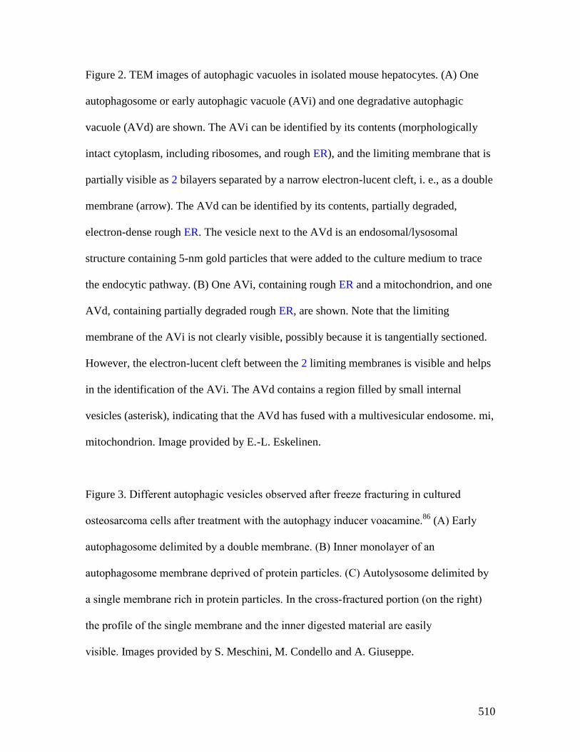
510
Figure 2. TEM images of autophagic vacuoles in isolated mouse hepatocytes. (A) One
autophagosome or early autophagic vacuole (AVi) and one degradative autophagic
vacuole (AVd) are shown. The AVi can be identified by its contents (morphologically
intact cytoplasm, including ribosomes, and rough ER), and the limiting membrane that is
partially visible as 2 bilayers separated by a narrow electron-lucent cleft, i. e., as a double
membrane (arrow). The AVd can be identified by its contents, partially degraded,
electron-dense rough ER. The vesicle next to the AVd is an endosomal/lysosomal
structure containing 5-nm gold particles that were added to the culture medium to trace
the endocytic pathway. (B) One AVi, containing rough ER and a mitochondrion, and one
AVd, containing partially degraded rough ER, are shown. Note that the limiting
membrane of the AVi is not clearly visible, possibly because it is tangentially sectioned.
However, the electron-lucent cleft between the 2 limiting membranes is visible and helps
in the identification of the AVi. The AVd contains a region filled by small internal
vesicles (asterisk), indicating that the AVd has fused with a multivesicular endosome. mi,
mitochondrion. Image provided by E.-L. Eskelinen.
Figure 3. Different autophagic vesicles observed after freeze fracturing in cultured
osteosarcoma cells after treatment with the autophagy inducer voacamine.86
(A) Early
autophagosome delimited by a double membrane. (B) Inner monolayer of an
autophagosome membrane deprived of protein particles. (C) Autolysosome delimited by
a single membrane rich in protein particles. In the cross-fractured portion (on the right)
the profile of the single membrane and the inner digested material are easily
visible. Images provided by S. Meschini, M. Condello and A. Giuseppe.

511
Figure 4. Cryoelectron microscopy can be used as a three-dimensional approach to
monitor the autophagic process. Four computed sections of an electron tomogram of the
autophagic vesicle-rich cytoplasm in a hemophagocyte of a semi-thin section after high-
pressure freezing preparation. The dashed area is membrane-free (1A) but tomography
reveals newly formed or degrading membranes with a parallel stretch (1B). In another
computed section (1C) of the same tomogram, the parallel stretch of the 2 membranes
shows only one membrane bilayer, and this membrane composition has disappeared in
the computed section shown in 1D. Image published previously1851
and provided by M.
Schneider and P. Walter.
Figure 5. LC3-I conversion and LC3-II turnover. (A) Expression levels of LC3-I and LC3-II
during starvation. Atg5+/+
(wild-type) and Atg5-/-
MEFs were cultured in DMEM without amino
acids and serum for the indicated times, and then subjected to immunoblot analysis using anti-
LC3 antibody and anti-tubulin antibody. E-64d (10 µg/ml) and pepstatin A (10 µg/ml) were
added to the medium where indicated. Positions of LC3-I and LC3-II are indicated. The
inclusion of lysosomal protease inhibitors reveals that the apparent decrease in LC3-II is due to
lysosomal degradation as easily seen by comparing samples with and without inhibitors at the
same time points (the overall decrease seen in the presence of inhibitors may reflect decreasing
effectiveness of the inhibitors over time). Monitoring autophagy by following steady state
amounts of LC3-II without including inhibitors in the analysis can result in an incorrect
interpretation that autophagy is not taking place (due to the apparent absence of LC3-II).
Conversely, if there are high levels of LC3-II but there is no change in the presence of inhibitors

512
this may indicate that induction has occurred but that the final steps of autophagy are blocked,
resulting in stabilization of this protein. This figure was modified from data previously published
in reference 24
, and is reproduced by permission of Landes Bioscience, copyright 2007. (B)
Lysates of 4 human adipose tissue biopsies were resolved on 2 12% polyacrylamide gels, as
described previously.1852
Proteins were transferred in parallel to either a PVDF or a nitrocellulose
membrane, and blotted with anti-LC3 antibody, and then identified by reacting the membranes
with an HRP-conjugated anti-rabbit IgG antibody, followed by ECL. The LC3-II/LC3-I ratio was
calculated based on densitometry analysis of both bands. *, p< 0.05. (C) HEK 293 and HeLa
cells were cultured in nutrient-rich medium (DMEM containing 10% FCS) or incubated for 4 h
in starvation conditions (Krebs-Ringer medium) in the absence (-) or presence (+) of E-64d and
pepstatin at 10 µg/ml each (Inhibitors). Cells were then lysed and the proteins resolved by SDS-
PAGE. Endogenous LC3 was detected by immunoblotting. Positions of LC3-I and LC3-II are
indicated. In the absence of lysosomal protease inhibitors, starvation results in a modest increase
(HEK 293 cells) or even a decrease (HeLa cells) in the amount of LC3-II. The use of inhibitors
reveals that this apparent decrease is due to lysosome-dependent degradation. This figure was
modified from data previously published in reference 145
, and is reproduced by permission of
Landes Bioscience, copyright 2005. (D) Sequence and schematic representation of the different
forms of LC3B. The sequence for the nascent (proLC3) from mouse is shown. The glycine at
position 120 indicates the cleavage site for ATG4. After this cleavage, the truncated LC3 is
referred to as LC3-I, which is still a soluble form of the protein. Conjugation to PE generates the
membrane-associated LC3-II (equivalent to Atg8–PE).

513
Figure 6. Effect of different inhibitors on LC3-II accumulation. SH-SY5Y human
neuroblastoma cells were plated and allowed to adhere for a minimum of 24 h, then
treated in fresh medium. Treatments were as follows: rapamycin (Rap), (A) 1 µM, 4 h or
(B) 10 µM, 4 h; E-64d, final concentration 10 µg/ml from a 1 mg/ml stock in ethanol
(ETOH); NH4Cl (NH4+), final concentration 10 mM from a 1 M stock in water; pepstatin
A (Pst), final concentration 10 µg/ml from a 1 mg/ml stock in ethanol, or 68.6 µg/ml
from a 6.86 mg/ml stock in DMSO; ethanol or DMSO, final concentration 1%. Pre-
incubations in (B) were for 1 or 4 h as indicated. 10 mM NH4Cl (or 30 µM chloroquine,
not shown) were the most effective compounds for demonstrating the accumulation of
LC3-II. E-64d was also effective in preventing the degradation of LC3-II, with or without
a preincubation, but ammomium chloride (or chloroquine) may be more effective.
Pepstatin A at 10 µg/ml with a 1 h pre-incubation was not effective at blocking
degradation, whereas a 100 µM concentration with 4 h pre-incubation had a partial effect.
Thus, alkalinizing compounds are more effective in blocking LC3-II degradation, and
pepstatin A must be used at saturating conditions to have any noticeable effect. Images
provided by C. Isidoro. Note that the band running just below LC3-I at approximately
17.5 kDa may be a processing intermediate of LC3-I; it is detectable in freshly prepared
homogenates, but is less visible after the sample is subjected to a freeze-thaw cycle.
Figure 7. GFP-LC3 processing can be used to monitor delivery of autophagosomal membranes.
(A) Atg5-/-
MEFs engineered to express Atg5 under the control of the Tet-off promoter were
grown in the presence of doxycyline (10 ng/ml) for one week to suppress autophagy. Cells were
then cultured in the absence of drug for the indicated times, with or without a final 2 h starvation.

514
Protein lysates were analyzed by western blot using anti-LC3 and anti-GFP antibodies. The
positions of untagged and GFP-tagged LC3-I and LC3-II, and free GFP are indicated. This figure
was modified from data previously published in reference 214
, FEBS Letters, 580, Hosokawa N,
Hara Y, Mizushima N, Generation of cell lines with tetracycline-regulated autophagy and a role
for autophagy in controlling cell size, pp. 2623-2629, copyright 2006, with permission from
Elsevier. (B) Differential role of unsaturating and saturating concentrations of lysosomal
inhibitors on GFP-LC3 cleavage. HeLa cells stably transfected with GFP-LC3 were treated with
various concentrations of chloroquine (CQ) for 6 h. Total lysates were prepared and subjected to
immunoblot analysis. (C) CQ-induced free GFP fragments require classical autophagy
machinery. Wild-type and Atg5-/-
MEFs were first infected with adenovirus GFP-LC3 (100 viral
particles per cell) for 24 h. The cells were then either cultured in regular culture medium with or
without CQ (10 µM), or subjected to starvation in EBSS buffer in the absence or presence of CQ
for 6 h. Total lysates were prepared and subjected to immunoblot analysis. Panel B and C are
modified from the data previously published in reference 216
.
Figure 8. Movement of activated pDendra2-hp62 (SQSTM1; orange) from the nucleus
(middle) to the aggregate in ARPE-19 cells, revealed by confocal microscopy. Cells were
exposed to 5 µM MG132 for 24 h to induce the formation of perinuclear aggregates.1853
The cells were then exposed to a UV pulse (the UV-induced area is shown by red lines
that are inside of the nucleus) that converts Dendra2 from green to red, and the time
shown after the pulse is indicated. SQSTM1 is present in a small nuclear aggregrate, and
is shuttled from the nucleus to a perinuclear large protein aggregate (detected as red).
Scale bar, 5 µm. Image provided by K. Kaarniranta.

515
Figure 9. Changes in the detection and localization of GFP-LC3 upon the induction of
autophagy. U87 cells stably expressing GFP-LC3 were treated with PBS, rapamycin (200
nM), or rapamycin in combination with 3-MA (2 mM) for 24 h. Representative
fluorescence images of cells counterstained with DAPI (nuclei) are shown. Scale bar, 10
µm. This figure was modified from Figure 6 published in Badr et al. Lanatoside C
sensitizes glioblastoma cells to tumor necrosis factor–related apoptosis-inducing ligand
and induces an alternative cell death pathway. Neuro-Oncology, 13(11):1213-24, 2011,
by permission of Oxford University Press.
Figure 10. The GFP and mRFP signals of tandem fluorescent LC3 (tfLC3, mRFP-GFP-LC3)
show different localization patterns. HeLa cells were cotransfected with plasmids expressing
either tfLC3 or LAMP1-CFP. Twenty-four hours after the transfection, the cells were starved in
Hanks’ solution for 2 hours, fixed and analyzed by microscopy. The lower panels are a higher
magnification of the upper panels. Bar, 10 µm in the upper panels and 2 µm in the lower panels.
Arrows in the lower panels point to (or mark the location of) typical examples of colocalized
signals of mRFP and LAMP1. Arrowheads point to (or mark the location of) typical examples of
colocalized particles of GFP and mRFP signals. This figure was previously published in
reference 223
, and is reproduced by permission of Landes Bioscience, copyright 2007.
Figure 11. GFP fluorescence in the autolysosome can be recovered upon neutralization of the pH.
(A) GFP-LC3 emits green fluorescence in the autolysosomes of post-mortem processed heart
sections. Cryosections of 3.8% paraformaldehyde fixed ventricular myocardium from 3-week old
516
GFP-LC3 transgenic mice at the baseline (control) or starved for 24 h (starved) were processed
for immunostaining using a standard protocol (buffered at pH 7.4). Most of the GFP-LC3 puncta
are positive for LAMP1, suggesting that the autolysosomes had recovered GFP fluorescence. (B)
Colocalization between GFP-LC3 direct fluorescence (green) and indirect immunostaining for
GFP (red). Sections processed as in (A) were immunostained for GFP using a red fluorescence-
tagged secondary antibody, and the colocalization with GFP fluorescence was examined by
confocal microscopy. Almost all of the red puncta emit green fluorescence. Images provided by
Xuejun Wang.
Figure 12. Saponin extraction allows quantification of LC3-II fluorescence by FACS.
(A) Schematic diagram of the effects of the saponin wash. Due to the reorganization of the
EGFP-LC3 reporter protein, induction of autophagosome formation does not change the total
levels of fluorescence in EGFP-LC3-transfected cells. However, extraction of EGFP-LC3-I with
saponin results in a higher level of fluorescence in cells with proportionally higher levels of
EGFP-LC3-II-containing autophagosomes. This figure was previously published in reference 283
.
(B) Saponin extraction can also be used to measure flux of endogenous LC3 protein. Human
osteosarcoma cells were starved of amino acids and serum by incubation in EBSS, for the
indicated times in the presence or absence of a 1 h chloroquine (50 µM) treatment. Cells were
then washed with PBS containing 0.05% saponin and processed for FACS analysis for
endogenous LC3. These data are provided by K.E. Eng and G.M. McInerney.
Figure 13. Assessing autophagy with multispectral imaging cytometry. (A) Bright Detail
Intensity (BDI) measures the foreground intensity of bright puncta (that are 3 pixels or less)

517
within the cell image. For each cell, the local background around the spots is removed before
intensity calculation. Thus, autophagic cells with puncta have higher BDI values. (B) Media
control (untreated wild type), rapamycin-treated wild-type and Atg5-/-
MEFs were gated based on
BDI. Representative images of cells with high or low BDI values. Scale bar, 10 µm. Images
provided by M.L. Albert.
Figure 14. Regulation of the SQSTM1 protein during autophagy. (A) The level of SQSTM1
during starvation. Atg5+/+
and atg5-/-
MEFs were cultured in DMEM without amino acids and
serum for the indicated times, and then subjected to immunoblot analysis using anti-
SQSTM1antibody (Progen Biotechnik). This figure was previously published in reference 24
, and
is reproduced by permission of Landes Bioscience, copyright 2007. (B) The level of SQSTM1 in
the brain of neural-cell specific Atg5 knockout mice. This image was generously provided by Dr.
Taichi Hara (Tokyo Medical and Dental University).
Figure 15. S. cerevisae cells transformed with a plasmid encoding HA-Atg1 were
cultured to mid-log phase and shifted to SD-N (minimal medium lacking nitrogen that
induces a starvation response). Immunoblotting was done with anti-HA antibody. The
upper band corresponds to autophosphorylation of Atg1. This figure was modified from
data previously published in reference 459
, and is reproduced by permission of the
American Society for Cell Biology, copyright 2011.
Figure 16. Confocal microscopy image of HCT116 cells immunostained with human-specific
antibody to ATG12. Cells were starved for 8 h or treated with chloroquine (50 µM) for 3 h. Scale

518
bar, 10 µm. Image provided by M. Llanos Valero, M.A de la Cruz and R. Sanchez-Prieto.
Figure 17. Automated WIPI1 puncta image acquisition and analysis monitors the
induction and inhibition of autophagy. Stable U2OS clones expressing GFP-WIPI1 were
selected using 0.6 μg/ml G418 and then cultured in 96-well plates. Cells were treated for
3 h with nutrient-rich medium (control), nutrient-free medium (EBSS), or with 233 nM
wortmannin. Cells were fixed in 3.7% paraformaldehyde and stained with DAPI (5 μg/ml
in PBS). An automated imaging and analysis platform was used to determine the number
of both GFP-WIPI1 puncta-positive cells and the number of GFP-WIPI1 puncta per
individual cell.422
Cells without GFP-WIPI1 puncta are highlighted in red (cell detection)
and purple (nuclei detection), whereas GFP-WIPI1 puncta-positive cells are highlighted
in yellow (GFP-WIPI1 puncta detection), green (cell detection) and blue (nuclei
detection). Bars, 20 µm. These images were provided by S. Pfisterer and T. Proikas-
Cezanne.
Figure 18. pGFP-Atg8a can be used to monitor autophagy in Drosophila melanogaster.
The autophagosome marker pGFP-Atg8a, results in expression of Atg8a fused to GFP
from the endogenous Atg8a promoter.240
Live imaging of gastric caeca from Drosophila
melanogaster midgut pGFP-Atg8a puncta (green) and Hoechst 33342 (blue). Midgut
from early third instar larvae prior to the onset of cell death (top) and from dying midgut
at 2 h after puparium formation (bottom). Bar, 25 µm. Figure provided by D. Denton and
S. Kumar.

519
Figure 19. S. cerevisae cells were cultured to mid-log phase and shifted to SD-N for the indicated
times. Samples were taken before (+) and at the indicated times after (–) nitrogen starvation.
Immunoblotting was done with anti-phospho-Slt2 and anti-phospho-Hog1 antibody. This figure
was modified from data previously published in reference 459
, and is reproduced by permission of
the American Society for Cell Biology, copyright 2011.
Figure 20. Autophagosomes with recognizable cargo are rare in cells. (A) To assess relative rates
of autophagosome formation, the fusion inhibitor bafilomycin A1 (10 nM) was applied for 2 h
prior to fixation with 2% glutaraldehyde in order to trap newly formed autophagosomes. Two
different PINK1 shRNA lines exhibit increased AV formation over 2 h compared to the control
shRNA line. * p > 0.05 vs. Control. (B) Autophagosomes in bafilomycin A1-treated control cells
contain a variety of cytoplasmic structures (left, arrow), while mitochondria comprise a
prominent component of autophagosomes in A14 bafilomycin A1-treated (PINK1 shRNA) cells
(right, arrow). Scale bar, 500 nm. These data indicate induction of selective mitophagy in
PINK1-deficient cells. This figure was modified from Figure 2 published in Chu CT. A pivotal
role for PINK1 and autophagy in mitochondrial quality control: implications for Parkinson
disease. Human Molecular Genetics 2010; 19:R28-R37.
Figure 21. Human fibroblasts showing colocalization of mitochondria with lysosomes.
The degree of colocalization of mitochondria with lysosomes in human fibroblasts was
measured via live cell imaging microscopy at 37°C and 5% CO2 atmosphere using the
ApoTome® technique. LysoTracker® Red DND-99 staining was applied to mark
lysosomal structures (red), and MitoTracker® Green FM to visualize mitochondria

520
(green). Hoechst 33342 dye was used to stain nuclei (blue). A positive colocalization is
indicated by yellow signals (merge) due to the overlap of LysoTracker® Red and
MitoTracker® Green staining (white arrows). Scale bar, 10 µm. Statistical evaluation is
performed by calculating the Pearson’s coefficient for colocalizing pixels. Image
provided by L. Burbulla and R. Krüger.
Figure 22. Detection of mitophagy in primary cortical neurons using mitochondria-
targeted Keima. Neurons transfected with mito-Keima were visualized using 458 nm
(green, mitochondria at neutral pH) and 561 nm (red, mitochondria in acidic pH) laser
lines and 575 nm band pass filter. Compared with the control (A) wild-type PINK1
overexpression (B) increases the number of the mitochondria exposed to acidic
conditions. Scale bar, 2 µm. (C) Quantification of red dots suggests increased mitophagy
in wild-type PINK1 but not in the kinase dead PINK1K219M
-overexpressing neurons.
Figure provided by V. Choubey and A. Kaasik.
Figure 23. LysoTracker Red stains lysosomes and can be used to monitor autophagy in
Drosophila. Live fat body tissues from Drosophila were stained with LysoTracker Red (red) and
Hoechst 33342 (blue) to stain the nucleus. Tissues were isolated from fed (left) or 3 h starved
(right) animals. Bar, 25 µm. This figure was modified from data presented in reference 239
,
Developmental Cell, 7, Scott RC, Schuldiner O, Neufeld TP, Role and regulation of starvation-
induced autophagy in the Drosophila fat body, pp. 167-78, copyright 2004, with permission from
Elsevier.

521
Figure 24. GFP::LGG-1 and GFP::LGG-2 are autophagy markers in C. elegans. (A-F) Animals
were generated that carry an integrated transgene expressing a GFP-tagged version of lgg-1, the
C. elegans ortholog of mammalian MAP1LC3. Representative green fluorescence images in the
pharyngeal muscles of (A) control RNAi animals without starvation, (B) control RNAi animals
after 9 d of starvation, (C) atg-7 RNAi animals after 9 d of starvation, (D) starvation-
hypersensitive gpb-2 mutants without leucine after 3 d of starvation, and (E) gpb-2 mutants with
leucine after 3 d of starvation. The arrows show representative GFP::LGG-1-positive punctate
areas that label pre-autophagosomal and autophagosomal structures. (F) The relative levels of
PE-conjugated and unconjugated GFP::LGG-1 were determined by western blotting. These
figures were modified from data previously published in Kang, C., Y.J. You, and L. Avery. 2007.
Dual roles of autophagy in the survival of Caenorhabditis elegans during starvation. Genes &
Development. 21:2161-2171, Copyright © 2007, Genes & Development by Cold Spring Harbor
Laboratory Press and Kang, C., and L. Avery. 2009. Systemic regulation of starvation response in
Caenorhabditis elegans. Genes & development. 23:12-17, Copyright © 2011, Genes &
Development by Cold Spring Harbor Laboratory Press, www.genesdev.org. (G-H) GFP:LGG-2
serves as a marker for autophagosomes in early C. elegans embryos. (G) GFP::LGG-2 expressed
in the germline from an integrated transgene reveal the formation of autophagosomes (green)
around sperm inherited membranous organelles (red). DNA of the 2 pronuclei is stained (blue).
(H) Later during development, GFP::LGG-2-positive structures are present in all cells of the
embryo. Scale bar, 10 µm. Images provided by V. Galy.
Figure 24*. Transmission electron micrograph of erythroblasts obtained from the blood of
regular donors after 10 days of culture in the presence of KITLG/SCF, IL3, EPO and
dexamethasone. Original magnification 3000X. This figure shows 2 erythroblasts containing

522
autophagic vacuoles. One erythroblast (red arrow) has the morphology of a live cell with several
autophagic vacuoles that have engulfed cytoplasmic organelles. The other erythroblast (black
arrow) has the electron-dense cytoplasm characteristic of a dead cell and is in the process of
shedding its autolysosomes from the cytoplasm to the extracellular space (courtesy of A.R.
Migliaccio and M. Zingariello).
Figure 25. A large dystrophic neurite from a brain biopsy of a patient with Gerstmann-
Sträussler-Scheinker (GSS) disease not unlike those reported for Alzheimer disease.1854
This
structure is filled with innumerable autophagic vacuoles, some of which are covered by a double
membrane. Electron dense lysosomal-like structures are also visible. The red arrow points to a
double-membrane autophagic vacuole. Scale bar, 200 nm. Image provided by P. Liberski.
Figure 26. A high-power electron micrograph from a brain biopsy showing autophagic vacuoles
in a case of ganglioglioma. Scale bar, 200 nm. Image provided by P. Liberski.
Figure 27. Detection of macroautophagy in tobacco BY-2 cells. (A) Induction of
autophagosomes in tobacco BY-2 cells expressing YFP-NtAtg8 (shown in green for ease of
visualization) under conditions of nitrogen limitation (Induced). Arrowheads indicate
autophagosomes that can be seen as a bright green dot. No such structure was found in cells
grown in normal culture medium (Control). Bar, 10 µm. N, nucleus; V, vacuole. (B)
Ultrastructure of an autophagosome in a tobacco BY-2 cell cultured for 24 h without a nitrogen
source. Bar, 200 µm. AP, autophagosome; P, plastid; CW, cell wall. This image was provided by
Dr. Kiminori Toyooka, (RIKEN Plant Science Center).

523
Figure 28. Confocal microscopy deconvolved (AutoQuant X3) images and colocalization
image analysis (ImageJ 1.47; Imaris 7.6) through a local approach showing perinuclear
mitochondrial biogenesis in hippocampal neuronal cultures. The upper channels show
TOMM20 (green channel), BrdU (for visualization of newly synthesized mitochondrial
DNA, red channel), and merged fluorescence channels. Overlay, corresponds to the
spatial pattern of software thresholded colocalized structures (white spots) layered on the
merged fluorescence channels. Surface Plot, or luminance intensity height, is
proportional to the colocalization strength of the colocalized structures (white spots). Plot
Profile, corresponds to the spatial intensity profiles of the fluorescence channels of the
white line positioned in the Merge image. Yellow arrows indicate a qualitative evaluation
of the spatial association trends for the fluorescence intensities. Arrows pointing up
indicate an increase in the colocalization, while arrows pointing down show a decrease.
Scale bar = 2 m. This figure was was modified from previously published data1855
and
provided by Dr. Fulvio Florenzano.
Figure XX. Macroautophagy in the digestive gland of Ruditapes decussatus (Mollusca,
Bivalvia) subjected to a strict starvation of 2 months. This image was provided by Dr. Stephen
Baghdiguian.