guidelines for non-operative diagnostic procedures and
TRANSCRIPT

CEff 090821 1 V2 Final
Guidelines for non-operative diagnostic procedures and reporting
in breast cancer screening
August 2021 Authors: Dr Andrew HS Lee (Editor) Dr Pauline Carder Dr Rahul Deb
Professor Ian O Ellis Dr Miles Howe Mrs Jacquie A Jenkins Professor Sarah E Pinder
Unique document number G150 Document name Guidelines for non-operative diagnostic procedures and reporting in breast
cancer screening Version number 2 Produced by The authors are members of the Non-operative Diagnosis Working Group of
the UK National Coordinating Committee for Breast Pathology, which is responsible for supporting pathology quality assurance in the UK National Health Service Breast Screening Program (NHSBSP) and for preparation of dataset standards in breast cancer pathology for the Royal College of Pathologists. Membership of the UK National Coordinating Committee for Breast Pathology: Dr K Bichbiche, Ms R Bennett, Dr C Boyd, Prof G Callagy, Dr P Carder, Miss L Chagla, Dr D Coleman, Dr R Deb, Prof IO Ellis, Dr M Howe, Mrs J Jenkins, Prof L Jones, Ms O Kearins, Ms S Kodikara, Ms L Langford, Mr K Lea, Dr AHS Lee, Dr R Macavinchey, Dr S Natu, Dr DM Parham, Prof SE Pinder (Chair), Dr S Pritchard, Dr E Provenzano, Dr C Purdie, Dr CM Quinn, Prof EA Rakha, Dr I Roxannis, Dr A Shaaban, Dr B Tanchel, Dr CA Wells and Dr W Williams.
Date active August 2021 (to be implemented within three months) Date for full review August 2024 Comments This document will replace the 1st edition of Guidelines for non-operative
diagnostic procedures and reporting in breast cancer screening, published in 2016. In accordance with the College’s pre-publications policy, this document was on the Royal College of Pathologists’ website for consultation from 19 November to 17 December 2020. Responses and authors’ comments are available to view on publication of the final document. Dr Brian Rous Clinical Lead for Guideline Review
The Royal College of Pathologists 6 Alie Street, London E1 8QT Tel: 020 7451 6700 Fax: 020 7451 6701 Web: www.rcpath.org
Registered charity in England and Wales, no. 261035 © 2021, The Royal College of Pathologists
This work is copyright. You may download, display, print and reproduce this document for your personal, non-commercial use. Requests and inquiries concerning reproduction and rights should be addressed to the Royal College of Pathologists at the above address. First published: 2021.

CEff 090821 2 V2 Final
Contents Foreword ............................................................................................................................................ 4 1 Introduction ............................................................................................................................... 5 2 Use of non-operative diagnostic techniques ............................................................................. 6
2.1 Image guidance for breast biopsy ................................................................................... 7 2.2 Sampling techniques and procedures ............................................................................. 8 2.3 Core biopsy – general principles ..................................................................................... 9 2.4 Ultrasound guided core biopsy ...................................................................................... 10 2.5 Stereotactic guided core biopsy .................................................................................... 10 2.6 Prone stereotactic core biopsy ...................................................................................... 11 2.7 Large volume sampling techniques ............................................................................... 11 2.8 Complications of core biopsy ........................................................................................ 12
3 Core biopsy reporting guidelines ............................................................................................ 13
3.1 Core biopsy specimen information and handling ........................................................... 13 3.2 Recording of data on the National Breast Screening System ....................................... 14 3.3 Using the core biopsy reporting form............................................................................. 16 3.4 RCPath dataset items ................................................................................................... 16 3.5 Core biopsy reporting categories................................................................................... 18 3.6 B1 normal tissue ............................................................................................................ 18 3.7 B2 benign lesion ............................................................................................................ 19 3.8 B3 lesion of uncertain malignant potential ..................................................................... 20 3.9 B4 suspicious ................................................................................................................ 23 3.10 B5 malignant ................................................................................................................. 24 3.11 Assessment of prognostic and predictive factors .......................................................... 26 3.12 Rare malignancies ......................................................................................................... 26 3.13 Problems and pitfalls in diagnosis ................................................................................. 27
4 Axillary lymph node assessment and preoperative sampling ................................................. 30
4.1 L codes for fine needle aspiration cytology ................................................................... 31 4.2 L codes for needle core biopsy ..................................................................................... 31

CEff 090821 3 V2 Final
4.3 Pitfalls ............................................................................................................................ 32
5 How to perform fine needle aspiration cytology ...................................................................... 32
5.1 Aspiration procedure ..................................................................................................... 32 5.2 Ultrasound-guided FNAC .............................................................................................. 34 5.3 Spreading the slides ...................................................................................................... 34 5.4 Fixation methods ........................................................................................................... 35
6 Diagnostic coding ................................................................................................................... 36 7 Criteria for audit ...................................................................................................................... 36 8 References ............................................................................................................................. 37 Appendix A Fine needle aspiration cytology reporting guidelines ................................................ 40 Appendix B Cytological features of specific lesions diagnosed on fine needle aspiration cytology .................................................................................................... 45 Appendix C Quality assurance ..................................................................................................... 51 Appendix D Recommended SNOMED codes for breast pathology ............................................. 60 Appendix E NHSBSP wide bore needle biopsy form ................................................................... 66 Appendix F Reporting proforma for breast core biopsy ............................................................... 68 Appendix G Reporting proforma for vacuum-assisted excision .................................................... 70 Appendix H Reporting proforma for breast fine needle aspiration cytology ................................. 72 Appendix I Reporting proforma for axillary fine needle aspiration cytology ................................ 73 Appendix J Reporting proforma for axillary core biopsy .............................................................. 74 Appendix K Reporting proforma for breast core biopsy in list format ........................................... 75 Appendix L Reporting proforma for breast fine needle aspiration cytology in list format ............. 80 Appendix M Reporting proforma for axillary fine needle aspiration cytology in list format ............ 81 Appendix N Reporting proforma for axillary core biopsy in list format .......................................... 82 Appendix O Summary table – explanation of levels of evidence .................................................. 83 Appendix P AGREE II compliance monitoring sheet .................................................................. 84
NICE has accredited the process used by the Royal College of Pathologists to produce its cancer datasets. Accreditation is valid for five years from 25 July 2017. More information on accreditation can be viewed at www.nice.org.uk/accreditation. For full details on our accreditation visit: www.nice.org.uk/accreditation

CEff 191120 4 V2 Draft
Foreword The cancer datasets published by the Royal College of Pathologists (RCPath) are a combination of textual guidance, educational information and reporting proformas. The datasets enable pathologists to grade and stage cancers in an accurate, consistent manner in compliance with international standards and provide prognostic information, thereby allowing clinicians to provide a high standard of care for patients and appropriate management for specific clinical circumstances. This guideline has been developed to cover most common circumstances. However, we recognise that guidelines cannot anticipate every pathological specimen type and clinical scenario. Occasional variation from the practice recommended in this guideline may therefore be required to report a specimen in a way that maximises benefit to the patient. Each dataset contains core data items (see Appendices E–M) that are mandated for inclusion in the Cancer Outcomes and Services Dataset (COSD – previously the National Cancer Data Set) in England. Core data items are items that are supported by robust published evidence and are required for cancer staging, optimal patient management and prognosis. Core data items meet the requirements of professional standards (as defined by the Information Standards Board for Health and Social Care [ISB]) and it is recommended that at least 95% of reports on cancer resections should record a full set of core data items. Other non-core data items are described. These may be included to provide a comprehensive report or to meet local clinical or research requirements. All data items should be clearly defined to allow the unambiguous recording of data. The following stakeholders were contacted to consult on this document:
• National Breast Radiology Clinical and Professional Group
• National Breast Surgical Clinical and Professional Group
• British Society of Breast Radiology
• Association of Breast Surgery. Evidence for the revised dataset was obtained by searching Medline from 2015 to 2019 for systematic reviews and guidelines about non-operative diagnosis of lesions of the breast, including needle core biopsy, vacuum-assisted biopsy and fine needle aspiration cytology. A search for guidelines was also performed using the Google search engine. A Medline search for nipple discharge cytology was performed from 2000 to 2019. The level of evidence for the recommendations has been summarised (Appendix N). Unless otherwise stated, the level of evidence corresponds to ‘Good practice point (GPP): Recommended best practice based on the clinical experience of the authors of the writing group’. The sections of this dataset that indicate compliance with each of the AGREE II standards are indicated in Appendix O. No major organisational changes or cost implications have been identified that would hinder the implementation of the dataset for the core items. A formal revision cycle for all cancer datasets takes place on a three-yearly basis. However, each year, the College will ask the authors of the dataset, in conjunction with the relevant subspecialty advisor to the College, to consider whether or not the dataset needs to be updated or revised. A full consultation process will be undertaken if major revisions are required, i.e. revisions to core data items (the only exception being changes to international tumour grading and staging schemes that have been approved by the Specialty Advisory Committee on Cellular Pathology and affiliated professional bodies; these changes will be implemented without further consultation). If minor revisions or changes to non-core data items are required, an abridged consultation process will be undertaken, whereby a short note of the proposed changes will be placed on the College website for two weeks for members’ attention. If members do not object to the changes, the changes

CEff 090821 5 V2 Final
will be incorporated into the dataset and the full revised version (incorporating the changes) will replace the existing version on the College website. The dataset has been reviewed by the Clinical Effectiveness team, Working Group on Cancer Services and Lay Governance Group. It was placed on the College website for consultation with the membership from 19 November to 17 December 2020. All comments received from the Working Group and the membership were addressed by the authors to the satisfaction of the Chair of the Working Group and the Clinical Lead for Guideline Review. This dataset was developed without external funding to the writing group. The College requires the authors of datasets to provide a list of potential conflicts of interest; these are monitored by the Clinical Effectiveness team and are available on request. The authors have declared no conflicts of interest. Acknowledgement The NHS Breast Screening Programme is grateful to the members of the Guidelines Working Group of the UK National Coordinating Committee for Breast Pathology for their work in updating the Guidelines for non-operative diagnostic procedures and reporting in breast cancer screening. The NHS Breast Screening Programme will reference this updated guidance in its future publications. 1 Introduction
The aim of assessment is to obtain a definitive and timely diagnosis of all potential abnormalities detected during screening.1 This is best achieved by using ‘triple assessment’, comprising imaging (usually mammography and ultrasound), plus clinical examination and image-guided needle biopsy for histological examination, if indicated. Definitive non-operative diagnosis of malignancy allows rapid referral for treatment, ideally in one operative procedure. Definitive non-operative diagnosis of benign conditions is equally useful, usually leading to discharge from the clinic and return to routine recall. In the early days of breast screening, fine needle aspiration cytology (FNAC) was the procedure of choice, but it is now recommended that needle core biopsy (NCB) or vacuum-assisted biopsy (VAB) is used for assessment of significant screening detected abnormalities.1 This is because current evidence suggests that core biopsy has greater sensitivity and specificity in evaluating microcalcification, asymmetry and architectural distortion than does FNAC. It also aids definitive benign diagnosis. Invasive carcinoma can be distinguished from ductal carcinoma in situ (DCIS) on core biopsy (but not with FNAC). Oestrogen receptor (ER), progesterone receptor (PR) and HER2 (human epidermal growth factor receptor 2) status can be assessed on the core biopsies because invasive carcinoma can be recognised. Histological grade can be more accurately assessed on core biopsy. FNAC may be used in addition to core biopsy if an urgent diagnosis is required or if core biopsy is not possible.1 FNAC should not be used alone in the assessment of lesions in the breast detected by screening mammography, unless core biopsy is contraindicated. The purpose of these guidelines is to provide pathologists with an update on the role of non-operative diagnosis in breast screening assessment, and on the handling and reporting of biopsy specimens. A similar approach is recommended for symptomatic breast lesions. The document concentrates on NCB and VAB. It also describes the mechanisms used to assess and assure the quality of non-operative diagnosis in breast screening.

CEff 090821 6 V2 Final
This document constitutes the fifth edition of guidelines for non-operative diagnosis in breast cancer screening. It updates and replaces the previous guidelines published in 2016.2
2 Use of non-operative diagnostic techniques
Detailed guidance on assessment procedures is provided in the National Health Service Breast Screening Programme (NHSBSP) guidelines, Clinical guidance for breast cancer screening assessment (4th edition).1 All cases should be thoroughly assessed prior to needle biopsy. The radiological findings can be categorised into five categories: 1. normal/no significant abnormality 2. benign 3. indeterminate/probably benign 4. suspicious of malignancy 5. highly suspicious of malignancy. The number is preceded by U for ultrasound assessment, M for mammography, MRI for magnetic resonance imaging and R can be used for overall radiological assessment.3 All needle sampling procedures carried out on screen-detected abnormalities must be discussed at a multidisciplinary meeting, where findings from all modalities are discussed and further management is decided. These guidelines also detail the methods of choice for sampling the different types of mammographic abnormality. This approach must be adhered to in the National Breast Screening Programme as it is recognised that very rare false-positive interpretation of needle biopsy specimens can occur. All cases should be subject to multidisciplinary review to ensure concordance before proceeding to definitive treatment. Both NCB and VAB procedures may result in removal or destruction of the mammographically detected lesion. The lesion may therefore not be identified in a subsequent operative specimen. In situations where such a discrepancy highlights a ‘potential false-positive result’, the biopsy should be reviewed according to the protocols described in Good Practice Guide No 9: Reporting, recording and auditing B5 core biopsies with normal/benign surgery.4 A decision must be reached as to:
• whether the histological findings of the core biopsy have been appropriately interpreted
• whether the appropriate area of lesion has been removed in the surgical specimen or if the patient has undergone neoadjuvant treatment
• whether there is a complete pathological response or whether it is possible that the lesion remains in the breast.
The findings of such reviews should be available for discussion as part of the quality assurance process. Core biopsy results should not be interpreted in isolation. The multidisciplinary meeting should make a judgement about whether the biopsy is concordant with radiological and clinical findings, and whether the biopsy is representative of the lesion. If there is discordance, further management must be discussed. Inevitably, false-negative results are significantly higher for impalpable lesions. When the imaging findings are considered to be suspicious of malignancy and the biopsy is normal or benign, management should be reviewed at a multidisciplinary

CEff 090821 7 V2 Final
meeting and a decision made whether to repeat the sampling procedure or to refer for open biopsy or localisation biopsy. In cases where there is disagreement between modalities, with a failure to achieve consensus after multidisciplinary discussion, repeat core biopsy, VAB or surgical biopsy is likely to be the most appropriate course of action. No more than two non-surgical needle biopsy procedures (for example core biopsy followed by VAB), carried out on separate occasions, should normally be needed to achieve a non-operative diagnosis of a screen-detected abnormality. Frozen section for the diagnosis of screen-detected lesions is inappropriate, except in very exceptional circumstances when FNAC or core biopsy is contraindicated. Evidence from published series of multiple NCB sampling has shown that for certain types of mammographic abnormality, particularly moderate- to low-level suspicion microcalcification, a larger volume of tissue is required for accurate diagnosis.5 For such lesions, where the use of conventional 14G-core biopsy carries a high risk of an equivocal result, use of larger-volume sampling techniques may increase the accuracy of biopsy. VAB has a lower equivocal sample rate and increased accuracy in the detection of small invasive tumours associated with an area of DCIS.6−8 Consideration of the likely underlying histological nature of the lesion from the imaging features should therefore be taken into account when deciding on the sampling method to be used. VAB may also be useful after a B1, B3 or B4 diagnosis on 14G core biopsy, and recent guidelines propose more thorough sampling or excision, using VAB as an alternative to diagnostic surgical biopsy of the majority, although not all, of B3 lesions (VAB used in this way is described as a vacuum-assisted excision [VAE]).9
2.1 Image guidance for breast biopsy
Automated NCB is now considered to be the minimum standard for breast biopsy with FNAC reserved for sampling axillary lymph nodes.10 Core biopsy provides more reliable results and more information on which to base the diagnosis and subsequent management options. FNAC may still rarely be used for some small breast lesions, patients with implants or lesions difficult to access with a larger core device. Increasingly, VAB is used in circumstances where core biopsy may not be reliable.
2.1.1 When to use ultrasound guidance
Most soft tissue lesions in the breast are visible using modern high-frequency ultrasound apparatus. Ultrasound is therefore the imaging method of choice for sampling non-palpable soft tissue lesions and allows real-time demonstration of the needle traversing the lesion. Ultrasound is usually used to guide needle biopsy of palpable masses to ensure accurate sampling. Some clusters of microcalcification, particularly coarser comedo-type calcification, are visible on high-frequency ultrasound and may therefore be sampled by ultrasound guidance. If ultrasound guidance is used for sampling of areas of microcalcification, the specimens should be X-rayed to confirm sampling of the microcalcification. A marker should be placed at the biopsy site if it is thought that small clusters of calcification may have been completely removed, and in small lesions to confirm concordance. This will also assist future localisation for re-biopsy or surgery, should this be necessary.
2.1.2 When to use stereotactic guidance
X-ray stereotaxis is used for image-guided biopsy of most indeterminate and suspicious microcalcifications, areas of parenchymal distortion/stellate lesions and small soft-tissue masses that cannot be adequately visualised by ultrasound. It is common practice to ultrasound areas of microcalcification detected on mammography. As mentioned in section 2.1.1 some microcalcification is visible on ultrasound and so may be sampled under ultrasound guidance.
CEff 090821 8 V2 Final
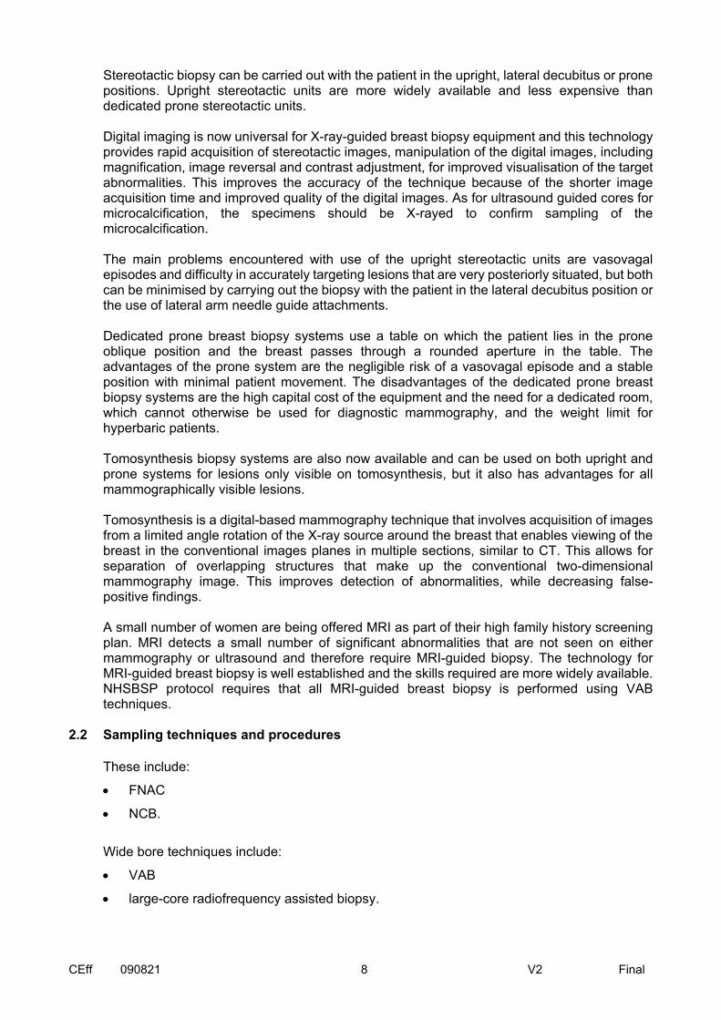
Stereotactic biopsy can be carried out with the patient in the upright, lateral decubitus or prone positions. Upright stereotactic units are more widely available and less expensive than dedicated prone stereotactic units. Digital imaging is now universal for X-ray-guided breast biopsy equipment and this technology provides rapid acquisition of stereotactic images, manipulation of the digital images, including magnification, image reversal and contrast adjustment, for improved visualisation of the target abnormalities. This improves the accuracy of the technique because of the shorter image acquisition time and improved quality of the digital images. As for ultrasound guided cores for microcalcification, the specimens should be X-rayed to confirm sampling of the microcalcification. The main problems encountered with use of the upright stereotactic units are vasovagal episodes and difficulty in accurately targeting lesions that are very posteriorly situated, but both can be minimised by carrying out the biopsy with the patient in the lateral decubitus position or the use of lateral arm needle guide attachments. Dedicated prone breast biopsy systems use a table on which the patient lies in the prone oblique position and the breast passes through a rounded aperture in the table. The advantages of the prone system are the negligible risk of a vasovagal episode and a stable position with minimal patient movement. The disadvantages of the dedicated prone breast biopsy systems are the high capital cost of the equipment and the need for a dedicated room, which cannot otherwise be used for diagnostic mammography, and the weight limit for hyperbaric patients. Tomosynthesis biopsy systems are also now available and can be used on both upright and prone systems for lesions only visible on tomosynthesis, but it also has advantages for all mammographically visible lesions. Tomosynthesis is a digital-based mammography technique that involves acquisition of images from a limited angle rotation of the X-ray source around the breast that enables viewing of the breast in the conventional images planes in multiple sections, similar to CT. This allows for separation of overlapping structures that make up the conventional two-dimensional mammography image. This improves detection of abnormalities, while decreasing false-positive findings. A small number of women are being offered MRI as part of their high family history screening plan. MRI detects a small number of significant abnormalities that are not seen on either mammography or ultrasound and therefore require MRI-guided biopsy. The technology for MRI-guided breast biopsy is well established and the skills required are more widely available. NHSBSP protocol requires that all MRI-guided breast biopsy is performed using VAB techniques.
2.2 Sampling techniques and procedures
These include: • FNAC
• NCB.
Wide bore techniques include:
• VAB
• large-core radiofrequency assisted biopsy.

CEff 090821 9 V2 Final
All of these procedures can be carried out by members of the breast team who have had specialist training in image-guided breast biopsy: a radiologist, a radiographic practitioner or breast clinician. For simplicity radiologist or assessing clinician is used below. Ultrasound-guided NCB is the technique of first choice for sampling impalpable breast lesions, as it is easier to perform, more comfortable for the patient and less time-consuming than the X-ray-guided techniques (see section 2.1 above). For impalpable lesions detected by mammography, the assessing clinician must be certain that the abnormality seen on ultrasound is the same as the abnormality seen on mammography. Ultrasound can only be used when the assessing clinician is convinced that the abnormality is clearly visible using this technique. X-ray-guided NCB or VAB should be used where there is any doubt about whether the ultrasound appearances correspond to the mammographic abnormality.
2.3 Core biopsy: general principles
Core biopsy of the breast is a safe and effective method for obtaining a non-operative diagnosis of breast lesions. Core biopsy should be performed with caution in patients who are anticoagulated, or on aspirin or clopidogrel. The use of these medications is not absolutely contraindicated and local policies should be available.11 The consent process should follow local rules and the procedure and common complications be explained to the patient. Formal written consent is not normally required. An assistant is required to compress the breast between needle passes. Breast core biopsy should be performed with a spring-loaded device, usually 14G diameter. Local anaesthetic should be used to the skin and down to the lesion. A small (2 mm) skin nick that traverses the superficial fascia should be made with a scalpel blade (a No. 11 blade is ideal) to facilitate the passage of the needle into the breast. The skin entry site should be optimised for both cosmesis and accurate targeting. The skin nick can be visible for some months after the biopsy, so approaches through the cleavage line and upper inner quadrant should be avoided; lateral, inferior and periareoalar approaches are preferable. If the breast tissue is very fibrous, insertion of the needle in a radial direction makes manipulation easier. The only major complication of breast needle biopsy is pneumothorax. To avoid this, the needle should be kept as near as possible parallel to the chest wall when fired. This means the skin entry site for deep lesions needs to be further away from the lesion than for superficial lesions. For lesions 10 mm or larger, the tip of the needle should abut the lesion before firing. When biopsying lesions less than 10 mm in diameter, the needle tip should be sufficiently short of the lesion before firing to ensure that the lesion is included in the sampling trough. It is the operator’s responsibility to confirm the patient’s identification and label the specimen pot before leaving the room. After the procedure, the biopsy site should be compressed for a minimum of five minutes. The patient should be given written information about where and when they will receive the result and potential complications of the procedure. The patient should be advised that mild post-procedure pain, lumpiness and bruising is common and not to exercise their upper limbs for the rest of the day. Wound infection is a rare complication, but the patient should be advised to seek medical advice if the site of the biopsy becomes increasingly red or the puncture site oozes purulent material. The patient information sheet should advise the patient to telephone the hospital (the number should be on the patient information sheet) if the breast swells appreciably or if they become short of breath.

CEff 090821 10 V2 Final
2.4 Ultrasound-guided core biopsy
The patient is positioned to provide optimal access to the area to be biopsied. This may involve, for example, raising and supporting the left side for biopsy of lesions situated in the lateral aspect of the left breast. For lesions that are situated in the lateral aspect of the right breast, it may be necessary to turn the patient on the couch so that a right-handed operator can easily access such lesions using a lateral approach. An assistant should work from the opposite side of the couch. The lesion is demonstrated and surrounding breast tissue immobilised. Local anaesthetic is infiltrated both superficially and deeply down to and around the lesion. For posteriorly placed lesions, local anaesthetic can be infiltrated posteriorly to displace the lesion anteriorly. A 2–3 mm skin incision, parallel to Langer’s lines, is made to allow insertion of the core biopsy needle along the direction of the long axis of the ultrasound probe. The core biopsy needle is advanced until the tip is a few millimetres proximal to the edge of the lesion. The core biopsy gun is then fired and the needle is visualised passing through the lesion. The magnification of the field of view of the ultrasound image should be set so that the tip of the needle will still be visible after taking the sample. An image showing the needle passing through the lesion is usually recorded. The needle is withdrawn and the specimen is delivered into fixative. Two or three passes are usually sufficient in most cases to obtain diagnostic material from soft-tissue mass lesions. At the end of the procedure, firm pressure is applied by the assistant over the site of the biopsy to reduce bruising.
2.5 Stereotactic-guided core biopsy
For needle biopsy using a stereotactic device with a conventional upright mammography machine, the patient is seated. Increasingly, VAB is preferred for stereotactic breast biopsy. A superior or lateral approach, with the breast in the cranio-caudal position, is suitable for most lesions, but latero-medial, medio-lateral or oblique approaches may be needed for lesions that are inferiorly positioned or are situated laterally in the axillary tail region. After demonstrating the lesion on a straight scout film, paired stereotactic views are obtained with the X-ray tube angled 15 degrees either side of the central straight tube position. The position of the lesion on the stereotactic views is used to determine the position of the needle guide on the X and Y axes so that, when a needle of known length is introduced through the guide into the breast, the centre of the needle sampling trough will correspond to the chosen target. Digital equipment will not allow firing of the device if the lesion is too close to the detector and may damage its surface. Apparatus that facilitate a lateral approach to lesions are preferred when the breast compression thickness is small. The lateral arm approach also avoids the needle being held directly in the eyeline of the patient, allows improved access to lesions in the inferior part of the breast, and eliminates the risk of the needle tip hitting the surface of the X-ray cassette holder. An alternative approach in small breasts is to use a radiolucent spacer below the breast, to increase the distance between the lesion and the surface of the detector. X-ray-guided biopsy using tomosynthesis is also now available. The technique is largely similar to stereotactic biopsy except that the image to select the target is acquired by continuous X-ray source arc rotation rather than from two images taken at 15 degree angles. When available, it is a quicker localisation technique and also facilitates biopsy of lesions only seen on tomosynthesis images.
CEff 090821 11 V2 Final
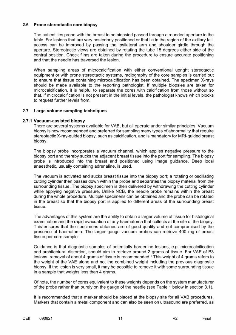
2.6 Prone stereotactic core biopsy
The patient lies prone with the breast to be biopsied passed through a rounded aperture in the table. For lesions that are very posteriorly positioned or that lie in the region of the axillary tail, access can be improved by passing the ipsilateral arm and shoulder girdle through the aperture. Stereotactic views are obtained by rotating the tube 15 degrees either side of the central position. Check films are taken during the procedure to ensure accurate positioning and that the needle has traversed the lesion. When sampling areas of microcalcification with either conventional upright stereotactic equipment or with prone stereotactic systems, radiography of the core samples is carried out to ensure that tissue containing microcalcification has been obtained. The specimen X-rays should be made available to the reporting pathologist. If multiple biopsies are taken for microcalcification, it is helpful to separate the cores with calcification from those without so that, if microcalcification is not present in the initial levels, the pathologist knows which blocks to request further levels from.
2.7 Large volume sampling techniques 2.7.1 Vacuum-assisted biopsy
There are several systems available for VAB, but all operate under similar principles. Vacuum biopsy is now recommended and preferred for sampling many types of abnormality that require stereotactic X-ray-guided biopsy, such as calcification, and is mandatory for MRI-guided breast biopsy. The biopsy probe incorporates a vacuum channel, which applies negative pressure to the biopsy port and thereby sucks the adjacent breast tissue into the port for sampling. The biopsy probe is introduced into the breast and positioned using image guidance. Deep local anaesthetic, usually containing adrenaline, is used. The vacuum is activated and sucks breast tissue into the biopsy port; a rotating or oscillating cutting cylinder then passes down within the probe and separates the biopsy material from the surrounding tissue. The biopsy specimen is then delivered by withdrawing the cutting cylinder while applying negative pressure. Unlike NCB, the needle probe remains within the breast during the whole procedure. Multiple specimens can be obtained and the probe can be rotated in the breast so that the biopsy port is applied to different areas of the surrounding breast tissue. The advantages of this system are the ability to obtain a larger volume of tissue for histological examination and the rapid evacuation of any haematoma that collects at the site of the biopsy. This ensures that the specimens obtained are of good quality and not compromised by the presence of haematoma. The larger gauge vacuum probes can retrieve 400 mg of breast tissue per core sample. Guidance is that diagnostic samples of potentially borderline lesions, e.g. microcalcification and architectural distortion, should aim to retrieve around 2 grams of tissue. For VAE of B3 lesions, removal of about 4 grams of tissue is recommended.8 This weight of 4 grams refers to the weight of the VAE alone and not the combined weight including the previous diagnostic biopsy. If the lesion is very small, it may be possible to remove it with some surrounding tissue in a sample that weighs less than 4 grams. Of note, the number of cores equivalent to these weights depends on the system manufacturer of the probe rather than purely on the gauge of the needle (see Table 1 below in section 3.1).
It is recommended that a marker should be placed at the biopsy site for all VAB procedures. Markers that contain a metal component and can also be seen on ultrasound are preferred, as

CEff 090821 12 V2 Final
these facilitate easier subsequent localisation for surgery, if needed; if not, provide future reference as to where prior biopsy has been performed. A marker is mandatory if there is any risk that the whole of the target lesion might be removed by the needle biopsy. The pathology department should ideally be made aware of any subsequent biopsy that may contain a marker, as this may influence how the tissue is prepared for sectioning.
2.8 Complications of needle core biopsy
Although NCB and FNAC are remarkably complication free, some uncommon problems should be considered.
2.8.1 Pain
Pain is common with fine needle aspiration, but is transitory and not usually severe. Aspiration from painful areas of benign breast change is sometimes associated with some pain when the needle comes into contact with the painful area. Carcinomas, particularly those with abundant fibroelastotic stroma, are often also painful, which can be a guide to the aspirator that the needle has hit the lesion. If pain is anticipated for fine needle aspiration (FNA), local anaesthesia to the skin and close to the sample site is recommended. FNA is now largely confined to sampling axillary lymph nodes and many prefer to routinely use local anaesthesia for this procedure. Local anaesthesia should be used for all core biopsies in the breast and axilla, with sufficient anaesthetic delivered superficially at the skin entry site and around the biopsy target.
2.8.2 Haematoma Where possible, all imaging investigations should be complete before sampling is performed, as haematoma formation, if it occurs, can cause confusion on subsequent imaging. The risk of significant haematoma after FNA and NCB are about the same at 1%, rising to approximately 4% for vacuum biopsy procedures. Haematoma is minimised by appropriate manual pressure applied over the biopsy site for five to ten minutes. For VAB, especially when used for complete lesion excision, a compression dressing applied for four to six hours should be considered. Patients should be advised not to take vigorous exercise following a breast biopsy to minimise the risk of delayed haematoma.
2.8.3 Pneumothorax
This is a rare complication occurring in less than 1:10,000 breast biopsies and occurring mainly in women with small breasts, after biopsy of medial and posterior lesions, or when sampling axillary nodes. It occurs most commonly after freehand non-image-guided breast biopsy and is a very rare problem with image-guided biopsy. Large pneumothoraces should be obvious but the problem may go undetected if the pneumothorax is small. If there is any clinical concern that a pneumothorax may have occurred, the patient should be sent for a chest X-ray before being allowed home.
2.8.4 Fainting
Fainting is of special significance during upright stereotactic procedures where the patient has to be released from the machine and laid flat. The procedure usually has to be abandoned. For women with a history of syncope, the use of sublingual lorazepam has been shown to minimise the risk.
2.8.5 Removal of lesion by core biopsy
Small lesions including foci of microcalcification may, particularly if extensively sampled, be removed by core biopsy. This risk increases when greater numbers of core samples are taken

CEff 090821 13 V2 Final
or with VAB. It is recommended that markers are inserted at the site of biopsy at the time of the biopsy to ensure that the site can be identified subsequently. On occasions, however, a small, sole invasive focus (with or without surrounding DCIS) may be removed in the needle biopsy samples with no further invasion in the subsequent excision specimen. In such circumstances, the core biopsy sample should be used to provide information on histological grade and tumour type.
2.8.6 Seeding of tumour
Seeding of malignant cells has become increasingly recognised as a result of the increased use of core biopsy. Rarely, this may cause histopathological diagnostic difficulties in the subsequent excision. Groups of cells may be seeded, particularly from papillary lesions (benign or malignant). Small clusters of cells are seen, sometimes showing degenerate features, usually with associated fibrosis and inflammation consistent with the site of previous biopsy. The track is often linear. Seeding is rarely recognised more than a few millimetres from the source of the cells and the correct identification is usually straightforward. The clinical significance of this phenomenon is not yet clear.
3 Core biopsy reporting guidelines
This section of this document is designed to assist in classification of NCB and VAB samples. The diagnostic terminology and entities referred to are described in more detail in the Pathology reporting of breast disease in surgical excision specimens.12
3.1 Core biopsy specimen information and handling
Proper interpretation of core biopsies requires details of the patient’s history and clinical and radiological findings, and it is essential that this information is provided on the request form. The completed request form should include clinical details, specifying the radiographic changes and the site of biopsies. It is not sufficient to complete the request form with R or U codes (see page 6 for details of this system). Reliable pathological interpretation requires that radiological details such as mass lesion, deformity, calcification, etc. are recorded, as well as the assessing clinician’s impression, such as R3, R4 or R5. It is essential that the patient and clinical details including the location of the biopsy on the specimen pot and request card are correct and match. Particular care is needed if multiple biopsies are taken from a patient. It is also essential that the details are checked in the pathology laboratory before handling the specimen and when reporting. A radiograph should be taken of all biopsies performed from microcalcifications to determine the presence of calcium. Whenever possible, a radiological comment regarding the presence of representative microcalcification of the mammographic lesion in the sample should be provided to the pathologist along with the specimen X-ray. The pathologist must be able to view the core biopsy X-rays on a monitor of suitable quality.13 However, it is the responsibility of the radiologist and the multidisciplinary meeting to decide whether the calcification in the mammogram correlates with the calcification seen histologically.
Examination of further levels should be performed if calcification in a pattern consistent with that seen on the specimen X-ray is not apparent on histological examination of initial levels. Optimal fixation is paramount. Biopsies should be placed immediately in a formalin fixative solution and sent promptly to the laboratory. Optimal fixation is essential, particularly for ER and HER2 analysis, for which a minimum of six hours and a maximum of 72 hours are recommended.12 This has implications for scheduling of laboratory work. Specimens may be fixed rapidly with the aid of microwave techniques, but such techniques must be validated, including assessment of immunohistochemistry.

CEff 090821 14 V2 Final
There are different approaches to the macroscopic description of core biopsies and VAB. Some laboratories record the number of cores and the length of each. This has the advantage that the number of cores taken in the clinic can be confirmed and also that the number and length of cores can be checked in the histological slides. Some radiology departments weigh their VABs to ensure an adequate amount of tissue has been obtained. The weight can be estimated from the number of cores and the type of device (see Table 1 below). An alternative approach to the handling of cores is to put the cores into containers in the clinic so that, in the laboratory, the cores can be placed directly into the cassette without further handling. This reduces the risk of loss of tissue, but macroscopic description is not provided.
Table 1: Mean weight of cores from turkey breast phantom and number of cores equivalent to 4 g of tissue.
Core and manufacturer Weight of one core (g) Number of cores equating to approximately 4 g
11-G Original Mammotome 0.084 48 10-G Vacora 0.142 28 10-G EnCore Enspire 0.221 18 9-G ATEC Sapphire needle 0.121 33 8-G Original Mammotome 0.192 21 8-G Mammotome Revolve 0.334 12 7-G EnCore Enspire 0.363 11
Taken with permission from reference 9.
After processing, it is essential to ensure that the biopsy is properly embedded and that the block is adequately cut into when the sections are taken. Haematoxylin and eosin stained sections from one level are usually sufficient for core biopsies from mass lesions, but core biopsies taken for the investigation of microcalcification should have a minimum of three levels examined. In problematic cases, further levels and immunohistochemical studies may be helpful. Information from all core biopsies of screening-detected lesions should be entered on to National Breast Screening System (NBSS), either directly by the pathologist or using the form in Appendix E. [Level of evidence – GPP.]
3.2 Recording of data on the National Breast Screening System
NBSS provides an interface for recording of pathology data related to breast screening patients. As the vast majority of pathologists prefer to provide the data as a component of their histopathology report, rather than enter the data directly onto the system, the information must be provided in a clear and interpretable manner for easy extraction. In particular, for patients with complex or multiple abnormalities, steps should be taken to ensure that data is recorded accurately for the correct lesions. The data required for NBSS are listed in appendices E and F. It is hoped that future versions of NBSS will include methods to extract relevant data directly into NBSS from pathology data systems.
3.2.1 Lesion identification
This should be done by the radiologist at the time of assessment. For convenience, with patients who have more than one abnormality, the most suspicious or main lesion should be

CEff 090821 15 V2 Final
recorded as lesion 1 and other lesions recorded separately. Where lymph node assessment and needle biopsy is carried out, this should be recorded. Pathologists should clearly record information for each lesion that has been sampled so that transcription of the data is straightforward.
3.2.2 Cytology form
The method of localisation should be indicated. Options available are palpation, stereotactic, prone stereo, X-ray, ultrasound or MRI. There is an option of ‘not stated’ but it should not be necessary to use this. The specimen type should also be recorded. It is important to select ‘Node aspirate’ if the sample is from an axillary node. The cytology classification (C1–C5 or LC1–LC5) must be included in the cytopathology report.
3.2.3 Core needle biopsy form
The core needle biopsy form has scope for recording of more data in relation to the lesion, and pathologists are encouraged to record as much data as possible. This will help with future analysis and audit. The first part of the form includes details of method of localisation:
• intention with regard to diagnostic
• therapeutic with regard to vacuum-assisted specimens (i.e. VAB or VAE)
• whether the sample was from a node
• whether the presence or absence of calcification on specimen X-ray is completed at the time of sampling.
If the pathologist has access to NBSS, it is possible to check this information for correlation. The information for the section headed ‘Pathology result’ should be provided by the pathologist, including specimen number, name of reporting pathologist and the B category. In the rare instance when it is not possible to distinguish between invasive and in situ disease, it should be recorded as ‘Not assessable’ rather than ‘Not stated’.
These above fields are mandatory for all biopsies. The remaining fields are optional but should be completed if at all possible. There are options for recording more information with regard to benign and malignant lesions, as well as grading and hormone receptor and HER2 status, if performed on the core biopsy. If the intention of a VAB is diagnostic rather than excision, the NBSS VAB form should be completed with a B category. If a B3 lesion such as a radial scar or papilloma has been excised or more thoroughly sampled as a VAE, then the diagnosis should be entered on the new NBSS VAE form. The use of the terms VAB and VAE relies on the intention of the clinicain when performing the procedure and should be provided by them on the request form. If the intention is diagnostic, typically fewer cores are sampled and this should be given a B code. A VAE is performed when the intention is to exclude associated or adjacent malignancy and generally more extensive sampling takes place. Some VAEs will not excise the whole lesion, but it should be noted that the same is true for some surgical diagnostic excisions. A proforma can be used for reporting VAEs (appendix G). For reporting purposes, it is treated like a surgical biopsy and no B category is needed.14 As in a surgical biopsy classical or pleomorphic LCIS in a VAE is recorded as malignant, despite the differences in management of these variants.14 However, on NBSS, an ‘E’ code will be entered by the screening office with two main options: E2 for benign pathology and E5 for malignant pathology. This is similar to the H2 for benign pathology and H5 for malignant pathology coding on surgical specimens.
CEff 090821 16 V2 Final
Pathologists should record clearly on the report whether the diagnosis is benign or malignant, but do not need to provide an ‘E’ code on the pathology report. There are also NBSS codes for use by the screening office if the specimen cannot be reported (E0) or where the specimen is reported as containing normal breast tissue only (E1). Regardless of how data are entered on to NBSS, pathologists should be involved in quality assurance of the information entered on to the system on a regular basis.
3.3 Using the core biopsy reporting form
The core biopsy reporting forms used may be the separate reporting form or the form generated specifically by the NBSS, which comes with the patient details already filled in by the computer. These both request essentially the same information. The forms are typically submitted along with the sample and a separate local specimen request form, and may be used directly or, more usually, the information is provided as part of the overall histology report. In the latter case, it is helpful to maintain the same terminology and order of data items for the screening unit staff, to simplify transposition of the information to NBSS. How the national screening system treats this information has been included as Appendix C. Information on the nature of the mammographic abnormality and clinical characteristics should be provided by the breast screening assessing clinician requesting the pathology examination.
3.4 RCPath dataset items
The RCPath dataset proformas include a subset of data items included in the NHSBSP form. RCPath dataset items should be collected in all cases of invasive cancer or carcinoma or in situ. For cases being collected through the breast screening programme, it is acceptable to complete the breast screening form, but for cases outside the screening programme, the RCPath dataset should be followed if the NHSBSP form is not being used for all cases.
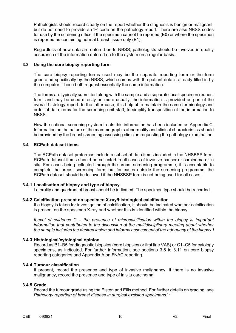
3.4.1 Localisation of biopsy and type of biopsy Laterality and quadrant of breast should be indicated. The specimen type should be recorded.
3.4.2 Calcification present on specimen X-ray/histological calcification If a biopsy is taken for investigation of calcification, it should be indicated whether calcification is present on the specimen X-ray and whether this is identified within the biopsy. [Level of evidence C – the presence of microcalcification within the biopsy is important information that contributes to the discussion at the multidisciplinary meeting about whether the sample includes the desired lesion and informs assessment of the adequacy of the biopsy.]
3.4.3 Histological/cytological opinion Record as B1–B5 for diagnostic biopsies (core biopsies or first line VAB) or C1–C5 for cytology specimens, as indicated. For further information, see sections 3.5 to 3.11 on core biopsy reporting categories and Appendix A on FNAC reporting.
3.4.4 Tumour classification If present, record the presence and type of invasive malignancy. If there is no invasive malignancy, record the presence and type of in situ carcinoma.
3.4.5 Grade
Record the tumour grade using the Elston and Ellis method. For further details on grading, see Pathology reporting of breast disease in surgical excision specimens.12

CEff 090821 17 V2 Final
[Level of evidence B – invasive tumour grade is a recognised important prognostic factor that is used in treatment planning; accurate assessment is expected.]
3.4.6 ER status/progesterone receptor status/HER2 status ER status predicts response to endocrine therapies. The data for progesterone receptor and response to hormone therapy is less clear but is recommended by NICE and is a requirement for some clinical trial recruitment. Overexpression of HER2 protein in breast cancer is predictive of response to HER2-targeted treatment. NICE recommends testing the status of all three markers on the initial histological specimen and that this should be done at the time of initial histopathological diagnosis.15 [Level of evidence A – ER status predicts response to endocrine therapies. Overexpression of HER2 predicts response to HER2-targeted treatments.]
3.4.7 Recording clinical basic information Centre/location Give the name of the assessment centre, clinic, department, etc., and where the specimen was obtained. Side Indicate right or left. For specimens with biopsies from multiple sites, use a separate form for each site. Localisation technique
Choose one of the following terms:
• palpation
• ultrasound guided
• stereotactic
• MRI.
Number of cores If known, indicate the number of core biopsy samples taken. Presence of calcification on specimen X-ray If the biopsy is performed for investigation of radiological calcification, indicate whether there is calcification visible on the specimen radiograph, if known. State if the radiograph has not been seen.
Histological calcification Indicate whether calcification has been identified in the sample and, if present, whether it is associated with benign or malignant disease, or both. Pathologist The name of the pathologist giving the opinion, who must be registered at the screening office. Date Enter the date of issuing the report. Case for review This is a field to indicate that a specimen has been sent for a further opinion or that the case is a particularly interesting example.

CEff 090821 18 V2 Final
Recording the opinion See the section on reporting categories, below. Comment field This free-text field is included for extra information to be recorded.
3.5 Core biopsy reporting categories
Five reporting categories are used for diagnostic biopsies. They should not be used for excision specimens, including those by vacuum-assisted techniques. It is important to remember that histological examination of core biopsy samples is performed to fulfil the assessment process role by giving a pathology category classification (B1–B5), and is not designed to give a definitive diagnosis, although this is possible in the majority of cases. Thus, while most core biopsy samples can be readily categorised as normal, benign or malignant, it must be recognised that a small proportion (less than 10% in most units) of samples cannot. In recognition of this, the following reporting guidelines have been devised and should be used for all screen-detected lesions: microcalcification, architectural deformities, mass lesions etc. It is recommended that this approach also be adopted for symptomatic practice. It is important to remember that although there are five reporting categories similar to those used in FNAC, these are not equivalent. These categories should take account purely of the histological nature of the specimen and not the clinical or imaging characteristics. A lesion, for example, should not be classified as benign (B2) simply because radiological–pathological correlation appears appropriate, if only normal histology is seen. Similarly, it is not feasible for pathology interpretation to judge independently whether a sample is adequate and from the mammographic lesion. This judgement requires multidisciplinary discussion. For these reasons, there is no inadequate biopsy category for core biopsy specimens. For VAE of a lesion that has already been diagnosed on a previous biopsy, a B category is not appropriate. VAE is regarded as equivalent to a surgical diagnostic biopsy. The B category of a diagnostic biopsy should not be changed because a later VAE or surgical excision gives a different diagnosis, unless there was an error in the interpretation of the diagnostic biopsy and any revised report should make clear that there has been a change. Sometimes pressure is put on pathologists to issue provisional reports to be ready for multidisciplinary meetings or help meet clinical targets. This is discouraged and not considered good practice. Issuing verbal reports before the report is authorised also confers risk to patient management and is also discouraged.16 Sometimes it is necessary to send slides and/or blocks to another centre for example for a second opinion, for assessment of markers such as HER2 or if patient care is transferred. Such transfers should be undertaken promptly using a trackable transport system. Mechanisms must be in place for rapid communication of any results or diagnostic disagreement.17
3.6 B1 normal tissue
This indicates a core of normal tissue, whether or not breast glandular structures are present. This category is, therefore, equally appropriate for a core including normal breast ducts and lobules or mature adipose tissue or stroma only. A B1 report should include a description of

CEff 090821 19 V2 Final
the components present and comment should be made regarding the presence of breast epithelial structures. Normal histology may indicate that the lesion has not been sampled. This is, however, not necessarily so. In the case of certain benign lesions, such as hamartomas and lipomas, apparently normal histological features would be expected on core biopsy. Minor architectural distortions seen mammographically may also result in minimal changes such as a slight increase in stromal fibrous tissue on biopsy. A minor degree of fibrocystic change is usually best categorised as B1. In these circumstances, it is the remit of the multidisciplinary meeting to determine if the lesion of interest has been sampled, if the core biopsy can be considered representative, and if a B1 result can explain the clinical and radiological findings. Lactational change should be categorised as B1. Cores with B1 diagnoses may contain microcalcification of sufficient size to be radiologically visible, e.g. within involutional lobules or in the stroma. It is important in these cases that discussion between pathology and radiology colleagues is undertaken to confirm whether the microcalcification in the histological specimen is representative of that seen on the mammogram. Foci of calcification within involuted lobules are common and frequently too small to be visible mammographically. Therefore, a report that merely records the presence of this calcification, without additional comment on its nature, size and site, may be misleading and lead to false reassurance. It is evident that mammograms do not demonstrate microcalcification, either singly or in clusters less than 100 µm in diameter.18 The resolution of digital mammography is lower than film/screen mammography, but calcifications of similar size are more visible and easier to detect on digital mammography. The pathologist should not categorise a biopsy as B1 because the biopsy may not reflect the clinical or radiological abnormality.19 The pathologist should describe the histological features and base the B category on these features. Nevertheless, the pathologist may make a comment in the report that the biopsy may not be representative of the lesion. It is the role of the multidisciplinary meeting to judge whether the core biopsy is adequate. Exceptionally, some specimens may be classified as uninterpretable because of, for example, excessive crush artefacts or because the sample consists of blood clot only. Such samples should also be classified as B1.
3.7 B2 benign lesion
A core is classified as B2 benign when it contains a benign abnormality. This category is appropriate for a range of benign lesions, including fibroadenomas, fibrocystic change, sclerosing adenosis and duct ectasia, and extends to include other non-parenchymal lesions, such as abscesses and fat necrosis. In some cases, it may be difficult to determine whether a specific lesion is present, for example, if minor fibrocystic changes are seen. The multidisciplinary approach is once again vital in these cases to determine whether the histopathological features are in keeping with the radiological and clinical findings. It may be appropriate and prudent to classify the lesion as B1 rather than B2, if only very minor changes are present. Sometimes skin lesions will be sampled. If a definite benign diagnosis is possible, then B2 categorisation is appropriate. Sometimes a definite diagnosis is difficult; e.g. some adnexal tumours may be difficult to categorise on core biopsy, in which case B3 may be more appropriate.

CEff 090821 20 V2 Final
3.8 B3 lesion of uncertain malignant potential
This category mainly consists of lesions that provide benign histology on core biopsy, but either are known to show heterogeneity or to have an increased risk of associated malignancy (albeit lower than for B4). The level of risk is very different for the different entities. The management of B3 lesions is discussed in detail in a separate document.9 More comments about management are included in section 3.8.9. It is essential that a search is made for epithelial atypia and that such atypia is reported, even if there is another reason for a B3 categorisation, as the risk of malignancy associated with atypical intraductal epithelial proliferation (AIDEP) is relatively high. For all B3 diagnoses, a comment should be made about whether epithelial atypia is present.
3.8.1 Atypical intraductal epithelial proliferation
There is a range of intraductal epithelial atypia, short of that required for a definite diagnosis of DCIS, that is best classified as B3 or B4. Different patterns of atypia may be seen: resembling atypical ductal hyperplasia (ADH), flat epithelial atypia, apocrine atypia and atypia that does not conform to one of these patterns. A common pattern resembles what would be called ADH on a surgical specimen: a monotonous proliferation of evenly spaced cells with small regular nuclei that raises the possibility of low-grade DCIS, but has insufficiently developed features or insufficient extent for this diagnosis.12 These range in severity, from those that are insufficient for a definite diagnosis of DCIS but highly suspicious, to those that only show a minor degree of atypia, normally architectural, which requires further assessment and judgement of appropriate categorisation as B3 or B4; most are best classified as B3. The definition of ADH is derived from surgical resection specimens and relies on a combination of architectural, cytological and size extent criteria. For this reason, accurate diagnosis of ADH is not possible on core biopsy, and the term AIDEP should be used. It has, however, been shown that core biopsy samples that include atypical intraductal epithelial proliferative foci, of insufficient extent for classification as DCIS, may form, on subsequent surgical resection, part of an established in situ neoplastic lesion, with or without associated invasion. This view is based on studies that describe the subsequent surgical diagnoses in cases described as ADH in non-operative core biopsy. Studies have shown that subsequent excision biopsy contains malignancy (either in situ or invasive) in 30–40% of these patients.20 This is not surprising as ADH is defined as an intraductal epithelial proliferation showing the features of low-grade DCIS, but in fewer than two duct spaces or of less than 2 mm in diameter. The limited tissue sampling that can be undertaken by core biopsy guns (often by stereotactic methods for foci of microcalcification) may thus provide insufficient material for definitive diagnosis of low-grade DCIS if only a few ducts spaces are obtained. In these cases, a diagnosis of AIDEP should be made, along with a classification of B3 of uncertain malignant potential or, less commonly, B4 suspicious of malignancy, depending on the severity and extent of the lesion. Immunohistochemistry for basal cytokeratins, such as CK14, CK 5/6 and ER can play a useful role in assessing epithelial proliferations. The epithelial cells in DCIS and ADH are typically completely negative for basal cytokeratins and uniformly positive for ER, whereas usual type epithelial hyperplasia shows patchy expression. The surrounding myoepithelial cells are usually positive for basal cytokeratins. However, there are pitfalls. Occasionally, DCIS is positive for basal cytokeratins, but this is usually high grade. Columnar cell change and apocrine change are both negative for basal cytokeratins, so

CEff 090821 21 V2 Final
assessment of atypia in these lesions must rely on morphology. For a more detailed discussion, see the Pathology reporting of breast disease in surgical excision specimens.12
3.8.2 Flat epithelial atypia
Columnar cell lesions are discussed in greater detail in the Pathology reporting of breast disease in surgical excision specimens.12 Most columnar cell change, with or without hyperplasia, shows no atypia and is best categorised as B2 (or sometimes as B1 if it is very focal). Flat epithelial atypia is categorised as B3 on core biopsy. If there is a more complex architecture (usually cribriform or micropapillary), the considerations in the above section on atypical intraductal proliferations apply. Flat epithelial proliferations with high-grade nuclei should be categorised as B4 if the changes are limited, and as high-grade DCIS (B5a) only if the features are sufficient for an unequivocal malignant diagnosis.
3.8.3 Lobular neoplasia
A pathologist may consider a small to medium cell regular dyscohesive epithelial proliferation within lobules to represent a classical lobular neoplasia: either atypical lobular hyperplasia (ALH) or lobular carcinoma in situ (LCIS). It should be classified as B3. The distinction between ALH and LCIS cannot always be reliably made on core biopsy, so the overarching term ‘lobular neoplasia’ is preferable. If preferred, subcategorisation into ‘at least ALH’ and ‘LCIS’ can be made, but there appears to be no significant difference in upgrade rate (i.e. ‘risk of adjacent DCIS’ or ‘invasive carcinoma’), so there is no clinical benefit to this distinction in core biopsy. Classical lobular neoplasia does not have the same management implications as a diagnosis of DCIS or invasive malignancy and does not per se require therapeutic excision. Lobular neoplasia is, however, often a coincidental finding in a core biopsy from a screen-detected lesion, and multidisciplinary discussion is essential as the abnormality identified radiologically may not be represented. These cases must be managed cautiously.9 Pleomorphic LCIS is best classified as B5a (see below in 3.8.4). Occasionally, lobular neoplasia shows marked distension of the acini, often with necrosis, but without marked nuclear pleomorphism.21 There are only limited data on the behaviour of this variant, which is now called florid LCIS, but in view of the overlap of features with the DCIS, it is best classified as B4. There are limited data on the optimum management of both pleomorphic and florid LCIS, but the WHO Classification of Tumours recommends excision for both when diagnosed on core biopsy. E-cadherin immunohistochemistry can be useful to help distinguish lobular neoplasia and DCIS in difficult cases. DCIS typically shows complete membrane expression, whereas lobular neoplasia usually shows reduced or absent E-cadherin membrane expression. Basal cytokeratins are typically absent in lobular neoplasia, as described above in DCIS. On occasions, it may be difficult to classify an epithelial proliferation as either lobular neoplasia or low-grade DCIS, and, in these circumstances, a B4 classification may be appropriate.
3.8.4 Phyllodes tumour The presence of a cellular stroma within a fibroepithelial lesion on core biopsy should prompt a search for other features that may aid in separating phyllodes tumour from a fibroadenoma.22 The following favour phyllodes tumour:
• stromal overgrowth: x10 field of stroma with no glandular elements

CEff 090821 22 V2 Final
• fragmentation: defined as a stromal fragment with epithelium at one or both ends
• mitoses: one or two per 10 high-power fields favour phyllodes tumour, but can be seen as fibroadenomas; and three or more per 10 high-power fields more strongly favour phyllodes tumour.
Marked atypia of stromal cells is usually only seen with other features suggestive of phyllodes tumour. If there are multiple features, a definite diagnosis of phyllodes tumour may be possible. If the features are of a benign phyllodes tumour, B3 classification is appropriate. Often the differential diagnosis lies between a cellular fibroadenoma and a benign phyllodes tumour, but definite categorisation is not possible. Such ‘cellular fibroepithelial lesions’ should also be designated B3 and the report should state that ‘phyllodes tumour cannot be excluded’. It is important to remember that phyllodes tumours are much less common than fibroadenomas (about 50 times) and minor changes should not be over-interpreted, as this will lead to excision of large numbers of fibroadenomas. Markedly atypical changes may merit designation as B4 and occasionally as B5. An important pitfall is that some phyllodes tumours contain areas resembling typical fibroadenoma. Clinical factors, particularly tumour size and an increase in size, should be considered in multidisciplinary discussion.
3.8.5 Papillary lesions Papillary lesions may show significant intralesional heterogeneity and the limited sampling achieved with core biopsy may miss areas of in situ carcinoma. The majority of these lesions should, therefore, be designated B3. On rare occasions, when a very small lesion is seen within the diameter of the core, a benign B2 classification may be considered. Conversely, when a sample of a papillary lesion in a core biopsy shows atypia, e.g. strongly suspicious of papillary carcinoma in situ, a B4 designation may occasionally be more appropriate. It is important that even focal epithelial atypia is sought, as the chance of malignancy in the subsequent excision specimen is much higher than in lesions without atypia (30–40% versus 5–10%).20 Immunohistochemistry for myoepithelial markers can be helpful. Benign papillomas contain a myoepithelial layer, both at the edge and within the lesion between the epithelium and the fibrovascular core. However, in papillary carcinoma in situ, myoepithelial cells are usually absent within the lesion. Myoepithelial cells may be seen surrounding papillary DCIS, but are usually absent at the periphery of encysted or encapsulated papillary carcinoma. Benign papillomas with involvement by DCIS typically show retention of a myoepithelial layer – such lesions are usually best designated as B4 unless the atypical component is very extensive. Basal cytokeratins and ER are useful for distinguishing usual type epithelial hyperplasia and DCIS, as discussed above. Nipple adenomas often show papillary features and so are usually best classified as B3.
3.8.6 Radial scar
Biopsies that show features of a radial scar, namely fibroelastotic stroma with entrapped glands with surrounding myoepithelial layer, should be categorised as B3. If reliable distinction from tubular carcinoma is not possible, then immunohistochemistry with a panel of myoepithelial markers is valuable. As described above for papillary lesions, epithelial atypia should be sought, as the chance of malignancy in the subsequent excision specimen is much higher if atypia is present.20
CEff 090821 23 V2 Final
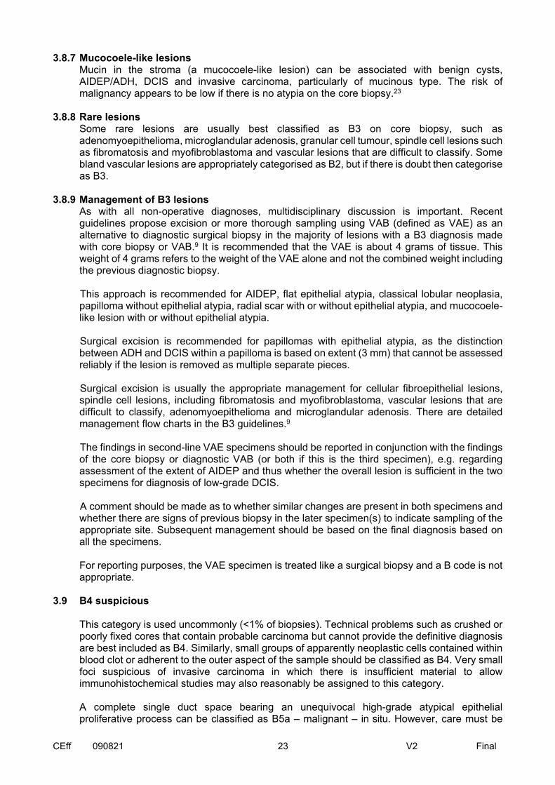
3.8.7 Mucocoele-like lesions Mucin in the stroma (a mucocoele-like lesion) can be associated with benign cysts, AIDEP/ADH, DCIS and invasive carcinoma, particularly of mucinous type. The risk of malignancy appears to be low if there is no atypia on the core biopsy.23
3.8.8 Rare lesions
Some rare lesions are usually best classified as B3 on core biopsy, such as adenomyoepithelioma, microglandular adenosis, granular cell tumour, spindle cell lesions such as fibromatosis and myofibroblastoma and vascular lesions that are difficult to classify. Some bland vascular lesions are appropriately categorised as B2, but if there is doubt then categorise as B3.
3.8.9 Management of B3 lesions
As with all non-operative diagnoses, multidisciplinary discussion is important. Recent guidelines propose excision or more thorough sampling using VAB (defined as VAE) as an alternative to diagnostic surgical biopsy in the majority of lesions with a B3 diagnosis made with core biopsy or VAB.9 It is recommended that the VAE is about 4 grams of tissue. This weight of 4 grams refers to the weight of the VAE alone and not the combined weight including the previous diagnostic biopsy. This approach is recommended for AIDEP, flat epithelial atypia, classical lobular neoplasia, papilloma without epithelial atypia, radial scar with or without epithelial atypia, and mucocoele-like lesion with or without epithelial atypia. Surgical excision is recommended for papillomas with epithelial atypia, as the distinction between ADH and DCIS within a papilloma is based on extent (3 mm) that cannot be assessed reliably if the lesion is removed as multiple separate pieces. Surgical excision is usually the appropriate management for cellular fibroepithelial lesions, spindle cell lesions, including fibromatosis and myofibroblastoma, vascular lesions that are difficult to classify, adenomyoepithelioma and microglandular adenosis. There are detailed management flow charts in the B3 guidelines.9 The findings in second-line VAE specimens should be reported in conjunction with the findings of the core biopsy or diagnostic VAB (or both if this is the third specimen), e.g. regarding assessment of the extent of AIDEP and thus whether the overall lesion is sufficient in the two specimens for diagnosis of low-grade DCIS. A comment should be made as to whether similar changes are present in both specimens and whether there are signs of previous biopsy in the later specimen(s) to indicate sampling of the appropriate site. Subsequent management should be based on the final diagnosis based on all the specimens. For reporting purposes, the VAE specimen is treated like a surgical biopsy and a B code is not appropriate.
3.9 B4 suspicious
This category is used uncommonly (<1% of biopsies). Technical problems such as crushed or poorly fixed cores that contain probable carcinoma but cannot provide the definitive diagnosis are best included as B4. Similarly, small groups of apparently neoplastic cells contained within blood clot or adherent to the outer aspect of the sample should be classified as B4. Very small foci suspicious of invasive carcinoma in which there is insufficient material to allow immunohistochemical studies may also reasonably be assigned to this category. A complete single duct space bearing an unequivocal high-grade atypical epithelial proliferative process can be classified as B5a – malignant – in situ. However, care must be
CEff 090821 24 V2 Final
taken if one or only part of a duct space is seen containing a highly atypical epithelial process, particularly if no necrosis is present; this may be regarded as suspicious rather than definitively malignant. In particular, great care should be taken if the epithelial cells show any features of an apocrine phenotype, which may represent an atypical apocrine proliferation rather than DCIS. Another lesion that can be allocated to this category is a non-high grade intraductal proliferation with a significant degree of atypia probably representing intermediate or low-grade DCIS, where relatively few involved duct spaces are represented in the biopsy. A pragmatic approach is usually required by reporting an atypical intraductal proliferation and qualifying this according to the degree of suspicion, i.e. ‘at least ADH, probably low-grade DCIS’, and, on the basis of extent or severity of atypia, allocating the case either to the B3 or to B4 category. As discussed in the section above on lobular neoplasia, lesions that are difficult to classify as LCIS or DCIS, as well as non-pleomorphic LCIS with necrosis, are often best classified as B4. The management of cases classified as B4 will usually be either diagnostic excision biopsy of the area or repeat core biopsy or VAB to obtain definitive diagnosis. Definitive therapeutic surgery should not be undertaken as a result of a B3 or B4 core biopsy diagnosis except after a definite diagnosis of a phyllodes tumour.
3.10 B5 malignant
This category is appropriate for cases of unequivocal malignancy on core biopsy. The B5 category is further subdivided into B5a, B5b and B5c. B5a is the appropriate classification for unequivocal DCIS of all grades and pleomorphic LCIS, the report stating whether the lesion is DCIS or LCIS (classical lobular neoplasia is categorised as B3). B5b is used for all invasive primary breast carcinomas and rare invasive malignancies, including malignant phyllodes tumours, lymphomas and metastatic tumours. B5c is used when it is not possible to say whether the carcinoma is invasive or in situ. This category should be rarely applied. It is most frequently used when there are large fragments of carcinoma with no surrounding stroma. If there is unequivocal DCIS and features suspicious of invasion, but not sufficient for a definite diagnosis of invasive carcinoma, then B5a categorisation should be used. Fragments of papillary carcinoma are usually best categorised as B5a. Please also see comment below regarding encysted/encapsulated papillary carcinoma, which should be categorised as B5a and not B5c.
Category B5a: in situ Ductal carcinoma in situ One of the benefits of core biopsy compared with FNAC is that it can allow distinction between in situ and invasive carcinoma. However, as a result of the biopsy missing an area of invasion, approximately 20–30% of patients with a core biopsy diagnosis of DCIS will have invasive carcinoma identified in the subsequent excision specimen.24 The nuclear grade of the DCIS should be indicated on the core biopsy (see Pathology reporting of breast disease in surgical excision specimens).12 Architecture and the presence of necrosis may also be noted. The presence or absence of associated calcifications should be recorded, particularly if the biopsy was for investigation of calcification.

CEff 090821 25 V2 Final
Paget’s disease of the nipple should also be categorised as B5a. Immunohistochemistry can be helpful. Paget’s disease is usually luminal cytokeratin and HER2 positive, whereas Bowen’s disease expresses basal cytokeratins and melanoma is HMB45 and melan-A positive. S100 can be positive in Paget’s disease as well as melanoma.
Malignant papillary lesions Encysted or encapsulated papillary carcinoma should be categorised as B5a. Recent literature has shown that encapsulated/encysted papillary carcinomas usually lack a myoepithelial layer and probably represent an indolent form of invasive carcinoma. Regardless of whether these are invasive lesions or in situ cancers, the clinical outcome is good with adequate local therapy alone, similar to DCIS. The current recommendation is that these lesions should be categorised as B5a. It is recommended that the pathology report describes the lesion so that it is clear that it is not conventional DCIS. Lobular neoplasia Pleomorphic LCIS shows marked nuclear pleomorphism like that seen in high-grade DCIS. It can be confused with DCIS, particularly when associated with necrosis and calcification. These lesions should be categorised as B5a, as the current recommended management is similar to high-grade DCIS. Immunostaining with E-cadherin will often help differentiate between high-grade DCIS and pleomorphic LCIS. In exceptional circumstances, lobular neoplasia may be impossible to distinguish from small-cell solid DCIS. Staining for E-cadherin and β-catenin should be helpful to differentiate between the two. Membrane expression of E-cadherin and β-catenin is typically absent in lobular neoplasia and present in DCIS. If the distinction between classical lobular neoplasia and DCIS is not possible, then B4 categorisation is prudent. Classical lobular neoplasia (ALH/LCIS, see section 3.8.3) should be categorised as B3.
Category B5b: invasive Invasive carcinoma A major advantage of core biopsy over FNAC is the ability to diagnose invasion positively. Invasive carcinoma can be unequivocally identified in core biopsy with a positive predictive value of almost 100%. False-positive diagnosis is very rare.25 As noted above, however, the negative predictive value for invasion is only 80% when only DCIS is identified. Rarely, carcinoma is seen only in lymphovascular spaces. Provided the changes are sufficient for an unequivocal diagnosis, this should also be categorised as B5b.
Foci of invasive carcinoma less than 1 mm Microinvasive carcinoma is an entity requiring full assessment of the overall lesion and cannot be definitely diagnosed on core biopsy. If the core biopsy shows a small area of invasion less than 1 mm, it is recommended that levels are examined to see if the area is larger than 1 mm. Unequivocal invasive carcinomas less than 1 mm across in largest diameter should be categorised as B5b if there is no associated DCIS, as the sample may not be representative and invasive carcinoma may be present elsewhere. A comment about the small size should be made. If there is DCIS and definite invasion less than 1 mm in size, categorisation as B5a is recommended, but the report must mention the small area of invasion.

CEff 090821 26 V2 Final
If there is DCIS and an area suspicious of invasion but no definite invasion, then categorisation as B5a is appropriate. If there is an area less than 1 mm suspicious of invasion but no definite invasion and no DCIS, then categorisation as B4 is appropriate. B5c should not be used for invasive carcinoma less than 1 mm. It is important that the lesions discussed in this section have the histological features carefully described in the report so that it is clear what is present in the biopsy.
3.11 Assessment of prognostic and predictive factors
All invasive carcinomas should be graded and typed on core biopsy where possible. Current evidence suggests that concordance between grade on core biopsy and that in the definitive excision specimen can be achieved in approximately 70% of cases.26 It should, however, be made clear to the clinicians that the grade may differ (almost invariably by only one level) from that in the subsequent resection specimen. A phrase such as ‘Provisional (core) grade’ is suggested. In particular, mitotic count may be lower in the core biopsy than in the excision specimen, therefore leading to underestimation of grade on the core. Assessment of histological grade can also be performed on core biopsy of nodal metastases. Assessment of histological type is useful to identity patients with invasive lobular carcinomas, who may be offered MRI if they are considering breast-conserving surgery to identify multifocal disease. Grade and type are also useful when neoadjuvant therapy is given and there may not be any residual tumour in the surgical specimen. ER and HER2 assessment on core biopsies have been shown to correlate well with subsequent surgical excision specimens.27 There is less good correlation with PR. NICE recommends that ER, PR and HER2 are assessed on the core biopsy to facilitate planning of patient management.17 ER is not part of the minimum dataset for DCIS, but is assessed in some centres, particularly if it will influence treatment, for example in a patient who is not fit for surgery or where adjuvant hormone therapy is considered. As with determination on excision biopsy samples, a standard protocol and method of assessment should be used. For best results, the core biopsy should be fixed for at least six and no more than 72 hours. For detailed guidance on assessment of ER, progesterone receptor and HER2, please refer to Pathology reporting of breast disease in surgical excision specimens.12 For patients to be recommended neoadjuvant chemotherapy or primary endocrine treatment based on core biopsy features, the biopsy must contain sufficient carcinoma for definitive diagnosis, assessment of histological grade, and reliable determination of ER, PR and HER2 status.
3.12 Rare malignancies
Spindle cell carcinomas and metaplastic carcinomas should be designated as B5b. The use of an antibody panel, including a range of anti-cytokeratin antibodies (high- and low-molecular weight cytokeratins and broad-spectrum antibody), will assist in diagnosis. When a definite histological diagnosis cannot be made, the abnormality should be reported as spindle cell lesion of uncertain histogenesis or nature and classified as B3 or B4.

CEff 090821 27 V2 Final
Lymphoma If suspicion of lymphoma is raised histologically, expert haematopathological opinion may be valuable. Malignant lymphoma should be classified as B5b. The majority of these lesions are of high-grade B-cell morphology and may mimic epithelial malignancy. As in other organs, the cells frequently show less cohesion and a higher nuclear to cytoplasmic ratio and do not demonstrate the architectural features of carcinoma. The correct diagnosis is supported by immunohistochemistry (CD45, CD20, CD3, CD30, etc.) to differentiate from an epithelial or other malignancy such as melanoma (and demonstrate the appropriate phenotype). Low-grade lymphomas may be more difficult to distinguish from a chronic inflammatory process. Infiltration of the lobular epithelium should be sought and if the degree of lymphoid infiltrate is high, it should raise the possibility of a neoplastic process. A panel of lymphoid markers is necessary to demonstrate the phenotype of the cells present to allow correct diagnosis. Molecular tests such as looking for an IgH clone may be useful.
Metastasis to the breast Metastasis to the breast from primary malignancies elsewhere is well recognised, although in practice rarely biopsied if the diagnosis is recognised clinically. A full clinical history is essential to avoid misdiagnosis of a metastatic adenocarcinoma as a primary carcinoma. A wide range of tumours can metastasise to the breast, but the most frequently seen are lymphomas, carcinomas of the lung, ovary (serous papillary), kidney and prostate, carcinoid tumours and malignant melanoma. The diagnosis should be considered if the features of a malignancy are not typical of mammary origin.28 Immunohistochemistry is often helpful, but no marker is completely sensitive or specific, so it is important to use a panel of antibodies. Breast carcinoma usually expresses cytokeratin 7 and 18 (and not cytokeratin 20), GATA3 and epithelial membrane antigen. Approximately 80% of primary breast tumours are ER positive. TTF-1 is useful for identifying pulmonary and thyroid carcinoma, WT1 for identifying ovarian carcinoma, and S100, melan-A and HMB45 for identifying melanoma. PAX8 is a useful marker of gynaecological, renal and thyroid carcinomas. More details of antibodies are available.28
Sarcomas Primary breast sarcomas are rare. Mammary sarcomas most commonly originate in association with malignant phyllodes tumour or as part of a metaplastic carcinoma, but in a core biopsy the epithelial component may not be represented. The most common associated sarcomas are liposarcoma, fibrosarcoma, osteosarcoma, chondrosarcoma and rhabdomyosarcoma. Angiosarcoma is the most common primary breast sarcoma and most commonly arises in the dermis after previous radiotherapy. It may be the cause of false-negative diagnosis, as it may be relatively subtle and bland. Primary and secondary leiomyosarcoma may be found in the breast. All these lesions can be difficult to diagnose definitively in core samples. If unequivocal malignancy is present, they should be graded as B5b. A high index of suspicion and judicious use of immunohistochemistry can facilitate or support a diagnosis.
3.13 Problems and pitfalls in diagnosis
There are recognised problem areas and potential pitfalls in core needle biopsy diagnosis.

CEff 090821 28 V2 Final
Minor degrees of epithelial atypia Mild atypia of epithelium within lobular units is one of the most common problems encountered in core biopsy samples. Care must be taken not to overdiagnose such minimal degrees of atypia, which may represent usual epithelial hyperplasia (UEH), apocrine change or reactive changes, e.g. adjacent to previous sampling procedure. Conversely, more severe degrees of atypia must be sought that may reflect cancerisation of lobules by high-grade DCIS. The degree of atypia should be helpful in distinguishing the process, and the nuclear chromatin and presence of mitoses (although rarely seen) may aid in the diagnosis. Similarly, UEH and other forms of benign hyperplasia such as that of gynaecomastoid type are commonly seen in cores from benign fibroadenomas. This often shows apparent dyscohesion due to the trauma of the core biopsy sampling process, and ‘telescoping’ of the epithelium is seen within the duct spaces, thus resembling a hyperplastic process. As with UEH in surgical excision specimens, the lack of uniformity and distribution/streaming of the epithelial cells with bland nuclear features and paucity of mitoses is of assistance in reaching a diagnosis. AIDEP should not be diagnosed in these cases unless uniformity of nuclear size and shape and regular, evenly placed nuclei are seen. UEH of gynaecomastoid type with a micropapillary architecture should not be mistaken for micropapillary ADH/DCIS. As discussed above, immunohistochemistry for basal cytokeratins and ER can be helpful in distinguishing UEH from DCIS. Apocrine atypia and apocrine DCIS Apocrine atypia, particularly in association with a sclerosing lesion, such as sclerosing adenosis, may be especially difficult to identify correctly in non-operative diagnostic samples. In core biopsy, large nuclei, often with prominent nucleoli, may be mistaken for DCIS if pleomorphism is also present. The typical granular eosinophilic cytoplasmic appearance of apocrine cells should be sought. Pure apocrine DCIS is relatively rare and pleomorphic apocrine LCIS is even less common, so when an atypical apocrine proliferation is seen within ducts in a core biopsy, additional features of malignancy, such as significant atypia, intraluminal necrosis and the presence of mitoses as well as multiple duct involvement, should be sought for confirmatory evidence. In addition, multiple duct involvement indicating a more extensive lesion may provide further supportive evidence. Mild or moderate degrees of apocrine proliferation with atypical features in a duct space should be assessed with caution, and it may be prudent not to record a definite diagnosis, but to classify such a process as B3 of uncertain malignant potential. Conversely, papillary apocrine change should not be mistakenly classified as other than benign.
Lactational change Focal lactational change may be seen in women who are neither lactating nor pregnant and indeed are nulliparous and/or post-menopausal. The involved acini are usually lined by plump vacuolated cells with a ‘hobnail’ architecture, but may less frequently appear atypical with irregular, large or pyknotic nuclei. The epithelial cells may appear degenerative and, rarely, the benign nature of the process may be mistaken for cancerisation of lobules by DCIS. The recognition of the vacuolation of the cytoplasm and the typical hobnail architecture will enable the correct diagnosis to be established.
Sclerosing lesions/tubular carcinoma There is a risk of overdiagnosis of invasive carcinoma when confronted by sclerosing adenosis in a core biopsy, particularly as the normal lobular arrangement may be less apparent than on

CEff 090821 29 V2 Final
an excision biopsy specimen. Immunohistochemical staining for myoepithelial markers can be useful in this situation.
Stromal proliferations and spindle cell lesions Spindle cell proliferations may cause difficulties in diagnosis in core biopsy samples. The most common lesion seen on core biopsy is scarring and there are, usually, associated changes such as fat necrosis or haemosiderin-laden macrophages to enable a diagnosis and categorisation as B2. Occasionally, scarring may show atypical spindle cells and a definite diagnosis may not be possible on core biopsy. Myofibroblastoma is composed of short bundles of bland spindle cells with intervening eosinophilic collagen bundles and sometimes adipose tissue, and typically expresses CD34 and desmin. Fibromatosis is a bland spindle cell proliferation that is best categorised as B3. Nuclear expression of β-catenin is frequently seen, but is not specific. Cytokeratins and CD34 are not expressed. Spindle cell or metaplastic carcinomas need to be considered in many spindle cell lesions, as they can show a wide range of appearances, including resembling fibromatosis. Evidence of epithelial differentiation must be sought; this may range from small cohesive foci to conventional carcinoma. Immunohistochemistry for a panel of cytokeratins, including both luminal and basal cytokeratins, should be performed. Occasionally, a phyllodes tumour may just show spindle cells on core biopsy and evidence for an epithelial component should be sought, e.g. by performing additional levels. CD34 expression supports the diagnosis of phyllodes tumour. Primary sarcomas of the breast are very rare; the most common of these is angiosarcoma. Skin lesions such as dermatofibroma and melanoma need to be considered in the differential diagnosis of spindle cell lesions. When a definitive histological diagnosis cannot be made, the abnormality should be reported as a spindle cell lesion of uncertain nature and classified as B3 or B4. Radiation-induced changes Radiotherapy changes to the breast may be difficult to differentiate from foci of recurrent or residual carcinoma, both in situ and invasive. The radiation induces a degree of atypia of the breast epithelium, but also in the histiocyte population, which is prominent as a result of the radiotherapy and also recent surgery. The macrophages may also show degenerative features. Thus, carcinoma cells can conversely mimic macrophages. Immunocytochemistry can be helpful in difficult cases, as irradiated neoplastic cells retain cytokeratin expression, whilst macrophages demonstrate a histiocytic phenotype, e.g. CD68 reactivity.
Infiltrating lobular carcinoma Small foci of invasive lobular carcinoma can be missed in histological sections and be dismissed as chronic inflammation or stromal cells. The targetoid infiltrative pattern of classical lobular carcinoma may be of assistance but a reactive lymphocyte process can also have a peri-ductal or peri-lobular distribution. Cytokeratin immunohistochemistry to demonstrate the neoplastic cells is of value in difficult cases, but recognition of the abnormal cell proliferation requires vigilance, as the features can be subtle.

CEff 090821 30 V2 Final
4 Axillary lymph node assessment and preoperative sampling
Axillary nodal status remains the most powerful prognostic factor in patients with invasive carcinoma of the breast. Recently, sentinel node biopsy has become the standard method for staging of disease. Those patients with involved nodes may then require further treatment of the axilla, including surgery. A preoperative diagnosis of nodal metastasis means that patients can proceed straight to axillary clearance. Thus, preoperative axillary staging can reduce the number of patients having two axillary surgical procedures. All patients seen in symptomatic and screening assessment clinics who have suspicious mammographic and/or ultrasound findings should have detailed ultrasound assessment of the axilla.3 Level 1 axillary nodes are usually easily visualised in all patients and these nodes can be assessed for risk of metastatic involvement.29 The criteria accepted as indications for ultrasound-guided needle biopsy or FNA of axillary lymph nodes vary between units. The evidence around what cortical thickness can be considered to be abnormal is not clear so the criteria and procedure for sampling should be agreed locally and subject to audit.1
Morphological lymph node abnormality is more predictive of metastatic involvement than cortical thickening. When axillary lymph nodes are involved, ultrasound and needle biopsy or FNA will detect disease in only 45–50% of cases.30 The chances of detection are higher in high-grade invasive breast cancer and when there are four or more nodes involved. The yield from sampling normal morphology lymph nodes with no cortical thickening is very low and is not recommended. Both core biopsy and FNA may be used to sample abnormal axillary nodes. FNA is preferred for smaller nodes and for nodes close to vessels. Core biopsy may be preferred when the lymph node is large (>20 mm) or when FNA is negative, inadequate or equivocal from clearly radiologically abnormal nodes. The sensitivity of core biopsy for malignancy in lymph nodes is a little higher than for FNA. The technique for sampling lymph nodes is the same as that used for ultrasound-guided breast biopsy. Local anaesthetic is used for the skin and superficial tissues down to the node. Too much infiltration of local anaesthetic should be avoided for FNA, as a pool of anaesthetic around the node makes an inadequate sample more likely. For FNA, sampling with a 21-gauge needle appears to give better results for axillary nodes. Techniques with and without suction are down to personal preference and have similar results. Core biopsy in the axilla has the potential to cause more collateral damage to adjacent structures, particularly arteries and veins, compared with the breast. Core biopsy needles that offer a two-stage sampling option may be preferred where the sampling trough can be advanced manually through the node before the cutting outer sheath is advanced. This technique minimises the risk of damaging structures around the targeted node. The lymph node targeted on ultrasound is frequently not the sentinel node subsequently targeted at surgery.29 Injection of microbubbles with ultrasound tracking has been shown to be successful in identifying the sentinel node, and current trials are investigating the role of VAB in sampling sentinel nodes identified in this way.31 Specificity is high for both NCB and VAB, but false-positive diagnosis has been described with FNAC of axillary nodes and is likely to be more frequent with FNAC than with core biopsy, as is well recognised in sampling of lesions in the breast. About half of nodal metastases can be detected, but the sensitivity depends on how patients are selected and the number of passes.

CEff 090821 31 V2 Final
Given the necessary expertise, FNA may allow equivalent sensitivity at a lower cost. FNA is preferred by some units due to the proximity of large vessels and nerves. Core biopsy provides sections for ready identification of a small volume of disease and allows immunohistochemistry in cases of equivocal morphology. With FNAC it is helpful to prepare material for immunocytochemistry, as this may be useful on occasions. Needle washings can be used for this. Limited volume disease can be missed with either technique. Lymph node FNA and core biopsy in breast cancer patients is intended for assessment of the presence or absence of metastatic carcinoma. If there is suspicion of malignant lymphoma (axillary lymphadenopathy in the absence of a known cause or malignancy in the breast), the patient should be referred for assessment in line with local protocols for diagnosis of malignant lymphoma. All patients with primary invasive breast cancer with negative results for metastatic disease on FNA or core of the axillary nodes are candidates for sentinel node biopsy or other axillary procedure for definitive staging.
4.1 L codes for fine needle aspiration cytology
For FNAC assessment of axillary lymph nodes, the following diagnostic categories should be used. LC1 Inadequate: no lymphoid cells or technically inadequate. LC2 Benign: benign lymphoid cells regardless of whether specific reactive features are
seen or not. LC3 Atypia: atypical cells present, lymphoid or other of uncertain nature and significance.
Can be used for atypical lymphoid proliferations – usually low-grade lymphomas where immunohistochemistry and flow samples not available.
LC4 Suspicious of malignancy: either metastasis or lymphoma. Usually, only occasional cells present either singly or in small groups.
LC5 Malignant: metastatic carcinoma or other malignancy (including lymphoma). 4.2 L codes for needle core biopsy
For NCB assessment of axillary nodes, the following diagnostic categories should be used. LB1 Inadequate: no lymph node/lymphoid tissue. Lymph node tissue with artefact that
prevents interpretation should be categorised as LB1. LB2 Benign: either normal lymph node or lymph node with benign changes such as
reactive hyperplasia, dermatopathic lymphadenopathy, foreign body reaction, sarcoidosis, tuberculosis, etc.
LB3 Atypia: lymphoid tissue with atypical cells present, lymphoid or other of uncertain nature and significance.
LB4 Suspicious of malignancy, including metastatic carcinoma or other malignancy (including lymphoma).
LB5 Malignant, metastatic carcinoma or other malignancy (including lymphoma).
In instances where there is a discrepancy between the lymph node cytology or biopsy and the radiological impression, repeat FNAC or consider core biopsy, as lymph node involvement may be focal. The utility of axillary ultrasound and needle biopsy should continue to be reviewed, based on current recommended practice in relation to sentinel node biopsy and axillary dissection.

CEff 090821 32 V2 Final
4.3 Pitfalls
The primary aim of preoperative assessment of axillary nodes in patients with breast cancer is to detect nodal metastases from the mammary carcinoma. It is important to recognise other diagnoses and avoid misdiagnosis. Other malignancies may involve the nodes. To avoid misdiagnosis, it is useful to compare the morphology of the neoplastic cells present in the lymph node FNA or core biopsy with the tumour in the preoperative core biopsy or FNA of the breast ,as metastases are usually similar to the primary tumour. If the tumour has a different appearance or has features unusual for a mammary carcinoma, metastasis from other sites should be considered. Melanoma should be in the differential diagnosis. Malignant lymphoma must also be considered. Immunohistochemistry, particularly on core biopsy, can often resolve these differential diagnoses. Benign lymph node inclusions are a diagnostic pitfall. Melanocyte rests are the most common, but epithelial inclusions and, rarely, mesothelial inclusions can occur. Macrophages as part of reactive conditions such as dermatopathic lymphadenopathy or granulomatous lymphadenitis may mimic carcinoma cells. Immunohistochemistry for macrophage markers such as CD68 and epithelial markers can be helpful. Lymphocytes from inflammatory disorders affecting adjacent structures including the skin may mimic a lymph node. Small metastases should be reported as malignant. If there are only a few cells in the metastasis it is suggested that this is indicated in the report so that it can be discussed at the multidisciplinary meeting. Occasionally, after a diagnosis of malignancy on the core or FNAC of the axillary nodes, the surgical specimen may show no nodal metastasis. The original core biopsy or FNAC should be reviewed to confirm the original diagnosis. If the carcinoma is small on the core or there are only scanty cells on the FNAC, it may be reasonable that no metastases are found in the surgical specimen. Sometimes, nodes low in the axilla are missed at surgery – ultrasound of the axilla should be considered to search for such nodes.32 If the patient has had preoperative systemic treatment, features suggesting treated carcinoma, such as fibrosis, should be sought in the nodes in the surgical specimen.
5 How to perform fine needle aspiration cytology 5.1 Aspiration procedure
The success of FNAC is directly related to the skill and experience of the operators. The number of staff involved should be restricted to the minimum possible. An assistant skilled in specimen preparation, preferably a biomedical scientist or a pathologist, is helpful. If a trained biomedical scientist or pathologist is available to immediately assess the adequacy of the aspirate using a rapid staining technique, recall for repeat cytology can be avoided, therefore reducing delay and distress.
5.1.1 Equipment Use 22- or 23-gauge needles of appropriate type and length. A needle with a trocar may be preferred, as it is more rigid and less likely to become blocked or contaminated during insertion. A 10 or 20 ml syringe is used to apply suction. A short extension tube between the needle and syringe is usually required for image-guided procedures. A syringe holder makes manipulation of the syringe with simultaneous suction much easier.
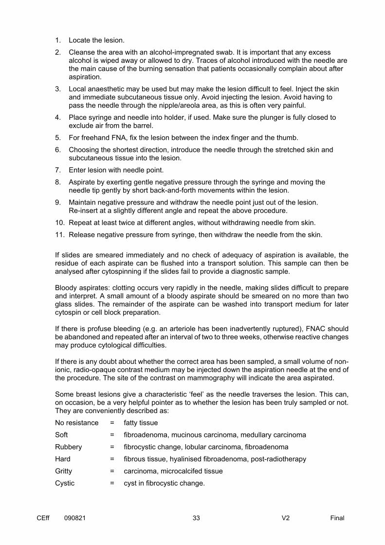
CEff 090821 33 V2 Final
1. Locate the lesion. 2. Cleanse the area with an alcohol-impregnated swab. It is important that any excess
alcohol is wiped away or allowed to dry. Traces of alcohol introduced with the needle are the main cause of the burning sensation that patients occasionally complain about after aspiration.
3. Local anaesthetic may be used but may make the lesion difficult to feel. Inject the skin and immediate subcutaneous tissue only. Avoid injecting the lesion. Avoid having to pass the needle through the nipple/areola area, as this is often very painful.
4. Place syringe and needle into holder, if used. Make sure the plunger is fully closed to exclude air from the barrel.
5. For freehand FNA, fix the lesion between the index finger and the thumb. 6. Choosing the shortest direction, introduce the needle through the stretched skin and
subcutaneous tissue into the lesion. 7. Enter lesion with needle point. 8. Aspirate by exerting gentle negative pressure through the syringe and moving the
needle tip gently by short back-and-forth movements within the lesion. 9. Maintain negative pressure and withdraw the needle point just out of the lesion.
Re-insert at a slightly different angle and repeat the above procedure. 10. Repeat at least twice at different angles, without withdrawing needle from skin. 11. Release negative pressure from syringe, then withdraw the needle from the skin.
If slides are smeared immediately and no check of adequacy of aspiration is available, the residue of each aspirate can be flushed into a transport solution. This sample can then be analysed after cytospinning if the slides fail to provide a diagnostic sample. Bloody aspirates: clotting occurs very rapidly in the needle, making slides difficult to prepare and interpret. A small amount of a bloody aspirate should be smeared on no more than two glass slides. The remainder of the aspirate can be washed into transport medium for later cytospin or cell block preparation. If there is profuse bleeding (e.g. an arteriole has been inadvertently ruptured), FNAC should be abandoned and repeated after an interval of two to three weeks, otherwise reactive changes may produce cytological difficulties. If there is any doubt about whether the correct area has been sampled, a small volume of non-ionic, radio-opaque contrast medium may be injected down the aspiration needle at the end of the procedure. The site of the contrast on mammography will indicate the area aspirated.
Some breast lesions give a characteristic ‘feel’ as the needle traverses the lesion. This can, on occasion, be a very helpful pointer as to whether the lesion has been truly sampled or not. They are conveniently described as: No resistance = fatty tissue Soft = fibroadenoma, mucinous carcinoma, medullary carcinoma Rubbery = fibrocystic change, lobular carcinoma, fibroadenoma Hard = fibrous tissue, hyalinised fibroadenoma, post-radiotherapy Gritty = carcinoma, microcalcifed tissue Cystic = cyst in fibrocystic change.

CEff 090821 34 V2 Final
5.2 Ultrasound-guided FNAC The lesion is demonstrated and the surrounding breast tissue is immobilised by applying pressure with the palm of the hand holding the probe. Infiltration of the skin with local anaesthetic may be carried out. The FNAC needle, attached by a short connecting tube to a 10 cc syringe held by the assistant, is introduced into the breast along the line of the long axis of the ultrasound probe and will be easily visualised if it is kept parallel to the surface of the probe. The needle tip is guided into the lesion and an image is taken to record that the needle is correctly positioned. The needle is then moved back and forth within the lesion, with simultaneous rotation and negative pressure being applied by the assistant. Aspiration is continued until material is seen within the hub of the needle. The aspirate is then delivered onto slides, and dry and wet preparations are made in accordance with guidance from the pathologist. Two to three separate samples are commonly obtained to increase the chances of obtaining a diagnostic cellular sample. Needle washings may also be made, flushing the needle and connecting tube with 3–5 ml of cellular fixative. Ultrasound jelly may present a problem in interpretation for pathologists seeing it for the first time and should not be confused with calcium salts or necrosis. It should not be used during the aspiration procedure and, if used previously, should be carefully removed.
5.3 Spreading the slides
A number of methods can be used to spread the slides obtained by placing a drop of aspirated material from the needle on a glass slide. Many of these are variations on a theme, but the essential aim is to get a thin layer of material on the slide to allow rapid drying for air dried fixation without appreciable squash artefacts due to excess pressure (Figure 1).

CEff 090821 35 V2 Final
Figure 1: Spreading with a slide. Three basic methods (1, 2 or 3) can be used, all producing similar effects. Alternatively, the slide may be spread using a pipette or a needle (4).
All pathologists have received slides from clinicians where the aspirate has been well taken but has been ruined by poor spreading technique. It is sometimes difficult to remedy this, but multidisciplinary discussion and making aspirators aware of the problems, especially visually and microscopically, often helps to alleviate the problem. Should such problems persist, alternative preparative techniques, such as cytospin or thin preparations, may be considered.
5.4 Fixation methods 5.4.1 Wet fixed smears
These smears must be fixed immediately after spreading and before they have a chance to dry, by dropping into a pot of fixative, or flooding the slide with a drop of fixative if no container is available. Spray fixation can be used.
5.4.2 Air dried smears
After spreading, the slide should be dried rapidly by waving in the air or by using a fan. Alternatively, a hair dryer can be used, but this must be on a cold setting, as warm air will ‘cook’ the cells and lead to artefacts.
5.4.3 Transport medium
In some units, transport medium is used for specimens, which means that optimum preparations can be made in the laboratory after cytocentrifugation. This method is best used where clinicians are not used to making cytological smears and do not follow proper fixation

CEff 090821 36 V2 Final
techniques. It can be superior to delayed fixation of wet preparations where air-drying can make interpretation difficult.
6 Diagnostic coding
SNOMED Topography (T) or relevant SNOMED-CT code must be recorded for the anatomical site. SNOMED Morphology (M) or relevant SNOMED-CT code must be recorded for the diagnosis/tumour morphology. It is noted, however, that SNOMED is now in a practical transition phase, as part of the intended full implementation by the NHS and Public Health England (PHE) of SNOMED-CT. SNOMED ceased to be licensed by the International Health Terminology Standards Development Organisation from 26 April 2017. A list of applicable M SNOMED and SNOMED-CT codes is provided in Appendix A. Mapping SNOMED-CT terminology is provided in Appendix I.
7 Criteria for audit
As recommended by the Royal College of Pathologists as key performance indicators (see Key Performance Indicators – Proposals for implementation, July 2013):
• histopathology cases must be reported, confirmed and authorised within seven and ten calendar days of the procedure - standard: 80% of cases must be reported within seven calendar days and 90% within
ten calendar days. These turnaround times apply to all biopsies and not just breast screening biopsies. The following standard is also suggested:
• completeness of histopathology core items recorded - standard: reports should contain 100% of the core items.
Breast screening biopsies should be reported within five working days and this can also be audited.33
Please see also Appendix C (Quality assurance) for details on auditing core biopsy performance.

CEff 090821 37 V2 Final
8 References 1. Borrelli C, Cohen S, Duncan A, Given-Wilson R, Jenkins J, Kearins O et al. Clinical guidance
for Breast Cancer Screening Assessment (4th edition). London, UK: NHS Breast Screening Programme, Public Heath England, 2016. Available at: https://www.gov.uk/government/publications/breast-screening-clinical-guidelines-for-screening-management
2. Lee AHS, Carder P, Deb R, Howe M, Knox F, Shrimankar J et al.; on behalf of the Non-
operative diagnosis working group of the National Coordinating Group for Breast Pathology. Guidelines for Non-Operative Diagnostic Procedures and Reporting in Breast Cancer Screening. London, UK: Royal College of Pathologists, 2016. Available at: www.rcpath.org/profession/guidelines/cancer-datasets-and-tissue-pathways.html
3. Royal College of Radiologists. Guidance on Screening and Symptomatic Breast Imaging.
Fourth edition. Available at: https://www.rcr.ac.uk/publication/guidance-screening-and-symptomatic-breast-imaging-fourth-edition
4. NHSBSP Evaluation Group, Wheaton M, Kearins O. Reporting, recording and auditing B5
core biopsies with normal/benign surgery. NHSBSP Good Practice Guide No 9. Sheffield, UK: NHS Cancer Screening Programmes, 2007. Available at: https://assets.publishing.service.gov.uk/government/uploads/system/uploads/attachment_data/file/442171/nhsbsp-gpg9.pdf
5. Ciatto S, Houssami N, Ambrogetti D, Bianchi S, Bonardi R, Brancato B et al. Accuracy and
underestimation of malignancy of breast core needle biopsy: the Florence experience of over 4000 consecutive biopsies. Breast Cancer Res Treat 2007;101:291–297.
6. Villa A, Tagliafico A, Chiesa F, Chiaramondia M, Friedman D, Calabrese M. Atypical ductal
hyperplasia diagnosed at 11-gauge vacuum-assisted breast biopsy performed on suspicious clustered microcalcifications: could patients without residual microcalcifications be managed conservatively? AJR Am J Roentgenol 2011;197:1012–1018.
7. Jackman RJ, Marzoni FA Jr, Rosenburg J. False negative diagnoses at stereotactic vacuum
assisted needle breast biopsy: long-term follow-up of 1,280 lesions and review of the literature. AJR Am J Roentgenol 2009;192:341–351.
8. Yu YH, Liang C, Yuan XZ. Diagnostic value of vacuum-assisted breast biopsy for breast
carcinoma: a meta-analysis and systematic review. Breast Cancer Res Treat 2010;120:469–479.
9. Pinder SE, Shaaban A, Deb R, Desai A, Gandhi A, Lee AHS et al. NHS breast screening
multidisciplinary working group guidelines for the diagnosis and management of lesions of uncertain malignant potential on core biopsy (B3 lesions). Clin Radiol 2018;73:682–692.
10. O’Flynn EA, Wilson AR, Michell MJ. Image-guided breast biopsy: state-of-the-art. Clin Radiol
2010;65:259–270. 11. British Society of Breast Radiology. Guidelines for Performing Breast and Axillary Biopsies in
Patients on Anticoagulant and Antiplatelet Therapy. Available at: https://breastradiology.org/media/1022/bsbr-anticoag-guidelines-august-2018.pdf
12. Ellis IO, Carder P, Hales S, Lee AHS, Pinder SE, Rakha E et al. Pathology Reporting of
Breast Disease in Surgical Excision Specimens Incorporating the Dataset for Histological Reporting of Breast Cancer. London, UK: Royal College of Pathologists, 2016. Available at: www.rcpath.org/profession/guidelines/cancer-datasets-and-tissue-pathways.html

CEff 090821 38 V2 Final
13. Writing Group of the National Quality Assurance Coordinating Group for Breast Screening Pathology. Quality Assurance Guidelines for Breast Pathology Services (2nd edition). NHSBSP publication No. 2. Sheffield, UK: NHS Cancer Screening Programmes, 2011.
14. Public Health England. Breast Screening: How to Record Vacuum-Assisted Excisions.
Accessed July 2019. Available at: www.gov.uk/government/publications/breast-screening-how-to-record-vacuum-assisted-excisions/breast-screening-how-to-record-vacuum-assisted-excisions
15. NICE. Early and Locally Advanced Breast Cancer: Diagnosis and Management. NICE
guideline [NG101] 2018. Available at: www.nice.org.uk/guidance/ng101/chapter/Recommendations#referral-diagnosis-and-preoperative-assessment
16. Public Health England. Breast Screening: Issuing Provisional or Verbal Pathology Reports.
Available at: https://www.gov.uk/government/publications/breast-screening-issuing-provisional-or-verbal-pathology-reports
17. Public Health England. Breast screening: transferring specimens between laboratories. Available at: https://www.gov.uk/government/publications/breast-screening-transferring-specimens-between-laboratories
18. Dahlstrom JE, Sutton S, Jain S. Histologic-radiologic correlation of mammographically detected microcalcification in stereotactic core biopsies. Am J Surg Pathol 1998;22:256–259.
19. Deb R, Jenkins JA, Rowlands D, Ellis IO, Lee AHS. National survey of B1 and B2 reporting
of breast needle core biopsies. J Clin Pathol 2016;69:271–274. 20. Rakha EA, Lee AHS, Jenkins J, Murphy AE, Hamilton LJ, Ellis IO. Characterization and
outcome of breast needle core biopsy diagnoses of lesions of uncertain malignant potential (B3) in abnormalities detected by mammographic screening. Int J Cancer 2011;129:1417–1424.
21. Fadare O, Dadmanesh F, Alvarado-Cabrero I, Snyder R, Stephen Mitchell J, Tot T et al.
Lobular intraepithelial neoplasia [lobular carcinoma in situ] with comedo-type necrosis: A clinicopathologic study of 18 cases. Am J Surg Pathol 2006;30:1445–1453.
22. Lee AHS, Hodi Z, Ellis IO, Elston CW. Histological features useful in the distinction of
phyllodes tumour and fibroadenoma on needle core biopsy of the breast. Histopathology 2007;51:336–344.
23. Rakha EA, Shaaban AM, Haider SA, Jenkins J, Menon S, Johnson C et al. Outcome of pure
mucocele-like lesions diagnosed on breast core biopsy. Histopathology 2013;62:894–898. 24. Brennan ME, Turner RM, Ciatto S, Marinovich ML, French JR, Macaskill P et al. Ductal
carcinoma in situ at core-needle biopsy: meta-analysis of underestimation and predictors of invasive breast cancer. Radiology 2011;260:119–128.
25. Rakha EA, El-Sayed ME, Reed J, Lee AHS, Evans AJ, Ellis IO. Screen-detected breast
lesions with malignant needle core biopsy diagnoses and no malignancy identified in subsequent surgical excision specimens (potential false-positive diagnosis). Eur J Cancer 2009;45:1162–1167.
26. Harris GC, Denley HE, Pinder SE, Lee AHS, Ellis IO, Elston CW et al. Correlation of
histologic prognostic factors in core biopsies and therapeutic excisions of invasive breast carcinoma. Am J Surg Pathol 2003;27:11–15.

CEff 090821 39 V2 Final
27. Arnedos M, Nerurkar A, Osin P, A’Hern R, Smith IE, Dowsett M. Discordance between core needle biopsy (CNB) and excisional biopsy (EB) for estrogen receptor (ER), progesterone receptor (PgR) and HER2 status in early breast cancer (EBC). Ann Oncol 2009;20:1948–1952.
28. Lee AHS, Hodi Z, Soomro I, Sovani V, Abbas A, Rakha E, Ellis IO. Histological clues to the
diagnosis of metastasis to the breast from extramammary malignancies. Histopathology 2020;77:303-313
29. Britton P, Moyle P, Benson JR, Goud A, Sinnatamby R, Barter S et al. Ultrasound of the
axilla: where to look for the sentinel lymph node. Clin Radiol 2010;65:373–376. 30. Houssami N, Ciatto S, Turner RM, Cody HS, Macaskill P. Preoperative ultrasound-guided
needle biopsy of axillary nodes in invasive breast cancer: meta-analysis of its accuracy and utility in staging the axilla. Ann Surg 2011;254:243–251.
31. Sever AR, Mills P, Weeks J, Jones SE, Fish D, Jones PA et al. Preoperative needle biopsy
of sentinel lymph nodes using intradermal microbubbles and contrast-enhanced ultrasound in patients with breast cancer. AJR Am J Roentgenol 2012;199:465–470.
32. Cornford EJ, James JJ, McMahon M, Macmillan RD, Lee AH. The importance of low-lying
axillary sentinel nodes in breast cancer patients. Clin Radiol 2011;66:974–976. 33. Association of Breast Surgery. Best Practice Guidelines for Surgeons in Breast Cancer
Screening. Published January 2018. Available at: https://associationofbreastsurgery.org.uk/media/64269/screening-guidelines.pdf
34. Association of Breast Surgery. Summary Statement: Guidelines for the Management of
Spontaneous Nipple Discharge in the Absence of a Breast Lump. Available at: http://http://associationofbreastsurgery.org.uk/media/64951/abs-summary-statement-nipple-discharge-v1.pdf
35. Kooistra BW, Wauters C, van de Ven S, Strobbe L. The diagnostic value of nipple discharge
cytology in 618 consecutive patients. Eur J Surg Oncol. 2009;35:573–577. 36. Montroni I, Santini D, Zucchini G, Fiacchi M, Zanotti S, Ugolini G et al. Nipple discharge: is its
significance as a risk factor for breast cancer fully understood? Observational study including 915 consecutive patients who underwent selective duct excision. Breast Cancer Res Treat. 2010;123:895–900
37. Salhany KE, Page DL. Fine needle aspiration of mammary lobular carcinoma in situ and
atypical lobular hyperplasia. Am J Clin Pathol 1989;92:22–26. 38. Public Health England. National Breast Screening Pathology Audit 2020.
https://www.gov.uk/government/publications/national-breast-screening-pathology-audit/national-breast-screening-pathology-audit

CEff 191120 40 V2 Draft
Appendix A FNAC reporting guidelines This section of the document is designed to assist classification and reporting of FNAC samples. It should, however, be noted that FNAC alone is not an appropriate method of assessment of abnormalities detected by breast screening. FNAC does provide the advantage of providing a rapid diagnosis compared with core biopsy and may be used in conjunction with core needle biopsy where the necessary expertise exists. It can also be used for assessment of symptomatic patients. Reporting categories In ideal circumstances, one should aim for a definitive benign or malignant diagnosis. The proportion where this is possible will increase with experience of both the pathologist and aspirator. C1 inadequate The designation of an aspirate as ‘inadequate’ is, to a certain extent, a subjective matter and may depend on the experience of the aspirator and/or the interpreter. It is generally based on the presence of sufficient numbers of epithelial cells to provide a sample adequate for confident assessment. There are a number of reasons for labelling a smear as inadequate. These fall into three main groups:
• hypocellularity
• error in aspiration, spreading or staining
• excessive blood. In some cases, diagnostic information may be present and may be conveyed in the accompanying text description, e.g. adipose tissue fragments could support a clinical diagnosis of lipoma. Aspirates from certain lesions, such as cysts, abscesses, intramammary lymph nodes, fat necrosis and nipple discharge specimens may not contain epithelial cells but should not be classified as inadequate. Preparative artefacts include:
• crush: when too much pressure is used during smearing
• drying: when the dry smears are allowed to dry too slowly (dry smears should be dried quickly, wafting in the air can speed up drying) or when the wet fixed smears have been allowed to dry out before fixation
• thickness of smear: when an overlay of blood, protein-rich fluid or cells is obscuring the picture, making assessment impossible.
It is helpful to make a comment explaining why the specimen is inadequate. C2 benign • Indicates an adequate sample showing no evidence of malignancy or specific lesions
regarded as atypical and, if representative, a negative report can be issued.
• The aspirate in this situation is poorly to moderately cellular and tends to consist mainly of regular duct epithelial cells. These are generally arranged as monolayers and the cells have the characteristic benign cytological features. The background is usually composed of dispersed individual and paired naked nuclei. Should cystic structures be a component of the aspirated breast, a mixture of foamy macrophages and regular apocrine cells may be part of the picture. Fragments of fibrofatty and/or fatty tissue are common findings.
• A positive diagnosis of specific conditions, e.g. fibroadenoma, fat necrosis, granulomatous mastitis, breast abscess, lymph node, etc., may be suggested if sufficient specific features are present to establish the diagnosis with confidence and may be helpful in multidisciplinary correlation.

CEff 090821 41 V2 Final
• Care should be taken when correlating cytology features and radiology. For example, a few cohesive groups of epithelial cells in an aspirate from a well-defined lesion thought to be a cyst may not be representative of the lesion.
C3 atypia probably benign The aspirate here can have all the characteristics of a benign aspirate, as described in the previous paragraph. There are, however, also certain features not commonly seen in benign aspirates. These could be any, or a combination, of the following:
• nuclear pleomorphism
• some loss of cellular cohesiveness
• nuclear and cytoplasmic changes resulting from, for example, hormonal (pregnancy, pill, hormone replacement therapy) or treatment influences (see section 4.3 for diagnostic pitfalls)
• increased cellularity accompanying the above features. In addition, specific lesions that are regarded as showing an increased risk of malignancy can be identified on FNAC and should be reported as C3. These include papillary lesions and suspected phyllodes tumours. In both of these lesions, there may not be any cytological atypia, but the possibility of malignancy in a focal area of these lesions warrants a report of C3. C4 suspicious of malignancy This category should be used for those aspirates where there are atypical features in the smear, such that the pathologist is almost certain that they come from a malignant lesion, although a confident diagnosis cannot be made. This may be for three main reasons: 1. the specimen is scanty, poorly preserved or poorly prepared, but some cells with features of
malignancy are present 2. the sample may show some malignant features without overt malignant cells present – the
degree of abnormality should be more severe than in the previous category 3. the sample has an overall benign pattern with large numbers of naked nuclei and/or cohesive
sheets of cells, but with occasional cells showing distinct malignant features.
If an aspirate is reported as C4 because of low cellularity, repeat aspiration is often helpful. If, however, the aspirate is cellular but considered to be suspicious of malignancy, it is unlikely that repeat aspiration will be helpful.
Definitive therapeutic surgery should not be undertaken as a result of a C3 or C4 diagnosis. C5 malignant • This indicates an adequate sample containing cells characteristic of carcinoma, or other
malignancy.
• The pathologist should feel at ease in making such a diagnosis. Malignancy should not be diagnosed on the basis of a single criterion. Combination of the features listed in Table 2 will be necessary to achieve this diagnosis.
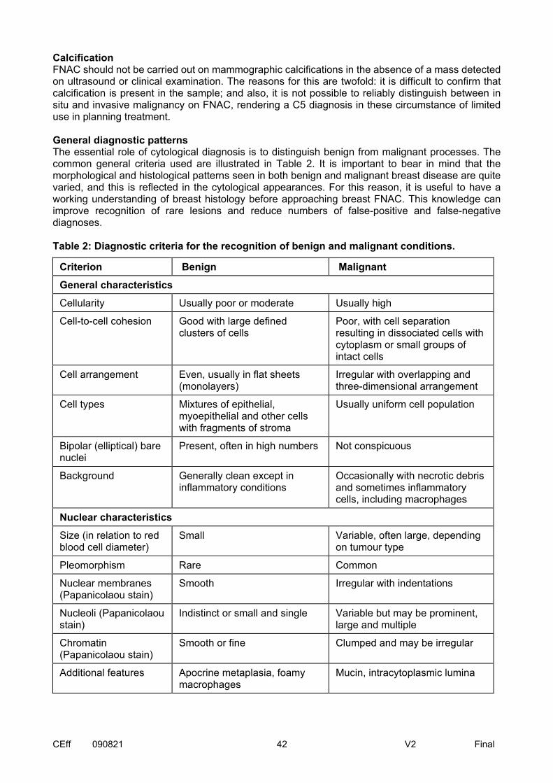
CEff 090821 42 V2 Final
Calcification FNAC should not be carried out on mammographic calcifications in the absence of a mass detected on ultrasound or clinical examination. The reasons for this are twofold: it is difficult to confirm that calcification is present in the sample; and also, it is not possible to reliably distinguish between in situ and invasive malignancy on FNAC, rendering a C5 diagnosis in these circumstance of limited use in planning treatment. General diagnostic patterns The essential role of cytological diagnosis is to distinguish benign from malignant processes. The common general criteria used are illustrated in Table 2. It is important to bear in mind that the morphological and histological patterns seen in both benign and malignant breast disease are quite varied, and this is reflected in the cytological appearances. For this reason, it is useful to have a working understanding of breast histology before approaching breast FNAC. This knowledge can improve recognition of rare lesions and reduce numbers of false-positive and false-negative diagnoses. Table 2: Diagnostic criteria for the recognition of benign and malignant conditions.
Criterion Benign Malignant General characteristics Cellularity Usually poor or moderate Usually high
Cell-to-cell cohesion Good with large defined clusters of cells
Poor, with cell separation resulting in dissociated cells with cytoplasm or small groups of intact cells
Cell arrangement Even, usually in flat sheets (monolayers)
Irregular with overlapping and three-dimensional arrangement
Cell types Mixtures of epithelial, myoepithelial and other cells with fragments of stroma
Usually uniform cell population
Bipolar (elliptical) bare nuclei
Present, often in high numbers Not conspicuous
Background Generally clean except in inflammatory conditions
Occasionally with necrotic debris and sometimes inflammatory cells, including macrophages
Nuclear characteristics
Size (in relation to red blood cell diameter)
Small Variable, often large, depending on tumour type
Pleomorphism Rare Common
Nuclear membranes (Papanicolaou stain)
Smooth Irregular with indentations
Nucleoli (Papanicolaou stain)
Indistinct or small and single Variable but may be prominent, large and multiple
Chromatin (Papanicolaou stain)
Smooth or fine Clumped and may be irregular
Additional features Apocrine metaplasia, foamy macrophages
Mucin, intracytoplasmic lumina

CEff 090821 43 V2 Final
Nipple discharge cytology Nipple discharge cytology specimens are rarely taken in breast screening patients. However, nipple discharge is the principal complaint in about 5% of symptomatic patients.34 Nipple discharge may be divided into physiological and pathological categories. Physiological discharge is usually bilateral and from multiple ducts. Pathological discharge is usually unilateral, from a single duct, spontaneous and persistent. Most pathological nipple discharge has a benign cause, with intraductal papilloma and duct ectasia being the most common. A few per cent of patients have an associated carcinoma.34 In patients with an underlying carcinoma, this is usually detected by clinical or radiological examination (mammography and retroareolar ultrasound). Nevertheless, a small proportion of such carcinomas are not apparent clinically or radiologically. Some of these occult carcinomas may be detected by nipple discharge cytology. Ductography and ductoscopy can identify intraductal lesions, but are poor at distinguishing whether they are benign or malignant. If the nipple discharge is spontaneous, a sample may be obtained by touching the nipple to the microscope slide. If necessary, the relevant area can be massaged towards the nipple and the discharge touched onto the slide. The nipple should not be moved across the slide, as this increases the number of squamous cells and anucleate squames on the slide. The discharge can then be spread and the slide either put into alcohol for Papanicolaou staining or air dried for Giemsa staining, as described in sections 5.4 and 5.5. There is no standard reporting system for nipple discharge cytology. The C1 to C5 system for FNAC can be adapted, but the categories are not completely equivalent. The criteria used to judge an FNAC as inadequate do not apply to nipple discharge specimens, as epithelial cells are not normally seen. A nipple discharge specimen is only inadequate if it is poorly prepared, preventing assessment. Specimens frequently contain only acellular material, foamy macrophages and anucleate squames and this appearance should be categorised as benign. Categorisation of epithelial clusters is difficult. They often have a rounded papillary outline and most epithelial groups have some degree of nuclear atypia, usually mild. This atypia may be degenerative, but occasionally this appearance is seen with low-grade DCIS. Categorising such an appearance as ‘C3 equivocal’ is potentially misleading, as the risk of malignancy is much lower than for a C3 categorisation with FNAC. An alternative description as ‘atypia of uncertain significance’ may be more appropriate to reflect the low risk. An unequivocally malignant nipple discharge specimen is rare. It is much more common to see features suspicious of malignancy. The features of malignancy are similar to those seen in FNAC: a cellular sample with groups of cells and single cells with moderate to marked nuclear atypia. Assessing the positive predictive value of the above categories is difficult because some studies only include patients with surgical excision with no follow-up of the other patients. It is also the case that the criteria for surgery are not uniform and differ between series. The definition of positive cytology is variable. It is not surprising, therefore, that the sensitivity of suspicious or malignant cytology varies in recent series between 17 and 70%.35,36 Clearly, there is high false-negative rate. As with FNAC, the triple approach should be applied. If there is concern about a clinical or radiological abnormality, then core biopsy is often indicated. From a practical perspective, suspicious or malignant nipple discharge cytology results are useful, as further investigation is necessary. Management of patients with mildly atypical epithelial groups with normal clinical and radiological findings is difficult as the risk of malignancy is low.

CEff 090821 44 V2 Final
Nipple discharge cytology is not recommended by the Association of Breast Surgery because of the limitations described above.34 Nevertheless, it is still performed in some centres.

CEff 090821 45 V2 Final
Appendix B Cytological features of specific lesions diagnosed on FNAC Benign lesion 1 Fibroadenoma
Typical fibroadenomas are characterised by three features: numerous staghorn branching groups of epithelial cells, frequent bipolar bare nuclei and stromal fragments resembling the stroma seen on histology of fibroadenomas. On occasions, aspirates from fibroadenoma may contain atypical epithelial cells, but identification of the pattern at low-power examination will prevent false-positive diagnoses (C3 and above). Fortunately, this usually happens in actively growing lesions in teenage women, rather than in the screening age range. The clue to the diagnosis is the presence of ‘stripped’ bipolar nuclei. Smears containing these in significant numbers should not be diagnosed as malignant unless there are clear features of a benign epithelial lesion (with benign epithelial clumps) and also malignant clumps and dissociated malignant cells recognisable as a distinctly separate cell population. These smears, where the needle has passed through both a benign and a malignant lesion, may be very difficult to classify, but the two distinct populations of epithelial cells should aid their recognition. Smears from some malignant tumours contain bare nuclei. These bare or stripped nuclei are not bipolar and have obvious malignant features identical to co-existing intact tumour cells. Often in fibroadenomas, two cell types can be recognised in the cell clumps, even in the atypical examples. It is recognised that LCIS, DCIS and invasive carcinoma may arise in fibroadenomas, like any other breast tissue, and the presence of significant atypia in a fibroadenoma-like lesion should result in a C3 or C4 diagnosis.
2 Apocrine cells
Apocrine cells in smears may appear pleomorphic and may dissociate. Degenerate apocrine cells in cyst fluids may also have an atypical appearance. Recognition of the dusty blue cytoplasm, with or without cytoplasmic granules with air-dried slides or pink cytoplasm on wet fixed slides, coupled with the prominent central nucleolus, is the key to identifying cells as apocrine. Awareness of the marked pleomorphism, which may occur in degenerate apocrine cells, and careful assessment of the cellularity and chromatin pattern, should allow the distinction from the rare apocrine carcinoma. If there is doubt about the nature of apocrine cells, it is better to err on the side of caution and give a suspicious or atypical report. One particularly difficult lesion is atypical apocrine change in sclerosing adenosis, especially if this is associated, as it often is, with a complex sclerosing lesion or radial scar, giving a mammographically worrying appearance. In this case, the highly pleomorphic apocrine cells may not always appear obviously apocrine in smears. Features that may be helpful are the abundant cytoplasm with granules and the absence of necrosis. Spindling of cells in the centre of the clumps (myoepithelial cells from the sclerosing adenosis), surrounded by or intermingled with the atypical apocrine cells, may be seen.

CEff 090821 46 V2 Final
3 Spreading artefacts Excessive pressure during spreading of slides may produce dissociation of cells from benign clumps. If the cells within these clumps are also somewhat pleomorphic due to degenerative or atypical changes, the dissociation may cause the cells to resemble dissociated malignant cells. The clue to this is often the finding of nuclear lysis and trails of chromatin due to the overspreading artefact. Fibroadenomata are the most likely lesions to produce these problems when over-spread.
4 Papilloma
Aspiration of papillomas usually produces cellular aspirates with ‘staghorn’ or ‘antler horn’ clusters of cells similar on low-power appearance to those seen in fibroadenomas, although they may appear three-dimensional. In some cases, connective tissue cores may be seen within these clusters. These may be diagnostic of papilloma but are not a common feature. Fibroadenomas do not contain large numbers of foam cells. Bare nuclei are seen in papilloma but there are generally not as many as in fibroadenomas. Apocrine metaplasia may also be present. While it is important clinically to distinguish papilloma from intracystic papillary carcinoma, this may not be possible on cytological grounds. Some features of malignancy, such as nuclear pleomorphism, increased nuclear cytoplasmic ratio and cellular crowding or overlapping, may occur with some benign forms of papilloma. No single feature can differentiate the two conditions. Papillary lesions with no epithelial cell atypia should be reported as C3 on FNAC. If there is epithelial cell atypia of a significant degree or loss of cohesion not due to overspreading, a report of C4 is appropriate. Due to limited sampling, it is difficult to make a C5 diagnosis on papillary lesions on FNAC.
5 Atypical lobular hyperplasia and lobular carcinoma in situ
It is not possible to distinguish, reliably, ALH and LCIS, and even invasive lobular carcinoma, on fine needle aspiration smears alone. The difference between LCIS and ALH is one of extent of lobule involvement seen in histological sections and is not based on the cytological appearances of the cell. The cells are similar or identical in morphology. The cytological features of ALH have been well described.37 Cytologically dissociated small epithelial cells with rounded or squared-off nuclei are seen. These are present singly or in small groups with nuclear moulding. The cells may contain intracytoplasmic lumina (private acini), seen best on mucin staining where they appear like a ‘bull’s-eye’ with an alcian blue-stained microvillous membrane and a periodic acid Schiff-stained mucin droplet in the centre. ALH and LCIS are usually seen as a chance finding in association with another lesion, which can result in complex appearances in fine needle aspiration smears.
6 Atypical ductal hyperplasia
ADH is most often encountered in breast screening in patients who present with microcalcification on mammography. As stated earlier, FNAC is of very limited or no benefit in this situation and, if not carried out, the potential pitfalls can be avoided. Most cases of DCIS detected by breast screening are of the ‘comedo’ or large cell type, and these do not present a problem, because, if they are aspirated, the characteristic features of malignant cells are present along with necrosis and dissociation.
CEff 090821 47 V2 Final
The difficulty comes in the distinction of low-grade DCIS of cribriform or micropapillary type from ADH. Low-grade cribriform or micropapillary ductal carcinoma in situ does not produce necrosis or large numbers of dissociated cells, and is mainly recognised by its architectural pattern within the cell clusters. ADH is similar but, unlike the monotony of the cell clusters in cribriform DCIS, the clusters of ADH still show a biphasic pattern, at least in part. They differ from the cell groups found in benign breast lesions in that they have a three-dimensional appearance and usually show some cytological atypia, which may be severe in some cases.
7 Columnar cell change
This may produce dissociation and some authors have noted that the cells may resemble lobular carcinoma cells. Some of the cells are columnar in nature, resembling bronchial epithelial cells. Again, this change is most often seen in association with mammographic microcalcification, and FNAC should not be performed in these circumstances.
8 Lactational change Even in the screening age group, focal lactational changes can occur. This is uncommon but can produce occasional dissociated cells within an otherwise benign-appearing smear. The dissociated cells may possess nucleoli and have larger nuclei than the surrounding benign cells. They do, however, have a moderate quantity of pale-blue cytoplasm on Giemsa staining with lipid droplets in the cytoplasm. Caution in interpreting occasional dissociated cells in an otherwise benign pattern should be exercised, even in the screening age range. In these cases, it is necessary to ask specifically: could these be lactational/secretory cells? Outside the screening age, a history of pregnancy/lactation should always be sought, and clinicians should always tell the pathologist of lactation or pregnancy.
9 Radiotherapy changes
These can lead to a false-positive cytological diagnosis, especially when the history of previous irradiation is not provided. However, the aspirate is usually not very cellular and the interpretation of poorly cellular smears, especially with a history of irradiation, should be undertaken with caution, as in item 3 (Spreading artefacts). Irradiation can cause marked nuclear pleomorphism and dissociation. Mammography may also not be helpful or even false positive in this situation, which may lead to an inaccurate clinical impression.
10 Intramammary lymph nodes
These should not cause a problem if the pathologist recognises the cells as lymphoid. Awareness that these can occur and can be aspirated should be enough to avoid an error. Lymphomas may be more difficult to distinguish from carcinoma, but the lack of clumps should suggest the possibility. Careful assessment including immunocytochemistry should distinguish the occasional carcinoma showing almost complete dissociation with a plasmacytoid appearance. Examples of bone marrow in aspirates of lesions stated to be in the breast are rarely seen; the origin of these is assumed to be rib or myelolipoma.
11 Degenerate cells in cyst fluids Degeneration of cells within cysts or nipple discharge specimens can give pleomorphic appearances, especially when these are larger apocrine cells. Cautious interpretation of cells within degenerate cysts is advised.

CEff 090821 48 V2 Final
Uncommon lesions 1 Granulomatous mastitis Epithelioid macrophages in granulomatous mastitis can mimic carcinoma cells. They are
associated with other inflammatory cells in the smear and numerous macrophages may be seen. The smear is also very cellular. In the presence of inflammation and a cellular smear, the finding of multinucleate macrophages should alert the observer to the possibility of granulomatous mastitis.
The rare cribriform carcinomas with multinucleate giant cells do not usually contain other
inflammatory cells. They are, therefore, distinguishable from granulomatous mastitis by their dimorphic picture of small malignant cells in clumps and singly, and more basophilic ‘osteoclast-like’ giant cells with larger nuclei and prominent nucleoli. Mononuclear forms of the multinucleate cells may also be present.
2 Granular cell tumour This can present a worrisome appearance in smears: there may be marked dissociation of
cells with pink cytoplasm, which, although they have small nuclei generally, may contain occasional larger nuclei, giving a pleomorphic appearance. However, the cells do not look epithelial and benign epithelial clumps are seen between the dissociated cells of the tumour. The cells have eosinophilic granular cytoplasm on Papanicolaou or haematoxylin and eosin staining, and a mottled pale-mauve cytoplasm on Giemsa stains, similar in appearance to apocrine cells.
3 Adenomyoepithelial lesions These lesions can show malignant cytological features because of dissociation of pleomorphic
cells, which are in fact myoepithelial. However, obvious benign clumps and bipolar bare nuclei are present. Malignancy can arise within these lesions.
4 Collagenous spherulosis
This lesion produces rounded globules staining a granular purple colour on Giemsa stains with surrounding spindle cells. There is a resemblance to adenoid cystic carcinoma, with which the lesion can be confused. The globules can also be seen in papilloma and ductal adenoma. Biopsy in these rare conditions is advised.
Potential false-negative diagnosis The most common cause of false-negative cytological diagnosis is an aspiration miss. There
are, however, types of carcinoma which by their nature may lead to a false-negative cytological diagnosis.
1 Tubular carcinoma Tubular carcinoma cells often have much in common with benign breast epithelial cells,
including uniformity, nuclear size and, often, absence of immediately obvious nuclear abnormalities. Knowledge of the mammographic findings, a lack of bare nuclei, individual cells with cytoplasm and occasional tubular profiles are pointers to the diagnosis. Paradoxically, the nuclei are often more regular and orderly than benign ductal epithelium and there is a single cell population in the clumps.
Often, it is not possible to give an unequivocal diagnosis but care should always be taken in
interpreting smears from stellate opacities to avoid false-negative results from this type of tumour. It should be noted that tubules can occasionally be obtained from benign lesions, including radial scars, tubular adenomas and fibroadenomas.

CEff 090821 49 V2 Final
2 Lobular carcinoma Aspirates from this type of carcinoma are often difficult to interpret. The cellularity of these
specimens is usually less than that seen in ‘ductal’ carcinoma and, due to the growth pattern of this tumour, there is often a mix of benign and malignant cells in an aspirate.
A number of patterns can be observed, ranging in cytological appearance from benign-looking
uniform cells to atypical cells not dissimilar to those seen in invasive ‘ductal’ carcinoma. The presence of small three-dimensional collections of cells with only slightly enlarged nuclei is helpful.
A large number of cells with intracytoplasmic lumina (private acini), in association with the
above features, is an indication of lobular carcinoma, although not specific. Nuclear irregularities and small protrusions from the nucleus (‘noses’) may also be seen.
3 Apocrine carcinoma This rare type of carcinoma produces cellular smears. Difficulty in interpretation is related to
the subtle appearance of the neoplastic apocrine cells and their resemblance to benign apocrine cells with degenerative changes. Clustering of cells and papillary formations are seen in benign as well as malignant lesions and are of little help.
The key feature of a malignant aspirate is the uniform cell population with nuclear atypia, which
should not be confused with degenerative changes. Necrosis is also a helpful feature. Until one is aware of the marked atypical changes associated with apocrine cells in fibrocystic change, the diagnosis of apocrine carcinoma should always be approached with caution.
4 Ductal carcinoma in situ It should be noted that DCIS and invasive ‘ductal’ carcinoma cannot be distinguished
accurately by cytology alone. While some cases of DCIS are overtly malignant, low-grade DCIS may present difficulties. A clue in some cases can be obtained from the architectural pattern within the rigid and monomorphic clumps. In some cases, a report of intraductal proliferation (atypical or suspicious) may be all that can be given and, in such cases, biopsy may be the only way to resolve the problem.
5 Carcinoma with extensive fibroelastosis These tumours may give sparsely cellular smears, which can lead to difficulties in diagnosis.
Often, it is not possible to be definitive and the need for caution in the interpretation of poorly cellular smears is again emphasised.
Other unusual lesions 1 Silicone, soya oil or paraffin granuloma This may occasionally be problematic because of cell dissociation, but the appearances are
made easier with the recognition of multinucleate cells and oil or silicone droplets in the cytoplasm of the macrophages. Clinical data will be helpful here and clinicians should understand the need to supply the pathologist with proper clinical information on all breast lumps sampled by FNAC.
2 Benign stromal lesions These lesions are occasionally aspirated when they produce an irregular mass on
mammography or palpation. One of the more usual lesions to be mistaken for carcinoma radiologically is fibromatosis. Nodular fasciitis may, however, also be sampled. On aspiration, there are small numbers of stromal cells that are dissociated from each other. The cells are spindle in shape and have regular nuclear characteristics.
CEff 090821 50 V2 Final
3 Phyllodes tumours The benign variants of phyllodes tumour may not be recognised as such on fine needle
aspiration and may give a picture similar to fibroadenoma. Clues to the diagnosis include the presence of intact stromal cells, occasionally with nuclear abnormalities, and the finding of pieces of cellular mucoid connective tissue in the aspirate. Fibroadenomas can also show both these features, however, and the recognition of benign phyllodes tumours often depends on clinical and sonographic features.
Occasionally, phyllodes tumours can also produce a false-positive diagnosis of malignancy.
Malignant phyllodes tumours show a pattern of benign-appearing epithelial clumps, with spindle cells showing obvious malignant nuclear features.
4 Metastatic tumours Metastatic tumours in the breast should always be considered in FNAC where a peculiar
pattern unusual for breast tumours is seen. Melanoma and oat cell carcinoma are the most common. In melanoma, pigment and large intranuclear cytoplasmic inclusions may be visible.
Ovarian metastases are often papillary with psammoma bodies (an uncommon feature of
breast tumours); large clear cells full of glycogen may suggest a renal metastasis; and squamous carcinoma cells may be from a primary breast lesion but may also be from a metastatic lesion, etc. The triple approach may often resolve this problem.
5 Lymphoma The recognition of the lymphoid nature of an apparent primary breast tumour depends on the
recognition of the spectrum of lymphoid cell types and the absence of clumps of cells. Immunocytochemistry may be necessary in some cases.
6 Malignant stromal tumours The most common sarcoma to be aspirated from the breast is the angiosarcoma. This can
show variable cytological features but is often accompanied by a large amount of blood. Clumps of cells may occasionally be seen but the pattern is often that of malignant-appearing spindle or ovoid cells.
Sarcomas also give a picture of dissociated malignant spindle cells. The major diagnostic
dilemma is between spindle cell carcinoma and sarcoma. When this is a problem, immunocytochemistry for epithelial markers may be necessary.

CEff 090821 51 V2 Final
Appendix C Quality assurance Background Accurate non-operative diagnosis is an essential component of a successful breast screening programme. Accurate diagnosis of malignancy allows for patients with cancer to have a therapeutic procedure as the first surgical procedure. Accurate diagnosis of benign lesions means that most patients avoid surgery completely, which is important in reducing the morbidity associated with screening. Screening detects many borderline lesions and, although it is not possible to achieve 100% diagnostic accuracy, it should be possible to achieve performance in line with that of other similar units. To this extent, comparisons using standard reports are invaluable, but only as part of an effective overall screening quality assurance (QA) programme. Definitions The definitions shown in Table 3 are intended to relate to the clinical evaluation of the effectiveness of core biopsy, and not specifically to evaluation of the laboratory component. Thus, normal (B1) core biopsy results are not excluded from the calculations, as in some evaluations in the literature. Pathologists wishing to evaluate their statistics purely to see their own accuracy in diagnosis may wish to calculate the figures slightly differently. Table 3: Definitions of QA standards for core biopsy.
QA standard Definition Absolute sensitivity
The number of carcinomas diagnosed as such (B5), expressed as a percentage of the total number of carcinomas sampled
Complete sensitivity The number of carcinomas that were not definitely negative on core, expressed as a percentage of the total number of carcinomas
Specificity (full) The number of correctly identified benign lesions (the number of B2 results minus the number of false negatives), expressed as a percentage of the total number of benign lesions sampled
Positive predictive value of a B5 diagnosis
The number of correctly identified cancers (number of B5 results minus the number of false-positive results), expressed as a percentage of the total number of positive results (B5)
Positive predictive value of a B4 diagnosis
The number of cancers identified as suspicious (number of B4 results minus the number of false suspicious results), expressed as a percentage of the total number of suspicious results (B4)
Positive predictive value of a B3 diagnosis
The number of cancers identified as atypia (number of B3 results minus the number of benign atypical results), expressed as a percentage of the total number of atypical results (B3)
False-negative case A case that over the next three years turns out to be carcinoma, having had a negative (B2) core result; this will by necessity include some cases where a different area from the lesion was sampled but who present with an interval cancer
False-positive case A case that was given a B5 result who turns out at open surgery to have a benign lesion, including atypical hyperplasia
False-negative rate The number of false-negative results, expressed as a percentage of the total number of carcinomas sampled
False-positive rate The number of false-positive results, expressed as a percentage of the total number of carcinomas sampled

CEff 090821 52 V2 Final
How to calculate quality assurance statistics (biopsy quality assurance) Pathology statistics for the National Health Service Breast Screening Programme (NHSBSP) can be produced automatically from data input onto the NBSS database, which cross-references the core biopsy result with the histology or subsequent outcome. A NBSS report can generate the wide bore needle statistics (biopsy quality assurance [BQA]), which are used to monitor performance for QA purposes. Further rules used in deriving QA statistics Cases with both a non-invasive and invasive cancer should count as invasive unless they are in opposite breasts, in which case they should be counted twice (once for each breast). In cases with a malignant and a benign diagnosis, the malignant result overrides the benign result unless they are from opposite breasts. Cases with open episodes are listed at the bottom of the report. Tables 4 and 5 can be produced for internal QA purposes for all clients, all tests, and all clients and tests combined. These can be run at screening service, laboratory, clinical team or individual pathologist level. Client level data collates all the pathology samples that an individual client had and considers the most significant biopsy result. This provides information about a service’s performance. All client data are used for the standards in table 6. If a patient has lesions in both breasts these are counted as two clients. The number of tests is expected to be greater than the number of clients because some lesions will have more than one biopsy taken. Test level data gives the outcome for all biopsies for each lesion. The tables can also be produced for any date range (using the date of biopsy or, if not available, the date of reporting), any geographic location, the method of localisation (palpable, ultrasound or stereotactic) and by radiological appearances (spiculated mass, rounded opacity, microcalcification, stellate lesion or asymmetrical density). It is possible to request a report that lists the screening numbers of clients involved in any of the cells in Table 4. For example, a list of cases in cell box 65 is produced with the title: ‘Cases with B4 results not biopsied but with closed episodes – please check’. Note that all cases in box 61 are regarded as malignant and that all cases in box 66 are regarded as benign. Total cases screened in period ......... Total assessed ......... Total WBN performed .........

CEff 090821 53 V2 Final
Table 4: Core biopsy QA standard report (BQA). Core biopsy diagnosis
Final histology B5 B5a B5b B5c B4 B3 B3 with atypia
B3 without atypia
B3 not specified
B2 B1 Total
Total malignant
Box 1
Box 2
Box 3
Box 4
Box 5
Box 6
Box 7 Box 8 Box 9 Box 10
Box 11
Box 12
Invasive Box 13
Box 14
Box 15
Box 16
Box 17
Box 18
Box 19 Box 20 Box 21 Box 22
Box 23
Box 24
Non-invasive Box 25
Box 26
Box 27
Box 28
Box 29
Box 30
Box 31 Box 32 Box 33 Box 34
Box 35
Box 36
Total benign Box 37
Box 38
Box 39
Box 40
Box 41
Box 42
Box 43 Box 44 Box 45 Box 46
Box 47
Box 48
Benign, proven malignancy*
Box 49
Box 50
Box 51
Box 52
Box 53
Box 54
Box 55 Box 56 Box 57 Box 58
Box 59
Box 60
No further histology
Box 61
Box 62
Box 63
Box 64
Box 65
Box 66
Box 67 Box 68 Box 69 Box 70
Box 71
Box 72
Total B results Box 73
Box 74
Box 75
Box 76
Box 77
Box 78
Box 79 Box 80 Box 81 Box 82
Box 83
Box 84
*Benign excision but with definite malignancy on core biopsy. The entry in each box in Table 4 is calculated from the numbers of core biopsies with a B code (B1, B2, etc.) and cross-referenced with the worst histology diagnosis. The table and calculations (see below) should be produced for all core biopsy tests (headed ‘all tests’) and also for all clients (headed ‘all clients). The report for all clients records the most significant biopsy result (with the highest B number) if there are two or more biopsy results. Only closed episodes should be used. The figures in the tables are then used to calculate values for each of the BQA measures. The calculations are shown in Table 5 (the numbers in bold correspond to the box numbers in Table 4). It is recognised that the specificities and false-negative rates are approximate and will be more accurate the longer the date range of analysis is from the date of calculation. Table 5: Calculation of biopsy performance measures.
Absolute sensitivity (this assumes that all B5 results without further histology are carcinomas)
(1 + 61) x 100 12 + 61
Complete sensitivity (1 + 5 + 6 + 61) x 100 12 + 61
Specificity (biopsy cases only) 46 x 100 48
Specificity (full) (this assumes that all B3 cases with no further histology are benign)
(46 + 70 ) x 100 (48 + 66 + 70 + 71)
Positive predictive value (B5 diagnosis) (73 − 37) x 100 73
Positive predictive value (B4 diagnosis) 77 – 41 – 65 x 100 (77 – 65)

CEff 090821 54 V2 Final
Positive predictive value (B3 diagnosis) 6 x 100 78
False-negative rate
10 x 100 12 + 61
False-positive rate 37 x 100 12 + 61
B1 core biopsy rate 83 x 100 84
B1 core biopsy rate from cancers 11 x 100 12 + 61
Suspicious rate
77+ 78 x 100 84
Core biopsy miss rate from cancers Sum of false-negative rate and B1 core biopsy rate from cancers
Table 6: Suggested thresholds for core biopsy performance.
Acceptable (%)
Achievable (%)
Rate for England 2016 – 2019 (%)*
Current median (%)* 2016 – 2019
AS for all carcinomas >92 >95 97.5 97.8
AS for DCIS after maximum of two attempts
>85 >90
Complete sensitivity >99 >99.5 99.9 99.9
Specificity (full) (SPEC) (including non-biopsied cases)
>75 >85 77.9 78.9
Positive predictive value B5 (+PV) >99.5 >99.9 99.97
False-positive rate (F+) <0.2 <0.1
False-negative rate (B2 from cancer) <0.5 <0.2 0.07
B1 core biopsy rate from cancers <0.5 <0.3
Miss rate (B1 + B2) from cancer at first attempt
<5 <1 0.10
Suspicious rate (B3 + B4) <10 <5
B3 rate 4 to 9 4.5 to 8.5 7.9
B4 rate <1.5 <1 0.6
Positive predictive value B4 – – 68.5 66.7
Positive predictive value B3 – – 9.7 9.3
*Figures from audit of National Breast Screening Pathology Audit 2020.38
AS: Absolute sensitivity; DCIS: Ductal carcinoma in situ.

CEff 090821 55 V2 Final
How to interpret the results The figures are interrelated and a strategy to improve one figure will affect others – thus, attempts to improve the sensitivity may increase the false-positive rate, attempts to improve the specificity will increase the false-negative rate, and so on. Also, attempts to reduce the benign biopsy rate by not biopsying the majority of lesions called benign on core biopsy will reduce the specificity where this is based on benign surgical histology results rather than on all biopsied cases. In general, the performance of pathologists as assessed by the positive predictive values is good, although some pathologists are more cautious in diagnosis. This caution can be inferred from the statistics in the units with high positive predictive values for B4 and B3 diagnoses, and also in units that have a high suspicious rate. QA and key performance indicators relating to core biopsy National medians and use of control charts Control charts, also known as funnel plots, are helpful in identifying where variation in performance is significantly different to the average and are used in the national breast screening pathology audit.38 Both the upper and lower control limit lines are plotted at 95% (two standard deviations from the mean) and 99% (three standard deviations from the mean) confidence intervals. Any data points within the control limits are deemed to be subject to natural variation. Data points outside of the control limits (either above or below the control limit lines) are significantly different and are deemed to be a result of special cause variation. Breast screening services or pathologists (if individual performance statistics are produced) falling outside of the control limits are referred to as outliers. As expected, the confidence intervals narrow as the number of cases increase. Hence, it is sensible to examine the numerator and denominator that comprise the statistic when looking at key performance indicators (KPIs). In some cases, even over an aggregated period of several years, the numbers are very small and the addition or reduction of one or two cases may be sufficient for a service no longer to remain an outlier. Also, while performance on an indicator may be statistically significant, it does not always mean that it will have clinical relevance. The BQA reports produce statistics for screening cases only on 12 KPIs, some of which have acceptable and achievable standards. Control charts may identify outliers that represent statistically high or low levels of performance in comparison to the average. Depending on the indicator being assessed, being a high or low outlier may demonstrate optimal performance, while, for others, investigations should take place to determine the nature of this special cause variation. Reasons for special cause variation could be attributable to a number of different factors, such as data inaccuracies, population/case mix, staff, laboratory procedures, processing, protocols or equipment (both for radiology and pathology). Where there are no core performance targets for an indicator, it may be useful to assess the performance of a screening service against the national median value. It may be appropriate to consider a wider review of symptomatic as well as screening performance. Examination of more cases should give a better indicator of pathology reporting performance in areas of concern The Screening Quality Assurance Service (SQAS) and Professional Clinical Advisor (PCA) for pathology should be contacted for advice on undertaking audit where the service is deemed to be performing significantly less well in comparison to other services nationally. If there are serious concerns over aspects of pathology reporting performance, it may be advisable to raise this with the hospital providing the screening service.

CEff 090821 56 V2 Final
Where data is produced at the screening service level, this will pertain to the laboratories that provide pathology support for that service. The majority of units nationally have pathology provision at one laboratory. However, just under 20% of units send specimens to multiple laboratories. In these circumstances, it is important that the statistics are produced by individual hospital location to assess whether all laboratories are performing similarly, to identify if performance is different at any particular lab. As pathology performance is operator dependent, it may be advisable to seek advice from the radiology PCA and SQAS to audit certain cases where there is suspicion that the target lesion has been missed or where there is perceived to be a very low threshold for needle biopsies at a service. The BQA reports The NBSS computer system can produce BQA reports by all tests or by client (which gives the most significant needle biopsy result only). The latter should be used to assess the performance of the screening service and the related laboratory or laboratories. See Table 2 for definitions of standards and Table 5 for suggested thresholds and current median values. The wide bore needle (BQA) data available from NBSS can be used to produce the following tables. • Table B: this presents the non-operative results from NCB and/or VAB and cross-matches
with the outcome achieved on VAE. • Table C: this presents the non-operative results from NCB and/or VAB and cross-matches
with the outcome achieved on surgery. • Table D: NCB/VAB to VAE or surgery (combines outcomes from Tables B and C). • Table F: this presents the non-operative results from VAE and cross-matches with the
outcome achieved at surgery. Absolute and complete sensitivity If a service is a high outlier for absolute sensitivity, this demonstrates optimal performance due to the unequivocal identification of malignancy. Low outliers on this indicator should examine rates of complete sensitivity. If this is not also low, or conversely it is identified as being a high outlier, the pathologist may possibly be categorising lesions with sufficient features for a B5 diagnosis as B3 or B4. This is problematic, as it may result in unnecessary diagnostic open biopsies. This may not be a pathology issue and the diagnostic equipment used for targeting the lesion should be assessed, as lack of VAB may result in less tissue for examination, resulting in more B3 or B4 diagnoses, which may have yielded a B5 outcome with a larger specimen. If absolute and complete sensitivity are low, the B1 core biopsy rate from cancers and false-negative rate should be examined, as cancers may have been reported non-operatively as B1 or B2, which requires investigation by the radiologist to assess whether the operator has correctly identified the target lesion or whether the cancer has been missed by the pathologist. Also, it may be helpful to review the multidisciplinary team (MDT) decision process to identify why no further needle biopsies were undertaken. Specificity (full) Full specificity demonstrates the most variation in performance at the service level. Much of this is due to the variability in access to and use of VACB within assessment clinics. Full specificity is more likely to be high with accurate identification of benignity and this is not problematic. Low outliers should assess the availability of VACB, as this will sometimes lead to more definitive B3 diagnoses, which may not require further investigation in the absence of atypia.

CEff 090821 57 V2 Final
The B1 core biopsy rate should also be examined, as this may be high and could indicate sub-optimal or mis-sampling by the operator or may demonstrate a low threshold for sampling by the radiologist or advanced practitioner. The proportion of women assessed undergoing needle tests could be compared with the national average and the positive predictive value (PPV) of referral (the number of cancers detected, expressed as a proportion of all women referred from screening for assessment), which may indicate this could be contributory factor. If the PPV of referral is low, this may be due to radiological aspects of performance. It is suggested that a sample (proportionate to the size of the service) of B1–B3 slides are anonymised and reviewed to confirm correct diagnosis. Another factor is the distinction between B1 and B2 by the pathologist, in particular how minor changes, such as mild fibrocystic change, are classified (as highlighted by the national B1/B2 audit). Also, some pathologists inappropriately use the B1 category if the biopsy does not explain the radiological or clinical abnormality. The MDT should judge whether the core biopsy has adequately sampled the lesion. PPV of B5 diagnosis Most services and laboratories are high outliers for PPV B5, as there are very few false-positive outcomes in the NHSBSP. Services that do not achieve 100% should carefully review all potential false-positive cases, which are on the increase due to the detection of small cancers that are removed in their entirety non-operatively by VACB or needle core biopsy, and also the increasing use of neo-adjuvant chemotherapy. Any true false-positive cases should be reviewed as recommended by national guidance, which includes review of the pathology specimen followed by review of the MDT decision.3 Any proven error should be reported within the Trust and pathology department via the established clinical governance procedures. It should also be reported to the local director of breast screening, who should escalate details of the review to the QA service via the established reporting channels. It is good practice to share the specifics of these rare cases with the National Coordinating Committee for Breast Pathology. PPV of B4 diagnosis Many services and laboratories will not report many specimens with an outcome of B4 on either an annual basis or over a longer aggregated period. Less than 1% of all needle biopsy specimens are reported as B4. As a result, the confidence intervals of this statistic for an individual service are wide. Consequently, caution must be exercised when reviewing it. Low and high outliers, although statistically significant, may not be clinically relevant. A high PPV could indicate overcaution in reporting of malignancy, while a low PPV may indicate a low threshold for reporting the suspicious category. It is recommended that the frequency of use of the B3 category, as a proportion of the overall numbers of core biopsies, is examined to assess whether there is an excess use of this classification that is not explained by VACB use. If the PPV B3 is high, this may represent overcaution by the pathologist or suboptimal sampling by the operator. PPV of B3 diagnosis Low outliers should relate this information with the overall percentage of the B3 category and benign biopsy rates. A low threshold for reporting biopsies as B3 will result in a high B3 rate and a subsequent increased benign biopsy rate. This, in turn, will lead to a low PPV for B3. Pathology services should also investigate the use of VAB at the associated breast service. A high volume of VAB procedures may result in no further diagnostic intervention following a B3 result, especially when no epithelial atypia is present.

CEff 090821 58 V2 Final
High outliers should examine the number, and type, of needle biopsies undertaken at assessment, as adequate diagnostic work-up in the first instance may have prevented an unnecessary surgical biopsy with malignant histology. The proportion of B3 diagnoses with and without atypia is now recorded and, in the future, analysis of the PPVs for these two groups will be performed. Negative predictive value of B2 diagnosis This indicator measures the percentage of B2 results that were not malignant in the surgical specimen. Many services will be high outliers for negative predictive value, which indicates that no cancers had a definitive outcome of B2 non-operatively. Services that are a low outlier on this indicator should review the MDT decision to establish why further needle biopsies were not undertaken prior to open surgical biopsy. False-negative rate Many services will be low outliers on this indicator, which demonstrates good performance and accurate targeting of the lesion by the operator. Services that are high outliers should review the MDT decision-making process to assess the targeting of the lesion and the reasons for failure to repeat core biopsies. False-positive rate The majority of services will be low outliers on this indicator, as true false-positives are an exceptionally rare occurrence in the programme. Any potential false-positive cases should be reviewed according to national guidance, which includes review of the pathology specimen followed by review by the relevant MDT.3 In most potential false-positive cases, the malignancy has been removed by the NCB or VAB, or the patient received preoperative systemic treatment with complete pathological response. Any proven error should be reported within the Trust and pathology department via the established clinical governance procedures. It should also be reported to the local director of breast screening, who should escalate details of the review to the QA service. It is good practice to share the specifics of these rare cases with the NCCBSP. B1 core biopsy rate from cancers Most services are low outliers on this indicator, with no cancers having the most significant core result of B1, which demonstrates good performance. It is recommended that a review of the MDT process is undertaken at services for any cancers that had a B1 outcome reported non-operatively. It is possible that the correct diagnosis was missed by the pathologist or the target lesion may not have been sampled. Suspicious rate High outliers should examine the proportion of cases that are B3 and B4 to establish whether the service are outliers for the reporting of both categories. High rates of B3 outcomes could be partly attributable to the availability and use of VACB. The benign biopsy rate should be examined, as high B3 rates in conjunction with a high suspicious rate may indicate overcaution by the pathologist or MDT decision-making process. A high suspicious rate may also correlate with a low absolute sensitivity. Lack of, or underutilisation of, VACB may prevent a more definitive diagnosis by the pathologist. Low outliers on this indicator may demonstrate good performance if this is in conjunction with high PPV B5 and B4. If levels of complete sensitivity are low, the service is underutilising the B3/B4 categories, which may be due to the operator missing the target lesion or a pathology issue. In future, there will be separate analyses of the proportion of B3 diagnoses and the proportion of B4 diagnoses. Education and training The Training and Education Sub-group of the National Co-ordinating Committee for Breast Pathology is responsible for organising breast screening pathology courses, including a biannual

CEff 090821 59 V2 Final
non-operative diagnosis course. Information on these courses can be obtained from Nottingham International Breast Education Centre (https://www.nuh.nhs.uk/education-at-nottingham-breast-institute/). Additional experience may be gained by secondment to neighbouring centres of expertise and by participating in EQA schemes. It is recognised that courses can only provide baseline knowledge, and that acceptable levels of performance, particularly in core biopsy and cytological diagnosis, can only be realistically achieved by experience in routine practice. Regular self-audit of non-operative diagnosis results should be undertaken and is of educational value.

CEff 090821 60 V2 Final
Appendix D Recommended SNOMED codes for breast pathology Neoplasms The following are SNOMED3 equivalents of the ICD-O codes that are recognised internationally. Codes marked with an asterisk (*) are proposed codes that have not yet been formally included in ICD-O. The licensing rights to SNOMED are held by IHTSDO. Morphological codes SNOMED code SNOMED-CT terminology SNOMED-CT code Adenocarcinoma NOS M-81403 Adenocarcinoma, no
subtype (morphologic abnormality)
35917007
Adenoid cystic carcinoma
M-82003 Adenoid cystic carcinoma (morphologic abnormality)
11671000
Adenoma of nipple M-85060 Adenoma of the nipple (morphologic abnormality)
65787003
Adenomyoepithelioma (benign)
M-89830 Adenomyoepithelioma (morphologic abnormality)
128765009
Adenomyoepithelioma (malignant)
M-89833* Adenomyoepithelioma with carcinoma (morphologic abnormality)
703644009
Angiosarcoma M-91203 Hemangiosarcoma (morphologic abnormality)
39000009
Apocrine carcinoma M-85733 Adenocarcinoma with apocrine metaplasia (morphologic abnormality)
22694002
Atypical medullary carcinoma
M-85133 Atypical medullary carcinoma (morphologic abnormality)
128698005
Carcinoma with osteoclast-like giant cells
M-80353 Carcinoma with osteoclast-like giant cells (morphologic abnormality)
128631001
Cribriform carcinoma M-82013 Cribriform carcinoma (morphologic abnormality)
30156004
DCIS M-85002 Intraductal carcinoma, non-infiltrating, no International Classification of Diseases for Oncology subtype (ICDO) (morphologic abnormality)
86616005
Ductal adenoma M-85030 Intraductal papilloma (morphologic abnormality)
5244003
Ductal carcinoma/NST M-85003 Infiltrating duct carcinoma (morphologic abnormality)
82711006
Encysted papillary carcinoma
M-85042 Noninfiltrating intracystic carcinoma (morphologic abnormality)
89277004

CEff 090821 61 V2 Final
Morphological codes SNOMED code SNOMED-CT terminology SNOMED-CT code
Fibroadenoma M-90100 Fibroadenoma, no ICDO subtype (morphologic abnormality)
65877006
Fibroadenoma juvenile M-90300 Juvenile fibroadenoma (morphologic abnormality)
46212000
Fibromatosis-like carcinoma
M-85723 Adenocarcinoma with spindle cell metaplasia (morphologic abnormality)
68358000
Granular cell tumour M-95800 Granular cell tumour (morphologic abnormality)
12169001
Haemangioma M-91200 Hemangioma, no ICDO subtype (morphologic abnormality)
2099007
Hamartoma M-90203 Hamartoma (morphologic abnormality)
51398009
Inflammatory carcinoma M-85303 Inflammatory carcinoma (morphologic abnormality)
32968003
Intraductal papilloma M-85030 Intraductal papilloma (morphologic abnormality)
5244003
Intraductal papilloma with DCIS
M-85032 Noninfiltrating intraductal papillary adenocarcinoma (morphologic abnormality)
30566004
Invasive micropapillary carcinoma
M-85073* Invasive micropapillary carcinoma of breast (morphologic abnormality)
703578005
Invasive papillary carcinoma
M-85033 Intraductal papillary adenocarcinoma with invasion (morphologic abnormality)
64524002
LCIS M-85202 Lobular carcinoma in situ (morphologic abnormality)
77284006
Lipoma M-88500 Lipoma, no ICDO subtype (morphologic abnormality)
46720004
Lobular carcinoma M-85203 Lobular carcinoma (morphologic abnormality)
89740008
Low-grade adenosquamous carcinoma
M-85703 Adenocarcinoma with squamous metaplasia (morphologic abnormality)
15176003
Lymphoma NOS M-95903 Malignant lymphoma, no ICDO subtype (morphologic abnormality)
21964009
Medullary carcinoma M-85103 Medullary carcinoma (morphologic abnormality)
32913002
Metaplastic carcinoma NOS
M-85753 Metaplastic carcinoma (morphologic abnormality)
128705006
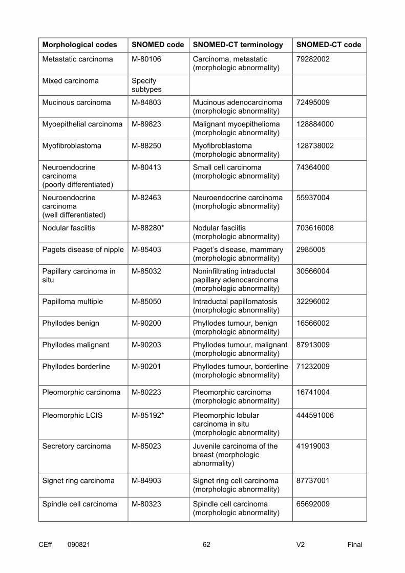
CEff 090821 62 V2 Final
Morphological codes SNOMED code SNOMED-CT terminology SNOMED-CT code
Metastatic carcinoma M-80106 Carcinoma, metastatic (morphologic abnormality)
79282002
Mixed carcinoma Specify subtypes
Mucinous carcinoma M-84803 Mucinous adenocarcinoma (morphologic abnormality)
72495009
Myoepithelial carcinoma M-89823 Malignant myoepithelioma (morphologic abnormality)
128884000
Myofibroblastoma M-88250 Myofibroblastoma (morphologic abnormality)
128738002
Neuroendocrine carcinoma (poorly differentiated)
M-80413 Small cell carcinoma (morphologic abnormality)
74364000
Neuroendocrine carcinoma (well differentiated)
M-82463 Neuroendocrine carcinoma (morphologic abnormality)
55937004
Nodular fasciitis M-88280* Nodular fasciitis (morphologic abnormality)
703616008
Pagets disease of nipple M-85403 Paget’s disease, mammary (morphologic abnormality)
2985005
Papillary carcinoma in situ
M-85032 Noninfiltrating intraductal papillary adenocarcinoma (morphologic abnormality)
30566004
Papilloma multiple M-85050 Intraductal papillomatosis (morphologic abnormality)
32296002
Phyllodes benign M-90200 Phyllodes tumour, benign (morphologic abnormality)
16566002
Phyllodes malignant M-90203 Phyllodes tumour, malignant (morphologic abnormality)
87913009
Phyllodes borderline M-90201 Phyllodes tumour, borderline (morphologic abnormality)
71232009
Pleomorphic carcinoma M-80223 Pleomorphic carcinoma (morphologic abnormality)
16741004
Pleomorphic LCIS M-85192* Pleomorphic lobular carcinoma in situ (morphologic abnormality)
444591006
Secretory carcinoma M-85023 Juvenile carcinoma of the breast (morphologic abnormality)
41919003
Signet ring carcinoma M-84903 Signet ring cell carcinoma (morphologic abnormality)
87737001
Spindle cell carcinoma M-80323 Spindle cell carcinoma (morphologic abnormality)
65692009

CEff 090821 63 V2 Final
Morphological codes SNOMED code SNOMED-CT terminology SNOMED-CT code
Squamous cell carcinoma
M-80703 Squamous cell carcinoma, no ICDO subtype (morphologic abnormality)
28899001
Syringomatous adenoma of nipple
M-84070 Syringoma (morphologic abnormality)
71244007
Tubular adenoma M-82110 Tubular adenoma, no ICDO subtype (morphologic abnormality)
19665009
Tubular carcinoma M-82113 Tubular adenocarcinoma (morphologic abnormality)
4631006
Undifferentiated carcinoma
M-80203 Carcinoma, undifferentiated (morphologic abnormality)
38549000
Other conditions
Term SNOMED code SNOMED-CT terminology SNOMED-CT code
Abscess M-41610 Abscess (morphologic abnormality)
44132006
Accessory/ ectopic breast
D4-48012 Accessory breast (disorder) 18166000
Apocrine metaplasia M-73310 Apocrine metaplasia (morphologic abnormality)
81274009
Atypical apocrine hyperplasia
M-73315 Atypical apocrine metaplasia (morphologic abnormality)
103673004
Atypical ductal hyperplasia
M-72175 Atypical intraductal hyperplasia (morphologic abnormality)
6660000
Atypical lobular hyperplasia
M-72105 Atypical lobular hyperplasia (morphologic abnormality)
33889003
Calcification M-55400 Calcified structure (morphologic abnormality)
54497001
Collagenous spherulosis
M-72171 Collagenous spherulosis (morphologic abnormality)
447298005
Columnar cell atypia M-67020 Columnar cell atypia (morphologic abnormality)
55465005
Columnar cell lesions M-74240 Blunt duct adenosis (morphologic abnormality)
58811002
Complex sclerosing lesion
M-78731 Radial scar (morphologic abnormality)
133855003
Cyst NOS M-33400 Cyst (morphologic abnormality)
12494005
Duct ectasia M-32100 Duct ectasia (morphologic abnormality)
110420004
Epithelial hyperplasia without atypia
M-72170 Intraductal hyperplasia (morphologic abnormality)
67617000

CEff 090821 64 V2 Final
Term SNOMED code SNOMED-CT terminology SNOMED-CT code
Excision margins tumour free
M-09400 Surgical margin uninvolved by tumour (finding)
55182004
Fat necrosis M-54110 Fat necrosis (morphologic abnormality)
79682009
Fibrocystic change M-74320 Fibrocystic disease (morphologic abnormality)
28092006
Fibromatosis M-76100 Angiomatosis (morphologic abnormality)
14350002
Fistula M-39300 Acquired fistula (morphologic abnormality)
51711001
Foreign body reaction M-44140 Foreign body giant cell granuloma (morphologic abnormality)
37058002
Galactocele M-33220 Galactocele associated with childbirth (disorder)
87840008
Gynaecomastia M-71000 Hypertrophy (morphologic abnormality)
56246009
Infarction M-54700 Infarct (morphologic abnormality)
55641003
Inflammation acute M-41000 Acute inflammation (morphologic abnormality)
4532008
Inflammation chronic M-43000 Chronic inflammation (morphologic abnormality)
84499006
Inflammation granulomatous
M-44000 Granulomatous inflammation (morphologic abnormality)
6266001
Involutional change M-79140 Menstrual involution of breast (morphologic abnormality)
33429008
Juvenile hypertrophy D7-90404 Pubertal breast hypertrophy (disorder)
198113009
Lactational change M-82040 Lactating adenoma (morphologic abnormality)
128651002
Metaplasia atypical M-73005 Atypical metaplasia (morphologic abnormality)
125544002
Metaplasia chondroid M-73600 Cartilaginous metaplasia (morphologic abnormality)
112671001
Metaplasia epithelial (clear cell, etc.)
M-73200 Epithelial metaplasia (morphologic abnormality)
54725001
Metaplasia osseous M-73400 Osseous metaplasia (morphologic abnormality)
38109001
Metaplasia squamous M-73220 Squamous metaplasia (morphologic abnormality)
83577005
Microglandular adenosis
M-72480 Microglandular hyperplasia (morphologic abnormality)
2953007

CEff 090821 65 V2 Final
Term SNOMED code SNOMED-CT terminology SNOMED-CT code
Microglandular hyperplasia
M-72450 Adenofibromyomatous hyperplasia (morphologic abnormality)
88000003
Morphological description only
M-09350 Morphologic description only (finding)
85728002
Mucocoele-like lesion M-33440 Mucous cyst (morphologic abnormality)
19633006
Normal: NOS M-00100 Normal tissue (finding) 30389008
PASH M-72430 Stromal hyperplasia (morphologic abnormality)
75235002
Plasma cell mastitis M-43060 Plasma cell inflammation (morphologic abnormality)
26246006
Pregnancy M-68080 Pregnancy pattern (morphologic abnormality)
68737009
Radial scar M-78731 Radial scar (morphologic abnormality)
133855003
Radiotherapy effect M-11600 Radiation injury (morphologic abnormality)
81018009
Sclerosing adenosis M-74220 Fibrosing adenosis (morphologic abnormality)
50916005
Surgical wound or cavity
M-14020 Surgical wound (morphologic abnormality)
112633009
Weddellite M-55400 Calcified structure (morphologic abnormality)
54497001

CEff 090821 66 V2 Final
Appendix E NHSBSP wide bore needle biopsy form Surname: ................................ Forenames: .............................. Date of birth: ................................... Screening no: ......................... Hospital no: .............................. NHS no: .......................................... Date performed: ...................... Location: .............................….. Operator: ................Centre: ........... Kv: .......................................... Total exposures: ....................... Total films: ...................................... Projection: ............................... Marker: ...................................... Localisation type: .......................... Side: Right □ Left □ Quadrant: Upper outer quadrant □ Lower outer quadrant □
Upper inner quadrant □ Lower inner quadrant □ Retroareolar □ Axilla □
Localisation type: Palpation □ Stereotactic □ Ultrasound □ Number of cores: ............. Specimen type: Core biopsy □ Vacuum-assisted excision biopsy □ Vacuum-assisted diagnostic biopsy □ Vacuum-assisted biopsy – not further specified □ Nipple/skin biopsy □ Calcification present on specimen X-ray? Yes □ No □ Radiograph not seen □ Comment: ............................................................................................................................................ ............................................................................................................................................................. Histological opinion: B1 Unsatisfactory/normal tissue only □
B2 Benign □ B3 Uncertain malignant potential with epithelial atypia □ B3 Uncertain malignant potential without epithelial atypia □ B4 Suspicious □ B5 Malignant type: (a) in situ □
(b) invasive □ (c) not assessable □ Histological calcification: Absent □ Benign □ Malignant □ Both □

CEff 090821 67 V2 Final
Optional further information Benign lesion Complex sclerosing lesion/radial scar □ Fibroadenoma □ Multiple papilloma □ Periductal mastitis/duct ectasia □ Fibrocystic change □ Solitary papilloma □ Sclerosing adenosis □ Solitary cyst □ Columnar cell change □ Other (please specify): ……………… Epithelial proliferation: Not present □ Present without atypia □ Present with atypia (ductal) □ Present with atypia (lobular) □ Malignant lesion In situ carcinoma: Not present □ Ductal □ Lobular □ Pagets □ DCIS grade: High □ Intermediate □ Low □ Not assessable □ Invasive carcinoma: Present □ Not present □ Size invasive tumour: ..........mm (largest dimension, if available) Type: No special type (ductal NST) □
Pure special type (90% purity specify components present below) □: Mixed tumour type (50–90% special type component, specify components present below) □: Other malignant tumour (please specify): ......................................
Specify type component(s) present for pure special type and mixed tumour types: Tubular/cribriform □ Lobular □ Mucinous □ Medullary/atypical medullary □ Ductal/no special type □ Other (please specify) ............................
Invasive grade: 1 □ 2 □ 3 □ Not assessable □ Oestrogen receptor status: Positive (≥1%) □ Negative (<1%) □ Percentage positive tumour cells ……. On-slide positive control material: Present □ Absent □ Progesterone receptor status: Positive (≥1%) □ Negative (<1%) □ Percentage positive tumour cells ……. On-slide positive control material: Present □ Absent □ HER2 immunohistochemical score: 0 negative □ 1+ negative □ 2+Borderline □ 3+ Positive □ FISH/CISH ratio: ........... Status: Amplified □ Non-amplified □ Borderline □ Not performed □ HER2 copy no: ……… Chromosome 17 no: …. Final HER2 status†: Positive □ Negative □ Not performed □

CEff 090821 68 V2 Final
Appendix F Reporting proforma for breast core biopsy Surname: ……………………………….. Forenames: …………………. Date of birth: …………….… Sex: ….………………………………….. Hospital: …………….……….. Hospital no: ...………….….. NHS no: ………………………….……... Date of surgery: …………….. Date of report: ………....….. Authorisation: …………………….…….. Report no: …………………… Date of receipt: …….………. Pathologist: …………….………………. Surgeon: ……………………………………….…………..… Side†: Left □ Right □ Quadrant†: Upper outer quadrant □ Lower outer quadrant □ Upper inner quadrant □ Lower inner quadrant □
Retroareolar □ Number of cores if known: ............. Specimen type†: Needle core biopsy □ Vacuum-assisted excision biopsy □ Vacuum-assisted diagnostic biopsy □ Vacuum-assisted biopsy – not further specified □ Calcification present on specimen X-ray? Yes □ No □ Radiograph not seen □ Comment: ............................................................................................................................................ Histological opinion†: B1 (Normal) □
B2 (Benign) □ B3 (Uncertain malignant potential with epithelial atypia) □ B3 (Uncertain malignant potential without epithelial atypia) □ B4 (Suspicious) □ B5a (Malignant in situ) □ B5b (Malignant invasive) □ B5c (Malignant not assessable) □
If biopsy taken for assessment of calcification: Histological calcification: Not identified □ Benign □ Malignant □ Both benign and malignant □ In situ carcinoma†: Not identified □ Ductal □ Lobular□ DCIS grade†: High □ Intermediate □ Low □ Cannot be assessed □ Invasive carcinoma† Not identified □ Present □ Type†: No special type (ductal NST) □
Pure special type (90% purity; specify components present below) □ Mixed tumour type (50–90% special type component; specify components present below) □ Other malignant tumour (please specify): ......................................
Specify type component(s) present for pure special type and mixed tumour types†:
Tubular/cribriform □ Lobular □ Mucinous □ Medullary/atypical medullary □ Ductal/no special type □ Other □ (please specify): ............................
Invasive carcinoma grade†: 1 □ 2 □ 3 □ Cannot be assessed □

CEff 090821 69 V2 Final
Oestrogen receptor status†: Positive (≥ 1%) □ Negative (<1%) □ Percentage positive tumour cells =…………….. On-slide positive control material: Present □ Absent □ Progesterone receptor status†: Positive (≥ 1%) □ Negative (<1%) □ Percentage positive tumour cells =…………….. On-slide positive control material: Present □ Absent □ HER2 IHC score†: 0 negative □ 1+ negative □ 2+ Borderline □ 3+ Positive □ Not performed □ FISH/CISH ratio: ........... Status†: Amplified □ Non-amplified □ Borderline □ Not performed □ HER2 copy no.: ……… Chromosome 17 no.: ……….. Final HER2 status†: Positive □ Negative □ SNOMED† codes: T …….… M …..…… Date reported: ............................................. Pathologist: ............................................
†Data items that are currently part of the Cancer Outcomes and Services Dataset (COSD) version 8.
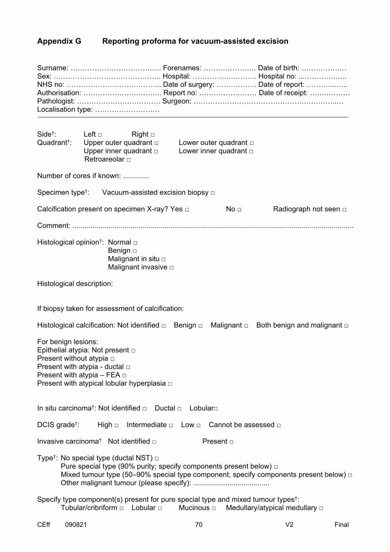
CEff 090821 70 V2 Final
Appendix G Reporting proforma for vacuum-assisted excision Surname: ……………………………….. Forenames: …………………. Date of birth: …………….… Sex: ….………………………………….. Hospital: …………….……….. Hospital no: ...………….….. NHS no: ………………………….……... Date of surgery: …………….. Date of report: ………....….. Authorisation: …………………….…….. Report no: …………………… Date of receipt: …….………. Pathologist: …………….………………. Surgeon: ……………………………………….…………..… Localisation type: ……………………… Side†: Left □ Right □ Quadrant†: Upper outer quadrant □ Lower outer quadrant □ Upper inner quadrant □ Lower inner quadrant □
Retroareolar □ Number of cores if known: ............. Specimen type†: Vacuum-assisted excision biopsy □ Calcification present on specimen X-ray? Yes □ No □ Radiograph not seen □ Comment: ............................................................................................................................................ Histological opinion†: Normal □
Benign □ Malignant in situ □ Malignant invasive □
Histological description: If biopsy taken for assessment of calcification: Histological calcification: Not identified □ Benign □ Malignant □ Both benign and malignant □ For benign lesions: Epithelial atypia: Not present □ Present without atypia □ Present with atypia - ductal □ Present with atypia – FEA □ Present with atypical lobular hyperplasia □ In situ carcinoma†: Not identified □ Ductal □ Lobular□ DCIS grade†: High □ Intermediate □ Low □ Cannot be assessed □ Invasive carcinoma† Not identified □ Present □ Type†: No special type (ductal NST) □
Pure special type (90% purity; specify components present below) □ Mixed tumour type (50–90% special type component; specify components present below) □ Other malignant tumour (please specify): ......................................
Specify type component(s) present for pure special type and mixed tumour types†:
Tubular/cribriform □ Lobular □ Mucinous □ Medullary/atypical medullary □

CEff 090821 71 V2 Final
Ductal/no special type □ Other □ (please specify): ............................ Invasive carcinoma grade†: 1 □ 2 □ 3 □ Cannot be assessed □ Oestrogen receptor status†: Positive (≥ 1%) □ Negative (<1%) □ Percentage positive tumour cells =…………….. On-slide positive control material: Present □ Absent □ Progesterone receptor status†: Positive (≥ 1%) □ Negative (<1%) □ Percentage positive tumour cells =…………….. On-slide positive control material: Present □ Absent □ HER2 IHC score†: 0 negative □ 1+ negative □ 2+ Borderline □ 3+ Positive □ Not performed □ FISH/CISH ratio: ........... Status†: Amplified □ Non-amplified □ Borderline □ Not performed □ HER2 copy no.: ……… Chromosome 17 no.: ……….. Final HER2 status†: Positive □ Negative □ SNOMED† codes: T …….… M …..…… Date reported: ............................................. Pathologist: ............................................
†Data items that are currently part of the Cancer Outcomes and Services Dataset (COSD) version 8.

CEff 090821 72 V2 Final
Appendix H Reporting proforma for breast FNAC Surname: ……………………………… Forenames: …………………….. Date of birth: …………….… Sex: ….………………………………… Hospital: …………….…………... Hospital no: ...………….….. NHS no: ……………………………….. Date of surgery: ………………… Date of report: ……....….. Authorisation: …………………….…… Report no: ………………………. Date of receipt:…….………. Pathologist: …………………………… Surgeon: ……………………………………….…………..… Side†: Left □ Right □ Location†: Upper outer quadrant □ Lower outer quadrant □ Upper inner quadrant □ Lower inner quadrant □
Retroareolar □ Cytological opinion†: C1 □ (Inadequate/unsatisfactory)
C2 □ (Benign) C3 □ (Uncertain) C4 □ (Suspicious) C5 □ (Malignant)
Comment: ............................................................................................................................................ ............................................................................................................................................................. SNOMED† codes: T …….… M …..…… Date reported: ............................................. Pathologist: ............................................
†Data items that are currently part of the Cancer Outcomes and Services Dataset (COSD) version 8.
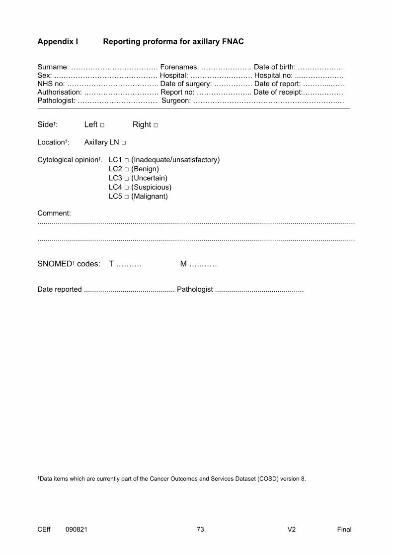
CEff 090821 73 V2 Final
Appendix I Reporting proforma for axillary FNAC Surname: ……………………………… Forenames: ………………… Date of birth: …………….… Sex: ….………………………………… Hospital: …………….………. Hospital no: ...………….….. NHS no: ……………………………….. Date of surgery: ……………. Date of report: ………....….. Authorisation: …………………………. Report no: ………………….. Date of receipt:…….………. Pathologist: …………………………… Surgeon: ……………………………………….…………..… Side†: Left □ Right □ Location†: Axillary LN □ Cytological opinion†: LC1 □ (Inadequate/unsatisfactory)
LC2 □ (Benign) LC3 □ (Uncertain) LC4 □ (Suspicious) LC5 □ (Malignant)
Comment: ............................................................................................................................................................. ............................................................................................................................................................. SNOMED† codes: T …….… M …..…… Date reported ............................................. Pathologist ............................................
†Data items which are currently part of the Cancer Outcomes and Services Dataset (COSD) version 8.

CEff 090821 74 V2 Final
Appendix J Reporting proforma for axillary core biopsy Surname: ………………………………… Forenames: …………………. Date of birth: …………….… Sex: ….…………………………………… Hospital: …………….……….. Hospital no: ...………….….. NHS no: ………………………………….. Date of surgery: …………….. Date of report: ………....….. Authorisation: …………………….……… Report no: …………………….Date of receipt: …….………. Pathologist: ………………………………. Surgeon: ……………………………………….…………..….. Side†: Left □ Right □ Location†: Axillary LN □ Opinion† LB1 □ (Inadequate/Unsatisfactory)
LB2 □ (Normal/Benign) LB3 □ (Uncertain) LB4 □ (Suspicious) LB5 □ (Malignant)
Comment.............................................................................................................................................. ............................................................................................................................................................. SNOMED† codes: T …….… M …..…… Date reported: ............................................. Pathologist: ............................................
†Data items that are currently part of the Cancer Outcomes and Services Dataset (COSD) version 8.

CEff 191120 75 V2 Draft
Appendix K Reporting proforma for breast core biopsy in list format Element name
Values Implementation notes
COSD v8 mapping
COSD v9 mapping
Side Single selection value list: • Left • Right
CR0820 Left = [L] Left Right = [R] Right Not selected = [9] Not known
pCR0820 Left = [L] Left Right = [R] Right Not selected = [9] Not known
Quadrant Single selection value list: • Upper outer
quadrant • Lower outer
quadrant • Upper inner
quadrant • Lower inner
quadrant • Retroareolar
Number of cores
Integer
Specimen type Single selection value list: • Needle core biopsy • Vacuum-assisted
excision biopsy • Vacuum-assisted
diagnostic biopsy • Vacuum-assisted
biopsy – not further specified
CR0760 All values = [BU] Biopsy NOS
pCR0760 All values = [BU] Biopsy NOS
Calcification present on specimen X-ray
Single selection value list: • Yes • No • Radiograph not
seen

CEff 090821 76 V2 Final
Histological opinion
Single selection value list: • B1 (Normal) • B2 (Benign) • B3 (Uncertain
malignant potential with epithelial atypia)
• B3 (Uncertain malignant potential without epithelial atypia)
• B4 (Suspicious) • B5a (Malignant in
situ) • B5b (Malignant
invasive) • B5c (Malignant not
assessable)
BR4260 B1 = [B1] Normal B2 = [B2] Benign B3 (Uncertain malignant potential with epithelial atypia) = [B3] Uncertain malignant potential B3 (Uncertain malignant potential without epithelial atypia) = [B3] Uncertain malignant potential B4 = [B4] Suspicious B5a = [B5a] Malignant in-situ B5b = [B5b] Malignant invasive B5c = [B5c] Malignant not assessable
pBR4260 B1 = [B1] Unsatisficatory/normal tissue only B2 = [B2] Benign B3 (Uncertain malignant potential with epithelial atypia) = [B3b] Uncertain malignant potential with epithelial atypia B3 (Uncertain malignant potential without epithelial atypia) = [B3a] Uncertain malignant potential without epithelial atypia B4 = [B4] Suspicious B5a = [B5a] Malignant in-situ B5b = [B5b] Malignant invasive B5c = [B5c] Malignant not assessable
Histological calcification
Single selection value list: • Not identified • Benign • Malignant • Both benign and
malignant • Not applicable
In situ carcinoma
Multiple select value list: • Not identified • Ductal • Lobular

CEff 090821 77 V2 Final
DCIS grade Single selection value list: • High • Intermediate • Low • Cannot be
assessed • Not applicable
Not applicable if ‘In situ carcinoma’ is ‘Not identified’ or ‘Lobular’ only.
BR4160 High = [H] High Intermediate = [I] Intermediate Low = [L] Low Cannot be assessed = [X] Not assessable Not applicable = Leave COSD value blank
pBR4160 High = [H] High Intermediate = [I] Intermediate Low = [L] Low Cannot be assessed = [X] Not assessable Not applicable = Leave COSD value blank
Invasive carcinoma
Single selection value list: • Not identified • Present
Type Single selection value list: • No special type
(ductal NST) • Pure special type
(90% purity; specify components present below)
• Mixed tumour type (50–90% special type component; specify components present below)
• Other malignant tumour
Type, Other – specify
Free text Only required if ‘Type, Other’ – Malignant tumour’ is selected.
Specify type component(s) present for pure special type and mixed tumour types
Multiple select value list: • Tubular/cribriform • Lobular • Mucinous • Medullary/atypical
medullary • Ductal/no special
type • Other

CEff 090821 78 V2 Final
Specify type component(s) present for pure special type and mixed tumour types, Other – specify
Free text Only required if ‘Specify type component(s) present for pure special type and mixed tumour types, Other’ is selected.
Invasive carcinoma grade
Single selection value list: • 1 • 2 • 3 • Cannot be
assessed
CR0860 1 = [G1] Well differentiated 2 = [G2] Moderately differentiated 3 = [G3] Poorly differentiated Cannot be assessed =[GX] Grade of differentiation is not appropriate or cannot be assessed
pCR0860 1 = [G1] Well differentiated 2 = [G2] Moderately differentiated 3 = [G3] Poorly differentiated Cannot be assessed =[GX] Grade of differentiation is not appropriate or cannot be assessed
Oestrogen receptor status
Single selection value list: • Positive • Negative • Not performed
BR4220 Positive = [P] Positive Negative = [N] Negative Not performed = [X] Not performed
pBR4220 Positive = [P] Positive (> or = 1%) Negative = [N] Negative <1% Not performed = [X] Not performed
Oestrogen receptor status, percentage positive tumour cells
Integer, range 0–100
On-slide positive control material
Single selection value list: • Present • Absent

CEff 090821 79 V2 Final
HER2 IHC score
Single selection value list: • 0 • 1+ • 2+ • 3+ • Not performed
BR4280 0 = [N] Negative 1 = [N] Negative 2 = [B] Borderline 3 = [P] Positive Not performed = [X] Not performed
pBR4280 0 = [N1] Negative (0) 1+ = [N2] Negative (1+) 2+ = [B] Borderline (2+) 3+ = [P] Positive (3+) Not performed = [X] Not performed
FISH/CISH ratio
Number
FISH/CISH Status
Single selection value list: • Amplified • Non-amplified • Borderline • Not performed
BR4310 Amplified = [P] Positive Non-amplified = [N] Negative Borderline = Leave COSD value blank Not performed = Leave COSD value blank
pBR4310 Amplified = [P] Positive (Amplified) Non-amplified = [N] Negative (Non-amplified) Borderline = [B] Borderline Not performed = [X] Not performed
HER2 copy no Number
Chromosome 17 no
Number
Final HER2 status
Single selection value list: • Positive • Negative • Not performed
Comment Free text
SNOMED Topography code
May have multiple codes. Look up from SNOMED tables
CR6410 pCR6410
SNOMED Morphology code
May have multiple codes. Look up from SNOMED tables
CR6420 pCR6420

CEff 090821 80 V2 Final
Appendix L Reporting proforma for breast FNAC in list format Element name
Values Implementation notes
COSD v8 mapping
COSD v9 mapping
Side Single selection value list: • Left • Right
CR0820 Left = [L] Left Right = [R] Right Not selected = [9] Not known
pCR0820 Left = [L] Left Right = [R] Right Not selected = [9] Not known
Quadrant Single selection value list: • Upper outer quadrant • Lower outer quadrant • Upper inner quadrant • Lower inner quadrant • Retroareolar
Cytological opinion
Single selection value list: • C1
(Inadequate/unsatisfactory) • C2 (Benign) • C3 (Uncertain) • C4 (Suspicious) • C5 (Malignant)
BR4240 C1 = [C1] Inadequate/unsatisfactory specimen C2 = [C2] Benign C3 = [C3] Uncertain C4 = [C4] Suspicious of malignancy C5 = [C5] Malignant
pBR4240 C1 = [C1] Inadequate/unsatisfactory specimen C2 = [C2] Benign C3 = [C3] Uncertain C4 = [C4] Suspicious of malignancy C5 = [C5] Malignant
Comment Free text
SNOMED Topography code
May have multiple codes. Look up from SNOMED tables
CR6410 pCR6410
SNOMED Morphology code
May have multiple codes. Look up from SNOMED tables
CR6420 pCR6420

CEff 090821 81 V2 Final
Appendix M Reporting proforma for axillary FNAC in list format Element name
Values Implementation notes
COSD v8 mapping
COSD v9 mapping
Side Single selection value list: • Left • Right
CR0820 Left = [L] Left Right = [R] Right Not selected = [9] Not known
pCR0820 Left = [L] Left Right = [R] Right Not selected = [9] Not known
Location Axillary LN
Cytological opinion
Single selection value list: • LC1 (Inadequate/
unsatisfactory) • LC2 (Benign) • LC3 (Uncertain) • LC4 (Suspicious) • LC5 (Malignant)
BR4250 LC1 = [C1] Inadequate/ unsatisfactory specimen LC2 = [C2] Benign LC3 = [C3] Uncertain LC4 = [C4] Suspicious of malignancy LC5 = [C5] Malignant
pBR4250 LC1 = [C1] Inadequate/ unsatisfactory specimen LC2 = [C2] Benign LC3 = [C3] Uncertain LC4 = [C4] Suspicious of malignancy LC5 = [C5] Malignant
Comment Free text
SNOMED Topography code
May have multiple codes. Look up from SNOMED tables
CR6410 pCR6410
SNOMED Morphology code
May have multiple codes. Look up from SNOMED tables
CR6420 pCR6420

CEff 090821 82 V2 Final
Appendix N Reporting proforma for axillary core biopsy in list format Element name
Values Implementation notes
COSD v8 mapping
COSD v9 mapping
Side Single selection value list: • Left • Right
CR0820 Left = [L] Left Right = [R] Right Not selected = [9] Not known
pCR0820 Left = [L] Left Right = [R] Right Not selected = [9] Not known
Location Axillary LN
Opinion Single selection value list: • LB1 (Inadequate/
Unsatisfactory) • LB2 (Normal/Benign) • LB3 (Uncertain) • LB4 (Suspicious) • LB5 (Malignant)
BR4270 LB1 = Not mappable LB2 = Not mappable LB3 = [B3] Uncertain malignant potential LB4 = [B4] Suspicious LB5 = [B5] Malignant
pBR4270 LB1 = [LB1] Inadequate/ unsatisfactory LB2 = [LB2] Normal/benign LB3 = [LB3] Uncertain LB4 = [LB4] Suspicious LB5 = [LB5] Malignant
Comment Free text
SNOMED Topography code
May have multiple codes. Look up from SNOMED tables
CR6410 pCR6410
SNOMED Morphology code
May have multiple codes. Look up from SNOMED tables
CR6420 pCR6420

CEff 090821 83 V2 Final
Appendix O Summary table – explanation of levels of evidence (Adapted from Palmer K et al. BMJ 2008;337:1832.)
Grade (level) of evidence
Nature of evidence
Grade A
At least one high-quality meta-analysis, systematic review of randomised controlled trials or a randomised controlled trial with a very low risk of bias and directly attributable to the target cancer type
or
A body of evidence demonstrating consistency of results and comprising mainly well-conducted meta-analyses, systematic reviews of randomised controlled trials or randomised controlled trials with a low risk of bias, directly applicable to the target cancer type.
Grade B
A body of evidence demonstrating consistency of results and comprising mainly high-quality systematic reviews of case-control or cohort studies and high-quality case-control or cohort studies with a very low risk of confounding or bias, and a high probability that the relation is causal and which are directly applicable to the target cancer type
or
Extrapolation evidence from studies described in A.
Grade C
A body of evidence demonstrating consistency of results and including well-conducted case-control or cohort studies and high- quality case-control or cohort studies with a low risk of confounding or bias and a moderate probability that the relation is causal and which are directly applicable to the target cancer type
or
Extrapolation evidence from studies described in B.
Grade D
Non-analytic studies such as case reports, case series or expert opinion
or
Extrapolation evidence from studies described in C.
Good practice point (GPP)
Recommended best practice based on the clinical experience of the authors of the writing group.

CEff 090821 84 V2 Final
Appendix P AGREE II compliance monitoring sheet The cancer datasets of the Royal College of Pathologists comply with the AGREE II standards for good quality clinical guidelines (www.agreetrust.org). The sections of this dataset that indicate compliance with each of the AGREE II standards are indicated in the table. AGREE standard Section of guideline Scope and purpose 1 The overall objective(s) of the guideline is (are) specifically described Introduction 2 The health question(s) covered by the guideline is (are) specifically described Introduction 3 The population (patients, public, etc.) to whom the guideline is meant to apply
is specifically described Foreword
Stakeholder involvement 4 The guideline development group includes individuals from all the relevant
professional groups Foreword
5 The views and preferences of the target population (patients, public, etc.) have been sought
N/A
6 The target users of the guideline are clearly defined Introduction Rigour of development 7 Systematic methods were used to search for evidence Foreword 8 The criteria for selecting the evidence are clearly described Foreword 9 The strengths and limitations of the body of evidence are clearly described Foreword 10 The methods for formulating the recommendations are clearly described Foreword 11 The health benefits, side effects and risks have been considered in
formulating the recommendations Foreword and Introduction
12 There is an explicit link between the recommendations and the supporting evidence
All sections
13 The guideline has been externally reviewed by experts prior to its publication Foreword 14 A procedure for updating the guideline is provided Foreword Clarity of presentation 15 The recommendations are specific and unambiguous All sections 16 The different options for management of the condition or health issue are
clearly presented All sections
17 Key recommendations are easily identifiable 3, 5, 7 Applicability 18 The guideline describes facilitators and barriers to its application Foreword 19 The guideline provides advice and/or tools on how the recommendations can
be put into practice Appendices
20 The potential resource implications of applying the recommendations have been considered
Foreword
21 The guideline presents monitoring and/or auditing criteria 7 Editorial independence 22 The views of the funding body have not influenced the content of the
guideline Foreword
23 Competing interest of guideline development group members have been recorded and addressed
Foreword