guidelines for oral and maxillofacial surgery
TRANSCRIPT

GUIDELINES FOR
ORAL AND MAXILLOFACIAL SURGERY
Version 1
النسخة الإلكترونية هي النسخة المضبوطة وفق إجراء ضبط الوثائق۔ النسخ الورقية غير
مضبوطة وتقع على مسؤولية حاملها۔
Electronic copy is controlled under document control procedure.
Hard copy is uncontrolled & under responsibility of beholder.
يسمح بالوصول وبالاحتفاظ بهذه الوثيقة مع مصدرها أو مع المسؤول عن تطبيقها أو مع
المطبق عليهم۔
It is allowed ONLY to access and keep this document with who
issued, who is responsible and to whom it is applicable.
:تصنيف امن المعلومات بيانات مفتوحة شارك–
سري
حساس–مشارك سري–مشارك
Information security code: Open Shared -
Confidential
Shared-Sensitive Shared-Secret
Health Policies and Standards Department
Health Regulation Sector (2021)

Guidelines for Oral and Maxillofacial Surgery Page 2 of 159 Ref. No. HRS/HPSD/OMFS/V1/2021
INTRODUCTION
Dubai Health Authority (DHA) is the responsible entity for regulating, licensing and monitoring health
facilities and healthcare professionals in the Emirate of Dubai. The Health Regulation Sector (HRS) is
an integral part of DHA and was founded to fulfil the following overarching strategic objectives and
program:
Objective #1: Position Dubai as a global medical destination by introducing a value-based,
comprehensive, integrated and high quality service delivery system.
Objective #2: Direct resources to ensure healthy and safe environment for Dubai population.
Strategic Program #5: Oral & Dental Care- This program focuses on improving the oral health
outcomes and ensure that all individuals have access to high quality treatments and effective
prevention programs for dental care.
ACKNOWLEDGMENT
This document was developed by Dental Services Department, Primary Healthcare Services Sector
(PHCSS). It has further been reviewed by the Health Policy and Standards Department (HPSD).
HRS would like to acknowledge and thank all parties that participated and worked toward developing
these guidelines to ensure improving the quality and safety of healthcare services.
The Health Regulation Sector
Dubai Health Authority
Guidelines for Oral and Maxillofacial Surgery Page 3 of 159 Ref. No. HRS/HPSD/OMFS/V1/2021
TABLE OF CONTENTS
INTRODUCTION................................................................................................................................ 2
ACKNOWLEDGMENT ........................................................................................................................ 2
EXECUTIVE SUMMARY ..................................................................................................................... 9
DEFINITIONS .................................................................................................................................. 11
ABBREVIATIONS ............................................................................................................................ 12
A. GUIDELINES FOR THE MANAGEMENT OF WISDOM TEETH AND IMPACTED CANINES ............... 14
1. BACKGROUND ........................................................................................................................ 15
2. SCOPE .................................................................................................................................... 16
3. PURPOSE ................................................................................................................................ 16
4. APPLICABILITY ....................................................................................................................... 16
5. RECOMMENDATION ONE: CLASSIFICATION OF WISDOM TEETH ............................................... 16
6. RECOMMENDATION TWO: CLASSIFICATION OF IMPACTED CANINES ........................................ 18
7. RECOMMENDATION THREE: CLINICAL MANAGEMENT OF WISDOM/IMPACTED CANINE TEETH . 19
8. RECOMMENDATION FOUR: CLINICAL STEPS FOR WISDOM/IMPACTED CANINE TEETH
EXTRACTION .................................................................................................................................. 21
9. RECOMMENDATION FIVE: SUMMARY OF THE MANAGEMENT OF EXTRACTION OF IMPACTED
TEETH ............................................................................................................................................ 24
10. RECOMMENDATION SIX: COMPLICATIONS OF TEETH EXTRACTION ...................................... 25
11. RECOMMENDATION SEVEN: SPECIAL ENDORSEMENTS ........................................................ 26
B. GUIDELINES FOR THE MANAGEMENT OF PATIENTS ON ANTICOAGULANTS/ ANTIPLATELETS
UNDERGOING DENTAL/ORAL SURGICAL PROCEDURES .................................................................. 29
1. BACKGROUND ........................................................................................................................ 30
2. SCOPE .................................................................................................................................... 30
3. PURPOSE ................................................................................................................................ 30
4. APPLICABILITY ....................................................................................................................... 30

Guidelines for Oral and Maxillofacial Surgery Page 4 of 159 Ref. No. HRS/HPSD/OMFS/V1/2021
5. RECOMMENDATION ONE: PATIENT DIAGNOSIS ....................................................................... 31
6. RECOMMENDATION TWO: CLINICAL MANAGEMENT PATIENTS WHO ARE AT RISK OF BLEEDING
FROM AN ORAL SURGICAL PROCEDURE ........................................................................................... 31
7. RECOMMENDATION THREE: SPECIAL ENDORSEMENT .............................................................. 32
C. GUIDELINES FOR ANTIBIOTIC PROPHYLAXIS FOR PATIENTS UNDERGOING DENTAL/ORAL
MAXILLOFACIAL PROCEDURES....................................................................................................... 33
1. BACKGROUND ........................................................................................................................ 34
2. SCOPE .................................................................................................................................... 34
3. PURPOSE ................................................................................................................................ 34
4. APPLICABILITY ....................................................................................................................... 34
5. RECOMMENDATION ONE: CLASSIFICATION OF INDICATIONS FOR ANTIBIOTIC PROPHYLAXIS.... 34
6. RECOMMENDATION TWO: CLINICAL MANAGEMENT ................................................................ 40
7. RECOMMENDATION THREE: SPECIAL ENDORSEMENTS ............................................................ 41
D. GUIDELINES FOR THE MANAGEMENT OF PREGNANT PATIENTS UNDERGOING DENTAL/ORAL
SURGICAL PROCEDURES................................................................................................................. 42
1. BACKGROUND ........................................................................................................................ 43
2. SCOPE .................................................................................................................................... 43
3. PURPOSE ................................................................................................................................ 43
4. APPLICABILITY ....................................................................................................................... 43
5. RECOMMENDATION ONE: CLINICAL MANAGEMENT OF PREGNANT PATIENTS .......................... 43
6. RECOMMENDATION TWO: SPECIAL CONSIDERATION ............................................................... 45
E. GUIDELINES FOR THE DIAGNOSIS AND MANAGEMENT OF ORAL SOFT TISSUE LESIONS ......... 46
1. BACKGROUND ........................................................................................................................ 47
2. SCOPE .................................................................................................................................... 47
3. PURPOSE ................................................................................................................................ 47
4. APPLICABILITY ....................................................................................................................... 48

Guidelines for Oral and Maxillofacial Surgery Page 5 of 159 Ref. No. HRS/HPSD/OMFS/V1/2021
5. RECOMMENDATION ONE: DIAGNOSIS AND INVESTIGATION ..................................................... 48
6. RECOMMENDATION TWO: CLASSIFICATION OF ORAL SOFT TISSUE LESIONS ............................ 49
7. RECOMMENDATION THREE: CLINICAL MANAGEMENT OF ORAL SOFT TISSUE LESIONS ............. 49
8. RECOMMENDATION FOUR: SPECIAL CONSIDERATIONS ............................................................ 49
F. GUIDELINES FOR THE MANAGEMENT OF ODONTOGENIC/CERVICOFACIAL INFECTIONS ......... 52
1. BACKGROUND ........................................................................................................................ 53
2. SCOPE .................................................................................................................................... 53
3. PURPOSE ................................................................................................................................ 53
4. APPLICABILITY ....................................................................................................................... 53
5. RECOMMENDATION ONE: DIAGNOSIS AND DIFFERENTIAL DIAGNOSIS ..................................... 54
6. RECOMMENDATION TWO: CLASSIFICATION OF ODONTOGENIC\CERVICOFACIAL INFECTIONS: .. 54
7. RECOMMENDATION THREE: SPECIAL CONSIDERATIONS .......................................................... 57
G. GUIDELINES FOR ORAL MANAGEMENT OF PATIENTS AT RISK OF, OR HAVING MEDICATION
RELATED OSTEONECROSIS OF THE JAW (MRONJ).......................................................................... 60
1. BACKGROUND ........................................................................................................................ 61
2. SCOPE .................................................................................................................................... 62
3. PURPOSE ................................................................................................................................ 62
4. APPLICABILITY ....................................................................................................................... 63
5. RECOMMENDATION ONE: ASSESSMENT OF RISK FACTORS ...................................................... 63
6. RECOMMENDATION TWO: DIAGNOSIS AND MANAGEMENT ..................................................... 66
7. RECOMMENDATION THREE: MANAGEMENT STRATEGIES FOR PATIENTS AT RISK OF MRONJ .... 67
8. RECOMMENDATION FOUR: TREATMENT OF PATIENTS WITH EXTABLISHED MRONJ ................. 71
9. RECOMMENDATION FIVE: SPECIAL CONSIDERATIONS.............................................................. 72
H. GUIDELINES FOR THE MANAGEMENT OF TEMPRO-MANDIBULAR JOINT DISORDERS IN
PRIMARY CARE............................................................................................................................... 74
1. BACKGROUND ........................................................................................................................ 75

Guidelines for Oral and Maxillofacial Surgery Page 6 of 159 Ref. No. HRS/HPSD/OMFS/V1/2021
2. SCOPE .................................................................................................................................... 76
3. PURPOSE ................................................................................................................................ 76
4. APPLICABILITY ....................................................................................................................... 76
5. RECOMMENDATION ONE: PATIENT ASSESSMENT, DIAGNOSIS AND INVESTIGATION................. 77
6. RECOMMENDATION TWO: TREATMENT OF TEMPOROMANDIBULAR DISORDERS (TMD)............ 78
7. RECOMMENDATION THREE: SPECIAL CONSIDERATIONS .......................................................... 79
I. GUIDELINES FOR THE MANAGEMENT OF SKELETAL MALOCCLUSION ..................................... 81
1. BACKGROUND ........................................................................................................................ 82
2. SCOPE .................................................................................................................................... 82
3. PURPOSE ................................................................................................................................ 82
4. APPLICABILITY ....................................................................................................................... 82
5. RECOMMENDATION ONE: PATIENT ASSESSMENT, DIAGNOSIS AND INVESTIGATION................. 83
6. RECOMMENDATION TWO: CLINICAL MANAGEMENT ................................................................ 84
7. RECOMMENDATION THREE: SPECIAL CONSIDERATIONS .......................................................... 86
J. GUIDELINES FOR THE MANAGEMENT OF FACIAL FRACTURES ................................................. 87
1. BACKGROUND ........................................................................................................................ 88
2. SCOPE .................................................................................................................................... 88
3. PURPOSE ................................................................................................................................ 88
4. APPLICABILITY ....................................................................................................................... 88
5. RECOMMENDATION ONE: INITIAL MANAGEMENT .................................................................... 88
6. RECOMMENDATION TWO: DIAGNOSTIC MODALITIES............................................................... 89
7. RECOMMENDATION THREE: CLINICAL MANAGEMENT.............................................................. 90
8. RECOMMENDATION FOUR: SPECIAL CONSIDERATIONS ............................................................ 91
KEY PERFORMANCE INDICATORS (KPIs) ......................................................................................... 94
REFERENCES .................................................................................................................................. 99

Guidelines for Oral and Maxillofacial Surgery Page 7 of 159 Ref. No. HRS/HPSD/OMFS/V1/2021
APPENDICES:................................................................................................................................ 122
APPENDIX 1: MAIN MEDICAL CONDITIONS ASSOCIATED WITH INCREASED RISK OF BLEEDING ........ 122
APPENDIX 2: MAIN DRUG GROUPS ASSOCIATED WITH HIGH RISK OF BLEEDING ............................. 123
APPENDIX 3: MAJOR DRUG INTERACTIONS BETWEEN ANTICOAGULANTS/ANTIPLATELETS AND MOST
COMMONLY PRESCRIBED MEDICATIONS IN THE DENTAL FIELD ...................................................... 124
APPENDIX 4: DENTAL PROCEDURES THAT ARE LIKELY TO CAUSE BLEEDING .................................. 126
APPENDIX 5: CLINICAL STEPS FOR PATIENTS WHO ARE AT RISK OF BLEEDING FROM AN ORAL
SURGICAL PROCEDURE ................................................................................................................. 127
APPENDIX 6: ANTIBIOTIC PROPHYLAXIS FOR ORAL/DENTAL PROCEDURE ...................................... 128
APPENDIX 7: AAOS/ADA RECOMMENDATIONS FOR PREVENTION OF ORTHOPAEDIC IMPLANT
INFECTION.................................................................................................................................... 129
APPENDIX 8: PHYSIOLOGICAL CHANGES IN PREGNANCIES AND THEIR MANAGEMENT .................... 130
APPENDIX 9: LIST OF DRUGS DURING PREGNANCY ........................................................................ 131
APPENDIX 10: TERATOGENS AND THEIR EFFECTS ON THE FETUS .................................................. 135
APPENDIX 11: FDA CATEGORIZATION FOR RISK FACTORS FOR METHEMOGLOBINEMIA IN PREGNANT
WOMEN ....................................................................................................................................... 136
APPENDIX 12: CLASSIFICATION OF ORAL MUCOSAL LESIONS ......................................................... 137
APPENDIX 13: MANAGEMENT OF MUCOSAL DISEASES................................................................... 138
APPENDIX 14: MANAGEMENTS OF CYSTS ...................................................................................... 139
APPENDIX 15: MANAGEMENT OF VASCULAR LESIONS ................................................................... 140
APPENDIX 16: MANAGEMENTS OF MALIGNANT TUMORS OF SOFT TISSUE ..................................... 141
APPENDIX 17: SALIVARY GLANDS BENIGN AND MALIGNANT TUMOR ............................................. 142
APPENDIX 18: MANAGEMENT OF SIMPLE & LIFE-THREATENING INFECTIONS ................................. 143
APPENDIX 19: MEDICATIONS ASSOCIATED WITH MRONJ ............................................................... 144
APPENDIX 20: GENERAL DENTIST GUIDANCE FOR PATIENTS ON OR PLANNED FOR
ANTIRESORPTIVE/ANTIANGIOGENIC THERAPY .............................................................................. 146
Guidelines for Oral and Maxillofacial Surgery Page 8 of 159 Ref. No. HRS/HPSD/OMFS/V1/2021
APPENDIX 21: TMD ASSESSMENT AND DIAGNOSIS ........................................................................ 147
APPENDIX 22: TREATMENT OF TMD .............................................................................................. 148
APPENDIX 23: REFERAL PATHWAY ................................................................................................ 149
APPENDIX 24: TREATMENT OPTIONS FOR SKELETAL DEFORMITIES................................................ 150
APPENDIX 25: MANAGEMENT OF COMBINED ORTHODONTICS AND ORTHOGNATHIC SURGERY ...... 151
APPENDIX 26: CLASSIFICATION OF FACIAL FRACTURES.................................................................. 154
APPENDIX 27: METHODS OF TREATMENT FOR UPPER THIRD FACIAL FRACTURES ........................... 155
APPENDIX 28: METHODS OF TREATMENT FOR MIDDLE THIRD FACIAL FRACTURES ......................... 156
APPENDIX 29: METHODS OF TREATMENT FOR MANDIBULAR FRACTURES ...................................... 158
APPENDIX 30: ALGORITHM FOR SECURING THE AIRWAY OF A PATIENT WITH MAXILLOFACIAL
TRAUMA....................................................................................................................................... 159

Guidelines for Oral and Maxillofacial Surgery Page 9 of 159 Ref. No. HRS/HPSD/OMFS/V1/2021
EXECUTIVE SUMMARY
Clinical guidelines to enhance the standard of care in health facilities are increasingly becoming part of
current practice and will become more common over the next decade. These Clinical Guidelines aim to
improve the quality and the level of healthcare provided to the clients. Healthcare providers can use
these guidelines to answer specific questions in day-to-day practice and as an information source for
continuing professional education.
This document presents a framework for Oral and Maxillofacial Surgeons to:
Provide guidance for the proper management for the extraction of wisdom teeth and impacted
canines.
Manage patients on anticoagulants/antiplatelet undergoing dental/oral surgical procedure, in
order to achieve effective results.
Provide an overview on the topic of antibiotic prophylaxis to make informed decisions on
prophylactic antibiotic use in the prevention of local & systemic infections.
Provide an overview on the management of pregnant patients undergoing dental/oral &
maxillofacial treatment.
Diagnose and manage oral soft tissue lesions.
Introduce decision-making criteria regarding diagnosis, management and treatment plan of
Odontogenic/Cervicofacial infections and to reduce inappropriate variation in practice.
Provide an overview on the topic of oral & dental management of patients at risk of or having
Medication-Related Osteonecrosis of the Jaw (MRONJ).

Guidelines for Oral and Maxillofacial Surgery Page 10 of 159 Ref. No. HRS/HPSD/OMFS/V1/2021
Elaborate on the in diagnosis and primary care of patient with Tempro-Mandibular Joint (TMJ)
Disorders and also specify a referral pathway to aid in the referring process of such patients.
Standardize management of skeletal malocclusions and maximize patient outcome both
functionally and aesthetically.
Manage oral and maxillofacial fractures to reach optimal outcomes.

Guidelines for Oral and Maxillofacial Surgery Page 11 of 159 Ref. No. HRS/HPSD/OMFS/V1/2021
DEFINITIONS
Temporomandibular disorders (TMD): are disorders that involve the temporomandibular joint, which
can involve the joint itself, surrounding muscles and supporting structures.
Temporomandibular Joint (TMJ): is also known as the jaw joint. It is the joint that connects the lower
jawbone to the skull. Two joints are present at both sides of the face in front of the ears. Muscles and
ligaments are connected to the joint and the joint is surrounded by a protective capsule. A cartilaginous
disc is located between the bones and slides forward and backward with jaw movement.

Guidelines for Oral and Maxillofacial Surgery Page 12 of 159 Ref. No. HRS/HPSD/OMFS/V1/2021
ABBREVIATIONS
AAPD : American Academy of Pediatric Dentistry
ADA : American Dental Association
AHA : American Heart Association
ARD : Acute Respiratory Distress
ASA : American Society of Anesthesiologists
BMI : Body Mass Index
CAD : Coronary Artery Disease
CBCT : Cone Beam Computed Tomography
CHD : Congenital Heart Disease
CT : Computerized Tomography
CVA : Cerebrovascular Accident
DHA : Dubai Health Authority
DHIC : Dubai Health Insurance Corporation
DIC : Disseminated Intravascular Coagulation
DM : Diabetes Mellitus
EBP : Essential Benefit Plan
ESRD : End‐Stage Renal Disease
FDA : Food and Drug Administration
HIV : Human Immunodeficiency Virus
HPSD : Health Policies and Standards Department

Guidelines for Oral and Maxillofacial Surgery Page 13 of 159 Ref. No. HRS/HPSD/OMFS/V1/2021
HRS : Health Regulation Sector
HTN : Hypertension
IE : Infective Endocarditis
IM : Intramuscular
IV : Intra vascular
MI : Myocardial Infarction
MRI : Magnetic Resonance Imaging
OPG : Dental Orthopantomogram
PCA : Post Conceptual Age
PET : Positron Emission Tomography
PHCSS : Primary Healthcare Services Sector
PJI : Prosthetic Joint Infections
SPECT : Single Photon Emission Computed Tomography
TIA : Transient Ischemic Attack
TMD : Temporomandibular disorders
TMJ : Temporomandibular Joint
UAE : United Arab Emirates

Guidelines for Oral and Maxillofacial Surgery Page 14 of 159 Ref. No. HRS/HPSD/OMFS/V1/2021
A. GUIDELINES FOR THE MANAGEMENT OF WISDOM TEETH AND IMPACTED
CANINES

Guidelines for Oral and Maxillofacial Surgery Page 15 of 159 Ref. No. HRS/HPSD/OMFS/V1/2021
1. BACKGROUND
Third molars (wisdom teeth) are the most posterior and the last teeth to erupt in the oral cavity.
They are four in number, two in each arch. They erupt between the ages of 18-24 years. Either a
wisdom tooth fails to erupt into the dental arch or it erupts fully and functions within the arch.
Most common impacted teeth are maxillary and mandibular third molar, maxillary canine,
mandibular second premolar and supernumerary teeth.
Impacted and fully erupted teeth may cause problems for some people like swelling and pain, or
they might cause no symptoms at all. Extraction of symptomatic and asymptomatic teeth is a
common surgical procedure that needs proper investigation and management.
1.1. Etiology of teeth impaction:
1.1.1. Local factors:
a. Arch length discrepancy
b. Premature loss of primary teeth
c. Presence of a supernumerary tooth
d. Ectopic position of a tooth germ
e. Local pathology e.g. odontome
f. Thickened overlying osseous or mucosal tissue
g. Obstacles to eruption e.g. ankylosed primary molar
h. Cleft palate
i. Biomechanical impediments secondary to childhood maxillofacial surgery or
dento-alveolar trauma.
Guidelines for Oral and Maxillofacial Surgery Page 16 of 159 Ref. No. HRS/HPSD/OMFS/V1/2021
1.1.2. Systemic factors:
a. Cleidocranial dysostosis
b. Other hereditary or syndromic conditions.
2. SCOPE
2.1. Provide guidance to Oral Maxillofacial Surgeons regarding the proper management for the
extraction of wisdom teeth and impacted teeth.
3. PURPOSE
3.1. To guide for proper decision-making for diagnosing & managing extraction of wisdom
teeth and common impacted teeth.
3.2. To reduce the risk of post extraction complications.
3.3. To improve patient care.
4. APPLICABILITY
4.1. DHA licensed Oral and Maxillofacial Surgeons.
5. RECOMMENDATION ONE: CLASSIFICATION OF WISDOM TEETH
5.1. Wisdom teeth can be classifies as follows:
5.1.1. Totally erupted Third Molars
5.1.2. Partially erupted Third Molars (Soft tissue or bony impacted)
5.1.3. Impacted Third Molars
5.2. Impacted third molars are classified as per Winter’s classification as follows:
5.2.1. Mesioangular
5.2.2. Distoangular

Guidelines for Oral and Maxillofacial Surgery Page 17 of 159 Ref. No. HRS/HPSD/OMFS/V1/2021
5.2.3. Horizontal
5.2.4. Vertical
5.2.5. Buccal/Lingual Obliquity
5.2.6. Transverse
5.3. Impacted third molars are classified as per Pell and Gregory radiographic classification as
follows:
5.3.1. Classification with respect to mandibular ramus:
a. Class I: the crown is located anterior to the anterior border of the
ramus.
b. Class II: ½ of the crown covered by the anterior border of the ramus.
c. Class III: the crown is fully covered by the anterior border of the
ramus.
5.3.2. Classification for the occlusal plane:
a. Class A: at the same level as, or a little below than that of the second
molar.
b. Class B: at the middle of the crown of the second molar or at the
same level as the cervical line.

Guidelines for Oral and Maxillofacial Surgery Page 18 of 159 Ref. No. HRS/HPSD/OMFS/V1/2021
c. Class C: apical to the cervical line of the second molar.
6. RECOMMENDATION TWO: CLASSIFICATION OF IMPACTED CANINES
6.1. Classification of impacted maxillary canine:
Classification of impacted maxillary canines
CLASS 1 Impacted cuspids located in palate.
CLASS 2 Impacted cuspids located in labial or buccal surface of
maxilla.
CLASS 3 Impacted cuspids located in palatine and maxillary bone, the
crown is on the palate and root passes through the root of
the adjacent teeth and ends in the labial or buccal surface.
CLASS 4 Impacted cuspids located in alveolar process, usually
vertically between incisor and first bicuspids.
CLASS 5 Impacted cuspids located in edentulous maxilla.

Guidelines for Oral and Maxillofacial Surgery Page 19 of 159 Ref. No. HRS/HPSD/OMFS/V1/2021
7. RECOMMENDATION THREE: CLINICAL MANAGEMENT OF WISDOM/IMPACTED CANINE
TEETH
7.1. Clinical assessment
7.1.1. The general agreement indicates that a wisdom tooth should be removed if
pathology or symptoms are present or the chance for their future appearance
exists.
7.1.2. Decision of extraction will be undertaken according to the following indications:
a. Indications for wisdom teeth extraction:
I. Non-restorable caries
II. Non-treatable pulpal and/or periapical pathology
III. Cellulitis
IV. Abscess and osteomyelitis
V. Internal/external resorption of the tooth or adjacent teeth
VI. Fracture of tooth
VII. Disease of follicle including cyst/tumour
VIII. Tooth/teeth impeding surgery or reconstructive jaw surgery and when a
tooth is involved in or within the field of tumour resection.
b. Indications for impacted teeth extraction:
I. Arch space- tooth size discrepancy
II. Dental caries of the impacted or adjacent teeth
III. Widening of periodontal ligament space of the adjacent tooth

Guidelines for Oral and Maxillofacial Surgery Page 20 of 159 Ref. No. HRS/HPSD/OMFS/V1/2021
IV. Root resorption of the adjacent tooth
V. Disease of follicle including cyst/tumour.
7.2. Radiographic assessment
7.2.1. It is essential that proper radiographs be taken for the tooth to be removed. In
general, Periapical Radiographs (PA) provide the most accurate & detailed
information concerning the tooth, its roots and the surrounding tissue.
7.2.2. Panoramic radiographs (OPG) are used frequently, but their greatest usefulness
is for impacted teeth as opposed to erupted teeth & its relationship of associated
vital structures.
7.2.3. The various Imaging modalities are as follows:
a. Intra oral periapical radiograph with parallelism & using SLOB rule (SLOB:
Same Lingual Opposite Buccal)
b. Orthopantomogram (OPG)
c. Cone beam computed tomography (CBCT) in case of deeply impacted teeth,
presence of pathology, or to check proximity to the inferior alveolar nerve
(IAN).
7.3. Laboratory testing (in case required).
7.4. Treatment options for impacted teeth.
7.4.1. According to the clinical & radiological findings, treatment option could be as
follows:
a. Observation and follow up

Guidelines for Oral and Maxillofacial Surgery Page 21 of 159 Ref. No. HRS/HPSD/OMFS/V1/2021
b. Simple or Surgical extraction
c. Intervention by orthodontic means or extraction of a neighbouring
permanent
d. Surgical exposure and orthodontic alignment
e. Coronectomy or Partial Odontectomy.
8. RECOMMENDATION FOUR: CLINICAL STEPS FOR WISDOM/IMPACTED CANINE TEETH
EXTRACTION
8.1. Fundamental requirements for a successful extraction:
8.1.1. Adequate access & visualization of the field of surgery.
8.1.2. An unimpeded pathway for the removal of the tooth.
8.1.3. The use of controlled force to luxate & remove the tooth.
8.2. Clinical steps for wisdom teeth extraction:
8.2.1. There are two techniques for wisdom tooth extraction: Simple (closed) or
Surgical (open).
8.2.2. Simple tooth extraction (closed/uncomplicated) is the most frequently used
technique. The different steps involved in a Simple tooth extraction are as
follows:
a. Anesthesia
b. Tooth removal
c. Socket debridement (only if necessary)
d. Control haemorrhage

Guidelines for Oral and Maxillofacial Surgery Page 22 of 159 Ref. No. HRS/HPSD/OMFS/V1/2021
e. Wound closure
f. Post-operative instructions.
8.2.3. Surgical tooth extraction (open/complicated) is used whenever excessive force
is necessary to remove the tooth, a substantial amount of the crown is missing,
or when the tooth is impacted and the access is difficult. The different steps
involved in a Surgical tooth extraction are as follows:
a. Anesthesia
b. Incision and mucoperiosteal flap
c. Removal of bone
d. Tooth removal
e. Socket debridement (only if necessary) and bone smoothening
f. Control haemorrhage
g. Wound closure
h. Post-operative instructions.
8.3. Clinical steps for surgical exposure of impacted canines:
8.3.1. Factors to be considered before the surgical management of impacted canines
are as follows:
a. Labial-palatal position of impacted canine.
b. Impaction position relative to the Mucogingiva Junction (MGJ) in an apical-
coronal dimension.

Guidelines for Oral and Maxillofacial Surgery Page 23 of 159 Ref. No. HRS/HPSD/OMFS/V1/2021
c. Evaluation of the amount of keratinized gingiva (KG) mainly with facial
impactions.
d. The mesial-distal position of the canine relative to the lateral incisor.
8.3.2. There are two techniques for surgical exposure of impacted canines: Open
Surgical technique and Closed Surgical Technique.
8.3.3. The different steps involved in an Open Surgical technique are as follows:
a. Surgical uncovering of the canine with a mucoperiosteal flap.
b. Dissection of the bone (bone covering the canine is being removed).
c. An attachment with a chain is bonded to the tooth.
d. The palatal flap is repositioned and sutured back with the chain above the
mucosa.
e. Shortly after the surgery, orthodontic force is applied via the chain.
f. The canine is orthodontically moved beneath the palatal mucosa by forced
eruption.
8.3.4. The different steps involved in an Closed Surgical technique are as follows:
a. Surgical uncovering of the canine, removing a window of tissue around it and
placing pack to cover the exposed area.
b. The treatment approaches vary depending on whether the attachment with
a chain is bonded to the exposed tooth at surgery or if spontaneous eruption
of the palatally impacted canine is expected post surgically.

Guidelines for Oral and Maxillofacial Surgery Page 24 of 159 Ref. No. HRS/HPSD/OMFS/V1/2021
c. Orthodontic force is applied via the chain and the canine is orthodontically
moved above the mucosa.
9. RECOMMENDATION FIVE: SUMMARY OF THE MANAGEMENT OF EXTRACTION OF
IMPACTED TEETH
9.1. Pre-Operative
9.1.1. Medical History.
9.1.2. Medication history.
9.1.3. Radiographic examination of the tooth to be removed.
9.1.4. Assessment of extensiveness of the procedure.
9.1.5. Informed consent (all risks & possible complications have to be clearly explained
to the patient).
9.2. Operative
9.2.1. Monitor premonitory signs (sweating, dyschromia, finger dysesthesia).
9.2.2. Proper Anesthesia selection.
9.2.3. Proper flap design (if needed).
9.2.4. Bone removal (if needed).
9.2.5. Tooth removal.
9.2.6. Socket debridement/curettage only if necessary. In case of periapical lesion is
visible on the preoperative radiograph & there was no granuloma attached to
the tooth after removal. Or if ant debris is obvious (tooth fragment, calculus, or
filling material).
Guidelines for Oral and Maxillofacial Surgery Page 25 of 159 Ref. No. HRS/HPSD/OMFS/V1/2021
9.2.7. Finger compression of the expanded buccolingual plates of bone.
9.2.8. Smoothening of sharp edges (if needed).
9.2.9. Hemostasis through applying pressure by gauze, suturing or local/systemic
hemostatic agent.
9.3. Post-Operative Instructions
9.3.1. Take painkiller/antibiotic (if prescribed).
9.3.2. Bite firmly on gauze for 30–45 min.
9.3.3. In case bleeding continues, place another gauze for an extra hour.
9.3.4. Stay on cold soft diet for twenty-four (24) hours.
9.3.5. Avoid mouth rinsing for the first twenty-four (24) hours. After twenty-four (24)
hours, rinse with warm salt water, three (3) times a day for 3–4 days.
9.3.6. Avoid brushing and flossing in the first twenty-four (24) hours in the area of
surgery.
9.3.7. Follow-up in case nonresorbable sutures placed for wound closure.
10. RECOMMENDATION SIX: COMPLICATIONS OF TEETH EXTRACTION
10.1. Common complications during the surgery:
10.1.1. Mesioangular and horizontal positions of third molars are responsible for
development of distal cervical caries on the second molar.
10.1.2. Impacted tooth/root displacement: close proximity of the root apex to the
anatomical structures such as the maxillary sinus, and the mandibular canal.
a. Complications associated with impacted or adjacent tooth

Guidelines for Oral and Maxillofacial Surgery Page 26 of 159 Ref. No. HRS/HPSD/OMFS/V1/2021
b. Soft tissue complications
c. Nerve injuries (inferior alveolar nerve or lingual nerve)
d. Bone complications.
10.2. Possible Complications after the extraction:
10.2.1. Pain/discomfort
10.2.2. Swelling
10.2.3. Bruising
10.2.4. Hemorrhage/bleeding
10.2.5. Bad breath
10.2.6. Dry socket
10.2.7. Infection/inflammation
10.2.8. Trismus/limited mouth opening
10.2.9. Alter in sensation, paresthesia, or numbness of lip and tongue
10.2.10. Damage to adjacent teeth/restoration
10.2.11. TMJ pain can last for a few weeks to months
10.2.12. Oroantral communication (Sometimes, Upper wisdom teeth are located close to
maxillary sinus, if sinus membrane is perforated, closure of the oroantral
communication is required to correct the defect).
11. RECOMMENDATION SEVEN: SPECIAL ENDORSEMENTS
11.1. Informed consent and time out must be done before starting the extraction procedure.

Guidelines for Oral and Maxillofacial Surgery Page 27 of 159 Ref. No. HRS/HPSD/OMFS/V1/2021
11.2. Explain all risks & possible complications, including temporary or permanent damage to
the inferior alveolar nerve as; paraesthesia or numbness & to lingual nerve leading to
numbness of the tongue.
11.3. Unnecessary or vigorous curettage of the socket wall merely produces additional injury &
may delay healing.
11.4. In case tooth was removed due to periodontal disease, special attention should be given to
removing the granulation tissue by curetting the socket to avoid excessive bleeding.
11.5. Surgical removal of impacted teeth should be limited to patients with evidence of
pathology.
11.6. Prophylactic removal of pathology free wisdom teeth should be avoided as:
11.6.1. There is no reliable research to suggest that this practice benefits patients.
11.6.2. Patients who do have healthy wisdom teeth removed are being exposed to the
risks of surgery.
11.7. Symptoms free wisdom teeth to be monitored on a regular check-up.
11.8. Symptomatic impacted teeth have to be removed regardless of the difficulties of the
surgical management.
11.9. First episode of Pericoronitis, unless particularly severe, should not be considered as an
indication for surgery. Second or subsequent episodes should be considered the
appropriate indication for extraction.
11.10. Younger healthy patient might have less postoperative complications when compared with
older patient.

Guidelines for Oral and Maxillofacial Surgery Page 28 of 159 Ref. No. HRS/HPSD/OMFS/V1/2021
11.11. Palpation of impacted canine at age of 9 years old, because canine impaction can be
associated with functional or aesthetic impairment.
11.12. Surgery for removal of impacted teeth may be associated with several postoperative
complications; the surgeon should be prepared to manage them when they occur.

Guidelines for Oral and Maxillofacial Surgery Page 29 of 159 Ref. No. HRS/HPSD/OMFS/V1/2021
B. GUIDELINES FOR THE MANAGEMENT OF PATIENTS ON ANTICOAGULANTS/
ANTIPLATELETS UNDERGOING DENTAL/ORAL SURGICAL PROCEDURES

Guidelines for Oral and Maxillofacial Surgery Page 30 of 159 Ref. No. HRS/HPSD/OMFS/V1/2021
1. BACKGROUND
Anticoagulants and other known blood thinners have been used to combat thromboembolisms
and blood clots. These medications are considered crucial to high-risk individuals, especially those
who have had previous thromboembolic events. In the dental oral surgery field, there is an
exposure to a range of individuals with varying severities of medical conditions. This requires the
dental practitioner to master the art of medical history taking, which aids in identifying the
patients who are on anticoagulant medications and therefore are at a great risk of haemorrhage
and other complications after dental surgery.
2. SCOPE
2.1. To provide guidance that aids in the effective dental management of
anticoagulant/antiplatelet consuming patients which will positively reduce the demands of
secondary referrals from primary healthcare.
3. PURPOSE
3.1. To identify patients who are on anticoagulant/antiplatelet therapy.
3.2. To identify patients on anticoagulant/antiplatelet therapy who require dental
management in a hospital setting.
3.3. To implement proper management modalities thereby reducing the incidence of excessive
bleeding.
4. APPLICABILITY
4.1. DHA licensed Oral and Maxillofacial Surgeons.

Guidelines for Oral and Maxillofacial Surgery Page 31 of 159 Ref. No. HRS/HPSD/OMFS/V1/2021
5. RECOMMENDATION ONE: PATIENT DIAGNOSIS
5.1. Along with a thorough medical history review, the following factors greatly impact a
patient’s bleeding risk following an oral surgical procedure:
5.1.1. Main medical conditions associated with increased risk of bleeding Appendix 1.
5.1.2. Main drug groups associated with high risk of bleeding Appendix 2.
5.1.3. Major drug interactions between Anticoagulants/Antiplatelets and most
commonly prescribed medications in the dental field Appendix 3.
5.1.4. Dental procedures that are likely to cause bleeding Appendix 4.
6. RECOMMENDATION TWO: CLINICAL MANAGEMENT PATIENTS WHO ARE AT RISK OF
BLEEDING FROM AN ORAL SURGICAL PROCEDURE
6.1. Low bleeding risk procedures:
6.1.1. Maintain standard protocol in achieving hemostasis (By applying pressure to the
bleeding point using gauze for 30 minutes).
6.2. High bleeding risk procedures:
6.2.1. Consult with the patient’s general or specialist medical practitioner if needed.
6.2.2. Depending on the urgency of therapy, delay the elective procedure.
6.2.3. Obtain effective hemostatic measure.
6.2.4. Perform atraumatic surgical procedures.
6.2.5. Discharge with a concise, effective post-operative instructions with a verbalized
understanding by the patient.
Note: For a flowchart Refer to Appendix 5.

Guidelines for Oral and Maxillofacial Surgery Page 32 of 159 Ref. No. HRS/HPSD/OMFS/V1/2021
7. RECOMMENDATION THREE: SPECIAL ENDORSEMENT
7.1. Patients on long-term oral anticoagulants with significant medical problems such as; liver
disease, renal disease, thrombocytopenia or who are taking anti-platelet drugs pose an
increased risk of bleeding. Those patients should be managed in a hospital setting under
the care of the oral and maxillofacial department and the individuals’ respective medical
physicians or specialists.

Guidelines for Oral and Maxillofacial Surgery Page 33 of 159 Ref. No. HRS/HPSD/OMFS/V1/2021
C. GUIDELINES FOR ANTIBIOTIC PROPHYLAXIS FOR PATIENTS UNDERGOING
DENTAL/ORAL MAXILLOFACIAL PROCEDURES

Guidelines for Oral and Maxillofacial Surgery Page 34 of 159 Ref. No. HRS/HPSD/OMFS/V1/2021
1. BACKGROUND
Antibiotic prophylaxis still represents a common but often misused procedure in dental practice,
thus aggravating the risk for antimicrobial resistance and adverse effects occurrence.
2. SCOPE
2.1. To assist General Dental Practitioners and Oral & Maxillofacial Surgeons make informed
decisions on prophylactic antibiotic use in the prevention of local & systemic infections.
3. PURPOSE
3.1. To identify the procedures in oral surgery that would benefit from surgical antibiotic
prophylaxis.
3.2. To assist in deciding which antibiotics to prescribe and what regimen to follow if
prophylactic antibiotics are indicated.
4. APPLICABILITY
4.1. DHA licensed Oral and Maxillofacial Surgeons.
4.2. DHA licensed General Dental Practitioners
5. RECOMMENDATION ONE: CLASSIFICATION OF INDICATIONS FOR ANTIBIOTIC
PROPHYLAXIS
5.1. Indications for antibiotic prophylaxis are classified as follows:
1. Type of surgical procedure Severity & extensiveness

Guidelines for Oral and Maxillofacial Surgery Page 35 of 159 Ref. No. HRS/HPSD/OMFS/V1/2021
2. Wound class According to the risk of
contamination/infection
3. Patients underlying medical problems
according to ASA
ASA: American Society of
Anesthesiologists physical status
classifications
4. Patients underlying medical problems
according to AHA
AHA: American Heart Association
5. Patients underlying medical problems
According to ADA
ADA: American Dental Association
6. Patients underlying medical problems
according to AAPD
AAPD: American Academy of Pediatric
Dentistry
5.2. Type of surgical procedure:
5.2.1. Antibiotic prophylaxis is not indicated for:
a. Clean dental surgery in healthy patients
b. Lower third molar surgery
5.2.2. Antibiotic prophylaxis is not recommended for:
a. Routine periodontal surgery
5.2.3. But indicated for:
a. Minor surgery with a high degree of difficulty in which the duration of the
surgery is predicted to be long
b. Surgery to place dental implants
c. Minor oral surgical procedures in which a bone graft is inserted
d. Major clean contaminated maxillofacial surgery, such as orthognathic
surgery and surgery for large benign cysts and tumors

Guidelines for Oral and Maxillofacial Surgery Page 36 of 159 Ref. No. HRS/HPSD/OMFS/V1/2021
e. All forms of head and neck cancer surgery
f. Open reduction and internal fixation of facial bone fractures
5.3. Wound classification:
5.3.1. Antibiotic prophylaxis should not be used if the infection rate of a specific
procedure is ≤5%,
Wound Classification Estimated Infection Rate
1. Clean: No disruption of the mucosa such as the oral cavity. <1–5%.
2. Clean contaminated: Disruption of the mucosa such as the
oral cavity or surgery in an inflamed area.
3–11%.
3. Contaminated: Oncological surgery in which both oral
cavity and neck contact.
10-17%.
4. Dirty and infected wound >27%.
5.4. Patients underlying medical problems according to ASA:
5.4.1. Patients with ASA scores of 1 and 2 have lower infection rates than patients
with ASA scores of 3 or more.
5.4.2. Antibiotic prophylaxis is indicated for all surgical procedures carried out on
medically compromised patients. Patients could be oncological patients, patients
with congenital or immunological immune-depression, patients with immune-
depression due to medication, patients with infectious immune-depression
(AIDS), patients with metabolic disorders (diabetes) and patients with renal and
hepatic insufficiency while for those with ASA less than three (3) depends on
the physician’s discretion.
ASA PS Definition Examples, including, but not limited to:

Guidelines for Oral and Maxillofacial Surgery Page 37 of 159 Ref. No. HRS/HPSD/OMFS/V1/2021
Classification
ASA I A normal healthy
patient
Healthy, non-smoking, no or minimal alcohol use
ASA II A patient with mild
systemic disease
Mild diseases only without substantive functional
limitations
Examples include (but not limited to):
Current smoker, social alcohol drinker, pregnancy,
obesity (30 < BM < 40), well controlled DM/HTN,
mild lung disease
ASA III A patient with severe
systemic disease
Substantive functional limitations; One or more
moderate to severe diseases
Examples include (but not limited to):
Poorly controlled DM or HTN, COPD, morbid
obesity (BMI ≥40), active hepatitis, alcohol
dependence or abuse, implanted pacemaker,
moderate reduction of ejection fraction, ESRD
undergoing regularly scheduled dialysis, premature
infant PCA < 60 weeks, history (>3 months) of MI,
CVA, TIA, or CAD/stents.
ASA IV A patient with severe
systemic disease that is
a
constant threat to life
Examples include (but not limited to):
recent (< 3 months) MI, CVA, TIA, or CAD/stents,
ongoing cardiac ischemia or severe valve
dysfunction, severe reduction of ejection fraction,
sepsis, DIC, ARD or ESRD not undergoing regularly
scheduled dialysis
ASA V A moribund patient
who is not expected to
survive without the
operation
Examples include (but not limited to):
Ruptured abdominal/thoracic aneurysm, massive
trauma, intracranial bleed with mass effect,
ischemic bowel in the face of significant cardiac
pathology or multiple organ/ system dysfunction
5.5. Patients with underlying medical condition according to AHA:

Guidelines for Oral and Maxillofacial Surgery Page 38 of 159 Ref. No. HRS/HPSD/OMFS/V1/2021
5.5.1. The AHA recommends antibiotic prophylaxis only for those whose underlying
cardiac conditions are associated with the highest risk of adverse outcome. Such
conditions include:
a. Prosthetic cardiac valve or prosthetic material used for cardiac valve repair
b. A previous history of IE
c. Congenital heart disease (CHD):
I. Unrepaired cyanotic CHD including palliative shunts and conduits.
II. Completely repaired congenital heart defect with prosthetic material or
device, whether placed by surgery or by catheter intervention during the
first six months after the procedure
III. Repaired CHD with residual defects at the site or adjacent to the site of
a prosthetic patch or prosthetic device (which inhibits
endothelialisation)
d. Cardiac transplantation recipients who develop cardiac valvopathy.
5.5.2. Antibiotics are recommended for all dental procedures that involve:
a. Manipulation of gingival tissue/periapical region of teeth
b. Perforation of the oral mucosa for cardiac patients with the highest risk of
adverse outcome.
5.6. Patients with underlying medical condition according to ADA:
5.6.1. According to ADA, antibiotic prophylaxis is given to patients with medical
conditions and cardiac conditions and no need for the ones with prosthetic joints.

Guidelines for Oral and Maxillofacial Surgery Page 39 of 159 Ref. No. HRS/HPSD/OMFS/V1/2021
5.6.2. The updated evidence-based clinical guidelines published by ADA in 2015
concluded that there were no statistically significant association between dental
procedures and Prosthetic joint infection (PJI).
5.6.3. Key recommendations of the guideline:
a. In general, for patients with prosthetic joint implants prophylactic
antibiotics are not recommended prior to dental procedures to
prevent PJI
b. The practitioner and patient should consider possible clinical
circumstances that may suggest the presence of a significant
medical risk in providing dental care without antibiotic prophylaxis
as well as the risks of widespread antibiotic use. This should be
integrated with the practitioner’s professional judgment and the
patient’s needs and preferences.
c. In cases where antibiotics are deemed necessary, it is most
appropriate that the orthopaedic surgeon recommend the
appropriate antibiotic regimen and when reasonable write the
prescription.
5.7. Patients with underlying medical problems according to AADP:
5.7.1. Consultation with the patient’s physician is recommended for management of
patients with compromised immunity
5.7.2. Antibiotic prophylaxis is required in the following conditions:

Guidelines for Oral and Maxillofacial Surgery Page 40 of 159 Ref. No. HRS/HPSD/OMFS/V1/2021
a. Risk for distant site infection from a dental procedure
b. Immunosuppression secondary to:
I. Human Immunodeficiency Virus (HIV)
II. Severe combined immunodeficiency
III. Neutropenia
IV. Cancer chemotherapy; and
V. Hematopoietic stem cell or solid organ transplantation
c. Head and neck radiotherapy
d. Autoimmune disease
e. Sickle cell anaemia, chronic steroid usage, diabetes
f. Bisphosphonate therapy or other antiresorptive therapy; according to the
decision of the attending surgeon during patient’s assessment & treatment
planning.
6. RECOMMENDATION TWO: CLINICAL MANAGEMENT
6.1. The patients need to undergo the following investigations:
6.1.1. Dental X-rays:
a. Periapical
b. Dental Orthopantomogram (OPG)
6.1.2. Cone beam computed tomography CBCT
6.1.3. CT with contrast
6.1.4. Lab tests according to the patient’s health and medical condition

Guidelines for Oral and Maxillofacial Surgery Page 41 of 159 Ref. No. HRS/HPSD/OMFS/V1/2021
6.1.5. Medical reports.
6.2. For the antibiotic prophylaxis regimen for oral/dental procedures Appendix 6.
7. RECOMMENDATION THREE: SPECIAL ENDORSEMENTS
7.1. According to ADA, dental procedures are not associated with PJI and antibiotics provided
before care do not prevent PJI.
7.2. Potential harms of antibiotics include risk for anaphylaxis, antibiotic resistance, and
opportunistic infections.
7.3. Benefits of use may not exceed the harms for most patients.
7.4. Individual preferences and circumstances should be considered when deciding to use
antibiotic prophylaxis prior to dental procedures.
7.5. For prevention of orthopaedic implant infection refer to Appendix 7.
Guidelines for Oral and Maxillofacial Surgery Page 42 of 159 Ref. No. HRS/HPSD/OMFS/V1/2021
D. GUIDELINES FOR THE MANAGEMENT OF PREGNANT PATIENTS
UNDERGOING DENTAL/ORAL SURGICAL PROCEDURES

Guidelines for Oral and Maxillofacial Surgery Page 43 of 159 Ref. No. HRS/HPSD/OMFS/V1/2021
1. BACKGROUND
Pregnancy is a normal and healthy condition. Pregnant patients are not medically compromised
and hence they should not be denied dental treatment. Pregnancy is accompanied by a variety of
physiologic, anatomic and hormonal changes that can affect the plan of treatment.
2. SCOPE
2.1. To increase awareness of General Dental Practitioners and Oral & Maxillofacial Surgeons
in the management of the pregnant patients undergoing oral & maxillofacial treatment.
3. PURPOSE
3.1. To designate treatment to maximize the benefit to the mother while minimizing the risk to
the fetus.
3.2. To update General Dental Practitioners and Oral & Maxillofacial Surgeons in the
management of the pregnant patient.
3.3. To understand the physiological changes to ensure provision of better quality care for
pregnant women.
4. APPLICABILITY
4.1. DHA licensed Oral and Maxillofacial Surgeons.
4.2. DHA licensed General Dental Practitioners.
5. RECOMMENDATION ONE: CLINICAL MANAGEMENT OF PREGNANT PATIENTS
5.1. Pregnant patients may have some physiological changes (Appendix 8) and/or may be
taking some drugs, which may influence the clinical management of these patients.

Guidelines for Oral and Maxillofacial Surgery Page 44 of 159 Ref. No. HRS/HPSD/OMFS/V1/2021
5.2. Drugs in Pregnancy
5.2.1. Drugs used or prescribed in pregnant patient should aim to avoid any adverse
drug reactions. Certain drugs are known to cause miscarriage, teratogenicity and
low birth rate because most drugs cross the placenta by simple diffusion.
5.2.2. Medications prescribed to pregnant patients often require modification in
dosage, duration of the prescription and the frequency in which they are taken.
Drugs that can be prescribed during pregnancy are elaborated in Appendix 9.
5.2.3. Teratogenic drugs should be avoided in pregnant patients. These drugs could
cause either structural or functional birth defects. Effects of Teratogenic drugs
are elaborated in Appendix 10.
5.2.4. Certain drugs are responsible for developing acquired methemoglobinemia in
patients, which include oxidative drugs, glucose-6-phosphate dehydrogenase
and local anesthetics. Food and Drug Administration (FDA) has categorised
these drugs Appendix 11.
5.3. Ideal Patient position
5.3.1. The ideal position of the pregnant patient is positioning her left lateral lying
position with the right buttock and hip elevated by 15°.
5.4. Radiographs during pregnancy
5.4.1. While taking radiographs of pregnant patients, the dental staff must practice
ALARA principle (As Low As Reasonably Achievable).
5.4.2. The accepted cumulative dose of ionizing radiation during pregnancy is 5 cGy

Guidelines for Oral and Maxillofacial Surgery Page 45 of 159 Ref. No. HRS/HPSD/OMFS/V1/2021
5.4.3. Computed Tomography (CT) scanning delivers up to 4.0 cGy of radiation.
5.4.4. Panoramic and bitewing radiography generates about one third the radiation
exposure associated with a full-mouth series.
5.4.5. If protective measures like rectangular collimated beams, high-speed (E-speed)
films, thyroid collar and lead apron, are used, Dental radiography is safe for
pregnant patients.
5.4.6. Magnetic Resonance Imaging (MRI) does not use ionizing radiation; therefore, it
is safer than CT in pregnant patients.
6. RECOMMENDATION TWO: SPECIAL CONSIDERATION
6.1. All elective surgical procedures should be postponed until postpartum.
6.2. Regular dental visits should be planned as part of prenatal care.
6.3. Dental treatments should be planned during 2nd trimester safe period.
6.4. Pregnant patients should be educated on safe use of antibiotics to avoid progression of
localized abscess to fascial cellulitis.
6.5. Dental caries to be detected early before it leads to periapical abscess and invade
surrounding bone.
6.6. For the removal of wisdom teeth is to follow the guideline of wisdom teeth extraction.
6.7. Cooperation between the patient’s Gynaecologist and Oral and Maxillofacial surgeon is
important in successful treatment plan in case of major facial trauma & cervicofacial
infections, which may require special management of the pregnant patient.

Guidelines for Oral and Maxillofacial Surgery Page 46 of 159 Ref. No. HRS/HPSD/OMFS/V1/2021
E. GUIDELINES FOR THE DIAGNOSIS AND MANAGEMENT OF ORAL SOFT
TISSUE LESIONS

Guidelines for Oral and Maxillofacial Surgery Page 47 of 159 Ref. No. HRS/HPSD/OMFS/V1/2021
1. BACKGROUND
Oral mucosal conditions and diseases may be caused by local causes (bacterial or viral),
systematic diseases (metabolic or immunologic), drug related reactions or lifestyle factors such
as consumption of tobacco, betel quid or alcohol. Oral lesions can cause discomfort or pain that
interferes with mastication, swallowing and speech and they can produce symptoms such as
halitosis, xerostomia or oral dysesthesia, which interfere with daily social activities. Different oral
diseases, may be in the form of swelling (benign or malignant) or a mucosal or ulcerative lesion
(pre-malignant or malignant), so the knowledge of all the pathological conditions of the oral
cavity is mandatory for their successful treatment and management.
Clinicians may encounter doubtful radiographic or mucosal changes that are considered abnormal
while providing comprehensive dental treatment, but lack the typical features of malignancy and
may require observation over time. This can create a dilemma for a dentist deciding how to
proceed with such patients.
2. SCOPE
2.1. To offer some generalized recommendations for General Dental Practitioners and Oral &
Maxillofacial Surgeons to consider regarding the diagnosis & management of oral soft
tissue lesions
3. PURPOSE
3.1. To restore the function and form of the oral cavity.
3.2. To preserve the vital structures and reduce chances of reoccurrence.

Guidelines for Oral and Maxillofacial Surgery Page 48 of 159 Ref. No. HRS/HPSD/OMFS/V1/2021
3.3. To improve the quality of care received by patients.
3.4. To reduce morbidity and mortality and improve quality of life, where possible.
4. APPLICABILITY
4.1. DHA licensed Oral and Maxillofacial Surgeons.
4.2. DHA licensed General Dental Practitioners.
5. RECOMMENDATION ONE: DIAGNOSIS AND INVESTIGATION
5.1. Clinical examination
5.1.1. Any undiagnosed lesions usually should be followed up for 7 to 14 days, with or
without local treatment.
5.1.2. Lymph nodes examination should be in consideration.
5.2. Biopsy
5.2.1. If the lesion has not disappeared, but has not changed in appearance or surface
characteristics during the 7 to 14 days period, then the clinician must decide
whether a biopsy should be performed or the lesion merely should be re-
evaluated periodically.
5.2.2. Types of biopsy includes:
a. Oral cytology
b. Aspiration biopsy
c. Incisional biopsy
d. Excisional biopsy
5.3. Imaging
Guidelines for Oral and Maxillofacial Surgery Page 49 of 159 Ref. No. HRS/HPSD/OMFS/V1/2021
5.3.1. OPG and CBCT
5.3.2. CT (depending on size and character)
5.3.3. MRI
5.3.4. Nuclear medicine scan (bone scan, SPECT, PET or PET/CT scan)
5.3.5. Base line complete blood cell count
6. RECOMMENDATION TWO: CLASSIFICATION OF ORAL SOFT TISSUE LESIONS
6.1. The oral mucosal lesions can be broadly classified into the following:
6.1.1. Surface Lesions
6.1.2. Soft Tissue Enlargements
Note: For a detailed classification Refer to Appendix 12.
7. RECOMMENDATION THREE: CLINICAL MANAGEMENT OF ORAL SOFT TISSUE LESIONS
7.1. The clinical management of oral soft tissue lesions are elaborated in the Appendices
13,14,15,16,17 under the following categories:
7.1.1. Mucosal diseases
7.1.2. Cysts
7.1.3. Vascular lesions
7.1.4. Malignant tumors of soft tissue
7.1.5. Salivary gland benign and malignant tumors.
8. RECOMMENDATION FOUR: SPECIAL CONSIDERATIONS
8.1. A step-by-step approach will lead to successful management of patients with oral mucosal
lesions. This can be accomplished by gathering information and applying it in a systematic

Guidelines for Oral and Maxillofacial Surgery Page 50 of 159 Ref. No. HRS/HPSD/OMFS/V1/2021
manner excluding lesions from the differential diagnosis until the definitive diagnosis is
reached. If in doubt, patients should be referred to an oral medicine specialist or oral and
maxillofacial surgeon for further investigation and management.
8.2. Informed Consent
8.2.1. Patient’s or legal guardian’s consent must be taken prior to any surgery. It should
be obtained after the patient or the legal guardian has been informed of the
indications for the procedures, the goals of treatment, the known benefits, and
risks, the treatment options, and the favorable outcomes.
8.3. Perioperative Antibiotic Therapy
8.3.1. The use of systemic antibiotics may be indicated in certain circumstances, to
prevent infections related to surgery. The decision to employ prophylactic
perioperative antibiotics is at the discretion of the treating surgeon and based
on the patient’s clinical condition.
8.4. Use of Imaging Modalities
8.4.1. Includes panoramic radiograph, periapical and/or occlusal radiographs, maxillary
and/or mandibular radiographs, computed tomography, cone beam computed
tomography. In determining studies to be performed for imaging purposes,
principles of ALARA (as low as reasonably achievable) should be followed.
8.5. Documentation
8.5.1. Clinicians must document the cases thoroughly and accurately. Findings must be
documented and include a medical health history signed by the patient and a

Guidelines for Oral and Maxillofacial Surgery Page 51 of 159 Ref. No. HRS/HPSD/OMFS/V1/2021
thorough initial evaluation note that includes detailed descriptions of the clinical
appearance and location of the lesion or lesions. Follow-up notes should be
equally thorough and all notes should be signed or initialed by the dentist.
8.6. Recording details of the lesions
8.6.1. A precise measurement (in millimeters or centimeters) of the dimensions of the
lesion is needed. The clinician or staff member can sketch the general shape and
appearance of the lesion or lesions at each visit for comparison purposes. These
important details are unlikely to be remembered one month or six months in the
future.

Guidelines for Oral and Maxillofacial Surgery Page 52 of 159 Ref. No. HRS/HPSD/OMFS/V1/2021
F. GUIDELINES FOR THE MANAGEMENT OF ODONTOGENIC/CERVICOFACIAL
INFECTIONS

Guidelines for Oral and Maxillofacial Surgery Page 53 of 159 Ref. No. HRS/HPSD/OMFS/V1/2021
1. BACKGROUND
An odontogenic infection is an infection of the alveolus, jaws, or face that originates from a tooth
or from its supporting structures and is one of the most frequently encountered infections.
The most common causes of the odontogenic infections are dental caries, deep filling or failed
root canal treatments, Pericoronitis & periodontal disease. The infection starts locally around the
tooth & may remain localized to the region where it started, or may spread into adjacent areas.
The course of infection depends on:
1.1. Virulence of the bacteria
1.2. Host resistance factors
1.2. The regional anatomy.
2. SCOPE
2.1. To introduce decision-making criteria regarding diagnosis, management and treatment plan
of odontogenic/Cervicofacial infections and to reduce inappropriate variation in practice.
3. PURPOSE
3.1. To restore the function and form of the oral cavity.
3.2. To preserve the vital structures and reduce chances of reoccurrence.
3.3. To improve the quality of care received by patients.
4. APPLICABILITY
4.1. DHA licensed Oral and Maxillofacial Surgeons.

Guidelines for Oral and Maxillofacial Surgery Page 54 of 159 Ref. No. HRS/HPSD/OMFS/V1/2021
4.2. DHA licensed General Dental Practitioners.
5. RECOMMENDATION ONE: DIAGNOSIS AND DIFFERENTIAL DIAGNOSIS
5.1. Investigations
5.1.1. Radiographic
a. Panoramic radiograph (OPG)
b. Periapical radiographs
c. Computed tomography (CT), with or without contrast
d. MRI
e. Plain Facial X-rays.
5.1.2. Laboratory
a. CBC, FBC
b. C- Reactive Protein (CRP)
c. Procalcitonin
d. Erythrocyte Sedimentation Rate (ESR)
Note: Infection parameters are essential in monitoring the infection
5.1.3. Biopsy:
a. Aspiration
b. Incisional/excisional
6. RECOMMENDATION TWO: CLASSIFICATION OF ODONTOGENIC\CERVICOFACIAL
INFECTIONS:
6.1. According to spread to facial spaces:

Guidelines for Oral and Maxillofacial Surgery Page 55 of 159 Ref. No. HRS/HPSD/OMFS/V1/2021
6.1.1. The clinical presentation of an odontogenic infection is highly variable depending on
the source of the infection (anterior teeth vs posterior teeth; maxillary vs mandibular
teeth), whether the infection is localized or if it has become disseminated, as the
table below demonstrates.
Type of Infection Clinical Presentation
1. Dentoalveolar
infection
Swelling of the alveolar ridge with periodontal,
periapical, and Subperiosteal abscess.
2. Submental space
infection
Firm midline swelling beneath the chin. Caused by
infection from the mandibular incisors.
3. Submandibular
space infection
Swelling of the submandibular triangle of the neck
around the angle of the mandible. Infection is caused by
mandibular molar infections. Trismus is typical.
4. Sublingual space
infection
Swelling of the floor of the mouth with possible
elevation of the tongue and dysphagia.
5.
Retropharyngeal
space infection
Stiff neck, sore throat, dysphagia, raspy voice. These
infections are caused by infections of the molars. The
retropharyngeal space infection has a high potential to
spread to the mediastinum.
6. Buccal space
infection
Swelling of the cheek. Caused by infection of premolar
or molar tooth.
7. Masticator space
infection
Swelling on either side of the mandibular ramus and is
caused by infection of the mandibular third molar.
Trismus is present.
8. Canine space
infection
Swelling of the anterior cheek with loss of the nasolabial
fold and possible extension to the infraorbital region.
6.2. According to severity, life threatening cases:

Guidelines for Oral and Maxillofacial Surgery Page 56 of 159 Ref. No. HRS/HPSD/OMFS/V1/2021
6.2.1. Odontogenic facial space infections may cause life-threatening complications such
as respiratory obstruction, bacteraemia, sepsis, descending mediastinitis, orbital
abscess, Cavernous Sinus Thrombosis, osteomyelitis, Adult Respiratory Distress
Syndrome, brain abscess & other thoracic complications. As the table below
demonstrates.
Type of Infection Clinical Presentation
1. Respiratory
obstruction
Swelling of floor of mouth, trismus, edema, and abscess
formation leading to narrowing and eventually to the
loss of airway.
2. Descending
Necrotizing
Mediastinitis
Most common primary oropharyngeal infection is
odontogenic with mandibular second or third molar
abscess. Characteristic radiographic findings in the neck
and chest of gas in the tissues, an air-fluid level, loss of
normal cervical lordosis, and mediastinal widening.
3. Orbital Abscess Organisms from an odontogenic source may gain
entrance to the orbit through local tissue planes, by
hematogenous spread, or via involvement of the
paranasal sinuses.
4. Cavernous Sinus
Thrombosis
Seven percent of all cases of thrombosis of the
cavernous sinus are of dental origin.
Contrast enhanced CT scan may reveal the primary
source of infection.
5. Osteomyelitis Osteomyelitis may develop in the jaws after a chronic
Odontogenic Infections or for a variety of other
reasons.
6. Adult Respiratory
Distress Syndrome
Caused by sepsis secondary to the Odontogenic
Infections have been reported in literature. ARDS can

Guidelines for Oral and Maxillofacial Surgery Page 57 of 159 Ref. No. HRS/HPSD/OMFS/V1/2021
be caused by many conditions, among which the most
common are sepsis and septic shock.
7. Brain abscess Anaerobic species are responsible for the majority of
cases of odontogenic (78%) brain abscess.
8. Thoracic
Complications
A diffuse brawny induration, with pitting edema or
crepitation at the base of the neck and the thorax. It
was documented that one case was secondary to a
gravitating Odontogenic Infections of submaxillary and
Para-pharyngeal spaces, and from there to the
mediastinum and pleura, through the retro visceral
space and Sibson's fascia.
6.2.2. Management of Simple & Life-Threatening Infections are elaborated in the
Appendix 18.
7. RECOMMENDATION THREE: SPECIAL CONSIDERATIONS
7.1. In patients with compromised defence mechanisms in their ability to respond normally to
an infective challenge is impaired. Their underlying medical condition could make the dental
infection progress rapidly, increase the risk of invasive fungal infection and make
antimicrobial treatment more complex.
7.2. Complications post-operatively are common, like the unexpected long intubation duration
& such complications must be explained clearly to the patient before signing the consent
form.
7.3. Medical conditions that may result in an immunocompromised host are listed below:
7.3.1. AIDS/ Human immunodeficiency virus infection
7.3.2. Diabetes

Guidelines for Oral and Maxillofacial Surgery Page 58 of 159 Ref. No. HRS/HPSD/OMFS/V1/2021
7.3.3. Steroid Therapy
7.3.4. Cancer Chemotherapy
7.3.5. Elderly
7.3.6. Asplenia
7.3.7. Sickle Cell Disease
7.3.8. Hypogammaglobulinemia
7.3.9. Hepatitis
7.3.10. Malignancies
7.3.11. Multiple Sclerosis
7.3.12. Malnutrition
7.3.13. Ulcerative Colitis
7.3.14. Crohn’s Disease
7.3.15. Excessive Alcohol
7.3.16. Emotional Stress
7.3.17. Physical Stressors, such as inadequate sleep
7.3.18. Chronic Fatigue Syndrome
7.3.19. Autoimmune Disorders in general
7.4. A general dentist can treat a case when the case is detected early, the vast majority of
odontogenic infections may be safely managed by the general dentist; however, several
factors must be considered in determining whether an infection should be managed by a

Guidelines for Oral and Maxillofacial Surgery Page 59 of 159 Ref. No. HRS/HPSD/OMFS/V1/2021
specialist. The decision should be based upon location, severity, surgical access and status
of host defences.
7.5. A general dentist shall refer the case to an Oral and Maxillofacial Surgeon in the following
cases:
7.5.1. Difficulty breathing
7.5.2. Difficulty swallowing
7.5.3. Dehydration
7.5.4. Moderate to severe trismus (inter-incisal opening <25 mm)
7.5.5. Swelling extending beyond the alveolar process
7.5.6. Elevated temperature >101°F (38.3°C)
7.5.7. Malaise and toxic appearance
7.5.8. Compromised host defences
7.5.9. Need for general anesthesia
7.5.10. Failed prior treatment.

Guidelines for Oral and Maxillofacial Surgery Page 60 of 159 Ref. No. HRS/HPSD/OMFS/V1/2021
G. GUIDELINES FOR ORAL MANAGEMENT OF PATIENTS AT RISK OF, OR
HAVING MEDICATION RELATED OSTEONECROSIS OF THE JAW (MRONJ)

Guidelines for Oral and Maxillofacial Surgery Page 61 of 159 Ref. No. HRS/HPSD/OMFS/V1/2021
1. BACKGROUND
Management of patients with, or at a risk for, Medication-Related Osteonecrosis of the Jaw
(MRONJ) has been controversial. This document contains current best practices associated to
procedures for the diagnosis, staging, and management strategies. It was previously
recommended to change the nomenclature from Bisphosphonate-Related Osteonecrosis of the
Jaw (BRONJ) to the term Medication-Related Osteonecrosis of the Jaw (MRONJ) by the
American Association of Oral & Maxillofacial Surgeons. The change is justified to accommodate
the growing number of osteonecrosis cases involving the maxilla and mandible associated with
other antiresorptive (denosumab) and antiangiogenic therapies.
MRONJ adversely affects quality of life; producing significant morbidity therefore, strategies for
management of patients with, or at risk for, MRONJ are set forth & updated to serve for the best
outcome for the patients.
Patients may be considered to have MRONJ if all the following characteristics are present:
1.1. Current or previous treatment with antiresorptive or antiangiogenic agents.
1.2. Exposed bone or bone that can be probed through an intraoral or extraoral fistula in the
maxillofacial region that has persisted for longer than eight (8) weeks.
1.3. No history of radiation therapy to the jaws or obvious metastatic disease to the jaws.
It is important to understand that patients at risk for or with established MRONJ also can present
with other common clinical conditions not to be confused with MRONJ, which can include, but
are not limited to: Alveolar Osteitis, Sinusitis, Gingivitis, Periodontitis, Caries, Periapical
pathology, Odontalgia, Atypical Neuralgias, Fibro-Osseous lesions, Sarcoma, Chronic Sclerosing

Guidelines for Oral and Maxillofacial Surgery Page 62 of 159 Ref. No. HRS/HPSD/OMFS/V1/2021
Osteomyelitis and Temporomandibular Joint Disorders. It is important to remember that exposed
bone can occur in patients not exposed to antiresorptive or antiangiogenic agents.
MRONJ pathophysiology is not completely elucidated. Its unique localization to the jaws which
may be explained by several hypothesis:
Inflammation or infection: Jaws are uniquely different from other bones. Their relative
vascularity and high degree of bone turnover (activity) may result in a greater uptake of
bisphosphonate agents than other bones. The presence of oral bacterial flora contamination
(infection) occurs frequently via periodontal disease, periapical abscesses, trauma to the
fragile thin mucosa, and with extractions. The fragility of the mucosal barrier is demonstrated
in cases of lingual mandibular sequestration with ulceration resembling mild cases of osteo-
necrosis.
Inhibition of angiogenesis: Reduced blood vessel formation can impair post interventional
healing.
Toxicity: Bisphosphonates may exert a direct toxic effect on the oral mucosa.
Note: For medications associated with MRONJ refer to Appendix 19.
2. SCOPE
2.1. To introduce decision-making criteria regarding diagnosis, management and treatment
plan of patients at risk of, or having Medication-Related Osteonecrosis of the Jaw
(MRONJ).
3. PURPOSE
3.1. To provide a risk estimates for patients of developing MRONJ.

Guidelines for Oral and Maxillofacial Surgery Page 63 of 159 Ref. No. HRS/HPSD/OMFS/V1/2021
3.2. To provide comparisons of the risks and benefits of medications related to osteonecrosis
of the jaw (ONJ) to facilitate medical decision making for the treating physician, dentist,
dental specialist and patients.
3.3. To create a guidance to clinicians regarding the differential diagnosis of MRONJ in patients
with a history of exposure to antiresorptive or antiangiogenic agents.
3.4. To create a guidance to clinicians regarding MRONJ prevention measures and
management strategies for patients with MRONJ based on disease stage.
4. APPLICABILITY
4.1. DHA licensed Oral & Maxillofacial Surgeons.
4.2. DHA licensed General Dental Practitioners.
5. RECOMMENDATION ONE: ASSESSMENT OF RISK FACTORS
5.1. Risk factors for MRONJ can be grouped into:
5.1.1. Drug-related
5.1.2. Local risk factors
5.1.3. Demographic/systemic factors.
5.2. Drug-Related Risk Factors
5.2.1. Potency of the Bisphosphonate (non-nitrogen-containing bisphosphonates are
associated with a lower risk for ONJ than nitrogen-containing bisphosphonates).
5.2.2. IV Route of Administration results in a greater drug exposure than the oral
route.

Guidelines for Oral and Maxillofacial Surgery Page 64 of 159 Ref. No. HRS/HPSD/OMFS/V1/2021
5.2.3. Duration of Therapy (cumulative dose): Longer duration of treatment poses a
risk to develop MRONJ.
Note: Compared with patients with cancer receiving anti- resorptive treatment,
the risk of ONJ for patients with osteoporosis exposed to antiresorptive
medications is approximately 100 times smaller.
5.3. Local Risk Factors
5.3.1. Dentoalveolar Surgery (extent/degree of trauma)
a. Extractions
b. Dental implant placement
c. Periapical surgery
d. Periodontal surgery involving osseous injury.
5.3.2. Local Anatomy
a. MRONJ is more common in the mandible than in the maxilla (2:1 ratio) and
more common in areas with thin mucosa overlying bony prominences such
as tori, bony exostoses and the mylohyoid ridge.
5.3.3. Concomitant Oral Disease
a. Patients with a history of inflammatory (possibly infective) dental disease,
e.g., periodontal and dental abscesses are at a greater risk for developing
MRONJ.
Note: Patients at increased risk of MRONJ include those:

Guidelines for Oral and Maxillofacial Surgery Page 65 of 159 Ref. No. HRS/HPSD/OMFS/V1/2021
Receiving the antiresorptive agents at higher dosages and more frequent
treatment schedules associated with cancer-related indications, as
compared with those for prevention/treatment of osteoporosis.
Receiving the drugs for more than 3 years.
With periodontitis or dentures.
Patients receiving intravenous bisphosphonates and having dento-
alveolar surgery are seven (7) times more likely to develop MRONJ, than
patients who do not have such surgery. Caution must be applied in
assessing this relationship, given that the presence of a dental infection
or abscess is a frequent indication for surgery/extraction.
5.4. Demographic and Systemic Factors
5.4.1. Age
a. With each passing decade- there is a 9% increased risk for MRONJ in
multiple myeloma patients treated with IV bisphosphonates.
5.4.2. Cancer Type
a. Multiple Myeloma more than breast cancer
b. Osteopenia/osteoporosis concurrent with cancer.
5.4.3. Concomitant Risk Factors
a. Corticosteroid therapy
b. Diabetes
c. Smoking

Guidelines for Oral and Maxillofacial Surgery Page 66 of 159 Ref. No. HRS/HPSD/OMFS/V1/2021
d. Alcohol use
e. Poor oral hygiene
f. Chemotherapeutic drugs.
6. RECOMMENDATION TWO: DIAGNOSIS AND MANAGEMENT
6.1. Clinical signs & symptoms of MRONJ
6.1.1. The reported incidence of MRONJ varies, but it is generally considered to be
between 1% and 10% of patients taking IV bisphosphonates for the
management of bone metastatic disease and between 0.001% and 0.01% in
patients taking oral bisphosphonates for the management of osteoporosis.
NOTE: It is important for the physicians to be vigilant about MRONJ regarding
the signs and symptoms which include pain; exposed necrotic bone; signs of
infection such as fistulae swelling, cellulitis and pus exudation; hypoesthesia or
paraesthesia in the lower lip or chin region; loosening of teeth; and halitosis.
6.1.2. The various stages of MRONJ are as below:
a. Stage 0: No clinical Evidence of exposed bone, but presence
of non-specific symptoms or clinical and/or radiographic
abnormalities.
b. Stage 1: Exposed bone, symptomatic.
c. Stage 2: Exposed bone with associated pain and adjacent
soft tissue inflammatory swelling or infection.

Guidelines for Oral and Maxillofacial Surgery Page 67 of 159 Ref. No. HRS/HPSD/OMFS/V1/2021
d. Stage 3: Stage 2 symptoms + pathological fracture or
extra-oral fistula or radiographic evidence of osteolysis
extending to the inferior border of mandible or floor of the
maxillary sinus.
6.2. General dentist guidance for patients on or planned for antiresorptive/antiangiogenic
therapy is elaborated in Appendix 20.
7. RECOMMENDATION THREE: MANAGEMENT STRATEGIES FOR PATIENTS AT RISK OF
MRONJ
7.1. Before antiresorptive or antiangiogenic therapy
7.1.1. Implementation of dental screening, a comprehensive oral/dental examination
& radiographic examination before initiating antiresorptive or antiangiogenic
therapy to lower the risk of ONJ including, but not limited to, an assessment of:
a. Oral Hygiene
b. Charting dental caries and periodontal disease
c. Bitewings, periapical and Orthopantomogram (OPG) radiographs where
necessary. Repeat OPG every three (3) years
d. Assessment of patient’s ability to maintain his/her dentition
(interest/understanding, capacity manual dexterity/financial
considerations)
e. Check for stability & retention of prosthesis & restorations if present
f. Extraction.

Guidelines for Oral and Maxillofacial Surgery Page 68 of 159 Ref. No. HRS/HPSD/OMFS/V1/2021
7.1.2. The main goal is to optimize patient’s dental & oral condition as much as possible
to avoid future surgical procedures.
7.1.3. It is important to identify acute infection and sites of potential infection to
prevent future sequelae that could be exacerbated once drug therapies begin.
7.1.4. Considerations during the clinical and radiographic assessments include patient
motivation, patient education regarding dental care, fluoride application,
chlorhexidine rinses, tooth mobility, periodontal disease, presence of root
fragments, caries, periapical pathology, edentulism, and denture stability.
7.1.5. Emphasize healthy diet, oral hygiene, fluoride toothpaste and mouthwash,
stopping smoking/alcohol, regular dental checks & reporting any symptoms.
7.5.1. Extraction of non-restorable teeth & those with poor prognosis, other elective
dentoalveolar surgeries should be done by an Oral & Maxillofacial Surgery
specialist. In such cases & based on experience with osteoradionecrosis, it
appears advisable that antiresorptive or antiangiogenic therapy should be
delayed, if systemic conditions permit, until there is mucosal closure of
extraction site (14 to 21 days) or until there is adequate osseous healing.
7.1.6. Treatment plan, meaning & prevention of MRONJ, risks, benefits, and potential
complications must be discussed, before proceeding, with the patient & his/her
family or those who make decisions for the patient.
7.2. After antiresorptive or antiangiogenic therapy

Guidelines for Oral and Maxillofacial Surgery Page 69 of 159 Ref. No. HRS/HPSD/OMFS/V1/2021
7.2.1. A general dentist, hygienist, prosthodontist, or endodontist may safely perform
non-invasive surgical procedures that do not involve manipulation of the bone.
These generally include the following:
a. Delivering local anesthesia
b. Scaling and prophylaxis
c. Placing restorations including crowns and fixed prosthesis
d. Fabricating dentures
e. Conventional root canal therapy.
7.2.2. Invasive surgical procedures should be performed by a specialist Oral &
Maxillofacial surgeon in the following scenarios:
a. When treatment of dental and/or periodontal diseases has failed, surgical
intervention may be unavoidable.
b. Referral to tertiary hospital for further management by an oral &
maxillofacial surgery unit.
c. Before and after any surgical procedures involving bone, the patient should
gently rinse with a chlorhexidine-containing rinse until healed. The regimen
may be extended based on the patient’s healing progress but use twice daily
for 4-8 weeks would be a common regimen.
d. Tooth extraction is covered under antibiotic prophylaxis accompanied by
smoothening of sharp bony edges and closure of wound. Monitoring is
continued till complete mucosal healing is achieved.

Guidelines for Oral and Maxillofacial Surgery Page 70 of 159 Ref. No. HRS/HPSD/OMFS/V1/2021
e. No current evidence against or in favour of antibiotic prophylaxis and the
duration however, it should be left as case by case basis according to the
attending physician.
f. There is no definitive evidence that the risk will diminish or reduce if the
patient temporarily discontinues the medication (drug holiday). The half-life
of bisphosphonates in jawbone is long in some studies up to eleven (11)
years; however, denosumab half-life is about twenty eight (28) days hence
better able to manage any complications related to bone necrosis.
g. If systemic conditions permit, the clinician may consider discontinuation of
oral BPs for a period of two (2) months before and three (3) months after
elective invasive dental surgery to lower the risk of MRONJ. It is
recommended that patients at risk of developing MRONJ should be reviewed
post operatively at the following intervals: 0ne (1) week, one (1) month,
three (3) months, six (6) months, twelve (12) months. Thereafter, high risk
patients should be placed on a four to six (4-6) month preventive recall
program.
NOTE: Further changes can be done to the above criteria in case any new
evidence emerges in the treatment &/or according to the decision of
attending physician.

Guidelines for Oral and Maxillofacial Surgery Page 71 of 159 Ref. No. HRS/HPSD/OMFS/V1/2021
8. RECOMMENDATION FOUR: TREATMENT OF PATIENTS WITH EXTABLISHED MRONJ
MRONJ Staging Treatment Strategies
At Risk- Clinically normal, asymptomatic
patients who have received antiresorptive
therapy
No treatment beyond routine dental care
Patient education
Stage 0- No clinical evidence of exposed
bone, but presence of non-specific
symptoms or clinical and/or radiographic
abnormalities
Conservative local treatment measures
Analgesics and antibiotics as indicated
Communication with prescribing physician
Stage 1- Exposed and necrotic bone in
patients who are asymptomatic and have no
evidence of infection
Antimicrobial mouth rinse
Conservative therapy- improve oral hygiene
Consider surgical treatment to remove
necrotic bone
Smooth sharp bone to relieve soft tissue
irritation, remove loose sequestra
Analgesics and antibiotics as indicated
Clinical follow-up every 3-6 months
Review indications for continued anti-
resorptive therapy with prescribing
physician

Guidelines for Oral and Maxillofacial Surgery Page 72 of 159 Ref. No. HRS/HPSD/OMFS/V1/2021
Stage 2-Exposed and necrotic bone
associated with pain and/or signs of
infection in the region of bone exposure with
or without purulent drainage
Stage 1 measures plus:
Surgical treatment to remove necrotic bone
Systemic antibiotic to treat any infection
Topical antibiotic mouth rinses
Stage 3- Exposed and necrotic bone in
patients with pain, infection, and at least one
of the following: exposure and necrosis
extending beyond the local alveolar tissues;
radiographic evidence of osteolysis
extending to the inferior mandibular border
or the maxillary sinus floor; pathologic
fracture; oro-antral, oro-nasal or oro-
cutaneous communication.
Stage 2 measures plus:
Surgical debridement/resection as needed
for control of pain or at sites of persistent
active infection.
In extended cases, consider resection
including jaw reconstruction.
Systemic antibiotic to treat any infection.
9. RECOMMENDATION FIVE: SPECIAL CONSIDERATIONS
9.1. Implementation of dental screening, a comprehensive oral/dental examination and
radiographic examination before initiating antiresorptive or antiangiogenic therapy will
eventually lower the risk of MRONJ.
9.2. If an extraction is indicated, explore all alternatives to retain teeth if possible.

Guidelines for Oral and Maxillofacial Surgery Page 73 of 159 Ref. No. HRS/HPSD/OMFS/V1/2021
9.3. Informed consent: Treatment plan, meaning & prevention of MRONJ, risks, benefits, and
potential complications must be discussed with the patient & his/her family or those who
make decisions for the patient prior to any surgical procedure.

Guidelines for Oral and Maxillofacial Surgery Page 74 of 159 Ref. No. HRS/HPSD/OMFS/V1/2021
H. GUIDELINES FOR THE MANAGEMENT OF TEMPRO-MANDIBULAR JOINT
DISORDERS IN PRIMARY CARE

Guidelines for Oral and Maxillofacial Surgery Page 75 of 159 Ref. No. HRS/HPSD/OMFS/V1/2021
1. BACKGROUND
Temporomandibular Joint (TMJ) is a hinge that connects the jaw to the temporal bones of the
skull, which are in front of each ear. It aids in the up and down and side to side jaw movement to
enable one to talk, chew and yawn.
Problems with the jaw and the muscles in the face that control it are known as
Temporomandibular Disorders (TMD).
The exact cause of TMD is not known, though it is considered to be due to injury to the jaw, the
joint, or the muscles of the head and neck; like from a heavy blow or whiplash. The other causes
are:
• Grinding or clenching of teeth, which puts a lot of pressure on the joint
• Movement of the soft cushion or disc between the ball and socket of the joint
• Arthritis in the joint
• Stress, which can cause to tighten facial and jaw muscles or clench the teeth.
Common symptoms due to TMD include the following:
• Pain or tenderness in the face, jaw joint area, neck and shoulders and in or around the ear
when chewing, speaking, or opening the mouth wide
• Problems when trying to open the mouth wide
• Jaws that get "stuck" or "lock" in the open- or closed-mouth position
• Clicking, popping, or grating sounds in the jaw joint when opening or closing the mouth or
chew. This may or may not be painful.
• A tired feeling in the face

Guidelines for Oral and Maxillofacial Surgery Page 76 of 159 Ref. No. HRS/HPSD/OMFS/V1/2021
• Trouble chewing or a sudden uncomfortable bite; as if the upper and lower teeth are not
fitting together properly
• Swelling on the side of the face
• It may also present as toothaches, headaches, neck aches, dizziness, earaches, hearing
problems, upper shoulder pain, and ringing in the ears (tinnitus).
2. SCOPE
2.1. To help Dentists in the diagnosis and primary care of TMD patients by providing them with
a clear specialist referral pathway which can aid in the referring process of such patients.
3. PURPOSE
3.1. To guide dental GPs for a better diagnosis and management of TMD patients.
3.2. To identify patients improve with limited or no treatment (which can be provided in
primary care).
3.3. To relieve TMD related pain.
3.4. To identify the correct investigation sequence in a TMD patient.
3.5. To identify a proper referring process for TMD patients.
4. APPLICABILITY
4.1. DHA licensed Oral and Maxillofacial Surgeons.
4.2. DHA licensed General Dental Practitioners.

Guidelines for Oral and Maxillofacial Surgery Page 77 of 159 Ref. No. HRS/HPSD/OMFS/V1/2021
5. RECOMMENDATION ONE: PATIENT ASSESSMENT, DIAGNOSIS AND INVESTIGATION
5.1. Healthcare providers should record the patient’s description of symptoms, take a detailed
medical and dental history and examine intra-orally and extra-orally, including the head,
neck, face, jaw and occlusion. For assessment and diagnosis pathway refer to Appendix 21.
5.2. Occupational and lifestyle influences should also be considered during assessment.
5.3. Imaging studies may also be recommended.
5.4. Symptoms of TMD may include pain or tenderness, headache, toothache, muscle spasm,
joint sounds (clicking), deviated or limited mandibular opening.
5.5. TMD can be divided into two categories:
5.5.1. Group 1- Masticatory Muscle disorders; the characteristics of which are
mentioned below:
a. Protective Co-contraction
b. Local Muscle Soreness
c. Myofacial Pain
d. Myospasm
e. Chronic Centrally Mediated Myalgia
5.5.2. Group 2- Temporomandibular Joint Disorders; the characteristics of which are
mentioned below:
a. Derangement of Condyle Disc Complex
b. Disc Displacement with Reduction
c. Disc Displacement without Reduction

Guidelines for Oral and Maxillofacial Surgery Page 78 of 159 Ref. No. HRS/HPSD/OMFS/V1/2021
d. Structural Incompatibilities
e. Inflammatory Disorders
5.6. When history and examination of the TMJ is indefinite, Imaging can aid in the diagnosis of
TMD. Several imaging modalities are used to get added information about suspected TMD
aetiologies.
5.7. Imaging includes:
5.7.1. Panoramic Radiography– shows acute fractures, dislocations and severe
degenerative articular and asymmetry of condylar shape and size.
5.7.2. Computed Tomography (CT) - for evaluation of subtle bony morphology.
5.7.3. Magnetic Resonance Imaging (MRI) - the method of choice to study disease
processes involving the TMJ soft tissues.
6. RECOMMENDATION TWO: TREATMENT OF TEMPOROMANDIBULAR DISORDERS (TMD)
6.1. Treatment of TMD Depends on the clinical signs & symptoms and radiographic or MRI
findings. The clinical signs and symptoms and related treatment are elaborated in
Appendix 22.
6.2. Treatment depends on the cause of TMD. If it is muscular in origin, a conservative approach
is the only viable treatment, as surgery will not restore function in such cases.
6.3. The nonsurgical approach or conservative approach is also most commonly utilized in all
TMD cases. Kindly refer to “Standards Of Care For Prosthodontics/Guidelines For Non-
Surgical Management Of Temporomandibular Joint Disorder”

Guidelines for Oral and Maxillofacial Surgery Page 79 of 159 Ref. No. HRS/HPSD/OMFS/V1/2021
7. RECOMMENDATION THREE: SPECIAL CONSIDERATIONS
7.1. Primary Care
7.1.1. Patients with TMD firstly present to a general dental practitioner. A diagnosis is
made after the GP assesses and thoroughly examines the TMJ. The patient
should be given advice about habits such as clenching or grinding of the teeth.
Furthermore, patients with clicks should be reassured that the condition is not
serious and is usually self-limiting. First line of treatment also may include
prescribing analgesics and fabricating a night guard. If symptoms were not
resolved within approximately six months, a referral to a specialist should be
considered for further management. If the dentist is concerned of a more serious
condition, referral should be made for specialist assessment.
7.2. Secondary Care
7.2.1. Patient referral to the Oral and Maxillofacial specialist should be done if they
meet any of the following criteria:
a. Refractory TMJ dysfunction - defined as dysfunction that has failed to
respond to conservative or primary care measures after 6 months
b. Limitation or progressive difficulty in mouth opening
c. Persistent inability to manage a normal diet
d. Pain or reduced jaw function in patients with known rheumatic joint disease
e. Recurrent dislocation of TMJ and or associated syndromes

Guidelines for Oral and Maxillofacial Surgery Page 80 of 159 Ref. No. HRS/HPSD/OMFS/V1/2021
7.2.2. It should also be noted that MRI interpretation is based on certain criteria and
requires a set of understanding, hence if sufficient training doesn’t exist
specialists are encouraged to seek assistance from a more experienced colleague.
7.2.3. For the referral Pathway, refer to Appendix 23.
7.3. The majority of patients do not require any aggressive types of treatment. Therefore, we
strongly recommend using conservative treatments. More research is needed on the safety
and success of most treatments for TMD.
7.4. Although a referral pathway has been explained in this guideline, nevertheless it’s
recommended to initiate therapy in primary care only if the GP is confident enough to
proceed.

Guidelines for Oral and Maxillofacial Surgery Page 81 of 159 Ref. No. HRS/HPSD/OMFS/V1/2021
I. GUIDELINES FOR THE MANAGEMENT OF SKELETAL MALOCCLUSION

Guidelines for Oral and Maxillofacial Surgery Page 82 of 159 Ref. No. HRS/HPSD/OMFS/V1/2021
1. BACKGROUND
The etiology of maxillofacial skeletal deformities may be congenital, developmental or acquired.
Deformities may be evident at birth or may manifest during subsequent growth and development,
creating functional, degenerative, cosmetic, and/or psychosocial problems. The timing of
corrective surgery can be critical and may occur during or after completion of growth.
Radiographic evaluation prior to and following treatment is critical but should be used judiciously
as clinically indicated.
Skeletal deformities are managed by different corrective surgeries such as orthognathic surgery,
which is defined as the art and science of diagnosis, treatment planning and execution of
treatment combining orthodontics and oral and maxillofacial surgery to correct musculoskeletal,
dento-osseous and soft tissue deformities of the jaws and associated structures.
Orthodontic consultation and treatment in conjunction with surgical correction are frequently
necessary and highly favourable in most cases. Treatment planning can involve single or multiple
separate, staged surgical and nonsurgical treatments.
2. SCOPE
2.1. To create a standardized clinical guideline for management of skeletal malocclusions, and
maximize patient outcome both functionally, and aesthetically.
3. PURPOSE
3.1. To create and restore normal form, function, occlusion and health.
4. APPLICABILITY
4.1. DHA licensed Oral and Maxillofacial Surgeons.

Guidelines for Oral and Maxillofacial Surgery Page 83 of 159 Ref. No. HRS/HPSD/OMFS/V1/2021
4.2. DHA licensed Orthodontists.
4.3. DHA licensed General Dental Practitioners.
5. RECOMMENDATION ONE: PATIENT ASSESSMENT, DIAGNOSIS AND INVESTIGATION
5.1. Consultation with the orthodontist and the oral & maxillofacial surgeon has to be done to
set the treatment plan.
5.2. Patient’s assessment includes, at a minimum:
5.2.1. Medical history
5.2.2. Physical examination
5.2.3. Diagnostic records, including:
a. Panoramic radiograph
b. Cephalometric analysis
c. Photographic documentation
d. Dental models and bite assessment
5.3. In selected cases, CT scanning and computer-assisted surgical techniques may be required,
including the following:
5.3.1. 3-dimensional modelling
5.3.2. Digital planning
5.3.3. Rapid prototyping of surgical guides
5.3.4. Bone scanning
5.4. In case of excessive TMJ growth associated with abnormal or disproportionate growth and
cases of delayed pubertal onset, additional information can be obtained from:

Guidelines for Oral and Maxillofacial Surgery Page 84 of 159 Ref. No. HRS/HPSD/OMFS/V1/2021
5.4.1. Repeated Bone scanning
5.4.2. Serial cephalometric radiographs
5.4.3. Clinical examinations, photographs and models
6. RECOMMENDATION TWO: CLINICAL MANAGEMENT
6.1. Each patient may require an individualized treatment based on factors:
6.1.1. Degree of patient and/or family understanding of the origin and natural course
of the condition or disorder and therapeutic goals and acceptance of proposed
treatment.
6.1.2. Obtaining informed consent which requires clear explanation of the risks,
benefits, potential complications & other treatment options to the patient,
his/her family, or those who make decisions.
6.1.3. Presence of coexisting major systemic disease (e.g., disease that increases a
patient's American Society of Anesthesiologists classification to II, III, or IV).
6.1.4. Age of patient.
6.1.5. Active and/or disproportionate maxillofacial growth.
6.1.6. Presence and severity of temporomandibular joint and/or muscular disorders.
6.1.7. Severity of maxillofacial skeletal deformity (e.g., severe hemi facial microsomia
syndromes, distorted or unusual anatomy, malocclusions with large occlusal
discrepancies (generally >1 cm)).

Guidelines for Oral and Maxillofacial Surgery Page 85 of 159 Ref. No. HRS/HPSD/OMFS/V1/2021
6.1.8. Presence and severity of acquired maxillary and/or mandibular skeletal, dento-
osseous, or soft tissue deformities (e.g., secondary to facial trauma,
compromised dentoalveolar health, previous surgical treatment).
6.1.9. Presence of para-functional habits (e.g., bruxism, clenching, tongue thrusting,
finger sucking).
6.1.10. Presence of local or systemic conditions that may interfere with the normal
healing process and subsequent tissue homeostasis (e.g., previously irradiated
tissue, diabetes mellitus, chronic renal disease, liver disease, blood disorder,
steroid therapy, contraceptive medication, immunosuppression, malnutrition)
6.1.11. Presence of behavioural, psychological, neurologic, and/or psychiatric disorders,
including habits (e.g., substance abuse, including tobacco and alcohol), seizure
disorders, self-mutilation that may affect surgery, healing, and/or response to
therapy.
6.1.12. Degree of patient’s and/or family’s cooperation and/or compliance.
6.1.13. Presence of sleep-disordered breathing, including obstructive sleep apnoea,
upper airway resistance syndrome, and the potential for obstructive sleep
apnoea (recognizing that patients with borderline airway issues can be pushed
into frank obstructive sleep apnoea with an imprudent treatment plan).
6.1.14. Maxillofacial skeletal surgery after prior adjunctive hard and soft tissue surgery
(e.g., pharyngeal flap, cleft repair, distraction osteogenesis).
6.2. For treatment options for skeletal deformities Refer to Appendix 24.

Guidelines for Oral and Maxillofacial Surgery Page 86 of 159 Ref. No. HRS/HPSD/OMFS/V1/2021
6.3. For Management of Combined Orthodontics and Orthognathic Surgery refer to Appendix
25.
7. RECOMMENDATION THREE: SPECIAL CONSIDERATIONS
7.1. Procedures used for the correction of maxillofacial skeletal deformities may also be
necessary to correct Obstructive Sleep Apnoea (OSA). It is recognized that obstructive
sleep apnoea due to upper airway obstruction can effectively be corrected with maxilla-
mandibular advancement procedures, whether or not traditional cephalometric landmarks
and analysis diagnose a specific maxillofacial skeletal abnormality.
7.2. The surgical correction of maxillofacial skeletal deformities requires clear mutual
understanding by both the surgeon and patient of the stated treatment objectives and
expectations regarding the proposed treatment, and expected outcome recognizing the
different treatment modalities for the same deformity may not only be acceptable but may
also present different risks, benefits, and outcomes.
Guidelines for Oral and Maxillofacial Surgery Page 87 of 159 Ref. No. HRS/HPSD/OMFS/V1/2021
J. GUIDELINES FOR THE MANAGEMENT OF FACIAL FRACTURES

Guidelines for Oral and Maxillofacial Surgery Page 88 of 159 Ref. No. HRS/HPSD/OMFS/V1/2021
1. BACKGROUND
Facial fracture is simply defined as an injury resulting in a break to any part of the face that may
involve single or multiple bones. Numerous mishaps may lead to facial fracture such as car
accident, assaults, slips and falls, or during sports activities.
2. SCOPE
2.1. Management of oral & maxillofacial fractures to reach to the best outcomes.
3. PURPOSE
3.1. To restore the functions of mastication, breathing, vision if involved
3.2. To restore occlusion
3.3. To restore facial geometry and aesthetics
3.4. To reduce further injuries.
4. APPLICABILITY
4.1. DHA licensed Oral & Maxillofacial Surgeons.
5. RECOMMENDATION ONE: INITIAL MANAGEMENT
5.1. For life threatening cases, according to the advanced trauma life support (ATLS)
regarding the scenarios of significant haemorrhage or airway compromise that needs to be
addressed as soon as possible. Such, cases are to be sent immediately to the operation
theatre, since stabilization measures cannot secure patient’s life long enough. While stable
patients, may undergo further investigations before being managed surgically or
otherwise.
5.2. General principles of management:

Guidelines for Oral and Maxillofacial Surgery Page 89 of 159 Ref. No. HRS/HPSD/OMFS/V1/2021
5.2.1. Stabilization: to prevent further cervical or spinal injury & to monitor risk for
airway obstruction or injuries to vital organs such as major facial nerves or
vessels.
5.2.2. History and examination: history regarding the time and cause of the fracture
& medical history which might interfere with the healing of the patient. Intraoral
examination, facial examination and assessment of the nerves function both
motor & sensory, and vision & extraocular muscles movements’ examination.
5.2.3. Wound care: The wound is cleansed by irrigation with saline and removal of any
debris or foreign fragments. Bleeding is controlled by several methods such as
direct pressure and suturing. This is best performed within the first 12 hours or
ideally within the first six (6) hours, in order to reduce the rates of infection and
enhance the aesthetic result. If both fail to control, the bleeding then electro-
cautery may be performed.
5.2.4. Antibiotic coverage: needed for any fracture that is viewed as an open fracture
because of a communication to the oral environment through a laceration,
crossing a tooth bearing area, or large hematoma, which may result in infection.
6. RECOMMENDATION TWO: DIAGNOSTIC MODALITIES
6.1. Facial fractures may be further classified into simple or complex fractures & displaced or
non-displaced fractures or could be also classified depending on the location of the fracture
on the face Appendix 26.
6.2. The use of radiographs & imaging aids clinicians in diagnosis:

Guidelines for Oral and Maxillofacial Surgery Page 90 of 159 Ref. No. HRS/HPSD/OMFS/V1/2021
6.2.1. CT scan of Facial Bones: one of the fundamental standard care measures in
evaluating facial fractures due to its high accuracy in detecting even subtle
fractures.
6.2.2. CT brain/MRI: fractures involving the brain. It is requested to rule out any
intracranial injury.
6.2.3. Orthopantomogram (OPG): may be used to evaluate mandibular fracture.
6.2.4. Plain radiographs: different views as lateral oblique in mandibular fracture,
Submentovertex in zygomatic arch fracture, Water’s view in midface fractures,
and reverse Towne’s in condylar fractures.
6.3. Labs
6.3.1. As part of the preoperative assessment, the measurement of haemoglobin and
haematocrit values is usually obtained. A baseline measurement is essential to
assess the blood loss and inform the need for transfusion.
6.3.2. Other lab tests are requested according to the general health & condition of the
patient.
7. RECOMMENDATION THREE: CLINICAL MANAGEMENT
7.1. Upper third fracture:
7.1.1. Frontal bone fracture is considered one of the life-threatening fractures due to
its close relation to the brain, particularly whenever posterior wall is fractured
and/or displaced cranially. Inadequate management may lead to sinusitis,
meningitis and brain abscesses.
Guidelines for Oral and Maxillofacial Surgery Page 91 of 159 Ref. No. HRS/HPSD/OMFS/V1/2021
7.1.2. The decision for the type of treatment of frontal bone fracture depends on
several criteria such as:
e. Extent of displacement
f. Cerebrospinal Fluid (CSF) leak
g. Obstruction of the nasofrontal duct
h. In the presences of swelling, absence of a laceration that can be used as a surgical
access, or neurological/neurosurgical unfitness, or hemodynamic.
7.2. Middle third fracture
7.3. Lower third fracture
Note: Refer to Appendices 27, 28, 29 for methods for the management of Upper, Middle
and Lower Facial Fractures.
8. RECOMMENDATION FOUR: SPECIAL CONSIDERATIONS
8.1. In cases of maxillary and mandibular fractures, dental occlusion has to be maintained
during stabilization and reduction.
8.2. The greenstick fracture with extra ocular muscle entrapment in children requires early
intervention. Since children heal quickly, muscle entrapment can result in fibrosis and
shortening of the muscle within couple of days.
8.3. Instructions for orbital fractures:
8.3.1. If eyelid closure is affected, topical ophthalmic ointments are helpful in
moisturizing the cornea.

Guidelines for Oral and Maxillofacial Surgery Page 92 of 159 Ref. No. HRS/HPSD/OMFS/V1/2021
8.3.2. Patients should be instructed to avoid nose blowing and heavy lifting or any
activity that elevates blood pressure, at least 2 weeks post-surgery.
8.3.3. For 6 weeks, patients should avoid activities (airline travel, scuba diving) that
involve changes in air pressure to prevent air embolization.
8.3.4. Follow up 4 and 8 weeks after the injury, long-term follow-up to 12 months.
8.4. The following maxillofacial injuries may have an adverse effect on the airway:
8.4.1. Nasopharyngeal airway may be blocked with posteroinferior displacement of a
fractured maxilla.
8.4.2. Oropharynx may be blocked with the tongue in case of a bilateral fracture of the
anterior mandible. It is both a life threatening and lifesaving condition, a high-
risk consent must be obtained from the patient or his/her relatives
8.4.3. Oropharynx and larynx may be blocked by any of the following (fractured or
exfoliated teeth, bone fragments, vomitus, blood, and secretions, dentures,
debris, and shrapnel) which may lead to lung aspiration
8.4.4. Airway obstruction could also happen as a result of haemorrhage from distinct
vessels or severe nasal bleeding.
8.4.5. Delayed airway compromise could also result from soft tissue swelling and
edema.
8.4.6. Cervical airway obstruction risk is increased by trauma of the larynx and trachea.
8.4.7. Oral & Maxillofacial surgeons are directly involved in the management of facial
trauma. Thorough understanding of the management of facial trauma and its

Guidelines for Oral and Maxillofacial Surgery Page 93 of 159 Ref. No. HRS/HPSD/OMFS/V1/2021
sequelae is essential for the general dental practitioner, since patients will
require further treatment following the reduction surgeries. Once a fracture is
recognized, appropriate referrals to specialists are necessary Appendix 30.
Guidelines for Oral and Maxillofacial Surgery Page 94 of 159 Ref. No. HRS/HPSD/OMFS/V1/2021
KEY PERFORMANCE INDICATORS (KPIs)
1. Patient Happiness: Overall Assessment
DHA Pillar Patient Happiness
Indicator
Name
Overall Assessment
Measure Type Outcome
Data Source Survey data
Measure
Description
People who had a very favorable overall assessment of the facility during
measurement period
Measure
Denominator
All survey respondents who meet inclusion criteria
Measure
Numerator
Survey respondent whose overall assessment of the facility was very high
- patients with the highest possible score (scale has 2-7 options) or the
two highest options (scale has 8+ options)
Measure
Inclusion
Criteria
Total number of valid responses to surveys that ask a patient to give their
overall assessment of a facility
Measure
Exclusion
Criteria
None
Source DHA
International
Benchmark
None: Dubai facility surveys are not sufficiently uniform to allow
benchmarking
Higher is
Better
Yes
Risk Adjust
This Measure
No

Guidelines for Oral and Maxillofacial Surgery Page 95 of 159 Ref. No. HRS/HPSD/OMFS/V1/2021
2. Patient Happiness: Recommendation to Others
DHA Pillar Patient Happiness
Indicator
Name
Recommendation to Others
Measure Type Outcome
Data Source Survey data
Measure
Description
Percentage of patients who were very likely to recommend the facility to
other people during measurement period
Measure
Denominator
All survey respondents who meet inclusion criteria
Measure
Numerator
Survey respondent whose recommendation was very high - patients with
the highest possible score (scale has 2-7 options) or the two highest
options (scale has 8+ options)
Measure
Inclusion
Criteria
Total number of valid responses to surveys that ask whether the patient
would recommend the facility to others
Measure
Exclusion
Criteria
None
Source DHA
International
Benchmark
None: Dubai facility surveys are not sufficiently uniform to allow
benchmarking
Higher is
Better
Yes
Risk Adjust
This Measure
No

Guidelines for Oral and Maxillofacial Surgery Page 96 of 159 Ref. No. HRS/HPSD/OMFS/V1/2021
3. Patient Happiness: Doctors Made Sure Patient Understood All Information
DHA Pillar Patient Happiness
Indicator Name Doctors Made Sure Patient Understood All Information
Measure Type Outcome
Data Source Survey data
Measure
Description
Percentage of patients who answered favorably ('yes') that doctors made
sure he/she understood all information
Measure
Denominator
All survey respondents who met inclusion criteria
Measure
Numerator
Survey respondent indicated 'yes,' doctors made sure that the patient
understood all information
Measure
Inclusion
Criteria
Valid response to the survey question ('yes' or 'no')
Measure
Exclusion
Criteria
None
Source DHA
International
Benchmark
None: Dubai facility surveys are not sufficiently uniform to allow
benchmarking
Higher is
Better
Yes
Risk Adjust
This Measure
No

Guidelines for Oral and Maxillofacial Surgery Page 97 of 159 Ref. No. HRS/HPSD/OMFS/V1/2021
4. Patient Safety: Rate of Medication Error
DHA Pillar Patient Safety
Indicator Name Rate of Medication Error
Measure Type Outcome
Data Source Internal facility records, reports, or survey data
Measure
Description
Rate of prescriptions per 100,000 with a dispensing error during
measurement period
Measure
Denominator
Number of medication prescriptions during measurement period
Measure
Numerator
Number of prescriptions in which a medication error occurs (e.g.
dispensing error, prescribing error, administering and preparing error,
patient compliance error, vaccine error, administering a medicine for a
known allergy patient, dose-related adverse drug reaction)
Measure Inclusion
Criteria
All filled prescriptions
Measure Exclusion
Criteria
Unsafe condition and near miss incident, adverse drug reactions
Source TEC required measures
http://apps.who.int/iris/bitstream/10665/252274/1/9789241511643-eng.pdf
International
Benchmark
2.28 Per 100,000 (in the U.S.)
Source: https://www.nationwidechildrens.org/newsroom/news-
releases/2017/07/study-finds-rate-of-medication-errors-resulting-in-
serious-medical-outcomes-rising .
One medication error occurs for every five doses given in US hospitals
and 1-2% of patients admitted to US hospitals are harmed by
medication errors. Source: http://stateclaims.ie/wp-
content/uploads/2017/11/Medication-Incidents-Report-2016.pdf
Higher is Better No
Risk Adjust This
Measure
No

Guidelines for Oral and Maxillofacial Surgery Page 98 of 159 Ref. No. HRS/HPSD/OMFS/V1/2021
5. Patient Safety: Rate of Medical Error
DHA Pillar Patient Safety
Indicator Name Rate of Medical Error
Measure Type Outcome
Data Source Internal facility records, reports, or survey data
Measure
Description
Rate of medical errors (errors in diagnosis, medication, surgery, equipment
use, lab findings interpretation) per 100,000 patients in measurement
period
Measure
Denominator
All qualifying patients in measurement period
Measure
Numerator
Medical errors as defined through proven reports (e-medical systems)
during measurement period
Measure
Inclusion
Criteria
All patients with at least one medical encounter in measurement year
Measure
Exclusion
Criteria
None
Source TEC required measures
http://apps.who.int/iris/bitstream/10665/252274/1/9789241511643-
eng.pdf
International
Benchmark
To be discussed with DHA
Higher is
Better
No
Risk Adjust
This Measure
No

Guidelines for Oral and Maxillofacial Surgery Page 99 of 159 Ref. No. HRS/HPSD/OMFS/V1/2021
REFERENCES
1. AAPD American Academy of Paediatric Dentistry (2018). Guideline on Use of N2O for Pediatric
Dental Patients. Council on Clinical Affairs, Clinical Guidelines. Available on:
http://www.aapd.org/media/policies_guidelines/g_nitrous.pdf (accessed on: 08/06/2020).
2. American Academy of Orthopaedic Surgeons. Information statement: Guideline on prevention of
orthopaedic implant infection in patients undergoing dental procedures. (2014). Available on:
https://aaos.org/globalassets/quality-and-practice-resources/dental/pudp_guideline.pdf
(accessed on: 26/05/2020).
3. American Academy of Orthopedic Surgeons, American Dental Association. Guideline on the
prevention of orthopaedic implant infection in patients undergoing dental procedures. J Bone
Joint Surgery 2013; 95 (8):745-7
https://www.jbjs.org/reader.php?id=101703&rsuite_id=1162609&native=1&source=The_Journ
al_of_Bone_and_Joint_Surgery/95/8/745/abstract&topics=if#info (accessed on:
18/06/2020).
4. American Association of Endodontics–Guide to Clinical Endodontics. Available on:
http://www.aae.org/managedfiles/pub/0/04guide%20to%20clinical%20endo.pdf (accessed
on: 01/06/2020).
5. American Association of Oral and Maxillofacial Surgeons Position Paper on Medication-Related
Osteonecrosis of the Jaw-2014 Update. J Oral Maxillofacial Surgery 72:1938-1956, 2014
Available on:
Guidelines for Oral and Maxillofacial Surgery Page 100 of 159 Ref. No. HRS/HPSD/OMFS/V1/2021
https://www.aaoms.org/docs/govt_affairs/advocacy_white_papers/mronj_position_paper.pdf
(accessed on: 10/06/2020).
6. American Dental Association. (2016). Guidelines for the use of sedation and general anesthesia
by dentists. Available on:
http://www.ada.org/~/media/ADA/Education%20and%20Careers/Files/ADA_Sedation_Use_
Guidelines.pdf (accessed on: 09/06/2020).
7. American Dental Association’s Centre for Science Information. Anticoagulant and Antiplatelet
Medications and Dental Procedures. 2018 Available on: https://www.ada.org/en/member-
center/oral-health-topics/anticoagulant-antiplatelet-medications-and-dental- (accessed on:
07/06/2020).
8. Amiya. A, Chandel. S. (2014, June) Wisdom Tooth-Complications in Extraction. The journal of
contemporary dental practice. 15(1):34-6. Available on:
https://www.thejcdp.com/doi/JCDP/pdf/10.5005/jp-journals-10024-1484 (accessed on:
28/05/2020).
9. Anupama M, Ali FM, Prasant MC, Farheen A. Management of minor surgical procedures in
pregnant patients: A brief review. J Dent Orofac Res 2014; 10(1):39-41. Available on:
http://www.jdorjournal.com/pdf/archives/Vol%2010%20issue%201%202014/Paper10.pdf
(accessed on: 01/06/2020).
10. Ashraf F. Fouad, Chair, B. Ellen Byrne, Anibal R. Diogenes, Christine M. Sedgley, Bruce Y. Cha.
(2017) AAE Guidance on Antibiotic Prophylaxis for Patients at Risk of Systemic Disease.

Guidelines for Oral and Maxillofacial Surgery Page 101 of 159 Ref. No. HRS/HPSD/OMFS/V1/2021
Available on: https://www.aae.org/specialty/wp-
content/uploads/sites/2/2017/06/aae_antibiotic-prophylaxis.pdf (accessed on: 30/06/2020).
11. Baddour LM, Epstein AE, Erickson CC, et al. on behalf of the American Heart Association
Rheumatic Fever, Endocarditis, and Kawasaki Disease Committee of the Council on Cardiovascular
Disease in the Young; 2011;142(2):159-165. Available on: https://jada.ada.org/article/S0002-
8177(14)61495-1/fulltext (accessed on: 09/06/2020).
12. Bali, R. K., Sharma, P., Gaba, S., Kaur, A., & Shanghais, P. (2015). A review of complications of
odontogenic infections. National Journal of Maxillofacial Surgery, 6(2), 136-143. Available on:
http://www.njms.in/article.asp?issn=0975-
5950;year=2015;volume=6;issue=2;spage=136;epage=143;aulast=Bali (accessed on:
10/05/2020).
13. Barak, M., Bahouth, H., Leiser, Y., & Abu El-Naaj, I. (2015). Airway Management of the Patient
with Maxillofacial Trauma: Review of the Literature and Suggested Clinical Approach. Biomed
research international, 2015, 724032. doi:10.1155/2015/724032 Available on:
https://www.hindawi.com/journals/bmri/2015/724032/ (accessed on: 04/06/2020).
14. Bassam M Kinaia, K. A. (2016). Surgical Management of Impacted Canines: A Literature Review
and Case Presentations. Journal of Dentistry and Oral Biology , 1-7. Available on:
http://www.remedypublications.com/open-access/surgical-management-of-impacted-canines-a-
literature-review-and-case-presentations-2445.pdf (accessed on: 24/06/2020).
15. Bedogni, A., Campisi, G., & Fusco, V. (2018). Medication related osteonecrosis of the jaw
(MRONJ). Available on: https://www.qeios.com/read/594095 (accessed on: 24/06/2020).
Guidelines for Oral and Maxillofacial Surgery Page 102 of 159 Ref. No. HRS/HPSD/OMFS/V1/2021
16. Boyette, J. R., Pemberton, J. D., & Bonilla-Velez, J. (2015). Management of orbital fractures:
challenges and solutions. Clinical ophthalmology (Auckland, N.Z.), 9, 2127–2137.
doi:10.2147/OPTH.S80463. Available on: https://www.dovepress.com/management-of-orbital-
fractures-challenges-and-solutions-peer-reviewed-article-OPTH (accessed on: 13/06/2020).
17. Butler, T. (2009). Case Report: A Cautionary Tale of Impacted Palatal Premolars and Molar Root
Resorption. Available on:
https://www.magonlinelibrary.com/doi/abs/10.12968/denu.2009.36.9.552 (accessed on:
18/06/2020).
18. Carlson, ER, Ghali, GE & Herb-Brower, KE. (2012). Diagnosis and management of pathological
conditions. Journal of oral and maxillofacial surgery: official journal of the American Association
of Oral and Maxillofacial Surgeons, 70(11 Suppl 3), e232-71. doi:10.1016/j.joms.2012.07.037
Available on: https://www.joms.org/article/S0278-2391(12)01078-6/fulltext (accessed on:
28/05/2020).
19. Chiara Cassina, S. N. (2018). Open versus closed surgical exposure for permanent impacted
canines: a systematic review and meta-analyses. European Journal of Orthodontics, 1-9. Available
on: https://academic.oup.com/ejo/article/40/1/1/3924287 (accessed on: 24/06/2020).
20. Chouinard, A., Troulis, M. J., & Lahey, E. T. (2016). The Acute Management of Facial
Fractures. Current Trauma Reports,2(2), 55-65. doi:10.1007/s40719-016-0040-4. Available on:
https://link.springer.com/article/10.1007/s40719-016-0040-4 (accessed on: 29/05/2020).
21. Clinical success in third molar extraction by J-M KORBENDAU, X KORBENDAU-Current Care
Guidelines for Third Molar Teeth: Irja Vent Original Title: Current Care Guidelines for Third Molar.

Guidelines for Oral and Maxillofacial Surgery Page 103 of 159 Ref. No. HRS/HPSD/OMFS/V1/2021
Available on: https://www.clinicalkey.com/service/content/pdf/watermarked/1-s2.0-
S0278239115000099.pdf?locale=en_US&searchIndex= (accessed on: 19/06/2020).
22. Cobourne, O. Y. (2010). Management of unerupted maxillary incisors. Available on:
https://www.rcseng.ac.uk/-/media/files/rcs/fds/publications/incisor-guideline-2016.pdf
(accessed on: 19/06/2020).
23. Commissioning guide: Temporomandibular joint disorders. (2014). National Institute for Health
and Care Excellence, 1-22. Available on: https://www.rcseng.ac.uk/-/media/files/rcs/library-
and-publications/non-journal-publications/tmj-commissioning-guide-amended-sep-2014--
commissioning-guide.pdf (accessed on: 29/06/2020).
24. Coulthard P, B. E. (2014). Surgical techniques for the removal of mandibular wisdom teeth
(Review). Cochrane Database of Systematic Reviews, 13-16. Available on:
https://www.cochranelibrary.com/cdsr/doi/10.1002/14651858.CD004345.pub2/full?highlight
Abstract=mandibular%7Cfor%7Cwisdom%7Ctechniqu%7Cthe%7Csurgic%7Ctechniques%7Cf
our%7Cof%7Cremov%7Cremoval%7Csurgical%7Cteeth%7Ctooth (accessed on:
27/06/2020).
25. Council on Clinical Affairs Review Council. (2014). Antibiotic Prophylaxis for Dental Patients at
Risk for Infection. Available on:
https://www.aapd.org/media/Policies_Guidelines/BP_Prophylaxis.pdf (accessed on:
05/05/2020).
26. Criteria for admission of odontogenic infections at high risk of deep. European Annals of
Otorhinolaryngology, Head and Neck diseases, 4. Available on: https://www.em-
Guidelines for Oral and Maxillofacial Surgery Page 104 of 159 Ref. No. HRS/HPSD/OMFS/V1/2021
consulte.com/article/1013974/criteria-for-admission-of-odontogenic-infections-a (accessed on:
07/06/2020).
27. D Gebauer, S.V. (2016). Clinical guideline for the management of odontogenic. Australian Dental
Journal, 7. Available on: https://onlinelibrary.wiley.com/doi/full/10.1111/adj.12538(accessed
on: 18/06/2020).
28. Damm DD, Jones DM: Bisphosphonate-related osteonecrosis of the jaws: a potential alternative
to drug holidays. 2013 Gen Dent 61:33 Available on:
https://pubmed.ncbi.nlm.nih.gov/23928436/ (accessed on: 08/05/2020).
29. De Leeuw R, Klasser GD; American Academy of Orofacial Pain. Orofacial Pain: Guidelines for
Assessment, Diagnosis, and Management. 5th ed (2013). Chicago, Ill.: Quintessence Publ.
30. De-León A., Chaveli-López B. and Gavaldá-Esteve, C. Dental management of patients receiving
anticoagulant and/or antiplatelet treatment. Journal of clinical and experimental dentistry. 2014;
6: e155-61. Available on: http://www.medicinaoral.com/odo/volumenes/v6i2/jcedv6i2p155.pdf
(accessed on: 09/06/2020).
31. Department of Health Western Australia. (2015). Diagnostic Imaging Pathways -
Temporomandibular Joint Disorders. Diagnostic Imaging Pathways, 1-5. Available on:
http://www.imagingpathways.health.wa.gov.au/index.php/image-galleries/medical-
images/musculoskeletal-trauma?id=100?tmpl=component&format=pdf (accessed on:
07/05/2020).
32. Dinkova A., Kirova D. and Delev D. Management of patients on anticoagulant therapy undergoing
dental surgical procedures: Review article. Journal of IMAB. 2013; 19:321-6. Available on:

Guidelines for Oral and Maxillofacial Surgery Page 105 of 159 Ref. No. HRS/HPSD/OMFS/V1/2021
http://www.journal-imab-bg.org/issue-2013/issue4/vol19book4p321-326.htm (accessed on:
19/06/2020).
33. Dipti Shastri, P. T. (2014). Management of Impacted 2nd Premolar Impaction by Buccal
Approach: A Case Report. JBR Journal of Interdisciplinary Medicine and Dental Science, 1-4.
Available on: https://www.omicsonline.org/open-access/management-of-impacted-nd-premolar-
impaction-by-buccal-approach-a-case-report-jimds-1000124.pdf (accessed on: 17/06/2020).
34. Dreizin, D., Nam, A. J., Diaconu, S. C., Bernstein, M. P., Bodanapally, U. K. , & Munera, F. (2018).
Multidetector CT of Midfacial Fractures: Classification Systems, Principles of Reduction, and
Common Complications. RadioGraphics, 38(1), 248-274. doi:10.1148/rg.2018170074 Available
on: https://pubs.rsna.org/doi/10.1148/rg.2018170074 (accessed on: 01/05/2020).
35. Ebrahim A-AL-Awadhi, J. B. (2013). Guidelines for the assessment of the impacted maxillary
canine. DentalUpdate, 770-777. Available on:
https://www.magonlinelibrary.com/doi/abs/10.12968/denu.2013.40.9.770 (accessed on:
07/05/2020). Available on:
36. Eric Whaites, Nicholas Drage: Essentials of Dental Radiography and Radiology, 5th Edition, 2013,
Churchill Livingstone ISBN: 978-0-7020-4599. https://www.elsevier.com/books/essentials-of-
dental-radiography-and-radiology/drage/978-0-7020-4599-8 (accessed on: 07/06/2020).
37. Ermine, Nicoletta & Panzarella, Vera & Ciavarella, et.al. (2011). Antibiotic prophylaxis in dentistry
and oral surgery: Use and misuse. International Dental Journal. 59. 263 - 270.
10.1922/IDJ_2282Campisi08. Available on: https://doi.org/10.1922/IDJ_2282Campisi08
(accessed on: 07/06/2020).

Guidelines for Oral and Maxillofacial Surgery Page 106 of 159 Ref. No. HRS/HPSD/OMFS/V1/2021
38. Feely, MA, Collins, CS, Daniels, PR, Kebede, EB, Jatoi, A, Mauck, KF. Preoperative testing before
noncardiac surgery: guidelines and recommendations. Am Family Physician. 2013; 87:414–418.
Available on: https://pubmed.ncbi.nlm.nih.gov/23547574/ (accessed on: 19/06/2020).
39. Fernandes, M. J., Ogden, G. R. et. al. (2010). Actuarial life-table analysis of lower impacted wisdom
teeth in general dental practice. Community Dent Oral Epidemiol. 2010; 38(1):58–67. Available
on: https://onlinelibrary.wiley.com/doi/abs/10.1111/j.1600-0528.2009.00501.x (accessed on:
17/06/2020).
40. Ferri J, Ricard D, Genay A. (2008). Posterior vertical deficiencies of the mandible: presentation of
a new corrective 1448 technique and retrospective study of 21 cases. J Oral Maxillofacial Surgery
66:35. Available on: https://www.joms.org/article/S0278-2391(07)01501-7/fulltext (accessed
on: 01/06/2020).
41. Finkelstein, M. W., E. L., & Hellstein, J. W. (2002, June 04). A Guide to Clinical Differential
Diagnosis of Oral Mucosal Lesions. Retrieved March 27, 2017, from Available on:
https://www.dentalcare.com/en-us/professional-education/ce-courses/ce110. (Accessed on:
23/05/2020).
42. Flynn, T. R. (2014). Principles of Management and Prevention of Odontogenic Infections.
Contemporary Oral and Maxillofacial Surgery, 296-318. doi:10.1016/b978-0-323-09177-
0.00016-5. Available on: https://www.slideshare.net/sarangsureshhotchandani/principles-of-
management-prevention-of-odontogenic-infection (accessed on: 22/05/2020).
43. G William Arnett, Michael J Gunson. Facial planning for orthodontists and oral surgeons.
American Journal of Orthodontics and Dentofacial Orthopedics 126 (3), 290-295, 2004 Available
Guidelines for Oral and Maxillofacial Surgery Page 107 of 159 Ref. No. HRS/HPSD/OMFS/V1/2021
on: https://www.ajodo.org/article/S0889-5406(04)00523-2/fulltext (accessed on:
29/06/2020).
44. Gaffen M, Haas D. Survey of local anaesthetic use by Ontario dentists. J Can Dental Association
2009; 75: 649a 649g. Available on: https://www.cda-adc.ca/jadc/vol-75/issue-9/649.pdf
(accessed on: 22/05/2020).
45. GAUER, R., & SEMIDEY, M. (2015). Diagnosis and Treatment of Temporomandibular Disorders.
American Academy of Family Physicians, 91(6), 378-386. Available on:
https://www.aafp.org/afp/2015/0315/p378.html (accessed on: 01/06/2020).
46. Gintaras Juodzbalys, a. P. (2013). Mandibular Third Molar Impaction: Review of Literature and a
Proposal of a classification. Journal of Oral and Maxillofacial Research. Available on:
https://europepmc.org/backend/ptpmcrender.fcgi?accid=PMC3886113&blobtype=pdf
(accessed on: 07/05/2020).
47. Giridhar Reddy, D. G. (2018). Surgical and Orthodontic Considerations in Management of
Impacted Canines. International Journal of Oral Health and Medical Research, 69-71. Available
on: http://www.ijohmr.com/upload/Reddy%20G%20et%20al.pdf (accessed on: 17/06/2020).
48. Granate-Marques, A., Polis-Yanes, C., Seminario-Amez, M., Jane-Salas, E., & Lopez-Lopez, J.
(2019). Medication-related osteonecrosis of the jaw associated with implant and regenerative
treatments: Systematic review. Medicina Oral Patología Oral Y Cirugia Bucal. Available on:
http://www.medicinaoral.com/medoralfree01/aop/22691.pdf (accessed on: 06/06/2020).
Guidelines for Oral and Maxillofacial Surgery Page 108 of 159 Ref. No. HRS/HPSD/OMFS/V1/2021
49. Grant T. Mcintyre (2004). Treatment Planning in Class III Malocclusion. Dental update 31(1):13-
20. Available on: https://www.magonlinelibrary.com/doi/abs/10.12968/denu.2004.31.1.13
(accessed on: 16/06/2020).
50. Guideline on Prevention of Osteonecrosis of the Jaw (ONJ) in Patients on Bisphosphonate
Therapies. NSW government (2010). Available on:
https://www.aub.edu.lb/fm/CaMOP/Documents/GL2010_010.pdf (accessed on: 22/06/2020).
51. Gwen R.J. Swennen. 3D Virtual Treatment Planning of Orthognathic Surgery (2016).
52. Hassona Y., Malamos D., Shaqman M., Baqain Z. and Scully C. Management of dental patients
taking direct oral anticoagulants: Dabigatran. Oral Diseases. 2018; 24:228-32. Available on:
https://onlinelibrary.wiley.com/doi/abs/10.1111/odi.12755 (accessed on: 14/06/2020).
53. Huang, G. J., Cunha-Cruz, J., Rothen, M., Spiekerman, C., Drangsholt, M., Anderson, L., & Roset, G.
A. (2014, April). A prospective study of clinical outcomes related to third molar removal or
retention. Am J Public Health (20141044)728–734. Available on:
https://ajph.aphapublications.org/doi/10.2105/AJPH.2013.301649 (accessed on:
01/06/2020).
54. J Durham, S Davies, & V Aggarwal. (2013). Temporomandibular disorders (TMDs): An update
and management guide for primary care from the UK Specialist Interest Group in Orofacial pain
and TMDs. RCS, 1-22. Available on: https://www.rcseng.ac.uk/-
/media/files/rcs/fds/publications/temporomandibular-disorders-2013.pdf (accessed on:
22/06/2020).

Guidelines for Oral and Maxillofacial Surgery Page 109 of 159 Ref. No. HRS/HPSD/OMFS/V1/2021
55. John Grubb, Carla Evans (2007). Orthodontic Management of Dentofacial Skeletal Deformities.
Clinical Plastic Surgery. Available on: https://www.plasticsurgery.theclinics.com/article/S0094-
1298(07)00070-3/fulltext (accessed on: 23/05/2020).
56. Johns MS. Inquest into the death of Daniel Bradley Salmon. Coroners Court SA [Internet]. 2006.
Available on:
http://www.courts.sa.gov.au/CoronersFindings/Lists/Coroners%20Findings/Attachments/341
/SALMON%20Daniel%20Brindley.pdf. (accessed on: 07/06/2020).
57. Koss, C A, Baras, D C, Lane S D et al. Investigation of metronidazole use during pregnancy and
adverse birth outcomes. Anti Agen Chemothe 2012; 56: 4800–4805. Available on:
https://aac.asm.org/content/56/9/4800 (accessed on: 03/06/2020).
58. Krishnan B, Sheikh M.,H. Rafa el G, Orafi H. (2009) Indications for removal of impacted
mandibular third molars: a single institutional experience in Libya. J Maxillofacial Oral Surg. 2009;
8(3):246–248. Available on: https://link.springer.com/article/10.1007%2Fs12663-009-0060-5
(accessed on: 21/06/2020).
59. Kurien, S., & Kattimani, V. S. (2013). Management of Pregnant Patient in Dentistry. Journal of
International Oral Health, 88-97. Available on:
https://www.ncbi.nlm.nih.gov/pmc/articles/PMC3768073/ (accessed on: 22/05/2020).
60. Lazarovici, T. S., & Yoffe, T. (2014). Epidemiology of Medication-Related Osteonecrosis of the
Jaw. Medication-Related Osteonecrosis of the Jaws. Available on:
https://link.springer.com/chapter/10.1007/978-3-662-43733-9_5 (accessed on: 06/06/2020).

Guidelines for Oral and Maxillofacial Surgery Page 110 of 159 Ref. No. HRS/HPSD/OMFS/V1/2021
61. Lee, K. H., & Polonowita, A. D. (2009). Oral white lesions: Pitfalls of diagnosis. Med J Aust,
190(5), 274-277. doi:10.5694/j.1326-5377.2009.tb02395.x Available on:
https://onlinelibrary.wiley.com/doi/abs/10.5694/j.1326-5377.2009.tb02395.x (accessed on:
23/05/2020).
62. Liedman, S. A., MD. (2010). Soft-tissue sarcomas: Overview of management, with a focus on
surgical treatment considerations. Cleveland Clinic Journal of Medicine, 77(Suppl 1), s13-s17.
Retrieved March, 2010, from Available on: https://www.ccjm.org/content/77/3_suppl_1/S13
(accessed on: 08/06/2020).
63. Liew Z, Ritz B, Rebordosa C et al. Acetaminophen use during pregnancy, behavioural problems,
and hyperkinetic disorders. JAMA Pediatric 2014; 168: 313–320. Available on:
https://jamanetwork.com/journals/jamapediatrics/fullarticle/1833486 (accessed on:
06/06/2020).
64. Lodi G, Sardella A, Salis A, Demarosi F, Tarozzi M, Carrassi A. Tooth extraction in patients taking
intravenous bisphosphonates: a preventive protocol and case series. J Oral Maxillofacial Surg. Jan
2010; 68 (1):107-110. Available on: https://www.joms.org/article/S0278-2391(09)01482-
7/fulltext (accessed on: 01/06/2020).
65. Louis, M., Agrawal, N., & Truong, T. A. (2017). Midface Fractures II. Seminars in plastic
surgery, 31(2), 94–99. Doi: 10.1055/s-0037-1601373. Available on: https://www.thieme-
connect.de/products/ejournals/abstract/10.1055/s-0037-1601373 (accessed on:
22/06/2020).

Guidelines for Oral and Maxillofacial Surgery Page 111 of 159 Ref. No. HRS/HPSD/OMFS/V1/2021
66. Louis, M., Agrawal, N., Kaufman, M., & Truong, T. A. (2017). Midface Fractures I. Seminars in
plastic surgery, 31(2), 85–93. Doi: 10.1055/s-0037-1601372 https://www.thieme-
connect.de/products/ejournals/abstract/10.1055/s-0037-1601372 (accessed on:
23/05/2020).
67. M Eshghpour, A. N. (2014). Pattern of mandibular third molar impaction: A cross-sectional study
in northeast of Iran. Available on: http://www.njcponline.com/article.asp?issn=1119-
3077;year=2014;volume=17;issue=6;spage=673;epage=677;aulast=Eshghpour (accessed on:
22/06/2020).
68. Maricevich, R., & Braun, T. (2017). Soft Tissue Management in Facial Trauma. Seminars in Plastic
Surgery, 31(02), 073-079. doi:10.1055/s-0037-1601381. Available on: https://www.thieme-
connect.de/products/ejournals/abstract/10.1055/s-0037-1601381(accessed on: 17/06/2020).
69. Mercuri L. A heuristic approach to the management of muscle-related temporomandibular
disorders (2013). Faculty Dental Journal, 4(3). Available on:
https://publishing.rcseng.ac.uk/doi/10.1308/204268513X13703528618924 (accessed on:
11/05/2020).
70. Mikasa, K., Aoki, N., Aoki, Y., Abe, S., Iwata, S., Ouchi, K., Yoshida, K. (2016). JAID/JSC Guidelines
for the Treatment of Respiratory Infectious Diseases: The Japanese Association for Infectious
Diseases/Japanese Society of Chemotherapy – The JAID/JSC Guide to Clinical Management of
Infectious Disease/Guideline-preparing Committee Respiratory Infectious Disease WG. Journal of
Infection and Chemotherapy, 22(7). Doi: 10.1016/j.jiac.2015.12.019 Available on: https://jiac-
j.com/retrieve/pii/S1341321X16000283 (accessed on: 08/06/2020).

Guidelines for Oral and Maxillofacial Surgery Page 112 of 159 Ref. No. HRS/HPSD/OMFS/V1/2021
71. Miloro M, Ghali GE, Larsen P, Waite P. Petersons principles of oral and maxillofacial surgery.
Connecticut, USA: People’s Medical Publishing House, 2012. Available on:
https://books.google.ae/books/about/Peterson_s_Principles_of_Oral_and_Maxill.html?id=Gxo8
AwAAQBAJ&printsec=frontcover&source=kp_read_button&redir_esc=y#v=onepage&q&f=false
(accessed on: 22/06/2020).
72. Miranda M., Martinez L., Franco R., Baralattani Jr. A., and Bollero P. Differences between warfarin
and new oral anticoagulants in dental clinical practice. Oral Implantology. 2016; 9:151-6. Available
on: https://www.ncbi.nlm.nih.gov/pmc/articles/PMC5159924/ (accessed on: 03/06/2020).
73. Montefusco V, Gay F, Spina F, et al. Antibiotic prophylaxis before dental procedures may reduce
the incidence of osteonecrosis of the jaw in patients with multiple myeloma treated with
bisphosphonates. Leuk Lymphoma. Nov 2008;49(11):2156-2162. Available on:
https://www.tandfonline.com/doi/full/10.1080/10428190802483778 (accessed on:
21/06/2020).
74. Morris, L. M., & Kellman, R. M. (2013). Complications in Facial Trauma. Facial Plastic Surgery
Clinics of North America, 21(4), 605-617. Doi: 10.1016/j.fsc.2013.07.005 Available on:
https://www.sciencedirect.com/science/article/pii/S1064740613000941?via%3Dihub
(accessed on: 27/05/2020).
75. Müller, S. (2010). Melanin-associated pigmented lesions of the oral mucosa: presentation,
differential diagnosis, and treatment. Dermatologic Therapy, 23(3), 220-229. Available on:
https://onlinelibrary.wiley.com/doi/abs/10.1111/j.1529-8019.2010.01319.x (accessed on:
29/06/2020).

Guidelines for Oral and Maxillofacial Surgery Page 113 of 159 Ref. No. HRS/HPSD/OMFS/V1/2021
76. Nathwani S. and Wanis C. Novel oral anticoagulants and exodontia: the evidence. British Dental
Journal. 2017; 222:623-8. Available on: https://www.nature.com/articles/sj.bdj.2017.364
(accessed on: 16/05/2020).
77. National Institute for Health and Care Excellence (NICE). Available on: https://www.nice.org.uk
(accessed on: 16/05/2020).
78. Neelima A. Malik. Textbook of Oral and Maxillofacial Surgery. 3rd ed. 2012. Available on:
file:///C:/Users/RBindra/Downloads/Textbook%20of%20Oral%20and%20Maxillofacial%20S
urgery%20(%20PDFDrive%20).pdf (accessed on: 16/05/2020).
79. North West Medicines Information Centre. Surgical management of the primary care dental
patient on warfarin. 2007. Reviewed in 2009. Available on:
https://www.app.dundee.ac.uk/tuith/Static/info/warfarin.pdf (accessed on: 14/06/2020).
80. Ogle, O. E. (2017). Odontogenic Infections. Dental Clinics of North America, 61(2), 235-252. doi:
10.1016/j.cden.2016.11.004 Available on:
https://www.sciencedirect.com/science/article/abs/pii/S0011853216301331?via%3Dihub
(accessed on: 15/06/2020).
81. Okeson, J. (2013). Management of temporomandibular disorders and occlusion (7th ed.).
Available on:
https://books.google.ae/books?hl=en&lr=&id=RxmGDwAAQBAJ&oi=fnd&pg=PP1&dq=Manage
ment+of+temporomandibular+disorders+and+occlusion&ots=5kNPb4YzxC&sig=GhN5QJaNpo9
MhnRWpcVf1eTKoGY&redir_esc=y#v=onepage&q=Management%20of%20temporomandibular
%20disorders%20and%20occlusion&f=false (accessed on: 22/05/2020).

Guidelines for Oral and Maxillofacial Surgery Page 114 of 159 Ref. No. HRS/HPSD/OMFS/V1/2021
82. Oral and Maxillofacial Surgeons, A. A. (2017). Parameters of Care: Clinical Practice Guidelines for
Oral and Maxillofacial Surgery (AAOMS ParCare 2017). Available on:
https://www.aaoms.org/docs/member- center/parcare/parcare_9_pathological_conditions.pdf
(accessed on: 22/05/2020).
83. Oral Health Division, Ministry of health Malaysia (2015). Clinical practice guideline of antibiotic
prophylaxis in oral surgery for prevention of surgical site infection. Second edition. Available on:
https://www.moh.gov.my/moh/resources/Penerbitan/CPG/Dental%20&%20Oral/CPG_Antibi
otic_Prophylaxis_in_Oral_Surgery_Nov_2015.pdf (accessed on: 26/05/2020).
84. Oral health management of patients at risk of medication-related osteonecrosis of the jaw.
(2017). Dundee: Scottish Dental Clinical Effectiveness Programme. Available on:
https://www.sdcep.org.uk/wp-content/uploads/2017/04/SDCEP-Oral-Health-Management-of-
Patients-at-Risk-of-MRONJ-Guidance-full.pdf (accessed on: 24/05/2020).
85. Oral health management of patients prescribed bisphosphonates: Dental clinical guidance.
(2011). Dundee: SDCEP. Available on:
http://www.wales.nhs.uk/documents/Oral%20Health%20Management%20of%20Patients%2
0Prescribed%20Bisphosphonates%202014%20%28Final%29.pdf (accessed on: 21/05/2020).
86. Osteoporosis Medications and Medication-Related Osteonecrosis of the Jaw. ADA oral health
topics. Available on: https://www.ada.org/en/member-center/oral-health-topics/osteoporosis-
medications (accessed on: 25/05/2020).
87. Oteri, G. (2011). Prevention and the Role of the Dentist. Bisphosphonates and Osteonecrosis of
the Jaw: A Multidisciplinary Approach, 105-120. Available on:

Guidelines for Oral and Maxillofacial Surgery Page 115 of 159 Ref. No. HRS/HPSD/OMFS/V1/2021
https://link.springer.com/chapter/10.1007%2F978-88-470-2083-2_10 (accessed on:
07/06/2020).
88. Otto, S., Kwon, T., & Assaf, A. T. (2014). Definition, Clinical Features and Staging of Medication-
Related Osteonecrosis of the Jaw. Medication-Related Osteonecrosis of the Jaws Available on:
https://link.springer.com/chapter/10.1007%2F978-3-662-43733-9_4 (accessed on:
18/06/2020).
89. Ouanounou, A., & Haas, D. A. (2016). Drug therapy during pregnancy: Implications for dental
practice. British Dental Journal,220(8), 413-417. doi: 10.1038/sj.bdj.2016.299. Available on:
https://www.nature.com/articles/sj.bdj.2016.299 (accessed on: 16/06/2020).
90. P. Kamolratanakul, P. Jansisyanont. A review of antibiotic prophylaxis protocols in oral and
maxillofacial surgery. Journal of Oral and Maxillofacial Surgery, Medicine, and Pathology 2018 ;30
395–404. Available on:
https://www.sciencedirect.com/science/article/abs/pii/S2212555818300589?via%3Dihub
(accessed on: 27/06/2020).
91. Pacheco-Vergara, M. J., & Cartes-Velásquez, R. A. (2016). Referrals, procedures and
complications in oral surgery services. Literature review. Revista Odontológica Mexicana,20(1).
doi: 10.1016/j.rodmex.2016.02.010. Available on:
http://revistas.unam.mx/index.php/rom/article/view/53989 (accessed on: 24/05/2020).
92. Perry D., Noakes T. and Helliwell P. Guidelines for the management of patients on oral
anticoagulants requiring dental surgery. British Dental journal. 2007; 203:389-93. Reviewed in
2011. Available on: https://www.nature.com/articles/bdj.2007.892 (accessed on: 27/05/2020).

Guidelines for Oral and Maxillofacial Surgery Page 116 of 159 Ref. No. HRS/HPSD/OMFS/V1/2021
93. Petroni P, Marini CP (2015) Trauma in pregnant patient. Curr Probl Surg. Available on:
https://www.sciencedirect.com/science/article/abs/pii/S0011384015000830?via%3Dihub
(accessed on: 12/06/2020).
94. Prajapati, V. K., & Das, A. K. (2014). Dental Consideration in Pregnancy: A Review. International
Journal of Scientific Study, 2(8), 191-194. Available on: https://www.ijss-
sn.com/uploads/2/0/1/5/20153321/ijss_nov_ra04.pdf (accessed on: 16/06/2020).
95. Prakash, S. K. (2013 sep-oct). Dental abscess: A microbiological review. Dental Research Journal,
10(5), 585-591. Available on: https://www.ncbi.nlm.nih.gov/pmc/articles/PMC3858730/
(accessed on: 11/06/2020).
96. Prasannasrinivas Deshpande, M. V. (2013). Proximity of Impacted Mandibular Third Molars to the
Inferior Alveolar Canal and Its Radiographic Predictors. Available on:
https://link.springer.com/article/10.1007%2Fs12663-012-0409-z (accessed on: 08/06/2020).
97. PROMaF protocol: Prevention and research on Medication-related Osteonecrosis of the jaws
2014. Available on:
http://www.policlinico.pa.it/portal/pdf/news/2014/PROMaF/PROMaFxOperatoriSanitari-
dic2014.pdf. (Accessed on: 06/06/2020).
98. R. G., S. J., L. J., & C. A. (2015). A Clinical Study of Oral Mucosal Lesions in Patients Visiting a
Tertiary Care Centre in Central India. Indian J Otolaryngol Head Neck Surg., 68(4), 413–416. Doi:
10.1007/s12070-015-0868-x. Available on:
https://link.springer.com/article/10.1007%2Fs12070-015-0868-x (accessed on: 23/06/2020).

Guidelines for Oral and Maxillofacial Surgery Page 117 of 159 Ref. No. HRS/HPSD/OMFS/V1/2021
99. Raymond J. Fonseca. Oral and Maxillofacial Surgery. 3rd ed. (2018). Available on:
file:///C:/Users/RBindra/Downloads/Oral%20and%20Maxillofacial%20Surgery%20(%20PDF
Drive%20).pdf (accessed on: 23/06/2020).
100. Reiter S, Goldsmith C, Emodi-Perlman A, et al. Masticatory muscle disorders diagnostic criteria:
the American Academy of Orofacial Pain. Available on:
https://onlinelibrary.wiley.com/doi/abs/10.1111/j.1365-2842.2012.02337.x (accessed on:
01/06/2020).
101. Renton, T., Al-Haboubi, M., Pau, A., Shepherd, J., & Gallagher, J. E. (2012, September). What has
been the United Kingdom's experience with retention of third molars? Journal of Oral and
Maxillofacial Surgery. 2012; 70 (9, suppl 1): S48–S57. Available on:
https://www.joms.org/article/S0278-2391(12)00591-5/fulltext (accessed on: 03/06/2020).
102. Ruiz-Gutierrez M. and Oliva Mella P. Management of patients on anticoagulant therapy
undergoing dental extractions: a systematic review. Rev Fac Odontol Univ Antioq. 2016;
27:389-403. Available on: http://www.scielo.org.co/scielo.php?script=sci_arttext&pid=S0121-
246X2016000100389 (accessed on: 24/06/2020).
103. Russel R. Reid (2007). Facial Skeletal Growth and Timing of Surgical Intervention. Clin Plast
Surg. Available on: https://www.plasticsurgery.theclinics.com/article/S0094-1298(07)00060-
0/abstract (accessed on: 18/06/2020).
104. Salmerón-Escobar JI, del Amo-Fernández de Velasco A (2006). Antibiotic prophylaxis in Oral
and Maxillofacial Surgery. Med Oral Patol Oral Cir Buccal; 11: E292-6. Available on:
http://www.medicinaoral.com/pubmed/medoralv11_i3_p292.pdf (accessed on: 14/06/2020).

Guidelines for Oral and Maxillofacial Surgery Page 118 of 159 Ref. No. HRS/HPSD/OMFS/V1/2021
105. Sara M. El-Khateeb, M. P. (2015). Radiographic assessment of impacted teeth and associated
pathosis prevalence: Pattern of occurrence at different ages in Saudi Male in Western Saudi
Arabia. Available on: https://pubmed.ncbi.nlm.nih.gov/22762969/ (accessed on: 14/06/2020).
106. Saussez, S., Javadian, R., Hupin, C., Magremanne, M., Chantrain, G., Loeb, I., & Decaestecker, C.
(2009). Bisphosphonate-related osteonecrosis of the jaw and its associated risk factors: A
belgian case series. The Laryngoscope, 119(2), 323-329. doi:10.1002/lary.20076. Available on:
https://onlinelibrary.wiley.com/doi/full/10.1002/lary.20076(accessed on: 03/06/2020).
107. Scottish Dental Clinical Effectiveness Programme (2015). Management of Dental Patients
taking Anticoagulants or Antiplatelet Drugs. Available on: https://www.sdcep.org.uk/wp-
content/uploads/2015/09/SDCEP-Anticoagulants-Guidance.pdf (accessed on: 02/06/2020).
108. Serrant, P. S., & Clark, S. (2013). The characteristics of bisphosphonate patients developing
bisphosphonate-related osteonecrosis of the jaw attending an OMFS department. Dental
Update, 40(9), 740-742. Available on:
https://www.magonlinelibrary.com/doi/abs/10.12968/denu.2013.40.9.740 (accessed on:
02/06/2020).
109. Shetty, L. S., & Gupta, A. A. (2015). Pregnant Oral and Maxillofacial Patient - Catch 22
Situation. Dentistry, 05(09). doi:10.4172/2161-1122.1000329. Available on:
https://www.longdom.org/open-access/pregnant-oral-and-maxillofacial-patient--catch-22-
situation-2161-1122-1000329.pdf (accessed on: 01/06/2020).
110. Singh, O., Agrawal, P., Garg, R., & Agarwal, A. (2014). Drugs and Pregnancy: An Update. Journal
of SAFOG, Vii-Xi. Available on: https://www.jsafog.com/doi/JSAFOG/pdf/10.5005/jsafog-6-

Guidelines for Oral and Maxillofacial Surgery Page 119 of 159 Ref. No. HRS/HPSD/OMFS/V1/2021
3-vii (accessed on: 27/06/2020).
111. Skouteris, C. A. (2018). Implications of Physiologic Changes in the Dental Management of the
Pregnant Patient. Dental Management of the Pregnant Patient, 25-32. Doi:
10.1002/9781119286592.ch3. Available on:
https://onlinelibrary.wiley.com/doi/abs/10.1002/9781119286592.ch3 (accessed on:
17/06/2020).
112. Spallaccia, F., Rivaroli, A., Basile, E., & Cascone, P. (2013). Disk Repositioning Surgery of the
Temporomandibular Joint with Bioabsorbable Anchor. Journal of Craniofacial Surgery, 24(5),
1792-1795. doi: 10.1097/scs.0b013e3182965c2c. Available on:
https://journals.lww.com/jcraniofacialsurgery/Abstract/2013/09000/Disk_Repositioning_Sur
gery_of_the.70.aspx (accessed on: 20/06/2020).
113. Subramanyam, R. (2019). Oral Pathology in Clinical Dentistry: A systematic approach. Retrieved
20 July 2019. Available on: http://www.jicdro.org/article.asp?issn=2231-
0754;year=2014;volume=6;issue=2;spage=72;epage=76;aulast=Subramanyam (accessed on:
19/06/2020).
114. Sundararajan K, Gopaldas JA, et. al. (2015). Morbidity and mortality in patients admitted with
submandibular space infections to the Intensive Care Unit. Anaesth Intensive Care; 43:420.
Available on: https://aaic.net.au/document/?D=20150009 (accessed on: 11/06/2020).
115. T. C. (2016). Oral lesions / and other dermatological conditions of the mouth. Available on:
http://www.pcds.org.uk/clinical-guidance/oral-lesions (accessed on: 01/06/2020).
116. U.S department of health and human services. (2017). TMJ disorders. National Institutes of

Guidelines for Oral and Maxillofacial Surgery Page 120 of 159 Ref. No. HRS/HPSD/OMFS/V1/2021
Health, 1-18. https://www.nidcr.nih.gov/sites/default/files/2017-12/tmj-disorders.pdf
(accessed on: 03/05/2020).
117. Vargesson, N. (2015). Thalidomide-induced teratogenesis: History and mechanisms. Birth
Defects Research Part C: Embryo Today: Reviews, 105(2), 140-156. doi:10.1002/bdrc.21096.
Available on: https://onlinelibrary.wiley.com/doi/full/10.1002/bdrc.21096 (accessed on:
01/05/2020).
118. Vujcich, N., & Gebauer, D. (2018). Current and evolving trends in the management of facial
fractures. Australian Dental Journal, 63. doi:10.1111/adj.12589 Available on:
https://onlinelibrary.wiley.com/doi/full/10.1111/adj.12589 (accessed on: 01/06/2020).
119. Watters W, Rethman MP, Hanson NB, et al (2013). Prevention of orthopaedic implant infection
in patients undergoing dental procedures. J Am Acad Orthop Surg; 21(3): 180-9. Available on:
https://journals.lww.com/jaaos/Abstract/2013/03020/Prevention_of_Orthopaedic_Implant_
Infection_in.7.aspx (accessed on: 11/06/2020).
120. Wazir, S., Khan, M., Mansoor, N., & Wazir, A. (2013). Odontogenic Fascial Space Infections In
Pregnancy - A Study. Pakistan Oral & Dental Journal, 33(1), 17-22. Available on:
http://podj.com.pk/archive/April_2013/PODJ-4.pdf (accessed on: 21/06/2020).
121. Weltman N., Al-Attar Y., Cheung J., Duncan D., Katchky A., Azarpazhooh A. and Abrahamyan L.
(2015). Management of Dental Extractions in Patients taking Warfarin as Anticoagulant
Treatment: A Systematic Review. Can Dent Assoc.;81: f20 Available on:
https://pubmed.ncbi.nlm.nih.gov/26679334/(accessed on: 22/06/2020).
122. Westcott, K., & Irvine, G. H. (2002, August). Appropriateness of referrals for removal of wisdom

Guidelines for Oral and Maxillofacial Surgery Page 121 of 159 Ref. No. HRS/HPSD/OMFS/V1/2021
teeth. Available on: https://www.bjoms.com/article/S0266-4356(02)00120-1/fulltext
(accessed on: 07/06/2020).
123. Williams, P. M., Poh, C. F., Hovan, A. J., S. N., & Rosin, M. P. (2008). Evaluation of a Suspicious
Oral Mucosal Lesion. 74(3). Retrieved April, 2008, from Available on: http://www.cda-
adc.ca/jcda/vol-74/issue-3/275.html (accessed on: 04/06/2020).
124. Wilson W, Taubert KA, Gevitz M, et al. Prevention of infective endocarditis: Guidelines from the
American Heart Association 2008; 139(3):253. Available on:
https://www.ahajournals.org/doi/10.1161/CIRCULATIONAHA.106.183095 (accessed on:
01/06/2020).
125. Yamagata, K., Uchida, F., Kanno, N., Yanagawa, T., & Bukawa, H. (2017). Medication-Related
Osteonecrosis of the Jaw. Osteonecrosis. doi: 10.5772/67980. Available on:
https://www.intechopen.com/books/osteonecrosis/medication-related-osteonecrosis-of-the-
jaw (accessed on: 30/05/20).

Guidelines for Oral and Maxillofacial Surgery Page 122 of 159 Ref. No. HRS/HPSD/OMFS/V1/2021
APPENDICES:
APPENDIX 1: MAIN MEDICAL CONDITIONS ASSOCIATED WITH INCREASED RISK OF BLEEDING
Medical Condition Increased Bleeding Due to
1. Chronic Renal Failure Associated platelet dysfunction
2. Liver Disease (caused by alcohol
dependency, chronic viral hepatitis,
autoimmune hepatitis, primary biliary
cirrhosis)
Reduced production of coagulation
factors.
Reduction in platelet number and
function due to splenomegaly
Alcohol excess can also result in direct
bone marrow toxicity and reduced
platelet numbers.
3. Haematological malignancy or
myelodysplastic disorder
Impaired coagulation or platelet function
(even in remission).
4. Recent or current
chemotherapy/radiotherapy1
Pancytopenia including reduced platelet
numbers.
5. Advanced heart failure Resulting liver failure.
6. Mild forms of inherited bleeding
disorders including all types of
hemophilia and Von Willebrand’s disease
Defective or reduced levels of
coagulation factors.
7. Idiopathic Thrombocytopenic purpura
(ITP)
Reduced platelet numbers.
1 Chemotherapy or radiotherapy to the head or neck less than three months ago, or total body irradiation less than six
months ago.

Guidelines for Oral and Maxillofacial Surgery Page 123 of 159 Ref. No. HRS/HPSD/OMFS/V1/2021
APPENDIX 2: MAIN DRUG GROUPS ASSOCIATED WITH HIGH RISK OF BLEEDING
Drug Group Effect
1. Other anticoagulants or antiplatelet
drugs2
Patients can be on dual, multiple or
combined anticoagulant/antiplatelet
therapies. These patients are likely to
have a higher risk of bleeding
complications than those on single drug
regimes.
2. Cytotoxic drugs or drugs associated with
bone marrow suppression3
These can reduce platelet numbers
and/or impair liver function affecting
production of coagulation factors.
3. Non- Steroidal anti-inflammatory drugs
(NSAIDS) e.g. aspirin, ibuprofen,
diclofenac and naproxen
Impair platelet function to various
extents.
4. Drugs affecting the nervous system
Selective serotonin reuptake
inhibitors (SSRIs)
Carbamazepine
SSRIs have the potential to impair
platelet aggregation and although
unlikely to be clinically significant in
isolation, may in combination with
antiplatelet drugs increase the bleeding
time.
Carbamazepine can affect both liver
function and bone marrow production of
platelet. Patients most at risk are those
recently started on this medication or
following dose adjustment.
2 Patients may be taking non-prescribed aspirin and this antiplatelet agent can in effect convert a prescribed monotherapy
into a dual therapy.
3 Drugs prescribed to patients with inflammatory bowel disease or autoimmune/rheumatological conditions

Guidelines for Oral and Maxillofacial Surgery Page 124 of 159 Ref. No. HRS/HPSD/OMFS/V1/2021
APPENDIX 3: MAJOR DRUG INTERACTIONS BETWEEN ANTICOAGULANTS/ANTIPLATELETS
AND MOST COMMONLY PRESCRIBED MEDICATIONS IN THE DENTAL FIELD
Oral Anticoagulants Interactions (and possible side effects)
Warfarin Penicillin including co-amoxiclav (reports of increased INR with
amoxicillin)
Metronidazole, erythromycin, clarithromycin (anticoagulant effect
enhanced in a minority of patients)
NSAIDs aspirin, ibuprofen, diclofenac (may increase bleeding risk)
Carbamazepine (reduced anticoagulant effect)
Miconazole, fluconazole (established and clinically important
increase in anticoagulation effect)
Phenindione
Acenocoumarol
Oral antiplatelet Drugs Interactions and possible side effects
Aspirin
NSAIDs- ibuprofen, diclofenac (may increase bleeding risk although
note that antiplatelet effect of aspirin may be reduced by ibuprofen
if used regularly).
Clopidogrel
NSAIDs aspirin, ibuprofen, diclofenac (may increase bleeding risk)
Erythromycin (may reduce antiplatelet effect)
Carbamazepine (may reduce antiplatelet effect)
Fluconazole (may reduce antiplatelet effect)
Omeprazole (may reduce antiplatelet effect)
Dipyridamole Aspirin (may increase bleeding risk)
Prasugrel NSAIDs aspirin, ibuprofen, diclofenac (may increase bleeding risk)

Guidelines for Oral and Maxillofacial Surgery Page 125 of 159 Ref. No. HRS/HPSD/OMFS/V1/2021
Ticagrelor
NSAIDs aspirin, ibuprofen, diclofenac (may increase bleeding risk)
Clarithromycin (plasma concentration of Ticagrelor may be
increased)
Carbamazepine (plasma concentration of Ticagrelor may be reduced)
NOACs Interactions and possible side effects
Apixaban
NSAIDs aspirin, ibuprofen, diclofenac (may increase bleeding risk)
Carbamazepine (plasma concentration of apixaban may be reduced)
Dabigatran
NSAIDs aspirin, ibuprofen, diclofenac (may increase bleeding risk)
Clarithromycin (may increase bleeding risk)
Carbamazepine (plasma concentration of dabigatran may be
reduced)
Rivaroxaban
NSAIDs - aspirin, ibuprofen, diclofenac (may increase bleeding risk)
Carbamazepine (plasma concentration of Rivaroxaban may be
reduced)
Injectable Anticoagulants Interactions and possible side effects
Dalteparin NSAIDs- aspirin, ibuprofen, diclofenac (may increase bleeding risk)
Enoxaparin
Tinazaparin
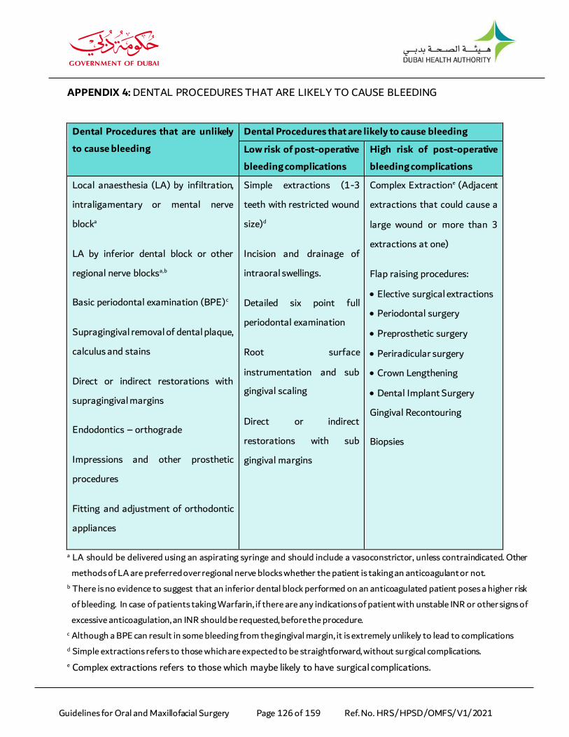
Guidelines for Oral and Maxillofacial Surgery Page 126 of 159 Ref. No. HRS/HPSD/OMFS/V1/2021
APPENDIX 4: DENTAL PROCEDURES THAT ARE LIKELY TO CAUSE BLEEDING
Dental Procedures that are unlikely
to cause bleeding
Dental Procedures that are likely to cause bleeding
Low risk of post-operative
bleeding complications
High risk of post-operative
bleeding complications
Local anaesthesia (LA) by infiltration,
intraligamentary or mental nerve
blocka
LA by inferior dental block or other
regional nerve blocksa,b
Basic periodontal examination (BPE)c
Supragingival removal of dental plaque,
calculus and stains
Direct or indirect restorations with
supragingival margins
Endodontics – orthograde
Impressions and other prosthetic
procedures
Fitting and adjustment of orthodontic
appliances
Simple extractions (1-3
teeth with restricted wound
size)d
Incision and drainage of
intraoral swellings.
Detailed six point full
periodontal examination
Root surface
instrumentation and sub
gingival scaling
Direct or indirect
restorations with sub
gingival margins
Complex Extractione (Adjacent
extractions that could cause a
large wound or more than 3
extractions at one)
Flap raising procedures:
Elective surgical extractions
Periodontal surgery
Preprosthetic surgery
Periradicular surgery
Crown Lengthening
Dental Implant Surgery
Gingival Recontouring
Biopsies
a LA should be delivered using an aspirating syringe and should include a vasoconstrictor, unless contraindicated. Other
methods of LA are preferred over regional nerve blocks whether the patient is taking an anticoagulant or not.
b There is no evidence to suggest that an inferior dental block performed on an anticoagulated patient poses a higher risk
of bleeding. In case of patients taking Warfarin, if there are any indications of patient with unstable INR or other signs of
excessive anticoagulation, an INR should be requested, before the procedure.
c Although a BPE can result in some bleeding from the gingival margin, it is extremely unlikely to lead to complications
d Simple extractions refers to those which are expected to be straightforward, without surgical complications.
e Complex extractions refers to those which maybe likely to have surgical complications.

Guidelines for Oral and Maxillofacial Surgery Page 127 of 159 Ref. No. HRS/HPSD/OMFS/V1/2021
APPENDIX 5: CLINICAL STEPS FOR PATIENTS WHO ARE AT RISK OF BLEEDING FROM AN ORAL
SURGICAL PROCEDURE

Guidelines for Oral and Maxillofacial Surgery Page 128 of 159 Ref. No. HRS/HPSD/OMFS/V1/2021
APPENDIX 6: ANTIBIOTIC PROPHYLAXIS FOR ORAL/DENTAL PROCEDURE
Antibiotic Prophylaxis Regimen for Oral/dental procedures
Regimen: Single dose 30-60 min before procedure
Situation Agent Adults Children
Oral Amoxicillin 2 gms 50 mg/kg
Unable to take oral Ampicillin
OR
Cefazolin or
ceftriaxone
2 g IM* or IV+
1 g IM or IV
50 mg/kg IM or IV
50 mg/kg IM or IV
Allergic to penicillin
or ampicillin oral
Cephalexin ɸδ
OR
Clindamycin
OR
Azithromycin
OR
clarithromycin
2 g
600 mg
500 mg
50 mg/kg
20 mg/kg
15 mg/kg
Allergic to penicillin
or ampicillin and
unable to take oral
medication
Cefazolin or
Ceftriaxone δ
OR
Clindamycin
1 g IM or IV
600 mg IM or IV
50 mg/kg IM or IV
20 mg/kg IM or IV
*IM: Intramuscular
+IV: Intravenous
ɸ Or other first- or second-generation oral cephalosporin in equivalent adult or pediatric
dosage.
δ Cephalosporin should not be used in an individual with a history of anaphylaxis,
angioedema, or urticaria with penicillin or ampicillin.

Guidelines for Oral and Maxillofacial Surgery Page 129 of 159 Ref. No. HRS/HPSD/OMFS/V1/2021
APPENDIX 7: AAOS/ADA RECOMMENDATIONS FOR PREVENTION OF ORTHOPAEDIC IMPLANT
INFECTION
AAOS/ADA Prevention Of Orthopedic Implant Infection Clinical Practice Guideline Protocol Recommendations
Recommendation 1
The practitioner might consider discontinuing the practice of routinely prescribing prophylactic antibiotics for patients with hip and knee prosthetic joint implants undergoing dental procedures.
Grade of Recommendation: Limited
Recommendation 2
We are unable to recommend for or against the use of topical oral antimicrobials in patients with prosthetic joint implants or other orthopedic implants undergoing dental procedures.
Grade of Recommendation: Inconclusive
Recommendation 3
In the absence of reliable evidence linking poor oral health to prosthetic joint infection, it is the opinion of the work group that patients with prosthetic joint implants or other orthopedic implants maintain appropriate oral hygiene.
Grade of Recommendation: Consensus
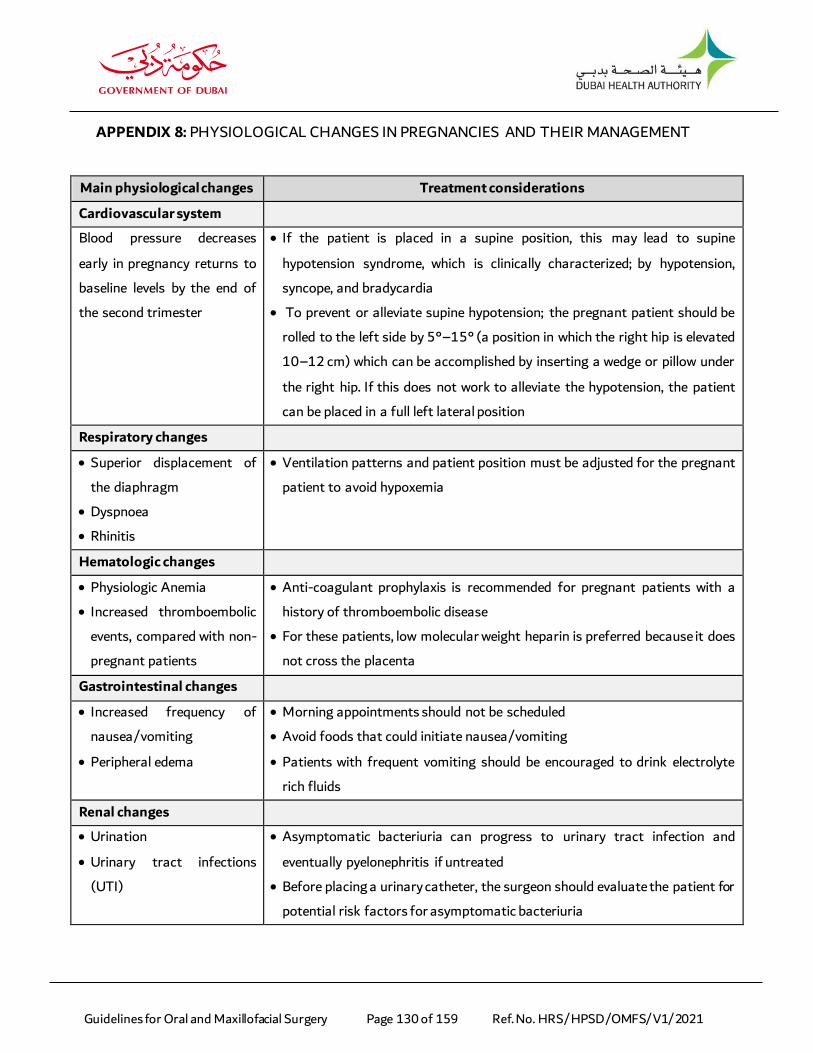
Guidelines for Oral and Maxillofacial Surgery Page 130 of 159 Ref. No. HRS/HPSD/OMFS/V1/2021
APPENDIX 8: PHYSIOLOGICAL CHANGES IN PREGNANCIES AND THEIR MANAGEMENT
Main physiological changes Treatment considerations
Cardiovascular system
Blood pressure decreases
early in pregnancy returns to
baseline levels by the end of
the second trimester
If the patient is placed in a supine position, this may lead to supine
hypotension syndrome, which is clinically characterized; by hypotension,
syncope, and bradycardia
To prevent or alleviate supine hypotension; the pregnant patient should be
rolled to the left side by 5°–15° (a position in which the right hip is elevated
10–12 cm) which can be accomplished by inserting a wedge or pillow under
the right hip. If this does not work to alleviate the hypotension, the patient
can be placed in a full left lateral position
Respiratory changes
Superior displacement of
the diaphragm
Dyspnoea
Rhinitis
Ventilation patterns and patient position must be adjusted for the pregnant
patient to avoid hypoxemia
Hematologic changes
Physiologic Anemia
Increased thromboembolic
events, compared with non-
pregnant patients
Anti-coagulant prophylaxis is recommended for pregnant patients with a
history of thromboembolic disease
For these patients, low molecular weight heparin is preferred because it does
not cross the placenta
Gastrointestinal changes
Increased frequency of
nausea/vomiting
Peripheral edema
Morning appointments should not be scheduled
Avoid foods that could initiate nausea/vomiting
Patients with frequent vomiting should be encouraged to drink electrolyte
rich fluids
Renal changes
Urination
Urinary tract infections
(UTI)
Asymptomatic bacteriuria can progress to urinary tract infection and
eventually pyelonephritis if untreated
Before placing a urinary catheter, the surgeon should evaluate the patient for
potential risk factors for asymptomatic bacteriuria
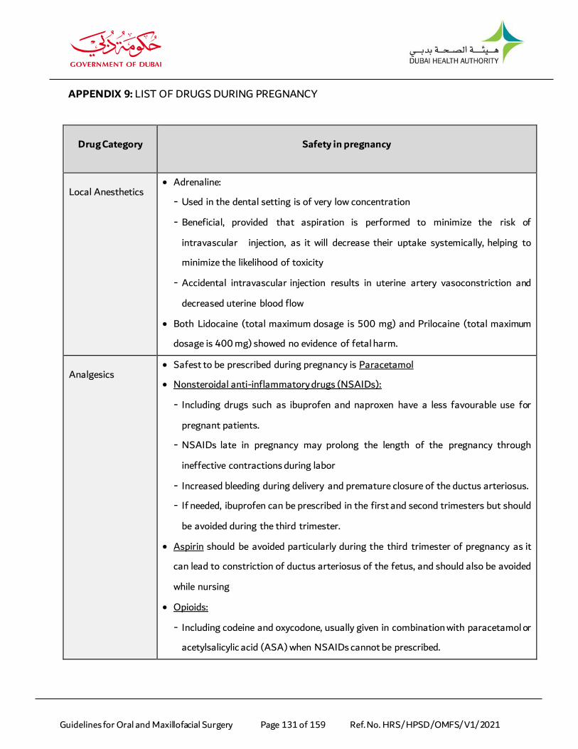
Guidelines for Oral and Maxillofacial Surgery Page 131 of 159 Ref. No. HRS/HPSD/OMFS/V1/2021
APPENDIX 9: LIST OF DRUGS DURING PREGNANCY
Drug Category Safety in pregnancy
Local Anesthetics Adrenaline:
- Used in the dental setting is of very low concentration
- Beneficial, provided that aspiration is performed to minimize the risk of
intravascular injection, as it will decrease their uptake systemically, helping to
minimize the likelihood of toxicity
- Accidental intravascular injection results in uterine artery vasoconstriction and
decreased uterine blood flow
Both Lidocaine (total maximum dosage is 500 mg) and Prilocaine (total maximum
dosage is 400 mg) showed no evidence of fetal harm.
Analgesics
Safest to be prescribed during pregnancy is Paracetamol
Nonsteroidal anti-inflammatory drugs (NSAIDs):
- Including drugs such as ibuprofen and naproxen have a less favourable use for
pregnant patients.
- NSAIDs late in pregnancy may prolong the length of the pregnancy through
ineffective contractions during labor
- Increased bleeding during delivery and premature closure of the ductus arteriosus.
- If needed, ibuprofen can be prescribed in the first and second trimesters but should
be avoided during the third trimester.
Aspirin should be avoided particularly during the third trimester of pregnancy as it
can lead to constriction of ductus arteriosus of the fetus, and should also be avoided
while nursing
Opioids:
- Including codeine and oxycodone, usually given in combination with paracetamol or
acetylsalicylic acid (ASA) when NSAIDs cannot be prescribed.

Guidelines for Oral and Maxillofacial Surgery Page 132 of 159 Ref. No. HRS/HPSD/OMFS/V1/2021
- Oxycodone is the safest, whereas codeine has been reported to cause increased risk
of congenital malformations including cleft lip and palate and other cardiac and
circulatory malformations. Prescribing codeine (preferably in the second or third tri-
mesters) for a short duration, when needed, is acceptable.
- Chronic use has been associated with fetal dependence, premature delivery,
neonatal respiratory depression and delayed growth.
If there is severe chronic pain, an interprofessional approach is best.
General recommendations for the use of analgesics during pregnancy
General Eliminate the source of pain, if at all possible
For
Paracetamol
Paracetamol is the analgesic of choice in the otherwise healthy
pregnant patient
Use a dose of 500–1,000 mg every 4 hours to a maximum of 4 grams
per day
For NSAIDs NSAIDs can be used cautiously in first and second trimesters
NSAIDs should be avoided during the third trimester
If NSAIDs are used in the pregnant patient, it is recommended to use
the lowest effective dose for as short a period of time as possible
For opioids Opioid analgesics can be cautiously prescribed to the pregnant dental
patient.
If opioid analgesics are prescribed, low dose and short duration are
recommended
Antibiotics If a patient is allergic to penicillin, clindamycin can be given
Erythromycin is no longer considered a preferred alternative and is best avoided.
Furthermore, it has been recommended not to use the estolate form of this drug, as
it has been associated with cholestatic hepatitis.
Metronidazole use during pregnancy is controversial. It has been associated with
increased risk for preterm birth, teratogenesis and fetal harm. In the first trimester,
however, there was no evidence that there is an increase in the rate of congenital
anomalies. Use of metronidazole is justified for significant oral and maxillofacial
infections in the pregnant patient because the cause of dental abscesses comprises of

Guidelines for Oral and Maxillofacial Surgery Page 133 of 159 Ref. No. HRS/HPSD/OMFS/V1/2021
the complex mix of strict anaerobes and facultative anaerobes. Metronidazole is highly
active against most anaerobes.
Antifungals Amphotericin B remains the drug of choice for the treatment of deep and life-
threatening fungal infections in pregnancy
Antivirals Acyclovir and valacyclovir are antiviral antibiotics that can be used to treat herpetic
infections
Steroids Specific consideration for the use of corticosteroids during pregnancy is to reduce
inflammation and decrease edema
Dexamethasone is used in pregnancy only when benefit to the mother outweighs risk
to the fetus
Prednisone and prednisolone have been used clinically in pregnant women without
adverse effects on the fetus
Triamcinolone and beclomethasone have not been associated with fetal defects in
humans. They are safe locally but systemic use can harm the mother and the fetus and
thus should be avoided during pregnancy
Pregnancy specific complications that arise are premature rupture of embryonic
membranes, hypertension and gestational diabetes mellitus
Sedatives &
Anxiolytics
Nitrous Oxide (N2O) is given in the first trimester if possible, and if it is necessary to
be given at all in pregnancy, it should be administered for less than 30 minutes and
with at least 50% oxygen. Nitrous oxide causes vasoconstriction and may reduce
uterine blood supply.
Benzodiazepines are commonly administered for patients with anxiety requiring
sedation. These drugs, when taken during pregnancy, were linked to fetal malforma-
tions, fetal abortion and craniofacial defects such as cleft lip and palate. When
prescribed chronically in the third trimester they cause fetal dependence and
withdrawal. They may be used with caution when sedation for dentistry is indicated
The use of diazepam and midazolam are particularly hazardous and must be avoided
in the first trimester and last month of the third trimester of pregnancy
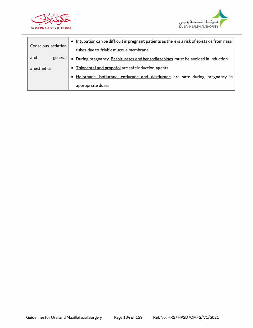
Guidelines for Oral and Maxillofacial Surgery Page 134 of 159 Ref. No. HRS/HPSD/OMFS/V1/2021
Conscious sedation
and general
anesthetics
Intubation can be difficult in pregnant patients as there is a risk of epistaxis from nasal
tubes due to friable mucous membrane
During pregnancy, Barbiturates and benzodiazepines must be avoided in induction
Thiopental and propofol are safe induction agents
Halothane, isoflurane, enflurane and desflurane are safe during pregnancy in
appropriate doses

Guidelines for Oral and Maxillofacial Surgery Page 135 of 159 Ref. No. HRS/HPSD/OMFS/V1/2021
APPENDIX 10: TERATOGENS AND THEIR EFFECTS ON THE FETUS
Teratogens Effects on Fetus
Ethyl alcohol Fetal alcohol syndrome
Tobacco Low birth rate, cleft lip and palate
Cocaine Cognitive delay, Placental abruption
Thalidomide Thalidomide embryopathy (damage of the limbs, face, eyes, ears,
genitalia, and internal organs and the vertebral column. facial palsies)
Methyl mercury Microcephaly, Brain damage
Anticonvulsants (all)
Carbamazepine
Valproic acid
Lamotrigine
Phenobarbital
Topiramate
Orofacial clefts, Cardiac Malformations,
Spina bifida
Neural tube defects
Neural tube defects
Urinary malformations
Abnormalities in all subjects
Warfarin (e.g.
Coumadin)
Warfarin embryopathy (midface and long bone deficiency) spontaneous
abortion.
Angiotensin-
converting enzyme
inhibitors
Oliguria, renal dysgenesis, lung and limb abnormalities
Retinoids Spontaneous abortion Multiple malformations

Guidelines for Oral and Maxillofacial Surgery Page 136 of 159 Ref. No. HRS/HPSD/OMFS/V1/2021
APPENDIX 11: FDA CATEGORIZATION FOR RISK FACTORS FOR METHEMOGLOBINEMIA IN
PREGNANT WOMEN
FDA
Category
Explanation
Category
A
Controlled studies in women fail to demonstrate a risk to the foetus in the first
trimester (and there is no evidence of a risk in later trimesters), and the possibility
of foetal harm appears
Category
B
Either animal-reproduction studies have not demonstrated a foetal risk but there
are no controlled studies in pregnant women, or animal-reproduction studies have
shown an adverse effect (other than a decrease in fertility) that was not confirmed
in controlled studies in women in the first trimester (and there is no evidence of a
risk in later trimesters).
Category
C
Either studies in animals have revealed adverse effects on the foetus (teratogenic
or embryocidal, or other) and there are no controlled studies in women, or studies
in women and animals are not available. Drugs should be given only if the potential
benefit justifies the potential risk to the foetus
Category
D
There is positive evidence of human foetal risk, but the benefits from use in
pregnant women may be acceptable despite the risk (e.g., if the drug is needed in
a life-threatening situation or for a serious disease for which safer drugs cannot
be used or are ineffective).
Category
X
Studies in animals or human beings have demonstrated foetal abnormalities, or
there is evidence of foetal risk based on human experience, or both, and the risk of
the use of the drug in pregnant women clearly outweighs any possible benefit. The
drug is contraindicated in women who are or may become pregnant.

APPENDIX 12: CLASSIFICATION OF ORAL MUCOSAL LESIONS

APPENDIX 13: MANAGEMENT OF MUCOSAL DISEASES

Guidelines for Oral and Maxillofacial Surgery Page 139 of 159 Ref. No. HRS/HPSD/OMFS/V1/2021
APPENDIX 14: MANAGEMENTS OF CYSTS

Guidelines for Oral and Maxillofacial Surgery Page 140 of 159 Ref. No. HRS/HPSD/OMFS/V1/2021
APPENDIX 15: MANAGEMENT OF VASCULAR LESIONS

APPENDIX 16: MANAGEMENTS OF MALIGNANT TUMORS OF SOFT TISSUE

Guidelines for Oral and Maxillofacial Surgery Page 142 of 159 Ref. No. HRS/HPSD/OMFS/V1/2021
APPENDIX 17: SALIVARY GLANDS BENIGN AND MALIGNANT TUMOR

APPENDIX 18: MANAGEMENT OF SIMPLE & LIFE-THREATENING INFECTIONS
1. Understand the Chief Complaint; obtain proper history,
and tests.
2. Lab Investigations (CBC/FBC, CRP, Procalcitonin and
radiological)
3.Reach a Proper Diagnosis.
4. Is the Infection Life Threatening or Simple
Signs to keep in mind
that an infection is life
threatening or severe:
1. Dyspnoea
2. Stridor
3. Dysphagia
4. Odynophagia
5. Trismus
6. Crepitus
Determine Threat to the Airway (1 + one of the following
present)
Trismus (unable to open more than 2cm or width of two
fingers)
Airway Deviation
Involvement of Maxillary Molars +/- any lower tooth
Systemic signs: Fever, Hypotension, Tachycardia
In case, the patient was systemically
unwell and had an airway
obstruction. Refer to a tertiary
setting hospital for further
management, such as:
1- Urgent OT
2- IV Antibiotics
3- Analgesics
4- Management of medical
conditions
5- Observation
In case, the patient was systemically
unwell with no airway obstruction.
Refer to a tertiary setting hospital for
further management, such as:
1- IV Antibiotics
2- Analgesics
3- Management of medical conditions
4- Observation
5- Determine if there is a need for
surgical intervention
If patient is systemically well & there
was no airway obstruction (simple
infection), we either:
1- Manage it in the Emergency Dental
Clinic, perform the Emergency
Dental Treatment by eliminating
the source of infection & prescribe
the medications needed
2- Or/and refer to the OMFS
Specialist, for further management
Outpatient Clinic
General Dentist
Patient attends with an infection
A- Initial Examination:
1. Airway
2. Breathing
3. Circulation
If satisfactory, proceed
with Vital Signs
B- Physical Examination:
1.Extra oral: Assess
extent and location of
any facial swelling
2. Intraoral: Assess focal
swelling, halitosis,
gross dental caries,
broken dentition,
drainage of pus, and
trismus

Guidelines for Oral and Maxillofacial Surgery Page 144 of 159 Ref. No. HRS/HPSD/OMFS/V1/2021
APPENDIX 19: MEDICATIONS ASSOCIATED WITH MRONJ
Medication Indication
Anti-resorptive agents
Bisphosphonates
Zoledronic acid (Zometa,
Reclast)
Pamidronate (Aredia)
Alendronate (Fosamax)
Ibandronate (Boniva)
Risedronate (Actonel, Atelvia)
Etidronate (Didronel)
Tiludronate (Skelid)
RANK-ligand inhibitor
• Denosumab (Xgeva and Prolia)
IV bisphosphonates: are antiresorptive medications
used to manage cancer-related conditions, including
hypercalcemia of malignancy, skeletal-related events
(SREs) associated with bone metastases in the
context of solid tumors such as breast, prostate, and
lung cancers, and for management of lytic lesions in
the setting of multiple myeloma.
Once yearly infusion of zolendronate (Reclast®)
Parenteral formulation of ibandronate (Boniva®)
administered every three months for the
management of osteoporosis.
Oral bisphosphonates: Used in less common
conditions, such as Paget disease & osteogenesis
imperfecta. However, their most common use is for
osteoporosis and osteopenia
Denosumab (Prolia®) is administered
subcutaneously every 6 months for osteoporotic
patients.
Denosumab (Xgeva®) is used in metastatic bone
disease from solid tumors when administered
monthly.
Denosumab is effective in decreasing SREs related to
metastatic bone disease from solid tumors when

Guidelines for Oral and Maxillofacial Surgery Page 145 of 159 Ref. No. HRS/HPSD/OMFS/V1/2021
administered monthly Denosumab therapy is not
indicated for the treatment of multiple myeloma.
Interestingly, in contrast to BPs, RANKL inhibitors
do not bind to bone and their effects on bone
remodelling are mostly diminished within six (6)
months of treatment cessation.
Anti-Angiogenic Agents
For the treatment of gastrointestinal tumors, renal cell carcinomas, neuroendocrine tumors and others
Sunitinib (Sutent)
Sorafenib (Nexavar)
Bevacizumab (Avastin)
Ziv-aflibercept (Zaltrap)
Renal cell carcinoma
Gastrointestinal stromal tumor
Neuroendocrine tumor
Hepatocellular carcinoma
Thyroid cancer
Metastatic colorectal cancer
Non-squamous, non-small cell lung cancer
Glioblastoma
Cervical cancer

Guidelines for Oral and Maxillofacial Surgery Page 146 of 159 Ref. No. HRS/HPSD/OMFS/V1/2021
APPENDIX 20: GENERAL DENTIST GUIDANCE FOR PATIENTS ON OR PLANNED FOR
ANTIRESORPTIVE/ANTIANGIOGENIC THERAPY
Does the Patients on anti-resorptive treatment have any dental problems?
Maintain periodic dental check-ups Any procedure that involves
manipulation of the bone
Refer to Oral & Maxillofacial
surgery department with a recent
radiograph
General dentists can perform the
following non-invasive procedures
according to treatment plan:
1. Delivering local anesthesia
2. scaling and prophylaxis
3. Placing restorations including
crowns and fixed prosthesis
4. Fabricating dentures
5. Conventional root canal therapy
NO YES
YES NO
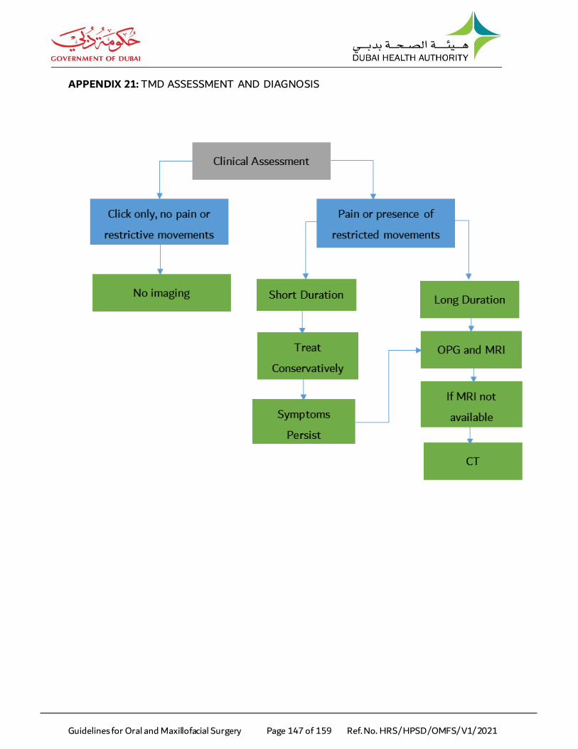
Guidelines for Oral and Maxillofacial Surgery Page 147 of 159 Ref. No. HRS/HPSD/OMFS/V1/2021
APPENDIX 21: TMD ASSESSMENT AND DIAGNOSIS
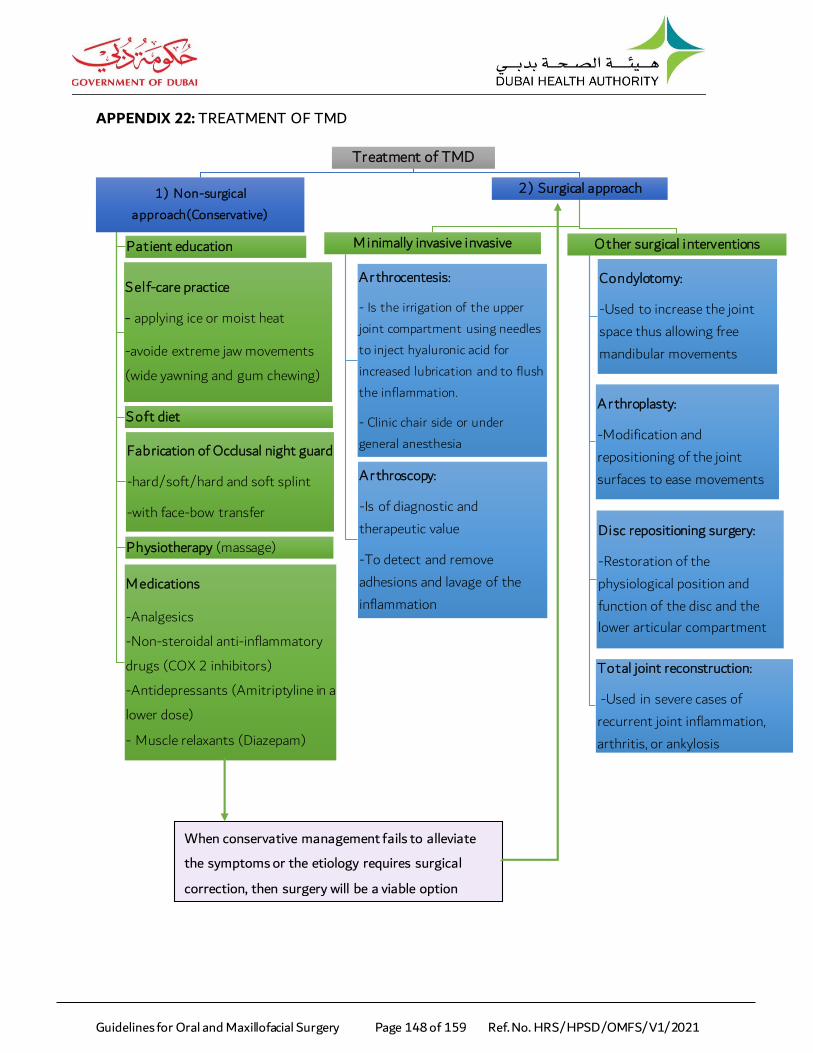
Guidelines for Oral and Maxillofacial Surgery Page 148 of 159 Ref. No. HRS/HPSD/OMFS/V1/2021
APPENDIX 22: TREATMENT OF TMD
Treatment of TMD
1) Non-surgical
approach(Conservative)
Patient education
Self-care practice
- applying ice or moist heat
-avoide extreme jaw movements
(wide yawning and gum chewing)
Soft diet
Medications
-Analgesics
-Non-steroidal anti-inflammatory
drugs (COX 2 inhibitors)
-Antidepressants (Amitriptyline in a
lower dose)
- Muscle relaxants (Diazepam)
Fabrication of Occlusal night guard
-hard/soft/hard and soft splint
-with face-bow transfer
Physiotherapy (massage)
2) Surgical approach
Minimally invasive invasive
Arthrocentesis:
- Is the irrigation of the upper
joint compartment using needles
to inject hyaluronic acid for
increased lubrication and to flush
the inflammation.
- Clinic chair side or under
general anesthesia
Arthroscopy:
-Is of diagnostic and
therapeutic value
-To detect and remove
adhesions and lavage of the
inflammation
Other surgical interventions
Condylotomy:
-Used to increase the joint
space thus allowing free
mandibular movements
Arthroplasty:
-Modification and
repositioning of the joint
surfaces to ease movements
Disc repositioning surgery:
-Restoration of the
physiological position and
function of the disc and the
lower articular compartment
Total joint reconstruction:
-Used in severe cases of
recurrent joint inflammation,
arthritis, or ankylosis
When conservative management fails to alleviate
the symptoms or the etiology requires surgical
correction, then surgery will be a viable option

Guidelines for Oral and Maxillofacial Surgery Page 149 of 159 Ref. No. HRS/HPSD/OMFS/V1/2021
APPENDIX 23: REFERAL PATHWAY

Guidelines for Oral and Maxillofacial Surgery Page 150 of 159 Ref. No. HRS/HPSD/OMFS/V1/2021
APPENDIX 24: TREATMENT OPTIONS FOR SKELETAL DEFORMITIES

Guidelines for Oral and Maxillofacial Surgery Page 151 of 159 Ref. No. HRS/HPSD/OMFS/V1/2021
APPENDIX 25: MANAGEMENT OF COMBINED ORTHODONTICS AND
ORTHOGNATHIC SURGERY
Phases
1) Pre-orthodontic
preparatory phase
This phase includes establishing a good oral hygiene and elimination of caries
in patient's mouth. The amount of keratinized gingiva must be managed
before any orthodontic intervention to avoid gingival recession.
2) Pre-surgical
orthodontic phase
Duration
12-18
months
In skeletal malocclusions, the teeth tend to compensate for the skeletal
discrepancies in order to achieve a functional occlusion.
The aim of this phase is to eliminate the dental compensation in order to
expose the true extent of skeletal deformity.
The teeth have to be decompensated and repositioned back to their ideal
position in three dimensions
A) Anterior posterior segment involves the
correction of maxillary and mandibular incisors
B) Transverse segment
C) Vertical segment: the amount of
decompensation depends on:
-Maxillary incisors are
retroclined, and mandibular
incisors are proclined to allow a
functional bite.
-Decompensation requires up
righting the proclined
mandibular incisors, and
advancing the maxillary incisors
to create an adequate lip
support.
-Extraction of maxillary second
premolars, and mandibular first
premolars is often required to
gain a class I posterior pre-
surgical occlusion, and relieve
the compensation and
crowding
Class II malocclusions
-Mandibular incisors are proclined, and
the maxillary incisors are retroclined.
Class III malocclusions
-Extraction of maxillary first premolars
and mandibular second premolars is
often required to gain a class I
posterior presurgical occlusion and
relieve the compensation and crowding.
1) The amount of dental display of
the maxillary incisors to the upper lip
2) The inter-labial gap
3) The lower anterior facial height
When the dental display
and gingival display are
excessive, the extruded
teeth that have
compensated are
intruded orthodontically.
Management of Combined Orthodontics and Orthognathic Surgery
Treatment options for skeletal deformities
When the lower facial height is
normal, the deep bite is
corrected by intruding the
incisors and extruding the first
molars and premolars.
However, if the lower facial
height is reduced (as in deep
Class II skeletal conditions),
the arches are not levelled
until the mandible is advanced.
At the end of this phase, new set of records is taken.
Study models are fabricated
to mimic the surgical plan.
3D planning model (optional)
Face bow recording is required to
mount the models on a semi-
adjustable articulator in cases of
single jaw maxillary advancement,
or in bimaxillary procedures.

Guidelines for Oral and Maxillofacial Surgery Page 152 of 159 Ref. No. HRS/HPSD/OMFS/V1/2021
3) Surgical
A) Anterior
posterior
segment:
Treatment options for skeletal deformities
Management of Combined Orthodontics and Orthognathic Surgery
Phases
B) Vertical
C) Transverse
D) Rotational
1) Retrognathism/
Hypoplasia
2) Prognathism/
Hyperplasia
3) Sagittal maxillary
deficiency
Concave face
profile
Class II Angle
malocclusion
Because the location of the incisors often
dictates the degree of surgical movement, the
orthodontist positions the maxillary and
mandibular incisors in the ‘‘ideal’’ position in
the anterior-posterior and vertical planes.
Failure to decompensate adequately limits the
surgical correction and aesthetic outcome
Skeletal advancement is achieved by the
bilateral sagittal split osteotomy (BSSO) of
the mandible using techniques well described
in the literature. Post- surgical management
includes using ‘‘light’’ Class II elastics to
override proprioception and to guide the new
occlusion in the immediate postoperative
period
Concave facial profile
Class III malocclusion with lingually inclined
mandibular incisors & proclined maxillary
anterior teeth
Treatment options:
1- Intraoral vertical ramus osteotomy (IVO)
2- Bilateral sagittal split ramus osteotomy
(BSSO)
Maxillary advancement must be also
considered in such cases to limit the
amount of mandibular setback required.
The surgeon must keep the tongue size in
consideration, as it will be confined to a
smaller intraoral volume, and there would
be a potential for sleep apnea
Concave face profile.
They might be combined with
mandibular Prognathism (class
III).
In such cases, a combined mid-
face skeletal advancement along
with mandibular surgery will
give better aesthetic results
Vertical
maxillary excess
In many cases, correction of significant VME requires double jaw
surgery: differential Le Fort I impaction of the maxilla either as a
single segment or a multi segment with mandibular BSSO
Surgical Correction of skeletal malocclusions has to address all the
dimensions
Clockwise rotation is done to correct mandibular retrognathia
Anticlockwise rotation is done to correct mandibular prognathia
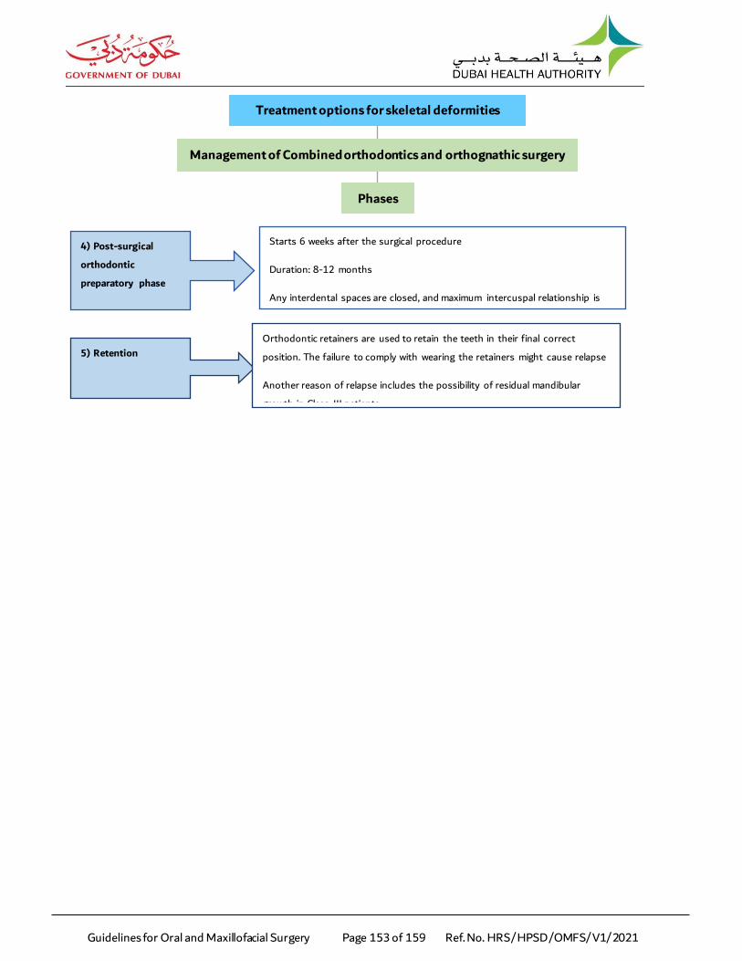
Guidelines for Oral and Maxillofacial Surgery Page 153 of 159 Ref. No. HRS/HPSD/OMFS/V1/2021
Treatment options for skeletal deformities
Management of Combined orthodontics and orthognathic surgery
Phases
4) Post-surgical
orthodontic
preparatory phase
5) Retention
Starts 6 weeks after the surgical procedure
Duration: 8-12 months
Any interdental spaces are closed, and maximum intercuspal relationship is
reached between the jaws. It ends by debonding
Orthodontic retainers are used to retain the teeth in their final correct
position. The failure to comply with wearing the retainers might cause relapse
Another reason of relapse includes the possibility of residual mandibular
growth in Class III patients

Guidelines for Oral and Maxillofacial Surgery Page 154 of 159 Ref. No. HRS/HPSD/OMFS/V1/2021
APPENDIX 26: CLASSIFICATION OF FACIAL FRACTURES
Vertical Facial
Thirds
Upper
Frontal Bone,
may involve
frontal sinus
Anterior wall
Posterior Wall
Anterior +
Posterior Wall
Mid Third
Orbital
Roof
Lateral or
Medial Wall
Floor
Combination
Nose
Septal
Bony
Compound
Zygoma
Zygoma Body
Zygomatic
Arch
Naso Orbital
Ethomoid
Complex
Involving
detattchment
of medial
canthal
ligament
Maxilla
Lefort I
Lefort II
Lefort III
Lower
Mandible
Anatomical
site
Favorable vs
unfavorable
Open vs Closed

Guidelines for Oral and Maxillofacial Surgery Page 155 of 159 Ref. No. HRS/HPSD/OMFS/V1/2021
APPENDIX 27: METHODS OF TREATMENT FOR UPPER THIRD FACIAL FRACTURES

Guidelines for Oral and Maxillofacial Surgery Page 156 of 159 Ref. No. HRS/HPSD/OMFS/V1/2021
APPENDIX 28: METHODS OF TREATMENT FOR MIDDLE THIRD FACIAL FRACTURES

Guidelines for Oral and Maxillofacial Surgery Page 157 of 159 Ref. No. HRS/HPSD/OMFS/V1/2021

Guidelines for Oral and Maxillofacial Surgery Page 158 of 159 Ref. No. HRS/HPSD/OMFS/V1/2021
APPENDIX 29: METHODS OF TREATMENT FOR MANDIBULAR FRACTURES
Facial Fracture
Lower Third
Mandible
Nondisplaced, incomplete,
asymptomatic fractures
Conservative: careful
observation, blended soft diet,
and follow up
Condylar/subcondylar fracture, grossly
comminuted fractures, minimaly
displaced
Nonsurgical closed reduction and
maxillomandibular fixation
Followed by a period of
immobilization of 4 to 8 weeks
Displaced fractures
Surgical open reduction and
internal fixation
Dentoalveolar fractures located in the mandible or maxilla may be treated by reduction and stabilization (either as closed or open
reduction). According to the displacements severity the treatment will be selected. Rigid splint is used in the closed reduction while
in extreme displacement cases open technique is performed.

Guidelines for Oral and Maxillofacial Surgery Page 159 of 159 Ref. No. HRS/HPSD/OMFS/V1/2021
APPENDIX 30: ALGORITHM FOR SECURING THE AIRWAY OF A PATIENT WITH
MAXILLOFACIAL TRAUMA
Cricothyriodotomy Supraglottic airway device Tracheostomy Oral Endotracheal tube
Conversion
Comminuted
Mandibular Fracture
Contraindication for nasal
intubation
Prolonged Mechanical
Ventilation/Panfacial
Trauma/Poor direct
laryngoscopic view
Submental Intubation
Replace with an Oral
Endotracheal Tube
Prolonged mechanical
ventilation/
Pan facial
Trauma/Mandibular
Fracture/Failed Oral
Intubation
Tracheostomy
Yes No
Yes
Yes
No
No
No
Yes
Nasal Intubation