guidelines for the management of pediatric severe …management of pediatric severe traumatic brain...
TRANSCRIPT

Dow
nloadedfrom
https://journals.lww.com
/pccmjournalby
BhDMf5ePH
Kav1zEoum1tQ
fN4a+kJLhEZgbsIH
o4XMi0hC
ywCX1AW
nYQp/IlQ
rHD3oaxD
/vH2r76VBJLc78Tw
9i0ooXPDln19rdU
Vr3qDoxk=
on07/09/2019
Downloadedfromhttps://journals.lww.com/pccmjournalbyBhDMf5ePHKav1zEoum1tQfN4a+kJLhEZgbsIHo4XMi0hCywCX1AWnYQp/IlQrHD3oaxD/vH2r76VBJLc78Tw9i0ooXPDln19rdUVr3qDoxk=on07/09/2019
Copyright © 2019 by the Society of Critical Care Medicine and the World Federation of Pediatric Intensive and Critical Care Societies.Unauthorized reproduction of this article is prohibited
Pediatric Critical Care Medicine www.pccmjournal.org S1
15Herman and Faye Sarkowsky Endowed Chair, Head, Division of Pediatric Neu-rology, University of Washington, Seattle Children's Hospital, Seattle, WA.
This document was endorsed by the American Association of Neurologi-cal Surgeons/Congress of Neurological Surgeons.
Any opinions, findings, and conclusions or recommendations expressed in this material are those of the authors and do not necessarily reflect the views of the U.S. Army Contracting Command, Aberdeen Proving Ground, Natick Contracting Division, Stanford University, or the Brain Trauma Foundation. The information contained in the Guidelines for the Manage-ment of Pediatric Severe Traumatic Brain Injury reflects the current state of knowledge at the time of publication. The Brain Trauma Foundation, Ameri-can Association of Neurological Surgeons, Congress of Neurological Sur-geons, and other collaborating organizations are not engaged in rendering professional medical services and assume no responsibility for patient outcomes resulting from application of these general recommendations in specific patient circumstances. Accordingly, the Brain Trauma Foun-dation, American Association of Neurological Surgeons, and Congress of Neurological Surgeons consider adherence to these clinical practice guidelines will not necessarily assure a successful medical outcome. The information contained in these guidelines reflects published scientific evi-dence at the time of completion of the guidelines and cannot anticipate subsequent findings and/or additional evidence, and therefore should not be considered inclusive of all proper procedures and tests or exclusive of other procedures and tests that are reasonably directed to obtaining the same result. Medical advice and decisions are appropriately made only by a competent and licensed physician who must make decisions in light of all the facts and circumstances in each individual and particular case and on the basis of availability of resources and expertise. Guidelines are not intended to supplant physician judgment with respect to particular patients or special clinical situations and are not a substitute for physician-patient consultation. Accordingly, the Brain Trauma Foundation, American Association of Neurological Surgeons, and Congress of Neurological Surgeons consider adherence to these guidelines to be voluntary, with the ultimate determination regarding their application to be made by the physician in light of each patient’s individual circumstances.
Supplemental digital content is available for this article. Direct URL citations appear in the printed text and are provided in the HTML and PDF versions of this article on the journal’s website (http://journals.lww.com/pccmjournal).
Supported, in part, by the U.S. Army Contracting Command, Aberdeen Prov-ing Ground, and Natick Contracting Division, through a contract awarded to Stanford University (W911 QY-14-C-0086), a subcontract awarded to Oregon Health & Science University. Previous editions were supported by funding from multiple sources through the Brain Trauma Foundation.
1Ake N. Grenvik Professor of Critical Care Medicine, Vice Chair, Depart-ment of Critical Care Medicine, Professor of Anesthesiology, Pediatrics, Bioengineering, and Clinical and Translational Science, Director, Safar Center for Resuscitation Research, University of Pittsburgh School of Medicine, UPMC Children’s Hospital of Pittsburgh, Pittsburgh, PA.
2Department of Neurology and Department of Anesthesiology, Critical Care and Pain Medicine, Boston Children's Hospital, Harvard Medical School, Boston, MA.
3Professor, Pacific Northwest Evidence-based Practice Center, Depart-ment of Medical Informatics and Clinical Epidemiology, Oregon Health & Science University, Portland, OR.
4Associate Professor, Pacific Northwest Evidence-based Practice Cen-ter, Department of Medical Informatics and Clinical Epidemiology, Ore-gon Health & Science University, Portland, OR.
5Diane and Bruce Halle Endowed Chair in Pediatric Neurosciences, Chief, Pediatric Neurosurgery, Director, BARROW Neurological Insti-tute at Phoenix Children's Hospital, Phoenix, AZ.
6Chair, Department of Neurological Surgery, Oregon Health & Science University, Portland, OR.
7Research Associate, Pacific Northwest Evidence-based Practice Cen-ter, Department of Medical Informatics and Clinical Epidemiology, Oregon Health & Science University, Portland, OR.
8Research Assistant, Pacific Northwest Evidence-based Practice Center, Department of Medical Informatics and Clinical Epidemiology, Oregon Health & Science University, Portland, OR.
9Professor and Chief, Critical Care Medicine, Children’s National Medical Center, Washington, DC.
10Emeritus Professor of Pediatrics, University of Utah, Salt Lake City, UT.11Department of Neurosurgery, Stanford University, Stanford, CA.12Department of Pediatrics, British Columbia’s Children’s Hospital, Clini-
cal Investigator, Child and Family Research Institute, University of British Columbia, Vancouver, BC. Canada.
13School of Nursing/School of Medicine, Department of Pediatrics, Divi-sion of Pediatric Critical Care Medicine, Duke University, Durham, NC.
14Professor & Vice Chair Strategic Affairs, Anesthesiology & Pain Medi-cine, Professor, Pediatrics, Director, Harborview Injury Prevention and Research Center (HIPRC), University of Washington, Seattle, WA.
Copyright © 2019 by the Society of Critical Care Medicine and the World Federation of Pediatric Intensive and Critical Care Societies
DOI: 10.1097/PCC.0000000000001735
Guidelines for the Management of Pediatric Severe Traumatic Brain Injury, Third Edition: Update of the Brain Trauma Foundation Guidelines
Patrick M. Kochanek, MD, MCCM1; Robert C. Tasker, MA, MD, FRCP2; Nancy Carney, PhD3;
Annette M. Totten, PhD4; P. David Adelson, MD, FACS, FAAP, FAANS5;
Nathan R. Selden, MD, PhD, FACS, FAAP6; Cynthia Davis-O’Reilly, BS7; Erica L. Hart, MST8;
Michael J. Bell, MD9; Susan L. Bratton, MD, MPH, FAAP10; Gerald A. Grant, MD11;
Niranjan Kissoon, MD, FRCP(C), FAAP, MCCM, FACPE12;
Karin E. Reuter-Rice, PhD, CPNP-AC, FCCM, FAAN13; Monica S. Vavilala, MD14;
Mark S. Wainwright, MD, PhD15
20

Copyright © 2019 by the Society of Critical Care Medicine and the World Federation of Pediatric Intensive and Critical Care Societies.Unauthorized reproduction of this article is prohibited
Copyright © 2019 by the Society of Critical Care Medicine and the World Federation of Pediatric Intensive and Critical Care Societies.Unauthorized reproduction of this article is prohibited
Kochanek et al
S2 www.pccmjournal.org March 2019 • Volume 20 (Suppl) • Number 3
Dr. Kochanek received funding from the Society of Critical Care Medicine (Editor-in-Chief of Pediatric Critical Care Medicine), from serving as an expert witness on cases in pediatric critical care. Drs. Carney and Tot-ten’s, Ms. Davis-O’Reilly’s, and Ms. Hart’s institutions received funding from Stanford University. Dr. Selden disclosed that he has stock options (current $0 value) in Cerebrotech for scientific advisory board service (this device is not clinically available and is not referenced in the work). Dr. Reuter-Rice received funding from textbook royalties and curriculum content, and she received support for article research from Robert Wood Johnson Foundation funding 2013–2016. Dr. Wainwright received fund-ing from Sage Therapeutics. The remaining authors have disclosed that they do not have any potential conflicts of interest.
Hector R. Wong, MD, is a Guest Editor.
For information regarding this article, E-mail: [email protected].
edu (Pediatr Crit Care Med 2019; 20:S1–S82)Key Words: critical care; evidence-based medicine; guidelines; pediatrics; systematic review; traumatic brain injury
Severe Traumatic Brain Injury in Infants, Children, and Adolescents in 2019: Some Overdue Progress, Many Remaining Questions, and Exciting Ongoing Work in the Field of Traumatic Brain Injury ResearchIn this Supplement to Pediatric Critical Care Medicine, we are pleased to present the Third Edition of the Guidelines for the Management of Pediatric Severe Traumatic Brain Injury (TBI). This body of work updates the Second Edition of the guidelines that was published in 2012 (1). It represents a substantial effort by a multidisciplinary group of individuals assembled to reflect the team approach to the treatment of these complex, critically ill patients that is essential to optimizing critical care and improv-ing outcomes. This work also represents the strong and always-evolving partnership between investigators from the medical and research communities, forged in Chicago in 2000, from which the first pediatric TBI guidelines were developed. The mutual trust and respect we share have been the foundation of our commit-ment to bringing evidence-based care to children with TBI.
Updating these guidelines was particularly exciting to the individuals who have participated in the previous two edi-tions because several new studies have been published which begin to address a number of major gaps in the pediatric TBI literature—gaps that were specifically identified as targets for future research in earlier editions. For example, we are now able to include reports on the effects of commonly used seda-tives and analgesics on intracranial pressure (ICP). Similarly, initial head-to-head comparisons of the influence of agents in routine “real world” use such as hypertonic saline (HTS), fen-tanyl, and others now inform these guidelines (2, 3). A total of 48 new studies were included in this Third Edition. Although some progress has been made and should be celebrated, overall the level of evidence informing these guidelines remains low. High-quality randomized studies that could support level I recommendations remain absent; the available evidence pro-duced only three level II recommendations, whereas most rec-ommendations are level III, supported by low-quality evidence.
Based in part on a number of requests from the readership to individual clinical investigators, we have included a companion article in the regular pages of Pediatric Critical Care Medicine
that presents a “Critical Pathway” algorithm of care for both first-tier and second-tier (refractory intracranial hypertension) approaches. The algorithm reflects both the evidence-based recommendations from these guidelines and consensus-based expert opinion, vetted by the clinical investigators, where evi-dence was not available. An algorithm was provided in the First but not Second Editions of the guidelines, and we believe that given the new reports available, along with the existing gaps in evidence, a combination of evidence-based and consensus-based recommendations provides additional and much-needed guid-ance for clinicians at the bedside. The algorithm also addresses a number of issues that are important but were not previously covered in the guidelines, given the lack of research and the focus on evidence-based recommendations. This includes addressing issues such as a stepwise approach to elevated ICP, differences in tempo of therapy in different types of patients, scenarios with a rapidly escalating need for ICP-directed therapy in the setting of impending herniation, integration of multiple monitoring tar-gets, and other complex issues such as minimal versus optimal therapeutic targets and approaches to weaning therapies. We hope that the readership finds the algorithm document helpful, recognizing that it represents a challenging albeit important step.
Designing and developing this pediatric TBI evidence-based guidelines document required an expert administrative man-agement team, and to that end, we are extremely grateful to the staff of the Pacific Northwest Evidence-based Practice Center, Oregon Health & Science University, for their vital contribu-tion to this work. We are also grateful to the Brain Trauma Foundation and the Department of Defense for supporting the development and publication of these guidelines docu-ments. We are grateful to the endorsing societies for recogniz-ing the importance of this work and for the considerable work of the clinical investigators in constructing the final document. We are also pleased to have collaborated with the Congress of Neurological Surgeons and the journal Neurosurgery that is copublishing the Executive Summary document of these guide-lines for its readership. We are also grateful to Hector Wong for serving as Guest Editor, along with the external reviewers of this final document. Finally, we thank each of the clinical investiga-tors and coauthors on this project. We believe that the consider-able uncompensated time and effort devoted to this important project will help to educate clinicians worldwide and enhance the outcomes of children with severe TBI. Clinical investigators provided Conflict of Interest Disclosures at the beginning of the process, which were re-reviewed at the time of publication. No clinical investigator made inclusion decisions or provided assessments on publications for which they were an author.
Looking forward, it is important to recognize that these guide-lines were written as the Approaches and Decisions in Acute Pediatric TBI Trial (ADAPT) (4–6), one of the most important in the field of pediatric TBI, was coming to a close. The ADAPT completed enrollment of 1,000 cases of severe pediatric TBI and is one example of the recent heightened general interest in TBI as a disease. This new interest in the importance of TBI has emerged in part from the recognition of the high prevalence of TBI across the injury severity spectrum, particularly concussion, and from
Copyright © 2019 by the Society of Critical Care Medicine and the World Federation of Pediatric Intensive and Critical Care Societies.Unauthorized reproduction of this article is prohibited
Supplement
Pediatric Critical Care Medicine www.pccmjournal.org S3
the need for new classification systems and new trial design for TBI in both children and adults (7, 8). In addition, the emerg-ing links between TBI and a number of neurodegenerative dis-eases have broadened the interest in TBI, have led to additional support of TBI research, and have produced an unprecedented level of research in TBI and a quest for new therapies (9–11). We expect that the results of ADAPT, along with those of other ongo-ing and recently completed research in the field, will help pro-vide new insight and clarity into the acute medical management (MM) of infants, children, and adolescents with severe TBI, and mandate further refinement of the recommendations in these documents. We know that we speak for the entire team of clinical investigators in welcoming the opportunity to incorporate addi-tional high-level evidence into future updates of these guidelines.
METHODSThe methods for developing these guidelines were organized in two phases: a systematic review, assessment, and synthesis of the literature; and use of that product as the foundation for evidence-based recommendations. These guidelines are the product of the two-phased, evidence-based process.
Based on almost 2 decades of collaboration, the team of clinical investigators and methodologists (Appendix A, Supplemental Digital Content 1, http://links.lww.com/PCC/A774) is grounded in and adheres to the fundamental princi-ples of evidence-based medicine to derive recommendations, and is committed to maintaining the distinction between evi-dence and consensus. It is important that this distinction is clear to promote transparency and inspire innovative future research that will expand the evidence base for TBI care.
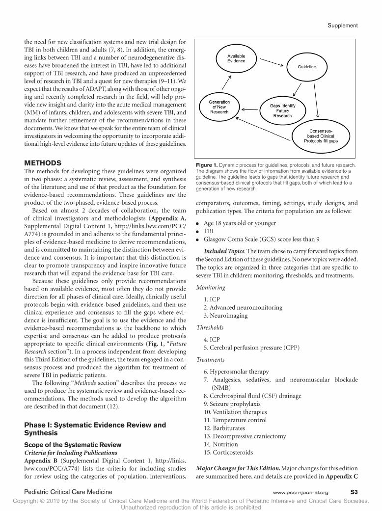
Because these guidelines only provide recommendations based on available evidence, most often they do not provide direction for all phases of clinical care. Ideally, clinically useful protocols begin with evidence-based guidelines, and then use clinical experience and consensus to fill the gaps where evi-dence is insufficient. The goal is to use the evidence and the evidence-based recommendations as the backbone to which expertise and consensus can be added to produce protocols appropriate to specific clinical environments (Fig. 1, “Future Research section”). In a process independent from developing this Third Edition of the guidelines, the team engaged in a con-sensus process and produced the algorithm for treatment of severe TBI in pediatric patients.
The following “Methods section” describes the process we used to produce the systematic review and evidence-based rec-ommendations. The methods used to develop the algorithm are described in that document (12).
Phase I: Systematic Evidence Review and Synthesis
Scope of the Systematic ReviewCriteria for Including PublicationsAppendix B (Supplemental Digital Content 1, http://links.lww.com/PCC/A774) lists the criteria for including studies for review using the categories of population, interventions,
comparators, outcomes, timing, settings, study designs, and publication types. The criteria for population are as follows:
● Age 18 years old or younger ● TBI ● Glasgow Coma Scale (GCS) score less than 9
Included Topics. The team chose to carry forward topics from the Second Edition of these guidelines. No new topics were added. The topics are organized in three categories that are specific to severe TBI in children: monitoring, thresholds, and treatments.
Monitoring
1. ICP2. Advanced neuromonitoring3. Neuroimaging
Thresholds
4. ICP5. Cerebral perfusion pressure (CPP)
Treatments
6. Hyperosmolar therapy7. Analgesics, sedatives, and neuromuscular blockade
(NMB)8. Cerebrospinal fluid (CSF) drainage9. Seizure prophylaxis10. Ventilation therapies11. Temperature control12. Barbiturates13. Decompressive craniectomy14. Nutrition15. Corticosteroids
Major Changes for This Edition. Major changes for this edition are summarized here, and details are provided in Appendix C
Figure 1. Dynamic process for guidelines, protocols, and future research. The diagram shows the flow of information from available evidence to a guideline. The guideline leads to gaps that identify future research and consensus-based clinical protocols that fill gaps, both of which lead to a generation of new research.
Copyright © 2019 by the Society of Critical Care Medicine and the World Federation of Pediatric Intensive and Critical Care Societies.Unauthorized reproduction of this article is prohibited
Copyright © 2019 by the Society of Critical Care Medicine and the World Federation of Pediatric Intensive and Critical Care Societies.Unauthorized reproduction of this article is prohibited
Kochanek et al
S4 www.pccmjournal.org March 2019 • Volume 20 (Suppl) • Number 3
(Supplemental Digital Content 1, http://links.lww.com/PCC/A774).
● The clinical investigators and methods team identified three primary endpoints considered important health out-comes for pediatric patients with TBI:
● To improve overall outcomes (mortality, morbidity, function) ● To control ICP ● To prevent posttraumatic seizures (PTSs)
● Two new meta-analyses were added to the evidence base for temperature control.
● The title of “Hyperventilation” was changed to “Ventilation Therapies.”
● Recommendations are provided as level I, II, or III.
In some cases, publications from the second edition were not included in this 3rd Edition. Our rationale for excluding pre-viously included studies was based on identification of current material that superseded our earlier work (See Appendix E, Supplemental Digital Content 1, http://links.lww.com/PCC/A774). Similarly, we removed or changed recommendations from the 2nd Edition when the current literature provided new and/or more accurate information (see Appendix A, Supple-mental Digital Content 1, http://links.lww.com/PCC/A774).
Study Selection and Compilation of EvidenceLiterature Search Strategies. The research librarian who worked on the Second Edition reviewed and updated the search strategies for that edition and executed the searches for this Third Edition. Ovid/MEDLINE was searched from 2010 to May of 2015, and an update was performed to include articles published and indexed through June of 2017. Publications rec-ommended by peers that were not captured in the search were reviewed, and those meeting inclusion criteria were included in the final library. The search strategy is in Appendix D (Supple-mental Digital Content 1, http://links.lww.com/PCC/A774).
Abstract and Full-Text Review. Abstracts for publications cap-tured in the search were reviewed independently by two members of the methods team. Articles were retained for full-text review if at least one person considered them relevant based on the abstract. Two methods team members read each full-text article and deter-mined whether it met the inclusion criteria (Appendix B, Supple-mental Digital Content 1, http://links.lww.com/PCC/A774). The included and excluded full-text articles for each topic were also reviewed by one or more clinical investigators who took the lead on each topic, and full-text articles were available for review by all authors. The key criteria for inclusion were as follows: the study population was pediatric patients (age, ≤ 18 yr old) with severe TBI (defined as GCS score of 3–8) and the study assessed an included outcome. Publications with samples that included adults, mod-erate or mild severities, or pathologies other than TBI (indirect evidence) were considered when direct evidence was limited or not available. Discrepancies between reviewers were resolved via consensus or by a third reviewer. A list of studies excluded after full-text review is in Appendix E (Supplemental Digital Content 1, http://links.lww.com/PCC/A774).
Use of Indirect Evidence and Intermediate OutcomesDirect evidence comes from studies that compare important health outcomes (e.g., mortality, morbidity, function) between two or more intervention groups or between an intervention group and a control group that represent the population of interest, in this case pediatric patients with severe TBI. When direct evidence was limited or not available, indirect evidence was used to support a recommendation. Indirect evidence has been defined in previous work by this methods team (1, 13, 14) and other evidence-based methods groups (15, 16). In this edition, we included two types of indirect evidence.
1. Evidence That Improvement in an Intermediate Out-come Is Associated With Important Health Outcomes
In some cases, there is a lack of direct evidence that uti-lization of a specific treatment option results in improved patient outcomes such as mortality or morbidity, but there is evidence about changes in an intermediate outcome, which is then associated with improved mortality or morbidity. The most notable intermediate outcome for the treatment of TBI is management of ICP. Multiple studies (cited in the ICP Monitoring topic of this guideline) consistently demonstrate that patients whose ICP is successfully maintained at or under a maximum threshold have reduced mortality and improved function. As a consequence, the clinical investigators elected to identify “Control of ICP” as an important intermediate outcome, and use the available indirect evidence to support the recommendations about monitoring ICP and for treat-ments designed to lower ICP.
Intermediate outcomes and indirect evidence of this nature were used in three topics for this edition of the guidelines: ICP Monitoring, Ventilation Therapies, and Temperature Control. In each of these topics, an intermediate outcome was used as the endpoint because, although direct evidence was lacking that intervening improves mortality or function, indirect evi-dence was available associating management of the intermedi-ate outcome with improved mortality or function.
For ICP monitoring, the intermediate outcome was man-aged ICP; indirect evidence that patients with managed ICP had better outcomes was used to support the recommenda-tion. For ventilation therapies, the intermediate outcomes were prevention of severe hypocarbia (SH). There were no pediat-ric studies that directly related hyperventilation to poor out-comes. However, there was evidence of an association between SH and mortality; thus, studies that demonstrated this associa-tion were used as indirect evidence. For temperature control, the intermediate outcomes were mean and peak CSF myelin basic protein concentrations and phenytoin levels.
2. Evidence From Samples With Mixed Ages, Severities, or Pathologies
In some cases, when direct evidence was lacking, we consid-ered studies that included patients with mixed severities (mild, moderate, and severe TBI), mixed ages, or mixed pathologies (traumatic and non-TBI) using the following criteria:
1. How relevant to (or different from) our target population is the population in the indirect study?
Copyright © 2019 by the Society of Critical Care Medicine and the World Federation of Pediatric Intensive and Critical Care Societies.Unauthorized reproduction of this article is prohibited
Supplement
Pediatric Critical Care Medicine www.pccmjournal.org S5
2. To what extent does the relevant physiology of the popu-lation in the indirect study approximate the relevant physiology of the population of interest?
3. To what extent are differences in physiology expected to influence the outcome?
4. In what direction would these differences influence the observed effect?
In this edition, indirect evidence from studies with mixed severities, ages, or pathologies was included in the topics about analgesics, sedatives, and NMB; CSF drainage; and seizure prophylaxis.
When indirect evidence was included, it is noted in the table describing the quality of the body of evidence.
Quality Assessment of Individual StudiesAll included studies were assessed for potential for bias, which is an approach to assessing the internal validity or quality of an individual study. This assessment is a core component of system-atic review methods. It is an approach to considering and rating studies in terms of how the study design and conduct addressed issues such as selection bias, confounding, and attrition. The cri-teria used for this edition are described in Appendix F (Supple-mental Digital Content 1, http://links.lww.com/PCC/A774).
Two reviewers independently evaluated each study using the criteria appropriate for the study design (i.e., random-ized controlled trials [RCTs], observational studies, studies of thresholds) and rated the study as class 1, 2, or 3 evidence based on the combination of study design and conduct. Class 1 is the highest class and is limited to good-quality RCTs. Class 2 includes moderate-quality RCTs and good-quality cohort or case-control studies. Class 3 is the lowest class and is given to low-quality RCTs, moderate- to low-quality cohort or case-control studies, and treatment series and other non-comparative designs. Differences in ratings were reconciled via consensus or the inclusion of a third reviewer as needed.
Data AbstractionData were abstracted from studies by a member of the meth-ods team and checked for accuracy by a second member. Infor-mation was recorded about the study population, design, and results. Key elements of each included study are presented in the Summary of Evidence tables for each topic. Complete abstraction tables are available upon request.
SynthesisThe final phase of the evidence review is the synthesis of indi-vidual studies into information that the clinical investigators and the methods team use to develop recommendations. This synthesis is described for each topic in the section titled “Eval-uation of the Evidence,” following the Recommendations and preceding the Evidence Summary.
Identification of Subtopics and SynthesisFor each monitoring, thresholds, or treatment topic, the clini-cal investigators identified important subtopics or clinical
questions. The studies in each topic were reviewed to deter-mine if quantitative synthesis—meta-analysis—was feasible. This involved determining if the patient populations, specif-ics of the intervention, and the outcomes were similar enough across several studies that the study results could be combined. The result of this assessment is included in the Quality of the Body of Evidence table for each subtopic. For this edition, we did not identify any topics for which quantitative synthesis was appropriate according to current standards. For this reason, the evidence was synthesized qualitatively.
Quality of the Body of EvidenceAssessing the quality of the body of evidence involves four domains: the aggregate quality of the included individual stud-ies, the consistency of the results across studies, whether the evidence provided is direct or indirect, and the precision of the estimates of the outcomes. The criteria and ratings are out-lined below, and more detailed definitions are given in Appen-dix G (Supplemental Digital Content 1, http://links.lww.com/PCC/A774). In addition, the number of studies and number of included subjects are considered. Based on these, an overall assessment is made as to whether the quality of the body of evidence is high, moderate, low, or insufficient. The assessment of the body of evidence for each subtopic is included in a sum-mary table in each section following the recommendations.
Criteria Quality of Individual Studies: This identifies the quality of the individual studies. It details how many studies are class 1, class 2, and class 3.
Consistency: Consistency is the extent to which the results and conclusions are similar across studies. It is rated high (all are similar), moderate (most are similar), or low (no one con-clusion is more frequent). It is not applicable when the body of evidence consists of a single study.
Directness: We define directness as whether the study pop-ulation is the same as the population of interest and whether the outcomes are clinical rather than intermediate outcomes. Evidence is labeled as direct, indirect, or mixed.
Precision: Precision is the degree of certainty surround-ing the effect estimate for a given outcome. Precision is rated high, moderate, or low. How this is determined depends on the type of analysis used in a specific study but may include consideration of the width of CIs, other indicators of vari-ance, or the magnitude of p values used to determine statisti-cal significance.
Ratings. These criteria are then considered when assigning a rating to the body of evidence.
The ratings are defined as follows:
● High: High confidence that the evidence reflects the true effect. Further research is very unlikely to change the confi-dence in the estimate of effect.
● Moderate: Moderate confidence that the evidence reflects the true effect. Further research may change the confidence in the estimate of effect and may change the estimate.
Copyright © 2019 by the Society of Critical Care Medicine and the World Federation of Pediatric Intensive and Critical Care Societies.Unauthorized reproduction of this article is prohibited
Copyright © 2019 by the Society of Critical Care Medicine and the World Federation of Pediatric Intensive and Critical Care Societies.Unauthorized reproduction of this article is prohibited
Kochanek et al
S6 www.pccmjournal.org March 2019 • Volume 20 (Suppl) • Number 3
● Low: Low confidence that the evidence reflects the true effect. Further research is likely to change the confidence in the estimate of effect and is likely to change the estimate.
● Insufficient: Evidence is unavailable or does not permit a conclusion.
A determination of quality of the body of evidence requires a judgment about the relative importance of the criteria, and these may vary across topics and subtopics. The following gen-eral examples are provided to illustrate the variations that are possible but are not intended as exhaustive decision rules. If two or more class 1 studies demonstrate contradictory findings for a particular topic, the overall quality of the body of evidence may be assessed as low because there is uncertainty about the effect. Similarly, class 1 or 2 studies that provide indirect evidence may only constitute low-quality evidence overall. In some cases, the body of evidence may be a single study, but the rating may vary. A single study may constitute a high-quality body of evidence if it is a large, multisite, class 1 RCT; a moderate-quality body of evidence if it is a single-site, class 2 study with a sizable sample and moderate precision; or insufficient evidence if the sample is small and the precision of the estimate of effect is low.
ApplicabilityApplicability is the extent to which research findings are useful for informing recommendations for a broader population (usually the population that is the target of the recommendations). What is important to consider when assessing applicability will vary depending on the topic, and the assessment is context specific. Consequently, there is currently no generally accepted universal rating system for applicability. Common considerations focus on the characteristics of the patient population (e.g., to which patients are the results applicable?) and the settings for care deliv-ery (e.g., where could a similar result be expected?). Even if the patient population meets the inclusion criteria established for the review, there may be specific characteristics that affect applicabil-ity. The characteristics of the setting in which a study was con-ducted may also be important to consider. For example, a study conducted in a Veterans Administration (VA) Medical Center may or may not be applicable to other settings, depending on how sim-ilar the Veterans are to the population of interest or how similar the context of the VA is to the care setting of interest. Additional characteristics to be considered may include the geographic loca-tion (e.g., country, state, urban, or rural) and the type of hospital (e.g., level of trauma center). The geographic area and type of hos-pital are considered because it is possible that the patients, practice patterns, and available services are different across environments. In this edition, we consider the applicability of individual studies in the “Quality of the Body of Evidence and Applicability section” immediately following the recommendations.
Phase II: Development of Recommendations
Inclusion of RecommendationsClass 1, 2, or 3 studies constitute the evidence on which the recommendations are based. Under our current methods, identification of evidence is necessary but not sufficient for the
development of recommendations. No recommendations were made without a basis in evidence.
Once evidence was identified, whether it could be used to inform recommendations was based on the quality of the body of evidence and consideration of applicability. Given this, there were cases in which evidence was identified, but the quality was low and applicability concerns restricted our abil-ity to translate the evidence into recommendations. Even if a recommendation was not made, the evidence was included for future consideration because in the future, new studies may be added, resulting in changes in the assessment of the quality of the body of evidence.
Level of RecommendationRecommendations in this edition are designated as level I, level II, or level III. The level of recommendation is determined by the assessment of the quality of the body of evidence, rather than the class of the included studies. The levels were primarily based on the quality of the body of evidence as follows:
● Level I recommendations were based on a high-quality body of evidence.
● Level II recommendations were based on a moderate-qual-ity body of evidence.
● Level III recommendations were based on a low-quality body of evidence.
Applicability could result in a level III recommendation (e.g., a “moderate-quality body of evidence” with significant applicability concerns). In this edition, applicability alone was not used to downgrade a recommendation. However, given the lack of standards and developed methods in this area, we cited applicability issues that were identified and discussed by the clinical investigators.
“Insufficient” was used in cases where there were no studies identified or because the body of evidence had major quality limitations. If the evidence was insufficient, no recommenda-tions were made.
Recommendation Review and RevisionPreliminary Topic Reviews. After completion of the literature review, identification of new studies, quality assessment, and data abstraction, the methods team sent drafts for each topic to two clinical investigators. The clinical investigators read the included studies and the draft recommendations, provided input, and suggested additional studies for consideration. Methods team members incorporated the input, acquired and reviewed new studies, and provided the clinical investigators with new pub-lications and a revised summary of the evidence for each topic.
Clinical Investigator Review Meeting. In a day-long meet-ing in 2016, each topic was presented and discussed by the group. Based on these discussions, the methods team revised the draft guidelines.
Review of Complete Draft. The complete draft of all topics and the other sections of the guidelines (e.g., Methods; Appen-dices, Supplemental Digital Content 1, http://links.lww.com/PCC/A774) was sent to all clinical investigators for review and

Copyright © 2019 by the Society of Critical Care Medicine and the World Federation of Pediatric Intensive and Critical Care Societies.Unauthorized reproduction of this article is prohibited
Supplement
Pediatric Critical Care Medicine www.pccmjournal.org S7
comment. Phone conferences and e-mail exchanges occurred through April 2018 to answer questions, discuss the draft, and finalize the document.
Peer ReviewAfter revisions were made based on input from the clini-cal investigators, the complete, revised Third Edition and an Executive Summary were sent to the journal Pediatric Critical Care Medicine for peer review. A comprehensive peer review was also conducted by members of the American Association of Neurological Surgeons/Congress of Neurological Surgeons Joint Guidelines Review Committee, in collaboration with the clinical investigators and methods team, to facilitate publica-tion in the journal Neurosurgery.
MONITORING
ICP MonitoringRecommendationsStrength of Recommendations: Weak
Levels I and IIThere was insufficient evidence to support a level I or II recom-mendation for this topic.
Level IIITo Improve Overall Outcomes. III.1. Use of ICP monitoring is suggested.
Changes From Prior Edition. There are no content changes from the Second Edition to the recommendations. Three new class 3 retrospective observational studies were added to the evidence base for this topic (17–19).
IntroductionSecondary injury to the brain after severe TBI is a result of a pathophysiologic cascade of events that reduces perfusion of surviving neural tissue, oxygen and metabolite delivery, and clearance of metabolic waste and toxins. Brain swelling resulting from vasogenic and or cytotoxic edema, occurring within the closed compartment of the skull, leads to intracranial hyper-tension, cerebral herniation syndromes, further focal ischemic injury, and brainstem compression. Sustained elevation of ICP thus represents a key pathophysiologic variable in the occur-rence of secondary brain injury phase following TBI (20–22).
Since the late 1970s, significant improvements in both survival and functional outcome after severe TBI have been achieved using intensive care management protocols that center on the measurement of ICP and medical and surgi-cal treatment of intracranial hypertension (23). Tilford et al (24) demonstrated that a PICU with higher occurrence of ICP monitoring in severely brain injured children, accompanied by specific ICP-directed medical interventions, resulted in a trend toward lower mortality than two comparison ICUs. Similarly, Tilford et al (23) demonstrated improved outcomes after severe TBI in an era during which the overall rates of ICP monitoring in these patients increased. Attempts to evaluate the indepen-dent benefit of direct ICP measurement to improve outcomes,
per se, are confounded by the numerous therapeutic interven-tions that have been introduced simultaneously with increased ICP monitoring and have not been subjected individually to controlled trials. These confounders include protocol-driven prehospital care, tracheal intubation and oxygenation, aggres-sive treatment of systemic hypotension and hypovolemia, osmolar treatment of cerebral edema, rapid cranial CT imag-ing to detect mass lesions, and improved enteral and parenteral nutrition, among others.
Several studies demonstrate an association between intra-cranial hypertension and/or systemic hypotension and poor outcome after severe TBI (25–27). It is less clear, however, whether intracranial hypertension or reduced cerebral per-fusion secondary to intracranial hypertension is the primary mechanism of secondary injury. CPP equals mean arterial blood pressure (MAP) minus mean ICP (28) and is the most readily available correlate of global cerebral perfusion (29–32). The relative value of ICP monitoring as a means of evaluating and manipulating CPP, versus avoidance of cerebral herniation events, is also unclear (33).
The lack of controlled trials on ICP monitoring limited the strength of the recommendations contained in the previous edi-tion of the Guidelines for the Management of Pediatric Severe TBI (34). This dearth of strong evidence in children is associated with mixed adoption of guidelines-directed management in the United States and abroad (35–37). In addition, a single prospective controlled study carried out in South America in predominantly adult patients found no difference in outcome when comparing ICP monitoring-directed therapy or clinical-radiologic–directed therapy (38). Although the data in this study are not separable by age subgroup, the study did recruit patients more than 12 years old, and its results have therefore likely informed ongoing debate regarding the evidence for ICP monitoring in severe TBI and levels of adoption at individual centers. A 2007 survey of U.S. neurosur-geons and nonneurosurgeons caring for such patients found about 60% agreement and conformity with guidelines recommendations (35). In the United Kingdom in 2006, only 59% of children pre-senting with severe TBI underwent ICP monitoring, with only half of clinical units caring for such children using monitoring technol-ogy (36, 37). The use of monitoring in children less than 2 years old with severe TBI may be even less likely. Keenan et al (39) observed use of ICP monitoring in only 33% of patients in this young age group at multiple centers in the state of North Carolina. There is also significant variability in the use of various interventions for the treatment of intracranial hypertension at different centers (24).
Because a monitor is required to have an objective measure of ICP for directed critical care therapies, the outcome benefits of monitoring are considered to be supported inferentially.
Evaluation of the EvidenceQuality of the Body of Evidence. Studies included for this topic address the question about whether the information derived from the ICP monitor to inform treatment decisions improves out-comes for pediatric patients with TBI. Three large class 3 stud-ies—two using patients as the unit of measure (17, 19) and one using hospitals as the unit of measure (18)—provided low-quality

Copyright © 2019 by the Society of Critical Care Medicine and the World Federation of Pediatric Intensive and Critical Care Societies.Unauthorized reproduction of this article is prohibited
Copyright © 2019 by the Society of Critical Care Medicine and the World Federation of Pediatric Intensive and Critical Care Societies.Unauthorized reproduction of this article is prohibited
Kochanek et al
S8 www.pccmjournal.org March 2019 • Volume 20 (Suppl) • Number 3
direct evidence to support the recommendation. One RCT (40), two prospective studies (41, 42), 10 retrospective studies (30, 43–51), and three treatment series (52–54) provided indirect evidence that higher ICP is associated with poorer outcomes. The overall quality of the body of evidence is low (Table 1).
Applicability. The studies providing direct evidence (17–19) reported multicenter data from large samples in the United States. The findings were inconsistent, in that two (17, 18) sug-gested better outcomes for patients who are monitored and the third (19) suggested no benefit. The small observational studies and treatment series were conducted in the United States, Israel, United Kingdom, Spain, Lithuania, Switzerland, and Sweden (30, 40–54). There were no major applicability concerns.
Summary of the Evidence. Three class 3 studies provided direct evidence to support the recommendation (17–19). Six-teen class 3 studies from the Second Edition provided indirect evidence that patients with lower ICP have better outcomes (30, 40–54) (Table 2).
Evidence SynthesisAre Children With Severe TBI at Risk of Intracranial Hyper-tension?. A number of small studies demonstrated a occur-rence of intracranial hypertension in children with severe TBI (42, 43, 47, 49, 51–54). Some of these studies identified other clinical factors that, in combination with severe TBI in a child, are indicative of a high occurrence of intracranial hyperten-sion. In these patients, “diffuse cerebral swelling” on CT scan is 75% specific for the presence of intracranial hypertension (54). In a study of 56 brain injured patients (39 of whom suffered from severe TBI), 32% of children had an initial ICP measure-ment greater than 20 mm Hg, but 50% had ICP max greater
than 20 mm Hg at some point during their intensive care course (52). Intracranial hypertension (ICP > 20 mm Hg) may also be significantly more prevalent in children with severe TBI who do not demonstrate spontaneous motor function (80%) than those who do (20%) (42). These studies suggested that children presenting with severe TBI are at notable risk for intracranial hypertension. No specific markers have been identified which reliably determine the presence or absence of intracranial hypertension without monitoring in this popula-tion, and thus reliable noninvasive methods to detect intracra-nial hypertension are not currently available.
Are ICP Data Useful in Managing Pediatric Severe TBI?. Fifteen studies involving 857 pediatric patients demonstrated an association between intracranial hypertension (generally > 20 mm Hg) and poor neurologic outcome or death (30, 40–44, 46–54). Only one small study of 48 patients failed to demon-strate a clear association between intracranial hypertension and poor outcome (45), but in this study, children with higher peak ICP were immediately and successfully treated with decompressive craniectomy. These studies suggest that ICP is an important prognostic variable. It also plays a strong role both independently and as a component of CPP in directing the management of pediatric severe TBI patients.
Does ICP Monitoring and Treatment Improve Outcome?. Three recent retrospective studies using large patient popula-tions provide direct evidence for the recommendation for this topic—two using patients as the unit of analysis (17, 19) and one using hospitals as the unit of analysis (18). Alkhoury and Kyriakides (17) and Bennett et al (18) suggest that improved clinical outcomes were associated with the use of ICP monitor-ing for the control of intracranial hypertension. Alkhoury and
TABLE 1. Intracranial Pressure Monitoring: Quality of the Body of Evidence
Components of Overall Quality: Class 3 Studies
TopicNo. of Studies
Study Design Recommendation
Meta-Analysis Possible
(Yes or Noa)
Total No. of
Subjects (n)
Consistency (High,
Moderate, Low)
Directness (Direct or Indirect)
Precision (High,
Moderate, Low)
Quality of Evidence
(High, Moderate,
Low, or Insufficient)
Use of ICP monitoring (patients as unit of measure)
2 retrospective III.1. No 6,191 Low Direct Moderate Low
Use of ICP monitoring (hospitals as unit of measure)
1 retrospective III.1. NA 4,667b NA Direct Moderate Low
Association of elevated ICP with outcomes
1 randomized controlled trial
2 prospective10 retrospective3 treatment series
III.1. No 945 Moderate Indirect Low Low
ICP = intracranial pressure, NA = not applicable.a Meta-analysis to synthesize results is only possible if several studies address the same criteria/question, and other design criteria are met.b Probable overlap of patients across Bennett et al (18, 19).n indicates sample size.

Copyright © 2019 by the Society of Critical Care Medicine and the World Federation of Pediatric Intensive and Critical Care Societies.Unauthorized reproduction of this article is prohibited
Supplement
Pediatric Critical Care Medicine www.pccmjournal.org S9
TABLE 2. Intracranial Pressure Monitoring: Summary of Evidence
Class 3 Studies
Reference Type of Trauma Center Geographic Location
Study Design n
Age (yr) Outcomes Data Class Results
Use of information from ICP monitors to inform treatment: recommendation III.1.
Bennett et al (19)a
Multisite (30 children’s hospitals participating in two national databases)
United States
Retrospective registry reviewn = 3,084 (1,002 with ICP
monitoring; 2,082 without)Age: mean, 7.03; range, NRPrimary: composite of mortality,
discharge to hospice or poor functional survival (placement of both a new tracheostomy and a new GT; mortality; poor functional survival)
Class 3ICP-monitored
group had greater treatment intensity than nonmonitored group. Unmeasured differences between the ICP and non-ICP groups may have contributed to the subsequent treatment intensity.
No ICP vs ICPComposite 484 patients (15.7%) had primary outcome. 241 (11.6%) vs 243 (24.3%)Mortality 197 (9.5%) vs 185 (18.5%)Poor functional survival 55 (5.5%) vs 43 (2.1%)Mortality and poor functional survival rates were
higher for ICP-monitored group.With propensity matching weights to adjust for
patient-level differences and clustering by hospital, no significant difference in functional survival for no ICP monitor group vs ICP monitor group (OR, 1.31; 95% CI, 0.99–1.74)
ICP monitoring not significantly associated with hospital mortality but was associated with the composite outcome including mortality, discharge to hospice, or either tracheostomy or GT placement.
The ICP-monitored group had longer hospital LOS, more mechanical ventilation days, more days of osmolar therapy, more days of inotropes or pressors, and more days of pentobarbital.
Alkhoury and Kyriakides (17)a
Level I or level II trauma centers
Multiple states (National Trauma Data Bank)
United States
Retrospectiven = 3,107Age: ICP monitor group: mean,
8.8 No ICP monitor group:
mean, 8.4; range, NRMortality, hospital LOS, ICU
LOS, ventilator days
Class 3Differential loss to
follow-up; groups different at baseline
Mortality ICP monitoring was associated with a reduction
in mortality only for patients with a GCS score of 3 (OR, 0.64; 95% CI, 0.43–1.00).
Monitoring of ICP was performed in only 7.7% of patients who met recommended monitoring criteria.
LOS ICP monitoring group had a longer hospital stay
than other groups: 21.0 vs 10.4 d; p < 0.001. Longer ICU stay: 12.6 vs 6.3 d; p < 0.001Ventilator days 9.2 vs 4.7; p < 0.001
Bennett et al (18)a
Multisite (36 children’s hospitals participating in the Pediatric Health Information System database)
United States
RetrospectiveGCS NRHead Abbreviated Injury Score
at least 3n = 4,667Age: mean, NR; range, 0–18Mortality or severe disability
rates per hospital, ICP
Class 3Blinding not specified;
differential loss to follow-up not specified
Mortality or severe disability Hospitals with higher standardized ICP
monitoring rates had lower rates of mortality or severe disability (p < 0.001 for the slope of poor outcomes by hospital monitoring rate).
55% of patients (2586/4,667) had ICP monitoring. ICP monitoring independently associated with ages 1 yr old and older (OR, 3.1; 95% CI, 2.5–3.8) vs age < 1 yr old.
Adjusted logistic model indicated that 12.7% (95% CI, 7.7–20.4) of the total variance in ICP monitoring was between-hospital variance not explained by identified patient factors.
(Continued)

Copyright © 2019 by the Society of Critical Care Medicine and the World Federation of Pediatric Intensive and Critical Care Societies.Unauthorized reproduction of this article is prohibited
Copyright © 2019 by the Society of Critical Care Medicine and the World Federation of Pediatric Intensive and Critical Care Societies.Unauthorized reproduction of this article is prohibited
Kochanek et al
S10 www.pccmjournal.org March 2019 • Volume 20 (Suppl) • Number 3
Association of elevated ICP with outcomes (indirect evidence supporting the need for ICP monitoring)
Grinkeviciūte et al (45)
PICUKaunas, Lithuania
Retrospectiven = 48Age: mean, 10.6; range, 2.4
mo to 18 yrSurvival, dichotomized GOS at
6 mo postinjury, ICP, CPP
Class 3No control for
confoundersIndirect evidence Association of ICP
with outcomes
Survival 47 (97.9%) for children admitted to the PICUGOS 43 (89.6%) favorable outcomeICP and CPP Differences in peak ICP (22.2 vs 24.6 mm Hg,
respectively) in groups with favorable vs unfavorable outcomes were not statistically significant; also, no difference was seen between groups in minimum CPP.
There was no difference in ICP maximum in groups with good (22.2 mm Hg) vs poor (24.6 mm Hg) outcomes.
Jagannathan et al (46)
PICU, University of Virginia Health System
Charlottesville, VA
Retrospectiven = 96Age: mean, 15.1; range, 3–18ICP
Class 3Unclear if analysis of
ICP monitoringcontrolled for
confoundersIndirect evidence Association of ICP
with outcomes
ICP ICP control achieved in 82/96 (85%) overall. 20/23 (87%) achieved ICP control with external
ventricular drain. Of three not achieving ICP control, two died and one had craniectomy.
Refractory ICP was associated with 100% mortality; the method used to control ICP had no correlation with mortality.
Death was associated with refractory raised ICP, p < 0.0001, but not with ICP maximum, irrespective of the surgical or medical methods(s) used for successful reduction of ICP.
Adelson et al (40)
Multisite multinational hospitals
Pittsburgh, PA
Randomized controlled trialn = 7548 in multicenter study27 in single-center studyAge: mean, 6.89; range, 0–13Mortality, GOS-E at 3 and 6
mo postinjury
Class 3No control for
confounders (class 2 for hypothermia trial)
Indirect evidence Association of ICP
with outcomes
Mortality 8 of 48 deaths (17%)GOS-E ICP of 20 was most sensitive and specific for
good outcome. The percent time with ICP < 20 mm Hg differed
significantly in the good (90.8% ± 10.8%) vs poor (68.6% ± 35.0%) outcome groups, p < 0.05.
Mean ICP was lower in patients who had a good outcome versus those with a poor outcome (good, 11.9 mm Hg; poor, 24.9 mm Hg); p = 0.036.
Wahlström et al (50)
Neuro-ICUs at university hospitals
Umea, Sweden
Retrospectiven = 41Age: median, 8.8; range, 3 mo
to 14.2 yrSurvival, dichotomized GOS at
median 12 mo postinjury, ICP
Class 3No control for
confoundersIndirect evidence Association of ICP
with outcomes
Survival 38 (93%)GOS 80% favorable outcomesICP ICP in three nonsurvivors was significantly
higher than in 38 survivors (mean, 43 ± 26 vs 13 ± 4 mm Hg).
Relationship between ICP and outcome in survivors was not statistically analyzed.
TABLE 2. (Continued). Intracranial Pressure Monitoring: Summary of Evidence
Class 3 Studies
Reference Type of Trauma Center Geographic Location
Study Design n
Age (yr) Outcomes Data Class Results
(Continued)

Copyright © 2019 by the Society of Critical Care Medicine and the World Federation of Pediatric Intensive and Critical Care Societies.Unauthorized reproduction of this article is prohibited
Supplement
Pediatric Critical Care Medicine www.pccmjournal.org S11
TABLE 2. (Continued). Intracranial Pressure Monitoring: Summary of Evidence
Class 3 Studies
Reference Type of Trauma Center Geographic Location
Study Design n
Age (yr) Outcomes Data Class Results
Cruz et al (43)Federal University
of São Paulo, and Clean Field Hospital
São Paulo, Brazil
Retrospectiven = 45Age: Favorable outcomes: 6 Unfavorable outcomes: 6.3;
range, 1–12Mortality, modified
dichotomized GOS at 6 mo postinjury, ICP
Class 3No control for
confoundersIndirect evidence Association of ICP
with outcomes
Mortality 2 (4.4%)GOS 37 favorable 8 unfavorableICP ICP peaked on day 4 in both groups. ICP was significantly higher on days 2–5
in children with unfavorable vs favorable outcomes, p = 0.02.
Daily mean ICP values ranged between 15 and 21 mm Hg on days 2–5 in the favorable outcome group and between 19 and 26 mm Hg on days 2–5 in the unfavorable outcome group.
Uncontrolled ICP > 40 mm Hg occurred in the two children who died.
4.4% died; 13.3% had severe disability. Higher ICP for days 1–5 was significantly
associated with decreased cerebral O2
extraction and worse clinical outcome, p ≤ 0.02.
Pfenninger and Santi (49)
Pediatric intensive care
Bern, Switzerland
Retrospectiven = 51Age: mean, 8.1; range, 1 mo
to 16 yrMortality, GOS at 6 to 12 mo
postinjury, ICP
Class 3No control for
confoundersIndirect evidence Association of ICP
with outcomes
Mortality 14 (27.5%) diedGOS 14 (27.5%) dead (GOS 1) 1 (2%) permanent vegetative state (GOS 2) 1 (2%) severe disability (GOS 3) 35 (68.5%) good recovery (GOS 4–5)ICP ICP > 40 mm Hg was associated with higher
mortality, p < 0.001. Thirteen of 16 patients with ICP 20–40 mm Hg
had good outcomes or moderate disability. Three of three patients with ICP < 20 mm Hg had good outcomes or moderate disability.
Moderate to severe intracranial hypertension (mean sustained ICP ≥ 20 mm Hg) was associated with poor outcome, p < 0.05.
69% of monitored patients had sustained ICP > 20 mm Hg.
Chambers et al (30)
Neurosurgical Centre at Newcastle General Hospital
Newcastle, United Kingdom
Retrospectiven = 84Age: median, 10; range, 3 mo
to 16 yrGOS at 6 mo postinjury, ICP,
CPP
Class 3No control for
confounders; unclear if patient selection was unbiased
Indirect evidence Association of ICP
with outcomes
GOS Individual patient data NR.ICP and CPP Overall, thresholds of 35 mm Hg for ICP and
45 mm Hg for CPP were the best predictors of outcome.
The receiver operating characteristic–defined cutoffs varied depending on the Marshall CT classification and ranged from 21 to 59 mm Hg.
ICP maximum predictive of poor outcome was > 35 mm Hg.
(Continued)

Copyright © 2019 by the Society of Critical Care Medicine and the World Federation of Pediatric Intensive and Critical Care Societies.Unauthorized reproduction of this article is prohibited
Copyright © 2019 by the Society of Critical Care Medicine and the World Federation of Pediatric Intensive and Critical Care Societies.Unauthorized reproduction of this article is prohibited
Kochanek et al
S12 www.pccmjournal.org March 2019 • Volume 20 (Suppl) • Number 3
White et al (51)
Division of Pediatric Critical Care Medicine
Washington, DC
Retrospectiven = 136n = 37 with ICP monitoringAge: Survivors: median, 6.8 Nonsurvivors: median, 7.7;
range, 0–17Survival at discharge, ICP
Class 3No control for
confounders for ICP analysis
Indirect evidence Association of ICP
with outcomes
Survival 104 (76%) survivedICP 14% of survivors and 41% of nonsurvivors had
ICP > 20 mm Hg in the first 72 hr. Those with lower mean ICP were more likely to
be survivors, p < 0.005. ICP maximum and ICP measured 6, 12, and
24 hr after admission were all significantly lower in survivors.
Downard et al (44)
Neurosurgery and Emergency Medicine, Oregon Health & Science University and Department of Pediatrics, Emanuel Hospital and Health Center Portland, OR
Retrospectiven = 118Age: mean, 7.4; range, 0–15Mortality, GOS at last recorded
patient interaction, ICP
Class 3Retrospective reviewIndirect evidence Association of ICP
with outcomes
Mortality 33 (28%) diedGOS 33 (28%) dead (GOS 1) 13 (11%) permanent vegetative state or severe
disability (GOS 2–3) 25 (21%) moderate disability (GOS 4) 47 (40%) good recovery (GOS 5)ICP In a stepwise logistic regression analysis, mean ICP
> 20 mm Hg in the initial 48 hr was significantly associated with an increased risk of death.
Michaud et al (48)
Level 1 trauma center, Harborview Medical Center
Seattle, WA
Retrospectiven = 75n = 51 with ICP monitoringAge: mean, 8.2; range, 3 mo
to 16 yrMortality, GOS at hospital
discharge, ICP
Class 3Retrospective reviewIndirect evidence Association of ICP
with outcomes
Mortality 25 (33%) diedGOS 25 (33%) dead 4 (5%) vegetative state 14 (19%) severe disability 9 (12%) moderate disability 23 (31%) good recoveryICP 94% of children with ICP maximum < 20 mm
Hg vs 59% with ICP maximum > 20 mm Hg survived, p = 0.02.
48% of children with ICP elevation > 1 hr survived compared with 89% of children with ICP elevated for < 1 hr.
Outcome was also better in children with ICP elevation for < 1 hr.
No statistically significant relationship was found between peak ICP and degree of disability.
Barzilay et al (52)
PICU, The Chaim Sheba Medical Center
Tel Aviv, Israel
Treatment seriesn = 56n = 41 TBIAge: mean, 6.2; range, NRMortality, dichotomized GOS at
hospital discharge, ICP
Class 3Uncontrolled seriesIndirect evidence Association of ICP
with outcomes Mixed pathologies
Mortality 15 (27%) diedGOS 15 (27%) died 17 (30%) poor recovery 24 (43%) good recoveryICP For children with severe TBI, ICP maximum
was 16.9 ± 3.1 in survivors (n = 32) and 53.7 ± 10.8 in nonsurvivors (n = 9); p < 0.01.
TABLE 2. (Continued). Intracranial Pressure Monitoring: Summary of Evidence
Class 3 Studies
Reference Type of Trauma Center Geographic Location
Study Design n
Age (yr) Outcomes Data Class Results
(Continued)

Copyright © 2019 by the Society of Critical Care Medicine and the World Federation of Pediatric Intensive and Critical Care Societies.Unauthorized reproduction of this article is prohibited
Supplement
Pediatric Critical Care Medicine www.pccmjournal.org S13
Kasoff et al (47)
Department of Neurosurgery
New York
Retrospectiven = 25Age: mean, 8.8; range, 3 mo
to 17 yrMortality, ICP
Class 3Selection not specified
(25 cases selected over a 3-yr period)
Indirect evidence Association of ICP
with outcomes
Mortality 5 (20%) diedICP Mean of peak ICP in patients who died (n = 5)
was 81 mm Hg (range, 55–120 mm Hg). Mean of peak ICP was 18.7 mm Hg (range,
10–30 mm Hg) in patients who did not require additional treatment for ICP; no deaths; no statistical analysis presented.
Children with elevated ICP had a lower survival rate than children with normal ICP; no statistical analysis presented.
Alberico et al (41)
Medical College of Virginia Hospital pediatric service
Richmond, VA
Prospectiven = 100Age: mean, 13.39; range,
0–19Mortality, dichotomized GOS at
3 mo and 1 yr, ICP
Class 3No control for
confoundersIndirect evidence Association of ICP
with outcomes
Mortality 24 (24%) diedGOS 43 (43%) good outcomeICP 70% good outcome in children with ICP < 20 mm
Hg with treatment vs 8% good outcome in children with ICP refractory to treatment (> 20 mm Hg), p < 0.05
Reducible ICP was significantly associated with better outcome than nonreducible ICP.
Esparza et al (53)
Pediatric Neurosurgery
Madrid, Spain
Treatment seriesn = 56Age: mean, 7.6; range, 3 mo
to 14 yrMortality, GOS (timing unclear),
ICP
Class 3Uncontrolled seriesIndirect evidence Association of ICP
with outcomes
Mortality 18 (32%) diedGOS 18 (32%) died 0 vegetative state 1 (1.8%) severe disability 2 (3.6%) moderate disability 35 (62.5%) good recoveryICP Thirteen of 13 patients (100%) with ICP
> 40 mm Hg had poor outcome (severe disability, vegetative, or dead), and all the patients with poor outcome died.
Four of 14 patients (≈28%) with ICP > 20–40 mm Hg had poor outcome.
Two of 29 patients (≈7%) with ICP 0–20 mm Hg had poor outcome.
Outcomes: 93% good, 7% poor for patients with ICP
maximum ≤ 20 mm Hg 71% good, 29% poor for patients with ICP
maximum > 20 to 40 mm Hg 0% good, 100% poor for patients with ICP
maximum > 40 to 60 mm Hg 0% good, 100% poor for patients with ICP
maximum > 60 mm Hg (no significance test reported)
TABLE 2. (Continued). Intracranial Pressure Monitoring: Summary of Evidence
Class 3 Studies
Reference Type of Trauma Center Geographic Location
Study Design n
Age (yr) Outcomes Data Class Results
(Continued)

Copyright © 2019 by the Society of Critical Care Medicine and the World Federation of Pediatric Intensive and Critical Care Societies.Unauthorized reproduction of this article is prohibited
Kochanek et al
S14 www.pccmjournal.org March 2019 • Volume 20 (Suppl) • Number 3
Kyriakides (17) found that the use of ICP monitoring versus no ICP monitoring was associated with a reduction in mortal-ity in more severely injured patients but also showed monitor-ing of ICP was performed in only 7.7% of patients who met recommended monitoring criteria (17). Interestingly, the ICP monitoring group had a longer hospital stay, longer ICU stay, and more ventilator days. In another retrospective study of 36 institutions, using the Pediatric Health Information Systems database, Bennett et al (18) showed that hospitals with higher standardized ICP monitoring rates had better patient outcomes
with lower rates of mortality or severe disability. However, a sub-sequent study by Bennett et al (19), using a propensity-weighted effectiveness analysis that linked two national databases (n = 3,084; 1,002 with ICP monitoring and 2,082 without), reported no significant difference in functional survival between groups, no significant association between monitoring and hospital mortality, but an association between monitoring and higher mortality, discharge to hospice, or either tracheostomy or gas-trostomy tube placement (19). The ICP-monitored group had greater treatment intensity than the nonmonitored group, and
Shapiro and Marmarou (54)
Albert Einstein College of Medicine
New York
Treatment seriesn = 22Age: range, 3 mo to 15 yrMortality, outcomes (not
specified), PVI, ICP
Class 3Uncontrolled seriesIndirect evidence Association of ICP
with outcomes
Mortality 5 (23%) diedOutcome Four of 17 survivors were severely disabled. Thirteen of 17 had a good outcome or were
moderately disabled.PVI and ICP Two of the five deaths were due to uncontrolled
ICP. Sixteen of 22 patients had PVI measured before
and after therapy. Drainage increased PVI and decreased ICP in
14 of 16. 86% of children had ICPs exceeding 20 mm
Hg. ICP could be controlled in 14 of the 16 children whose pressure-volume index was measured, and in those patients, there were no deaths.
Bruce et al (42)Children’s
Hospital of Philadelphia
Philadelphia, PA
Prospectiven = 85n = 40 with ICP monitoringAge: mean, 7.1; range, 4 mo
to 18 yrMortality, dichotomized GOS at
6 mo, ICP
Class 3No control for
confoundersIndirect evidence Association of ICP
with outcomes
Mortality 8 (9%) diedGOS 8 (9%) died 3 (3.5%) persistent vegetative state 74 (87.5%) good recovery or moderate disabilityICP Of those who had ICP monitoring (n = 40): level
of ICP related to outcome: ICP < 20 (n = 9): 67% good recovery/moderate disability; 11% severe disability/persistent vegetative state; 22% died
ICP > 20 ≤ 40 (n = 17): 88% good recovery/moderate disability; 6% severe disability/persistent vegetative state; 6% died
ICP > 40 (n = 14): 57% good recovery/moderate disability; 7% severe disability/persistent vegetative state; 36% died
CPP = cerebral perfusion pressure, GCS = Glasgow Coma Scale, GOS = Glasgow Outcome Scale, GOS-E = Glasgow Outcome Scale Extended, GT = gastrostomy tube, ICP = intracranial pressure, LOS = length of stay, NR = not reported, OR = odds ratio, PVI = pressure-volume index, TBI = traumatic brain injury.a New study.n indicates sample size.
TABLE 2. (Continued). Intracranial Pressure Monitoring: Summary of Evidence
Class 3 Studies
Reference Type of Trauma Center Geographic Location
Study Design n
Age (yr) Outcomes Data Class Results

Copyright © 2019 by the Society of Critical Care Medicine and the World Federation of Pediatric Intensive and Critical Care Societies.Unauthorized reproduction of this article is prohibited
Supplement
Pediatric Critical Care Medicine www.pccmjournal.org S15
authors caution that the findings could be due to unmeasured differences between the groups that may have contributed to the subsequent treatment intensity.
Multiple studies contribute indirect evidence to support the recommendation for this topic. For example, two studies of combined treatment strategies suggest that improved clinical outcomes are associated with successful control of intracranial hypertension (41, 46). A prospective observational study of 100 children with severe TBI treated with varying combinations of hyperventilation, diuretics, CSF drainage, sedation, pharmaco-logic paralysis, and barbiturates reported that children whose ICP was successfully lowered had better 1-year outcomes than children whose ICP was uncontrollable (but worse than those without intracranial hypertension) (41). A retrospective review of a prospectively acquired TBI database showed that reduced survival and worsened outcomes in children with severe TBI were associated with intracranial hypertension refractory to treatment, rather than peak ICP per se (46). In this study, successful control of ICP, irrespective of treatment modality (osmolar therapy, CSF drainage, decompression, etc), was deemed to be important.
The decision to insert and use any monitoring device depends on understanding the data and information derived from the monitor that permits targeted evidence-based care. Because there are no imaging or other biomarkers that indicate a patient with intracranial hypertension, it is recommended that ICP is measured to determine if intracranial hypertension is present. Given that much of present care is predicated on prevention and treatment of elevated ICP, detection of elevated ICP with monitoring is considered to be more capable of allow-ing for timely delivery and accurate titration of treatment than without the use of an ICP monitor. Although they represent only class 3 evidence for long-term outcomes related to ICP monitoring, these studies support the association of successful ICP monitor–based management of intracranial hypertension with improved survival and neurologic outcome.
Indications From Adult GuidelinesConsistent with the recommendations in this edition, the Fourth Edition of the adult guidelines provides a level III rec-ommendation to monitor ICP (14).
Advanced Neuromonitoring
RecommendationsStrength of Recommendation: Weak
Levels I and IIThere was insufficient evidence to support a level I or II recom-mendation for this topic.
Level IIITo Improve Overall Outcomes. III.1. If brain tissue oxygen-ation (Pbro
2) monitoring is used, maintaining a level greater
than 10 mm Hg is suggested.Note 1. There was insufficient evidence to support a rec-
ommendation for the use of a monitor of Pbro2 to improve
outcomes.
Note 2. Use of advanced neuromonitoring (brain oxygen-ation) should only be for patients with no contraindications to invasive neuromonitoring such as coagulopathy and for patients who do not have a diagnosis of brain death.
Changes From the Prior Edition. There are no content changes from the Second Edition to the recommendations. The notes are new to this edition. Two new class 3 treatment series were added to the evidence base for this topic (55, 56).
IntroductionAdvanced monitoring systems provide information about cerebrovascular and metabolic function. In children with severe TBI, the addition to ICP monitoring of advanced neu-romonitoring techniques such as microdialysis, electrophysiol-ogy assessments, and examination of cerebral autoregulation may help identify patients needing particular treatments (57). If treatment then prevents unwanted cerebral pathophysiologic processes and is shown to improve function and outcome, the use of these advanced monitoring systems may be warranted as part of optimal critical care (58, 59).
Evaluation of the EvidenceQuality of the Body of Evidence. Studies included for this topic addressed the use of advanced neuromonitoring methods to improve outcomes for children with severe TBI, and what thresh-old value should be targeted for measures of cerebrovascular and metabolic function. No studies meeting inclusion criteria were identified that evaluated the use of Pbro
2 monitoring and linked
their use to improvements in outcomes. Four studies—three treat-ment series (55, 56, 60) and one prospective cohort (61)—consti-tuted the evidence for the recommendation about a threshold, if Pbro
2 monitoring was used. The studies were small, with moder-
ate consistency and low precision, reporting direct evidence. The overall quality of the body of evidence is low (Table 3).
Applicability. The included studies were small and con-ducted at single sites. They included a range of ages for pedi-atric patients. Two were conducted in the United States (56, 60) and two in South Africa (55, 61). The applicability of the evidence is limited.
Summary of EvidenceFour class 3 studies (55, 56, 60, 61), two new (55, 56) and two from the Second Edition, provided evidence to support the rec-ommendation (60, 61). One prospective cohort (61) included patients from one of the treatment series (55) (Table 4).
Evidence Synthesis. What Threshold Value Should Be Targeted for Measures of Cerebrovascular and Metabolic Function?
The four studies that focused on this question looked for an association between Pbro
2 levels and favorable or unfavorable
outcome (55, 56, 60, 61).Stippler et al (56) analyzed over 8,000 hours of monitoring
of 46 children with severe TBI who were treated according to a protocol targeting Pbro
2 at 25 mm Hg. Overall levels were high
and 30 mm Hg represented the highest combined sensitivity and

Copyright © 2019 by the Society of Critical Care Medicine and the World Federation of Pediatric Intensive and Critical Care Societies.Unauthorized reproduction of this article is prohibited
Copyright © 2019 by the Society of Critical Care Medicine and the World Federation of Pediatric Intensive and Critical Care Societies.Unauthorized reproduction of this article is prohibited
Kochanek et al
S16 www.pccmjournal.org March 2019 • Volume 20 (Suppl) • Number 3
TABLE 3. Advanced Neuromonitoring: Quality of the Body of Evidence
Components of Overall Quality: Class 3 Studies
Topic
No. of Studies
Study DesignRecommen-
dations
Meta-Analysis Possible
(Yes or Noa)
Total No. of
Subjects (n)
Consisten-cy (High,
Moderate, Low)
Directness (Direct or Indirect)
Precision (High,
Moderate, Low)
Quality of Evidence
(High, Moderate,
Low, or Insufficient)
Brain tissue O2 monitoring thresholds
1 prospective 3 treatment series
III.1 No, different designs
114b Moderate Direct Low Low
a Meta-analysis to synthesize results is only possible if several studies address the same criteria/question, and other design criteria are met.b The two articles by Figaji et al (55, 61) are assumed to include the same patients.n indicates sample size.
TABLE 4. Advanced Neuromonitoring: Summary of EvidenceClass 3 Studies
Reference Type of Trauma Center Geographic Location
Study Design n
Age (yr) Outcomes Data Class Results
Pbro2 monitoring thresholds: recommendation III.1.
Stippler et al (56)a
Pediatric Neurotrauma Center, University of Pittsburgh
Pittsburgh, PA
Treatment seriesn = 46Age: mean, 9.4; range,
0.1–16.5Mortality, dichotomized GOS at
6 mo postinjury, ICP, CPP
Class 3Uncontrolled
series
Mortality 2 deaths (4.3%) during acute hospitalizationGOS 70% of patients had favorable outcomesICP There was no significant difference in ICP during the
first 6 d postinjury between patients with favorable vs unfavorable outcomes.
CPP (with protocol targeting maintenance of Pbro2 > 25 mm Hg)
A Pbro2 of 30 mm Hg was associated with the highest combined sensitivity (20.0%)/specificity (80.7%) for favorable neurologic outcome at 6 mo.
Pbro2 alone was not independently associated with outcome. High, rather than the expected low, values of PbrO2
were observed in some patients with refractory intracranial hypertension and low CPP.
Figaji et al (55)a
Red Cross War Memorial Children’s Hospital
Cape Town, South Africa
Treatment seriesn = 28 (appear to be part of
the cohort for Figaji et al [61])
Age: mean, 5.8; range, 9 mo to 11 yr
GOS at 6 mo postdischarge, Fio2, Pao2, Pbro2
Class 3Uncontrolled
series
GOS A greater delta Pbro2/delta Pao2 was associated with a
lower probability of favorable outcome. Estimate: –1.839; 95% CI, 0.03–0.78; p = 0.02 GOS outcomes: GOS 1 (died) (n = 3, 12.5%) GOS 2 (n = 0) GOS 3 (n = 4, 16.7%) GOS 4 (n = 7, 29.2%) GOS 5 (n = 10, 41.7%)Fio2, Pao2, Pbro2
Effect of increased Fio2 on Pao2 and Pbro2: Induced hyperoxia significantly increased both Pao2, p
< 0.0001, and Pbro2, p < 0.0001. When Fio2 was increased, Pao2 significantly increased
(p < 0.0001) and Pbro2 significantly increased (p < 0.0001).
Normobaric hyperoxia increased Pbro2, but the responses varied in patients.
(Continued)

Copyright © 2019 by the Society of Critical Care Medicine and the World Federation of Pediatric Intensive and Critical Care Societies.Unauthorized reproduction of this article is prohibited
Supplement
Pediatric Critical Care Medicine www.pccmjournal.org S17
Figaji et al (61)Red Cross
Children’s Hospital
Cape Town, South Africa
Prospectiven = 52Age: mean, 6.5; range, 9 mo
to 14 yrMortality at 6 mo postinjury
PCPC
Class 3Unclear if
outcome assessment was unbiased
Pbro2 < 5 mm Hg for >1 hr orPbro2 < 10 mm Hg for >2 hrMortality Adjusted OR, 26.8; 95% CI, 2.7–265; p = 0.005PCPC Unfavorable: severe disability or death Adjusted OR, 27.4; 95% CI, 1.9–391; p = 0.015Independent of other significant factors such as ICP, CT,
low Pao2, and CPP mortality
Narotam et al (60)
Level II trauma center
Creighton University Medical Center
Omaha, NE
Treatment seriesn = 16Age: mean, 14; range, 1.5–18Mortality at 3 mo postinjury
Class 3Uncontrolled
series
Mortality Normal initial Pbro2 (≥ 10 mm Hg) No patients diedMean initial Pbro2
10 survivors vs 6 deaths 16.07 ± 18.7 vs 6.76 + 6.69 mm Hg; p = 0.247Mean final Pbro2
Survivors vs nonsurvivors Pbro2, 25.0 ± 11.57 vs 8.53 ± 11.0 mm Hg; p = 0.01
CPP = cerebral perfusion pressure, GOS = Glasgow Outcome Score, ICP = intracranial pressure monitoring, OR = odds ratio, Pbro2 = brain tissue oxygen, PCPC = pediatric cerebral performance category.a New study.Different abbreviations such as pBto2/Pbto2 and Ptio2 are used to denote brain tissue oxygen and brain tissue oxygen tension; we use PbrO2 for consistency, which may differ from what was used by the authors of the cited studies.
TABLE 4. (Continued). Advanced Neuromonitoring: Summary of Evidence
Class 3 Studies
Reference Type of Trauma Center Geographic Location
Study Design n
Age (yr) Outcomes Data Class Results
specificity for favorable outcomes. However, the sensitivity was low (20%), and the unexpected observations of high rather than low Pbro
2 in patients with intracranial hypertension and com-
promised CPP suggest the need for studies designed to under-stand what may be a complex relationship between Pbro
2 and
outcome. Figaji et al (61) used a treatment threshold of 20 mm Hg and reported an association of poor outcome with Pbro
2 less
than 10 mm Hg, which was even stronger for Pbro2 less than 5
mm Hg. In another analysis of a subgroup of the studied patients, Figaji et al (55) reported that in patients whose Pbro
2 changed
more in response to changes in Pao2, outcomes were worse.
Narotam et al (60) also reported an association between unfavor-able outcome and Pbro
2 less than 10 mm Hg. Combined, this evi-
dence suggests that if this advanced monitoring modality is used, it would be prudent to target the more conservative threshold of greater than 10 mm Hg. Although all these studies suggest that higher Pbro
2 levels are associated with better outcomes to some
degree, the relationship appears complex and the studies were not designed to isolate the effect of treating to specific thresholds or to compare the utility of Pbro
2 to other measures.
Indications From Adult GuidelinesDue to insufficient evidence, there are no recommendations in the Fourth Edition of the adult guidelines regarding Pbro
2
monitoring or thresholds (14).
Neuroimaging
Recommendations
Strength of Recommendation: Weak
Levels I and IIThere was insufficient evidence to support a level I or II recom-mendation for this topic.
Level IIITo Improve Overall Outcomes. III.1. Excluding the possibility of elevated ICP on the basis of a normal initial (0–6 hr after injury) CT examination of the brain is not suggested in coma-tose pediatric patients.
III.2. Routinely obtaining a repeat CT scan greater than 24 hours after the admission and initial follow-up is not suggested for decisions about neurosurgical intervention, unless there is either evidence of neurologic deterioration or increasing ICP.
Changes From the Prior Edition. Recommendation III.1. is new to this edition. Two new class 3 studies—one retrospective observational study (62) and one treatment series (63)—were added to the evidence base for this topic.
IntroductionCT of the head is the current preferred imaging technique for rapid detection of intracranial injury, signs of mass effect, and/

Copyright © 2019 by the Society of Critical Care Medicine and the World Federation of Pediatric Intensive and Critical Care Societies.Unauthorized reproduction of this article is prohibited
Copyright © 2019 by the Society of Critical Care Medicine and the World Federation of Pediatric Intensive and Critical Care Societies.Unauthorized reproduction of this article is prohibited
Kochanek et al
S18 www.pccmjournal.org March 2019 • Volume 20 (Suppl) • Number 3
or cerebral edema for trauma victims with severe TBI. Acute CT imaging is universally performed in high-income countries during initial evaluation, and detection of intracranial injury is common (62–75%) among these severely injured patients (64, 65).
The Rotterdam head CT score grades severity of injury and increasing scores are associated with greater mortality (66). Risk of mortality increases with Rotterdam scores in children in a similar pattern to adults (67).
Although an abnormal head CT has been an indication to monitor ICP, a question remains regarding the risk of intra-cranial hypertension in patients with a normal head CT. In the Second and Third Editions of the adult TBI guidelines, ICP monitoring was indicated for patients with a normal initial head CT if two or more features were present: age more than 40 years old, unilateral or bilateral motor posturing, or systolic blood pressure (SBP) less than 90 mm Hg (68, 69). The First and Second Editions of the Pediatric TBI Guidelines referenced this recommendation (1, 70). However, for the Fourth Edition of the adult TBI guidelines, this recommendation was removed due to lack of evidence meeting current standards (14). No decision rule regarding CT and indications for ICP monitoring has been endorsed for infants and children with severe TBI.
An initial CT is obtained acutely to evaluate intracranial injury, need for neurosurgical procedures, and for signs of intracranial hypertension; however, routine repeat CT imag-ing is more controversial. Repeating a CT scan in children with severe TBI is usually considered when there is 1) no evidence of neurologic improvement; 2) persistent or increasing ICP; or 3) an inability to assess neurologic status (e.g., sedation, para-lytic agents) (71).
Evaluation of the EvidenceQuality of the Body of Evidence. Studies included for this topic addressed the questions about whether initial CTs should be
used to rule out intracranial hypertension, and whether rou-tine repeated CT scans, after the initial scan, should be used to make decisions about neurosurgical intervention. The over-all quality of the body of evidence is low and is based on one new class 3 treatment series (63), one new class 3 retrospective study (62), and one class 3 retrospective study from the Second Edition of these guidelines (72) (Table 5).
Applicability. The recommendations are supported by three small, uncontrolled studies (62, 63, 72). Applicability is considered limited.
Summary of Evidence. Three class 3 studies—two new (62, 63) and one from the Second Edition—provide evidence to support the recommendations (72) (Table 6).
Evidence SynthesisUse of CT to Rule Out Intracranial Hypertension. Bailey et al (63) conducted a retrospective chart review of moderate to severe pediatric TBI cases to evaluate factors associated with ICP monitoring and to determine to what extent normal ICP (< 20 mm Hg) can be predicted by normal initial head CT scans (n = 299). ICP monitors were placed in 13% of children (9/68) with normal initial head CTs (Rotterdam and Marshall 1) and 31% (30/98) of those with Marshall 2 (diffuse injury, open cisterns, and midline shift 0 mm to 5) scores. Seven of nine children (78%) with an initial normal head CT but who were unable to localize pain when examined by a pediatric neurosurgeon after initial resuscitation developed ICP greater than 20 mm Hg during the first 24 hours of pressure monitor-ing. This series of nine patients constitutes a highly selected and uncontrolled sample, providing low-quality evidence to support the recommendation to not exclude the possibility of intracranial hypertension based on a normal initial head CT.
Use of Repeated CT Scans to Make Decisions About Neu-rosurgical Interventions. Bata and Yung (62) conducted a retrospective review of children with mild, moderate, and
TABLE 5. Neuroimaging: Quality of the Body of Evidence
Components of Overall Quality: Class 3 Studies
TopicNo. of Studies Study Design
Recommen-dation
Meta- Analysis Possible
(Yes or Noa)
Total No. of Subjects
(n)
Consisten-cy (High,
Moderate, Low)
Direct-ness
(Direct or Indirect)
Precision (High,
Moderate, Low)
Quality of Evidence
(High, Moderate, Low, or
Insufficient)
Use of initial CT to rule out intracranial hypertension
1 treatment series (subset of a retrospective review)
III.1. NA 9 NA Direct NA Low
Use of repeated CT scans to make decisions about neurosurgical interventions
2 retrospective III.2. No 73 severe Moderate Direct Low Low
NA = not applicable.a Meta-analysis to synthesize results is only possible if several studies address the same criteria/question, and other design criteria are met.n indicates sample size.

Copyright © 2019 by the Society of Critical Care Medicine and the World Federation of Pediatric Intensive and Critical Care Societies.Unauthorized reproduction of this article is prohibited
Supplement
Pediatric Critical Care Medicine www.pccmjournal.org S19
TABLE 6. Neuroimaging: Summary of Evidence
Class 3 Studies
Reference Type of Trauma Center Geographic Location
Study Design n Age (yr)
Outcomes Data Class Results
Use of initial CT to rule out intracranial hypertension: recommendation III.1.
Bailey et al (63)a
Primary Children’s Medical Center
Salt Lake City, UT
Treatment series subset of a retrospective review
n = 9Age: ICP monitor: median, 7 No ICP monitor: median, 6.7; range, NREarly intracranial hypertension with
normal initial head CT (Marshall I)
Class 3Extrapolated a subset
of nine patients, resulting in a highly selected sample without control for confounders
Early intracranial hypertension Of nine patients with a Marshall
score of I who had ICP monitoring, seven had early intracranial hypertension. This was not significantly different from those with radiographic pathology meeting Marshall 2 criteria.
Use of repeated CT scans to make decisions about neurosurgical interventions: recommendation III.2.
Bata and Yung (62)a
Women’s and Children’s Hospital
North Adelaide, SA, Australia
Retrospectiven = 7133 severeAge: median, 10 yr; range, NRNeurosurgical procedures resulting
from repeat CT, change in management or need for further imaging resulting from repeat CT
Class 3No control for potential
confounders
36 had routine repeated CTs.35 had clinically indicated repeat CTs.Neurosurgical procedures Five patients required surgical
intervention (two severe had routine repeat CT; two severe and one moderate had clinically indicated repeat CT).
Medical management Of the 33 severe cases, 10 had
change in medical management following repeat CT.
OR for delayed ICP monitoring or EVD was not significantly different between patients with worse repeat head CT vs those with stable radiologic lesions:
ICP monitoring OR, 2.48; 95% CI, 0.38–16.29;
p = 0.31EVD insertion OR, 3.6; 95% CI, 0.21–61; p = 0.40
Figg et al (72)Level 1 trauma
centerGrand Rapids, MI
Retrospectiven = 40Age: mean, 9.6; range, 2 mo to 17 yrUrgent neurosurgical procedures
resulting from serial CT scans
Class 3No control for potential
confounders
115 repeat CT scans conducted87 routine (76%)24 for increased ICP (21%)4 for neurologic change 3%)Neurosurgical procedures Five patients (4.3%) had a surgical
intervention based on findings from the serial CT scans; however, all five scans were ordered due to clinical indicators (ICP or neurologic status), not as routine follow-up.
Findings from repeat scans after the admission and initial follow-up study
No change: 61 (53%) Improvement: 39 (34%) Worsening: 15 (13%)
EVD = external ventricular drain, ICP = intracranial pressure, NR = not reported, OR = odds ratio.a New study.n indicates sample size.

Copyright © 2019 by the Society of Critical Care Medicine and the World Federation of Pediatric Intensive and Critical Care Societies.Unauthorized reproduction of this article is prohibited
Copyright © 2019 by the Society of Critical Care Medicine and the World Federation of Pediatric Intensive and Critical Care Societies.Unauthorized reproduction of this article is prohibited
Kochanek et al
S20 www.pccmjournal.org March 2019 • Volume 20 (Suppl) • Number 3
severe TBI treated over a decade. They evaluated whether routine head CT scans altered surgical or MM. Among 1,675 admissions with head trauma, 71 met criteria for the analysis (33 severe). Of five patients who had progression of intracra-nial injury demonstrated on repeat imaging, two occurred in patients who were reimaged without worsening signs and symptoms of intracranial injury. Both had severe TBI. The other three repeat CTs were obtained because of signs and symptoms of worsening injury. The likelihood of hav-ing delayed ICP monitoring or an external ventricular drain (EVD) after initial trauma evaluation was not significantly greater in subjects with progression of intracranial injury ver-sus those with stable radiologic lesions.
Figg et al (72) retrospectively reviewed 40 pediatric patients treated from January 1990 to December 2003 for severe TBI, and examined whether serial CT scans led to urgent neuro-surgical operative intervention. One hundred fifteen serial CT scans were ordered (76% routine follow-up, 21% increased ICP; 3% neurologic change). Results of these scans showed no change (53%), improvement (34%), and worsening (13%). Five patients (4.3%) had a surgical intervention supported by results of the serial CT scan (one evacuation of epidural hema-toma; one evacuation of a subdural hematoma; one burr hole; and three for additional EVDs). All five scans were ordered based on a clinical indicator (ICP or neurologic status), not as routine follow-up.
Both reports by Bata and Yung (62) and Figg et al (72) were studies conducted at a single center. They lacked consistent cri-teria for routine reimaging; time elapsed after injury or initial CT imaging also varied. Thus, the quality of evidence is low to support the recommendation against use of serial CT scans to make decisions about neurosurgical interventions.
Indications From Adult GuidelinesThe adult guidelines do not include neuroimaging as a topic (14).
THRESHOLDS
Thresholds for Treatment of Intracranial Hypertension
RecommendationsStrength of Recommendation: Weak
Levels I and IIThere was insufficient evidence to support a level I or II recom-mendation for this topic.
Level IIITo Improve Overall OutcomesIII.1. Treatment of ICP targeting a threshold of less than 20 mm Hg is suggested.
Changes From Prior Edition. There are no content changes from the Second Edition to the recommendations. Two new class 3 retrospective observational studies were added to the evidence base for this topic (73, 74), and one class 3 study from the Second Edition was removed (53).
IntroductionIn children with severe TBI, mortality is often due to refractory sustained increases in ICP, defined as intracranial hypertension. Because management of severe TBI in the PICU is largely focused on the management of raised ICP and preservation of perfusion as represented by CPP (MAP minus the mean ICP [28]), at pres-ent, preventing intracranial hypertension is central to current neurocritical care of these children. Prevention of severe intracra-nial hypertension is also thought to be important to avoid cere-bral herniation events leading to a cascade of often fatal sequelae. Brief increases in ICP that return to normal in less than 5 minutes are believed to be insignificant, although some have challenged that belief in adult patients (75). However, sustained increases of greater than or equal to 20 mm Hg for greater than or equal to 5 minutes likely warrant treatment (76). Based in large part on adult studies, an ICP treatment threshold of 20 mm Hg has been used in most centers for decades.
Normal values for MAP and hence CPP are lower in chil-dren, particularly in infants and young children, but optimal ICP in the postinjury period is undefined. It has been shown in anesthetized children without TBI that the lower CPP limit of autoregulation of cerebral blood flow (CBF) is similar in young children versus older children—and does not decrease below ≈60 mm Hg (77). Thus, young children have less autoregulatory reserve than older children. For example, the difference in CPP between normal and the lower limit of autoregulation is smaller in infants and young children than it is in older children. This suggests the possible need to set a lower ICP therapeutic target to maintain adequate perfusion for infants and young children when compared with older children or adults with TBI.
Although an ICP threshold of 20 mm Hg is generally used, an even lower threshold may be physiologically appropriate for infants and young children. It should also be recognized that some of the studies defining the ICP threshold used therapies that are no longer routinely recommended such as aggressive hyperventi-lation or moderate hypothermia. Finally, in light of the heteroge-neity of the pathology and pathophysiology in pediatric TBI, ICP management may need to be individualized in some cases.
Evaluation of the EvidenceQuality of the Body of Evidence. Studies included for this topic addressed the question about the target threshold for treating intracranial hypertension to produce the best outcomes. The evidence consists of 10 small (30, 40, 43, 45, 47, 49, 51, 73, 74, 78) and two somewhat larger class 3 observational studies providing direct evidence (41, 44). The consistency is moderate and preci-sion low, rendering a low overall quality of evidence (Table 7).
Applicability. The included studies were conducted in Spain, Switzerland, United States, United Kingdom, Brazil, and Lithuania. Ten (30, 40, 43, 45, 47, 49, 51, 73, 74, 78) of the twelve (30, 40, 41, 43–45, 47, 49, 51, 73, 74, 78) have small samples and most have other design flaws that taken together, call into question their applicability.
Summary of Evidence. Twelve class 3 studies, two new (73, 74) and 10 from the Second Edition (30, 40, 41, 43–45, 47, 49, 51, 78), provide evidence to support the recommendations (Table 8).
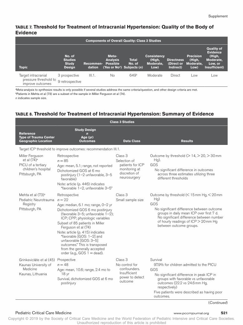
Copyright © 2019 by the Society of Critical Care Medicine and the World Federation of Pediatric Intensive and Critical Care Societies.Unauthorized reproduction of this article is prohibited
Supplement
Pediatric Critical Care Medicine www.pccmjournal.org S21
TABLE 7. Threshold for Treatment of Intracranial Hypertension: Quality of the Body of Evidence
Components of Overall Quality: Class 3 Studies
Topic
No. of Studies Study
DesignRecommen-
dation
Meta- Analysis Possible
(Yes or Noa)
Total No. of
Subjects (n)
Consistency (High,
Moderate, Low)
Directness (Direct or Indirect)
Precision (High,
Moderate, Low)
Quality of Evidence
(High, Moderate,
Low, or Insufficient)
Target intracranial pressure threshold to improve outcomes
3 prospective
9 retrospective
III.1. No 649b Moderate Direct Low Low
a Meta-analysis to synthesize results is only possible if several studies address the same criteria/question, and other design criteria are met.b Patients in Mehta et al (73) are a subset of the sample in Miller Ferguson et al (74).n indicates sample size.
TABLE 8. Threshold for Treatment of Intracranial Hypertension: Summary of Evidence
Class 3 Studies
Reference Type of Trauma Center Geographic Location
Study Design n
Age (yr) Outcomes Data Class Results
Target ICP threshold to improve outcomes: recommendation III.1.
Miller Ferguson et al (74)a
PICU of a tertiary children’s hospital
Pittsburgh, PA
Retrospectiven = 85Age: mean, 5.1; range, not reportedDichotomized GOS at 6 mo
postinjury (1–2 unfavorable, 3–5 favorable)
Note: article (p. 446) indicates “favorable 1–2, unfavorable 3–5”
Class 3Selection of
patients for ICP monitoring at discretion of neurosurgery
Outcome by threshold (> 14, > 20, > 30 mm Hg).
GOS No significant difference in outcomes
across three estimates utilizing three different thresholds
Mehta et al (73)a
Pediatric Neurotrauma Registry
Pittsburgh, PA
Retrospectiven = 22Age: median, 6.1 mo; range, 0–2 yrDichotomized GOS 6 mo postinjury
(favorable 3–5; unfavorable 1–2); ICP; CPP; physiologic variables
Subset of 85 patients in Miller Ferguson et al (74)
Note: article (p. 415) indicates “favorable [GOS: 1–2] and unfavorable [GOS: 3–5] outcomes.” This is transposed from the generally accepted order (e.g., GOS 1 = dead).
Class 3Small sample size
Outcome by threshold (< 15 mm Hg, < 20 mm Hg)
GOS No significant difference between outcome
groups in daily mean ICP over first 7 d. No significant difference between number of hourly readings of ICP > 20 mm Hg between outcome groups.
Grinkeviciūte et al (45)Kaunas University of
MedicineKaunas, Lithuania
Prospectiven = 48Age: mean, 10.6; range, 2.4 mo to
18 yrSurvival, dichotomized GOS at 6 mo
postinjury
Class 3No control for
confounders. Insufficient power to detect outcome
Survival 97.9% for children admitted to the PICUGOS No significant difference in peak ICP in
groups with favorable vs unfavorable outcomes (22.2 vs 24.6 mm Hg, respectively)
Five patients were described as having poor outcomes.
(Continued)
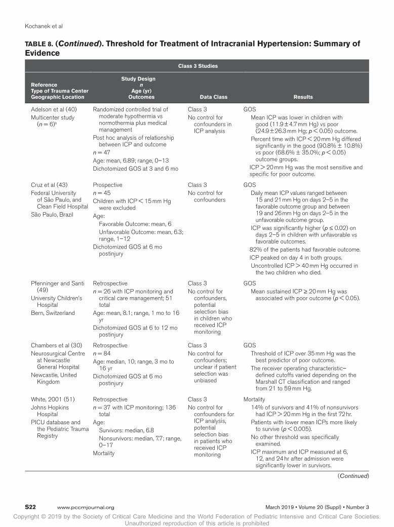
Copyright © 2019 by the Society of Critical Care Medicine and the World Federation of Pediatric Intensive and Critical Care Societies.Unauthorized reproduction of this article is prohibited
Copyright © 2019 by the Society of Critical Care Medicine and the World Federation of Pediatric Intensive and Critical Care Societies.Unauthorized reproduction of this article is prohibited
Kochanek et al
S22 www.pccmjournal.org March 2019 • Volume 20 (Suppl) • Number 3
Adelson et al (40)Multicenter study
(n = 6)b
Randomized controlled trial of moderate hypothermia vs normothermia plus medical management
Post hoc analysis of relationship between ICP and outcome
n = 47Age: mean, 6.89; range, 0–13Dichotomized GOS at 3 and 6 mo
Class 3No control for
confounders in ICP analysis
GOS Mean ICP was lower in children with
good (11.9 ± 4.7 mm Hg) vs poor (24.9 ± 26.3 mm Hg; p < 0.05) outcome.
Percent time with ICP < 20 mm Hg differed significantly in the good (90.8% ± 10.8%) vs poor (68.6% ± 35.0%; p < 0.05) outcome groups.
ICP > 20 mm Hg was the most sensitive and specific for poor outcome.
Cruz et al (43)Federal University
of São Paulo, and Clean Field Hospital
São Paulo, Brazil
Prospectiven = 45Children with ICP < 15 mm Hg
were excludedAge: Favorable Outcome: mean, 6 Unfavorable Outcome: mean, 6.3;
range, 1–12Dichotomized GOS at 6 mo
postinjury
Class 3No control for
confounders
GOS Daily mean ICP values ranged between
15 and 21 mm Hg on days 2–5 in the favorable outcome group and between 19 and 26 mm Hg on days 2–5 in the unfavorable outcome group.
ICP was significantly higher (p ≤ 0.02) on days 2–5 in children with unfavorable vs favorable outcomes.
82% of the patients had favorable outcome. ICP peaked on day 4 in both groups. Uncontrolled ICP > 40 mm Hg occurred in
the two children who died.
Pfenninger and Santi (49)
University Children’s Hospital
Bern, Switzerland
Retrospectiven = 26 with ICP monitoring and
critical care management; 51 total
Age: mean, 8.1; range, 1 mo to 16 yr
Dichotomized GOS at 6 to 12 mo postinjury
Class 3No control for
confounders, potential selection bias in children who received ICP monitoring
GOS Mean sustained ICP ≥ 20 mm Hg was
associated with poor outcome (p < 0.05).
Chambers et al (30)Neurosurgical Centre
at Newcastle General Hospital
Newcastle, United Kingdom
Retrospectiven = 84Age: median, 10; range, 3 mo to
16 yrDichotomized GOS at 6 mo
postinjury
Class 3No control for
confounders; unclear if patient selection was unbiased
GOS Threshold of ICP over 35 mm Hg was the
best predictor of poor outcome. The receiver operating characteristic–
defined cutoffs varied depending on the Marshall CT classification and ranged from 21 to 59 mm Hg.
White, 2001 (51)Johns Hopkins
HospitalPICU database and
the Pediatric Trauma Registry
Retrospectiven = 37 with ICP monitoring; 136
totalAge: Survivors: median, 6.8 Nonsurvivors: median, 7.7; range,
0–17Mortality
Class 3No control for
confounders for ICP analysis, potential selection bias in patients who received ICP monitoring
Mortality 14% of survivors and 41% of nonsurvivors
had ICP > 20 mm Hg in the first 72 hr. Patients with lower mean ICPs more likely
to survive (p < 0.005). No other threshold was specifically
examined. ICP maximum and ICP measured at 6,
12, and 24 hr after admission were significantly lower in survivors.
TABLE 8. (Continued). Threshold for Treatment of Intracranial Hypertension: Summary of Evidence
Class 3 Studies
Reference Type of Trauma Center Geographic Location
Study Design n
Age (yr) Outcomes Data Class Results
(Continued)
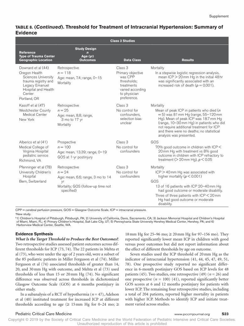
Copyright © 2019 by the Society of Critical Care Medicine and the World Federation of Pediatric Intensive and Critical Care Societies.Unauthorized reproduction of this article is prohibited
Supplement
Pediatric Critical Care Medicine www.pccmjournal.org S23
TABLE 8. (Continued). Threshold for Treatment of Intracranial Hypertension: Summary of Evidence
Class 3 Studies
Reference Type of Trauma Center Geographic Location
Study Design n
Age (yr) Outcomes Data Class Results
Downard et al (44)Oregon Health
Sciences University trauma registry and Legacy Emanuel Hospital and Health Center
Portland, OR
Retrospectiven = 118Age: mean, 7.4; range, 0–15Mortality
Class 3Primary objective
was CPP thresholds; treatments varied according to physician preference.
Mortality In a stepwise logistic regression analysis,
mean ICP > 20 mm Hg in the initial 48 hr was significantly associated with an increased risk of death (p = 0.001).
Kasoff et al (47)Westchester County
Medical CenterNew York
Retrospectiven = 25Age: mean, 8.8; range,
3 mo to 17 yrMortality
Class 3No control for
confounders, selection bias unclear
Mortality Mean of peak ICP in patients who died (n
= 5) was 81 mm Hg (range, 55–120 mm Hg). Mean of peak ICP was 18.7 mm Hg (range, 10–30 mm Hg) in patients who did not require additional treatment for ICP and there were no deaths; no statistical analysis was presented.
Alberico et al (41)Medical College of
Virginia Hospital pediatric service
Richmond, VA
Prospectiven = 100Age: mean, 13.39; range, 0–19GOS at 1-yr postinjury
Class 3No control for
confounders
GOS 70% good outcome in children with ICP <
20 mm Hg with treatment vs 8% good outcome in children with ICP refractory to treatment (> 20 mm Hg), p < 0.05
Pfenninger et al (78)University Children’s
HospitalBern, Switzerland
Retrospectiven = 24Age: mean, 6.6; range, 3 mo to 14
yrMortality; GOS (follow-up time not
specified)
Class 3No control for
confounders
Mortality ICP > 40 mm Hg was associated with
higher mortality (p < 0.001)GOS 13 of 16 patients with ICP 20–40 mm Hg
had good outcome or moderate disability. Three of three patients with ICP < 20 mm
Hg had good outcome or moderate disability.
CPP = cerebral perfusion pressure, GOS = Glasgow Outcome Scale, ICP = intracranial pressure.a New study.b 1) Children’s Hospital of Pittsburgh, Pittsburgh, PA; 2) University of California, Davis, Sacramento, CA; 3) Jackson Memorial Hospital and Children’s Hospital of Miami, Miami, FL; 4) Primary Children’s Hospital, Salt Lake City, UT; 5) Pennsylvania State University Hershey Medical Center, Hershey, PA; and 6) Harborview Medical Center, Seattle, WA.
Evidence SynthesisWhat Is the Target Threshold to Produce the Best Outcomes?. Two retrospective studies assessed patient outcomes across dif-ferent thresholds for ICP (73, 74). The 22 patients in Mehta et al (73), who were under the age of 2 years old, were a subset of the 85 pediatric patients in Miller Ferguson et al (74). Miller Ferguson et al (74) associated thresholds of greater than 14, 20, and 30 mm Hg with outcome, and Mehta et al (73) used thresholds of less than 15 or 20 mm Hg (74). No significant difference was observed across thresholds in dichotomized Glasgow Outcome Scale (GOS) at 6 months postinjury in either study.
In a subanalysis of a RCT of hypothermia (n = 47), Adelson et al (40) instituted treatment for increased ICP at different thresholds according to age (≥ 15 mm Hg for 0–24 mo; ≥
18 mm Hg for 25–96 mo; ≥ 20 mm Hg for 97–156 mo). They reported significantly lower mean ICP in children with good versus poor outcomes but did not report information about the effect of different thresholds by age on outcome.
Seven studies used the ICP threshold of 20 mm Hg as the indicator of intracranial hypertension (41, 44, 45, 47, 49, 51, 78). One prospective study reported no significant differ-ence in 6-month postinjury GOS based on ICP levels for 48 patients (45). Two studies, one retrospective (49) (n = 26) and one prospective (n = 100) (41), reported significantly better GOS scores at 6 and 12 months postinjury for patients with lower ICP. The remaining four retrospective studies, including a total of 204 patients, reported higher mortality in patients with higher ICP. Methods to identify ICP and initiate treat-ment varied across studies.
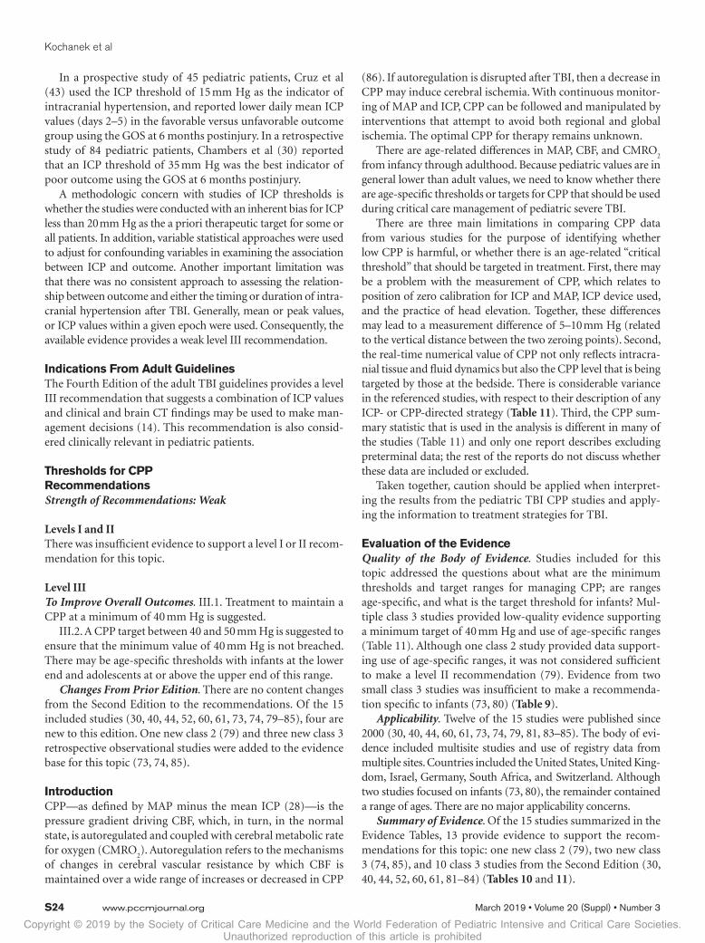
Copyright © 2019 by the Society of Critical Care Medicine and the World Federation of Pediatric Intensive and Critical Care Societies.Unauthorized reproduction of this article is prohibited
Copyright © 2019 by the Society of Critical Care Medicine and the World Federation of Pediatric Intensive and Critical Care Societies.Unauthorized reproduction of this article is prohibited
Kochanek et al
S24 www.pccmjournal.org March 2019 • Volume 20 (Suppl) • Number 3
In a prospective study of 45 pediatric patients, Cruz et al (43) used the ICP threshold of 15 mm Hg as the indicator of intracranial hypertension, and reported lower daily mean ICP values (days 2–5) in the favorable versus unfavorable outcome group using the GOS at 6 months postinjury. In a retrospective study of 84 pediatric patients, Chambers et al (30) reported that an ICP threshold of 35 mm Hg was the best indicator of poor outcome using the GOS at 6 months postinjury.
A methodologic concern with studies of ICP thresholds is whether the studies were conducted with an inherent bias for ICP less than 20 mm Hg as the a priori therapeutic target for some or all patients. In addition, variable statistical approaches were used to adjust for confounding variables in examining the association between ICP and outcome. Another important limitation was that there was no consistent approach to assessing the relation-ship between outcome and either the timing or duration of intra-cranial hypertension after TBI. Generally, mean or peak values, or ICP values within a given epoch were used. Consequently, the available evidence provides a weak level III recommendation.
Indications From Adult GuidelinesThe Fourth Edition of the adult TBI guidelines provides a level III recommendation that suggests a combination of ICP values and clinical and brain CT findings may be used to make man-agement decisions (14). This recommendation is also consid-ered clinically relevant in pediatric patients.
Thresholds for CPPRecommendationsStrength of Recommendations: Weak
Levels I and IIThere was insufficient evidence to support a level I or II recom-mendation for this topic.
Level IIITo Improve Overall Outcomes. III.1. Treatment to maintain a CPP at a minimum of 40 mm Hg is suggested.
III.2. A CPP target between 40 and 50 mm Hg is suggested to ensure that the minimum value of 40 mm Hg is not breached. There may be age-specific thresholds with infants at the lower end and adolescents at or above the upper end of this range.
Changes From Prior Edition. There are no content changes from the Second Edition to the recommendations. Of the 15 included studies (30, 40, 44, 52, 60, 61, 73, 74, 79–85), four are new to this edition. One new class 2 (79) and three new class 3 retrospective observational studies were added to the evidence base for this topic (73, 74, 85).
IntroductionCPP—as defined by MAP minus the mean ICP (28)—is the pressure gradient driving CBF, which, in turn, in the normal state, is autoregulated and coupled with cerebral metabolic rate for oxygen (CMRO
2). Autoregulation refers to the mechanisms
of changes in cerebral vascular resistance by which CBF is maintained over a wide range of increases or decreased in CPP
(86). If autoregulation is disrupted after TBI, then a decrease in CPP may induce cerebral ischemia. With continuous monitor-ing of MAP and ICP, CPP can be followed and manipulated by interventions that attempt to avoid both regional and global ischemia. The optimal CPP for therapy remains unknown.
There are age-related differences in MAP, CBF, and CMRO2
from infancy through adulthood. Because pediatric values are in general lower than adult values, we need to know whether there are age-specific thresholds or targets for CPP that should be used during critical care management of pediatric severe TBI.
There are three main limitations in comparing CPP data from various studies for the purpose of identifying whether low CPP is harmful, or whether there is an age-related “critical threshold” that should be targeted in treatment. First, there may be a problem with the measurement of CPP, which relates to position of zero calibration for ICP and MAP, ICP device used, and the practice of head elevation. Together, these differences may lead to a measurement difference of 5–10 mm Hg (related to the vertical distance between the two zeroing points). Second, the real-time numerical value of CPP not only reflects intracra-nial tissue and fluid dynamics but also the CPP level that is being targeted by those at the bedside. There is considerable variance in the referenced studies, with respect to their description of any ICP- or CPP-directed strategy (Table 11). Third, the CPP sum-mary statistic that is used in the analysis is different in many of the studies (Table 11) and only one report describes excluding preterminal data; the rest of the reports do not discuss whether these data are included or excluded.
Taken together, caution should be applied when interpret-ing the results from the pediatric TBI CPP studies and apply-ing the information to treatment strategies for TBI.
Evaluation of the EvidenceQuality of the Body of Evidence. Studies included for this topic addressed the questions about what are the minimum thresholds and target ranges for managing CPP; are ranges age-specific, and what is the target threshold for infants? Mul-tiple class 3 studies provided low-quality evidence supporting a minimum target of 40 mm Hg and use of age-specific ranges (Table 11). Although one class 2 study provided data support-ing use of age-specific ranges, it was not considered sufficient to make a level II recommendation (79). Evidence from two small class 3 studies was insufficient to make a recommenda-tion specific to infants (73, 80) (Table 9).
Applicability. Twelve of the 15 studies were published since 2000 (30, 40, 44, 60, 61, 73, 74, 79, 81, 83–85). The body of evi-dence included multisite studies and use of registry data from multiple sites. Countries included the United States, United King-dom, Israel, Germany, South Africa, and Switzerland. Although two studies focused on infants (73, 80), the remainder contained a range of ages. There are no major applicability concerns.
Summary of Evidence. Of the 15 studies summarized in the Evidence Tables, 13 provide evidence to support the recom-mendations for this topic: one new class 2 (79), two new class 3 (74, 85), and 10 class 3 studies from the Second Edition (30, 40, 44, 52, 60, 61, 81–84) (Tables 10 and 11).

Copyright © 2019 by the Society of Critical Care Medicine and the World Federation of Pediatric Intensive and Critical Care Societies.Unauthorized reproduction of this article is prohibited
Supplement
Pediatric Critical Care Medicine www.pccmjournal.org S25
TABLE 9. Cerebral Perfusion Pressure: Quality of the Body of Evidence
TopicNo. of Studies Study Design
Recommen-dations
Meta- Analysis Pos-
sible (Yes or Noa)
Total No. of
Subjects (n)
Consistency (High,
Moderate, Low)
Directness (Direct or Indirect)
Precision (High,
Moderate, Low)
Quality of Evidence
(High, Moderate,
Low, or Insufficient)
Components of overall quality: class 2 study
CPP threshold Mixed ages
1 retrospective III.2. NA 317 NA Direct Low Low
Components of overall quality: class 3 studies
CPP threshold Mixed ages
1 prospective2 retrospective9 treatment series
III.1.III.2.
No; different thresholds; different populations
836 Low Direct Low Low
CPP threshold for infants < 2 yr old
1 retrospective
1 treatment series
None No 39 Moderate Direct Low Insufficient
CPP = cerebral perfusion pressure, NA = not applicable.a Meta-analysis to synthesize results is only possible if several studies address the same criteria/question, and other design criteria are met.n indicates sample size.
TABLE 10. Cerebral Perfusion Pressure: Summary of Evidence
Reference Type of Trauma Center Geographic Location
Study Design
n Age (yr)
Outcomes Data Class Results
Class 2 study
CPP age-specific ranges: recommendation III.2.
Allen et al (79)a
22 (of 46 designated) trauma centers in New York: 20 are level I centers and two level II
New York
Retrospectiven = 317 pediatricAge: 0–5: 55 6–11: 65 12–17: 197Mortality at 14 d postinjury,
CPP, hypotension, elevated ICP
Class 2Relevant criteria
for minimizing bias were met
Predefined CPP thresholds by age group 40 and 30 mm Hg for 0–5 yr 50 and 35 mm Hg for 6–11 yr 60 and 50 for 12 yr old and olderCPP categories CPP-H: events above high threshold CPP-B: events between high and low threshold CPP-L: events below low thresholdMortality Rates of survival and relative risk of mortality at 14 d
postinjury: Regarding age (0–11 vs 12–17 yr), there was
a difference between survivors (n = 101) and nonsurvivors (n = 19) for CPP-B.
For CPP-L, there was a significant difference in the 0- to 11-yr-olds, not present in 12- to 17-yr-olds.
Refer to Tables 3 and 4 in the publication for specific data about relationship between mortality and CPP thresholds by age.
Data suggest that CPP targets should be age specific: Above 50 mm Hg in 6- to 17-yr-olds Above 44 mm Hg in 0- to 5-yr-olds
(Continued)

Copyright © 2019 by the Society of Critical Care Medicine and the World Federation of Pediatric Intensive and Critical Care Societies.Unauthorized reproduction of this article is prohibited
Copyright © 2019 by the Society of Critical Care Medicine and the World Federation of Pediatric Intensive and Critical Care Societies.Unauthorized reproduction of this article is prohibited
Kochanek et al
S26 www.pccmjournal.org March 2019 • Volume 20 (Suppl) • Number 3
Class 3 studies
Minimum threshold and target ranges for managing CPP: recommendations III.1. and III.2.
Miller Ferguson et al (74)a
PICU of a tertiary children’s hospital
Pittsburgh, PA
Retrospectiven = 85Age: mean, 5.1; range, NRDichotomized GOS
at 6 mo postinjury (1–2 unfavorable, 3–5 favorable)
Note: article (p. 446) indicates “favorable 1 to 2, unfavorable 3 to 5.”
Class 3Selection of
patients for ICP monitoring at discretion of neurosurgery
Outcome by threshold (< 40, < 45, < 50, < 55, < 60 mm Hg during first 5 d after ICP monitor placement)
GOS No significant difference in outcomes across five
estimates utilizing five different thresholds.
Vavilala et al (85)a
Five Pediatric Trauma Centers
Seattle, WA; Pittsburg PA; Chicago, IL; Torrance, CA; Columbus, OH
Retrospectiven = 236Age: mean, 8.0; range, NRDichotomized discharge GOS
Class 3Outcome
assessment not blinded, unclear if groups are similar, some, but not all key confounders controlled for
Discharge GOS (survivors only) Adjusted risk ratio (95% CI) Reference: any CPP ≤ 40 (no surgery)Operating room All CPP > 40 mm Hg 0.61 (0.58–0.64)ICU All CPP > 40 mm Hg 0.73 (0.63–0.84) CPP > 40 mm Hg was associated with favorable
discharge GOS
Kapapa et al (83)
(Location not cited)
Treatment seriesAnalysis of CPP in relation to
age-specific lower limit (up to 1 mo, > 40 mm Hg; 2 mo up to 1 yr, > 45 mm Hg; 1 yr up to 7 yr, > 50 mm Hg; > 7 yr, 55–60 mm Hg)
n = 36Age: mean, NR; range, 0–16Dichotomized GOS at varied
timepoints
Class 3Uncontrolled
series
GOS Patients with CPP values below the age-specific lower
limit for just a single occurrence had a significantly worse outcome (p = 0.013).
Chaiwat et al (81)
Level I pediatric trauma center
Seattle, WA
Treatment seriesn = 36 patients (two inflicted
TBI)Age: mean, 9.1; range, 0.8–16Dichotomized GOS
dichotomized at 6 mo postinjury
Class 3Uncontrolled
series
GOS On univariate analysis, CPP < 40 mm Hg during the first
72 hr had no association with poor outcome. When logistic regression was performed, using a number
of factors, only impaired autoregulatory index remained an independent predictor of poor outcome.
Figaji et al (61)
Cape Town, South Africa
Treatment seriesn = 52Age: mean, NR; range, 9 mo
to 14 yrDichotomized GOS at ≥ 6 mo
postinjury
Class 3Uncontrolled
series
GOS Median (interquartile range) for lowest CPP was
significantly lower in patients with unfavorable outcome: 29 mm Hg (20–45 mm Hg) vs 44 mm Hg (35–51 mm Hg), p = 0.023
Patients with unfavorable outcome had more episodes of CPP < 40 mm Hg: 3 (0–10) vs 0 (0–1), p = 0.03
There was no difference in the number of episodes of CPP < 50 mm Hg.
TABLE 10. (Continued). Cerebral Perfusion Pressure: Summary of Evidence
Reference Type of Trauma Center Geographic Location
Study Design n Age (yr)
Outcomes Data Class Results
(Continued)
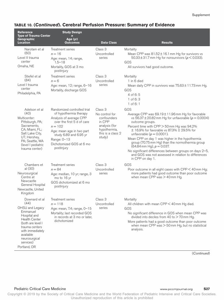
Copyright © 2019 by the Society of Critical Care Medicine and the World Federation of Pediatric Intensive and Critical Care Societies.Unauthorized reproduction of this article is prohibited
Supplement
Pediatric Critical Care Medicine www.pccmjournal.org S27
Narotam et al (60)
Level II trauma center
Omaha, NE
Treatment seriesn = 16Age: mean, 14; range,
1.5–18Mortality, GOS at 3 mo
postinjury
Class 3Uncontrolled
series
Mortality Mean CPP was 81.52 ± 16.1 mm Hg for survivors vs
50.33 ± 31.7 mm Hg for nonsurvivors (p < 0.033).GOS All survivors had good outcome.
Stiefel et al (84)
Level I trauma center
Philadelphia, PA
Treatment seriesn = 6Age: mean, 12; range, 6–16Mortality, discharge GOS
Class 3Uncontrolled
series
Mortality 1 in 6 died Mean daily CPP in survivors was 75.63 ± 11.73 mm Hg.GOS 4 of 6: 5 1 of 6: 3 1 of 6: 1
Adelson et al (40)
Multicenter: Pittsburgh, PA; Sacramento, CA; Miami, FL; Salt Lake City, UT; Hershey, PA; Seattle, WA (level I pediatric trauma center)
Randomized controlled trial of hypothermia therapy
Analysis of average CPP over the first 5 d of care
n = 102Age: mean age in two part
study 6.89 and 6.95 yrRange: 0–13Dichotomized GOS at 6 mo
postinjury
Class 3No control for
confounders in CPP analysis (for hypothermia, this is a class 2 study)
GOS Average CPP was 69.19 ± 11.96 mm Hg for favorable
vs 56.37 ± 20.82 mm Hg for unfavorable (p = 0.0004) outcome groups.
Percent time with CPP > 50 mm Hg was 94.2% ± 16.9% for favorable vs 87.3% ± 29.5% for unfavorable (p = 0.0001).
Mean CPP on day 1 was higher in the hypothermia group (70.75 mm Hg) than the normothermia group (64.84 mm Hg), p = 0.037.
No significant differences between groups on days 2–5, and GOS was not assessed in relation to differences in CPP on day 1.
Chambers et al (30)
Neurosurgical Centre at Newcastle General Hospital
Newcastle, United Kingdom
Treatment seriesn = 84Age: median, 10 yr; range, 3
mo to 16 yrGOS dichotomized at 6 mo
postinjury
Class 3Uncontrolled
series
GOS Poor outcome in all eight cases with CPP < 40 mm Hg;
more patients had good outcome than poor outcome when mean CPP was > 40 mm Hg.
Downard et al (44)
OHSU and Legacy Emmanuel Hospital and Health Center (both are level I trauma centers with immediately available neurosurgical services)
Portland, OR
Treatment seriesn = 118Age: mean, 7.4; range, 0–15Mortality; last recorded GOS
in records at 3 mo or later, dichotomized
Class 3Uncontrolled
series
Mortality All children with mean CPP < 40 mm Hg died.GOS No significant difference in GOS when mean CPP was
divided into deciles from 40 to > 70 mm Hg. More patients had a good outcome than poor outcome
when mean CPP was > 50 mm Hg, but no statistical analysis.
TABLE 10. (Continued). Cerebral Perfusion Pressure: Summary of Evidence
Reference Type of Trauma Center Geographic Location
Study Design n
Age (yr) Outcomes Data Class Results
(Continued)
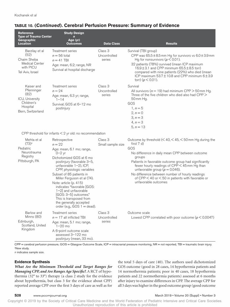
Copyright © 2019 by the Society of Critical Care Medicine and the World Federation of Pediatric Intensive and Critical Care Societies.Unauthorized reproduction of this article is prohibited
Copyright © 2019 by the Society of Critical Care Medicine and the World Federation of Pediatric Intensive and Critical Care Societies.Unauthorized reproduction of this article is prohibited
Kochanek et al
S28 www.pccmjournal.org March 2019 • Volume 20 (Suppl) • Number 3
Evidence SynthesisWhat Are the Minimum Threshold and Target Ranges for Managing CPP, and Are Ranges Age Specific?. A RCT of hypo-thermia (32° to 33°) therapy (a class 2 study for the evidence about hypothermia, but class 3 for the evidence about CPP) reported average CPP over the first 5 days of care as well as for
the total 5 days of care (40). The authors used dichotomized GOS outcome (good in 28 cases, 14 hypothermia patients and 14 normothermia patients; poor in 40 cases, 18 hypothermia patients and 22 normothermia patients) assessed at 6 months after injury to examine differences in CPP. The average CPP for all 5 days was higher in the good outcome group (good outcome
Barzilay et al (52)
Chaim Sheba Medical Center with PICU
Tel Aviv, Israel
Treatment seriesn = 56 totaln = 41 TBIAge: mean, 6.2; range, NRSurvival at hospital discharge
Class 3Uncontrolled
series
Survival (TBI group) CPP was 65.5 ± 8.5 mm Hg for survivors vs 6.0 ± 3.9 mm
Hg for nonsurvivors (p < 0.01). 32 patients (78%) survived (mean ICP maximum
16.9 ± 3.1 and CPP minimum 65.5 ± 8.5 torr) compared with nine patients (22%) who died (mean ICP maximum 53.7 ± 10.8 and CPP minimum 6 ± 3.9 torr) (p < 0.01).
Kaiser and Pfenninger (82)
ICU, University Children’s Hospital
Bern, Switzerland
Treatment seriesn = 24Age: mean, 6.3 yr; range,
1–14Survival; GOS at 6–12 mo
postinjury
Class 3Uncontrolled
series
Survival All survivors (n = 19) had minimum CPP > 50 mm Hg.
Three of the five children who died also had CPP > 50 mm Hg.
GOS 1, n = 5 2, n = 0 3, n = 3 4, n = 3 5, n = 13
CPP threshold for infants < 2 yr old: no recommendation
Mehta et al (73)a
Pediatric Neurotrauma Registry
Pittsburgh, PA
Retrospectiven = 22Age: mean, 6.1 mo; range,
0–2 yrDichotomized GOS at 6 mo
postinjury (favorable 3–5, unfavorable 1–2), ICP, CPP, physiologic variables
Subset of 85 patients in Miller Ferguson et al (74).
Note: article (p. 415) indicates “favorable [GOS: 1–2] and unfavorable [GOS: 3–5] outcomes.” This is transposed from the generally accepted order (e.g., GOS 1 = dead).
Class 3Small sample size
Outcome by threshold (< 40, < 45, < 50 mm Hg during the first 7 d)
GOS No difference in daily mean CPP between outcome
groups Patients in favorable outcome group had significantly
fewer hourly readings of CPP < 45 mm Hg than unfavorable group (p = 0.046).
No difference between number of hourly readings of CPP < 40 or < 50 in patients with favorable or unfavorable outcomes
Barlow and Minns (80)
Edinburgh, Scotland, United Kingdom
Treatment seriesn = 17 all inflicted TBIAge: mean, 5.1 mo; range,
1–20 moA 6-point outcome scale
assessed 3–122 mo postinjury (mean, 33 mo).
Class 3Uncontrolled
series
Outcome scale Lowest CPP correlated with poor outcome (p < 0.0047)
CPP = cerebral perfusion pressure, GOS = Glasgow Outcome Scale, ICP = intracranial pressure monitoring, NR = not reported, TBI = traumatic brain injury.a New study.n indicates sample size.
TABLE 10. (Continued). Cerebral Perfusion Pressure: Summary of Evidence
Reference Type of Trauma Center Geographic Location
Study Design n
Age (yr) Outcomes Data Class Results

Copyright © 2019 by the Society of Critical Care Medicine and the World Federation of Pediatric Intensive and Critical Care Societies.Unauthorized reproduction of this article is prohibited
Supplement
Pediatric Critical Care Medicine www.pccmjournal.org S29
TABLE 11. Summary of Treatments, Cerebral Perfusion Pressure Target, and Cerebral Perfusion Pressure Statistics Used in Studies
Variations in Treatment and Measurement of CPP
Reference
Treatments Used
CPP Target Strategy
CPP Statistic Used Time PeriodHyperventilation
Induced Hypothermia
Barbiturate Coma
Decompressive Craniectomy
Adelson et al (40)
NO YES YES YES Age-related
45 mm Hg
Mean CPP
Days 1–5
Allen et al (79)a Threshold Threshold Threshold Threshold Threshold Number of events below minimum, above maximum, or between thresholds
Any time during ICU stay
Barlow and Minns (80)
Threshold Threshold Threshold Threshold Threshold Lowest CPPTiming NR
Barzilay et al (52) YES YES YES NO — Lowest CPPTiming NR
Chaiwat et al (81) — — — — — Lowest CPPFirst 72 hr
Chambers et al (30)
Threshold Threshold Threshold Threshold Threshold Minimum CPPRange: 6.3–173 hr;
median: 41.2
Downard et al (44)
YES NO NO YES Age-related45 mm Hg
Mean CPPFirst 48 hr
Figaji et al (61) YES YES YES YES Age-related45 mm Hg
Initial and lowest CPPMean duration
123.7 ± 67.1 hr
Kaiser and Pfenninger (82)
YES YES YES NO — Mean CPPMean time of 9 d; range
1–15 d
Kapapa et al (83) YES YES YES YES Age-related40 mm Hg
Lowest CPPAny point during
hospitalization with monitor
Mehta et al (73)a NO YES YES YES ICP of 20 and CPP 50 mm Hg
Mean CPPDaily mean for first 7 d
Miller Ferguson et al (74)a
NO YES YES YES ICP of 20 and CPP 50 mm Hg
Mean CPP daily mean for first 5 d
Narotam et al (60)
YES NO NO NO ICP-related Mean CPPTiming NR
Stiefel et al (84) — — — — Age-related40 mm Hg
Mean CPPFrom admission for at
least 72 hr
Vavilala et al (85)a YES — YES YES CPP > 40 mm Hg
Below or above threshold
At any time in operating room or ICU
CPP = cerebral perfusion pressure, ICP = intracranial pressure monitoring, NR = not reported.a New study.In the studies that describe therapy, “YES” denotes use of therapy and “NO” denotes where treatment is not used. “Threshold” denotes where the study is aimed at defining a threshold about burden from CPP insult and outcome, rather than it being an intervention study. Dash indicates where no information is given in the report.

Copyright © 2019 by the Society of Critical Care Medicine and the World Federation of Pediatric Intensive and Critical Care Societies.Unauthorized reproduction of this article is prohibited
Copyright © 2019 by the Society of Critical Care Medicine and the World Federation of Pediatric Intensive and Critical Care Societies.Unauthorized reproduction of this article is prohibited
Kochanek et al
S30 www.pccmjournal.org March 2019 • Volume 20 (Suppl) • Number 3
69.19 ± 11.96 mm Hg vs poor outcome 56.37 ± 20.82 mm Hg; p = 0.0004). In addition, the percent time with CPP greater than 50 mm Hg was higher in the good outcome group (good outcome 94.2% ± 16.9% vs poor outcome 87.3% ± 29.5%; p = 0.0001). In contrast, a recent retrospective study of 85 pedi-atric patients with severe TBI (mean age, 5.1 ± 0.8 yr) analyzed outcome in five threshold groups (< 40, 45, 50, 55, and 60 mm Hg). No significant difference across groups was found in the dichotomized GOS at 6 months postinjury.
Five treatment series found higher CPP associated with better outcomes and their findings were as follows (52, 60, 61, 80, 84). Barzilay et al (52) studied 41 consecutive TBI admissions to their PICU with coma for at least 6 hours before admission. Survivors had higher minimum CPP than nonsurvivors (65.5 ± 8.5 vs 6.0 ± 3.9 mm Hg; p < 0.01). All patients with head trauma were treated for 5 days with dexamethasone, and neither the tim-ing nor duration of ICP and CPP derangements was specified. “Preterminal” data were not removed, resulting in the low aver-age CPP for nonsurvivors. Figaji et al (61) studied prospectively 52 children with TBI and found median lowest CPP experienced during the course of monitoring was higher in those with better outcome. By using dichotomized GOS outcome assessed at least 6 months after injury, those with favorable outcome had higher low-est CPP median (interquartile range) of 44 mm Hg (35–51 mm Hg) versus 29 mm Hg (20–45 mm Hg) in comparison with those with unfavorable outcome (p = 0.023). Narotam et al (60) ana-lyzed data from 16 children 1.5–18 years old (mean, 14 yr), 15 of whom had GCS less than or equal to 8. All 10 survivors had GOS 5 at 3 months. Mean CPP was higher in survivors (81.52 ± 16.1 mm Hg) than nonsurvivors (50.33 ± 31.7 mm Hg; p = 0.033). Stiefel et al (84) studied brain tissue oxygen monitoring in six patients (age, 6–14 yr old; GCS = 3–7) and found that mean daily CPP in the five survivors was 75.63 ± 11.73 mm Hg. Last, in a sample of TBI cases restricted to 17 young children with inflicted injury (age, 1–20 mo old; mean, 5.1 mo), Barlow and Minns (80) reported that lowest CPP during intensive care was associated with poorer outcomes in a 6-point scale 3–122 months (mean, 33 mo) after injury (p = 0.0047).
Four studies reported findings in relation to a threshold in CPP of 40 mm Hg (30, 44, 61, 81). In the study reported by Figaji et al (61), the authors found that more episodes of CPP less than 40 mm Hg were observed in those with unfavorable (3 [0–10]) versus favorable (0 [0–1]) outcome (p = 0.0.03); a more complex relationship between CPP and outcome involved data from auto-regulation of CBF. Chambers et al (30) analyzed 84 children age 3 months to 16 years old (median, 10 yr) and examined minimum CPP in relation to dichotomized GOS at 6 months. Sixty-three out of 76 cases with CPP greater than 40 mm Hg had good out-come, and all eight cases with CPP less than 40 mm Hg had poor outcome (p < 0.0001; Fisher exact test). Downard et al (44) ana-lyzed 118 pediatric TBI cases of children up to 15 years old (mean age, 7.4 yr; 99 cases with GCS = 3–8) and reported dichotomized GOS at 3 months or later in relation to CPP thresholds. Seventy-two out of 96 patients with CPP greater than 40 mm Hg had good outcome, whereas all 22 cases with CPP less than 40 mm Hg died. The difference in mortality was statistically significant
(p < 0.0001, Fisher exact test). Chaiwat et al (81) analyzed 36 cases of TBI for predictors of poor outcome. ICP of greater than 20 mm Hg and CPP less than 40 mm Hg during the first 72 hours were not associated with outcome. However, on logistic regres-sion, an estimate of impaired CBF autoregulation using Doppler ultrasonography—the autoregulatory index (ARI)—was an independent predictor of poor outcome (adjusted odds ratio [OR], 23.1; 95% CI, 1.9–279.0). Impaired ARI was an indepen-dent risk factor when the authors entered CPP less than 40 mm Hg, SBP less than fifth percentile for age and gender during the first 72 hours after TBI, low middle cerebral artery (MCA) veloc-ity, and impaired ARI into the model (adjusted OR, 29.8; 95% CI, 1.7–521.4). Because ARI is calculated as the percent change in cerebrovascular resistance (CVR) per percent change in CPP, and CVR is defined as the ratio of CPP to MCA velocity, it is impos-sible to disentangle the relationship between outcome and CPP. ARI represents a research tool.
Five class 3 studies contain data concerning CPP threshold above 40 mm Hg (3, 5, 10, 12, 14). Two retrospective treatment series support the idea that there may be an age-related CPP thresh-old above 40 mm Hg. Kapapa et al (83) analyzed 16 children under 16 years old and reported dichotomized GOS in relation to age-specific lower limits in CPP (i.e., > 40 mm Hg, infants up to 1 mo old; > 45 mm Hg, infants 2 mo to 1 yr old; > 50 mm Hg, children between 1 and 7 yr old; 55–60 mm Hg, children > 7 yr old). The authors found that patients with CPP value below the age-specific lower limit for just a single occurrence had a significantly worse outcome (p = 0.013). Kaiser and Pfenninger (82) reported findings in 24 consecutive admissions to their PICU of patients with a GCS less than 8, average age 6.3 years (10 patients between 1 and 5 yr old), and showed that all survivors had CPP greater than 50 mm Hg (p < 0.005, Fisher exact test). Two studies did not observe a threshold above 40 mm Hg (44, 61). In the study reported by Figaji et al (61) (see above), the authors also reported outcome in relation to the number of episodes during monitoring that CPP was less than 50 mm Hg: there was no difference in the number of episodes in those with unfavorable (8 [2–18.5]) versus favorable (3 [0–8.8]) outcome (p = 0.137). Of note, two thirds of the children in this series were younger than 8 years old. As discussed above, in the study reported by Downard et al (44), 100% of children with mean CPP less than 40 mm Hg died when compared with only 25% of children who had a CPP greater than 40 mm Hg. The difference in mortality was statistically significant (p < 0.0001, Fisher exact test). Last, in the study of young children with abusive TBI reported by Barlow and Minns (80) (see above), only one infant in the series of 17 had lowest CPP greater than 50 mm Hg.
These studies, in aggregate, suggest that in the pediatric age range, there may be an age-related threshold between 40 and 50 mm Hg, with infants at the lower end and adolescents at the upper end of this range. Finally, studies specifically focused on assessment of the optimal upper limit for CPP management in pediatric TBI were lacking.
Indications From Adult GuidelinesThe recent Fourth Edition of the adult guidelines does not fur-ther inform the pediatric guidelines for this topic (14).

Copyright © 2019 by the Society of Critical Care Medicine and the World Federation of Pediatric Intensive and Critical Care Societies.Unauthorized reproduction of this article is prohibited
Supplement
Pediatric Critical Care Medicine www.pccmjournal.org S31
TREATMENTS
Hyperosmolar Therapy
RecommendationsStrength of Recommendations: Weak
Level IThere was insufficient evidence to support a level I recommen-dation for this topic.
Level IIFor ICP Control. II.1. Bolus HTS (3%) is recommended in patients with intracranial hypertension. Recommended effec-tive doses for acute use range between 2 and 5 mL/kg over 10–20 minutes.Level IIIFor ICP Control. III.1. Continuous infusion HTS is suggested in patients with intracranial hypertension. Suggested effective doses as a continuous infusion of 3% saline range between 0.1 and 1.0 mL/kg of body weight per hour, administered on a slid-ing scale. The minimum dose needed to maintain ICP less than 20 mm Hg is suggested.
III.2. Bolus of 23.4% HTS is suggested for refractory ICP. The suggested dose is 0.5 mL/kg with a maximum of 30 mL.
Safety Recommendation (applies to all recommendations for this topic). In the context of multiple ICP-related therapies, avoiding sustained (> 72 hr) serum sodium greater than 170 mEq/L is suggested to avoid complications of thrombocytope-nia and anemia, whereas avoiding a sustained serum sodium greater than 160 mEq/L is suggested to avoid the complication of deep vein thrombosis (DVT).
Note. Although mannitol is commonly used in the manage-ment of raised ICP in pediatric TBI, no studies meeting inclu-sion criteria were identified for use as evidence for this topic.
Changes From Prior Edition. Recommendation II.1. is new to this Third Edition. It replaces the level II recommendation from the Second Edition suggesting the use of HTS in general, with doses ranging between 6.5 and 10 mL/kg. Recommenda-tions III.2. is new to this Third Edition. The recommendation from the Second Edition to maintain serum osmolarity less than 360 mOsm/L was removed from this edition. The Safety Recommendation is new to this edition. One new class 2 pro-spective observational study (2) and five new class 3 studies—four retrospective observational (85, 87–89) and one treatment series (90)—were added to the evidence base for this topic.
IntroductionHyperosmolar Therapy for Intracranial Hypertension. IV administration of hyperosmolar agents was shown to reduce ICP in 1919 (91). Mannitol was introduced into clinical use in 1961 (92). Despite widespread use of a number of osmolar agents (mannitol, urea, and glycerol) up until the late 1970s (93, 94), mannitol gradually became the agent of choice to manage ICP (93). Subsequently, HTS was introduced in the 1990s (95), and although both are currently used, HTS use has increased while mannitol use has decreased (96). Additionally, a goal of euvolemia rather than dehydration as a therapeutic target is achieved by
assessing fluid balance, central venous pressure, urine output, blood urea nitrogen, serum creatinine, and clinical examination. The placement of a Foley catheter is also routinely used to quan-tify urine output and to avoid potential bladder rupture.
Mannitol. Mannitol is commonly used to manage raised ICP in pediatric TBI (96). Despite this fact, mannitol has not been subjected to contemporary controlled clinical trials ver-sus placebo, other osmolar agents, or other therapies in chil-dren. Most of the investigations on the use of mannitol were carried out in the 1970s on mixed disease populations in both children and adults (93).
Mannitol can reduce ICP by two distinct mechanisms. Mannitol can reduce ICP by reducing blood viscosity. This effect is immediate and results from a viscosity-mediated reflex vasoconstriction (intact autoregulation) which allows CBF to be maintained despite a reduced level of cerebral blood volume (CBV) (97, 98). The effect of mannitol administration on blood viscosity is transient (< 75 min) (97). Mannitol administration also reduces ICP by an osmotic effect, which develops more slowly (over 15–30 min), due to the gradual movement of water from the brain parenchyma into the systemic circulation. The effect persists up to 6 hours and requires an intact blood-brain barrier (99, 100). Mannitol may accumulate in injured brain regions (101), where a reverse osmotic shift may occur—with fluid moving from the vas-cular compartment into the brain parenchyma, possibly increas-ing ICP. This phenomenon has been suggested to occur when mannitol is used for extended periods of time (102, 103).
Mannitol is excreted unchanged in urine, and a risk of the development of acute tubular necrosis and renal failure has been suggested with mannitol administration with serum osmolarity levels greater than 320 mOsm in adults (104–106). However, the literature supporting this finding is limited in scope and was generated at a time when dehydration rather than euvolemia was the therapeutic target.
HTS. In the initial description in 1919 of the reduction in ICP by IV administration of hyperosmolar agents, HTS was the agent used (91). Its use in the treatment of increased ICP, however, failed to gain clinical acceptance. Resurgence in inter-est in this treatment emerged in the late 1980s (107), leading to the studies providing current evidence.
Like mannitol, the penetration of sodium across the blood-brain barrier is low (108). Sodium thus shares both the favor-able rheologic and osmolar gradient effects involved in the reduction in ICP. HTS has other theoretical beneficial effects including restoration of normal cellular resting membrane potential and cell volume (109, 110), stimulation of arterial natriuretic peptide release (111), inhibition of inflammation (108), and enhancement of cardiac output (112). Possible side effects of HTS include rebound in ICP, central pontine myelin-olysis, renal impairment, subarachnoid hemorrhage, natriure-sis, high urinary water losses, hyperchloremic acidosis, and masking of the development of diabetes insipidus (108).
Much higher levels of serum osmolarity (≈360 to 370 mOsm) may be tolerated in children when induced with HTS (87, 113) versus mannitol although this point remains contro-versial (35).
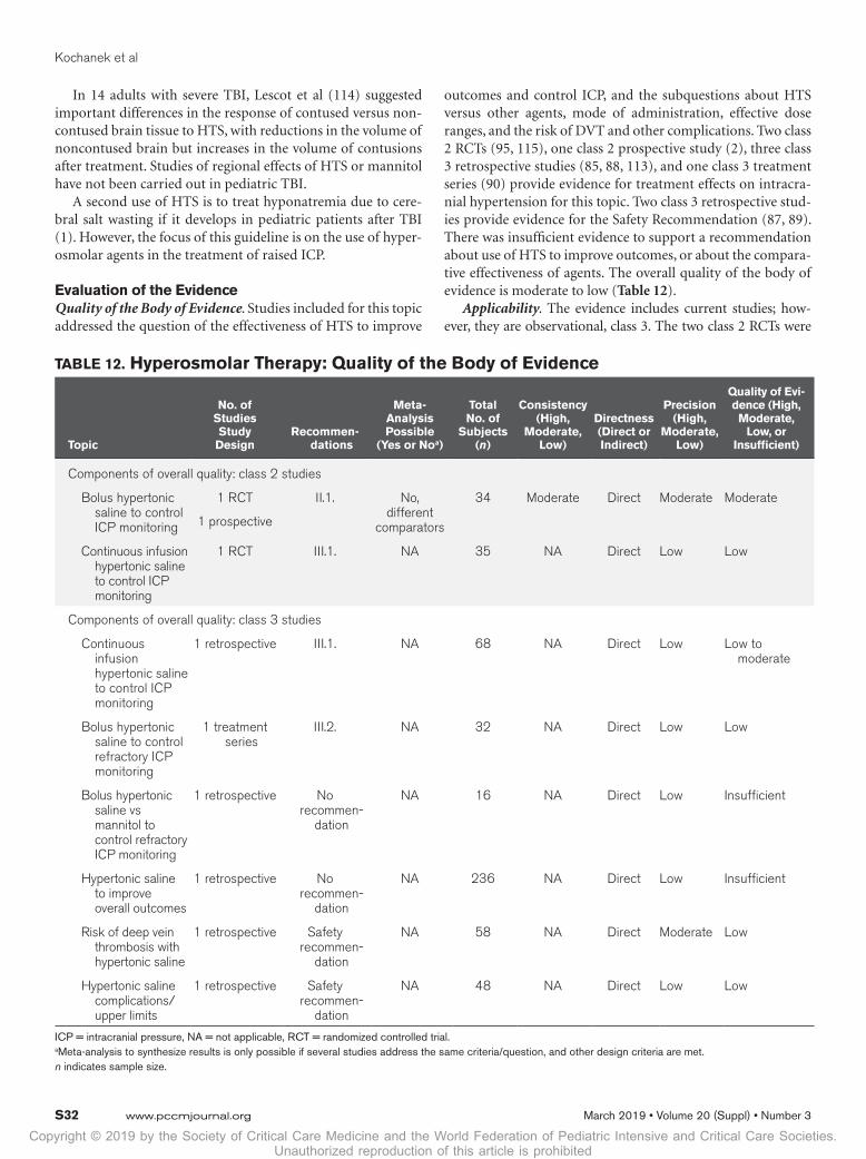
Copyright © 2019 by the Society of Critical Care Medicine and the World Federation of Pediatric Intensive and Critical Care Societies.Unauthorized reproduction of this article is prohibited
Copyright © 2019 by the Society of Critical Care Medicine and the World Federation of Pediatric Intensive and Critical Care Societies.Unauthorized reproduction of this article is prohibited
Kochanek et al
S32 www.pccmjournal.org March 2019 • Volume 20 (Suppl) • Number 3
In 14 adults with severe TBI, Lescot et al (114) suggested important differences in the response of contused versus non-contused brain tissue to HTS, with reductions in the volume of noncontused brain but increases in the volume of contusions after treatment. Studies of regional effects of HTS or mannitol have not been carried out in pediatric TBI.
A second use of HTS is to treat hyponatremia due to cere-bral salt wasting if it develops in pediatric patients after TBI (1). However, the focus of this guideline is on the use of hyper-osmolar agents in the treatment of raised ICP.
Evaluation of the EvidenceQuality of the Body of Evidence. Studies included for this topic addressed the question of the effectiveness of HTS to improve
outcomes and control ICP, and the subquestions about HTS versus other agents, mode of administration, effective dose ranges, and the risk of DVT and other complications. Two class 2 RCTs (95, 115), one class 2 prospective study (2), three class 3 retrospective studies (85, 88, 113), and one class 3 treatment series (90) provide evidence for treatment effects on intracra-nial hypertension for this topic. Two class 3 retrospective stud-ies provide evidence for the Safety Recommendation (87, 89). There was insufficient evidence to support a recommendation about use of HTS to improve outcomes, or about the compara-tive effectiveness of agents. The overall quality of the body of evidence is moderate to low (Table 12).
Applicability. The evidence includes current studies; how-ever, they are observational, class 3. The two class 2 RCTs were
TABLE 12. Hyperosmolar Therapy: Quality of the Body of Evidence
Topic
No. of Studies Study
DesignRecommen-
dations
Meta- Analysis Possible
(Yes or Noa)
Total No. of
Subjects (n)
Consistency (High,
Moderate, Low)
Directness (Direct or Indirect)
Precision (High,
Moderate, Low)
Quality of Evi-dence (High, Moderate,
Low, or Insufficient)
Components of overall quality: class 2 studies
Bolus hypertonic saline to control ICP monitoring
1 RCT
1 prospective
II.1. No, different
comparators
34 Moderate Direct Moderate Moderate
Continuous infusion hypertonic saline to control ICP monitoring
1 RCT III.1. NA 35 NA Direct Low Low
Components of overall quality: class 3 studies
Continuous infusion hypertonic saline to control ICP monitoring
1 retrospective III.1. NA 68 NA Direct Low Low to moderate
Bolus hypertonic saline to control refractory ICP monitoring
1 treatment series
III.2. NA 32 NA Direct Low Low
Bolus hypertonic saline vs mannitol to control refractory ICP monitoring
1 retrospective No recommen-
dation
NA 16 NA Direct Low Insufficient
Hypertonic saline to improve overall outcomes
1 retrospective No recommen-
dation
NA 236 NA Direct Low Insufficient
Risk of deep vein thrombosis with hypertonic saline
1 retrospective Safety recommen-
dation
NA 58 NA Direct Moderate Low
Hypertonic saline complications/upper limits
1 retrospective Safety recommen-
dation
NA 48 NA Direct Low Low
ICP = intracranial pressure, NA = not applicable, RCT = randomized controlled trial.a Meta-analysis to synthesize results is only possible if several studies address the same criteria/question, and other design criteria are met.n indicates sample size.

Copyright © 2019 by the Society of Critical Care Medicine and the World Federation of Pediatric Intensive and Critical Care Societies.Unauthorized reproduction of this article is prohibited
Supplement
Pediatric Critical Care Medicine www.pccmjournal.org S33
from the 1990s (95, 115). With the exception of one (85), the studies were conducted at single centers with sample sizes less than 100. Six were conducted in the United States (2, 85, 87, 89, 95, 113), one in Australia (90), one in Switzerland (115), and one in Canada (88). The studies included a range of ages. The small sample sizes and single-center designs limit the applicability.
Summary of the EvidenceOf the nine studies summarized in the evidence table, two class 2 studies (2, 95) provided evidence to support recommendation II.1. One class 2 study (115) and one class 3 study (113) provided evidence to support recommendation III.1. One class 3 study (90) provided evidence to support recommendation III.2. There was insufficient evidence to support a recommendation for the use of HTS to improve outcomes. Two class 3 studies (87, 89) supported the safety recommendations for this topic (Table 13).
Evidence SynthesisBolus Administration of HTS to Control ICP. Fisher et al (95) carried out a double-blind randomized controlled crossover
study comparing IV administration of 3% saline (513 mEq/L, 1,027 mOsm/L) and 0.9% saline (154 mEq/L, 308 mOsm/L) in 18 children with severe TBI. Bolus doses of each agent were equal and ranged between 6.5 and 10 mL/kg. During the 2-hour trial, HTS use was associated with an approximately 7 mEq/L increase in serum sodium concentration, lower ICP, and reduced need for other interventions. Concomitant therapies used for patient management in this study included thiopental, dopamine, mannitol, and hyperventilation. CSF drainage was not used.
More recently, Shein et al (2) carried out a prospective cohort study comparing the effects of bolus IV administration of 3% saline (3 mL/kg [range, 2–5 mL/kg] over 10–20 min), fentanyl, pentobarbital, or mannitol, on ICP and CPP in 16 children with severe TBI. The study featured use of a data acquisition system sampling every 5 seconds, and over 2.7 million timepoints were analyzed for values of ICP, MAP, and CPP. The response to these therapies (collectively, 362 doses) was assessed in real world use rather than a randomized com-parison to treat ICP greater than 20 mm Hg for greater than
TABLE 13. Hyperosmolar Therapy: Summary of Evidence
Reference Type of Trauma Center Geographic Location
Study Design n Age (yr)
Outcomes Data Class Results
Class 2 studies
Bolus hypertonic saline to control ICP: recommendation II.1.
Shein et al (2)a
Children’s HospitalUniversity of
PittsburghPittsburgh, PA
Prospectiven = 16 in analysisAge: mean, 44 mo;
range, 34–124 moICP and CPP
Class 2Differences in patients by
treatment, limited control for confounding
Hypertonic saline vs other drugs for intracranial hypertension
Decrease in ICP; increase in CPP Associated with a two-fold faster resolution
of intracranial hypertension than either fentanyl or pentobarbital
Adjusted hazard ratio, 2.171 (95% CI, 1.062–4.439)
Hypertonic saline may be first-line treatment given favorable hemodynamics and resolution of intracranial hypertension.
Comparison drugs: beneficial effects on ICP only
Fentanyl: ICP decreased; CPP decreased. Highest rate of treatment failure rate
Pentobarbital: ICP decreased; CPP no significant change
Note: Mannitol not included due to limited use (seven doses out of 362 total; four out 196 analyzed)
Fisher et al (95)San Diego Children’s
HospitalSan Diego, CA
RCTn = 18Age: mean, 8.3; range,
0.6–14.5ICP
Class 2Randomization and allocation
concealment methods not reported; crossover study lacking reporting on first-period comparison of baseline characteristics; small sample size
3% saline vs 0.9% During the 2-hr trial, hypertonic saline was
associated with a lower ICP and reduced need for additional interventions (thiopental and hyperventilation) to control ICP.
Serum sodium concentration increased ≈7 mEq/L after 3% saline.
(Continued)

Copyright © 2019 by the Society of Critical Care Medicine and the World Federation of Pediatric Intensive and Critical Care Societies.Unauthorized reproduction of this article is prohibited
Copyright © 2019 by the Society of Critical Care Medicine and the World Federation of Pediatric Intensive and Critical Care Societies.Unauthorized reproduction of this article is prohibited
Kochanek et al
S34 www.pccmjournal.org March 2019 • Volume 20 (Suppl) • Number 3
Continuous infusion hypertonic saline to control ICP: recommendation III.1.
Simma et al (115)
Children’s Hospital of Zurich
Zurich, Switzerland
RCTn = 35Age: mean, 87 mo;
range, 12–173 moICP, CPP, need for
other interventions, fluid requirements, ICU stay, and survival rate
Class 2Not blinded, insufficient power
Hypertonic saline (1.7%) vs lactated Ringer’s solution
No difference between groups in survival rate and length of hospital stay
Patients treated with hypertonic saline required fewer interventions than those treated with lactated Ringer’s to maintain ICP control (p < 0.01).
The hypertonic saline treatment group had shorter length of ICU stay (p = 0.04), shorter duration of mechanical ventilation (p = 0.10 not significant), and fewer complications than the lactated Ringer’s-treated group (p = 0.09) for two or more complications, not significant, without p value reported for one complication).
Class 3 studies
Continuous infusion hypertonic saline to control ICP: recommendation III.1.
Peterson et al (113)
San Diego Children’s Hospital
San Diego, CA
Retrospectiven = 68Age: mean, 7.8; range,
NRICP, 6-mo GOS score
Class 3No control for confounders
3% hypertonic saline, continuous infusion Survival rate was higher than expected
based on Trauma and Injury Severity Score (41 predicted, 58 actual).
53% had good outcome, 20.5% moderate, 10% severe, 1.5% vegetative, and 15% died; three died of uncontrolled ICP.
No patients developed renal failure. Central pontine myelinolysis, subarachnoid
hemorrhage, or rebound increases in ICP were not observed.
Bolus hypertonic saline to control refractory ICP: recommendation III.2.
Piper and Harrigan (90)a
John Hunter HospitalNewcastle, NSW,
Australia
Treatment seriesn = 32 (4 [87.5%] with
GCS ≥ 9)Age: mean, 14; range,
8 mo to 17 yrICP, mortality, GOS, ICU
LOS
Class 3No control for confounding;
differences across patients
All received 23.4% hypertonic saline infused over 10 min, maximum dose of 30 mL
All-cause mortality 6% (2/32) at 7 d No deaths after 7 dGOS 48% 5 (normal life activities) 26% 4 (disabled but independent) 19% 3 (conscious but severely disabled) 0% 2 (persistent vegetative state) 6% 1 (deceased)ICP Mean ICP response to HTS (pre HTS ICP-
ICP 60 min post) 10 mm Hg (range, 1–30; sd, 8)ICU LOS Mean 10 d (range, 2–25; sd, 6)
TABLE 13. (Continued). Hyperosmolar Therapy: Summary of Evidence
Reference Type of Trauma Center Geographic Location
Study Design n Age (yr)
Outcomes Data Class Results
(Continued)
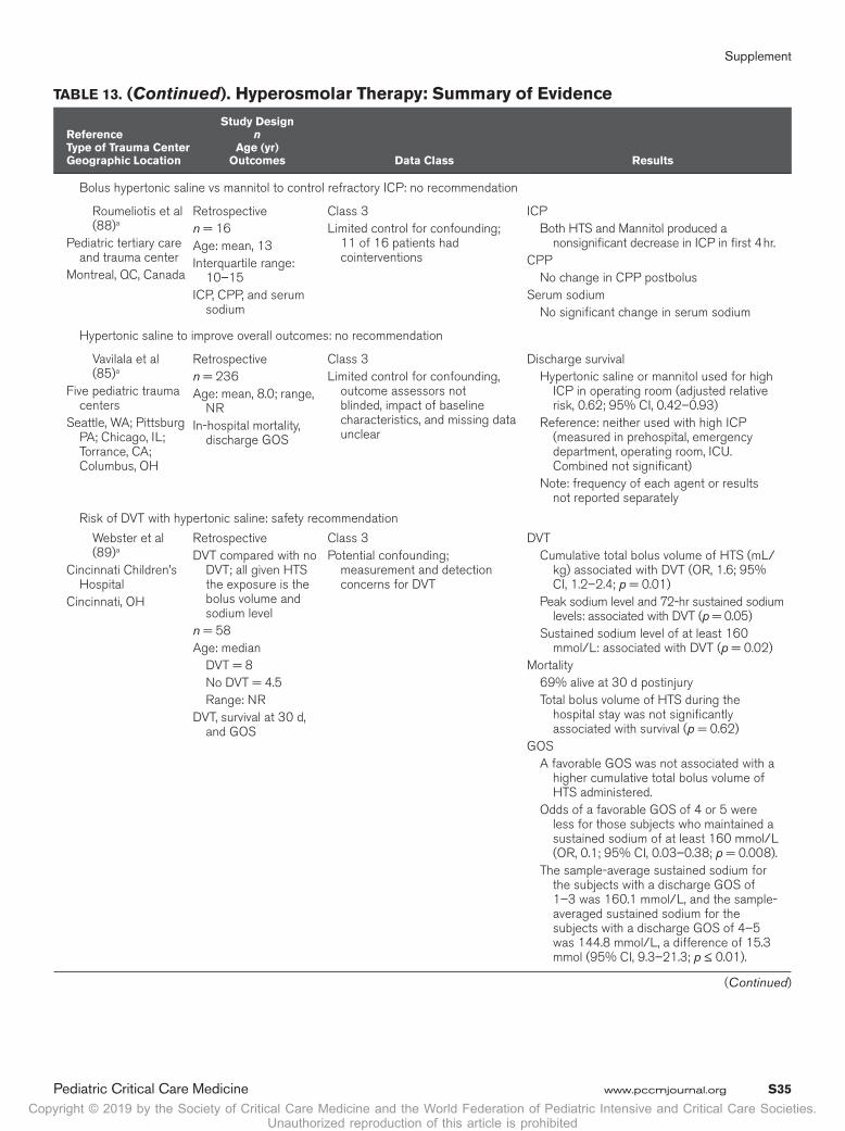
Copyright © 2019 by the Society of Critical Care Medicine and the World Federation of Pediatric Intensive and Critical Care Societies.Unauthorized reproduction of this article is prohibited
Supplement
Pediatric Critical Care Medicine www.pccmjournal.org S35
TABLE 13. (Continued). Hyperosmolar Therapy: Summary of Evidence
Reference Type of Trauma Center Geographic Location
Study Design n
Age (yr) Outcomes Data Class Results
Bolus hypertonic saline vs mannitol to control refractory ICP: no recommendation
Roumeliotis et al (88)a
Pediatric tertiary care and trauma center
Montreal, QC, Canada
Retrospectiven = 16Age: mean, 13Interquartile range:
10–15ICP, CPP, and serum
sodium
Class 3Limited control for confounding;
11 of 16 patients had cointerventions
ICP Both HTS and Mannitol produced a
nonsignificant decrease in ICP in first 4 hr.CPP No change in CPP postbolusSerum sodium No significant change in serum sodium
Hypertonic saline to improve overall outcomes: no recommendation
Vavilala et al (85)a
Five pediatric trauma centers
Seattle, WA; Pittsburg PA; Chicago, IL; Torrance, CA; Columbus, OH
Retrospectiven = 236Age: mean, 8.0; range,
NRIn-hospital mortality,
discharge GOS
Class 3Limited control for confounding,
outcome assessors not blinded, impact of baseline characteristics, and missing data unclear
Discharge survival Hypertonic saline or mannitol used for high
ICP in operating room (adjusted relative risk, 0.62; 95% CI, 0.42–0.93)
Reference: neither used with high ICP (measured in prehospital, emergency department, operating room, ICU. Combined not significant)
Note: frequency of each agent or results not reported separately
Risk of DVT with hypertonic saline: safety recommendation
Webster et al (89)a
Cincinnati Children’s Hospital
Cincinnati, OH
RetrospectiveDVT compared with no
DVT; all given HTS the exposure is the bolus volume and sodium level
n = 58Age: median DVT = 8 No DVT = 4.5 Range: NRDVT, survival at 30 d,
and GOS
Class 3Potential confounding;
measurement and detection concerns for DVT
DVT Cumulative total bolus volume of HTS (mL/
kg) associated with DVT (OR, 1.6; 95% CI, 1.2–2.4; p = 0.01)
Peak sodium level and 72-hr sustained sodium levels: associated with DVT (p = 0.05)
Sustained sodium level of at least 160 mmol/L: associated with DVT (p = 0.02)
Mortality 69% alive at 30 d postinjury Total bolus volume of HTS during the
hospital stay was not significantly associated with survival (p = 0.62)
GOS A favorable GOS was not associated with a
higher cumulative total bolus volume of HTS administered.
Odds of a favorable GOS of 4 or 5 were less for those subjects who maintained a sustained sodium of at least 160 mmol/L (OR, 0.1; 95% CI, 0.03–0.38; p = 0.008).
The sample-average sustained sodium for the subjects with a discharge GOS of 1–3 was 160.1 mmol/L, and the sample-averaged sustained sodium for the subjects with a discharge GOS of 4–5 was 144.8 mmol/L, a difference of 15.3 mmol (95% CI, 9.3–21.3; p ≤ 0.01).
(Continued)
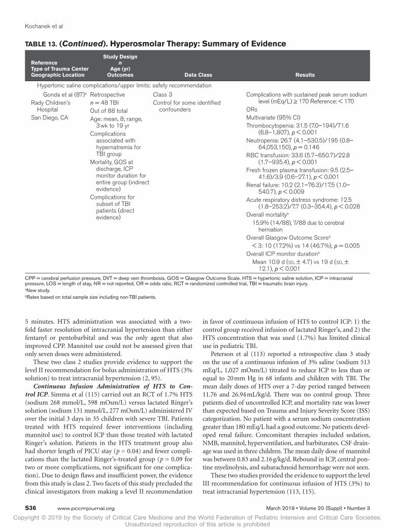
Copyright © 2019 by the Society of Critical Care Medicine and the World Federation of Pediatric Intensive and Critical Care Societies.Unauthorized reproduction of this article is prohibited
Copyright © 2019 by the Society of Critical Care Medicine and the World Federation of Pediatric Intensive and Critical Care Societies.Unauthorized reproduction of this article is prohibited
Kochanek et al
S36 www.pccmjournal.org March 2019 • Volume 20 (Suppl) • Number 3
Hypertonic saline complications/upper limits: safety recommendation
Gonda et al (87)a
Rady Children’s Hospital
San Diego, CA
Retrospectiven = 48 TBIOut of 88 totalAge: mean, 8; range,
3 wk to 19 yrComplications
associated with hypernatremia for TBI group
Mortality, GOS at discharge, ICP monitor duration for entire group (indirect evidence)
Complications for subset of TBI patients (direct evidence)
Class 3Control for some identified
confounders
Complications with sustained peak serum sodium level (mEq/L) ≥ 170 Reference: < 170
ORsMultivariate (95% CI)Thrombocytopenia: 31.5 (7.0–194)/71.6
(6.8–1,807), p < 0.001Neutropenia: 26.7 (4.1–530.5)/195 (0.8–
64,053,150), p = 0.146RBC transfusion: 33.6 (5.7–650.7)/22.8
(1.7–935.4), p < 0.001Fresh frozen plasma transfusion: 9.5 (2.5–
41.6)/3.9 (0.6–27.1), p < 0.001Renal failure: 10.2 (2.1–76.3)/17.5 (1.0–
540.7), p < 0.009Acute respiratory distress syndrome: 12.5
(1.8–253.2)/7.7 (0.3–354.4), p < 0.028Overall mortalityb
15.9% (14/88), 7/88 due to cerebral herniation
Overall Glasgow Outcome Scoreb
< 3: 10 (17.2%) vs 14 (46.7%), p = 0.005Overall ICP monitor durationb
Mean 10.9 d (sd, ± 4.7) vs 19 d (sd, ± 12.1), p < 0.001
CPP = cerebral perfusion pressure, DVT = deep vein thrombosis, GOS = Glasgow Outcome Scale, HTS = hypertonic saline solution, ICP = intracranial pressure, LOS = length of stay, NR = not reported, OR = odds ratio, RCT = randomized controlled trial, TBI = traumatic brain injury.a New study.b Rates based on total sample size including non-TBI patients.
TABLE 13. (Continued). Hyperosmolar Therapy: Summary of Evidence
Reference Type of Trauma Center Geographic Location
Study Design n
Age (yr) Outcomes Data Class Results
5 minutes. HTS administration was associated with a two-fold faster resolution of intracranial hypertension than either fentanyl or pentobarbital and was the only agent that also improved CPP. Mannitol use could not be assessed given that only seven doses were administered.
These two class 2 studies provide evidence to support the level II recommendation for bolus administration of HTS (3% solution) to treat intracranial hypertension (2, 95).
Continuous Infusion Administration of HTS to Con-trol ICP. Simma et al (115) carried out an RCT of 1.7% HTS (sodium 268 mmol/L, 598 mOsm/L) versus lactated Ringer’s solution (sodium 131 mmol/L, 277 mOsm/L) administered IV over the initial 3 days in 35 children with severe TBI. Patients treated with HTS required fewer interventions (including mannitol use) to control ICP than those treated with lactated Ringer’s solution. Patients in the HTS treatment group also had shorter length of PICU stay (p = 0.04) and fewer compli-cations than the lactated Ringer’s-treated group (p = 0.09 for two or more complications, not significant for one complica-tion). Due to design flaws and insufficient power, the evidence from this study is class 2. Two facets of this study precluded the clinical investigators from making a level II recommendation
in favor of continuous infusion of HTS to control ICP: 1) the control group received infusion of lactated Ringer’s, and 2) the HTS concentration that was used (1.7%) has limited clinical use in pediatric TBI.
Peterson et al (113) reported a retrospective class 3 study on the use of a continuous infusion of 3% saline (sodium 513 mEq/L, 1,027 mOsm/L) titrated to reduce ICP to less than or equal to 20 mm Hg in 68 infants and children with TBI. The mean daily doses of HTS over a 7-day period ranged between 11.76 and 26.94 mL/kg/d. There was no control group. Three patients died of uncontrolled ICP, and mortality rate was lower than expected based on Trauma and Injury Severity Score (ISS) categorization. No patient with a serum sodium concentration greater than 180 mEq/L had a good outcome. No patients devel-oped renal failure. Concomitant therapies included sedation, NMB, mannitol, hyperventilation, and barbiturates. CSF drain-age was used in three children. The mean daily dose of mannitol was between 0.83 and 2.16 g/kg/d. Rebound in ICP, central pon-tine myelinolysis, and subarachnoid hemorrhage were not seen.
These two studies provided the evidence to support the level III recommendation for continuous infusion of HTS (3%) to treat intracranial hypertension (113, 115).

Copyright © 2019 by the Society of Critical Care Medicine and the World Federation of Pediatric Intensive and Critical Care Societies.Unauthorized reproduction of this article is prohibited
Supplement
Pediatric Critical Care Medicine www.pccmjournal.org S37
Bolus Administration of HTS for Refractory ICP. Piper and Harrigan (90) reported a treatment series on the use of 23.4% saline to treat refractory intracranial hypertension in 32 infants and children with severe TBI. Refractory intracranial hypertension was defined as an ICP greater than 20 mm Hg for greater than 5 minutes and not responding to a stepwise protocol that included sedation, analgesia, head elevation, mild hyperventilation, mild hypothermia (36.5°C), NMB, and use of inotropic support to support age-appropriate CPP. Contrain-dication to the administration of 23.4% saline in this study was a serum sodium level of greater than 155 mmol/L. The dose of 23.4% saline was 0.5 mL/kg administered over a period of 10 minutes, with a maximum dose of 30 mL. The mean reduction in ICP with treatment was 10 mm Hg (range, 1–30 mm Hg), and the highest serum sodium level observed in any patient was 161 mmol/L. GOS greater than 3 was achieved in 74% of the patients. This study provided the evidence to support the level III recommendation for the use of 23.4% saline to treat refractory intracranial hypertension.
Use of Hyperosmolar Therapy to Improve Outcomes. There was insufficient evidence to support a recommendation for the use of hyperosmolar therapy to improve overall outcomes. A single study, Vavilala et al (85) assessed acute care clinical indicators associated with in-hospital mortality and discharge GOS score in a retrospective cohort study of 236 infants and children with severe TBI across five medical centers. The use of HTS or mannitol for high ICP in the operating room was associated with favorable outcome versus no hyperosmolar therapy. However, when the comparison included the emer-gency department (ED), operating room, and ICU, a signifi-cant effect was not observed.
Safety RecommendationsTwo class 3 studies contributed evidence supporting the safety recommendations for this topic (87, 89). Webster et al (89) car-ried out a single-center case-control study of 58 infants and children with severe TBI treated with bolus and/or continuous infusion of HTS. Eight patients developed DVTs. There was no association between the volume of HTS administered during the initial 72 hours and DVT; however, a serum sodium level greater than or equal to 160 mEq/L sustained for 72 hours was significantly associated with DVT (p = 0.02). Gonda et al (87) reported a retrospective cohort study of 48 infants and chil-dren with severe TBI in which bolus and infusion treatment with 3% saline were used. A sustained level (defined as ≥ 72 hr) of serum sodium greater than or equal to 170 mEq/L was sig-nificantly associated with thrombocytopenia and the need for erythrocyte transfusion. In addition, although the association with renal failure was not significant (p = 0.064), this could indicate a trend toward harm that merits further study.
Indications From Adult GuidelinesThe Fourth Edition of the adult TBI guidelines inform the pediatric guidelines in that class 2 and 3 studies comparing HTS to mannitol have been carried out and suggest that HTS may be more effective that mannitol with regard to ICP burden
and ICP greater than 25 mm Hg, but no differences in mortal-ity or long-term outcome were noted (14, 116, 117).
Analgesics, Sedatives, and NMB
RecommendationsStrength of Recommendations: Weak
Levels I and IIThere was insufficient evidence to support a level I or II recom-mendation for this topic.
Level IIIFor ICP Control. III.1. With use of multiple ICP-related ther-apies, as well as appropriate use of analgesia and sedation in routine ICU care, avoiding bolus administration of midazolam and/or fentanyl during ICP crises is suggested due to risks of cerebral hypoperfusion.
Note 1. In the absence of outcome data, the specific indi-cations, choice, and dosing of analgesics, sedatives, and neu-romuscular blocking agents should be left to the treating physician.
Note 2. Based on guidance from the U.S. Food and Drug Administration, prolonged continuous infusion of propofol for either sedation or the management of refractory intracra-nial hypertension is not recommended.
Changes From Prior Edition. Recommendation III.1. is new to this Third Edition. The recommendation about the use of thiopental from the Second Edition has been removed and the study by de Bray et al (118) has been removed. Five new class 3 studies—two prospective (2, 119), two retrospec-tive (85, 120), and one treatment series (3)—were added to the evidence base for this topic.
IntroductionIn management of pediatric severe TBI requiring tracheal intubation and mechanical ventilation, analgesics and seda-tives are needed for comfort and tolerance (121). Neuromus-cular blocking agents are not needed routinely, except at the time of rapid sequence intubation or when severe acute lung injury mechanics pose a problem with supportive ventilation (122). This topic evaluates the use of these agents during ICU treatment in patients who are appropriately sedated, when needed specifically for management of ICP, and for optimizing cerebral perfusion.
In a mechanically ventilated patient, appropriate use of analgesia and sedation will treat any pain or distress and mitigate patient-ventilator dyssynchrony, both of which may result in episodic rise in CBV and raise ICP (123). Routine care (e.g., oral hygiene, tracheal tube suctioning) while the patient is awake or aware may also lead to unanticipated rises in ICP (124). Pediatric practice is currently limited in the choice of sedative/analgesics that are available. Practice is markedly different in adult than in pediatric neurocritical care; espe-cially in the frequent use of propofol, which in pediatrics has a U.S. Food and Drug Administration warning (125). As

Copyright © 2019 by the Society of Critical Care Medicine and the World Federation of Pediatric Intensive and Critical Care Societies.Unauthorized reproduction of this article is prohibited
Copyright © 2019 by the Society of Critical Care Medicine and the World Federation of Pediatric Intensive and Critical Care Societies.Unauthorized reproduction of this article is prohibited
Kochanek et al
S38 www.pccmjournal.org March 2019 • Volume 20 (Suppl) • Number 3
a consequence, given the relatively long half-life of the drugs that are administered, frequently the neurologic examination can be obscured.
Neuromuscular blocking agents may reduce ICP by either optimizing patient-ventilator interactions or by prevention of shivering.
Evaluation of the EvidenceQuality of the Body of Evidence. Studies included for this topic addressed the questions of the effectiveness of combinations of NMB, ketamine, fentanyl, midazolam, etomidate, and pento-barbital to control ICP for children with severe TBI. There was insufficient evidence to derive a recommendation about mixed NMB, ketamine, etomidate, or pentobarbital. The overall qual-ity of the body of evidence is low (Table 14).
Applicability. For the question of the use of NMB, the two studies were conducted in multiple sites and sample sizes were moderate, indicating cautious confidence in their applicabil-ity. The remaining studies enrolled small samples and were conducted at single sites, indicating limited confidence in their applicability.
Summary of the EvidenceTwo new class 3 studies provided evidence to support the rec-ommendation (2, 3) (Table 15).
Evidence SynthesisUse of Fentanyl and/or Midazolam for ICP Control. One small retrospective study found that in 31 pediatric patients
who received high-dose fentanyl, low-dose midazolam, or high-dose fentanyl plus low-dose midazolam, there was an increase in ICP for all treatment conditions (3). It is unclear if the finding was due to other patient factors, and the study did not address use of sedation beyond the context of ICP. In a prospective observational study of 16 pediatric patients, fen-tanyl decreased ICP, but less effectively than HTS or pentobar-bital (2). Furthermore, it decreased CPP, and had the highest treatment failure rate. This study suggests that in the setting of adequate analgesia/sedation, HTS might be preferable over fentanyl. Taken together, these studies provide the evidence to support the recommendation for this topic.
The findings from the studies by Welch et al (3) and Shein et al (2) should be interpreted cautiously given they included routine use of analgesia and sedation in ICU care. An ICP crisis could be caused by pain or anxiety in the absence of appropri-ate analgesia and sedation. In that setting, additional fentanyl and/or midazolam may reduce ICP. However, these two stud-ies may indicate that these agents are ineffective at reducing ICP in patients receiving adequate analgesia and sedation. In that setting, they confer either lesser efficacy than other tier 1 choices such as HTS and/or reduce MAP, negating benefit on CPP. Alternatively, a tier 2 intervention such as pentobarbital may be required at that juncture of care (2).
Use of NMB for ICP Control. The NMB studies suggest differences between subjects who received a NMB agent and those who did not (85, 120). However, for reasons discussed in the “Introduction section,” the studies evaluated also did not include the indication for a given dose of NMB.
TABLE 14. Analgesics, Sedatives, and Neuromuscular Blockade: Quality of the Body of Evidence
Components of Overall Quality: Class 3 Studies
Topic
No. of Studies Study
DesignRecommen-
dations
Meta- Analysis Possible
(Yes or Noa)
Total No. of
Subjects (n)
Consistency (High,
Moderate, Low)
Directness (Direct or Indirect)
Precision (High,
Moderate, Low)
Quality of Evidence
(High, Moderate,
Low, or Insufficient)
Use of neuromuscular blockade (mixed)
2 retrospective No recommen-
dation
NA 326 Moderate Direct Low Insufficient
Use of ketamine 1 prospective No recommen-
dation
NA 30 NA Indirect Low Insufficient
Use of fentanyl and/or midazolam
1 treatment series III.1. NA 31 NA Direct Low Low
Use of etomidate 1 treatment series No recommen-
dation
NA 8 NA Direct Low Insufficient
Use of fentanyl or pentobarbital
1 prospective III.1. NA 16 NA Direct Low Low
NA = not applicable.a Meta-analysis to synthesize results is only possible if several studies address the same criteria/question, and other design criteria are met.n indicates sample size.
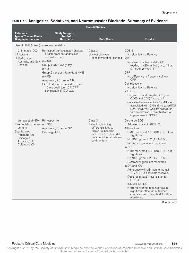
Copyright © 2019 by the Society of Critical Care Medicine and the World Federation of Pediatric Intensive and Critical Care Societies.Unauthorized reproduction of this article is prohibited
Supplement
Pediatric Critical Care Medicine www.pccmjournal.org S39
TABLE 15. Analgesics, Sedatives, and Neuromuscular Blockade: Summary of Evidence
Class 3 Studies
Reference Type of Trauma Center Geographic Location
Study Design n Age (yr)
Outcomes Data Class Results
Use of NMB (mixed): no recommendation
Chin et al (120)a
17 hospitalsUnited States,
Australia, and New Zealand
Retrospective (secondary analysis of data from an randomized controlled trial)
n = 90Group 1 NMB every dayn = 31Group 2 none or intermittent NMBn = 59Age: mean, 9.5; range, NRGOS-E at discharge and 3, 6, and
12 mo postinjury; ICP; CPP; complications; ICU LOS
Class 3Unclear allocation
concealment; not blinded
GOS-E No significant differenceICP Increased number of daily ICP
readings > 20 mm Hg (4.4 ± 1.1 vs 2.4 ± 0.5; p = 0.015)
CPP No difference in frequency of low
CPPComplications No significant differenceICU LOS Longer ICU and hospital LOS (p =
0.003 and 0.07) for group 1 Consistent administration of NMB was
associated with ICH and increased ICU LOS. However, it was not associated with an increase in complications or improvement in GOS-E.
Vavilala et al (85)a
Five pediatric trauma centers
Seattle, WA; Pittsburg PA; Chicago, IL; Torrance, CA; Columbus, OH
Retrospectiven = 236Age: mean, 8; range, NRDischarge GOS
Class 3Selection; blinding;
differential loss to follow-up; baseline differences unclear; did not control for all relevant confounders
Discharge GOS Adjusted risk ratio (95% CI)All locations NMB monitored 1.13 (0.85–1.51) not
significant No NMB given 1.37 (1.24–1.53) Reference: given, not monitoredIn OR NMB monitored 1.33 (0.93–1.9) not
significant No NMB given 1.42 (1.28–1.58) Reference: given, not monitoredIn OR and ICU Adherence = NMB monitoring (all
112/121 OR patients received) Odds ratio: 18.8% overall; range,
0–32.1 ICU 3% (0–4.8) NMB monitoring does not have a
significant effect on outcomes compared with using NMB without monitoring
(Continued)

Copyright © 2019 by the Society of Critical Care Medicine and the World Federation of Pediatric Intensive and Critical Care Societies.Unauthorized reproduction of this article is prohibited
Copyright © 2019 by the Society of Critical Care Medicine and the World Federation of Pediatric Intensive and Critical Care Societies.Unauthorized reproduction of this article is prohibited
Kochanek et al
S40 www.pccmjournal.org March 2019 • Volume 20 (Suppl) • Number 3
Use of ketamine: no recommendation
Bar-Joseph et al (119)a
Meyer Children’s Hospital, Rambam Medical Center
Israel
ProspectivePre/postn = 305 (17%) not TBIAge: mean, NR; range, 6 mo to 18 yrICP; CPPAll elevated ICP (> 18 mm Hg)
resistant to first-tier therapies received a single ketamine dose (1–1.5 mg/kg)
Group 1: to prevent further ICP increase during a potentially distressing intervention
Group 2: additional measure to lower ICP
Class 3Selection bias; no control for
confoundersIndirect evidence Mixed severities; GCS
NR; mixed pathologies
ICP and CPP Post ketamine administration Overall in both groups ICP decreased by 30% (from
25.8 ± 8.4 to 18.0 ± 8.5 mm Hg) (p < 0.001)
After 65 ketamine administrations 61 ICP decreased 3 increase < 2 mm Hg 1 increase > 2 mm Hg CPP increased from 54.4 ± 11.7 to
58.3 ± 13.4 mm Hg (p < 0.005).Group 1 Mean ICP decreased from 25.2 ± 5.4
to 17.9 ± 5.5 mm Hg within the first 2 min of ketamine administration (p < 0.001) and during distressing activity increased slightly up to 19.6 ± 6.7 at minute 7 and then decreased again.
Group 2 ICP decreased by 33% (from
26.0 ± 9.1 to 17.5 ± 9.1 mm Hg) (p < 0.0001) within 2 min following ketamine administration.
Result contradicts prior concerns that ketamine increases ICP.
Use of fentanyl and/or midazolam: recommendation III.1.
Welch et al (3)a
Trauma PICUSt. Louis Children’s
HospitalSt. Louis, MO
Treatment seriesn = 318 (26%) abusive head traumaAge: mean, 8; range, 0–18ICP, CPP, and MAP, difference
between predrug and postdrug administration
Class 3Uncontrolled series
AUC used to represent cumulative ICH exposure
The mean change AUC, ICP increased after drug administration
Overall Increase in ICP of 17 mm Hg; p =
0.04 Mean change in AUC for ICP All significant High-dose fentanyl: p = 0.02 Low-dose midazolam; p = 0.006 High-dose fentanyl plus low-dose
midazolam; p = 0.007 Dosing of fentanyl and midazolam did
not reduce the ICP. Age and postresuscitation GCS did
not correlate with change AUC-ICH, time after injury significantly correlated with change AUC-ICH r = –0.12, p = 0.02
TABLE 15. (Continued). Analgesics, Sedatives, and Neuromuscular Blockade: Summary of Evidence
Class 3 Studies
Reference Type of Trauma Center Geographic Location
Study Design n
Age (yr) Outcomes Data Class Results
(Continued)

Copyright © 2019 by the Society of Critical Care Medicine and the World Federation of Pediatric Intensive and Critical Care Societies.Unauthorized reproduction of this article is prohibited
Supplement
Pediatric Critical Care Medicine www.pccmjournal.org S41
Use of etomidate: no recommendation
Bramwell et al (126)
Primary Children’s Medical Center, PICU
Salt Lake City, UT
Treatment seriesn = 8Age: mean, NR; range, NRICP
Class 3Uncontrolled series
Etomidate administration resulted in a decrease in ICP vs baseline (p < 0.05) without change in MAP; thereby increasing CPP at each 5 min interval. At 6 hr after etomidate administration, adrenocorticotropic hormone stimulation tests showed adrenal suppression in four of the eight patients; however, no patient required treatment with steroids.
Use of fentanyl or pentobarbital: recommendation III.1.
Shein et al (2)a
University of Pittsburgh
Pittsburgh, PA
Prospectiven = 16 in analysisAge: mean, 44 mo; range, 34–124
moOutcomes: ICP and CPP
Class 3Inadequate control for
confounders; small sample
Hypertonic saline vs other drugs for ICPDecrease in ICP; increase in CPPAssociated with a two-fold faster
resolution of ICH than either fentanyl or pentobarbital.
Adjusted hazard ratio, 2.171 (1.062–4.439)
Fentanyl ICP decreased but had highest rate of
treatment failure ratePentobarbital ICP decreased; CPP no significant
change
AUC = area under the curve, CPP = cerebral perfusion pressure, GCS = Glasgow Coma Scale, GOS = Glasgow Outcome Scale, GOS-E = Glasgow Outcome Scale Extended, ICH = intracranial hypertension, ICP = intracranial pressure, LOS = length of stay, MAP = mean arterial pressure, NMB = neuromuscular blockade, NR = not reported, OR = operating room.a New study.
TABLE 15. (Continued). Analgesics, Sedatives, and Neuromuscular Blockade: Summary of Evidence
Class 3 Studies
Reference Type of Trauma Center Geographic Location
Study Design n
Age (yr) Outcomes Data Class Results
Use of Ketamine for ICP Control. Traditionally, ketamine has been considered contraindicated in patients with raised ICP, with the concern that it increases CBF and might increase ICP further. This reasoning has been questioned in a variety of studies and systematic reviews, and the report by Bar-Joseph et al (119) adds to this evidence. However, a level III recom-mendation on ketamine was not made because the GCS score for patients was not indicated, precluding the ability to con-firm that the inclusion criteria were met for the publication.
Use of Etomidate for ICP Control. Use of etomidate for ICP control is largely of historical interest because we now have other sedatives and analgesics available that do not suppress adrenal function (126). A single dose of etomidate is, however, commonly used for intubation in pediatric TBI (122).
Indications From Adult GuidelinesThe clinical investigators do not believe that the recom-mendations about analgesics, sedatives, and NMB from the adult guidelines can be used to guide treatment decisions in children.
CSF Drainage
RecommendationsStrength of Recommendation: Weak
Levels I and IIThere was insufficient evidence to support a level I or II recom-mendation for this topic.
Level IIIFor ICP Control. III.1. CSF drainage through an EVD is sug-gested to manage increased ICP.
Changes From Prior Edition. The recommendation from the Second Edition about use of lumbar drain (LD) was elimi-nated. One new class 3 treatment series was added to the evi-dence base for this topic (127).
IntroductionThe EVD can be used not only to measure ICP in children fol-lowing TBI but may also provide added therapeutic benefit of CSF drainage. CSF drainage through an EVD has frequently
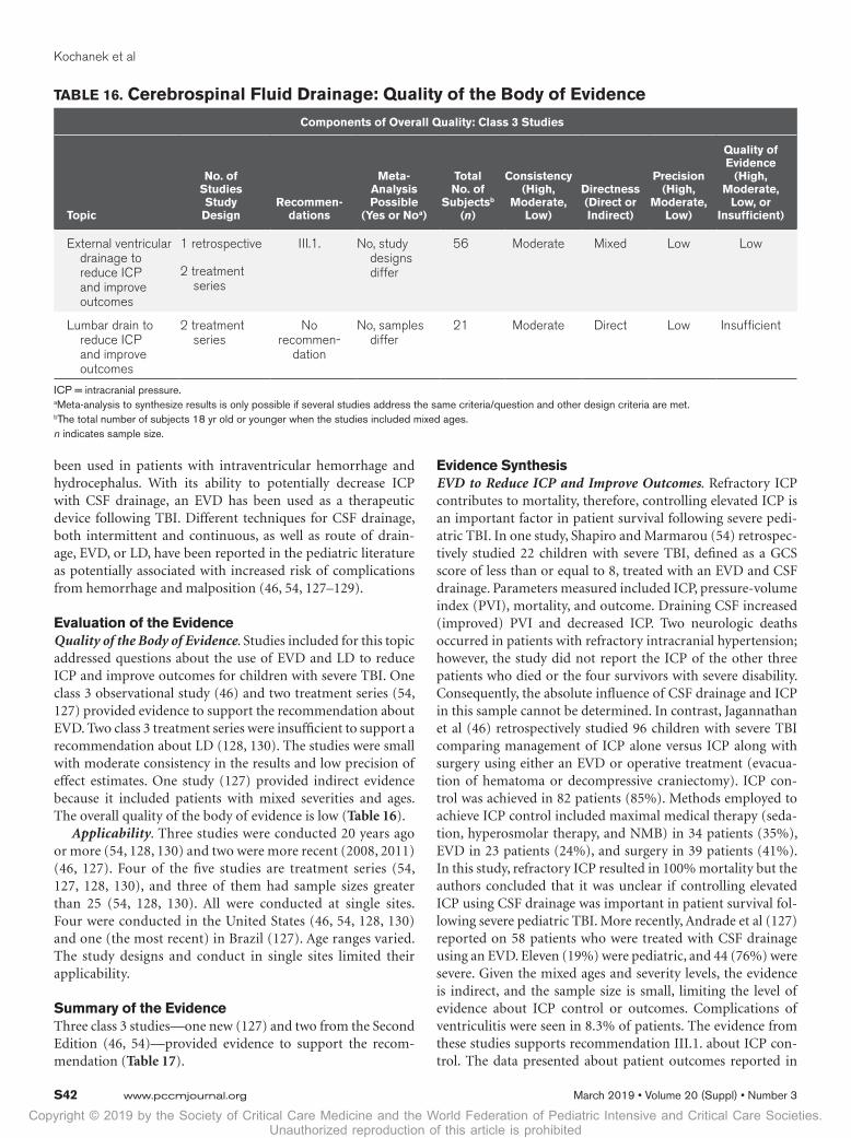
Copyright © 2019 by the Society of Critical Care Medicine and the World Federation of Pediatric Intensive and Critical Care Societies.Unauthorized reproduction of this article is prohibited
Copyright © 2019 by the Society of Critical Care Medicine and the World Federation of Pediatric Intensive and Critical Care Societies.Unauthorized reproduction of this article is prohibited
Kochanek et al
S42 www.pccmjournal.org March 2019 • Volume 20 (Suppl) • Number 3
been used in patients with intraventricular hemorrhage and hydrocephalus. With its ability to potentially decrease ICP with CSF drainage, an EVD has been used as a therapeutic device following TBI. Different techniques for CSF drainage, both intermittent and continuous, as well as route of drain-age, EVD, or LD, have been reported in the pediatric literature as potentially associated with increased risk of complications from hemorrhage and malposition (46, 54, 127–129).
Evaluation of the EvidenceQuality of the Body of Evidence. Studies included for this topic addressed questions about the use of EVD and LD to reduce ICP and improve outcomes for children with severe TBI. One class 3 observational study (46) and two treatment series (54, 127) provided evidence to support the recommendation about EVD. Two class 3 treatment series were insufficient to support a recommendation about LD (128, 130). The studies were small with moderate consistency in the results and low precision of effect estimates. One study (127) provided indirect evidence because it included patients with mixed severities and ages. The overall quality of the body of evidence is low (Table 16).
Applicability. Three studies were conducted 20 years ago or more (54, 128, 130) and two were more recent (2008, 2011) (46, 127). Four of the five studies are treatment series (54, 127, 128, 130), and three of them had sample sizes greater than 25 (54, 128, 130). All were conducted at single sites. Four were conducted in the United States (46, 54, 128, 130) and one (the most recent) in Brazil (127). Age ranges varied. The study designs and conduct in single sites limited their applicability.
Summary of the EvidenceThree class 3 studies—one new (127) and two from the Second Edition (46, 54)—provided evidence to support the recom-mendation (Table 17).
Evidence SynthesisEVD to Reduce ICP and Improve Outcomes. Refractory ICP contributes to mortality, therefore, controlling elevated ICP is an important factor in patient survival following severe pedi-atric TBI. In one study, Shapiro and Marmarou (54) retrospec-tively studied 22 children with severe TBI, defined as a GCS score of less than or equal to 8, treated with an EVD and CSF drainage. Parameters measured included ICP, pressure-volume index (PVI), mortality, and outcome. Draining CSF increased (improved) PVI and decreased ICP. Two neurologic deaths occurred in patients with refractory intracranial hypertension; however, the study did not report the ICP of the other three patients who died or the four survivors with severe disability. Consequently, the absolute influence of CSF drainage and ICP in this sample cannot be determined. In contrast, Jagannathan et al (46) retrospectively studied 96 children with severe TBI comparing management of ICP alone versus ICP along with surgery using either an EVD or operative treatment (evacua-tion of hematoma or decompressive craniectomy). ICP con-trol was achieved in 82 patients (85%). Methods employed to achieve ICP control included maximal medical therapy (seda-tion, hyperosmolar therapy, and NMB) in 34 patients (35%), EVD in 23 patients (24%), and surgery in 39 patients (41%). In this study, refractory ICP resulted in 100% mortality but the authors concluded that it was unclear if controlling elevated ICP using CSF drainage was important in patient survival fol-lowing severe pediatric TBI. More recently, Andrade et al (127) reported on 58 patients who were treated with CSF drainage using an EVD. Eleven (19%) were pediatric, and 44 (76%) were severe. Given the mixed ages and severity levels, the evidence is indirect, and the sample size is small, limiting the level of evidence about ICP control or outcomes. Complications of ventriculitis were seen in 8.3% of patients. The evidence from these studies supports recommendation III.1. about ICP con-trol. The data presented about patient outcomes reported in
TABLE 16. Cerebrospinal Fluid Drainage: Quality of the Body of Evidence
Components of Overall Quality: Class 3 Studies
Topic
No. of Studies Study
DesignRecommen-
dations
Meta- Analysis Possible
(Yes or Noa)
Total No. of
Subjectsb (n)
Consistency (High,
Moderate, Low)
Directness (Direct or Indirect)
Precision (High,
Moderate, Low)
Quality of Evidence
(High, Moderate,
Low, or Insufficient)
External ventricular drainage to reduce ICP and improve outcomes
1 retrospective
2 treatment series
III.1. No, study designs differ
56 Moderate Mixed Low Low
Lumbar drain to reduce ICP and improve outcomes
2 treatment series
No recommen-
dation
No, samples differ
21 Moderate Direct Low Insufficient
ICP = intracranial pressure.a Meta-analysis to synthesize results is only possible if several studies address the same criteria/question and other design criteria are met.b The total number of subjects 18 yr old or younger when the studies included mixed ages.n indicates sample size.

Copyright © 2019 by the Society of Critical Care Medicine and the World Federation of Pediatric Intensive and Critical Care Societies.Unauthorized reproduction of this article is prohibited
Supplement
Pediatric Critical Care Medicine www.pccmjournal.org S43
TABLE 17. Cerebrospinal Fluid Drainage: Summary of Evidence
Class 3 Studies
Reference Type of Trauma Center Geographic Location
Study Design n
Age (yr) Outcomes Data Class Results
External ventricular drain to reduce ICP and improve outcomes: recommendation III.1.
Andrade et al (127)a
Clinicas Hospital University of São Paulo Medical School
São Paulo, Brazil
Treatment seriesn = 58n = 11, < 17 yearsn = 44 Glasgow Coma Scale
4–8Age: mean, 29; range, 4–65Mortality, neurologic assessment,
complications
Class 3No control for
confounders, enrollment criteria unclear
Indirect evidence Mixed ages and
severities
All patients treated with ventricular CSF drainage
Mortality 3 of 11Neurologic assessment Among patients < 17 yr old Favorable outcome 6 (54.5%) Unfavorable outcome 5 (45.5%) (not
separated by severity) No significant difference in outcome,
between groups separated by age (adults vs children), p > 0.05
Complications: not separated by age or severity
Overall rate of infection (ventriculitis): 8.3% Infection did not contribute to clinical
worsening or death, p > 0.05
Jagannathan et al (46)
University of Virginia Health System
Charlottesville, VA
Retrospectiven = 96n = 23 treated with CSF
using EVD (other groups: craniectomy and medical management)
Age: mean, 5.1; range, 3–18Mortality, GOS at mean 2 yr, ICP,
complications
Class 3Control for confounders
unclear for ICP, only patients with 2-yr follow-up included in analysis
Mortality 3 of 23GOS No significant difference in mean or median
GOS across treatment groupsICP Overall 20/23 (87%) achieved ICP control
with EVD. Of three not achieving ICP control, two died, one had a craniectomy and then died.
Refractory ICP was associated with 100% mortality; the method used to control ICP had no correlation with mortality.
Complications Higher rate of meningitis (p < 0.05)
in patients with drain (5/23, 22%) compared with patients treated with craniectomy (3/40, 7%)
Shapiro and Marmarou (54)
Albert Einstein College of Medicine
New York
Treatment seriesn = 22Age: mean, NR; range, 3 mo to
15 yrMortality, outcome (scale not
specified), ICP
Class 3Uncontrolled series
Mortality 5/22Outcome 4/17 severe disabilities 13/17 good
outcome or moderately disabledICP Decreased 14/16 16 of 22 patients had PVI measured before
and after therapy. Drainage increased PVI. Two of the five
deaths were due to uncontrolled ICP.
(Continued)

Copyright © 2019 by the Society of Critical Care Medicine and the World Federation of Pediatric Intensive and Critical Care Societies.Unauthorized reproduction of this article is prohibited
Copyright © 2019 by the Society of Critical Care Medicine and the World Federation of Pediatric Intensive and Critical Care Societies.Unauthorized reproduction of this article is prohibited
Kochanek et al
S44 www.pccmjournal.org March 2019 • Volume 20 (Suppl) • Number 3
these studies were insufficient to determine the influence of EVD on outcomes.
LD to Reduce ICP and Improve Outcomes. The evidence was insufficient to support a recommendation about LD.
Indications From Adult GuidelinesIn the adult guidelines, the level III recommendations for CSF drainage that might be additive to the pediatric recommendations include the following: the EVD system zeroed to the midbrain with continuous drainage of CSF may be more effective in lower-ing the ICP burden than intermittent drainage; and the use of CSF drainage to lower ICP in patients with an initial GCS less than 6 during the first 12 hours after injury may be considered (14).
Seizure Prophylaxis
RecommendationsStrength of Recommendation: Weak
Levels I and IIThere was insufficient evidence to support a level I or II recom-mendation for this topic.
Level IIIFor Seizure Prevention (Clinical and Subclinical). III.1. Pro-phylactic treatment is suggested to reduce the occurrence of early (within 7 d) PTSs.
Note. At the present time, there is insufficient evidence to recommend levetiracetam over phenytoin based on either effi-cacy in preventing early PTS (EPTS) or toxicity.
Changes From Prior Edition. Recommendation III.1. is modified from the Second Edition of these guidelines, with phenytoin removed. The note regarding levetiracetam is new to this Third Edition. Three new class 3 studies—one prospec-tive observational (131), one retrospective observational (132), and one treatment series (133)—have been added to the evi-dence base for this topic.
IntroductionPTSs are defined as occurring early, within 7 days of injury, or late, beyond 8 days of recovery (134). Risk factors associated with the occurrence of PTS include location of the lesion, cere-bral contusions, retained bone and metal fragments, depressed skull fracture, focal neurologic deficits, loss of consciousness, GCS greater than 10, severity of injury, length of posttraumatic amnesia, subdural or epidural hematoma, penetrating injury, and age. Infants and children have lower seizure thresholds (135), adding to the challenge of recognition of subtle clini-cal seizures (132) in critically ill children. The occurrence of electrographic seizures (seizures detected by continuous elec-troencephalogram recording) following severe TBI is higher in children than adults, occurring in up to 70% of cases (136).
LD to reduce ICP and improve outcomes: no recommendation
Baldwin and Rekate (128)
Children’s Hospital and Medical Centers
Arizona
Treatment seriesn = 5Age: mean, NR; range, 8–14Mortality, outcome (scale
undefined), ICP
Class 3Uncontrolled series
Mortality 2/5Outcome One moderate disability, two good recoveryICP Decreased in 5/5 LD was added to an aggressive ICP control
protocol that included EVD.
Levy et al (130)St. Joseph’s Hospital
and Phoenix Children’s Hospital
Arizona
Treatment seriesn = 16Age: mean, NR; range, 1–15Mortality, GOS at 6 mo
postinjury, ICP
Class 3Uncontrolled series
Mortality 2/16GOS Eight good recovery; three moderate
disability, three severe disabilitiesICP Decreased in 14/16 Lumbar drainage was added to an
aggressive ICP control protocol that included EVD.
CSF = cerebrospinal fluid, EVD = external ventricular drainage, GOS = Glasgow Outcome Scale, ICP = intracranial pressure, LD = lumbar drain, NR = not reported, PVI = pressure-volume index.a New study.n indicates sample size.
TABLE 17. (Continued). Cerebrospinal Fluid Drainage: Summary of Evidence
Class 3 Studies
Reference Type of Trauma Center Geographic Location
Study Design n
Age (yr) Outcomes Data Class Results
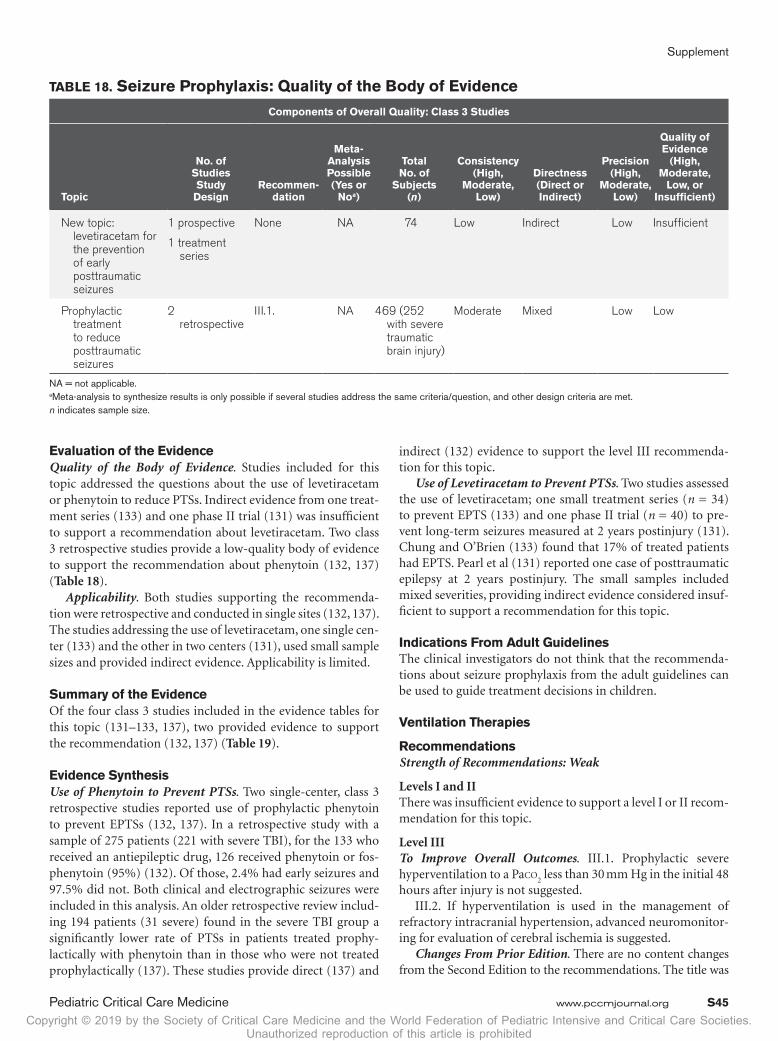
Copyright © 2019 by the Society of Critical Care Medicine and the World Federation of Pediatric Intensive and Critical Care Societies.Unauthorized reproduction of this article is prohibited
Supplement
Pediatric Critical Care Medicine www.pccmjournal.org S45
Evaluation of the EvidenceQuality of the Body of Evidence. Studies included for this topic addressed the questions about the use of levetiracetam or phenytoin to reduce PTSs. Indirect evidence from one treat-ment series (133) and one phase II trial (131) was insufficient to support a recommendation about levetiracetam. Two class 3 retrospective studies provide a low-quality body of evidence to support the recommendation about phenytoin (132, 137) (Table 18).
Applicability. Both studies supporting the recommenda-tion were retrospective and conducted in single sites (132, 137). The studies addressing the use of levetiracetam, one single cen-ter (133) and the other in two centers (131), used small sample sizes and provided indirect evidence. Applicability is limited.
Summary of the EvidenceOf the four class 3 studies included in the evidence tables for this topic (131–133, 137), two provided evidence to support the recommendation (132, 137) (Table 19).
Evidence SynthesisUse of Phenytoin to Prevent PTSs. Two single-center, class 3 retrospective studies reported use of prophylactic phenytoin to prevent EPTSs (132, 137). In a retrospective study with a sample of 275 patients (221 with severe TBI), for the 133 who received an antiepileptic drug, 126 received phenytoin or fos-phenytoin (95%) (132). Of those, 2.4% had early seizures and 97.5% did not. Both clinical and electrographic seizures were included in this analysis. An older retrospective review includ-ing 194 patients (31 severe) found in the severe TBI group a significantly lower rate of PTSs in patients treated prophy-lactically with phenytoin than in those who were not treated prophylactically (137). These studies provide direct (137) and
indirect (132) evidence to support the level III recommenda-tion for this topic.
Use of Levetiracetam to Prevent PTSs. Two studies assessed the use of levetiracetam; one small treatment series (n = 34) to prevent EPTS (133) and one phase II trial (n = 40) to pre-vent long-term seizures measured at 2 years postinjury (131). Chung and O’Brien (133) found that 17% of treated patients had EPTS. Pearl et al (131) reported one case of posttraumatic epilepsy at 2 years postinjury. The small samples included mixed severities, providing indirect evidence considered insuf-ficient to support a recommendation for this topic.
Indications From Adult GuidelinesThe clinical investigators do not think that the recommenda-tions about seizure prophylaxis from the adult guidelines can be used to guide treatment decisions in children.
Ventilation Therapies
RecommendationsStrength of Recommendations: Weak
Levels I and IIThere was insufficient evidence to support a level I or II recom-mendation for this topic.
Level IIITo Improve Overall Outcomes. III.1. Prophylactic severe hyperventilation to a Paco
2 less than 30 mm Hg in the initial 48
hours after injury is not suggested.III.2. If hyperventilation is used in the management of
refractory intracranial hypertension, advanced neuromonitor-ing for evaluation of cerebral ischemia is suggested.
Changes From Prior Edition. There are no content changes from the Second Edition to the recommendations. The title was
TABLE 18. Seizure Prophylaxis: Quality of the Body of Evidence
Components of Overall Quality: Class 3 Studies
Topic
No. of Studies Study
DesignRecommen-
dation
Meta- Analysis Possible (Yes or
Noa)
Total No. of
Subjects (n)
Consistency (High,
Moderate, Low)
Directness (Direct or Indirect)
Precision (High,
Moderate, Low)
Quality of Evidence
(High, Moderate,
Low, or Insufficient)
New topic: levetiracetam for the prevention of early posttraumatic seizures
1 prospective
1 treatment series
None NA 74 Low Indirect Low Insufficient
Prophylactic treatment to reduce posttraumatic seizures
2 retrospective
III.1. NA 469 (252 with severe traumatic brain injury)
Moderate Mixed Low Low
NA = not applicable.a Meta-analysis to synthesize results is only possible if several studies address the same criteria/question, and other design criteria are met.n indicates sample size.

Copyright © 2019 by the Society of Critical Care Medicine and the World Federation of Pediatric Intensive and Critical Care Societies.Unauthorized reproduction of this article is prohibited
Copyright © 2019 by the Society of Critical Care Medicine and the World Federation of Pediatric Intensive and Critical Care Societies.Unauthorized reproduction of this article is prohibited
Kochanek et al
S46 www.pccmjournal.org March 2019 • Volume 20 (Suppl) • Number 3
TABLE 19. Seizure Prophylaxis: Summary of Evidence
Class 3 Studies
Reference Type of Trauma Center Geographic Location
Study Design n
Age (yr) Outcomes Data Class Results
Levetiracetam for the prevention of EPTSs: no recommendation
Chung et al (133)a
Nationwide Children’s Hospital, PICU
Columbus, OH
Treatment seriesn = 34Age: median, 6 mo;
range, 5 d to 16 yrEffect of levetiracetam
on EPTSs, GCS, ICP monitoring
Class 3Uncontrolled seriesFifteen of the 69 patients
underwent continuous electroencephalogram monitoring, which was not a standard practice.
Indirect evidence Mixed severities
Early seizures 6/34 patients (17%) had clinical seizures
despite levetiracetam prophylaxis. Note: This is higher than rate reported in the
literature for patients who receive phenytoin prophylaxis (2–15%).
EPTSs occurred in younger patients (median age, 4 mo; p < 0.001) and abusive head trauma patients (p < 0.0004).
Initial GCS score for patients with seizures slightly higher (median, 9) than patients without seizures (median, 7), p = 0.04.
GCS score for patients with and without seizures were 9 (mean, 7–12) and 7 (mean, 3–12), p = 0.04.
Elevated ICP monitoring (of those monitored) for patients with and without seizures was 0/1 (0%) and 11/14 (79%), respectively.
Pearl et al (131)a (note: these are the results for children included in a larger study by Klein et al [138])
Children’s National Medical Center, Washington, DC; and Mid-Atlantic Epilepsy and Sleep Center, Bethesda, MD
Prospective (phase II trial)
n = 40Age: mean, NR; range,
6–17Effect of levetiracetam
on posttraumatic epilepsy at 2 yr postinjury
Class 3Unclear that selection was
unbiased; no control for confounders
Indirect evidence Mixed severities
Posttraumatic epilepsy 1 of 40 subjects (2.5%) developed
posttraumatic epilepsy (defined as seizures > 7 d after trauma).
In the adult subjects in the same study (Klein et al [138]), there were higher rates.
Acute and chronic treatment with levetiracetam (55/kg/d) was safe and well tolerated in children 6–17 yr old with traumatic brain injury. No children discontinued treatment because of side effects.
Prophylactic phenytoin to reduce posttraumatic seizures: recommendation III.1.
Liesemer et al (132)a
Primary Children’s Medical Center
Salt Lake City, UT
Retrospectiven = 54 moderaten = 221 severeMedian age: 7.4 w/o
seizures; 1.4 w/seizures
Range: NREffect of phenytoin on
EPTSs (within first 7 d)
Class 3Selection process not
specified; blinding and qualifications of outcome assessors not specified.
Indirect evidence Mixed severities
Early seizures No seizures or impact seizure only vs EPTS:Received AED before seizure Yes 125 (52%) vs 8 (24%) No 116 (48%) vs 26 (76%), p < 0.01AED treatment is protective Odds ratio, 0.2 (95% CI, 0.07–0.5) Completed AED course (7 d of treatment) 78 (32%) vs 8 (24%), p < 0.01AED used Fosphenytoin or phenytoin: 123 (51%) vs 3
(9%) Phenobarbital: 2 (1%) vs 4 (12%) Both: 0 (0) vs 1 (3) 23/34 (68%) developed EPTS in the first
12 hr postinjury
(Continued)
Copyright © 2019 by the Society of Critical Care Medicine and the World Federation of Pediatric Intensive and Critical Care Societies.Unauthorized reproduction of this article is prohibited
Supplement
Pediatric Critical Care Medicine www.pccmjournal.org S47
changed from “Hyperventilation” to “Ventilation Therapies.” No new studies have been added to the evidence base for this topic.
IntroductionPatients with severe TBI are comatose and may lack both air-way protective reflexes and normal ventilatory drive. Thus, airway protection and controlled mechanical ventilation and oxygenation are necessary. Hyperventilation has been used in the management of severe pediatric TBI for the rapid reduc-tion of ICP since the 1970s (139). It reduces ICP by producing hypocapnia-induced cerebral vasoconstriction with a reduc-tion in CBF and CBV. The use of hyperventilation was based on the assumption that hyperemia was common after pediatric TBI and it was thought to reduce ICP by reducing luxury perfu-sion (20). Subsequent pediatric studies, however, showed that hyperemia is uncommon (58, 140), that low (rather than high) CBF is associated with unfavorable outcome (58, 140), and that hyperventilation can produce hypoperfusion or ischemia (58, 140). Concerns have thus been raised about the safety of hyper-ventilation therapy (141, 142). After TBI, the CBF response to changes in Paco
2 can also be unpredictable (142). Hypocarbia
has also been shown experimentally to reduce the buffering capacity of CSF, an effect which may increase vulnerability of the brain to abrupt increases in ICP in response to increases in Paco
2 (143). Thus, ventilation targeting normal arterial levels
of Co2 (35–45 mm Hg) is currently recommended.
Despite a prior recommendation in the 2003 guidelines against prophylactic hyperventilation, several subsequent reports suggested that it was still a commonly used therapy in children (36, 144). This trend, however, may be reversing based on a report of the pretrial survey of the ADAPT study (4).
Finally, although clinical studies are lacking on the topic of reversal of transtentorial herniation in children after TBI, titrating the use of hyperventilation to effect (i.e., reversal of pupillary dilation and resolution of Cushing’s triad) is recom-mended as an integral component of the approach to the emer-gent treatment of transtentorial herniation, and this approach
is supported by studies in adults (145, 146). The use of hyper-ventilation is addressed in the TBI treatment algorithm article which is a companion to these guidelines.
Evaluation of the EvidenceQuality of the Body of Evidence. Studies included for this topic addressed the use of hyperventilation to manage pediatric patients with severe TBI. One class 3 retrospective study (141) contributed indirect evidence, and one class 3 treatment series (139) contributed direct evidence, and together they provided a low-quality body of evidence to support the recommenda-tions for this topic (Table 20).
Applicability. The studies supporting the recommendation about use of hyperventilation—one large (141) and one small (139)—were both conducted at single sites, which limited their applicability.
Summary of the EvidenceTwo class 3 studies from the Second Edition provided evidence to support the recommendations (139, 141) (Table 21).
Evidence SynthesisUse of Hyperventilation to Manage Severe TBI in Children. Of the two class 3 studies included as evidence for this topic, neither represented a comparison of hyperventilation to nor-mal ventilation, or to any other therapy targeting control of ICP (139, 141). Similarly, there were no reports in children spe-cifically addressing the effects of varying levels or duration of hyperventilation on ICP or outcome, or studies of the transient application of hyperventilation in the setting of impending herniation or ICP crisis. Last, neither study had a standardized protocol to assess Paco
2, measuring it only intermittently.
One report described the effects of hyperventilation on CBF and brain physiology, and reported GOS at 6 mo (139). Skippen et al (139) carried out a selected treatment series of 23 children (3 mo to 16 yr old) with isolated severe TBI. CBF was measured by Xenon-enhanced CT during Paco
2 adjustments to greater
Lewis et al (137)Harbor University of
California Pediatric Trauma Center
Torrance, CA
Retrospectiven = 31 severen = 194 totalAge: median, 6 yr;
range, 3 mo to 15 yrGCS: 3–8 (31–16%);
9–15 (163–84%)Effect of prophylactic
phenytoin on EPTS
Class 3Control for confounders
only in analysis of predictors of seizure, not for comparison of groups based on seizure prophylaxis
EPTS For children with GCS 3–8, treatment with
prophylactic phenytoin was associated with a reduced rate of seizures (2/13, 15%) compared with patients not treated with prophylactic medication (9/17, 53%), p = 0.04 one-tailed Fisher; p = 0.057, two-tailed.
Rate of seizures in total group of 194 was 9.3%.
AED = antiepileptic drug, EPTS = early posttraumatic seizure, GCS = Glasgow Coma Scale, ICP = intracranial pressure, NR = not reported.a New study.
TABLE 19. (Continued). Seizure Prophylaxis: Summary of Evidence
Class 3 Studies
Reference Type of Trauma Center Geographic Location
Study Design n
Age (yr) Outcomes Data Class Results
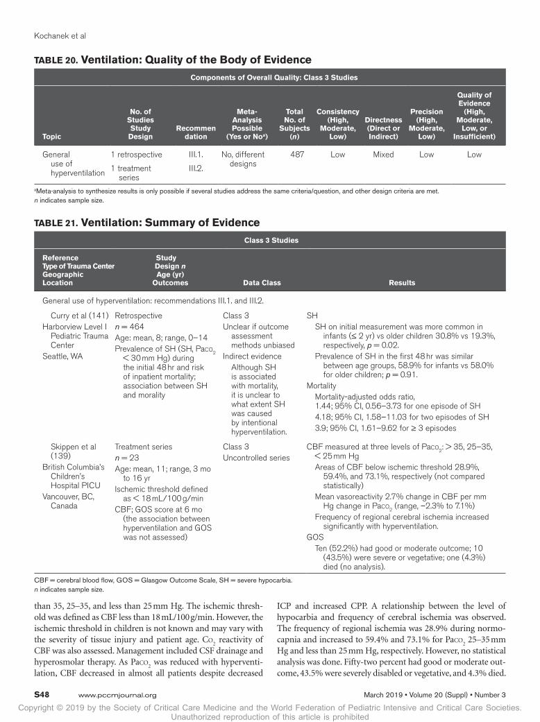
Copyright © 2019 by the Society of Critical Care Medicine and the World Federation of Pediatric Intensive and Critical Care Societies.Unauthorized reproduction of this article is prohibited
Copyright © 2019 by the Society of Critical Care Medicine and the World Federation of Pediatric Intensive and Critical Care Societies.Unauthorized reproduction of this article is prohibited
Kochanek et al
S48 www.pccmjournal.org March 2019 • Volume 20 (Suppl) • Number 3
than 35, 25–35, and less than 25 mm Hg. The ischemic thresh-old was defined as CBF less than 18 mL/100 g/min. However, the ischemic threshold in children is not known and may vary with the severity of tissue injury and patient age. Co
2 reactivity of
CBF was also assessed. Management included CSF drainage and hyperosmolar therapy. As Paco
2 was reduced with hyperventi-
lation, CBF decreased in almost all patients despite decreased
ICP and increased CPP. A relationship between the level of hypocarbia and frequency of cerebral ischemia was observed. The frequency of regional ischemia was 28.9% during normo-capnia and increased to 59.4% and 73.1% for Paco
2 25–35 mm
Hg and less than 25 mm Hg, respectively. However, no statistical analysis was done. Fifty-two percent had good or moderate out-come, 43.5% were severely disabled or vegetative, and 4.3% died.
TABLE 20. Ventilation: Quality of the Body of Evidence
Components of Overall Quality: Class 3 Studies
Topic
No. of Studies Study
DesignRecommen
dation
Meta- Analysis Possible
(Yes or Noa)
Total No. of
Subjects (n)
Consistency (High,
Moderate, Low)
Directness (Direct or Indirect)
Precision (High,
Moderate, Low)
Quality of Evidence
(High, Moderate,
Low, or Insufficient)
General use of hyperventilation
1 retrospective
1 treatment series
III.1.
III.2.
No, different designs
487 Low Mixed Low Low
a Meta-analysis to synthesize results is only possible if several studies address the same criteria/question, and other design criteria are met.n indicates sample size.
TABLE 21. Ventilation: Summary of Evidence
Class 3 Studies
Reference Type of Trauma Center Geographic Location
Study Design n Age (yr)
Outcomes Data Class Results
General use of hyperventilation: recommendations III.1. and III.2.
Curry et al (141)Harborview Level I
Pediatric Trauma Center
Seattle, WA
Retrospectiven = 464Age: mean, 8; range, 0–14Prevalence of SH (SH, Paco2
< 30 mm Hg) during the initial 48 hr and risk of inpatient mortality; association between SH and morality
Class 3Unclear if outcome
assessment methods unbiased
Indirect evidence Although SH
is associated with mortality, it is unclear to what extent SH was caused by intentional hyperventilation.
SH SH on initial measurement was more common in
infants (≤ 2 yr) vs older children 30.8% vs 19.3%, respectively, p = 0.02.
Prevalence of SH in the first 48 hr was similar between age groups, 58.9% for infants vs 58.0% for older children; p = 0.91.
Mortality Mortality-adjusted odds ratio,
1.44; 95% CI, 0.56–3.73 for one episode of SH 4.18; 95% CI, 1.58–11.03 for two episodes of SH 3.9; 95% CI, 1.61–9.62 for ≥ 3 episodes
Skippen et al (139)
British Columbia’s Children’s Hospital PICU
Vancouver, BC, Canada
Treatment seriesn = 23Age: mean, 11; range, 3 mo
to 16 yrIschemic threshold defined
as < 18 mL/100 g/minCBF; GOS score at 6 mo
(the association between hyperventilation and GOS was not assessed)
Class 3Uncontrolled series
CBF measured at three levels of Paco2: > 35, 25–35, < 25 mm Hg
Areas of CBF below ischemic threshold 28.9%, 59.4%, and 73.1%, respectively (not compared statistically)
Mean vasoreactivity 2.7% change in CBF per mm Hg change in Paco2 (range, –2.3% to 7.1%)
Frequency of regional cerebral ischemia increased significantly with hyperventilation.
GOS Ten (52.2%) had good or moderate outcome; 10
(43.5%) were severe or vegetative; one (4.3%) died (no analysis).
CBF = cerebral blood flow, GOS = Glasgow Outcome Scale, SH = severe hypocarbia.n indicates sample size.

Copyright © 2019 by the Society of Critical Care Medicine and the World Federation of Pediatric Intensive and Critical Care Societies.Unauthorized reproduction of this article is prohibited
Supplement
Pediatric Critical Care Medicine www.pccmjournal.org S49
Again, no statistical comparison of outcomes was conducted, and outcomes were not reported by hyperventilation treatment.
A second report examined the association between hypo-carbia and outcome at hospital discharge in a large pediatric series of severe TBI victims who were all mechanically venti-lated (141). Curry et al (141) carried out a retrospective cohort study of 464 patients less than 15 years old with an admis-sion GCS score less than 9 and a head Abbreviated Injury Score greater than or equal to 3, and with a Paco
2 recorded
in the first 48 hours of admission for the years 2000 to 2005. The authors examined the prevalence of SH (Paco
2, < 30 mm
Hg) and its relationship with neurologic outcome before (375 patients) and after (89 patients) the publication of the 2003 Pediatric TBI Guidelines (147). They found a nonsignificant change in the prevalence of SH from 60% of patients before to 52% after (p = 0.19). Patients with one documented epi-sode of SH, controlling for ED GCS score, lowest ED SBP, ISS, Paco
2 sampling frequency, and year of admission had adjusted
ORs for mortality of 1.44 (95% CI, 0.56–3.73) for one episode of SH, 4.18 (95% CI, 1.58–11.03) for two episodes, and 3.93 (95% CI, 1.61–9.62) for greater than or equal to three epi-sodes, compared with patients with mild or no hypocarbia. These findings, although retrospective, show an association of SH with poor outcomes. However, there might be other con-tributors to hypocarbia such as marked reduction in metabolic rates or acidosis from systemic shock. Furthermore, although SH was associated with mortality, it is unclear to what extent SH was caused by intentional hyperventilation. Thus, the evi-dence from this study is indirect, and the exact contribution of induced hyperventilation to poor outcome cannot be clearly defined from this study.
Indications From Adult Guidelines. The recent Fourth Edi-tion of the adult guidelines does not further inform the pediat-ric guidelines for this topic (14).
Temperature Control/Hypothermia
RecommendationsStrength of Recommendation: Moderate
Level IThere was insufficient evidence to support a level I recommen-dation for this topic.
Level IITo Improve Overall Outcomes. II.1. Prophylactic moderate (32–33°C) hypothermia is not recommended over normother-mia to improve overall outcomes.
Level IIIFor ICP Control. III.1. Moderate (32–33°C) hypothermia is suggested for ICP control.
Safety Recommendation 1. If hypothermia is used and rewarming is initiated, it should be carried out at a rate of 0.5–1.0°C every 12–24 hours or slower to avoid complications.
Safety Recommendation 2. If phenytoin is used during hypo-thermia, monitoring and dosing adjusted to minimize toxicity, especially during the rewarming period, are suggested.
Note: See the last paragraph of the “Evidence Synthesis sec-tion” for a discussion that explains the seemingly contradictory recommendations.
Changes From Prior Edition. Recommendations II.1. is modified with regard to timing from the Second Edition of these guidelines. Recommendation III.1 on the use of hypother-mia for ICP control is now at a Level III (rather than a level II) given that none of the studies of higher class had ICP control as a primary outcome and given the safety concerns that were identi-fied. A level II recommendation about rewarming rate from the Second Edition has been removed and replaced with the more specific safety recommendation. A level III recommendation about use of moderate hypothermia from the Second Edition has been removed. The safety recommendation about phenyt-oin is new. One class 3 treatment series from the Second Edition has been removed (148). Two new meta-analyses (149–152), three new RCTs—one class 1 (153), one class 2 (154), and one class 3 (155)—and three new secondary analyses of RCTs—one class 2 (156) and two class 3 (157, 158)—have been added to the evidence base for this topic.
IntroductionThe definitions of hypothermia and hyperthermia vary. Posttraumatic hypothermia is often classified as a core body temperature less than 35°C, whereas a temperature greater than 38.0–38.5°C represents fever/pyrexia if it results from an altered thermoregulatory set point, and represents hyper-thermia if it is imposed upon a normal set point. For sim-plicity, the term hyperthermia is used to reflect an elevated core body temperature throughout this topic. Experimental studies in animal models and clinical studies in children demonstrated that hyperthermia correlates with poor out-comes and it has been recommended that hyperthermia following TBI in children should be prevented (159, 160). However, no studies of the influence of hyperthermia on outcomes in children were identified for inclusion in this guideline.
There are compelling reasons to explore the rationale for the use of therapeutic hypothermia to limit secondary brain injury based on its role in decreasing cerebral metabolic demands, inflammation, lipid peroxidation, excitotoxicity, cell death, and acute seizures (161, 162). Clinical studies reviewed on temperature regulation for these guidelines addressed global functional outcome and its effect on ICP. The influence on outcomes of reduction of ICP following severe TBI in chil-dren remains to be determined. As discussed in other topics, the lowering of severely elevated ICP with respect to the treat-ment threshold may be a desirable outcome.
Evaluation of the EvidenceQuality of the Body of Evidence. Studies included for this topic compared hypothermia with normothermia in the treatment of severe TBI in pediatric patients, and considered its influ-ence on mortality, neurologic outcome, and control of ICP. Two meta-analyses (149–152); five RCTs—one class 1 (153), three class 2 (40, 144, 154), and one class 3 (155); and three

Copyright © 2019 by the Society of Critical Care Medicine and the World Federation of Pediatric Intensive and Critical Care Societies.Unauthorized reproduction of this article is prohibited
Copyright © 2019 by the Society of Critical Care Medicine and the World Federation of Pediatric Intensive and Critical Care Societies.Unauthorized reproduction of this article is prohibited
Kochanek et al
S50 www.pccmjournal.org March 2019 • Volume 20 (Suppl) • Number 3
secondary analyses—one class 2 (156) and two class 3 (157, 158) provide evidence for this topic. Findings are consistent
across the meta-analyses and class 1 and 2 RCTs. The overall quality of the body of evidence is moderate (Table 22).
TABLE 22. Temperature Control/Hypothermia: Quality of the Body of Evidence
TopicNo. of Studies Study
DesignRecommen-
dation
Meta- Analysis Possible (Yes or
Noa)Total No. of Subjects (n)
Consistency (High,
Moderate, Low)
Directness (Direct or Indirect)
Precision (High,
Moderate, Low)
Quality of Evidence
(High, Moderate,
Low, or Insufficient)
Components of overall quality: meta-analyses
Comparison of hypothermia and normothermia
Two meta- analyses
Tasker et al (150)Crompton et al (149, Tasker and Akhondi-Asl [151], and Crompton and Sharma [152])
II.1. NA 504b High Direct High Moderate
Components of overall quality: class 1 study
Comparison of hypothermia and normothermia
1 RCT
Adelson et al (153)
II.1. & Safety Rec. 1.
NA 77 NA Direct Moderate High
Components of overall quality: class 2 studies
Comparison of hypothermia and normothermia
4 RCTs No 350 High Direct Moderate Moderate
Beca et al (154) II.1.
III.1.
NA 50 NA Direct NA NA
Adelson et al (40) II.1.
III.1.
NA 75 NA Direct NA NA
Hutchison et al (144)
II.1.
III.1.
NA 225 NA Direct NA NA
Hutchison et al (156)
II.1. NA 225 (2008 sample)
NA Direct NA NA
Components of overall quality: class 3 studies
Comparison of hypothermia and normothermia
1 RCT
2 retrospective
Safety recommen dation 2
No 119 High for Empey et al (155) and Bourdages et al (157)
Mixed Low Insufficient
Su et al (158) No recommen
dation
NA 84 NA Indirect NA NA
Empey et al (155) Safety recommen dation 2
NA 19 NA Indirect NA NA
Bourdages et al (157) No recommen
dation
NA 16 NA Direct NA NA
NA = not applicable, RCT = randomized controlled trial.a Meta-analysis to synthesize results is only possible if several studies address the same criteria/question, and other design criteria are met.b The total number of subjects for two meta-analyses was calculated with sample sizes of included studies in Tasker et al (150), except for Adelson et al (40), for which the sample size from the original publication was used, and Salonia et al (163), which was included in Crompton et al (149) and not Tasker et al (150).
n indicates sample size.

Copyright © 2019 by the Society of Critical Care Medicine and the World Federation of Pediatric Intensive and Critical Care Societies.Unauthorized reproduction of this article is prohibited
Supplement
Pediatric Critical Care Medicine www.pccmjournal.org S51
Applicability. The meta-analyses and three of the RCTs are recent (149–155). Study sites were located in the United States, New Zealand, Australia, Canada, France, and the United Kingdom. Sam-ple sizes for individual studies ranged from 16 to 225 patients, and ages ranged from 0 to 18 years. Applicability concerns are minimal.
Summary of the EvidenceOf the 10 studies included in the evidence tables, two new meta-analyses (one fair [150] and one poor quality [149, 151,
152]), three new RCTs (one class 1 [153], one class 2 [154],
and one class 3 [155]), and three new secondary analysis (one
class 2 [156] and two class 3 [157, 158]) were added to two
class 2 RCTs (40, 144) from the Second Edition. Together they
provided evidence to support the recommendations for this
topic. Two new studies were reviewed and included in the evi-
dence table (157, 158) but not used to support a recommen-
dation (Tables 23 and 24).
TABLE 23. Temperature Control/Hypothermia: Summary of Evidence
Meta-Analyses
Reference Type of Trauma Center Geographic Location
Study Design n Age (yr) Outcomes
Hypothermia Protocol Data Class Results
Comparison of hypothermia and normothermia: recommendations II.1. and III.1.
Tasker et al (150)a
Recommendation II.1.Adelson et al (153)Adelson et al (40)Beca et al (154)Biswas et al (164)Hutchison et al (144)Li et al (165)
MA7 RCTsn = 470Age: 0–17MortalityProtocol varied across studies
Fair-quality MADuplicate review not
specified; search not comprehensive; exclusions not specified; publication bias not assessed; conflict of interest NR
Mortality With conventional MA, no difference
in mortality. With Bayesian MA, probability of
reducing mortality with hypothermia compared with normothermia is 0.40 (with RR < 1 or RRR > 0). Probability of RRR of death > 20%, with hypothermia rather than normothermia, 0.28
Crompton et al (149, Tasker and Akhondi-Asl (151), and Crompton and Sharma (152)a
Recommendation II.1.Adelson et al (153)Adelson et al (40)Beca et al (154)Biswas et al (164)Hutchison et alSalonia et al (163)
MAEight pediatric studies7 RCTs; 1 observationaln = 454Age: 3 mo to 18 yrMortality, neurologic outcome,
GOSProtocol varied across studies
Poor-quality MADuplicate review
not specified; extent of literature search unclear; exclusions not specified; quality of individual studies not assessed or reported; publication bias not assessed; conflict of interest NR
Mortality No significant difference (RR, 1.53;
95% CI, 0.92–2.54; p = 0.10)Neurologic outcome No significant difference; 10%
decrease in favorable neurologic outcomes (RR, 0.90; 95% CI, 0.80–1.01; p = 0.06)
GOS No significant difference; GOS scores
decreased by 0.17 points (mean difference, –0.17; 95% CI, –0.64 to 0.31; p = 0.50)
Class 1 and 2 Studies
Adelson et al (153)a
15 sitesUnited States, New Zealand,
and AustraliaRecommendation II.1. and
safety recommendation 1
RCTCool Kids Trialn = 77Age: median, 10.9IQR: 3.4–14.6Hypothermia: median, 9.7; IQR,
4.2–14.5Normothermia: median, 12.5; IQR,
3.3–14.8Mortality, GOS and GOS-E at 3
mo, complicationsHypothermia 32–33°C for
48–72 hr with slow rewarming; 0.5–1.0°C every 12–24 hr
Class 1(Evidence for Safety
Rec. #1.)
Hypothermia vs normothermiaMortality within 3 mo 6 (15%) vs 2 (5%), p = 0.15GOS and GOS-E No significant difference for GOS
(p = 0.90) or GOS-E (p = 0.73)Complications (acute nonserious
infection and late nonserious infection): no difference, p = 0.0622.
Decompressive craniotomy: 17 (45%) vs 7 (18%), p = 0.0220
Trial terminated early for futility
(Continued)

Copyright © 2019 by the Society of Critical Care Medicine and the World Federation of Pediatric Intensive and Critical Care Societies.Unauthorized reproduction of this article is prohibited
Copyright © 2019 by the Society of Critical Care Medicine and the World Federation of Pediatric Intensive and Critical Care Societies.Unauthorized reproduction of this article is prohibited
Kochanek et al
S52 www.pccmjournal.org March 2019 • Volume 20 (Suppl) • Number 3
Beca et al (154)a
8 PICUsAustralia: Adelaide, Brisbane,
Melbourne, Perth, SydneyNew Zealand: AucklandCanada: Vancouver, BCRecommendations II.1. and
III.1.
RCTPhase 2 trialn = 50Hypothermia: n = 24
Normothermia: n = 26Age: Hypothermia: mean, 11.0; IQR,
6.9–14.2 Normothermia: mean, 9.5; IQR,
5.2–13.8Mortality, PCPC, PICU length of
stay, hospital length of stay, and mechanical ventilation; ICP, MAP, CPP; complications
Early hypothermia (32–33°C) for 72 hr with slow based on individual patient to maintain ICP and CPP (no > 0.5°C every 3 hr)
Class 2Criteria met except
blinding which was not possible or unclear; sample size likely not adequate
Hypothermia vs normothermiaMortality 3 (13%) vs 1 (4%), p = 0.34Poor outcome (included death) 3 (12%) vs 4 (17%)PCPC at 12 mo 4 (17%) vs 3 (12%), p = 0.70PICU length of stay (d) in survivors 12 vs 11, p = 0.87Hospital length of stay (d) in survivors 48 vs 40, p = 0.70Mechanical ventilation (d) in survivors 8 vs 10, p = 0.77 No predefined covariables were
significant in the multivariable model.ICP, MAP, CPP ICP was lower by 1.8 mm during
cooling (95% CI, 0.3–3.4; p = 0.02) No significant group difference in MAP or CPP during cooling, p = 0.44 and p = 0.77
No significant group difference in MAP, ICP, or CPP during rewarming, p = 0.68, p = 0.59, p = 0.07
Complications Net effect of hypothermia during
cooling: Heart rate drop: 23.4 beats/min; 95%
CI, 15.9–30.9; p < 0.001
Adelson et al (40)MulticenterUnited StatesRecommendations II.1. and
III.1.
2 RCTsTotal n = 752 samples/studies n = 48 (patients who met
inclusion criteria, multicenter) n = 27 (patients who did not
meet inclusion criteria, single center)
Age: Sample 1: mean, 6.89; range,
NR Hypothermia: mean, 6.92 Normothermia: mean, 6.86 Sample 2: mean, 6.95; range, NR Hypothermia: mean, 7.17 Normothermia: mean, 5.6Mortality, 3 and 6 mo GOS, ICP,
complicationsHypothermia 32–33°C for 48 hr;
slow rewarming 1°C every 3–4 hr, with slower rates for individual patients based on ICP
Class 2Unclear reporting
of randomization methods, allocation concealment methods, and attrition
Mortality and GOS No difference between groups in
mortality or 3 and 6 mo GOS.ICP Overall, there was no statistical
difference in mean ICP between the groups during the 5-d period, p = 0.37 except within the first 24 hr, when the ICP was reduced in the hypothermia group, p = 0.024.
Complications No difference between groups in
complication rates
TABLE 23. (Continued). Temperature Control/Hypothermia: Summary of Evidence
Reference Type of Trauma Center Geographic Location
Study Design n Age (yr) Outcomes
Hypothermia Protocol Data Class Results
(Continued)

Copyright © 2019 by the Society of Critical Care Medicine and the World Federation of Pediatric Intensive and Critical Care Societies.Unauthorized reproduction of this article is prohibited
Supplement
Pediatric Critical Care Medicine www.pccmjournal.org S53
Evidence SynthesisInfluence of Hypothermia on Mortality and Outcome. Two meta-analyses, one moderate quality (150) and one poor qual-ity (149, 151, 152), contributed to the evidence to support rec-ommendation II.1. against the use of hypothermia to improve outcomes. Together they included seven RCTs (n = 469) (40: samples 1 and 2, 153, 154, 163–165). The review by Crompton et al (149) included one study (163) that reported an analy-sis using patients from another study included in the review (40). The Tasker et al (150) analysis of these studies found no significant difference in mortality between hypothermia and normothermia groups, using conventional methods. With Bayesian analysis, the probability of reducing mortality with hypothermia versus normothermia was 0.40. The analysis by Crompton et al (149) originally reported significantly higher mortality (p = 0.03), no difference in neurologic outcomes (p = 0.06), and no difference in GOS scores (p = 0.50) for hypothermia versus normothermia. However, after repeating the analysis to correct for “double counting” in patient num-bers across the studies, there was no significant difference in mortality (p = 0.10) (151, 152).
Five RCTs—one class 1 (153) and four class 2 (40, 144, 154, 156)—were also used as evidence to support recommenda-tion II.1. Both the publications by Hutchison et al (144, 156) reported on the same set of patients, with the earlier focusing on mortality and outcomes and the latter on the association between hypotension and outcomes. The five studies included 427 patients, randomized to either hypothermia or normo-thermia. Adelson et al (40), Beca et al (154), and Adelson et al (153) reported no difference between groups in mortality or outcomes (40, 153, 154). Hutchison et al (144) reported no dif-ference between groups on outcomes overall and no significant difference in morality (p = 0.06), but characterized this as a non-significant trend toward increased mortality in the hypothermia group. This is one way to acknowledge the potential for harm that merits further study.
Both publications by Adelson et al (40, 153) reported no dif-ference between groups in complications. Beca et al (154) found decreased heart rate for hypothermia patients with cooling. Hutchison et al (144) reported significantly more hypotension, pressor requirements, and lower blood pressures and CPPs in the hypothermia group; the 2011 analysis of that sample identi-fied in the hypothermia group a significant association between
Hutchison et al (144)MulticenterCanada, France, and United
KingdomRecommendations
II.1 and III.1.
RCTn = 225Age: Hypothermia: mean, 9.8; range,
NR Normothermia: mean, 10.2;
range, NRMortality, PCPC at 4–6 mo
postinjury, ICP, complicationsHypothermia 32.5°C for 24 hr;
rewarming 0.5°C every 2 hr
Class 2Some differences
between groups on baseline prognostic factors
Mortality There were 23 deaths (21%) in
the hypothermia group and 14 deaths (12%) in the normothermia group (relative risk, 1.40; 95% CI, 0.90–2.27; p = 0.06). Although this is not significant, it suggests that additional studies to rule out or confirm this potential harm are needed.
PCPC No difference between groups on
functional outcomes at 6 mo (p = 0.14)
ICP ICP was lower during cooling and
higher during warming in the hypothermia group at 16 hr (p < 0.02), 24 hr (p < 0.01), 48 hr (p = 0.01), and 72 hr (p = 0.03).
Complications Significantly more episodes of
hypotension (p = 0.047), and lower mean blood pressures and CPPs (p < 0.001) in hypothermia group
CPP = cerebral perfusion pressure, GOS = Glasgow Outcome Scale, GOS-E = Glasgow Coma Scale Extended, ICP = intracranial pressure, IQR = interquartile range, MA = meta-analysis, MAP = mean arterial pressure, NR = not reported, PCPC = Pediatric Cerebral Performance Category, RCT = randomized controlled trial, RR = risk reduction, RRR = relative risk reduction.a New study.n indicates sample size.
TABLE 23. (Continued). Temperature Control/Hypothermia: Summary of Evidence
Reference Type of Trauma Center Geographic Location
Study Design n
Age (yr) Outcomes
Hypothermia Protocol Data Class Results
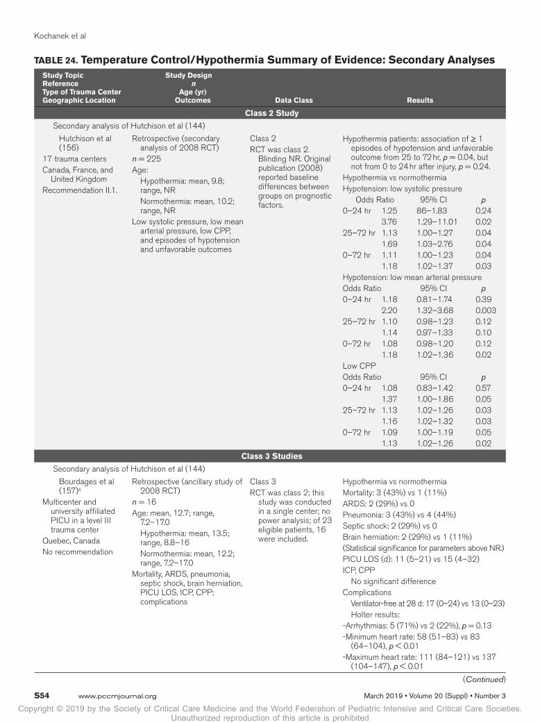
Copyright © 2019 by the Society of Critical Care Medicine and the World Federation of Pediatric Intensive and Critical Care Societies.Unauthorized reproduction of this article is prohibited
Copyright © 2019 by the Society of Critical Care Medicine and the World Federation of Pediatric Intensive and Critical Care Societies.Unauthorized reproduction of this article is prohibited
Kochanek et al
S54 www.pccmjournal.org March 2019 • Volume 20 (Suppl) • Number 3
TABLE 24. Temperature Control/Hypothermia Summary of Evidence: Secondary AnalysesStudy TopicReferenceType of Trauma CenterGeographic Location
Study Design n
Age (yr) Outcomes Data Class Results
Class 2 Study
Secondary analysis of Hutchison et al (144)
Hutchison et al (156)
17 trauma centersCanada, France, and
United KingdomRecommendation II.1.
Retrospective (secondary analysis of 2008 RCT)
n = 225Age: Hypothermia: mean, 9.8;
range, NR Normothermia: mean, 10.2;
range, NRLow systolic pressure, low mean
arterial pressure, low CPP, and episodes of hypotension and unfavorable outcomes
Class 2RCT was class 2.
Blinding NR. Original publication (2008) reported baseline differences between groups on prognostic factors.
Hypothermia patients: association of ≥ 1 episodes of hypotension and unfavorable outcome from 25 to 72 hr, p = 0.04, but not from 0 to 24 hr after injury, p = 0.24.
Hypothermia vs normothermiaHypotension: low systolic pressure Odds Ratio 95% CI p0–24 hr 1.25 86–1.83 0.24 3.76 1.29–11.01 0.0225–72 hr 1.13 1.00–1.27 0.04 1.69 1.03–2.76 0.040–72 hr 1.11 1.00–1.23 0.04 1.18 1.02–1.37 0.03Hypotension: low mean arterial pressureOdds Ratio 95% CI p0–24 hr 1.18 0.81–1.74 0.39 2.20 1.32–3.68 0.00325–72 hr 1.10 0.98–1.23 0.12 1.14 0.97–1.33 0.100–72 hr 1.08 0.98–1.20 0.12 1.18 1.02–1.36 0.02Low CPPOdds Ratio 95% CI p0–24 hr 1.08 0.83–1.42 0.57 1.37 1.00–1.86 0.0525–72 hr 1.13 1.02–1.26 0.03 1.16 1.02–1.32 0.030–72 hr 1.09 1.00–1.19 0.05 1.13 1.02–1.26 0.02
Class 3 Studies
Secondary analysis of Hutchison et al (144)
Bourdages et al (157)a
Multicenter and university affiliated PICU in a level III trauma center
Quebec, CanadaNo recommendation
Retrospective (ancillary study of 2008 RCT)
n = 16Age: mean, 12.7; range,
7.2–17.0 Hypothermia: mean, 13.5;
range, 8.8–16 Normothermia: mean, 12.2;
range, 7.2–17.0Mortality, ARDS, pneumonia,
septic shock, brain herniation, PICU LOS, ICP, CPP; complications
Class 3RCT was class 2; this
study was conducted in a single center; no power analysis; of 23 eligible patients, 16 were included.
Hypothermia vs normothermiaMortality: 3 (43%) vs 1 (11%)ARDS: 2 (29%) vs 0Pneumonia: 3 (43%) vs 4 (44%)Septic shock: 2 (29%) vs 0Brain herniation: 2 (29%) vs 1 (11%)(Statistical significance for parameters above NR.)PICU LOS (d): 11 (5–21) vs 15 (4–32)ICP, CPP No significant differenceComplications Ventilator-free at 28 d: 17 (0–24) vs 13 (0–23) Holter results:-Arrhythmias: 5 (71%) vs 2 (22%), p = 0.13-Minimum heart rate: 58 (51–83) vs 83
(64–104), p < 0.01-Maximum heart rate: 111 (84–121) vs 137
(104–147), p < 0.01
(Continued)
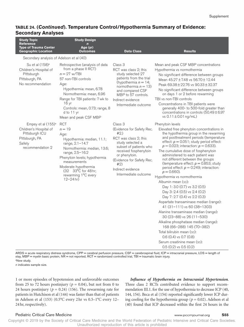
Copyright © 2019 by the Society of Critical Care Medicine and the World Federation of Pediatric Intensive and Critical Care Societies.Unauthorized reproduction of this article is prohibited
Supplement
Pediatric Critical Care Medicine www.pccmjournal.org S55
Secondary analysis of Adelson et al (40)
Su et al (158)a
Children’s Hospital of Pittsburgh
Pittsburgh, PANo recommendation
Retrospective (analysis of data from a phase II RCT)
n = 27 w/TBI57 non-TBI controlsAge: Hypothermia: mean, 6.78 Normothermia: mean, 6.96Range for TBI patients: 7 wk to
16 yr Controls: mean, 0.73; range, 8
d to 11 yrMean and peak CSF MBP
Class 3RCT was class 2; this
study selected 27 patients from the trial (hypothermia n = 14; normothermia n = 13) and compared CSF MBP to 57 controls.
Indirect evidence Intermediate outcome
Mean and peak CSF MBP concentrationsHypothermia vs normothermia No significant difference between groups Mean 45.27 ± 7.48 vs 56.70 ± 12.44 Peak 69.38 ± 22.76 vs 90.33 ± 32.37 No significant difference between groups
on days 1 or 2 before rewarmingTBI vs non-TBI controls Concentrations in TBI patients were
generally 400- to 500-fold greater than concentrations in controls (50.49 ± 6.97 vs 0.11 ± 0.01 ng/mL)
Empey et al (155)a
Children’s Hospital of Pittsburgh ICU
Pittsburgh, PASafety
recommendation 2
RCTn = 19Age: Hypothermia: median, 11.1;
range, 2.1–14.7 Normothermia: median, 13.6;
range, 2.5–16.2Phenytoin levels; hypothermia
measurementsModerate hypothermia
(32 33°C for 48 hr; rewarming 1°C every 12–24 hr)
Class 3(Evidence for Safety Rec.
#2.)RCT was class 2; this
study selected a subset of patients who received fosphenytoin or phenytoin.
(Evidence for Safety Rec. #2)
Indirect evidence Intermediate outcome
Phenytoin levels Elevated free phenytoin concentrations in
the hypothermia group in the rewarming and posttreatment periods (temperature effect: p = 0.051; study period effect: p = 0.023; interaction: p = 0.633).
The cumulative dose of fosphenytoin administered to each patient was not different between the groups (temperature effect: p = 0.853; study period effect: p = 0.249; interaction: p = 0.660).
Hypothermia vs normothermia Albumin mean (sd): Day 1: 3.0 (0.7) vs 3.2 (0.5) Day 3: 2.4 (0.5) vs 2.4 (0.2) Day 7: 2.7 (0.4) vs 2.2 (0.3) Aspartate transaminase median (range): 41 (31–111) vs 60 (38–1303) Alanine transaminase median (range): 30 (23–88) vs 26 (11–530) Alkaline phosphatase median (range): 168 (66–288) 145 (70–382) Total bilirubin mean (sd): 0.6 (0.4) vs 0.7 (0.8) Serum creatinine mean (sd): 0.5 (0.2) vs 0.5 (0.2)
ARDS = acute respiratory distress syndrome, CPP = cerebral perfusion pressure, CSF = cerebrospinal fluid, ICP = intracranial pressure, LOS = length of stay, MBP = myelin basic protein, NR = not reported, RCT = randomized controlled trial, TBI = traumatic brain injury.a New study.n indicates sample size.
TABLE 24. (Continued). Temperature Control/Hypothermia Summary of Evidence: Secondary Analyses
Study TopicReferenceType of Trauma CenterGeographic Location
Study Design n
Age (yr) Outcomes Data Class Results
1 or more episodes of hypotension and unfavorable outcomes from 25 to 72 hours postinjury (p = 0.04), but not from 0 to 24 hours postinjury (p = 0.24) (156). The rewarming rate for patients in Hutchison et al (144) was faster than that of patients in Adelson et al (153) (0.5°C every 2 hr vs 0.5–1°C every 12–24 hr, respectively).
Influence of Hypothermia on Intracranial Hypertension. Three class 2 RCTs contributed evidence to support recom-mendation III.1. for the use of hypothermia to decrease ICP (40, 144, 154). Beca et al (154) reported significantly lower ICP dur-ing cooling for the hypothermia group (p = 0.02). Adelson et al (40) found that ICP decreased within the first 24 hours in the

Copyright © 2019 by the Society of Critical Care Medicine and the World Federation of Pediatric Intensive and Critical Care Societies.Unauthorized reproduction of this article is prohibited
Copyright © 2019 by the Society of Critical Care Medicine and the World Federation of Pediatric Intensive and Critical Care Societies.Unauthorized reproduction of this article is prohibited
Kochanek et al
S56 www.pccmjournal.org March 2019 • Volume 20 (Suppl) • Number 3
hypothermia group, but no difference between groups over 5 days. Hutchison et al (144) found that ICP was significantly lower during cooling, but then higher during warming in the hypother-mia group at 16 hours (p < 0.02), 24 hours (p < 0.01), 48 hours (p = 0.01), and 72 hours (p = 0.03). Although the evidence does not suggest a long-term benefit for ICP control with hypother-mia, it does suggest that hypothermia produces an immediate decrease in ICP.
Safety Recommendations. Safety recommendation 1 cautions against rapid rewarming that may be a source of complications seen in previous work in which temperature was increased at a rate of 0.5°C every 2 hours (144). The current recommended parameters are based on the protocol used in the study by Adel-son et al (153), which found no significant difference between hypothermia and normothermia groups for adverse events.
Safety recommendation 2 is based on the RCT by Empey et al (155) which demonstrated elevated free phenytoin lev-els on rewarming from hypothermia (rate 1°C per 12–24 hr) although the cumulative doses administered to children in both groups were similar.
Seemingly Inconsistent Recommendations. Furthermore, the published studies targeting the effect of hypothermia on long-term outcomes in pediatric severe TBI implemented it early after injury and did not specifically randomize patients with intracranial hypertension to hypothermia versus another second-tier therapy. It was used in a prophylactic manner. We did not identify studies comparing the efficacy of second-tier therapies implemented for refractory raised ICP. It would thus be premature to dismiss hypothermia in this setting based on the available evidence.
Indications From Adult GuidelinesThe clinical investigators do not consider the recommenda-tions about temperature control from the adult guidelines applicable to guide treatment decisions in children.
BARBITURATES
RecommendationsStrength of Recommendations: Weak
Levels I and IIThere was insufficient evidence to support a level I or II rec-
ommendation for this topic.
Level IIIFor ICP Control. III.1. High-dose barbiturate therapy is sug-gested in hemodynamically stable patients with refractory intracranial hypertension despite maximal medical and surgi-cal management.
Safety Recommendation. When high-dose barbiturate ther-apy is used to treat refractory intracranial hypertension, con-tinuous arterial blood pressure monitoring and cardiovascular support to maintain adequate CPP are required because car-diorespiratory instability is common among patients treated with barbiturate coma.
Changes From Prior Edition .There are no content changes from the Second Edition to the recommendations (1). Two new class 3 studies—one retrospective observational (85) and one treatment series (166)—were added to the evidence base for this topic.
IntroductionHigh-dose barbiturates lower ICP by suppression of metab-olism and alteration of vascular tone (167–169) causing improved coupling of regional blood flow to metabolic demands resulting in higher brain oxygenation (170), with lower CBF and decreased ICP from decreased CBV. Barbi-turates may lower ICP when first-tier medical and surgical management have not resulted in adequate control. However, cardiorespiratory side effects are very common and potentially harmful, including decreased cardiac output, hypotension, and increased intrapulmonary shunt, resulting in lower CPP and hypoxia. Thus high-dose barbiturate therapy has been reserved for cases of intracranial hypertension resistant to first-tier medical and surgical care.
Diffuse brain swelling and generalized hyperemia after severe TBI are more common and more lethal (67) in young children compared with older children and adults (171–173). The rationale for treatment with barbiturate coma is based on the logic that uncontrolled ICP leads to ongoing secondary brain injury, and a higher risk of poor cognitive outcome or death. Near maximum reduction in cerebral metabolism and CBF occur when burst suppression is induced. Pentobarbital is the most commonly reported medication used in children and is dosed to achieve burst suppression, so continuous elec-troencephalogram monitoring is required to monitor optimal dosing. Although high-dose barbiturates are reserved for a high-risk group, their use in severe pediatric TBI care is not rare.
Evaluation of the EvidenceQuality of the Body of Evidence. Studies included for this topic addressed the influence of barbiturate therapy on out-comes and ICP. The evidence consists of one relatively large observational study (85) and three small treatment series (47, 166, 174). Although consistency and precision were moderate, the overall quality is low because the studies were rated class 3 (Table 25).
Applicability. Two of the included studies were over 25 years old (47, 174). Three were small and were conducted at single sites (47, 166, 174), whereas the larger study was conducted at multiple sites (85). All were conducted in the United States and included a range of ages from infants to teens. Applicability of these studies is limited.
Summary of EvidenceFour class 3 studies, two new (85, 166) and two from the Sec-ond Edition (47, 174), provided evidence to support the rec-ommendation (Table 26).

Copyright © 2019 by the Society of Critical Care Medicine and the World Federation of Pediatric Intensive and Critical Care Societies.Unauthorized reproduction of this article is prohibited
Supplement
Pediatric Critical Care Medicine www.pccmjournal.org S57
Evidence SynthesisInfluence of Barbiturate Therapy on Outcomes and ICP. Three treatment series (47, 166, 174) and one multicenter observa-tional study provided data for this topic (85). The treatment series included a total of 74 patients who received barbiturate therapy for refractory intracranial hypertension. Mortality was 43%. Two of the three studies assessed 43 patients for function at various timepoints, and reported that 19 (44%) had poor outcomes (166, 174). They also reported that of the total 63 patients, ICP control was achieved in 24 (38%). When high-dose barbiturates were added to additional therapies, rates for control of refractory intracranial hypertension were 28% and 52%, respectively. The third study reported a steep decline in ICP for patients treated with pentobarbital compared with those not treated and those treated with mannitol (47). One study found a nonsignificant lower risk of death in patients with controlled intracranial hypertension (risk reduction, 0.2; 95% CI, 0.03–1.3), and significantly better median discharge Pediatric Cerebral Performance Category (PCPC) scores for that group (p < 0.05) (166). They reported significantly bet-ter PCPC scores at 3–12 months follow-up (p < 0.05), but the analysis excluded patients who died (n = 14), and used dis-charge PCPC as the follow-up score for seven patients without follow-up data.
Patient and disease characteristics, treatment variation, and the uncontrolled nature of these studies limited the ability to associate the findings with the intervention. Mellion et al (166) reported that among patients who had ICP controlled, high-dose barbiturate therapy was employed significantly later after TBI (76 vs 29 median hours) compared with children whose ICP remained uncontrolled. Kasoff et al (47) reported that more than 90% of treated patients received ionotropic infu-sions. In two studies, despite monitoring and infusion thera-pies, 80% of patients experienced episodes of hypotension or a fall in CPP below the goal level (47, 166). However, reports of both efficacy and toxicity information for all three studies are based on reports from single centers and relatively few patients.
An observational analysis of 236 patients treated at five pedi-atric trauma centers by Vavilala et al (85) studied adherence to a set of clinical indicators to the 2003 Pediatric Guidelines for
severe TBI management. They tested the relationship between rates of adherence and in-hospital mortality and discharge GOS score. Ninety-four children (82%) developed high ICP (either ICP > 20 mm Hg or clinical signs of intracranial hyper-tension within the first 72 hr of admission). Cerebral edema based on radiologic imaging was present in 127 (53.8%). The estimated decrease in mortality was 78% (adjusted hazard ratio, 0.22; 95% CI, 0.18–0.25) when ICU patients received barbiturates for refractory ICP as recommended.
These studies provide data to support the recommendation for this topic. However, there is insufficient evidence to rec-ommend use of a particular barbiturate agent or regimen over another to treat refractory intracranial hypertension.
Indications From Adult GuidelinesThe recent Fourth Edition of the adult guidelines does not fur-ther inform the pediatric guidelines for this topic (14).
Decompressive Craniectomy
RecommendationsStrength of Recommendation: Weak
Levels I and IIThere was insufficient evidence to support a level I or II recom-mendation for this topic.
Level IIIFor ICP Control. III.1. Decompressive craniectomy (DC) is suggested to treat neurologic deterioration, herniation, or intracranial hypertension refractory to MM.
Changes From Prior Edition. The specification in the rec-ommendation from the Second Edition, “. . . with duraplasty, leaving the bone flap out . . .” has been removed, and for this edition, the recommendation is made specifically for ICP con-trol. One class 3 RCT from the First Edition which was removed from the Second Edition was returned to this edition (176). Fourteen new class 3 studies—five retrospective comparisons (176–180) and nine treatment series (181–189)—were added to the evidence base for this topic.
TABLE 25. Barbiturates: Quality of the Body of Evidence
Components of Overall Quality: Class 3 Studies
TopicNo. of Studies Study Design
Recommen dations
Meta- Analysis Possible
(Yes or Noa)
Total No. of
Subjects (n)
Consistency (High,
Moderate, Low)
Directness (Direct or Indirect)
Precision (High,
Moderate, Low)
Quality of Evidence
(High, Moderate,
Low, or Insufficient)
High-dose barbiturates for refractory intracranial hypertension
1 retrospective
3 treatment series
III.1.
Safety recommen dation
No
Due to study designs
310 Moderate Direct Moderate Low
a Meta-analysis to synthesize results is only possible if several studies address the same criteria/question, and other design criteria are met.n indicates sample size.

Copyright © 2019 by the Society of Critical Care Medicine and the World Federation of Pediatric Intensive and Critical Care Societies.Unauthorized reproduction of this article is prohibited
Copyright © 2019 by the Society of Critical Care Medicine and the World Federation of Pediatric Intensive and Critical Care Societies.Unauthorized reproduction of this article is prohibited
Kochanek et al
S58 www.pccmjournal.org March 2019 • Volume 20 (Suppl) • Number 3
TABLE 26. Barbiturates: Summary of Evidence
Class 3 Studies
ReferenceType of Trauma CenterGeographic Location
Study Design n
Age (yr)Mean (Range)
Outcomes Data Class Results
High-dose barbiturates for refractory intracranial hypertension: recommendation III.1.
Vavilala et al (85)a
Five pediatric trauma centers
Seattle, WA; Pittsburg PA; Chicago, IL; Torrance, CA; Columbus, OH
Retrospectiven = 236Age: mean, 8.0; range, NRIn-hospital mortality
Class 3Selection, blinding,
differential loss to follow-up, and baseline differences unclear; did not control for all relevant confounders
Discharge survival In ICU: barbiturates used with high ICP:
adjusted hazard ratio, 0.22 (95% CI, 0.18–0.26)
Reference: no barbiturates used with high ICP
(Also measured in OR: not significant; overall, ICU and OR combined: not significant)
Mellion et al (166)a
Level I pediatric trauma center
Salt Lake City, UT
Treatment seriesn = 36Age: Controlled intracranial
hypertension: median, 10.7; range, NR
Uncontrolled intracranial hypertension: median, 6.4; range, NR
Mortality; PCPC at discharge and follow-up (3–12 mo); ICP; harms including infections, impaired oxygenation, and CPP below target
Class 3Uncontrolled series
Mortality 22 of 36 survived Survival more common in responders than
nonresponders but not significant (relative risk of death, 0.2; 95% CI, 0.03–1.3)
PCPC 19 with favorable outcome (PCPC less
than 3) at 3 mo or longer after injury; three returned to normal function
ICP 10 out of 36 (28%) had controlled
refractory intracranial hypertension Of 14 deaths, 13 were without control of
ICP No control of refractory intracranial
hypertension was significantly associated with poor scores on PCPC (p < 0.05), but with dichotomized PCPC, function did not differ significantly between patients with and without controlled intracranial hypertension.
Pittman et al (174)Cardinal Glennon
Memorial Hospital for Children
Missouri
Treatment seriesn = 27 totaln = 7 for outcomesAge: mean, 9.0; range, 2 mo
to 15 yrMortality; GOS at 1, 6, and 12
mo postinjury; ICP CPP
Class 3Uncontrolled series
Mortality 6 of 27GOS Good recovery: 3 Moderate disability: 2 Vegetative: 2ICP 14/27 (52%) achieved ICP < 20 mm Hg Of 13 with persistently elevated ICP, six
died (22%)CPP Outcome not related to CPP
Kasoff et al (47)Westchester County
Medical CenterNew York
Treatment seriesn = 11Age: mean, 8.8; range, 3 mo
to 17 yrMortality
Class 3Uncontrolled series
Mortality 4/11 (36%) Hypotension (mean arterial pressure) <
80 mm Hg occurred in 9/11 (82%)
CPP = cerebral perfusion pressure, GOS = Glasgow Outcome Scale, ICP = intracranial pressure, NR = not reported, OR = operating room, PCPC = Pediatric Cerebral Performance Category.a New study.
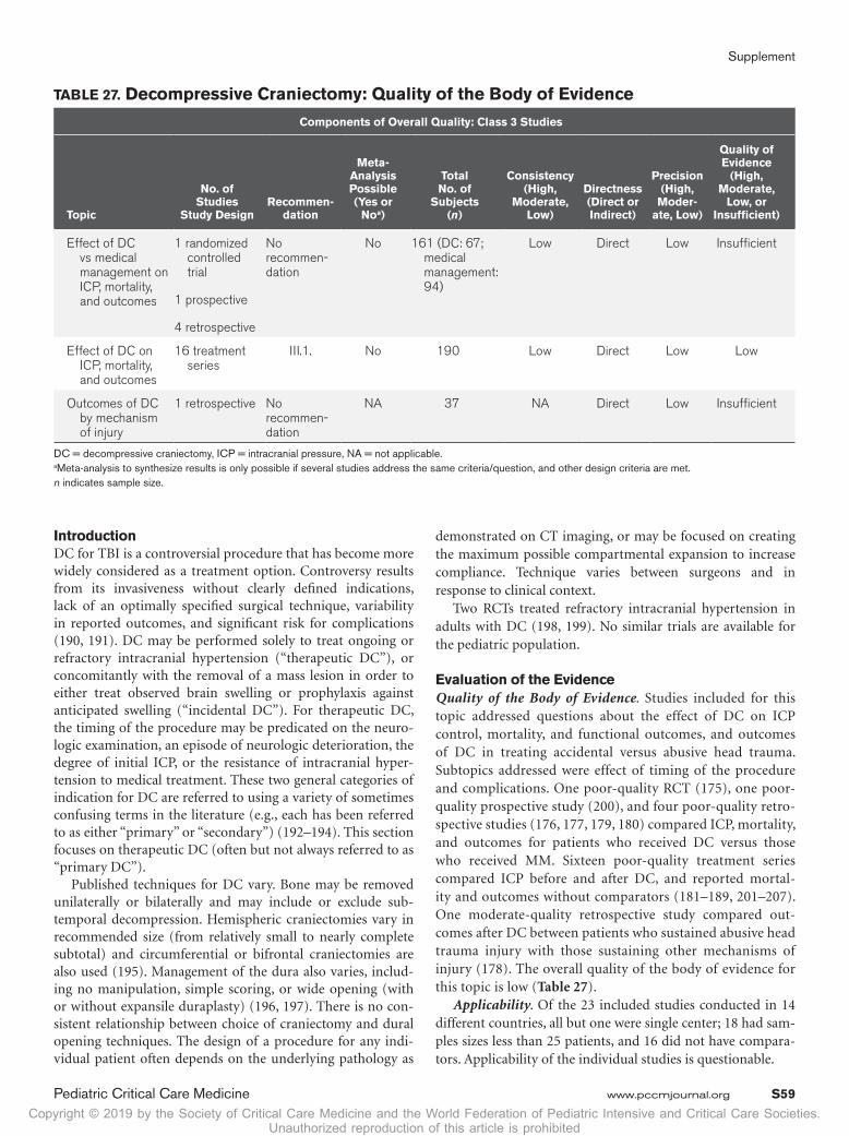
Copyright © 2019 by the Society of Critical Care Medicine and the World Federation of Pediatric Intensive and Critical Care Societies.Unauthorized reproduction of this article is prohibited
Supplement
Pediatric Critical Care Medicine www.pccmjournal.org S59
IntroductionDC for TBI is a controversial procedure that has become more widely considered as a treatment option. Controversy results from its invasiveness without clearly defined indications, lack of an optimally specified surgical technique, variability in reported outcomes, and significant risk for complications (190, 191). DC may be performed solely to treat ongoing or refractory intracranial hypertension (“therapeutic DC”), or concomitantly with the removal of a mass lesion in order to either treat observed brain swelling or prophylaxis against anticipated swelling (“incidental DC”). For therapeutic DC, the timing of the procedure may be predicated on the neuro-logic examination, an episode of neurologic deterioration, the degree of initial ICP, or the resistance of intracranial hyper-tension to medical treatment. These two general categories of indication for DC are referred to using a variety of sometimes confusing terms in the literature (e.g., each has been referred to as either “primary” or “secondary”) (192–194). This section focuses on therapeutic DC (often but not always referred to as “primary DC”).
Published techniques for DC vary. Bone may be removed unilaterally or bilaterally and may include or exclude sub-temporal decompression. Hemispheric craniectomies vary in recommended size (from relatively small to nearly complete subtotal) and circumferential or bifrontal craniectomies are also used (195). Management of the dura also varies, includ-ing no manipulation, simple scoring, or wide opening (with or without expansile duraplasty) (196, 197). There is no con-sistent relationship between choice of craniectomy and dural opening techniques. The design of a procedure for any indi-vidual patient often depends on the underlying pathology as
demonstrated on CT imaging, or may be focused on creating the maximum possible compartmental expansion to increase compliance. Technique varies between surgeons and in response to clinical context.
Two RCTs treated refractory intracranial hypertension in adults with DC (198, 199). No similar trials are available for the pediatric population.
Evaluation of the EvidenceQuality of the Body of Evidence. Studies included for this topic addressed questions about the effect of DC on ICP control, mortality, and functional outcomes, and outcomes of DC in treating accidental versus abusive head trauma. Subtopics addressed were effect of timing of the procedure and complications. One poor-quality RCT (175), one poor-quality prospective study (200), and four poor-quality retro-spective studies (176, 177, 179, 180) compared ICP, mortality, and outcomes for patients who received DC versus those who received MM. Sixteen poor-quality treatment series compared ICP before and after DC, and reported mortal-ity and outcomes without comparators (181–189, 201–207). One moderate-quality retrospective study compared out-comes after DC between patients who sustained abusive head trauma injury with those sustaining other mechanisms of injury (178). The overall quality of the body of evidence for this topic is low (Table 27).
Applicability. Of the 23 included studies conducted in 14 different countries, all but one were single center; 18 had sam-ples sizes less than 25 patients, and 16 did not have compara-tors. Applicability of the individual studies is questionable.
TABLE 27. Decompressive Craniectomy: Quality of the Body of Evidence
Components of Overall Quality: Class 3 Studies
Topic
No. of Studies
Study DesignRecommen -
dation
Meta- Analysis Possible (Yes or
Noa)
Total No. of
Subjects (n)
Consistency (High,
Moderate, Low)
Directness (Direct or Indirect)
Precision (High,
Moder-ate, Low)
Quality of Evidence
(High, Moderate,
Low, or Insufficient)
Effect of DC vs medical management on ICP, mortality, and outcomes
1 randomized controlled trial
1 prospective
4 retrospective
No recommen-dation
No 161 (DC: 67; medical management: 94)
Low Direct Low Insufficient
Effect of DC on ICP, mortality, and outcomes
16 treatment series
III.1. No 190 Low Direct Low Low
Outcomes of DC by mechanism of injury
1 retrospective No recommen-dation
NA 37 NA Direct Low Insufficient
DC = decompressive craniectomy, ICP = intracranial pressure, NA = not applicable.a Meta-analysis to synthesize results is only possible if several studies address the same criteria/question, and other design criteria are met.n indicates sample size.

Copyright © 2019 by the Society of Critical Care Medicine and the World Federation of Pediatric Intensive and Critical Care Societies.Unauthorized reproduction of this article is prohibited
Copyright © 2019 by the Society of Critical Care Medicine and the World Federation of Pediatric Intensive and Critical Care Societies.Unauthorized reproduction of this article is prohibited
Kochanek et al
S60 www.pccmjournal.org March 2019 • Volume 20 (Suppl) • Number 3
TABLE 28. Decompressive Craniectomy: Summary of Evidence
Class 3 Studies
Reference Type of Trauma Center Geographic Location
Study Design n
Age (yr) Mean
(Range) Outcomes Data Class Results
Effect of DC vs MM on ICP, mortality, and outcomes: no recommendation
Mhanna et al (177)a
Metro Health Medical Center
Cleveland, OH
Retrospectiven = 34DC = 17MM = 17Age: DC: mean, 10.2; range, NR MM: mean, 12.4; range, NRMortality; GOS 4 yr postinjury
(IQR, 1–6 yr)
Class 3Selection of patients to
receive DC was based on physician discretion; inadequate control for confounders; baseline differences between groups.
Mortality DC 5/17 vs control 3/17, p = 0.34 No significant difference between
groups for mortality or GOS.GOS Median GOS score DC = 4 (IQR, 3–5) vs control 3
(IQR, 3–4), p = 0.09 Comparing patients who died
or had a disability, there was a significant difference between the DC and control groups (71% [12/17] vs 100% [17/17], respectively; p = 0.022).
Thomale et al (180)a
Campus Virchow Medical Center
Germany
Retrospectiven = 53DC = 14No DC = 39Age: All patients: median, 8; range, 0–16 No DC: median, 7; range, 0–16 DC: median, 12; range, 1–16GOS at 3, 6, and 12 mo (mean follow-
up 5.2 ± 2.4 yr [range, 1–10.5]); ICU days
Class 3No control for confounding;
baseline differences between groups
GOS No significant difference in median
GOS at 3, 6, and 12 moICU days Significantly greater days in ICU for
DC group (p = 0.026) DC group had significantly lower
initial GCS but comparable outcomes.
Rubiano et al (179)a
Simon Bolivar Hospital
Colombia
RetrospectiveDC = 16; 7 pediatricControl = 20; 5 pediatricPediatric patients: Age: mean, 5.86;
range, 1–15Mortality; GOS 6 mo postinjury
Class 3Controlled for confounders
for total sample with mixed ages
Mortality for pediatric patients DC = 1/7 (14%) Non-DC = 2/5 (40%)GOS For pediatric patients: DC Non-DC1 2/7 (14%) 2/5 (40%)2 0 03 0 3/5 (60%)4 2/7 (29%) 05 3/7 (57%) 0For total sample (all ages):
mean GOS at 6 mo postinjury significantly higher in DC group than control group (unadjusted t = 4.26; p = 0.0002)
(Continued)

Copyright © 2019 by the Society of Critical Care Medicine and the World Federation of Pediatric Intensive and Critical Care Societies.Unauthorized reproduction of this article is prohibited
Supplement
Pediatric Critical Care Medicine www.pccmjournal.org S61
Josan and Sgouros (176)a
Birmingham Children’s Hospital
United Kingdom
Retrospectiven = 12Early DC (< 24 hr) = 6Non-DC = 6Age: Early DC: mean, 13; range, 2–16 Non-DC: mean, 11.5; range, 7–15Mortality; GOS 1 yr postinjury
Class 3Small sample; baseline
differences between groups; no control for confounding
Statistical analysis not performed.Mortality DC = 0/6 Non-DC = 2/6 (33%). One of
whom received late DC at 9 d.GOS DC Non-DC1 0 2/6 (33%)2 0 03 0 1/6 (17%)4 2/6 (33%) 05 4/6 (67%) 3/6 (50%)More patients with better outcomes in
DC group than non-DC group
Taylor et al (175)
Royal Childrens’ Hospital Melbourne, VIC, Australia
Randomized controlled trialn = 27DC = 13Medical = 14Age: median, 120.9 mo; range,
13.6–176.4 moOne patient GCS = 11 in DC groupGOS and HSUI 6 mo postinjury; ICP
after randomization/surgery
Class 3Allocation concealment,
blinding, and adequate sample size unclear. Baseline differences NR. No intent-to-treat analysis
GOS and HSUI There was no significant difference
between groups in GOS or HSUI 6 mo postinjury.
MM DCGOS Fav. 2 7 Unf. 12 6HSUI Fav. 1 6 Unf. 1 37ICP 48 hr after randomization or
surgeryRange (sd)DC 17.4 (3.4) vs MM 21.9 (8.5)Number of episodes > 20 mm Hg DC-107 vs MM-223Number of episodes > 30 mm Hg DC-9 vs MM-29There was no significant difference
between groups in mean ICP 48 hr after randomization or surgery.
(Continued)
TABLE 28. (Continued). Decompressive Craniectomy: Summary of Evidence
Class 3 Studies
Reference Type of Trauma Center Geographic Location
Study Design n
Age (yr) Mean (Range)
Outcomes Data Class Results

Copyright © 2019 by the Society of Critical Care Medicine and the World Federation of Pediatric Intensive and Critical Care Societies.Unauthorized reproduction of this article is prohibited
Copyright © 2019 by the Society of Critical Care Medicine and the World Federation of Pediatric Intensive and Critical Care Societies.Unauthorized reproduction of this article is prohibited
Kochanek et al
S62 www.pccmjournal.org March 2019 • Volume 20 (Suppl) • Number 3
Cho et al (200)Taichung Veterans
HospitalTaiwan (Republic
of China)
Prospectiven = 23Group A: low ICP/medical = 6Group B: high ICP/medical = 7Group C: high ICP/DC = 10Age: mean, 5.91 mo; range, 2–14 mo Group A: mean, 5.33 mo; range,
2–11 mo Group B: mean, 7.42 mo; range,
3–14 mo Group C: mean, 5.2 mo; range,
2–14 moMortality; dichotomized scores on COS
measured between 6 mo and 6 yr postinjury (mean, 3.2 yr)
(COS ranges from 1 [best] to 5 [worst]).
COS 1 to 2 = goodCOS 3 to 5 = poorICP
Class 3No control for confounders;
very small sample; no power calculation; significant differences in baseline ICP and Children’s Coma Scale scores
Mortality Three patients died—all from group
B.COS Mean COS for group B significantly
worse than groups A or C (p = 0.0058)
ICP In group C, DC lowered the mean
ICP measurements from 54.9 to 11.9 mm Hg. Effect of medical treatment on ICP for groups A and B was NR, so group differences unknown.
Although DC was performed based on ICP elevation alone, a mean of 32 mL of subdural blood was removed during the surgery, indicating that this sample includes both primary and secondary DC patients.
Outcomes of DC by mechanism of injury: no recommendation
Oluigbo et al (178)a
Children’s Hospital and University of Colorado
Denver/Aurora, CO
Retrospectiven = 37n = 14 abusive head trauman = 23 other mechanismsAge: mean, 6; range, 9 wk to 15 yrAbusive head trauma: mean, 2.2;
range, NRAccidental: mean, 8.4; range, NRMortality; KOSCHI (scored same as
GOS) at mean 23.9 mo (range, 1–94 mo)
Class 3Outcome assessors not
blinded; baseline difference in age for accidental trauma
Mortality 5 of 14: abusive head trauma,
35.7% (p < 0.05) 1 of 23: other mechanisms 4.3% OR = 12.2 (p = 0.02) for abusive
head traumaKOSCHI poor outcome (score of 1,
2, or 3) Nonaccidental: 57.2% Other mechanisms: 30.4% Authors report no significant
difference between groups for poor outcome and an OR for poor outcome of 3.04 for the abusive head trauma group.
COS = Children’s Outcome Scale, DC = decompressive craniectomy, Fav. = favorable, GCS = Glasgow Coma Scale, GOS = Glasgow Outcome Scale, HSUI = Health State Utility Index, ICP = intracranial pressure, IQR = interquartile range, KOSCHI = King’s Outcome Scale for Childhood Head Injury, MM = medical management, NR = not reported, OR = odds ratio, Unf. = unfavorable.a New study.n indicates sample size.
TABLE 28. (Continued). Decompressive Craniectomy: Summary of Evidence
Class 3 Studies
Reference Type of Trauma Center Geographic Location
Study Design n
Age (yr) Mean (Range)
Outcomes Data Class Results

Copyright © 2019 by the Society of Critical Care Medicine and the World Federation of Pediatric Intensive and Critical Care Societies.Unauthorized reproduction of this article is prohibited
Supplement
Pediatric Critical Care Medicine www.pccmjournal.org S63
TABLE 29. Decompressive Craniectomy: Summary of Evidence
Treatment Series
Reference Type of Trauma Center Geographic Location
Study Design n
Age (yr) Mean (Range)
Outcomes Data Class Results
Effect of DC on ICP, mortality, and outcomes: recommendation III.1.
Pechmann et al (187)a
University Medical Center Freiburg
Germany
Treatment seriesn = 12Age: mean, 8.5; range,
2–14Mortality; GOS mean 3.3
mo (± 2 mo) postinjury; range, 3–83 mo; ICP; complications
Class 3Small
uncontrolled series
Mortality 1/12 (8%)GOS 1 = 1/12 (8%) 2 = 2/12 (17%) 3 = 2/12 (17%) 4 = 5/12 (42%) 5 = 2/12(16%)ICP Initial decrease of ICP < 20 mm Hg in all children. Three
of 12 showed a secondary increase in ICP.Complications• Formation of hygroma (83%)• Aseptic bone resorption of the reimplanted bone flap (50%)• Posttraumatic hydrocephalus (42%)• Infection: secondary infection or dysfunction of
ventriculoperitoneal shunt (25%) or cranioplasty (33%)• Epilepsy (33%)Due to complications, 75% of patients required further surgery
in addition to cranioplasty with up to eight interventions.
Prasad et al (208)a
All India Institute of Medical Sciences
New Delhi, India
Treatment seriesn = 71Severe = 36Age: mean, 1.6; range, 1 mo
to 3 yrMortality; GOS-E; mean
19.6 mo (range, 2–42 mo); complications
Class 3Small
uncontrolled series
Mortality 18/36 severe (50%)GOS-E 7 to 8 for all who survived except 1 (reported for all
severities)Complications (reported for all severities) Ventilator-associated pneumonia in 22 of 71 cases Late onset seizures in 2 Septicemia in 6 Wound infection in 7 Subdural hygroma in 11 Hydrocephalus in 13
Desgranges et al (183)a
Hospital Femme Mere Enfant
Lyon, France
Treatment seriesn = 12Age: mean, 8; range, NRMortality; ICP; complications
(IBL); Mortality
Class 3Small
uncontrolled series
Mortality 4/12 (33%)ICPBefore DC (n = 12) After DC (n = 12)46 ± 18 mm Hg 10 ± 4 mm HgDC induced significant decrease in ICP (p = 0.0005), mean
arterial pressure (p = 0.04) between the immediate pre- and postoperative periods.
Complications Median IBL during DC was 49% (17–349%) of EBV. Children with IBL ≥ 50% of EBV had higher preoperative
ICP (p = 0.03) and INR (p = 0.02) than those with an IBL < 50% of EBV.
MortalityIBL ≥ 50% IBL<50%3/6 50% 1/6 (17%)
(Continued)

Copyright © 2019 by the Society of Critical Care Medicine and the World Federation of Pediatric Intensive and Critical Care Societies.Unauthorized reproduction of this article is prohibited
Kochanek et al
S64 www.pccmjournal.org March 2019 • Volume 20 (Suppl) • Number 3
TABLE 29. (Continued). Decompressive Craniectomy: Summary of Evidence
Treatment Series
Reference Type of Trauma Center Geographic Location
Study Design n
Age (yr) Mean (Range)
Outcomes Data Class Results
Khan et al (185)a
University HospitalKarachi, Pakistan
Treatment seriesn = 25, 21 severeAge: mean, 6; range, 1–15Mortality; dichotomized
GOS at mean 5 mo (± 2 sd); complications (blood loss)
Class 3Small
uncontrolled series
Mortality (of entire sample n = 25) 9 patients died (36%)GOS (of entire sample n = 25) 16 had good outcome (GOS 4 [n = 6] and 5 [n = 10]).Complications Significantly greater mortality among patients with >
300 mL operative blood loss (p = 0.001)
Csókay et al (182)a
3 Children’s HospitalsHungary and Wales
Treatment seriesn = 8Age: mean, 7.13; range,
1–12Mortality; GOS at 1 yr
postinjury; ICP; effect of timing
Class 3Small
uncontrolled series
Mortality 2/8 (25%)GOSDischarge 1 yr1 = 2/8 (25%) 1 = 2/8 (25%)2 = 1/8 (12.5%) 2 = 1/8 (13%)3 = 1/8 (112.5%) 3 = 0/84 = 4/8 (50%) 4 = 2/8 (25%)5 = 0 5 = 3/8 (37%)ICP For six patients with pre- and postoperative ICP
measures, the average preoperative ICP was 23.3 mm Hg compared with 15.3 mm Hg postoperative ICP (calculated from data provided in Tables 1 and 2).
Timing At above 20 mm Hg, fast progression of ICP (within
15 min) can occur, limiting the time available to perform DC with a successful patient outcome.
Perez Suarez et al (189)a
Nino Jesus Pediatric Children’s Hospital
Madrid, Spain
Treatment seriesn = 14Age: mean, 5.5; range, 11
mo to 15 yrMortality; GOS at 2
yr postinjury; ICP; complications
Class 3Small
uncontrolled series
Mortality 2/14 (14%)GOS 1 = 2/14 (14%) 2 = 0 3 = 0 4 = 7/14 (50%) 5 = 5/14 (36%)ICPIn 13 patients, craniectomy initially decreased ICP to <
25 mm Hg.Complications: Hygroma 8/14 (57%) Infections 3/14 (21%) Aseptic resorption of bone flap 3/14 (21%)
(Continued)

Copyright © 2019 by the Society of Critical Care Medicine and the World Federation of Pediatric Intensive and Critical Care Societies.Unauthorized reproduction of this article is prohibited
Supplement
Pediatric Critical Care Medicine www.pccmjournal.org S65
Adamo et al (181)a
Albany Medical Center Hospital
New York
Treatment seriesn = 7Age: mean, 13.86 mo;
range, 2–24 moMortality; KOSCHI
(scored same as GOS); complications
Class 3Small
uncontrolled series
Mortality All patients survivedKOSCHIDischarge 1 yr3a = 4/7 (57%) 4a = 3/7 (43%)3b = 3/7 (43%) 4b = 1/7 (14%) 3b = 3/7 (43%)Complications 7/7 (100%) hydrocephalus 2/7 (29%) bone resorption
Jagannathan et al (203)
University of Virginia Health System
Virginia
Treatment seriesn = 23Age: mean, 11.9; range,
2–19Mortality; GOS 5 yr mean
(range, 11–126 mo); ICP
Class 3Small
uncontrolled series
Mortality 7 (30%; one intraoperative; five postoperative; one
in rehabilitation) Mortality primarily in patients with multisystem trauma.
GOSMean 4.2 (range, 1–5)n Score9 55 41 31 27 1ICP ICP control in 19/23 patients. High ICP associated with
increased mortality.
Figaji et al (184)a
Red Cross War Memorial Children’s Hospital
Cape Town, South Africa
Treatment seriesn = 12Age: mean, 8.125; range,
5–12Mortality; GOS at median
35 mo postinjury (range, 1–6 yr); ICP; complications
Class 3Small
uncontrolled series
Mortality 1/12 (8%)GOS 1 = 1/12 (8%) 2 = 0 3 = 0 4 = 6/12 (50%) 5 = 5/12 (42%)ICP Control of ICP and clinical improvement was seen in
all but one patient. Mean sustained ICP reduction postcraniectomy was 53.5% (range, 39–61%) in both pre- and postoperative ICP monitoring patients.
Complications 1/12 (8%) bone flap sepsis after replacement 1/12 (8%) slight subsidence of the flap at follow-up 2/12 (17%) asymptomatic subdural hygromas
Kan et al (204)Primary Children’s
Medical CenterUtah
Treatment seriesn = 51n = 6 for intractable ICPAge: mean, 6.6; range, NRMortality; ICP
Class 3Small
uncontrolled series
Mortality Five of six patients died (83%).ICP Three of the four patients with postoperative ICP
monitoring had ICP < 20 mm Hg.
TABLE 29. (Continued). Decompressive Craniectomy: Summary of Evidence
Treatment Series
Reference Type of Trauma Center Geographic Location
Study Design n
Age (yr) Mean (Range)
Outcomes Data Class Results
(Continued)

Copyright © 2019 by the Society of Critical Care Medicine and the World Federation of Pediatric Intensive and Critical Care Societies.Unauthorized reproduction of this article is prohibited
Kochanek et al
S66 www.pccmjournal.org March 2019 • Volume 20 (Suppl) • Number 3
TABLE 29. (Continued). Decompressive Craniectomy: Summary of Evidence
Treatment Series
Reference Type of Trauma Center Geographic Location
Study Design n
Age (yr) Mean (Range)
Outcomes Data Class Results
Rutigliano et al (206)Level 1 regional
trauma centerStoney Brook
University Health Center
New York
Treatment seriesn = 6Age: mean, 14.5; range,
12–19Mortality; FIM score at
hospital discharge; ICP
Class 3Small
uncontrolled series
Mortality All patients survived.FIM 3: Independent 2: Assistance 1: DependentICP Five of the six patients had sustained postoperative
ICP < 20 mm Hg. One had ICP elevations requiring a second surgery for debridement, with no subsequent ICP elevations.
Skoglund, 2006 (207)
Neonatal ICUSahlgrenska
University HospitalGoteborg, Sweden
Treatment seriesn = 8 Peds (19 total)Age: Mean 12.6 Range: 7
to 16Mortality; GOS at minimum
1 yr postinjury (range 1 to 6 yr).
(ICP and effect of size of DC on outcome were also considered, but in only 9 patients, and ages not specified.)
Class 3Small
uncontrolled series
Mortality 1/8 (12.5%)GOS 1: 1 (12.5%) 2: 0 3: 3 (37.5%) 4: 1 (12.5%) 5: 3 (37.5%)
Figaji et al (201)Red Cross War
Memorial Children’s Hospital
Cape Town, South Africa
Treatment seriesn = 5Age: mean, 8; median, 6;
range, 5–12GOS at a range of 14–42
mo; ICP; complications
Class 3Small
uncontrolled series
GOS All patients had early clinical improvement after surgery
and were GOS 4 or 5 at long-term follow-up (14–40 mo).ICP In the four patients with postoperative ICP monitoring, two
had no ICP elevations and two had mild, easily controlled elevations.
Complications 1 subdural hygroma 1 cerebrospinal fluid leak and bone flap sepsis
Messing-Jünger et al (186)a
Heinrich University Hospital
Dusseldorf, Germany
Treatment series (publication is comparative but seven pediatric patients in the sample were reported as a treatment series)
n = 7Age: mean, NR; range,
1–16Mortality, GOS (follow-up
time not specified)
Class 3Small
uncontrolled series
Mortality 2/7 (29%) died between 24 and 48 hr and showed signs
of decerebration at admission.GOS (in 6 of 7 patients; 1 lost to follow-up) 1 = 2/7 (29%) 2 = 0 3 = 1/7 (14%) 4 = 1/7 (14%) 5 = 2/7 (29%)
(Continued)
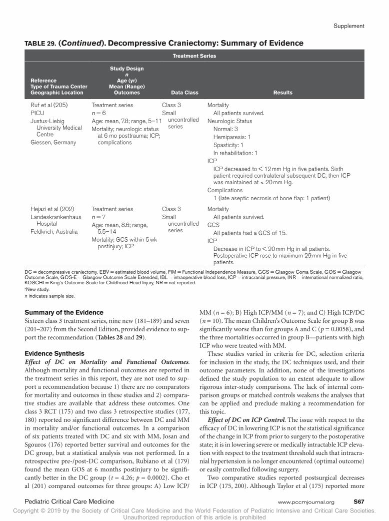
Copyright © 2019 by the Society of Critical Care Medicine and the World Federation of Pediatric Intensive and Critical Care Societies.Unauthorized reproduction of this article is prohibited
Supplement
Pediatric Critical Care Medicine www.pccmjournal.org S67
Summary of the EvidenceSixteen class 3 treatment series, nine new (181–189) and seven (201–207) from the Second Edition, provided evidence to sup-port the recommendation (Tables 28 and 29).
Evidence SynthesisEffect of DC on Mortality and Functional Outcomes. Although mortality and functional outcomes are reported in the treatment series in this report, they are not used to sup-port a recommendation because 1) there are no comparators for mortality and outcomes in these studies and 2) compara-tive studies are available that address these outcomes. One class 3 RCT (175) and two class 3 retrospective studies (177, 180) reported no significant difference between DC and MM in mortality and/or functional outcomes. In a comparison of six patients treated with DC and six with MM, Josan and Sgouros (176) reported better survival and outcomes for the DC group, but a statistical analysis was not performed. In a retrospective pre-/post-DC comparison, Rubiano et al (179) found the mean GOS at 6 months postinjury to be signifi-cantly better in the DC group (t = 4.26; p = 0.0002). Cho et al (201) compared outcomes for three groups: A) Low ICP/
MM (n = 6); B) High ICP/MM (n = 7); and C) High ICP/DC (n = 10). The mean Children’s Outcome Scale for group B was significantly worse than for groups A and C (p = 0.0058), and the three mortalities occurred in group B—patients with high ICP who were treated with MM.
These studies varied in criteria for DC, selection criteria for inclusion in the study, the DC techniques used, and their outcome parameters. In addition, none of the investigations defined the study population to an extent adequate to allow rigorous inter-study comparisons. The lack of internal com-parison groups or matched controls weakens the analyses that can be applied and preclude making a recommendation for this topic.
Effect of DC on ICP Control. The issue with respect to the efficacy of DC in lowering ICP is not the statistical significance of the change in ICP from prior to surgery to the postoperative state; it is in lowering severe or medically intractable ICP eleva-tion with respect to the treatment threshold such that intracra-nial hypertension is no longer encountered (optimal outcome) or easily controlled following surgery.
Two comparative studies reported postsurgical decreases in ICP (175, 200). Although Taylor et al (175) reported more
Ruf et al (205)PICUJustus-Liebig
University Medical Centre
Giessen, Germany
Treatment seriesn = 6Age: mean, 7.8; range, 5–11Mortality; neurologic status
at 6 mo posttrauma; ICP; complications
Class 3Small
uncontrolled series
Mortality All patients survived.Neurologic Status Normal: 3 Hemiparesis: 1 Spasticity: 1 In rehabilitation: 1ICP ICP decreased to < 12 mm Hg in five patients. Sixth
patient required contralateral subsequent DC, then ICP was maintained at ≤ 20 mm Hg.
Complications 1 (late aseptic necrosis of bone flap: 1 patient)
Hejazi et al (202)Landeskrankenhaus
HospitalFeldkrich, Australia
Treatment seriesn = 7Age: mean, 8.6; range,
5.5–14Mortality; GCS within 5 wk
postinjury; ICP
Class 3Small
uncontrolled series
Mortality All patients survived.GCS All patients had a GCS of 15.ICP Decrease in ICP to < 20 mm Hg in all patients.
Postoperative ICP rose to maximum 29 mm Hg in five patients.
DC = decompressive craniectomy, EBV = estimated blood volume, FIM = Functional Independence Measure, GCS = Glasgow Coma Scale, GOS = Glasgow Outcome Scale, GOS-E = Glasgow Outcome Scale Extended, IBL = intraoperative blood loss, ICP = intracranial pressure, INR = international normalized ratio, KOSCHI = King’s Outcome Scale for Childhood Head Injury, NR = not reported.a New study.n indicates sample size.
TABLE 29. (Continued). Decompressive Craniectomy: Summary of Evidence
Treatment Series
Reference Type of Trauma Center Geographic Location
Study Design n
Age (yr) Mean (Range)
Outcomes Data Class Results

Copyright © 2019 by the Society of Critical Care Medicine and the World Federation of Pediatric Intensive and Critical Care Societies.Unauthorized reproduction of this article is prohibited
Kochanek et al
S68 www.pccmjournal.org March 2019 • Volume 20 (Suppl) • Number 3
episodes of ICP greater than 20 and greater than 30 mm Hg in the MM group (> 20 DC-107, MM-223; > 30 DC-9, MM-29), there was no significant difference between groups in mean ICP 48 hours after randomization or surgery. Cho et al (201) reported mean ICP decreased in the DC group from 54.9 mm Hg presurgery to 11.9 mm Hg postsurgery. Changes in ICP for the MM group were not reported, so a comparison between groups could not be made.
Eleven treatment series compared ICP before and after DC (182–184, 187, 189, 201–206). Two reported a decrease in mean ICP after DC; in Desgranges et al (183), the decrease was statistically significant and Csókay et al (182) did not provide a statistical analysis (182, 183). In the other nine studies, of 88 patients with postoperative ICP monitoring, the ICP dropped to acceptable levels in 77 patients (87.5%) (184, 187, 189, 201–206). Although these studies did not compare ICP between DC and MM groups, they provided weak and limited evidence that DC may be effective in lowering ICP to below the threshold for treatment in patients’ refractory to MM, and are the evidence base to support the level III.1. recommendation. This limited conclusion would support choosing to perform DC for ICP control when intracranial hypertension is resistant to nonsur-gical management and observed ICP levels are considered haz-ardous to the patient.
Outcomes of DC by Mechanism of Injury. One moderate-quality class 3 retrospective study compared outcomes of chil-dren with severe TBI, with and without abusive head trauma, all of whom received DC (178). Significantly, more patients with abusive injury died compared with those whose TBIs were sustained by other mechanisms (p < 0.05; OR, 12.2 [p = 0.02]). Outcome measured between 1 and 94 months with the King’s Outcome Scale for Childhood Head Injury showed no significant difference between groups for poor outcome, and an OR of poor outcome of 3.04 for the abusive head trauma group. Given this is one relatively small study with internal validity concerns, it was considered insufficient to support a recommendation.
Effect of Timing of the DC Procedure. One small class 3 treatment series noted that the ICP in two of eight patients rose rapidly to greater than 25 mm Hg within 15 minutes of onset of increase, resulting in death in both cases (182). The report did not contain sufficient data to assess what charac-teristics of these patients might have been indications for very early DC.
Complications. Of nine class 3 treatment series that system-atically assessed complications, two reported blood loss associ-ated with mortality (181, 183–185, 187–189, 201, 205). In one, of 12 patients, one of six with intraoperative blood loss (IBL) less than 50% of estimated blood volume (EBV) died, whereas three of six with IBL greater than 50% of EBV died (183). The second study (n = 25; 21 severe) found significantly greater mor-tality in patients with greater than 300 mL operative blood loss (p = 0.001) (185).
Tables 7 and 8 contain cumulative frequencies for complica-tions reported in the remaining seven studies (181, 184, 187–189, 201, 205). One of the studies reported complications for a
total sample of 71 patients, 36 of which were severe; all patients in the other samples are severe (188). These data indicate a high occurrence of complications among patients who receive DC and suggest that potential complications should be taken into account when making decisions about DC as a treatment. As with the subtopic of timing of DC, there are insufficient data in these reports to assess patient characteristics that might be risk factors for complications. Of note, many of the nonsur-gical complications listed are also associated with severe TBI, itself, and relevant data from comparison group is not always available (Table 30).
Indications From Adult GuidelinesBecause the findings of the literature review for pediatric TBI provide levels of evidence and recommendations specific to the pediatric population, the clinical investigators do not think that the recommendations about DC from the adult guidelines are required to guide treatment decisions in children (14).
Nutrition
Recommendations
Strength of Recommendations: Weak
Level IThere was insufficient evidence to support a level I recommen-dation for this topic.
Level IITo Improve Overall Outcomes. II.1. Use of an immune-modu-lating diet is not recommended.
TABLE 30. Complications From Decompressive Craniectomy
ComplicationOccurrence
(Total n = 164) Percent
Hygroma 32 19.5
Hydrocephalus 25 15
Ventilator-associated pneumonia
22 13.4
Aseptic bone resorption 12 7
Infection 10 6
Septicemia 6 3.7
Epilepsy/seizures 6 3.7
Infection or dysfunction of cranioplasty
4 2.4
Infection or dysfunction of ventriculoperitoneal shunt
3 2
Bone flap sepsis 2 1.2
Slight subsidence of flap 1 0.6
n indicates sample size.

Copyright © 2019 by the Society of Critical Care Medicine and the World Federation of Pediatric Intensive and Critical Care Societies.Unauthorized reproduction of this article is prohibited
Supplement
Pediatric Critical Care Medicine www.pccmjournal.org S69
Level IIITo Improve Overall Outcomes. III.1. Initiation of early enteral nutritional support (within 72 hr from injury) is suggested to decrease mortality and improve outcomes.
Changes From Prior Edition. The level III recommendation from the Second Edition has been removed. Recommendation III.1. is new to this Third Edition. One new class 3 retrospec-tive observational study was added to the evidence base for this topic (209).
IntroductionSimilar to adults, children with severe TBI require energy to support recovery (210, 211). Although the exact mechanisms remain unclear, TBI causes an increase in metabolism, which thereby requires increased caloric support during the critical phase of injury. In addition, developing children have greater nutritional needs for normal growth and development. The decision to administer nutritional support, including the tim-ing, the quantity, the manner, and the composition of such support, may have effects on short- and long-term outcome.
Hyperglycemia is a consistent stress response to severe illness or injury including severe TBI (212). Although treat-ment of hyperglycemia with insulin to achieve glucose con-trol has been studied in critically ill and injured pediatric patients, initial studies found conflicting results. The first-reported randomized trial found significantly lower infection rates, length of stay, and mortality, but greater risk of hypo-glycemia among pediatric patients treated with tight glucose control compared with those treated with insulin to achieve higher glucose targets (213). Subsequent studies among gen-eral pediatric critically ill and injured patients as well as car-diac surgical patients found no influence on mortality from tight control compared with higher glucose targets, but con-sistently reported greater rates of severe hypoglycemia with concern for harm (214–216).
Because mortality was lower among pediatric patients with critical illness and injury compared with adults, there
remained a concern that studies had not enrolled a subset of sufficiently ill patients to demonstrate a benefit from tighter glucose control. The most recent report restricted enrollment to hyperglycemia patients treated with either mechanical ven-tilation or vasoactive medications and the primary outcome was the mean number of 28-day ICU-free days. The study was terminated early due to low probability of benefit and signifi-cantly greater rates of severe hypoglycemia (5.2% vs 2.0%) in the tight control group (215). Currently, there appears to be no benefit to targeting glucose concentrations lower than a range of 150–180 mg/d among pediatric critical care patients (215, 217).
Nevertheless, the severity and duration of posttraumatic hyperglycemia are consistently associated with worse outcomes that likely reflect worse injury and greater stress (218, 219). No studies of glucose control have focused solely on children with severe TBI. Currently, there are insufficient data to recommend for or against tight glucose control for children with severe TBI and persistent hyperglycemia.
Although enteral nutrition is preferred for critically injured patients, some may receive parenteral nutrition due to other abdominal injuries or concerns regarding aspiration risk. A recent large randomized clinical trial in general critically ill pediatric patients demonstrated increased rates of early infec-tion and prolonged length of stay among patients treated with early initiation of parenteral nutrition (within 24 hr of ICU admission) compared with those with initiation of parenteral nutrition after 1 week of critical illness. Both groups received early initiation of enteral nutrition, which was increased in accordance to local guidelines. The study found that early enteral feeding was tolerated as well as IV micronutrients (trace elements, minerals, and vitamins) starting from day 2 and continuing until the enteral nutrition provided reached 80% of the caloric targets (220). Currently, most pediatric critical care providers attempt enteral nutrition in preference to parenteral in trauma patients unless there are severe inju-ries to the bowel.
TABLE 31. Nutrition: Quality of the Body of Evidence
TopicNo. of Studies Study Design
Recommen-dations
Meta- Analysis Possible
(Yes or Noa)
No. of Subjects
(n)
Consistency (High,
Moderate, Low)
Directness (Direct or Indirect)
Precision (High,
Moderate, Low)
Quality of Evidence (High,
Moderate, Low, or Insufficient)
Components of overall quality: class 2 study
Immune-modulating diet
One randomized controlled trial
II.1. NA 40 NA Direct Low Moderate
Components of overall quality: class 3 study
New topic: timing of nutritional support
One retrospective
III.1. NA 90 NA Direct High Low
NA = not applicable.a Meta-analysis to synthesize results is only possible if several studies address the same criteria/question, and other design criteria are met.n indicates sample size.

Copyright © 2019 by the Society of Critical Care Medicine and the World Federation of Pediatric Intensive and Critical Care Societies.Unauthorized reproduction of this article is prohibited
Kochanek et al
S70 www.pccmjournal.org March 2019 • Volume 20 (Suppl) • Number 3
Evaluation of the EvidenceQuality of the Body of Evidence. Studies included for this topic addressed questions about the effect on overall outcomes of an immune-modulating diet and the timing of nutritional support. One small class 2 RCT provided moderate-quality evidence to support recommendation II.1. (221). One class 3 observational study provided low quality of evidence to sup-port recommendation III.1. (209) (Table 31).
Applicability. The RCT supporting the level II recom-mendation was small, and conducted at a single site in Greece, limiting its applicability (221). The single-center study supporting the level III recommendation did not include infants (209). Due to its retrospective design, it is not known whether the observed positive results were because children do better if they were fed, or were fed because they
were less severely injured. Thus, the applicability of this study is in question.
Summary of the EvidenceOne class 2 study from the Second Edition (221) and one new class 3 study (209) provided evidence to support the recom-mendation (Table 32).
Evidence SynthesisImmune-Modulating Diet. No new evidence has been found since the Second Edition of these guidelines that suggests best practice in the quantity, manner, and composition of nutritional support for pediatric patients with TBI. Briassoulis et al (221), a class 2 RCT, showed no difference in outcomes for children pro-vided an immune-enhancing diet versus regular formula.
TABLE 32. Nutrition: Summary of Evidence
Reference Type of Trauma Center Geographic Location
Study Design n
Age (yr)Mean (Range) Outcomes Data Class Results
Class 2 Study
Immune-modulating diet: recommendation II.1.
Briassoulis et al (221)PICU University HospitalAthens, Greece
Randomized controlled trialn = 40Age: mean, 120 mo; range,
72–126 moIE: mean, 127 mo; range, NRStandard: mean, 112 mo; range,
NRHospital mortality; infection; LOS;
metabolic indices
Class 2Attrition NR; unclear
if intention-to-treat analysis conducted
IE vs regular formulaMortality Survival: 80% vs 95%; no significant
differenceInfection Fewer positive gastric cultures in IE
group (p < 0.02), but no significant difference in infections
LOS 16.7 vs 12.2 d; no significant differenceLength of mechanical ventilation 11 vs 8 d; no significant differenceMetabolic indices The IE group was more likely to have
positive nitrogen balance at 5 d (69% vs 31%; p < 0.05).
Class 3 study
Timing of nutritional support: recommendation III.1.
Taha et al (209) a
Level 1 pediatric trauma center
California
Retrospectiven = 109; 90 analyzed (19 died
before nutritional support)Age: median, 13; range, 8–18;
under 8 excludedEffect of time of initiation on
neurologic status at discharge (normal, disability, death) and ICU LOS
Class 3Unclear if groups
were similar at baseline; no control for confounders; outcome assessors not blinded.
Effect of timing on neurologic status Time to initiation/time to full caloric intake
by discharge status (mean ± sd in days) Home: 1.51 ± 1.23/3.38 ± 3.32 Disability: 3.08 ± 2.70/6.99 ± 4.73 Coma/death: 1.88 ± 0.99/7.12 ± 6.44Early initiation and achieving full caloric
intake significantly related to more favorable discharge status (p < 0.05).
Effect of timing on ICU LOS Early initiation and achieving full caloric
intake significantly related to shorter ICU LOS (p < 0.01).
IE = immune enhancing, LOS = length of stay, NR = not reported.a New study.

Copyright © 2019 by the Society of Critical Care Medicine and the World Federation of Pediatric Intensive and Critical Care Societies.Unauthorized reproduction of this article is prohibited
Supplement
Pediatric Critical Care Medicine www.pccmjournal.org S71
Timing of Nutritional Support. The retrospective study by Taha et al (209) suggested that initiation of early (enteral) nutrition may reduce mortality and morbidity. Conducted in children 8–18 years old, the study found that when full caloric intake was achieved, a shortened length of ICU stay with improved outcomes at the time of discharge could be demon-strated. However, it is not known whether the children who were fed had better outcomes because they were fed, or if they were fed because they were less severely injured. Thus, the rec-ommendation it supports is a weak level III.
Indications From Adult GuidelinesThe clinical investigators do not think that the recommenda-tions about nutrition from the adult guidelines can be used to guide treatment decisions in children.
Corticosteroids
RecommendationsStrength of the Recommendation: Weak
Levels I and IIThere was insufficient evidence to support a level I or II recom-mendation for this topic.
Level IIITo Improve Overall OutcomesIII.1. The use of corticosteroids is not suggested to improve outcome or reduce ICP.
Note: Recommendation III.1. is not intended to circumvent use of replacement corticosteroids for patients needing chronic steroid replacement therapy, those with adrenal suppression, and those with injury to the hypothalamic-pituitary steroid axis.
Changes From Prior Edition. The Level II recommendation from the Second Edition has been downgraded to Level III. The note about use of replacement corticosteroids is new to this edi-tion. No new studies were added to the evidence base for this topic.
IntroductionCorticosteroids can restore altered vascular permeability (222), inhibit tumor-induced angiogenesis (223), decrease edema
and CSF production (224, 225), and diminish free radical pro-duction (224). These effects provide a rationale for potential steroid benefit in neurologic diseases. This topic summarizes the clinical evidence for glucocorticoid administration as a therapy to improve outcome in pediatric severe TBI.
Treatment of refractory hypotension (i.e., shock) with cor-ticosteroids was not addressed by the included studies. Studies of adult patients with critical illness–related corticosteroid insufficiency (CIRCI) due to major trauma (defined by change in baseline hydrocortisone with an adrenocorticotropic hor-mone stimulation test) did not demonstrate a mortality benefit with steroid therapy. However, steroid treatment was associ-ated with increased risk of ventilator-associated pneumonia and significant fewer ventilator-free days, and current inter-national guidelines recommend against treatment for adult patients with major trauma and CIRCI (226). Similar trials are lacking in children. However, children with known primary or secondary adrenal insufficiency and major trauma should receive corticosteroid replacement therapy to avoid acute adre-nal insufficiency.
Evaluation of the EvidenceQuality of the Body of Evidence. Two reports of a single small class 3 RCT were included for this topic (227, 228). They each addressed the question of the use of corticosteroids to improve outcomes for children with TBI. Because of the small sample size, low precision and class 3 evidence, the overall quality of evidence is low (Table 33).
Applicability. Both reports are over 25 years old, and the dose of steroids is greater than the dose used for acute laryngotracheo-bronchitis (229) and below that used for reversal of vasodilatory septic shock (230). Thus, the relevance of the evidence is limited and applicability is a concern. Questions about randomization, blinding, sample size, and statistical analysis render the publica-tions class 3. However, in adults, a large class 1 RCT, the Corti-costeroid randomisation after significant head injury (CRASH) trial, provided strong evidence against the use of steroids for acute care of severe TBI. This evidence confirms the findings from the older trial, mitigating the applicability concerns (231).
TABLE 33. Corticosteroids: Quality of the Body of Evidence
Components of Overall Quality: Class 3 Study
Topic
No. of Studies
Study DesignRecommen-
dation
Meta- Analysis Possible
(Yes or Noa)
Total No. of
Subjects (n)
Consistency (High,
Moderate, Low)
Directness (Direct or Indirect)
Precision (High,
Moderate, Low)
Quality of Evidence
(High, Moderate,
Low, or Insufficient)
Use of corticosteroids to improve outcomes
One randomized controlled trial with two publications
III.1. NA 25 NA Direct Low Low
NA = not applicable.a Meta-analysis to synthesize results is only possible if several studies address the same criteria/question, and other design criteria are met.n indicates sample size.

Copyright © 2019 by the Society of Critical Care Medicine and the World Federation of Pediatric Intensive and Critical Care Societies.Unauthorized reproduction of this article is prohibited
Kochanek et al
S72 www.pccmjournal.org March 2019 • Volume 20 (Suppl) • Number 3
TABLE 34. Corticosteroids: Summary of Evidence
Class 3 Study (Same Sample)
Reference Type of Trauma Center Geographic Location
Study Design n
Age (yr) Mean (Range)
Outcomes Data Class Results
Use of corticosteroids to improve outcomes: recommendation III.1.
Fanconi et al (227), Klöti et al (228)
ICU
Zúrich, Switzerland
Randomized controlled trial
n = 25; 13 steroid, 12 placebo (Fanconi et al [228])
n = 24; 12/12 (Klöti et al [229]) (same patients)
Age:
Steroid group: mean, 7.5; range, 1.8–14.6
Placebo group: mean, 7.4; range, 1.4–15.8
GOS at 6 mo postinjury, ICP, CPP, duration of monitoring and intubation; free cortisol levels; and complications
Class 3
Randomization and allocation concealment methods not described; unclear if outcome assessors were blinded; unclear if sample size was adequate; did not use intent-to-treat analysis.
GOS, ICP, CPP, duration of intubation
Steroid treatment resulted in no differences vs placebo in 6 mo GOS, ICP, CPP, duration of ICP monitoring, or duration of intubation
Free cortisol levels
Steroid treatment vs placebo significantly suppressed endogenous free cortisol levels from day 1 to day 6.
Complications
Steroid treatment resulted in a trend toward increased bacterial pneumonia (7/13 vs 2/12 vs placebo, respectively, p = 0.097).
CPP = cerebral perfusion pressure, GOS = Glasgow Outcome Score, ICP = intracranial pressure.
Summary of the EvidenceTwo reports of one class 3 study provide evidence to support the recommendation (227, 228) (Table 34).
Evidence SynthesisDexamethasone and ICP/CPP. Fanconi et al (227) performed a randomized, prospective, placebo-controlled clinical trial on 25 pediatric patients with severe TBI using dexamethasone at 1 mg/kg/d for 3 days (n = 13) versus placebo (n = 12). Baseline character-istics did not differ between groups. Dexamethasone treatment did not influence ICP (mean of 14 mm Hg in both groups), CPP, num-ber of interventions required, duration of intubation, or 6-month GOS versus placebo. However, steroid treatment versus placebo significantly suppressed endogenous free cortisol levels up to day 6. In addition, steroid treatment resulted in a trend toward increased bacterial pneumonia versus placebo (7/13 vs 2/12, respectively, p = 0.097). Limitations included use of the Richmond screw to assess ICP, fluid restriction, and the use of hyperventilation to a Paco
2 of
25–30 mm Hg as part of standard care.Klöti et al (228) reported on 24 of the same 25 patients from
the study described above. Additional outcomes in this report included duration of ICP monitoring; steroid treatment produced no difference between groups. The small sample size for this trial limited the ability to make definitive conclusions regarding neuro-logic outcomes or complications. However, suppression of corti-sol production by steroid treatment was clearly documented.
Indications From Adult GuidelinesThere is class 1 evidence in the adult guidelines that the use of steroids is not recommended for improving outcome or
reducing ICP (level I) (14). In adult patients with severe TBI, high-dose methylprednisolone was associated with increased mortality and is therefore contraindicated (231). As the CRASH trial’s findings were similar to the small, older pediat-ric trial, they support the existing recommendation and lessen the concerns about the applicability of the older trials.
ONGOING AND FUTURE RESEARCH
Climbing the Mountain With No TopAs stated in the “Methods section” of this document, evidence-based guidelines rarely (if ever) contain enough data to fully populate a clinical protocol. This is certainly the case with the treatment of severe pediatric TBI. The goal is to position the guidelines in a dynamic process as illustrated in Figure 1.
Available evidence is used to generate treatment guidelines. The guidelines provide recommendations based on the avail-able evidence and identify gaps that become the future research agenda. In the interim, those gaps can be filled by creating clinical protocols using consensus where evidence is lacking. Together the gaps and protocols provide structure and identify patient samples for the generation of new research. The new research populates the evidence base which can then be used to further develop the guidelines. It is a recursive cycle—a mountain with no top.
The primary goal of this team has been to generate evidence-based guidelines for the treatment of pediatric TBI. Secondary goals—as important—have been to include and train new clinical investigators and methodologists in the technology, and to gener-ate strong research in response to gaps identified in the guidelines documents. For the Second Edition of these guidelines, 11 new

Copyright © 2019 by the Society of Critical Care Medicine and the World Federation of Pediatric Intensive and Critical Care Societies.Unauthorized reproduction of this article is prohibited
Supplement
Pediatric Critical Care Medicine www.pccmjournal.org S73
TABLE 35. Current Status of Pediatric Traumatic Brain Injury Guidelines
Topic RecommendationsDirect/Indirect/
Treatment SeriesOngoing Research
ICP monitoring To improve overall outcomes
Level III
1. Use of ICP monitoring is suggested.
19 Studies
All class 3
3 Direct: all retrospective
16 Indirect
• 1 RCT
• 2 Prospective
• 10 Retrospective
• 3 Treatment series
None known
Advanced neuromonitoring
To improve overall outcomes
Level III
1. If Pbro2 monitoring is used, maintaining a level > 10 mm Hg is suggested.
4 Direct studies
All class 3
1 Prospective
3 Treatment series
ADAPT hypotheses
• Pbro2 monitoring is associated with favorable outcomes.
• A threshold Pbro2 value is associated with favorable outcomes.
Neuroimaging To improve overall outcomes
Level III
1. Excluding the possibility of elevated ICP on the basis of a normal initial (0–6 hr after injury) CT examination of the brain is not suggested in comatose pediatric patients.
2. Routinely obtaining a repeat CT scan >24 hr after the admission and initial follow-up is not suggested for decisions about neurosurgical intervention, unless there is either evidence of neurologic deterioration or increasing ICP.
3 Direct studies
All class 3
2 Retrospective
1 Treatment series
None known
ICP thresholds To improve overall outcomes
Level III
1. Treatment of ICP targeting a threshold of < 20 mm Hg is suggested.
12 Direct studies
All class 3
3 Prospective
9 Retrospective
None known
CPP thresholds To improve overall outcomes
Level III
1. Treatment to maintain a CPP at a minimum of 40 mm Hg is suggested.
2. A CPP target between 40 to 50 mm Hg is suggested to ensure that the minimum value of 40 mm Hg is not breached. There may be age-specific thresholds with infants at the lower end and adolescents at or above the upper end of this range.
15 Direct studies
1 Class 2, 14 class 3
1 Prospective
4 Retrospective
10 Treatment series
None known
(Continued)

Copyright © 2019 by the Society of Critical Care Medicine and the World Federation of Pediatric Intensive and Critical Care Societies.Unauthorized reproduction of this article is prohibited
Kochanek et al
S74 www.pccmjournal.org March 2019 • Volume 20 (Suppl) • Number 3
Hyperosmolar therapy
For ICP control
Level II
1. Bolus hypertonic saline (3%) is recommended in patients with intracranial hypertension. Recommended effective doses for acute use range between 2 and 5 mL/kg over 10–20 min.
For ICP control
Level III
1. Continuous infusion hypertonic saline is suggested in patients with intracranial hypertension. Suggested effective doses as a continuous infusion of 3% saline range from between 0.1 and 1.0 mL/kg of body weight per hour, administered on a sliding scale. The minimum dose needed to maintain ICP < 20 mm Hg is suggested.
2. Bolus of 23.4% hypertonic saline is suggested for refractory ICP. The suggested dose is 0.5 mL/kg with a maximum of 30 mL.
9 Direct studies
3 Class 2, 6 class 3
2 RCTs
1 Prospective
5 Retrospective
1 Treatment series
ADAPT hypotheses
• Hyperosmolar therapies improve outcomes.
• Hypertonic saline is more effective than mannitol for ICP control.
Analgesics, sedatives, and neuromuscular blockade
For ICP control
Level III
1. With use of multiple ICP-related therapies, as well as appropriate use of analgesia and sedation in routine ICU care, avoiding bolus administration of midazolam and/or fentanyl during ICP crises is suggested due to risks of cerebral hypoperfusion.
6 Studies
All class 3
1 Direct prospective
1 Indirect prospective
2 Direct retrospective
2 Direct treatment series
None known
CSF For ICP control
Level III
1. CSF drainage through an external EVD is suggested to manage increased ICP.
5 Studies
All class 3
1 Direct retrospective
3 Direct treatment series
1 Indirect treatment series
ADAPT hypotheses
• Continuous CSF drainage improves outcomes.
• Continuous CSF drainage reduces other ICP therapies.
Seizure prophylaxis For seizure prevention (clinical and subclinical)
Level III
1. Prophylactic treatment is suggested to reduce the occurrence of early (within 7 d) PTS.
4 Studies
All class 3
1 Direct retrospective
1 Indirect prospective
1 Indirect retrospective
1 Indirect treatment series
None known
Ventilation To improve overall outcomes
Level III
1. Prophylactic severe hyperventilation to a Paco2 < 30 mm Hg in the initial 48 hr after injury is not suggested.
2. If hyperventilation is used in the management of refractory intracranial hypertension, advanced neuromonitoring for evaluation of cerebral ischemia is suggested.
2 Studies
Both class 3
1 Indirect retrospective
1 Direct treatment series
ADAPT hypotheses
• Prophylactic hyperventilation (Co2 < 30 mm Hg) is associated with unfavorable outcomes.
• Moderate hyperventilation is associated with favorable outcomes.
TABLE 35. (Continued). Current Status of Pediatric Traumatic Brain Injury Guidelines
Topic RecommendationsDirect/Indirect/
Treatment SeriesOngoing Research
(Continued)

Copyright © 2019 by the Society of Critical Care Medicine and the World Federation of Pediatric Intensive and Critical Care Societies.Unauthorized reproduction of this article is prohibited
Supplement
Pediatric Critical Care Medicine www.pccmjournal.org S75
Temperature control
To improve overall outcomes
Level II
1. Prophylactic moderate (32–33°C) hypothermia is not recommended over normothermia to improve overall outcomes.
For ICP control
Level III
1. Moderate (32–33°C) hypothermia is suggested for ICP control.
10 Studies
1 Class 1, 4 class 2, 3 class 3, 2 meta-analyses
2 Direct meta-analyses
5 Direct RCTs
1 Indirect RCT
1 Direct retrospective
1 Indirect retrospective
None known
Barbiturates For ICP control
Level III
1. High-dose barbiturate therapy is suggested in hemodynamically stable patients with refractory intracranial hypertension despite maximal medical and surgical management.
4 Direct studies
All class 3
1 Retrospective
3 Treatment series
None known
DC For ICP control
Level III
1. DC is suggested to treat neurologic deterioration, herniation, or intracranial hypertension refractory to medical management.
23 Direct studies
All class 3
1 RCT
1 Prospective
5 Retrospective
16 Treatment series
None known
Nutrition To improve overall outcomes
Level II
1. Use of an immune-modulating diet is not recommended.
To improve overall outcomes
Level III
1. Initiation of early enteral nutritional support (within 72 hr from injury) is suggested to decrease mortality and improve outcomes.
2 Direct studies
1 Class 2, 1 class 3
1 RCT
1 Retrospective
ADAPT hypotheses
• Increased caloric intake is associated with favorable outcomes.
• Early institution of feeding is associated with favorable outcomes.
• Early glucose administration is associated with unfavorable outcomes.
• A threshold value for hyperglycemia is associated with favorable outcomes.
Corticosteroids To improve overall outcomes
Level III
1. The use of corticosteroids is not suggested to improve outcome or reduce ICP.
1 Direct study
Class 3
1 RCT with 2 publications
None known
ADAPT = Approaches and Decisions in Acute Pediatric TBI, CPP = cerebral perfusion pressure, CSF = cerebrospinal fluid, DC = decompressive craniectomy, EVD = external ventricular drain, ICP = intracranial pressure, Pbro2 = brain tissue oxygen, PTS = posttraumatic seizures, RCT = randomized controlled trial.
TABLE 35. (Continued). Current Status of Pediatric Traumatic Brain Injury Guidelines
Topic RecommendationsDirect/Indirect/
Treatment SeriesOngoing Research

Copyright © 2019 by the Society of Critical Care Medicine and the World Federation of Pediatric Intensive and Critical Care Societies.Unauthorized reproduction of this article is prohibited
Kochanek et al
S76 www.pccmjournal.org March 2019 • Volume 20 (Suppl) • Number 3
TABLE 36. Roadmap to Future Research: Pediatric Traumatic Brain Injurya
TopicLevel of
Recommen dation
Overall Outcomes vs. Intermediate
Class of Studies
Direct vs Indirect
EvidenceNumber of
Studies
ICP monitoring
Advanced neuromonitoring
Neuroimaging
ICP thresholds
Cerebral perfusion pressure thresholds
Hyperosmolar therapy
Analgesics, sedatives, neuromuscular block
Cerebrospinal fluid drainage
Seizure prophylaxis
Ventilation therapies
Temperature control
Barbiturates
Decompressive craniectomy
Nutrition
Corticosteroids
ICP = intracranial pressure.a Lighter cells indicate stronger body of evidence. Full explanation of shading is provided in FUTURE DIRECTIONS section.
investigators joined the team, and multiple new studies have been generated by our team as well as by other members of the TBI clinical research community. As mentioned in the “Introduction section,” one of the most important studies of pediatric TBI, designed and executed by a guidelines clinical investigator, is concluding—the ADAPT. We look forward to strong class 2 pub-lications from this project that will address 12 a priori hypoth-eses across five guidelines topics (Table 35), as well as post hoc analyses of other topics and questions. ADAPT is an important example of the utilization of a strong research design (compara-tive effectiveness) to address questions that have eluded examina-tion via RCTs. It is also an important demonstration that part of the value of a guideline is found in both the evidence-based rec-ommendations and in highlighting what cannot be said due to lack of evidence; those gaps provide opportunities for innovation and direction for research capable of fully populating a clinical protocol with evidence-based recommendations.
Current StatusTable 35 outlines the current status of research for each of the topics in this edition in terms of target outcomes, level of recom-mendations, class and quantity of studies, and current research in progress with a design capable of generating class 2 evidence. Blank cells in the “Future Research” column are obvious target priorities, with a caveat. A blank cell may indicate that there is no longer a need for further research of that topic, or that the ques-tions within the topic may require reconsideration and refram-ing; this is what the TBI community needs to determine as it
develops agendas and priorities for future research. For example, the clinical research community may benefit from reconsidera-tion of the question about the use of monitors to measure ICP and a reframing of that question in the context of goal-directed therapy. Indeed, for clinical environments that are fully resourced, goal-directed therapy might be an appropriate context in which all topics and questions are reconsidered, and new ones generated. Furthermore, for cells populated with studies from ADAPT, the direction of future research may be defined by the findings from those studies; however, the questions may remain unanswered).
FUTURE DIRECTIONSTable 36 illustrates target priorities for future pediatric TBI research. The rows are the current topics. The columns are characteristics of a body of evidence used to quantify quality. The cells that intersect row and column are coded in shades of gray. The range of shades from dark to light indicates:
● Level III to II to I ● Intermediate outcomes to relevant patient outcomes ● Class 3 to 2 to 1 ● Indirect evidence to direct evidence ● Smaller number of studies to larger number of studies ● Thus, the lighter the cell, the stronger the body of evidence
for the topic and characteristic; the darker the cell, the greater the need for change in research approach.
The dominance of lighter cells (indicating higher quality) in the columns for “Overall Outcomes vs Intermediate” and

Copyright © 2019 by the Society of Critical Care Medicine and the World Federation of Pediatric Intensive and Critical Care Societies.Unauthorized reproduction of this article is prohibited
Supplement
Pediatric Critical Care Medicine www.pccmjournal.org S77
“Direct vs Indirect Evidence” is due in part to the criteria we used to include studies into the evidence base. We only included studies with intermediate outcomes when there were insuf-ficient data for overall outcomes; similarly, we only included indirect evidence when direct evidence was lacking or scarce.
Table 36 illustrates that for 11 of 15 topics, only class 3 stud-ies were included, and 12 topics had only level III recommen-dations. Clearly, the TBI clinical research community needs to continue to find innovative methods to generate high-quality class 2 and 1 studies. What Table 36 does not illustrate is the following:
1. The integration of individual treatments in the context of goal-directed therapy. Management of patients with TBI is not a function of the application of individual treat-ments. No treatment or management approach exists independent of other treatments and approaches, or independent of the ecology of the treatment setting. The design of meaningful and effective future research must be consistent with this clinical reality. Thus, although Table 36 points to areas for improvement, it creates an illusion of independence of treatments by illustrating treatment characteristics with independent cells.
2. New topics for investigation, or the integration of more than one topic as a treatment “approach.” Which topics are complete? What new topics should be identified and included? How can the clinical reality of multiple treat-ments be accurately represented in research that inte-grates topics in ecologically valid designs?
3. Consistency in data collection across studies. Future research should include consistency in data collection across research projects, such as utilization of the Common Data Elements of the National Institutes of Health (232–235).
The clinical investigators and methods team recommend that the pediatric TBI research community systematically addresses these questions through creating a prioritized research agenda and advocating for additional high-quality research that can populate the evidence base for future guidelines designed to improve outcomes for children who sustain TBI.
ACKNOWLEDGMENTSWe thank the following people at the Pacific Northwest Evi-dence-based Practice Center at Oregon Health & Science University for their invaluable assistance in producing this document: Roger Chou, MD, Elaine Graham, MLS, Andrew Hamilton, Hyon Hildebrant, BA, Shaun Ramirez, MPH, and Leah Williams, BS. We also thank Jamshid Ghajar, MD, PhD, from the Brain Trauma Foundation and Stanford University.
REFERENCES 1. Kochanek PM, Carney N, Adelson PD, et al: Guidelines for the acute
medical management of severe traumatic brain injury in infants, children, and adolescents—second edition. [Erratum appears in Pediatr Crit Care Med. 2012; 13:252]. Pediatr Crit Care Med 2012; 13(Suppl 1):S1–S82
2. Shein SL, Ferguson NM, Kochanek PM, et al: Effectiveness of phar-macological therapies for intracranial hypertension in children with
severe traumatic brain injury–results from an automated data collec-tion system time-synched to drug administration. Pediatr Crit Care Med 2016; 17:236–245
3. Welch TP, Wallendorf MJ, Kharasch ED, et al: Fentanyl and midazolam are ineffective in reducing episodic intracranial hypertension in severe pediatric traumatic brain injury. Crit Care Med 2016; 44:809–818
4. Bell MJ, Adelson PD, Hutchison JS, et al; Multiple Medical Therapies for Pediatric Traumatic Brain Injury Workgroup: Differences in medical therapy goals for children with severe traumatic brain injury-an interna-tional study. Pediatr Crit Care Med 2013; 14:811–818
5. Bell MJ, Adelson PD, Wisniewski SR; Investigators of the ADAPT Study: Challenges and opportunities for pediatric severe TBI-review of the evidence and exploring a way forward. Childs Nerv Syst 2017; 33:1663–1667
6. Kurz JE, Poloyac SM, Abend NS, et al; Investigators for the Approaches and Decisions in Acute Pediatric TBI Trial: Variation in anticonvulsant selection and electroencephalographic monitoring following severe traumatic brain injury in children-understanding resource availability in sites participating in a comparative effectiveness study. Pediatr Crit Care Med 2016; 17:649–657
7. Jha RM, Kochanek PM: Adding insight to injury: A new era in neu-rotrauma. Lancet Neurol 2017; 16:578–580
8. Kochanek PM, Bell MJ: Tackling the challenges of clinical trials for severe traumatic brain injury in children: Screening, phenotyping, and adapting. Crit Care Med 2015; 43:1544–1546
9. DeKosky ST, Asken BM: Injury cascades in TBI-related neurodegen-eration. Brain Inj 2017; 31:1177–1182
10. Johnson VE, Stewart JE, Begbie FD, et al: Inflammation and white matter degeneration persist for years after a single traumatic brain injury. Brain 2013; 136:28–42
11. Kochanek PM, Jackson TC, Ferguson NM, et al: Emerging therapies in traumatic brain injury. Semin Neurol 2015; 35:83–100
12. Kochanek PM, Tasker RC, Bell MJ, et al: Management of Pediatric Severe Traumatic Brain Injury: 2019 Consensus and Guidelines-Based Algorithm for First and Second Tier Therapies. Pediatr Crit Care Med 2019; 20:269–279
13. Badjatia N, Carney N, Crocco TJ, et al: Guidelines for prehospital management of traumatic brain injury 2nd edition. Prehosp Emerg Care 2008; 12(Suppl 1):S1–S52
14. Carney N, Totten AM, O’Reilly C, et al: Guidelines for the manage-ment of severe traumatic brain injury, fourth edition. Neurosurgery 2017; 80:6–15
15. Guyatt GH, Oxman AD, Kunz R, et al; GRADE Working Group: GRADE guidelines: 8. Rating the quality of evidence–indirectness. J Clin Epidemiol 2011; 64:1303–1310
16. Morton SC, Murad MH, O’Connor E, et al: Quantitative Synthesis—An Update. Methods Guide for Comparative Effectiveness Reviews. (Prepared by the Scientific Resource Center under Contract No. 290-2012-0004-C). AHRQ Publication No. 18-EHC007-EF. Rockville, MD: Agency for Healthcare Research and Quality; 2018
17. Alkhoury F, Kyriakides TC: Intracranial pressure monitoring in children with severe traumatic brain injury: National Trauma Data Bank-based review of outcomes. JAMA Surg 2014; 149:544–548
18. Bennett TD, Riva-Cambrin J, Keenan HT, et al: Variation in intracranial pressure monitoring and outcomes in pediatric traumatic brain injury. Arch Pediatr Adolesc Med 2012; 166:641–647
19. Bennett TD, DeWitt PE, Greene TH, et al: Functional outcome after intracranial pressure monitoring for children with severe traumatic brain injury. JAMA Pediatr 2017; 171:965–971
20. Bruce DA, Alavi A, Bilaniuk L, et al: Diffuse cerebral swelling following head injuries in children: The syndrome of “malignant brain edema.” J Neurosurg 1981; 54:170–178
21. Muizelaar JP, Marmarou A, DeSalles AA, et al: Cerebral blood flow and metabolism in severely head-injured children. Part 1: Relationship with GCS score, outcome, ICP, and PVI. J Neurosurg 1989; 71:63–71
22. Sharples PM, Stuart AG, Matthews DS, et al: Cerebral blood flow and metabolism in children with severe head injury. Part 1: Relation to age, Glasgow Coma Score, outcome, intracranial pressure, and time after injury. J Neurol Neurosurg Psychiatry 1995; 58:145–152
Copyright © 2019 by the Society of Critical Care Medicine and the World Federation of Pediatric Intensive and Critical Care Societies.Unauthorized reproduction of this article is prohibited
Kochanek et al
S78 www.pccmjournal.org March 2019 • Volume 20 (Suppl) • Number 3
23. Tilford JM, Aitken ME, Anand KJ, et al: Hospitalizations for critically ill children with traumatic brain injuries: A longitudinal analysis. Crit Care Med 2005; 33:2074–2081
24. Tilford JM, Simpson PM, Yeh TS, et al: Variation in therapy and outcome for pediatric head trauma patients. Crit Care Med 2001; 29:1056–1061
25. Chesnut RM, Marshall LF, Klauber MR, et al: The role of secondary brain injury in determining outcome from severe head injury. J Trauma 1993; 34:216–222
26. Marmarou A, Anderson R, D Ward J, et al: Impact of ICP instability and hypotension on outcome in patients with severe head trauma. J Neurosurg 1991; 75:S59–S66
27. Schoon P, Benito Mori L, Orlandi G, et al: Incidence of intracranial hypertension related to jugular bulb oxygen saturation disturbances in severe traumatic brain injury patients. Acta Neurochir Suppl 2002; 81:285–287
28. Steiner LA, Andrews PJ: Monitoring the injured brain: ICP and CBF. Br J Anaesth 2006; 97:26–38
29. Chambers IR, Treadwell L, Mendelow AD: The cause and incidence of secondary insults in severely head-injured adults and children. Br J Neurosurg 2000; 14:424–431
30. Chambers IR, Treadwell L, Mendelow AD: Determination of thresh-old levels of cerebral perfusion pressure and intracranial pressure in severe head injury by using receiver-operating characteristic curves: An observational study in 291 patients. J Neurosurg 2001; 94:412–416
31. Miller JD, Butterworth JF, Gudeman SK, et al: Further experience in the management of severe head injury. J Neurosurg 1981; 54:289–299
32. Rosner MJ, Daughton S: Cerebral perfusion pressure management in head injury. J Trauma 1990; 30:933–940
33. Prabhakaran P, Reddy AT, Oakes WJ, et al: A pilot trial comparing cerebral perfusion pressure-targeted therapy to intracranial pres-sure-targeted therapy in children with severe traumatic brain injury. J Neurosurg 2004; 100:454–459
34. Adelson PD, Bratton SL, Carney NA, et al; American Association for Surgery of Trauma; Child Neurology Society; International Society for Pediatric Neurosurgery; International Trauma Anesthesia and Critical Care Society; Society of Critical Care Medicine; World Federation of Pediatric Intensive and Critical Care Societies: Guidelines for the acute medical management of severe traumatic brain injury in infants, children, and adolescents. Chapter 5. Indications for intracranial pres-sure monitoring in pediatric patients with severe traumatic brain injury. Pediatr Crit Care Med 2003; 4:S19–S24
35. Dean NP, Boslaugh S, Adelson PD, et al: Physician agreement with evidence-based recommendations for the treatment of severe trau-matic brain injury in children. J Neurosurg 2007; 107:387–391
36. Morris KP, Forsyth RJ, Parslow RC, et al; UK Paediatric Traumatic Brain Injury Study Group; Paediatric Intensive Care Society Study Group: Intracranial pressure complicating severe traumatic brain injury in children: Monitoring and management. Intensive Care Med 2006; 32:1606–1612
37. Segal S, Gallagher AC, Shefler AG, et al: Survey of the use of intracra-nial pressure monitoring in children in the United Kingdom. Intensive Care Med 2001; 27:236–239
38. Chesnut RM, Temkin N, Carney N, et al; Global Neurotrauma Research Group: A trial of intracranial-pressure monitoring in trau-matic brain injury. N Engl J Med 2012; 367:2471–2481
39. Keenan HT, Nocera M, Bratton SL: Frequency of intracranial pressure monitoring in infants and young toddlers with traumatic brain injury. Pediatr Crit Care Med 2005; 6:537–541
40. Adelson PD, Ragheb J, Kanev P, et al: Phase II clinical trial of mod-erate hypothermia after severe traumatic brain injury in children. Neurosurgery 2005; 56:740–754
41. Alberico AM, Ward JD, Choi SC, et al: Outcome after severe head injury. Relationship to mass lesions, diffuse injury, and ICP course in pediatric and adult patients. J Neurosurg 1987; 67:648–656
42. Bruce DA, Raphaely RC, Goldberg AI, et al: Pathophysiology, treat-ment and outcome following severe head injury in children. Childs Brain 1979; 5:174–191
43. Cruz J, Nakayama P, Imamura JH, et al: Cerebral extraction of oxygen and intracranial hypertension in severe, acute, pediatric brain trauma: Preliminary novel management strategies. Neurosurgery 2002; 50:774–779
44. Downard C, Hulka F, Mullins RJ, et al: Relationship of cerebral perfu-sion pressure and survival in pediatric brain-injured patients. J Trauma 2000; 49:654–658
45. Grinkeviciūte DE, Kevalas R, Matukevicius A, et al: Significance of intracranial pressure and cerebral perfusion pressure in severe pedi-atric traumatic brain injury. Medicina (Kaunas) 2008; 44:119–125
46. Jagannathan J, Okonkwo DO, Yeoh HK, et al: Long-term outcomes and prognostic factors in pediatric patients with severe traumatic brain injury and elevated intracranial pressure. J Neurosurg Pediatr 2008; 2:240–249
47. Kasoff SS, Lansen TA, Holder D, et al: Aggressive physiologic moni-toring of pediatric head trauma patients with elevated intracranial pressure. Pediatr Neurosci 1988; 14:241–249
48. Michaud LJ, Rivara FP, Grady MS, et al: Predictors of survival and severity of disability after severe brain injury in children. Neurosurgery 1992; 31:254–264
49. Pfenninger J, Santi A: Severe traumatic brain injury in children–are the results improving? Swiss Med Wkly 2002; 132:116–120
50. Wahlström MR, Olivecrona M, Koskinen LO, et al: Severe traumatic brain injury in pediatric patients: Treatment and outcome using an intracranial pressure targeted therapy–the Lund concept. Intensive Care Med 2005; 31:832–839
51. White JR, Farukhi Z, Bull C, et al: Predictors of outcome in severely head-injured children. Crit Care Med 2001; 29:534–540
52. Barzilay Z, Augarten A, Sagy M, et al: Variables affecting outcome from severe brain injury in children. Intensive Care Med 1988; 14:417–421
53. Esparza J, M-Portillo J, Sarabia M, et al: Outcome in children with severe head injuries. Childs Nerv Syst 1985; 1:109–114
54. Shapiro K, Marmarou A: Clinical applications of the pressure-volume index in treatment of pediatric head injuries. J Neurosurg 1982; 56:819–825
55. Figaji AA, Zwane E, Graham Fieggen A, et al: The effect of increased inspired fraction of oxygen on brain tissue oxygen tension in children with severe traumatic brain injury. Neurocrit Care 2010; 12:430–437
56. Stippler M, Ortiz V, Adelson PD, et al: Brain tissue oxygen monitoring after severe traumatic brain injury in children: Relationship to outcome and association with other clinical parameters. J Neurosurg Pediatr 2012; 10:383–391
57. Friess SH, Kilbaugh TJ, Huh JW: Advanced neuromonitoring and imaging in pediatric traumatic brain injury. Crit Care Res Pract 2012; 2012:361310
58. Adelson PD, Srinivas R, Chang Y, et al: Cerebrovascular response in children following severe traumatic brain injury. Childs Nerv Syst 2011; 27:1465–1476
59. Ragan DK, McKinstry R, Benzinger T, et al: Depression of whole-brain oxygen extraction fraction is associated with poor outcome in pediat-ric traumatic brain injury. Pediatr Res 2012; 71:199–204
60. Narotam PK, Burjonrappa SC, Raynor SC, et al: Cerebral oxygen-ation in major pediatric trauma: Its relevance to trauma severity and outcome. J Pediatr Surg 2006; 41:505–513
61. Figaji AA, Zwane E, Thompson C, et al: Brain tissue oxygen ten-sion monitoring in pediatric severe traumatic brain injury. Part 1: Relationship with outcome. Childs Nerv Syst 2009; 25:1325–1333
62. Bata SC, Yung M: Role of routine repeat head imaging in paediatric traumatic brain injury. ANZ J Surg 2014; 84:438–441
63. Bailey BM, Liesemer K, Statler KD, et al: Monitoring and prediction of intracranial hypertension in pediatric traumatic brain injury: Clinical factors and initial head computed tomography. J Trauma Acute Care Surg 2012; 72:263–270
64. Levi L, Guilburd JN, Linn S, et al: The association between skull frac-ture, intracranial pathology and outcome in pediatric head injury. Br J Neurosurg 1991; 5:617–625

Copyright © 2019 by the Society of Critical Care Medicine and the World Federation of Pediatric Intensive and Critical Care Societies.Unauthorized reproduction of this article is prohibited
Supplement
Pediatric Critical Care Medicine www.pccmjournal.org S79
65. Stein SC, Spettell C, Young G, et al: Delayed and progressive brain injury in closed-head trauma: Radiological demonstration. Neurosurgery 1993; 32:25–30
66. Maas AI, Hukkelhoven CW, Marshall LF, et al: Prediction of outcome in traumatic brain injury with computed tomographic characteristics: A comparison between the computed tomographic classification and combinations of computed tomographic predictors. Neurosurgery 2005; 57:1173–1182
67. Liesemer K, Riva-Cambrin J, Bennett KS, et al: Use of Rotterdam CT scores for mortality risk stratification in children with traumatic brain injury. Pediatr Crit Care Med 2014; 15:554–562
68. Bratton SL, Chestnut RM, Ghajar J, et al: Guidelines for the man-agement of severe traumatic brain injury 3rd edition. J Neurotrauma 2007; 24(Suppl 1):S1–S106
69. Bullock R, Chestnut R, Clifton L, et al: Guidelines for the management of severe traumatic brain injury 2nd edition. J Neurotrauma 2000; 17:451–553
70. Adelson PD, Bratton SL, Carney NA, et al; American Association for Surgery of Trauma; Child Neurology Society; International Society for Pediatric Neurosurgery; International Trauma Anesthesia and Critical Care Society; Society of Critical Care Medicine; World Federation of Pediatric Intensive and Critical Care Societies: Guidelines for the acute medical management of severe traumatic brain injury in infants, children, and adolescents. Chapter 1: Introduction. Pediatr Crit Care Med 2003; 4:S2–S4
71. Hollingworth W, Vavilala MS, Jarvik JG, et al: The use of repeated head computed tomography in pediatric blunt head trauma: Factors predicting new and worsening brain injury. Pediatr Crit Care Med 2007; 8:348–356; CEU quiz 357
72. Figg RE, Stouffer CW, Vander Kolk WE, et al: Clinical efficacy of serial computed tomographic scanning in pediatric severe traumatic brain injury. Pediatr Surg Int 2006; 22:215–218
73. Mehta A, Kochanek PM, Tyler-Kabara E, et al: Relationship of intracranial pressure and cerebral perfusion pressure with outcome in young children after severe traumatic brain injury. Dev Neurosci 2010; 32:413–419
74. Miller Ferguson N, Shein SL, Kochanek PM, et al: Intracranial hyper-tension and cerebral hypoperfusion in children with severe traumatic brain injury: Thresholds and burden in accidental and abusive insults. Pediatr Crit Care Med 2016; 17:444–450
75. Stein DM, Hu PF, Brenner M, et al: Brief episodes of intracranial hypertension and cerebral hypoperfusion are associated with poor functional outcome after severe traumatic brain injury. J Trauma 2011; 71:364–373
76. McLaughlin MR, Marion DW: Cerebral blood flow and vasorespon-sivity within and around cerebral contusions. J Neurosurg 1996; 85:871–876
77. Vavilala MS, Lee LA, Lam AM: The lower limit of cerebral auto-regulation in children during sevoflurane anesthesia. J Neurosurg Anesthesiol 2003; 15:307–312
78. Pfenninger J, Kaiser G, Lütschg J, et al: Treatment and outcome of the severely head injured child. Intensive Care Med 1983; 9:13–16
79. Allen BB, Chiu YL, Gerber LM, et al: Age-specific cerebral perfusion pressure thresholds and survival in children and adolescents with severe traumatic brain injury. Pediatr Crit Care Med 2014; 15:62–70
80. Barlow KM, Minns RA: The relation between intracranial pressure and outcome in non-accidental head injury. Dev Med Child Neurol 1999; 41:220–225
81. Chaiwat O, Sharma D, Udomphorn Y, et al: Cerebral hemodynamic predictors of poor 6-month Glasgow Outcome Score in severe pedi-atric traumatic brain injury. J Neurotrauma 2009; 26:657–663
82. Kaiser G, Pfenninger J: Effect of neurointensive care upon outcome following severe head injuries in childhood–a preliminary report. Neuropediatrics 1984; 15:68–75
83. Kapapa T, König K, Pfister U, et al: Head trauma in children, part 2: Course and discharge with outcome. J Child Neurol 2010; 25:274–283
84. Stiefel MF, Udoetuk JD, Storm PB, et al: Brain tissue oxygen monitor-ing in pediatric patients with severe traumatic brain injury. J Neurosurg 2006; 105:281–286
85. Vavilala MS, Kernic MA, Wang J, et al; Pediatric Guideline Adherence and Outcomes Study: Acute care clinical indicators associated with discharge outcomes in children with severe traumatic brain injury. Crit Care Med 2014; 42:2258–2266
86. van Beek AH, Claassen JA, Rikkert MG, et al: Cerebral autoregulation: An overview of current concepts and methodology with special focus on the elderly. J Cereb Blood Flow Metab 2008; 28:1071–1085
87. Gonda DD, Meltzer HS, Crawford JR, et al: Complications asso-ciated with prolonged hypertonic saline therapy in children with elevated intracranial pressure. Pediatr Crit Care Med 2013; 14:610–620
88. Roumeliotis N, Dong C, Pettersen G, et al: Hyperosmolar therapy in pediatric traumatic brain injury: A retrospective study. Childs Nerv Syst 2016; 32:2363–2368
89. Webster DL, Fei L, Falcone RA, et al: Higher-volume hypertonic saline and increased thrombotic risk in pediatric traumatic brain injury. J Crit Care 2015; 30:1267–1271
90. Piper BJ, Harrigan PW: Hypertonic saline in paediatric traumatic brain injury: A review of nine years’ experience with 23.4% hyper-tonic saline as standard hyperosmolar therapy. Anaesth Intensive Care 2015; 43:204–210
91. Weed LH, McKibben PS: Pressure changes in the cerebro-spinal fluid following intravenous injection of solutions of various concentra-tions. Amer J Physiol 1919; 48:512–530
92. Wise BL, Chater N: Use of hypertonic mannitol solutions to lower cerebrospinal fluid pressure and decrease brain bulk in man. Surg Forum 1961; 12:398–399
93. James HE, Langfitt TW, Kumar VS, et al: Treatment of intracranial hypertension. Analysis of 105 consecutive, continuous recordings of intracranial pressure. Acta Neurochir (Wien) 1977; 36:189–200
94. Pitlick WH, Pirikitakuhlr P, Painter MJ, et al: Effect of glycerol and hyperosmolality on intracranial pressure. Clin Pharmacol Ther 1982; 31:466–471
95. Fisher B, Thomas D, Peterson B: Hypertonic saline lowers raised intracranial pressure in children after head trauma. J Neurosurg Anesthesiol 1992; 4:4–10
96. Bennett TD, Statler KD, Korgenski EK, et al: Osmolar therapy in pediatric traumatic brain injury. Crit Care Med 2012; 40:208–215
97. Muizelaar JP, Lutz HA III, Becker DP: Effect of mannitol on ICP and CBF and correlation with pressure autoregulation in severely head-injured patients. J Neurosurg 1984; 61:700–706
98. Muizelaar JP, Wei EP, Kontos HA, et al: Mannitol causes compensa-tory cerebral vasoconstriction and vasodilation in response to blood viscosity changes. J Neurosurg 1983; 59:822–828
99. Bouma GJ, Muizelaar JP: Cerebral blood flow, cerebral blood vol-ume, and cerebrovascular reactivity after severe head injury. J Neurotrauma 1992; 9(Suppl 1):S333–S348
100. James HE: Methodology for the control of intracranial pressure with hypertonic mannitol. Acta Neurochir (Wien) 1980; 51:161–172
101. Kaieda R, Todd MM, Cook LN, et al: Acute effects of changing plasma osmolality and colloid oncotic pressure on the formation of brain edema after cryogenic injury. Neurosurgery 1989; 24:671–678
102. Kaufmann AM, Cardoso ER: Aggravation of vasogenic cerebral edema by multiple-dose mannitol. J Neurosurg 1992; 77:584–589
103. Polderman KH, van de Kraats G, Dixon JM, et al: Increases in spi-nal fluid osmolarity induced by mannitol. Crit Care Med 2003; 31:584–590
104. The Brain Trauma Foundation. The American Association of Neurological Surgeons. The Joint Section on Neurotrauma and Critical Care. Use of mannitol. J Neurotrauma 2000; 17:521–525
105. Becker DP, Vries JK: The alleviation of increased intracranial pres-sure by the chronic administration of osmotic agents. In: Intracranial Pressure. Brock M, Dietz H (Eds). Berlin, Heidelberg, Springer, 1972, pp 309–315
106. Feig PU, McCurdy DK: The hypertonic state. N Engl J Med 1977; 297:1444–1454
107. Worthley LI, Cooper DJ, Jones N: Treatment of resistant intracra-nial hypertension with hypertonic saline. Report of two cases. J Neurosurg 1988; 68:478–481

Copyright © 2019 by the Society of Critical Care Medicine and the World Federation of Pediatric Intensive and Critical Care Societies.Unauthorized reproduction of this article is prohibited
Kochanek et al
S80 www.pccmjournal.org March 2019 • Volume 20 (Suppl) • Number 3
108. Qureshi AI, Suarez JI: Use of hypertonic saline solutions in treatment of cerebral edema and intracranial hypertension. Crit Care Med 2000; 28:3301–3313
109. McManus ML, Soriano SG: Rebound swelling of astroglial cells exposed to hypertonic mannitol. Anesthesiology 1998; 88:1586–1591
110. Nakayama S, Kramer GC, Carlsen RC, et al: Infusion of very hyper-tonic saline to bled rats: Membrane potentials and fluid shifts. J Surg Res 1985; 38:180–186
111. Arjamaa O, Karlqvist K, Kanervo A, et al: Plasma ANP during hyper-tonic NaCl infusion in man. Acta Physiol Scand 1992; 144:113–119
112. Moss GS, Gould SA: Plasma expanders. An update. Am J Surg 1988; 155:425–434
113. Peterson B, Khanna S, Fisher B, et al: Prolonged hypernatremia con-trols elevated intracranial pressure in head-injured pediatric patients. Crit Care Med 2000; 28:1136–1143
114. Lescot T, Degos V, Zouaoui A, et al: Opposed effects of hypertonic saline on contusions and noncontused brain tissue in patients with severe traumatic brain injury. Crit Care Med 2006; 34:3029–3033
115. Simma B, Burger R, Falk M, Sacher P, Fanconi S: A prospective, randomized, and controlled study of fluid management in children with severe head injury: Lactated Ringer’s solution versus hypertonic saline. Ped Crit Care 1998; 26:1265–1270
116. Cottenceau V, Masson F, Mahamid E, et al: Comparison of effects of equiosmolar doses of mannitol and hypertonic saline on cerebral blood flow and metabolism in traumatic brain injury. J Neurotrauma 2011; 28:2003–2012
117. Mangat HS, Chiu YL, Gerber LM, et al: Hypertonic saline reduces cumulative and daily intracranial pressure burdens after severe trau-matic brain injury. J Neurosurg 2015; 122:202–210
118. de Bray JM, Granry JC, Monrigal JP, et al: Effects of thiopental on middle cerebral artery blood velocities: A transcranial Doppler study in children. Childs Nerv Syst 1993; 9:220–223
119. Bar-Joseph G, Guilburd Y, Tamir A, et al: Effectiveness of ketamine in decreasing intracranial pressure in children with intracranial hyper-tension. J Neurosurg Pediatr 2009; 4:40–46
120. Chin KH, Bell MJ, Wisniewski SR, et al; Pediatric Traumatic Brain Injury Consortium: Hypothermia Investigators: Effect of adminis-tration of neuromuscular blocking agents in children with severe traumatic brain injury on acute complication rates and outcomes: A secondary analysis from a randomized, controlled trial of therapeutic hypothermia. Pediatr Crit Care Med 2015; 16:352–358
121. Minardi C, Sahillioğlu E, Astuto M, et al: Sedation and analgesia in pediatric intensive care. Curr Drug Targets 2012; 13:936–943
122. Martinon C, Duracher C, Blanot S, et al: Emergency tracheal intubation of severely head-injured children: Changing daily practice after imple-mentation of national guidelines. Pediatr Crit Care Med 2011; 12:65–70
123. Kerr ME, Weber BB, Sereika SM, et al: Effect of endotracheal suc-tioning on cerebral oxygenation in traumatic brain-injured patients. Crit Care Med 1999; 27:2776–2781
124. Tume LN, Baines PB, Lisboa PJ: The effect of nursing interventions on the intracranial pressure in paediatric traumatic brain injury. Nurs Crit Care 2011; 16:77–84
125. Felmet K, Nguyen T, Clark RS, et al: The FDA warning against prolonged sedation with propofol in children remains warranted. Pediatrics 2003; 112:1002–1003
126. Bramwell KJ, Haizlip J, Pribble C, et al: The effect of etomidate on intracranial pressure and systemic blood pressure in pediat-ric patients with severe traumatic brain injury. Pediatr Emerg Care 2006; 22:90–93
127. Andrade AF, Paiva WS, Amorim RL, et al: Continuous ventricular cerebrospinal fluid drainage with intracranial pressure monitor-ing for management of posttraumatic diffuse brain swelling. Arq Neuropsiquiatr 2011; 69:79–84
128. Baldwin HZ, Rekate HL: Preliminary experience with controlled external lumbar drainage in diffuse pediatric head injury. Pediatr Neurosurg 1991; 17:115–120
129. Shore PM, Thomas NJ, Clark RS, et al: Continuous versus intermit-tent cerebrospinal fluid drainage after severe traumatic brain injury
in children: Effect on biochemical markers. J Neurotrauma 2004; 21:1113–1122
130. Levy DI, Rekate HL, Cherny WB, et al: Controlled lumbar drainage in pediatric head injury. J Neurosurg 1995; 83:453–460
131. Pearl PL, McCarter R, McGavin CL, et al: Results of phase II leveti-racetam trial following acute head injury in children at risk for post-traumatic epilepsy. Epilepsia 2013; 54:e135–e137
132. Liesemer K, Bratton SL, Zebrack CM, et al: Early post-traumatic sei-zures in moderate to severe pediatric traumatic brain injury: Rates, risk factors, and clinical features. J Neurotrauma 2011; 28:755–762
133. Chung MG, O’Brien NF: Prevalence of early posttraumatic seizures in children with moderate to severe traumatic brain injury despite levetiracetam prophylaxis. Pediatr Crit Care Med 2016; 17:150–156
134. Yablon SA: Posttraumatic seizures. Arch Phys Med Rehabil 1993; 74:983–1001
135. Holmes GL: Effects of seizures on brain development: Lessons from the laboratory. Pediatr Neurol 2005; 33:1–11
136. Herman ST, Abend NS, Bleck TP, et al; Critical Care Continuous EEG Task Force of the American Clinical Neurophysiology Society: Consensus statement on continuous EEG in critically ill adults and children, part I: Indications. J Clin Neurophysiol 2015; 32:87–95
137. Lewis RJ, Yee L, Inkelis SH, et al: Clinical predictors of post-trau-matic seizures in children with head trauma. Ann Emerg Med 1993; 22:1114–1118
138. Klein P, Herr D, Pearl PL, et al: Results of phase 2 safety and feasibil-ity study of treatment with levetiracetam for prevention of posttrau-matic epilepsy. Arch Neurol 2012; 69:1290–1295
139. Skippen P, Seear M, Poskitt K, et al: Effect of hyperventilation on regional cerebral blood flow in head-injured children. Crit Care Med 1997; 25:1402–1409
140. Adelson PD, Clyde B, Kochanek PM, et al: Cerebrovascular response in infants and young children following severe traumatic brain injury: A preliminary report. Pediatr Neurosurg 1997; 26:200–207
141. Curry R, Hollingworth W, Ellenbogen RG, et al: Incidence of hypo- and hypercarbia in severe traumatic brain injury before and after 2003 pediatric guidelines. Pediatr Crit Care Med 2008; 9:141–146
142. Muizelaar JP, Marmarou A, Ward JD, et al: Adverse effects of pro-longed hyperventilation in patients with severe head injury: A ran-domized clinical trial. J Neurosurg 1991; 75:731–739
143. Muizelaar JP, van der Poel HG, Li ZC, et al: Pial arteriolar vessel diameter and CO2 reactivity during prolonged hyperventilation in the rabbit. J Neurosurg 1988; 69:923–927
144. Hutchison JS, Ward RE, Lacroix J, et al; Hypothermia Pediatric Head Injury Trial Investigators and the Canadian Critical Care Trials Group: Hypothermia therapy after traumatic brain injury in children. N Engl J Med 2008; 358:2447–2456
145. Koenig MA, Bryan M, Lewin JL III, et al: Reversal of transtentorial herniation with hypertonic saline. Neurology 2008; 70:1023–1029
146. Qureshi AI, Geocadin RG, Suarez JI, et al: Long-term outcome after medical reversal of transtentorial herniation in patients with supraten-torial mass lesions. Crit Care Med 2000; 28:1556–1564
147. Adelson PD, Bratton SL, Carney NA, et al; American Association for Surgery of Trauma; Child Neurology Society; International Society for Pediatric Neurosurgery; International Trauma Anesthesia and Critical Care Society; Society of Critical Care Medicine; World Federation of Pediatric Intensive and Critical Care Societies: Guidelines for the acute medical management of severe traumatic brain injury in infants, children, and adolescents. Chapter 14. The role of temperature con-trol following severe pediatric traumatic brain injury. Pediatr Crit Care Med 2003; 4:S53–S55
148. Hendrick EB: The use of hypothermia in severe head injuries in child-hood. Arch Surg 1959; 79:362–364
149. Crompton EM, Lubomirova I, Cotlarciuc I, et al: Meta-analysis of ther-apeutic hypothermia for traumatic brain injury in adult and pediatric patients. Crit Care Med 2017; 45:575–583
150. Tasker RC, Vonberg FW, Ulano ED, et al: Updating evidence for using hypothermia in pediatric severe traumatic brain injury: Conventional and Bayesian meta-analytic perspectives. Pediatr Crit Care Med 2017; 18:355–362

Copyright © 2019 by the Society of Critical Care Medicine and the World Federation of Pediatric Intensive and Critical Care Societies.Unauthorized reproduction of this article is prohibited
Supplement
Pediatric Critical Care Medicine www.pccmjournal.org S81
151. Tasker RC, Akhondi-Asl A: Updating evidence for using therapeutic hypothermia in pediatric severe traumatic brain injury. Crit Care Med 2017; 45:e1091
152. Crompton E, Sharma P: The authors reply. Crit Care Med 2017; 45:e1091–e1092
153. Adelson PD, Wisniewski SR, Beca J, et al; Paediatric Traumatic Brain Injury Consortium: Comparison of hypothermia and normothermia after severe traumatic brain injury in children (Cool Kids): A phase 3, randomised controlled trial. Lancet Neurol 2013; 12:546–553
154. Beca J, McSharry B, Erickson S, et al; Pediatric Study Group of the Australia and New Zealand Intensive Care Society Clinical Trials Group: Hypothermia for traumatic brain injury in children-a phase II randomized controlled trial. Crit Care Med 2015; 43:1458–1466
155. Empey PE, Velez de Mendizabal N, Bell MJ, et al; Pediatric TBI Consortium: Hypothermia Investigators: Therapeutic hypothermia decreases phenytoin elimination in children with traumatic brain injury. Crit Care Med 2013; 41:2379–2387
156. Hutchison JS, Frndova H, Lo TY, et al; Hypothermia Pediatric Head Injury Trial Investigators; Canadian Critical Care Trials Group: Impact of hypotension and low cerebral perfusion pressure on outcomes in children treated with hypothermia therapy following severe traumatic brain injury: A post hoc analysis of the Hypothermia Pediatric Head Injury Trial. Dev Neurosci 2010; 32:406–412
157. Bourdages M, Bigras JL, Farrell CA, et al; Canadian Critical Care Trials Group: Cardiac arrhythmias associated with severe traumatic brain injury and hypothermia therapy. Pediatr Crit Care Med 2010; 11:408–414
158. Su E, Bell MJ, Kochanek PM, et al: Increased CSF concentrations of myelin basic protein after TBI in infants and children: Absence of significant effect of therapeutic hypothermia. Neurocrit Care 2012; 17:401–407
159. Natale JE, Joseph JG, Helfaer MA, et al: Early hyperthermia after traumatic brain injury in children: Risk factors, influence on length of stay, and effect on short-term neurologic status. Crit Care Med 2000; 28:2608–2615
160. Sakurai A, Atkins CM, Alonso OF, et al: Mild hyperthermia worsens the neuropathological damage associated with mild traumatic brain injury in rats. J Neurotrauma 2012; 29:313–321
161. Dietrich WD, Bramlett HM: The evidence for hypothermia as a neuro-protectant in traumatic brain injury. Neurotherapeutics 2010; 7:43–50
162. Polderman KH: Application of therapeutic hypothermia in the ICU: Opportunities and pitfalls of a promising treatment modality. Part 1: Indications and evidence. Intensive Care Med 2004; 30:556–575
163. Salonia R, Empey PE, Poloyac SM, et al: Endothelin-1 is increased in cerebrospinal fluid and associated with unfavorable outcomes in children after severe traumatic brain injury. J Neurotrauma 2010; 27:1819–1825
164. Biswas AK, Bruce DA, Sklar FH, et al: Treatment of acute traumatic brain injury in children with moderate hypothermia improves intracra-nial hypertension. Crit Care Med 2002; 30:2742–2751
165. Li H, Lu G, Shi W, et al: Protective effect of moderate hypothermia on severe traumatic brain injury in children. J Neurotrauma 2009; 26:1905–1909
166. Mellion SA, Bennett KS, Ellsworth GL, et al: High-dose barbiturates for refractory intracranial hypertension in children with severe trau-matic brain injury. Pediatr Crit Care Med 2013; 14:239–247
167. Demopoulos HB, Flamm ES, Pietronigro DD, et al: The free radical pathology and the microcirculation in the major central nervous sys-tem disorders. Acta Physiol Scand Suppl 1980; 492:91–119
168. Kassell NF, Hitchon PW, Gerk MK, et al: Alterations in cerebral blood flow, oxygen metabolism, and electrical activity produced by high dose sodium thiopental. Neurosurgery 1980; 7:598–603
169. Piatt JH Jr, Schiff SJ: High dose barbiturate therapy in neurosurgery and intensive care. Neurosurgery 1984; 15:427–444
170. Chen HI, Malhotra NR, Oddo M, et al: Barbiturate infusion for intrac-table intracranial hypertension and its effect on brain oxygenation. Neurosurgery 2008; 63:880–886
171. Aldrich EF, Eisenberg HM, Saydjari C, et al: Diffuse brain swelling in severely head-injured children. A report from the NIH Traumatic Coma Data Bank. J Neurosurg 1992; 76:450–454
172. Guerra SD, Carvalho LF, Affonseca CA, et al: Factors associated with intracranial hypertension in children and teenagers who suffered severe head injuries. J Pediatr (Rio J) 2010; 86:73–79
173. Zwienenberg M, Muizelaar JP: Severe pediatric head injury: The role of hyperemia revisited. J Neurotrauma 1999; 16:937–943
174. Pittman T, Bucholz R, Williams D: Efficacy of barbiturates in the treat-ment of resistant intracranial hypertension in severely head-injured children. Pediatr Neurosci 1989; 15:13–17
175. Taylor A, Butt W, Rosenfeld J, et al: A randomized trial of very early decompressive craniectomy in children with traumatic brain injury and sustained intracranial hypertension. Childs Nerv Syst 2001; 17:154–162
176. Josan VA, Sgouros S: Early decompressive craniectomy may be effective in the treatment of refractory intracranial hyperten-sion after traumatic brain injury. Childs Nerv Syst 2006; 22: 1268–1274
177. Mhanna MJ, Mallah WE, Verrees M, et al: Outcome of children with severe traumatic brain injury who are treated with decompressive craniectomy. J Neurosurg Pediatr 2015; 16:1–7
178. Oluigbo CO, Wilkinson CC, Stence NV, et al: Comparison of out-comes following decompressive craniectomy in children with acci-dental and nonaccidental blunt cranial trauma. J Neurosurg Pediatr 2012; 9:125–132
179. Rubiano AM, Villarreal W, Hakim EJ, et al: Early decompressive cra-niectomy for neurotrauma: An institutional experience. Ulus Travma Acil Cerrahi Derg 2009; 15:28–38
180. Thomale UW, Graetz D, Vajkoczy P, et al: Severe traumatic brain injury in children–a single center experience regarding therapy and long-term outcome. Childs Nerv Syst 2010; 26:1563–1573
181. Adamo MA, Drazin D, Waldman JB: Decompressive craniectomy and postoperative complication management in infants and tod-dlers with severe traumatic brain injuries. J Neurosurg Pediatr 2009; 3:334–339
182. Csókay A, Emelifeonwu JA, Fügedi L, et al: The importance of very early decompressive craniectomy as a prevention to avoid the sud-den increase of intracranial pressure in children with severe trau-matic brain swelling (retrospective case series). Childs Nerv Syst 2012; 28:441–444
183. Desgranges FP, Javouhey E, Mottolese C, et al: Intraoperative blood loss during decompressive craniectomy for intractable intracranial hypertension after severe traumatic brain injury in children. Childs Nerv Syst 2014; 30:1393–1398
184. Figaji AA, Fieggen AG, Argent A, et al: Surgical treatment for “brain compartment syndrome” in children with severe head injury. S Afr Med J 2006; 96:969–975
185. Khan SA, Shallwani H, Shamim MS, et al: Predictors of poor out-come of decompressive craniectomy in pediatric patients with severe traumatic brain injury: A retrospective single center study from Pakistan. Childs Nerv Syst 2014; 30:277–281
186. Messing-Jünger AM, Marzog J, Wöbker G, et al: Decompressive craniectomy in severe brain injury. Zentralbl Neurochir 2003; 64:171–177
187. Pechmann A, Anastasopoulos C, Korinthenberg R, et al: Decompressive craniectomy after severe traumatic brain injury in chil-dren: Complications and outcome. Neuropediatrics 2015; 46:5–12
188. Prasad GL, Gupta DK, Mahapatra AK, Sharma BS: Surgical results of decompressive craniectomy in very young children: A level one trauma centre experience from India. Brain Inj 2015; 29:1–8
189. Perez Suarez E, Serrano Gonzalez A, Perez Diaz C, et al: Decompressive craniectomy in 14 children with severe head injury: Clinical results with long-term follow-up and review of the literature. J Trauma 2011; 71:133–140
190. Honeybul S, Ho KM, Gillett GR: Long-term outcome following decompressive craniectomy: An inconvenient truth? Curr Opin Crit Care 2018; 24:97–104
191. Yue JK, Rick JW, Deng H, et al: Efficacy of decompressive craniec-tomy in the management of intracranial pressure in severe traumatic brain injury. J Neurosurg Sci 2017 Nov 7. [Epub ahead of print]

Copyright © 2019 by the Society of Critical Care Medicine and the World Federation of Pediatric Intensive and Critical Care Societies.Unauthorized reproduction of this article is prohibited
Kochanek et al
S82 www.pccmjournal.org March 2019 • Volume 20 (Suppl) • Number 3
192. Jehan F, Azim A, Rhee P, et al: Decompressive craniectomy versus craniotomy only for intracranial hemorrhage evacuation: A propensity matched study. J Trauma Acute Care Surg 2017; 83:1148–1153
193. Kolias AG, Adams H, Timofeev I, et al: Decompressive craniectomy following traumatic brain injury: Developing the evidence base. Br J Neurosurg 2016; 30:246–250
194. Tapper J, Skrifvars MB, Kivisaari R, et al: Primary decompressive craniec-tomy is associated with worse neurological outcome in patients with trau-matic brain injury requiring acute surgery. Surg Neurol Int 2017; 8:141
195. Kurzbuch AR: Does size matter? Decompressive surgery under review. Neurosurg Rev 2015; 38:629–640
196. Barthélemy EJ, Melis M, Gordon E, et al: Decompressive craniec-tomy for severe traumatic brain injury: A systematic review. World Neurosurg 2016; 88:411–420
197. Nagai M, Ishikawa M: Exploration of the most effective dural incision design in a decompressive craniectomy. World Neurosurg 2017; 100:224–229
198. Cooper DJ, Rosenfeld JV, Murray L, et al; DECRA Trial Investigators; Australian and New Zealand Intensive Care Society Clinical Trials Group: Decompressive craniectomy in diffuse traumatic brain injury. N Engl J Med 2011; 364:1493–1502
199. Hutchinson PJ, Kolias AG, Timofeev IS, et al; RESCUEicp Trial Collaborators: Trial of decompressive craniectomy for traumatic intracranial hypertension. N Engl J Med 2016; 375:1119–1130
200. Cho DY, Wang YC, Chi CS: Decompressive craniotomy for acute shaken/impact baby syndrome. Pediatr Neurosurg 1995; 23:192–198
201. Figaji AA, Fieggen AG, Peter JC: Early decompressive craniotomy in children with severe traumatic brain injury. Childs Nerv Syst 2003; 19:666–673
202. Hejazi N, Witzmann A, Fae P: Unilateral decompressive craniec-tomy for children with severe brain injury. Report of seven cases and review of the relevant literature. Eur J Pediatr 2002; 161:99–104
203. Jagannathan J, Okonkwo DO, Dumont AS, et al: Outcome following decompressive craniectomy in children with severe traumatic brain injury: A 10-year single-center experience with long-term follow up. J Neurosurg 2007; 106:268–275
204. Kan P, Amini A, Hansen K, et al: Outcomes after decompressive cra-niectomy for severe traumatic brain injury in children. J Neurosurg 2006; 105:337–342
205. Ruf B, Heckmann M, Schroth I, et al: Early decompressive craniec-tomy and duraplasty for refractory intracranial hypertension in chil-dren: Results of a pilot study. Crit Care 2003; 7:R133–R138
206. Rutigliano D, Egnor MR, Priebe CJ, et al: Decompressive craniec-tomy in pediatric patients with traumatic brain injury with intractable elevated intracranial pressure. J Pediatr Surg 2006; 41:83–87
207. Skoglund TS, Eriksson-Ritzén C, Jensen C, et al: Aspects on decom-pressive craniectomy in patients with traumatic head injuries. J Neurotrauma 2006; 23:1502–1509
208. Prasad GL, Gupta DK, Mahapatra AK, et al: Surgical results of grow-ing skull fractures in children: A single centre study of 43 cases. Childs Nerv Syst 2015; 31:269–277
209. Taha AA, Badr L, Westlake C, et al: Effect of early nutritional support on intensive care unit length of stay and neurological status at dis-charge in children with severe traumatic brain injury. J Neurosci Nurs 2011; 43:291–297
210. Malakouti A, Sookplung P, Siriussawakul A, et al: Nutrition support and deficiencies in children with severe traumatic brain injury. Pediatr Crit Care Med 2012; 13:e18–e24
211. Mtaweh H, Smith R, Kochanek PM, et al: Energy expenditure in chil-dren after severe traumatic brain injury. Pediatr Crit Care Med 2014; 15:242–249
212. Srinivasan V: Stress hyperglycemia in pediatric critical illness: The inten-sive care unit adds to the stress! J Diabetes Sci Technol 2012; 6:37–47
213. Vlasselaers D, Milants I, Desmet L, et al: Intensive insulin therapy for patients in paediatric intensive care: A prospective, randomised controlled study. Lancet 2009; 373:547–556
214. Agus MS, Steil GM, Wypij D, et al; SPECS Study Investigators: Tight glycemic control versus standard care after pediatric cardiac surgery. N Engl J Med 2012; 367:1208–1219
215. Agus MS, Wypij D, Hirshberg EL, et al; HALF-PINT Study Investigators and the PALISI Network: Tight glycemic control in criti-cally ill children. N Engl J Med 2017; 376:729–741
216. Macrae D, Grieve R, Allen E, et al; CHiP Investigators: A randomized trial of hyperglycemic control in pediatric intensive care. N Engl J Med 2014; 370:107–118
217. Jacobi J, Bircher N, Krinsley J, et al: Guidelines for the use of an insulin infusion for the management of hyperglycemia in critically ill patients. Crit Care Med 2012; 40:3251–3276
218. Chiaretti A, Piastra M, Pulitanò S, et al: Prognostic factors and out-come of children with severe head injury: An 8-year experience. Childs Nerv Syst 2002; 18:129–136
219. Elkon B, Cambrin JR, Hirshberg E, et al: Hyperglycemia: An inde-pendent risk factor for poor outcome in children with traumatic brain injury. Pediatr Crit Care Med 2014; 15:623–631
220. Fivez T, Kerklaan D, Mesotten D, et al: Early versus late parenteral nutrition in critically ill children. N Engl J Med 2016; 374:1111–1122
221. Briassoulis G, Filippou O, Kanariou M, et al: Temporal nutritional and inflammatory changes in children with severe head injury fed a regular or an immune-enhancing diet: A randomized, controlled trial. Pediatr Crit Care Med 2006; 7:56–62
222. Maxwell RE, Long DM, French LA: The effects of glucosteroids on exper-imental cold-induced brain edema. Gross morphological alterations and vascular permeability changes. J Neurosurg 1971; 34:477–487
223. Kasselman LJ, Kintner J, Sideris A, et al: Dexamethasone treatment and ICAM-1 deficiency impair VEGF-induced angiogenesis in adult brain. J Vasc Res 2007; 44:283–291
224. Pappius HM, McCann WP: Effects of steroids on cerebral edema in cats. Arch Neurol 1969; 20:207–216
225. Weiss MH, Nulsen FE: The effect of glucocorticoids on CSF flow in dogs. J Neurosurg 1970; 32:452–458
226. Annane D, Pastores SM, Rochwerg B, et al: Guidelines for the diag-nosis and management of critical illness-related corticosteroid insuf-ficiency (CIRCI) in critically ill patients (part I): Society of Critical Care Medicine (SCCM) and European Society of Intensive Care Medicine (ESICM) 2017. Intensive Care Med 2017; 43:1751–1763
227. Fanconi S, Klöti J, Meuli M, et al: Dexamethasone therapy and endog-enous cortisol production in severe pediatric head injury. Intensive Care Med 1988; 14:163–166
228. Klöti J, Fanconi S, Zachmann M, et al: Dexamethasone therapy and cortisol excretion in severe pediatric head injury. Childs Nerv Syst 1987; 3:103–105
229. Garbutt JM, Conlon B, Sterkel R, et al: The comparative effectiveness of prednisolone and dexamethasone for children with croup: A community-based randomized trial. Clin Pediatr (Phila) 2013; 52:1014–1021
230. Gibbison B, López-López JA, Higgins JP, et al: Corticosteroids in septic shock: A systematic review and network meta-analysis. Crit Care 2017; 21:78
231. Edwards P, Arango M, Balica L, et al; CRASH trial collaborators: Final results of MRC CRASH, a randomised placebo-controlled trial of intravenous corticosteroid in adults with head injury-outcomes at 6 months. Lancet 2005; 365:1957–1959
232. Adelson PD, Pineda J, Bell MJ, et al; Pediatric TBI Demographics and Clinical Assessment Working Group: Common data elements for pediatric traumatic brain injury: Recommendations from the work-ing group on demographics and clinical assessment. J Neurotrauma 2012; 29:639–653
233. Berger RP, Beers SR, Papa L, et al; Pediatric TBI CDE Biospecimens and Biomarkers Workgroup: Common data elements for pediatric traumatic brain injury: Recommendations from the biospecimens and biomarkers workgroup. J Neurotrauma 2012; 29:672–677
234. Duhaime AC, Holshouser B, Hunter JV, et al: Common data elements for neuroimaging of traumatic brain injury: Pediatric considerations. J Neurotrauma 2012; 29:629–633
235. McCauley SR, Wilde EA, Anderson VA, et al; Pediatric TBI Outcomes Workgroup: Recommendations for the use of common outcome measures in pediatric traumatic brain injury research. J Neurotrauma 2012; 29:678–705