guidelines for the welfare and use of animals in cancer ... guidelines for the welfare and ......
TRANSCRIPT

Guidelines for the welfare and use of animals in cancer research,* P Workman, EO Aboagye, F Balkwill, A Balmain, G Bruder, DJ Chaplin, JA Double, J Everitt, DAH Farningham, MJ Glennie, LR Kelland, V Robinson, IJ Stratford, GM Tozer, S Watson, SR Wedge and SA Eccles. 2010. British Journal of Cancer 102:1555-1577.*This reference was adopted by the Council on Accreditation with the caveat that Council recommends the use of the performance-based clinical signs signaling the need for immediate intervention listed in Box 5 on page 1571, in lieu of a more prescriptive engineering standard.
Reference Resources
Caveats from AAALAC’s Council on Accreditation regarding this resource:
This Reference Resource begins on the next page....
Guidelines
Guidelines for the welfare and use of animals in cancer research
P Workman*,1, EO Aboagye2, F Balkwill3, A Balmain4, G Bruder5, DJ Chaplin6, JA Double7, J Everitt8,DAH Farningham9,18, MJ Glennie10, LR Kelland11, V Robinson12, IJ Stratford13, GM Tozer14, S Watson15,SR Wedge16, SA Eccles*,1, An ad hoc committee of the National Cancer Research Institute19,Observers: V Navaratnam17 and S Ryder17
1Cancer Research UK Centre for Cancer Therapeutics, The Institute of Cancer Research, Cotswold Road, Sutton, Surrey SM2 5NG, UK; 2ComprehensiveCancer Imaging Centre, Imperial College London Faculty of Medicine, Hammersmith Hospital Campus, Du Cane Road, London W12 ONN, UK;3Centre for Cancer & Inflammation, Barts and The London School of Medicine and Dentistry, John Vane Science Centre, Charterhouse Square, LondonEC1M 6BQ, UK; 4Helen Diller Family Comprehensive Cancer Center, University of California San Francisco 1450 3rd Street, San Francisco, CA 94158,USA; 5Paterson Institute for Cancer Research, University of Manchester, Wilmslow Road, Manchester M20 4BX, UK; 6OXiGENE Inc., 701 GatewayBoulevard, San Francisco, CA 94080, USA; 7University of Bradford, Richmond Road, Bradford BD7 1DP, UK; 8GlaxoSmithkline Pharmaceutical R&D,PO Box 13398, Five Moore Drive, N2.2210.2B, Research Triangle Park, NC 27709-3398, USA; 9Cancer Research UK, Clare Hall Laboratories,Blanche Lane, South Mimms, Herts EN6 3LD, UK; 10Tenovus Laboratory, Cancer Sciences Division, Southampton University School of Medicine, GeneralHospital, Southampton SO16 6YD, UK; 11Cancer Research Technology Development Laboratories, Wolfson Institute for Biomedical Research,University College London, Gower Street, London WC1E 6BT, UK; 12National Centre for the Replacement, Ref inement and Reduction of Animalsin Research 20, Park Crescent, London W1B 1AL, UK; 13School of Pharmacy and Pharmaceutical Sciences, University of Manchester, StopfordBuilding, Oxford Road, Manchester M13 9PT, UK; 14Department of Oncology, K Floor, School of Medicine, University of Sheff ield, Beech Hill Road,Sheff ield S10 2RX, UK; 15Division of Pre-Clinical Oncology & PRECOS, D Floor West Block, Queen’s Medical Centre, University Hospital, NottinghamNG7 2UH, UK; 16Cancer Bioscience, AstraZeneca, Mereside, Alderley Park, Macclesf ield, Cheshire SK10 4TG, UK; 17The Home Off ice, ASPD(mail point 1B), 1st floor Seacole Building, 2 Marsham Street, London W1P 4DF, UK; 18Current address: Medical Research Council, 20 Park CrescentLondon W1B 1AL, UK
British Journal of Cancer (2010) 102, 1555–1577. doi:10.1038/sj.bjc.6605642 www.bjcancer.com& 2010 Cancer Research UK
Keywords: animal welfare; cancer research; fundamental and translational research; replacement, reduction and refinement (3Rs);pilot studies; tumour models; genetically engineered mouse models; human tumour xenografts; orthotopic models; metastatic models;therapy; imaging; pharmocokinetic, pharmacodynamic and efficacy studies; drugs; radiation therapy; imaging techniques; anaesthesia;restraint; humane endpoints; tumour burden; clinical signs; publication; best practice
������������������������������������������������������������
Abstract: Animal experiments remain essential to understand thefundamental mechanisms underpinning malignancy and to dis-cover improved methods to prevent, diagnose and treat cancer.Excellent standards of animal care are fully consistent with theconduct of high quality cancer research. Here we provide updatedguidelines on the welfare and use of animals in cancer research. Allexperiments should incorporate the 3Rs: replacement, reductionand refinement. Focusing on animal welfare, we present recom-mendations on all aspects of cancer research, including: studydesign, statistics and pilot studies; choice of tumour models(e.g., genetically engineered, orthotopic and metastatic); therapy(including drugs and radiation); imaging (covering techniques,anaesthesia and restraint); humane endpoints (including tumourburden and site); and publication of best practice.
LAY SUMMARY
In order for scientists to understand how cancers developand spread throughout the body and to discover new andmore effective ways to diagnose and treat cancer, it isnecessary to carry out research on live animals. Animal studies(over 95% of which are conducted in mice) are essentialto understand the complexities of the fundamental processesthat underpin cancer within living organisms. They arealso required by regulatory authorities before any trialsof new drugs can be tested in humans. Animal studies areonly performed after every feasible test has been conductedon cancer cells in the laboratory and where no alternativeexists. Adverse effects on the animals are minimised as far aspossible. However, it is a source of concern for societyand research scientists alike that, as we cannot replace allanimal experiments in the immediate future, the higheststandards of welfare are upheld. This publication builds ontwo previous sets of guidelines to provide updated andenhanced recommendations for the care and use of animalsin cancer research; to develop procedures that reduce, replaceor refine animal studies; and to communicate best practicethroughout the world. In all cases, however, experimentaldesigns and procedures should be tailored to the needs ofthe specific studies.
Received 5 March 2010; accepted 15 March 2010
*Correspondence: Professor P Workman and Dr SA Eccles;E-mail: [email protected] and [email protected]
We dedicate these Guidelines to Professor Lloyd Kelland and Dr PeterTwentyman, who made important contributions to these Guidelines and/or previous published versions, and who have now sadly passed away.19 The National Cancer Research Institute (NCRI) is a partnership of 21organisations from the government, charity and commercial sectors whosupport cancer research in the UK. Further information about NCRI canbe found at http://www.ncri.org.uk
British Journal of Cancer (2010) 102, 1555 – 1577
& 2010 Cancer Research UK All rights reserved 0007 – 0920/10 $32.00
www.bjcancer.com
Tra
nsl
ati
on
al
Th
era
peu
tics
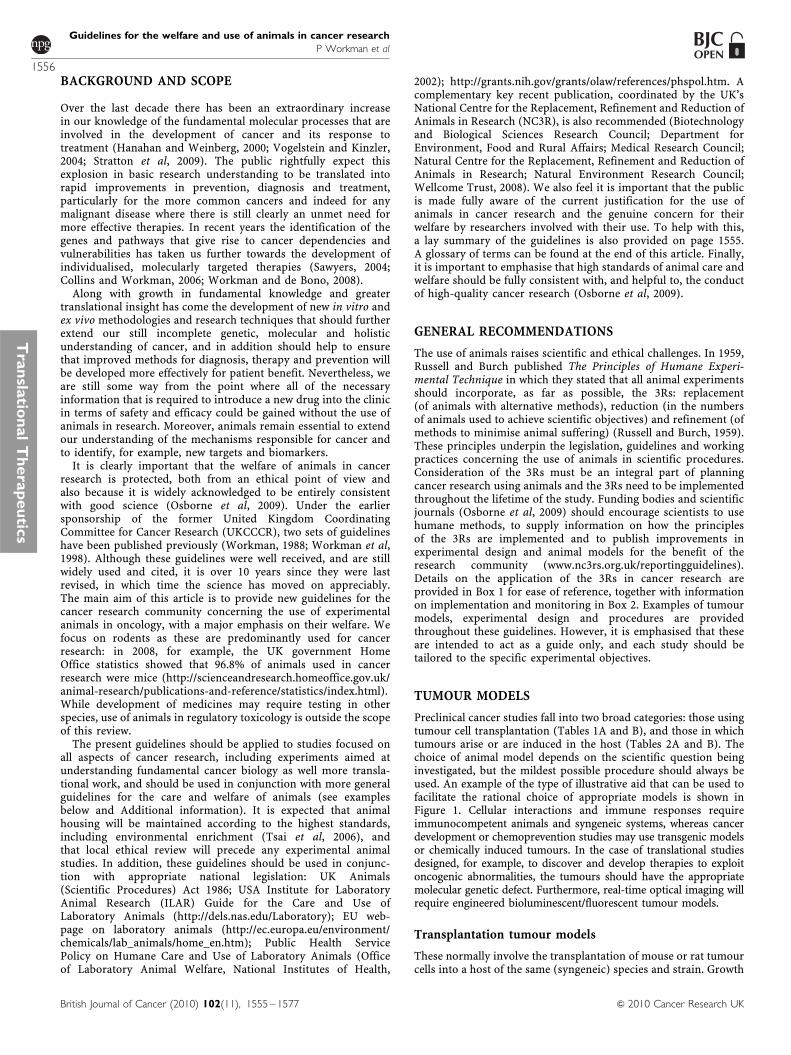
BACKGROUND AND SCOPE
Over the last decade there has been an extraordinary increasein our knowledge of the fundamental molecular processes that areinvolved in the development of cancer and its response totreatment (Hanahan and Weinberg, 2000; Vogelstein and Kinzler,2004; Stratton et al, 2009). The public rightfully expect thisexplosion in basic research understanding to be translated intorapid improvements in prevention, diagnosis and treatment,particularly for the more common cancers and indeed for anymalignant disease where there is still clearly an unmet need formore effective therapies. In recent years the identification of thegenes and pathways that give rise to cancer dependencies andvulnerabilities has taken us further towards the development ofindividualised, molecularly targeted therapies (Sawyers, 2004;Collins and Workman, 2006; Workman and de Bono, 2008).
Along with growth in fundamental knowledge and greatertranslational insight has come the development of new in vitro andex vivo methodologies and research techniques that should furtherextend our still incomplete genetic, molecular and holisticunderstanding of cancer, and in addition should help to ensurethat improved methods for diagnosis, therapy and prevention willbe developed more effectively for patient benefit. Nevertheless, weare still some way from the point where all of the necessaryinformation that is required to introduce a new drug into the clinicin terms of safety and efficacy could be gained without the use ofanimals in research. Moreover, animals remain essential to extendour understanding of the mechanisms responsible for cancer andto identify, for example, new targets and biomarkers.
It is clearly important that the welfare of animals in cancerresearch is protected, both from an ethical point of view andalso because it is widely acknowledged to be entirely consistentwith good science (Osborne et al, 2009). Under the earliersponsorship of the former United Kingdom CoordinatingCommittee for Cancer Research (UKCCCR), two sets of guidelineshave been published previously (Workman, 1988; Workman et al,1998). Although these guidelines were well received, and are stillwidely used and cited, it is over 10 years since they were lastrevised, in which time the science has moved on appreciably.The main aim of this article is to provide new guidelines for thecancer research community concerning the use of experimentalanimals in oncology, with a major emphasis on their welfare. Wefocus on rodents as these are predominantly used for cancerresearch: in 2008, for example, the UK government HomeOffice statistics showed that 96.8% of animals used in cancerresearch were mice (http://scienceandresearch.homeoffice.gov.uk/animal-research/publications-and-reference/statistics/index.html).While development of medicines may require testing in otherspecies, use of animals in regulatory toxicology is outside the scopeof this review.
The present guidelines should be applied to studies focused onall aspects of cancer research, including experiments aimed atunderstanding fundamental cancer biology as well more transla-tional work, and should be used in conjunction with more generalguidelines for the care and welfare of animals (see examplesbelow and Additional information). It is expected that animalhousing will be maintained according to the highest standards,including environmental enrichment (Tsai et al, 2006), andthat local ethical review will precede any experimental animalstudies. In addition, these guidelines should be used in conjunc-tion with appropriate national legislation: UK Animals(Scientific Procedures) Act 1986; USA Institute for LaboratoryAnimal Research (ILAR) Guide for the Care and Use ofLaboratory Animals (http://dels.nas.edu/Laboratory); EU web-page on laboratory animals (http://ec.europa.eu/environment/chemicals/lab_animals/home_en.htm); Public Health ServicePolicy on Humane Care and Use of Laboratory Animals (Officeof Laboratory Animal Welfare, National Institutes of Health,
2002); http://grants.nih.gov/grants/olaw/references/phspol.htm. Acomplementary key recent publication, coordinated by the UK’sNational Centre for the Replacement, Refinement and Reduction ofAnimals in Research (NC3R), is also recommended (Biotechnologyand Biological Sciences Research Council; Department forEnvironment, Food and Rural Affairs; Medical Research Council;Natural Centre for the Replacement, Refinement and Reduction ofAnimals in Research; Natural Environment Research Council;Wellcome Trust, 2008). We also feel it is important that the publicis made fully aware of the current justification for the use ofanimals in cancer research and the genuine concern for theirwelfare by researchers involved with their use. To help with this,a lay summary of the guidelines is also provided on page 1555.A glossary of terms can be found at the end of this article. Finally,it is important to emphasise that high standards of animal care andwelfare should be fully consistent with, and helpful to, the conductof high-quality cancer research (Osborne et al, 2009).
GENERAL RECOMMENDATIONS
The use of animals raises scientific and ethical challenges. In 1959,Russell and Burch published The Principles of Humane Experi-mental Technique in which they stated that all animal experimentsshould incorporate, as far as possible, the 3Rs: replacement(of animals with alternative methods), reduction (in the numbersof animals used to achieve scientific objectives) and refinement (ofmethods to minimise animal suffering) (Russell and Burch, 1959).These principles underpin the legislation, guidelines and workingpractices concerning the use of animals in scientific procedures.Consideration of the 3Rs must be an integral part of planningcancer research using animals and the 3Rs need to be implementedthroughout the lifetime of the study. Funding bodies and scientificjournals (Osborne et al, 2009) should encourage scientists to usehumane methods, to supply information on how the principlesof the 3Rs are implemented and to publish improvements inexperimental design and animal models for the benefit of theresearch community (www.nc3rs.org.uk/reportingguidelines).Details on the application of the 3Rs in cancer research areprovided in Box 1 for ease of reference, together with informationon implementation and monitoring in Box 2. Examples of tumourmodels, experimental design and procedures are providedthroughout these guidelines. However, it is emphasised that theseare intended to act as a guide only, and each study should betailored to the specific experimental objectives.
TUMOUR MODELS
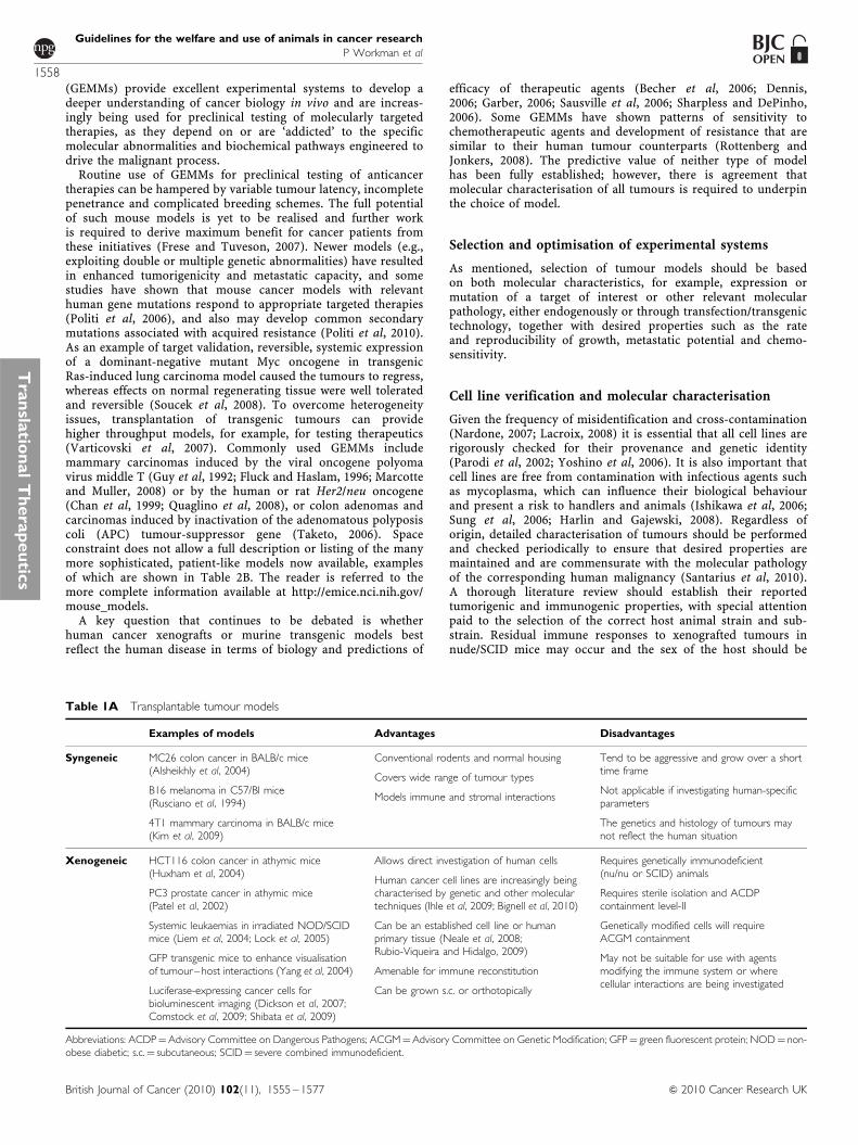
Preclinical cancer studies fall into two broad categories: those usingtumour cell transplantation (Tables 1A and B), and those in whichtumours arise or are induced in the host (Tables 2A and B). Thechoice of animal model depends on the scientific question beinginvestigated, but the mildest possible procedure should always beused. An example of the type of illustrative aid that can be used tofacilitate the rational choice of appropriate models is shown inFigure 1. Cellular interactions and immune responses requireimmunocompetent animals and syngeneic systems, whereas cancerdevelopment or chemoprevention studies may use transgenic modelsor chemically induced tumours. In the case of translational studiesdesigned, for example, to discover and develop therapies to exploitoncogenic abnormalities, the tumours should have the appropriatemolecular genetic defect. Furthermore, real-time optical imaging willrequire engineered bioluminescent/fluorescent tumour models.
Transplantation tumour models
These normally involve the transplantation of mouse or rat tumourcells into a host of the same (syngeneic) species and strain. Growth
Guidelines for the welfare and use of animals in cancer research
P Workman et al
1556
British Journal of Cancer (2010) 102(11), 1555 – 1577 & 2010 Cancer Research UK
Tra
nsla
tion
al
Th
era
peu
tics

of human (xenogeneic) tumour cells can be achieved usingimmunodeficient (e.g., nude or SCID) mice to prevent rejection(Table 1A). Most transplantable tumours are established subcuta-neously. These subcutaneous (s.c.) tumours are simple to initiatebut may lack relevance in terms of stromal/vascular interactionsand metastasis. More complex models may involve orthotopictransplantation at appropriate primary sites, or inoculation oftumour cells through routes which maximise the chance of
metastatic spread (Table 1B). There is an increasing trend toestablish xenograft tumours directly from human cancers, to avoidartificial selection of cells in tissue culture and changes in geneexpression and phenotype, which this may induce. Such trans-plants may better model the principal facets of clinical cancer, forexample, maintenance of tumour architecture, heterogeneity,expression of certain targets and response to therapy (Donget al, 2010), but can be less reproducible (especially as primarygrafts) and slower growing than well-established models (Nealeet al, 2008; Rubio-Viqueira and Hidalgo, 2009). Detailed molecularand genetic characterisation, facilitated by modern high-through-put technologies (e.g., see http://www.sanger.ac.uk/genetics/CGP),is now available for human cancer cell lines used for xenografts(Masters et al, 2001; Park et al, 2010) and is important tounderstand the biology of these models and to select the mostappropriate for each study.
Autochthonous tumour models
There are two broad categories: those arising in outbred orinbred rodents (Table 2A), or those from animals harbouringgenetic changes that alter tumour susceptibility (Table 2B).Certain mouse or rat strains are susceptible to spontaneousdevelopment of tumours. More commonly, tumours are inducedby chemical carcinogens, radiation, viruses or bacteria. Suchmodels may mimic some of the aetiological events in humancancer development; exposure to such agents may induce systemiceffects that are difficult to replicate in genetically engineeredmodels.
Major advances have been made in the development of sophis-ticated mouse models of cancer that mimic many of the geneticand biological characteristics of human malignancies, although thehost genetic background may affect tumour incidence and/ormalignant potential (Lifsted et al, 1998; Winter and Hunter, 2008).A range of technologies now allows the inducible expression ofoncogenes or inactivation of tumour-suppressor genes in vivo ina precisely controlled manner in virtually any tissue or celltype. (Chen et al, 2004; Christophorou et al, 2006; Sharpless andDePinho, 2006). Such genetically engineered mouse models
Box 1 THE 3Rs:REPLACEMENT, REDUCTION AND REFINEMENT
Replacement
Absolute replacement techniques avoid the use of animals; relative replacementtechniques include substituting non-vertebrate species
1. Investigate the potential of novel and existing alternative approachesto animals
2. Use in silico and/or in vitro pre-screens before commencing animalstudies
Reduction
Minimise the number of animals used to achieve specific scientific objectives
1. Ensure that all studies are scientifically robust and apply appropriatestatistical methods to experimental design
2. Reduce experimental variability by conducting studies of animals ofdefined health status and, wherever possible, using inbred strains
3. Minimise surplus breeding by avoiding unnecessarily narrowspecifications for animal sex, age and weight
4. Freeze rodent embryos, sperm and cancer cell lines notimmediately required for scientific studies
5. Prevent duplication by making specific strains and geneticallymodified lines available throughout the research community
6. Consider use of serial sampling or longitudinal imaging in whicheach animal acts as its own control to reduce study group sizes
Refinement
Continual review of improvements in experimental design, techniques andhusbandry to minimise adverse effects and improve welfare
1. Apply all available knowledge to predict adverse effects and ensurethat appropriate humane endpoints are developed and specialistcare is provided, especially when using genetically modified animals(e.g., immune deficient or tumour-prone strains)
2. Provide animals with an appropriate environment (e.g., nestingmaterial, shelter for rodents), including sufficient space andcomplexity to satisfy their normal species-typical behaviours
3. Undertake pilot studies of unfamiliar tumour cell lines or novelprocedures to establish experimental and humane endpoints
4. Perform post-mortem examinations as a routine part of all pilotstudies and to investigate any unexpected deaths
5. Include appropriate controls to understand individual and combinedeffects of tumours and treatments
6. Use anaesthesia and analgesia whenever appropriate. This shouldbe regularly reviewed by a vet to ensure that contemporary bestpractice is followed
7. Consider imaging methods to monitor non-superficial tumourburden and to aid the timely implementation of humaneendpoints
8. Do not allow animals to become moribund: death as an intentionalendpoint is unacceptable
9. Maintain and share detailed information, on all experimentalprocedures, including behaviour of the tumour and host animals undervarious conditions
10. Include principal details relating to the use of animals, such as studydesign, adverse effects, specialist care and the 3Rs, in all scientificpublications
Box 2 IMPLEMENTATION AND MONITORING
For assurance that best practice is implemented and rigorously applied, practicalguidelines should be clear and readily available, with staff fully engaged andeducated in their use
1. Establish a clear chain of responsibility to ensure that prompt action istaken where necessary: for example, if the condition of an animaldeteriorates unexpectedly
2. Establish humane endpoints, specialist care, monitoring methods andcriteria for intervention between researchers, veterinary and animalcare staff before initiating studies
3. Ensure that all staff involved in the use and care of animals are aware oftheir personal and legal responsibilities, and are trained to recogniseadverse effects and apply humane endpoints
4. Seek additional external expertise where unfamiliar models ortechniques are being introduced
5. Ensure adequate staffing levels are available for the duration of the study,particularly during crucial periods where additional observations are needed
6. Inspect animals at a frequency determined by the known biology of thetumour, the effects of any interventions and the clinical status of theanimals
7. Monitor adverse effects, including general signs of welfare and moredetailed indicators appropriate to the specific model and procedures
8. Give all staff appropriate training and professional developmentopportunities
9. Provide appropriate supervision of staff and confirm, review anddocument competence on a regular basis
Guidelines for the welfare and use of animals in cancer research
P Workman et al
1557
British Journal of Cancer (2010) 102(11), 1555 – 1577& 2010 Cancer Research UK
Tra
nsl
ati
on
al
Th
era
peu
tics
(GEMMs) provide excellent experimental systems to develop adeeper understanding of cancer biology in vivo and are increas-ingly being used for preclinical testing of molecularly targetedtherapies, as they depend on or are ‘addicted’ to the specificmolecular abnormalities and biochemical pathways engineered todrive the malignant process.
Routine use of GEMMs for preclinical testing of anticancertherapies can be hampered by variable tumour latency, incompletepenetrance and complicated breeding schemes. The full potentialof such mouse models is yet to be realised and further workis required to derive maximum benefit for cancer patients fromthese initiatives (Frese and Tuveson, 2007). Newer models (e.g.,exploiting double or multiple genetic abnormalities) have resultedin enhanced tumorigenicity and metastatic capacity, and somestudies have shown that mouse cancer models with relevanthuman gene mutations respond to appropriate targeted therapies(Politi et al, 2006), and also may develop common secondarymutations associated with acquired resistance (Politi et al, 2010).As an example of target validation, reversible, systemic expressionof a dominant-negative mutant Myc oncogene in transgenicRas-induced lung carcinoma model caused the tumours to regress,whereas effects on normal regenerating tissue were well toleratedand reversible (Soucek et al, 2008). To overcome heterogeneityissues, transplantation of transgenic tumours can providehigher throughput models, for example, for testing therapeutics(Varticovski et al, 2007). Commonly used GEMMs includemammary carcinomas induced by the viral oncogene polyomavirus middle T (Guy et al, 1992; Fluck and Haslam, 1996; Marcotteand Muller, 2008) or by the human or rat Her2/neu oncogene(Chan et al, 1999; Quaglino et al, 2008), or colon adenomas andcarcinomas induced by inactivation of the adenomatous polyposiscoli (APC) tumour-suppressor gene (Taketo, 2006). Spaceconstraint does not allow a full description or listing of the manymore sophisticated, patient-like models now available, examplesof which are shown in Table 2B. The reader is referred to themore complete information available at http://emice.nci.nih.gov/mouse_models.
A key question that continues to be debated is whetherhuman cancer xenografts or murine transgenic models bestreflect the human disease in terms of biology and predictions of
efficacy of therapeutic agents (Becher et al, 2006; Dennis,2006; Garber, 2006; Sausville et al, 2006; Sharpless and DePinho,2006). Some GEMMs have shown patterns of sensitivity tochemotherapeutic agents and development of resistance that aresimilar to their human tumour counterparts (Rottenberg andJonkers, 2008). The predictive value of neither type of modelhas been fully established; however, there is agreement thatmolecular characterisation of all tumours is required to underpinthe choice of model.
Selection and optimisation of experimental systems
As mentioned, selection of tumour models should be basedon both molecular characteristics, for example, expression ormutation of a target of interest or other relevant molecularpathology, either endogenously or through transfection/transgenictechnology, together with desired properties such as the rateand reproducibility of growth, metastatic potential and chemo-sensitivity.
Cell line verification and molecular characterisation
Given the frequency of misidentification and cross-contamination(Nardone, 2007; Lacroix, 2008) it is essential that all cell lines arerigorously checked for their provenance and genetic identity(Parodi et al, 2002; Yoshino et al, 2006). It is also important thatcell lines are free from contamination with infectious agents suchas mycoplasma, which can influence their biological behaviourand present a risk to handlers and animals (Ishikawa et al, 2006;Sung et al, 2006; Harlin and Gajewski, 2008). Regardless oforigin, detailed characterisation of tumours should be performedand checked periodically to ensure that desired properties aremaintained and are commensurate with the molecular pathologyof the corresponding human malignancy (Santarius et al, 2010).A thorough literature review should establish their reportedtumorigenic and immunogenic properties, with special attentionpaid to the selection of the correct host animal strain and sub-strain. Residual immune responses to xenografted tumours innude/SCID mice may occur and the sex of the host should be
Table 1A Transplantable tumour models
Examples of models Advantages Disadvantages
Syngeneic MC26 colon cancer in BALB/c mice(Alsheikhly et al, 2004)
B16 melanoma in C57/Bl mice(Rusciano et al, 1994)
4T1 mammary carcinoma in BALB/c mice(Kim et al, 2009)
Conventional rodents and normal housing
Covers wide range of tumour types
Models immune and stromal interactions
Tend to be aggressive and grow over a shorttime frame
Not applicable if investigating human-specificparameters
The genetics and histology of tumours maynot reflect the human situation
Xenogeneic HCT116 colon cancer in athymic mice(Huxham et al, 2004)
PC3 prostate cancer in athymic mice(Patel et al, 2002)
Systemic leukaemias in irradiated NOD/SCIDmice (Liem et al, 2004; Lock et al, 2005)
GFP transgenic mice to enhance visualisationof tumour–host interactions (Yang et al, 2004)
Luciferase-expressing cancer cells forbioluminescent imaging (Dickson et al, 2007;Comstock et al, 2009; Shibata et al, 2009)
Allows direct investigation of human cells
Human cancer cell lines are increasingly beingcharacterised by genetic and other moleculartechniques (Ihle et al, 2009; Bignell et al, 2010)
Can be an established cell line or humanprimary tissue (Neale et al, 2008;Rubio-Viqueira and Hidalgo, 2009)
Amenable for immune reconstitution
Can be grown s.c. or orthotopically
Requires genetically immunodeficient(nu/nu or SCID) animals
Requires sterile isolation and ACDPcontainment level-II
Genetically modified cells will requireACGM containment
May not be suitable for use with agentsmodifying the immune system or wherecellular interactions are being investigated
Abbreviations: ACDP¼Advisory Committee on Dangerous Pathogens; ACGM¼Advisory Committee on Genetic Modification; GFP¼ green fluorescent protein; NOD¼ non-obese diabetic; s.c.¼ subcutaneous; SCID¼ severe combined immunodeficient.
Guidelines for the welfare and use of animals in cancer research
P Workman et al
1558
British Journal of Cancer (2010) 102(11), 1555 – 1577 & 2010 Cancer Research UK
Tra
nsla
tion
al
Th
era
peu
tics
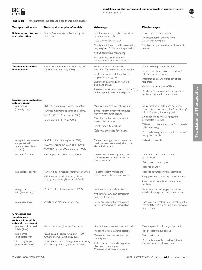
Table 1B Transplantation models used for therapeutic studies
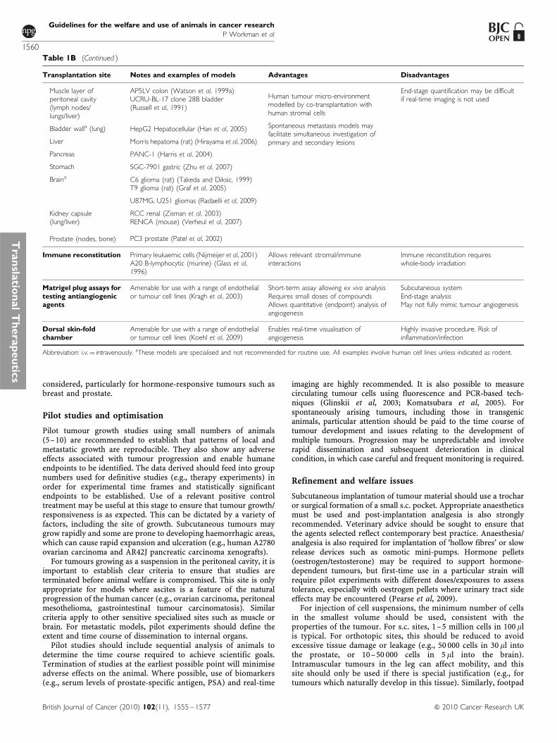
Transplantation site Notes and examples of models Advantages Disadvantages
Subcutaneous tumourtransplantation
A high % of established lines will growat this site
Simplest model for routine evaluationof anticancer agents
Uses cancer cells or tissue
Simple administration with anaestheticonly required for tissue transplantation
Allows continuous monitoring
Facilitates the use of bilateraltransplantation after pilot studies
Ectopic site for most tumours
Metastases rarely develop froms.c. tumour xenografts
May be poorly vascularised with necroticcentres
Tumour cells withinhollow fibres
Amenable for use with a wide range ofcell lines (Decker et al, 2004)
Allows multiple cell lines to beimplanted for simultaneous assessment
Useful for human cell lines that failto grow as xenografts
Short-term assay requiring ex vivoend-stage analysis
Provides a quick assessment of drug efficacyand may predict xenograft response
Careful scoring system required
Lack of vasculature may miss ‘indirect’effects or access issues
Inflammation around fibres can affectresponses
Variation in properties of fibres
Possibility of paracrine effects if multiplecell lines implanted in same animal
Experimental metastasis(site of spread)
Intravenous(primarily lung)
3DC13B lymphoma (Golay et al, 2006)
M24met melanoma (Becker et al, 1996)
A549 NSCLC (Kennel et al, 1999)
Lewis lung 3LL (Li et al, 2001)
Most cells injected i.v. colonise lung
Some (notably lymphoid tumours)may colonise other organs
Models end-stage of metastasis ina controlled manner
Simple model to establish
Cells may be tagged for imaging
Bolus injection of cells does not mimicnatural dissemination and the ‘conditioning’effect of primary tumour growth
Does not model the full spectrumof metastatic cascade
Difficult to monitor and quantify accuratelywithout imaging
Pilot studies required to establish incidenceand growth kinetics
Intra-peritoneal-(ascitesand peritoneal/omentum-associatedtumours)
GW-39 colon (Sharkey et al, 1991)
MGLVA1 gastric (Watson et al, 1999b)
OVCAR3 ovarian (Zavaleta et al, 2008)
Mimics late-stage ovarian cancer andcarcinomatosis associated with someabdominal cancers
Difficult to quantify
Intra-tibiala (bone) ARCaP prostate (Zhau et al, 2000) Mimics bone tumour growth seenwith myeloma or prostate and breastcancer metastases
Does not mimic natural tumourdissemination
Risk of infection and pain
Requires imaging
Intra-cardiaca (bone) MDA MB 231 breast (Serganova et al, 2009)
A375 melanoma (Nakai et al, 1992)Mat Ly-lu prostate (Blouin et al, 2008)
To some extent mimics thedissemination phase of metastasis
Requires advanced surgical technique
Risky procedure requiring particular care
Only suitable for a limited number ofcell lines
Intra-portalvein (liver, nodes)
LS174T colon (Mahteme et al, 1998) Localises tumour cells to liver
Appropriate for colon, pancreaticand breast cancers
Requires advanced surgical technique toavoid cell leakage into peritoneal cavity
Intrasplenic (liver) WiDR colon (Miyazaki et al, 1999) Easier procedure than mesentericvein or intraportal cell inoculation
Local growth in spleen may complicate theinterpretation of results unless splenectomyis performed
Orthotopic andspontaneousmetastasis models(sites of metastasis)
Intra-caecal/lymphoidfollicle (liver)
Intra-dermal(lung/nodes/liver)
Mammary fat pad(lungs/nodes/brain)
TK-3,-4,-9 colon (Tanaka et al, 1995)
NC65 renal (Nakatsugawa et al, 1999)A-07melanoma (Graff et al, 2005)
MDA-MB-231 breast (Serganova et al, 2009)4T1 breast (murine) (Mitra et al, 2006)
Relevant stromal/vascular cell interactions
Models the full metastatic cascade
Certain models may involve lymphnode spread
Cells may be genetically tagged toallow real-time imagingChemosensitivity more relevant
Most require delicate surgical procedures
Risk of local tumour spread
Risk of infection
Pilot studies must be used to determinethe time frame of disease events
Guidelines for the welfare and use of animals in cancer research
P Workman et al
1559
British Journal of Cancer (2010) 102(11), 1555 – 1577& 2010 Cancer Research UK
Tra
nsl
ati
on
al
Th
era
peu
tics
considered, particularly for hormone-responsive tumours such asbreast and prostate.
Pilot studies and optimisation
Pilot tumour growth studies using small numbers of animals(5–10) are recommended to establish that patterns of local andmetastatic growth are reproducible. They also show any adverseeffects associated with tumour progression and enable humaneendpoints to be identified. The data derived should feed into groupnumbers used for definitive studies (e.g., therapy experiments) inorder for experimental time frames and statistically significantendpoints to be established. Use of a relevant positive controltreatment may be useful at this stage to ensure that tumour growth/responsiveness is as expected. This can be dictated by a variety offactors, including the site of growth. Subcutaneous tumours maygrow rapidly and some are prone to developing haemorrhagic areas,which can cause rapid expansion and ulceration (e.g., human A2780ovarian carcinoma and AR42J pancreatic carcinoma xenografts).
For tumours growing as a suspension in the peritoneal cavity, it isimportant to establish clear criteria to ensure that studies areterminated before animal welfare is compromised. This site is onlyappropriate for models where ascites is a feature of the naturalprogression of the human cancer (e.g., ovarian carcinoma, peritonealmesothelioma, gastrointestinaI tumour carcinomatosis). Similarcriteria apply to other sensitive specialised sites such as muscle orbrain. For metastatic models, pilot experiments should define theextent and time course of dissemination to internal organs.
Pilot studies should include sequential analysis of animals todetermine the time course required to achieve scientific goals.Termination of studies at the earliest possible point will minimiseadverse effects on the animal. Where possible, use of biomarkers(e.g., serum levels of prostate-specific antigen, PSA) and real-time
imaging are highly recommended. It is also possible to measurecirculating tumour cells using fluorescence and PCR-based tech-niques (Glinskii et al, 2003; Komatsubara et al, 2005). Forspontaneously arising tumours, including those in transgenicanimals, particular attention should be paid to the time course oftumour development and issues relating to the development ofmultiple tumours. Progression may be unpredictable and involverapid dissemination and subsequent deterioration in clinicalcondition, in which case careful and frequent monitoring is required.
Refinement and welfare issues
Subcutaneous implantation of tumour material should use a trocharor surgical formation of a small s.c. pocket. Appropriate anaestheticsmust be used and post-implantation analgesia is also stronglyrecommended. Veterinary advice should be sought to ensure thatthe agents selected reflect contemporary best practice. Anaesthesia/analgesia is also required for implantation of ‘hollow fibres’ or slowrelease devices such as osmotic mini-pumps. Hormone pellets(oestrogen/testosterone) may be required to support hormone-dependent tumours, but first-time use in a particular strain willrequire pilot experiments with different doses/exposures to assesstolerance, especially with oestrogen pellets where urinary tract sideeffects may be encountered (Pearse et al, 2009).
For injection of cell suspensions, the minimum number of cellsin the smallest volume should be used, consistent with theproperties of the tumour. For s.c. sites, 1–5 million cells in 100 mlis typical. For orthotopic sites, this should be reduced to avoidexcessive tissue damage or leakage (e.g., 50 000 cells in 30 ml intothe prostate, or 10–50 000 cells in 5 ml into the brain).Intramuscular tumours in the leg can affect mobility, and thissite should only be used if there is special justification (e.g., fortumours which naturally develop in this tissue). Similarly, footpad
Table 1B (Continued )
Transplantation site Notes and examples of models Advantages Disadvantages
Muscle layer ofperitoneal cavity(lymph nodes/lungs/liver)
Bladder walla (lung)
Liver
Pancreas
Stomach
Braina
Kidney capsule(lung/liver)
Prostate (nodes, bone)
AP5LV colon (Watson et al, 1999a)UCRU-BL-17 clone 28B bladder(Russell et al, 1991)
HepG2 Hepatocellular (Han et al, 2005)
Morris hepatoma (rat) (Hirayama et al, 2006)
PANC-1 (Harris et al, 2004)
SGC-7901 gastric (Zhu et al, 2007)
C6 glioma (rat) (Takeda and Diksic, 1999)T9 glioma (rat) (Graf et al, 2005)
U87MG, U251 gliomas (Radaelli et al, 2009)
RCC renal (Zisman et al, 2003)RENCA (mouse) (Verheul et al, 2007)
PC3 prostate (Patel et al, 2002)
Human tumour micro-environmentmodelled by co-transplantation withhuman stromal cells
Spontaneous metastasis models mayfacilitate simultaneous investigation ofprimary and secondary lesions
End-stage quantification may be difficultif real-time imaging is not used
Immune reconstitution Primary leukaemic cells (Nijmeijer et al, 2001)A20 B-lymphocytic (murine) (Glass et al,1996)
Allows relevant stromal/immuneinteractions
Immune reconstitution requireswhole-body irradiation
Matrigel plug assays fortesting antiangiogenicagents
Amenable for use with a range of endothelialor tumour cell lines (Kragh et al, 2003)
Short-term assay allowing ex vivo analysisRequires small doses of compoundsAllows quantitative (endpoint) analysis ofangiogenesis
Subcutaneous systemEnd-stage analysisMay not fully mimic tumour angiogenesis
Dorsal skin-foldchamber
Amenable for use with a range of endothelialor tumour cell lines (Koehl et al, 2009)
Enables real-time visualisation ofangiogenesis
Highly invasive procedure. Risk ofinflammation/infection
Abbreviation: i.v.¼ intravenously. aThese models are specialised and not recommended for routine use. All examples involve human cell lines unless indicated as rodent.
Guidelines for the welfare and use of animals in cancer research
P Workman et al
1560
British Journal of Cancer (2010) 102(11), 1555 – 1577 & 2010 Cancer Research UK
Tra
nsla
tion
al
Th
era
peu
tics
injection, which has been traditionally used to potentiatelymphatic dissemination, is unacceptable without exceptionalscientific justification and should then only involve a single paw.
Surgical removal of a primary tumour may be justified, forexample, from s.c. sites, mammary fat pad or removal of the spleenfollowing intrasplenic injection, to allow time for outgrowth of anysecondary deposits. Surgery must be performed using steriletechniques with appropriate post-operative monitoring andcontrol of any pain and inflammation/infection.
Cell lines should be checked regularly for contaminatingmicroorganisms to avoid infection of host animals. This isespecially important if tumours are routinely passaged betweenanimals, which may be justified for those that are difficult toestablish from cell cultures. Asymptomatic infection of experi-mental animals may affect tumour properties, for example,metastasis (Rodriguez-Cuesta et al, 2005). Procedures can be usedto improve tumour take rate. For example, moderate doses ofwhole-body irradiation may further enhance engraftment oftumour cells in athymic mice (Baersch et al, 1997; Nijmeijeret al, 2001; Li et al, 2006), although the added stress and risk to theanimal must be considered. Co-administration of human tumourcells with allogeneic bone marrow transplantation may reducegraft-vs-host activity but preserve graft-vs-tumour effects inallogeneic leukaemia models (Prigozhina et al, 2002; Giver et al,2004).
Transplanted tumours (especially xenografts) may not develop withan incidence, growth rate or malignant potential required; howeverthis can often be enhanced by selection of tumorigenic/metastaticvariants (Bruns et al, 1999; Nguyen et al, 2009a). In addition,co-injection of tumour cells with extracellular matrix proteins and/orangiogenic factors (Collado et al, 2007), cancer-associated fibroblasts(Noel et al, 1993; Orimo et al, 2005) or mesenchymal stem cells(Karnoub et al, 2007; Spaeth et al, 2009) can increase tumorigenicity,better recapitulate the human tumour microenvironment andenhance metastatic potential. Cells may be transfected withfluorescent or bioluminescent markers allowing serial imaging ofinternal tumours/metastatic spread. However, such tagged cell linesshould be profiled to establish that their biological characteristics areunchanged and consideration should be given to the dependence ofluminescence/fluorescence on factors in the tumour microenviron-ment, for example. molecular oxygenation, necrosis, or ascites fluidfrom peritoneal tumours (Condeelis and Segall, 2003).
THERAPY
Preclinical discovery and development of therapeutics
There is a concerted effort to identify and develop small-moleculedrugs or biopharmaceuticals (e.g., antibodies, protein therapeutics,
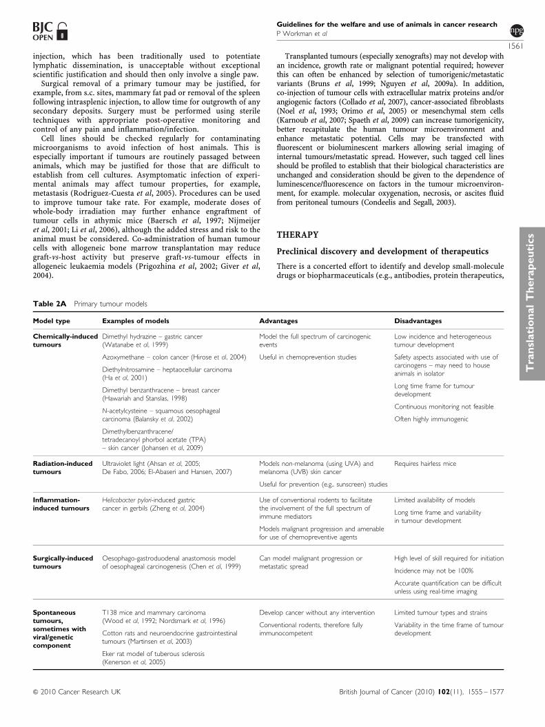
Table 2A Primary tumour models
Model type Examples of models Advantages Disadvantages
Chemically-inducedtumours
Dimethyl hydrazine – gastric cancer(Watanabe et al, 1999)
Azoxymethane – colon cancer (Hirose et al, 2004)
Diethylnitrosamine – heptaocellular carcinoma(Ha et al, 2001)
Dimethyl benzanthracene – breast cancer(Hawariah and Stanslas, 1998)
N-acetylcysteine – squamous oesophagealcarcinoma (Balansky et al, 2002)
Dimethylbenzanthracene/tetradecanoyl phorbol acetate (TPA)– skin cancer (Johansen et al, 2009)
Model the full spectrum of carcinogenicevents
Useful in chemoprevention studies
Low incidence and heterogeneoustumour development
Safety aspects associated with use ofcarcinogens – may need to houseanimals in isolator
Long time frame for tumourdevelopment
Continuous monitoring not feasible
Often highly immunogenic
Radiation-inducedtumours
Ultraviolet light (Ahsan et al, 2005;De Fabo, 2006; El-Abaseri and Hansen, 2007)
Models non-melanoma (using UVA) andmelanoma (UVB) skin cancer
Useful for prevention (e.g.. sunscreen) studies
Requires hairless mice
Inflammation-induced tumours
Helicobacter pylori-induced gastriccancer in gerbils (Zheng et al, 2004)
Use of conventional rodents to facilitatethe involvement of the full spectrum ofimmune mediators
Models malignant progression and amenablefor use of chemopreventive agents
Limited availability of models
Long time frame and variabilityin tumour development
Surgically-inducedtumours
Oesophago-gastroduodenal anastomosis modelof oesophageal carcinogenesis (Chen et al, 1999)
Can model malignant progression ormetastatic spread
High level of skill required for initiation
Incidence may not be 100%
Accurate quantification can be difficultunless using real-time imaging
Spontaneoustumours,sometimes withviral/geneticcomponent
T138 mice and mammary carcinoma(Wood et al, 1992; Nordsmark et al, 1996)
Cotton rats and neuroendocrine gastrointestinaltumours (Martinsen et al, 2003)
Eker rat model of tuberous sclerosis(Kenerson et al, 2005)
Develop cancer without any intervention
Conventional rodents, therefore fullyimmunocompetent
Limited tumour types and strains
Variability in the time frame of tumourdevelopment
Guidelines for the welfare and use of animals in cancer research
P Workman et al
1561
British Journal of Cancer (2010) 102(11), 1555 – 1577& 2010 Cancer Research UK
Tra
nsl
ati
on
al
Th
era
peu
tics
vaccines, gene therapy) targeted against cancer cells or associatedhost cells (Sawyers, 2004; Collins and Workman, 2006; Workmanand de Bono, 2008). A representative ‘test cascade’ for discoveringnew small-molecule inhibitors of cancer targets is shown in Figure 2.As a consequence of extensive in vitro testing, comparatively smallnumbers of prioritised compounds progress to examination in vivo(Collins and Workman, 2006). In vivo studies use sequential,
discriminatory tests to prioritise compounds at each stage. Differenttests may need to be applied to biopharmaceuticals, such asantibodies and vaccines, as they may work by recruiting hosteffectors (e.g., cytotoxic leukocytes). Epitope specificity can alsorequire the development of an antibody or vaccine initially usinganti-rodent reagents (before switching to the clinical form) or use ofa genetically modified mouse model. In addition, agents directed
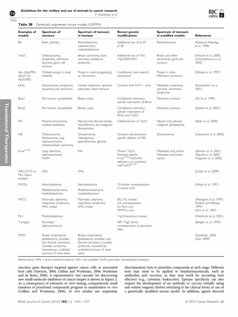
Table 2B Genetically engineered mouse models (GEMMs)
Examples ofmodels
Spectrum oftumours
Spectrum of tumoursin humans
Recent geneticmodifications
Spectrum of tumoursin modified models References
Rb Brain, pituitary Retinoblastoma,osteosarcoma,medulloblastoma
Additional loss of p107,p130
Retinoblastoma (Robanus-Maandaget al, 1998)
Trp53 Osteosarcoma,lymphoma, soft-tissuesarcoma, germ celltumours
Breast carcinoma, brain,sarcomas, leukaemia,endocrine
Additional loss of TercTrp53(ER)TAM
Breast and othercarcinomas, germ-celltumours
(Artandi et al, 2000)(Christophorou et al,2005)
Apc (ApcMIN,ApcD716,ApcD580)
Multiple polyps in smallintestine
Polyps in colon progressingto carcinomas
Conditional colon-specificinactivation
Polyps in colonMammary carcinoma
(Shibata et al, 1997)
Ink4a Fibrosarcoma, lymphoma,squamous-cell carcinoma
Familial melanoma, sporadicpancreatic, brain tumours
Crossed with Arf+/� mice Metastatic melanoma,sarcoma, carcinoma,lymphoma
(Krimpenfort et al,2001)
Brca1 No tumour susceptibility Breast ovary Conditional mammary-specific inactivation of Brca1
Mammary tumours (Xu et al, 1999)
Brca2 No tumour susceptibility Breast, ovary Conditional mammary-specific inactivation ofBrca2 and Trp53
Mammary tumours (Jonkers et al, 2001)
Nf1 Pheochromocytoma,myeloid leukaemia
Neural-crest-derived benignneurofibroma and malignantfibrosarcoma
Additional loss of Trp53 Neural-crest-derivedmalignant glioblastoma
(Reilly et al, 2000)
Nf2 Osteosarcoma,fibrosarcoma, lungadenocarcinoma,mepatocellular carcinoma
Schwannomas,meningiomas,ependymomas, gliomas
Schwann-cell precursor-specific ablation of Nf2
Schwannomas (Giovannini et al, 2000)
K-rasG12D Lung adenoma,adenocarcinomaPanIN
NA Mutant Trp53Pancreas-specificK-rasG12D+Ink4a/Arfdeficiency or combinedwithTrp53R172H
Metastatic lung cancerMetastatic pancreaticcancer
(Johnson et al, 2001)(Aguirre et al, 2003;Hingorani et al, 2005)
AML1/ETO orMLL fusionproteins
AML AML (Zuber et al, 2009)
MYCN Neuroblastoma,
Rhabdomyosarcoma,medulloblastoma
Neuroblastoma,
Rhabdomyosarcoma,medulloblastoma
TH-driven overexpressionin neural crest
(Weiss et al, 1997)
MYCC Pancreatic adenoma,melanoma, lymphoma,AML, breast
Pancreatic adenoma,melanoma, lymphoma,AML, breast
BCL-XL crossesLck overexpressionEu-Tta-c-mycMMTV-c-myc
(Pelengaris et al, 1999)(Felsher and Bishop,1999)(Sinn et al, 1987)
Ptc1 Medulloblastoma Trp53-knockout crosses (Wetmore et al, 2001)
T-antigen Pancreaticadenocarcinoma
RIP1-Tag2 drivenoverexpression in pancreaticislets
(Bergers et al, 1999)
PTEN Breast, endometrial,glioblastoma, prostate,and thyroid carcinoma,Cowden syndrome,hamartomas, urothelialtumours of renal pelvis
Breast, endometrial,glioblastoma, prostate, andthyroid carcinoma, Cowdensyndrome, hamartomas,urothelial tumours of renalpelvis
(Stambolic, 2000;Qian, 2009)
Abbreviations: AML¼ acute myeloid leukaemia; NA¼ not available; PanIN, pancreatic intraepithelial neoplasia.
Guidelines for the welfare and use of animals in cancer research
P Workman et al
1562
British Journal of Cancer (2010) 102(11), 1555 – 1577 & 2010 Cancer Research UK
Tra
nsla
tion
al
Th
era
peu
tics
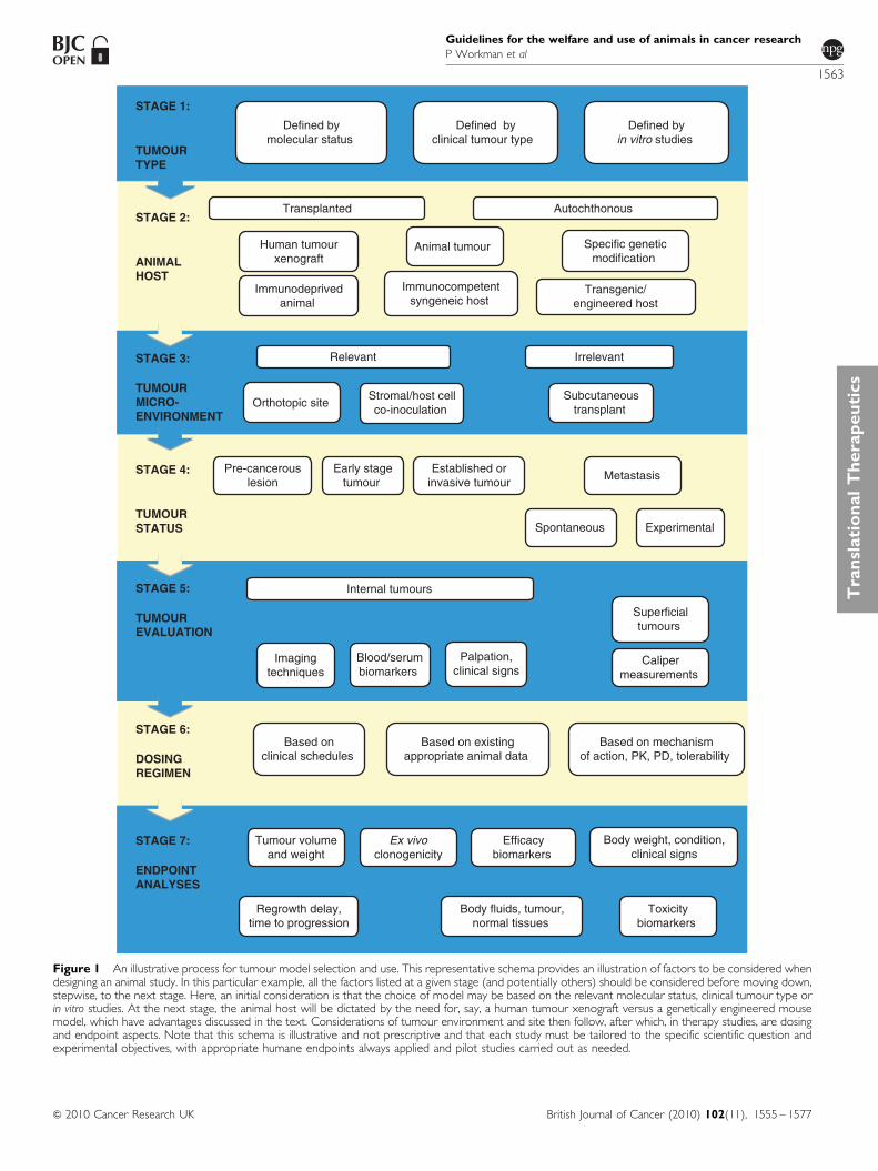
STAGE 3:
TUMOURMICRO-ENVIRONMENT
STAGE 2:
ANIMALHOST
STAGE 4:
TUMOURSTATUS
STAGE 5:
TUMOUREVALUATION
STAGE 7:
ENDPOINTANALYSES
STAGE 6:
DOSINGREGIMEN
STAGE 1:
TUMOURTYPE
Defined bymolecular status
Defined byin vitro studies
Human tumourxenograft
Immunodeprivedanimal
Immunocompetentsyngeneic host
Animal tumour Specific geneticmodification
Transgenic/engineered host
Transplanted Autochthonous
Relevant Irrelevant
Subcutaneoustransplant
Orthotopic siteStromal/host cellco-inoculation
Pre-cancerouslesion
Early stagetumour
Established orinvasive tumour
Metastasis
Spontaneous Experimental
Internal tumours
Superficialtumours
Imagingtechniques
Blood/serumbiomarkers
Palpation,clinical signs
Calipermeasurements
Based onclinical schedules
Based on existingappropriate animal data
Based on mechanismof action, PK, PD, tolerability
Tumour volumeand weight
Ex vivoclonogenicity
Regrowth delay,time to progression
Efficacybiomarkers
Body fluids, tumour,normal tissues
Body weight, condition,clinical signs
Toxicitybiomarkers
Defined byclinical tumour type
Figure 1 An illustrative process for tumour model selection and use. This representative schema provides an illustration of factors to be considered whendesigning an animal study. In this particular example, all the factors listed at a given stage (and potentially others) should be considered before moving down,stepwise, to the next stage. Here, an initial consideration is that the choice of model may be based on the relevant molecular status, clinical tumour type orin vitro studies. At the next stage, the animal host will be dictated by the need for, say, a human tumour xenograft versus a genetically engineered mousemodel, which have advantages discussed in the text. Considerations of tumour environment and site then follow, after which, in therapy studies, are dosingand endpoint aspects. Note that this schema is illustrative and not prescriptive and that each study must be tailored to the specific scientific question andexperimental objectives, with appropriate humane endpoints always applied and pilot studies carried out as needed.
Guidelines for the welfare and use of animals in cancer research
P Workman et al
1563
British Journal of Cancer (2010) 102(11), 1555 – 1577& 2010 Cancer Research UK
Tra
nsl
ati
on
al
Th
era
peu
tics

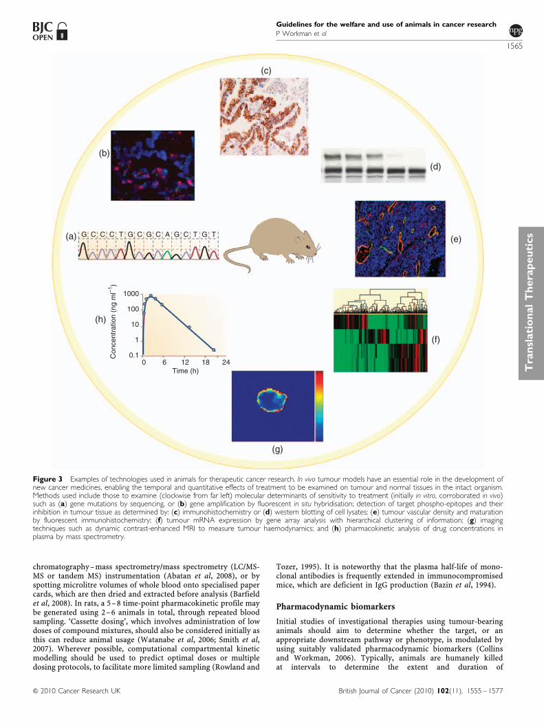
against the tumour microenvironment (e.g., angiogenesis, tumour-promoting stromal or inflammatory cells) will require appro-priate specialised assays. A range of technical platforms are usedpreclinically to define responses to therapy, the most informative ofwhich are adopted for use in patients (Figure 3). Careful assessmentof a therapy’s safety profile (outside the scope of this review) is alsorequired for regulatory submission.
Defining tolerable doses for efficacy studies
An investigational treatment should be examined at a potentialtherapeutic dose level and using a relevant dosing regimen thatcovers the longest duration anticipated. These parameters can, forexample, be estimated from consideration of mechanism of action,in vitro potency, pharmacokinetics, protein binding and pharmaco-dynamic biomarker data. Studies typically use two mice per doselevel with a doubling dose-escalation or dose-halving de-escalationdesign. For studies involving a single dosing event, an interval of24 h should be used before an alternative dose level is examined, toallow any acute adverse effects to be seen. For more chronicadministration schedules (e.g., daily for 21–28 days) this intervalshould be at least 5 days. Animals should be examined at least twicedaily (see humane endpoints below). Note that presence of a tumourmay reduce host tolerance to therapy. Studies of mice may be usedto predict dose requirements in other species through allometricscaling of pharmacokinetic parameters (Freireich et al, 1966).
Combination studies
There is a strong rationale to study combinations of agents in vivoto guide clinical studies. Relevant prior in vitro studies such asCombination Index or isobologram analyses to discriminate
additive, synergistic or antagonistic interactions should be completedto guide the selection of combinations and schedules. Compoundsare added to tumour cells in culture over a range of concentrations,alone or in combination, and the changes in sensitivity are observed.Compounds may also be added sequentially as the order ofadministration may significantly influence responses (Chou, 2006).Care needs to be taken with in vivo studies in addressing the choiceof individual drug doses and scheduling, particularly if overlappingtoxicities are likely. Pilot experiments must assess tolerability (seeabove), and pharmacokinetic data (see below) should also begenerated to determine whether interpretation of efficacy data isaffected by pharmacokinetic interactions (Siim et al, 2003).
Pharmacokinetic studies
In vitro and in silico methods are useful to predict absorption,distribution, metabolism and elimination (ADME) properties andto help prioritise compounds for evaluation in animals (Table 3;Singh, 2006). However, at present such methods are unable topredict accurately the full pharmacokinetic profile of an agent.Pharmacokinetic studies should use a validated and sufficientlysensitive detection method, ideally avoiding the need to poolseparate blood samples, thereby minimising animal usage. Typicalexperiments on mice use a single dose and 5– 8 time points (2–3mice per point) over 24– 48 h with small molecules (usuallyadministered p.o., i.v. or i.p. at doses of 0.5– 100 mg kg�1) and over1–21 days with biopharmaceuticals (administered i.v., i.p. or s.c. atdoses ranging from 10 to 1000mg per mouse).
More recently, repeat sampling of small volumes of blood from asuperficial vein in mice over a series of time points has beenestablished to reduce animal numbers. This can be employedeither for isolation of plasma and analysis by sensitive liquid
Number of compounds tested
Small-molecule compound collection
Automated high-throughput screening
Target inhibition in tumour cells
Enzyme/cellular selectivity and phenotypic assays
PK (including ‘cassette dosing’)
Tolerability
PK/PD (tumour and normal tissue surrogates)
Disease model efficacy
Safety studies
Clinical development in man
In vitro pharmacology
In vivo pharmacology
≤ 5 x 10⁶
≤ 106
≤ 103
≤ 102
≤ 102
2–4
4–8
8–40
10–50
1
X-ray crystallography,NMR, affinity assays,
pharmacophore models
Iterative medicinalchemistry
In vitro metabolism,permeability, CYP450,solubility, lipophilicity,protein binding, etc
Figure 2 Example of a drug discovery test cascade for identifying small-molecule antitumour drugs. A representative test cascade for identifying apotential small-molecule drug against a given target is shown. A subset of a compound library is initially screened vs the target in vitro, in recombinant proteinor cellular assays, using high-throughput automation to identify ‘hits’. Subsequent leads are examined in more detail by assessing their effect on downstreammolecular events in cells and their selectivity vs other proteins. A battery of additional in vitro tests is also used for measurement or prediction of physicalproperties and pharmacokinetic parameters. Only compounds with a promising balance of features are progressed to in vivo testing, usually in mice.Pharmacokinetic (PK) studies, used to understand drug exposure, may initially involve co-inoculation of low doses of compounds (‘cassette dosing’) tominimise animal usage. The tolerability of leads with favourable PK is then assessed at higher doses, before evaluating their pharmacodynamic (PD) effect ontumour and normal tissues at well-tolerated doses. Compounds that do not meet the anticipated level of performance at any stage may result in subsequentrounds of iterative medicinal chemistry to generate improved leads. Selected leads are progressed to efficacy testing to determine the link between targetinhibition and the effect on tumour growth or spread (metastasis). Safety studies on late-stage leads are also required before a candidate drug can beselected for examination in cancer patients (not covered here). The application of the test cascade means that compounds are filtered by the earlier stageassays so that a smaller number of compounds, and only those of higher quality, are taken into later stage in vivo assays in animals.
Guidelines for the welfare and use of animals in cancer research
P Workman et al
1564
British Journal of Cancer (2010) 102(11), 1555 – 1577 & 2010 Cancer Research UK
Tra
nsla
tion
al
Th
era
peu
tics
chromatography –mass spectrometry/mass spectrometry (LC/MS-MS or tandem MS) instrumentation (Abatan et al, 2008), or byspotting microlitre volumes of whole blood onto specialised papercards, which are then dried and extracted before analysis (Barfieldet al, 2008). In rats, a 5– 8 time-point pharmacokinetic profile maybe generated using 2–6 animals in total, through repeated bloodsampling. ‘Cassette dosing’, which involves administration of lowdoses of compound mixtures, should also be considered initially asthis can reduce animal usage (Watanabe et al, 2006; Smith et al,2007). Wherever possible, computational compartmental kineticmodelling should be used to predict optimal doses or multipledosing protocols, to facilitate more limited sampling (Rowland and
Tozer, 1995). It is noteworthy that the plasma half-life of mono-clonal antibodies is frequently extended in immunocompromisedmice, which are deficient in IgG production (Bazin et al, 1994).
Pharmacodynamic biomarkers
Initial studies of investigational therapies using tumour-bearinganimals should aim to determine whether the target, or anappropriate downstream pathway or phenotype, is modulated byusing suitably validated pharmacodynamic biomarkers (Collinsand Workman, 2006). Typically, animals are humanely killedat intervals to determine the extent and duration of
Con
cent
ratio
n (n
g m
l–1)
1000
G C C C T G C G C A G C T G T
100
10
1
0.1
Time (h)0 6 12 18 24
(a)
(h)
(b)
(c)
(d)
(e)
(f)
(g)
Figure 3 Examples of technologies used in animals for therapeutic cancer research. In vivo tumour models have an essential role in the development ofnew cancer medicines, enabling the temporal and quantitative effects of treatment to be examined on tumour and normal tissues in the intact organism.Methods used include those to examine (clockwise from far left) molecular determinants of sensitivity to treatment (initially in vitro, corroborated in vivo)such as (a) gene mutations by sequencing, or (b) gene amplification by fluorescent in situ hybridisation; detection of target phospho-epitopes and theirinhibition in tumour tissue as determined by: (c) immunohistochemistry or (d) western blotting of cell lysates; (e) tumour vascular density and maturationby fluorescent immunohistochemistry; (f) tumour mRNA expression by gene array analysis with hierarchical clustering of information; (g) imagingtechniques such as dynamic contrast-enhanced MRI to measure tumour haemodynamics; and (h) pharmacokinetic analysis of drug concentrations inplasma by mass spectrometry.
Guidelines for the welfare and use of animals in cancer research
P Workman et al
1565
British Journal of Cancer (2010) 102(11), 1555 – 1577& 2010 Cancer Research UK
Tra
nsl
ati
on
al
Th
era
peu
tics
pharmacodynamic changes and to investigate biomarkers intumour and normal tissues (e.g., blood or skin) that may berelevant to clinical development (Banerji et al, 2005). In vaccinestudies, responses are assessed by changes in immune status,
including evidence of tumour-infiltrating leukocytes by immuno-histochemistry, and specific cellular or humoral immunity(Gajewski, 2000). It should be possible to use much smaller groupsizes of 3– 5 in pharmacodynamic studies in comparison to thosein efficacy studies (see below). Simultaneous measurement of drugconcentrations and mechanistic biomarkers is recommended toreduce animal numbers and establish a pharmacokinetic –pharmacodynamic relationship. Judicious application of suchstudies in a drug discovery test cascade should be used toprioritise agents before entry into efficacy studies.
Efficacy determinations
All relevant information should be used to guide the designof tumour efficacy studies. Such studies generally involveexamination of treatment effects over a 2- to 4-week period andestablish how the therapeutic response relates to pharmacokineticand pharmacodynamic parameters. Typically, with treatmentsdelivered by an appropriate route of administration (Table 4), res-ponse is determined in 6– 10 animals per study group (vs a controlgroup) either by direct twice-weekly calliper measurement ofsuperficial tumours (Kelland, 2004), counting lung or livermetastases ex vivo, or using imaging methodologies (Edingeret al, 2002; Hoffman and Yang, 2005; Brindle, 2008; McCann et al,2009; Yang et al, 2009). Alternatively, post-treatment excision oftumours for in vitro determination of clonogenic survival, ordetermination of the dose required to inhibit tumour growth by50% (tumour control dose-TCD50) may be appropriate (seeRadiation therapy section below). Methods are available todetermine sample sizes for single- and combination-agent studiesand to allow for incomplete data sets (Tan et al, 2005). For certaintargets, alternative, surrogate in vivo efficacy models in non-tumour-bearing animals may be used, such as assessment of anti-oestrogenic activity by determining the effect on hypothalamicfunction (Kato et al, 1968).
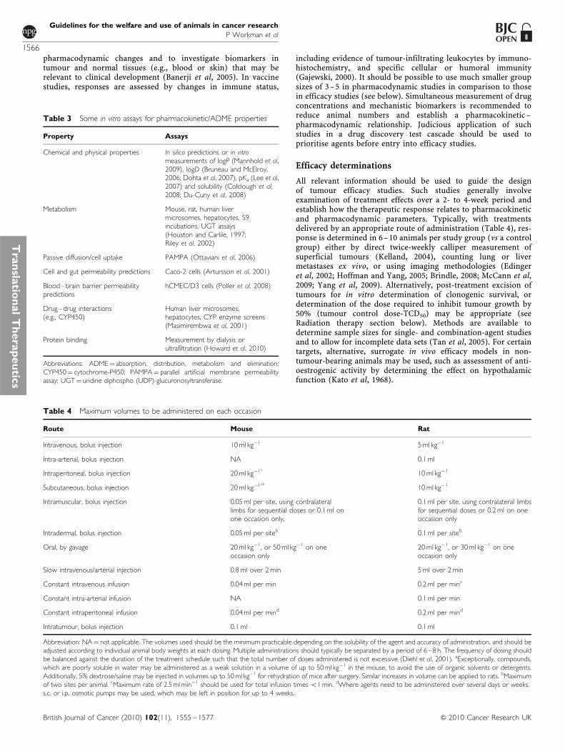
Table 4 Maximum volumes to be administered on each occasion
Route Mouse Rat
Intravenous, bolus injection 10 ml kg�1 5 ml kg�1
Intra-arterial, bolus injection NA 0.1 ml
Intraperitoneal, bolus injection 20 ml kg�1a
10 ml kg�1
Subcutaneous, bolus injection 20 ml kg�1a,b
10 ml kg�1
Intramuscular, bolus injection 0.05 ml per site, using contralaterallimbs for sequential doses or 0.1 ml onone occasion only,
0.1 ml per site, using contralateral limbsfor sequential doses or 0.2 ml on oneoccasion only
Intradermal, bolus injection 0.05 ml per siteb 0.1 ml per siteb
Oral, by gavage 20 ml kg�1, or 50 ml kg�1 on oneoccasion only
20 ml kg�1, or 30 ml kg�1 on oneoccasion only
Slow intravenous/arterial injection 0.8 ml over 2 min 5 ml over 2 min
Constant intravenous infusion 0.04 ml per min 0.2 ml per minc
Constant intra-arterial infusion NA 0.1 ml per min
Constant intraperitoneal infusion 0.04 ml per mind 0.2 ml per mind
Intratumour, bolus injection 0.1 ml 0.1 ml
Abbreviation: NA¼ not applicable. The volumes used should be the minimum practicable depending on the solubility of the agent and accuracy of administration, and should beadjusted according to individual animal body weights at each dosing. Multiple administrations should typically be separated by a period of 6–8 h. The frequency of dosing shouldbe balanced against the duration of the treatment schedule such that the total number of doses administered is not excessive (Diehl et al, 2001). aExceptionally, compounds,which are poorly soluble in water may be administered as a weak solution in a volume of up to 50 ml kg�1 in the mouse, to avoid the use of organic solvents or detergents.Additionally, 5% dextrose/saline may be injected in volumes up to 50 ml kg�1 for rehydration of mice after surgery. Similar increases in volume can be applied to rats. bMaximumof two sites per animal. cMaximum rate of 2.5 ml min�1 should be used for total infusion times o1 min. dWhere agents need to be administered over several days or weeks,s.c. or i.p. osmotic pumps may be used, which may be left in position for up to 4 weeks.
Table 3 Some in vitro assays for pharmacokinetic/ADME properties
Property Assays
Chemical and physical properties In silico predictions or in vitromeasurements of logP (Mannhold et al,2009), logD (Bruneau and McElroy,2006; Dohta et al, 2007), pKa (Lee et al,2007) and solubility (Colclough et al,2008; Du-Cuny et al, 2008)
Metabolism Mouse, rat, human livermicrosomes, hepatocytes, S9incubations, UGT assays(Houston and Carlile, 1997;Riley et al, 2002)
Passive diffusion/cell uptake PAMPA (Ottaviani et al, 2006)
Cell and gut permeability predictions Caco-2 cells (Artursson et al, 2001)
Blood–brain barrier permeabilitypredictions
hCMEC/D3 cells (Poller et al, 2008)
Drug–drug interactions(e.g., CYP450)
Human liver microsomes,hepatocytes, CYP enzyme screens(Masimirembwa et al, 2001)
Protein binding Measurement by dialysis orultrafiltration (Howard et al, 2010)
Abbreviations: ADME¼ absorption, distribution, metabolism and elimination;CYP450¼ cytochrome-P450; PAMPA¼ parallel artificial membrane permeabilityassay; UGT¼ uridine diphospho (UDP)-glucuronosyltransferase.
Guidelines for the welfare and use of animals in cancer research
P Workman et al
1566
British Journal of Cancer (2010) 102(11), 1555 – 1577 & 2010 Cancer Research UK
Tra
nsla
tion
al
Th
era
peu
tics
Administration of experimental agents
Various sources are available for advice on well-tolerated injectionvolumes and recommended administration schedules. It isimportant to note that, from an animal welfare point of view,frequency and duration of dosing are as important as the volumeand composition of the injected solution. Some commonly usedexamples are given in Table 4 and the following references: Diehlet al (2001); Morton et al (2001). More frequent dosing would needto be justified by pharmacokinetic or pharmacodynamic data. Asan illustration of standard procedures, for oral/i.p. or i.v. dosing inmice, volumes of 10 and 5 ml kg�1, respectively (equating to 200and 100 ml for a 20 g mouse), are widely accepted. However, thesmallest volume that can be accurately and safely administeredmust always be used.
Where possible, compounds should be administered in anaqueous solution (sterile water for injections, 0.9% saline or 5%dextrose/saline) that is as close to physiological pH as possible, ashighly acidic or basic solutions can be an irritant. If organicsolvents (like dimethylsulphoxide, DMSO) are necessary, theseshould not exceed 5 ml kg�1 or 10% of the injected volume.Detergents (such as Tween), solubilisers or emulsifiers should notexceed 20% of the injected volume. Cyclodextrins should notexceed 2 ml kg�1 or 45% of the injected volume, and where used at420% of the injected volume, animals need to be rehydratedwithin 2–4 h.
Experimental design including statistics
To maximise the scientific integrity of data generated while at thesame time using the minimum number of animals, statistical
expertise should be applied to all experimental design and analyses(Festing, 2002; Festing and Altman, 2002; Festing et al, 2002; seeBoxes 3 and 4).
Chemoprevention
These studies routinely use either carcinogen-induced rat tumours(e.g., azoxymethane-induced colorectal cancer) or mouse geneticmodels of carcinogenesis (e.g., ApcMin colorectal; Corpet andPierre, 2003; Cai et al, 2009). Generally, animals receive theputative chemopreventive agent in the diet or drinking water overan extended period at innocuous doses. Tumour development ismeasured at the end of the study and compared with animals ona relevant control diet. Relatively large numbers of rodents(e.g.; X14 per group; Cai et al, 2009) may be required forthe observed differences between the intervention and controlgroups to be robust. Mechanistic and pharmacodynamic end-points should also be included (Yang et al, 2001; Corpet andPierre, 2003).
Radiation therapy
External beam radiotherapy is primarily used for local tumourirradiation, which requires lead shielding to minimise normaltissue exposure. Typically, s.c. tumours are used and combinationtreatment with a novel therapy is tested. Endpoints includelocal control, growth delay and in vivo–in vitro clonogenic survival(TCD50). Time to re-growth is preferred to a single time pointanalysis. Local tissue toxicity is usually manifest as skin erythemabut should be minimised by restricting localised doses to less than30 Gy (single dose). Exploration of better tolerated, clinicallyrelevant fractionated doses (e.g., 2–5 Gy per fraction over 1–2weeks) is encouraged. Should moist desquamation occur, thisshould not be allowed to persist for more than 24 h. Irradiated s.c.tumours can show ulceration, which may reflect tumour response.However, if there is evidence of infection and/or no signs of tissuerepair the animal should be humanely killed. The acute and lateeffects of radiation treatment may also be examined in a relevantorgan, particularly when studying new combination paradigms.A common endpoint has been the development of fibrosis inlung tissue, although more recently measurement of breathing ratehas been implemented to detect symptoms before they becomedistressful to the animal (Jackson et al, 2010).
Radiotherapy can also be delivered in the form of targetedradionuclides (normally attached to antibodies; e.g., Martenssonet al, 2005). Normal tissue toxicity will depend on antigen
Box 3 EXPERIMENTAL STUDY DESIGN
1. Power analysis calculations should be applied to determine sample sizes.There are many commercially available statistical packages to supportsuch calculations.A number of variables need to be specified to perform the analysis.including the effect size of biological interest (specified by theexperimenter), the standard deviation, the significance level (normally setto 5%) and the desired power of the experiment. The desired powershould be set to a minimum of 80% (i.e., at least an 80% chance ofdeclaring the defined ‘meaningful biological change’ as being statisticallysignificant)Estimates of biological variability should be used in sample size and powercalculations. These estimates are established from accrued historicaldatabases, pilot studies or published data. Biological databases must becontinually updated and monitored with a regular review of group sizes.It is helpful to plot the variance estimates for control groups from a giventype of test with time, and constant attempts made to identify anyunderlying cause of variation, which may ultimately lead to a reductionin group size
2. Multiple treated groups will often be compared against one control toreduce the number of studies performed. As the control group is involvedin every comparison, i.e., to all treated groups, it is often appropriate to setthe control group size to be higher in comparison with the treated groups
3. When optimising animal model conditions, factorial design providesa set of tools for efficiently exploring multiple parameters simultaneously.Factorial design is a more efficient approach in comparison with thecommon one-variable-at-a-time approach, and leads to a more reliableunderstanding of the effects of parameters and their interactions. This inturn can lead to a better animal model
4. The power analysis calculation described above provides fixed samplesizes for each compound. An alternative, applicable in appropriatesituations, would be to adopt a sequential design. Compounds are testedon more than one occasion and stopping rules are devised, so thatextreme compounds (either highly effective or not effective at all) aredropped early from the study. The advantage to a sequential design is thaton average fewer animals will be used per compound in comparisonwith the fixed-sample-size case
Box 4 DATA ANALYSIS
1. Sometimes data need to be transformed before data analysis.The justification for transforming data should be given. A pertinentexample is determining the percentage inhibition of tumour growthfrom comparative tumour volume data. As the variance of tumourmeasurements increases with the mean, data should ideally be analysedon a logarithmic scale, with each animal exhibiting a difference in log10(tumour volume) from initial
2. Meaningful biological change, measurable endpoints and intendedstatistical analyses should be pre-defined. For the percentage inhibitionof tumour growth example, a suitable endpoint would be a comparisonof the change in tumour volume: i.e., log10 (final volume)�log10(initial volume) between the control and the treatment groups
3. When examining changes in means in one direction only (e.g., whenidentifying inhibition rather than change) then one-sided (rather thantwo-sided) statistical tests will be used. For a comparison of two groupsa t-test is adequate, whereas experimental data with multiple groups(vs a control) should be analysed by one-way ANOVA
Abbreviation: ANOVA¼ analysis of variance
Guidelines for the welfare and use of animals in cancer research
P Workman et al
1567
British Journal of Cancer (2010) 102(11), 1555 – 1577& 2010 Cancer Research UK
Tra
nsl
ati
on
al
Th
era
peu
tics
expression on tissues relative to the tumour and the nature of theemitter. Whole-body irradiation can also be used to suppress theimmune response of an animal, for example, or to treatdisseminated disease. Selected doses should not manifest toxicityover the duration of the experiment, for example, gut toxicitywithin 5 days or haematological toxicity within 30 days.
UV radiation (UVR)
The response of mouse skin to UVR may be used, for example, tostudy the aetiology of non-melanoma skin cancer (van Kranen andde Gruijl, 1999; Hedelund et al, 2006). Generally, experiments areperformed with hairless (Skh-hr2) mice. As mouse skin does notshow signs of burning, it is important to use a biologically relevant,non-burning dose of 0.2– 0.3 MED (minimal erythema dose; 50%skin thickening¼ 0.5 MED). Skin thickness should be measured2–3 times weekly after increasing the dose of UVR until 20– 30%thickening has occurred. If hyperplasia is maintained over 12– 15weeks skin tumours may form. A protective mouse restrainershould be used as UV radiation is damaging to eyes and ears.
IMAGING
General considerations
Imaging techniques now have a principal role in translationalcancer research, enabling sequential analysis of biological end-points in the same animal, with obvious welfare benefits. The mainutility of small-animal imaging is for monitoring deep-seatedtumours and metastases with or without treatment. Applicationsinclude studies of basic biological processes and of tissuepharmacokinetics and pharmacodynamic responses to treatment(Paulmurugan et al, 2002; Galbraith et al, 2003; Pillai et al, 2008;Tennant et al, 2009; Nguyen et al, 2009b). However, animalnumbers may not be reduced if, for example, full endpoint analysisrequires surgical intervention such as cannulation of bloodvessels or when contrast agents have a long half-life. Here,sequential imaging may not be possible and alternative tech-niques involving tissue excision may provide more information(usually at higher spatial resolution) from the same number ofanimals.
There is an increasing clinical need for pharmacodynamicimaging with molecularly targeted cancer therapeutics. However,interpretation of imaging signals is often difficult and animalmodels have an important role in rigorous validation of newtechniques. This needs to be accompanied by consideration ofunique animal welfare issues. Use of external imaging techniqueson small animals is not completely non-invasive as some form ofanaesthesia or physical restraint is necessary and surgery oradministration of contrast agents may be required.
Imaging techniques
The applications, advantages and disadvantages of commonlyused imaging technologies are summarised in Table 5 andhave also been reviewed recently (Workman et al, 2006; Brindle,2008; Weissleder and Pittet, 2008). Whole-body optical imagingis relatively simple and cost-effective (Edinger et al, 2002).Tumour cells are genetically modified to constitutively orinducibly express a fluorescent protein (e.g., eGFP, dsRed) or anenzyme that activates an exogenously administered substrate toa bioluminescent molecule (usually luciferase for activation of aluciferin). The whole animal is imaged using sensitive opticaldetectors, which may or may not incorporate a tomographicfacility (Figure 4). The potential influences of genetic modificationand/or substrate administration on immunogenicity and responseto treatment, as well as animal welfare, must be considered
(Tuchin, 1993; Dennis, 2002; Condeelis and Segall, 2003; Wellset al, 2006).
Intravital microscopy uses a wide variety of optical imagingtechniques, often incorporating fluorescent or bioluminescentgenetic reporters or markers, including nano-particles (Hoffman,2005). It has particular animal welfare issues because it involvessurgery to provide optical clarity and visualisation on a micro-scope stage or using fibre-optic light guides (Weissleder and Pittet,2008). Some intravital microscopy techniques (e.g., tumoursgrowing in the intestinal mesentery) require laparotomy withdeep anaesthesia, so that imaging is only possible for a few hoursunder terminal anaesthesia. Surgical implantation of ‘window’chambers for tumour implantation enables imaging to beperformed over days to weeks (Dewhirst et al, 1987; Lehr et al,1993; Brown et al, 2001; Reyes-Aldasoro et al, 2008). Here, generalanaesthesia is only essential for the initial surgery and imagingmay be performed with restrained animals. Strict aseptictechnique and good post-operative care and analgesia are essential(Richardson and Flecknell, 2005; Flecknell, 2008).
Most physical imaging techniques require use of exogenouscontrast agents and only positron emission tomography (PET) andsingle photon emission computed tomography (SPECT) aresufficiently sensitive to allow use at true tracer levels; so possiblepharmacological effects of contrast agents need to be carefullyconsidered. The same procedures for tolerability testing shouldapply to imaging agents as for new drugs. Some magneticresonance imaging (MRI) techniques use inherent properties oftissues to provide endogenous imaging contrast. For instance,BOLD (blood-oxygen-level-dependent) MRI allows assessment oftissue oxygenation. These techniques avoid the use of pharmaco-logical agents but results may be difficult to interpret.
Contrast-enhanced CT has the highest spatial resolution of allclinically applicable imaging techniques and is amenable to rapidkinetics. However, depending on the operating parameters andscan length, this may involve considerable ionising radiation doseper scan (0.02– 0.6 Gy; typically 0.1– 0.3 Gy) (Boone et al, 2004;Carlson et al, 2007; Brindle, 2008). Doses should be minimised toavoid compromising experimental results through interaction ofionising events with the biological processes of interest, as well aswelfare issues; as a guide, total radiation dose 41 Gy can affecttumour growth and whole-body doses 46 Gy are generally lethalto small rodents. Users of fused PET–CT or SPECT– CT systemsshould note that the radiation dose from the PET or SPECT can beas large as the CT dose. In addition, iodine-based contrast agentsare nephrotoxic and, if required for repeat studies, well-tolerateddoses should be established.
Anaesthesia and restraint for imaging
Physical restraint and/or general anaesthesia are required forsmall-animal imaging. Both procedures can affect animal well-being and introduce experimental artefacts. Body temperaturemust be maintained and monitored during general anaesthesiausing thermostatically controlled heating pads, microwaveable gelsor warm air blowers. Light general anaesthesia using an inhala-tional anaesthetic such as isofluorane or a short-lived i.v. injectablesuch as propafol should be used for pharmacological restraint,wherever possible. Deleterious effects of physical restraint can beminimised by appropriate design of restrainers, provisionof black-outs and acclimatisation (Warden et al, 2000; Narcisoet al, 2003; King et al, 2005). Preferred methods will dependon the species, imaging modality and device used. Where generalanaesthesia is not appropriate, sedation with use of gentlephysical restraint is encouraged, taking account of veterinaryadvice. Acclimatisation needs to be thorough, as a short periodof training can induce more stress (Warden et al, 2000; Narcisoet al, 2003).
Guidelines for the welfare and use of animals in cancer research
P Workman et al
1568
British Journal of Cancer (2010) 102(11), 1555 – 1577 & 2010 Cancer Research UK
Tra
nsla
tion
al
Th
era
peu
tics
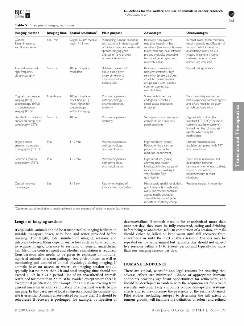
Length of imaging sessions
If applicable, animals should be transported to imaging facilities insuitable transport boxes, with food and water provided beforeimaging. The length, total number of imaging sessions andintervals between them depend on factors such as time requiredto acquire images, tolerance to restraint or general anaesthesia,half-life of the contrast agent and whether cannulation is required.Consideration also needs to be given to exposure of immune-deprived animals to a non-pathogen-free environment, as well asmonitoring and control of animal physiology during imaging. Ifanimals have no access to water, an imaging session shouldtypically last no more than 2 h and total imaging time should notexceed 2– 3 h in a 24-h period. Use of un-anaesthetised animalsrestrained for more than 2 h must be avoided except where there isexceptional justification, for example, for animals recovering fromgeneral anaesthesia after cannulation of superficial vessels beforeimaging. In this case, use of local analgesia around the cannulationsite is essential. Animals anaesthetised for more than 2 h should berehydrated if recovery is prolonged, for example, by injection of
dextrose/saline. If animals need to be anaesthetised more thanonce per day, they must be fully recovered, eating and drinkingbefore being re-anaesthetised. On completion of a session, animalsshould either be killed or kept warm until full recovery fromanaesthesia or until the next analysis session. Analysis may berepeated on the same animal but typically this should not exceedfive sessions within a 1- to 2-week period and typically no morethan one imaging session per day.
HUMANE ENDPOINTS
There are ethical, scientific and legal reasons for ensuring thatadverse effects are minimised. Choice of appropriate humaneendpoints provides significant opportunities for refinement, andshould be developed in tandem with the requirements for a validscientific outcome. Early endpoints reduce non-specific systemiceffects and so may increase the precision of the results obtained.Pilot studies, including autopsy to determine the full extent oftumour growth, will facilitate the definition of robust and refined
Table 5 Examples of imaging techniques
Imaging method Imaging time Spatial resolutiona Main purpose Advantages Disadvantages
Optical:Bioluminescenceand fluorescence
Sec–min Organ: 50 mm Wholebody: 1–5 mm
Monitoring tumour responseto treatment in deep-seated/orthotopic sites and metastaticspread; imaging geneexpression and protein–protein interactions
Relatively non-invasive(requires restraint), highsensitivity (amol–nmol); manyfluorescent and near-infraredprobes available; amenableto use of gene reporters;relatively cheap
In most cases, these methodsrequire genetic modification oftumour cells for detection;quantitation relies on 2Dimages in current imagingsystems; nude or shavedanimals are required
Three-dimensionalhigh-frequencyultrasonography
Sec–min 100 mm in-planeresolution
Relative measure oftissue blood flow;three-dimensionalmeasurement oftumour size
Relatively non-invasive(requires restraint), highsensitivity (single particle);absolute measurementsare possible with suitablecontrast agents, e.g..microbubbles
Specialized application
Magnetic resonanceimaging (MRI),spectroscopy (MRS)or spectroscopyimaging (MRSI)
Min–hours 100 mm in-planeresolution (7 T);much higher forspectroscopywithout imaging
Pharmacodynamics,pathophysiology,pharmacokinetics,anatomy
Some techniques useendogenous contrast;good spatial resolution(imaging)
Poor sensitivity (mmol), sothat exogenous contrast agentsand drugs need to be givenat high concentration
Standard or contrast-enhanced computedtomography (CT)
Sec–min 100 mm Pharmacodynamics,anatomy
Very good spatial resolutioncombined with relativelygood sensitivity
High radiation dose (forstandard CT, 2 cGy for mostcurrently available systems),limited number of contrastagents, which may benephrotoxic
Single photonemission computedtomography (SPECT)
Min 1–2 mm Pharmacodynamics,pathophysiology,pharmacokinetics
High sensitivity (pmol)Radiochemistry can beperformed in nuclearmedicine department
Limited radiochemicalsavailable compared with PET,less quantitative
Positron emissiontomography (PET)
Min 1–2 mm Pharmacodynamics,pathophysiology,pharmacokinetics
High sensitivity (pmol)allowing true tracerkinetics; unlimited range ofradiochemicals making itvery flexible; fullyquantitative
Poor spatial resolution; fullquantitation requirescannulation (for kinetic studies);requires specializedradiochemistry in mostsituations
Optical intravitalmicroscopy
Sec 1–5 mm Real-time imaging oftumour microcirculation
Microscopic spatial resolution,good sensitivity (single cell),many fluorescent contrastagents readily available,amenable to use of genereporters; relatively cheap
Requires surgical intervention
aOptimum spatial resolution is usually achieved at the expense of ability to obtain fast kinetics.
Guidelines for the welfare and use of animals in cancer research
P Workman et al
1569
British Journal of Cancer (2010) 102(11), 1555 – 1577& 2010 Cancer Research UK
Tra
nsl
ati
on
al
Th
era
peu
tics
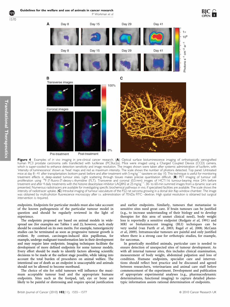
endpoints. Endpoints for particular models must also take accountof the known pathogenesis of the particular tumour model inquestion and should be regularly reviewed in the light ofexperience.
The endpoints proposed are based on animal models in wide-spread use (for examples see Tables 1 and 2); however, each studyshould be considered on its own merits. For example, tumorigenicitystudies can be terminated as soon as progressive tumour growth isevident. By contrast, carcinogen-induced skin papillomas, forexample, undergo malignant transformation late in their developmentand may require later endpoints. Imaging techniques facilitate thedevelopment of more defined endpoints for some tumour models.Every effort should be made to identify factors allowing scientificdecisions to be made at the earliest stage possible, while taking intoaccount the total burden of procedures on animal welfare. Theintentional use of death as an endpoint is unacceptable and animalsshould not be allowed to become moribund.
The choice of site for solid tumours will influence the maxi-mum acceptable tumour load and the appropriate humaneendpoints. Sites such as the footpad, tail, eye or bone arelikely to be painful or distressing and require special justification
and earlier endpoints. Similarly, tumours that metastasise tosensitive sites need great care. If brain tumours can be justified(e.g., to increase understanding of their biology and to developtherapies for this area of unmet clinical need), body weightloss is reportedly a sensitive endpoint (Redgate et al, 1991) andMRI or bioluminescent imaging (BLI) techniques can bevery useful (van Furth et al, 2003; Ragel et al, 2008; McCannet al, 2009). Intramuscular tumours are painful and only justifiedwhere there is a strong case for orthotopic studies, for example,for sarcomas.
In genetically modified animals, particular care is needed toensure detection of unexpected sites of tumour development. Aswith all internal tumour sites, this includes clinical examination,measurement of body weight, abdominal palpation and loss ofcondition. Humane endpoints, specialist care and interven-tions should reflect best practice and be discussed and agreedbetween researchers, veterinarians and animal care staff beforecommencement of the experiment. Development and publicationof appropriate experimental analyses (e.g., pharmacodynamicdeterminations, functional imaging) to capture detailed pheno-typic information assists rational determination of endpoints.
Day 8 Day 15 Day 29 Day 41
Day 8 Day 15 Day 29 Day 41
Transverse images
Coronal images
100%
Pre-treatment Post-treatment0%
1×
108
Pho
tons
s–1
cm
–2 s
r–1
Figure 4 Examples of in vivo imaging in pre-clinical cancer research. (A) Optical surface bioluminescence imaging of orthotopically xenograftedhuman PC3 prostate carcinoma cells transfected with luciferase (PC3luc2a). Mice were imaged using a Charged Coupled Device (CCD) camera,which is super-cooled to enhance detection sensitivity and image resolution. The images shown were taken after systemic administration of luciferin, with‘intensity of luminescence’ shown as ‘heat’ maps and red as maximum intensity. The scale shows the number of photons detected. Top panel: Untreatedmice at day 8–41 after transplantation; bottom panel: before and after treatment with 5 mg kg�1 taxotere on day 10. This technique is useful for monitoringtreatment effects in deep-seated tumour sites. Light scattering through tissues makes precise quantitation difficult. (B) PET imaging of tumour cellproliferation using 18F-30-fluoro-30-deoxy-L-thymidine (FLT). Transverse and coronal (0.5 mm) images of HCT116 tumour-bearing mice 24 h beforetreatment and after 4 daily treatments with the histone deacetylase inhibitor LAQ842 at 25 mg kg�1. 30- to 60-min summed images from a dynamic scan arepresented. Numerous radiotracers are available for investigating specific biochemical pathways in vivo, if specialised facilities are available. The scale shows theintensity of radiotracer uptake. (C) Intravital imaging of tumour vasculature of the P22 rat sarcoma growing in a dorsal skin flap window chamber. The imagewas obtained by multi-photon fluorescence microscopy after i.v. administration of 70 kDa FITC–dextran. High spatial resolution is obtained but surgicalintervention is required.
Guidelines for the welfare and use of animals in cancer research
P Workman et al
1570
British Journal of Cancer (2010) 102(11), 1555 – 1577 & 2010 Cancer Research UK
Tra
nsla
tion
al
Th
era
peu
tics
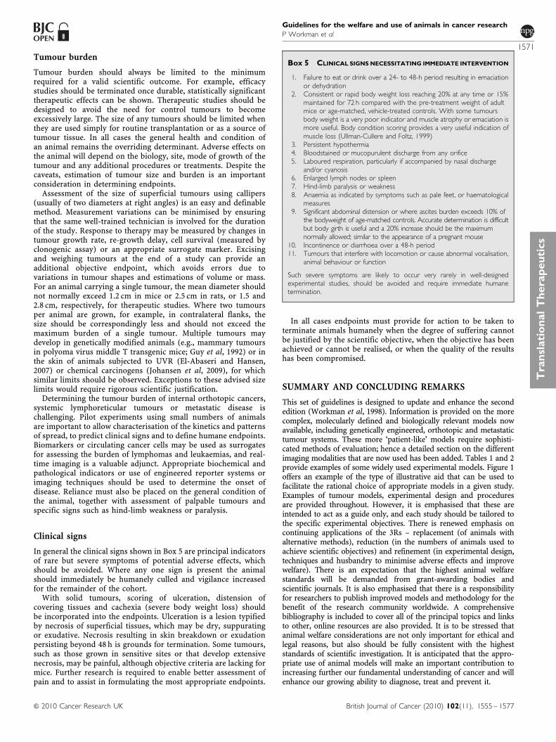
Tumour burden
Tumour burden should always be limited to the minimumrequired for a valid scientific outcome. For example, efficacystudies should be terminated once durable, statistically significanttherapeutic effects can be shown. Therapeutic studies should bedesigned to avoid the need for control tumours to becomeexcessively large. The size of any tumours should be limited whenthey are used simply for routine transplantation or as a source oftumour tissue. In all cases the general health and condition ofan animal remains the overriding determinant. Adverse effects onthe animal will depend on the biology, site, mode of growth of thetumour and any additional procedures or treatments. Despite thecaveats, estimation of tumour size and burden is an importantconsideration in determining endpoints.
Assessment of the size of superficial tumours using callipers(usually of two diameters at right angles) is an easy and definablemethod. Measurement variations can be minimised by ensuringthat the same well-trained technician is involved for the durationof the study. Response to therapy may be measured by changes intumour growth rate, re-growth delay, cell survival (measured byclonogenic assay) or an appropriate surrogate marker. Excisingand weighing tumours at the end of a study can provide anadditional objective endpoint, which avoids errors due tovariations in tumour shapes and estimations of volume or mass.For an animal carrying a single tumour, the mean diameter shouldnot normally exceed 1.2 cm in mice or 2.5 cm in rats, or 1.5 and2.8 cm, respectively, for therapeutic studies. Where two tumoursper animal are grown, for example, in contralateral flanks, thesize should be correspondingly less and should not exceed themaximum burden of a single tumour. Multiple tumours maydevelop in genetically modified animals (e.g., mammary tumoursin polyoma virus middle T transgenic mice; Guy et al, 1992) or inthe skin of animals subjected to UVR (El-Abaseri and Hansen,2007) or chemical carcinogens (Johansen et al, 2009), for whichsimilar limits should be observed. Exceptions to these advised sizelimits would require rigorous scientific justification.
Determining the tumour burden of internal orthotopic cancers,systemic lymphoreticular tumours or metastatic disease ischallenging. Pilot experiments using small numbers of animalsare important to allow characterisation of the kinetics and patternsof spread, to predict clinical signs and to define humane endpoints.Biomarkers or circulating cancer cells may be used as surrogatesfor assessing the burden of lymphomas and leukaemias, and real-time imaging is a valuable adjunct. Appropriate biochemical andpathological indicators or use of engineered reporter systems orimaging techniques should be used to determine the onset ofdisease. Reliance must also be placed on the general condition ofthe animal, together with assessment of palpable tumours andspecific signs such as hind-limb weakness or paralysis.
Clinical signs
In general the clinical signs shown in Box 5 are principal indicatorsof rare but severe symptoms of potential adverse effects, whichshould be avoided. Where any one sign is present the animalshould immediately be humanely culled and vigilance increasedfor the remainder of the cohort.
With solid tumours, scoring of ulceration, distension ofcovering tissues and cachexia (severe body weight loss) shouldbe incorporated into the endpoints. Ulceration is a lesion typifiedby necrosis of superficial tissues, which may be dry, suppuratingor exudative. Necrosis resulting in skin breakdown or exudationpersisting beyond 48 h is grounds for termination. Some tumours,such as those grown in sensitive sites or that develop extensivenecrosis, may be painful, although objective criteria are lacking formice. Further research is required to enable better assessment ofpain and to assist in formulating the most appropriate endpoints.
In all cases endpoints must provide for action to be taken toterminate animals humanely when the degree of suffering cannotbe justified by the scientific objective, when the objective has beenachieved or cannot be realised, or when the quality of the resultshas been compromised.
SUMMARY AND CONCLUDING REMARKS
This set of guidelines is designed to update and enhance the secondedition (Workman et al, 1998). Information is provided on the morecomplex, molecularly defined and biologically relevant models nowavailable, including genetically engineered, orthotopic and metastatictumour systems. These more ‘patient-like’ models require sophisti-cated methods of evaluation; hence a detailed section on the differentimaging modalities that are now used has been added. Tables 1 and 2provide examples of some widely used experimental models. Figure 1offers an example of the type of illustrative aid that can be used tofacilitate the rational choice of appropriate models in a given study.Examples of tumour models, experimental design and proceduresare provided throughout. However, it is emphasised that these areintended to act as a guide only, and each study should be tailored tothe specific experimental objectives. There is renewed emphasis oncontinuing applications of the 3Rs – replacement (of animals withalternative methods), reduction (in the numbers of animals used toachieve scientific objectives) and refinement (in experimental design,techniques and husbandry to minimise adverse effects and improvewelfare). There is an expectation that the highest animal welfarestandards will be demanded from grant-awarding bodies andscientific journals. It is also emphasised that there is a responsibilityfor researchers to publish improved models and methodology for thebenefit of the research community worldwide. A comprehensivebibliography is included to cover all of the principal topics and linksto other, online resources are also provided. It is to be stressed thatanimal welfare considerations are not only important for ethical andlegal reasons, but also should be fully consistent with the higheststandards of scientific investigation. It is anticipated that the appro-priate use of animal models will make an important contribution toincreasing further our fundamental understanding of cancer and willenhance our growing ability to diagnose, treat and prevent it.
Box 5 CLINICAL SIGNS NECESSITATING IMMEDIATE INTERVENTION
1. Failure to eat or drink over a 24- to 48-h period resulting in emaciationor dehydration
2. Consistent or rapid body weight loss reaching 20% at any time or 15%maintained for 72 h compared with the pre-treatment weight of adultmice or age-matched, vehicle-treated controls. With some tumoursbody weight is a very poor indicator and muscle atrophy or emaciation ismore useful. Body condition scoring provides a very useful indication ofmuscle loss (Ullman-Cullere and Foltz, 1999)
3. Persistent hypothermia4. Bloodstained or mucopurulent discharge from any orifice5. Laboured respiration, particularly if accompanied by nasal discharge
and/or cyanosis6. Enlarged lymph nodes or spleen7. Hind-limb paralysis or weakness8. Anaemia as indicated by symptoms such as pale feet, or haematological
measures9. Significant abdominal distension or where ascites burden exceeds 10% of
the bodyweight of age-matched controls. Accurate determination is difficultbut body girth is useful and a 20% increase should be the maximumnormally allowed; similar to the appearance of a pregnant mouse
10. Incontinence or diarrhoea over a 48-h period11. Tumours that interfere with locomotion or cause abnormal vocalisation,
animal behaviour or function
Such severe symptoms are likely to occur very rarely in well-designedexperimental studies, should be avoided and require immediate humanetermination.
Guidelines for the welfare and use of animals in cancer research
P Workman et al
1571
British Journal of Cancer (2010) 102(11), 1555 – 1577& 2010 Cancer Research UK
Tra
nsl
ati
on
al
Th
era
peu
tics

ACKNOWLEDGEMENTS
We thank authors Professor Fran Balkwill, Dr David Farningham,Professor Gill Tozer, Professor Sue Watson and Dr Steve Wedgefor leading and coordinating subgroups of the committee dealingwith specific topics.
We thank Dr Robert J Shaw (Discovery Statistics, AstraZeneca,Alderley Park) for statistical input. We are also grateful to Professor AndyGescher (University of Leicester, UK) for expert advice and a number ofother individuals who kindly gave advice on specific issues or commentedon previous drafts of the guidelines. We thank Dr Ben Onwuegbusi(Cancer Research UK) for help in acting as Committee Secretary.
REFERENCES
Abatan OI, Welch KB, Nemzek JA (2008) Evaluation of saphenousvenipuncture and modified tail-clip blood collection in mice. J AmAssoc Lab Anim Sci 47: 8 – 15
Aguirre AJ, Bardeesy N, Sinha M, Lopez L, Tuveson DA, Horner J, RedstonMS, DePinho RA (2003) Activated Kras and Ink4a/Arf deficiencycooperate to produce metastatic pancreatic ductal adenocarcinoma.Genes Dev 17: 3112 – 3126
Ahsan H, Aziz MH, Ahmad N (2005) Ultraviolet B exposure activates Stat3signaling via phosphorylation at tyrosine705 in skin of SKH1 hairlessmouse: a target for the management of skin cancer? Biochem Biophys ResCommun 333: 241 – 246
Alsheikhly AR, Zweiri J, Walmesley AJ, Watson AJ, Christmas SE (2004)Both soluble and membrane-bound forms of Flt3 ligand enhance tumorimmunity following ‘suicide’ gene therapy in a murine colon carcinomamodel. Cancer Immunol Immunother 53: 946 – 954
Artandi SE, Chang S, Lee SL, Alson S, Gottlieb GJ, Chin L, DePinho RA(2000) Telomere dysfunction promotes non-reciprocal translocationsand epithelial cancers in mice. Nature 406: 641 – 645
Artursson P, Palm K, Luthman K (2001) Caco-2 monolayers inexperimental and theoretical predictions of drug transport. Adv DrugDeliv Rev 46: 27 – 43
Baersch G, Mollers T, Hotte A, Dockhorn-Dworniczak B, Rube C, Ritter J,Jurgens H, Vormoor J (1997) Good engraftment of B-cell precursor ALLin NOD-SCID mice. Klin Padiatr 209: 178 – 185
Balansky RM, Ganchev G, D’Agostini F, De Flora S (2002) Effects ofN-acetylcysteine in an esophageal carcinogenesis model in rats treatedwith diethylnitrosamine and diethyldithiocarbamate. Int J Cancer 98:493 – 497
Banerji U, Walton M, Raynaud F, Grimshaw R, Kelland L, Valenti M,Judson I, Workman P (2005) Pharmacokinetic – pharmacodynamicrelationships for the heat shock protein 90 molecular chaperoneinhibitor 17-allylamino, 17-demethoxygeldanamycin in human ovariancancer xenograft models. Clin Cancer Res 11: 7023 – 7032
Barfield M, Spooner N, Lad R, Parry S, Fowles S (2008) Application of driedblood spots combined with HPLC – MS/MS for the quantification ofacetaminophen in toxicokinetic studies. J Chromatogr B Analyt TechnolBiomed Life Sci 870: 32 – 37
Bazin R, Boucher G, Monier G, Chevrier MC, Verrette S, Broly H, LemieuxR (1994) Use of hu-IgG-SCID mice to evaluate the in vivo stability ofhuman monoclonal IgG antibodies. J Immunol Methods 172: 209 – 217
Biotechnology and Biological Sciences Research Council; Department forEnvironment, Food and Rural Affairs; Medical Research Council;National Centre for the Replacement, Refinement and Reduction ofAnimals in Research; Natural Environment Research Council;Wellcome Trust (2008) Responsibility in the use of animals in bioscienceresearch: expectations of the major research council and charitablefunding bodies. National Centre for the Replacement, Refinement andReduction of Animals in Research, London. http://www.nc3rs.org.uk/downloaddoc.asp?id=719 (Accessed date 30 March 2010)
Becher OJ, Holland EC, Sausville EA, Burger AM (2006) Geneticallyengineered models have advantages over xenografts for preclinicalstudies. Cancer Res 66: 3355 – 3359
Becker JC, Pancook JD, Gillies SD, Mendelsohn J, Reisfeld RA (1996)Eradication of human hepatic and pulmonary melanoma metastases inSCID mice by antibody-interleukin 2 fusion proteins. Proc Natl Acad SciUSA 93: 2702 – 2707
Bergers G, Javaherian K, Lo KM, Folkman J, Hanahan D (1999) Effects ofangiogenesis inhibitors on multistage carcinogenesis in mice. Science284: 808 – 812
Bignell GR, Greenman CD, Davies H, Butler AP, Edkins S, Andrews JM,Buck G, Chen L, Beare D, Latimer C, Widaa S, Hinton J, Fahey C, Fu B,Swamy S, Dalgliesh GL, Teh BT, Deloukas P, Yang F, Campbell PJ,
Futreal PA, Stratton MR (2010) Signatures of mutation and selection inthe cancer genome. Nature 463: 893 – 898
Blouin S, Basle MF, Chappard D (2008) Interactions between micro-environment and cancer cells in two animal models of bone metastasis.Br J Cancer 98: 809 – 815
Boone JM, Velazquez O, Cherry SR (2004) Small-animal X-ray dose frommicro-CT. Mol Imaging 3: 149 – 158
Brindle K (2008) New approaches for imaging tumour responses totreatment. Nat Rev Cancer 8: 94 – 107
Brown EB, Campbell RB, Tsuzuki Y, Xu L, Carmeliet P, Fukumura D,Jain RK (2001) In vivo measurement of gene expression, angiogenesisand physiological function in tumors using multiphoton laser scanningmicroscopy. Nat Med 7: 864 – 868
Bruneau P, McElroy NR (2006) logD7.4 modeling using BayesianRegularized Neural Networks. Assessment and correction of the errorsof prediction. J Chem Inf Model 46: 1379 – 1387
Bruns CJ, Harbison MT, Kuniyasu H, Eue I, Fidler IJ (1999) In vivoselection and characterization of metastatic variants from humanpancreatic adenocarcinoma by using orthotopic implantation in nudemice. Neoplasia 1: 50 – 62
Cai H, Sale S, Schmid R, Britton RG, Brown K, Steward WP, Gescher AJ(2009) Flavones as colorectal cancer chemopreventive agents – phenol-o-methylation enhances efficacy. Cancer Prev Res (Phila PA) 2: 743 – 750
Carlson SK, Classic KL, Bender CE, Russell SJ (2007) Small animal absorbedradiation dose from serial micro-computed tomography imaging. MolImaging Biol 9: 78 – 82
Chan R, Muller WJ, Siegel PM (1999) Oncogenic activating mutations in theneu/erbB-2 oncogene are involved in the induction of mammary tumors.Ann N Y Acad Sci 889: 45 – 51
Chen D, Livne-bar I, Vanderluit JL, Slack RS, Agochiya M, Bremner R(2004) Cell-specific effects of RB or RB/p107 loss on retinal developmentimplicate an intrinsically death-resistant cell-of-origin in retinoblastoma.Cancer Cell 5: 539 – 551
Chen X, Yang G, Ding WY, Bondoc F, Curtis SK, Yang CS (1999) Anesophagogastroduodenal anastomosis model for esophageal adenocarcinogen-esis in rats and enhancement by iron overload. Carcinogenesis 20: 1801 – 1808
Chou TC (2006) Theoretical basis, experimental design, and computerizedsimulation of synergism and antagonism in drug combination studies.Pharmacol Rev 58: 621 – 681
Christophorou MA, Martin-Zanca D, Soucek L, Lawlor ER, Brown-SwigartL, Verschuren EW, Evan GI (2005) Temporal dissection of p53 functionin vitro and in vivo. Nat Genet 37: 718 – 726
Christophorou MA, Ringshausen I, Finch AJ, Swigart LB, Evan GI (2006)The pathological response to DNA damage does not contribute top53-mediated tumour suppression. Nature 443: 214 – 217
Colclough N, Hunter A, Kenny PW, Kittlety RS, Lobedan L, Tam KY,Timms MA (2008) High throughput solubility determination withapplication to selection of compounds for fragment screening. BioorgMed Chem 16: 6611 – 6616
Collado B, Carmena MJ, Clemente C, Prieto JC, Bajo AM (2007) Vasoactiveintestinal peptide enhances growth and angiogenesis of human experi-mental prostate cancer in a xenograft model. Peptides 28: 1896 – 1901
Collins I, Workman P (2006) New approaches to molecular cancertherapeutics. Nat Chem Biol 2: 689 – 700
Comstock KE, Hall CL, Daignault S, Mandlebaum SA, Yu C, Keller ET(2009) A bioluminescent orthotopic mouse model of human osteosarco-ma that allows sensitive and rapid evaluation of new therapeutic agentsin vivo. In Vivo 23: 661 – 668
Condeelis J, Segall JE (2003) Intravital imaging of cell movement intumours. Nat Rev Cancer 3: 921 – 930
Corpet DE, Pierre F (2003) Point: from animal models to preventionof colon cancer. Systematic review of chemoprevention in min mice
Guidelines for the welfare and use of animals in cancer research
P Workman et al
1572
British Journal of Cancer (2010) 102(11), 1555 – 1577 & 2010 Cancer Research UK
Tra
nsla
tion
al
Th
era
peu
tics
and choice of the model system. Cancer Epidemiol Biomarkers Prev 12:391 – 400
De Fabo EC (2006) Initial studies on an in vivo action spectrum formelanoma induction. Prog Biophys Mol Biol 92: 97 – 104
Decker S, Hollingshead M, Bonomi CA, Carter JP, Sausville EA (2004)The hollow fibre model in cancer drug screening: the NCI experience.Eur J Cancer 40: 821 – 826
Dennis C (2006) Cancer: off by a whisker. Nature 17: 739 – 741Dennis Jr MB (2002) Welfare issues of genetically modified animals. Ilar J
43: 100 – 109Dewhirst MW, Gustafson C, Gross JF, Tso CY (1987) Temporal effects of
5.0 Gy radiation in healing subcutaneous microvasculature of a dorsalflap window chamber. Radiat Res 112: 581 – 591
Dickson PV, Hamner B, Ng CY, Hall MM, Zhou J, Hargrove PW, McCarvilleMB, Davidoff AM (2007) In vivo bioluminescence imaging for earlydetection and monitoring of disease progression in a murine model ofneuroblastoma. J Pediatr Surg 42: 1172 – 1179
Diehl KH, Hull R, Morton D, Pfister R, Rabemampianina Y, Smith D,Vidal JM, van de Vorstenbosch C (2001) A good practice guide to theadministration of substances and removal of blood, including routes andvolumes. J Appl Toxicol 21: 15 – 23
Dohta Y, Yamashita T, Horiike S, Nakamura T, Fukami T (2007) A systemfor LogD screening of 96-well plates using a water-plug aspiration/injection method combined with high-performance liquid chromato-graphy-mass spectrometry. Anal Chem 79: 8312 – 8315
Dong X, Guan J, English JC, Flint J, Yee J, Evans K, Murray N, Macaulay C,Ng RT, Gout PW, Lam WL, Laskin J, Ling V, Lam S, Wang Y (2010)Patient-derived first generation xenografts of non-small cell lungcancers: promising tools for predicting drug responses for personalizedchemotherapy. Clin Cancer Res 16: 1442 – 1451
Du-Cuny L, Huwyler J, Wiese M, Kansy M (2008) Computational aqueoussolubility prediction for drug-like compounds in congeneric series.Eur J Med Chem 43: 501 – 512
Edinger M, Cao YA, Hornig YS, Jenkins DE, Verneris MR, Bachmann MH,Negrin RS, Contag CH (2002) Advancing animal models of neoplasiathrough in vivo bioluminescence imaging. Eur J Cancer 38: 2128 – 2136
El-Abaseri TB, Hansen LA (2007) EGFR activation and ultraviolet light-induced skin carcinogenesis. J Biomed Biotechnol 2007: 97939
Felsher DW, Bishop JM (1999) Reversible tumorigenesis by MYC inhematopoietic lineages. Mol Cell 4: 199 – 207
Festing M, Overend P, Gaine Das R, Cortina Borja M, Berdoy M (2002) Thedesign of Animal Experiments: Reducing the Use of Animals in ResearchThrough Better Experimental Design. Royal Society of Medicine Press:London
Festing MF (2002) The design and statistical analysis of animalexperiments. ILAR J 43: 191 – 193
Festing MF, Altman DG (2002) Guidelines for the design and statisticalanalysis of experiments using laboratory animals. ILAR J 43: 244 – 258
Flecknell P (2008) Analgesia from a veterinary perspective. Br J Anaesth101: 121 – 124
Fluck MM, Haslam SZ (1996) Mammary tumors induced by polyomavirus.Breast Cancer Res Treat 39: 45 – 56
Freireich EJ, Gehan EA, Rall DP, Schmidt LH, Skipper HE (1966)Quantitative comparison of toxicity of anticancer agents in mouse, rat,hamster, dog, monkey, and man. Cancer Chemother Rep 50: 219 – 244
Frese KK, Tuveson DA (2007) Maximizing mouse cancer models. Nat RevCancer 7: 645 – 658
Gajewski TF (2000) Monitoring specific T-cell responses to melanomavaccines: ELISPOT, tetramers, and beyond. Clin Diagn Lab Immunol 7:141 – 144
Galbraith SM, Maxwell RJ, Lodge MA, Tozer GM, Wilson J, Taylor NJ, Stirling JJ,Sena L, Padhani AR, Rustin GJ (2003) Combretastatin A4 phosphate hastumor antivascular activity in rat and man as demonstrated by dynamicmagnetic resonance imaging. J Clin Oncol 21: 2831 – 2842
Garber K (2006) Realistic rodents? Debate grows over new mouse models ofcancer. J Natl Cancer Inst 98: 1176 – 1178
Giovannini M, Robanus-Maandag E, van der Valk M, Niwa-Kawakita M,Abramowski V, Goutebroze L, Woodruff JM, Berns A, Thomas G (2000)Conditional biallelic Nf2 mutation in the mouse promotes manifestationsof human neurofibromatosis type 2. Genes Dev 14: 1617 – 1630
Giver CR, Li JM, Hossain MS, Lonial S, Waller EK (2004) Reconstructingimmunity after allogeneic transplantation. Immunol Res 29: 269 – 282
Glass B, Uharek L, Zeis M, Loeffler H, Mueller-Ruchholtz W, Gassmann W(1996) Graft-versus-leukaemia activity can be predicted by naturalcytotoxicity against leukaemia cells. Br J Haematol 93: 412 – 420
Glinskii AB, Smith BA, Jiang P, Li X-M, Yang M, Hoffman RM, Glinsky GV(2003) Viable circulating metastatic cells produced in orthotopic but notectopic prostate cancer models. Cancer Res 63: 4239 – 4243
Golay J, Cittera E, Di Gaetano N, Manganini M, Mosca M, Nebuloni M,van Rooijen N, Vago L, Introna M (2006) The role of complement in thetherapeutic activity of rituximab in a murine B lymphoma model homingin lymph nodes. Haematologica 91: 176 – 183
Graf MR, Sauer JT, Merchant RE (2005) Tumor infiltration by myeloidsuppressor cells in response to T cell activation in rat gliomas.J Neurooncol 73: 29 – 36
Graff BA, Benjaminsen IC, Melas EA, Brurberg KG, Rofstad EK (2005)Changes in intratumor heterogeneity in blood perfusion in intradermalhuman melanoma xenografts during tumor growth assessed byDCE-MRI. Magn Reson Imaging 23: 961 – 966
Guy CT, Cardiff RD, Muller WJ (1992) Induction of mammary tumors byexpression of polyomavirus middle T oncogene: a transgenic mousemodel for metastatic disease. Mol Cell Biol 12: 954 – 961
Ha WS, Kim CK, Song SH, Kang CB (2001) Study on mechanism ofmultistep hepatotumorigenesis in rat: development of hepatotumori-genesis. J Vet Sci 2: 53 – 58
Han Y, Chen XP, Huang ZY, Zhu H (2005) Nude mice multi-drug resistancemodel of orthotopic transplantation of liver neoplasm and Tc-99m MIBISPECT on p-glycoprotein. World J Gastroenterol 11: 3335 – 3338
Hanahan D, Weinberg RA (2000) The hallmarks of cancer. Cell 100: 57 – 70Harlin H, Gajewski TF (2008) Diagnosis and treatment of mycoplasma-
contaminated cell cultures. Curr Protoc Cytom Appendix 3: Appendix 3CHarris JC, Gilliam AD, McKenzie AJ, Evans SA, Grabowska AM, Clarke PA,
McWilliams DF, Watson SA (2004) The biological and therapeuticimportance of gastrin gene expression in pancreatic adenocarcinomas.Cancer Res 64: 5624 – 5631
Hawariah A, Stanslas J (1998) Antagonistic effects of styrylpyronederivative (SPD) on 7,12-dimethylbenzanthracene-induced rat mammarytumors. In Vivo 12: 403 – 410
Hedelund L, Lerche C, Wulf HC, Haedersdal M (2006) Carcinogenesisrelated to intense pulsed light and UV exposure: an experimental animalstudy. Lasers Med Sci 21: 198 – 201
Hingorani SR, Wang L, Multani AS, Combs C, Deramaudt TB, Hruban RH,Rustgi AK, Chang S, Tuveson DA (2005) Trp53R172H and KrasG12Dcooperate to promote chromosomal instability and widely metastaticpancreatic ductal adenocarcinoma in mice. Cancer Cell 7: 469 – 483
Hirayama T, Honda A, Matsuzaki Y, Miyazaki T, Ikegami T, Doy M, Xu G,Lea M, Salen G (2006) Hypercholesterolemia in rats with hepatomas:increased oxysterols accelerate efflux but do not inhibit biosynthesis ofcholesterol. Hepatology 44: 602 – 611
Hirose Y, Hata K, Kuno T, Yoshida K, Sakata K, Yamada Y, Tanaka T,Reddy BS, Mori H (2004) Enhancement of development of azoxy-methane-induced colonic premalignant lesions in C57BL/KsJ-db/dbmice. Carcinogenesis 25: 821 – 825
Hoffman RM (2005) The multiple uses of fluorescent proteins to visualizecancer in vivo. Nat Rev Cancer 5: 796 – 806
Hoffman RM, Yang M (2005) Dual-color, whole-body imaging in mice.Nat Biotechnol 23: 790; author reply 791
Houston JB, Carlile DJ (1997) Prediction of hepatic clearance frommicrosomes, hepatocytes, and liver slices. Drug Metab Rev 29: 891 – 922
Howard ML, Hill JJ, Galluppi GR, McLean MA (2010) Plasma proteinbinding in drug discovery and development. Comb Chem HighThroughput Screen 13: 170 – 187
Huxham LA, Kyle AH, Baker JH, Nykilchuk LK, Minchinton AI (2004)Microregional effects of gemcitabine in HCT-116 xenografts. Cancer Res64: 6537 – 6541
Ihle NT, Lemos Jr R, Wipf P, Yacoub A, Mitchell C, Siwak D, Mills GB,Dent P, Kirkpatrick DL, Powis G (2009) Mutations in the phosphatidyl-inositol-3-kinase pathway predict for antitumor activity of the inhibitorPX-866 whereas oncogenic Ras is a dominant predictor for resistance.Cancer Res 69: 143 – 150
Ishikawa Y, Kozakai T, Morita H, Saida K, Oka S, Masuo Y (2006) Rapid detectionof mycoplasma contamination in cell cultures using SYBR Green-based real-time polymerase chain reaction. In Vitro Cell Dev Biol Anim 42: 63 – 69
Jackson IL, Vujaskovic Z, Down JD (2010) Revisiting strain-relateddifferences in radiation sensitivity of the mouse lung: recognizing andavoiding the confounding effects of pleural effusions. Radiat Res 173:10 – 20
Johansen C, Vestergaard C, Kragballe K, Kollias G, Gaestel M, Iversen L(2009) MK2 regulates the early stages of skin tumor promotion.Carcinogenesis 30: 2100 – 2108
Guidelines for the welfare and use of animals in cancer research
P Workman et al
1573
British Journal of Cancer (2010) 102(11), 1555 – 1577& 2010 Cancer Research UK
Tra
nsl
ati
on
al
Th
era
peu
tics
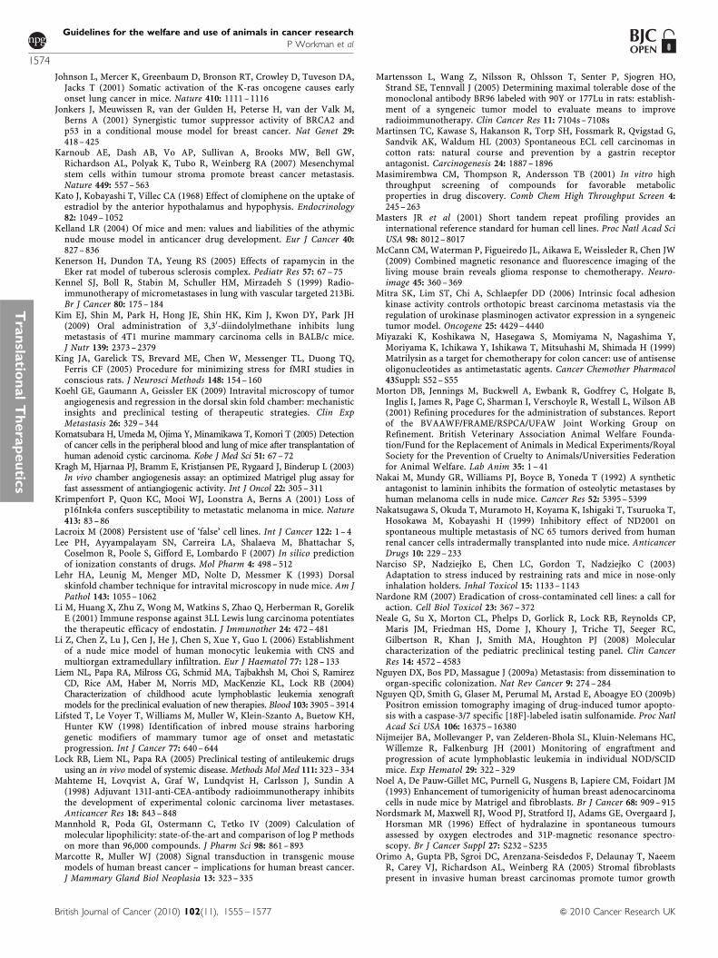
Johnson L, Mercer K, Greenbaum D, Bronson RT, Crowley D, Tuveson DA,Jacks T (2001) Somatic activation of the K-ras oncogene causes earlyonset lung cancer in mice. Nature 410: 1111 – 1116
Jonkers J, Meuwissen R, van der Gulden H, Peterse H, van der Valk M,Berns A (2001) Synergistic tumor suppressor activity of BRCA2 andp53 in a conditional mouse model for breast cancer. Nat Genet 29:418 – 425
Karnoub AE, Dash AB, Vo AP, Sullivan A, Brooks MW, Bell GW,Richardson AL, Polyak K, Tubo R, Weinberg RA (2007) Mesenchymalstem cells within tumour stroma promote breast cancer metastasis.Nature 449: 557 – 563
Kato J, Kobayashi T, Villec CA (1968) Effect of clomiphene on the uptake ofestradiol by the anterior hypothalamus and hypophysis. Endocrinology82: 1049 – 1052
Kelland LR (2004) Of mice and men: values and liabilities of the athymicnude mouse model in anticancer drug development. Eur J Cancer 40:827 – 836
Kenerson H, Dundon TA, Yeung RS (2005) Effects of rapamycin in theEker rat model of tuberous sclerosis complex. Pediatr Res 57: 67 – 75
Kennel SJ, Boll R, Stabin M, Schuller HM, Mirzadeh S (1999) Radio-immunotherapy of micrometastases in lung with vascular targeted 213Bi.Br J Cancer 80: 175 – 184
Kim EJ, Shin M, Park H, Hong JE, Shin HK, Kim J, Kwon DY, Park JH(2009) Oral administration of 3,30-diindolylmethane inhibits lungmetastasis of 4T1 murine mammary carcinoma cells in BALB/c mice.J Nutr 139: 2373 – 2379
King JA, Garelick TS, Brevard ME, Chen W, Messenger TL, Duong TQ,Ferris CF (2005) Procedure for minimizing stress for fMRI studies inconscious rats. J Neurosci Methods 148: 154 – 160
Koehl GE, Gaumann A, Geissler EK (2009) Intravital microscopy of tumorangiogenesis and regression in the dorsal skin fold chamber: mechanisticinsights and preclinical testing of therapeutic strategies. Clin ExpMetastasis 26: 329 – 344
Komatsubara H, Umeda M, Ojima Y, Minamikawa T, Komori T (2005) Detectionof cancer cells in the peripheral blood and lung of mice after transplantation ofhuman adenoid cystic carcinoma. Kobe J Med Sci 51: 67 – 72
Kragh M, Hjarnaa PJ, Bramm E, Kristjansen PE, Rygaard J, Binderup L (2003)In vivo chamber angiogenesis assay: an optimized Matrigel plug assay forfast assessment of antiangiogenic activity. Int J Oncol 22: 305 – 311
Krimpenfort P, Quon KC, Mooi WJ, Loonstra A, Berns A (2001) Loss ofp16Ink4a confers susceptibility to metastatic melanoma in mice. Nature413: 83 – 86
Lacroix M (2008) Persistent use of ‘false’ cell lines. Int J Cancer 122: 1 – 4Lee PH, Ayyampalayam SN, Carreira LA, Shalaeva M, Bhattachar S,
Coselmon R, Poole S, Gifford E, Lombardo F (2007) In silico predictionof ionization constants of drugs. Mol Pharm 4: 498 – 512
Lehr HA, Leunig M, Menger MD, Nolte D, Messmer K (1993) Dorsalskinfold chamber technique for intravital microscopy in nude mice. Am JPathol 143: 1055 – 1062
Li M, Huang X, Zhu Z, Wong M, Watkins S, Zhao Q, Herberman R, GorelikE (2001) Immune response against 3LL Lewis lung carcinoma potentiatesthe therapeutic efficacy of endostatin. J Immunother 24: 472 – 481
Li Z, Chen Z, Lu J, Cen J, He J, Chen S, Xue Y, Guo L (2006) Establishmentof a nude mice model of human monocytic leukemia with CNS andmultiorgan extramedullary infiltration. Eur J Haematol 77: 128 – 133
Liem NL, Papa RA, Milross CG, Schmid MA, Tajbakhsh M, Choi S, RamirezCD, Rice AM, Haber M, Norris MD, MacKenzie KL, Lock RB (2004)Characterization of childhood acute lymphoblastic leukemia xenograftmodels for the preclinical evaluation of new therapies. Blood 103: 3905 – 3914
Lifsted T, Le Voyer T, Williams M, Muller W, Klein-Szanto A, Buetow KH,Hunter KW (1998) Identification of inbred mouse strains harboringgenetic modifiers of mammary tumor age of onset and metastaticprogression. Int J Cancer 77: 640 – 644
Lock RB, Liem NL, Papa RA (2005) Preclinical testing of antileukemic drugsusing an in vivo model of systemic disease. Methods Mol Med 111: 323 – 334
Mahteme H, Lovqvist A, Graf W, Lundqvist H, Carlsson J, Sundin A(1998) Adjuvant 131I-anti-CEA-antibody radioimmunotherapy inhibitsthe development of experimental colonic carcinoma liver metastases.Anticancer Res 18: 843 – 848
Mannhold R, Poda GI, Ostermann C, Tetko IV (2009) Calculation ofmolecular lipophilicity: state-of-the-art and comparison of log P methodson more than 96,000 compounds. J Pharm Sci 98: 861 – 893
Marcotte R, Muller WJ (2008) Signal transduction in transgenic mousemodels of human breast cancer – implications for human breast cancer.J Mammary Gland Biol Neoplasia 13: 323 – 335
Martensson L, Wang Z, Nilsson R, Ohlsson T, Senter P, Sjogren HO,Strand SE, Tennvall J (2005) Determining maximal tolerable dose of themonoclonal antibody BR96 labeled with 90Y or 177Lu in rats: establish-ment of a syngeneic tumor model to evaluate means to improveradioimmunotherapy. Clin Cancer Res 11: 7104s– 7108s
Martinsen TC, Kawase S, Hakanson R, Torp SH, Fossmark R, Qvigstad G,Sandvik AK, Waldum HL (2003) Spontaneous ECL cell carcinomas incotton rats: natural course and prevention by a gastrin receptorantagonist. Carcinogenesis 24: 1887 – 1896
Masimirembwa CM, Thompson R, Andersson TB (2001) In vitro highthroughput screening of compounds for favorable metabolicproperties in drug discovery. Comb Chem High Throughput Screen 4:245 – 263
Masters JR et al (2001) Short tandem repeat profiling provides aninternational reference standard for human cell lines. Proc Natl Acad SciUSA 98: 8012 – 8017
McCann CM, Waterman P, Figueiredo JL, Aikawa E, Weissleder R, Chen JW(2009) Combined magnetic resonance and fluorescence imaging of theliving mouse brain reveals glioma response to chemotherapy. Neuro-image 45: 360 – 369
Mitra SK, Lim ST, Chi A, Schlaepfer DD (2006) Intrinsic focal adhesionkinase activity controls orthotopic breast carcinoma metastasis via theregulation of urokinase plasminogen activator expression in a syngeneictumor model. Oncogene 25: 4429 – 4440
Miyazaki K, Koshikawa N, Hasegawa S, Momiyama N, Nagashima Y,Moriyama K, Ichikawa Y, Ishikawa T, Mitsuhashi M, Shimada H (1999)Matrilysin as a target for chemotherapy for colon cancer: use of antisenseoligonucleotides as antimetastatic agents. Cancer Chemother Pharmacol43Suppl: S52 – S55
Morton DB, Jennings M, Buckwell A, Ewbank R, Godfrey C, Holgate B,Inglis I, James R, Page C, Sharman I, Verschoyle R, Westall L, Wilson AB(2001) Refining procedures for the administration of substances. Reportof the BVAAWF/FRAME/RSPCA/UFAW Joint Working Group onRefinement. British Veterinary Association Animal Welfare Founda-tion/Fund for the Replacement of Animals in Medical Experiments/RoyalSociety for the Prevention of Cruelty to Animals/Universities Federationfor Animal Welfare. Lab Anim 35: 1 – 41
Nakai M, Mundy GR, Williams PJ, Boyce B, Yoneda T (1992) A syntheticantagonist to laminin inhibits the formation of osteolytic metastases byhuman melanoma cells in nude mice. Cancer Res 52: 5395 – 5399
Nakatsugawa S, Okuda T, Muramoto H, Koyama K, Ishigaki T, Tsuruoka T,Hosokawa M, Kobayashi H (1999) Inhibitory effect of ND2001 onspontaneous multiple metastasis of NC 65 tumors derived from humanrenal cancer cells intradermally transplanted into nude mice. AnticancerDrugs 10: 229 – 233
Narciso SP, Nadziejko E, Chen LC, Gordon T, Nadziejko C (2003)Adaptation to stress induced by restraining rats and mice in nose-onlyinhalation holders. Inhal Toxicol 15: 1133 – 1143
Nardone RM (2007) Eradication of cross-contaminated cell lines: a call foraction. Cell Biol Toxicol 23: 367 – 372
Neale G, Su X, Morton CL, Phelps D, Gorlick R, Lock RB, Reynolds CP,Maris JM, Friedman HS, Dome J, Khoury J, Triche TJ, Seeger RC,Gilbertson R, Khan J, Smith MA, Houghton PJ (2008) Molecularcharacterization of the pediatric preclinical testing panel. Clin CancerRes 14: 4572 – 4583
Nguyen DX, Bos PD, Massague J (2009a) Metastasis: from dissemination toorgan-specific colonization. Nat Rev Cancer 9: 274 – 284
Nguyen QD, Smith G, Glaser M, Perumal M, Arstad E, Aboagye EO (2009b)Positron emission tomography imaging of drug-induced tumor apopto-sis with a caspase-3/7 specific [18F]-labeled isatin sulfonamide. Proc NatlAcad Sci USA 106: 16375 – 16380
Nijmeijer BA, Mollevanger P, van Zelderen-Bhola SL, Kluin-Nelemans HC,Willemze R, Falkenburg JH (2001) Monitoring of engraftment andprogression of acute lymphoblastic leukemia in individual NOD/SCIDmice. Exp Hematol 29: 322 – 329
Noel A, De Pauw-Gillet MC, Purnell G, Nusgens B, Lapiere CM, Foidart JM(1993) Enhancement of tumorigenicity of human breast adenocarcinomacells in nude mice by Matrigel and fibroblasts. Br J Cancer 68: 909 – 915
Nordsmark M, Maxwell RJ, Wood PJ, Stratford IJ, Adams GE, Overgaard J,Horsman MR (1996) Effect of hydralazine in spontaneous tumoursassessed by oxygen electrodes and 31P-magnetic resonance spectro-scopy. Br J Cancer Suppl 27: S232 – S235
Orimo A, Gupta PB, Sgroi DC, Arenzana-Seisdedos F, Delaunay T, NaeemR, Carey VJ, Richardson AL, Weinberg RA (2005) Stromal fibroblastspresent in invasive human breast carcinomas promote tumor growth
Guidelines for the welfare and use of animals in cancer research
P Workman et al
1574
British Journal of Cancer (2010) 102(11), 1555 – 1577 & 2010 Cancer Research UK
Tra
nsla
tion
al
Th
era
peu
tics
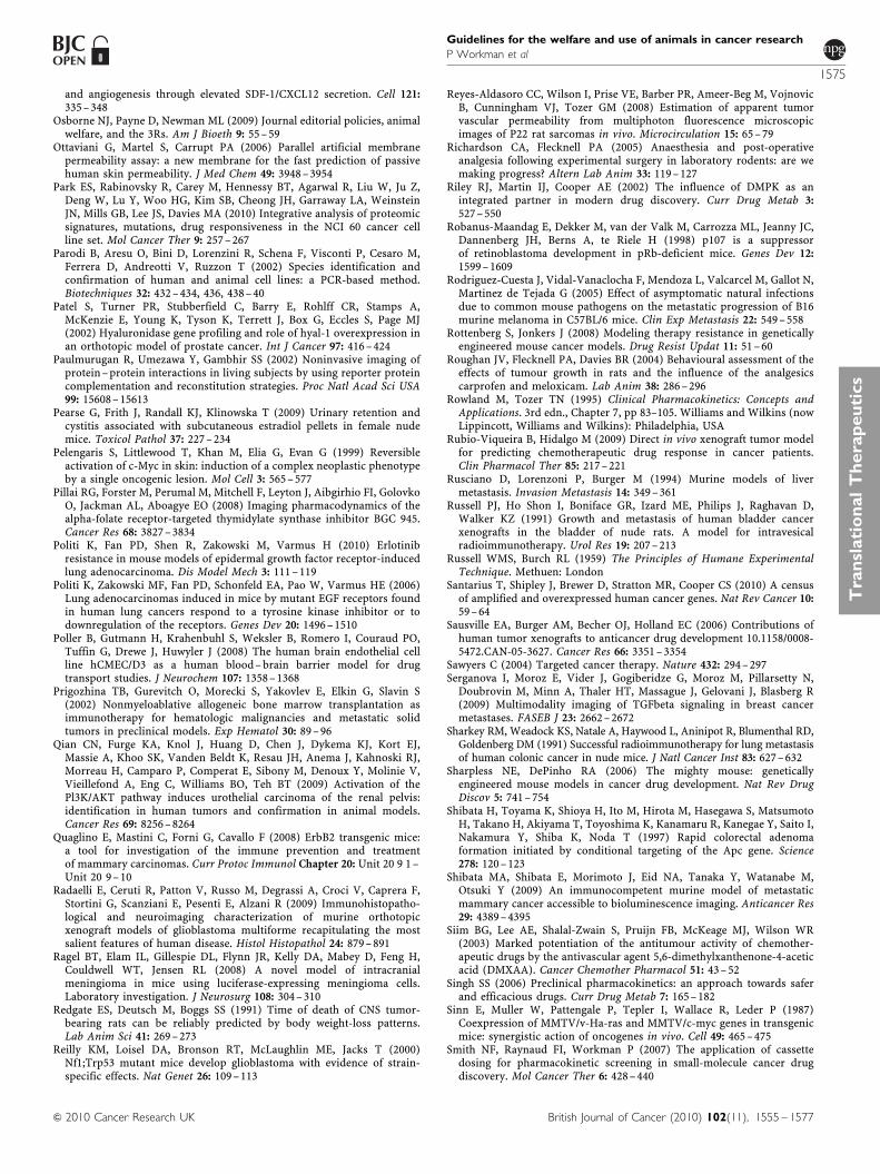
and angiogenesis through elevated SDF-1/CXCL12 secretion. Cell 121:335 – 348
Osborne NJ, Payne D, Newman ML (2009) Journal editorial policies, animalwelfare, and the 3Rs. Am J Bioeth 9: 55 – 59
Ottaviani G, Martel S, Carrupt PA (2006) Parallel artificial membranepermeability assay: a new membrane for the fast prediction of passivehuman skin permeability. J Med Chem 49: 3948 – 3954
Park ES, Rabinovsky R, Carey M, Hennessy BT, Agarwal R, Liu W, Ju Z,Deng W, Lu Y, Woo HG, Kim SB, Cheong JH, Garraway LA, WeinsteinJN, Mills GB, Lee JS, Davies MA (2010) Integrative analysis of proteomicsignatures, mutations, drug responsiveness in the NCI 60 cancer cellline set. Mol Cancer Ther 9: 257 – 267
Parodi B, Aresu O, Bini D, Lorenzini R, Schena F, Visconti P, Cesaro M,Ferrera D, Andreotti V, Ruzzon T (2002) Species identification andconfirmation of human and animal cell lines: a PCR-based method.Biotechniques 32: 432 – 434, 436, 438 – 40
Patel S, Turner PR, Stubberfield C, Barry E, Rohlff CR, Stamps A,McKenzie E, Young K, Tyson K, Terrett J, Box G, Eccles S, Page MJ(2002) Hyaluronidase gene profiling and role of hyal-1 overexpression inan orthotopic model of prostate cancer. Int J Cancer 97: 416 – 424
Paulmurugan R, Umezawa Y, Gambhir SS (2002) Noninvasive imaging ofprotein – protein interactions in living subjects by using reporter proteincomplementation and reconstitution strategies. Proc Natl Acad Sci USA99: 15608 – 15613
Pearse G, Frith J, Randall KJ, Klinowska T (2009) Urinary retention andcystitis associated with subcutaneous estradiol pellets in female nudemice. Toxicol Pathol 37: 227 – 234
Pelengaris S, Littlewood T, Khan M, Elia G, Evan G (1999) Reversibleactivation of c-Myc in skin: induction of a complex neoplastic phenotypeby a single oncogenic lesion. Mol Cell 3: 565 – 577
Pillai RG, Forster M, Perumal M, Mitchell F, Leyton J, Aibgirhio FI, GolovkoO, Jackman AL, Aboagye EO (2008) Imaging pharmacodynamics of thealpha-folate receptor-targeted thymidylate synthase inhibitor BGC 945.Cancer Res 68: 3827 – 3834
Politi K, Fan PD, Shen R, Zakowski M, Varmus H (2010) Erlotinibresistance in mouse models of epidermal growth factor receptor-inducedlung adenocarcinoma. Dis Model Mech 3: 111 – 119
Politi K, Zakowski MF, Fan PD, Schonfeld EA, Pao W, Varmus HE (2006)Lung adenocarcinomas induced in mice by mutant EGF receptors foundin human lung cancers respond to a tyrosine kinase inhibitor or todownregulation of the receptors. Genes Dev 20: 1496 – 1510
Poller B, Gutmann H, Krahenbuhl S, Weksler B, Romero I, Couraud PO,Tuffin G, Drewe J, Huwyler J (2008) The human brain endothelial cellline hCMEC/D3 as a human blood – brain barrier model for drugtransport studies. J Neurochem 107: 1358 – 1368
Prigozhina TB, Gurevitch O, Morecki S, Yakovlev E, Elkin G, Slavin S(2002) Nonmyeloablative allogeneic bone marrow transplantation asimmunotherapy for hematologic malignancies and metastatic solidtumors in preclinical models. Exp Hematol 30: 89 – 96
Qian CN, Furge KA, Knol J, Huang D, Chen J, Dykema KJ, Kort EJ,Massie A, Khoo SK, Vanden Beldt K, Resau JH, Anema J, Kahnoski RJ,Morreau H, Camparo P, Comperat E, Sibony M, Denoux Y, Molinie V,Vieillefond A, Eng C, Williams BO, Teh BT (2009) Activation of thePl3K/AKT pathway induces urothelial carcinoma of the renal pelvis:identification in human tumors and confirmation in animal models.Cancer Res 69: 8256 – 8264
Quaglino E, Mastini C, Forni G, Cavallo F (2008) ErbB2 transgenic mice:a tool for investigation of the immune prevention and treatmentof mammary carcinomas. Curr Protoc Immunol Chapter 20: Unit 20 9 1 –Unit 20 9 – 10
Radaelli E, Ceruti R, Patton V, Russo M, Degrassi A, Croci V, Caprera F,Stortini G, Scanziani E, Pesenti E, Alzani R (2009) Immunohistopatho-logical and neuroimaging characterization of murine orthotopicxenograft models of glioblastoma multiforme recapitulating the mostsalient features of human disease. Histol Histopathol 24: 879 – 891
Ragel BT, Elam IL, Gillespie DL, Flynn JR, Kelly DA, Mabey D, Feng H,Couldwell WT, Jensen RL (2008) A novel model of intracranialmeningioma in mice using luciferase-expressing meningioma cells.Laboratory investigation. J Neurosurg 108: 304 – 310
Redgate ES, Deutsch M, Boggs SS (1991) Time of death of CNS tumor-bearing rats can be reliably predicted by body weight-loss patterns.Lab Anim Sci 41: 269 – 273
Reilly KM, Loisel DA, Bronson RT, McLaughlin ME, Jacks T (2000)Nf1;Trp53 mutant mice develop glioblastoma with evidence of strain-specific effects. Nat Genet 26: 109 – 113
Reyes-Aldasoro CC, Wilson I, Prise VE, Barber PR, Ameer-Beg M, VojnovicB, Cunningham VJ, Tozer GM (2008) Estimation of apparent tumorvascular permeability from multiphoton fluorescence microscopicimages of P22 rat sarcomas in vivo. Microcirculation 15: 65 – 79
Richardson CA, Flecknell PA (2005) Anaesthesia and post-operativeanalgesia following experimental surgery in laboratory rodents: are wemaking progress? Altern Lab Anim 33: 119 – 127
Riley RJ, Martin IJ, Cooper AE (2002) The influence of DMPK as anintegrated partner in modern drug discovery. Curr Drug Metab 3:527 – 550
Robanus-Maandag E, Dekker M, van der Valk M, Carrozza ML, Jeanny JC,Dannenberg JH, Berns A, te Riele H (1998) p107 is a suppressorof retinoblastoma development in pRb-deficient mice. Genes Dev 12:1599 – 1609
Rodriguez-Cuesta J, Vidal-Vanaclocha F, Mendoza L, Valcarcel M, Gallot N,Martinez de Tejada G (2005) Effect of asymptomatic natural infectionsdue to common mouse pathogens on the metastatic progression of B16murine melanoma in C57BL/6 mice. Clin Exp Metastasis 22: 549 – 558
Rottenberg S, Jonkers J (2008) Modeling therapy resistance in geneticallyengineered mouse cancer models. Drug Resist Updat 11: 51 – 60
Roughan JV, Flecknell PA, Davies BR (2004) Behavioural assessment of theeffects of tumour growth in rats and the influence of the analgesicscarprofen and meloxicam. Lab Anim 38: 286 – 296
Rowland M, Tozer TN (1995) Clinical Pharmacokinetics: Concepts andApplications. 3rd edn., Chapter 7, pp 83–105. Williams and Wilkins (nowLippincott, Williams and Wilkins): Philadelphia, USA
Rubio-Viqueira B, Hidalgo M (2009) Direct in vivo xenograft tumor modelfor predicting chemotherapeutic drug response in cancer patients.Clin Pharmacol Ther 85: 217 – 221
Rusciano D, Lorenzoni P, Burger M (1994) Murine models of livermetastasis. Invasion Metastasis 14: 349 – 361
Russell PJ, Ho Shon I, Boniface GR, Izard ME, Philips J, Raghavan D,Walker KZ (1991) Growth and metastasis of human bladder cancerxenografts in the bladder of nude rats. A model for intravesicalradioimmunotherapy. Urol Res 19: 207 – 213
Russell WMS, Burch RL (1959) The Principles of Humane ExperimentalTechnique. Methuen: London
Santarius T, Shipley J, Brewer D, Stratton MR, Cooper CS (2010) A censusof amplified and overexpressed human cancer genes. Nat Rev Cancer 10:59 – 64
Sausville EA, Burger AM, Becher OJ, Holland EC (2006) Contributions ofhuman tumor xenografts to anticancer drug development 10.1158/0008-5472.CAN-05-3627. Cancer Res 66: 3351 – 3354
Sawyers C (2004) Targeted cancer therapy. Nature 432: 294 – 297Serganova I, Moroz E, Vider J, Gogiberidze G, Moroz M, Pillarsetty N,
Doubrovin M, Minn A, Thaler HT, Massague J, Gelovani J, Blasberg R(2009) Multimodality imaging of TGFbeta signaling in breast cancermetastases. FASEB J 23: 2662 – 2672
Sharkey RM, Weadock KS, Natale A, Haywood L, Aninipot R, Blumenthal RD,Goldenberg DM (1991) Successful radioimmunotherapy for lung metastasisof human colonic cancer in nude mice. J Natl Cancer Inst 83: 627 – 632
Sharpless NE, DePinho RA (2006) The mighty mouse: geneticallyengineered mouse models in cancer drug development. Nat Rev DrugDiscov 5: 741 – 754
Shibata H, Toyama K, Shioya H, Ito M, Hirota M, Hasegawa S, MatsumotoH, Takano H, Akiyama T, Toyoshima K, Kanamaru R, Kanegae Y, Saito I,Nakamura Y, Shiba K, Noda T (1997) Rapid colorectal adenomaformation initiated by conditional targeting of the Apc gene. Science278: 120 – 123
Shibata MA, Shibata E, Morimoto J, Eid NA, Tanaka Y, Watanabe M,Otsuki Y (2009) An immunocompetent murine model of metastaticmammary cancer accessible to bioluminescence imaging. Anticancer Res29: 4389 – 4395
Siim BG, Lee AE, Shalal-Zwain S, Pruijn FB, McKeage MJ, Wilson WR(2003) Marked potentiation of the antitumour activity of chemother-apeutic drugs by the antivascular agent 5,6-dimethylxanthenone-4-aceticacid (DMXAA). Cancer Chemother Pharmacol 51: 43 – 52
Singh SS (2006) Preclinical pharmacokinetics: an approach towards saferand efficacious drugs. Curr Drug Metab 7: 165 – 182
Sinn E, Muller W, Pattengale P, Tepler I, Wallace R, Leder P (1987)Coexpression of MMTV/v-Ha-ras and MMTV/c-myc genes in transgenicmice: synergistic action of oncogenes in vivo. Cell 49: 465 – 475
Smith NF, Raynaud FI, Workman P (2007) The application of cassettedosing for pharmacokinetic screening in small-molecule cancer drugdiscovery. Mol Cancer Ther 6: 428 – 440
Guidelines for the welfare and use of animals in cancer research
P Workman et al
1575
British Journal of Cancer (2010) 102(11), 1555 – 1577& 2010 Cancer Research UK
Tra
nsl
ati
on
al
Th
era
peu
tics
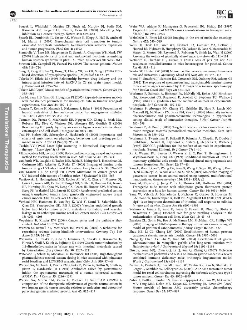
Soucek L, Whitfield J, Martins CP, Finch AJ, Murphy DJ, Sodir NM,Karnezis AN, Swigart LB, Nasi S, Evan GI (2008) Modelling Mycinhibition as a cancer therapy. Nature 455: 679 – 683
Spaeth EL, Dembinski JL, Sasser AK, Watson K, Klopp A, Hall B, AndreeffM, Marini F (2009) Mesenchymal stem cell transition to tumor-associated fibroblasts contributes to fibrovascular network expansionand tumor progression. PLoS One 4: e4992
Stambolic V, Tsao MS, Macpherson D, Suzuki A, Chapman WB, Mark TW(2000) High incidence of breast and endometrial neoplasia resemblinghuman Cowden syndrome in ptenþ /� mice. Cancer Res 60: 3605 – 3611
Stratton MR, Campbell PJ, Futreal PA (2009) The cancer genome. Nature458: 719 – 724
Sung H, Kang SH, Bae YJ, Hong JT, Chung YB, Lee CK, Song S (2006) PCR-based detection of mycoplasma species. J Microbiol 44: 42 – 49
Takeda N, Diksic M (1999) Relationship between drug delivery and theintra-arterial infusion rate of SarCNU in C6 rat brain tumor model.J Neurooncol 41: 235 – 246
Taketo MM (2006) Mouse models of gastrointestinal tumors. Cancer Sci 97:355 – 361
Tan M, Fang HB, Tian GL, Houghton PJ (2005) Repeated-measures modelswith constrained parameters for incomplete data in tumour xenograftexperiments. Stat Med 24: 109 – 119
Tanaka T, Konno H, Matsuda I, Nakamura S, Baba S (1995) Prevention ofhepatic metastasis of human colon cancer by angiogenesis inhibitorTNP-470. Cancer Res 55: 836 – 839
Tennant DA, Frezza C, MacKenzie ED, Nguyen QD, Zheng L, Selak MA,Roberts DL, Dive C, Watson DG, Aboagye EO, Gottlieb E (2009)Reactivating HIF prolyl hydroxylases under hypoxia results in metaboliccatastrophe and cell death. Oncogene 28: 4009 – 4021
Tsai PP, Stelzer HD, Schraepler A, Hackbarth H (2006) Importance andeffects of enrichment on physiology, behaviour and breeding perfor-mance in mice. Altex 23 (Suppl): 96 – 98
Tuchin VV (1993) Laser light scattering in biomedical diagnostics andtherapy. J Laser Appl 5: 43 – 60
Ullman-Cullere MH, Foltz CJ (1999) Body condition scoring: a rapid and accuratemethod for assessing health status in mice. Lab Anim Sci 49: 319 – 323
van Furth WR, Laughlin S, Taylor MD, Salhia B, Mainprize T, Henkelman M,Cusimano MD, Ackerley C, Rutka JT (2003) Imaging of murine braintumors using a 1.5 Tesla clinical MRI system. Can J Neurol Sci 30: 326 – 332
van Kranen HJ, de Gruijl FR (1999) Mutations in cancer genes ofUV-induced skin tumors of hairless mice. J Epidemiol 9: S58 – S65
Varticovski L, Hollingshead MG, Robles AI, Wu X, Cherry J, Munroe DJ,Lukes L, Anver MR, Carter JP, Borgel SD, Stotler H, Bonomi CA, NunezNP, Hursting SD, Qiao W, Deng CX, Green JE, Hunter KW, Merlino G,Steeg PS, Wakefield LM, Barrett JC (2007) Accelerated preclinical testingusing transplanted tumors from genetically engineered mouse breastcancer models. Clin Cancer Res 13: 2168 – 2177
Verheul HM, Hammers H, van Erp K, Wei Y, Sanni T, Salumbides B,Qian DZ, Yancopoulos GD, Pili R (2007) Vascular endothelial growthfactor trap blocks tumor growth, metastasis formation, and vascularleakage in an orthotopic murine renal cell cancer model. Clin Cancer Res13: 4201 – 4208
Vogelstein B, Kinzler KW (2004) Cancer genes and the pathways theycontrol. Nat Med 10: 789 – 799
Warden SJ, Bennell KL, McMeeken JM, Wark JD (2000) A technique forrestraining rodents during hindlimb interventions. Contemp Top LabAnim Sci 39: 24 – 27
Watanabe H, Uesaka T, Kido S, Ishimura Y, Shiraki K, Kuramoto K,Hirata S, Shoji S, Katoh O, Fujimoto N (1999) Gastric tumor induction by1,2-dimethylhydrazine in Wistar rats with intestinal metaplasia causedby X-irradiation. Jpn J Cancer Res 90: 1207 – 1211
Watanabe T, Schulz D, Morisseau C, Hammock BD (2006) High-throughputpharmacokinetic method: cassette dosing in mice associated with minusculeserial bleedings and LC/MS/MS analysis. Anal Chim Acta 559: 37 – 44
Watson SA, Michaeli D, Morris TM, Clarke P, Varro A, Griffin N, Smith A,Justin T, Hardcastle JD (1999a) Antibodies raised by gastrimmuneinhibit the spontaneous metastasis of a human colorectal tumour,AP5LV. Eur J Cancer 35: 1286 – 1291
Watson SA, Morris TM, Varro A, Michaeli D, Smith AM (1999b) Acomparison of the therapeutic effectiveness of gastrin neutralisation intwo human gastric cancer models: relation to endocrine and autocrine/paracrine gastrin mediated growth. Gut 45: 812 – 817
Weiss WA, Aldape K, Mohapatra G, Feuerstein BG, Bishop JM (1997)Targeted expression of MYCN causes neuroblastoma in transgenic mice.EMBO J 16: 2985 – 2995
Weissleder R, Pittet MJ (2008) Imaging in the era of molecular oncology.Nature 452: 580 – 589
Wells DJ, Playle LC, Enser WE, Flecknell PA, Gardiner MA, Holland J,Howard BR, Hubrecht R, Humphreys KR, Jackson IJ, Lane N, Maconochie M,Mason G, Morton DB, Raymond R, Robinson V, Smith JA, Watt N (2006)Assessing the welfare of genetically altered mice. Lab Anim 40: 111 – 114
Wetmore C, Eberhart DE, Curran T (2001) Loss of p53 but not ARFaccelerates medulloblastoma in mice heterozygous for patched. CancerRes 61: 513 – 516
Winter SF, Hunter KW (2008) Mouse modifier genes in mammary tumorigen-esis and metastasis. J Mammary Gland Biol Neoplasia 13: 337 – 342
Wood PJ, Stratford IJ, Sansom JM, Cattanach BM, Quinney RM, Adams GE(1992) The response of spontaneous and transplantable murine tumorsto vasoactive agents measured by 31P magnetic resonance spectroscopy.Int J Radiat Oncol Biol Phys 22: 473 – 476
Workman P, Balmain A, Hickman JA, McNally NJ, Rohas AM, MitchisonNA, Pierrepoint CG, Raymond R, Rowlatt C, Stephens TC, Wallace J(1988) UKCCCR guidelines for the welfare of animals in experimentalneoplasia. Br J Cancer 58: 109–113
Workman P, Aboagye EO, Chung YL, Griffiths JR, Hart R, Leach MO,Maxwell RJ, McSheehy PM, Price PM, Zweit J (2006) Minimally invasivepharmacokinetic and pharmacodynamic technologies in hypothesis-testing clinical trials of innovative therapies. J Natl Cancer Inst 98:580 – 598
Workman P, de Bono J (2008) Targeted therapeutics for cancer treatment:major progress towards personalised molecular medicine. Curr OpinPharmacol 8: 359 – 362
Workman P, Twentyman P, Balkwill F, Balmain A, Chaplin D, Double J,Embleton J, Newell D, Raymond R, Stables J, Stephens T, Wallace J(1998) UKCCCR guidelines for the welfare of animals in experimentalneoplasia (Second Edition). Br J Cancer 77: 1 – 10
Xu X, Wagner KU, Larson D, Weaver Z, Li C, Ried T, Hennighausen L,Wynshaw-Boris A, Deng CX (1999) Conditional mutation of Brca1 inmammary epithelial cells results in blunted ductal morphogenesis andtumour formation. Nat Genet 22: 37 – 43
Yang L, Mao H, Cao Z, Wang YA, Peng X, Wang X, Sajja HK, Wang L, DuanH, Ni C, Staley CA, Wood WC, Gao X, Nie S (2009) Molecular imaging ofpancreatic cancer in an animal model using targeted multifunctionalnanoparticles. Gastroenterology 136: 1514 – 1525; e2
Yang M, Reynoso J, Jiang P, Li L, Moossa AR, Hoffman RM (2004)Transgenic nude mouse with ubiquitous green fluorescent proteinexpression as a host for human tumors. Cancer Res 64: 8651 – 8656
Yang W, Velcich A, Mariadason J, Nicholas C, Corner G, Houston M,Edelmann W, Kucherlapati R, Holt PR, Augenlicht LH (2001) p21(WAF1/cip1) is an important determinant of intestinal cell response to sulindacin vitro and in vivo. Cancer Res 61: 6297 – 6302
Yoshino K, Iimura E, Saijo K, Iwase S, Fukami K, Ohno T, Obata Y,Nakamura Y (2006) Essential role for gene profiling analysis in theauthentication of human cell lines. Hum Cell 19: 43 – 48
Zavaleta CL, Goins BA, Bao A, McManus LM, McMahan CA, Phillips WT(2008) Imaging of 186Re-liposome therapy in ovarian cancer xenograftmodel of peritoneal carcinomatosis. J Drug Target 16: 626 – 637
Zhau HE, Li CL, Chung LW (2000) Establishment of human prostatecarcinoma skeletal metastasis models. Cancer 88: 2995 – 3001
Zheng Q, Chen XY, Shi Y, Xiao SD (2004) Development of gastricadenocarcinoma in Mongolian gerbils after long-term infection withHelicobacter pylori. J Gastroenterol Hepatol 19: 1192 – 1198
Zhu JS, Song MQ, Chen GQ, Li Q, Sun Q, Zhang Q (2007) Molecularmechanisms of paclitaxel and NM-3 on human gastric cancer in a severecombined immune deficiency mice orthotopic implantation model.World J Gastroenterol 13: 4131 – 4135
Zisman A, Pantuck AJ, Bui MH, Said JW, Caliliw RR, Rao N, Shintaku P,Berger F, Gambhir SS, Belldegrun AS (2003) LABAZ1: a metastatic tumormodel for renal cell carcinoma expressing the carbonic anhydrase type 9tumor antigen. Cancer Res 63: 4952 – 4959
Zuber J, Radtke I, Pardee TS, Zhao Z, Rappaport AR, Luo W, McCurrachME, Yang MM, Dolan ME, Kogan SC, Downing JR, Lowe SW (2009)Mouse models of human AML accurately predict chemotherapyresponse. Genes Dev 23: 877 – 889
Guidelines for the welfare and use of animals in cancer research
P Workman et al
1576
British Journal of Cancer (2010) 102(11), 1555 – 1577 & 2010 Cancer Research UK
Tra
nsla
tion
al
Th
era
peu
tics
Additional information
Committee to Update Science, Medicine, and Animals, NationalResearch Council (2004) Sciences, Medicine, and Animals: NationalResearch Council of The National Academies. The NationalAcademies Press: Washington, DC, USADennis C (2006) Cancer: off by a whisker. Nature 17: 739–741Garber K (2006) Realistic rodents? Debate grows over new mousemodels of cancer. J Natl Cancer Inst 98: 1176–1178Guidelines for the Care and Use of Mammals in Neuroscience andBehavioural Research. Committee on Guidelines for the Use ofAnimals in Neuroscience and Behavioural Research, (Institute forLaboratory Animal Research, Washington, D.C. 2003). NationalResearch CouncilFlecknell P (2008) Analgesia from a veterinary perspective. Br JAnaesth 101: 121–124Richardson CA, Flecknell PA (2005) Anaesthesia and post-operative analgesia following experimental surgery in laboratoryrodents: are we making progress? Altern Lab Anim 33: 119–127Roughan JV, Flecknell PA, Davies BR (2004) Behavioural assessmentof the effects of tumour growth in rats and the influence of theanalgesics carprofen and meloxicam. Lab Anim 38: 286–296The Royal Society (2004) The Use of Non-human Animals inResearch: a Guide for Scientists. Science Advice Section, The RoyalSociety: London, UK
Useful weblinks
http://scienceandresearch.homeoffice.gov.uk/animal-research/publications-and-reference/statistics/http://www.sanger.ac.uk/genetics/CGPhttp://emice.nci.nih.gov/mouse_modelshttp://dels.nas.edu/ilar_n/ilarhome/reports.shtmlhttp://ec.europa.eu/environment/chemicals/lab_animals/home_en.htmhttp://www.iasp-pain.org/AM/Template.cfm?Section=Animal_Researchhttp://ec.europa.eu/european_group_ethics/docs/opinion7_en.pdfhttp://conventions.coe.int/treaty/en/treaties/html/123.htmhttp://www.ecopa.eu/http://caat.jhsph.edu/http://www.imm.ki.se/sft/pdf/OECD19.pdfhttp://oacu.od.nih.gov/ARAC/index.htmhttp://www.research.psu.edu/arp/health/endpoints.htmlhttp://www.nc3rs.org.uk/news.asp?id=759http://ddgs.utu.fi/request.php?4http://www.lal.org.uk/index.php?option=com_content&view=article&id=56&Itemidhttp://www.nc3rs.org.uk
Glossary
Allometric scaling: Calculation of doses of drugs to beadministered to animals according to their relative sizes wherethe relationship of a biological variable to body mass is non-linear.For example, drug dosage can be linearly related to body surfacearea rather than to body weight.
Ascites: Cells/fluid in the peritoneal cavity.Autochthonous tumours: Tumours originating within the host
animal, either spontaneously, or due to genetic or pharmacologicalintervention.
Cachexia: Severe loss of weight and muscle mass that cannot bereversed nutritionally. Can be caused by release of biologicallyactive molecules (cytokines) from certain tumours.
Cassette dosing: Administration of multiple compounds to anindividual animal followed by individual measurements in thesame blood sample.
Clinically equivalent dose: A dose of a drug, which results inblood/tissue levels that reflect those that are achieved in patients.
Clonogenic assay: Measuring the effect of treatments on theability of tumour cells to proliferate expansively. Treatment maybe in vitro or initiated in vivo and the clonogenic ability ofexplanted cells tested in vitro.
Desquamation: Loss of skin integrity. Moist desquamation canbe a consequence of exposure to ionising radiation (UV or X-rays)where the skin thins and then begins to weep tissue fluid as theepithelial cells lose their barrier function.
Distension: Stretched beyond normal dimensions.Ectopic: Site of growth different from the tissue of origin, for
example, s.c. transplantation of tumours derived from internal organs.Erythema: Skin reddening and thickening in response to UV
irradiation, as in mild sunburn.Factorial design: Involves the inclusion of two or more variables
and measuring the response to each variable and interactionsbetween variables.
Genetically engineered mouse models (GEMMs): Animals inwhich the genetic material has been altered. For example,introduction of a mutation in cells of a particular organ mayresult in the development of benign or malignant tumours.
Hyperplasia: Refers to the proliferation of cells within an organor tissue beyond that which is ordinarily seen. Microscopically
cells resemble normal cells but are increased in numbers. It is abenign condition, unlike neoplasia, which is malignant.
Intravital microscopy: A technique, which allows direct obser-vation of small blood vessels within the organs of anesthetisedanimals.
Maximum Tolerated Dose (MTD): The highest dose of a drug inwhich the clinical condition of the experimental animal is maintained.
Metastasis: The spread of tumour cells from a primary site todistant sites in the body, usually through the blood or lymph.The term ‘experimental metastasis’ is sometimes used to describethe colonisation of organs after injection of cells directly into theperipheral circulation.
Oncogenesis: The process of malignant transformation resultingin tumour development.
Orthotopic: Anatomically correct site (opposite of ectopic), forexample, transplantation of renal tumour cells into the kidney ormammary carcinoma cells into the mammary fat pad.
Pharmacodynamics: The study of the action of and the durationof effects of agents in the body, including confirmation ofmechanism of action through identification of relevant biomarkersof activity.
Pharmacokinetics: The study of the process by which agents areabsorbed, distributed, metabolized and eliminated by the body,including measurement of the rate of excretion, metabolism, bloodand tissue concentrations.
Syngeneic tumour models: Cells transplanted between animalsof the same inbred strain.
Ulceration: An inflamed lesion on the skin or internal surfaceinvolving tissue destruction.
Xenogeneic tumour models: Cells transplanted between species(e.g., human to mouse). Requires recipients that cannot mount animmune response and reject the foreign tissue graft such asathymic mice which lack T-lymphocytes, or severe combinedimmunodeficient (SCID) mice.
This work is licensed under the Creative CommonsAttribution-NonCommercial-Share Alike 3.0 License.
To view a copy of this license, visit http://creativecommons.org/licenses/by-nc-sa/3.0/
Guidelines for the welfare and use of animals in cancer research
P Workman et al
1577
British Journal of Cancer (2010) 102(11), 1555 – 1577& 2010 Cancer Research UK
Tra
nsl
ati
on
al
Th
era
peu
tics