gupea.ub.gu.se · treatment response is large and probably at least partly mediated by genetic...
TRANSCRIPT


© 2013 Camilla Glad [email protected]
ISBN 978-91-628-8631-8 http://hdl.handle.net/2077/31992 Printed by Kompendiet/Aidla Trading AB, Gothenburg, Sweden, 2013. Published articles have been reprinted with permission of the copyright owner.
Cover: Word cloud of introduction section. http://www.wordle.net. Sizing represents the number of times the word appears in the text (more frequent = bigger size).

Till Lucas och Leia - de mest fantastiska barn på denna jord. ”Föräldrar till begåvade barn tror på ärftlighetsteorin”. Joachim Fuchsberger


Growth hormone (GH) is a polypeptide hormone which is secreted from the anterior pituitary in a pulsatile pattern. GH is best known by its strong effects on longitudinal growth in children, but the importance of GH is maintained also in adulthood due to the powerful effects on cellular differentiation and fuel homeostasis. GH deficient (GHD) adults often present with abdominal obesity, insulin resistance and an almost doubled risk for cardiovascular mortality. These problems are to a varying extent reversed during GH replacement therapy (GHRT). Variability in treatment response is large and probably at least partly mediated by genetic variation. The effects of GH are mediated by the GH receptor (GHR), a transmembrane glycoprotein expressed on most human cell types. In humans, there are two isoforms of the GHR which differ in regards to retention (fl-GHR) or exclusion (d3-GHR) of exon 3. The two isoforms are simply two different alleles of a common GHR polymorphism (the GHR d3/fl polymorphism), which has been suggested to influence GH sensitivity. This thesis is based on four studies, with the common, overall, aim to test the hypothesis that polymorphisms in genes within the GH-IGF-I axis influence body composition, metabolism and serum IGF-I concentrations. In an initial study we found that the GHR d3/fl polymorphism was not associated with the 1 year changes in IGF-I concentrations and body composition in response to GHRT. During this study, which was fairly small, we realized the need for a different genotyping method for analyses of the GHR d3/fl polymorphism in larger cohorts. Therefore, in the subsequent study, we evaluated the use of tagSNP rs6873545 as a marker for the GHR d3/fl polymorphism, and showed that it does indeed perfectly tag the different GHR d3/fl alleles. In the next study, we investigated the impact of the GHR SNP rs6873545 and five other polymorphisms in genes within the GH-IGF-I axis on the early and the long-term IGF-I responses to GHRT in a large cohort of GHD adults. In this study we found that the GHR SNP rs6873545 and the PIK3CB SNP rs361072 were associated with the early, but not the long-term, IGF-I response. In the last study, we analysed the tagSNP rs6873545 in a study representative of the general Swedish population and found that homozygosity for the d3-GHR was associated with an adverse anthropometric and metabolic profile. In conclusion, the results of this thesis suggests that 1) the GHR d3/fl polymorphism is indeed of functional importance, and influences both the response to GHRT in GHD adults as well as body composition and metabolism in the general Swedish population, and 2) other SNPs in genes within the GH-IGF-I axis may be of an equal importance in terms of the response to GHRT in GHD adults. Key words: growth hormone, growth hormone receptor, genetic association study, pharmacogenetic, growth hormone deficiency, polymorphism, candidate gene approach, metabolism, anthropometry. ISBN 978-91-628-8631-8
This thesis is based on the following papers and manuscripts, which are referred to in the text by their Roman numerals: Paper I. Influence of the exon 3-deleted/full-length growth hormone (GH) receptor
polymorphism on the response to GH replacement therapy in adults with severe GH deficiency. Barbosa EJ, Palming J, Glad CA, Filipsson H, Koranyi J, Bengtsson BA, Carlsson LM, Boguszewski CL, Johannsson G. J Clin Endocrinol Metab. 2009 Feb;94(2):639-44.
Paper II. Rapid and high throughput genotyping of the growth hormone receptor
exon 3 deleted/full-length polymorphism using a tagSNP. Glad CA, Johannsson G, Carlsson LM, Svensson PA. Growth Horm IGF Res. 2010 Jun;20(3):270-3.
Paper III. SNPs within the GH signalling pathway are associated with the early, but
not the long-term, IGF-I response to GH replacement therapy in GHD adults. Glad CA, Barbosa EJL, Filipsson Nyström H, Carlsson LMS, Nilsson S, Nilsson AG, Svensson PA, Johannsson G. Manuscript.
Paper IV. The growth hormone receptor exon 3 deleted/full-length polymorphism is
associated with body weight and body composition in the general
population. Glad CA, Carlsson LMS, Sjöström L, Nilsson S, Larsson I, Svensson PA, Johannsson G. Manuscript.


8
A Adenine BMD Bone mineral density BMI Body mass index bp Base pair C Cytosine CO Childhood-onset d3-GHR Exon 3 deleted growth hormone receptor DBP Diastolic blood pressure DEXA Dual energy x-ray absorptiometry DNA Deoxyribonucleic acid FFM Fat free mass fl-GHR Full-length growth hormone receptor G Guanine GH Growth hormone GHD Growth hormone deficiency GHR Growth hormone receptor GHRH Growth hormone releasing hormone GHRT Growth hormone replacement therapy GWAS Genome-wide association study HDL-C High density lipoprotein cholesterol HGP Human genome project HOMA-IR Homeostatic model assessment insulin resistance HWE Hardy-Weinberg equilibrium IGFBP-3 Insulin-like growth factor binding protein 3 IGF-I Insulin-like growth factor I ISS Idiopathic short stature ITT Insulin tolerance test JAK2 Janus kinase 2 LD Linkage disequilibrium LDL-C Low density lipoprotein cholesterol LTR Long terminal repeat MAF Minor allele frequency mRNA Messenger ribonucleic acid NFPA Non-functioning pituitary adenoma NFQ Non-fluorescent quencher NTC No-template control PCR Polymerase chain reaction PIK3CB Phosphoinisitide 3 kinase, catalytic subunit beta QoL Quality of life rhGH Recombinant human growth hormone RNA Ribonucleic acid SBP Systolic blood pressure SD Standard deviation SGA Small for gestational age SNP Single nucleotide polymorphism SOCS2 Suppressor of cytokine signalling 2 STAT5b Signal transducer and activator of transcription 5b T Thymine T2DM Type 2 diabetes mellitus tagSNP Tagging single-nucleotide polymorphism TBK Total body potassium

9
Genetics is the science of genes, heredity and variation in living organisms. The modern science of genetics, which seeks to understand the process of inheritance, began in the mid-19th century with the work of the German-Czech Augustinian monk and scientist Gregor Johann Mendel (1822-1884). He studied garden peas, and discovered that organisms inherit traits via discrete units of inheritance, unknown at that time, which are now known as genes. In 1910, Thomas Hunt Morgan (1866-1945) postulated the theory that genes are positioned on chromosomes, and in 1953 the structure of the deoxyribonucleic acid (DNA) was determined by James D. Watson (1928-) and Francis Crick (1916-2004) [1], although the contribution by Rosalind Franklin should not be forgotten. Since then, the central dogma of molecular biology has been stated and also re-stated, and we now know that the flow of information in a cell goes from DNA, via ribonucleic acid (RNA), to protein [2]. Molecularly, DNA is made up of four bases, termed adenine (A), thymine (T), cytosine (C) and guanine (G). The bases are complementary, in the sense that A always binds to T, and C always binds to G, and so DNA is composed of two anti-parallel intertwining nucleotide chains of these bases, making up a double-helix polymer (Figure 1) with the bases bound to a backbone of alternating sugars and phosphate groups [3].
The level of DNA organization within the human cell. Complementary bases bound to a sugar- and phosphate backbone make up the DNA double-helix polymer. The DNA is further packaged and organized into a set of chromosomes, which reside in the cell nucleus. Courtesy: D. Leja / National Human Genome Research Institute, http://www.genome.gov/.
In eukaryotes (animals, plants, fungi and protists), the vast majority of genetic information is stored within the cell nucleus, and the DNA is tightly packaged into long structures called chromosomes. In 2003, the joint efforts of the Human Genome Project (HGP) and the private

10
effort by Celera Genomics, led to finalization of the sequencing of the human genome (the entirety of the human genetic information). The complete (99%) sequence of the human genome was subsequently published in 2004, and so to date we know that it contains 3.2 billion base pairs (bp) organized on 22 autosomal and two sex (X and Y) chromosomes and into 20.000-25.000 protein coding genes. Structurally, genes constitute approximately 1.2% of the genome and they are distributed on all chromosomes [4], with the remaining 98.8% being non-protein coding sequence. These non-coding sequences may appear unimportant, but in fact within these sequences lay regions important for regulation of gene expression etc., including promoters which are essential for initiation of RNA synthesis. Genes, in turn, have both coding regions (exons) and non-coding intermediary regions (introns) [5]. The genetic information is transferred from DNA to protein via transcription of the genetic code (sequence of bases) into messenger RNA (mRNA), which is then translated into protein. The mRNA sequence is read in triplets, starting with a start codon (AUG) and ending with a stop codon (UAA, UGA or UAG). The codons in between start and stop determine which amino acids are to be incorporated in the polypeptide chain. In total, there are 64 possible triplets that specify the 20 most common different amino acids found in proteins.
In any two healthy unrelated individuals, only approximately 0.1% of the genome differs. As opposed to mutations, which are rare, polymorphisms are present in the human population with a frequency of >1%. There are different types of polymorphisms, including large, structural types such as deletions, insertions, duplications, translocations and inversions. However, in humans, the most common form of genetic variation (accounting for more than 90% of all genetic variation) is called a single-nucleotide polymorphism (SNP). On average, it is estimated that there is one SNP in every 300 bp, adding up to a total of 10 million of SNPs [6]. As the name implies, a SNP is merely a DNA sequence variation occurring when there are two alternative bases (alleles), or in some very rare circumstances more than two, present at a single nucleotide position (locus) in different individuals. A SNP may or may not have functional consequences, depending on its location in the genome. A polymorphism located within the coding sequence of a gene may alter the amino acid sequence of the subsequent protein (a non-synonymous SNP) and lead to changes in structure and function of the protein. Also, a SNP may introduce a stop codon, leading to premature cessation of the translation process, which in most cases disrupts the function of that protein. Due to the degeneracy of the genetic code, a SNP can also be silent (a synonymous SNP), meaning that it doesn’t change the amino acid sequence. The majorities of SNPs are located outside the coding parts of a gene and are referred to as being non-coding. However, these variants may still be functional if they are located in regions such as promoters, enhancers or silencers – which can be important for transcription factor binding etc. and thus lead to changes in gene expression. In combination with random processes and environment, genetic variation is the cause of natural selection and it makes us all unique. Our genetic make-up influences everything from eye color to disease vulnerability and drug response. To study the correlation between a genetic variant and a phenotype, such as a disease, a disease-associated quantitative trait or a drug response, a genetic association study can be performed. Using a candidate gene approach, genetic variants to be tested for association are selected based on previous information, such as knowledge of the corresponding gene function, its location in a region previously linked to disease, or simply because it for some reason is an interesting candidate variant. Today it is possible to perform genome-wide association studies (GWAS), where hundreds of thousands of SNPs are tested simultaneously for association with disease or a trait.

11
When two genetic variants are located close to each other on a chromosome, they are often inherited together more frequently than expected by chance; a phenomenon termed linkage disequilibrium (LD). When two variants are so closely associated that they are always inherited together, they are said to be in complete LD. As such, a genetic variant which is in perfect LD with another variant can be used as a tagSNP (marker).
For most medications there is a large interpatient variability in efficacy and toxicity [7]. Factors such as age, sex, underlying disease status and other drugs administered to the patient influence the response to a medical treatment [7, 8]. It is estimated that genetic variability account for 20-95% of the variability in drug disposition (absorption, distribution, metabolism and excretion) and effects [9]. Pharmacogenomics is the research field aimed to investigate the genetic basis for differences in drug efficacy and toxicity, ultimately aiming to maximize efficacy and safety of a treatment [7, 8]. While pharmacogenomics uses genome-wide approaches to search for polymorphisms affecting the response to a medical treatment, its counterpart pharmacogenetics does the same but rather using a candidate-gene (small-scale) approach.
In the 1920s, the presence of a growth-promoting substance from the anterior pituitary was demonstrated in experiments which showed the ability of pituitary extracts to restore and maintain growth in hypophysectomised animals [10]. In 1956, human growth hormone (GH) was isolated [11], and starting from the late 1950s GH deficient (GHD) children were treated with GH purified from cadaver pituitaries [12]. However, use of cadaver-purified GH was stopped, and the medication was withdrawn from the market in 1985 when it was shown to transmit Creutzfelt-Jacobs disease [13]. Since 1985, recombinant human GH (rhGH) has been commercially available. Human pituitary GH protein is a four-helical bundle protein consisting of 191 amino acids. The protein is produced after transcription of the GH1 gene located on chromosome 17, position 17q24.2, and it is secreted from the pituitary in a pulsatile pattern. As the name implies, GH is an important stimulator of postnatal longitudinal growth and it has been shown to directly stimulate the cells in the growth plate to induce growth [14]. In addition, GH has major effects on fuel metabolism by influencing muscle (protein anabolic), fat (lipolytic) and regulation of blood glucose levels [15]. GH also has a profound effect on the brain and quality-of-life [16]. In addition, GH has some important indirect effects, predominantly mediated through GH-induced production of insulin-like growth factor I (IGF-I) from the liver. IGF-I shares both structural and functional homology with the insulin peptide, and also mimics some of its actions, such as stimulation of glucose and amino acid uptake and inhibition of gluconeogenesis [17-19]. Also, IGF-I is important for the stimulation of postnatal body growth. About 75% of the IGF-I found in the circulation originates from the liver, however through a liver-specific knock-out of IGF-I in mice it has been shown that body growth is rather determined by locally produced (autocrine/paracrine) IGF-I [20].
At the molecular level, GH effects are mediated by the GH receptor (GHR), encoded by the GHR gene located on chromosome 5, region p13.1-p12 [21, 22]. The GHR is a transmembrane glycoprotein belonging to the type I cytokine receptor superfamily [23, 24]. At the cell surface, GHR can exist in preformed dimers, and binding of a GH molecule to its dimerized GHR induces a series of rotations and conformational changes within the subunits [25], which enables

12
transphosphorylation of the Janus kinase 2 (JAK2) protein and subsequent activation of the Janus kinase 2/Signal transducer and activator of transcription 5 (JAK/STAT) pathway [26, 27]. The result, amongst other, is an increased generation of IGF-I. In addition, activation of the suppressor of cytokine signalling (SOCS) genes by GH mediates an inhibitory effect on the GH signalling [28], thereby producing an intracellular negative feed-back loop in the GHR signalling cascade. Also, some of the metabolic effects of GH are the results of activation of phosphoinisitide 3 (PI3) kinases [29], such as PIK3CB, which are independent of the GHR-induced IGF-I generation (Figure 2).
Schematic and simplified view of the GHR signalling cascade. Binding of GH to the
GHR induces a series of conformational changes leading to phosphorylation of the associated JAK2 protein. A series of transphosphorylation steps then leads to activation of signalling molecules such as STAT5b and PI3K, and to translocation of these proteins into the cell nucleus. The result is a subsequent transcription of GH-inductive genes such as SOCS2, proteins of which then produces an intracellular negative feed-back loop on the GHR signalling. Note that some proteins such as the MAPK/ERK and IRS1 have been left out from the illustration due to space limitation and for clarity of the overall picture.
Patients with adult hypopituitarism and GHD, most frequently caused by pituitary tumours and the treatment thereof [30], suffer from a phenotype which resembles the metabolic syndrome. The phenotypic picture include increased amount of body fat (particularly abdominal) [31-33] and decreased lean body mass [31, 33-35], reduced bone mineral density (BMD) [36-38] and increased fracture risk [39], dyslipidaemia [40-44], hypertension [44] and insulin resistance [45]. They also have decreased well-being and quality of life (QoL) [16, 46-49]. Most importantly, the mortality rate of adults with hypopituitarism not replaced with GH is twice that expected in the normal population and is commonly due to cardiovascular and cerebrovascular diseases [50-55]. For these patients, GH replacement therapy (GHRT) improves many of the symptoms and clinical signs associated with the deficiency [56]. The dosing of GH is individualized and guided by clinical and biochemical responses. The recommended GH starting dose is 0.1 to 0.3 mg/day depending on sex and age. To mimic the rhythmical pattern of endogenous GH secretion, GH is administered in the evening (before bedtime) through subcutaneous injections [57].
Upon start of GH replacement, the individual responsiveness to GH is not known. Therefore, the treatment is initiated with a low dose of GH, independent of body weight, which is titrated upwards based on serum IGF-I levels and clinical response [58, 59]. Determination of the response to GHRT in adults includes clinical signs and symptoms of fluid retention, improvement in well-being, changes in body composition and biochemical serum markers of GH action, where the most sensitive is IGF-I. Despite stable serum IGF-I levels, metabolic endpoints

13
such as lipid profile and indices of glucose homeostasis have been shown to change over time [60], which suggests that there is a poor association between serum IGF-I and efficacy of GHRT. Sex, age, body mass index (BMI), GH dose and the route of oestrogen replacement in women have been linked to the response to GHRT [61-63], but the variability in GH-mediated effects on body composition, lipid- and glucose metabolism is less well known [64]. Most likely, the variability could be due to genetic factors. For instance, it has been suggested that 50% of the interindividual variation in baseline serum IGF-I levels in healthy subjects is genetically determined [65].
To date, candidate gene studies are the most common in the literature on pharmacogenomics in GHRT, and for obvious reasons the GHR gene has been an attractive candidate gene. The first report of GHR cDNA clones with differences within the coding region came in 1989, from Godowski et al [22]. In one of these clones the entire GHR exon 3 sequence was missing, giving rise to a GHR protein lacking 22 amino acids in the extracellular, GH binding, domain. Also, the resulting protein contains an aspartic acid instead of an alanine residue at the exon 2-4 junction [66]. The initial studies investigating the exon 3 deletion (d3) generated a hypothesis that the d3-GHR was caused by an alternative splicing event, and the expression of the two isoforms were subsequently studied. It was originally reported on a tissue-specific expression pattern of the two isoforms, with the d3-GHR primarily expressed in the placental villi and amnion [66, 67] and the full-length (fl)-GHR found exclusively in chorion and decidua [67]. Later, Mercado et al found that both isoforms were expressed in 19 tested tissues, but that the relative amount of the isoforms differed between the tissues [68]. However, following studies suggested that the expression pattern was rather subject-specific [69, 70] and/or developmentally regulated [70], than tissue-specific. In 1996, Stallings-Mann et al showed that the skipping of exon 3 was not tissue-specific, nor developmentally regulated, but actually subject-specific and due to a polymorphism in the GHR gene that follows a simple Mendelian mode of inheritance [71]. In 2000, Pantel et al shed some light on the origin of the polymorphism when they reported on the existence of two 99% identical retroelements flanking exon 3 [72]. The retroelements are 251 bp long, located 577 bp upstream and 1821 bp downstream of exon 3 and underlies the creation of the d3-GHR through an early homologous recombination event which has been passed on through generations. The resulting deletion involves besides the exon 3 also parts of introns 2 and 3. Several studies have investigated the actual effects of the loss of amino acid 7-28 in the GHR protein, but to date the importance of this region is still partly unknown. Even though the region appears to be conserved among GHR proteins in mammals, there is no homolog in the closely related prolactin receptor [73]. The GH binding capacities of the two GHR isoforms have been studied, but without demonstration of a significant difference [66, 74, 75]. Also, when modelled by crystallography, the amino acids encoded by exon 3 does not appear to interact in a significant matter with GH [76]. In 2004, Dos Santos et al transfected 293 HEK fibroblasts with vectors expressing both GHR isoforms, either alone or simultaneously, and showed that transduction of GH signalling was approximately 30% greater through d3-GHR homo- and heterodimers than through fl-GHR homodimers [77]. Efficacy of signal transduction was quantified by induction of the firefly luciferase gene coupled to the GH responsive STAT5 promoter. This, however, is to date the only study showing a functional difference between the two GHR isoforms.

14
The GHR d3/fl polymorphism has been thoroughly investigated in several paediatric and adult populations receiving GH therapy; with subjects being either GHD or non-GHD. Non-GHD populations include children with idiopathic short stature (ISS), Turner syndrome and children who are born small for gestational age (SGA). Although some studies have shown no effects of the d3/fl polymorphism on response to GH therapy in children [78-82], other studies have indicated that carriers of the d3-allele respond better in terms of growth velocity [77, 83-86], gain in height [85, 87] and final height [84], than non-carriers, suggesting that the d3-GHR confers a more GH-sensitive receptor. In accordance, in adult GHD populations receiving GHRT there are data suggesting an increased sensitivity to GH in patients carrying the d3-GHR. For instance, it has been shown that d3-GHR carriers experience a higher increase in serum IGF-I levels, more marked decrease in total- and LDL-cholesterol levels and a higher increase in serum HDL-cholesterol levels during short-term GHRT [88].
Surprisingly, there have been no reports on any influence of genetic variants within the GHR locus on adult height in genome wide association studies [89]. However, several SNPs in genes within or directly downstream of the GH signalling pathway have been identified, and previously shown to be of functional importance. First, two SNPs in the JAK2 gene have been associated with central adiposity (rs7849191) and blood lipid levels (rs3780378) [90]. STAT5b SNP rs6503691 has been associated with breast cancer [91], and SOCS2 SNP rs11107116 has been associated with adult height [92]. Lastly, PIK3CB SNP rs361072 has been associated with HOMA-IR [93], serum IGF-I levels and longevity [94].

15
The overall aim of this thesis was to test the hypothesis that polymorphisms in genes within the GH-IGF-I axis impact on body composition, metabolism and serum IGF-I concentrations. The specific aims of the different studies were: Paper I. to investigate the influence of the GHR d3/fl polymorphism on the response to
GHRT in GHD adults. Paper II. to facilitate the genotyping of the GHR d3/fl polymorphism. Paper III. to investigate the influence of genetic variants within the GH signalling pathway
on the IGF-I response to GHRT in GHD adults. Paper IV. to investigate the impact of the GHR d3/fl polymorphism on body composition
and metabolism in the general population.

16
The studies included in this thesis were approved by the regional ethical review board at the University of Gothenburg, and written informed consent was obtained from all participants prior to study inclusion.
This is an on-going prospective longitudinal study of adults with hypopituitarism and severe GHD who are treated at the outpatient Centre for Endocrinology and Metabolism (CEM) at the Sahlgrenska University Hospital, Gothenburg, Sweden. The first patient was included as early as in 1993, and as of today there are around 450 patients receiving GH treatment within this study making it the largest single-centre study of adult GHD. Patients from this study were included in Paper I (n=124), II (n=183) and III (n=313). General characteristics of the subjects included in Papers I and III are shown in Table 1. The most frequent GH stimulation test for confirmation of GHD was an insulin tolerance test (ITT; 77%), but other tests such as the GHRH-arginine, GHRH-pyridostigmine or glucagon were also used. In some patients, diagnosis was made based on a low serum IGF-I together with ≥ 3 additional pituitary deficiencies [57]. The most common cause of GHD was a non-functional pituitary adenoma (NFPA). All childhood-onset (CO) GHD subjects had been without GH treatment before retesting of GHD in transition from childhood to adult GH treatment. Patients with Cushing’s syndrome or acromegaly were in remission when entering the study protocol. All patients received rhGH with an average starting dose of 0.23 mg/day. The dose was titrated individually during GHRT based on age- and sex-adjusted serum IGF-I concentrations, after 1 and 4 weeks and every 3 months thereafter, to maintain IGF-I levels between the median and the upper limit of the normal reference range. When necessary, patients received adequate replacement therapy with glucocorticoids, thyroid hormone, desmopressin and/or sex hormones.
This is a long-term study where patients are enrolled consecutively, which generates some considerations to mention. Firstly, ever from the time when this study was started there have been several changes in treatment preferences, diagnostic criteria, biochemical methods for serum measurements and GH dosing strategies that may be of importance. During the time of inclusion into the study, different diagnostic tests have been used. Today, an ITT is the golden standard and indeed that test has been used for the majority of patients. One could speculate that different diagnostic tests and cut-offs could have led to some patients being included into the study with a somewhat different degree of severity of GHD. However, it is known that GH is commonly the first hormone that disappears during the development of hypopituitarism [95], and so most patients with pituitary disease and multiple pituitary hormonal deficiencies would be suspected to also have GHD. Serum IGF-I is the major response variable in Paper I and III, and during the years the biochemical methods for serum concentration measurements have changed. Although the methods have been shown to be correlated, there is still a concern in regards to some uncertainness in exact serum IGF-I levels and the comparisons thereof. Lastly, and most importantly, the first patients enrolled in the study (n≈50) were treated using an mg/kg body weight strategy adapted from the experience of peadiatricians. This treatment strategy was shown to generate GH doses in the unphysiological range, resulting in side-effects mostly related to fluid retention [33, 34, 96]. The strategy was later changed into a fix starting dose with individual dose titration, which reduced the problem with side-effects but showed similar good treatment results [58, 59].

17
Characteristics of the subjects in the GH-2 study.
Paper I Paper III
n 124 313 Male gender (%) 79 (63.7) 182 (58.1) Age (yrs; range) 50.1 (18-76) 49.7 (17-77) Adult onset GHD (%) 87.1 89.8 Isolated GHD (%) 9.7 11.3 Aetiology NFPA (%) 56 (45.2) 128 (40.9) Idiopathic (%) 12 (9.7) 29 (9.3) Prolactinoma (%) 13 10.5) 27 (8.6) Craniopharyngioma (%) 11 (8.9) 24 (7.7) Other* (%) 32 (25.8) 105 (33.5) Pre-study treatment Surgery (%) 73 (58.9) 163 (54.7) Radiotherapy (%) 6 (4.8) 16 (5.4) Both (%) 23 (18.5) 48 (16.1)
*other aetiologies include empty sella, previous cushing or acromegaly, meningioma, apoplexy, Sheehan, trauma, sarcoidosis, cystic lesion, histocytosis, congenital hypopituitarism, dysgerminoma, granular cells tomour, hamartoma, medulloblastoma, rhabdomyosarcoma, septo-opto dysplasia, TSH adenoma and Wegener granulomatosis. NFPA = nonfunctioning pituitary adenoma.
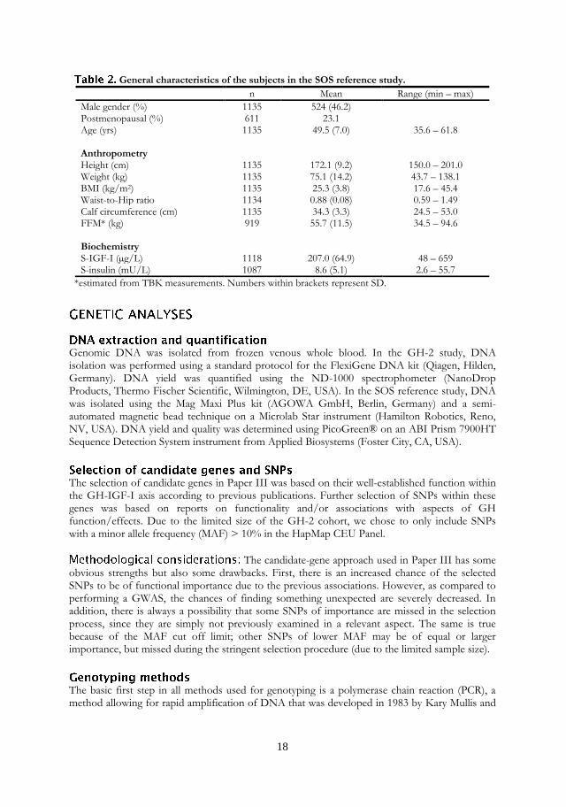
The SOS reference study includes subjects from the Swedish cities Mölndal and Örebro. The subjects were randomly selected from a population registry to constitute a reference group to the SOS intervention study, presented elsewhere [97]. The SOS reference study includes 1135 subjects (46.2% men), with an average age of 49.5 yrs (range 35.6 - 61.8) and average BMI of 25.3 kg/m2 (range 17.6 - 45.4). Extensive information about the subjects was collected at each subject’s physical examination by means of questionnaires. Age, sex, menopause status, self-reported diabetes (yes/no) and hypertension (yes/no) was reported for all subjects. Also, the subjects reported on a four-grade scale of leisure-time physical activity during the last 12 months period, with 4 being the most active. Body composition was measured by whole-body counting of the 40K isotope to determine total body potassium (TBK) content and estimate fat free mass (FFM) [98]. Systolic (SBP) and diastolic (DBP) blood pressure were measured on-site at examination day. Lab variables measured in serum included triglycerides, total cholesterol, high density lipoprotein (HDL) cholesterol and low density lipoprotein (LDL) cholesterol, insulin, IGF-I and IGF-binding protein 3 (IGFBP-3). Glucose was measured in whole blood. General characteristics of the subjects are shown in Table 2.
This study population includes subjects from two Swedish cities, which were at the time for inclusion regarded as representative of the Swedish population. However, the subjects were all investigated at the same time period, which makes the study vulnerable to societal effects. One should therefore be careful if comparing to more recent populations, whose subjects may have a somewhat different environmental background. Also, for a general population, 1135 subjects is a fairly small study. However, one of the strengths of this study is that the subjects were thoroughly phenotyped, using pre-determined standardized procedures.
18
General characteristics of the subjects in the SOS reference study.
n Mean Range (min – max)
Male gender (%) 1135 524 (46.2) Postmenopausal (%) 611 23.1 Age (yrs) 1135 49.5 (7.0) 35.6 – 61.8 Anthropometry Height (cm) 1135 172.1 (9.2) 150.0 – 201.0 Weight (kg) 1135 75.1 (14.2) 43.7 – 138.1 BMI (kg/m2) 1135 25.3 (3.8) 17.6 – 45.4 Waist-to-Hip ratio 1134 0.88 (0.08) 0.59 – 1.49 Calf circumference (cm) 1135 34.3 (3.3) 24.5 – 53.0 FFM* (kg) 919 55.7 (11.5) 34.5 – 94.6 Biochemistry S-IGF-I (µg/L) 1118 207.0 (64.9) 48 – 659 S-insulin (mU/L) 1087 8.6 (5.1) 2.6 – 55.7
*estimated from TBK measurements. Numbers within brackets represent SD.
Genomic DNA was isolated from frozen venous whole blood. In the GH-2 study, DNA isolation was performed using a standard protocol for the FlexiGene DNA kit (Qiagen, Hilden, Germany). DNA yield was quantified using the ND-1000 spectrophometer (NanoDrop Products, Thermo Fischer Scientific, Wilmington, DE, USA). In the SOS reference study, DNA was isolated using the Mag Maxi Plus kit (AGOWA GmbH, Berlin, Germany) and a semi-automated magnetic bead technique on a Microlab Star instrument (Hamilton Robotics, Reno, NV, USA). DNA yield and quality was determined using PicoGreen® on an ABI Prism 7900HT Sequence Detection System instrument from Applied Biosystems (Foster City, CA, USA).
The selection of candidate genes in Paper III was based on their well-established function within the GH-IGF-I axis according to previous publications. Further selection of SNPs within these genes was based on reports on functionality and/or associations with aspects of GH function/effects. Due to the limited size of the GH-2 cohort, we chose to only include SNPs with a minor allele frequency (MAF) > 10% in the HapMap CEU Panel.
The candidate-gene approach used in Paper III has some obvious strengths but also some drawbacks. First, there is an increased chance of the selected SNPs to be of functional importance due to the previous associations. However, as compared to performing a GWAS, the chances of finding something unexpected are severely decreased. In addition, there is always a possibility that some SNPs of importance are missed in the selection process, since they are simply not previously examined in a relevant aspect. The same is true because of the MAF cut off limit; other SNPs of lower MAF may be of equal or larger importance, but missed during the stringent selection procedure (due to the limited sample size).
The basic first step in all methods used for genotyping is a polymerase chain reaction (PCR), a method allowing for rapid amplification of DNA that was developed in 1983 by Kary Mullis and
19
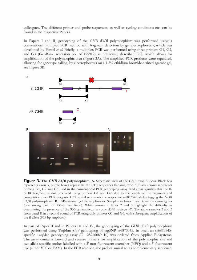
colleagues. The different primer and probe sequences, as well as cycling conditions etc. can be found in the respective Papers. In Papers I and II, genotyping of the GHR d3/fl polymorphism was performed using a conventional multiplex PCR method with fragment detection by gel electrophoresis, which was developed by Pantel et al. Briefly, a multiplex PCR was performed using three primers G1, G2, and G3 (GenBank accession no. AF155912) as previously described [72], which allows for amplification of the polymorphic area (Figure 3A). The amplified PCR products were separated, allowing for genotype calling, by electrophoresis on a 1.2% ethidium bromide-stained agarose gel, see Figure 3B.
The GHR d3/fl polymorphism. A. Schematic view of the GHR exon 3 locus. Black box represents exon 3, purple boxes represents the LTR sequences flanking exon 3. Black arrows represents primers G1, G2 and G3 used in the conventional PCR genotyping assay. Red cross signifies that the fl-GHR fragment is not produced using primers G1 and G2, due to the length of the fragment and competition over PCR reagents. C/T in red represents the respective rs6873545 alleles tagging the GHR d3/fl polymorphism. B. EtBr-stained gel electrophoresis. Samples in lanes 1 and 4 are fl-homozygotes (one strong band of 935-bp amplicon). White arrows in lanes 2 and 3 highlight the difficulty in determining the presence of the 935-bp amplicon in some d3/fl subjects. C. The same samples 2 and 3 from panel B in a second round of PCR using only primers G1 and G3, with subsequent amplification of the fl-allele (935-bp amplicon).
In part of Paper II and in Papers III and IV, the genotyping of the GHR d3/fl polymorphism was performed using TaqMan SNP genotyping of tagSNP rs6873545. In brief, an rs6873545-specific TaqMan genotyping assay (C__28966089_10) was ordered from Applied Biosystems. The assay contains forward and reverse primers for amplification of the polymorphic site and two allele-specific probes labelled with a 3’ non-fluorescent quencher (NFQ) and a 5’ fluorescent dye (either VIC or FAM). In the PCR reaction, the probes anneal to its complementary sequence.

20
The DNA polymerase then extends the template-bound primers and cleaves the probes that are hybridized to target DNA. Any non-complementary probe will be un-bound to DNA, and hence will not be cleaved in the reaction. Cleavage of target-bound probes generates allele-specific fluorescence, due to separation of the NFQ from reporter dye, which is then measured in an ABI Prism 7900HT Sequence Detection System instrument (Applied Biosystems; Figure 4).
The principles and components of the TaqMan SNP genotyping assay. For simplicity,
panels A-C represent a homozygote individual. A. Forward and reverse primers and two rs6873545-specific probes with bound fluorescent markers and non-fluorescent quencher (NFQ) are added in the PCR reaction. B. During the PCR, the target DNA is denatured, allowing for complementary binding of primers and probes. Due to the proximity of the fluorescent dye to the NFQ, no signal is generated. C. In the exonuclease reaction, elongation of the annealed primer results in cleavage of bound probe and release of the fluorescent dye from the proximity of the NFQ thus generating a fluorescent signal. D. The relative fluorescence is measured, and signals are visualized, in an allelic discrimination scatter plot.
In Paper III, most of the genotyping was performed using matrix-assisted laser desorption/ionization time-of-flight (MALDI-TOF) mass spectrometry on the iPLEX MassArray Sequenom platform. Genotyping was performed at the Mutation Analysis Facility (MAF), Karolinska Hospital, Huddinge, Sweden. The iPLEX assay is composed of several steps. First, two sets of primers per SNP are designed using the SpectroDesigner software (Sequenom Inc., San Diego, CA, USA); two amplification primers and two allele-specific extension primers. The amplification primers are designed to generate amplicons of 80-120bp in length, containing the polymorphic site, whereas the extension primers are designed in order to attach with the 3’ end immediately adjacent to its complimentary SNP allele. Experimentally, the procedure starts with a standard PCR, generating amplicons containing the SNPs of interest, followed by another round of PCR (iPLEX gold reaction) where the extension primers are used. The PCR products are then analysed on a MassARRAY compact mass spectrometer, where the amplicons are separated based on mass. Finally, the genotypes are called using the SpectroTYPER RT 4.0.5 software (Sequenom Inc.; Figure 5).

21
There are some differences between the different genotyping methods that could be mentioned. Genotyping using multiplex PCR with gel electrophoresis is a simple method using standard lab-utensils available at most labs around the world, suitable for genotyping of a small number of samples. However, it requires a large amount of DNA for the analysis which may not always be available. In addition, a second round of PCR is required in order to confirm the absence of fl-fragments whenever a perfect d3/d3 or d3/d3 with weak 935-bp amplicon (suspected d3/fl genotype) is detected, which highlight the shakiness of the multiplex efficiency, due to the complexity of the polymorphic area, and calls for another genotyping method for analysis in larger cohorts. Also, it raises the question of whether the d3-allele has been over reported in the literature, which may have skewed the conclusions drawn from these earlier reports. TaqMan SNP genotyping, on the other hand, requires much less template DNA and enables rapid and high throughput genotyping either in a 96-well or 384-well format. In addition, introduction of human errors is eliminated because allelic discrimination is performed automatically instead of by visual inspection. It does, however, require access to specific genotyping equipment, such as the ABI Prism 7900HT Sequence Detection System instrument. For genotyping of a large number of SNPs in large cohorts, the Sequenom platform is very advantageous. It allows for simultaneous analysis of up to 20 SNPs in a single pool, and it is rapid and much less expensive than TaqMan SNP genotyping of the same number of SNPs. However, the equipment is almost exclusively available at consortiums and core facilities, due to the vast cost. Also, the primer-pool design can be tricky, wherefore some SNPs may fail design and have to be analysed separately using TaqMan SNP genotyping.
SNP genotyping using the Sequenom platform. Scatter plot showing the separation of alleles. Blue triangles represent T7T samples, orange triangles represent A/A samples and green boxes represent heterozygotes (A/T).

22
In Paper I, we analysed the GHR d3/fl polymorphism in a cohort of 124 well-characterized GHD adults, who were studied after 12 months of GHRT. Using the conventional multiplex PCR developed by Pantel et al, a second PCR round was conducted whenever a perfect d3/d3 or d3/d3 with weak 935bp amplicon was suspected, which resulted in a reassignment of 57% of the originally assigned d3/d3 into d3/fl. We found that the frequency of the d3-GHR was 26% (10% d3/d3, 32% d3/fl and 58% fl/fl). Due to the small sample size and the fact that only 10% were homozygous for the d3-GHR, we divided the subjects into two genotype groups. Group 1 included only fl/fl subjects (n=72), and group 2 included combined d3/fl and d3/d3 subjects (n=52). Men and women were studied separately. At baseline, there were no significant differences in clinical characteristics of the subjects in group 1 and 2. At 12 months of GHRT, the men in group 1 (fl/fl) had a lower median daily GH dose than the men in group 2 (p=0.03). However, although there was a similar trend, the cumulative GH dose did not differ significantly (Table 3). There were no significant differences in the change in IGF-I levels and BF between the two groups.
12 months characteristics of the 124 GHD adult subjects on GHRT, according to sex and genotype group.
fl/fl d3/fl + d3/d3 All patients Men Women Men Women
GH dose (mg/day) 0.4 (0.1, 0.9) 0.3 (0.1, 0.7) 0.5 (0.3, 0.9) 0.4 (0.3, 0.8) 0.4 (0.3, 0.8)
Cum GH dose (mg) 124 (69, 405) 118 (69, 297) 128 (76, 256) 129 (74.9, 405) 128 (70.5, 220)
IGF-I (µg/L) 234 (73, 634) 258 (97, 609) 188 (92, 506) 304 (75, 634) 209 (73, 455)
∆ IGF-I (µg/L) 145.5 (-32, 481) 154.0 (-32, 368) 115.5 (27, 481) 187 (42, 438) 124 (-32, 231)
BF (kg) 19.3 (2.4, 41) 18.2 (3, 41) 25 (11, 41) 15.0 (2.4, 38.2) 22.1 (14.2, 38.0)
∆ BF (kg) -2.9 (-15, 10) -3.5 (-15, 10) -1.4 (-14.5, 3.8) -2.4 (-11, 7.8) -3.4 (-7.6, 8.7)
Data presented as median (range). ∆ values represent value at 12 months – value at baseline.
In Paper III, we analysed six SNPs in the GHR and the GH signalling pathway (JAK2, STAT5b, SOCS2 and PIK3CB) genes in a cohort of 313 well-characterized consecutive GHD adults, who underwent GH replacement for 12 months. The subjects were studied before and after 1 week, 6 months and 1 year of GH replacement. The rapid response was defined as the percentage of change in IGF-I levels from baseline to one week of GH replacement. The long-term response was investigated as 1) the percentage of change in serum IGF-I levels from baseline to 6 and 12 months, and 2) the percentage of change in serum IGF-I levels from 1 week to 6 and 12 months. GHR exon 3 deleted/full-length (d3/fl) polymorphism was analysed using tagSNP rs6873545. We found that the GHR d3/fl tagSNP rs6873545 and the PIK3CB SNP rs361072 were significantly associated with the rapid response to GH replacement (p=0.016 and p=0.025, respectively). Homozygotes of the major allele of SNP rs6873545 had on average a 45.3% larger increase in serum IGF-I levels, than individuals carrying the minor allele. Conversely, variant carriers of SNP rs361072 showed a 37.5% larger increase in serum IGF-I levels than homozygotes of the major allele. As compared to baseline, SNP rs6873545 (GHR) was associated

23
with the percentage of change in IGF-I levels at 6 months (p=0.041) and at 1 year (p=0.041). SNP rs361072 (PIK3CB) was significantly associated with the percentage of change in IGF-I levels at 6 months (p=0.047) of GH therapy. When comparing the serum IGF-I response between 1 week and 6 and 12 months, no SNP was significantly associated with the long-term responses to GH replacement therapy (Table 4).
Summary of SNP info, effect size in percentage of change in serum IGF-I and p-values from regression analyses in 313 adult GHD patients during 1 year of GHRT.
dbSNP ID Gene 1 week (n=253)
6 months (n=251)
1 year (n=256)
rs6873545 GHR -45.3 (0.016) -19.7 (0.457) -24.5 (0.382)
rs361072 PIK3CB 37.5 (0.025) -3.3 (0.894) -9.6 (0.700)
rs7849191 JAK2 8.8 (0.610) 1.7 (0.945) -0.8 (0.947)
rs6503691 STAT5b -9.1 (0.717) -3.9 (0.910) -4.0 (0.913)
rs11107116 SOCS2 29.9 (0.119) -19.2 (0.477) -16.4 (0.564)
Table shows results from linear regression analyses adjusted for sex, age and GH dose. The long-term (6 months and 1 year) percentage of change in IGF-I levels was calculated using the 1 week IGF-I levels as reference value, to denote that the long-term response corresponds to the change in IGF-I levels taking place after the early response has occurred. Effect sizes refer to the adjusted difference between the heterozygote group and homozygotes of the major allele for each SNP, negative values correspond to a lower increase in IGF-I levels during GHRT for the heterozygotes versus the homozygotes of the major allele. Significant p-values are shown in Italics.
In Paper II, we analysed the GHR d3/fl polymorphism in a cohort of 183 GHD adults, an extension to the cohort reported in Paper I. All subjects were genotyped using the conventional multiplex PCR, and also with TaqMan SNP genotyping of GHR SNP rs6873545 which has been reported as a tagSNP for the GHR d3/fl polymorphism by Lettre et al [99].
We found that the frequency of the d3-GHR was 24.0% (7.7% d3/d3, 32.2% d3/fl and 60.1% fl/fl). Genotyping success rate was 100% using tagSNP rs6873545, meaning that all samples were successfully called. In addition, the assigned genotypes were in total accordance with the genotypes achieved using the conventional genotyping method. Also, three samples which had previously not been assigned a genotype using the conventional method, due to technical difficulties, were successfully typed using the tagSNP. Figure 6 shows allelic discrimination plot from the TaqMan SNP genotyping of the GHR d3/fl polymorphism using tagSNP rs6873545.

24
Allelic discrimination of the GHR d3/fl polymorphism as determined by TaqMan
SNP genotyping. The plot shows signal intensity values of fluorescent reporter dyes FAMTM and VIC® for each sample and clear clustering of fl/fl (T/T; blue), fl/d3 (T/C; green) and d3/d3 (C/C; red) genotypes. Black dots represent no-template control (NTC).
In Paper IV, we analysed the GHR d3/fl polymorphism using tagSNP rs6873545 in the SOS reference study, which comprises 1135 subjects that were randomly selected from a population registry. We found that the frequency of the d3-GHR was 24.0% (5.6% d3/d3, 36.8% d3/fl and 57.6% fl/fl). Genotyping success rate was 99.1%. Subjects homozygous for the d3-GHR weighed approximately four kilos more (p=0.014), had larger waist-to-hip ratio (WHR; p=0.045), waist- (p=0.020) and calf circumference (p=0.0002) and more fat free mass (FFM) estimated from total body potassium (TBK; p=0.021) than grouped fl/d3 and fl/fl subjects (d3-recessive genetic model). Also, d3-GHR carriers had reduced levels of IGF-binding protein 3 (IGFBP-3), compared to fl/fl subjects (p=0.008), and borderline higher serum insulin levels (p=0.09). Genotype was not associated with serum IGF-I levels (p=0.4).

25
Half a decade has passed since Raben in 1962 first described the effects of GH administration to a 35-year-old GH deficient woman. After only two months of treatment she noted “increased vigour, ambition and sense of well-being” [100], which we now acknowledge the classical and profound effects of GH and IGF-I on the brain and QoL [101, 102]. The reports from Raben, and the one-year-later description of the physiological consequences of hypopituitarism in adults by Falkheden [103], spurred the interest in GH replacement to hypopituitary adults. After the introduction of recombinant human GH, the first treatment trials with GH to adults with hypopituitarism were conducted in the mid-1980s [33, 96], demonstrating profound metabolic effects, which further established the important role of GH in adults. Today, adults with hypopituitarism and GHD are regularly being considered for GH replacement due to the well-known consequences of adult GHD [56, 57, 104]. Long-term replacement therapy has also become more common, as discontinuation of treatment has been shown to negatively affect QoL, fat mass and distribution, lipid profile and markers of systemic inflammation [105], highlighting the need for these patients to be treated. However, in contrast to GH treatment in children, where the primary endpoints are growth velocity and final adult height, the evaluation of treatment response in adults is more complex and includes factors such as changes in body composition, lipid- and metabolic profile, bone health and QoL.
The variability in individual responsiveness to GH treatment is large, and dependent on multiple factors both in children and in adults. In children with GHD, Turner syndrome and children who are born SGA, mathematical models to predict growth response using patient characteristics and treatment modalities have been developed [106-111]. The generated algorithms have been shown to explain a high degree of observed variability of the response [107]. In GHD adults, the response to GH in body composition has been only poorly correlated with the GH dose, whereas factors such as age, sex, BMI and GHBP levels have been shown to weakly predict response [59, 63, 112, 113]. The IGF-I response has been correlated with GH dose [114], sex [115] and the route of oestrogen replacement [62, 116-120]. However, even after adjustments for these clinical factors the response variability remains largely unexplained, suggesting that the individual genetic background plays an important role in the responsiveness to GH.
The GHR d3/fl polymorphism has been considered an interesting candidate polymorphism for pharmacogenetic studies in GH replacement therapy, mainly because of its location within the GHR gene and also because it is highly frequent in most populations. The polymorphism has been investigated in both paediatric and adult populations of patients undergoing GH replacement therapy. In Paper I, we found that there was no association between the GHR d3/fl polymorphism and 12 months GH treatment response in terms of change in serum IGF-I levels and/or body composition. However, we did observe that fl/fl men required a lower mean daily GH dose to normalize serum IGF-I levels during treatment, than did grouped d3/fl and d3/d3 men. In Paper III, we found that the GHR d3/fl polymorphism, analysed using tagSNP rs6873545, was associated with the early, 1 week, response to GH replacement in terms of the percentage increase in serum IGF-I levels from baseline. When adjusted for sex, age and the baseline GH dose, fl/fl subjects had a larger percentage increase in serum IGF-I levels at 1 week than the two other genotype groups (d3/fl and d3/d3, analysed separately). The polymorphism was also associated with the long-term response to GH replacement when using the baseline

26
serum IGF-I as reference, however, this association was lost when using the 1 week serum IGF-I as reference value. The findings in Paper I and III are contrasting to previously published data, in that the fl/fl subjects required a lower GH dose (Paper I, men only) and were responding better to GH replacement (Paper III, men and women combined). In terms of serum IGF-I response to GHRT in GHD adults, there have been some other reports of the GHR d3/fl polymorphism with conflicting data. In a Dutch cohort of 99 adult GHD patients, subjects bearing at least one d3-allele had a larger increase in serum IGF-I concentrations after 12 months of GHRT [88]. Similarly, in a UK study of 194 GHD adults, d3-GHR homozygosity was associated with a greater serum IGF-I response to 12 months of GHRT [121]. Two other studies have, however, shown no effect of the GHR d3/fl polymorphism on the IGF-I response to GHRT in adults [122, 123]. In regards to an effect on the GH dose required during GHRT, in a study of 133 German GHD adults on 1 year of GHRT, d3-GHR carriers require approximately 25% less GH than fl/fl subjects [124], which is in contrast to the findings in our Paper I. In agreement with the findings in Paper I, both Giavoli et al [123] and Adetunji et al [122] have shown that there were no effects of the d3-GHR on BF response during 1 year of GHRT. The discrepancies between our study in Paper III and other studies may be reflective of several differences between the studies in regards to different cohort compositions of age, sex, aetiologies of GHD, other hormone deficiencies and their replacement, treatment regimens etc. In addition, major differences exist in regards to the size of cohorts, end-point studied, length of replacement, difficulties with the commonly used genotyping method and different genetic models used in analyses. In fact, recently, a co-dominant model of inheritance has been proposed in a Bayesian meta-analysis [125]. Most importantly, in addition to our cohort being larger than cohorts in previous reports, one of the other major strengths of the study in Paper III is that we chose to separate the response into an early (1 week), and long-term (6 and 12 months) response. The reason is that the GH treatment regime used, the individualized GH dose titration [59], unfortunately most likely masks the effect of genetic predictors of response since the response variable (IGF-I concentrations) is used as a marker to govern the GH dose adjustments consecutively during the treatment period. At 1 week of GH treatment, however, neither GH dose titration nor other confounders such as diet and life-style choices are expected to have impacted on the treatment response. This problem is further highlighted by the fact that the polymorphism was associated with the long-term IGF-I response when the baseline IGF-I was used as reference, but not when using the 1 week IGF-I as reference. This indicates that the observed association to long-term response was in fact driven by the short-term response. To our knowledge, this is the first study to investigate the association between the GHR d3/fl polymorphism and the very early treatment response to GH. Another strength of our study is that we used the percentage of change in IGF-I levels as the end-point, which we consider to be a more suitable variable for response prediction whereas previous studies have focused on crude serum IGF-I concentrations or delta values thereof. A delta value only tells the difference from baseline, but says nothing about the magnitude of change, as does the percentage of change. For instance, a patient starting GH therapy with a serum IGF-I of 50 µg/L which increases to 100 µg/L after 1 year of GHRT, will have a delta IGF-I of 50 µg/L, but a 100% increase in IGF-I levels indicating a good response. Conversely, a patient starting GH replacement with a serum IGF-I of 100 µg/L, which increases to 150 µg/L after 1 year of GH therapy, will have the same delta IGF-I as in the previous example (50 µg/L), but a lower percentage of change in serum IGF-I levels (20%). Therefore, the first patient probably has a more sensitive GH-IGF-I axis.

27
In Paper III, we found that PIK3CB SNP rs361072 was associated with the 1 week IGF-I response to GHRT. When using the baseline IGF-I as reference, the SNP was also associated with the 6 month IGF-I response.
PIK3CB encodes an isoform of the catalytic subunit of phosphoinositide 3-kinase (PI3K), a key effector of insulin signalling. PI3K is involved in mediating part of the metabolic responses to GH. Interestingly, the G-allele of PIK3CB SNP rs361072 has been shown to create a GATA binding site capable of increasing transcription of PIK3CB [93]. In addition, this SNP has also been shown to be associated with serum IGF-I levels and longevity [94]. In accordance with previous findings, which have shown that homozygotes of the G-allele of this SNP had higher free serum IGF-I levels [94], in our study the G-allele was associated with a higher percentage of increase in IGF-I levels during GHRT.
The role of GH in the regulation of human longitudinal growth and metabolism is undisputable and it is therefore not unlikely that a genetic factor influencing the individual sensitivity to GH would also have an impact. However, the influence of the GHR d3/fl polymorphism has been particularly evaluated in GHD populations, wherefore less is known about its effect in the general population with regards to anthropometry, body composition and metabolism. In Paper IV, randomly selected adults from the general Swedish population who were homozygous for the d3-GHR were heavier and had larger waist circumference and WHR than fl-GHR carriers, indicative of a less favourable phenotype. In addition, they also had tended to have higher fasting insulin levels. This may be surprising bearing in mind the lipolytic effects of GH in the therapeutic situation. Small birth size has been associated with late-onset diseases such as type 2 diabetes mellitus, hypertension and heart disease [126]. There are speculations on the reason for this observed association, and one possible explanation is that genetic variants that influence the disease risk also confers reductions in foetal growth [127]. In uterus, the foetus is exposed to high concentrations of placental GH, which is responsible for regulating its metabolism. However, the foetal longitudinal growth is mainly stimulated by insulin and IGF-I and not by GH. The d3-GHR has been found to be more prevalent among children born short for gestation age (SGA) with verified intrauterine growth restriction and also associated with a decreased third-trimester foetal growth velocity and an increased postnatal growth velocity [128]. In addition, associations between d3-GHR homozygosity and smaller birth size and earlier age at pubertal onset [129], and lower birth- and placental weight [130], have been shown. It is feasible to speculate that foetuses bearing the presumable more GH-sensitive d3-GHR become less insulin sensitive as a compensatory mechanism due to high activity of the GH-axis in these subjects, which could result in a poorer intrauterine growth. As such, the GHR d3/fl polymorphism could be a genetic factor influencing the association between insulin-mediated foetal growth (and therefore also birth weight), insulin resistance and other related metabolic aberrations which could last into adulthood [127]. The higher body weight, waist circumference and WHR in d3-GHR homozygotes observed in our randomly selected population could be related to an increased insulin exposure in these subjects and the reduced serum levels of IGFBP-3 in d3-GHR carriers might reflect a relative insulin resistance, which is known to be associated with an increased proteolysis of IGFBP-3 [131].

28
To our knowledge, our study was the first to investigate the putative influence of the GHR d3/fl polymorphism in a general, seemingly healthy, population of adult men and women. Previously, the polymorphism has been studied in subjects with normal glucose tolerance and impaired glucose tolerance including type 2 diabetes mellitus (T2DM). In that study, there was a significantly lower frequency of the d3-allele among the T2DM. However, amongst the patients with T2DM, subjects carrying the d3-GHR had significantly higher BMI [132], indicating that the d3-GHR confers a less favourable metabolic profile in this group. However, it is important to note that in this study, the genotype frequencies within the IGT and T2DM groups were not distributed in accordance with the HWE wherefore the results may be somewhat skewed.

29
Most of the work presented in this thesis has focused on the GHR d3/fl polymorphism. Figure 7 shows a summary of the main results. It was a surprise to find that in our large study of adults with GHD, the fl/fl subjects were the ones that showed the best early response to GHRT, which opposes to the general opinion. However, previous studies were smaller and have focused on the long-term response, which may explain part of the discrepancies. In terms of long-term response to GH replacement, confounders such as GH dose titration, diet and life-style choices will probably influence and mask some of the effect of genetics on the response. For that reason, the early response is more suitable to study in pharmacogenetic studies. In the general population, the d3-GHR was associated with a less favourable anthropometric profile. What could possibly be the benefit of 24% of the population to carry the d3-GHR, when it in fact predisposes to metabolic disturbances? The polymorphism has triggered the curiosity of many researchers for more than 20 years of time, and yet there are still no conclusive data on GH replacement predictive value etc. As for most medications, the response to GH replacement is probably multifactorial and I doubt that there will ever be one genetic variant that explains all variability. Most likely, there are still unknown genetic variants to be discovered that may be of an equal importance, and the GH response is most likely explained by a set of genetic variants each contributing with a fairly small, but still significant, effect. I would also like to suggest that while the early IGF-I response can be used to study the influence of genetic variants on GH responsiveness; it is the long-term outcome of GH treatment which is of the largest importance. Therefore, it would be of great importance to test variants such as the GHR d3/fl polymorphism for association with the long-term (10-15 yrs) incidence of heart disease, QoL etc, some of the most important parameters to be monitored during GH replacement.
Summary of main results.

30
Tillväxthormon är ett polypeptidhormon som produceras i hypofysen i hjärnan och som frisätts i ett pulsatilt mönster. Som namnet antyder är tillväxthormon väldigt viktigt för regleringen av längdtillväxt hos barn, men hormonet har viktiga funktioner även hos vuxna. Bland annat är tillväxthormon viktigt för regleringen av celldifferentiering och proliferation, och påverkar kroppssammansättning och metabolism. Vuxna som har brist på tillväxthormon, oftast på grund av hypofystumörer eller dess behandling, drabbas i stor utsträckning av bukfetma, diabetes mellitus och högt blodtryck och löper dessutom en nästan dubblerad risk för död i hjärt- kärlsjukdom. Med anledning av detta erbjuds dessa patienter idag ofta en livslång ersättningsbehandling med syntetiskt tillväxthormon. Hittills har det visat sig vara svårt att optimera behandlingen; det är stor variation i svaret på behandling och det saknas prediktorer för detta. Därför letar man numera i stor utsträckning efter genetiska variationer som påverkar hur olika personer svarar på behandlingen. I denna avhandling har jag undersökt genetiska variationer i tillväxthormonsystemet. Mycket fokus har legat på att studera en variant av receptorn för tillväxthormon, som tidigare visats påverka känsligheten för tillväxthormonbehandling. Sammantagningsvis fann jag att individer som bar på två kopior av den långa varianten av tillväxthormonreceptorn krävde lägre doser av tillväxthormon under 1 års behandling (bara män), samt att de svarade bättre på korttidsbehandling (män och kvinnor). Jag fann också att en genetisk variant av en signaleringspeptid nedströms om tillväxthormonreceptorn också var kopplad till ett bättre svar på korttidsbehandling med GH. Vidare visade jag att man kan använda en förenklad metod för analys av den kortare tillväxthormonreceptorn, som gör det lättare och mer tids- och materialeffektivt att analysera denna intressanta genetiska variant. Slutligen fann jag i en svensk normalpopulation med friska individer att personer som bar på två kopior av den kortare varianten av tillväxthormonreceptorn vägde mer och hade ett större midjeomfång, vilket tyder på en ogynnsam kroppssammansättning. Sammanfattningsvis visar denna avhandling att genetiska variationer inom tillväxthormon-systemet kan påverka behandlingssvaret för tillväxthormonbehandling hos vuxna samt inverka på fetmarelaterade markörer hos friska individer ur den svenska normalbefolkningen.

31
Jag skulle vilja uttrycka mina varma känslor av ödmjukhet och tacksamhet jämtemot alla kreativa, inspirerande och vänliga personer som jag haft förmånen att arbeta med under min tid som doktorand. Särskilt vill jag tacka: Gudmundur Johannsson, min huvudhandledare. Tack för att du givit mig en tydlig plats i din verksamhet. Tack för allt ansvar jag fått, och för att du litat på mina tankar och mitt omdöme, att du pushat mig att göra mer, att vara mer och till att också bli mer! Tack för alla kongressresor jag fått åka på och för att du alltid varit lika vänlig, hjälpsam och intresserad av våra projekt. Lena Carlsson Ekander, min bihandledare. Tack för att du en gång för länge sedan gav mig en plats på SOS, att du tillät mig att ta plats i gruppens projekt, och för att du ställde upp som bihandledare åt mig. Tack för alla trevliga samtal om forskningen, om livet och det vardagliga och för att du alltid har inspirerat till mer! Per-Arne Svensson, min bihandledare. Tack för att du först av alla gav mig en chans att nästla mig in på SOS! Tack för att du varit min närmsta överordnade att falla tillbaka på, som alltid hjälpt mig med glädje och som klurat och vänt och vridit på data. Tack för alla härliga stunder och alla roliga samtal på SOS! Alla medarbetare på SOS-sekretariatet, past and present, men speciellt Britt Gabrielsson, Kajsa Sjöholm, Magdalena Taube, Gerd Bergmark, Christina Torefalk, Lars Sjöström, Peter Jacobson, Lena Gripeteg, Sofie Ahlin, Åsa Anveden, Cristina Maglio, Carlo Pirazzi, Stefano Romeo, Rosellina Mancina, Maria Antonella Burza, Benedetta Motta, Maja Olsson, Vilborg Palsdottir, Daniel Hägg, Rahil Dahlén, Johanna Andersson Assarsson, Louise Olofsson, Erik Schéle, Atsuhito Saiki, Jenny Palming och Margareta Jernås. Speciellt tack till Maja, Vilborg, Rahil, Tuppen, Louisan & Erik för alla härliga stunder på tiden då det begav sig! Tack för alla after-yorks och skrivbordsstolsrallyn på Pav 8:3. Ett stort tack också till Johanna Andersson Assarsson för att du alltid tar dig tid att prata, för att du hjälpt mig med data och för alla fantastiska resor till metropolen Ullared! Tack också för att du lånade mig fettcellsnallen när jag behövde avreagera mig… Alla kollegor på endokrinsektionen och CEM, tack för alla trevliga och inspirerande samtal, möten och samarbeten, speciellt Daniel Olsson, Lise-Lott Norrman, Helena Filipsson Nyström, Oskar Ragnarsson, Mariam Elbornsson, Anna Nilsson, Ragnhildur Bergthorsdottir, Edna de Jesus Litenski Barbosa, Galina Götherström, Ingrid Hansson, Annika Reibring, Anna Olsson, Jenny Tiberg, Annika Alklind, Thord Rosén, Dimitris Chantzichristos, Penelope Trimpou, Olle Isaksson och Bengt-Åke Bengtsson. A huge thank you! specifically goes out to Edna for all the fruitful collaborations we’ve had throughout the latest years. Thank you for being so kind, humble, and for all the fun we’ve had! Ett speciellt tack till också Daniel Olsson, min vapendragare, för alla trevliga stunder vi delat tillsammans på möten, kongresser, GLS-middagar och luncher. Tack även till Prof. Bengt-Åke Bengtsson, för att du tagit dig tid att korrekturläsa min avhandling, för alla trevliga middagar och ditt upplyftande sällskap, och för att du delat med dig av din ovärderliga kunskap om GH behandling av vuxna. Tack!

32
Tack till alla medförfattare, speciellt till Staffan Nilsson för superb statistiksupport och till Ingrid Larsson för givande samtal kring kroppssammansättning och SOS referensstudie. Tack också till Björn Henning, för ovärderlig och snabb hjälp med allt det tekniska så som datorer, diskade tangentbord (funkar inte, prova ej!) och telefoner. Tack också till Mutation Analysis Facility på Karolinska Universitetssjukhuset, Huddinge, för mycket professionellt arbete med Sequenomprojektet. Speciellt tack till Kristina Duvefelt, Linda Berglind och Patrick Muller. Till Rahil, min vän. Tack för att du finns och för allt vi gjort tillsammans, för alla samtal om gott och om ont! För att du alltid litat på mig, och för att jag alltid kunnat lita tillbaks på dig! Till mina ”gamla” kurskamrater Eleonore Falk och Jessica Westlund, tack för att ni alltid finns där för mig med uppmuntran och roligheter! Tack för alla stunder vi delat, och för att ni inspirerar mig så. Ni är fantastiska. Till mina underbara Chalmers buddies; Gustav ”Dawson” Järund, Douglas Abbås, Malin Arne och Henrik Staberg, tack för att ni är så fina vänner! Jag är så glad att jag har er. Tack! Till min familj, som stöttat mig i vått och torrt och som alltid finns där för mig; mamma Barbro och pappa Kenneth, syster Maria och bror Erik. Tack för all omtanksamhet och för att ni är dem ni är! Tack också till Gustaf & Oscar, världens finaste och goaste små Skånepågar! Slutligen, ett stort tack till min alldeles egna lilla familj. Tack till mina barn Lucas & Leia för att ni förgyller min vardag, håller mig sysselsatt, och får mig att behålla fokus. Hur blev ni så perfekta? Aldrig kunde jag ens önskat mig så underbara barn, ni är mitt liv. Jag älskar er! Yuri, min karl, tack för att du alltid finns där för mig och för att du ställer upp och tar extra ansvar när jag är uppslukad av jobbet eller iväg på kongress... Tack för att du är en så fantastisk pappa till våra barn. Tack för att jag alltid kunnat lita på dig. Te amo!
This thesis was sponsored by grants from the Swedish Research Council, the Swedish Foundation for Strategic Research to the Sahlgrenska Center for Cardiovascular and Metabolic Research, the Swedish federal government under the LUA/ALF agreement, the University of Gothenburg, NovoNordisk Scandinavia AB and Pfizer.

33
1. Watson JD, Crick FH: Molecular structure of nucleic acids; a structure for
deoxyribose nucleic acid. Nature 1953, 171(4356):737-738.
2. Crick F: Central dogma of molecular biology. Nature 1970, 227(5258):561-563.
3. Ghosh A, Bansal M: A glossary of DNA structures from A to Z. Acta
crystallographica Section D, Biological crystallography 2003, 59(Pt 4):620-626.
4. Finishing the euchromatic sequence of the human genome. Nature 2004,
431(7011):931-945.
5. Gilbert W: Why genes in pieces? Nature 1978, 271(5645):501.
6. Kruglyak L, Nickerson DA: Variation is the spice of life. Nature genetics 2001,
27(3):234-236.
7. Evans WE, Johnson JA: Pharmacogenomics: the inherited basis for
interindividual differences in drug response. Annual review of genomics and human
genetics 2001, 2:9-39.
8. Weinshilboum R, Wang L: Pharmacogenomics: bench to bedside. Nature reviews
Drug discovery 2004, 3(9):739-748.
9. Kalow W, Tang BK, Endrenyi L: Hypothesis: comparisons of inter- and intra-
individual variations can substitute for twin studies in drug research.
Pharmacogenetics 1998, 8(4):283-289.
10. Evans HM, Long JA: The effect of the anterior lobe administered
intraperitoneally upon growth maturity, and ostreus cycles of the rat. Anat Rec
1921, 21:62-63.
11. Li CH, Papkoff H: Preparation and properties of growth hormone from human
and monkey pituitary glands. Science 1956, 124(3235):1293-1294.
12. Raben MS: Treatment of a pituitary dwarf with human growth hormone. The
Journal of clinical endocrinology and metabolism 1958, 18(8):901-903.
13. Mills JL, Fradkin J, Schonberger L, Gunn W, Thomson RA, Piper J, Wysowski D,
Brown P: Status report on the US human growth hormone recipient follow-up
study. Hormone research 1990, 33(2-4):116-119; discussion 120.
14. Isaksson OG, Jansson JO, Gause IA: Growth hormone stimulates longitudinal bone
growth directly. Science 1982, 216(4551):1237-1239.
15. Giustina A, Veldhuis JD: Pathophysiology of the neuroregulation of growth
hormone secretion in experimental animals and the human. Endocrine reviews
1998, 19(6):717-797.
16. Rosen T, Wiren L, Wilhelmsen L, Wiklund I, Bengtsson BA: Decreased
psychological well-being in adult patients with growth hormone deficiency.
Clinical endocrinology 1994, 40(1):111-116.
17. Bondy CA, Underwood LE, Clemmons DR, Guler HP, Bach MA, Skarulis M:
Clinical uses of insulin-like growth factor I. Annals of internal medicine 1994,
120(7):593-601.
18. Guler HP, Zapf J, Froesch ER: Short-term metabolic effects of recombinant human
insulin-like growth factor I in healthy adults. The New England journal of medicine
1987, 317(3):137-140.
19. Moses AC, Young SC, Morrow LA, O'Brien M, Clemmons DR: Recombinant
human insulin-like growth factor I increases insulin sensitivity and improves
glycemic control in type II diabetes. Diabetes 1996, 45(1):91-100.
20. Sjogren K, Liu JL, Blad K, Skrtic S, Vidal O, Wallenius V, LeRoith D, Tornell J,
Isaksson OG, Jansson JO et al: Liver-derived insulin-like growth factor I (IGF-I) is

34
the principal source of IGF-I in blood but is not required for postnatal body
growth in mice. Proceedings of the National Academy of Sciences of the United
States of America 1999, 96(12):7088-7092.
21. Barton DE, Foellmer BE, Wood WI, Francke U: Chromosome mapping of the
growth hormone receptor gene in man and mouse. Cytogenetics and cell genetics
1989, 50(2-3):137-141.
22. Godowski PJ, Leung DW, Meacham LR, Galgani JP, Hellmiss R, Keret R, Rotwein
PS, Parks JS, Laron Z, Wood WI: Characterization of the human growth hormone
receptor gene and demonstration of a partial gene deletion in two patients with
Laron-type dwarfism. Proceedings of the National Academy of Sciences of the
United States of America 1989, 86(20):8083-8087.
23. Bazan JF: Structural design and molecular evolution of a cytokine receptor
superfamily. Proceedings of the National Academy of Sciences of the United States of
America 1990, 87(18):6934-6938.
24. de Vos AM, Ultsch M, Kossiakoff AA: Human growth hormone and extracellular
domain of its receptor: crystal structure of the complex. Science 1992,
255(5042):306-312.
25. Brown RJ, Adams JJ, Pelekanos RA, Wan Y, McKinstry WJ, Palethorpe K, Seeber
RM, Monks TA, Eidne KA, Parker MW et al: Model for growth hormone receptor
activation based on subunit rotation within a receptor dimer. Nature structural &
molecular biology 2005, 12(9):814-821.
26. Leung KC: Regulation of cytokine receptor signaling by nuclear hormone
receptors: a new paradigm for receptor interaction. DNA and cell biology 2004,
23(8):463-474.
27. Zhu T, Goh EL, Graichen R, Ling L, Lobie PE: Signal transduction via the growth
hormone receptor. Cellular signalling 2001, 13(9):599-616.
28. Flores-Morales A, Greenhalgh CJ, Norstedt G, Rico-Bautista E: Negative regulation
of growth hormone receptor signaling. Mol Endocrinol 2006, 20(2):241-253.
29. Lanning NJ, Carter-Su C: Recent advances in growth hormone signaling. Reviews
in endocrine & metabolic disorders 2006, 7(4):225-235.
30. Sonksen PH: Replacement therapy in hypothalamo-pituitary insufficiency after
childhood: management in the adult. Hormone research 1990, 33 Suppl 4:45-51.
31. Binnerts A, Swart GR, Wilson JH, Hoogerbrugge N, Pols HA, Birkenhager JC,
Lamberts SW: The effect of growth hormone administration in growth hormone
deficient adults on bone, protein, carbohydrate and lipid homeostasis, as well as
on body composition. Clinical endocrinology 1992, 37(1):79-87.
32. Rosen T, Bosaeus I, Tolli J, Lindstedt G, Bengtsson BA: Increased body fat mass
and decreased extracellular fluid volume in adults with growth hormone
deficiency. Clinical endocrinology 1993, 38(1):63-71.
33. Salomon F, Cuneo RC, Hesp R, Sonksen PH: The effects of treatment with
recombinant human growth hormone on body composition and metabolism in
adults with growth hormone deficiency. The New England journal of medicine
1989, 321(26):1797-1803.
34. Bengtsson BA, Eden S, Lonn L, Kvist H, Stokland A, Lindstedt G, Bosaeus I, Tolli J,
Sjostrom L, Isaksson OG: Treatment of adults with growth hormone (GH)
deficiency with recombinant human GH. The Journal of clinical endocrinology and
metabolism 1993, 76(2):309-317.
35. Whitehead HM, Boreham C, McIlrath EM, Sheridan B, Kennedy L, Atkinson AB,
Hadden DR: Growth hormone treatment of adults with growth hormone

35
deficiency: results of a 13-month placebo controlled cross-over study. Clinical
endocrinology 1992, 36(1):45-52.
36. Bing-You RG, Denis MC, Rosen CJ: Low bone mineral density in adults with
previous hypothalamic-pituitary tumors: correlations with serum growth
hormone responses to GH-releasing hormone, insulin-like growth factor I, and
IGF binding protein 3. Calcified tissue international 1993, 52(3):183-187.
37. Holmes SJ, Economou G, Whitehouse RW, Adams JE, Shalet SM: Reduced bone
mineral density in patients with adult onset growth hormone deficiency. The
Journal of clinical endocrinology and metabolism 1994, 78(3):669-674.
38. Johansson AG, Burman P, Westermark K, Ljunghall S: The bone mineral density in
acquired growth hormone deficiency correlates with circulating levels of insulin-
like growth factor I. Journal of internal medicine 1992, 232(5):447-452.
39. Rosen T, Wilhelmsen L, Landin-Wilhelmsen K, Lappas G, Bengtsson BA: Increased
fracture frequency in adult patients with hypopituitarism and GH deficiency.
European journal of endocrinology / European Federation of Endocrine Societies
1997, 137(3):240-245.
40. Attanasio AF, Lamberts SW, Matranga AM, Birkett MA, Bates PC, Valk NK, Hilsted
J, Bengtsson BA, Strasburger CJ: Adult growth hormone (GH)-deficient patients
demonstrate heterogeneity between childhood onset and adult onset before and
during human GH treatment. Adult Growth Hormone Deficiency Study Group.
The Journal of clinical endocrinology and metabolism 1997, 82(1):82-88.
41. Cuneo RC, Salomon F, Watts GF, Hesp R, Sonksen PH: Growth hormone treatment
improves serum lipids and lipoproteins in adults with growth hormone
deficiency. Metabolism: clinical and experimental 1993, 42(12):1519-1523.
42. de Boer H, Blok GJ, Voerman HJ, Phillips M, Schouten JA: Serum lipid levels in
growth hormone-deficient men. Metabolism: clinical and experimental 1994,
43(2):199-203.
43. Johansson JO, Landin K, Tengborn L, Rosen T, Bengtsson BA: High fibrinogen and
plasminogen activator inhibitor activity in growth hormone-deficient adults.
Arteriosclerosis and thrombosis : a journal of vascular biology / American Heart
Association 1994, 14(3):434-437.
44. Rosen T, Eden S, Larson G, Wilhelmsen L, Bengtsson BA: Cardiovascular risk
factors in adult patients with growth hormone deficiency. Acta endocrinologica
1993, 129(3):195-200.
45. Johannsson G, Bengtsson BA: Growth hormone and the metabolic syndrome.
Journal of endocrinological investigation 1999, 22(5 Suppl):41-46.
46. Carroll PV, Christ ER, Bengtsson BA, Carlsson L, Christiansen JS, Clemmons D,
Hintz R, Ho K, Laron Z, Sizonenko P et al: Growth hormone deficiency in
adulthood and the effects of growth hormone replacement: a review. Growth
Hormone Research Society Scientific Committee. The Journal of clinical
endocrinology and metabolism 1998, 83(2):382-395.
47. de Boer H, Blok GJ, Van der Veen EA: Clinical aspects of growth hormone
deficiency in adults. Endocrine reviews 1995, 16(1):63-86.
48. Koltowska-Haggstrom M, Hennessy S, Mattsson AF, Monson JP, Kind P: Quality of
life assessment of growth hormone deficiency in adults (QoL-AGHDA):
comparison of normative reference data for the general population of England
and Wales with results for adult hypopituitary patients with growth hormone
deficiency. Hormone research 2005, 64(1):46-54.

36
49. Woodhouse LJ, Mukherjee A, Shalet SM, Ezzat S: The influence of growth
hormone status on physical impairments, functional limitations, and health-
related quality of life in adults. Endocrine reviews 2006, 27(3):287-317.
50. Bates AS, Van't Hoff W, Jones PJ, Clayton RN: The effect of hypopituitarism on
life expectancy. The Journal of clinical endocrinology and metabolism 1996,
81(3):1169-1172.
51. Bengtsson BA, Koppeschaar HP, Abs R, Bennmarker H, Hernberg-Stahl E, Westberg
B, Wilton P, Monson JP, Feldt-Rasmussen U, Wuster C: Growth hormone
replacement therapy is not associated with any increase in mortality. KIMS
Study Group. The Journal of clinical endocrinology and metabolism 1999,
84(11):4291-4292.
52. Besson A, Salemi S, Gallati S, Jenal A, Horn R, Mullis PS, Mullis PE: Reduced
longevity in untreated patients with isolated growth hormone deficiency. The
Journal of clinical endocrinology and metabolism 2003, 88(8):3664-3667.
53. Bulow B, Hagmar L, Mikoczy Z, Nordstrom CH, Erfurth EM: Increased
cerebrovascular mortality in patients with hypopituitarism. Clinical
endocrinology 1997, 46(1):75-81.
54. Rosen T, Bengtsson BA: Premature mortality due to cardiovascular disease in
hypopituitarism. Lancet 1990, 336(8710):285-288.
55. Tomlinson JW, Holden N, Hills RK, Wheatley K, Clayton RN, Bates AS, Sheppard
MC, Stewart PM: Association between premature mortality and hypopituitarism.
West Midlands Prospective Hypopituitary Study Group. Lancet 2001,
357(9254):425-431.
56. Johannsson G: Management of adult growth hormone deficiency. Endocrinology
and metabolism clinics of North America 2007, 36(1):203-220.
57. Ho KK: Consensus guidelines for the diagnosis and treatment of adults with GH
deficiency II: a statement of the GH Research Society in association with the
European Society for Pediatric Endocrinology, Lawson Wilkins Society,
European Society of Endocrinology, Japan Endocrine Society, and Endocrine
Society of Australia. European journal of endocrinology / European Federation of
Endocrine Societies 2007, 157(6):695-700.
58. Drake WM, Coyte D, Camacho-Hubner C, Jivanji NM, Kaltsas G, Wood DF, Trainer
PJ, Grossman AB, Besser GM, Monson JP: Optimizing growth hormone
replacement therapy by dose titration in hypopituitary adults. The Journal of
clinical endocrinology and metabolism 1998, 83(11):3913-3919.
59. Johannsson G, Rosen T, Bengtsson BA: Individualized dose titration of growth
hormone (GH) during GH replacement in hypopituitary adults. Clinical
endocrinology 1997, 47(5):571-581.
60. Gotherstrom G, Svensson J, Koranyi J, Alpsten M, Bosaeus I, Bengtsson B,
Johannsson G: A prospective study of 5 years of GH replacement therapy in GH-
deficient adults: sustained effects on body composition, bone mass, and metabolic
indices. J Clin Endocrinol Metab 2001, 86(10):4657-4665.
61. Boguszewski CL, Meister LH, Zaninelli DC, Radominski RB: One year of GH
replacement therapy with a fixed low-dose regimen improves body composition,
bone mineral density and lipid profile of GH-deficient adults. European journal of
endocrinology / European Federation of Endocrine Societies 2005, 152(1):67-75.
62. Cook DM, Ludlam WH, Cook MB: Route of estrogen administration helps to
determine growth hormone (GH) replacement dose in GH-deficient adults. The
Journal of clinical endocrinology and metabolism 1999, 84(11):3956-3960.

37
63. Johannsson G, Bjarnason R, Bramnert M, Carlsson LM, Degerblad M, Manhem P,
Rosen T, Thoren M, Bengtsson BA: The individual responsiveness to growth
hormone (GH) treatment in GH-deficient adults is dependent on the level of GH-
binding protein, body mass index, age, and gender. The Journal of clinical
endocrinology and metabolism 1996, 81(4):1575-1581.
64. Barbosa EJ, Koranyi J, Filipsson H, Bengtsson BA, Boguszewski CL, Johannsson G:
Models to predict changes in serum IGF1 and body composition in response to
GH replacement therapy in GH-deficient adults. European journal of
endocrinology / European Federation of Endocrine Societies 2010, 162(5):869-878.
65. Harrela M, Koistinen H, Kaprio J, Lehtovirta M, Tuomilehto J, Eriksson J, Toivanen
L, Koskenvuo M, Leinonen P, Koistinen R et al: Genetic and environmental
components of interindividual variation in circulating levels of IGF-I, IGF-II,
IGFBP-1, and IGFBP-3. J Clin Invest 1996, 98(11):2612-2615.
66. Sobrier ML, Duquesnoy P, Duriez B, Amselem S, Goossens M: Expression and
binding properties of two isoforms of the human growth hormone receptor. FEBS
letters 1993, 319(1-2):16-20.
67. Urbanek M, MacLeod JN, Cooke NE, Liebhaber SA: Expression of a human growth
hormone (hGH) receptor isoform is predicted by tissue-specific alternative
splicing of exon 3 of the hGH receptor gene transcript. Mol Endocrinol 1992,
6(2):279-287.
68. Mercado M, DaVila N, McLeod JF, Baumann G: Distribution of growth hormone
receptor messenger ribonucleic acid containing and lacking exon 3 in human
tissues. The Journal of clinical endocrinology and metabolism 1994, 78(3):731-735.
69. Wickelgren RB, Landin KL, Ohlsson C, Carlsson LM: Expression of exon 3-
retaining and exon 3-excluding isoforms of the human growth hormone-receptor
is regulated in an interindividual, rather than a tissue-specific, manner. The
Journal of clinical endocrinology and metabolism 1995, 80(7):2154-2157.
70. Zogopoulos G, Figueiredo R, Jenab A, Ali Z, Lefebvre Y, Goodyer CG: Expression
of exon 3-retaining and -deleted human growth hormone receptor messenger
ribonucleic acid isoforms during development. The Journal of clinical
endocrinology and metabolism 1996, 81(2):775-782.
71. Stallings-Mann ML, Ludwiczak RL, Klinger KW, Rottman F: Alternative splicing of
exon 3 of the human growth hormone receptor is the result of an unusual genetic
polymorphism. Proceedings of the National Academy of Sciences of the United States
of America 1996, 93(22):12394-12399.
72. Pantel J, Machinis K, Sobrier ML, Duquesnoy P, Goossens M, Amselem S: Species-
specific alternative splice mimicry at the growth hormone receptor locus revealed
by the lineage of retroelements during primate evolution. The Journal of biological
chemistry 2000, 275(25):18664-18669.
73. Kelly PA, Ali S, Rozakis M, Goujon L, Nagano M, Pellegrini I, Gould D, Djiane J,
Edery M, Finidori J et al: The growth hormone/prolactin receptor family. Recent
progress in hormone research 1993, 48:123-164.
74. Bass SH, Mulkerrin MG, Wells JA: A systematic mutational analysis of hormone-
binding determinants in the human growth hormone receptor. Proceedings of the
National Academy of Sciences of the United States of America 1991, 88(10):4498-
4502.
75. Urbanek M, Russell JE, Cooke NE, Liebhaber SA: Functional characterization of
the alternatively spliced, placental human growth hormone receptor. The Journal
of biological chemistry 1993, 268(25):19025-19032.

38
76. Clackson T, Wells JA: A hot spot of binding energy in a hormone-receptor
interface. Science 1995, 267(5196):383-386.
77. Dos Santos C, Essioux L, Teinturier C, Tauber M, Goffin V, Bougneres P: A common
polymorphism of the growth hormone receptor is associated with increased
responsiveness to growth hormone. Nature genetics 2004, 36(7):720-724.
78. Audi L, Carrascosa A, Esteban C, Fernandez-Cancio M, Andaluz P, Yeste D,
Espadero R, Granada ML, Wollmann H, Fryklund L: The exon 3-deleted/full-length
growth hormone receptor polymorphism does not influence the effect of puberty
or growth hormone therapy on glucose homeostasis in short non-growth
hormone-deficient small-for-gestational-age children: results from a two-year
controlled prospective study. The Journal of clinical endocrinology and metabolism
2008, 93(7):2709-2715.
79. Blum WF, Machinis K, Shavrikova EP, Keller A, Stobbe H, Pfaeffle RW, Amselem S:
The growth response to growth hormone (GH) treatment in children with
isolated GH deficiency is independent of the presence of the exon 3-minus
isoform of the GH receptor. The Journal of clinical endocrinology and metabolism
2006, 91(10):4171-4174.
80. Carrascosa A, Audi L, Esteban C, Fernandez-Cancio M, Andaluz P, Gussinye M,
Clemente M, Yeste D, Albisu MA: Growth hormone (GH) dose, but not exon 3-
deleted/full-length GH receptor polymorphism genotypes, influences growth
response to two-year GH Therapy in Short Small-for-Gestational-Age Children.
The Journal of clinical endocrinology and metabolism 2008, 93(1):147-153.
81. Carrascosa A, Audi L, Fernandez-Cancio M, Esteban C, Andaluz P, Vilaro E,
Clemente M, Yeste D, Albisu MA, Gussinye M: The exon 3-deleted/full-length
growth hormone receptor polymorphism did not influence growth response to
growth hormone therapy over two years in prepubertal short children born at
term with adequate weight and length for gestational age. The Journal of clinical
endocrinology and metabolism 2008, 93(3):764-770.
82. de Graaff LC, Meyer S, Els C, Hokken-Koelega AC: GH receptor d3 polymorphism
in Dutch patients with MPHD and IGHD born small or appropriate for
gestational age. Clinical endocrinology 2008, 68(6):930-934.
83. Binder G, Baur F, Schweizer R, Ranke MB: The d3-growth hormone (GH) receptor
polymorphism is associated with increased responsiveness to GH in Turner
syndrome and short small-for-gestational-age children. The Journal of clinical
endocrinology and metabolism 2006, 91(2):659-664.
84. Jorge AA, Marchisotti FG, Montenegro LR, Carvalho LR, Mendonca BB, Arnhold IJ:
Growth hormone (GH) pharmacogenetics: influence of GH receptor exon 3
retention or deletion on first-year growth response and final height in patients
with severe GH deficiency. The Journal of clinical endocrinology and metabolism
2006, 91(3):1076-1080.
85. Raz B, Janner M, Petkovic V, Lochmatter D, Eble A, Dattani MT, Hindmarsh PC,
Fluck CE, Mullis PE: Influence of growth hormone (GH) receptor deletion of exon
3 and full-length isoforms on GH response and final height in patients with
severe GH deficiency. The Journal of clinical endocrinology and metabolism 2008,
93(3):974-980.
86. Tauber M, Ester W, Auriol F, Molinas C, Fauvel J, Caliebe J, Nugent T, Fryklund L,
Ranke MB, Savage MO et al: GH responsiveness in a large multinational cohort of
SGA children with short stature (NESTEGG) is related to the exon 3 GHR
polymorphism. Clinical endocrinology 2007, 67(3):457-461.

39
87. Binder G, Trebar B, Baur F, Schweizer R, Ranke MB: Homozygosity of the d3-
growth hormone receptor polymorphism is associated with a high total effect of
GH on growth and a low BMI in girls with Turner syndrome. Clinical
endocrinology 2008, 68(4):567-572.
88. van der Klaauw AA, van der Straaten T, Baak-Pablo R, Biermasz NR, Guchelaar HJ,
Pereira AM, Smit JW, Romijn JA: Influence of the d3-growth hormone (GH)
receptor isoform on short-term and long-term treatment response to GH
replacement in GH-deficient adults. The Journal of clinical endocrinology and
metabolism 2008, 93(7):2828-2834.
89. Hirschhorn JN, Lettre G: Progress in genome-wide association studies of human
height. Hormone research 2009, 71 Suppl 2:5-13.
90. Ge D, Gooljar SB, Kyriakou T, Collins LJ, Swaminathan R, Snieder H, Spector TD,
O'Dell SD: Association of common JAK2 variants with body fat, insulin
sensitivity and lipid profile. Obesity (Silver Spring) 2008, 16(2):492-496.
91. Vaclavicek A, Bermejo JL, Schmutzler RK, Sutter C, Wappenschmidt B, Meindl A,
Kiechle M, Arnold N, Weber BH, Niederacher D et al: Polymorphisms in the Janus
kinase 2 (JAK)/signal transducer and activator of transcription (STAT) genes:
putative association of the STAT gene region with familial breast cancer.
Endocrine-related cancer 2007, 14(2):267-277.
92. Weedon MN, Lango H, Lindgren CM, Wallace C, Evans DM, Mangino M, Freathy
RM, Perry JR, Stevens S, Hall AS et al: Genome-wide association analysis
identifies 20 loci that influence adult height. Nature genetics 2008, 40(5):575-583.
93. Le Stunff C, Dechartres A, Mariot V, Lotton C, Trainor C, Miraglia Del Giudice E,
Meyre D, Bieche I, Laurendeau I, Froguel P et al: Association analysis indicates that
a variant GATA-binding site in the PIK3CB promoter is a Cis-acting expression
quantitative trait locus for this gene and attenuates insulin resistance in obese
children. Diabetes 2008, 57(2):494-502.
94. Bonafe M, Barbieri M, Marchegiani F, Olivieri F, Ragno E, Giampieri C, Mugianesi
E, Centurelli M, Franceschi C, Paolisso G: Polymorphic variants of insulin-like
growth factor I (IGF-I) receptor and phosphoinositide 3-kinase genes affect IGF-
I plasma levels and human longevity: cues for an evolutionarily conserved
mechanism of life span control. The Journal of clinical endocrinology and
metabolism 2003, 88(7):3299-3304.
95. Littley MD, Shalet SM, Beardwell CG, Ahmed SR, Applegate G, Sutton ML:
Hypopituitarism following external radiotherapy for pituitary tumours in adults.
The Quarterly journal of medicine 1989, 70(262):145-160.
96. Jorgensen JO, Pedersen SA, Thuesen L, Jorgensen J, Ingemann-Hansen T,
Skakkebaek NE, Christiansen JS: Beneficial effects of growth hormone treatment
in GH-deficient adults. Lancet 1989, 1(8649):1221-1225.
97. Sjostrom L, Larsson B, Backman L, Bengtsson C, Bouchard C, Dahlgren S, Hallgren
P, Jonsson E, Karlsson J, Lapidus L et al: Swedish obese subjects (SOS).
Recruitment for an intervention study and a selected description of the obese
state. International journal of obesity and related metabolic disorders : journal of the
International Association for the Study of Obesity 1992, 16(6):465-479.
98. Forbes GB, Hursh JB: Estimation of total body fat from potassium-40 content.
Science 1961, 133(3468):1918.
99. Lettre G, Butler JL, Ardlie KG, Hirschhorn JN: Common genetic variation in eight
genes of the GH/IGF1 axis does not contribute to adult height variation. Human
genetics 2007, 122(2):129-139.

40
100. Raben MS: Growth hormone. 2. Clinical use of human growth hormone. The New
England journal of medicine 1962, 266:82-86 concl.
101. Aberg MA, Aberg ND, Hedbacker H, Oscarsson J, Eriksson PS: Peripheral infusion
of IGF-I selectively induces neurogenesis in the adult rat hippocampus. The
Journal of neuroscience : the official journal of the Society for Neuroscience 2000,
20(8):2896-2903.
102. Johansson JO, Larson G, Andersson M, Elmgren A, Hynsjo L, Lindahl A, Lundberg
PA, Isaksson OG, Lindstedt S, Bengtsson BA: Treatment of growth hormone-
deficient adults with recombinant human growth hormone increases the
concentration of growth hormone in the cerebrospinal fluid and affects
neurotransmitters. Neuroendocrinology 1995, 61(1):57-66.
103. Falkheden T: Pathophysiological studies following hypophysectomy in man.
Gothenburg: University of Gothenburg; 1963.
104. Clayton PE, Cuneo RC, Juul A, Monson JP, Shalet SM, Tauber M: Consensus
statement on the management of the GH-treated adolescent in the transition to
adult care. European journal of endocrinology / European Federation of Endocrine
Societies 2005, 152(2):165-170.
105. Filipsson Nystrom H, Barbosa EJ, Nilsson AG, Norrman LL, Ragnarsson O,
Johannsson G: Discontinuing long-term GH replacement therapy--a randomized,
placebo-controlled crossover trial in adult GH deficiency. The Journal of clinical
endocrinology and metabolism 2012, 97(9):3185-3195.
106. Dahlgren J, Kristrom B, Niklasson A, Nierop AF, Rosberg S, Albertsson-Wikland K:
Models predicting the growth response to growth hormone treatment in short
children independent of GH status, birth size and gestational age. BMC medical
informatics and decision making 2007, 7:40.
107. Ranke MB, Lindberg A: Observed and predicted growth responses in prepubertal
children with growth disorders: guidance of growth hormone treatment by
empirical variables. The Journal of clinical endocrinology and metabolism 2010,
95(3):1229-1237.
108. Ranke MB, Lindberg A, Chatelain P, Wilton P, Cutfield W, Albertsson-Wikland K,
Price DA: Derivation and validation of a mathematical model for predicting the
response to exogenous recombinant human growth hormone (GH) in prepubertal
children with idiopathic GH deficiency. KIGS International Board. Kabi
Pharmacia International Growth Study. The Journal of clinical endocrinology and
metabolism 1999, 84(4):1174-1183.
109. Ranke MB, Lindberg A, Chatelain P, Wilton P, Cutfield W, Albertsson-Wikland K,
Price DA: Prediction of long-term response to recombinant human growth
hormone in Turner syndrome: development and validation of mathematical
models. KIGS International Board. Kabi International Growth Study. The
Journal of clinical endocrinology and metabolism 2000, 85(11):4212-4218.
110. Ranke MB, Lindberg A, Cowell CT, Wikland KA, Reiter EO, Wilton P, Price DA:
Prediction of response to growth hormone treatment in short children born small
for gestational age: analysis of data from KIGS (Pharmacia International
Growth Database). The Journal of clinical endocrinology and metabolism 2003,
88(1):125-131.
111. Ranke MB, Lindberg A, Martin DD, Bakker B, Wilton P, Albertsson-Wikland K,
Cowell CT, Price DA, Reiter EO: The mathematical model for total pubertal
growth in idiopathic growth hormone (GH) deficiency suggests a moderate role
of GH dose. The Journal of clinical endocrinology and metabolism 2003,
88(10):4748-4753.

41
112. Johansson AG: Gender difference in growth hormone response in adults. Journal
of endocrinological investigation 1999, 22(5 Suppl):58-60.
113. Verhelst J, Abs R, Vandeweghe M, Mockel J, Legros JJ, Copinschi G, Mahler C,
Velkeniers B, Vanhaelst L, Van Aelst A et al: Two years of replacement therapy in
adults with growth hormone deficiency. Clinical endocrinology 1997, 47(4):485-
494.
114. Bengtsson BA, Abs R, Bennmarker H, Monson JP, Feldt-Rasmussen U, Hernberg-
Stahl E, Westberg B, Wilton P, Wuster C: The effects of treatment and the
individual responsiveness to growth hormone (GH) replacement therapy in 665
GH-deficient adults. KIMS Study Group and the KIMS International Board. The
Journal of clinical endocrinology and metabolism 1999, 84(11):3929-3935.
115. Burman P, Johansson AG, Siegbahn A, Vessby B, Karlsson FA: Growth hormone
(GH)-deficient men are more responsive to GH replacement therapy than
women. The Journal of clinical endocrinology and metabolism 1997, 82(2):550-555.
116. Ceda GP, Calbiani B, Maggio M, Perelli-Ercolini S, Benedetti N, Caporotundo S,
Salimbeni I, Banchini A, Ugolotti G, Valenti G: Two-year treatment with rhGH of
one patient with chronic congestive heart failure and growth hormone deficiency.
Journal of endocrinological investigation 1999, 22(10 Suppl):51-52.
117. Janssen YJ, Helmerhorst F, Frolich M, Roelfsema F: A switch from oral (2 mg/day)
to transdermal (50 microg/day) 17beta-estradiol therapy increases serum insulin-
like growth factor-I levels in recombinant human growth hormone (GH)-
substituted women with GH deficiency. The Journal of clinical endocrinology and
metabolism 2000, 85(1):464-467.
118. Kam GY, Leung KC, Baxter RC, Ho KK: Estrogens exert route- and dose-
dependent effects on insulin-like growth factor (IGF)-binding protein-3 and the
acid-labile subunit of the IGF ternary complex. The Journal of clinical
endocrinology and metabolism 2000, 85(5):1918-1922.
119. Span JP, Pieters GF, Sweep CG, Hermus AR, Smals AG: Gender difference in
insulin-like growth factor I response to growth hormone (GH) treatment in GH-
deficient adults: role of sex hormone replacement. The Journal of clinical
endocrinology and metabolism 2000, 85(3):1121-1125.
120. Span JP, Pieters GF, Sweep FG, Hermus AR, Smals AG: Gender differences in
rhGH-induced changes in body composition in GH-deficient adults. The Journal
of clinical endocrinology and metabolism 2001, 86(9):4161-4165.
121. Moyes VJ, Walker DM, Owusu-Antwi S, Maher KT, Metherell L, Akker SA, Monson
JP, Clark AJ, Drake WM: d3-GHR genotype does not explain heterogeneity in GH
responsiveness in hypopituitary adults. Clinical endocrinology 2010, 72(6):807-
813.
122. Adetunji OR, MacFarlane IA, Javadpour M, Alfirevic A, Pirmohamed M, Blair JC:
The d3/fl-GH receptor gene polymorphism does not influence quality of life and
body composition in GH-deficient adults receiving GH replacement therapy.
European journal of endocrinology / European Federation of Endocrine Societies
2009, 161(4):541-546.
123. Giavoli C, Ferrante E, Profka E, Olgiati L, Bergamaschi S, Ronchi CL, Verrua E,
Filopanti M, Passeri E, Montefusco L et al: Influence of the d3GH receptor
polymorphism on the metabolic and biochemical phenotype of GH-deficient
adults at baseline and during short- and long-term recombinant human GH
replacement therapy. European journal of endocrinology / European Federation of
Endocrine Societies 2010, 163(3):361-368.

42
124. Meyer S, Schaefer S, Stolk L, Arp P, Uitterlinden AG, Plockinger U, Stalla GK,
Tuschy U, Weber MM, Weise A et al: Association of the exon 3 deleted/full-length
GHR polymorphism with recombinant growth hormone dose in growth
hormone-deficient adults. Pharmacogenomics 2009, 10(10):1599-1608.
125. Renehan AG, Solomon M, Zwahlen M, Morjaria R, Whatmore A, Audi L, Binder G,
Blum W, Bougneres P, Santos CD et al: Growth hormone receptor polymorphism
and growth hormone therapy response in children: a Bayesian meta-analysis.
American journal of epidemiology 2012, 175(9):867-877.
126. Barker DJ, Hales CN, Fall CH, Osmond C, Phipps K, Clark PM: Type 2 (non-
insulin-dependent) diabetes mellitus, hypertension and hyperlipidaemia
(syndrome X): relation to reduced fetal growth. Diabetologia 1993, 36(1):62-67.
127. Hattersley AT, Tooke JE: The fetal insulin hypothesis: an alternative explanation
of the association of low birthweight with diabetes and vascular disease. Lancet
1999, 353(9166):1789-1792.
128. Jensen RB, Vielwerth S, Larsen T, Greisen G, Leffers H, Juul A: The presence of the
d3-growth hormone receptor polymorphism is negatively associated with fetal
growth but positively associated with postnatal growth in healthy subjects. The
Journal of clinical endocrinology and metabolism 2007, 92(7):2758-2763.
129. Sorensen K, Aksglaede L, Petersen JH, Leffers H, Juul A: The exon 3 deleted growth
hormone receptor gene is associated with small birth size and early pubertal
onset in healthy boys. The Journal of clinical endocrinology and metabolism 2010,
95(6):2819-2826.
130. Padidela R, Bryan SM, Abu-Amero S, Hudson-Davies RE, Achermann JC, Moore
GE, Hindmarsh PC: The growth hormone receptor gene deleted for exon three
(GHRd3) polymorphism is associated with birth and placental weight. Clinical
endocrinology 2012, 76(2):236-240.
131. Bereket A, Lang CH, Blethen SL, Fan J, Frost RA, Wilson TA: Insulin-like growth
factor binding protein-3 proteolysis in children with insulin-dependent diabetes
mellitus: a possible role for insulin in the regulation of IGFBP-3 protease activity.
The Journal of clinical endocrinology and metabolism 1995, 80(8):2282-2288.
132. Strawbridge RJ, Karvestedt L, Li C, Efendic S, Ostenson CG, Gu HF, Brismar K:
GHR exon 3 polymorphism: association with type 2 diabetes mellitus and
metabolic disorder. Growth hormone & IGF research : official journal of the Growth
Hormone Research Society and the International IGF Research Society 2007,
17(5):392-398.