gy t oz a - zoological survey of india
TRANSCRIPT
OCC ,S ONAL ~p R 0.282
gy
f
c. A AVATI .v. RAMA
• 1
i t I oz a s
ZOO OGICA SU VEY 0 NO A
OCCASIONAL PAPER No. 282
RECORDS OF THE ZOOLOGICAL SURVEY OF INDIA
Taxonomy and Ecology of Ciliated Protozoa from Marginal Marine Environments of
East Coast of India
c. KALAVATI and A.V. RAMAN Andhra University, Department of Zoology, Division of
Protozoology, Visakhapatnam-530 003 (Andhra Pradesh)
Edited by the Director, Zoological Survey of India, Kolkata
Zoological Survey of India Kolkata

CITATION
Kalavati, C. and Raman, A.V. 2008. Taxonomy and Ecology of Ciliated Protozoa from Marginal Marine Environments of East Coast of India, Rec. zool. Surv. India, Dec. Paper No., 282 : 1-136, 11 colour plates (Published by the Director, Zool. Surv. India, Kolkata)
Published : March, 2008
ISBN 978-81-8171-191-5
© Govt. of India, 2008
All RIGHTS RESERVED
• No part of this publication may be reproduced, stored in a retrieval system or transmitted, in any form or by any means, electronic, mechanical, photocopying, recording or otherwise without the prior permission of the publisher.
• This book is sold subject to the condition that it shall not, by way of trade, be lent, re-sold, hired out or otherwise disposed of without the publisher's consent, in any form of binding or cover other than that in which it is published.
• The correct price of this publication is the price printed on this page. Any revised price indicated by a rubber stamp or by a sticker or by any other means is incorrect and should be unacceptabte.
PRICE India : Rs. 400.00
Foreign : $ 30; £ 2S
Published at the Publication Division by the Director, Zoological Survey of India, 234/4, A.l.C. Bose Road, 2nd MSO Building, Kolkata-700 020 and printed at Calcutta Repro Graphics Kolkata-700 006

In ever loving memory of our ,beloved Teacher
P. N. Gana'pati, F.N.A. ( 910 1984)

FOREWORD
In these modem times of a sudden and very rapidly expanding interest in the biodiversity and biocomplexity of organisms inhabiting the planet Earth, it would be well for research
biologists, in their broad ecological evaluations, to more fully appreciate the place and the
role(s) of microorganisms of diverse kinds in our global ecosystems. Aside from the prokaryotic bacteria, such "lower" eukaryotic forms as the protists (in effect, the algae and the protozoa,
plus the zoosporic and plasmodial fungi) represent a significant portion of the biomass of living
forms and are truly ubiquitous in their widespread distribution. What is more, these numerous organisms play an indispensable part at the base of the food chain and in the recycling of nutrients in the soil and water. Their healthly abundance is an absolute necessity for maintenance of a sustainable world. Obviously, then, we need to know more about them.
Not only have the protists generally been largely neglected in most studies, but those dwelling in marine niches have been even more ignored, despite the fact the oceans and seas
cover more than two-thirds of the surface of the Earth. It might be of interest to point out that phycologists have estimated that the salt-water algal diatoms alone provide well over 15% of the total global photosynthesis (carbon fixation and oxygen production), a process ultimately indispensable to all life.
Thus, the present book, emphasizing the cilioprotists, the fifth largest group of protists even
when fossil forms are included in the count, represents a valuable contribution to our basic knowledge of the ecology and taxonomy of the abundant, if neglected, marine forms such as are found along the eatem coast of India. This is the first monograph on these Indian ciliates, although shorter works have been produced on some of them by such colleagues as Drs. A. K. Das, P.N. Ganapati, G. Radhakrishna and M.V.N. Rao. ~nd the 'Nell-known 500 page book on the Ciliophora overall (part of a series on The Fauna of British India edited by R. B. S. Sewell), published in 1936 by the late Professor B. L. Bhatia, should be mentioned here as
well.
Authors Kalavati and Raman, distinguished biologists of Andhra University, Visakhapatnam, have purposely catered to the systematics of their ciliates at the alphataxonomy level, using observations on living specimens, as well as studies of fixed material stained by excellent standard teachniques, and adding original ecological data to their descriptions of involved families, genera and species. Covered by their thorough and authoritative investigation are some 127 named free-living species (of which seven are described as new) assignable to 28 recognized orders of the protist phylum Ciliophora. The scheme followed for classification at suprafamilial levels is essentially that found in the 1979 book by Corliss as updated and
modified by Small & Lynn in 1985.

(vi)
Readers/users will be delighted by the clarity of the 132 drawings of individual species and by the 11 impressive plates of colored photomicrographs of 96 variously stained specimens, not to mention the full-page blow-up of the phase-contrast picture of Euplotes vannus chosen to adorn the front and back covers of this treatise.
John O. Corliss Professor Emeritus
Department of Zoology University of Maryland
College Park, MD USA

No. 282
Records of the Zoological Survey of India
OCCASIONAL PAPER
2008
CONTENTS
1-136
1. INTRODUCTION .............................................................................................................. 1
2. ENVIRONMENT ............................................................................................................... 2
2.1 Study areas ................................................................................................................. 2
2.2 Climate..................................................................................... ............................... ... 8
2.3 Water quality .............................................................................................................. 9
3. MATERIAL AND METHODS ....................................................................................... 10
4 CILIATED PROTOZOA ................................................................................................. 12
4.1 Classified list of species.......................................................................................... 12
4.2 S}"stematic Account ................................................................................................. 21
4.3 Distribution ............................................................................................................. 109
5. SUMMARY ................................................................................................................... 114
6. ACKNOWLEDGEMENTS I.......................................................................................... 114
7. REFERENCES ............................................................................................................. ·· 114

1. INTRODUCTION
Ciliophora, with over 9,000 species described already and as many or even more of them yet to be discovered, constitute a highly differential assemblage of organisms. Despite considerable diversity, the group well represents one of the most homogenous phyla within the protistan sub-kingdom. Their remarkable cellular adaptation, efficient use of nutrients and their role as preys and predators have made ciliophorans a significant link in the 'microbial loop' both on land and in the Sea, the latter providing a rich assortment of habitats such as particulate and filamentous substrata, deep anoxic muds, sandy shores, rocky intertidal regions, planktonic and symbiotic habitats and the like.
Ehrenberg (1838) and Dujarden (1841) were probably the fust to initiate work on marine benthic ciliates following which Kent (1882) made an exceptional contribution through his 'Manual of Infusoria' By the turn of the century, Noland (1926), Kahl (1932-'35), Picken (1937) and more recently, Webb (1956) and Fenchel (1968) laid strong foundations to our knowledge on marine ciliates. Hundreds of interstitial or psammophilic species have since been described through the efforts of several marine ciliatologists and scores of reviews by them on the subject. In this context, mention should be made of the significant works carried out by Remane (1933), Kirby (1934), Dragesco (1962-1986), Faure-Fremiet (1948-1990), Borror (1968), Agameliev (1966-1970), Fenchel (1967, 1968, 1969), Uhlig (1969), Burkovsky (1969), Brown (1973), Elliot and Bamforth (1975), Hartwig (1974, 1977), Dye (1979), Smith (1981), Boynton and Small (1984),Carey (1986, 1992) and Song and his associates (1999-2007). During the past 25 years or so, the relationship of ciliates to marine ecosystems' structure, their possible role in ciliate evolution has been ably examined.
In the sea, planktonic Protozoa occur in the euphotic zone, their diversity and abundance being no less important there. Ever since Lohmann (1911) first discovered the significance of heterotrophic protists in seawater, significant studies have been made on the taxonomy and ecobiology of planktonic Protozoa through many parts of the world the most notable being Faure-Fremiet (1950), Dragesco (1960, 1963a, 1963b, 1965, 1966, 1970), Fenchel (1967), Foissner (1980, 1984, 1988) and Foissner et al. (1992) from European waters; Borror (1963, 1965, 1972, 1975), Corliss (1972, 1979), Elliot and Bamforth (1975), Hartwig, (1980), Hartwig and Parker (1977), Parker (1981), Lynn and Montagnes (1988), Lynn et al. (1988), Montagnes et al. (1988), Montagnes and Lynn (1991), Canada; Burkovsky (1970), Jankowski (1964), Agamaliev (1966, 1967, 1971, 1974a, 1974b, 1978), Boikova (1984), Mamaeva (1984), USSR; Carey and Maeda (1985), Maeda and Carey (1985), Maeda (1986), Japan and Hu and Song (1999, 2001a, 2001b), Song and Hu (1999), Song and Packroff (1997), Song and Wilbert (1997), Song et ale (1992, 2002), Gong and Song (2006) Gong et ai. (2001), Lin et al. (2005), Daode et al. (2006) and Xu et al. (2006a, 2006b) from China.
Seaweeds constitute yet another important biotope for a variety of marine macro and microorganims of which several morphologically and biologically divergent groups of ciliates

2 Rec. zool. Surv. India, Occ. Paper No. 282
have been reported. As phytal associates, their distribution on the seashore follows a distinct gradation with characteristic assemblages depending on the weed physiognomy, sediment deposition, degree of exposure, wave action and, many other tide dependent variables. There have been no significant investigations until now on this biotope
Fenchel and Jorgensen (1971) presented a good account of ciliates associated with decomposing environments. In general, while microphagous ciliates, Colpodium campylum, Glaucoma chellone, Cyclidium sp. have no specificity, species such as Glaucoma scintillans (for proteins), Cyclidium citrullus (carbohydrates), Spirostomum ambiguum and Loxocephalus luridus (for cellulose) exhibit high sensitivity to the nature of organic material (Legner, 1973). Histophagous ciliates abound in areas where the decomposition of animal remains takes place. Scuticociliates and prostomateans often dominate such assemblages.
In the Indian context, information on marine ciliates is meagre (Ganapati and Rao, 1958; Govindan Kutty and Balakrishnan Nair, 1966; Sarojini and Nagabhushanam, 1967; Rao and Ganapati, 1968; Krishnamurthy et al., 1979). More recently Radhakrishna (1984), Das and Nair (1987), Das et ale (1993), Das (1995) and, Kalavati et ale (1989, 1997) made some studies. Evidently, no in-depth studies are available. The present monograph, which is aimed at achieving this objective, contains a succinct account of diverse species of ciliates collected from marginal marine environments on the east coast of the India in particular, Chilka lagoon, Visakhaptanam coast and harbour and, the mangrove waterways and Kakinada bay in the Godavari delta.
2. ENVIRONMENT
2.1 Study Areas
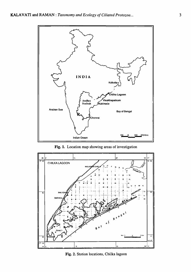
The peninsular coast of India, which extends on the east (Bay of Bengal) for nearly 800 km between Kolkata in the month (Latitude 17°40' 30" and 17°45' N; Longitude 88°16'15" and 88°25'30"E) and Cape Comorin in the south, encompasses a wide variety of environments such as bays, mangrove waterways, estuaries, harbours, coastal lagoons, coral reefs etc (Fig. 1). In addition, the open coast is interspersed with rocky, sandy or muddy shores some of which areas constitute ideal places for a great wealth of marine life. In the present investigation, four ecologically differing habitats namely, the Chilka Lake, Asia's largest brackishwater lagoon and a Ramsar site half-way between Kolkata and Visakhapatnam; Visakhapatnam harbour, a land-locked water body subject to a high incidence of pollution; the intertidal region along the sea coast of Visakhapatnam and mangroves of Godavari-Coringa estuarine system at Kakinada, were chosen. The following is a brief description of these locafions.
Chilka Lake
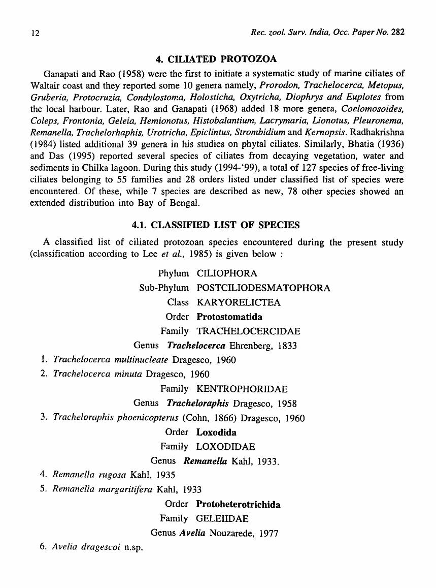
Chilka Lake, a shallow, brackishwater pear-shaped lagoon (latitude 19°28' Nand 19°54'N; longitude 84°6' E and 85°35' E) is located on the eastern seaboard of India halfway between Kolkata and Visakhapatnam (Fig. 2). The lagoon which runs almost parallel to the coast
KALA V ATI and RAMAN: Taxonomy and Ecology of Ciliated Protozoa ...
INDIA
Arabian Stta Bay of Bengal
100 0___ 100 2(\0 Kms
8!> b'
IQ'~'
4!J-
:10
Indian Ocean
Fig. 1. Location map showing areas of investigation
CHILKA LAGOON
.-•• I
,- -f. - ~.-
I -I
: .- T
, I _ : : i r-··~ --: ?r).j_ .. r -~ ... _,- -'~'YI j
- I - i
t-
;.... 1
• ,·I(i
r--!, - ., - .- . t -~ ! : Ir. I ;
1· ~- • . -+- t i - 'l'~ _. ___ + _ + , __ -llll U 'I W l(
.-. --L -_. -._ I
1!f!\6'
10
.o{)
11-_~:"-'-=~_..L ___ r--___________ -r ________ ---fHr .11'
~~-:J Fig. 2. Station locations, Chilka lagoon
3

4 Rec. zool. Surv. India, Occ. Paper No. 282
covers a total area of nearly 906.5 km2 in dry season that extends up to about 1,165.5 1on2
during the rainy season. The lagoon has an average depth of 1.4m. Topographically, Chilka Lake can be divided into two regions namely, the outer channel and the main area. The outer channel is unique in that its course does not lead directly from the sea into the lagoon, but runs parallel to it for a few kilometres. The total length of the channel is about 29 Ian and its maximum breadth is about 365 m. The outer channel together with a number of swamps that surround it is separated from the main area by islands and promontories. The channel mouth is considerably narrow and changes from time to time both in its position and dimension. The main area which is about 64.3 Ian long and 20.1 km at its greatest breadth constitutes the lagoon proper which ~an be arbitrarily divided into south, central and north sectors; the longer axis running south-west and north-east. At the southern end (Rambha Bay), the lagoon tapers into an irregularly curved point where its width is the least. The shores in the main area are composed, in some parts, by grassy slopes reaching down to the edge of the water and in other places by sand hills. Some parts are also rocky with projecting promontories.
Appreciable quantities of freshwater are discharged into the lagoon by a number of rivulets which open into the main area at several points. The most important source of freshwater into the lagoon is, however, related to the Mahanadi river system. During monsoon months (June, July and August), rivers Daya, Bhargavi and Nuna, bring large quantities of freshwater into the lagoon from the north-east side. The bottom sediments consist of mud mixed with sand, but in the outer channel the sediments are mostly sandy. There is an extensive weed growth in the lagoon. Altogether 33 species represented by 4 species of seagrasses (Halophila o valis, H. beccarii, Halodule uninervis, Potomogeton sp.), 4 macroalgae (Cladophora sp., Enteromorpha sp., Chaetomorpha sp., Gracilaria sp.) and several aquatic weeds (Pistia, Hydrilla, Najas, Vallisneria, Salvania, Ipomoea, Ceratophyllum, Ceramium, Polysiphonia) occur in Chilka Lake. Over the years, the extensive weed growth had increased siltation in the lagoon which in tum leads to a general decrease in depth. The effect of tides is negligible owing to the land-locked nature of the lagoon except at the mouth region where swift currents occur.
Visakhapatnam Harbour
Visakhapatnam harbour, a semi-enclosed water body on the east coast of India (latitude17° 41 '34" N; longitude 83°17' 45" E), is strategically located and is bound on the north, west and south sides by the mainland and the entrance to the sea, the Bay of Bengal, is through a narrow channel known as the entrance channel (Fig. 3). With the protection afforded by a high promontory into the sea known as "Dolphin's nose" on the south side and the "Ross hill" towards north, the harbour is practically immune to severe cyclonic storms that frequently cross the east coast of India. Topographically, the harbour can be conveniently divided into two major regions namely, the inner harbour and the outer harbour. The inner harbour which was created as early as 1933 consists of a central turning basin and 4 radiating navigable arms namely, the west arm, north-west arm, north arm and the southern lighter channel where the waters are practically stagnant. During the last 20 years or so, there has been an upsurge of industrial activity and urban development in this area as a result of which a number of

KALA V A TI and RAMAN: Taxonomy and Ecology of Ciliated Protozoa ... 5.
industrial undertakings such as Hindustan Petroleum Corporation Ltd., Coromandel Fertilizer Factory, Bharat Heavy Plates and Vessels, Hindustan Polymers, Zinc Smelter units, a shipbuilding yard, ore-handling units, Steel Plant, in addition to several small scale units were set-up in the vicinity of Visakhapatnam. In order to cater to the needs of growing industries and to accommodate bulk carriers, an outer harbour was built during 1976 which included construction of 3 breakwaters on the south (1543 m), east (1070 m) and north sides (412 m) and creation of a central 200 ha tranquil basin (depth -16.5 m). Over the years, the harbour has expanded enormously east-west for about 10 Ian covering an overall area of approximately 300 ha. In the inner harbour which is sheltered, the west and north-west arms are used for defence purpose since the Command Head Quarters of the Indian Navy is located here while the north arm is the main commercial channel of the port The channel is about 1.3 Jan long, 300 m wide and 10 m (mean) deep. Appreciable quantities of untreated domestic sewage from an estimated 1.5 million population of Visakhapatnam township are discharged into this channel forming the bulk of organic pollution. In addition, several intermittent drains bring sewage into this area adding to the overall organic load. Bordering the west periphery, there are extensive low-lying swampy areas with fluffy fungoid (sewage) growths which, when exposed during low tide, emit H2S. Within the channel, the waters are highly turbid with much suspended matter that imparts a distinct colouration. Earlier, a number of studies were carried out on the status of pollution in Visakhapatnam harbour. Raman (1995) summarised the information and classified north arm as a highly polluted area. The southern lighter channel which radiates from the turning basin also receives a part of the town's domestic wastes. There is a fish freezing plant on the banks of this channel where also sulphur is stored
83t 16'
~ N ..,. 'f.- Induslial Effluent .....
41'
63 16'
18'
Dolphin's Nose
17'
Fig. 3. Station locations, Visakhapatnam Harbour
co CO)
N .., 't--
N
t 42'
41'
19'

6 Ree. 2001. Surv. India, Dec. Paper No. 282
in open causing much pollution. Southern lighter channel is a highly polluted area since it -ends blindly with no means of flushing except tides.
Appreciable amounts of freshwater are discharged into Visakhapatnam harbour through a monsoon-fed river, known as "Mehadrigedda" The river which joins the north-west arm has varying rates of discharge from 0.9 (summer) to 12.1 m 3/sec (monsoon period). Although flash flooding with heavy discharges (183.0 m 3/sec) is not uncommon, estimations indicate that the annual mean discharge is about 2.1 m3/sec. "Mehadrigedda" is also the chief source of industrial pollution into the harbour since most of the major industries are located in its vicinity using it as a convenient conduit for the disposal of their effluents (Fig 3).
Visakhapatnam Coast
Visakhapatnam has an open coastline of 9.6 km most of which formed by wide stretches of co,\rse sand interspersed with rocky outcrops. There are extensive shingle beds enclosing pools of water, rocky outcrops reaching up to 6 m containing crevices and fissures and, relatively small boulders which remain submerged during high tide. The rocks form lowlying plat-forms and harbour a variety of seaweeds such as Uiva, Graci/aria, Chaetomorpha and Caulerpa etc. which support a variety of organisms including large populations of ciliates. Within the harbour, boulders placed on either side of entrance channel contain much fouling growth (Fig. 4). The intertidal region at Visakhapatnam is narrow and does not exceed 60-7Sm in width. There are no perennial rivers or streams opening into the Bay of Bengal close to Visakhapatnam.
18"
30'
83~ o·
Visakhapalnarn Harbour
Bay of Bengal
Fig. 4. Sampling locations, Visakhapatnam Coast

KALA V ATI and RAMAN: Taxonomy and Ecology of Ciliated Protozoa ... 7
Godavari-Coringa Mangroves
Kakinada Bay (Lat. 16°51 '-17°N; Lon. 82°41 '-82°22' E) (Fig. 5). a shallow bar-built water body, is bound on the south by dense mangrove vegetation and extensive mud flats intercepted by several estuarine gullies and streams emanating from one of India's largest river systems namely, the Godavari. Towards its lower reaches, river Godavari branches into two as Vashista Godavari and Gautami Godavari. Two major distributaries of Gautami Godavari namely, the Coringa arising at Yanam and Gaderu at Bhairavapalem open into Kakinada Bay on its southern side discharging spates of fresh water during the south-west monsoon period. The bay which opens widely (5.6 km) into the sea on the north side is bound on the west by the main land and on the east side there is a long narrow sand bar
17 H I ~. ~~~~~~~~~~~~~~~~~~~~
2 3 I - j-
~ ---- -; ----;--- -.~----t---+--- --L-6 i ' ; I ; I 7'- 1---- i . KAK!.NADA_I -- .: --, i . rrOWN I I
6r. : - /-1 i -1- - I :
~~ :-~--- -r- - - -+ -- --~- .. -1-- -_.j
-'-r , l
-----l---.----.;. ---- -\----- ~----I : : i
! I -T--'"
j I
---- .~.. - ! --- .--I :
-+._- -r ---j--..
+----I ,-- -
12 I I i 1.__ . .1 13 -- ---I - -t-. 1- : ' I "-
14 : I! ! K2 I '. " : -t- ~ - -
~~ -- 1- -----1--·- 1- i --+--.-- --r---·--·-17 -.-. ~ -LKAKlNADA-BAY . -.~
16° 18 55,19
20 21 22 23 24 25 26 27
16" 28 50. 29 -
30 31 -32 33 34 35 36 37
16;) 38 45' 39
40 41 -42 43 -44 45 46
J
II , ~
, -,
1
-r
I ~~- --- ~
: KG!
. . , , , K4 . - -r ------~5 . : .
~- --!
0_. ; .~ -, K8
1 K7 :. --- .. --.-.. ~--.- ~- - ..
f-
l ~BAY " . J _ . 'OF'
e~N.G~b_ I
L I
I -I
__ L - I -r---
t _oj
-.---~- ---
i -- r --
-f I
-I- -.~ 1 -
I - ~- --
Fig. S. Station locations, Mangroves of Kakinda Bay

8 Rec. zool. Surv. India, Occ. Paper No. 282
called "Hope Island" separating the bay from the sea. An important feature of Kakinada Bay is the presence of dense mangroves on the southern side up to a distance of nearly 15 lan. Surrounding Gaderu and Corioga rivers, these are high grown mangroves dominated by several species notably Avicennia marina, A. offic ina lis, Exocoecaria agallocha, Sonneratia ape ta la, Rhizophora apiculata and R. mucronata. Kakinada Bay supports a rich variety of fin and shell-fish species. In recent years, large scale conversion and reclamation of mangrove area adjoining Gaderu and Coring a for aquaculture has led to much denudation of mangrove vegetation with severe impingement in some localities.
2.2. Climate
In general, climate on the east coast of India (between Chilka Lake and Kakinada) is governed by its location in tropics and is mainly affected by seasonal monsoons that divide the year into 4 seasons as below:
Pre-monsoon period
South-west monsoon period
Post monsoon period
Cool weather season
Corresponds to March-May. Hot-weather (summer) season characterised by high atmospheric temperature and predominantly westerly winds.
June to September. Characterised by south-west winds; Weather mostly cloudy, marked by frequent rains. Maximum rainfall occurs during this period.
October-November is a period of severe cyclonic storms.
December-February. Atmospheric temperature generally low and varies between 25°C to 29°C.
Information on local climate is obtained from India Meteorological Department stations located at Hyderabad, Visakhapatnam and Gopalpur.
Air temperature
Overall, mean monthly air temperatures along the east coast range from a minimum of 19.7°C (December) to a maximum of 38.4°C (May). Seasonally, air temperatures are relatively high during April, May and June corresponding to summer months and low during December and January (cool weather).
Sunshine hours
In general, the skies along this coast of India are heavily clouded to overcast during southwest monsoon period (June-September) and clear during the remaining part of the year. At Visakhapatnam, the range is from a minimum of 2.0 hrs to a maximum of 9.8 hrs, the values being generally high (8.0-9.8 hrs) during December-April.
Winds
Winds are generally light to moderate in speed. During March-May, winds are south-

KALAVATI and RAMAN: Taxonomy and Ecology of Ciliated Protozoa ... 9
westerly, southerly or westerly. In October, winds become variable In direction. During December-February, the winds are mostly north-easterly.
Rainfall
Monthly total rainfall ranges from negligible quantities to a maximum of 507 mm (May '95), the bulk of which occurs during July-September corresponding to the south-west monsoon period.
Relative humidity
Relative humidity is in general high and ranges from 63-82 %, the observed lnean value being 76.2%
Tides
Tides are semi diurnal in nature and have a mean tidal range of 1.5 to 1. 8m. In Kakinada Bay, Ramasarma (1965) noted that with the onset of south-west monsoon (June-July), heavy drainage of freshwater occurs in the estuarine limits of Godavari River resulting in a gravity impelled flow of water in the direction of the confluence and a steep halocline ensues. The recovery cycle leading to estuarine conditions is slow beginning from October when neritic penetration gains prominence. In Visakhapatnam harbour (tidal range 0.9 m), tidal circulation had greatly .. retarded following the construction of outer harbour in 1976. In the interior channels, the effect of tide is negligible where much of the waters are effectively stagnant. In Chilka lagoon, there is no effect of tides owing to its land-locked nature.
Special weather phenomena
Storms and depressions originating in Bay of Bengal frequently cross the east coast of India in the neighbourhood of Kakinada and Visakhapatnam resulting in strong winds and widespread rains. For instance, in May '95 the storm which crossed the coast near Kakinada 300 Ion south of Visakhapatnam resulted in a rainfall of 507.5 nun in 3 days.
2.3. Water Quality
Hydrographically, Chilka lagoon, Visakhapatnam harbour and the Bay-Mangrove waterways of Kakinada may be considered as unique environments in view of their marked salinity regimes, topographical differences (location, extent, depth, tidal amplitude etc.), pollution impacts and the like. In Chilka lagoon which is subject to much annual inflows through rivers Daya, Bargabi and Nuna, variations in salinity are marked both spatially and in relation to time. Horizontally, salinity in the lagoon varies from 9-11 PSU in the southern region to almost freshwater «2.0) towards north. During the years 1996, 1997, conditions in the lagoon have further changed and in most areas salinity is <SPSU attributable to poor tidal incursion as a result of heavy siltation at the lagoon mouth. Sporadic outbursts of phytoplankton notably Oscillatoria limnetica and Microcystis sp (Cyanobacteria) have become

10 Rec. zool. Surv. India, Occ. Paper No. 282
a common feature in the lagoon due to excessive enrichment of nitrogen (max. nitrite 13.~4 mg.at.l-1) and phosphorus (max.1.72 mg.at.1- I) suggestive of the ongoing eutrophication.
The hydrographical conditions in the sea off Visakhapatnam are largely influenced by two current systems namely, the southerly current (July-December) and northerly current (JanuaryJune) which operate along the east coast of India during the above two periods. While the southerly current operates over an effective distance of 8-24 kIn from the coast, the northerly current covers a far extensive area (Ganapati and Murti, 1954). During southerly current period, fluctuations in salinity (min. 17-22 PSU) off Visakhapatnam are marked due to heavy discharges from the rivers opening into Bay of Bengal, north of Visakhapatnam (Ganapati and Ramasanna, 1958). During the northerly current, stable conditions of salinity (34 PSU) prevail owing to influx of Indian Ocean waters into Bay of Bengal. The two currents are initiated by the prevailing monsoons; southerly current being associated with north-east monsoon and northerly current with south-west monsoon. Upwelling takes place during March-April period when there is active replenishment of nutrients. This is also the period of much phytoplankton growth. Annually, sea water temperature off Visakhapatnam ranges between 24.5-32.5°C and, dissolved oxygen between 2.42 and 11.45 mg/l.
There is a high incidence of pollution in Visakhapatnam harbour owing to large scale discharge of industrial and domestic wastes. In the inner harbour, where practically stagnant conditions pre" ail, the effects are even severe. Raman (1995) had summarised information on pollution effects on harbour water quality and fauna inhabiting this area. More recently, Ratna Bharathi (1998) and Jayaprada (1998) carried out some detailed studies on protozooplankton and benthic microalgae respectively in relation to harbour water quality. The investigations revealed that the harbour waters are distinctly' characterised by a high range of temperature (median value 27.5°C-30.0°C), poor secchi-disc transparency (0.6-2.5m), high turbidity (100 NTU), varying salinity (range 5.21-33.49 PSU), generally low pH «7), widely fluctuating dissolved oxygen (0.0-20 mgfl), high permanganate oxidability (maximum 44.87 mg/l) and appreciable levels of inorganic nutrients (nitrites 0.056 mg/I; phosphate 2.60mg/l) indicative of eutrophic conditions. The observations also showed that water quality improves near the outer harbour owing to the proximity of the sea.
In the I(akinada Bay-Coringa region, the conditions are typically estuarine. The waters are in general, characterised by poor transparency (mean 0.30 m), high turbidity (max.200 NTU), widely varying salinity (freshwater to 34.36 PSU), dissolved oxygen (2.4-10.58 mg/ 1), nitrites (4.87-15.0 mg.at.l-1) and phosphates (2.51-5.09 mg.at.l-1). An important feature of the area is that considerable outwelling takes place from the mangroves affecting the Bay environment and nearby waterways.
3. MATERIAL AND METHODS
Taxonomic findings presented in the monograph are based on observations made during 1994-' 99 at select locations along the east coast of India namely, Chilka lagoon in Orissa State, Visakhapatnam harbour and the sea coast close to. Andhra University and, the mangrove

KALAVATI and RAMAN: Taxonomy and Ecology o/Ciliated Protozoa ... 11
waterways and sediments of Kakinada Bay and Coring a in the Godavari delta in Andhra Pradesh. Samples were collected through divergent means employing shallow water samplers (1-2 m), a van Veen or Petersen grab, a PVC corer, 12" x 1.5", for psammophilic species, band-picking in the case of weed inhabitants or epipelic forms and so on. The samples were obtained at random and in replicates taking care that they represented adequately the area examined. In the laboratory, the water samples were concentrated through filtration (Millipore, GF/C diameter 4.8m) under low suction. Propagation of the ciliates was carried out by maintaining the concentrated sample at a temperature of 27-28°C for 24hrs in filtered seawater enriched with cooked barley grain.
In the case of benthic ciliates, the organisms were extracted by sea water ice method of Uhlig (1966).
Epiphytic ciliates were collected by washing the weeds with copious filtered seawater collected ~om the same locality. Part of the sample was subjected to environmental deterioration method (Uhlig, 1966). The samples were then filtered through nylobolt and cultures maintained for at least 24 hrs prior to examination.
Soil extract enriched with starch appeared most suitable while maintaining laboratory cultures. All fresh observations were made using supravital stains such as 1 % methyl green in ~cetic acid, or 1 % aqueous neutral red, or 1 % acetocarmine or toludine blue at pH 4.6. Whenever necessary, the organisms were immobilised by mild centrifugation. For morphological details, cells were fixed in freshly prepared Nissenbaun's fixative or Carnoy's fluid and stained either with Erlich's haematoxylin or Harris haematoxylin or according to Feulgen's technique. Infraciliature is stained according to modified dry silver nitrate method (Roberts and Causton,1988) or ammonical silver carbonate (Frenandez-Galiano, 1976) or nigrosin-mercuric chloride-formalin technique (Borror, 1968) or dry silver method (Foissner, 1976)or protorgoal impregnation (Wilbert, 1975). All diagrams were drawn with the aid of a camera lucid a and measurements taken with an ocular micrometer. Samples stored either in Lugol' s iodine or 10% buffered formalin are available in Author's collections, Department of Zoology, Andhra University.
Descriptions of certain sedentary species (e.g. Peritrichia and Suctoria) have been excluded from the monograph. Similarly, some suctorids found frequently as planktonic forms could be identified up to generic level only. The taxa were arranged family wise as per Small and Lynn in Lee et al., (1985). All species identifications were based on Kahl (1935), Bick (1972), Corliss (1956, 1969) Curds (1982) and Carey (1992).
Abbreviations used
The following abbreviations are used in the text: ACR : Amphisiellid ciliary ro'.v; APK : Anterior polykinetids; AZM : Adoral zone of Membraenellae; CY.Py. Cytopyge; CV : Contractile vacuole; EPK : External Polykinetids; EPZ : External Polykinetid Zone; IPK: Internal Polykinetids; Ma.N : Macronuclues; Mi.N : Micronuclues; P : Peristome VPZ: Ventral Polykinetid Zone
12 Rec. zool. Surv. India, Occ. Paper No. 282
4. CILIA TED PROTOZOA
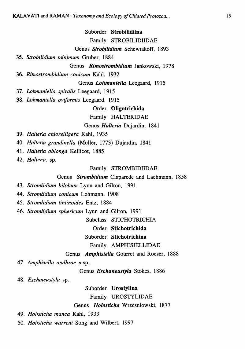
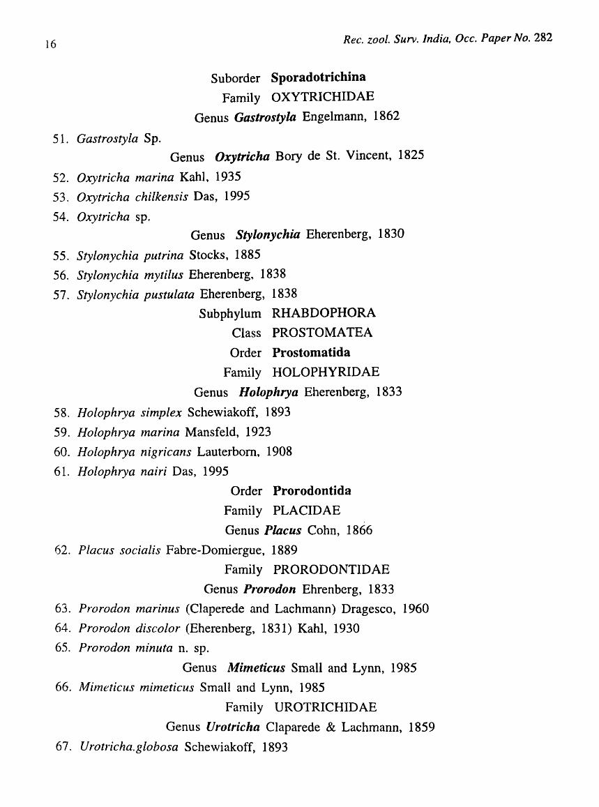
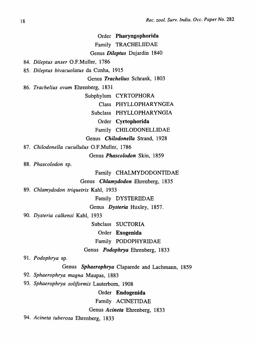
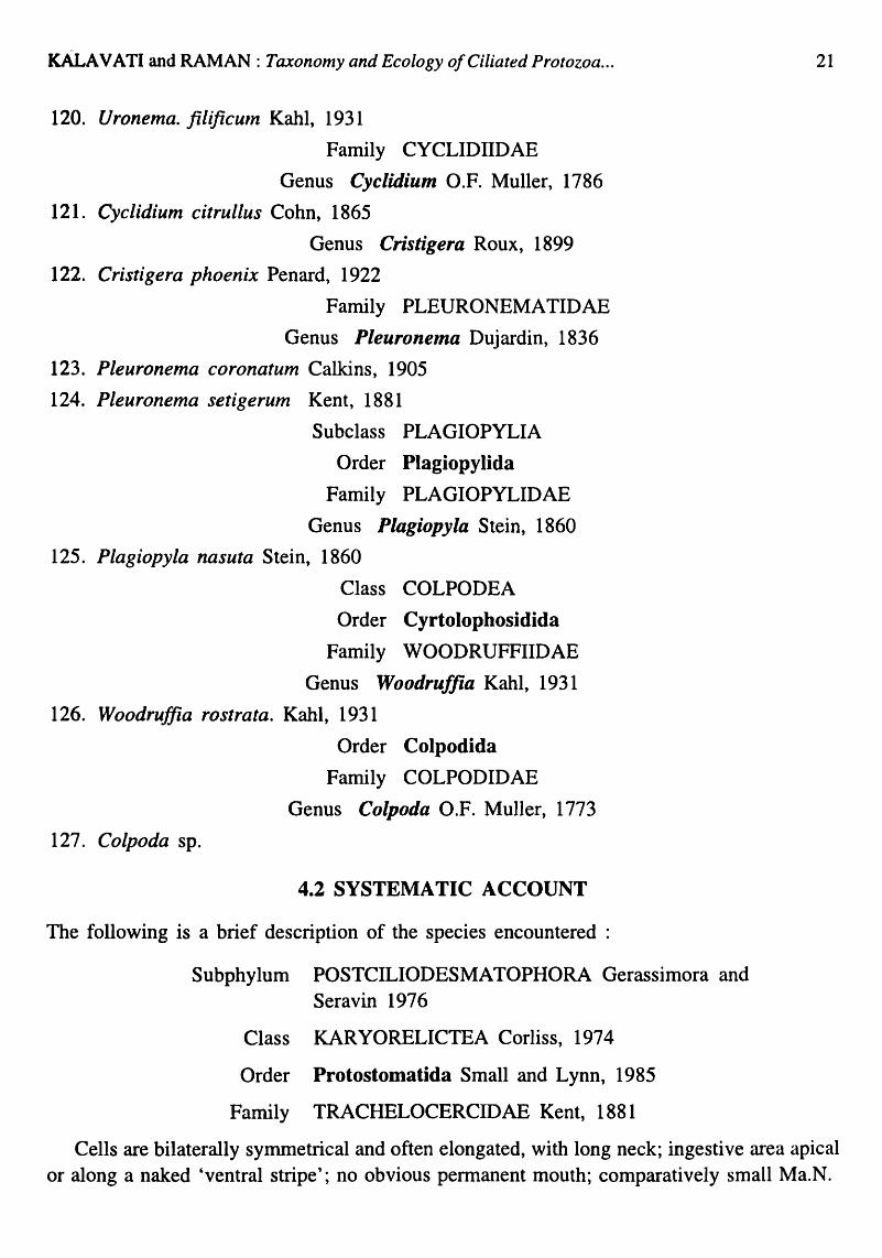
Ganapati and Rao (1958) were the first to initiate a systematic study of marine ciliates of Waltair coast and they reported some 10 genera namely, Prorodon, Trachelocerca, Metopus, Gruberia, Protocruzia, Con dy 10 stom a, Holosticha, Oxytricha, Diophrys and EupLotes from the local harbour. Later, Rao and Ganapati (1968) added 18 more genera, Coelomosoides, Coleps, Frontonia, Geleia, Hemionotus, Histobalantium, La cryma ria, Lionotus, PLeuronema, Remanella, Trachelorhaphis, Urotricha, Epiclintus, Strombidium and Kernopsis. Radhakrishna (1984) listed additional 39 genera in his studies on phytal ciliates. Similarly, Bhatia (1936) and Das (1995) reported several species of ciliates from decaying vegetation, water and sediments in Chilka lagoon. During this study (1994-'99), a total of 127 species of free-living ciliates belonging to 55 families and 28 orders listed under classified list of species were encountered. Of these, while 7 species are described as new, 78 other species showed an extended distribution into Bay of Bengal.
4.1. CLASSIFIED LIST OF SPECIES
A classified list of ciliated protozoan species encountered during the present study (classification according to Lee et ai., 1985) is given below:
Phylum CILIOPHORA
Sub-Phylum POSTCILIODESMA TOPHORA
Class KARYORELICTEA
Order Protostomatida
Family TRACHELOCERCIDAE
Genus Trachelocerca Ehrenberg, 1833
1. Trachelocerca multinucleate Dragesco, 1960
2. Trachelocerca minuta Dragesco, 1960
Family KENTROPHORIDAE
Genus Tracheloraphis Dragesco, 1958
3. Tracheloraphis phoenicopterus (Cohn, 1866) Dragesco, 1960
Order Loxodida
Family LOXODIDAE
Genus Remanella Kahl, 1933.
4. Remanella rugosa Kahl, 1935
5. Renlanella margaritifera Kahl, 1933
6. Avelia dragescoi n.sp.
Order Protoheterotrichida
Family GELEIIDAE
Genus Avelia Nouzarede, 1977

KALA V ATI and RAMAN: Taxonomy and Ecology of Ciliated Protozoa ...
Genus Geleia Kahl, 1933.
7. Geleia nigriceps Kahl, 1933
8. Geleia Jossata Kahl, 1933
9. Geleia decolor Kahl, 1933
Order Protocruziida
Family PROTOCRUZIIDAE
Genus Protocruzia da Faria, da Cunha and Pinto, 1922
10. Protocruzia adherans Mansfield, 1923
11. Protocruzia piggerina Cohn, 1866
Class SPIROTRICHEA
Subclass HETEROTRICHIA
Order Heterotrichida
Suborder Heterotrichina
Family BLEPHARISMIDAE
Genus Anigsteinia Isquith, 1968
12. Anigsteinia salinarum Isquith, 1968
Genus Blepharisma Perty, 1849
13. Blepharisma clarissimunt Anigstein, 1912
Genus Parablepharisma Kahl, 1932
14. Parablepharisma indica n.sp.
Family CLIMACOSTOMIDAE
Genus Fabrea Henneguy, 1890
15. Fabrea salina Henneguy, 1890
16. Fabrea corlissi n.sp.
Family CONDYLOSTOMA TIDAE
Genus Condylostoma Bory de St. Vincent, 1824
17. Condylostoma patens (O.F. Muller, 1786) Dujardin, 1841
18. Condylostoma arenarium Spiegel, 1926
19. Condylostonza minuta n. sp.
Family SPIROSTOMIDAE
Genus Gruberia Kahl, 1933.
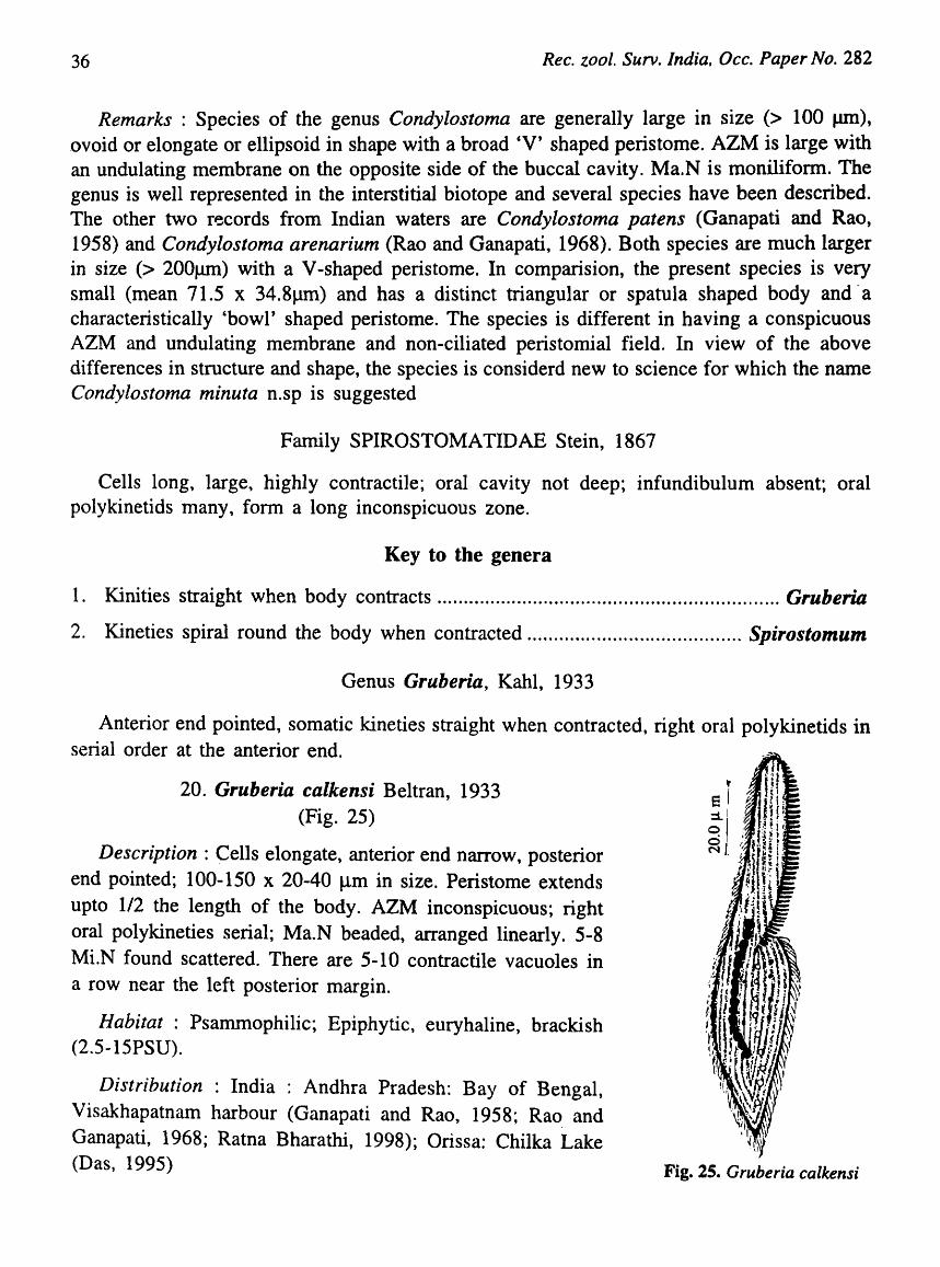
20. Gruberia calkensi Beltran, 1933.
Genus Spirostolnum Ehrenberg, 1837
21. Spirostomum. teres Claparede and Lachmann, 1859
22. Spirostomuln.minus Raux, 1901.
13

14 Rec. zoo I. Surv. India, Occ. Paper No. 282
Order Armophorida
Family CAENOMORPHIDAE
Genus Caenomorpha Perty, 1852
23. Caenomorpha capucina Kahl, 1933.
24. Caenomorpha Levanderi Kahl, 1927.
Family METOPIDAE
Genus Metopus Kahl, 1927
25. M etopus halophilus Kahl, 1925 in Corliss, 1960
26. Metopus. vestitutus Kahl, 1932
Order Phacodiniida
Family PHACODINIIDAE
Genus Phacodinium Prowazek, 1900
27. Phacodinium metchnicoJfi var Indica present study
Order Odontostomatida
Family EPALXELLIDAE
Genus Epalxella Corliss, 1960
28. EpaLxella straita Kahl, 1932
Subclass CHOREOTRICHIA
Order Choreotrichida
Suborder Tintinnina
Family CODONELLIDAE
Genus Tintinnopsis Stein, 1867
29. Tintinnopsis lohmanni Laackmann, 1906
30. Tintinnopsis beroidea Stein, 1867
Family PTYCHOCYCLIDIDAE
Genus Favella Jorgensen, 1924
31. Favella ehrenbergi (Jorgensen, 1924) Laval Pinto, 1981
Family TINTINNIDIIDAE
Genus Ti"tinnidium Kent, 1881 32. Tintinnidium jluviatale, Stein, 1863
Suborder Strombidinopsina
Family STROMBIDINOPSIDAE
Genus Strombidinopsis Kent, 1881
33. Strombidinopsis acuminatum Faure-Fremiet, 1924
34. Strombidinopsis cheshirii Snyder and Ohman, 1991
KALAVATI and RAMAN: Taxonomy and Ecology a/Ciliated Protozoa ...
Suborder Strobilidiina
Family STROBILIDIIDAE
Genus S.trQi)ilidium Schewiakoff, 1893
35. Strobilidium minimum Gruber, 1884
Genus Rimostrombidium Jankowski, 1978
36. Rimostrombidium conicum Kahl, 1932
Genus Lohmaniella Leegaard, 1915
37. Lohmaniella spiralis Leegaard, 1915
38. Lohmaniella oviformis Leegaard, 1915
Order Oligotrichida
Family HALTERIDAE
Genus Halteria Dujardin, 1841
39. Halter:a chlorelligera Kahl, 1935
40. Halteria grandinella (Muller, 1773) Dujardin, 1841
41. Halteria oblonga Kellicot, 188~
42. Haltera. sp.
Family STROMBIDIIDAE
Genus Strombidium Claparede and Lachmann, 1858
43. Stromlidium bilobum Lynn and Gilron, 1991
44. Stromfidium conicum Lohmann, 1908
45. Stromfidium tintinoides Entz, 1884
46. Stromlidium sphericum Lynn and Gilron, 1991
Subclass STICHOTRICHIA
Order Stichotrichida
Suborder Stichotrichina
Family AMPHISIELLIDAE
Genus Amphisiella Gourret and Roeser, 1888
47. Amph6iella andhrae n.sp.
48. Esch01eustyla sp.
Genus Eschaneustyla Stokes, 1886
Suborder Urostylina
Family UROSTYLIDAE
Genus Holosticha Wrzesniowski, 1877
49. HoZ,sticha manca Kahl, 1933
50. Hobsticha warreni Song and Wilbert, 1997
15
16
51. Gastrostyla Sp.
Rec. zool. Surv. India, Dec. Paper No. 282
Suborder Sporadotrichina
Family OXYTRICHIDAE
Genus Gastrostyla Engelmann, 1862
Genus Oxytricha Bory de St. Vincent, 1825
52. Oxytricha marina Kahl, 1935
53. Oxytricha chilkensis Das, 1995
54. Oxytricha sp. Genus Stylonychia Eherenberg, 1830
55. Stylonychia putrina Stocks, 1885
56. Stylonychia mytilus Eherenberg, 1838
57. Sty/onychia pustulata Eherenberg, 1838
Subphylum RHABDOPHORA
Class PROSTOMA TEA
Order Prostomatida
Family HOLOPHYRIDAE
Genus Holophrya Eherenberg, 1833
58. Holophrya simplex Schewiakoff, 1893
59. Holophlya marina Mansfeld, 1923
60. Holophrya nigricans Lauterbom, 1908
61. Holophrya nair; Das, 1995
Order Prorodontida
Family PLACIDAE
Genus Placus Cohn, 1866
62. Placus socialis Fabre-Domiergue, 1889
Family PRORODONTIDAE
Genus p,.orodon Ehrenberg, 1833
63. Prorodon Inarinus (Claperede and Lachmann) Dragesco, 1960
64. Prorodon discolor (Eherenberg, 1831) Kahl, 1930
65. Prorodon minuta n. sp.
Genus Mimeticus Small and Lynn, 1985
66. Mimeticus mimeticus Small and Lynn, 1985
Family UROTRICHIDAE
Genus Urotricha Claparede & Lachmann, 1859
67. Urotricha.globosa Schewiakoff, 1893

KALA V ATI and RAMAN: Taxonomy and EcoLogy of Ciliated Protozoa ...
Class LITOSTOMA TEA
Subclass HAPTORIA
Order Haptorida
Family DIDINIIDAE
Genus Didiniuln Stein, 1859
68. Didinium nasutuln O.P.Muller, 1786
Family ENCHEL YIDAE
Genus Enchelys O.F.Muller, 1773
69. Enchelys pectinata Kahl, 1933
70. Enchelys marina Meunier, 1907
Family LACRYMARIIDAE
Genus Lacrymaria Bory de St. Vincent, 1826.
71. Lacrymaria olar Kahl, 1933
72. Lacrymaria coronate Lachmann, 1859
73. Lacrymaria elegans Engelmann, 1862
74. Lacrymaria marina Kahl, 1933
75. Lacrymaria sapropeUca Kahl, 1927
Family SPATHIDIIDAE
Genus Spathidium Dujardin, 1841
76. Spathidium fossicola Kahl, 1933
Family TRACHELOPHYLLIDAE
Genus Lagynophrya Kahl, 1927
77. Lagynophrya salina Kirby, 1932
Genus Trachelophyllum Claparede and Lachmann, 1859
78. Trachelophyllum sp.
Order Pleurostomatida
Family AMPHILEPTIDAE
Genus Litonotus Wreniowski, 1870
79. Litonotus obtuses Maupas, 1888
Genus Amphileptus (Eherenberg, 1838) Buetschli, 1889
80. A111phileptus claparedei Stein, 1867
81. Amphileptus trachelioides J ach, 1893
Genus Loxophyllum Dujardin, 1841
82. Loxophyllum setigerunl Quennerstedt, 1867
83. Loxophyllum verrucosum Stokes, 1893.
17
18 Rec. zool. Surv. India, O~c. Paper No. 282
Order Pharyngophorida
Family TRACHELIIDAE
Genus Dileptus Dujardin 1840
84. Dileptus anser O.F.Muller, 1786
85. Dileptus bivacuolatus da Cunha, 1915
Genus Trachelius Schrank, 1803
86. Trachelius ovum Ehrenberg, 1831
Subphylum CYRTOPHORA
Class PHYLLOPHARYNGEA
Subclass PHYLLOPHARYNGIA
Order Cyrtophorida
Family CHILODONELLIDAE
Genus Chilodonella Strand, 1928
87. Chilodonella cucullulus O.F.Muller, 1786
Genus Phascolodon Skin, 1859
88. Phascolodon sp.
Family CHALMYDODONTIDAE
Genus Chlamydodon Ehrenberg, 1835
89. Chlamydodon triquetris Kahl, 1933
Family DYSTERIIDAE
Genus Dysteria Huxley, 1857.
90. Dysteria calkensi Kahl, 1933
91. Podophrya sp.
Subclass SUCTORIA
Order Exogenida
Family PODOPHYRIDAE
Genus Podophrya Ehrenberg, 1833
Genus Sphaerophrya Claparede and Lac hm ann , 1859
92. Sphaerophrya magna Maupas, 1883
93. Sphaerophrya soliformis Lauterbom, 1908
Order Endogenida
Family ACINETIDAE
Genus Acineta Ehrenberg, 1833
94. Acineta tuberosa Ehrenberg, 1833

KALAVATI and RAMAN: Taxonomy and Ecology o/Ciliated Protozoa ...
95. Trematosoma sp.
96. Trichophrya. sp.
97. Tokophrya sp.
98. Orthodonella sp.
Genus Trematosoma Batisse, 1973.
Family DENDROSOMATIDAE
Genus Trichophrya
Family TOKOPHRYIDAE
Genus Tokophrya Butschli, 1889.
Class NASSOPHOREA
Subclass NASSOPHORIA
Order Synhymeniida
Family ORTHODONELLIDAE
Genus Orthodonella Bhatia, 1936
Order Nassulida
Family NASSULIDAE
Genus Nassula Ehrenberg, 1833
99. Nassula notate (O.F. Muller) Buddbr, 1911
100. Nassula citrea Kahl, 1933
101. Furgasonia sp.
Family FURGASONIIDAE
Genus Furgosonia Jankowski, 1964
Order Peniculida
Family FRONTONIIDAE
Genus Frontonia Ehrenberg, 1838
102. Frontonia marina Fabre-Dom, 1891
Subclass HYPOTRICHIA
Order Euplotida
Family ASPIDISCIDAE
Genus Aspidisca Ehrenberg, 1838
103. Aspidisca lynceus Ehrenberg, 1833
104. Aspidisca costata (Dujardin, 1842) Claparede and Lachmann, 1859
105. Aspidisca aculeata (Ehrenberg, 1838) Mansfeld, 1926
19

20 Rec. zool. Surv. India, Dcc. Paper No. 282
Family EUPLOTIDAE
Genus Euplotes Ehrenberg, 1830
106. Euplotes charon (O.FMuller) Ehrenberg, 1830
107. Euplotes moebusi Kahl, 1935
Genus Euplotopsis Borror and Hill, 1995
108. Euplotopsis affinis Dujardin, 1841
Genus Euplotoides Borror and Hill, 1995
109. Euplotoides aediculatus Pearson, 1943
Genus Moneuplotes Jankowski, 1979.
110. Moneuplotes vannus (Mueller, 1786) Jankowski, 1979
111. Moneuplotes terricola Penard, 1922
Genus Paraeuplotes Witchennann, 1942
112. Paraeuplotes andhrae n.sp.
Family URONYCHIDAE
Genus Diophrys
113. Diophrys appendiculata Ehrenberg, 1838
Class OLIGOHYMENOPHOREA
Subclass HYMENOSTOMA TIA
Order Hymenostomatida
Family TETRAHYMENIDAE
Genus Tetrahymena Furgason, 1940
114. Tetrahymena pyriforlnis (coI11plex) Ehrenberg, 1838
115. Tetrahymena thermophila Nanney and McCoy,1976
Family TURANIELLIDAE
Genus Colpidium Stein, 1860
116. Colpidium campylum Stokes, 1886
117. Biggeria ganapatii.n.sp.
Order Scuticociliatida
Family CRYPTOCHILIDAE
Genus Biggeria Kahl, 1934
Family URONEMA TIDAE
Genus Uronema Dujardin, 1841
118. Uronema Inarinum Dujardin, 1841
119. Uronenla nigricans (O.F. Muller, 1786) Florentin, 1901
KALA V ATI and RAMAN: Taxonomy and Ecology of Ciliated Protozoa ...
120. Uronema. filificuln Kahl, 1931
Family CYCLIDIIDAE
Genus Cyclidium O.F. Muller, 1786
121. Cyclidium citrullus Cohn, 1865
Genus Cristigera Roux, 1899
122. Cristigera phoenix Penard, 1922
Family PLEURONEMATIDAE
Genus Pleuronema Dujardin, 1836
123. Pleuronema coronatum Calkins, 1905
124. Pleuronema setigerum Kent, 1881
Subclass PLAGIOPYLIA
Order Plagiopylida
Family PLAGIOPYLIDAE
Genus Plagiopyla Stein, 1860
125. Plagiopyla nasuta Stein, 1860
Class COLPODEA
Order Cyrtolopbosidida
Family WOODRUFFIIDAE
Genus Woodruffia Kahl, 1931
126. Woodruffia rostrata. Kahl, 1931
127. Colpoda sp.
Order Colpodida
Family COLPODIDAE
Genus Colpoda O.F. Muller, 1773
4.2 SYSTEMATIC ACCOUNT
The following is a brief description of the species encountered :
Subphylum POSTCILIODESMATOPHORA Gerassimora and Seravin 1976
Class KARYORELICTEA Corliss, 1974
Order Protostomatida Small and Lynn, 1985
Family TRACHELOCERCIDAE Kent, 1881
21
Cells are bilaterally symmetrical and often elongated, with long neck; ingestive area apical or along a naked 'ventral stripe'; no obvious permanent mouth; comparatively small Ma.N.

22 Rec. zoo I. Surv. India, Occ. Paper No. 282
Key to the genera
1. Ingestion apical ............................................................................................ Trachelocerca
2. Ingestion apical or along the vental stripe ............................................... Tracheloraphis
Genus Trachelocerca Ehrenberg, 1833.
Ingestion apical; perioral cilia longer than body cilia; cortex completely covered by kineties.
Key to speci~s
1. Cells spindle shaped, highly contractile, cytostome apical, Ma.N in 8-10 fragments Contractile vacuole 3, posterior ............................................................ T. multinucleatum
2. Cells small, elongate, narrow, ends bluntly pointed, Ma.N ill 4 closely placed fragments, contractile vacuole 8-10, in a row ...................................................................... T. minuta
1. Trachelocerca multinucleata Dragesco, 1960 (Fig. 6)
Description : Cells elongate, spindle shaped, highly contractile with pointed ends, 240-400 x 64-80 Jlm in size when contracted; cytostome apical surrounded by a row of short trichocysts; Ma.N in the form of 8-12 fragments ap-anged in two rows. There are three linearly arranged contractile vacuoles at the posterior end; somatic ciliature uniform, in 8-10 oblique rows; Perioral cilia distinctly longer than somatic cilia.
Habitat: Psammophilic, marine, polyhaline (28-31 PSU)
Distribution : India : Andhra Pradesh: Visakhapatnam coast (present record).
Elsewhere : Cocameau, Bay of Biscay (Faure- Fremiet, 19~8); First record from India.
).
Fig. 6. Trachelocerca multinucleata
2. Trachelocerca minuta Dragesco, 1960 (Fig 7; Plate I Fig. 1)
Description : Cells elongate, fiat, narrow with blunt ends and appear typically spindle shaped when contracted; 100-200 x 20-35 flm i~ size; cytostome apical, trichites indistinct;
KALA V ATI and RAMAN: Taxonomy and Ecology of Ciliated Protozoa ...
Ma.N. in four fragments closely placed in the center of the body. There are 8-10 contractile vacuoles arranged in a row near the right posterior margin; Perioral cilia longer than somatic cilia, Somatic cilia uniform, Monokineties in 18-20 oblique rows.
Habitat : Epiphytic, Mangrove sediments, mesohaline (2-IOPSU)
Distribution: India: Andhra Pradesh: Godavari-Coringa mangroves (present record); Waltair Coast (Rao and Ganapati, 1968), Orissa: Chilka lake (Das, 1995),
Elsewhere : Concameau, Bay of Biscay and Roscoff (Dragesco, 1960).
Genus Tracheloraphis Dragesco, 1958
8 ::l o o l/")
Description : Cells naked, ventral stripe 1/8 to 1/2 of Fig.7. T. minuta
the cortical diameter; Ingestion either apical or along the
23
ventral stripe. Somatic cilia in 12-26 kineties; Nuclear complex includes 10-12 Ma.N. and 6-8 MLN; Perioral cilia longer than body cilia.
i\
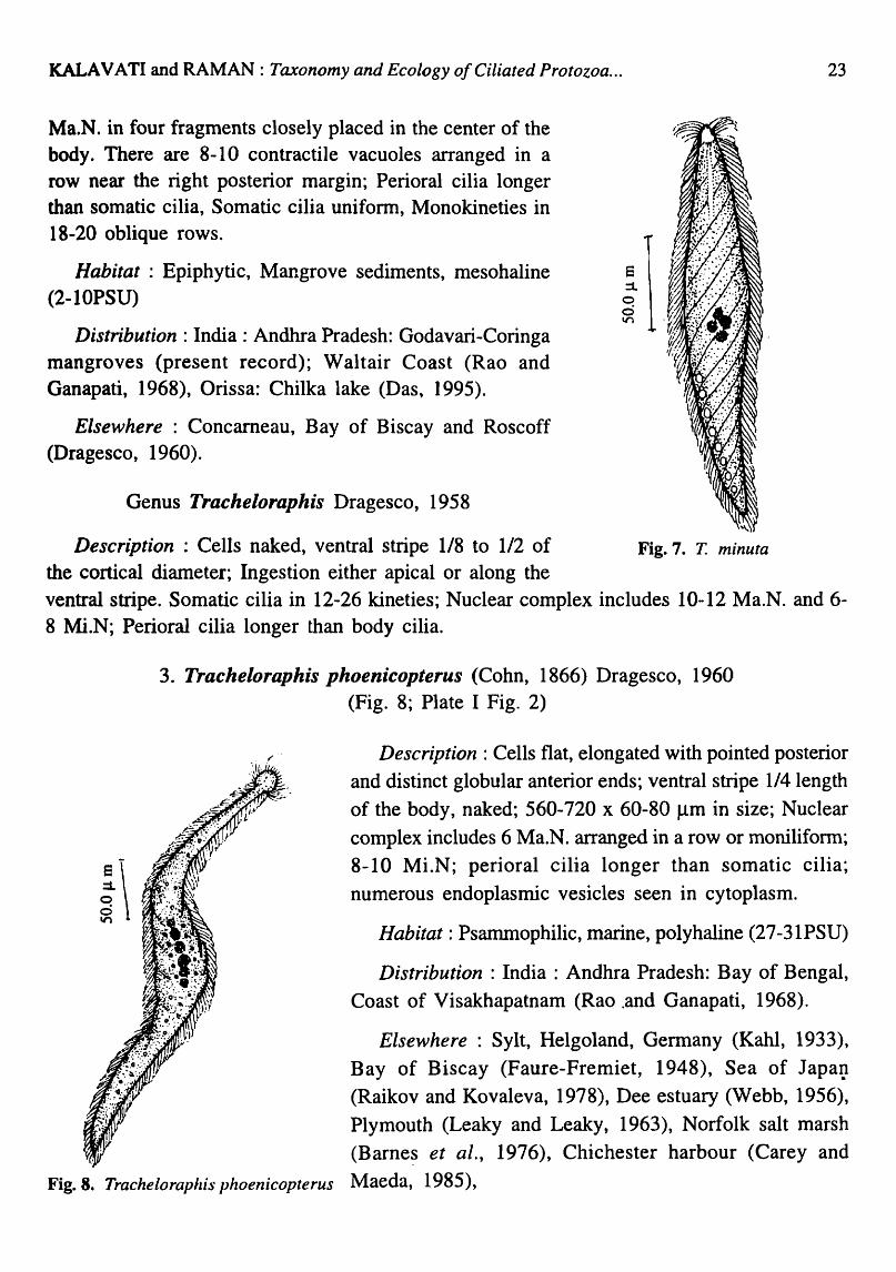
3. Tracheloraphis phoenicopterus (Cohn, 1866) Dragesco, 1960 (Fig. 8; Plate I Fig. 2)
,r .. Description : Cells flat, elongated with pointed posterior and distinct globular anterior ends; ventral stripe 1/4 length of the body, naked; 560-720 x 60-80 flm in size; Nuclear complex includes 6 Ma.N. arranged in a row or moniliform; 8-10 Mi.N; perioral cilia longer than somatic cilia; numerous endoplasmic vesicles seen in cytoplasm.
Habitat: Psammophilic, marine, polyhaline (27-31PSU)
Distribution : India : Andhra Pradesh: Bay of Bengal, Coast of Visakhapatnam (Rao .and Ganapati, 1968).
Elsewhere : Sylt, Helgoland, Germany (Kahl, 1933), Bay of Biscay (Faure-Fremiet, 1948), Sea of JapaI} (Raikov and Kovaleva, 1978), Dee estuary (Webb, 1956), Plymouth (Leaky and Leaky, 1963), Norfolk salt marsh (Barne~ et al., 1976), Chichester harbour (Carey and
Fig. 8. Tracheloraphis phoenicopterus Maeda, 1985),

24 Ree. zool. Surv. India, Oec. Paper No. 282
Order LOXODIDA Jankowski (Small and Lynn, 1985)
Family LOXODIDAE Btitschli, 1889
Cells are laterally compressed, ciliated only on right side; apex bent; cytostome slit-like.
Genus Remanella Kahl, 1933
Oral area long, leads into a tubular cavity not lined by extensions of oral dikinetids.
Key to the species
1. Cells flat, 14-15 kineties on right side, 2 Ma.N and 1 MLN ......................... R. rugosa
2. Cells compressed, ventral groove extends more than 1/2 the length of the body, 2 Ma.N ................................................................................................................... R. margaritifera
4. Remanella rugosa Kahl, 1935 (Fig. 9; Plate 1 Fig. 3)
Description : Cells flat, lancet-like, compressed; anterior end curved ventrally; right side convex, 260-350 x 70-100 Jlffi in size, Ma.N oval, 2, 15 x 8 Jlffi in size. MLN. small, spherical; somatic ciliation unifonn; monokineties, parallel, 14-15 kineties on right side; oral area long in a ventral groove, leads into a tubular cavity that extends upto 2/3rd the length of the body; endoplasm filled with numerous vesicles.
Habitat : Psammophilic, Marine, polyhaline (27.'5-31.5PSU)
Distribution : India : Andhra Pradesh: Bay of Bengal, Coast of Visakhapatnam (Rao and Ganapati, 1968).
Elsewhere: Germany (Kahl, 1933), Roscoff (Dragesco, 1960), Plymouth (Leaky and Leaky, 1963), South Wales (Wright, 1983) Chichester harbour (Carey and Maeda, 1985).
Fig. 9. Renlanella rugosa
5. Remanella margaritifera Kahl, 1933 (Fig. 10; Plate I Fig. 4)
Description : Cells flat, compressed, 350-480 x 50~80 Jlm in size; anterior end beak-like, posterior end narrow, pointed; oral area in a ventral groove that extends upto the middle of the body; Ma.N 2, spherical; MLN not seen. Somatic ciliation uniform, monokineties, parallel, 18-20 kinetids on the right side; Endoplasm filled with numerous vesicles.

KALAVATI and RAMAN: Taxonomy and Ecology o/Ciliated Protozoa ...
Habitat: Psammophilic, Marine, mesohaline (20-28.SPSU)
Distribution: India: Andhra Pradesh: Bay of Bengal, Coast of Visakhapatnam (Rao and Ganapati, 1968).
Elsewhere: Sylt, Helgoland (Kahl, 1933), Baltic sea (Bock, 1952), Roscoff (Dragesco, 1960), Caspian sea (Agamaliev, 1974), Sea of Japan (Raikov and Koveleva, 1968), Black sea (Petran, 1967), French coast of Atlantic (Dragesco, 1960), White sea (Burkovsky, 1967, Plymouth, England (Leaky and Leaky, 1963), North Yorkshire (Hartwig and Parker, 1977), Southwales (Wright, 1983), Chichester harbour (Carey and Maeda, 1985), Bay of Bengal, India (Rao and Ganapati, 1968).
Order PROTOHETEROTRICHIDA Nouzarede, 1977
Family GELEITDAE Kahl, 1933
Apex is tapering and bent to left, cytostome subapical near concave side; oral ciliature often more complex on right side.
Key to the genera
I
~ ~ t )
Fig. 10. R. margaritifera
25
1. Kinities on the left side of the mouth unorganised ................................................ Avelia
2. Kinities on the left side of the mouth in a series of files ..................................... Geleia
Genus Avelia Nouzarede, 1977
Cells small, cytostome slit-like surrounded by a ring of monokineties; kineties on the left of the mouth unorganised.
6. Avelia dragescoi n. sp. (Fig. 11)
Description: Cells small, flat; anterior end broad with a papilla-like projection; 100-112 (105.6 ± 2.0) x 20-25(23.1 ± 1.2) flm in size; cytostome apical, slit-like surrounded by a ring of monokinities; Ma.N single, oval, large 10.2 x 8.5Jlm in size; Mi.N small, adhaerent to Ma.N; numerous contractile vacuoles in the posterior half of the body; somatic ciliation uni fonn , monokinities, 8-12, parallel; endoplasm clear.
Habitat : Psammophilic, Marine, polyhaline (29.5-31.5PSU)
Distribution : India : Andhra Pradesh: Bay of Bengal, Coast of Visakhapatnam (present study)
Type slides: Z.S.I No. 2468

26 Rec. zool. Surv. India, Occ. Paper No. 282
Remarks: Nouzarede (1977) created genus Avelia to accommodate those geleid ciliates having a set of unorganised kinities on the left side. They are all very large, attaining a length
Fig. 11. Avelia dragescoi n. sp.
of 1-3mm and are highly contractile. Body is divisible into a long neck, beak-like hood and the mid body with a centrally placed Duclus. Buccal region is small and surrounded by peribuccal myonemes.The present species showed all above characters in addition to unorganised left oral kinities and hence assigned to genus Avelia. 4 species of Avelia, A. arcahonense (Nouzarede 1965) Nouzarede 1975, A. marhinicensis, Nouzarede 1977, A. gigas (Dragesco, 1965) Nouzarede 1975 and A. orbis Lynn and Small, 2000 are reported so far from interstitial sands. In comparision, the present species is very small (105.6 x 23.1mm) and is not analogous to anyone of them in size. The species is different in possessing a papillate oral hood and several contractile vacuoles in the posterior region hitherto undescribed in Avelia. Besides, the species appears unique in having a marginal shift in the position of the cytostome and associated trichites towards left. Taking cognizance of the above
characters, the species is considered new to science, for which the name A velia dragescoi n.sp. is suggested in honour of Professor J. Dragesco, the renowned ciliatologist.
Genus Geleia Kahl, 1933
Cells with long slit-like cytostome bordered on the right by monokineties; oral kinetosomes to the left of cytostome as a series of adjacent files. Anterior neck region flattened. Genus mostly restricted to interstitial sands
Key to the species
1. Cells small, cytostome near the middle region. Ma.N 2, Mi. N 1, somatic kineties 30-35, endoplasmic vesicles distributed .................................................................... G. nigriceps
2. Cells small, cytostome near the anterior 1/3 of the body. Ma. N 2 Mi.N single, small. Somatic kineties 20-26. Endoplasmic vesicles near left margin ...................... G. fossata
3. Cells large, cytostome subapical, close to the anterior end. Ma.N 2, Mi.N 2. Somatic kineties 32-36 .................................................................................................... G. decolor
7. Geleia nigriceps Kahl, 1933 (Fig. 12; Plate I Fig. 5)
Description: Cells elliptical, highly contractile; 200-300 x 40-70J.lm in size; anterior end

KALAVATI and RAMAN: Taxonol1lY and Ecology of Ciliated Protozoa ... 27
slightly bent, posterior end oval; cytostome ventral, subapical, slit-like almost extending up to the middle of the body; right margin of the oral cavity with 20-24 closely packed kineties. There are 2 spherical Ma.N and a single dot like Mi.N. in between the macronuclei. Somatic kineties 30-35, endoplasm filled with numerous small vesicles. ,8
;::1.
Habitat : Psammophilic, marine, polyhaline (28-31.5PSU).
'0 :0 jtrl
Distribution: India: Andhra Pradesh: Bay of Bengal, Coast of Visakhapatnam (present study); Orissa: Chilka lake (Das. 1995).
Elsewhere: Coast of California (Kirby, 1932), Sandy· beaches of Bermuda (Hartwig, 1977), South Wales (Wright, 1983).
8. Geleia fossataKahl, 1933 (Fig.13; Plate I Fig. 6)
Fig. 12. Geleia nigriceps
Description: Cells large, flat elongated, anterior apex tapering and bent to left; 300-500
Fig. 13. G. Jossata
x 3~-50Jlm in size; Cytostome subapical, in the anterior 1/3 of the body, long and slit-like; Right oral kinetids, monokineties. Left kineties not distinct, Ma:N 2, spherical; MLN single, small, dot like. Somatic kineties parallel, 20-26 monokinities. Endoplasm filled with numerous vesicles.
Habitat: Psammophilic, marine, polyhaline (28-31.5PSU).
Distribution : India : Andhra Pradesh: Bay of Bengal, Coast of Visakhapatnam (Rao and Ganapati,
1968).
Elsewhere : Sylt, Helgoland Germany (Kahl, 1933), Roscoff (Dragesco, 1960), Chichester harbour (Carey and Maeda, 1975), Norfolk salt marsh
(Barnes et al., 1976), Plymouth, England (Hartwig and Parker, 1977).

28 Ree. zool. Surv. India, Dec. Paper No. 282
9. Geleia decolar Kahl, 1933 (Fig. 14; Plate I Fig. 7)
Description: Cells large, 375-500 x 75-100Jlm in size, flat with rounded ends; cytostome ventral, subapical, close to anterior end; Ma.N 2, oval; Mi.N 2, both in the middle of the body; somatic kineties 30-36, monokinities; endoplasm clear without any vesicles.
Habitat: Psammophilic, marine, polyhaline (27-30.5PSU)
Distribution : India : Andhra Pradesh: Bay of Bengal, Coast of Visakhapatnam (Rao and Ganapati, 1968).
Elsewhere : Kiel, Northern Germany (Kahl, 1933), Alligator harbour, Florida, Gulf of Mexico (Borror, 1963), Baltic sea (Bock, 1952), North sea (Hartwig, 1974), Roscoff (Dragesco, 1960).
8 :1. o 9 -
Fig. 14. G. decolor
Order PROTOCRUZIIDA Jankowski in Small and Lynn, 1985
Family PROTOCRUZIIDAE Jankowski in Small and Lynn, 1985
Small ciliates, mono or dikinetid field on right side and dorsum with 5-8 left oral polykinetids. Right paroral dikinetid has pre, para and post cytostomal segments; right of the oral region with barren kinetosomes.
Genus Protocruzia da Faria, da Cunha and Pinto, 1922
With characters of the family.
Key to the species
1. Cells oval, anterior end bent, beak-like, 5-7 left serial monokinetids, 20-25 monokinetids
on right side .. o. 0 0.0. 0 0 0 •• 00000 ••••• 0 ••• 0 •••••••••••• 0 0 0 0 ••••••• o. 0.0 ••• 0 •• 00 ••••• 0 ••• 0 ••• o. 0" 0 0.' 0 0 •••• 0 0.0.0.0 P. adherens
2. Cells pyriform, peristome extends upto 2/3 length of the body, 3-5 left serial monokinetids, 15 20 diki . d . h . d . . - neti s on ng t SI e .................................................................. 0 ••••• P. p'gger,na
10. Protocruzia adherans Mansfield, 1923 (Fig. 15)
Description: Cells oval or elongate, 55-65 x 30-40J.lIl1 in size, anterior end bent, beaklike, posterior end globose, peristome starts at the anterior apex extends upto the middle of

KALA VA TI and RAMAN: Taxonomy and Ecology of Ciliated Protozoa ...
the body and leads into a small infundibulum;
cytostome small, slit-like, Ma.N spherical, 5-8 fJID
in diameter; Mi.N small, spherical and adherent to
Ma.N; somatic ciliation uniform, 20-25
monokineties; on right side 5-7 serial polykineties;
Contractile vacuole single, subterminal; cytoplasm
filled with small vesicles at the posterior region.
Habitat: Planktonic, brackish, mesohaline (11-
18PSU)
Distribution : India : Andhra Pradesh: Bay of
Bengal, Visakhapatnam harbour (present record).
Elsewhere: North Sea (Mansfeld, 1923), Black
Sea (Kahl, 1935); First record from Indian sea
coast.
~I 0: M.
.J
Fig. 15. Protocruzia adherans
11. Protocruzia piggerina Cohn, 1866 (Fig. 16)
29
Description: Cells small, pyriform, anterior end pointed and curved, posterior end globosed,
30-60 x 20-30J.lm in size; peristome less than half the length of the body, cytostome
Fig. 16. P piggerina
1 I e
Ii i J
conspicuous, 15-20 monokineties on the right
side of dorsum, 3-5 polykineties on the left. side; Ma.N oval, 3.2 x 8.0Jlffi in size; MLN
single, spherical, close to Ma.N. There is a
single subterminal contractile vacuole.
Habitat : epiphytic, marine, polyhaline
(28-32PSU)
Distribution: India: Andhra Pradesh: Bay
of Bengal, Visakhapatnam coast
(RadhaKrishna, 1984)
Elsewhere : Rio de Generio, Brazil (da
Faria eta al, 1922), Sylt, Helgoland, Gennany
(Kahl, 1935), Plymouth (Leakey and Leakey,
1963), Marsielle (Vacelet, 1961).

30 Rec. zool. Surv. India, Occ. Paper No. 282
Class SPIROTRICHIA Btitschli; 1889
Subclass HETEROTRICHIA Stein, 1859
Order HETEROTRICHIDA Stein, 1859
Family BLEPHARISMIDAE Jankowski in Small and Lynn, 1985
Cells pyriform or ellipsoid in shape, laterally compressed; narrow anteriorly; peristome on left margin; oral dikinetids precytostomal.
Key to the genera
1. Cells nonpigmented, right oral cilia as short linear dikinitids next to oral cavity ........ . A . t' · .............................................................................•............................................. nlgs elnra
2. Cells pigmented, right oral cilia in three short kinetosomal files .............. Blepharisma
3. Right oral cilia as dikinitids, with short bacc'iliform endosymbionts .. Parablapherisma
Genus Anigsteinia Isquith, 1968
Cells non pigmented; right oral cilia as short linear dikinetids next to oral cavity.
12. Anigsteinia salinarum Isquith, 1968 (Fig. 17; Plate I Fig. 8)
Description: Cells lanceolate, laterally compressed 170-200 x 50-65.5Jlm in size; posterior region transparent, anterior half deeply coloured, brownish; Adoral zone membranellae (AZM)
iI
Fig. 17. Anigsteinia salinarum
extends upto middle of the body along the ventral edge; peristomal cavity with undulating membrane on right side; somatic kineties 20-26; several small macro nuclei scattered in the anterior region; contractile vacuole not seen.
Habitat : Psammophilic, Epiphytic, Euryhaline (6.5-28.2PSU), a known polysaprobic indicator species.
Distribution : India : Andhra Pradesh: Bay of Bengal, Visakhapatnanl harbour coast (present record); Orissa : Chilka lagoon (present record).
Elsewhere: Luxhaven, Kiel, Germany (Kahl, 1935), Dee estuary (Webb, 1956), Periphyton in Caspian sea (Agamaliev, 1972), Salt marshes of Lousiana, U.S.A. (Elliot and Bamforth, 1975), Norfolk salt marsh (Barnes et al., 1976), North Yorkshire (Hartwig and Parker, 1977), Tees estuary (Parker, 1981); First record from India.

KALA VA TI and RAMAN : Taxonomy and Ecology of Ciliated Protozoa ... 31
Genus Blepharisma Perty, 1849
Cells pigmented, right oral cilia long, complex; parorals as three short kinetosomal files.
13. Blepherisma clarissimum Anigstein, 1912 (Fig. 18; Plate I Fig. 9)
Description: Cells ellipsoid or lancet-shaped, laterally compressed, 150-200 x 50-70J..lm in size; peristome on left margin, twisted at the posterior end; right oral cilia in groups; somatic ciliation uniform, dense monokineties; Ma.N divided in to 6-8 parts; contractile vacuole large, single and terminal; endoplasm pigmented and reddish in colour.
Habitat : Epiphytic or psammophilic, brackish, oligohaline (18-22PSU)
Distribution : India : Andhra Pradesh: Bay of Bengal, Visakhapatnam harbour coast (present record).
Elsewhere : Lu~haven, Kiel, Germany (Kahl, 1932), Mediterranean and Atlantic coast of France and America (Faure-Fremiet, 1950, 1951; Dragesco, 1960), Gulf of Naples (Nobili, 1957), Black Sea (Petran, 1967), periphyton in Caspian Sea (Agamaliev 1972), North Sea (Hatwig, 1973), North Yorkshire (Hartwig and Parker, 1977), Tees estuary (Parker 1981). This is the first report from India.
Fig. 18. Blepharisl1la clarissrnum
Genus Parablepharisma Kahl, 1932
Right oral cilia are as dikinetids; often with short bacciliform endosymbionts.
14. Parableplzarisma indica n. sp.
(Fig. 19; Plate I Fig. 10)
Description : Cells flat more or less sigmoid in shape, contractile, anterior end narrow,
slightly pointed; posterior end blunt; 110-160 x 40-50 Jim in size; peristome extends upto II 3-1/2 the length of the body; right oral cilia dikinetids with numerous adherent bacteria: Ma.N large, spherical and 22.5-36.4 Jlm in diameter; Mi.N small; 4-5 deep striations seen on
the surface of the cell; contractile vacuole large spherical and terminal.
Habitat : Epiphytic, Marine, polyhaline (27.2-30PSU)

32 Rec. zoo!. Surv. India, Occ. Paper No. 282
Distribution : India : Andhra Pradesh : Bay of Bengal, Visakhapatnam coast (present record).
Type slides : Z.S.I No. 2472
Remarks : Parablepherisma Kahl is a rare, poorly described genus and there are only three comparable species namely, P. pellitum (120-180fl111 in size.), P. chlamydophorum (150-200 J.111l) and P. collare (120-200IJffi), all of which are different in other morphological criteria such as the length of undulating membrane, peristome and macronucleus. The present species appears distinct with a characteristic sigmoid body, large spherical Ma.N and 4-5 deep striations on the cell surface. In view of its morphological distinctness, the species is considered new to science for which the name Parablepharisma indica n. Spa is suggested.
e
Fig. 19. Parablepharisma indica n. Spa
Family CLIMACOSTOMIDAE Repak, 1972
Cells large, often truncated, left oral polykineties delimit peristomal field; paroral ciliature
inconspicuous.
Genus Fabrea Henneguy, 1890
Cells pyriform, anterior end pointed; peristome extends down from anterior end to 2/5 or more of body length, its posterior portion closely wound. Ma.N sausage shaped or in four parts.
Key to species
Cells pyriform, peristomial field extends upto middle of the body. Infundibulum distinct,
Ma.N sausage shaped. A distinct black spot at the base of the peristomial field seen .
. ...................... ......... ~ .............................................................................................. F. salina
Cells small, pyriform, anterior end pointed, posterior truncate, peristomial field extends nearly upto posterior region, Ma.N elongated ................ 0 •••• 0 ................. F. corlissi n. Spa
] 5. Fabrea salina Hennuguy, 1890 (Fig. 20; Plate I Fig. 11)
Description: Cells pyriform, with pointed anterior and round posterior ends; 165-180 x 40-80Jlm in size; peristomeal field prominent, left oral polykineties delimit the peristome
which extends upto middle of the body and characteristically curved; infundibulum distinct;

KALA VA TI and RAMAN: Taxonomy and Ecology of Ciliated Protozoa ... 33
Ma.N sausage shaped; MLN spherical; There is a distinct blackspot at the base of the peristomial membranelle; somatic ciliation unifonn 30-35 dikineties; left oral ciliation polykineties.
Habitat : Planktonic, marine, polyhaline (28.5-31.5PSU)
Distribution : India : Andhra Pradesh : Bay of Bengal, Visakhapatnam coast (present record).
Elsewhere: Shores of Montery Bay, Berkeley and San Francisco Bay (Kirby, 1934).
16. Fabrea corlissi n. sp. (Fig. 21; Plate I Fig. 12)
Fig. 20. Fabrea salina
Description : Cells small, pyriform, anterior end pointed, postetior end globose, truncated, 65-75 x 35-45Jlm in size; peristome long extends upto the posterior end and characteristically curved; pCiivral ciliature inconspicuous; Ma.N cylindrical or elongated; 10-15 x 3.6-6.4Jlffi in size; MLN not seen; Somatic ciliation uniform; 30-40 dikineties.
Habitat : Planktonic, marine, polyhaline (29-31.5PSU)
Distribution : India : Andhra Pradesh: Visakhapatnam harbour and coast (present record)
Type slides: Z.S.I No. 2466
Fig. 21. F.eorlissi n. sp.
Remarks: Although genus Fabrea Henneguy is long known as an inhabitant of marine
ecosystem, there have been no additional
descriptions under this genus. The only known species F. salina is relatively large (165-180 x
40-80llm) in size with a characteristically curved peristome. In comparison, the species under
consideration is much smaller (65-75 x 35-
451lm) with a long peristome that extends almost
upto the posterior end. In view of the above differences in morphology, the present species
is considered new to science for which the name,
F. corlissi n.sp. is suggested in honour of
Professor. John. O. Corliss, the renouned
ciliatologist.
34 Ree. zool. Surv. India, Oee. Paper No. 282
Family CONDYLOSTOMIDAE Kahl in Doflein and Reichnow, 1929
Cells large, contractile, right oral cilia prominent, oral polykinetids delimit unciliated
peristomial field.
Genus Condylostoma Bory de St. Vincent, 1824
Anterior end truncated, posterior end bluntly pointed, peristome wide. Peristomal field
non ciliated.
Key to species
1. Peristome wide, 'U' shaped, directed anteriorly, posterior half of the body ovaVrounded. Ma.N beaded, arranged in crescent shape contractile vacuole, small, terminal ............ . •• •••• •••••••••••••••••••••• •••• •••••• ••••••••• •••••••••• •••• ••••• ••••••• I I ••••• I I •••• "" I ••••••••••••••••••••••••• I •••••••••• C. patens
2. Peristome relatively narrow, 'V' shaped, posterior end pointed, Ma.N a string of 10-12 linear beads, contractile vacuole small subterminal, collecting canal narrow, extends to middle of the body .. 0 0 0 00 0.00.00000. 0 0 0 0.00.0 ••• 0.0. 1.0 •• 0 0 0 0 •• 0 •••• 0 ••••••••••••••• 0 •••• I............... C. arenarium
3. Anterior end broad, posterior end narrow, peristome wide, bowl shaped with conspicuous non ciliated field. Ma.N beaded. Contractile vacuole large, terminal ... C. minuta n. sp.
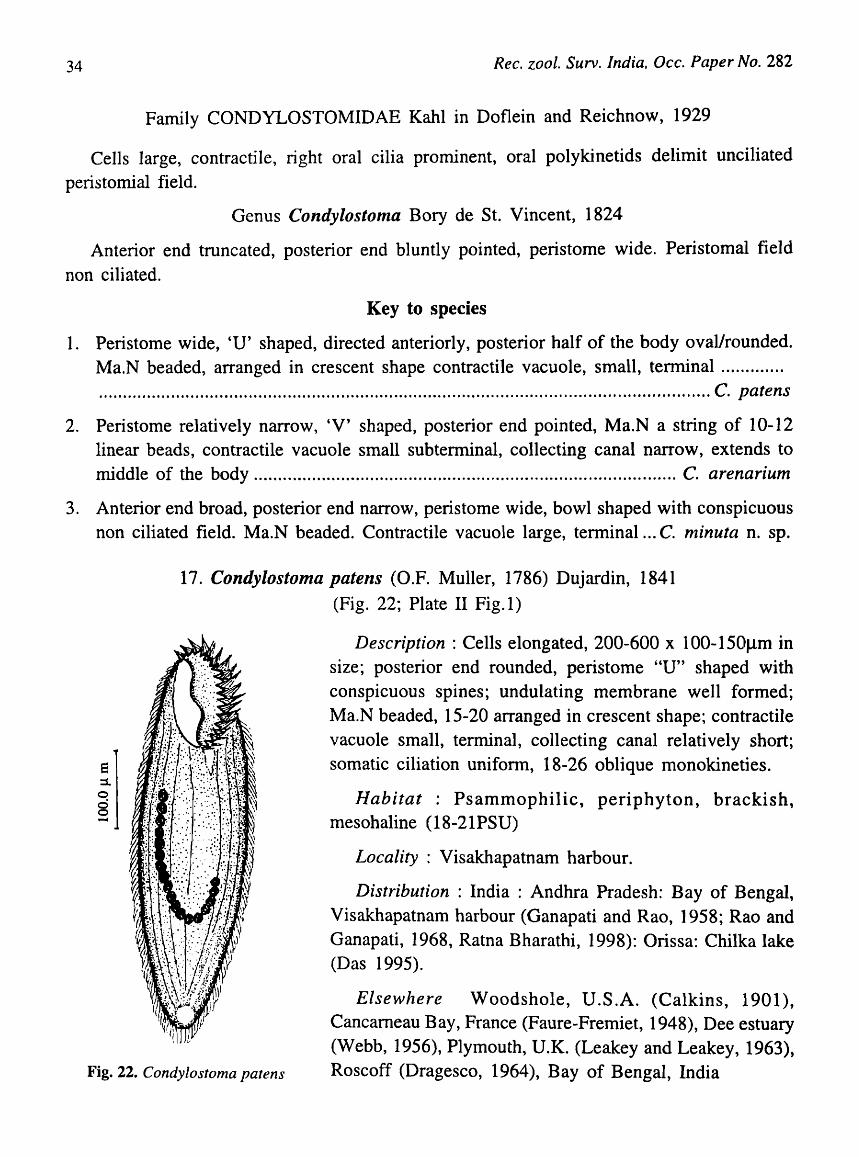
17. Condylostoma patens (D.F. Muller, 1786) Dujardin, 1841
(Fig. 22; Plate II Fig.l)
Fig. 22. Condylostoma patens
Description : Cells elongated, 200-600 x 100-150flm in size; posterior end rounded, peristome "U" shaped with conspicuous spines; undulating membrane well formed; Ma.N beaded, 15-20 arranged in crescent shape; contractile vacuole small, terminal, collecting canal relatively short; somatic ciliation unifonn, 18-26 oblique monokinetieso
Habitat : Psammophilic, periphyton, brackish, mesohaline (18-21PSU)
Locality : Visakhapatnam harbour.
Distribution : India : Andhra Pradesh: Bay of Bengal, Visakhapatnam harbour (Ganapati and Rao, 1958; Rao and Ganapati, 1968, Ratna Bharathi, 1998): Orissa: Chilka lake (Das 1995).
Elsewhere Woodshole, U.S.A. (Calkins, 1901), Cancameau Bay, France (Faure-Fremiet, 1948), Dee estuary (Webb, 1956), Plymouth, U.K. (Leakey and Leakey, 1963), Roscoff (Dragesco, 1964), Bay of Bengal, India

KALA VATI and RAMAN: Taxonomy and Ecology of Ciliated Protozoa ...
18. Condylostoma arenarium Spiegel, 1926 (Fig. 23; Plate II Fig.2)
35
Description : Cells spindle shaped. Anterior end round, posterior end sharply pointed, 200-500 x 70-80flffi in size. Peristome relatively small, narrow, "V" shaped, equipped with
peristomial spines and undulating membrane. Ma.N a string of 10-12 beads, arranged linearly; contractile vacuole single, tenninal. Collecting canal narrow, extends upto the middle of the body; somatic ciliation uniform, 26-32 monokineties.
Habitat : Psammophilic, marine, polyhaline (27 -32PSU)
Distribution : India : Andhra Pradesh : Bay of Bengal, Visakhapatnam coast (Rao and Ganapati, 1968).
Elsewhere : Helgoland, Gennany (Speigal, 1926), iGel Bay and Oresund (Bock, 1952), Hidden Sea Island (Munch, 1955), Atlantic and Meditterenean coasts of France (Dragesco, 1953,1960), Boreal seas of USSR (Raikov, 1960, Burkovsky, 1970), Sea of Japan, (Raikov, 1963), Alligator Harbour, Florida (Borror, 1963), Gulf of Mexico (Borror, 1962), Caspian sea (Agamaliev, 1970), Coast of Brazil (Katter, 1970), salt marshes of Lousiana, U.S.A. (Elliot and Bamforth, 1975), North Yorkshire (Hartwig and Parker, 1977), South Wales (Wright, 1982), Mugu lagoon, California (Smith, 1982) Fig. 23. C. arenarium
19. Condylostoma minuta n. sp. (Fig. 24; Plate II Fig. 3)
e :1. o ~
Fig. 24. C. nlinuta n. sp.
Description : Cells more or less triangular; anterior end broad, spatula-like and truncated; posterior end narrow, sometimes drawn out into a pointed spine-like structure; 60-80 (75.4 ± 3.6 x 30-50 ( 34.8 ± 4.0) J.l11l in size; peristome wide and characteristically bowl shaped; AZM and undulating membrane conspicuous; peristomial field non ciliated; Ma.N moniliform, with 8-10 linearly arranged beads; contractile vacuole single, large, tennina1; collecting canal not seen; somatic ciliation unifonn, 8-12 monokineties.
Habitat: Planktonic, marine, polyhaline (27.5-29PSU)
Distribution : India : Andhra Pradesh: Bay of Bengal, Visakhapatnam harbour (present record).
Type slides Z.S.I No.2462
36 Ree. zooI. Surv. India, Oee. Paper No. 282
Remarks : Species of the genus Condylostoma are generally large in size (> 100 fJIIl), ovoid or elongate or ellipsoid in shape with a broad 'V' shaped peristome. AZM is large with an undulating membrane on the opposite side of the buccal cavity. Ma.N is moniliform. The genus is well represented in the interstitial biotope and several species have been described. The other two records from Indian waters are Condylostoma patens (Ganapati and Rao, 1958) and Condylostoma arenarium (Rao and Ganapati, 1968). Both species are much larger in size (> 200fJ1Il) with a V -shaped peristome. In comparision, the present species is very small (mean 71.5 x 34.8~m) and has a distinct triangular or spatula shaped body and' a characteristically 'bowl' shaped peristome. The species is different in having a conspicuous AZM and undulating membrane and non-ciliated peristomial field. In view of the above differences in structure and shape, the species is considerd new to science for which the name Condylostoma minuta n.sp is suggested
Family SPIROSTOMATIDAE Stein, 1867
Cells long, large, highly contractile; oral cavity not deep; infundibulum absent; oral polykinetids many, form a long inconspicuous zone.
Key to the genera
1. Kinities straight when body contracts ................................................................ Gruberia
2. Kineties spiral round the body when contracted ........................................ Spirostomum
Genus Gruberia, Kahl, 1933
Anterior end pointed, somatic kineties straight when contracted, right oral polykinetids in serial order at the anterior end.
20. Gruberia calkensi Beltran, 1933 (Fig. 25)
Description : Cells elongate, anterior end narrow, posterior end pointed; 100-150 x 20-40 flm in size. Peristome extends upto 1/2 the length of the body. AZM inconspicuous; right oral polykineties serial; Ma.N beaded, arranged linearly. 5-8 MLN found scattered. There are 5-10 contractile vacuoles in a row near the left posterior margin.
Habitat: Psammophilic; Epiphytic, euryhaline, brackish (2.S-1SPSU).
Distribution : India : Andhra Pradesh: Bay of Bengal, Visakhapatnam harbour (Ganapati and Rao, 1958; Rao and Ganapati, 1968; Ratna Bharathi, 1998); Orissa: Chilka Lake (Das, 1995) Fig. 25. Gruberia calkensi

KALA V ATl and RAMAN: Taxonomy and Ecology of Ciliated Protozoa ... 37
Elsewhere: Woodshole, U.S.A. (Beltran, 1933).
Genus Spirostomum Ehrenberg, 1837
Cells blunt posteriorly. Somatic kineties incomplete and inserted in right oral region kineties spiral and coil round the body when contracted.
Key to the species
1. Cells elongated, peristome extending upto 1/2 the length of the body, Ma.N oval or elongated, contractile vacuole single, collecting canal long ................................ S. teres
2. Cells elongated, broad anteriorly, peristome small extends upto 1/3 of the length of the body. Ma.N long moniliform ............................................................................... S. minus
21. Spirostomum teres Claparede and Lachmann, 1859 (Fig. 26)
Description : Cells elongate, highly contractile, compressed dorsoventrally, posterior end truncated. Anterior end bluntly pointed and bent towards left; 250-320 x 30-45 flm in size; Peristome extending upto the middle of the body. Ma.N oval or elongated 15 x 22flm located in the central part; Mi.N small, spherical, contractile vacuole large, terminal at the posterior end; Collecting canal long, extends upto 2/3rd the length of the body; somatic ciliation uniform; 9-10 monokinities; Caudal cilia long and distinct probably thigmotactic.
Habitat : Epiphytic, oligohaline (3-6.5PSU)
Distribution : India : Andhra Pradesh : Bay of Bengal, Visakhapatnam harbour (present record). Orissa: Chilka Lake (Das, 1995)
Elsewhere: Dee estuary, U.K. (Webb, 1956); Oresund, Denmark and Asko, in Baltic Sea (Fenchel, 1969)
22. Spirostomum minus Roux, 1901 (Fig. 27)
Fig. 26. Spirostomum teres
Description: Cells elongated, posterior end broad and flat, 400-900 x 50-80 flm in size; peristome small, extends upto the 1/3 rd the length of the body, with short inconspicuous membranelle; Ma.N beaded or moniliform, linear; contractile vacuole single, large, terminal; Collecting canal extends upto the middle of the body; somatic ciliation uniform; 12-14 monokineties; thigmotactic caudal cilia absent. Cells often pigmented.

38 Rec. zool. Surv. India, Dec. Paper No. 282
Habitat : Epiphytic, brackish, mesohaline (22-28PSU).
Distribution : India: Andhra Pradesh: Bay of Bengal, Visakhapatnam coast (present record).
Elsewhere :Although Kahl (1935) reported the species from seawater, the species is found to be widely distributed in lakes and rivers in China (Shenn et aI., 1995). This is the first record form marine environment in India.
Order ARMOPHORIDA Jankowski, 1964
Family CAENOMORPHIDAE Poche, 1913
Cells small, round or conical twisted to left, somatic cilia as small zones of kineties or cirrus-like tufts.
Genus Caenomorpha Perty, 1852
Cells are more or less conical with spines at the posterior Fig. 27. S. minus
end; perizonal kineties as series of transverse rows; 2-8 body kineties; a dense caudal field present around caudal prolongation.
Key to species
1. Cells bell shaped; caudal and secondary prolongations long and equal. Ma.N sausage shaped ............................................................................................................... C. capucina
2. Cells hemispherical, caudal prolongation long, secondary prolongation short, Ma.N broadly oval, Mi.N small ............................................................................................. C. Le vande ri
iI
Fig. 28. Caenolnorpha capucina
23. Caenomorpha capucina Kahl, 1933 (Fig. 28)
Description : Cells bell shaped; anterior end hemispherical or globose, posterior region broad; 60-75 x 35-45.6J.lm in size; perizonal kineties as 3 transverse rows; caudal and secondary prolongations long and equal. Somatic kineties 8, Ma.N sausage shaped, Mi.N small, dot-like.
Habitat : epiphytic, oligohaline « 4PSU).
Distribution : India: Orissa: Chilka Lake (Das, 1995)
Elsewhere :Sylt and Kiel, Germany (Kahl, 1933).
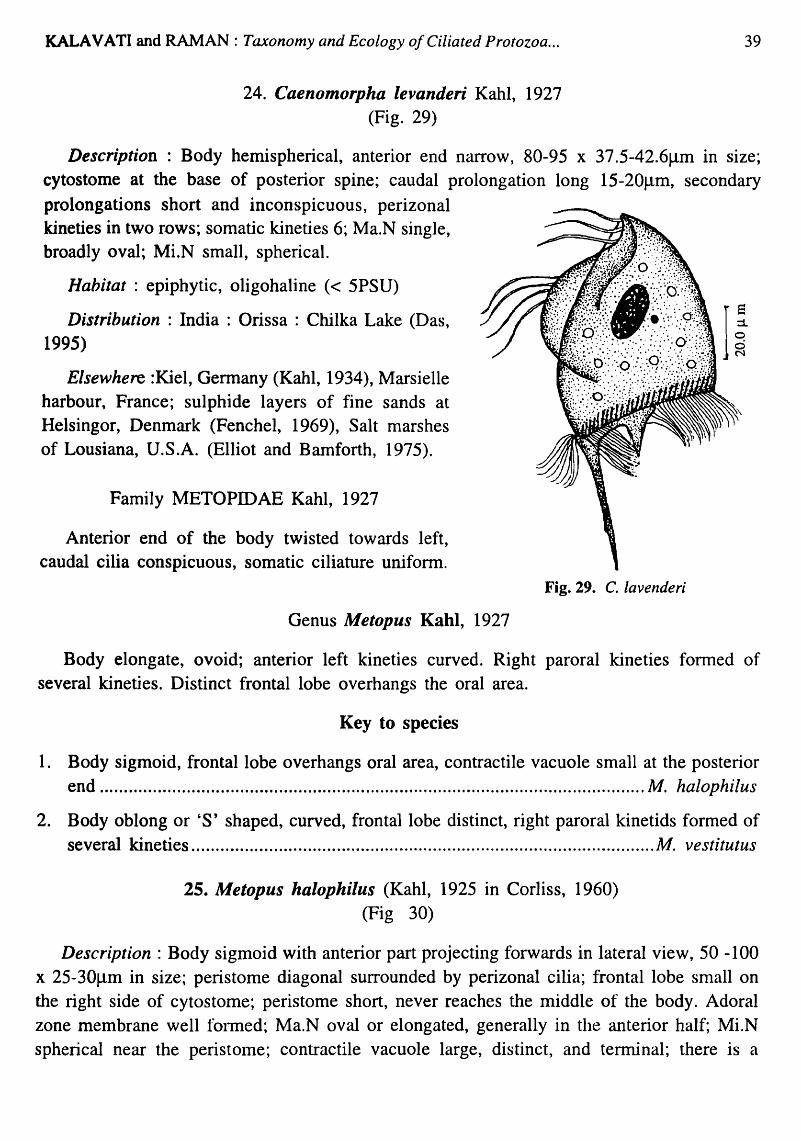
KALAVATI and RAMAN: Taxonomy and Ecology o/Ciliated Protozoa ...
24. Caenomorpha levanderi Kahl, 1927 (Fig. 29)
39
Description : Body hemispherical, anterior end narrow, 80-95 x 37 .5-42.6~m in size; cytostome at the base of posterior spine; caudal prolongation long 15-20~m, secondary prolongations short and inconspicuous, perizonal kineties in two rows; somatic kineties 6; Ma.N single, broadly oval; Mi.N small, spherical.
Habitat : epiphytic, oligohaline « 5PSU)
Distribution: India: Orissa: Chilka Lake (Das, 1995)
Elsewhere :Kiel, Germany (Kahl, 1934), Marsielle harbour, France; sulphide layers of fine sands at Helsingor, Denmark (Fenchel, 1969), Salt marshes of Lousiana, U.S.A. (Elliot and Bamforth, 1975).
Family METOPIDAE Kahl, 1927
Anterior end of the body twisted towards left, caudal cilia conspicuous, somatic ciliature unifonn.
Genus Metopus Kahl, 1927
Fig. 29. C. lavenderi
Body elongate, ovoid; anterior left kineties curved. Right paroral kineties formed of several kineties. Distinct frontal lobe overhangs the oral area.
Key to species
1. Body sigmoid, frontal lobe overhangs oral area, contractile vacuole small at the posterior end ................................................................................................................. M. halophilus
2. Body oblong or'S' shaped, curved, frontal lobe distinct, right paroral kinetids formed of several kineties ................................................................................................ M. vestitutus
25. Metopus halophilus (Kahl, 1925 in Corliss, 1960) (Fig 30)
Description: Body sigmoid with anterior part projecting forwards in lateral view, 50 -100 x 25-30~m in size; peristome diagonal surrounded by perizonal cilia; frontal lobe small on the right side of cytostome; peristome short, never reaches the middle of the body. Adoral zone membrane well fOlmed; Ma.N oval or elongated, generally in the anterior half; Mi.N spherical near the peristome; contractile vacuole large, distinct, and terminal; there is a

40 Ree. zool. Surv. India, Oec. Paper No. 282
distinct group of four caudal cilia at the posterior end probably thigmotactic. Ectobiotic bacteria cover the body.
Habitat: Psammophilic, abundant in organically enriched sediments brackishwater, mesohaline (20-26 PSU)
Distribution: India: Andhra Pradesh: Bay of Bengal, Visakhapatnam harbour (present record).
Elsewhere: Hellsingor, Denmark (Fenchel, 1970)
26. Metopus vestitutus Kahl, 1932 (Fig. 31)
Description: Cells flat oblong or's' shaped with a distinct long spine-like extension at the posterior end; frontal lobe distinct, 60-80 J.1m in length. Right paroral kineties form several kinetids; somatic
Fig. 30. Metopus halophilus
iI
Fig. 31. M. vestitutus
ciliation unifonn, 20-25, dikinetids; Ma.N single, oval; Mi.N small lor 2, central in position; contractile vacuole single, oval, subtennina1; ectobiotic bacteria cover the surface of the body.
Habitat: Psammophilic, often associated with decaying organic matter, brackishwater or mesohaline (5.6-10 PSU)
Distribution : India : Andhra Pradesh : Bay of Bengal, Visakhapatnam harbour (Rao and Ganapati, 1968, present record); Orissa : Chilka Lake (Das, 1995)
Elsewhere : Kiel Germany (Kahl, 1932), Hellsingor, Denmark (Fenchel, 1970), Salt marshes of Lousiana, U.S.A. (Elliot and Bamforth, 1975), Baltic Sea (Boikova, 1964b).
Order PHACODINIDA Small and Lynn, 1985
Family PHACODINIDAE Corliss, 1979
Cells are ovoid, laterally compressed; somatic kineties, polykinetids, linear or in loose groups.
Genus Phacodinium Prowazek, 1900
Cells are oval, cilia in cirrus-like fused groups; peristome long on left margin, cytostome posterior. Ma.N horse-shoe shaped, 5-9 MLN.

KALA VA TI and RAMAN: Taxonomy and Ecology of Ciliated Protozoa ...
27. Phacodinium metchlncoffi vaT. indica (Present study) (Fig. 32; Plate II Fig. 4)
41
Description : Cells oval, dorso-ventrally flattened, 40-50 x 20-35Jlm in size; anterior end pointed, posterior end rounded; peristome long; AZM well developed on the left margin, extending upto the posterior end. Cytostome posterior, Ma.N horse-shoe shaped, MLN. 5-6 in numbers; contractile vacuole single, large and terminal; there are 5 marked striations on the surface.
Habitat : Epiphytic or planktonic, brackish, mesohaline (15-24PSU)
Distribution : India : Andhra Pradesh : Bay of Bengal, Visakhapatnam harbour (present record);
Elsewhere : From mosses in Spain
Remarks: Morphologically, this species very closely resembles P. metchincofji, a freshwater form described from mosses in Spain, but is considerably small in size (40-50 x 20- 35flm) than P. metchincoJfi (135-148f..lm). Locally, this species is often found associated with Cheatomorpha under mesohaline conditions.
Since the difference is only with regard to size Fig. 32. Phacodinium metschenicoffi var. indica and the habitat, the species is considered "a
variety of P. metchincoJfi and is referred to as P. metchincojfi var indica.
Order ODONTOSTOMATIDA Sawaya, 1940
Family EPLAXELLIDAE Corliss, 1960
Cells box shaped; some times with short posterior spines; body cilia in small groups of kinetids at front and rear. Those at the anterior end parallel.
Genus Eplaxella Corliss, 1960
Caudal spines reduced or absent.
28. Eplaxella striata Kahl, 1932 in Corliss, 1960 (Fig. 33)
Description : Cells bean shaped, compressed; 40-55 x 35-45flm in size; lef't side flat and right side convex with a depression at the anterior end. Perizonal ciliary strip consists of 7 rows of long cilia in the anterior region and 4 short rows of cilia in the posterior region;

42 Ree. zool. Surv. India, Oee. Paper No. 282
buccal apparatus with 8 adoral membranelle; Ma.N massive, single, oval; MLN small and dot-like; contractile vacuole large, spherical, behind the buccal apparatus. Body filled with refractile granules.
Habitat: Planktonic, under eutrophic conditions; often seen near sewage outlet and waters rich in hydrogen sulphide.
Distribution: India: Andhra Pradesh: Bay of Bengal, Visakbapatnam harbour (present record).
Eise~vhere : Widely distributed in Europe and America (Bick, 1972), associated with putrifying organic sludge.
Subclass CHOREOTRICHIA Small and Lynn, 1985
Order CHOREOTRICHIDA Small and Lynn, 1985
Sub-order TINTINNINA Kofoid and Campbell, 1929
Family Codonellidae Kent, 1867
Fig. 33. Epalxella straita
With agglomerate of mineral particles on flask or bowl shaped lorica, collor not clear, if present mayor may nor have nuchal constriction.
Genus Tintinnopsis Stein, 1867
Lorica thin covered with agglomerate of mineral particles, aperture broad; aboral end closed.
e ::l o o N
F> ~4. Tintiflllopsis lohnlanni
29. Tintinnopsis lohmanni Laackmann (Fig 34; Plate II Fig. 6)
Description: Lorica variable, generally ovoid or bowl shaped, 60-80 x 30-40f.1m in size; an indistinct rim-like collar present. Cells heavily agglomerated; oral diameter relatively small. Aboral end pointed and closed.
Habitat: Planktonic, marine, euryhaline (12.16-32 PSU). A bloom forming species occurred in prodigous numbers (7,633 x 102 nos. 1-1) when salinity decreased suddenly and temperature relatively high (32.8°C).
Distribution: India: Andhra Pradesh : Bay of Bengal Visakhapatnam harbour and coast (present record); Orissa
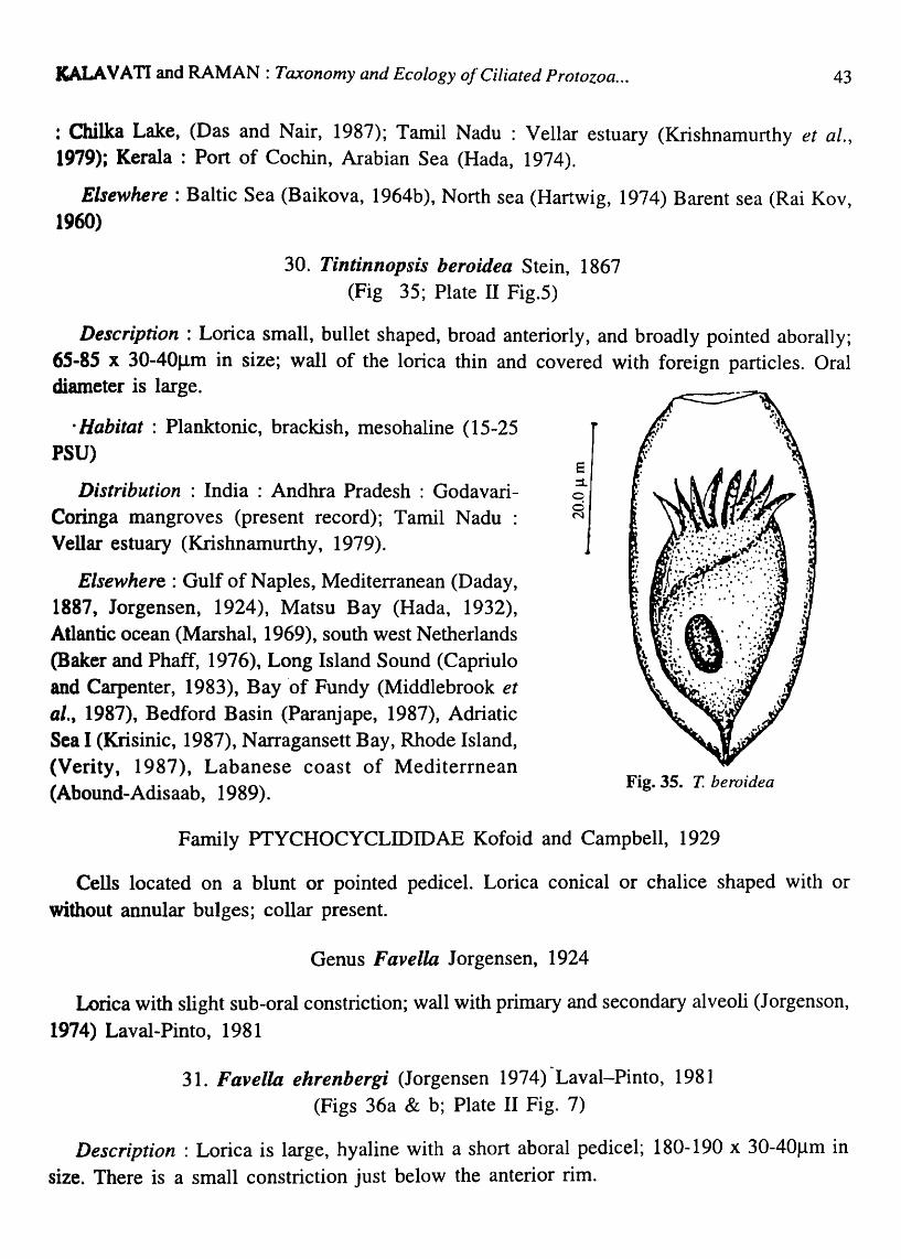
KALAVATI and RAMAN: Taxonomy and Ecology a/Ciliated Protozoa ... 43
: Chilka Lake, (Das and Nair, 1987); Tamil Nadu : Vellar estuary (Krishnamurthy et ai., 1979); Kerala : Port of Cochin, Arabian Sea (Hada, 1974),
Elsewhere: Baltic Sea (Baikova, 1964b), North sea (Hartwig, 1974) Barent sea (Rai Kov, 1960)
30. Tintinnopsis beroidea Stein, 1867 (Fig 35; Plate II Fig.5)
Description: Lorica small, bullet shaped, broad anteriorly, and broadly pointed aborally; 65-85 x 30-40f..1ffi in size; wall of the lorica thin and covered with foreign particles. Oral diameter is large.
· Habitat : Planktonic, brackish, mesohaline (15-25 PSU)
Distribution: India: Andhra Pradesh: GodavariCoringa mangroves (present record); Tamil Nadu : Vellar estuary (Krishnamurthy, 1979).
Elsewhere: Gulf of Naples, Mediterranean (Daday, 1887, Jorgensen, 1924), Matsu Bay (Hada, 1932), Atlantic ocean (Marshal, 1969), south west Netherlands (Baker and Phaff, 1976), Long Island Sound (Capriulo and Carpenter, 1983), Bay 'of Fundy (Middlebrook et al., 1987), Bedford Basin (Paranjape, 1987), Adriatic Sea I (Krisinic, 1987), Narragansett Bay, Rhode Island, (Verity, 1987), Labanese coast of Mediterrnean (Abound-Adisaab, 1989).
8 :1. o o N
Fig. 35. T. be ro ide a
Family PTYCHOCYCLIDIDAE Kofoid and Campbell, 1929
Cells located on a blunt or pointed pedicel. Lorica conical or chalice shaped with or without annular bulges; collar present.
Genus Favella Jorgensen, 1924
Lorica with slight sub-oral constriction; wall with primary and secondary alveoli (Jorgenson, 1974) Laval-Pinto, 1981
31. Favella ehrenbergi (Jorgensen 1974)-Laval-Pinto, 1981 (Figs 36a & b; Plate II Fig. 7)
Description : Lorica is large, hyaline with a short aboral pedicel; 180-190 x 30-40J,.lm in size. There is a small constriction just below the anterior rim.

44
e :1-o o 11"\
Ree. zool. Surv. India, Oee. Paper No. 282
Figs. 368 & 36b. Favella ehrenbergi
Habitat: Planktonic, marine, polyhaline (28.5-32PSU)
Distribution: India: Andhra Pradesh: Bay of Bengal, Visakhapatnam coast (present record).
Elsewhere: Mediterranean (Jorgensen, 1924, Rassoulzadegan et ala 1989, Abboud-Adisaab, 1989), New York Blight (Gold and Morales, 1975), Akkeshi Bay (Takaguchi, 1976), west coast of Sweden (Hedin, 1975), Long Island Sound (Capriulo and Carpenter, 1983), Adriatic Sea, (Krisnic, 1987), Narragansett Bay, Rhode Island (Verity, 1987), Limfjord, Denmark, (Andersen and Sorensen, 1986).
Family TITINNIDIIDAE Kofoid and Campbell, 1929
Lorica is soft, tubular with agglomerate; Collar absent.
Genus Tintinnidiu.m Kent, 1881
With Characters of the family
32. Tintinnidium fluviatile Stein, 1863 (Fig. 37; Plate II Fig. 8)
Description : Lorica tubular with a broad oral aperture at its distal end; 60-80 x 20-40J.lm in size; lorica matrix soft, sticky, thin with fine sediment like agglomerate. Cell is often preserved in lorica. .
Habitat : Planktonic, brackish, oligohaline «6PSU). A common inhabitant of freshwater bodies, also found in brackish water

KALA v A TI and RAMAN : Taxonomy and Ecology of Ciliated Protozoa ...
Distribution: India: Andhra Pradesh: Godavari-Coringa mangroves (present record); Orissa: Chilka Lake (Das, 1995)
Elsewhere: New York Bight (Gold and Morales, 1975), Long Island Sound (Capriulo and Carpenter, 1983) Damarisscotta river estuary, Maine (Sanders, 1987), Narrangsett Bay, Rhode Island (Verity, 1987)
Suborder STROMBIDINOPSINA Small and Lynn, 1985
Family STROMBIDINOPSIDAE Small and Lynn, 1985
Free swimming aloricate; many somatic kineties typically extending the entire length of the body; Ma.N 2, identical, spherical or ovoid.
Genus Strombidinopsis Kent, 1881
With the characters of the family
Key to species
e :1. o ~
Fig. 37. Tlntinnidium fluviatile
45
1. Cells elongate, conical, posterior end pointed, AZM closed, Ma.N 2, spherical, MLN 'ditin' t S . t m s c.................................................................................................... . accumlna um
2. Cells oval or pyriform with blunt posterior end. External and internal polykinetid zone more or less similar. Ma.N oval ....................................................................... S. chesseri
iI
33. Strombidinopsis accuminatum Faure-Fremiet, 1924 .. ".
(Fig. 38)
Description: Cells elongate and conical tapering to a spine-like point posteriorly; 70-125 x 28.6-52.5Jlm in size; external polykinetid zone (EPK) surrounding the anterior end, consists of 13-17 polykineties; oral cavity accentric with three independent inner polykinetids. Somatic kineties 15-16, parallel. Ma.N 2, oval closely placed; 3-5 x 6-
10Jlm in size.
Habitat Planktonic, brackishwater, mesohaline (15-25 PSU)
Distribution: India: Andhra Pradesh: Visakhapatnam harbour (Ratna Bharathi, 1998) Godavari-Coringa
Fig. 38. Strombidinopsis acuminatum mangroves (present record).

46 Rec. zool. Surv. India, Occ. Paper No. 282
Elsewhere :Kingston harbour, Jamaica (Lynn et al., 1991), Gulf of Mexico (Lynn et al.,
1991)
34. Strombidinopsis cheshiri Snyder and Ohman, 1991 (Fig. 39; Plate III Fig. 1)
Description: Cells oval, round or pyriform with tapering posterior end; 35-65 x 25-45Jlm in size; external and internal polykinetid zones more or less similar and in close juxta position
not completely separate; There are about 15-20 polykinetids in EPZ; Cilia of the external polykinetids long and equal; Somatic kineties parallel, 12-15 in number extending from anterior to posterior end; oral cavity deep and eccentric, Ma.N 2, oval, 8 x 5Jlm in size, central in position.
Habitat : Planktonic, marIne, polyhaline (28-30.5PSU)
Distribution India: Andhra Pradesh : Visakhapatnam harbour (Ratna Bharathi, 1998) Godavari-Coringa mangroves (present record).
Elsewhere: Coastal regions of southern California (Snyder and Ohman, 1991). Northern regions of British Columbia (Montagnes and Taylor, 1991)
e ::1. o o M
Fig. 39. St. cheshiri
Suborder STROBILIDINA Small and Lynn, 1985
,Family STROBILIDIDAE Kahl in Doflein and Reichnow, 1929
Lorica absent; one to several body kineties shorter than body; somatic cilia relatively short, somatic kineties composed of monokinetids with a flap covering part of cilia directing to the right and a closed circle of EPK.
Genus Strobilidium Schewiakoff, 1893
Small to medium sized species (120-170Jlm); almost cylindrical in form; posterior end generally narrow; peristome eccentrically placed in the apical area (Maeda, 1986).
35. Strobilidium minimum Gruber, 1884 (Fig.40; Plate III Fig. 2)
Description: Cells ovoid or variable in shape; posterior end rounded. Anterior end truncated. SO-7Sx30-40Jlm in size There 'is a cytoplasmic flap with a close circle of external polykineties

KALAVATI and RAMAN: Taxonomy and Ecology o/Ciliated Protozoa .. ~ 47
overlying the somatic kineties; somatic cilia short; Ma.N 2, spherical; contractile vacuole large, one in the posterior half.
Habitat : Planktonic, marine (25-29.5PSU) especially under eutrophic conditions
Distribution : India : Andhra Pradesh Visakhapatnam harbour (Ratna Bharathi, 1998)
e :L
~ -
Elsewhere: Kiel and Sylt, Germany (Kahl, 1932)
Genus Rimostrombidium Jankowski, 1978
36. Rimostrombidium conicum Kahl, 1932 .'(Fig. -41; Plate III Fig.3)
Description : Cells conical, posterior end bluntly Fig. 40. Strobilidium minimum
Fig. 41. Rimostrombidium conicum
pointed, 40-60 x 30-50Jlm in size. 30-36 polykinetids in the external polykinetid zone which is located in an eccentric pit leading to cytostome. Somatic kineties incomplete and do not extend to posterior end. Ma.N horse shoe shaped. Just below the external polykinetids zone.
Habitat : Planktonic, manne, polyhaline (28.5-32PSU)
Distribution : India : Andhra Pradesh : Visakhapatnam harbour (Ratna Bharathi, 1998) and coast (present record)
Elsewhere: Sylt, Germany (Kahl, 1933)
Family LOHAMANNIELLIDAE Montagnes and Lynn, 1991
Somatic kineties composed of monokinetids restricted to the posterior part of the cell.
Genus Loll1nanniella Leegaard, 1915
Medium sized, oval, globular body, adoral zone consisting of two kinds of membranelle which encircle the apical area. Buccal cavity closed \vith a membrane -like keal.
!(ey to species
1. Cells subspherical, EPK with 32-38 polykineties, IPK with 18 polykineties. Somatic kineties six, Ma.N 'C' shaped ........................................................................... L. ~piralis

48 Ree. zool. Surv. India, Dec. Paper No. 282
2. Cells ovoid, EPK with 19 polykinetids, IPK with 5, somatic kineties 6, Ma.N reniform .......................................................................................................................... L. oviformis
37. Lohmanniella spiralis Lesgaard, 1915 (Fig. 42; Plate III Figs. 4 & 5)
Description : Cells typically sub-spherical; 35-60 x 40-56J.1m in size; EPZ consists of 30-38 polykineties; IPZ with 18 polykineties. There are 6 parallel somatic kineties. Cilia directed
towards right and partially covered by cytoplasmic flap. Ma.N 'C' shaped; MLN spherical, close to Ma.N.
Habitat : Planktonic, marine, polyhaline (28-32 PSU)
Distribution : India : Andhra Pradesh : Visakhapatnam harbour (Ratna Bharathi, 1998) and coast (present record)
Elsewhere : Widely distributed in Adriatic Sea (Revelante and Gilmartin, 1983), Atlantic coast of Canada (Paranjepe et al., 1985), Isles of Shoals (Lynn and Montagnes, 1988) and Cal shot, Southampton, UK (Leakey et al., 1983), Limfjord,
Fig. 42. Lohmaniella spiralis
Denmark (Andersen and Sorensen, 1986); first record from India.
iI
38. Lohmanniella oviformis Leegaard, 1915 (Fig. 43; Plate ill Fig. 6)
Fig. 43. L. oViformis
Description : Cells sub-spherical or ovoid; 30-36 x 28-32f.1ID in size; EPZ with 19 polykineties; IPZ with 5 polykineties; somatic kineties 5 radiating from posterior end; Ma.N bean shaped, located on one side of the cell.
Habitat : Planktonic, brackishwater, mesohaline (15-22 PSU)
Distribution : India : Andhra Pradesh : Visakhapatnam harbour (Ratna Bharathi, 1998) and coast (present record)
Elsewhere :In Neritic waters near Isles of Shoals, Gulf of Maine (Lynn and Montagnes, 1988), fjords in Denmark (Fenchel et al., 1990), Limfjord, Denmark (Andersen and Sorensen, 1986).

KALA VA TI and RAMAN : Taxonomy and Ecology of Ciliated Protozoa ... 49
Order OLIGOTRICHIDA Btitschli, 1887
Family HALTERIIDAE Claparede and Lachmann, 1858
Cilia of girdle form long and bristle-like cirri, ventral kinety apparently absent. (Montagnes and Lynn, 1991)
Genus Halteria Dujardin, 1841
Body almost globular, peristomial area depressed and buccal cavity located eccentrically. AZM usually possess two kinds of membranels, one shorter than the other. Long equatorial cirri present at the nliddle of the body (Carey, 1986)
Key to species
1. Cells spherical, equatorial band of cirri present. Oral polykineties form an anterior collar and ventral lappet ..................................................................................... H. chlorelligera
2. Cells sub-spherical, equatorial cirri in 7 groups of three each, Ma.N. elongate peristome with undulating membrane on right edge ................................................. H. grandinella
3. Cells cylindrical, equatorial cilia few, long trailing cilia present posteriorly ................ . ........................................................................................................................... H. oblonga
4. Cells globular, equatorial cirri in 7 rows of2 each, there are 10-15 membranelle surrounding apical area ........................................................................................................ Halteria sp.
39. Baltena chlorelligera Kahl, 1935 (Fig. 44; Plate III Figs. 7&8)
Description : Cells spherical, 36-40 x 20-24.8J.lm in size; anterior border with a small undulating membrane on the right edge and tactile cirri on left; an equatorial band of cirri present in the middle region; oral polykineties form an anterior collor and ventral lappet; somatic ciliature reduced; Ma.N large, oval.
Habitat : Planktonic, oligohaline (4-15PSU); generally a freshwater species
Distribution : India : Andhra Pradesh : Visakhapatnam harbour (Ratna Bharathi, 1998) and Godavari. Coringa mangroves (present record); Orissa: Ponds (Bhatia, 1936), Chilka Lake (Das, 1995); Punjab : (Battish and Satwant, 1991)
Elsewhere: Germany (Kahl, 1935), Spain (Margalief and Lopez, 1945), France (Faure-Fremiet, 1924).
e :1. o ~
Fig. 44. Halteria chlorelligera

50 Rec. zool. Surv. India, Dcc. Paper No. 282
40. Halteria grandinella (Muller, 1773) Dujardin, 1841 (Fig. 45; Plate III Fig. 9)
Description : Cells sub-spherical, 20-32 x 25-45~m in size; anterior border with AZM, open posterior end bluntly pointed, somatic cirri in 7 groups with 3 in each group forming
an equatorial belt. Ma.N elongate, Mi.N oval, peristome with undulating membrane on right edge, oral polykineties fonn a crown-like structure at the anterior end; contractile vacuole large near the center of the body. :1.
o Habitat : Planktonic / epiphytic, ~
brackish, mesohaline (18-28.5PSU); generally a freshwater species but reported from marine Habitats
Distribution : India : Andhra Pradesh : Visakhapatnam harbour (Ratna Bharathi, 1998) and coast (present record) Fig. 45. H. g randinella
Elsewhere :Transilvanean (Entz, 1904), France (Faure-Fremiet, 1924), Barcelona province (Margalief and Lopez, 1945), Solar Lake, Red Sea shore (Wilbert and Kahal1, 1981). This is the first record from Bay of Bengal.
Fig. 46. H. oblonga
41. Halteria oblonga Kellicot, 1885 (Fig. 46)
Description : Cells cylindrical or oblong with rounded ends, 35-40 x 15-20Jlffi in size; peristomial area extends half the body length; undulating membrane on the right edge; There are few cilia in the equatorial region and long trailing cilia at the posterior end; Ma.N spherical, contractile vacuole single in the anterior half, cytoplasm often filled with green globules.
Habitat: Epiphytic or among weeds; generally a freshwater or low saline species, oligohaline «4.0 PSU) mangrove.
Distribution : India : Andhra Pradesh : GodavariCoringa mangrove creeks (present record); Orissa : Chilka Lake (Das, 1995).

KALAVATI and RAMAN: Taxonomy and Ecology of Ciliated Protozoa ... 51
Remarks: Generally a freshwater species, widely distributed in ponds and lakes associated with mosses.
42. Halteria sp. (Fig. 47; Plate IV Fig. 1).
Description : Cells globular, 45-50Jlm In diameter, anterior apical collor present encircled by 10-15 membranelle. There is an equatorial band of cirri in 7 rows of two each; Ma.N oval or oblong; peristome extends deep into the body, contractile vacuoles 2, in the anterior half.
Habitat : Epiphytic or among weeds, oligohaline « 2.0PSU)
Distribution: India: Orissa: Chilka Lake (Das, 1995)
.9
Fig. 47. Halteria sp.
Family STROMBIDIDAE Faure-Fremiet, 1970
Girdle kinetids and ciliation in numerous patterns. Girdle cilia when present usually short, ventral kinety present (Montagnes and Lynn, 1991).
Genus Strombidium Cl~parede and Lachmann, 1858
Cells conical, irregularly ovoid or posteriorly elongated, somatic ciliature absent, trichites form a characteristic band, paratens makes nearly a closed circle around the body.
Key to species
1. Cells oval, APK with 14-18 polykineties, ventral polykinetid zone 10-18, polykineties continuous with APK, Ma.N bilobed ............................................................... S. bilobum
2. Cells conical, APK with 16 polykinetids, ventral poykinetid zone with 10-14 polykineties, Ma.N spherical, Supraequatorial girdle distinct .............................................. S. conicum
3. Cells small, apical area with distinct protuberance appears bilobed, Ma.N oval. No supraequatorial girdle seen ............................................................................ S. tintinoides
4. Cells spherical, APK continuous with ventral polykinetid zone, 14-19 polykinitids. Ma.N spherical .~ ....................................................................................................... S. sphericum

52 Rec. zool. Surv. India, Occ. Paper No. 282
43. Strombidium bilobum Lynn and Gilron, 1991 (Fig. 48; Plate IV Fig. 2)
Description: Cells characteristically oval, 24-36.4 x 18.4-24.6flm in size; APZ with 12-16 polykineties of equal length encircling the anterior end; ventral polykinetid zone with 8-12 polykineties which are continuous with anterior polykineties, located in a ventral grove which forms a sunken pit extending to right; girdle of dikenetids equatorial, surrounding the cell; ventral kinety short, 2-4 dikinetids; Ma.N elongate, bilobed.
Habitat: Planktonic, marine, polyhaline (28-32PSU)
Distribution : India : Andhra Pradesh : Visakhapatnam harbour (Ratna Bharathi, 1998) and coast (present record)
Elsewhere: Kingston harbour, Jamaica (Lynn and Gilron, 1991).
Fig. 48. Strombidium bilobum
44. Strombidium conicum Lohmann, 1908 (Fig.49; Plate IV Fig.3)
e :::1. o o -
Description : Cells conical, 68-100 x 36-58Jlm in size with an anterior protruberance; 16 polykineties of equal length in anterior polykinetid zone and 10-14 in ventral polykinetid zone; EPZ and VPZ with distnict separation; oral groove consists of monokineties on right side; a girdle of monokineties with short cilia encircles the cell in the anterior region below the oral groove; Ma.N single, spherical.
il Fig. 49. S. conicum
Habitat ; Planktonic, marine, polyhaline (28-32PSU).
Distribution : India : Andhra Pradesh : Visakhapatnam harbour (Ratna Bharathi, 1998) and coast (present record).
Elsewhere :African coast of Atlantic, Antarctic waters (Hada, 1970), Kiel Bight, Germany (Smetcek,

KALA VA TI and RAMAN: Taxonomy and Ecology of Ciliated Protozoa ... 53
1981), coastal regions of Peru (Tumantseva and Kopylon, 1985), Gulf of Maine (Revalente and Gilmartin, 1987) and Great South Bay, New York (Montagnes et al., 1988a).
45. Strombidium tin tin 0 ides Entz, 1884. (Fig. 50)
Description : Cells small, 30-45Jlm in size, apical area with a distinct protuberance and appears bilobed, posterior end drawn out into a blunt point, anterior polykinetid zone in two groups of 8-12 polykinetids, ventral polykinetid zone with 6-8 polykineties; supraequatorial girdle absent; Ma.N oval, contractile vacuole spherical at the posterior end.
Habitat : Planktonic, marine, polyhaline (29-31.2PSU). A rare species noticed oply on 2 occassions.
Distribution : India : Andhra Pradesh : Bay of Bengal, Visakhapatnam coast (present record)
Elsewhere : Sylt, Germany (Kahl, 1935) Fig. SO. S. tintinnoides
46. Strombidium sphericum Lynn and Gilron, 1991 (Fig. 51; Plate IV Fig. 4)
Fig. 51. S. sphericum
Description : Body distinctly spherical, 15-20Jlm in diameter; anterior polykinetid zone continuous with ventral polykinetid zone located in an oral groove; There are 14 to 16 polykineties; girdle subequatorial, completely surrounding the cell, comprised of dikinetids; Ma.N spherical, central.
Habitat : Psammophilic I planktonic, brackishwater, mesohaline (18-21 PSU)
Distribution: India: Andhra Pradesh: Bay of Bengal, Visakhapatnam coast (present record)
Elsewhere: Kingston harbour, Jamaica

54 Rec. zool. Surv. India, Occ. Paper No. 282
Subclass STICHOTRICHIA Small and Lynn, 1985
Order STICHOTRICHIDA Faure-Fremiet, 1961
Suborder STICHOTRICHINA Faure-Fremiet, 1961
Family AMPHISIELLIDAE Small and Lynn, 1985
At least one or more frontal cirral files well below the mid ventrum.
Key to the genera
1. One fronto-ventral cirra! file extending almost upto the transverse cirral group. Collar of oral polykinitids resembles cirri on anterior right side ................................. Amphisiella
2. With oblique longitudinal cirral file .................................... 0 ......... 0 ....... 0.0 ••• Eschneustyla
Genus Amphisiella Gourret and Roeser, 1888
Cirri in three ventral rows, fronto-ventral cirri 6txtends upto transverse cirral group. The "Amphisiellid Ciliary Row" (ACR) originate from two right most amalgam; more than one cirrus left to ACR. Transverse cirri obliquely arranged originate from more than one amalgam; caudal cirri absent.
47. Amphiseilla andhrae n. sp (Fig. 52)
Description : Cells elongate, ) 10-180 (164) x 30-40 (35) J.1m in size. Anterior end narrow, constricted, posterior end rounded; head region not pronounced; peristome large with a well developed adoral zone; AZM large extends upto the middle of the body; single row of ventral cirri with two discontinuous ACR which originate from the right amalgam. 8-10 transverse cirri, obliquely arranged in a group; Ma.N single, spherical; MLN small, single; contractile vacuole large, terminal at the posterior end.
Habitat : Epiphytic, marine (23.0-28.0PSU)
Distribution : India : Andhra Pradesh : Bay of Bengal, Visakhapatnam coast (present record).
Type slides : Z.S.I No.2474
Remarks : Genus Amphiseilla is characterised by elongate body with a pronounced head region. They exhibit
il
a characteristic twisting of the body during locomotion. Fig. 52. Amphisiella andhrae n. sp.
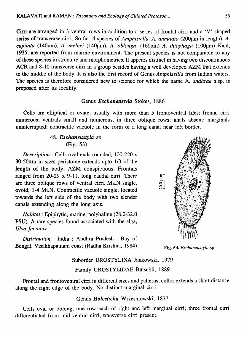
KALAVATI and RAMAN: Taxonomy and Ecology o/Ciliated Protozoa ... 55
Cirri are arranged in 3 ventral rows in addition to a series of frontal cirri and a 'V' shaped series of transverse cirri. So far, 4 species of Amphisiella, A. annulata (200flm in length), A. capitata (140fll1l), A. melnei (140flm), A. oblonga, (160J.1ll1) A. thiophaga (1 OOJlIIl) Kahl, 1935, are reported from marine environment. The present species is not comparable to any of these species in structure and morphometrics. It appears distinct in having two discontinuous ACR and 8-10 transverse cirri in a group besides having a well developed AZM that extends to the middle of the body. It is also the first record of Genus Amphiseilla from Indian waters. The species is therefore considered new to science for which the name A. andhrae n.sp. is proposed after its locality.
Genus Eschaneustyla Stokes, 1886
Cells are elliptical or ovate; usually with more than 5 frontoventral files; frontal cirri numerous; ventrals small and numerous, in three oblique rows; anals absent; nlarginals uninterrupted; contractile vacuole in the form of a long canal near left border.
48. Eschaneustyla sp. (Fig. 53)
Description ; Cells oval ends rounded, 100-220 x 30-50Jl.ID in size; peristome extends upto 1/3 of the length of the body, AZM conspicuous. Frontals
ranged from 20-29 x 9-11, long caudal cirri. There ~~.I are three oblique rows of ventral cirri. Ma.N single, .. ~ ovoid; 1-4 Mi.N. Contractile vacuole single, located towards the left side of the body with two slender canals extending along the long axis.
Habitat: Epiphytic, marine, polyhaline (28.0-32.0 PSU). A rare species found associated with the alga, Ulva Jaciatus
Distribution : India : Andhra Pradesh : Bay of Bengal, Visakhapatnam coast (Radha Krishna, 1984) Fig. 53. Eschaneustyla sp.
Suborder UROSTYLINA Jankowski, 1979
Family UROSTYLIDAE Btitschli, 1889
Frontal and frontoventral cirri in different sizes and patterns, collor extends a short distance along the right edge of the body_ No distinct marginal cirri
Genus Holosticha Wrzesniowski, 1877
Cells oval or oblong, one row each of right and left marginal cirri; three frontal cirri differentiated from mid-ventral cirri, transverse cirri present.

56 Ree. zool. Surv. India, Dec. Paper No. 282
Key to species
1. Cells oval, transparent, 6 frontoventral, 5 anal and 2 ventral cirri. Marginal cirri in two rows. Ma.N single, oval. Mi.N 2 ...................................................................... H. manca
2. Cells elongately oval, 2 frontal and 10-12 transverse cirri, Ma.N in 10-15 segments .. ••••••••••• ••• •••••• 0 ••••••••••••••••••••• 0 ••••••••••••• 0 •••••••••••• 0 ••••••••••••••••••••••••••••••••••••••••••••••••••••• H. warreni
49. Holosticha manca Kahl, 1933 (Fig. 54; Plate IV Fig.5)
Description : Cells oval and transparent; 107.5-120 x 42.8-70.0J..lm in size; six frontoventral cirri, 5 anal and 2 ventral cirri present. Marginal cirri widely separated in two rows; Ma.N single, oval; two small, spherical Mi.N on either side of the Ma.N; cytostome anteriolateral; cytostomal cilia long. AZM well formed; contractile vacuole absent.
Habitat : Planktonic and epiphytic, low saline, oligohaline (2-12PSU)
Distribution : India : Andhra Pradesh : Bay of Bengal, Visakhapatnam harbour jetties Ganapati and Rao 1958), coast (Radha Krishna, 1984); Orissa!: Chilka Lake (Das, 1995)
e o o N
Elsewhere: Ponds and lakes in Germany Kahl (1933).
Fig. 55. H. warreni
Fig. 54. Holosticha manca
50. Holosticha warreni Song and Wilbert, 1997 (Fig. 55; Plate IV Fig. 6)
Description : Cells elongate, oval, 80-100 x 20-30flm in size; right margin convex; left side slightly curved with one buccal, two frontal and 10-12 transverse cirri . Ventral cirri in pairs extended to the middle of the body; Ma.N in 10-15 segmen~s; contractile vacuole absent.
Habitat : "Mangrove creeks and sediments rich in organic matter, Psammophilic, low saline. « 8PSU).
Distribution : India : Andhra Pradesh : Godavari -Coringa mangrove creeks (present record); Orissa: Chilka Lake (Das, 1995)
Elsewhere : Coast of Qingdao, China.

KALAVATI and RAMAN: Taxonomy and Ecology o/Ciliated Protozoa ...
Suborder SPORADOTRICHINA Faure-Fremiet, 1961
Family OXYTRICHIDAE Ehrenberg, 1838
57
Frontoventral cirri scattered on ventrum, transverse cirri usually distinct atleast one row each of right and left marginal cirri with atleast 5 and usually 20 or more in each row
Key to the genera
1. Oral cavity not wide. Front-oventral cirri in a more or less linear file ......................... . ........................................................................................................................... Gastrostyla
2. Body laterally flexible. Franta-ventral cirri not linear ..................................... Oxytricha
3. Oral cavity wide. Caudal cirri promonent ..................................................... Stylonychia
Genus Gastrostyla Engelmann, 1862
Cells are oval or elongate. Frontal cirri arranged more or less linearly, forms atleast one oblique row; rows of right and left marginal cirri confluent posteriorly; ventrals irregular; anals 5; Ma.N divided in to 2-8 parts.
51. Gastrostyla sp. (Fig. 56)
Description: Cells are oval or ellipsoid, 200-280 x 80-100J..lm in size; dorsal surface convex; ventral side flat. There are 6 frontal and 5 transverse cirri. Right and left marginal cirri are continuous; ventrals form a continuous row; Ma.N 4, spherical; Mi.N 4, snlall; contractile vacuole single at the base of the buccal area.
Habitat : Mangrove creeks and sediments, brackish, mesohaline (14PSU)
Distribution : India : Andhra Pradesh : Godavari-Coringa mangrove creeks (present record). Fig. 56. Gastrostyla sp.
Genus Oxytricha Bory de St. Vincent, 1825
Cells are ellipsoid, laterally compressed; fronto-ventral and transverse cirri distinct; rows of right and left marginal cirri conflu.ent posteriorly, sometimes resulting in elongate posterior caudal cirri (Borrof, 1972).
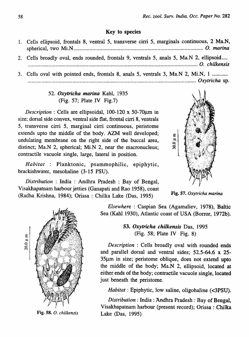
58 Rec. zool. Surv. India, Occ. Paper No. 282
Key to species
1. Cells ellipsoid, frontals 8, ventral 5, transverse cirri 5, marginals continuous, 2 Ma.N, spherical, two Mi.N ........................................................................................... O. marina
2. Cells broad~y oval, ends rounded, frontals 9, ventrals 5, anals 5, Ma.N 2, ellipsoid .... ........................................................................................................................ O. chilkensis
3. Cells oval with pointed ends, frontals 8, anals 5, ventrals 3, Ma.N 2, Mi.N, 1 .......... . ......................................................... .............................................................. Oxytricha sp.
52. Oxytricha marina Kahl, 1935 (Fig. 57; Plate IV Fig.7)
Description: Cells are ellipsoidal, 100-120 x 50-70flm in size; dorsal side convex, ventral side flat, frontal cirri 8, ventrals 5, transverse cirri 5, marginal cirri continuous, peristome extends upto the middle of the body. AZM well developed; undulating membrane on the right side of the buccal area, distinct; Ma.N 2, spherical; Mi.N 2, near the macronucleus; contractile vacuole single, large, lateral in position.
8 ::1. o d ~
Habitat : Planktonic, psammophilic, epiphytic, brackishwater, mesohaline (3-15 PSU).
Distribution : India: Andhra Pradesh: Bay of Bengal, Visakhapatnam harbour jetties (Ganapati and Rao 1958), coast (Radha Krishna, 1984); Orissa :, Chilka Lake (Das, 1995) Fig. 57. Oxytricha marina
E ::1. o o N
Fig. 58. O. chilkensis
Elsewhere : Caspian Sea (Agamaliev, 1978), Baltic Sea (Kahl 1930), Atlantic coast of USA (Borror, 1972b).
53. Oxytricha chilkensis Das, 1995 (Fig. 58; Plate IV Fig. 8)
Description : Cells broadly oval with rounded ends and parallel dorsal and ventral sides; 52.5-64.6 x 25-35Jlm in size; peristome oblique, does not extend upto the middle of the body; Ma.N 2, ellipsoid, located at either ends of the body; contractile vacuole single, located just beneath the peristome.
Habitat: Epiphytic, low saline, oligohaline «3PSU).
Distribution: India: Andhra Pradesh : Bay of Bengal, Visakhapatnam harbour (present record); Orissa: Chilka Lake (Das, 1995)

KALA VA TI and RAMAN: Taxonomy and Ecology of Ciliated Protozoa ...
54. Oxytricha sp. (Fig. 59)
Description : Cells small, oval with pointed
ends, both sides convex, peristome extends to
more than half the length of the body; 30-35 x
15-20Jlm in size; frontals 8, ventrals 3, anals 5,
marginal cirri continuous; Ma.N 2, spherical;
Mi.N single, dot-like; endoplasm filled with food
vacuoles.
Habitat: Psammophilic (in rock pools), marine,
polyhaline (28.l-30.5PSU).
Distribution : India : Andhra Pradesh : Bay of
Bengal, Visakhapatnam coast (Rao and Ganapati,
1968).
e o o N
Fig. 59. Oxytricha sp.
Genus Stylonychia Eherenberg, 1830
59
Body is flexible and contractile; rows of right and left marginal cirri do not confluent
posteriorly. No caudal cirri.
Key to species
1. Cells elongate, dorso-ventrally flattened, AZM conspicuous, frontals 8, ventrals 5,
transverse cirri 2-4, caudals 3, fringed. Ma.N 2, spherical. Mi.N 2, small .................. ..
............................................................................................................................. S. putrina
2. Cells oval with broad anterior end, dorsal side with small bristles, marginal cirri
discontinuous. Frontals 8, ventrals 5, transverse 5, contractile vacuole large near the base
of the peristome .................................................................................................. S. mytilus
3. Cells broadly oval, peristome relatively narrow, frontaIs 8, ventraIs 5, transverse 5,
caudal 3, long and fringed, two Ma.N ........................................................... S. pustulata

60
55. Stylonychia putrina Stocks, 1885
(Fig. 60; Plate V. Fig. 1)
Rec. zool. Surv. India, Occ. Paper No. 282
Description: Cells elongate, dorso-ventrally
flattened, sides parallel; 120-138.5 x 30-38.6Ilm in size; buccal area with well developed AZM.
There are 8 frontal cirri, 5 ventral cirri and 2-
4 transverse cirri which extend beyond the
posterior region. Caudal cirri 3, Ma.N 2,
spherical, Mi.N 2, small and dot-like, contractile
vacuole 1, small at the base of the peristome.
II Habitat : Psammophilic and among weeds.
Brackish, mesohaline (18.S-23.5PSU)
Distribution: India: Andhra Pradesh: Bay
of Bengal, Visakhapatnam harbour (Ratna
Bharathi 1998), coast (Radha Krishna, 1984).
Elsewhere ; Europe and America (Bick,
1972). Fig. 60. Stylonychia putrina
!I
Fig. 61. S. mytilus
56. Slylonychia mytilus Ehrenberg, 1838 (Fig. 61; Plate V Fig. 2)
Cells oval with broad anterior end. peristome wide,
AZM well developed, 100-160 x 80-120Jlm in size.
There are 8 frontal cirri, 5 ventrals behind buccal area
and 5 transverse cirri; caudal cirri 3, fringed; Ma.N 2,
oval; Mi.N 2, spherical; contractile vacuole single, large,
located in the posterior region.
Habitat: Planktonic, Psammophilic and among algal
mats, brackish, mesohaline (18-25PSU).
Distribution : India : Andhra Pradesh : Bay of Bengal, Visakhapatnam harbour (Ratna Bharathi 1998),
coast (Radha Krishna, 1984).
Elsewhere : U.S.A. (Bick, 1972), Caspiall Sea (Agamaliev, 1978).

KALAVATI and RAMAN: Taxonomy and Ecology of Ciliated Protozoa ...
57. Stylonychia pustulata Ehrenberg, 1838 (Fig. 62; Plate V Fig. 3)
Description : Cells are oval with broad anterior end, peristome relatively narrow, 140-150 x 80-90J.1m in size; AZM moderate, undulating membrane on the right side, more conspicuous. There are 8 frontal cirri, 5 ventrals and 5 transverse cirri; caudal cirri 3, long and fringed at the tip; Ma.N two, spherical; MLN small; contractile vacuole 2, small, 1 in the anterior half and the other in the posterior.
Habitat : Mangrove creeks and sediments with decaying leaves, oligohaline «6PSU)
Distribution : India: Andhra Pradesh : Bay of Bengal, Godavari-Coringa mangroves (present record).
Elsewhere : Dee estuary (Webb, 1956). This is the first record from mangrove creeks/sediments in India.
e ::1. o V"i N
Fig. (it. S. pustulata
Sub-phylum RHABDOPHORA Small, 1976
Class PROSTOMATEA Schewiakoff, 1896
Order PROTOSTOMATIDA Schewiakoff, 1896
Family HOLOPHRYIDAE Perty, 1852
Cytostome is simple, apical without any oral basket, toxicysts absent.
Genus Holophrya Eherenberg, 1833
61
Cells globose, oval and ellipsoid, ciliation unifonn, cytostome apical, round and simple, cytopharyngeal apparatus simple, rhabdose type
Key to species
1. Cells oval to spherical, cytostome apical, round. Ma.N spherical, central, 18-20 somatic kineties ............................................................................................................... H. simplex
2. Cells ellipsoid, ends pointed, cytostome apical, Ma.N oval, 2.0-22 somatic kineties .... . ...... ...... .................................................................. .......... ........... ........................ H. marina
3. Cells round, cytostome small, depressed, Ma.N massive, oval or elongated, somatic kineties 40-50 ................................................................................................. H. nigricans
4. Cells globose, cytostome apical with indistinct cytopharynx, Ma.N oval, somatic kineties 26-32 ...................................................................................................................... H. nair;

62 Rec. zoof. Surv. India, Dcc. Paper No. 282
58. Holophrya simplex Schewiakoff, 1893 (Fig. 63; Plate V Figs. 4&5)
Description: Cells are oval to spherical in shape, delicate, greenish in colour, 25-30 x 18-20J..lm in size; cytostome round, apical, leads to conical cytopharynx that is supported by a
basket of trichites; oral kineties absent; Ma.N spherical, 8-10J.lffi in size, central in position; somatic ciliation uniform; 18-20 monokineties; toxicyst absent, contractile vacuole posterior, single and large.
Habitat : Planktonic, brackish, oligohaline (2-12PSU); generaily a freshwater species occurring in lakes and ponds
Distribution: India: Andhra Pradesh: Bay of Bengal, Visakhapatnam harbour (present record); Orissa : Chilka Lake (Das, 1995); Punjab : Chandigarh and Himalayan region (Vasista and Gulati, 1972).
Elsewhere : Mesopsammon and periphyton in Hawaii and Capsian Sea (Agameliev 1972); Lahore, Pakistan (Gulati, 1925).
Fig. 63. Holophrya simplex
59. Holophrya marina Mansfeld, 1923 (Fig. 64; Plate V Figs. 6&7)
Fig. 64. H. marina
Description Cells thin, ellipsoid, both ends pointed, 20-24 x 12-16.5J.lm in size; cytostome apical, simple, and opening into a small conical cytopharynx; Ma.N oval, 3.5-5.4~m, always in the anterior half of the body; somatic ciliation uniform, 20-22 monkineties, contractile vacuole small, subterminal.
Habitat : Planktonic / epiphytic, marine, polyhaline (29.5-30.0PSU).
Distribution: India: Andhra Pradesh: Bay of Bengal, Visakhapatnam coast (present record).
Elsewhe re : A rare species usually found in the epilithic algal mats in a marine aquarium, Berlin, Germany (Kahl, 1930)
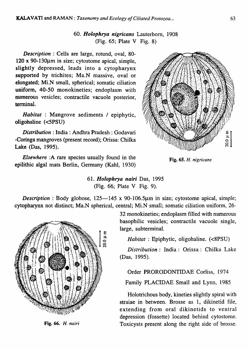
KALA V A TI and ~AN : Taxonomy and Ecology of Ciliated Protozoa ...
60. Holophrya nigricans Lauterbom, 1908 (Fig. 65; Plate V Fig. 8)
Description : Cells are large, rotund, oval, 80-120 x 90-130f.lm in size; cytostome apical, simple, slightly depressed, leads into a cytopharynx supported by trichites; Ma.N massive, oval or elongated; Mi.N small, spherical; somatic ciliation uniform, 40-50 monokineties; endoplasm with numerous vesicles; contractile vacuole posterior, terminal.
Habitat : Mangrove sediments / epiphytic, oligohaline «5PSU)
63
Distribution : India: Andhra Pradesh : Godavari -Coringa mangroves (present record); Orissa: Chilka Lake (Das, 1995). iI
Elsewhere :A rare species usually found in the epilithic algal mats Berlin, Germany (Kahl, 1930)
Fig. 65. H. nig ricans
61. Holophrya nairi Das, 1995 (Fig. 66; Plate V Fig. 9).
Description: Body globose, 125-145 x 90-106.5J.lm in size; cytostome apical, simple; cytopharynx not distinct; Ma.N spherical, central; MLN small; somatic ciliation uniform, 26-
Fig. 66. H. nairi
32 monokineties; endoplasm filled with numerous basophilic vesicles; contractile vacuole single, large, subterminal.
Habitat : Epiphytic, oligohaline. «8PSU)
Distribution: India: Orissa: Chilka Lake (Das, 1995).
Order PRORODONTIDAE Corliss, 1974
Family PLACIDAE Small and Lynn, 1985
Holotrichous body, kineties slightly spiral with straiae in between. Brosse as 1, dikinetid file, extending from oral dikinetids to ventral depression (fossette) located behind cytostonle. Toxicysts present along the right side of brosse.

64 Rec. zoot. Surv. India, Occ. Paper No. 282
Genus Placus Cohn, 1866
Ceils small, ellipsoid, pellicle with conspicuous spiral furrows, cytostome narrow slit at the anterior end, cytostomal cilia distinct, brosse dikinetids extend forward only to oral
dikinetid; ventral depression absent.
62. Placus socialis Fabre-Domiergue, 1889 (Fig. 67; Plate VI. Fig. 1)
Description : Cells small, oval in shape, 40-50 x 20-30Jlm in size; cytostome apical slit like, often bordered by trichites; "Brosse", the brush like-cilia arise from obliquely arranged
kineties that extend upto oral dikinetids. There is no ventral depression. Ma.N large, sausage shaped; MLN spherical, close to Ma.N; There are 2 contractile vacuole one near the cytopharynx and the other at the posterior end. There is a small cleft like cytopyge.
Habitat: Psammophilic, marine, polyhaline (29.5-30.0PSU).
Distribution: India: Andhra Pradesh: Bay of Bengal, Visakhapatnam harbour (present record).
Elsewhere: Sediments of White Sea. (Burkovsky, 1970)
Family PRORODONTIDAE Kent, 1881
Oral basket made of double trichites ending deep in cytoplasm. Unique distinctive "brush" of cilia arises from specialised short kineties on dorsal surface near the anterior end.
Genus Prorodon Ehrenberg, 1833
Fig. 67. Placus socialis
Oral basket made up of double trichites. Cytostome is without cilia. Somatic ciliation is uniform.
Key to species
1. Cells oval, ends rounded, oral basket clear, extending upto 1/3 the distance from anterior end. Kineties not inclined upon brush ......................... ' .................................. P. marinus
2. Cells oval, posterior end narrow, oral basket extending about 1/4 the distance from anterior end, kineties reach the brush from left ............................................. P. discolor

KALA VATI and RAMAN: Taxonomy and Ecology o/Ciliated Protozoa ... 65
3. Cells small, oval or ovocylindrical, monokineties parllel to dorsal brush reaching the brush from both sides ............................................................................... P. minuta n. sp.
63. Prorodon marinus (Claperede and Lachmann) Dragesco, 1960 (Fig. 68; Plate VI. Fig. 2)
Description: Cells are oval, flat, 100-200 x SO-SOJ.1m in size; cytostome apical with anterodorsal brosse. Kineties parallel and are not inclined upon the brush; Ma.N spherical, 5.0f.1m in diameter; 2 Mi.N, small and dot-like; Somatic kineties parallel, 25-30; contractile vacuole single, posterior.
Habitat : Planktonic, marine, polyhaline (2S.5-31.5PSU).
Distribution: India: Andhra Pradesh: Bay of Bengal, Visakhapatnam coast (present record); Orissa : Chilka lake (Das, 1995)
Elsewhere :Salt marshes of Lousiana, U.S.A.
(Elliot and Bamforth, 1975).
iI
Fig. 69. P. discolor
Fig. 68. Prorodon marinus
64. Prorodon discolor (Eherenberg, 1831 Kahl, 1930) (Fig. 69; Plate VI. Fig. 3)
Description: Cells are oval, flat, 100-140 x 50-85~m in size. Brosse kineties originate at the ventral edge of the oral kinetids as 3 longitudinal files of dikinetids. Somatic kineties, 20-25 monokineties and reach the brush from left; Ma.N massive, central, spherical or elongated; 2 small Mi.N located close to Ma.N; cytostome wide; cytopharynx small; contractile vacuole posterior, single.
Habitat Planktonic, low saline, oligohaline (8-11.5 PSU).
Distribution : India : Andhra Pradesh Visakhapatnam harbour (Ratna Bharathi, 1998) Godavari-Coringa mangroves (present record); Orissa Chilka Lake (Das, 1995).

66 Rec. zool. Surv. India, Occ. Paper No. 282
Elsewhere : Widely distributed in rivers and ponds in Europe, Germany, China
65. Prorodon minuta n. sp. (Fig. 70; Plate VI. Fig. 4)
Description : Cells are very small minute, oval or ovocylindrical, 40-50 x 24-32Jlm in size; cytostome apical, leading into the cytopharynx, supported by double trichites; Ma.N
elongated (8 x 3.2J.lm); Mi.N spherical, close to the Ma.N; dorsal brush straight; somatic kineties reach the brush from both sides, 20-30 (mean 24) longitudinal monokineties; contractile vacuole, small, single, terminal.
Habitat : Planktonic, brackish, mesohaline (18.5-22.0PSU)
Distribution: India: Andhra Pradesh: Visakhapatnam harbour (Ratna Bharathi, 1998).
Type slides : Z.S.I NO.2464
Remarks : As many as 52 species of Prorodon have been reported so far from both marine and brackishwater environs. Hiller and Bardele (1988) identified four subgroups based on the arrangement of kineties in relation to the dorsal brush. The present species shows typical
iI
'Syntrop' type of arrangements (Kineties reaching the Fig. 70. P. minuta n. sp.
brush from both sides) as in the case of P.africanus
(Dragesco, 1970) and P. reabei (Czapik, 1965). The species deferred from both of them in size (being very small, 40-50 x 24-32mm), having fewer number of somatic kineties and an elongated macronucleus. The species is therefore considered new to science for which the name Prorodon minuta n. sp is suggested.
Genus Mimeticus Small and Lynn, 1985
Brosse is in the form of a series of rows of dikinetids perpendicular to long axis of the body.
66. Mimeticus Inimeticus Small and Lynn, 1985 (Fig. 71; Plate VI. Fig. 5)
Description : Cells are cylindrical, 120-150 x 20-30Jlm in size; cytostome sub-apical, slightly displaced, surrounded by 3 longitudinal files of dikinetids. The anterior kinetosome of the somatic kineties reach the brush from right side; Ma.N bean shaped; 3 or 4 Mi.N; somatic kineties 40-50 monokineties.
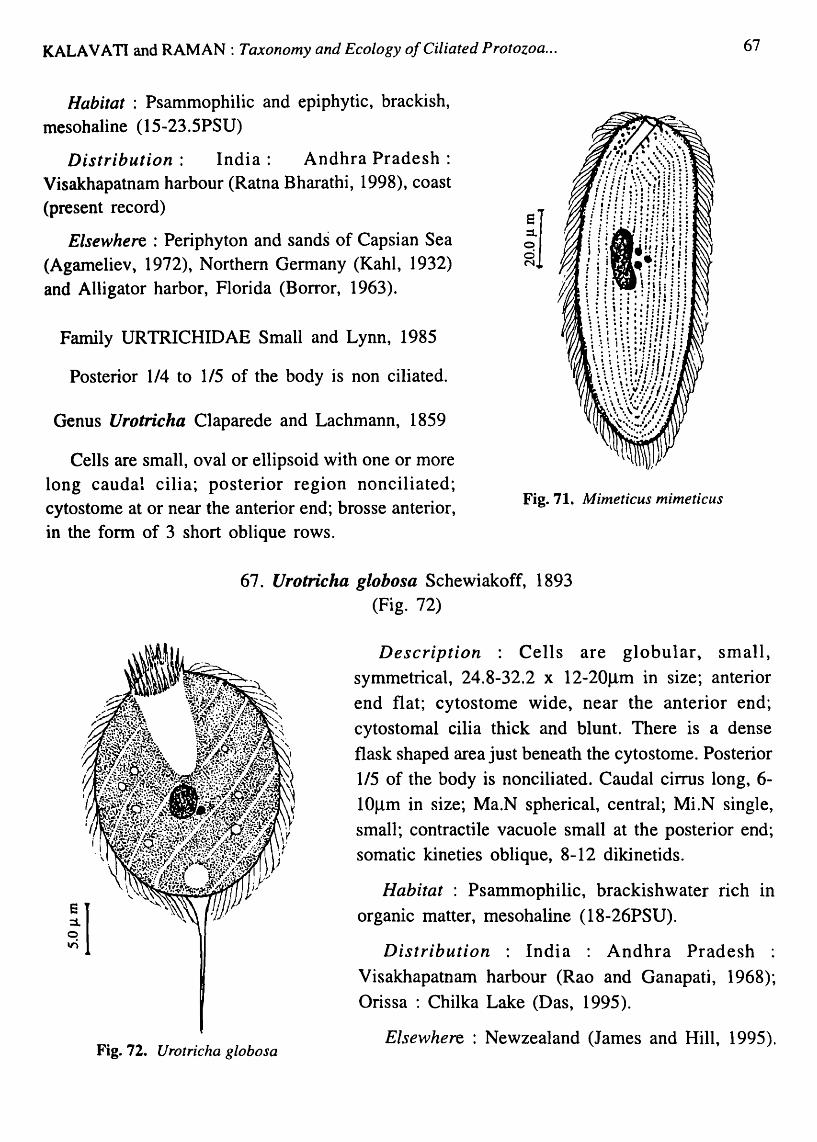
KALA V A T1 and RAMAN: Taxonomy and Ecology of Ciliated Protozoa ...
Habitat : Psammophilic and epiphytic, brackish, mesohaline (15-23.SPSU)
Distribution: India: Andhra Pradesh: Visakhapatnam harbour (Ratna Bharathi, 1998), coast (present record)
Elsewhere : Periphyton and sands of Capsian Sea (Agameliev, 1972), Northern Gennany (Kahl, 1932) and Alligator harbor, Florida (Borror, 1963).
Family URTRICHIDAE Small and Lynn, 1985
Posterior 1/4 to lIS of the body is non ciliated.
Genus Urotricha Claparede and Lachmann, 1859
Cells are small, oval or ellipsoid with one or more long caudal cilia; posterior region nonciliated; cytostome at or near the anterior end; brosse anterior, in the form of 3 short oblique rows.
iI
Fig. 71. Mimeticus mimeticus
67. Urotricha globosa Schewiakoff, 1893 (Fig. 72)
67
Description : Cells are globular, small, symmetrical, 24.8-32.2 x 12-20J..lm in size; anterior end flat; cytostome wide, near the anterior end; cytostomal cilia thick and blunt. There is a dense flask shaped area just beneath the cytostome. Posterior lIS of the body is nonciliated. Caudal cirrus long, 6-IOflm in size; Ma.N spherical, central; MLN single, small; contractile vacuole small at the posterior end; somatic kineties oblique, 8-12 dikinetids.
Fig. 72. Urotricha globosa
Habitat : Psammophilic, brackishwater rich In organic matter, mesohaline (18-26PSU).
Distribution : India : Andhra Pradesh : Visakhapatnam harbour (Rao and Ganapati, 1968); Orissa : Chilka Lake (Das, 1995).
Elsewhere : Newzealand (James and Hill, 1995).

68 Ree. zool. Surv. India, Dec. Paper No. 282
Class LITOSTOMATEA Small and Lynn, 1981
Subclass HAPTORIA Corliss, 1974
Order HAPTORIDEA Corliss, 1974
Family DIDINIDAE Poche, 1913
Oral area is often conical; somatic cilia as series of short kineties; one or more girdles around the body.
Genus Didinium Stein, 1859
Cells are with two girdles; one equatorial and the other behind the oral area.
68. Didinium nasutum O.F.Muller, 1786 (Fig 73; Plate VI. Fig. 6).
Description: Cells broadly oval dorsoventrally flattened 60-80 x 45-651lm in size; anterior end drawn out into a conical projection; cilia in two girdles, one near the base of the
Fig. 73. Didinium nasutum
proboscis, the other near mid body; Ma.N horse shoe shaped; Mi.N adjacent to Ma.N; contractile vacuole single, large, posterior.
Habitat Planktonic, brackish, mesohaline (20-23 PSU).
Distribution India Andhra Pradesh Visakhapatnam harbour (Ratna Bharathi, 1998).
Elsewhere: Southampton, Britain (Leakey et aI, 1993).
Remarks: D. nasutum is a freshwater species with cosmopolitan Distribution. An identical brackishwater species was reported from Southampton, Britain (Leakey et ai, 1993). This species also resembles D. nasutum occurring in mesohaline waters.
Family ENCHELYIDAE Eherenberg, 1838
Oral region is flat at the apex; somatic kineties abut on oral region without arching; cytostome-cytopharynx complex, circular to ovoid.
Genus Enchelys O. F. Muller, 1773
Cells are completely ciliated. No membranous dome on flat oral region

KALAVATI and RAMAN: Taxonomy and Ecology of Ciliated Protozoa ... 69
Key to species
1. Cells flask shaped. oral aperture slit-like, Ma.N single, band-like, curved or bean shaped .......................................................................................................................... E. pectinata
2. Cells flask shaped, oral aperture apical, oval wide. Ma.N flat and ribbon-like ............ . ..... ....... ...... .............................. ..... ......... ............................................................... E. marina
69. Enchelys pectinata Kahl, 1933 (Fig. 74)
Description : Cells are flask shaped anterior end narrow, posterior end round; 40-60 x 32-58 Jlm in size; oral aperture apical, slit-like; cytopharynx supported by trichites; cilia located around the oral aperture long and closely packed. Ma.N single, band-like or typically bean shaped; 4.8 x 6.4Jlm in size; somatic ciliation uniform monokineties; contractile vacuole single, terminal.
Habitat: Planktonic, brackish, mesohaline (15-22PSU) ~ o
Distribution : India: Andhra Pradesh: Visakhapatnam ~ harbour (Ratna Bharathi, 1998).
Elsewhere : Germany (Kahl, 1933), Dee estuary, Britain (Webb, 1956). First record from Bay of Bengal, India
Fig. 75. E. marina
Fig. 74. Enchelys pectinata
70. Enchelys marina Meunier, 1907 (Fig.75; Plate VI. Fig.7)
Description : Cells flask shaped posterior end rounded, anterior end flat and wide; 120-150 x 60-70 flm in size; oral aperture apical, oval; cytopharynx small, supported by trichites; Ma.N ribbon-like; Endoplasm with numerous inclusions, greenish in colour; contractile vacuole large, spherical and terminal.
Habitat : Epiphytic, Marine, polyhaline (28.5-31.5PSU)
Distribution: India: Andhra Pradesh: Visakhapatnam harbour (present record). Elsewhere :German coast of North Sea (Kahl, 1935); First record from Bay of Bengal, India.

70 Rec. zool. Surv. India, Oec. Paper No. 282
Family LACRYMARIIDAE de Fronentel, 1876
Cells long flask shaped; apex bulb-like with several short oblique kineties; body highly contractile.
Genus Lacrymaria Bory de St. Vincent, 1826
Cells polymorphic, cylindrical, spindle or flask shaped; long contractile proboscis; cytopharynx usually distinct
Key to species
1. Neck without annulations ............................................................................................... (1)
2. Neck with annulations .................................................................................................... (2)
1 a. Neck long and higly contractile; proboscis well developed, Ma.N dumb-bell shaped. Somatic kineties 12-18 ............................................................................................. L. olor
1 b. Neck with a distinct corona and collar; somatic kineties 18-24. Ma.N oval or oblong ••••••••••••••••••••••••••••••••••••••••••••••••••••••••••••••• 000 ••••••••••••••••••••••••••••••••••••••••••••••••••• o •••• L. co ronata
Ie. Neck long contractile, apical lobe dome-like; somatic kineties 18-20, Ma.N oval ....... . ••••••••••• f f ••• o ••••••• f ••• 0 ••••• 0 ••••••••••••••••••••••••••••••••••••••••••••••••••••••••••••••••••••••••••••••••••• L. sapropelica
2a. Neck short with 4-5 annulations, Ma.N spherical. Somatic kineties 12-16 ... L. elegans
2b. Neck with 2-3 annulations; apical lobe nonciliated. Somatic kineties 6-8, Ma.N oval. ••••••••••••••••••••••• •• • ••••••••••••••••••••••••••••••••••••••••••••••••••••••••••••••••••••••••••••••••••• 0 ••••••••••••••• L. marina
Fig. 76. Lacrymaria olor
71. Lacrymaria olor Kahl, 1933 (Fig. 76; Plate VI. Fig. 8)
Description : Cells elongate, neck long and highly contractile, 150-178 x 25-3SJ..Lm in size; Proboscis well developed; cytostome apical on corona. Ma.N dumb-bell shaped, somatic kineties 12-18, spiraling round the body; contractile vacuole 2 on the either side of the nucleus.
Habitat: Epiphytic, Psammophilic, brackish, mesohaline (5-15PSU)
Distribubtion : India: Andhra Pradesh : Visakhapatnam harbour (Rao and Ganapati, 1968, Ratna Bharathi, 1998) Orissa: Chilka Lake (Das, 1995).
Elsewhere : Widely distributed in stagnant slow flowing water (Bick, 1972). The species was earlier recorded from

KALA V A TI and RAMAN: Taxonomy and Ecology of Ciliated Protozoa ... 71
brackishwater in Germany (Kahl, 1935) White Stable, UK. (Maghraby and Perkins, 1956), Dee Estuary, U.K. (Webb, 1956)
72. Lacrymaria coronata. Lachmann, 1859 (Fig. 77; Plate VI. Fig.9)
Description : Cells are large, 100-140 x 30-45flm in size; when contracted, cytostome apical, on a corona with collor; somatic kineties 18-24, spiraling around the body when
contracted; Ma.N single, oval, central and oblong; Mi.N small, J~\
spherical; contractile vacuole single, posterior, tenninal. Rhabdos composed delicate nematodesmata.
Habitat·: Planktonic, psammophilic, brackish, mesohaline (13.4-21.5PSU).
Distribution : India· : Andhra Pradesh : Visakhapatnam harbour (Rao and Ganapati, 1968, Ratna Bharathi, 1998). Orissa : Chilka Lake (Oas, 1995).
Elsewhere: West coast of Africa (Dragesco, 1950), German coast of Baltic Sea (Bock, 1952), White Stable, UK (Maghraby and Perkins, 1956), Dee estuary (Webb, 1956), Capsian sea (Agamaliev , 1967), Norfolk salt marsh (Barnes et al., 1976), North Yorkshire (Hartwig and Parker, 1977), Mugu lagoon, California, USA (Smith,1982), Chichester harbour (Carey and Maeda, 1985).
iI
Fig. 78. L. e legans
Fig. 77. L coronata
73. Lacrymaria elegans Engelmann, 1862 (Fig. 78; Plate VII. Fig.1)
Description : Cells are ovocylindrical, 65-75 x 30-40flm in size; neck short, stout with 4-5 annulations; Ma.N spherical; somatic kineties 12-16, parallel, straight; contractile vacuole single tenninal.
Habitat: Psammophilic, epiphytic, mesohaline (6-10 PSU)
Distribution : India : Andhra Pradesh : Visakhapatnam harbour (Ratna Bharathi, 1998). Orissa : Chilka Lake (Das, 1995).
Elsewhere : A common freshwater species reported from rivers and ponds in Germany (Kahl, 1935), Italy (Madoni, 1978) and China (Shen et al., 1995).
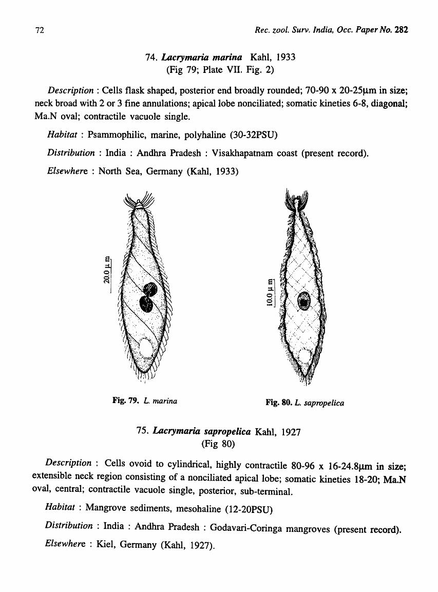
72 Rec. zool. Surv. India, Occ. Paper No. 282
74. Lacrymaria marina Kahl, 1933 (Fig 79; Plate VII. Fig. 2)
Description: Cells flask shaped, posterior end broadly rounded; 70-90 x 20-25f..1m in size; neck broad with 2 or 3 fine annulations; apical lobe nonciliated; somatic kineties 6-8, diagonal; Ma.N oval; contractile vacuole single.
Habitat : Psammophilic, marine, polyhaline (30-32PSU)
Distribution: India: Andhra Pradesh: Visakhapatnam coast (present record).
Elsewhere: N"orth Sea, Germany (Kahl, 1933)
Fig. 79. L. marina Fig. 80. L. sapropelica
75. Lacrymaria sapropelica Kahl, 1927 (Fig 80)
Description: Cells ovoid to cylindrical, highly contractile 80-96 x 16-24.8JlIl1 in size; extensible neck region consisting of a nonciliated apical lobe; somatic kineties 18-20; Ma.N oval, central; contractile vacuole single, posterior, sub-terminal.
Habitat: Mangrove sediments, mesohaline (12-20PSU)
Distribution: India: Andhra Pradesh: Godavari-Coringa mangroves (present record).
Elsewhere: Kiel, Germany (Kahl, 1927).

IALAVATI and RAMAN: Taxonomy and Ecology o/Ciliated Protozoa ... 73
Family SPATHIDllDAE Kahl in Doflein and Reichnow, 1929
Apex somewhat fan shaped; oral region a narro\\' plateau along apex that may extend veDtrally; oral dikinetids as arched extensions of body kineties (Small and Lynn, 1985)
Genus Spathidium Dujardin, 1841
Body long or ovoid, apex fan-shaped
76. Spathidium fossicola Kahl, 1933 (Fig. 81)
Description : Cells bow 1 shaped, wide 240-260 x 185-220Jlm in size; anterior region broad with non-ciliated apical ridge lined by trichocysts; oral aperture is a slit, lying along the length of the ridge; somatic ciliation uniform, 30-40 monokineties; Ma.N ribbon-like; contractile vacuole sub-tenninal, single.
Habitat : Psammophilic, marine, polyhaline (21.33-31PSU)
Distribution : India : Andhra Pradesh : Vi~akhapatnam coast (present record).Orissa : Chilka Lake, (Das, 1995)
Elsewhere: Sylt, Germany (Kahl, 1933),
Fig. 81. Spathidium fossicola
Family TRACHELOPHYLLIDAE Kent, 1886
Body lo~g, ovoid or flask shaped, oral region a small, simple, dome-shaped, sometimes pointed; anterior kinetids, dikinetids.
Key to the Genera
1. Apex slightly bent ventrally ......................................................................... Lagynophrya
2. Apex straight, body much flattened ...................................................... Trachelophyllum
Genus Lagynophrya Kahl, 1927
Cells pyriform; apex slightly bent ventrally, proboscis retracted.

74
-Fig. 82. Lagynophrya salina
Ree. zool. Surv. India, Dec. Paper No. 282
77. Lagynophrya salina Kirby, 1932 (Fig. 82; Plate VII. Fig. 3)
Description: Cells pyriform, 36-55 x 15-20 ~m in size, dorsoventrally flattened with a short non ciliated retractile cone-like proboscis at the anterior end which is slightly bent ventrally and leads into cytopharynx. Cytopharynx extends nearly half the length of the body and is lined by a basket of fine packed trichites; Ma.N two, identical and spherical; somatic ciliation uniform, 20-22 monokinities; contractile vacuole small at the posterior extremity.
Habitat : Psammophilic, marine, polyhaline (29.5-
32.0PSU).
Distribution : India: Andhra Pradesh: Visakhapatnam coast (present record).
Elsewhere: Coast of California USA (Kirby, 1932), Northern Germany (Kahl, 1933)
Genus Trachelophyllum Claparede and Lachmann, 1859
Cells flattened, apex a low dome, straight. Body much flattened
78. Trachelophyllum sp. (Fig. 83; Plate VIT. Fig. 4)
Description; Cells are elongate flat and leaf-like; 112-128 x 24-36Jlm in size; anterior end drawn out into a small neck, tip truncate; cytostome small apical; cytopharynx narrow and lined by trichites; Ma.N 2, oval; MLN 2, small, spherical; somatic ciliation uniform, kinities widely spaced, 8-10 monokinities; contractile vacuole terminal.
Habitat Epiphytic, marine, polyhaline (28.5-32PSU)
Distribution India: Andhra Pradesh: Visakhapatnam coast (present record).
Fig.83. Trachelophyllum sp.
Order PLEUROSTOMATIDA Schewiakoff, 1896
Family AMPHILEPTIDAE Biitschli, 1889
Oral dikinetids are continuous with two oral kineties and parallel to cytostome; oral area

ICALAVATI and RAMAN: Taxonomy and Ecology of Ciliated Protozoa ... 75
lub-apical; ovoid, flattened along ventral margin of laterally compressed body; both sides ciliated.
Key to the genera
1. Kinities on right and left side, parallel through out .......................................... Litonotus
2. Kinities on the left converge as anterior suture ........................................... Amphileptus
3. Toxicysts in long marginal bands or warts ................................................. Loxophyllum
Genus Litonotus Wrzesniowski, 1870
Kineties on right and left side parallel through out the body length. Toxicysts present in oral region.
79. Utonotus obtusus Maupas, 1888 (Fig. 84; Plate VII. Fig. 5)
Description: Fragile, flat, leaf-like, anterior end bluntly pointed, posterior end rounded, 60-75 x 20-28Jlffi in size. There are two perioral kineties to the right of cytostome; somatic kineties 5 on the right ~lde and 4 on the left. Ma.N two, oval or bean shaped 5-7Jlm in size; Mi.N small; Extrusomes present at the posterior right margin and along the right edge of the cytostome; contractile vacuole single, subterminal.
Habitat: Psammophilic, brackish, mesohaline (18-22 PSU)
Distribution : India : Andhra Pradesh : Visakhapatnam harbour (Ratna Bharathi, 1998).
Elsewhere: Alligator harbor, Florida.
e :::1. o o N
Fig. 84. L{tonotus obtusues
Genus Amphileptus (Eherenberg, 1838) Btitschli, 1889
Kineties on the left side converge as anterior suture; toxicysts only in oral region.
Key to species
1. Cells elongate, anterior end bent. Ma.N 2, spherical. Mi.N single, 2-3 contractile vacuoles ....................................................................................................................... A. claparedei
2. Cells broad, leaf-like. Ma.N 2. MLN 2. Several contractile vacuoles .......................... .. ............................ .... ....... ..... .......... ............... ........ .......... .... ........ .............. ... A. trachelioides

76 Ree. zool. Surv. India, Dec. Paper No. 282
80. Amphileptus claparedei Stein, 1867 (Fig. 85; Plate VIT. Fig. 6)
Description: Cells elongate, laterally compressed, flat, 130-156 x 25-40 flm in size; anterior end bent, cytostome long and slit-like; oral cilia thick about 2/5rh from the ventral margin and distinct; Ma.N 2, identical, spherical, 4-6flm in diameter; Mi.N small, dot-like and distinct; somatic ciliation uniform, 20-25 longitudinal parallel monokinities. Ciliary rows on the left side of the body sparsely distributed. 2-3 contractile vacuoles located at
the posterior extremity. E r 0::1. I Habitat : Planktonic, brackish, mesohaline (18-22
PSU). g 1 Distribution: India: Andhra Pradesh: Visakhapatnam
harbour (Ratna Bharathi, 1998); coast (present record).
Elsewhere: North Sea, Germany (quoted from Kahl, 1935)
e ::I. o 9 -
Fig. 86. A. trichelioides
Fig. 85. Amphileptus claparedei
81. Amphileptus trachelioides J ach, 1893 (Fig. 86; Plate VII. Fig.7)
Description: Cells are broad, flat and leaf-like. 300-400
x 120-150flm with long ventral slit-like cytostome; oral
ciliature thick and distinct; Ma.N 2, spherical; MLN small,
spherical, 2, in between the Ma.N; an irregular row of
tnchites seen in the cytostomal region. Several small
contractile vacuoles present in two parallel rows on either
side of the body.
Habitat : Epiphytic, marine, polyhaline (29-32PSU).
Distribution : India : Andhra Pradesh : Visakhapatnam
harbour (Ratna Bharathi, 1998)
Elsewhere: Germany (Kahl, 1933).
Genus Loxophyllum Dujardin, 1841
Cells leaf-like, flattened, ends pointed when contracted; toxicysts in long marginal bands or in warts or both; Ma.N moniliform.
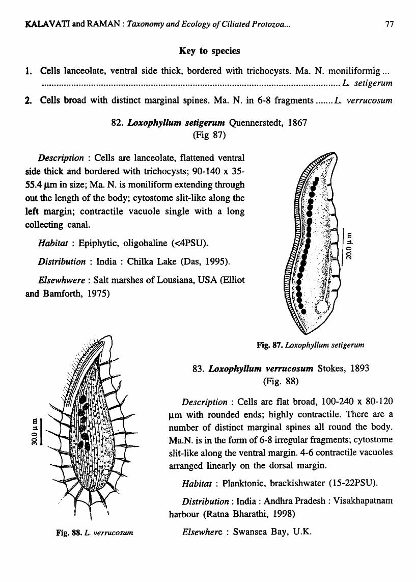
KALAVATI and RAMAN: Taxonomy and Ecology o/Ciliated Protozoa ... 77
Key to species
1. Cells lanceolate, ventral side thick, bordered with trichocysts. Ma. N. moniliformig ... ......................................................................................... ...... .......................... L. setig e rum
2. Cells broad with distinct marginal spines. Ma. N. in 6-8 fragments ....... L. verrucosum
82. Loxophyllum setigerum Quennerstedt, 1867 (Fig 87)
Description : Cells are lanceolate, flattened ventral side thick and bordered with trichocysts; 90-140 x 35-55.4 J..Lm in size; Ma. N. is moniliform extending through out the length of the body; cytostome slit-like along the left margin; contractile vacuole single with a long collecting canal.
Habitat: Epiphytic, oligohaline «4PSU).
Distribution : India : Chilka Lake (Oas, 1995).
Elsewhwere : Salt marshes of Lousiana, USA (Elliot
and Bamforth, 1975)
iI
Fig. 88. L. verrucosum
Fig. 87. Loxophyllum setigerum
83. Loxophyllum ven-ucosum Stokes, 1893 (Fig. 88)
Description: Cells are flat broad, 100-240 x 80-120 flm with rounded ends; highly contractile. There are a number of distinct marginal spines all round the body. Ma.N. is in the fonn of 6-8 irregular fragments; cytostome slit-like along the ventral margin. 4-6 contractile vacuoles arranged linearly on the dorsal margin.
Habitat: Planktonic, brackishwater (lS-22PSU).
Distribution: India: Andhra Pradesh: Visakhapatnam harbour (Ratna Bharathi, 1998)
Elsewhere: Swansea Bay, U.K.

78 Rec. zool. Surv. India, Dcc. Paper No. 282
Order PHARYNGOPHORIDA Small and Lynn, 1985
Family TRACHELIIDAE Ehrenberg, 1838
Cytostome is apical; eccentric, oral dikineties extend along a contractile proboscis; toxicysts on the ventral side of proboscis.
Key to the genera
1. Body long, posterior end usually pointed ...................................................... ...... Dileptus
2. Body oval to round, posterior end rounded ..................................................... Trachelius
Genus Dileptus Dujardin 1840
Body long, posterior end pointed; proboscis contractile.
Key to species
1. Cells spindle shaped, posterior end pointed. Ma.N. several fragments. CV small in a row ............................................................................................................................... D. anser
2. Cells elongate. Ma.N. one, elongate. 2 CV on either side of Ma.N .... . D. bivacuolatus
84. Dileptus anser O.F.Muller, 1786 (Fig. 89; Plate VII. Fig. 8)
Description : Cells are slender spindle-shaped, highly contractile, with a row of trichites on the ventral side. Posterior end drawn out into a fine pointed structure; 300-325 x 55-85 Jlm in size; cytostome small, circular at the base of the proboscis leading into a small cytopharynx supported by trichites; Ma.N. in the form numerous small fragments; CV small, numerous, dispersed in the endoplasm; somatic ciliature uniform, 20-24 parallel monokineties.
Habitat : Mangrove sediments, brackish, mesohaline (10-25PSU).
Distribution : India : Andhra Pradesh : GodavariCoringa mangroves (present record).
Remarks : Cosmopolitan in distribution. Found in still and slow flowing water with organic detritus (Bick, 1972).
18 :::l o vi
Fig. 89. Dileptus anser

'KALA VA TI and RAMAN : Taxonomy and Ecology of Ciliated Protozoa ...
85. Dileptus bivQcuolatus da Cunha, 1915 (Fig 90; Plate VII. Fig. 9)
79
Description: Cells are elongate, 100-150Jlffi in size; anterior neck region distinct, contractile and prehensile; Ventral surface with trichocysts. Cytostome oval located at the base of neck, supported by trichites forming a cytopharyngeal basket; Ma.N. single, elongated 26x12Jlm; centrally located; two distinct CV on either side of nucleus; somatice ciliature unifonn, 10-12 oblique monokineties.
Habitat: Psammophilic, brackish, mesohaline (15-22 PSU). Ii Distribution : India : Andhra Pradesh : Visakhapatnam harbour (present record)
Remarks : This species is generally found in small streams rich in organic matter. Kahl (1934), however, noticed it 'in salt water and considered it polysaprobic.
Genus Trachelius Schrank, 1803.
Body is oval to round with an anterior proboscis; somatic kineties oblique along the ventral edge of the proboscis.
Fig. 90. D. bivacuolatus
Fig. 91. Trachelius ovum
86. Trachelius ovum Ehrenberg, 1831 (Fig. 91; Plate VIT. Fig. 1 0)
Description : Cell shape variable; 350-450x 1 00-150~m in size proboscis on the ventral side, finger-like with a nonciliated ventral edge; cytostome circular at the base of proboscis; oral aperture supported by a row of trichites; Ma.N. single, oval; somatic kineties 10-15, parallel, monokineties ~ CV 3-5, dispersed in endoplasm.
Habitat: Psammophilic and epiphytic, oligohaline (8.6-12PSU).
Distribution: India: Andhra Pradesh: Visakhapatnam harbour (present record)
Remarks: T. ovum is a comInon inhabitant of all types of standing and flowing water rich in oxygen. Hentschell (1916) reported the species from periphyton. According to Kolkwitz (1950), this is a beta mesosaprobic indicator organisln.

80 Rec. zool. Surv. India, Dec. Paper No. 282
Sub-phylum CYRTOPHORA Small, 1976
Class PHYLLOPHARYNGEA de Puytorac et al., 1974
Order CRYPTOPHORIDA Faure-Fremiet in Corliss, 1976
Sub-order CHLAMYDOD01ITINA Deuroux, 1976
Family CHILODONELLIDAE Deroux, 1970
Cells dorsoventrally flattened; anterior end tapering. Anterior preoral kineties arch at the tip. Right ventral kineties continuous with posterior kineties and arch preorally towards left; oral kineties 3, 2 circum oral dikineties and 1 pre-oral dikinetid.
Genus Chilodonella Strand, 1928
Oral kineties 3 in number, preoral kinety complete.
87. Chilodonella cucullulus O.F. Muller, 1786 (Fig. 92; Plate VIII. Fig. 1)
Description : Cells dorsoventrally flattened, 100-150 x 50-70llm; anterior end broader than the posterior; dorsal surface convex, ventral surface flat; There is a distinct cross row
e ::1. o d N
Fig. 92. Chilodonella eueullulus
of bristles in the anterior dorsal region; oral basket
with 3 pre-oral and 10-12 cytopharyngeal trichites;
Ma.N. oval and appears as a characteristic concentric structure; Mi.N. small spherical; 1-6 contractile vacuoles, scattered; cytoplasm filled with granules;
Somatic ciliature uniform. 18-20 monokinetids.
Habitat : Epiphytic, oligohaline «3PSU).
Distribution: India: Orissa: Chilka Lake (Das, 1995).
Renzarks : A fresh or brackish water species recorded from a wide variety of aquatic habitats and
is known to tolerate a wide range of environmental conditions (Bick, 1972).
Genus Phascolodon Skin, 1859
Body vase-like, oral region cup shaped with an anteroventral depression.

KALAVATI and RAMAN: Taxonomy and Ecology o/Ciliated Protozoa ...
88. Phascolodon sp. (Fig 93)
81
Description :Cells oval with a broad anterior end slightly compressed dorsoventrally and with well developed edges of body forming wing-like extensions; 65-8Sx30-45flm in size
stylUS absent; ventral surface concave or flat; dorsal surface convex or arched; oral region cup-shaped located in an anteroventral depression; ciliature confined
to a V -shaped area on the ventral side which narrows laterally behind cytostome; cytostome ellipsoid with
oral basket of trichites; Ma. N. oval with a small Mi.N adherent to it; there are 2 CV one on the right anterior and the other on left posterior edge.
Habitat: Psammophilic and among algae, brackish, mesohaline (22.8-2S.8PSU).
Distribution : India : Andhra Pradesh Visakhapatnam harbour (present record)
Remarks : Normally a freshwater inhabitant. This is the first record of Phacolodon sp. in a brackish water region in India. Fig. 93. Phascolodon sp.
Family CHLAMYDODONTIDAE Stein, 1859
Ventral body kineties run from right ventral body surface to right dorsal and antericr left surfaces; thigmotactic cilia seen at the end of dorsum.
Genus Chlamydodon Ehrenberg, 1835
Cells are ovoid. There is a characteristic cross straited band at perimeter field of body kineties.
89. Chlamydodon triquetris Kahl, 1933 (Fig 94; Plate VIn. Fig. 2)
Description : Cells lanceolate, flattened, 120-140 x 45-S0flm in size; anterior end curved with a beak- like projection towards left; cytostome oval in the anterior 1/3 of the body surrounded by pharyngeal basket containing 13-15 nematodesmata; somatic kineties 48-50 monokineties. A characteristic band of straited ribbon noticed along the edges. Ma.N. single, elongated. Mi.N. 3-4, small and distributed.
Habitat: Psammophilic and among algal mats, marine, polyhaline (29.S-32.5PSU).
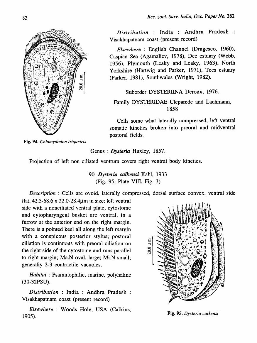
82 Rec. zool. Surv. India, Occ. Paper No. 282
Distribution : India : Andhra Pradesh : Visakhapatnam coast (present record)
Elsewhere : English Channel (Dragesco, 1960), Caspian Sea (Agamaliev, 1978), Dee estuary (Webb, 1956), Plymouth (Leaky and Leaky, 1963), North Yorkshire (Hartwig and Parker, 1971), Tees estuary (Parker, 1981), Southwales (Wright, 1982).
Suborder DYSTERIINA Deroux, 1976.
Family DYSTERIDAE Cleparede and Lachmann, 1858
Cells some what laterally compressed, left ventral somatic kineties broken into preoral and midventral postoral fields.
Fig. 94. Chlamydodon triquetris
Genus: Dysteria Huxley, 1857.
Projection of left non ciliated ventrum covers right ventral body kineties.
90. Dysteria calkensi Kahl, 1933 (Fig. 95; Plate VIII. Fig. 3)
Description : Cells are ovoid, laterally compressed, dorsal surface convex, ventral side
flat, 42.5-68.6 x 22.0-28.4J..lm in size; left ventral side with a nonciliated ventral plate; cytostome and cytopharyngeal basket are ventral, in a furrow at the anterior end on the right margin. There is a pointed keel all along the left margin with a conspicous posterior stylus; postoral ciliation is continuous with preoral ciliation on the right side of the cytostome and runs parallel to right margin; Ma.N oval, large; Mi.N small; generally 2-3 contractile vacuoles.
Habitat : Psammophilic, marine, polyhaline (30-32PSU).
Distribution : India : Andhra Pradesh Visakhapatnam coast (present record)
Elsewhere : Woods Hole, USA (Calkins, 1905).
E ::1. o d N
Fig. 95. Dysteria calkensi

KALA V AT! and RAMAN: Taxonomy and Ecology of Ciliated Protozoa ...
Subclass SUCTORIA Claparede and Lachmann, 1858
Order EXOGENIDA Collins, 1912
Family PODOPHRYIDAE Haeckel, 1866
83
Trophonts are small, spherical or pyriform; tentacles apical or evenly distributed with or without a stalk or lorica.
Key to the genera
1. Cells subspherical with a stalk ........................................................................ Podophrya
2. Cells spherical without a stalk .................................................................... Sphaerophrya
Genus Podophrya Ehrenberg, 1833
Cell subspherical, normally with a rigid stalk, suctorial tentacles in fasicles or distributed on the entire surface (Kudo, 1966).
91. Podophrya Spa (Fi~. 96; Plate VIII. Fig. 4)
Description: Trophonts small, subspherical, 20-30 x 25-35flm in size; capiatate suctorial tentacles of various lengths uniformly distributed on the entirte surface of the cell; stalk small and rigid; Ma.N spherical. There is one large contractile vacuole.
iI Fig. 96. Podophrya Spa

84 Rec. zool. Surv. India, Occ. Paper No. 282
Habitat: Planktonic, marine, polyhaline (27-33PSU).
Distribution : India : Andhra Pradesh : Visakhapatnam harbour (Ratna Bharathi, 1998); coast (present record).
Genus Sphaerophrya Claparede and Lachmann, 1859
Cells are spherical with out a stalk. Tentacles mayor may not be distributed.
Key to species
1. Tentacles of different lengths ............................................................................. S. magna
2. Tentacles of equal length ............................ 00 ••••••••••••••••••••••••••••••••••••••••••••••••• S. soli/ormis
92. Sphaerophrya magna Maupas, 1883 (Fig. 97; Plate Vill. Fig. 5)
Description: Cells spherical, 28-48~m in diameter without a stalk; numerous tentacles of different lengths evenly distributed all over the surface; Ma.N oval; contractile vacuole not seen.
Habitat : In the decaying mangrove leaves and creeks, brackish, mesohaline «12PSU).
Distribution : India : Andhra Pradesh : Godavari-Coringa mangroves (present record).
Remarks : Generally a freshwater species found associated with decaying vegetation. This is the first record of the species under brackish water conditions.
iI Fig. 97. Sphaerophrya magna
93. Sphaerophrya soliformis Lauterbom, 1908 (Fig. 98; Plate VIII. Fig. 6)
Description: Cells large, spherical without a stalk, 100-120J.1m in diameter. Tentacles fine, of equal length and uniformly distributed, often capitate; Ma.N oval, eccentric; single spherical contractile vacuole.
Habitat: Planktonic or on PFU, brackish, mesohaline (15-2SPSU).
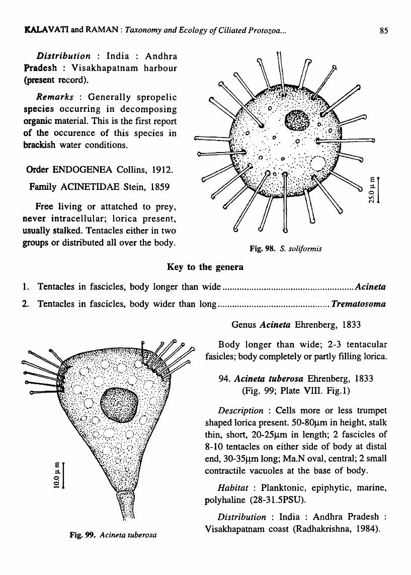
ICALAVATI and RAMAN: Taxonomy and Ecology o/Ciliated Protozoa ... 85
Distribution : India : Andhra Pradesh : Visakhapatnam harbour (present record).
Remarks : Generally spropelic species occurring in decomposing organic material. This is the frrst report of the occurence of this species in brackish water conditions.
Order ENDOGENEA Collins, 1912.
Family ACINETIDAE Stein, 1859
Free living or attatched to prey, never intracellular; lorica present, usually stalked. Tentacles either in two groups or distributed allover the body.
~I
Fig. 98. S. soliformis
Key to the genera
1. Tentacles in fascicles, body longer than wide ...................................................... Acineta
2. Tentacles in fascicles, body wider than long .............................................. Trematosoma
iI Fig. 99. Acineta tuberosa
Genus Acineta Ehrenberg, 1833
Body longer than wide; 2-3 tentacular fasicles; body completely or partly filling lorica.
94. Acineta tuberosa Ehrenberg, 1833 (Fig. 99; Plate VIII. Fig. 1 )
Description : Cells more or less trumpet shaped lorica present. 50-80flm in height, stalk thin, short, 20-25flm in length; 2 fascicles of 8-10 tentacles on either side of body at distal end, 30-35Hm long; Ma.N oval, central; 2 small contractile vacuoles at the base of body.
Habitat : Planktonic, epiphytic, marine, polyhaline (28-31.5PSU).
Distribution : India : Andhra Pradesh : Visakhapatnam coast (Radhakrishna, 1984).
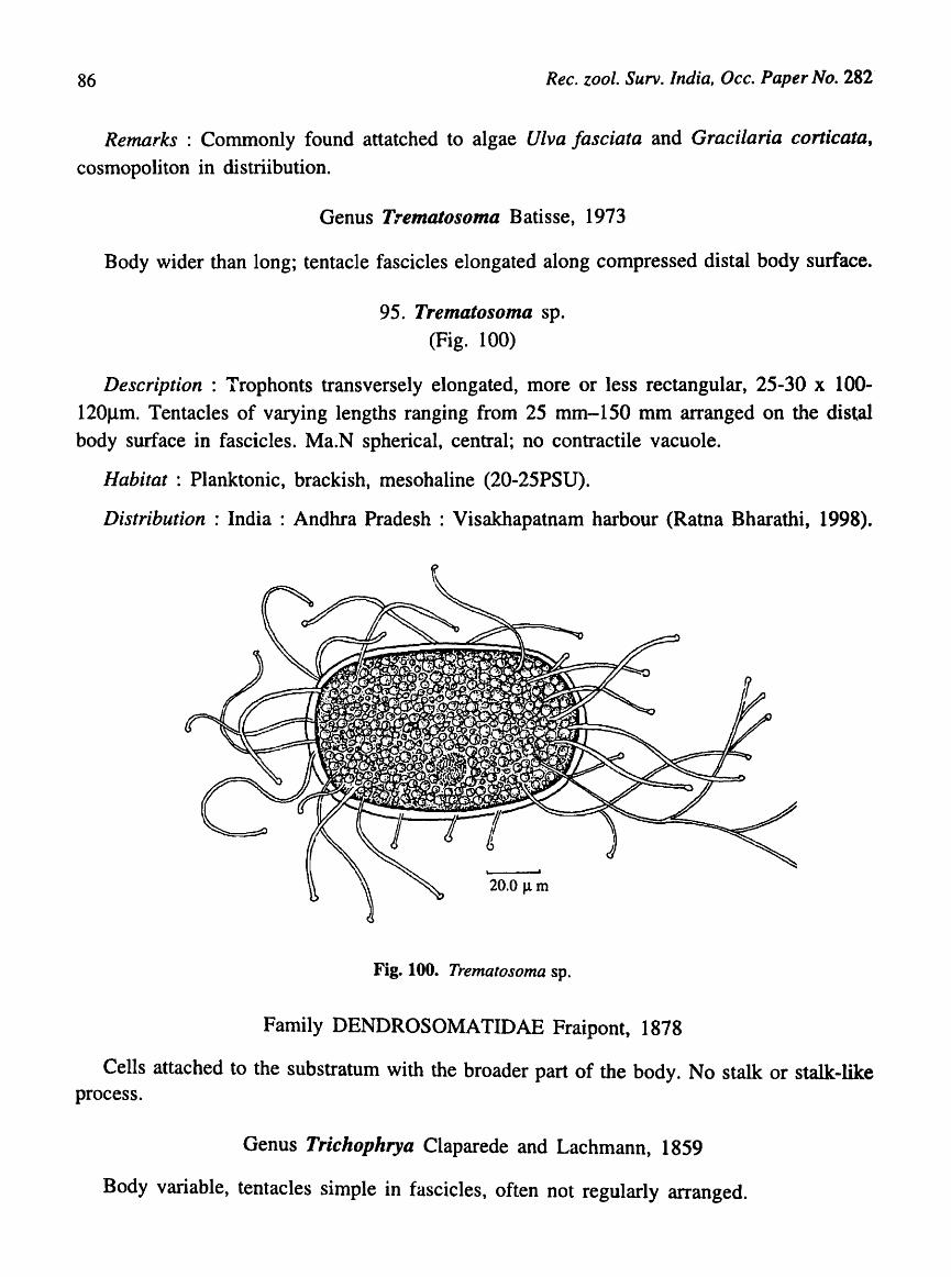
86 Rec. zool. Surv. India, Occ. Paper No. 282
Remarks : Commonly found attatched to algae Ulva Jasciata and Gracilaria co rticata ,
cosmopoliton in distriibution.
Genus T:-ematosoma Batisse, 1973
Body wider than long; tentacle fascicles elongated along compressed distal body surface.
95. Trematosoma sp. (Fig. 100)
Description: Trophonts transversely elongated, more or less rectangular, 25-30 x 100-120Jlm. Tentacles of varying lengths ranging from 25 mm-150 mm arranged on the distal body surface in fascicles. Ma.N spherical, central; no contractile vacuole.
Habitat : Planktonic, brackish, mesohaline (20-25PSU).
Distribution : India : Andhra Pradesh : Visakhapatnam harbour (Ratna Bharathi, 1998).
20.0 J.l m
Fig. 100. Trematosoma sp.
Family DENDROSOMATIDAE Fraipont, 1878
Cells attached to the substratum with the broader part of the body. No stalk or stalk-like process.
Genus Trichophrya Claparede and Lachmann, 1859
Body variable, tentacles simple in fascicles, often not regularly arranged.
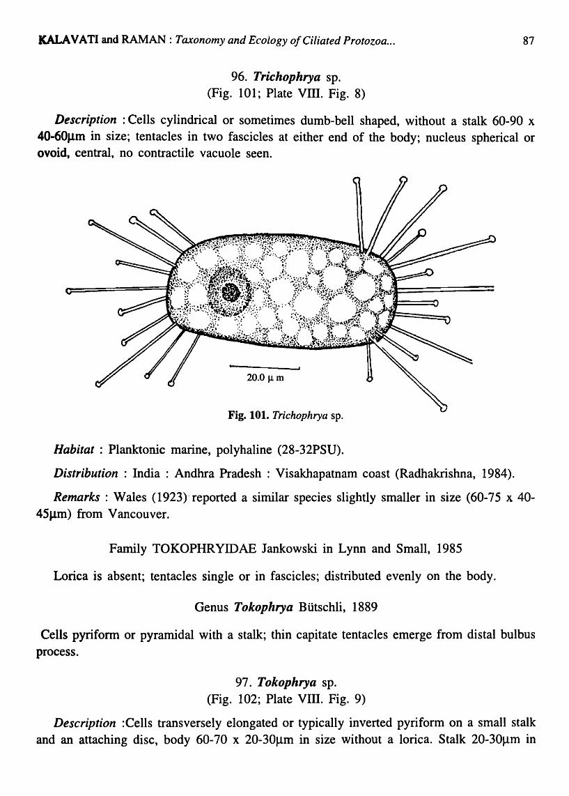
KALA V ATI and RAMAN : Taxonomy and Ecology of Ciliated Protozoa ...
96. Trichophrya sp. (Fig. 101; Plate VIII. Fig. 8)
87
Description: Cells cylindrical or sometimes dumb-bell shaped, without a stalk 60-90 x 40-60J.1m in size; tentacles in two fascicles at either end of the body; nucleus spherical or ovoid, central, no contractile vacuole seen.
20.0 J.1 m
Fig. 101. Trichophrya sp.
Habitat: Planktonic marine, polyhaline (28-32PSU).
Distribution: India: Andhra Pradesh: Visakhapatnam coast (Radhakrishna, 1984).
Remarks: Wales (1923)" reported a similar species slightly smaller in size (60-75 x 40-45J.1m) from Vancouver.
Family TOKOPHRYIDAE Jankowski in Lynn and Small, 1985
Lorica is absent; tentacles single or in fascicles; distributed evenly on the body.
Genus Tokophrya Btitschli, 1889
Cells pyriform or pyramidal with a stalk; thin capitate tentacles emerge from distal bulbus process.
97. Tokophrya sp. (Fig. 102; Plate VIII. Fig. 9)
Description :Cells transversely elongated or typically inverted pyriform on a small stalk and an attaching disc, body 60-70 x 20-30Jlm in size without a lorica. Stalk 20-30Jlm in
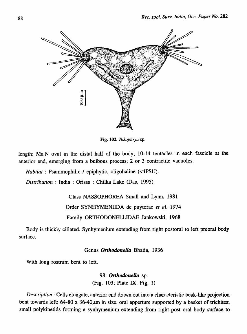
88 Rec. zool. Surv. India, Occ. Paper No. 282
iI
Fig. 102. Tokophrya sp.
length; Ma.N oval in the distal half of the body; 10-14 tentacles in each fascicle at the
anterior end, emerging from a bulbous process; 2 or 3 contractile vacuoles.
Habitat : Psammophilic I epiphytic, oligohaline «4PSU).
Distribution: India: Orissa: Chilka Lake (Das, 1995).
Class NASSOPHOREA Small and Lynn, 1981
Order SYNHYMENIIDA de puytorac et ale 1974
Family ORTHODONELLIDAE Jankowski, 1968
Body is thickly ciliated. Synhymenium extending from right postoral to left preoral body
surface.
Genus Orthodonella Bhatia, 1936
With long rostrum bent to left.
98. Orthodonella sp.
(Fig. 103; Plate IX. Fig. 1)
Description: Cells elongate, anterior end drawn out into a characteristic beak-like projection
bent towards left; 64-80 x 36-40J..lm in size, oral apperture supported by a basket of trichites;
small polykinetids forming a synhymenium extending from right post oral body surface to

KALA VA TI and RAMAN: Taxonomy and Ecology of Ciliated Protozoa ...
left preoral body surface; Ma.N single' oval; Mi.N spherical close to Ma.N; somatic ciliation uniform, 30-40 parallel monokineties; contractile vacuole single, lubterminal.
Habitat Planktonic, marine, polyhaline (25-32PSU).
Distribution : India : Andhra Pradesh Visakhapatnam harbour (Ratna Bharathi, 1998).
Order NASSULIDA Jankowski, 1967
Family NASSULIDAE de Fromentel, 1874
Cyrtos large with complete' nematodesmata; few to many oral .polyldnetids forming a fringe from postoral region sidewise to left, sometimes onto the dorsum. Single genus. Fig. 103. Orthodonella sp.
Genus NASSULA Ehrenberg, 1833
Generally large with a dense ciliation with characters of the family
Key to species
89
1. Cells large, ellipsoid with rounded ends, Cytostome located at 1/3 distance from anterior end. Ma.N spherical, contractile vacuoles 2 ..... o. 0' 0.' 0 .... ' 0 ••••••••••••••••• 000 ............... N. notata
~j ~I c, .., I't
I ! I
i
Fig. 104. Nassula notata
2. Cells small ellipsoid, fragile, cytostome 1/4 distance from anterior end. Ma.N cylindrcal, contactile vacuole large single ....... 00 •••••••••••••••••••••••••••••••••••••••••••••••••••••••• N. citl'ea
99. Nassula notata (O.F. Muller) Buddbr, 1911 (Fig. 104; Plate IX. Fig. 2)
Description : Cells ellipsoid, ends rounded 120-140 x 40-56J.1m, cytostome located at 1/3 distance of body length. Oral basket strong and distinct, cytostome deep in oral basket, oral polykinetids left to cytostome; Ma.N large, spherical in posterior half of body; Mi.N small; 2 large contractile vacuoles in posterior region; endoplasm generally brightly coloured.
Habitat: Planktonic, marine, polyhaline (27-31PSU).

90 Ree. zoo I. Surv. India, Dec. Paper No. 282
Distribution: India: Andhra Pradesh: Visakhapatnam coast (Radhakrishna, 1984). Orissa: Chilka Lake, India (Das, 1995).
Elsewhere: North Sea (Kahl, 1934), Mediterranean, Luxhaven, France (Kahl, 1935),
100. Nassula citrea Kahl, 1933 (Fig. 105; Plate IX. Fig. 3)
Description : Cells small, ellipsoid, somewhat fragile 60-70 x 25-301lm in size; cytostome located 1/4 the distance of body length; oral polykinetids in rows of 4 kinetosomes; oral basket clear, strong; cytostome deep in oral basket; Ma.N oval or cylindrical; Mi.N small; Three large contractile vacuoles seen at posterior end; somatic ciliation dense, uniform, 18-20 monokineties.
Habitat: Mangrove creeks/sediments, oligohaline (6-11.SPSU)
Distribution : India : Andhra Pradesh : GodavariCoringa mangroves (present report).
Elsewhere: Sylt, Germany (Kahl, 1931), Dee estuary (Webb, 1956).
Fig. 105. N. citrea
Suborder Parahymenostomatina Grain et al., 1976
Family FURGOSONIDAE Corliss, 1979
Fig. 106. Furgosonia sp.
Cells with three left oral polykinitids and one short right paroral kinetid; not confined to oral cavity.
Genus Furgosonia Jankowski, 1964
With characters of the family.
101. Furgosonia sp. (Fig. 106)
Description : Cells oval, small, 40-48x32-38J.1m in size; somatic kinities > 30; uniformly distributed membranelle on the left of oral aperture; paroral ciliature dikinities; Ma.N. central, spherical; MLN. small, adjacent to macronucleus; contractile vacuole single, posterior.

SALA VA TI and RAMAN: Taxonomy and Ecology of Ciliated Protozoa ... 91
Habitat: Planktonic, lowsaline, oligohaline (5.59PSU)
Distribution: India: Andhra Pradesh: Visakhapatnam harbour (Ratna Bharathi, 1998).
Order PENICULIDA Faure-Fremiet in Corliss, 1979
Suborder FRONTONIINA Small & Lynn, 1985
Family FRONTONIDAE Kahl, 1926
Oral nematodesmata more pominent to a side andlor of the mouth; three long large polykinitids in oral cavity; cytostome expands along post oral suture.
Genus Frontonia Eherenberg, 1838
Cells ovoid to ellipsoid, oral groove in anterior 1/3 of body; postoral kinities usually to left of oral polykinitids.
102. Frontonia marina Fabre-Dom, 1891 (Fig. 107)
Description : Cells oval, 150-195 x 80-120llm in size; anterior end flat and rounded. Oral groove lies in the anterior 1/3 of body on ventral side. Left edge more curved than the right and forms a prominent ectoplasmic lip posteriorly; 3 long, large polykinities seen in the oral cavity; preoral and postoral sutures long; somatic kinities converge near sutures; Ma.N oval, single; Mi.N 1-4, small, closely located; contractile vacuole large, posterior.
Habitat : Psammophilic, mangrove sediments. Brackish, mesohaline (12-26 PSU)
Distribution : India : Andhra Pradesh : Visakhapatnam harbour (Ratna Bharathi, 1998), coast, Godavari-Coringa mangroves (present study); Orissa: Chilka Lake (Das, 1995).
Elsewhere : Baltic Sea (Kahl, 1933), French coast of Atlantic (Dragesco, 1960), Niva Bay and Asko harbour (Fenchel, 1969) Salt marshes of Lousiana, U.S.A. (Elliot and Bamforth, 1975), Plymouth (Hartwig and Parker, 1977), Solar Lake, Red Sea shore
(Wilbert and Kahan, 1981). Fig. 107. Frontonia marina
Subclass HYPOTRICHIA Stein, 1859
Order EUPLOTIDA Small and Lynn 1985
Family ASPIDISCIDAE Eherenberg, 1838
Cells nearly circular in frontal outline, flattened, disc-shaped and strongly thigmotactic. There are no left marginal cirri. Frontoventrals and transverse cirri tend to be relatively thick,

92 Ree. zool. Surv. India, Dec. Paper No. 282
short flayed and thigmotactic. No apparent endoral or paroral structures. Ma.N usually 'C' or horse shoe shaped (Borror and Hills, 1995).
Genus Aspidisea Eherenberg, 1838
7 -15 frontoventrals and 5 or more transverse cirri; right caudal cirri and dorsal cilia absent; AZM reduced to few collor membranelle within a ventral fossa. Lapet membranelles located within a ventral buccal cavity (Borror and Hill 1995).
Key to species
1. Cells oval, dorsal side smooth, frontoventrals 7, transverse cirri 5. A characteristic spine seen between the two left transverses cirri ...................................................... A. lynceus
2. Cells oval, dorsal side ridged, frontoventrals 7, transverse cirri 8 .................. A. costata
3. Cells oval, left margin serrated, ventral side ridged, frontoventrals 8, transverse cirri 6 ........................................................................................................................... A. aculeata
103. Aspidisea lyneeus Ehrenberg 1838 (Fig. 108; Plate IX. Fig. 4)
Description: Cells ovoid or circular, flat and disc
like, 25-30 x 30-35J.1m in size; dorsal side smooth
without ribs. Frontoventrals arranged in the anterior
half of body, four of them forming an arch, transverse
cirri 5, left serial oral polykinetids separate; Ma.N horse
shoe shaped, large and massive; A characteristic spine
seen between the two left transverse cirri.
Habitat : planktonic, psammophilic, occurs
abundantly in decaying organic matter, brackish.
Distribution : India : Andhra Pradesh
Visakhapatnam harbour (Ratna Bharathi, 1998).
Elsewhere : Plymouth (Hartwig and Parker 1977),
River Toronto, North Italy (Madoni, 1984).
r j
~: Q
d N
Fig. 108. Aspidisca lynceus
104. Aspidisea eostata (Dujardin, 1842) Claparede and Lachmann, 1859 (Fig. 109; Plate IX. Fig. 5&6)
Description : Cells oval, compressed; ventral side flat. Dorsal side convex, ridged; 32-56 x 25-45 flm in size; AZM reduced; buccal complex small, ventral, in left posterior part of

IALAVATI and RAMAN : Taxonomy and Ecology of Ciliated Protozoa ... 93
the body and always enclosed in a membrane; Frontoventral cirri 7, arranged in anterior half. Transverse cirri 8, arranged in a row, 2 of which are located at the base of buccal complex. Ma.N large, 'C' shaped. Mi.N is small, spherical and anterior in position. Contractile vacuole small, located at the root of transverse cirri.
Habitat: planktonic, psammophilic, epiphytic, marine, polyhaline (29-32PSU).
Distribution : India : Andhra Pradesh : Visakhapatnam coast (Radhakrishna, 1984), harbour (Ratna Bharathi, 1998); Punjab (Bhatia 1936) ; West Bengal (Das et ai, 1993).
Elsewhere : Cosmopoliton, found in various types of water bodies. Recorded from Australia (Patterson et al., 1984), Plymouth, Britan (Hartwig and Parker, 1977) and Northern Italy (Madoni, 1981) in brackishwater environments
Fig. 109. A. costata
105. Aspidisca aculeata (Ehrenberg, 1838) Mansfeld, 1926 (Fig. 110; Plate IX. Fig. 7)
Fig. 110. A. aculeata
Description: Cells more or less circular, 35-45 x 25-30flm in size; left margin of the body serrated and transparent; ventral'surface ridged; buccal complex in two sections, one lateral and anterior to frontoventrals and the other posterior near transverse cirri. Frontoventrals 8, Transverse cirri 6. Ma.N horse-.shoe shaped, MLN small and spherical.
Habitat: Mangrove creeks/sediments, oligohaline
«10PSU).
Distribution: India: Andhra Pradesh: GodavariCoring a mangroves (present record).
Elsewhere: Germany (Mansfeld, 1923), among the algal mats in the coast of New Hampshire (Borror, 1973).

94 Ree. zool. Surv. India, Dec. Paper No. 282
Family EUPLOTIDAE
Cells are flattened ventrally; Argyrome well developed; macronucleus single, C-shaped or horse-shoe shaped; Frontoventral, transverse cirri, varying numbers of non hypertrophied left marginal cirri and ventrally located caudal cirri present; AZM conspicuous; little separation between collor and lappet region; paroral ciliature in the fonn of a patch, deep within the posterior end of buccal cavity which is situated by the side of an endoral monokinetal row
of cilia (Borrot and Hill, 1995).
Key to the genera
1. Dorsal argyrome system double or complex, Frontoventral cirri 10 ................ Euplotes
2. Dorsal argyrome double. Frontoventral cirri 9 .............................................. Euplotoides
3. Dorsal argyrome, double, triple or complex. Frontoventrals 7-9 ................. Euplotopsis
4. Dorsal argyrome single. Frontoventrals typically 10 ................................... Moneuplotes
5. Cirri reduced to caudals. Transverse cirri in a row. Dorsal argyrome double .............. . .................................................................................................................... .... Paraeuplotes
Genus Euplotes Ehrenberg, 1830
Primarily marine, frontoventrals typically 10, dorsal argyrome systems double to complex. Dorsal surface usually have several ribs. AZM well developed. Macronucleus more or less C- shaped.
Key to species
1. Cells elliptical, Ma.N 'c' shaped, frontoventrals 10, transverse 5, caudal 5, dorsal argyrome complex ............................................................................................................... E. charon
2. Cells small, oval, Ma.N spherical, frontoventrals 10, transverse 5, caudal cirri 4, dorsal argyrome complex ........................................................................................... E. moebiusi
106. Euplotes charon (Muller) Ehrenberg, 1830 (Fig. 111; Plate IX. Figs. 8-10)
Description :Cells elliptical, 70-90 x 60-85J.1ID in size; ventral surface flat, 3-5 conspicuous striations on the pellicle. AZM well developed, frontoventrals 10, transverse 5, caudal cirri 5, Ma.N 'c' shaped, MLN small, spherical, close to the Ma.N, dorsal argyrome complex, contractile vacuole small, posterior.
Habitat: Psammophilic and planktonic, marine, polyhaline (29.5-33.5PSU).
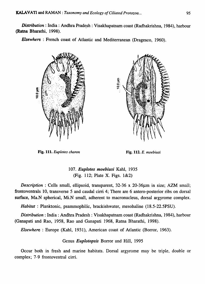
KALAVATI and RAMAN: Taxonomy and Ecology of Ciliated Protozoa ... 95
Distribution: India: Andhra Prad~sh : Visakhapatnam coast (Radhakrishna, 1984), harbour (Ratna Bharathi, 1998).
Elsewhere: French coast of Atlantic and Mediterranean (Dragesco, 1960).
Fig. 111. Euplotes charon Fig. 112. E. moebiusi
107. Eupiotes moebiusi Kahl, 1935 (Fig. 112; Plate X. Figs. 1 &2)
Description : Cells small, ellipsoid, transparent, 32-36 x 20-36Jlm in size; AZM small; frontoventrals 10, transverse 5 and caudal cirri 4; There are 6 antero-posterior ribs on dorsal surface, Ma.N spherical, MLN small, adherent to macronucleus, dorsal argyrome complex.
Habitat : Planktonic, psammophilic, brackishwater, mesohaline (18.5-22.5PSU).
Distribution: India: Andhra Pradesh: Visakhapatnam coast (Radhakrishna, 1984), harbour (Ganapati and Rao, 1958, Rao and Ganapati 1968, Ratna Bharathi, 1998).
Elsewhere: Europe (Kahl, 1931), American coast of Atlantic (Borror, 1963).
Genus Eupiotopsis Borror and Hill, 1995
Occur both in fresh and marine habitats. Dorsal argyrome may be triple, double or complex; 7-9 frontoventral cirri.
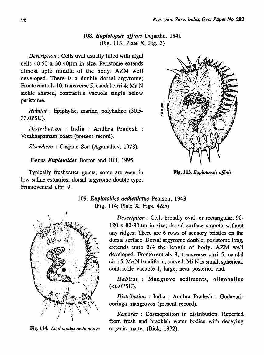
96 Rec. zool. Surv. India, Oec. Paper No. 282
108. Euplotopsis a/finis Dujardin, 1841 (Fig. 113; Plate X. Fig. 3)
Description : Cells. oval usually filled with algal cells 40-50 x 30-40Jlm in size. Peristome extends almost upto middle of the body. AZM well developed. There is a double dorsal argyrome; Frontoventrals 10, transverse 5, caudal cirri 4; Ma.N sickle shaped, contractile vacuole single below peristome.
Habitat: Epiphytic, marine, polyhaline (30.5-33.0PSU).
Distribution : India : Andhra Pradesh Visakhapatnam coast (present record).
Elsewhere: Caspian Sea (Agamaliev, 1978).
Genus Euplotoides Borror and Hill, 1995
Typically freshwater genus; some are seen in low saline estuaries; dorsal argyrome double type; Frontoventral cirri 9.
Fig. 113. Euplotopsis affinis
109. Euplotoides aediculatus Pearson, 1943 (Fig. 114; Plate X. Figs. 4&5)
Fig. 114. Euplotoides aediculatus
Description : Cells' broadly oval, or rectangular, 90-120 x 80-90J..lm in size; dorsal surface smooth without any ridges; There are 6 rows of sensory bristles on the dorsal surface. Dorsal argyrome double; peristome long, extends up to 3/4 the length of body. AZM well developed. Frontoventrals 8, transverse cirri 5, caudal cirri 5. Ma.N bandifonn, curved. Mi.N is small, spherical; contractile vacuole 1, large, near posterior end.
Habitat : Mangrove sediments, oligobaline «6.0PSU).
Distribution : India : Andhra Pradesh Godavaricoringa mangroves (present record).
Remarks : Cosmopoliton in distribution. Reported from fresh and brackish water bodies with decaying organic matter (Bick, 1972).

KALA V A TI and RAMAN: Taxonomy and Ecology of Ciliated Protozoa ... 97
Genus Moneuplotes Jankowski, 1979
All marine, non-encysting; Frontoventral cirri typically 10, transverse and caudal cirri vary in number, dorsal argyrome single type, no algal cells in the endoplasm.
Key to species
1. Cells broadly oval, macronucleus 'C' shaped, frontoventrals 10, transverse 5, caudal cirri 4 .......................................................................................................................... M. vannus
2. Cells small, triangular, transverse 6, frontoventrals 10, caudal cirri, macronucleus band-like .................................................................................................................... M. te rricola
110. Moneuplotes vannus (Muller, 1786) Jankowski, 1979 (Fig. 115;· Plate X. Figs. 6-8)
Description : Cells broadly oval with characteristic refractile :granules seen in central region, 120-140 x 80-100J.1m in size; Dorsal surface convex, smooth, dorsal argyrome simple,
single pattern; ventral surface flat with 10 frontoventrals, 5 transverse (in two groups) and 4 caudal cirri. Peristome well marked with powerful AZM extending upto 2/3 the length of body. Ma.N 'C' shaped; MLN single, near the ann of the Ma.N; contractile vacuole single, large.
Habitat : Planktonic, psammophilic, epiphytic, marine, polybaline (28.5-33.4PSU).
Locality: Visakhapatnam harbour, coast.
Distribution : India : Andhra Pradesh : Visakhapatnam coast (Ganapati and Rao, 1958, Rao and Ganapati 1968, Radhakrishna, 1984) harbour (Ratna Bharathi, 1998).
Elsewhere :Common inhabitant of interstitial sands rich in organic matter. Widely distributed in Baltic Sea (Dragesco, 1960), North American coast of Atlantic, Maine to Florida (Borror, 1963), European coast of Atlantic (Mechelon and Darner, 1984).
Fig. lIS. Moneuplotes vannus
111. Moneuplotes terricola Penard, 1922 (Fig.116; Plate X. Fig. 9)
Description: Cells small, more or less triangular, 50-60 x 35-40J.1m in size; anterior end flat, posterior end bluntly pointed; dorsal side smooth, argyrome simple; Frontoventrals 10, transverse 6, and caudal cirri 5. Ma.N band-like and 'L' shaped; contractile vacuoles small, 2.

98 Rec. zool. Surv. India, Occ. Paper No. 282
Habitat : Psammophilic, marine.
Distribution : India : Andhra Pradesh Visakhapatnam coast (present record)
Elsewhere: Northern Germany (Kahl, 1933).
Remarks: Borror and Hill 1995, considered E. terricola as an aberrant species, since there is no information on cortical structure. In our preparations stained according to modified Kleins dry silver technique and with silver carbonate, the species exhibited a simple single argyrome as is typical of "vannus group" The species is therefore assigned to the genus Moneuplotes Jankowski, as M. terricola.
Genus Paraeuplotes Wichterman, 1942
No marginal cirri, there is only one small group of caudal cirri, row of transverse cirri, extensive. AZM
Fig. 116. M. terricola
E ,:1 Q
ic:i , '"
well developed. Undulating membrane absent, macronucleus 'C' shaped (Borror, 1972).
112. Paraeuplotes andhrae n. sp. (Fig. 117)
Description : Cells oval, 37-50 x 30-45f.!ffi in size; ventral surface concave, dorsal side convex, with a ridge. AZM well developed. There is a ciliary ring in the ventral surface,
posterior half of which is marked by a ciliary plate and the anterior end by 2 ciliary tuft. 5-6 well formed caudal cirri. Ma.N bean shaped. MLN located close to Ma.N often in indentation; contractile vacuoles 4-5.
§. Habitat: Epiphytic, marine, polyhaline (30-32PSU); :0 o a rare form found associated with Ulva Jasciata . ...
Fig. 117. Paraeuplotes andhrae n. sp
Distribution : India : Andhra Pradesh Visakhapatnam coast (Radhakrishna, 1984).
Remarks : Paraeuplotes Witchermann is a rare genus with uncertain taxonomic position. There is no addition to this, genus since its inception in 1942. P. tortugensis. the type species was found associated with the coral, Euricea crassa, on the coast of Tolugas, Florida. In comparison, the pr~sent species is much smaller (37-50 x 30-45f.!m) in size'than P. tortugensis (85 x 75J..lm)

KALA V A TI and RAMAN: Taxonomy and Ecology of Ciliated Protozoa ... 99
and found associated with the alga, Ulva Jasciata, on the coast of Visakhapatnam, Bay of Bengal. Besides the species has a characteristic wide ciliary ring on ventral surface. In view of the morphological differences and occurrence in association with algae, the species is considered new to science for which the name Paraeuplotes andhrae n.sp. is suggested.
Family URONYCHIDAE Jankowski in Small and Lynn, 1985
There are 3 hypertrophied dorsally located caudal cirri. AZM separated into a distinct collor and lapet (Borror and Hill, 1995).
Genus Diophrys Dujardin, 1840
Cells with more or less parallel sides, dorsoventrally flattened and circular in cross section; buccal cavity ventral; Frontoventral cirri variable (but fewer) in number; 3 hypertrophied dorsal caudal cirri, paroral and endoral membranes conspicuous; usually 2 or more macronuclei.
113. Diophrys appendiculata Ehrenberg, .1838 (Fig. 118; Plate XI. Fig. 1)
Description : Cells bluntly rounded, buccal complex wide and extends upto half the length of body. Right edge of buccal complex raised with a conspicuous undulating membrane; frontoventrals 5, in two groups; ventral cirri 2, weak; anal cirri 5. There are 3 hypertrophied dorsal caudal cirri; Ma.N 2, oval; 2, small Mi.N present; contractile vacuoles small and many.
Habitat: Psammophilic, marine, polyhaline (28-31PSU).
Distrib':ltion : India : Andhra Pradesh : Visakhapatnam coast (Ganapati and Rao, 1958; Rao and Ganapati, 1968, Radh~shna, 1984)
Elsewhere : Gulf of Mexico (Smith, 1904), Europe (Kahl, 1935), Alligator harbour, Florida. USA. (Borror, 1963). Fig. 118. Diophrys appendiculata
Class OLIGOHYMENOPHOREA, de Puytarac et al., 1974
Order HYMENOSTOMATIDA Delorge and Herouard, 1896.
Suborder TETRAHYMENINA Faure-Fremeit in Corliss, 1956 Family TETRAHYMENIDAE Corliss, 1952
The first anterior body kinetid in each kinety is dikenetid. Oral area with undulating membrane and 3 left oral polykinetids; kinetosomes unifonnly spaced; Paroral dikinetid ciliated throughout.

100 Ree. zool. Surv. India, Oee. PapefHIJ. 282
Genus Tetrahymena Furgason, 1940
Oral polykinetids straight, never sigmoid
Key to species
1. Actively swimming pyriform cells, tetrahymenal complex located 1/3 of body length from anterior apex; macronucleus oval ...................................... T. pyriformis (complex)
2. Elliptical cells with sharply pointed anterior end, tetrahymenal complex nearer the anterior end, macronucleus ova! .............................................................................. T. thermophila
114. TetrahymelUl pyriformis (complex) Ehrenberg, 1838 (Fig. 119; Plate XI. Figs. 2&3)
Description : Cells small, actively swimming, pyriform in shape; anterior end bluntly pointed, posterior end rounded, 40-60 x 35-50Jlm in size; characteristic 'tetrahymena! complex' consisting of undulating membrane and 3 polykinetids seen in anterior 1/3 of cell. Cytostome reduced; Ma.N oval; Mi.N absent; somatic ciliation uniform, 15-20 longitudinal parallel monokineties.
Habitat: Planktonic, brackishwater, euryhaline (4-23.4PSU)
Distribution : India : Andhra Pradesh : Visakhapatnam harbour (Ratna Bharathi, 1998) and Godavari Coringa mangroves (present record); Orissa: Chilka Lake (Das, 1995).
Elsewhere : Cosmopoliton, widely distributed in Europe and North America (Nanney and McCoy, 1976).
Pig. 119. Tetrahymena pyriformis (complex)
115. Tetrahymena thermophila Nanney and Mc Coy, 1976 (Fig. 120; Plate XI. Fig. 4)
Description : Body elliptical, 60-80 x 20-40mm in size, anterior end sharply pointed, posterior end rounded, cytostome and tetrahymena! complex small, close to anterior apex. Ma.N oval~ MLN small adherent to Ma.N; somatic ciliation uniform, 15-20 straight monokineties, contractile vacuole single, at posterior end.
Habitat: Planktonic, brackish, mesohaline (20-25PSU), tolerant to high temperature (Ca. 37°·C).
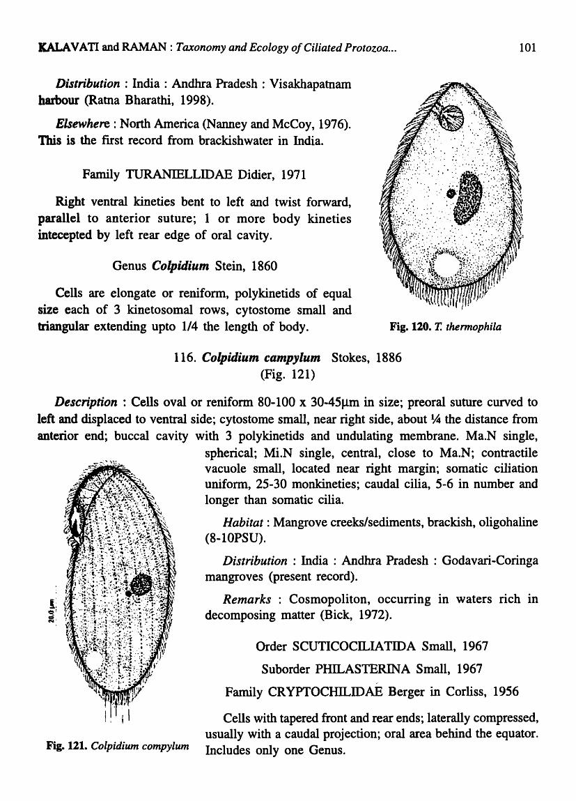
KALA VA TI and RAMAN : Taxonomy and Ecology of Ciliated Protozoa ...
Distribution : India : Andhra Pradesh : Visakhapatnam harbour (Ratna Bharathi, 1998).
Elsewhere: North America (Nanney and McCoy, 1976). This is the first record from brackishwater in India.
Family TURANIELLIDAE Didier, 1971
Right ventral kineties bent to left and twist forward, parallel to anterior suture; 1 or more body kineties intecepted by left rear edge of oral cavity.
Genus Colpidium Stein, 1860
Cells are elongate or renifonn, polykinetids of equal size each of 3 kinetosomal rows, cytostome small and triangular extending upto 1/4 the length of body. Fig. 120. T. thermophila
116. Colpidium campylum Stokes, 1886 (Fig. 121)
101
Description: Cells oval or reniform 80-100 x 30-45Jlm in size; preoral suture curved to left and displaced to ventral side; cytostome small, near right side, about ~ the distance from anterior end; buccal cavity with 3 polykinetids and undulating membrane. Ma.N single,
spherical; Mi.N single, central, close to Ma.N; contractile vacuole small, located near right margin; somatic ciliation unifonn, 25-30 monkineties; caudal cilia, 5-6 in number and longer than somatic cilia.
Habitat: Mangrove creeks/sediments, brackish, oligohaline (8-10PSU).
Distribution: India: Andhra Pradesh: Godavari-Coringa mangroves (present record).
Remarks: Cosmopoliton, occurring in waters rich in decomposing matter (Bick, 1972).
Order SCUTICOCILIA TID A Small, 1967
Suborder PHILASTERINA Small, 1967 /
Family CRYPTOCHILIDAE Berger in Corliss, 1956
Cells with tapered front and rear ends; laterally compressed, usually with a caudal projection; oral area behind the equator.
Fig. 121. Colpidium compylum Includes only one Genus.
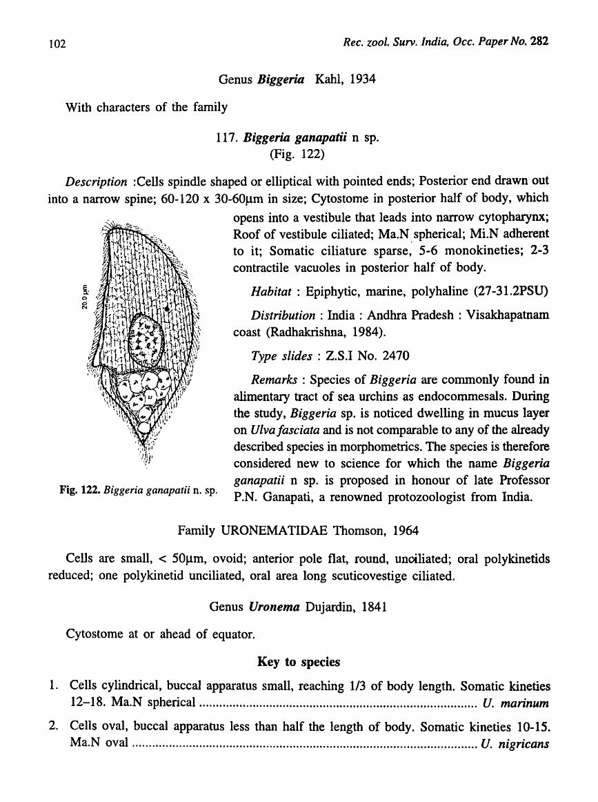
102 Rec. zool. Surv. India, Occ. Paper No. 282
Genus Biggeria Kahl, 1934
With characters of the family
117. Biggeria ganapatii n sp. (Fig. 122)
Description :Cells spindle shaped or elliptical with pointed ends; Posterior end drawn out into a narrow spine; 60-120 x 30-60}lm in size; Cytostome in posterior half of body, which
Fig. 122. Biggeria ganapatii n. sp.
opens into a vestibule that leads into narrow cytopharynx; Roof of vestibule ciliated; Ma.N. spherical; MLN adherent to it; Somatic ciliature sparse, 5-6 monokineties; 2-3 contractile vacuoles in posterior half of body.
Habitat : Epiphytic, marine, polyhaline (27 -31.2PSU)
Distribution : India : Andhra Pradesh : Visakhapatnam coast (Radhakrishna, 1984).
Type slides : Z.SJ No. 2470
Remarks : Species of Biggeria are commonly found in alimentary tract of sea urchins as endocommesals. During the study, Biggeria sp. is noticed dwelling in mucus layer on Viva Jasciata and is not comparable to any of the already described species in morphometries. The species is therefore considered new to science for which the name Biggeria ganapatii n sp. is proposed in honour of late Professor P.N. Ganapati, a renowned protozoologist from India.
Family URONEMATIDAE Thomson, 1964
Cells are small, < 50}lm, ovoid; anterior pole flat, round, unctliated; oral polykinetids reduced; one polykinetid unciliated, oral area long scuticovestige ciliated.
Genus Uronema Dujardin, 1841
Cytostome at or ahead of equator.
Key to species
1. Cells cylindrical, buccal apparatus small, reaching 1/3 of body length. Somatic kine ties 12-18. Ma.N spherical.............. .......... ...... ..... ..... ...... ...... .... ........................... U. marinum
2. Cells oval, buccal apparatus less than half the length of body. Somatic kineties 10-15. Ma.N oval ....................................................................................................... U. nigricans

KALA V A TI and RAMAN: Taxonomy and Ecology of Ciliated Protozoa ... 103
3. Cells broadly oval. Buccal apparatus reaching middle of the body. Ma.N massive, elongate, somatic kineties 20-25 ....................................................................................... U. filificum
118. Uronema marinum Dujardin, 1841 (Fig. 123)
Description : Cells cylindrical, round in cross section; anterior region flat, non ciliated; 30-45 x 10-121lm in size; cytostome near equator with distinct paroral membrane; somatic ciliation uniform, 12-18 (usually 15) monokineties; caudal cilium long, buccal apparatus never reaching centre of the body; Ma.N spherical in anterior half of body; Mi.N small and close Ma.N; contractile vacuole large and posterior; anterior part of body filled with numerous refractile granules.
Habitat: Planktonic, marine, polyhaline (29.5-32.0PSU).
Distribution : India : Andhra Pradesh : Visakhapatnam coast (Radhakrishna, 1984); Orissa: Chilka Lake (Das, 1995).
Elsewhere: Gulf of Mexico (Smith, 1904), Mediterranean, l salt marshes Lousiana (Elliot and Bamforth, 1975), Limfjord, \ Denmark (Anderson and Sorensen, 1986), Fig. 123. Uronema marinUln
E --Q d ...
Fig. 124. U. nigricans
119. Uronema nigricans (O.F. Muller, 1786) Florentin, 1901 (Fig. 124; Plate XI. Fig. 5)
Description : Cells oval, 24-48 x 12-18.4 Ilm in size; anterior region hemispherical non ciliated; buccal apparatus small, reaches 1/3 of body length consisting of an undulating membrane and 3 polykineties; somatic ciliation uniform, 10-15 monokineties; Ma.N single, oval; Mi.N spherical; contractile vacuole single, near left cell margin; caudal cilium single, long.
Habitat: Epiphytic, brackishwater, euryhaline (5.59-22.SPSU).
Distribution: India: Andhra Pradesh: Visakhapatnam harbour (Ratna Bharathi, 1998).
Elsewhere : In periphyton in Caspian Sea (Thompson and Erans, 1968; Agameliev, 1978), Solar Lake on the east coast of Senai (Wilbert and Kahan, 1981).
Remarks : The species occurred in Visakhapatnam harbour in blooms proportions (4, 833 x 102 nos I-I) when the salinity dropped to 5.0PSU and oxygen undetectable.
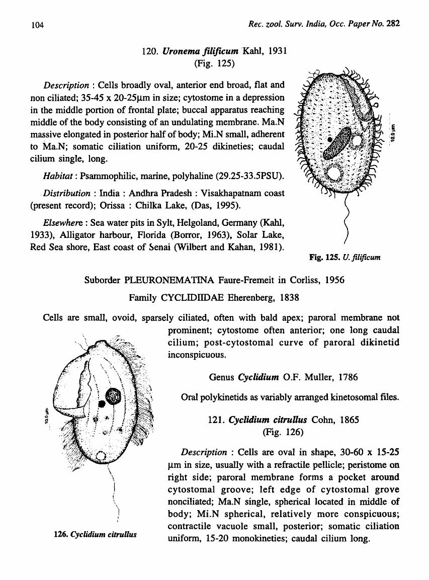
104 Rec. zool. Surv. India, Occ. Paper No. 282
120. Uronema filificum 'Kahl, 1931 (Fig. 125)
Description : Cells broadly oval, anterior end broad, flat and non ciliated; 35-45 x 20-25flm in size; cytostome in a depression in the middle portion of frontal plate; buccal apparatus reaching middle of the body consisting of an undulating membrane. Ma.N massive elongated in posterior half of body; MLN small, adherent to Ma.N; somatic ciliation uniform, 20-25 dikineties; caudal cilium single, long.
Habitat: Psammophilic, marine, polyhaline (29.25-33.5PSU) .
. Distribution : India : Andhra Pradesh : Visakhapatnam coast (present record); Orissa : Chilka Lake, (Das, 1995).
Elsewhere: Sea water pits in Sylt, Helgoland, Germany (Kahl, 1933), Alligator harbour, Florida (Borror, 1963), Solar Lake, Red Sea shore, East coast of 8enai (Wilbert and Kahan, 1981).
Fig. 125. U. filificum
Suborder PLEURONEMA TINA Faure-Fremeit in Corliss, 1956
Family CYCLIDIIDAE Eherenberg, 1838
Cells are small, ovoid, sparsely ciliated, often with bald apex; paroral membrane not
i .0 d ..
,
\ \
\
I I' ,
126. Cyclidium citruUus
prominent; cytostome often anterior; one long caudal cilium; post-cytostomal curve of paroral dikinetid inconspicuous.
Genus Cyclidium O.F. Muller, 1786
Oral polykinetids as variably arranged kinetosomal files.
121. Cyclidium citrullus Cohn, 1865 (Fig. 126)
Description: Cells are oval in shape, 30-60 x 15-25 flm in size, usually with a refractile pellicle; peristome on right side; paroral membrane forms a pocket around cytostomal groove; left edge of cytostomal grove nonciliated; Ma.N single, spherical located in middle of body; MLN spherical, relatively more conspicuous; contractile vacuole small, posterior; somatic ciliation uniform, 15-20 monokineties; caudal cilium long.

IIALA VATI and RAMAN : Taxonomy and Ecology of Ciliated Protozoa ... 105
Habitat: Epiphytic, brackish, mesohaline (22-25.8PSU).
Distribution: India: Andhra Pradesh: Visakhapatnam harbour (present record); Orissa : ChiIka Lake, (Das, 1995).
Elsewhere: Periphyton in west coast of Caspian Sea (Agameliev, 1978), Limfjord, Denmark (Andersen and Sorensen, 1986).
Genus Cristigera Roux, 1899
Oral polykinetids as one file of kinetosomesl
122. Cristigera phoenix Penard, 1922 (Fig. 127)
Description : Small ovoid with flat nonciliated anterior end, 30-50 x 20-30llm in size; posterior cilia longer than anterior cilia; single, long caudal cilium present; MaiN spherical, centraL; -MLN small; peristome near mid ventral line; 8-100 paroral monokineties; one small contractile vacuole in middle of the body.
Habitat: Epiphytic, marine, euryhaline (12.2-30.11PSU)
Distribution : India : Andhra Pradesh Vi'sakhapatnam coast (Radhakrishna, 1984)
Elsewhere :Salt marshes at Lousiana, USA (Elliot and Bamforth, 1975).
I ! ;
I I I
1 ..
Fig. 127. Cristigera phoenix
Suborder PLEURONEMA TINA Faure-Fremiet in Corliss, 1956
Family PLEURONEMATIDAE Kent, 1881
Second oral polykinetid long, linear; mid segment with not more than two rows of zigUS kinetosomes; prominent post equatorial velum, with long stiff caudal cilia; cytostome in anterior ~ of body I
Genus Pleuronema Dujardin, 1836
Right ventral body kineties extend beyond rear end of paroral dikinetid and terminates at a postoral suture; Left ventral kineties opposite.

106 Ree. zool. Surv. India, Dec. Paper No. 282
Key to species
1. Cells small, velum prominent. Ma.n oval, Mi.N single 5-8 caudal cilia ..... P. senigera
2. Cells large, velum prominent Ma.N spherical, MLN one or two, 4 caudal cilia .......... . ....................................................................................................................... P. coronatum
123. Pleuronema setigerum Calkins, 1905 (Fig. 128)
Description : Body oval with rounded ends; ventral surface slightly concave; 4O-56x20-28 Ilm in size; mouth sub-equatorial; velum conspicuous; Ma.N oval, large; Mi.N very small dot-like; somatic ciliation uniform, relatively sparse at anterior region, 20 monokineties; 5-8 stiff caudal cilia at posterior region.
Habitat : Psammophilic, marine, polyhaline (28.5-32.4PSU).
Distribution : India : Andhra Pradesh : Visakhapatnam coast (Rao and Ganapati, 1968, present record)
Elsewhere: Gulf of Mexico (Noland, 1937), Woods Hole, USA (Calkins, 1905), Alligator harbour, Florida (Borror, 1963). Fig. 128. Pleuronema setigerum
Fig. 129. P. corona tum
124. Pleuronema coronatum Kent 1881 (Fig. 129)
Description: Cells relatively large, broadly rounded, 70-90 x 35-50J.1m in size; mouth sub-equatorial. Velum membrane reaches almost upto posterior end and highly extensible; Ma.N spherical, anterior, 5J.1m in diameter; 1 or 2 Mi.N; contractile vacuole large, posterior; Caudal cilia four; Somatic ciliation uniform, 40-42 monokineties.
Habitat : Psammophilic, marine, polyhaline (30-33.4PSU).
Distribution : India : Andhra Pradesh : Visakhapatnam coast (present record)
Elsewhere: Gulf of Mexico (Noland, 1937), Alligator harbour, Florida (Borror, 1963).
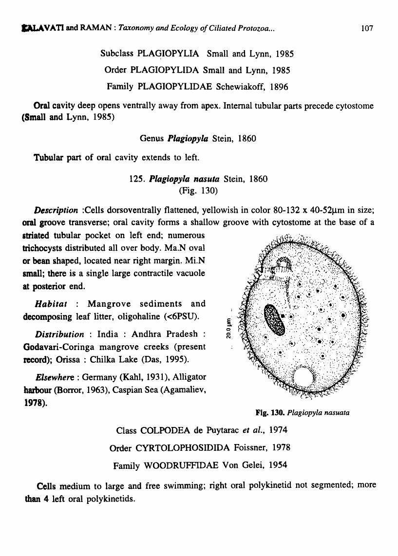
MLAVATI and RAMAN: Taxonomy and Ecology o/Ciliated Protozoa ...
Subclass PLAqIOPYLIA Small and Lynn, 1985
Order PLAGIOPYLIDA Small and Lynn, 1985
Family PLAGIOPYLIDAE Schewiakoff, 1896
107
Oral cavity deep opens ventrally away from apex. Internal tubular parts precede cytostome (Small and Lynn, 1985)
Genus Plagiopyla Stein, 1860
Tubular part of oral cavity extends to left.
125. Plagiopy/Q. nasuta Stein, 1860 (Fig. 130)
Description :Cells dorsoventrally flattened, yellowish in color 80-132 x 40-52J.1m in size; oral groove transverse; oral cavity forms a shallow groove with cytostome at the base of a
sttiated tubular pocket on left end; numerous
trichocysts distributed all over body. Ma.N oval
or bean shaped, located near right margin. Mi.N
small; there is a single large contractile vacuole
at posterior end.
Habitat : Mangrove sediments and
decomposing leaf litter, oligohaline «6PSU).
Distribution : India : Andhra Pradesh :
Godavari-Coringa mangrove creeks (present
record); Orissa : Chilka Lake (Das, 1995).
Elsewhere: Germany (Kahl, 1931), Alligator
barbour (Borror, 1963), Caspian Sea (Agamaliev,
1978).
o o N.
Fig. 130. Plagiopyia nasuata
Class COLPODEA de Puytarac et al., 1974
Order CYRTOLOPHOSIDIDA Foissner, 1978
Family WOODRUFFIDAE Von Gelei, 1954
Cells medium to large and free swimming; right oral polykinetid not segmented; more
than 4 left oral polykinetids.

108 Rec. zool. Surv. India, Occ. Paper No. 282
Genus Woodruffia Kahl, 1931
Free swimming; left oral polykinetids never enter preoral suture, left oral polykinetids more than 20.
126. Woodruffia rostrata Kahl, 1931 (Fig. 131; Plate XI. Fig. 6)
Description : Cells flat leaf-like, dorsoventally compressed, body elliptical in outline, broad in middle and narrow towards apex, 120-180 x 100-120J.1m in size, cytostome a narrow, diagnal slit on left side, at anterior end; 30-35 left oral short polykinetids seen; Ma.N single, spherical, surrounded by transparent hyaline space; 15-20J.1m in diameter; Mi.N 2, small and spherical; contractile vacuoles 2, present in posterior region; somatic ciliation uniform, 12-15 parallel rows of dikenetids.
Habitat : Epiphytic, brackish, mesohaline (23.6-25PSU).
Locality : Visakhapatnam harbour and coast.
Distribution : India : Andhra Pradesh : Visakhapatnam coast, harbour (present record).
Elsewhere : Northern Germany (Kahl 1931). Kahl reported this species in salt water culture along with Oscillanora. Fig. 131. Woodruffla rostrata
Order COLPODIDA de Puytorac et al., 1974
Family COLPODIDAE Ehrenberg, 1838.
Genus Colpoda O.F. Muller, 1773
Left oral polykinetid extends upto perimeter of oral opening. Oral cavity not tubular
127. Colpoda sp. (Fig. 132; Plate XI. Fig. 7)
Description: Cells typically kidney shaped, 40-50 x 30-35Jlm in size; buccal cavity with deep oral funnel that begins at the grove near left side of body; cytostomal complex is clearly defined by a notch below keel with 2 oral dikinetids of equal size; left oral polykinetid extends upto perimeter of oral opening; Ma.N large slightly elongated, 10-14m in length; Mi.N small, in close proximity to Ma.N; somatic cilia uniform, in dikineties often seen in deep groves; single, large contractile vacuole at posterior region.

KALA VA TI and RAMAN : Taxonomy and Ecology of Ciliated Protozoa ...
Habitat : Epiphytic, marine, euryhaline (22.2-33.60PSU).
Distribution : India : Andhra Pradesh : Vilakbapatnam coast (present record).
Remarks : Species of genus Colpoda are lonerally freshwater and associated with decaying vegetation. The present species is always found associated with Ulva Jaciata and Chaetomorpha under marine conditions (salinity 22.2-33.6PSU) along Visakhapatnam coast. The species is very small in size (40-SO x 30-35mm) and not comparable to any qtber described species, however, a new species has not been created to accommodate this species, for want of infonnation on the detailed morphology._
4.3 DISTRIBUTION
Fig. 132. Colpoda sp.
109
Species distribution varied considerably at all 4 areas of investigation viz., Visakhapatnam harbour and the local coast, Chilka Lagoon and the mangrove waterways and Kakinada bay in the Godavari delta. A total of 60 species were recorded at Visakhapatnam harbour; 62, in the coastal waters of Vis akhapatnam , 26 in Chilka Lagoon and 20 in Godavari-Coringa mangrove regions. Spirotricheans remained the dominant group at all locations followed by Litostomateans and Oligohymenophorans. Karyorelicteans were seen only in the coastal waters and interstitial sediments of Visakhapatnam coast. Table 1 contains detailed infonnation on their distribution and nature of the species as planktonic, psammophilic and epiphytic forms
Table 1. Ciliated Protozoans : Species .Distribution
s. No. Species Visakha- Visakha- Chilka Godavari-patnam patnam Lagoon Coringa Harbour Coast Mangroves
1. Trachelocerca multinucleata - PS - -
2. T. minuta - - E E
3. Tracheloraphis phoenicopterus - PS - -
4. Remanella rugosa - PS - -5. R. margaritifera - PS - -

110 Ree. zool. Surv. India, Dec. Paper No. 282
Table 1 Contd.
s. No. Species Visakha- Visakha- ChUka Godavari-patnam patnam Lagoon Coriol8
Harbour Coast Magroves
6. Avelia dragescoi n. sp. - PS - -7. Geleia nigriceps - PS - -8. G. Jossata - PS - -9. G. decolor - PS - -
10. Protocru'l.ia adherans P - - -11. 'P. piggerina - E - -12. Anigsteinia salinarum P PS E -13. Blepharisma clarissmum PS, E - - -14 Parablepharisma indica n. sp - E - -
15. Fabrea salina - p - -16. F. corlissi n. sp. P P - -17. Condylostoma patens PS, E - - -18. C. arenarium - PS - -19. C. minuta n. sp. P - - -20. Gruberia calkensi PS, E - E -21. Spirostomum teres E - E -22. S. minus - E - -23. Caenomorpha capucina - - E -24. C. lavenderi - - E -25. Me top us halophilus PS - - -26. M. vestitus PS - PS -27. Phacodinium metchnicoJfi P,E - -
var. indica
28. Eplaxella straita P - - -29. Tintinnopsis lohmanni P P P -30. T. beroidea - - - P
31. Favella ehrenbergi - P - -32. Tintinnidiun fluviatile - - - P 33. Strombidinopsis acuminatum P - - -34. St. cheshirii P - - -
35. Strobilidium minimum P - - -

KALA VATI and RAMAN: Taxonomy and Ecology of Ciliated Protozoa ... 111
fable 1 ColIItL
& No. Species Visakha- Visakha- Chilka Godavari-patnam patnam Lagoon Coringa Harbour Coast Magroves
36. Rimostrombidium conicum P P - -
37. Lohmaniella spiralis P P - -
38. L. oViformis P - - -
39. Halteria chlorelligera P - P P 40. H. grandinella P,E P,E - -
41. H. oblonga - - E E
42. Halteria sp. - - E -43. Strombidium bilobum P - - -
44. S. conicum P - - -
45. S. tintinnoides - P - -46. S. sphericum - PS, P - -47. Amphisiella andhrae n.sp - E - -48. Eschaneustyla sp. - E - -49. Holosticha manca P,E - E -
50. H. warreni - - E PS 51. Gastrostyla sp. - - - PS 52. Oxytricha marina P P, PS, E P,E -53. O. chilkensis - - E -
54. Oxytricha sp. - PS - -
55. Stylonychia putrina PS, E - - -56. S. mytilus P, PS,E P, PS, E - -
57. S. pustulata - - - PS 58. Holophrya simplex P - P -
59. H. marina - P. E - -
60. H. nigricans - - E PS 61. H. nairi - - E -
62. Placus socia lis - PS - -63. Prorodon marinus - P - -
64. P. discolor P - - P 65. P. minuta n.sp. P - - -
66. Mimeticus mimeticus. P P.E - -
67. Urotricha globosa PS - - -

112 Rec. zool. Surv. India, Occ. Paper No. 282
Table 1 Contd.
S. No. Species Visakba- Visakba- Chilka Godavari-patnam patnam Lagoon Coringa Harbour Coast Magroves
68. Didinium nasutum P - - -69. Enchelys pectinata P - - -70. E. marina - E - -71. Lacrymaria olor E, PS - E -72. L .coronata P, PS P, PS P -73. L. elegans PS, E - E -74. L. marina - PS - -75. L. sapropelica - - - PS
76. Spathidium fossicola - PS - -77. Lagynophrya salina - PS - -78. Trachelophyllum sp. - E - -79. Litonotus obtusus PS - - -80. Amphileptus claparedei P P - -81. A. trichelioides E - - -82. Loxophyllum setigerum - - E, P -83. L. verrucosum P - - -84. Dileptus anser - - - PS
85. D. bivacuolatus PS - - -86. Trachelius ovum PS, E - - -87. Chilodonella cucullulus - - E -88. Phascolodon sp. PS, E - - -89. Chlamydodon triquetris - PS, E - -90. Dysteria calkensi - PS - -91. Po4ophrya sp. P P - -92. Sphaerophrya magna - - - PS
93. S. soliformis P - - -94. Acineta tuberosa - P,E - -95. Trematosoma sp. P - - -96. Trichophrya sp. - P - -97. Tokophrya sp. - - PS, E -98. Orthodonella sp. P - - -

'DLA VA TI and RAMAN : Taxonomy and Ecology of Ciliated Protozoa ... 113
TaItIe 1 C01lttl.
"No. Species Visakha- Visakha- Chilka Godavari-patnam patnam Lagoon Coringa Harbour Coast Magroves
99. Nassula notata P P - -
100. N. citrea - - P PS
101. Furgosonia sp. P - - -
102. Frontonia marina PS PS - PS
'103. Aspidisca lynceus P, PS - - -104. A. costata P, PS P, PS - -105. A. aculeata - - - PS
106. Euplotes charon PS, P PS, P - -107. E. moebiusi P, PS P,PS - -
108. Euplotopsis affinis - E - -109. Euplotoides aediculatus - - - PS
110. Moneuplotes vannus P, PS,E P,PS, E E -111. M. terricola - PS - -112. Paraeuplotes andhrae D. sp. - E - -113. Diophrys appendiculata - PS - -114. Tetrahymena pyriformis P - P P
(complex)
115. T. thermophila P - - -116. Colpidium compylum - - - PS
117. Biggeria ganapatii n. sp. - E - -118. Uronema marinum - P - -119. U. nigricans - E - -120. U. filificum - PS - -121. Cyclidium citrullus E - - -122. Cristigera phoenix - E - -123. Pleuronema setigerum - PS - -
124. P. coronatum - PS - -125. Plagiopyla nasuata - - - PS
126. Woodruffia rostrata E E - -127. Colpoda sp. - E - -
P = Planktonic; PS = Psammophilic/ Magrove sediments; E = Epiphytic.
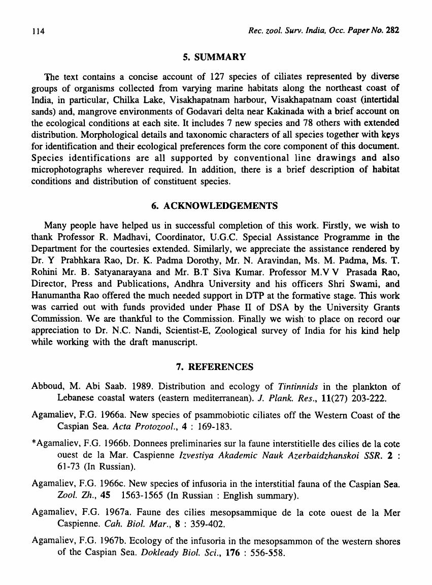
114 Rec. zool. Surv. India, Occ. Paper No. 282
S. SUMMARY
The text contains a concise account of 127 species of ciliates represented by diverse groups of organisms collected from varying marine habitats along the northeast coast of India, in particular, Chilka Lake, Visakhapatnam harbour, Visakhapatnam coast (intertidal sands) and, mangrove environments of Godavari delta near Kakinada with a brief account on the ecological conditions at each site. It includes 7 new species and 78 others with extended distribution. Morphological details and taxonomic characters of all species together with keys for identification and their ecological preferences form the core component of this document. Species identifications are all supported by conventional line drawings and also microphotographs wherever required. In addition, there is a brief description of habitat conditions and distribution of constituent species.
6. ACKNOWLEDGEMENTS
Many people have helped us in successful completion of this work. Firstly, we wish to thank Professor R. Madhavi, Coordinator, U.G.C. Special Assistance Programme in the Department for the courtesies extended. Similarly, we appreciate the assistance rendered by Dr. Y Prabhkara Rao, Dr. K. Padma Dorothy, Mr. N. Aravindan, Ms. M. Padma, Ms. T. Rohini Mr. B. Satyanarayana and Mr. B.T Siva Kumar. Professor M.V V Prasada Rao, Director, Press and Publications, Andhra University and his officers Shri Swami, and Hanumantha Rao offered the much needed support in DTP at the fonnative stage. This work was carried out with funds provided under Phase II of DSA by the University Grants Commission. We are thankful to the Commission. Finally we wish· to place on record ollC appreciation to Dr. N.C. Nandi, Scientist-E, Z,oological survey of India for his kind help while working with the draft manuscript.
7. REFERENCES
Abboud, M. Abi Saab. 1989. Distribution and ecology of Tintinnids in the plankton of Lebanese coastal waters (eastern mediterranean). J. Plank. Res., 11(27) 203-222.
Agamaliev, F.G. 1966a. New species of psammobiotic ciliates off the Western Coast of the Caspian Sea. Acta Protozool., 4 : 169-183.
* Agamaliev, F.G. 1966b. Donnees preliminaries sur la faune interstitielle des cilies de la cote ouest de la Mar. Caspienne lzvestiya Akademic Nauk Azerbaidzhanskoi SSR. 2 : 61-73 (In Russian).
Agamaliev, F.O. 1966c. New species of infusoria in the interstitial fauna of the Caspian Sea. Zool. Zh., 4S 1563-1565 (In Russian : English summary).
Agamaliev, F.G. 1967a. Faune des cilies mesopsammique de la cote ouest de la Mer Caspienne. Cah. Bioi. Mar., 8 : 359-402.
Agamaliev, F.G. 1967b. Ecology of the infusoria in the mesopsammon of the western shores of the Caspian Sea. Dokleady BioI. Sci., 176 : 556-558.

KALAVATI and RAMAN: Taxonomy and Ecology o/Ciliated Protozoa ... 115
A.amaliev, 1968. Materials on morphology of some psammophilic ciliates of the caspian sea. Acta Protozool., 6 : 225-244. (In Russian)
Alamaliev, F.G. 1969b. A contribution to Zoogeography of psammophilous ciliates of the Capsian Sea. Zool. Zh., 48 : 957-961.
Asamaliev, F.G. 1969a. Ciliates of the mesopsammon of the Caspian Sea. Sterlkov, A. A., Sukhanova, K. M. and Raikov, I. B. eds. Progress in Protistology: Abstracts of papers read at the III International Congress of Protozoology, Leningrad : 184-185.
Asamaliev, F.G. 1970. Vertical distribution of psammophilous ciliates in the Capsian Sea. Zool. Zh., 49 : 1277-1284.
Agamaliev, F.G. 1971. Complements to the fauna of psanunophilic ciliates of the western coast of the Caspian Sea. Acta Protozool., 8 : 379-404. (In Russian: English summary).
Agamaliev, F.G. 1972. Ciliates from microbenthos of Islands of Asperonskij and Bakinskij archipelagos of the Caspian Sea. Acta Protozool., 10 : 1-27.
Agamaliev, F.G. 1973. Ciliates of microbenthos in the Krasnovodsk Bay of the Capsian Sea. Zool. Zh., 52 : 1597-1601. (In Russian).
Agamaliev, F.G. 1974a. Ciliates on the solid surface over growth of the Caspian Sea. Acta Protozool., 13 : 53-85. (In Russian: English summary).
Agamaliev, F.G. 1974b. Ciliates of the Turkman Bay (Caspian Sea). Zoo I. Zh., 53 : 19-24. (In Russian : English summary).
Agamaliev, F.G. 1974c. Benthic infusoria in the western Caspian Sea. Hydrobiol. J., 10 : 18-23.
Agamaliev, F.G. 1975. Infusorians off the western shore of the central Caspian. Oceanology, 15 : 208-211.
Ag amalie v , F.G. 1978. Morphology of some free-living ciliates of the Caspian sea. Acta Protozool., 17 : 419-444. (In Russian: English summary).
Agamaliev, F.G. 1980. Infusoria of brackish water and saline lakes of the Apsheron Pennisula. Zool. Zh., 59 : 335-341. (In Russian)
Aleem, A.A. 1957. Succession of marine fouling organisms on the test panels immersed in deep water at La jolla, California. Hydrobiologia, 11 : 40-58.
Andersen, P. and Sorensen, H.M. 1986. Population dynamics and trophic coupling in pelagic microorganisms in eutrophic coastal waters. Mar. Ecol. Prog. Ser., 33 : 99-103.
Anigstein, L. 1912. Uber zwei neue marine ciiaten. Arch. Protistenk, 24 : 12-16.
Baker, C. and Phaff, W.I. 1976. Tintinnida from coastal waters of south west Netherlands 1. The genus Tintinopsis. Hydrobiologia, SO : 101-111.

116 Ree. zoo I. Surv. India, Oee. Paper No. 282
Balech, E. and EI-Sayed, S. Z. 1965. Microplankton of the Weddell Sea. In : Biology of Antarctic Seas, II (Ed. G.A. Llano). American Geophysical Union, Washington, D.C. : 107-124.
Barnes, R.S.K., Sattelle, D.B., Everton, I.I. Nicholas, W. and Scott, D.H. 1976. Intertidal sands and interstial fauna associated with different stages .of salt-marsh development. Est. Coast. Mar. Sci., 4 : 497-511.
Battish, S. K. and Satwant, K. 1991. Protozoan fauna of India: Ciliophora, Him. J. Env. Zool., 5 : 50-52.
Battisse, A. 1975. Propositions porn une novelle systematique des Acinetiens (Ciliophora, Kinetofragminiphora, Suctorida) C.R. A cad. Sci., (Paris) 280 : 1797-800.
Beltran, E. 1933. Gruberia Calkinsi, a brackish water ciliate. Bioi. Bull., 64 : 33.
Bhatia, B.L. 1936. Protozoa: Ciliophora. In : The fauna of British India including~Ceylon and Burma. Taylor and Francis, London: 1-499.
Bick, H. 1972. Ciliated Protozoa. An illustrated Guide to the species used as biological indicators in fresh water biology. WHO, Geneva: 1-198.
Bick, H. and Kunze, S. 1971. Eine Zusammenstellung von autokologischen und saprobiologischen Befunden an susswasserciliaten. Int. Revue Ges. Hydrobiol., ~6 : 337-384.
Biemacka, I. 1962. Die protozoen fauna in der Danziger Bucht 1. Die protozoen in eingen Buchten dr seekuste. Polskie Arch. Hydrobiol., 10 : 39-100.
Boaden, P. 1. S. 1963. The interstitial fauna of some north water beaches. J. Mar. Bioi. Assoc. (U.K.) 43 : 79-96.
Bock, KJ. 1952a. Zur okologie der ciliaten des marinen sand grundes der kieler. Bucht. 1. Kieler Meeresforschum Gen., 9 : 77-89.
Bock, K.I. 1952b. Uber einige holo-und spirotriche ciliaten aus den marinen sand gebieeten der kieler Bucht. Zoo I. Anz., 149 : 107-115.
Bock, K.J. 1953. Zur okologie der ciliaten des marinen sand grundes der kieler Bucht. II Kieler Meeres forschungen., 9 : 252-256.
Bock, K.I. 1955. Condylostoma vastum n. sp. and Aspidisca paertinens n. sp. zwei sandbenohrende ciliaten aus dem kustengebict der kieler Bucht. Zool. Anz., 154 : 302-304
Bock, K.I. 1960. Biologische unter suchunger, nisbersondere der ciliaten fauna in der deucb Abwasser belasterten schlei (west Hche ostee) Kieler Meeresforschungen, 16 : 57-68.
Boikova, E. 1984. Ecological character of protozoans (Ciliata, Flagellata) in the Baltic Sea. Ophelia, 3 : 23-32.
Borror, A.C. 1962. Ciliate protozoa of the Gulf of Mexico. Bull. Mar. Sci., 12 : 333-349.

KALA VATI and RAMAN : Taxonomy and Ecology of Ciliated Protozoa ... 117
Borror A.C., 1963a. Morpholology and ecology of the benthic ciliated protozoa of Alligator harbour. Florida. Arch. Protistenk, 106 : 465-534.
Borror, A.C. 1963b. Morphology and ecology of some uncommon ciliates from Alligator harbour. Florida. Trans. Amer. Micro. Soc., 82 : 125-131.
Borror, A. C. 1965. New and little known tidal marsh ciliates. Trans. Amer. Micro. Soc., 84 : 550-565.
Borror, A. C. 1968a. Ecology of intertidal ciliates. Trans. Amer. Micro. Soc., 87 : 233-243.
Borror, A.C. 1968b. Nigrosin-HgC12-Formalin. A stain fixative for ciliates. Stain. Tech., 43 : 293-294.
Borror, A.C. 1972a. Tidal marsh ciliates (Protozoa) : Morphology, ecology and systematics. Acta. Protozool., 10 : 29-71.
Borror,.A. C. 1972b. Revision of the order: Hypotrchida (Ciliophora: Protozoa). J. Protozool., 19 : 1-23.
Borror, A. C. 1975. Environmental requirements of selected estuarine ciliated protozoa. Ecol. Res. Sere EPA No. 66013 .. 74-031,49
Borror, A. C. 1980. Spatial distribution of marine ciliates micro .. ecologic and biogeographic aspects of protozoan ecology. J. Protozool., 27 : 10-13.
Borror, A. C. and Hill, B.F. 1995. The order Euplotida (Ciliophora: Taxonomy with division of Euplotes into several genera. J. Euk. Microbiol., 42(5) : 457-466.
*Bory de Vincent, J.B. 1826. Essaid une classification des Animaux Microscopiques Paris : 1-104. (quoted from KahI, 1935)
Boynton, J.E. and Small, E.C. 1984. Ciliates by the slice. Science Teacher. 51 : 34-38.
Brown, P.J. 1973. Interstitial Marsh ciliates. J. Protozool., 20 : 496.
Bullington, W.E. 1940. Some ciliates from Totugas Carnegie instilution of Washington, Totugas Laboratory Papers, 32 : 188 .. 221.
Burkill, P. H. 1982. Ciliates and other microplankton component of a nearshore food web: Standing stocks and production processes. Ann. Inst. Oceanogr. Paris, 58 : 335-350.
Burkovsky, 1 V. 1967. On the ecology of psammophilous infusoria. Zoo I. Zh., 46 : 987-992.
Burkovsky, lV. 1968. Quantitative data on the vertical distribution of psammophilic infusoria in the velikaya salma. Kandalaksha Bay, the White Sea. Zoo I. Zh., 47 : 1407-1410.
Burkovsky, I. V. 1970a. The ciliates of the mesopsammon of the Kandalaksha Gulf (White Sea). I. Acta Protozool., 7 : 475-489 (In Russian : English summary).
Burkovsky, I.V 1970b. The ciliates of the mesopsammon of the Kandalaksha Gulf (White Sea) ll. Acta Protozool., 8 : 47-65.

118 Ree. zool. Surv. India, Dec. Paper No. 282
Burkovsky, I.V 1970c. Ciliates of the sand and littoral and sublittoral Kandalaksha. Gulf (White Sea) and analysis on the fauna of benthic ciliates of other sea. Acta Protozool., 8 : 183-201. (In Russian)
Burkovsky, I.V 1971. Ecology of psammophilous ciliates in the White Sea. Zool. Zh., SO : 1285-1302. (In Russian)
Burkovsky, I.V 1978. Structure dynamics and production of a community of marine psammophilic ciliates. Zool. Zh., 57 : 325-337.
*Butschli, O. 1889. Protozoa in "Bronns Klassen und Ordnungen"
*Calkins, G.N. 1901. Marine protozoa from woods hole. Bull. US. Fish. Comm., 11· : 413-468.
Capriulo, G.M. and Carpenter, EJ. 1983. Abundance, species composition and feeding impact of tintinnid microzooplankton in central long island sound. Mar. Ecol. Prog. Ser., 10 : 277-288.
Carey, P.G. 1986. A method of long term laboratory maintenance of marine psammophilic ciliates. 1. Protozool., 33(3) : 442-443.
Carey, P.G. (ed.) 1992. Marine interstitial ciliates. London, New York, Tokyo, Melbourne: 1-341.
Carey, P.G. and Maeda, M., 1985. Horinzontal distribution of psammophilic ciliates in the sediments of Chicester harbour area. 1. Nat. Hist., 19 : 555-S75.
*Claparede, E. and Lachmann, J. 1858. Etude sur les infusoires et les Rhizopodes Geneva: 1858-1861.
*Cohn, F. 1866a. Nuene infusorien in seaquarium. Ziet. wissenschaftiche. Zool., 16 : 253-302.
*Cohn, F. 1866b. Uber die fortpflanzung von Nassula elegans : Zeitschr. f. wiss. Zoll., 16 : 1866.
Collin, B. 1912. Etude monographique sur les Acinetiens II. Morphologie, physiologie, systematique. Arch. Zoo I. expo Gen., 51 : 1-457.
Corliss, J.D. 1956. On the evolution and systematics of ciliated protozoa. Syst. Zool., 5 : 68-91, 121-140.
Corliss, J.~. 1959. An illustrated key to the higher groups of the ciliated protozoa with definition of tenns. J. Protozool., 6 : 265-281.-
Corliss, J.D. 1960. The problem of homonyms among generic names of ciliated protozoa with proposal of several new names. 1. Protozool., 7 : 269-278.
Corliss, J .0. 1961. The Ciliated Protozoa : Characterisation, classification and guide to literature. London, New york. Pergmon Press: 1-310.
Corliss, J. O. 1972. The ciliated protozoa and other organisms. Some unresolved questions of rnajor phylogenetic significance. Amer. Zool., 12(4) : .739-754

KALA V A TI and RAMAN: Taxonomy and Ecology of Ciliated Protozoa ... 119
Corliss, J. O. 1973. Protozoan Ecology: A note on its current status. Amer. Zool., 13 : 145-148.
Corliss, J.O. 1974. The changing world of ciliate systematics historical analysis of the past efforts and newly proposed phylogenetic scheme of classification for the protistan phylum Ciliophora Syst. Zool., 23 : 91-138.
Corliss, J.O. 1979. The Ciliated Protozoa: Characterization, classification and guide to the literature, 2nd ed. : Pergamon Press London, Oxford: 1-455.
Curds, C.R. 1982. British and other freshwater ciliated protozoa Part. 1. Ciliophora Kinetofragminophora. (publ) Linnean Society of london. Cambridge Univ. Press 1-386.
Curds, C.R. 1975. A guide to the species of the genus Euplotes (Hypotrichida, Ciliata). Bulletin of British Museum. Nat. Hist. Zoology, 28 : 1-61.
Czapik, A. 1965. Prorodon raabei n. sp. et sa biologic. Acta Protozoo!., 3 : 21-26.
Czapik, A. and Jordon, A. 1976a. Les observations sur les cilies d'une mare. Acta Protozoo!., 15 : 277-287.
Czapik, A. and Jordon, A. 1976b. Les cilies psammophiles de la mer Baltique aux enviros de gdainsk. Acta Protozool., 15 : 423-445.
Czapik, A. and Jordon, A. 1977. Les cilies psammophilies de la mer Baltique aux environs de gdanisk. Part II Acta Proto Zool., 16 : 165-168.
*Daday, E. Von. 1887. Monographie des familie des Tintinnodeen. Mitt. Zoo I. Stat. Neapel., 7 : 473-591.
Das, A. K. 1995. Protozoa. In : Fauna of Chilka Lake, Wetland Ecosystem Series, I : 137-209. Zool. Surv. India.
Das, A.K. and Nair, K.N. 1987. Protozoa (Free living). Fauna of Orissa, State Fauna Series, No. l(Part-l) : 25-52. Zool. Survey of India.
Das, A. K., MandaI, A.K. and Sarkar, N.C. 1993. Protozoa, 1. Free-living Protozoa. Fauna of West Bengal, State fauna series, 3(Part-12) : 1-133. Zool. Surv. India.
Damodar Naidu, W. 1980. Studies on tintinnids (Protozoa: Ciliates) of Port Novo region, South India, Ph.D., Thesis, Annamalai University: 1-281.
Daode, J., Song, W and Warren, A. 2006. Redescriptions of three marine peritrichous ciliates, Zoothamnium alternans Claparede et Lachmann, 1859, Z. sinense Song, 1991 and Z. commune Kahl, 1933 (Ciliophora, Peritrichia), from North China. Acta Protozoo!., 45 : 27-39.
Da Faria, J. G., Da Cunha, A.M. and Pinto, C. 1922. Estudos sobre protozoairios do mar. Mem. de· In st. Oswaldo Cruz, 15 : 186-208.
Delage, Y and Herouard, E. 1896. La cellulae et les protzoaires Trait'e Zoologie Concrete Vol. 1 Schleichet Freses Paris : 1-584.

120 Ree. zool. Surv. India, Oee. Pape.r No. 282
Deroux, G. 1970. Le serie "Chlamydonellienne" chez les chlamydodontidae, Holotriches, cyrtophorina. Faure-Fremiet. Protistolgica, 6(2): 155-182.
Deroux, G. 1976. Plancorticale des cyrtophorida Ill-les structures differenciatrices chez les Dysterina. Protistolgica, 12 : 505-538.
Didier, P. 1969. Observations preliminaires sur l' ultrastructure buccale de Faurella thermalis, 1966 cilie. hymenostome peniculien. Ann. stat. Bioi. Besse-eu-chandesse. No.3. (1968) : 267-270.
Doflein, F. and Reichnow, E. (1927-1929). Lehrbuch des protozoen kunde: Eine Darsteling des Natur geschichle du protozoen mit besonderen Bercicksichtigung der parastichem und pathogenen formem 5th ed. Fischer. Jena : 1-1262.
Dragesco, J. 1953a. Diagnoses preliminaires de quelques cilies noureaux des sables de Banyuls. Sur. Mer (1) Vie et Milieu, 4 : 633-637.
Dragesco, J. 1953b. Sur l' ecologie des cilies psammophiles littoraux de la region de Banyulssur-mer (Pyr-or) Note preliminaire Sur. Mer (1) Vie et Milieu., 4 : 627-632.
Dragesco, J. 1954a. Diagnoses preliminaires de quelque cilies psammophiles nouveax. Bull. de Soc. Zool. de France, 79 : 57-62.
Dragesco, J. 1954b. Diagnoses preliminaires de quelques cilies nouveaux des sables. Bull. de Soc. Zool. de France, 79 : 62-70.
Dragesco, J. 1960. Les Cilies mesopsammiques littoraux. Systematique morphologie, ecologie. Trans. stat. Bioi. Roscoff. N.S., 12 : 1-356.
Dragesco, J. 1963a. Complements a la connaissance des cilies mesopsammiques de Roscoff 1. Holotriches. Cah Bioi. Mar., 4 : 91-119.
Dragesco, J. 1963b. Complements a la connaissance des cilies mesopsammiques de Roscoff. IT Heterotriches, ill Hypertriches. Cah Bioi. Mar., 4 : 251-275.
Dragesco, J. 1965. Cilies mesopsammiques d' Afrique Noire. Cah Bioi. Mar., 6 : 357-399.
Dragesco, 1. 1966. Observations sur quelques cilies Libres Arch. Protistenk., 109 : 155-206.
Dragesco, J. 1970. Cilies libres du cameroun. Ann. Fac. Sci. Yaounde: 1-141.
Dragesco, J and Dragesco-Keineis, A. 1986. Cities tibres de I' Afrique inter tropical. Faune tropical, 26: 1-559.
*Dujardin, F. 1841. Histire naturelle des zoophytes. lnfusories, Paris: Rout 678 pp
Dye, A.H. 1979. Quantitative estimatien of protozoa from sandy substrates. Est. Coast Mar. Sci., 8 : 199-204.
Eddy, S. 1925. Distribution of the marine protozoa in the Friday harbour waters (San Juan Channel) washington sound. Trans. Amer. Micro. Soc., 44 : 97-106.

KALA VATI and RAMAN : Taxonomy and Ecology of Ciliated Protozoa ... 121
'Ehrenberg, C.G. 1830. Beitrage zur kenntnis der organisation des infusorien und ther geographischem verbreitung besonders in siberien (Quoted from Corliss, 1979). Abh. Akad. Wiss. Berlin., : 1-88.
-Ehrenberg, C.G. 1833. Driller beitragen zur erkenntniss der grosser organisation in der richtung des kleinsten raumes. Abhandlengen der akademie der wissen schafter Berlin : 145-336.
-Ehrenberg, C. G. 1838. Die Infusionstiere als volkomene organismen : Leipzig 1838 : 1- 612.
BDiot, P.B. and Bamforth, S. S. 1974. Nutritional diversity in a brackish interstial sulphide biome. J. Protozool., 21 : (Suppl) 85.
BIliott, P.B. and Bamforth, S.S. 1975. Interstitial protozoa and algae of Louisiana salt marshes. J. Protozool., 22 : 514-519.
-Bngelmann, T.W. 1862. Zur Naturgeschichte der Infusionstiere : Beitrage zur Entwicklungs . geschichte der infusorien. Z. Wiss. Zool., 11 : 347-393.
*Bntz, G. 1884. Umber die Inf. des Golfes van Neapel : Mitt. Zool. Stat. Neapai., 5.
·Botz, G. 1904. Die fauna der kantinentalen kochsalzwasses : Math nature Ber. Ungam, 19.
*Fabre-Domergue, P. 1889. Note sur une nouvelle forme de colpoda (henneguyi) : Anne de micro, 2.
Faure-Fremiet, E. 1924. Contributions a la connaissance des infusoires planktoniques. Bull. BioI. de la France et de la Belg., Supplement No.6: 1-171.
Faure-Fremiet, E. 1948. The ecology of some infusorian communities of intertidal pools. J. Anim. Ecol., 17 : 127-130.
Faure-Fremiet, E. 1950a. Morphologie comparee et systematique des cilies. Bull. Soc. Zool. Fr., 75 : 109-122.
Faure-Fremiet, E. 1950b. The marine sand-dwelling ciliates of cape-cod shores. BioI. Bull., 99 : 349-350.
Faure-Fremiet, E. 1950c. Ecologie des cilies psammophiles littoraux. Bull. Bioi. de la France et de La Belgieque., 84 : 35-75.
Faure-Fremiet, E. 1950d. Ecology of ciliate infusoria. Endeavol., 9 : 183-187.
Faure-Fremiet, E. 1951a. The marine sand-dwelling ciliates of cope-cod. Bioi. Bull., 100 : 59-70.
Faure-Fremiet, E. 1951b. Ecologie des protistee littoraux. Annee. Biologique., 27 : 205-215.
Faure-Fremiet, E. 1954b. Les problems de differential chez les protista. Bull. Soc. Zool. Fr., 79 : 311-329.
Faure-Fremiet, E. 1959. La famille des nassulidae (Ciliata, Gymnostomatida) et la genre Nassulopsis n.gen C.R. A cad. Sci., 249 : 1429-1433.

122 Rec. zool. Surv. India, Occ. Paper No. 282
Faure-Fremiet, E. 1961. Quelques considerations sur les cilies mesopsammiques a' propos d'un recent travailde J. Drageso. Can. Biol. Mar., 2 : 177-186.
Faure-Fremiet, E. 1963. Remarques sur la morphologie comparee des cilies. Trichostomatida. C.R. A cad. Sci., 257 : 3089-3094.
Faure-Fremiet, E. 1969. Remarques sur la systematique des cilies oligotrichida. Protistologica, 5 : 345-352.
Fenchel, T. 1967. The ecology of marine micrbenthos : I. The quantitative importance of ciliates as compared with metazoans in various types of sediments. Ophelia, 4 : 121-137.
Fenchel, T. 1968. The ecology of marine microbenthos : II. The food of marine benthic ciliates. Ophelia, 5 : 73-121.
Fenchel, r. 1969. The ecology of marine micrbenthos : IV structure and function of the benthic ecosystems. Its chemical and physical factors and the microfauna communities with special reference to ciliated protozoa. Ophelia, 6 : 1-182.
Fenchel, T. 1987. Ecology of protozoa: The biology of Free-living phagotrophic protists. Brock/Springer series in contemporary Bioscience. Springer-Verlag, Berlin. 197 pp.
Fenchel, T. and Jansson, B.O. 1966. On the vertical distribution of the microfauna in the sediments of a brackish beach. Ophelia, 3 : 161-177.
Fenchel, T. Kristensen. 1990. Ramussen, L. 1990. Water column anoxia: Vertical zonation of planktonic protozoa. Mar. Ecol. Prog. Ser., 62 : 1-10.
Fernandez-Galiano, D.1976. Silver impregnation of ciliated protozoa. Procedure yeilding good results with Pyridinated Silver Carbonate Method. Trans. Amer. Micro. Soc., 95(4) : 557-560.
Fish, lD. and Goodwin, BJ. 1976. Observations on the peritrichous ciliate Scyphidia ubiquita from the west coast of Wales and a description of a new species. Jour. Zool., 179 : 361-371.
Fjeld, P. 1955. On some marine psammobiotic ciliates from Droback (Norway) Nytt. Magasii. for:.. Zoologi., 3 : 5-65.
Foissner, W. 1976. Erfahrungen mit einer rocknen SHberimpragnationsmethode Zur Darstellung argyrophiler Strukturen bei Protisten Verh. zool. bot. Ges. Wien, 115 : 68-79
Foissner, W. 1980. Taxonomische studien uber die ciliaten des Grossglocknergebietes. (Hobe Tausen Osterrich) 1. Familien : Holophryidae, Prorodontidae, Plagiocampidae, Colepidae, Enchelydidae and Lacrymaridae). Ann. Natur. Mus. Wien., (Bot. & Zool.) 84 : 49-85.
Foissner, W. 1984. Infraciliatur, Silberlinien system und Biometrie einiger neuer und wenig bekannter terres trischer, limnischer und mariner Ciliaten (Prtozoa : Ciliophora) aus den klassen Kinetofragminophora, Colpoqea und Polyhymenophora. Stap/ia., 12 : 1-165.

KALAVATI and RAMAN: Taxonomy and Ecology o/Ciliated Protozoa ... 123
Foillner, W. 1988. Taxonomic and nomenclatural revision of SLADECEK'S list of ciliates (Protozoa: Ciliophora) as indicators of water quality. Hydrobiologia, 166 : 1-64.
Poissner,W, Berger, H. and Kohmann, F.1992. Taxonomische und okologische Revision der ciliaten des Saprobiensystems-Band II : Peritrichea, Heterotrichida, Odontostomatida, Informations berichte des Bayer, Landesamtes fur wasserweirtschaJt, Heft 5/92, 502.
*Fraipont, J. 1878. Recherches sur les acinetans de la cote d'Ostende. Bull. A cad. Belg. Cl. Sci., 44 : 770-814.
*Fromentel, E. de. 1874. Etudes sur les microzoaires au ift,fusoires proprement dits comprenant de novelles recherches sur leur organisation, leur classification, et la description des especes nove lies ou pen connues. G. masion Paris : 364.
Purgason, W.H. 1940. The significant cytostomal pattern of the "Glaucoma-colpidium group" and a proposed new genus and species Tetrahymena geleii. Arch. Protistenk., 94 : 224-266.
Ganapati, P.N. and Murthy, V.S.R. 1954. Salinity and temperature variations of the surface waters off Visakhapatnam. Mem. Oceanogr.ser. Andhra Univ., 49(1) : 125-142.
Oanapati, P.N. and Rama Sarma, D.V 1958. Hydrography in relation to the production of plankton off Waltair Coast. Mem. Oceanogr. Ser,' Andhra Univ., 62(2) : 168-192.
Ganapati, P.N. and Satyavathi, P. 1958. Report on the Foramninfera in bottom sediments in the Bay of Bengal off the east coast of India. Mem. Oceanogr. Ser., Andhra Univ., 62(2) : 100-127.
Ganapati, P.N. and Rao, M.V.N. 1968. Systematic survey of marine ciliates from Visakhapatnam .. Mem. Oceanogr. Ser., Andhra Univ., 62(2) : 75-90.
Gelei, O. Von. 1954. Umber die Leben sgemeinscheft einiger temporarer Tumpel auf einer Bergwiese in Borzsonygebirge (ober Ungarn) m Ciliaten. Acta Bioi. Hung., 5 : 259-343.
Gerassiomova, Z.P and Seravin, LIN. 1976. Ectoplasmic fibrillar systems of infusoria and its role for th~ understanding of their phylogeny. Zool. Zh., 55 : 645-656.
Gold, K. and Morales, B.A. 1975. Seasonal changes in lorical size and species of tintinnida in the New york Bight. J. Protozool., 22 : 520-528.
Gong, J., Song. W., Hu, X., Ma, H. and Zhu, M. 2001. Morphology and infraciliature of Holosticha bradburyae nor.spec.(Ciliophora, Hypotrichida) from the yellow sea China. Hydrobiologia, 464 : 63-69.
Gong, J and Song, W. 2006. Redescriptions of three cyrtophorid ciliates from marine biofilm, with establishment of a New Genus, Wilbertella nov. gen. (Ciliophora: Cyrtophorida : LyncheUidae). Acta Protozool., 4S : 153 -165
Govindan Kutti, A. G and Balakrishna Nair, N. 1966. Prelimenary observations on the interstitial fauna of south west coast of India. Hydrobiol., 28 : 101-122.

124 Rec. zool. Surv. India, Occ. Paper No. 282
Gourret, P. and Roeser, P. 1888. Contribution a 'lie' tude des protozoaire de la corse. Arch. Bioi. Paris, 8 : 139-204.
* Graziano, C. 1989. On the ecology of tintinnids (Ciliophora: Oligotrichida) in the North Irish sea. Est. Coast Shelf. Sci., 29 : 233-245.
Grain,J., Peck, R.K., Didier, P, Rodrigues de, and SantaRosa, M. 1976. Importance de la ~croscopie electronique dans les etudes de systematique chez les unicellularies : Un example : les cilies du genre Cyclogramma C.R. Acad. Sci. Paris, 282 : 735-738.
* Grubbq:, A. 1884. Die protozoens des hafens von genua : Nova Acta Ac. C.L. C.N. 46.
Gulati, A.N. 1925. An account of some fresh water ciliates from Lahore. J. Bombay nat. Hist. Soc., 30 : 744-755.
Hada, Y. 1932. Report of the biological survey of Mutsu Bay 26. The pelagic ciliata suborder: Tintinnoinea. Sci. Rep. Tohoku. Imp. Univ. 4th ser., 553-573.
Hada, Y 1970. The protozoan plankton of the Antarctic and sub-Antarctic seas. JARE Sci. Rep. Ser., E, 31 : 1-50.
Hada, Y 1974. The protozoan plankton collected from the Port of Cochin on the Arabian sea coast of India. Univ. Liter., 15 : 73-97.
*Haeckel, E. 1866. Generelle morphologie der orgaai'smem II allgemeine Entwicklungsgesschibte der o rgan ismem. G. Reimer Gerlin : 1-462.
Hartwig, E. 1973a. Die Ciliaten des 'Gezeiten' -Sandstrandes der Nordseeinsel Sylt. I Systematikse-Mikrofauna des Meeresbodens., 18 : 387-453.
Hartwig, E. 1973b. Die ciliaten des Gezeiten-Sandstrandes der Nordseeinsel sylt II Okologie. Mikrofauna des Meeresbodens, 21 : 1-171.
Hartwig, E.1974. Verzeichnis der the Bereich der deutschen Meerskuste angetroffenem interstitillen ciliaten. Mitheljungen des Hamburgischen Zoofgischen Museum and Institute, 71 : 7-21.
Hartwig, E. 1977. On the interstitial ciliate fauna of Bermuda. Can. Bioi. Mar., 18 : 113-126.
Hartwig,E. 1980. A bibliography of interstitial ciliates (Protozoa) Arch Protistenk., 123 : 422-438.
Hartwig, E. and Parker, J.G. 1977. On the systematics and ecology of interstitial ciliates of sandy beaches in North Yorkshire. J. Mar. Bioi. Ass. U.K., 57 : 735-760.
Hartwig, E., Gluth, G. and Weiser, W. 1976. Investigations on the ecophysiology of Geleia nigriceps Kahl (Ciliophora : Gymnostomata) inhabiting a sandy Beach in Bermuda). Oecoiogia., 31 : 159-175.
Hedin, H. 1975. On the ecology of Tintinnids in Swedish West Coast. Zoon., 3 : 125-140.
*Henneguy, L.F. 1890. Sur us Inf. heterotriche (Fabrea salina). Anne. de Microgr., 3 : 118-
KALA VA TI tlnd RAMAN : Taxonomy and Ecology of Ciliated Protozoa ... 125
136.
Hiller. S. and Bardele, C.F. 1988. Prorodon akilitolophon n. spec. and the "Dorsal Brush" as a character to identify certain subgroups in the genus Prorodon Arch. Protistenk., 136 : 213-236.
HU, X and Song, W. 1999. on the morphology of the marine hypotrichous ciliate, Holosticha diademata, (Ciliphora,Hypotrichida), with comparision of its related species from the mantle cavity of cultured scallops. J. Ocea'1 Univ. Qingdo., 29 : 469-473 (In Chinese with English summary).
HU, X and Song,W. 2001a. Redescription of little known marine ciliate, Stichotricha marina, Stein 1867 (Ciliphora, Hypotrichida ) from the mant1e cavity of cultured scallops. Hydrobiolo g ia, 464 : 71-77
HUt X and Song, W. 2001b. Morphology and morphogenesis of Holosticha heterofossneri nov. Spec from the yellow sea, China (Ciliphora, Hypotrichida ). Hydrobiologia, 448 : 171-179
Huxley, T.H. 1857. On Dysteria, a new genus of infusoria. Quart. J. Micr. Sci., 5 : 78-82.
Isquith,I.R. 1968. Anigsteinia n. gen. a member of the family: Spirostomatidae. J. Protozool., 15, suppl. 26.
James, M.R. and Hall, J. A. 1995. Planktonic ciliated protozoa, their distribution and relationship to environmental variables in a marine coastal ecosystem. J. Plank. Res., 17(4) : 659-683.
Jankowski, A.W. 1964a. Morphology and evolution of Ciliophora. 1. The new system of sapropelebiotic het~ro.trichid. Zool. Zh., 43 : 503-517.
Jankowski, A. W. 1964b. Morphology and evaluation of ciliophora : III. Diagnosis and phylogenesis of 53 sapropelebionts mainly of the order Heterotrichida Arch. Protistenk., 107 : 185-2.94.
Jankowski, A.W. 1968. Taxonomy of the suborder Nassulina Jank 1967 (Ciliophora: Ambihymenida). Zool. Zh., 47 : 970-1001.
Jankowski, A.W.1979. Concepts of a new system of the phylum Ciliophora. In Principles of construction of the microsystem of the unicellular animals. Krylor, M. V Staroborator. Y.1. (eds.) Proc. Zool. Inst., (Trudy. Zool. Inst. Akad. Nank. SSAR) 94 : 1-134.
Jankowski, A.W. 1979a. Systematic revision of the class Polyhymenophora (Spirotricha) morphology, systamatics and evolution. Tezisy. Doke. Zool. Inst. Akad. Nank. USSR, 86 : 39-40.
Jankowski, A.W. 1979b. Systematics and phylogeny of the order Hypotrichida Stein. 1859. (Protozoa). Trudy. Zool. Inst. Akad. Nank. SSAR., 86 : 48-85.
Jayaprada. C. 1998. Ecology of benthic microalgae in relation to pollution in Visakhapatnam harbour, East coast of India. Ph. D. Thesis, Andhra University. (unpublished): 1-170.
126 Rec. 1.001. Surv. India, Occ. Paper No. 282
Jones, E.E. 1974. The protozoa of the Mobile Bay, Alabama. Monograph, Vol. 1. University of South Alabama Press. Mobile: 1-113.
Jordan, A. 1974. Morphologie et biologie de Prorodon discolor ehr-blochm-schew. Acta. Protozool., 13 : 5-8.
Jorgensen, E. 1924. Mediterranean Tintinnidae. Rep. Danish. Oceanogr. Exped. 1908-1910 Medit 2 (BioI)., 13 : 1-110.
Jorgensen, E. 1927. Ciliata, Tintinnidae. In Die Tierwelt der Nord und ostee, lief, 8 (Teil iLC), Grimpe. G. and Wagler, E. (eds.), Leipzig., : 1-29.
Kahl, A. 1927a. Neue und erganzende Beobechtungen Heterotrachen ciliaten. Arch. Protistenk., 57 : 121-203.
Kahl, A. 1927b. Neue und erganzende Beobechtungen Holotrichen ciliaten. 1. Arch. Protistenk., 60 : 34-129.
Kahl, A. 1930. Urtiere oder Protozoa I: Wimpertiere oder ciliata (Infusoria) I. Allege meiner Teil und Prostomata Tierwelf Dtl., 18 : 1-180.
Kahl, A. 1931. Urtiere oder Protozoa I : Wimpertiere estieu oder ciliata (Infusoria) 2. Holotricha auberden 1m 1. Teil behan delten Prostomata. Tierwelt., 21 : 181-398.
Kahl, A. 19~2. Urtiere oder Protozoa I. Wimpertiere oder ciliata (Infusoria) 3. Spirotricha In Dahl, F. (ed.) Die Tierwelt deutschlands und der angrenzenden meeresteili Gustev Fischer, Jena., 25 : 399-650.
Kahl, A. 1935. urtiere oder Protozoa I. Wimpertiere oder ciliata (Infusoria) Nachtrag I In Dahl, F. (ed) Die Tierwelt Deutchlands und der angrenzenden meeresteile. Gustav Fischer, Jena. 30 : 650-842.
Kalavatir C., Radhakrishna, G and Narasimhamurti, C.C. 1989. Studies on coastal pbytal ciliates from Visakhapatnam, India, in relation to pollution. Asian Mar. Bioi., 6 : 167-172.
Kalavati, C., Raman, A.V., Vaidehi, J. and Ratna Bharati, V 1997. Effects of pollution on planktonic ciliates in Visakhapatnam harbour. Ind. J. Mar. Sci., 26 : 68-73.
Kattar, M.R. 1970. Estudo dos protozoarios ciliados psamofilos do littoral brasileiro. Bioi. Mar. (Sau Paulo) N.S., 27 : 123-206.
* Kellicott, D.S., 1885. New infusorian. Proc. Amer. Soc. Micro., 7&8 : 1884-85.
* Kent, s. 1880-82. A manual of the infusoria: London. 1880/82.
Kiesselbach, A. 1936. Zur ciliaten fauna der nord lichen adria. Thalassia., 2 : 1-53.
-Kirby, H. 1932. Two protozoa from Brine. Trans. Amer. Micro. Soc., 51 : 8-15.
Kirby, H. 1934. Some ciliates from salt marshes in California. Arch. Protistenk., 82 : 114-133.

KALA VA TI and RAMAN: Taxonomy and Ecology of Ciliated Protozoa ... 127
Kofoid, C.A. and Campbell, A.S. 1929. A conspectus of the marine and fresh water ciliates belonging to the sub order Tintinnoinea with descriptions of new species principally from Agassiz expedition to the eastern tropical pacific 1904-1905. Univ. Cal. Publ. Zool., 34 : 1-403.
Kolkwitz, R. 1950. Okologie der saprobien sch Reihi, ver, Wass. Boden-U lufthys., : 4-64
Kovaleva, V. G. 1966. Infusoria of the mesop~ammon in sand bays of Black Sea. Zoo I. Zh., 45 : 1600-1611.
Kovaleva, V G. 1967. New data on the infusorian fauna of the mesopsammon of Barents Sea. Acta protozool., 5 : 81-88.
Kovaleva, V. G. and Golemansky, V 1979. Psammobiont ciliates of the Bulgarian coast of the Black Sea. Acta Protozool., 18 : 265-284.
Krsinic, F. 1987. Tintinnines (Ciliophora : oligotrichida : Tintinnina) in Eastern Adriatic Bays. Est. Coast Shelf Sci., 24 : 527-538.
Krishnamurthy, K. and Santhanam, R. 1975. Ecology of tintinnids (Protozoa: Ciliatea) in Port Novo region. Ind. J. Mar. Sci., 4 : 181-184.
Krishnamurthy, K., Damodara Naidu, W and Santhanam, R. 1979. Further studies on Tintinnids (Protozoa: Ciliata). Arch. Protistenk., 122 : 171-180.
*Laackmann, H. 1908. Ungeschl~cht1ich und geslechtliche des standigen Gehalts des Meeres an Plankton. Wiss. Meersters. N.F., 10 : 129-370.
Lackey, J.B. 1936. Occurrence and distribution of marine protozoan species in the Woods Hole area. Bioi. Bull., 70 : 264-278.
Lackey, I.B. 1938. A study of some ecological factors. affecting the distribution of protozoa. Ecological Monographs, 8 : 501-527.
Lauterbom, R. 1908. Protozoen stundien V. Zur kenntnis einiger Rhizopoden und Infusorien. aus. Oem. Gebiete des oberrheins. Wiss Zool., 90 : 645-669.
Laval Peuto, M. 1981. Construction of the lorica in ciliate Tintinnine. In vivo study of Favella ehrenbergii: variability of phenotypes during the cycle, biology, statistics, biometry. Protistologica., 17 : 249-272.
Laval-Peuto,M. and Rassoulzadegan, F. 1988. Autoflourescence of marine planktonic oligotrichina and other ciliates. Hydrobiologia., 159 : 99-110.
Leakey, I.B. and Leakey, ~.W. 1963. Microscopic algae and protozoa in the waters near Plymouth in August 1962. J. Mar. Bioi. Assoc. (UK), 43 : 797-805.
Leakey, R.J.G., Burkill, P.H. and Sleigh, M.A. 1992. Planktonic ciliates in Southampton waters: Their abundance, biomass, production and their role in pelagic carbon flow. Mar.Biol., 114 : 67-83.
Leakey, R.I.G., Burkill, P.H~ and Sleigh, M.A. 1993. Planktonic ciliates in Southampton waters: Quantitative taxonomic studies. J. Mar. Bioi. Ass. V .. K., 73 : 579-594.

128 Ree. zool. Surv. India, Oee. Paper No. 282
Leakey, R. J. G., Fenton, N and Clarke, A. 1994a. The annual cycle of planktonic ciliates in near shore waters at Signy Island, Antarctica. I. Plank. Res. 16(7)": 841-856
Leakey, R.J.G., Burkill, P.H. and Sleigh, M.A. 1994b. Ciliate growth rates from Plymouth Sound: Comparasion of direct and indirect estimates. 1. Mar. Bioi. Ass. U.K., 74 : 849-861.
Leakey, R.I.G., Burkill, P.H. and Sleigh, M.A. 1996. Planktonic ciliates in the North western Indian Ocean : their abundance and biomass in water of contrasting productivity. J. Plank. Res., 18 : 1063-1071.
Lee, J.J, Rutner, S.H. and Bovee, E.C. (eds.). 1985. An illustrated guide to the Protozoa. Society of Protozoologists. Allen Press, Lawrence, Kansas : 1-629.
Lee, J. I. 1986. Protozoa as indicators of ecosystems. Insect Sci. Applic., 7(3) : 349-353.
Lee, 1.1. and Soldo, A.T. (eds) 1992. Protocols in Protozoology. Publ. Society of Protozoology. Lawrence, Kmsas.
Leegard, C. 1915. Untersuchurgen uber einige plankton ciliaten des meeres. Nyt. Mag. For. Nat. Vid., S3 : 1915.
Legner, M. 1973. Experimental approach to the role of protozoa in aquatic ecosysytems. Amer. Zool., 13 : 177-192.
Lepsi, J. 1926. Zour kenntis einiger holotrichen. Arch. Protistenk., 53 : 378-406:
Lepsi, 1. 1962. Uber einiger insbesondere psammobionte ciliaten von remanischen schwarzrrean ufer. Zool. Anzeiger., 168 : 460-465.
Lessard, E.I. and Murrel, M. 1996. Di~tribution, abundance and size composition of heterotrophic dinoflagellates and ciliates in the Sargasso Sea near Bermuda. Deep sea Res., 43 : 1045-1065.
Lin, X, song, Wand Warren,A. 2005. Taxonomic studies on three marine pleurostomatid ciliates : Kentrophyllum verrucosum (Stokes, 1893) Petz, Song et Wilbert, 1995, Epiphyllum solifanne (Faure-Fremiet, 1908) gen. n., comb. n. and Amphileptus sikorai sp. n., with the establishment of a new genus Epiphyllum (Ciliophora: Pleurostomatida). Acta Protozool., 44 : 129-145
Lohmann, H. 1911. Uber das Nannoplankton und die zentrifugierung kleinster wasser proben zur Gewinnung desselben in Le bendem zubstandie. Int. Rev. Ges. Hydrobiol. Hydrogr., 4(1/2) : 1-38.
*Lohmann, H. and Vnters, Z. 1908. Feststellung des vollst. Gehaltes des meeres an plankton. Wiss Mersunters N.F. 10, Kiel.
Lynn, D.H. and Montagnes, DJ.S. 1988. Taxonomic description of some conspicuous species of Strobilidiine ciliates (Ciliophora : Choreotrichia) from the Isles of Shoals, Gulf of Maine. I. Ma r. Bioi. Ass. U.K., 68 : 639-658.

KALA VA TI and RAMAN: Taxonomy and Ecology of Ciliated Protozoa ... 129
Lynn, D.H., Montagnes, DJ.S. and Small, E.B. 1988. Taxonomic descriptions of some conspicuous species of the family strombidiidae (Ciliophora: Oligotrichidae) from the Isles of Sholas, Gulf of Maine. J. Mar. 11101. Ass., U.K., 68 : 259-276.
Lynn, D.H. and Montagnes, D.J.S. 1991. Global production of heterotrophic marine planktonic ciliates. In ProtozoQ and their role in marine processes (eds.) P.C. Reid, P.H. Burkill and C.M. Turley.(1991) NATO ASI series G : Ecological sciences 25, SpringerVerlag, Heidenberg and Berlin: 281-308.
Lynn, D.H., Roff, J.C. and Hoporoft, R.R. 1991. Annual abundance and biomass of aloricate ciliates in tropical neritic waters off Kingston, Jamaica. Mar. BioI., 110 : 437-448.
Lynn, D.H. and Gilron, G.L. 1993. Strombidid ciliates from coastal waters near Kingston harbour, Jamaica. (Ciliophora, Oligotricha, Strombidiidae) J. Mar. Biol. Assoc. UK. 73 : 47-65.
Lynn, D.H., Small, E.B. 2002. Phylum Ciliophora Doflein, 1901. In : An Illustrated Guide to the Protozoa. II edition Vol. 1. JJ. Lee, P.C. Bradbury, and G.F. Leedale (eds.). Society of Protozoologists, Lawrence, Kansas. (year 2000) : 371-676.
Madoni, P. 1978. Biological Water Assessement Methods, Proceedings of 3rd Technical seminar. Instituto di Ecologia, universita di Parma, Italy : 297-310.
Madoni, P. 1984. Ecological characterisation of different types of water courses by the multivariate analysis of ciliated protozoa population. Acta. Hydrobiol., 100(2) : 171-188.
Madoni, P and Ghetti, P.F. 1981a. The structure of ciliated protozoa communities in biological sewage treatment plants. Hydrobiologia, 83 : 207-215.
Maeda, M. 1986. An illustrated guide to the species the families Halteridae and Strobilididae (Oligotrichida, Ciliophora) free swimming protozoa common in aquatic environment. Bull. Ocean Res. inst., Univ. Tokyo, 21 : 1-67.
Maeda, M. and Carey, P.G. 1985. An illustrated guide to the species of the family Strombidiidae (Oligotrichida : Ciliophora), free swimming protozoa common in aquatic environment. Bull. Ocean Res. Inst., Univ. Tokyo, 19 : 1-68.
el Magbrabf, A.M. and Perkins, E.J. 1956. Additions to the marine fauna of whitstable. Ann. Mag. Nat. His.. .Ser., ~2(9) : 481-496.
Mamaeva, N.V 1983. Planktonic infusorians of the central part of the Indian Ocean. Soviet. J. Mar. BioI., 8 ': 254-258.
Mamaeva, N.V 1984. Planktonic infusorians in the Bering sea. Soviet J. Mar. Biol., 9 : 180--185.
Mansfeld, K. 1923. 16 new oder wenag bekommte marine infusoria. Arch. Protistenk., 46 : 97-140
Mare, M.F. 1942. A study of marine benthic community with speeial reference to the microorganisms. J. Mar. BioI. Assoc. UK., 2S : 517-554.

130 Rec. zool. Surv. India, Occ. Paper No. 282
Margaleif-Lopez, R. 1945. Infusorios de las aguas continentales de la provincia de Barcalona. BoLetin de La Real Sociedad Espahola de Historic Na~ral Ma 43 (No. 7-8) : 369-381.
Marshal, S. M. 1969. Fich Ident. Zooplankton, Zooplankton sheet. Cons. Int. Explor. Mer. Copenhagen, : 117-127.
Maupas, E. 1883. Contribution ai' etude morphologique et anatomique des infusoires cilies. Arch. 2001. Expt. Et. Generde., 11 : 427-464.
Maginite, G.E. 1935. Ecological aspects of California marine estuary. Amer. Naturalist., 113 : 427-437.
Maupas, E. 1888. Recherches experimentales sur la multiplication des infusoires cilies. Arch. 2001. Exp. Gen. (Ser. 2)., 6 : 165-277.
*Meunier. 1907. Microplankton des mers de Barents et de Kara : Duc 0' Orleans Campagme. Arctique de : 1-355.
Middlebrook, K., Emerson, C.W., Roff, J.C. and Lynn, D.H. 1987. Distribution and abundance of tintinnids in the Quoddy region of the Bay of Fundy. Can. J. Zool., 65 : 594-601.
Montagnes, D.l.S. and Lynn, D.H. 1991. Taxonomy of choreotrichs, the major marine planktonic ciliates with emphasis on the aloricate forms. Mar. Microb. Food Webs., 5(1) : 59-74.
Montagnes, D.l.S., Lynn, D.H., Stoecker, O.K. and Small, E.B. 1988a. Taxonomic descriptions of one new species and redescription of four species in the family Strombidiidae (Ciliophora : Oligotrichida). 1. Protozool., 35(2) : 189-197.
Montagnes, DJ.S. and Taylor, F.l.R. 1994. The salient features of five marine ciliates in the class Spirotrichea (Oligotrichs) with notes on their culturing and behaviour. 1. Euk. Microbiol., 41(6) : 569- 586.
Muller, D.F. 1786. Animalcula Infusoria: Kopenhages U. Leipzig. 1-367.
Munch, H.D. 1955. Systematisch-okologische beilrage zur fauna des kusten grund wassers 2 class : Spirotrichia, Peritrichia, Oligotrichaeta: copepoda, rotateria Thesis, Undergraduate University, Leipzig.
Nanney, D. L. and McCoy, J. W. 1976. Characterisation of the Tetrahymena pyriformis cmplex. Trans. Am. Micros. Soc., 95 : 664-682.
Nobill, R. 1957. Contributo all' ecologia des ciliata psammofili del Golfo di Napoli. Bollettino di Zoologia, 24 : 211-225.
Noland, L.E. 1937. Observations on marine cIliates of the Gulf coast of Florida. Trans. Amer. Micro. Soc., 56 : 160-171.
Nouzerede, M. 1965. Etude de Quelques Cilies Mesopsammiques de la Familie des Geleiidae Kahl. Progress in Protozoology. 2nd International CongreSF"of Protozoology, London : 1-278.

KALA V A TI and RAMAN: Taxonomy and Ecology of Ciliated Protozoa ... 131
Nouzerede, M. 1975. Sur un nouveau genre de protozoaires ciliates geants mesopsammiques appartenant a la family des Geleiidae Kahl. C. R. A cad. Sci., Paris, 280 : 625-628.
Nouzarede, M. 1976. Cytologie Fonctionnelle et Morphollogie Experimentale de quelques Protozoireas cilies Mesopasammiques Geants de 1a Familie des Geleiidae (Kahl) Bull. Stat. Bioi. Arcachon (N.S.) 28(suppl. 1): 1-315
Nubite, R. 1957. Contributo all ecologia der ciliata psammofile del, Golfo de Napoli. Bollettino di Zoologia., 24 : 211-225.
Paranjape, M. 1987. The seasonal cycle and vertical distribution of the tintinnines in Bedford basin, Nova scotia. Can. l. Zool., 65 : 41-48.
Paranjape, M., Conover, R.J., Harding, G.C. and Prouse, N.J. 1985. Micro and macro zooplankton on the Nova Scotia Shelf in tqe pre-spring bloom period: A comparison of their potential resource utilization Can. l. Fish. Aqua. Sci., 42 : 1481-1492.
Parker, J.G. 1981. Ciliated protozoa of the polluted Tees estuary. Est. coastal Shelf Sci., 12 : 337-340.
Patten, B.C., Mulford, R.A. and Warinner, J.E. 1963. An annual phytoplankton cycle in lower Chesapeake Bay. Chesapeake. Sci., 4(1) : 1-20.
Pearse, A.S. 1932. Animals in brackishwater ponds and pools at Dry Tortugas Cemegie Institution of Washigton paper of tortuges Laboratory, 28 : 125-141.
Perkins, E.G. 1958. The food relationships of the microbenthos with particular reference to that found at whitstable kent. Anna. And. Megazine of Nat. Hist., 13(1) : 64-77.
Penard, E. 1922. Etudes sur les infusories d'eau douce (zusammenfarsen des hamptwerk verf). George & Cie. Geneva: 1-331.
Perty, M. 1852. Zur kenntnis kleinster lebensformen, nach Bau, Funktionen, systematik, mit specialverzeichniss der in der schweiz neobachteten. lent & Reinert, Bern., : 1-228.
Petran, A. 1963. Contributte la cunoasterea microfaunei de ciliate psammoflle dim manee neagre litoraul Romanax studi si cercaterii de biologie A cad. RPR, 15 : 187-197.
Petran, A. 1967. Cercetari asupra faunei de ciliate psammobionte la plagele diu sudullitoralului romansese a1 marti negre. Ecology Marind., 2 : 169-191.
Petran, A. 1968. Sur I' ecologie des cilies psammobiontes de la mar noire (littoral, Roumain) Revue. Rourmaine de Biologie, 13 : 441-446.
Petran, A. 1972. Sur la faune des sediments sableux du littoral roumain de la mer noire Cercetari Marine., 2 : 177-184.
Pfannkuche, 0., Jelinek, H and Hartwig, E. 1975. Sur fauna eines susswa'§serwaltes te elbe aestuar Arch. Hydrobiol., 76 : 475-498.
Picken, L.E.R. 1937. The structure of some protozoan communities. J. Ecol., 25 : 368-384.
132 Ree. zool. Surv.lndia, Occ. Paper No. 282
Pierson, B.F. 1943. A comparative morphological study of several species of Euplotes closely related to Euplotes patella. 1. Morphol., 72 : 125-165.
Poche, Fr. 1913. Das system der protozoa. Arch. Protistenk., 30 : 125-321
*Prowazek, S.V 1899-1903. Protozocnstudies I-Ill. Arb. Aus. EI. Zoo I. Inst. D. Univ. Wien., : 11-14.
Puytorac, P. de, Batisse, A. Bohatier, 1. Corliss, 1.0., Deroux, G., Didier, P., Dragesco, J., Fryd versavel, G., Grain, J., Savoie, A. and Tuffrau, M. 1974. Preposition d'une classification du phylum ciliophora Doflein 1901. C.R. A cad. Sci. Paris, 278 : 2799-2802.
* Quennertstedt, 1867. Bidrag fill sveriges infusorie-fauna. Acta. Univ. Lund., 4 : 1-47.
Radha Krishna, G. 1984. Studies on some aspects of ecology of ciliates associated with algae of Visakhapatnam coast Ph.D. Thesis. Andhra University, Waltair. (Unpublished).
Raikov, I.B. 1960. The interstital infusoria of the sand littoral of the Dolniye Zelelntzy Bay Mourmansk. Trudy Murmanskogo BioI. Inst., 2 : 172-185. (In Russian). -.
Raikov, I.B. 1962. Les cilies mesopsammiques <ler littoral de la Mer Blanche (USSR) avec une description de quelque especes nouvelles are per connues. Cah. de BioI. Mar., 3 : 325-361.
Raikov, I.B. 1963. Ciliates of the mesopsammon of the Ussuri Gulf, Sea of Japan. Zool. Zh., 42 : 1753-1766. (In Russian).
Raikov, I.B. and Kovaleva, 1967. Complements to the fauna of psammobiotic ciliates of the Japan Sea. (project GuID. Acta Protozool., 6 : 309-333.
Raman, A.V 1995. Pollution effects in Viskhapatnam harbour, India: An overview of 23 years of investigations and monitoring. Helgolander Meeresunters, 49 : 633-645.
Ramasharma, D.V 1965 Hydrogrpahy of Godavari estuary. Ph.D. Thesis, Andhra University, Waltair. (unpublished)
Rao, G.e. and Ganapati, P.N. 1968. The interstial fauna inhabiting the beach sands of waltair coast. Proc. Nat. Inst. India., (B). 34 : 82-125.
Rassoulzadegan, F. 1977. Evolution annulle des cilies pelagiques en Mediterranee noreoccidentale.cilie oligotriches 'non-tintinnds'(oligotrichina). Annis. Inst. Oceanogr., Paris 53 : 125-134
Rassoulzadegan, F., Laval-Peuto, M and Sheldon, R.W. 1988. Partitioning of the food of marine ciliates between pico and nanoplankton. Hydrobiologia., 159 : 75-88.
Ratna Bharati, V 1992. Planktonic protozoa in polluted north channel in Visakhapatnam harbour. M.Phil. Dissert. Andhra University: 1-37. (unpublished)
Ratna Bharati, V 1998. Protozooplankton in relation to pollution in Visakhapatnam harbour, East coast of India. Ph. D. Thesis, Andhra University, Waltair : 1-130. (unpublished)

KALA V A TI and RAMAN: Taxonomy and Ecology of Ciliated Protozoa ... 133
Reddy, M.P .M. 1962. Limnological studies on the Chilka lake and wave refraction studies in relation to shoreline development. Ph.D. Thesis Andhra University, Waltair : 1-191. (unpublished)
Remane, A. 1933. Vereilung und organisation des benthischen Mikrofauna des Kieler Bucht. Wiss Meersunters N.F., 21 : (1933) 161-221.
Repak, A. 1972. A rediscription of Climacostomatum virens (Ehrenberg) stein and a proposal of a new heterotrich ciliate family climacostomidae Fam. N. 1. Protozool., 19 : 417-427.
Revelante, N. and Gilmartin, M. 1983. Microzooplankton distribution in the Northern Adriatic Sea with emphasis on the relative abundance of ciliated protozoans. Oceanol. Acta., 6 : 407-415.
Revel an te, N and Gilmartin, M. 1980. Macroplankton density indices as indicators of eutrophication in the northern Adriatic Sea. Hydrobiologia, 70 : 277-286.
Roberts, Mcl. D. and Causton, H. 1988. Silver nitrate impregnation of ciliated protozoa. Arch Protistenk., 135 : 299-318.
*Roux, J. 1899. Observations sur quelques Inf. des environs de Geneva: Rev. Suisse Zool., 6 : 1-10.
*Roux, J. 1901. Faune infusorienne des Eaux Stagnantes des Environs de Geneva. Mem. Inst. Nat. Genevois., 19 : 1-148.
Sanders, R. W. 1987. Tintinnids and other microzooplankton, seasonal distribution and relationships to the resources and hydrography in a marine estuary. J. Plank. Res., 9 : 65-77.
Sarojini, R. and Nagabhushanum, R. 1967. A comparative study of some free living ciliated protozoa. 1. Anim. MorphoI. Physiol., 14 : 158-161.
Sauerbrey, E. 1928. Beobachtungen uber eigine new oden wenig bekannte marine ciliates. Arch. Protistenk., 62 : 355-407.
Sawaya, M.P. 1940. Sobre un ciliasdo novo de S. paulo Blepharisma sinusum n. sp. (Ciliata, Heterotrichida) e sobre a sub-ordeum Odontostomata. Nom. Mov. Bol. Fac. Filos. Ciene. ~.Paulo. Zool., 4(19) : 303-308.
Sawyer, C.N. 1952. Some new aspects of phosphates in relation to lake fertilization Sewage Industr. Wastes., 24 : 768-776.
*Schewiakoff, W. 1893. Uber die geographische verbrietung der suswasser protozoes. Mem. Ac. Imp. Se. St. Petersburg, (7) : 41.
*Schewiakoff, W. 1906. Infusoria spirotricha (Russich) : Mem. AC. Imp. Sc.St. Petersburg (8), 4. (phys,-math, Klarse) (bearbeitat von Schouteden in Ann. de Biol. Laeustre 1, 1906) Ubersetzt von hoare.

134 Rec. lool. Surv. India, Ooc. Paper No. 282
*Schrank, F. and Von, P. 1803. Fauna boica, 3(2) : 1-372.
Shen, Y.F., Feng, W.S., Gu, M.R., Wang, S.D., Wu, J.Z. and Tan, Y Y 1994. MonitoIll6~ River Pollution in China. Architecture and Building Press China : 1-308.
Small, E.B. 1967. The scuticociliatida, a new order of the class ciliatea (phylum Protozoa, sub-phylum Ciliophora). Trans. Amer. Micr. 'Soc., 86 : 345-370.
Small, E.B. 1972. Free living protozoa of the Chesapeake Bay exclusive of foraminifera and flagellates. Chesapeake Science, 13 : 96-97.
Small, E.B. 1976. A proposed subphylitic division of the phylum Ciliophora Doflien. 1901. Trans. Amer. Micro. Soc., 95 : 739-751.
Small, E.B. and Lynn, D. 1985. Phylum ciliophora. Doflien 1901. In An illustrated guide to protozoa. Lee, J.J. Hunter, S.H. and Bovee, E.D. (eds.). Publ.Society of Protozool., Lawerence, Kunsas : 393-375.
Smetacek, V. 1981. The annual cycle of protozQoplankton in the Kiel Bight. Mar. Bioi., 63 : 1-11.
*Smith, J.C. 1904. A preliminary contribution to the protozoan fauna of the Gulf: Bioi. stet Bull., 2.
Smith, T.P. 1982. Systematics and ecology of the benthic ciliates of points Mugu lagoon Southern California. Ph. D. Thesis, California University.
Snyder, R. A and Ohman, M. D. 1991. Description of a new species of Strobidinopsidae (Ciliophora: Choreotrichida) from coastal waters of Southern California, USA. Trans. Am. Microscop. Soc., ~110 : 237-243.
Song, W. 1997. A new genus and two new species of marine peritrichous ciliates (Protozoa : Ciliophora, Peritrichida) from Qnigdo, China. Ophelia, 47 : 203-204
Song, W. and Wilbert, N. 1997. Morphological studies on some freeliving ciliates (Ciliophora : Heterotrichida, Hypotrichida) from marine biotypes in Qfngdao, China with descriptions of 3 new species. Holosticha warreni n. sp., Tachysoma ovata n.sp and T. dragescoi n.sp. Eur. 1. Protistol., 33 : 48-62.
Song, W. and Packoff, G. 1997 Taxonomie und Morphologie von marine ciliten aus China mit bescbreibung von Zwei neuen Arten Strombidium globosaneum nov.spec. and S. platum nov. ( Protozoa, Ciliophora) Arch. Protistenk., 147 : 331-336
Song, Wo, Wlbert, N and Warren, A. 2002 New contribution to the Morphology aDd Taxonomy of four marine hypotrichous ciliates from Qingdo, China (Protozoa : Ciliophora) Acta Protozool., 41 : 145-162
Spiegel, A. 1926. Einige neue marine ciliaten. Arch. Protistenk., SO : 184-190.
*Stein~ Fr. 1859. Characteristik neuer Infusorien Ganugen. Lotos, Prag, 9, 2-5 : 57-60.

KALA VA TI and RAMAN: Taxonomy and Ecology of Ciliated Protozoa ... 135
·Stein, Fr. 1867. Der organisms der infusorions thiere nach eigenen Forschungen in Systematischer Rieben folge Bearbeitet der In/. fiere II. Leipzig.
·Stokes, A.C. 1885. Some new infusoria from American freshwaters. Ann. Mag. Nat. Hist., (Ser. 5) 15 : 437-449.
·Stokes, A.C. 1886. Some new infusoria. Am. Nat., 19 : 433-443.
• Stokes , A.C. 1894. Notices of presumably undescribed infusoria. Proc. Amer. Philos. Soc., 33 : 338-345.
• Strand, E. 1928. Miscellanea nomenclatoric zoologica et palaeontologica. 1-11 Arch Naturgesch., 92(A8) (year, 1926) : 30-75.
Tagucbi, S. 1976. Microzooplankton and ses(on in Akkeshi Bay, Japan. Hydrobiologia, SO : 195-204.
Thompson, J.C. 1964. A redisdcription of Uronema marinum and a proposed new family Uronemetidae. Va. J. Sci., 16 : 165-169.
Thompson, J.C. and Evans, F.R. 1968. A redescription of Uronema nigricans J. Protozool., 15 : 369-374.
Thompson, J .C. 1972. Ciliated protozoa of the Antartic peninsula. In. Llano. G.A. ed. Antartic terrestrial biology Vol. 20. American geophysical Union, Washington. D.C. : 261-288.
Thompson, J.C. and Croom, J.M. 1978. Sysytematic.s and ecology of ciliated protozoa from king George Island, Antartic Peninsula*. In pawson D.L. ed Biology of Antartic seas, American geophysical Union, Washington. D.C.
Tuffrau, M. 1960. Revision of the genus Euplotes, fondee sioi, la comparison des structures superficielles. Hydrobiologia, 15 : 1-77.
Timanstera, N.I. and Kopylov, A. I. 1985. Reproduction and production rates of planktonic infusoria in coastal watres off Peru. Oceanologia., 25 : 390-394.
Uhlig, G. 1966. Unter suchungen zur extraktion des vailien Mikrofauna aus marinem sediments. Zoo I. Auz. (Suppl.) 29 : 151-157.
Uhlig, 1969. Quantitative methods in the study of interstitial fauna. Trans. Amer Micro. Soc., 87 : 226-232.
Vacelet, E. 1960. Note preliminaire sur la fauna infusorienne des "sables a amphioxus" de la baie de marsieIles. Trauax de la station marine d'endoune. 20 : 53-57.
Vacelet, 1961 a. La faune infusoriene des "Sables a amphioxus" des environs de Marsielles. Bull. de 1. Inst. Ocaenogr., Monaco., 58 : 1-12.
Vacelet, 1961 b. Les cities de la microfauna de 'sables mal calibres' des environs de marsielles. Travaux de la station Marine d'endoume., 22 : 13-19.

136 Rec. zoo 1. Surv. India, Dcc. Paper No. 282
Vashista, H.S. and Oulati, S. 1972. The systematics of the fresh water Protozoa. Res. Bull (N.S.) Punjab Univ., 23(1 and 2) : 101-103.
Verity, P.O. 1984. The physiology and ecology of tintinnids in Nassngansett Bay, Rhode Island. Ph. D. Dissertation, University of Rhode Island, Kingston.
Verity, P. 1987. Abundance, community composition, size distribution and production rates of tintinnids in Narrangansett Bay, Rhode Island. Est. Coast. Shelf Sci., 24 : 671-690.
Wailes, G.H. 1928. Freshwater .and marine protozoa from British Columbia with descriptives of new species. Museum notes, Vancouver., 3 : 25-30.
Webb, M.G. 1956. An ecological study of brackish water ciliates. 1. Anim. Ecol., 25 : 148-175.
Welch, E.B. 1968. Phytoplankton and related water quality conditions in an enriched estuary. J. Watt Pollute Control. Fed., 40 : 1711-1727.
Wilbert, N. 19975 Eine verbesserte Technik der protorgolimpragnation fur ciliaten. Mikrokosmos., 64 : 171-179
Wilbert, N. and Kahan, D. 1981. Ciliates of solar lake on the Red sea shore. Arch. Protistenk., 124 : 70-95.
Witcherman, R. 1942. A new ciliate from a coral of Tortugas and its symbiotic Zooxanthellae. Pap. totugas lab. 33 : 105-111.
Wright,l.M. 1983. Sand dwelling ciliates of South Wales. Cah. de Bioi. mar., 24 : 187-214.
*Wrzesniowski, A. 1870. Beobachtungen uber Infusorians. aus der umgeburg von warschau : Zs F. Wiss. Zool., 20.
Xu, D., Song, W, Lin, X, and Warren, A. 2006.0n two marine oligotrich ciliates, Spirostrombidiu~ agathae n. sp. and schizostomum (Kahl, 1932) n. comb. from China, with a key to the" identification of seven well-characterized Spirostrombidium spp. (Ciliophora : Oligotrichida). Acta Protozool., 45 : 433--442
Xu D, Song W, Warren A, Roberts D, and Hu X. Redescriptions of two marine planktonic ciliates from China, Parastrombldium!aurei (Kahl, 1932) Maeda, 1986 and Strombidium capitatum (Leegaard, 1915) Kahl, 1932 (Ciliophora, Oligotrichea). Eur. 1. Protistology, 43(1) : 27-35
Yagiu, R. and Shigenka, Y 1956. A new marine ciliate, Blepharisma candidum D. sp. J. Sci. Hiroshima Univ., (sere is, B. Division), 16 : 81-86.
Yocom, H.B. 1930. Two new species of Euplotes from Puget Sound. Publ. Puget Sound Marine BioI. Sation.
* not referred to in original

KALAV ATI and RAMAN: Taxonomy and Ecology of Ciliated Protozoa ...
PLATE-I
4
9 o
Figs. 1-12 : 1. Trachelocerca minuta 2 .. Tracheloraphis phoellicopterus 3. Remandla rugosa 4 .. R,emanella margaririfera 5. Geleia nigriceps 6. Geleia Jossata 7. Geleia decoLor 8 .. Anigsteinia salinarum 9. Blepharisma clarissimum 10. Parablephansma indica n.sp 11 .. Fabrea saUna 12. Fabrea corlissi n. sp

Rec. zool. Surv. India. Occ.Paper No. 282
PLATE ... II
5 6
Figs. 1·8 : I. Condylostoma patens 2. Condylostolna arenarium 3. Condylostoma minuta n.sp 4. Phacodinium tnetchnlcoJfi var indica 5. Tintinnopsis beroidea 6. Tintinnopsis [ohmann; 7. Favella ehrenberg; 8. Tintillnidhl1njllH'iatale

KALA V ATI and RAMAN: Taxonomy and Ecology,of Ciliated Protozoa ...
PLATE .. III
2
4
8 9
Figs.l .. 9: 1. Strombidinopsis cheshirii 2. Strobilidium minimum 3. Rimostrombidium conicum 4. Lohmaniella. Spiralis 5. Lohmanie.lla. Spiralis ( stained with nigrosin) 6. Lohmaniella oviformis 7. Halteria chlorelligera 8. Halteria chlor,elligera (stained with nigrosin) 9. Halteria grandinella

Rec, zoo!. Surv. India. Oce, Paper No. 282
PLATE-IV
8
Figs. 1-8 : I. Halteria, Sp 2. Strornbidiunl bilobum 3. Strombidium conicum 4. Strombi,dium sphericum 5. Holosticha ,mauca 6. Holosticha u arrent 7, ,Oxytricha marina 8. Oxytricha chilkensis

KALA V ATI and RAMAN: Taxonomy and Ecology of Ciliated Protozoa. , ..
P ATE-V
1 3
5 6
7 9
,Figs. 1.9 : 1. Stylonychia putrina 2. Stylonychia mytilus 3. Stylonychia pustulata 4. Holophrya simplex 5,. Holophrya simp.lex (stained with silver ,carbonate) 6,. Holophrya marina 7. Holophrya marina (stained with sHver carbonate) 8. Holophrya nigricans 9. Holophrya nairi

Rec. zoo!. Surv. India. Occ. Paper No. 282
PLATE VI
1 2 3
8 I 9
Fig. 1-9 : 1. Placus socia lis. 2. Prorodon marinus 3. Prorodon discolor 4. Prorodon minuta 5. Mimeticus .mimeticus6. Didinium nasutum 7. Enchelys .marina 8. Lacrymaria olar. 9 Laerymaria coronata

KA A V ATland RAMAN: Taxonon:lY and Ecology of Ciliat,ed Protozoa ...
.. (, f' ,
i I
/;; ':' ,. .
./! 1.11 ',1// , • :,,# ,
7
PLATE-VII
2
Fig • . ·10: 1. Lacrymaria elegans 2. Lacrymaria marina J. Lagynophrya salina 4. Trachelophyllum sp. 5. Litollotus obtuses 6. ATrlphileptus claparedei. 7.. AI11phileptus trachelioides 8. DUeptus anser. 9. Dileptus bivacliolatus 10. TracheUus Olum

Rec. zool. Surv. In.dia, Oec. Paper No. 282
PLATE-VIII
Figs.1 .. 9 : 1. Chilodonella cucuUulus 2. Chlamydodon triquetris. 3. Dysteria calkensi. 4. Podophrya sp. 5. Sphaerophrya magna 6. Sphaerophrya soliformis 7. Acineta tuberose 8. Trichophrya. sp. 9. Tokophryasp.

KALA V ATI and RAMAN: Taxonomy and Ecology of CiUated Protozoa ...
PLATE, .. IX
3
9
Figs.l-lO: . Orthodollella sp. 2. Nassula natala. 1. Nassula citrea 4,. Aspidisca lynceus 5. Aspidisca co~:tata 6. Aspidisca costara (stained with nigrosin) 7. Aspidisca aculeate 8 Euplotes charon 9. Euplotes charon (stained with nigrosin) 10. Euplotes charon (st.ained with si lver carbonate)

Ree. zoo I. Surv. India, Oec. Paper No. 282
P A'TE-X
4 6
7 8 9
Figs. 1-9: l. Euplotes ntoebusi 2. Eup/or,es lnoebusi (stained with nigrosin) 3. Euplotopsis affinis 4. Euplotoldes aediculatus 5. Euplotoides ae.dicu.latus (stained with nigrosin) 6.. Moneuplotes vannus 7. Moneuplotes vannus(stainedwith nigrosin) 8 Moneuplotes vannus (stained with silverc.arbonat,e) 9. Moneuplotes terricola

KALA'V ATI and RAMAN: Ta"ollo,ny alld Ecology of Ciliated Protozoa ...
PATE-XI
1 3
4 5
Figs.} ',: 1. Diophrys appendiculata 2. Tetrahymena pyriformis,( contplex) 3, Tetrahym,ena pyrifonnis (stained with H.aemotoxylin) 4. Tetrah)mena thennophila 5. Uronema ni,gricans 6. Woodruffia rostrata. 7" Colpoda sp.