halotag technology: focus on imaging,...
TRANSCRIPT

Revised 12/15 TM260
T E C H N I C A L M A N U A L
HaloTag® Technology: Focus on ImagingInstructions for Use of Products G2991, G3221, G1001, G1002, G2801, G2802, G8251, G8252, G8272, G8273, G8581, G8582, G8471, G8472, G9281, G9211, P1691, P6751, P6711, P6741, P1681, P6771, and P6761

HaloTag® Technology: Focus on ImagingAll technical literature is available at: www.promega.com/protocols/
Visit the web site to verify that you are using the most current version of this Technical Manual. E-mail Promega Technical Services if you have questions on use of this system: [email protected]
Promega Corporation · 2800 Woods Hollow Road · Madison, WI 53711-5399 USA · Toll Free in USA 800-356-9526 · 608-274-4330 · Fax 608-277-2516 1www.promega.com TM260 · Revised 12/15
1. Introduction to HaloTag® Technology and Cell Imaging .............................................................................2
2. Product Components and Storage Conditions ............................................................................................42.A. Fluorescent Ligands ....................................................................................................................42.B. Customizable Reactive Ligands ....................................................................................................7
3. Generating a HaloTag® Fusion Protein .....................................................................................................83.A. Beginning with an Expression-Validated HaloTag® ORF Clone .......................................................83.B. Creating Your Own HaloTag® Fusion Protein ................................................................................8
4. Mammalian Live-Cell Labeling, Cell Fixation, ICC, SDS-PAGE, Flow Cytometry and Imaging........................94.A. Rapid Labeling (15–60 minutes) .................................................................................................94.B. No-Wash Labeling (overnight) ................................................................................................... 124.C. Pulse-Chase Labeling ................................................................................................................ 134.D. Fixing Cells after Labeling ......................................................................................................... 164.E. Multiplexing with HaloTag® Using Immunocytochemistry (ICC) .................................................. 174.F. Quantitation of HaloTag® Protein Fusions, SDS-PAGE Analysis ................................................... 204.G. Flow Cytometry Analysis ........................................................................................................... 22
5. Protein Detection Using HaloTag® Monoclonal Antibody (for Western blotting) ........................................ 24
6. How to Generate HaloTag® Ligands from Building Blocks ........................................................................ 256.A. General Protocol for Reporter Group Labeling ........................................................................... 256.B. Protein Labeling with HaloTag® Amine Ligand ........................................................................... 266.C. Antibody Labeling with HaloTag® Succinimidyl Ester Ligand ....................................................... 266.D. Specific Example Protocol for Generating an Alexa Fluor® 488 HaloTag® Ligand ........................... 27
7. Labeling Proteins in Mammalian Cell Lysates or Proteins Expressed in Cell-Free Systems .......................... 27
8. Labeling E. coli Lysate to Analyze Protein Expression Level ..................................................................... 298.A. Expressing HaloTag® Fusions in E. coli ..................................................................................... 298.B. Labeling E. coli Lysate to Analyze Protein Expression Level ......................................................... 298.C. Labeling E. coli for Imaging Analysis ......................................................................................... 30
9. General Information, Tips and Troubleshooting ...................................................................................... 329.A. General Information ................................................................................................................. 329.B. Composition of Buffers and Solutions ......................................................................................... 349.C. Troubleshooting ....................................................................................................................... 349.D. Literature Cited and Additional References ................................................................................ 37
10. Related Products ............................................................................................................................... 38
11. Summary of Changes .......................................................................................................................... 41

2 Promega Corporation · 2800 Woods Hollow Road · Madison, WI 53711-5399 USA · Toll Free in USA 800-356-9526 · 608-274-4330 · Fax 608-277-2516TM260 · Revised 12/15 www.promega.com
1. Introduction to HaloTag® Technology and Cell Imaging
Understanding the functional role of proteins and how they interact within the cell is increasingly important. Often multiple recombinant protein fusions are needed to accomplish this research, which at times can be slow and cumbersome because of inefficiencies from moving between different applications. As a result, there is a need for a recombinant protein tag that affords flexibility between expression and localization, protein purification, protein interaction discovery, screening and further functional analyses.
The HaloTag® platform(a–e) addresses the need for flexibility in functional protein analysis. This modular technology is based on the formation of a covalent bond between a fusion tag and synthetic ligands, and is designed to enable complete characterization of protein function in cellular and biochemical environments. There are a variety of HaloTag® Ligands(a–d) and kits available for fluorescent cell imaging and protein capture/purification, as well as reactive customizable Ligand Building blocks(a,e,f).
The HaloTag® reporter protein is an engineered, catalytically inactive derivative of a hydrolase, which forms a covalent bond with HaloTag® ligands (Figure 1). Under physiological conditions this covalent bond forms rapidly and is highly specific and essentially irreversible, yielding a complex that is stable even under stringent conditions (1–3). The HaloTag® protein is a 33kDa monomeric protein not endogenous to mammalian, plant or E. coli cells; therefore, there are no levels of nonspecific activity, only high labeling specificity. This technology has been used successfully in many systems, including: mammalian cells (4), bacteria (5), yeast (6), plants (7) and animal models (8).
Advantages of Cell Imaging Using HaloTag® Fluorescent Ligands
Gain Precision with Temporal and Spatial Control of Labeling: The modular nature of HaloTag® technology and the rapid binding kinetics with its ligands allow direct imaging of both protein trafficking and turnover. The user has spatial and temporal control of labeling because available ligands have both varied fluorescent spectra and also cell-permeant and impermeant properties. For example, a researcher can label only the surface pool of a membrane-bound protein with a cell-impermeant ligand and follow its biology, internalization or other fate. Or a protein can first be labeled with a ligand of one “color”, and after a period of time labeled again with a ligand containing a dye of a different color. Such pulse-chase labeling allows direct observation of protein trafficking and/or turnover over time.

Promega Corporation · 2800 Woods Hollow Road · Madison, WI 53711-5399 USA · Toll Free in USA 800-356-9526 · 608-274-4330 · Fax 608-277-2516 3www.promega.com TM260 · Revised 12/15
1045
0TA
HaloTag® Ligand
TMR(functional group)
Chloroalkane(reactive linker)
Phe272
Site of covalent interaction
Protein of Interest
HaloTag® Binding Pocket
HaloTag® Protein
TEV site
Figure 1. HaloTag® fusion protein displaying the linker to scale with TEV site and the binding pocket for the covalent interaction with HaloTag® ligands.
Label Live, then Image Live or Fixed Cells, and/or Directly Run a Gel for Quantification: All HaloTag® fluorescent ligands allow live-cell labeling and imaging. Labeling can be done rapidly with a wash step (within an hour) or overnight using a No-Wash protocol, depending on your workflow needs. Fixed cells manipulated by standard immunocytochemistry protocols can also be imaged because of the stability of the covalent bond between the HaloTag® fusion protein and ligands, and the stability of the dyes themselves. This stability also allows labeled cells to be analyzed directly by SDS-PAGE using a fluorescent scanner.
Design Only One Genetic Construct for Multiple Applications: Obtain new functionality without designing and cloning a new expression construct. Beyond cell imaging, the same fusion protein can be purified as a single pure protein, or pulled down with its partners to discover new protein interactions. Switch from HaloTag® fluorescent ligands to HaloTag® Purification and Pull-Down Systems using HaloLink™ Resin to efficiently capture and recover HaloTag® fusion proteins or complexes.

4 Promega Corporation · 2800 Woods Hollow Road · Madison, WI 53711-5399 USA · Toll Free in USA 800-356-9526 · 608-274-4330 · Fax 608-277-2516TM260 · Revised 12/15 www.promega.com
2. Product Components and Storage Conditions
2.A. Fluorescent Ligands
Cell-Permeant “Rapid” Ligands (for intracellular labeling)
Product Size Cat.# Structure
HaloTag® TMR Ligand
15µl G8252
6274
MA
Functional Reporter Reactive Linker
HaloTag® R110Direct™ Ligand502Ex/527EmC31H34CIN3O6MW = 580
HaloTag® diAcFAM Ligand494Ex/526Em (after hydrolysis)C35H36CINO10MW = 666
OO
O
OO
OOO
NH
OO Cl
HaloTag® Coumarin Ligand353Ex/434Em C22H31CIN2O5MW = 439 O O
NH
OO Cl
H2NO
HaloTag® TMRDirect™ Ligand555Ex/585EmC35H42CIN3O6MW = 636
NH
OO Cl
O
NO
O2C
+N
–
HaloTag® TMR Ligand555Ex/585EmC35H42CIN3O6MW = 636
NH
OO Cl
O
NO
O2C
+N
–
HaloTag® Alexa Fluor® 488 Ligand494Ex/517EmC47H72CIN5O12S2MW = 999
NH
OO Cl
O
CO2–
NH2+H2N
SO OO–
SO OO–
O
NH+2
HaloTag® Oregon Green® Ligand496Ex/516Em (after hydrolysis)C35H34CIF2NO10MW = 702
NH
OO Cl
O
F
O
OO
O
O
O
O F
NH
OO ClO
CO2
–
NH2
O
+NH2
Cell-Permeant, rapid labeling
Cell-Impermeant (for cell-surface labeling), rapid labeling
Cell-Permeant, “no-wash” labeling
30µl G8251
HaloTag® Oregon Green® Ligand
15µl G2802
6274
MA
Functional Reporter Reactive Linker
HaloTag® R110Direct™ Ligand502Ex/527EmC31H34CIN3O6MW = 580
HaloTag® diAcFAM Ligand494Ex/526Em (after hydrolysis)C35H36CINO10MW = 666
OO
O
OO
OOO
NH
OO Cl
HaloTag® Coumarin Ligand353Ex/434Em C22H31CIN2O5MW = 439 O O
NH
OO Cl
H2NO
HaloTag® TMRDirect™ Ligand555Ex/585EmC35H42CIN3O6MW = 636
NH
OO Cl
O
NO
O2C
+N
–
HaloTag® TMR Ligand555Ex/585EmC35H42CIN3O6MW = 636
NH
OO Cl
O
NO
O2C
+N
–
HaloTag® Alexa Fluor® 488 Ligand494Ex/517EmC47H72CIN5O12S2MW = 999
NH
OO Cl
O
CO2–
NH2+H2N
SO OO–
SO OO–
O
NH+2
HaloTag® Oregon Green® Ligand496Ex/516Em (after hydrolysis)C35H34CIF2NO10MW = 702
NH
OO Cl
O
F
O
OO
O
O
O
O F
NH
OO ClO
CO2
–
NH2
O
+NH2
Cell-Permeant, rapid labeling
Cell-Impermeant (for cell-surface labeling), rapid labeling
Cell-Permeant, “no-wash” labeling
30µl G2801
HaloTag® diAcFAM Ligand
15µl G8273
6274
MA
Functional Reporter Reactive Linker
HaloTag® R110Direct™ Ligand502Ex/527EmC31H34CIN3O6MW = 580
HaloTag® diAcFAM Ligand494Ex/526Em (after hydrolysis)C35H36CINO10MW = 666
OO
O
OO
OOO
NH
OO Cl
HaloTag® Coumarin Ligand353Ex/434Em C22H31CIN2O5MW = 439 O O
NH
OO Cl
H2NO
HaloTag® TMRDirect™ Ligand555Ex/585EmC35H42CIN3O6MW = 636
NH
OO Cl
O
NO
O2C
+N
–
HaloTag® TMR Ligand555Ex/585EmC35H42CIN3O6MW = 636
NH
OO Cl
O
NO
O2C
+N
–
HaloTag® Alexa Fluor® 488 Ligand494Ex/517EmC47H72CIN5O12S2MW = 999
NH
OO Cl
O
CO2–
NH2+H2N
SO OO–
SO OO–
O
NH+2
HaloTag® Oregon Green® Ligand496Ex/516Em (after hydrolysis)C35H34CIF2NO10MW = 702
NH
OO Cl
O
F
O
OO
O
O
O
O F
NH
OO ClO
CO2
–
NH2
O
+NH2
Cell-Permeant, rapid labeling
Cell-Impermeant (for cell-surface labeling), rapid labeling
Cell-Permeant, “no-wash” labeling
30µl G8272
HaloTag® Coumarin Ligand
15µl G8582
6274
MA
Functional Reporter Reactive Linker
HaloTag® R110Direct™ Ligand502Ex/527EmC31H34CIN3O6MW = 580
HaloTag® diAcFAM Ligand494Ex/526Em (after hydrolysis)C35H36CINO10MW = 666
OO
O
OO
OOO
NH
OO Cl
HaloTag® Coumarin Ligand353Ex/434Em C22H31CIN2O5MW = 439 O O
NH
OO Cl
H2NO
HaloTag® TMRDirect™ Ligand555Ex/585EmC35H42CIN3O6MW = 636
NH
OO Cl
O
NO
O2C
+N
–
HaloTag® TMR Ligand555Ex/585EmC35H42CIN3O6MW = 636
NH
OO Cl
O
NO
O2C
+N
–
HaloTag® Alexa Fluor® 488 Ligand494Ex/517EmC47H72CIN5O12S2MW = 999
NH
OO Cl
O
CO2–
NH2+H2N
SO OO–
SO OO–
O
NH+2
HaloTag® Oregon Green® Ligand496Ex/516Em (after hydrolysis)C35H34CIF2NO10MW = 702
NH
OO Cl
O
F
O
OO
O
O
O
O F
NH
OO ClO
CO2
–
NH2
O
+NH2
Cell-Permeant, rapid labeling
Cell-Impermeant (for cell-surface labeling), rapid labeling
Cell-Permeant, “no-wash” labeling
30µl G8581
Cell-Impermeant “Rapid” Ligands (for cell-surface labeling)
Product Size Cat.# Structure
HaloTag® Alexa Fluor® 488 Ligand
15µl G1002
6274
MA
Functional Reporter Reactive Linker
HaloTag® R110Direct™ Ligand502Ex/527EmC31H34CIN3O6MW = 580
HaloTag® diAcFAM Ligand494Ex/526Em (after hydrolysis)C35H36CINO10MW = 666
OO
O
OO
OOO
NH
OO Cl
HaloTag® Coumarin Ligand353Ex/434Em C22H31CIN2O5MW = 439 O O
NH
OO Cl
H2NO
HaloTag® TMRDirect™ Ligand555Ex/585EmC35H42CIN3O6MW = 636
NH
OO Cl
O
NO
O2C
+N
–
HaloTag® TMR Ligand555Ex/585EmC35H42CIN3O6MW = 636
NH
OO Cl
O
NO
O2C
+N
–
HaloTag® Alexa Fluor® 488 Ligand494Ex/517EmC47H72CIN5O12S2MW = 999
NH
OO Cl
O
CO2–
NH2+H2N
SO OO–
SO OO–
O
NH+2
HaloTag® Oregon Green® Ligand496Ex/516Em (after hydrolysis)C35H34CIF2NO10MW = 702
NH
OO Cl
O
F
O
OO
O
O
O
O F
NH
OO ClO
CO2
–
NH2
O
+NH2
Cell-Permeant, rapid labeling
Cell-Impermeant (for cell-surface labeling), rapid labeling
Cell-Permeant, “no-wash” labeling
30µl G1001
HaloTag® Alexa Fluor® 660 Ligand15µl G8472
Structure not available30µl G8471
Promega Corporation · 2800 Woods Hollow Road · Madison, WI 53711-5399 USA · Toll Free in USA 800-356-9526 · 608-274-4330 · Fax 608-277-2516 5www.promega.com TM260 · Revised 12/15
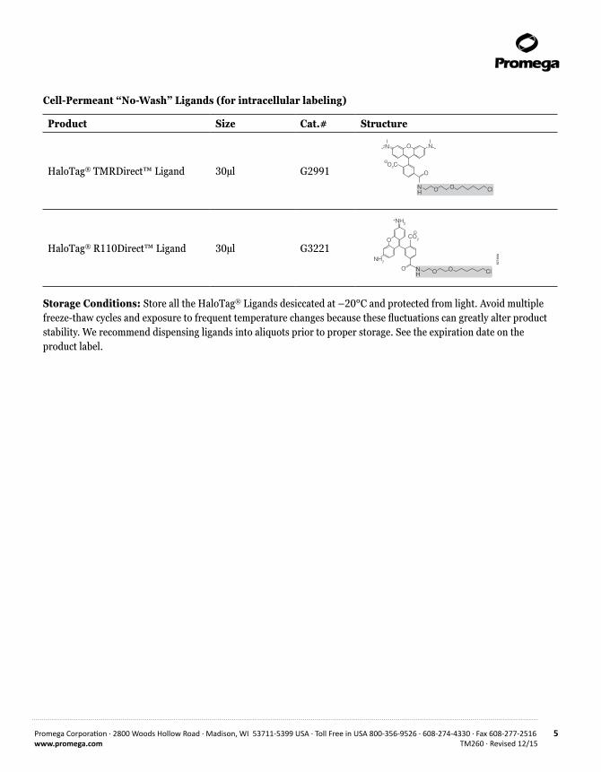
Cell-Permeant “No- Wash” Ligands (for intracellular labeling)
Product Size Cat.# Structure
HaloTag® TMRDirect™ Ligand 30µl G2991
6274
MA
Functional Reporter Reactive Linker
HaloTag® R110Direct™ Ligand502Ex/527EmC31H34CIN3O6MW = 580
HaloTag® diAcFAM Ligand494Ex/526Em (after hydrolysis)C35H36CINO10MW = 666
OO
O
OO
OOO
NH
OO Cl
HaloTag® Coumarin Ligand353Ex/434Em C22H31CIN2O5MW = 439 O O
NH
OO Cl
H2NO
HaloTag® TMRDirect™ Ligand555Ex/585EmC35H42CIN3O6MW = 636
NH
OO Cl
O
NO
O2C
+N
–
HaloTag® TMR Ligand555Ex/585EmC35H42CIN3O6MW = 636
NH
OO Cl
O
NO
O2C
+N
–
HaloTag® Alexa Fluor® 488 Ligand494Ex/517EmC47H72CIN5O12S2MW = 999
NH
OO Cl
O
CO2–
NH2+H2N
SO OO–
SO OO–
O
NH+2
HaloTag® Oregon Green® Ligand496Ex/516Em (after hydrolysis)C35H34CIF2NO10MW = 702
NH
OO Cl
O
F
O
OO
O
O
O
O F
NH
OO ClO
CO2
–
NH2
O
+NH2
Cell-Permeant, rapid labeling
Cell-Impermeant (for cell-surface labeling), rapid labeling
Cell-Permeant, “no-wash” labeling
HaloTag® R110Direct™ Ligand 30µl G3221
6274
MA
Functional Reporter Reactive Linker
HaloTag® R110Direct™ Ligand502Ex/527EmC31H34CIN3O6MW = 580
HaloTag® diAcFAM Ligand494Ex/526Em (after hydrolysis)C35H36CINO10MW = 666
OO
O
OO
OOO
NH
OO Cl
HaloTag® Coumarin Ligand353Ex/434Em C22H31CIN2O5MW = 439 O O
NH
OO Cl
H2NO
HaloTag® TMRDirect™ Ligand555Ex/585EmC35H42CIN3O6MW = 636
NH
OO Cl
O
NO
O2C
+N
–
HaloTag® TMR Ligand555Ex/585EmC35H42CIN3O6MW = 636
NH
OO Cl
O
NO
O2C
+N
–
HaloTag® Alexa Fluor® 488 Ligand494Ex/517EmC47H72CIN5O12S2MW = 999
NH
OO Cl
O
CO2–
NH2+H2N
SO OO–
SO OO–
O
NH+2
HaloTag® Oregon Green® Ligand496Ex/516Em (after hydrolysis)C35H34CIF2NO10MW = 702
NH
OO Cl
O
F
O
OO
O
O
O
O F
NH
OO ClO
CO2
–
NH2
O
+NH2
Cell-Permeant, rapid labeling
Cell-Impermeant (for cell-surface labeling), rapid labeling
Cell-Permeant, “no-wash” labeling
Storage Conditions: Store all the HaloTag® Ligands desiccated at –20°C and protected from light. Avoid multiple freeze-thaw cycles and exposure to frequent temperature changes because these fluctuations can greatly alter product stability. We recommend dispensing ligands into aliquots prior to proper storage. See the expiration date on the product label.

6 Promega Corporation · 2800 Woods Hollow Road · Madison, WI 53711-5399 USA · Toll Free in USA 800-356-9526 · 608-274-4330 · Fax 608-277-2516TM260 · Revised 12/15 www.promega.com
2. Product Components and Storage Conditions (continued)
5530
MB
Wavelength (nm)
Fluo
resc
ence
(RFU
)
300 400 500 600 700 8000
0.2 × 10⁶
0.4 × 10⁶
0.6 × 10⁶
0.8 × 10⁶
1.0 × 10⁶
1.2 × 10⁶
1.4 × 10⁶
LigandExcitation maximum
(dotted lines) Emission maximum
(solid lines) HaloTag® CoumarinHaloTag® Alexa Fluor® 488
HaloTag® Alexa Fluor® 660
HaloTag® Oregon Green®
HaloTag® diAcFAMHaloTag® R110 Direct™HaloTag® TMR Direct™
362nm 450nm499nm492nm492nm498nm552nm654nm
518nm520nm521nm528nm578nm690nm
Figure 2. Excitation and emission spectra and table for the HaloTag® Ligands. The wavelengths listed for the HaloTag® diAcFAM and Oregon Green® ligands were generated after hydrolysis of the diacetyl groups. Spectra for Alexa Fluor® 488 and Oregon Green® Ligands are identical and overlap each other. HaloTag® Alexa Fluor® 660 Ligand will have a reduced fluorescent emission at pH ≤ 4.0, concurrent with a shift in its absorbance peak at this low pH.

Promega Corporation · 2800 Woods Hollow Road · Madison, WI 53711-5399 USA · Toll Free in USA 800-356-9526 · 608-274-4330 · Fax 608-277-2516 7www.promega.com TM260 · Revised 12/15
2.B. Customizable Reactive Ligands
HaloTag® Ligand Building Blocks
Product Size Cat.# Structure
HaloTag® Succinimidyl Ester (O2) Ligand 5mg P1691
5476
MA
HN
OO
Cl +
N
HN
OO
O
O
Cl
O
O
ON
O
O
N
HN
O
O
R–NH2
OHN
O
O
+H
R–
HR–
HaloTag® ProteinHaloTag® Succinimidyl Ester (O4) Ligand 5mg P6751
6083
MA
HN
OO
OO +
HaloTag® Protein
O
O
ON
O
O
NH
HN
O
O
Cl
R-NH2
HN
O
O
OO
O
O
OHN
O
O
Cl+
NH
R
R
O
O
HaloTag® Amine (O2) Ligand 5mg P6711
5479
MA
O
-Cl+H3N OCl +
HN
OO
ClR
O
+ DIPEA
HN
O
RHaloTag® Protein
N
O
O
OR
O
HaloTag® Amine (O4) Ligand 5mg P6741
6081
MA
-Cl+H3N
OO
DIPEACl
HN
Cl
R
R
O
O
O
O
O
HaloTag® Protein
RHN
N
O
+ +O
O
OO
OO
OO
HaloTag® Iodoacetamide (O2) Ligand 5mg P1681
5477
MA
HN
OO Cl
O
I + R -SH
HN
OS
RHaloTag® Protein
HN
OO
ClO
SR
HaloTag® Iodoacetamide (O4) Ligand 5mg P6771
6082
MA
HN
O
O
O
O
O
I + R-SH
HN
O
S
R
Cl
HN
O
O
O
O
O
S ClR
O
O
HaloTag® Protein
HaloTag® Thiol (O4) Ligand 5mg P6761
6084
MA
HS
O
O
O
O+
SR
Cl
S
O
O
O
O
Cl
O
O
HaloTag® ProteinR
R-I
Storage Conditions: Store Cat.# P1691 and P6751 at or below –70°C under inert atmosphere. Store Cat.# P6711 and P6741 at or below –20°C in an air-tight container in the absence of light. Store Cat.# P1681, P6771 and P6761 at or below –20°C under inert atmosphere in the absence of light.
HaloTag® Antibodies
P R O D U C T S I Z E C O N C . C AT. #
Anti-HaloTag® pAb(a) 200µg 1mg/ml G9281
Anti-HaloTag® Monoclonal Antibody(a) (for Westerns) 200µg 1mg/ml G9211
Note: A complete listing of HaloTag® Purification and Pull-Down Systems and Reagents, including biotin ligands, is provided in Section 10, Related Products.

8 Promega Corporation · 2800 Woods Hollow Road · Madison, WI 53711-5399 USA · Toll Free in USA 800-356-9526 · 608-274-4330 · Fax 608-277-2516TM260 · Revised 12/15 www.promega.com
3. Generating a HaloTag® Fusion Protein
3.A. Beginning with an Expression-Validated HaloTag® ORF Clone
Promega offers expression-validated HaloTag® ORF clones in partnership with Kazusa DNA Research Institute. Cloning and validating the protein-coding region of a gene into a vector requires a significant amount of time, and often, there is a high failure rate prior to validating a clone. To circumvent this time sink for our customers, Promega, in partnership with Kazusa, has produced premade, bench-validated ORF clones. This content is compatible with and optimized for HaloTag® protein analysis and TnT® T7 cell-free expression technologies. In addition, these validated ORF clones can be easily transferred into alternate vectors using the Flexi® Cloning System.
Find your protein precloned into a HaloTag® Vector and validated at: www.promega.com/FindMyGene
3.B. Creating Your Own HaloTag® Fusion Protein
Conventional Cloning into a Multiple Cloning Region
The pHTN HaloTag® CMV-neo Vector (Cat.# G7721, for N-terminal HaloTag® fusions) and the pHTC HaloTag® CMV-neo Vector (Cat.# G7711, for C-terminal HaloTag® fusions) are non-Flexi HaloTag® vectors with a common multiple cloning region designed to express an N-terminally or C-terminally tagged HaloTag® fusion protein in mammalian cells from a CMV promoter. In addition these vectors contain a neomycin (G418) selection cassette that allows antibiotic selection of stable cell lines expressing the fusion protein. The multiple cloning region allows the user to transfer the HaloTag® protein plus linker sequence into any vector of choice.
Flexi® Cloning Method
The Flexi® Vector System is a directional cloning method for protein-coding sequence. It is based on rare-cutting restriction enzymes, SgfI and PmeI, and provides a rapid, and high-fidelity way to transfer protein-coding regions without the need to resequence. All Flexi® Vectors carry the lethal barnase gene, which is replaced by the DNA fragment of interest, providing positive selection for the successful ligation of the insert. We recommend cloning into an N-terminal vector (first as pFN28A HaloTag® CMV-neo Flexi® Vector, Cat.# G8441) with later transfer into a C-terminal vector (such as pFC27K HaloTag® CMV-neo Flexi® Vector, Cat.# G8431) if needed.

Promega Corporation · 2800 Woods Hollow Road · Madison, WI 53711-5399 USA · Toll Free in USA 800-356-9526 · 608-274-4330 · Fax 608-277-2516 9www.promega.com TM260 · Revised 12/15
4. Mammalian Live-Cell Labeling, Cell Fixation, ICC, SDS-PAGE, Flow Cytometry and Imaging
4.A. Rapid Labeling (15–60 minutes)
This protocol is intended for labeling live cells with HaloTag® TMR, diAcFAM, Oregon Green®, Coumarin, Alexa Fluor® 488 or Alexa Fluor® 660 Ligands.
Representative data are shown in Figures 3 and 4.
Materials to Be Supplied by the User(Solution compositions are provided in Section 9.B.)• chambered cover glass with cells expressing HaloTag® fusion protein• complete culture medium appropriate for your cells at 37°C • culture medium, lacking phenol red at 37°C (optional)• 1X PBS (pH 7.5, optional for washes)• confocal microscope or wide-field fluorescent microscope equipped with appropriate filter sets and/or lasers
(see Section 9.A and Figure 2)• 37°C + CO2 cell culture incubator
1. Prepare a 1:200 dilution of HaloTag® Ligand in warm culture medium just prior to addition to cells. This is a 5X working stock solution.
2. Label cells by replacing one-fifth of the existing volume of medium with the 5X HaloTag® Ligand working stock solution, and mix gently.
This results in the recommended final labeling concentrations of 5µM TMR; 3.5µM Alexa Fluor® 660; 1µM diAcFAM, Oregon Green® or Alexa Fluor® 488; and 10µM Coumarin Ligand.
3. Incubate for 15 minutes in a 37°C + CO2 cell culture incubator.
4. For cell-permeant ligands, gently replace the ligand-containing medium with an equal (or greater) volume of warm fresh medium (or 1X PBS [pH 7.5]). Repeat this two times, ending with warm complete medium, for a total of three complete rinses, and proceed to Step 5.
For cell-impermeant ligands (Alexa Fluor®-containing ligands) replace the ligand-containing medium with an equal (or greater) volume of warm fresh medium twice, and proceed to Step 7. Because they are cell-impermeant, these ligands do not require washing out of unbound ligand.
5. Incubate cells in complete culture medium at 37°C + CO2 in a cell culture incubator for 30 minutes to wash unbound ligand.
6. Replace the medium with an equal volume of fresh warm culture medium. Use of medium lacking phenol red may improve imaging.
7. Transfer to a microscope, and capture images.

10 Promega Corporation · 2800 Woods Hollow Road · Madison, WI 53711-5399 USA · Toll Free in USA 800-356-9526 · 608-274-4330 · Fax 608-277-2516TM260 · Revised 12/15 www.promega.com
4.A. Rapid Labeling (15–60 minutes) (continued)
6300
TA
A.
B.
C.
D.
Live Live Control Fixed
HaloTag® TMR Ligand
HaloTag® diAcFAM Ligand
HaloTag® Coumarin Ligand
HaloTag® Oregon Green® Ligand
Figure 3. HaloTag® ligand labeling of live cells followed by fixation. HEK293 cells stably transfected with the HaloTag® gene fused to three copies of a nuclear localization sequence and nontransfected HEK293 controls labeled with HaloTag® TMR (Panel A), HaloTag® diAcFAM (Panel B), HaloTag® Coumarin (Panel C) or HaloTag® Oregon Green® Ligand (Panel D) as described in Section 4.A. In each panel showing stably transfected cells, labeling is restricted to the nucleus. Nontransfected (live control in figure) cells show no labeling. For HaloTag® TMR, diAcFAM and Oregon Green® Ligand labeling, images were collected on the Olympus FV500 confocal microscope with appropriate filter sets. For HaloTag® Coumarin Ligand labeling images were collected by epifluorescent illumination using DAPI filter cube and a CCD camera (Hammamatsu Orca). The top row of each panel is fluorescent signal only. The bottom row is differential interference contrast (DIC) with fluorescence overlay.

Promega Corporation · 2800 Woods Hollow Road · Madison, WI 53711-5399 USA · Toll Free in USA 800-356-9526 · 608-274-4330 · Fax 608-277-2516 11www.promega.com TM260 · Revised 12/15
6225
TB
A. B.
C. D.
Figure 4. Live-cell labeling of cell surface HaloTag® fusion proteins. Panels A and B show HEK293 cells stably expressing HaloTag®-ECS (ExtraCellular Surface; comprised of a signal sequence and single transmembrane domain of b1-integrin) fusion protein labeled with HaloTag® Alexa Fluor® 488 Ligand and then imaged. Panels C and D show U2OS cells expressing HaloTag®-EDG1 (GPCR receptor) fusion labeled with Alexa Fluor® 660 Ligand and then imaged. All labeling was done as described in Section 4.A. Confocal images show fluorescence (Panels A and C) is restricted to the cell surface, also seen clearly in the panels with the DIC/fluorescence overlay (Panel B and D). Images were generated on an Olympus FV500 confocal microscope using the appropriate filter sets.

12 Promega Corporation · 2800 Woods Hollow Road · Madison, WI 53711-5399 USA · Toll Free in USA 800-356-9526 · 608-274-4330 · Fax 608-277-2516TM260 · Revised 12/15 www.promega.com
4.B. No-Wash Labeling (overnight)
This protocol is intended for labeling of live cells with the cell-permeant HaloTag® TMRDirect™ or R110Direct™ Ligand. This protocol can be used to label cells that are adherent or nonadherent, and adherent cells can already be plated or still be in suspension at time of labeling. Representative data are shown in Figure 5.
Note: We do not recommend using the HaloTag® TMR Ligand (Cat.# G8251, G8252) for this protocol.
Materials to Be Supplied by the User(Solution compositions are provided in Section 9.B.)• cells expressing HaloTag® fusion protein (in suspension or plated)• chambered cover glass or other cell culture device• complete culture medium appropriate for your cells at 37°C• culture medium lacking phenol red (optional) at 37°C• confocal microscope or wide-field fluorescent microscope equipped with appropriate filter sets and/or lasers
(Section 9.A and Figure 2)• 37 °C + CO2 cell culture incubator
1. Prepare a 1:200 dilution of HaloTag® TMRDirect™ or R110Direct™ Ligand in warm culture medium just prior to addition to cells. This is a 5X working stock solution.
2. For adherent cells: Replace one-fifth of the existing volume of medium with the 5X HaloTag® ligand working stock solution, and mix gently.
For cell suspensions: Add 5X ligand working stock to existing cell suspension, resulting in a 1X final concen-tration.
Step 2 results in the recommended final labeling concentration of 100nM HaloTag® TMRDirect™ or R110Direct™ Ligand.
3. Plate cells (if necessary), and incubate overnight in a 37°C + CO2 cell culture incubator.
4. Gently replace the ligand-containing medium with an equal (or greater) volume of warm fresh medium, or fix cells (for end-point assays; see Section 4.D).
5. Transfer to an imaging device, and capture images.

Promega Corporation · 2800 Woods Hollow Road · Madison, WI 53711-5399 USA · Toll Free in USA 800-356-9526 · 608-274-4330 · Fax 608-277-2516 13www.promega.com TM260 · Revised 12/15
75
30
TA
Figure 5. No-wash, live-cell labeling results in high signal-to-noise ratio and specificity. U2OS cells stably expressing HaloTag® protein fused to three copies of a nuclear localization sequence were labeled with the HaloTag® R110Direct™ Ligand as described in Section 4.B. HaloTag® labeling shows a high signal-to-noise ratio and specificity: the label is restricted to the nucleus of all cells. Panel A shows fluorescence image of cells labeled with R110Direct™ Ligand; Panel B is an overlay of the fluorescent and DIC image. Images were generated on an Olympus FV500 confocal microscope using the appropriate filter sets.
4.C. Pulse-Chase Labeling
This protocol is intended to serve as a guide for pulse-chase labeling cells expressing HaloTag® fusions in order to directly observe protein turnover or protein trafficking. This protocol is intended for labeling live cells with any of the HaloTag® Ligands. Representative data are shown in Figure 6.
Materials to Be Supplied by the User(Solution compositions are provided in Section 9.B.)• chambered cover glass with cells expressing HaloTag® fusion protein • complete culture medium appropriate for your cells at 37°C• culture medium lacking phenol red at 37°C (optional)• 1X PBS (pH 7.5, optional for washes)• confocal microscope or wide-field fluorescent microscope equipped with appropriate filter sets and lasers
(Section 9.A and Figure 2)• 37°C + CO2 cell culture incubator

14 Promega Corporation · 2800 Woods Hollow Road · Madison, WI 53711-5399 USA · Toll Free in USA 800-356-9526 · 608-274-4330 · Fax 608-277-2516TM260 · Revised 12/15 www.promega.com
4.C. Pulse-Chase Labeling (continued)
1. Prepare a 1:200 dilution of pulse labeling ligand in warm culture medium just prior to addition to cells. This is a 5X working stock solution.
2. Label cells by replacing one-fifth of the existing volume of medium with the 5X HaloTag® ligand working stock solution, and mix gently. This results in the recommended final labeling concentrations of 5µM TMR, 3.5µM Alexa Fluor® 660; 1µM diAcFAM, Oregon Green® or Alexa Fluor® 488; 10μM Coumarin; and 100nM TMRDirect™ or R110Direct™ Ligands.
Note: If protein fusion is membrane-bound with HaloTag® protein on the surface, pulse labeling should be done with an impermeable ligand (Alexa Fluor® 488 or Alexa Fluor® 660 Ligand) to label the surface (plasma membrane) pool first.
3. For Rapid-Labeling ligands (TMR, Oregon Green®, diAcFAM, Coumarin and Alexa Fluor® Ligands), incubate for 15 minutes in a 37°C + CO2 cell culture incubator.
For No-Wash-Labeling ligands (TMRDirect™ and R110Direct™ Ligands), incubate overnight in a 37°C + CO2 cell culture incubator.
4. If chase labeling is to take place immediately following the pulse, then proceed immediately to Step 5.
If chase labeling is not to take place immediately, simply replace the ligand-containing medium with an equal volume of warm fresh medium and either allow time to pass prior to chase and/or execute intended biology as per your specific needs prior to Step 5.
5. Prepare a 1:1,000 dilution of chase ligand in warm culture medium just prior to addition to cells, and gently replace the medium currently on your cells with an equal volume of this 1X chase labeling ligand.
6. For Rapid-Labeling ligands (TMR, Oregon Green®, diAcFAM, Coumarin and Alexa Fluor® Ligands), incubate for 15 minutes in a 37°C + CO2 cell culture incubator.
For No-Wash-Labeling ligands (TMRDirect™ and R110Direct™ Ligands), incubate overnight in a 37°C + CO2 cell culture incubator.
7. For the cell-permeant Rapid Labeling ligands (TMR, Oregon Green®, diAcFAM and Coumarin Ligands) gently replace the ligand-containing medium with an equal (or greater) volume of warm fresh medium (or 1X PBS [pH 7.5]). Repeat this two times, ending with warm complete medium, for a total of three complete rinses and proceed to Step 8.
For the cell-impermeant Rapid Labeling ligands (Alexa Flour® 488 or 660 Ligand), gently replace the ligand-containing medium with an equal (or greater) volume of warm fresh medium twice and proceed to Step 10.
For No-Wash-Labeling ligands (TMRDirect™ and R110Direct™ ), gently replace the ligand-containing medium with an equal (or greater) volume of warm fresh medium, and proceed to Step 10.
8. Incubate cells in complete culture medium at 37°C + CO2 in a cell culture incubator for 30 minutes to wash out unbound ligand.
9. Replace the medium with an equal volume of fresh warm culture medium (use of medium lacking phenol red may improve imaging).
10. Transfer to a microscope, and capture images.

Promega Corporation · 2800 Woods Hollow Road · Madison, WI 53711-5399 USA · Toll Free in USA 800-356-9526 · 608-274-4330 · Fax 608-277-2516 15www.promega.com TM260 · Revised 12/15
1051
8TA
A.
C. D.
B.
Figure 6. Pulse-chase labeling of cells expressing an intracellular HaloTag® protein using No-Wash labeling results in direct observation protein turnover. U2OS cells transiently expressing HaloTag® protein alone were labeled with HaloTag® TMRDirect™ Ligand for 20 hours followed by HaloTag® R110Direct™ Ligand for 20 hours and then imaged by confocal microscopy. The HaloTag® protein is small enough to be seen both in the cytoplasm and the nucleus. TMRDirect™ fluorescence is seen labeling the protein pool made prior to and during the first 20 hours of expression by the cells (Panel A), while R110Direct™ fluorescence is seen as labeling the protein pool made in the last 20 hours of expression by cells (Panel B). Panel C is an overlay of the two fluorescent channels and the DIC. Panel D is an overlay of the two fluorescent channels. Scale bar is 20µm.

16 Promega Corporation · 2800 Woods Hollow Road · Madison, WI 53711-5399 USA · Toll Free in USA 800-356-9526 · 608-274-4330 · Fax 608-277-2516TM260 · Revised 12/15 www.promega.com
4.D. Fixing Cells after Labeling
This protocol is intended to serve as a guide for fixation of cells expressing a HaloTag® fusion protein. The covalent bond that forms between the ligand and HaloTag® protein during live-cell labeling and the stability of the dyes allows you to subsequently fix, permeabilize and wash the cells using standard conditions without significant loss of the specific fluorescent signal. We recommend the use of paraformaldehyde (PFA) as a fixative because it crosslinks proteins in cells and at the membrane and has the added benefit of reducing cell loss from the growth surface.
Fixed cells can be treated with detergents, such as Triton® X-100, to permeabilize cells for downstream immunocyto-chemical applications. The conditions here are sufficient to permeabilize the plasma membrane. Alternative or additional detergents might be necessary to permeabilize other structures. Representative data are shown in Figures 3, 7 and 8.
Materials to Be Supplied by the User(Solution compositions are provided in Section 9.B.)
All solutions should be at room temperature unless otherwise specified.• 4% paraformaldehyde/0.2M sucrose/1X PBS (pH 7.5), warmed to 37°C• 1X PBS buffer (pH 7.5)• 1X PBS + 0.1% Triton® X-100• confocal microscope or wide-field fluorescent microscope equipped with appropriate filter sets and lasers
(Section 9.A and Figure 2).
1. Follow Steps 1–3 of Section 4.A (Rapid Labeling) or 4.B (No-Wash Labeling), or Steps 1–6 of Section 4.C (Pulse-Chase Labeling) to label cells with a HaloTag® ligand, if desired.
2. Replace the medium with an equal volume of warm 4% paraformaldehyde/0.2M sucrose/1X PBS (pH 7.5), and incubate for 10 minutes at room temperature.
3. Replace fixative with an equal volume of 1X PBS + 0.1% Triton® X-100, and incubate for 10 minutes at room temperature.
4. Replace the detergent-containing solution with an equal volume of 1X PBS, and then either transfer to a microscope and capture images, store at 4°C, or proceed to Section 4.E for immunocytochemistry protocol.

Promega Corporation · 2800 Woods Hollow Road · Madison, WI 53711-5399 USA · Toll Free in USA 800-356-9526 · 608-274-4330 · Fax 608-277-2516 17www.promega.com TM260 · Revised 12/15
4.E. Multiplexing with HaloTag® Using Immunocytochemistry (ICC)
This protocol is intended to serve as a guide for multiplexing with HaloTag® technology in fixed cells by ICC using any antibody of interest. The particular antibody used in this protocol is a purified rabbit polyclonal antibody raised against the HaloTag® protein (9). The antibody was purified using Protein G affinity resin and has been shown to detect HaloTag® fusion proteins in both immunocytochemistry and Western blot applications with high sensitivity and specificity. Further, this antibody labels HaloTag® fusion proteins independently of HaloTag® ligands. Because these labels do not interfere with one another, it is possible to colabel HaloTag® fusion proteins with a fluorescent ligand and the Anti-HaloTag® pAb in conjunction with an anti-rabbit secondary antibody bearing a spectrally distinct fluorescent tag. Representative data are shown in Figures 7 and 8.
Materials to Be Supplied by the User(Solution compositions are provided in Section 9.B.)
All solutions should be at room temperature. • cells fixed following the steps in Section 4.D• fluorophore-conjugated anti-rabbit secondary antibody (Ab) of choice• PBS + 2% serum from same host as secondary Ab + 0.01% Triton® X-100• PBS + 1% serum from same host as secondary Ab• PBS + 0.1% sodium azide (optional, for storage)• confocal microscope or wide-field fluorescent microscope equipped with appropriate filter sets and lasers
(Section 9.A and Figure 2)
1. Replace the 1X PBS with an equal volume of PBS + 2% serum from same host as secondary Ab + 0.01% Triton® X-100, and block for 1 hour at room temperature.
2. Dilute the Anti-HaloTag® pAb 1:500 in PBS + 1% serum from same host as secondary Ab to a final labeling concentration of 2μg/ml.
3. Replace the blocking solution with the antibody solution, and incubate for 1 hour at room temperature.
4. Wash cells twice with PBS + 1% serum from same host as secondary Ab for 10 minutes at room temperature.
5. Dilute the secondary antibody according to the manufacturer's recommendations in PBS + 1% s serum from same host as secondary Ab.
6. Replace the wash solution with the secondary antibody solution, and incubate for 30 minutes at room temperature.
7. Wash cells twice with PBS + 1% serum from same host as secondary Ab for 10 minutes each wash at room temperature.
8. Replace wash solution with 1X PBS.
9. Transfer to a microscope, and capture images. Store cells at 4°C in 1X PBS or PBS + 0.1% sodium azide.

18 Promega Corporation · 2800 Woods Hollow Road · Madison, WI 53711-5399 USA · Toll Free in USA 800-356-9526 · 608-274-4330 · Fax 608-277-2516TM260 · Revised 12/15 www.promega.com
4.E. Multiplexing with HaloTag® Using Immunocytochemistry (ICC) (continued)
4883
TA
A. B.
C. D.
Figure 7. Multiplex HaloTag® labeling using TMR ligand with an antibody of interest. HeLa cells transiently transfected with p65-HaloTag® fusion (3) construct (p65 is a cytoplasmic signaling protein) were labeled with HaloTag® TMR Ligand as described in Section 4.A. Cells were then fixed as described in Section 4.D, stained with a mouse Anti-bIII Tubulin mAb followed by Alexa Fluor® 488-conjugated goat anti-mouse IgG (Molecular Probes) as described in section 4.E. TMR fluorescence is seen in the cytoplasm (Panel A), and Alexa Fluor® 488 fluorescence shows microtubules (Panel B). Panel C shows an overlay of both fluorescence channels with the DIC image, while Panel D shows an overlay of just the fluorescence channels. Images were generated on an Olympus FV500 confocal microscope in sequential mode using appropriate filter sets.

Promega Corporation · 2800 Woods Hollow Road · Madison, WI 53711-5399 USA · Toll Free in USA 800-356-9526 · 608-274-4330 · Fax 608-277-2516 19www.promega.com TM260 · Revised 12/15
7528
TA
Figure 8. Distinct labeling of a single HaloTag® fusion by a HaloTag® Ligand and Anti-HaloTag® pAb. HEK293 cells stably transfected with a p65-HaloTag® construct (p65 is primarily cytoplasmic in cells at rest) were labeled with HaloTag® TMR Ligand as described in Section 4.A, fixed as described in Section 4.D, then processed for immunocytochemistry using the Anti-HaloTag® pAb as described in Section 4.E. The secondary antibody used was Alexa Fluor® 488-conjugated anti-rabbit-IgG (Jackson ImmunoResearch). Distinct labeling is demonstrated by the fact that the red fluorescence associated with specific HaloTag® TMR Ligand labeling (Panel A) does not prevent green fluorescence associated with specific Anti-HaloTag® pAb labeling (Panel B). Merging the fluorescent images (Panel C) confirms that the HaloTag® fusion protein is labeled specifically (primarily cytoplasmic) as expected. Images were collected on the Olympus FV500 confocal microscope in sequential mode using appropriate filter sets.

20 Promega Corporation · 2800 Woods Hollow Road · Madison, WI 53711-5399 USA · Toll Free in USA 800-356-9526 · 608-274-4330 · Fax 608-277-2516TM260 · Revised 12/15 www.promega.com
4.F. Quantitation of HaloTag® Protein Fusions, SDS-PAGE Analysis
The following protocol is intended to serve as a guide for SDS-PAGE-based HaloTag® applications. Because the covalent bond between the HaloTag® protein and HaloTag® ligand withstands denaturation and the fluorescent dyes in the ligands are stable, you can directly quantify labeled fusion protein following SDS-PAGE using a fluorescent scanner (i.e., cell-to-gel analysis).
Such gels also can be subsequently used for Western blot analysis using the Anti-HaloTag® mAb at a final labeling concentration of 1µg/ml (1:1,000 dilution; see Section 5 below for protocol).
Note: Although all of the HaloTag® ligands perform in this function, the HaloTag® TMR, TMRDirect™ or R110Direct™ Ligands are recommended for direct SDS-PAGE fluorescence scanning of an intracellularly expressed protein fusion. Either of the AlexaFluor®-containing ligands is recommended for extracellularly expressed proteins that place HaloTag® on the surface of cells. Representative data are shown in Figure 9.
Materials to be Supplied by the User(Solution compositions are provided in Section 9.B.)• cells expressing HaloTag® fusion protein• 1X PBS (pH 7.5)• 1X SDS sample buffer• heat block or water bath at 95°C• equipment and running buffer necessary for SDS-PAGE• fluorescent scanner
1. To label cells, follow one of the following protocols: Steps 1–3 of Section 4.A or 4.B (Rapid or No Wash Labeling) Steps 1-6 of Section 4.C (Pulse-Chase Labeling).
Note: There is no need to go through the 30-minute wash step for an intracellularly expressed protein fusion labeled by Rapid Labeling protocol as all unbound ligand is very small and will migrate on the gel much faster than the labeled fusion protein (with sample dye front).
2. Replace ligand containing media with 1X PBS (pH 7.5) in order to avoid a prominent band on the gel stemming from complete media.
3. Lyse cells by replacing the 1X PBS from the wells with approximately 100–150μl of 1X SDS sample buffer per cm2 of cell growth area.
4. Collect cell lysate, and incubate for 5 minutes at 95°C.
5. Perform SDS-PAGE by loading approximately 10μl (5–10μg total protein) of each sample per well of the gel, or store samples at –20°C for later use.

Promega Corporation · 2800 Woods Hollow Road · Madison, WI 53711-5399 USA · Toll Free in USA 800-356-9526 · 608-274-4330 · Fax 608-277-2516 21www.promega.com TM260 · Revised 12/15
6. Analyze the gel on a fluorescence scanner.
Note: The dye front might contain fluorescent material that can complicate detection (unbound ligand and/or tracking dyes used in sample buffers). To eliminate these sources of nonspecific fluorescence, simply run the gel until the dye front migrates off of the gel or cut the dye front off of the bottom of the gel before scanning.
7. After scanning, the proteins can be transferred to nitrocellulose for Western blot analysis if used promptly (i.e., gel is not fixed, remains moist and is not left in buffer or deionized water).
47
81
TA
121110987654321
– ~33kDa
Figure 9. SDS-PAGE analysis shows fast, efficient and highly specific labeling of the HaloTag® protein in live cells. CHO-K1 untransfected control cells (lanes 1–6) or cells transiently transfected with a vector containing HaloTag® alone (lanes 7–12) were labeled with 5μM HaloTag® TMR Ligand for different periods of time at 37°C (0.5, 1, 2, 5, 15 or 30 minutes) and treated as described in Section 4. Following SDS-PAGE, the gel was analyzed on a Hitachi FMBIO® fluorescence imaging system.

22 Promega Corporation · 2800 Woods Hollow Road · Madison, WI 53711-5399 USA · Toll Free in USA 800-356-9526 · 608-274-4330 · Fax 608-277-2516TM260 · Revised 12/15 www.promega.com
4.G. Flow Cytometry Analysis
Representative data are shown in Figure 10.
Materials to Be Supplied by the User(Solution compositions are provided in Section 9.B.)• large-well format plate of HaloTag®-expressing cells and negative control cells that do not express the HaloTag®
fusion protein• complete culture medium appropriate for your cells at 37°C• for adherent cells: 1X PBS (pH 7.5) and 1X PBS containing 3mM EDTA (nonenzymatic) or trypsin solution
(see Step 2 below)• flow cytometer with appropriate filter sets and lasers for ligand used (Section 9.A and Figure 2)• 37°C + CO2 cell culture incubator
1. Label cells expressing HaloTag® protein with ligand of choice (Section 4.A, Rapid Labeling or 4.B, No-Wash Labeling).
Recommended controls:
a. Label cells that are not expressing HaloTag® protein to assess background with chosen ligand.
b. Prepare unlabeled cells expressing HaloTag® protein to assess background from endogenous cell fluorescence or morphological changes due to expression.
2. For adherent cells with HaloTag® fusion protein expressed on the cell surface: Rinse cells twice with an equal volume of 1X PBS, incubate in 1X PBS with 3mM EDTA or use other nonenzymatic solution at 37°C for 5–15 minutes (depending on cell line) in order to gently get the cells off of the plate.
For adherent cells with HaloTag® fusion protein expressed inside the cell: Suspend cells using trypsin-containing solution in a manner appropriate for the cell line.
3. Collect, pellet, count and resuspend cells in medium at 37°C to a concentration of 0.5 to 1 × 10⁶ cells/ml.
4. Analyze cells by flow cytometry.

Promega Corporation · 2800 Woods Hollow Road · Madison, WI 53711-5399 USA · Toll Free in USA 800-356-9526 · 608-274-4330 · Fax 608-277-2516 23www.promega.com TM260 · Revised 12/15
7527
TA
Figure 10. Successful flow cytometry analysis of labeled HaloTag® protein-expressing cells. U2OS cells stably expressing HaloTag® protein fused to three copies of a nuclear localization sequence were labeled with the HaloTag® R110Direct™ Ligand as described in Section 4.B, and known numbers (1%, 10% or 100% labeled) were gated from unlabeled cells. Flow cytometry of HaloTag® protein expressing cells was performed using a MoFlo® instrument with approximately 20,000 cells represented on each graph. R1 indicates cells labeled with R110, R2 are dead cells (by propidium iodide), and R3 indicates unlabeled cells. Channel overlap between propidium iodide and R110Direct™ fluorescence was not compensated for in analysis. Results show that labeled cells are successfully gated from unlabeled cells.

24 Promega Corporation · 2800 Woods Hollow Road · Madison, WI 53711-5399 USA · Toll Free in USA 800-356-9526 · 608-274-4330 · Fax 608-277-2516TM260 · Revised 12/15 www.promega.com
5. Protein Detection Using HaloTag® Monoclonal Antibody (for Western blotting)
The following protocol is meant to serve as a general guide for the use of Anti-HaloTag® Monoclonal Antibody (Cat.# G9211) in Western blotting. The Anti-HaloTag® pAb (Cat.# G9281) is recommended for all other immuno-logical detection methods. Representative data are shown in Figure 11.
Materials to Be Supplied by the User• mammalian cells expressing HaloTag® fusion• appropriate mammalian lysis buffer (e.g., Mammalian Lysis Buffer, Cat.# G9381)• SDS-PAGE gel and gel running apparatus• nitrocellulose or PVDF membrane and Western blotting apparatus• TBST (1X TBS with 0.1% Tween®)• 5% BSA in TBST
• anti-mouse IgG secondary antibody appropriately conjugated for Western blotting analysis and corresponding detection substrate (e.g., Anti-Mouse IgG (H+L), AP Conjugate Cat.# S3721) and Western Blue® Stabilized Substrate for Alkaline Phosphatase, Cat.# S3841)
• rotating platform for gentle agitation
1. Lyse mammalian cells expressing HaloTag® fusion using an appropriate lysis buffer of choice.
Recommended control: Cells that are not expressing HaloTag® (untransfected).
2. Run lysates on SDS-PAGE, and transfer to nitrocellulose or PDVF membrane as per manufacturer protocol.
3. Place resulting nitrocellulose or PVDF membranes in TBST containing 5% BSA to block for an hour at room temperature or overnight at 4°C, with gentle agitation.
4. Replace blocking buffer with a solution of anti-HaloTag® mAb at 1:1,000 dilution in TBST, and leave at room temperature for 1 hour with gentle agitation.
5. Replace primary antibody-containing solution with TBST. Wash 3 times for 15 minutes at room temperature with gentle agitation.
6. Replace wash with anti-mouse IgG conjugated secondary antibody at manufacturer recommended concentration (e.g., for anti-mouse IgG-AP conjugated use at 1:7,500 dilution) in TBST and leave for 30 minutes at room temperature with gentle agitation.
7. Replace secondary solution with TBST. Wash 3 times for 15 minutes at room temperature with gentle agitation.
8. Detect HaloTag® protein bands using appropriate substrate (e.g., AP substrate).

Promega Corporation · 2800 Woods Hollow Road · Madison, WI 53711-5399 USA · Toll Free in USA 800-356-9526 · 608-274-4330 · Fax 608-277-2516 25www.promega.com TM260 · Revised 12/15
1051
7TA
M 1 2
220
12010080
60
50
kDa
EZH2-HaloTag®
fusion protein
Figure 11. Western blot detection of EZH2-HaloTag® (HT) fusion protein (~120kDa) using Anti-Halo-Tag® Monoclonal Antibody (mAb). Blot shows specific and robust detection of the HaloTag® fusion from HEK293T cell lysate. Western blotting using AP-based secondary antibody and substrate was used as described in protocol of Section 5. Lane 2 is the untransfected control.
6. How to Generate HaloTag® Ligands from Building Blocks
The following protocols are meant to serve as a guide for the generation of HaloTag® ligands using HaloTag® Ligand Building Blocks. Specific example of generating a fluorophore-containing ligand is described in Figure 12.
6.A. General Protocol for Reporter Group Labeling (conjugation to 2-[(6-chloro-hexyloxy)-ethoxy]-ethylamine)
Materials to Be Supplied by the User• succinimidyl ester reporter group• base (triethylamine, diisopropylethylamine, etc.)• DMF (dimethylformamide)• instrument and reagents for HPLC or silica gel chromatography
1. Add 1.5 to 3 equivalence of (02) or (04) amine to one equivalent of the succinimidyl ester of the reporter group in DMF and follow by base (triethylamine, diisopropylethylamine, etc.).
2 Stir reaction for 8–16 hours at room temperature.
3. Purify by preparative scale HPLC or silica gel chromatography, depending on lipophylicity of your construct.

26 Promega Corporation · 2800 Woods Hollow Road · Madison, WI 53711-5399 USA · Toll Free in USA 800-356-9526 · 608-274-4330 · Fax 608-277-2516TM260 · Revised 12/15 www.promega.com
6.B. Protein Labeling with HaloTag® Amine Ligand
Materials to Be Supplied by the User• protein of interest for conjugation• water, 0.1M MES (pH 4.7–6.0) or 0.1 M sodium phosphate (pH 7.3)• DMSO or DMF• EDC (1-ethyl-3-(3-dimethylaminopropyl) carbodiimide hydrochloride)• Sephadex® G-25 (for gel filtration separation of ligated protein)
1. Dissolve the protein to be modified at a concentration of 10mg/ml in either water, 0.1M MES (pH 4.7–6.0), or 0.1M sodium phosphate (pH 7.3).
2. Prepare HaloTag® Amine (02 or 04) Ligand stock solution at 5mg/ml in DMSO or DMF.
3. Take a 10-fold-molar-excess-to-protein aliquot of this HaloTag® ligand stock solution and dilute it in buffer (same as what is used in Step 1).
4. Add this aqueous HaloTag® ligand stock solution to the protein solution; the final protein concentration will be 5 mg/ml or greater.
5. Add EDC (1-ethyl-3-(3-dimethylaminopropyl) carbodiimide hydrochloride) in ten-fold molar excess to protein as a 0.1 to 0.5M aqueous solution and react for 2 hours at room temperature.
6. Purify the ligated protein by gel filtration separation using Sephadex® G-25 in the buffer of choice.
6.C. Antibody Labeling with HaloTag® Succinimidyl Ester Ligand
Materials to Be Supplied by the User• antibody of interest for conjugation• 0.1M sodium phosphate, 0.15M sodium chloride (pH 7.2) [pH 7.0 to 7.6 can be used]• DMSO or DMF• Sephadex® G-25 (for gel filtration separation of ligated antibody) or means of dialysis against 0.1M sodium
phosphate, 0.15M sodium chloride, 10mM EDTA (pH 7.2)
1. Dissolve antibody in 0.1M sodium phosphate, 0.15M sodium chloride (pH 7.2) at 1–5 mg/ml.
2. Prepare HaloTag® Succinimidyl Ester (02 or 04) Ligand stock solution in 5mg/ml DMSO or DMF.
3. Add 10–40µl of HaloTag® Ligand stock solution per millilter of 1mg/ml antibody solution and react at room temperature for 30 minutes.
Note: A 12-fold molar excess works well to ensure at least one label per antibody. Most protocols for general antibody labeling uses 20-fold excess, but higher concentrations of ligand to antibody will label more amines. Further, excess labels may or may not hinder the antibody-epitope interaction.
4. Purify the ligated antibody by gel filtration using Sephadex® G-25 or by dialysis against 0.1M sodium phosphate, 0.15M sodium chloride, 10mM EDTA (pH 7.2).

Promega Corporation · 2800 Woods Hollow Road · Madison, WI 53711-5399 USA · Toll Free in USA 800-356-9526 · 608-274-4330 · Fax 608-277-2516 27www.promega.com TM260 · Revised 12/15
6.D. Specific Example Protocol for Generating an Alexa Fluor® 488 HaloTag® Ligand
1. Begin with 5mg of Alexa Fluor® 488 NHS ester (7.8 × 10⁻⁶ mol) from Life Technologies.
2. In purchased vial, add a small magnetic stir bar and dissolve the dye in 0.5ml DMF (stored over molecular sieves to remove excess water).
3. With continuous stirring, add a 3-fold excess of amino ligand (2.3 × 10⁻⁵ mol) followed by one drop of diisopro-plyethylamine (excess).
Note: Add the amine ligand as a stock solution in either DMF or methylene chloride. It stores well at –78°C in solution.
If using a dye-SE, you can use 1.5 to 3 equivalence of ligand to dye-SE.
4. Stir for 12 hours and monitor by analytical HPLC (C12 or C18) using 0.1% TFA, aqueous phase and acetonitrile, organic phase.
5. Once the reaction is determined to be complete, dilute the reaction mixture with 1ml of water and inject on a preparative HPLC column (Varian Microsorb 60-8 C18; 250 × 21.4 mm). Purify the compound using the same mobile phase as in the analytical HPLC in Step 4.
7. Labeling Proteins in Mammalian Cell Lysates or Proteins Expressed in Cell-Free Systems
Fluorescent labeling of HaloTag® fusion protein with the HaloTag® TMRDirect™ Ligand provides a rapid and convenient method to monitor protein expression and the purification efficiency. HaloTag® TMRDirect™ Ligand (555Ex/585Em) is used in the example below because it is provided with the HaloTag® Mammalian Protein Detection and Purification System (Cat.# G6795). If desired, HaloTag® R110Direct™ Ligand (502Ex/527Em) can easily be substituted for this ligand.
For further information regarding HaloTag® labeling after expression refer to Technical Manual TM348 HaloTag® Mammalian Protein Detection and Purification Systems at: www.promega.com/protocols

28 Promega Corporation · 2800 Woods Hollow Road · Madison, WI 53711-5399 USA · Toll Free in USA 800-356-9526 · 608-274-4330 · Fax 608-277-2516TM260 · Revised 12/15 www.promega.com
7. Labeling Proteins in Mammalian Cell Lysates or Proteins Expressed in Cell-Free Systems (continued)
Materials to be Supplied by the User(Solution compositions are provided in Section 9.B.)• lysate containing HaloTag® fusion protein• DMSO or 1X PBS (pH 7.5), optional• 4X SDS sample buffer• heat block or water bath at 70°C• equipment and running buffer necessary for SDS-PAGE• fluorescent scanner
1. Dilute the HaloTag® TMRDirect™ Ligand stock solution (100µM) twofold in DMSO to make a 50µM working solution that can be stored, protected from light, at –20°C; alternatively, the stock solution can be prepared in 1X PBS but cannot be stored.
2. Combine 10μl of lysate containing the HaloTag® fusion protein or the equivalent amount of unbound fraction with 19μl of HaloTag® Protein Purification Buffer and 1μl of 50μM HaloTag® TMRDirect™ Ligand.
3. Incubate at room temperature for 15 minutes protected from light.
4. Add 10μl of 4X SDS sample buffer (Section 9.B), and heat at 70°C for 3 minutes.
5. Load 10μl onto an SDS-polyacrylamide gel.
6. Scan the gel on a fluorescent scanner and quantitate band intensities.
Note: The dye front might contain fluorescent material that can complicate detection. It might be necessary to run the gel until the dye front migrates off of the gel or cut the dye front off of the bottom of the gel before scanning it.

Promega Corporation · 2800 Woods Hollow Road · Madison, WI 53711-5399 USA · Toll Free in USA 800-356-9526 · 608-274-4330 · Fax 608-277-2516 29www.promega.com TM260 · Revised 12/15
8. Labeling E. coli Lysate to Analyze Protein Expression Level
These protocols are intended to serve as a guide for the labeling of E. coli lysates and intact bacteria.
8.A. Expressing HaloTag® Fusions in E. coli
Materials to be Supplied by the User(Solution compositions are provided in Section 9.B.)• transformed E. coli KRX colonies expressing HaloTag® fusion protein (Single Step (KRX) Competent Cells
[Cat.# L3002])• LB containing appropriate antibiotics• glucose• l-Rhamnose Monohydrate (Cat.# L5701 or L5702)• bacterial growth incubator at 25°C and 37°C
Note: These expression conditions are unique to E. coli KRX cells.
1. Inoculate a colony into 2ml LB + appropriate antibiotics, and grow overnight at 37°C.
Note: For toxic proteins add 0.4% glucose to the medium.
2. Dilute the overnight culture 1:100 into 2ml auto induction medium.
8.B. Labeling E. coli Lysate to Analyze Protein Expression Level
Mode of InductionApproximate O.D. at Induction
Growth Temperature Growth Time
Late: 0.15% glucose + 0.2% rhamnose A600 = 1–1.2 25°C 24 hours
Early: 0.05% glucose + 0.05% rhamnose A600 = 0.6–0.8 25°C 16–18 hours
Materials to be Supplied by the User• 50mM HEPES (pH 7.5)• 10X Fast Break™ Cell Lysis Reagent (Cat.# V8571, V8572 or V8573)• lysozyme• RQ1 RNase-Free DNase (Cat.# M6101)• 4X SDS sample buffer• freezer at –70°C• centrifuge capable of 14,000 × g at 4°C• heat block or water bath at 70°C• equipment and running buffer necessary for SDS-PAGE• fluorescent scanner

30 Promega Corporation · 2800 Woods Hollow Road · Madison, WI 53711-5399 USA · Toll Free in USA 800-356-9526 · 608-274-4330 · Fax 608-277-2516TM260 · Revised 12/15 www.promega.com
8.B. Labeling E. coli Lysate to Analyze Protein Expression Level (continued)
1. Pellet 1.5ml culture and resuspend in 0.5ml of 50mM HEPES (pH 7.5). Freeze for 20 minutes at –70°C.
2. Thaw the cells at room temperature and lyse by adding 0.5ml lysis buffer (1X FastBreak™ Cell Lysis Reagent + 2mg/ml + lysozyme + 0.02 units RQ1 RNase-free DNase) for 30 minutes with constant rotation.
3. Keep 0.5ml for analysis of crude lysate and prepare from the other 1ml soluble lysate by centrifugation at 14,000 × g for 30 minutes at 4°C.
4. Label both crude and soluble lysates with the HaloTag® fluorescent ligand. Dilute the ligand into a 50µM working solution (below). Incubate for 20 minutes at room temperature.
Component Volume
Lysate 20µl
Ligand (50µM) 1µl
1X 50mM HEPES (pH 7.5) 9µl
Total Volume 30µl
5. Add 10µl of 4X SDS loading buffer, and incubate for 2 minutes at 70°C.
6. Analyze 10–20µl by SDS-PAGE.
7. First detect by a fluorescent scanner, and then stain using Coomassie® blue.
Note: The dye front might contain fluorescent material that can complicate detection. It might be necessary to run the gel until the dye front migrates off of the gel or cut the dye front off of the bottom of the gel prior to scan.
8.C. Labeling E. coli for Imaging Analysis
Representative data are shown in Figure 12.
Materials to Be Supplied by the User• E. coli culture expressing HaloTag® fusion protein (Section 8.A)• bacterial growth incubator at 25°C• centrifuge capable of 2,000 × g• 1X PBS• chambered coverglass• 0.1% gelatin• confocal microscope equipped with appropriate filter sets and lasers (Section 9.A and Figure 2)

Promega Corporation · 2800 Woods Hollow Road · Madison, WI 53711-5399 USA · Toll Free in USA 800-356-9526 · 608-274-4330 · Fax 608-277-2516 31www.promega.com TM260 · Revised 12/15
1. To label bacteria, dilute overnight culture 1:10 in LB, add HaloTag® TMRDirect™ Ligand or HaloTag® TMR Ligand to a final concentration of 1µM (1:100 dilution for TMRDirect™ or 1:5,000 for TMR) and label at 25°C without mixing for 1 hour.
2. Prepare the chambered coverglass by precoating the glass chambers with 0.1% gelatin (200µl/chamber of 8-well coverglass) for 1 hour at room temperature.
3. In order to wash unbound ligand, follow Steps a–e below, 3 times using 1X PBS.
a. Pellet the cells at 2000 × g for 5 minutes.
b. Resuspend in 1ml of 1X PBS.
c. Incubate without mixing for 30 minutes at room temperature.
d. Pellet the cells at 2000 × g for 5 minutes.
e. Discard the 1X PBS.
4. For imaging purposes, after the third wash, resuspend the cells in 200µl of 1X PBS, and proceed to Step 5.
For SDS-PAGE analysis, you can collect the cells, lyse and analyze for labeling efficiency on a gel as described above in Section 8.B.
5. To plate the labeled E. coli, first aspirate the gelatin from the chambered cover glass and then immediately add the cells; plating these at undiluted, 1:2.5, 1:5 and 1:10 dilutions. Allow 2–3 hours for the cells to adhere before imaging.
6. Transfer to a confocal microscope, and capture images.
10
51
6TA
A. B.
Figure 12. Distinct labeling of HaloTag® expressed in E. coli. E. coli expressing HaloTag® fusion proteins were labeled with the HaloTag® TMR Ligand as described in Section 8.C and then imaged using a confocal mirrorscope. Bright TMR labeling with low background is shown in these images. Imaging using the fluorescent channel is shown in Panel A, and the fluorescent/DIC overlay is shown in Panel B. Images were generated on an Olympus FV500 confocal microscope using the appropriate filter sets. Scale bar = 20µm.
32 Promega Corporation · 2800 Woods Hollow Road · Madison, WI 53711-5399 USA · Toll Free in USA 800-356-9526 · 608-274-4330 · Fax 608-277-2516TM260 · Revised 12/15 www.promega.com
9. General Information, Tips and Troubleshooting
9.A. General Information
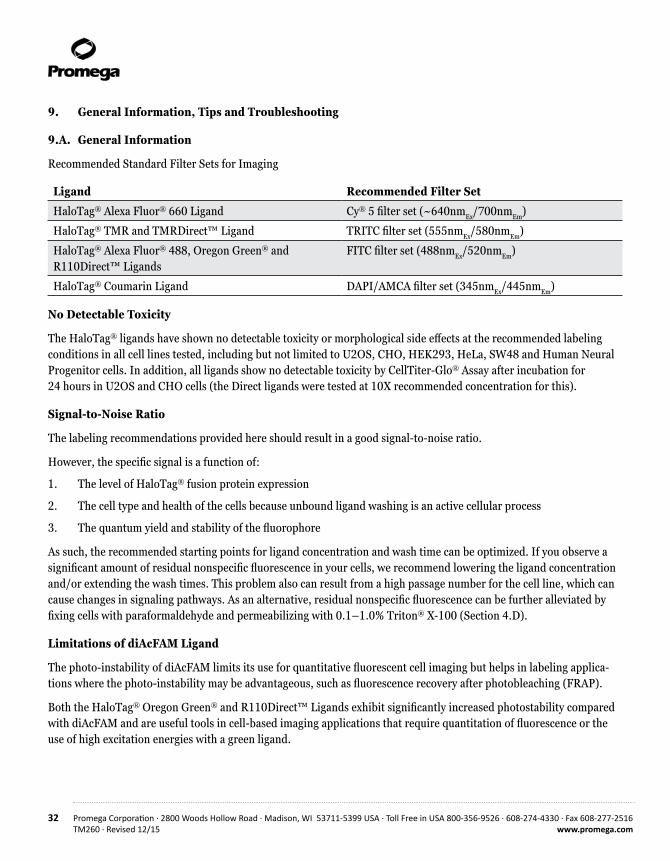
Recommended Standard Filter Sets for Imaging
Ligand Recommended Filter Set
HaloTag® Alexa Fluor® 660 Ligand Cy® 5 filter set (~640nmEx/700nmEm)
HaloTag® TMR and TMRDirect™ Ligand TRITC filter set (555nmEx/580nmEm)
HaloTag® Alexa Fluor® 488, Oregon Green® and R110Direct™ Ligands
FITC filter set (488nmEx/520nmEm)
HaloTag® Coumarin Ligand DAPI/AMCA filter set (345nmEx/445nmEm)
No Detectable Toxicity
The HaloTag® ligands have shown no detectable toxicity or morphological side effects at the recommended labeling conditions in all cell lines tested, including but not limited to U2OS, CHO, HEK293, HeLa, SW48 and Human Neural Progenitor cells. In addition, all ligands show no detectable toxicity by CellTiter-Glo® Assay after incubation for 24 hours in U2OS and CHO cells (the Direct ligands were tested at 10X recommended concentration for this).
Signal-to-Noise Ratio
The labeling recommendations provided here should result in a good signal-to-noise ratio.
However, the specific signal is a function of:
1. The level of HaloTag® fusion protein expression
2. The cell type and health of the cells because unbound ligand washing is an active cellular process
3. The quantum yield and stability of the fluorophore
As such, the recommended starting points for ligand concentration and wash time can be optimized. If you observe a significant amount of residual nonspecific fluorescence in your cells, we recommend lowering the ligand concentration and/or extending the wash times. This problem also can result from a high passage number for the cell line, which can cause changes in signaling pathways. As an alternative, residual nonspecific fluorescence can be further alleviated by fixing cells with paraformaldehyde and permeabilizing with 0.1–1.0% Triton® X-100 (Section 4.D).
Limitations of diAcFAM Ligand
The photo-instability of diAcFAM limits its use for quantitative fluorescent cell imaging but helps in labeling applica-tions where the photo-instability may be advantageous, such as fluorescence recovery after photobleaching (FRAP).
Both the HaloTag® Oregon Green® and R110Direct™ Ligands exhibit significantly increased photostability compared with diAcFAM and are useful tools in cell-based imaging applications that require quantitation of fluorescence or the use of high excitation energies with a green ligand.

Promega Corporation · 2800 Woods Hollow Road · Madison, WI 53711-5399 USA · Toll Free in USA 800-356-9526 · 608-274-4330 · Fax 608-277-2516 33www.promega.com TM260 · Revised 12/15
Use of HaloTag® Ligands in Serum-Containing Medium
All HaloTag® ligands may be directly diluted into serum-containing medium. However, the HaloTag® diAcFAM and Oregon Green® Ligands must be added to the cells immediately following this dilution because the diacetyl groups can be hydrolyzed by serum esterases. This deacetylation converts the HaloTag® diAcFAM and Oregon Green® Ligands into cell-impermeant fluorescent ligand derivatives. Once inside the cell, these ligands are modified, and, as will all of the other cell-permeant ligands, excess unbound ligand is eliminated from the cell in a time- and temperature- dependent manner by an active cellular mechanism.
While all cell types tested allow entry of the cell-permeant ligands and access to all major subcellular compartments, there are differences in rates of efflux. In general, efflux of the HaloTag® diAcFAM and Oregon Green® Ligands is significantly slower than that of other ligands, and the rate of efflux can vary significantly between cell types. Cells that have delayed efflux of unbound modified ligand might have limiting amounts of this active mechanism or suffer from poor health. Thus, while the use of serum results in a strict timing requirement for the addition of ligand to the cell culture, the presence of serum minimizes stress to cells and maintains their ability to actively eliminate unbound ligand, which results in lower background.
Streamlining the Protocol
The cell-impermeant Alexa Fluor® 488 and Alexa Fluor® 660 Ligands do not require a 30-minute wash step as unbound ligand does not enter cells. Among the cell-permeant ligand options, HaloTag® TMR and Coumarin Ligands are easily washed from cells. Therefore, you may be able to streamline the protocol in Section 4.A, Steps 4–6, without affecting the signal-to-noise ratio.

34 Promega Corporation · 2800 Woods Hollow Road · Madison, WI 53711-5399 USA · Toll Free in USA 800-356-9526 · 608-274-4330 · Fax 608-277-2516TM260 · Revised 12/15 www.promega.com
9.B. Composition of Buffers and Solutions
4% paraformaldehyde/0.2M sucrose/1X PBS (pH 7.5)
Prepare fresh for each use.
LB Broth (1X) 1% casein peptone Type M 0.5% NaCl 0.5% yeast extract
1X PBS buffer (pH 7.5) 137mM NaCl 2.68mM KCl 1.47mM KH2PO4
8.1mM Na2HPO4
4X SDS-sample buffer 0.24M Tris 2% SDS 50.4% glycerol 0.4M DTT 3mM bromophenol blue
Titrate to pH 6.8 using HCl.
1X SDS-sample buffer
1:4 dilution of 4X SDS Sample Buffer in filtered water.
Tris-buffered saline (TBS) 20mM Tris-HCl (pH 7.5) 150mM NaCl
9.C. Troubleshooting
For questions not addressed here, please contact your local Promega Branch Office or Distributor. Contact information available at: www.promega.com. E-mail: [email protected]
Symptoms Possible Causes and CommentsFusion protein not expressed or expressed only at Poor transfection efficiency. Optimize transfection conditions low levels and use high-quality endotoxin-free DNA.
Consider a construct in which the order of the HaloTag® protein and the protein of interest are reversed; N- or C-terminal fusions may perform differently.
Check reading frame of construct by sequencing or performing a rapid cell-free translation (such as TnT® T7 Quick Coupled Transcription Translation System, Cat.# L1170) followed by labeling, SDS-PAGE and fluorescence scanning of gel.
Weak or inappropriate regulatory elements; consider changing promoters and/or other regulatory elements.

Promega Corporation · 2800 Woods Hollow Road · Madison, WI 53711-5399 USA · Toll Free in USA 800-356-9526 · 608-274-4330 · Fax 608-277-2516 35www.promega.com TM260 · Revised 12/15
Symptoms Possible Causes and CommentsWeak or absent fluorescent signal in labeled cells Avoid proteolysis if lysing cells in PBS; add protease inhibitors during lysis, and perform lysis at 4°C.
Culture cells for a longer period of time before labeling to ensure that you have adequate protein expression and cell density.
Optimize cell health. Expose cells only to complete culture medium at 37°C under proper CO2 conditions throughout labeling and imaging.
Optimize cell-labeling protocols. Increase labeling time.
Store the HaloTag® ligands desiccated at –20°C, and protect them from light. Dispense the HaloTag® ligand into aliquots, avoiding multiple freeze-thaw cycles.
Use freshly diluted HaloTag® Ligand, and add immediately to label cells; avoid deacetylation. See Section 9.A.
Ensure that you are using the appropriate filer set for imaging. See Section 9.A and Figure 2.
Adjust the settings on your fluorescence detection instrument (e.g., laser power, PMT gain and aperture for a confocal microscope).
High background fluorescence in live cells Increase time of wash and perform washes using complete medium (i.e., with serum).
Image cells in complete medium lacking phenol red.
Strictly follow temperature recommendations during labeling.
Adjust instrumentation settings (e.g., reduce laser power, reduce gain on PMT or aperture for a confocal microscope).
High background fluorescence in fixed cells Increase time of wash and/or perform washes at a higher Triton® X-100 concentration or with a different detergent.
Use an alternative culture dish or glass because some HaloTag® ligands can bind to charged surfaces.

36 Promega Corporation · 2800 Woods Hollow Road · Madison, WI 53711-5399 USA · Toll Free in USA 800-356-9526 · 608-274-4330 · Fax 608-277-2516TM260 · Revised 12/15 www.promega.com
9.C. Troubleshooting (continued)
Symptoms Possible Causes and CommentsCells detached from surface Handle cells gently to ensure that they remain attached to the
surface and/or reduce the number of times you replace the medium (i.e., reduce the number of steps by using Direct™ ligands in the no-wash protocol instead of the ligands in the rapid labeling protocol).
Perform all labeling, washes and imaging steps using complete culture medium, and keep cells in a 37°C + CO2 incubator as much as possible.
Increase seeding density or time in culture to allow cells to proliferate and adhere more tightly.
Use an attachment matrix such as poly-l-lysine, fibronectin or collagen.
Cell death or toxicity Modify transfection protocol or try less toxic transfection reagent. Ensure DNA is endotoxin-free.

Promega Corporation · 2800 Woods Hollow Road · Madison, WI 53711-5399 USA · Toll Free in USA 800-356-9526 · 608-274-4330 · Fax 608-277-2516 37www.promega.com TM260 · Revised 12/15
9.D. Literature Cited and Additional References
Literature Cited
1. Los, G. et al. (2005) HaloTag® interchangeable labeling technology for cell imaging and protein capture. Cell Notes 11, 2–6.
2. Los, G. and Wood, K. (2007) The HaloTag: A novel technology for cell imaging and protein analysis. Methods. Mol. Biol. 356, 195–208.
3. Los, G. et al. (2008) HaloTag: A novel protein labeling technology for cell imaging and protein analysis. ACS Chem. Biol. 3, 383–82.
4. Huybrechts, S. J. et al. (2009) Peroxisome dynamics in cultured mammalian cells. Traffic 10, 1722–33.
5. Lee, H.L. et al. (2010) Superresolution imaging of targeted proteins in fixed and living cells using photo- activatable organic fluorophores. J. Am. Chem. Soc. 132, 15099–101.
6. Reek-Peterson, S.L. and Vale, R.D. (2009) Regulation of the processivity and intracellular localization of Saccharomyces cerevisiae dynein by dynactin. Proc. Natl. Acad. Sci. USA 106, 5669–74.
7. Lang, C. et al. (2006) HaloTag™ : A new versatile reporter gene system in plant cells. J. Exp. Bot. 57, 2985–92.
8. Kosaka, N. et al. (2009) In vivo stable tumor-specific painting in various colors using dehalogenase-based protein-tag fluorescent ligands. Bioconjugate Chem. 20, 1367–74.
9. Learish, R.D. (2007) A polyclonal HaloTag® antibody for Western blotting and immunocytochemistry. Promega Notes 95, 23–4.
Additional References
Learn more about the HaloTag® technology, visit www.promega.com/halotag
Learn more about cell imaging, visit www.promega.com/imaging
He, Y. et al. (2011) Identification of a lysosomal pathway that modulates glucocorticoid signaling and the inflammatory response. Sci. Signal. 4 (180).
Strauch, R.C. et al. (2011) Reporter protein-targeted probes for magnetic resonance imaging. J Amer Chem Soc. 133, 16346–9.
Hong, H. et al. (2011) HaloTag: a novel reporter gene for positron emission tomography. Am. J. Transl. Res. 3, 392–403.
Takemoto, K. et al. (2011) Chromophore-assisted light inactivation of HaloTag® fusion proteins labeled with eosin in living cells. ACS Chem. Biol. 6, 401–6.
Camarda, G. et al. (2010) Regulated oligomerization and molecular interactions of the early gametocyte protein Pfg27 in Plasmodium falciparum sexual differentiation. Int. J. Parasitology 40, 663-673.
Benink, H.A. et al. (2009) Direct pH measurement by using subcellular targeting of 5(and 6-) carboxyseminaphthor-hodafluor in mammalian cells. BioTechniques 47, 769–74.

38 Promega Corporation · 2800 Woods Hollow Road · Madison, WI 53711-5399 USA · Toll Free in USA 800-356-9526 · 608-274-4330 · Fax 608-277-2516TM260 · Revised 12/15 www.promega.com
9.D. Literature Cited and Additional References (continued)
Schröder, J. et al. (2009) In vivo labeling method using a genetic construct for nanoscale resolution microscopy. Biophys J. 96, L1–3.
Yamaguchi, K. et al. (2009) Pulse-chase experiment for the analysis of protein stability in cultured mammalian cells by covalent fluorescent labeling of fusion proteins. Methods Mol. Biol. 577, 121–31.
Ogawa, M. et al. (2008) Arabidopsis CLV3 peptide directly binds CLV1 ectodomain. Science 319, 294.
Reck-Peterson, S.L. et al. (2006) Single-molecule analysis of dynein processivity and stepping behavior. Cell 126, 335–48.
Eisenstein, M. (2006) Helping cells to tell a colorful tale. Nat. Meth. 3, 647–55.
Zhang, Y. et al. (2006) HaloTag® protein-mediated site-specific conjugation of bioluminescent proteins to quantum dots. Angew. Chem. Int. Ed. Engl. 45, 4936–40.
Shinohara, H. and Matsubayahshi, Y. (2007) Functional immobilization of plant receptor-like kinase onto microbeads towards receptor array construction and receptor-based ligand fishing. Plant J. 52, 175–84.
Hata, T. and Nakayama, M. (2007) Rapid single-tube method for small-scale affinity purification of polyclonal antibodies using HaloTag® Technology. J. Biochem. Biophys. Meth. 70, 679–82.
10. Related Products
Biotin Ligands
Biotin-Containing Ligands
30µl G8282
5529
MA
NH
OO Cl
S
HNNH
OO Reactive Linker
Functional Reporter
HaloTag® Biotin Ligand
HaloTag® PEG-Biotin Ligand 30µl G859155
27M
A
NH
OO
S
HNNH
OO O
NH
OO ClOO
Spacer
Reactive Linker
Functional Reporter

Promega Corporation · 2800 Woods Hollow Road · Madison, WI 53711-5399 USA · Toll Free in USA 800-356-9526 · 608-274-4330 · Fax 608-277-2516 39www.promega.com TM260 · Revised 12/15
HaloTag® Purification and Pull-Down Systems
Product Size Cat.#HaloTag® Mammalian Pull-Down and Labeling System 24 reactions G6500
HaloTag® Complete Pull-Down System 1 each G6509
HaloTag® Mammalian Pull-Down System 24 reactions G6504
HaloTag® Mammalian Protein Detection and Purification System 1 each G6795
HaloTag® Mammalian Protein Purification System 1 each G6790
HaloTag® Mammalian Protein Detection and Purification System Sample Pack 1 each G6799
HaloTag® Protein Purification System 1 each G6280
HaloTag® Protein Purification System Sample Pack 1 each G6270
HaloCHIP™ System 20 reactions G9410
HaloLink™ Protein Array Six Slide System 6 Slides G6190
HaloTag® Purification and Pull-Down Reagents
Product Size Cat.#HaloLink™ Resin 1.25ml G1912
2.5ml G1913
10ml G1914
25ml G1915
HaloLink™ Magnetic Beads 40 reactions G9311
Transfection-Grade DNA Purification
Product Size Cat.#PureYield™ Plasmid Miniprep System* 100 preps A1223
PureYield™ Plasmid Midiprep System* 25 preps A2492
Wizard MagneSil Tfx™ System* 4 × 96 preps A2380
Endotoxin Removal Resin 100ml A2191
4/40 Wash Solution 115ml A2221
*Available in additional sizes.

40 Promega Corporation · 2800 Woods Hollow Road · Madison, WI 53711-5399 USA · Toll Free in USA 800-356-9526 · 608-274-4330 · Fax 608-277-2516TM260 · Revised 12/15 www.promega.com
10. Related Products (continued)
Green Fluorescent Protein Fusion Vector
Product Size Cat.#Monster Green® Fluorescent Protein phMGFP Vector 20µg E6421
Cell Viability Analysis
Product Size Cat.#MultiTox-Fluor Multiplex Cytotoxicity Assay 10ml G9200
MultiTox-Glo Multiplex Cytotoxicity Assay 10ml G9270
CytoTox-Glo™ Cytotoxicity Assay 10ml G9290
CellTiter-Fluor™ Cell Viability Assay 10ml G6080
Caspase-Glo® 3/7 Assay 2.5ml G8090
CellTiter-Glo® Luminescent Cell Viability Assay 10ml G7570
CellTiter-Blue® Cell Viability Assay 20ml G8080
CellTiter 96® AQueous One Solution Cell Proliferation Assay 200 assays G3582
Western Blot Analysis
Product Size Cat.#ProtoBlot® II AP System with Stabilized Substrate, Rabbit 1 each W3960
Western Blue® Stabilized Substrate for Alkaline Phosphatase 100ml S3841
BCIP/NBT Color Development Substrate 1.25/2.5ml S3771
Blot-Qualified BSA 10g W3841
Tween® 20 2.5ml W3831

Promega Corporation · 2800 Woods Hollow Road · Madison, WI 53711-5399 USA · Toll Free in USA 800-356-9526 · 608-274-4330 · Fax 608-277-2516 41www.promega.com TM260 · Revised 12/15
Cloning Vectors
Conventional
Product Size Cat.#pHTN HaloTag® CMV-neo Vector 20µg G7721
pHTC HaloTag® CMV-neo Vector 20µg G7711
Flexi® Cloning Vectors
Product Size Cat.#pFN28A HaloTag® CMV-neo Flexi® Vector 20µg G8441
pFC27K HaloTag® CMV-neo Flexi® Vector 20µg G8431
Many additional Flexi® vectors are available for your convenience. You can see all HaloTag® Flexi® Vectors at www.promega.com by searching for “halotag flexi vectors”. Additionally, information about the full line of reagents and tools available for the HaloTag® technology is available at www.promega.com/halotag
11. Summary of Changes
The following change was made to the 12/15 revision of this document:
1. The disclaimer text was updated.

42 Promega Corporation · 2800 Woods Hollow Road · Madison, WI 53711-5399 USA · Toll Free in USA 800-356-9526 · 608-274-4330 · Fax 608-277-2516TM260 · Revised 12/15 www.promega.com
(a)BY USE OF THIS PRODUCT, RESEARCHER AGREES TO BE BOUND BY THE TERMS OF THIS LIMITED USE STATEMENT. If the researcher is not willing to accept the conditions of this limited use statement, and the product is unused, Promega will accept return of the unused product and provide the researcher with a full refund.
Researcher may use this product for research use only; no transfer or commercial use of this product is allowed. Commercial use means any and all uses of this product by a party in exchange for consideration, including, but not limited to (1) use in further product manufacture; (2) use in provision of services, information or data; and (3) resale of the product or its derivatives, whether or not such product or derivatives are resold for use in research. With respect to any uses outside this label license, including any commercial, diagnostic, therapeutic or prophylactic uses, please contact Promega for supply and licensing information. PROMEGA MAKES NO REPRESENTATIONS OR WARRANTIES OF ANY KIND, EITHER EXPRESSED OR IMPLIED, INCLUDING FOR MERCHANTABILITY OR FITNESS FOR A PARTICULAR PURPOSE WITH REGARDS TO THE PRODUCT. The terms of this label license shall be governed under the laws of the State of Wisconsin, USA. (b)U.S. Pat. No. 7,867,726, Japanese Pat. No. 4748685 and other patents pending.(c)U.S. Pat. No. RE42931, Japanese Pat. No. 4748685 and other patents pending. (d)The fluorescent dye (the “Dye”) of this product (the “Product”) is provided under an agreement between Molecular Probes, Inc., and Promega Corporation, and the manufacture, use, sale or import of this Product is subject to: (i) one or more of US Patents and pending US applications and corresponding international equivalents owned by Molecular Probes, Inc. (a wholly owned subsidiary of Invitrogen Corporation); and (ii) one or more of US Patents and pending US applications and corresponding international equivalents owned by Promega Corporation. The purchase of this Product conveys to the buyer the non-transferable right to use the purchased amount of the Product in research conducted by the buyer (whether the buyer is an academic or for-profit entity). The use of this Product for providing medical or diagnostic results or services, including without limitation testing, analysis, or screening, or for providing clinical information or clinical analysis in return for compensation on a per-test basis, is expressly excluded. The buyer cannot sell or otherwise transfer (a) this Product, (b) its components or (c) materials made using this Product or its components to a third party or otherwise use this Product or its components or materials made using this Product or its components for Commercial Purposes. Commercial Purposes means any activity by a party for consideration and may include but is not limited to: (1) use of the Product or its components in manufactur-ing; (2) use of the Product or its components to provide a service, information, or data; (3) use of the Product or its components for therapeutic, diagnostic or prophylactic purposes; or (4) resale of the Product or its components, whether or not such Product or its components are resold for use in research. For information on purchasing a license to the Dye for purposes other than as set forth above, contact Molecular Probes, Inc., Business Development, 29851 Willow Creek Road, Eugene, OR 97402-9132. Tel: (541) 465-8300. Fax: (541) 335-0504. For information on purchasing a license to this Product for purposes other than as set forth above, contact Promega Corporation. (e)Patent Pending.(f)Certain applications of this product may require licenses from others.
© 2004–2015 Promega Corporation. All Rights Reserved.
Caspase-Glo, CellTiter 96, CellTiter-Blue, CellTiter-Glo, Flexi, HaloTag, MagneSil, Monster Green, ProtoBlot, TnT, Western Blue, and Wizard are registered trademarks of Promega Corporation. CellTiter-Fluor, CytoTox-Glo, FastBreak, HaloCHIP, HaloLink, PureYield, R110Direct, Tfx and TMRDirect are trademarks of Promega Corporation.
Alexa Fluor and Oregon Green are registered trademarks of Molecular Probes, Inc. Coomassie is a registered trademark of Imperical Chemical Industries, Ltd. Cy and Sephadex are registered trademarks of GE Healthcare Biosciences. FMBIO is a registered trademark of Hitachi Software Engineer-ing Company, Ltd. MoFlo is a registered trademark of Dako Denmark A/S. Triton is a registered trademark of Union Carbide Chemicals & Plastics Technology Corporation. Tween is a registered trademark of ICI Americas, Inc.
Products may be covered by pending or issued patents or may have certain limitations. Please visit our Web site for more information.
All prices and specifications are subject to change without prior notice.
Product claims are subject to change. Please contact Promega Technical Services or access the Promega online catalog for the most up-to-date information on Promega products.