handbook 2 edition 2020 nd - pharmacy
TRANSCRIPT

MOH/S/FAR/37.21(GU)-e
HANDBOOK 2nd Edition 2020
PHARMACEUTICAL SERVICES PROGRAMME
Clinical Pharmacy Working Committee (Critical Care Subspecialty)
Pharmacy Practice & Development Division Ministry of Health Malaysia
Second Edition (Reviewed October 2020) Published by: Pharmaceutical Services Programme Ministry of Health Malaysia Lot 36, Jalan Prof Diraja Ungku Aziz, 46200 Petaling Jaya, Selangor Malaysia Tel : (603) – 7841 3200 Fax : (603) – 7968 2222 Website : https://www.pharmacy.gov.my
©ALL RIGHTS RESERVED
This document is copyrighted. The publication of the Critical Care Pharmacy Handbook 2nd was coordinated by the Pharmaceutical Care Branch of the Pharmacy Practice and Development Division, Ministry of Health Malaysia with contribution from Clinical Pharmacy Working Committee (Critical Care Subspecialty). The publisher reserve copyright and renewal on all published materials and such material may not be reproduced in any form without written permission from the publisher.
i
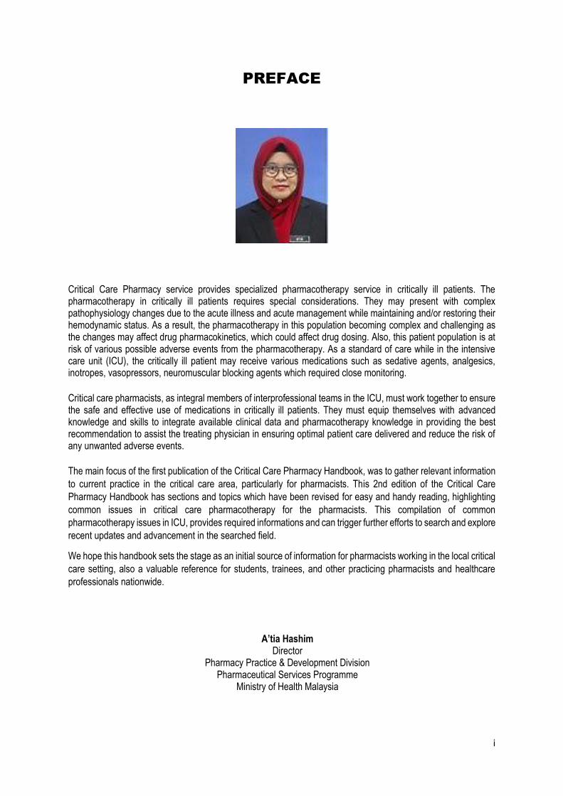
PREFACE
Critical Care Pharmacy service provides specialized pharmacotherapy service in critically ill patients. The pharmacotherapy in critically ill patients requires special considerations. They may present with complex pathophysiology changes due to the acute illness and acute management while maintaining and/or restoring their hemodynamic status. As a result, the pharmacotherapy in this population becoming complex and challenging as the changes may affect drug pharmacokinetics, which could affect drug dosing. Also, this patient population is at risk of various possible adverse events from the pharmacotherapy. As a standard of care while in the intensive care unit (ICU), the critically ill patient may receive various medications such as sedative agents, analgesics, inotropes, vasopressors, neuromuscular blocking agents which required close monitoring.
Critical care pharmacists, as integral members of interprofessional teams in the ICU, must work together to ensure the safe and effective use of medications in critically ill patients. They must equip themselves with advanced knowledge and skills to integrate available clinical data and pharmacotherapy knowledge in providing the best recommendation to assist the treating physician in ensuring optimal patient care delivered and reduce the risk of any unwanted adverse events.
The main focus of the first publication of the Critical Care Pharmacy Handbook, was to gather relevant information
to current practice in the critical care area, particularly for pharmacists. This 2nd edition of the Critical Care
Pharmacy Handbook has sections and topics which have been revised for easy and handy reading, highlighting
common issues in critical care pharmacotherapy for the pharmacists. This compilation of common
pharmacotherapy issues in ICU, provides required informations and can trigger further efforts to search and explore
recent updates and advancement in the searched field.
We hope this handbook sets the stage as an initial source of information for pharmacists working in the local critical
care setting, also a valuable reference for students, trainees, and other practicing pharmacists and healthcare
professionals nationwide.
A’tia Hashim
Director Pharmacy Practice & Development Division
Pharmaceutical Services Programme Ministry of Health Malaysia

ii
WORKING COMMITTEE
Advisors
A’tia Hashim Director
Pharmacy Practice & Development Division, Pharmaceutical Services Programme
Ministry of Health, Malaysia
Rozita Mohamad Deputy Director
Pharmacy Practice and Development Division Pharmaceutical Services Programme
Ministry of Health, Malaysia
Nor Hasni Haron Senior Principal Assistant Director
Pharmacy Practice and Development Division Pharmaceutical Services Programme
Ministry of Health, Malaysia

iii
Editors & Contributors (Alphabetical order)
Afifah Azhari Hospital Raja Perempuan Zainab II, Kelantan
Mohd Farizh Che Pa Hospital Tuanku Ja’afar, Negeri Sembilan
Alia Hayati Baharudin Hospital Tuanku Fauziah, Kangar, Perlis
Mohd Shafie Zabidi Hospital Sultanah Aminah, Johor
Charlene Tay Szu Lynn Hospital Kuala Lumpur, Kuala Lumpur
Ngua Ching Zin Hospital Umum Sarawak, Sarawak
Chew Soo Fong Hospital Melaka, Melaka
Nicole Foo Yen Fei Hospital Umum Sarawak, Sarawak
Chong Lai Peng Hospital Melaka, Melaka
Norirmawath Saharuddin Hospital Raja Permaisuri Bainun, Perak
CW Mohd Hafidz CW Ahmad Hospital Tengku Ampuan Afzan, Kuantan, Pahang
Nur Salima Shamsudin Pharmaceutical Services Programme, MOH
Fahmi Hassan Pharmaceutical Services Programme, MOH
Nurul Akma Hanan Hospital Tuanku Ja’afar, Negeri Sembilan
Faridah Yusof Hospital Sultanah Bahiyah, Alor Setar, Kedah
Oh Hoey Lin Hospital Raja Perempuan Bainun Ipoh, Perak
Ignatius Leon Guang Woei Hospital Selayang, Selangor
Puteri Juanita Zamri Hospital Selayang, Selangor
Janattul Ain Jamal Hospital Tengku Ampuan Afzan, Kuantan, Pahang
Rahela Ambaras Khan Hospital Kuala Lumpur, Kuala Lumpur
Jerry Liew Ee Siung Hospital Queen Elizabeth, Sabah
Roslita Alivi Hospital Sultan Ismail, Johor
Lim Chin How Hospital Umum Sarawak, Sarawak
Siti Nur Aziela Ab Manap Hospital Raja Perempuan Zainab II, Kelantan
Lim Shiao Hui Hospital Pulau Pinang, Pulau Pinang
Siti Nuraidah Mamat Hospital Sultanah Nur Zahirah, Terengganu
Mohd ‘Izzat Ismorning Pharmaceutical Services Programme, MOH
Tey Su Anne Hospital Putrajaya, Putrajaya
External Reviewer
Assoc. Prof. Dr. Mohd Makmor Bakry Dean
Faculty of Pharmacy Universiti Kebangsaan Malaysia

iv
Content
Page
Preface i
Advisors ii
Editors & Contributors iii
External Reviewer iii
Content iv
Part I Introduction 1 Chapter 1 Critical Care Pharmacist 1 Part II Common Clinical Issues & Management in Critically Ill Patient 11 Chapter 2 Shock 11 Chapter 3 Organs Dysfunction 18 Chapter 4 Cardiovascular Problems 38 Chapter 5 Pulmonary Disorders 49 Chapter 6 Hematologic Disorders 58 Chapter 7 Deep Vein Thrombosis & Pulmonary Embolism 65 Chapter 8 Stress-Ulcer 69 Chapter 9 Delirium 74 Chapter 10 Fluid & Electrolytes 76 Chapter 11 Nutritional Support 87 Chapter 12 Acute Medical Crisis 100 Chapter 13 Feeding Intolerance 119 Part III Other Pharmacotherapy in Critical Care 124 Chapter 14 Neuromuscular Blocking Agents 124 Chapter 15 Analgesic & Sedative Agents 130 Part IV Special Considerations in Critically Ill Patient 139 Chapter 16 Special Populations: Obese, Hypoalbuminaemia & Burn 139 Chapter 17 Critically Ill Trauma Patient 145 Chapter 18 Antimicrobial Dosing in Critically Ill Patient: Pharmacokinetic & Pharmacodynamic
(PKPD) Considerations 151 Chapter 19 Advanced Cardiac Life Support 158 Chapter 20 Drug Overdose and Poisoning 164

1
CHAPTER 1
Critical Care Pharmacist
he beginnings of caring for critically ill patients date back to Florence Nightingale’s work during the
Crimean War in 1854. The first multidisciplinary ICU was established in the United State (US) in 1958, and the American Board of Medical Subspecialties first recognized the subspecialty of critical care medicine in 1986. Critical care pharmacy services began around the 1970’s, growing to become one of the largest practice areas for clinical pharmacists, with its section in the Society of Critical Care Medicine, which is the largest international professional organization in the field. Critical care pharmacists have been recognized by the Society of Critical Care Medicine (SCCM) as one of the essential team members for the delivery of care for critically ill patients.
Figure 1.1: The landmark events in critical care medicine and critical care pharmacy service in the US
Critical Care Pharmacy Service in Malaysia was initiated in the early year 2000. The first critical care pharmacist (locally known as ICU pharmacist) from Hospital Selayang (Selangor), received an official critical care pharmacy training in the United States (US). Subsequently, in 2006, critical care pharmacy attachment programme was conducted in Hospital Selayang, supervised by trained local ICU pharmacist and an invited facilitator from the US, Dr. Eljim Patrick Tesoro, an ASHP-Accredited Specialty, Residency in Critical Care, Univerisity of Illinois, Chicago. A total of 20 clinical pharmacists across Malaysia participated in the programme. In 2007, the Pharmaceutical Services Programme, Ministry of Health, Malaysia has recognized Hospital Selayang as a training centre for critical care pharmacy, and conducted it first comprehensive 30-day training in November 2007, which was attended by 3 local clinical pharmacists, from Hospital Alor Setar (Kedah), Hospital Tengku Ampuan Afzan (Pahang) and Hospital Sultan Ismail (Johor).
T
PA
RT
1: INT
RO
DU
CT
ION
2
The critical care pharmacy service in Malaysia has evolved since then, where local clinical pharmacists (ICU pharmacists) officially worked either as a full-time or part-time basis in all Intensive Care Units (ICUs) of major / state hospitals across Malaysia. In 2008, ICU pharmacists from all major / state hospitals were selected as a member of Clinical Pharmacy Working Committee (Critical Care Pharmacy Subspecialty), Pharmaceutical Services Programme, Ministry of Health Malaysia. The committee was developed to assist in the development and expansion of critical care pharmacy service in Malaysia. The members of committee continuously worked together in identifying training needs for the critical care pharmacists at local and international level, and contributed in planning and implementation of planned activities in parallel to the direction by the Pharmaceutical Services Programme, MOH Malaysia. The expansion and recognition of the critical care pharmacy service was evidenced by the development of critical care pharmacy training module in 2011. More hospitals have been recognised as ‘training centres’ and conducted training based on the developed module for the local ICU pharmacists. The specialty of critical care pharmacist in Malaysia has also been recognized by other health-care professional, with involvements in development of national policy / guidelines. Members from the Clinical Pharmacy Working Committee (Critical Care Pharmacy Subspecialty) has continuously work together with the Malaysian Society of Intensive Care (MSIC), in the development and reviewing of the National Guide to Antimicrobial Therapy in the Adult ICU, the local antimicrobial therapy guide for physicians treating the critically ill septic patients. To date, 10 major state hospitals have been recognised by the Pharmaceutical Services Programme, Ministry of Health Malaysia as the training centres for critical care pharmacy in Malaysia, with active preceptorship. In addition, the Clinical Pharmacy Working Committee (Critical Care Pharmacy Subspecialty) has published the first Critical Care Pharmacy Handbook in 2012, a quick and comprehensive reference for the local ICU pharmacist to acquire basic knowledge and skills as Critical Care Pharmacist as per recommended by the global guidelines. Critical Care Pharmacist Services The American College of Critical Care Medicine published an updated guideline defining recommended critical care services and personnel according to the level of care being provided; defined as levels I, II, and III
Level I Comprehensive critical care for a wide variety of patient populations with a high level of specialization Requires broad range of comprehensive support, including pharmacy services, respiratory therapy,
clinical nutrition, pastoral care, and social services Often fulfils an academic mission
Level II
Comprehensive critical care but may not provide care for certain patient populations Must have transfer protocols in place for patients with special needs Comprehensive support services must be available. May or may not have an academic mission
Level III
Provides stabilization but has limited ability to provide comprehensive critical care Must have transfer protocols in place for patients requiring level I and II critical care services Support services are often limited in scope
Having the qualifications and competence in providing pharmaceutical care in ICU is essential and can be achieved by several pathways, including advanced degrees, residency, fellowship, and other specialized practice experiences. ICUs with an academic mission should provide protected time for pharmacist participation in scholarly activities and appropriate knowledge and skills to provide education to critical care nurses, physician trainees, and physicians. Meanwhile, the non-academic centres should provide time for maintenance of competence and maintain current certification. This report encompasses clinical and non-clinical pharmacy services and stratifies levels of service as fundamental, desirable, or optimal to patient care (Table 1.1).

3
Table 1.1: Recommended levels of care by critical care pharmacist
FUNDAMENTAL MUST be provided for the safe delivery of pharmaceutical care
Predominant commitment of time to critically ill patients
Prospective evaluation of all drug therapy for safety and efficacy, with intervention as needed
Evaluation of parenteral nutrition orders
Identification of adverse drug events
Pharmacokinetic monitoring
Provision of drug information and IV drug compatibility data
Provision of informal drug therapy-related education to ICU team members
Documentation of clinical activities
Implementation and maintenance of policies and procedures related to safe and effective drug use
Service on ICU and hospital committees
DESIRED ADDITIONAL services and clinical functions for the specialized care
Rounds as a member of an interdisciplinary critical care team
Conducts medication histories and evaluates need for continuance of therapy
Provides formal nutrition consultations
Responds to resuscitation events
Provides didactic lectures to health professional students and postdoctoral trainees
Trains student pharmacists, residents, and fellows through experiential education
Coordinates the development and implementation of drug therapy protocols and critical care pathways
Provides advanced documentation of services to include the clinical significance and economic value of interventions
Actively engages in critical care pharmacotherapy research, including screening and enrolment of prospective study patients, study coordination, research study design, and data analysis
Disseminates case reports and practice insights to other practitioners by publication within the pharmacy and medical literature
OPTIMAL Integrated, specialized, and dedicated model of patient care aimed at optimizing patient outcomes
Assists physicians with patients and/or family members to make informed decisions regarding treatment options
Provides formal accredited educational sessions for medical staff, students, and residents
Teaches advanced cardiac life support
Develops and coordinates critical care pharmacy residencies and/or fellowships
Develops pharmacist and technician training programs for personnel working in the ICU
proactively involved in designing, prioritizing, and promoting new pharmacy program and services
Secures funds for clinical research studies through investigator-initiated grants and contracts
Publishes peer-reviewed reports in the pharmacy and medical literature of original critical care clinical
Fundamental activities reflect services associated with order entry and distribution duties that are necessary for the safe provision of pharmaceutical care; desirable activities add some clinical functions necessary for the specialized care of critically ill patients; and optimal activities reflect an integrated, specialized and dedicated model of direct patient care functions that aim to maximize outcomes.

4
Table 1.2: Published data on impact of implementing critical care pharmacy services
The impact on implementing critical care pharmacy services to healthcare:
Drug costs in ICU Medical-surgical ICU: reported on annual savings of $67,664 Neurosurgical ICU: reported on reduction in pharmacy acquisition costs from $4833 to $3239/patient,
reduction in ICU days from 8.56 to 7.24 days Several other studies in a wide range of ICU setting Adverse drug events/drug-drug interactions 66% reduction in preventable adverse drug events Low incidence of QTc-interval prolongation with ICU monitoring by a pharmacist using a standard
algorithm: 19% versus 39%. Decreased drug-drug interactions rate by 65% Infectious disease morbidity, mortality and costs Compared to ICU with clinical pharmacists, ICU without clinical pharmacists significantly associated with
higher mortality rate, longer LOS and greater Medicare charges for patient with nosocomial-acquired infections, community-acquired infections and sepsis.
Thromboembolic or infarction-related event (TIE) Among patient with TIE, mortality and bleeding complications increased by 37% and 49%, respectively, in
the absence of clinical pharmacist in ICU.
Conclusion The implementation of critical care pharmacy service in Malaysia has impact on the overall care of Malaysian critically ill patients, through provision of pharmaceutical care in collaboration with other healthcare professionals in ICU. The roles of critical care pharmacists in Malaysia have been well accepted and recognised. Similar to other nations, the local critical care pharmacy services aimed to continuously provides optimize cares to the critically ill patients, suitable to the local needs and availability. Optimising medication is a central and key role expected of pharmacists in all clinical areas, not only in critical care. By working as a team in critical care setting, critical care pharmacists able to see the entire case-mix and thus able to manage the pharmaceutical care of an extreme range of health problems, as well as quickly assimilate information and management paths for conditions. Working in the ICU setting appears not only providing the critical care pharmacists an opportunity to use their clinical knowledge and sharpen their clinical skills, but also expose to recent updates on current practices in ICU, to enhance patient care and practicing the evidence based-medicine in critical care, as a valued member of the inter-professional team.

5
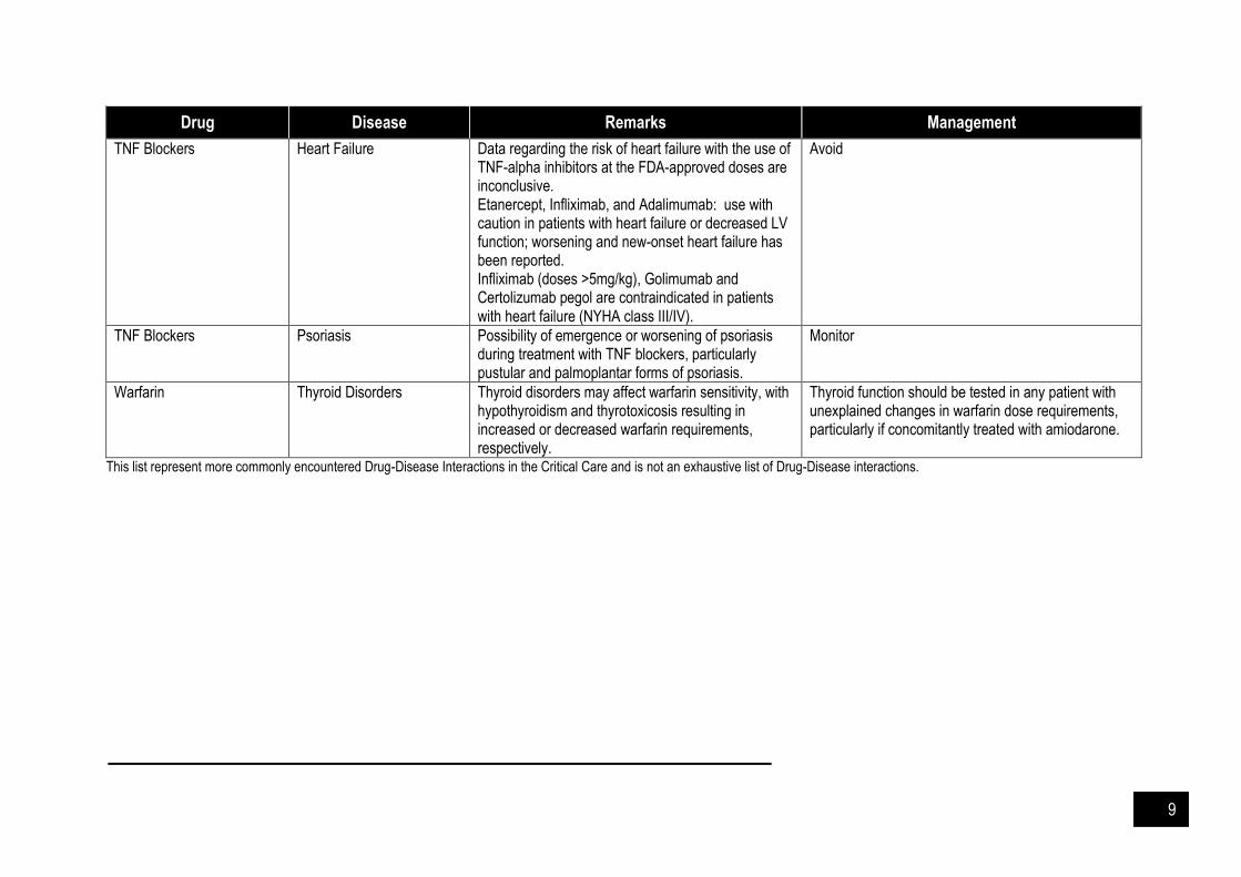
Table 1.3: Commonly encountered drug-disease interactions in ICU
Drug Disease Remarks Management
ACE Inhibitors, Gold salts and Interferon
Psoriasis Occasional triggers of a psoriatic flare. Use with caution.
Aminoglycosides Myasthenia Gravis Cause significant increase in weakness and respiratory depression. Aminoglycoside-related postoperative respiratory depression caused the greatest frequency of drug-induced neuromuscular blockade.
Avoid or use only if absolutely necessary with close monitoring.
Amiodarone Thyroid Disorders The iodine-rich amiodarone affects the thyroid gland, causing overt hypothyroidism or thyrotoxicosis in 14%-18% of cases.
Monitor thyroid function.
Androgens (Testosterone) Heart Failure (HF) Edema US Endocrine Society Guideline recommend not to use in uncontrolled or poorly controlled HF.
Antiarrhythmics (Sotalol, Ibutilide)
Heart Failure Negative inotropic, precipitate HF, proarrhythmic. Amiodarone is the preferred choice in arrhythmias in HF.
Antimalarials Psoriasis Exacerbate Not contraindicated
Antipsychotics Parkinson’s Disease Parkinsonism Use with caution
Beta Blockers COPD, Asthma Non selective beta blockers can precipitate bronchospasm.
Selective beta blockers are generally safe. Combined alpha/beta blockers to be used cautiously at low dose. Data limited.
Beta Blockers Diabetes Facilitation of hypoglycaemia. Use with caution.
Beta Blockers Peripheral Vascular Disease, Raynaud’s Phenomenon
Non selective beta blockers implicated. Reduction in cardiac output and blockade of β2-receptor-mediated skeletal muscle vasodilation contribute to the vascular insufficiency.
Selective agents can be used cautiously.
6
Drug Disease Remarks Management
Beta Blockers Heart Failure Relative contraindications to beta blockers in heart failure: • Heart rate <60 bpm • Symptomatic hypotension • Greater than minimal evidence of fluid retention • Signs of peripheral hypoperfusion • PR interval >0.24 sec • 2nd - or 3rd -degree AV block • History of asthma or reactive airways • Peripheral artery disease with resting limb ischemia
Use with caution. Avoid beta blockers with intrinsic sympathomimetic activity.
Beta Blockers Psoriasis May aggravate existing disease. Not contraindicated. However, when there is a clear relationship between exacerbation of psoriasis and intake of β-blocker, it sometimes helps to switch from a non-cardioselective β2-blocker to a cardioselective β1- blocker.
Beta Blockers (propranolol, oxprenolol, timolol, and practolol)
Myasthenia Gravis (MG) β-adrenergic blocking drugs occasionally associated with increasing weakness in MG patients.
Use with caution
Calcium Channel Blockers (Short acting - Verapamil, Diltiazem, Nifedipine)
Heart Failure Negative inotropic, increase sympathetic activity Avoid use of shorter acting dihydropyridines. Long-acting agents appear to be safe.
Chemotherapeutic Agents (Cyclophosphamide, Trastuzumab, Bevacizumab, Anthracycline-like chemo agents)
Heart Failure Cardiotoxic To decrease the risk of cardiotoxicity while maintaining efficacy, altered schedules of drug administration, modifications of the anthracycline molecule, and adjunctive treatment with beta-adrenergic blockers or dexrazoxane is advocated.
Corticosteroids Psoriasis Rebound that invariably follows their use. The flare-up may be even worse than the original attack.
Avoid
COX-2 Selective Inhibitors Heart Failure Exacerbation of heart failure. Use with caution
Fluoroquinolones Myasthenia gravis Neuromuscular blocking activity and may exacerbate muscle weakness.
Avoid

7
Drug Disease Remarks Management
Lignocaine and Procaine (may cause worsening if given intravenous)
Myasthenia Gravis Interference with propagation of the nerve action potential at the nerve terminal and reduced ACh release may account for the presynaptic effects. Local anesthetics also lead to reduced sensitivity of the postjunctional membrane to acetylcholine.
Use with caution
Lithium Psoriasis Well recognised cause of exacerbation. It may even cause pustular or erythrodermic psoriasis in a significant proportion of affected patients.
Lithium does not aggravate a pre-existing psoriasis in all cases, and therefore is not contraindicated in all patients with psoriasis.
Magnesium Sulfate Myasthenia Gravis Mg2+ interferes with neuromuscular transmission by inhibiting release of ACh. Mg competitively blocks Ca2+ entry at the motor nerve terminal. There may also be a milder postsynaptic effect.
Relative contraindication
Muscle Relaxants Myasthenia Gravis Sensitive to nondepolarizing neuromuscular blockers Intermediate and short-acting nondepolarizing agents can be used with careful monitoring
Use with caution
NSAIDs Heart Failure Worsen heart failure Use with caution
NSAIDs, Aspirin Peptic Ulcers Systemic inhibition of GI mucosal COX activity. Use with caution
NSAIDs, Aspirin Asthma Can induce bronchospasm. Rarely, this reaction leads to death in aspirin-sensitive asthmatics.
Avoid in aspirin sensitive asthma, use with caution in others.
PDE- 3 Enzyme Inhibitor (Anagrelide)
Heart Failure Positive inotropic, vasodilatory, leading to fluid retention and heart failure.
Avoid
PDE-3 Enzyme Inhibitor (Cilostazol)
Heart Failure Increased mortality Contraindicated
PDE-5 Enzyme Inhibitor (Sildenafil)
Heart Failure, Coronary Heart Disease
Potentially hazardous in patients with active coronary ischemia; congestive heart failure, borderline low blood volume and low blood pressure status.
Use with caution
Penicillamine Myasthenia Gravis Induces autoimmune Myasthenic syndrome. Reversible.
Avoid

8
Drug Disease Remarks Management
Phenytoin, Gabapentin Myasthenia Gravis Symptoms occasionally presented in patients with MG following phenytoin treatment. There are reports of seropositive MG occurring after three months of gabapentin therapy for painful neuropathy.
Use with caution
Prednisolone, Glucocorticoids in high doses
Myasthenia Gravis 50% of patients experience a transient deterioration. During crisis, use only if patient’s airway is protected.
Procainamide, Quinidine, Quinine
Myasthenia Gravis Procainamide - direct effect on neuromuscular transmission Quinine and quinidine - aggravate weakness in MG.
Avoid
Statins Myasthenia Gravis Small number of reports of myasthenic weakness temporally-associated with statin
Use with caution
Sulfonamide Antibiotics, Penicillin (but not the semi synthetic ones)
Systemic Lupus Erythema-tosus (SLE)
Exacerbate SLE Avoid
Telithromycin Myasthenia Gravis Black box warning on possibility of exacerbating or unmasking MG. Should not use.
Avoid
Theophylline Cardiac Disease Can reduce theophylline clearance by as much as 50%
Monitor level closely
Theophylline Primary Hepatic Disease Can reduce theophylline clearance by as much as 50%
Monitor level closely
Theophylline Cystic Fibrosis, Hyper-thyroidism
Increase clearance May need to increase dose
9
Drug Disease Remarks Management
TNF Blockers
Heart Failure Data regarding the risk of heart failure with the use of TNF-alpha inhibitors at the FDA-approved doses are inconclusive. Etanercept, Infliximab, and Adalimumab: use with caution in patients with heart failure or decreased LV function; worsening and new-onset heart failure has been reported. Infliximab (doses >5mg/kg), Golimumab and Certolizumab pegol are contraindicated in patients with heart failure (NYHA class III/IV).
Avoid
TNF Blockers Psoriasis Possibility of emergence or worsening of psoriasis during treatment with TNF blockers, particularly pustular and palmoplantar forms of psoriasis.
Monitor
Warfarin Thyroid Disorders Thyroid disorders may affect warfarin sensitivity, with hypothyroidism and thyrotoxicosis resulting in increased or decreased warfarin requirements, respectively.
Thyroid function should be tested in any patient with unexplained changes in warfarin dose requirements, particularly if concomitantly treated with amiodarone.
This list represent more commonly encountered Drug-Disease Interactions in the Critical Care and is not an exhaustive list of Drug-Disease interactions.

10
References and Suggested Readings Borthwick M. The role of the pharmacist in the intensive care unit. J Intensive Care Soc. 2019. 20(2): 161-164. Grenvik A, Pinsky MR. Evolution of the intensive care unit as a clinical center and critical care medicine as a
discipline. Crit Care Clinics. 2009. 25(1): 239-50. Haupt MT BC, Brilli RJ, et al. Guidelines on critical care services and personnel: recommendations based on a
system of categorization of three levels of care. Crit Care Med 2003. 31: 2677-83. Kane SL, Weber RJ, Dasta JF. The impact of critical care pharmacists on enhancing patient outcomes. Intensive
Care Med. 2003. 29(5): 691-8. Leape LL, Cullen DJ, Clapp MD, Burdick E, Demonaco HJ, Erickson JI, et al. Pharmacist participation on physician
rounds and adverse drug events in the intensive care unit. JAMA. 1999. 282(3): 267-70. MacLaren R, Devlin JW, Martin SJ, Dasta JF, Rudis MI, Bond C. Critical care pharmacy services in United States
hospitals. Ann Pharmacother. 2006. 40(4): 612-8. MacLaren R, Martin SJ, Fike D. Clinical and economic outcomes of involving pharmacists in the direct care of
critically ill patients with infections. Crit Care Med. 2008. 36: 3184-9. MacLaren R. Effects of pharmacist participation in intensive care units on clinical and economic outcomes of
critically ill patients with thromboembolic or infarction-related events. Pharmacotherapy. 2009. 29(7): 761–768. Montazeri M, Cook DJ. Impact of a clinical pharmacist in a multidisciplinary intensive care unit. Crit Care Med.
1994. 22(6): 1044-8. Ng TM, Bell AM, Hong C, Hara JM, Touchette DR, Danskey KN, et al. Pharmacist monitoring of QTc interval–
prolonging medications in critically ill medical patients: A pilot study. Ann Pharmacother. 2008. 42(4): 475-82. Rivkin A, Yin H. Evaluation of the role of the critical care pharmacist in identifying and avoiding or minimizing
significant drug-drug interactions in medical intensive care patients. J Crit Care. 2011. 26(1): 104. Rudis MI BK. SCCM and ACCP Clinical Pharmacy task force on critical care pharmacy services. Position paper
on critical care pharmacy services 2000. Timmins A. The contribution of the intensive care pharmacist in the United Kingdom. Pharmaceutical Journal.
2000. 265(7112): 341-3. Weant KA, Armitstead JA, Ladha AM, Sasaki-Adams D, Hadar EJ, Ewend MG. Cost effectiveness of a clinical
pharmacist on a neurosurgical team. Neurosurgery. 2009. 65(5): 946-51.
11
CHAPTER 2
Shock
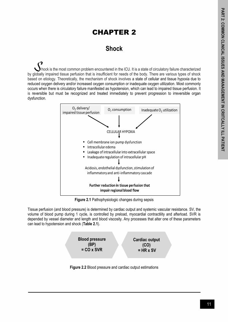
hock is the most common problem encountered in the ICU. It is a state of circulatory failure characterized by globally impaired tissue perfusion that is insufficient for needs of the body. There are various types of shock based on etiology. Theoretically, the mechanism of shock involves a state of cellular and tissue hypoxia due to reduced oxygen delivery and/or increased oxygen consumption or inadequate oxygen utilization. Most commonly occurs when there is circulatory failure manifested as hypotension, which can lead to impaired tissue perfusion. It is reversible but must be recognized and treated immediately to prevent progression to irreversible organ dysfunction.
Figure 2.1 Pathophysiologic changes during sepsis Tissue perfusion (and blood pressure) is determined by cardiac output and systemic vascular resistance. SV, the volume of blood pump during 1 cycle, is controlled by preload, myocardial contractility and afterload. SVR is depended by vessel diameter and length and blood viscosity. Any processes that alter one of these parameters can lead to hypotension and shock (Table 2.1).
Figure 2.2 Blood pressure and cardiac output estimations
Blood pressure (BP)
= CO x SVR
Cardiac output (CO)
= HR x SV
PA
RT
2: CO
MM
ON
CL
INIC
AL
ISS
UE
S A
ND
MA
NA
GE
ME
NT
IN C
RIIT
CA
LL
Y IL
L P
AT
IEN
T
S

12
Table 2.1: Clinical characteristic by types of shock The hemodynamic characteristics in table below are those recognized in the mentioned shock state, which often does not occur in practice. Patients often have features of more than one shock states1
Type- Characteristic
Cause Examples HR BP CO Capillary refill Extremity
temperature SVR Treatment
Distributive- severe peripheral vasodilatation
Septic Gram positive/negative, fungal/viral Flash or delayed Warm or cool
Low or high
Antibiotics, fluids, vasopressors
Non-septic
Inflamatory shock (burns), neurogenic shock (TBI, spinal cord injury), anaphylactic shock
Flash or normal Warm Low
Fluid resuscitation, vasopressors
Cardiogenic- intracardiac causes of cardiac pump failure that result in reduced cardiac output
Cardiomyopathic MI, myocarditis
Delayed Cool High
Inotropes, caution with fluids, ECMO
Arrhythmogenic
Tachyarrythmia (atrial/ventricular tachycardia), bradyarrhythmia (complete heart block)
Mechanical Severe vulvular insufficiency
Hypovolemic- reduced intravascular volume (reduced preload which in turn, reduces CO)
Hemorrhagic Trauma, GI bleeding
Delayed Cool High
Stop bleeding, fluid resuscitation
Non- hemorrhagic
GI losses (diarrhea), skin losses (burns), renal losses (excessive drug-induced or osmotic diuresis)
Obstructive- extracardiac causes of cardiac pump failure and poor right ventricle output
Pulmonary vascular
Hemodynamically significant pulmonary embolus
Delayed Cool High Pericardiocentesis, chest tube
Mechanical Tamponade, tension pneumothorax
13
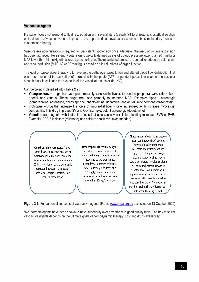
Vasoactive Agents If a patient does not respond to fluid resuscitation with several liters (usually ≥4 L) of isotonic crystalloid solution or if evidence of volume overload is present, the depressed cardiovascular system can be stimulated by means of vasopressor therapy. Vasopressor administration is required for persistent hypotension once adequate intravascular volume expansion has been achieved. Persistent hypotension is typically defined as systolic blood pressure lower than 90 mmHg or MAP lower than 65 mmHg with altered tissue perfusion. The mean blood pressure required for adequate splanchnic and renal perfusion (MAP, 60 or 65 mmHg) is based on clinical indices of organ function. The goal of vasopressor therapy is to reverse the pathologic vasodilation and altered blood flow distribution that occur as a result of the activation of adenosine triphosphate (ATP)-dependent potassium channels in vascular smooth muscle cells and the synthesis of the vasodilator nitric oxide (NO). Can be broadly classified into (Table 2.2): Vasopressors – drugs that have predominantly vasoconstrictive action on the peripheral vasculature, both
arterial and venous. These drugs are used primarily to increase MAP. Example: alpha-1 adrenergic (noradrenaline, adrenaline, phenylephrine, phentolamine, dopamine) and anti-diuretic hormone (vasopressin)
Inotropes – drug that increase the force of myocardial fiber shortening subsequently increase myocardial contractility. This drug improved SV and CO. Example: beta-1 adrenergic (dobutamine)
Vasodilators – agents with inotropic effects that also cause vasodilation, leading to reduce SVR or PVR. Example: PDE-3 inhibitors (milrinone) and calcium sensitizer (levosimendan).
Figure 2.3: Fundamental concepts of vasoactive agents (From: www.shpa.org.au assessed on 13 October 2020) *No inotropic agents have been shown to have superiority over any others in good quality trials. The key to select vasoactive agents depends on the ultimate goals of hemodynamic therapy, cost and drugs availability.
14
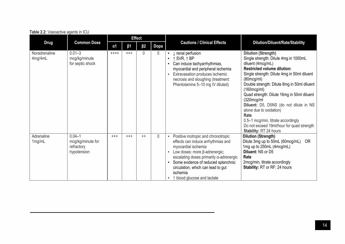
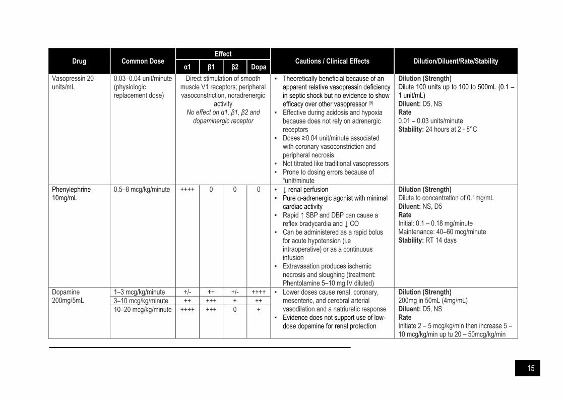
Table 2.2: Vasoactive agents in ICU
Drug Common Dose Effect
Cautions / Clinical Effects Dilution/Diluent/Rate/Stability α1 β1 β2 Dopa
Noradrenaline 4mg/4mL
0.01–3 mcg/kg/minute for septic shock
++++ +++ 0 0 ▪ ↓ renal perfusion ▪ ↑ SVR, ↑ BP ▪ Can induce tachyarrhythmias,
myocardial and peripheral ischemia ▪ Extravasation produces ischemic
necrosis and sloughing (treatment: Phentolamine 5–10 mg IV diluted)
Dilution (Strength)
Single strength: Dilute 4mg in 1000mL diluent (4mcg/mL) Restricted volume dilution: Single strength: Dilute 4mg in 50ml diluent (80mcg/ml) Double strength: Dilute 8mg in 50ml diluent (160mcg/ml) Quad strength: Dilute 16mg in 50ml diluent (320mcg/ml Diluent: D5, D5NS (do not dilute in NS alone due to oxidation) Rate 0.5–1 mcg/min, titrate accordingly Do not exceed 19ml/hour for quad strength
Stability: RT 24 hours
Adrenaline 1mg/mL
0.04–1 mcg/kg/minute for refractory hypotension
+++ +++ ++ 0 ▪ Positive inotropic and chronotropic effects can induce arrhythmias and myocardial ischemia
▪ Low doses: more β-adrenergic; escalating doses primarily α-adrenergic
▪ Some evidence of reduced splanchnic circulation, which can lead to gut ischemia
▪ ↑ blood glucose and lactate
Dilution (Strength) Dilute 3mg up to 50mL (60mcg/mL) OR 1mg up to 250mL (4mcg/mL) Diluent: NS or D5 Rate 2mcg/min, titrate accordingly Stability: RT or RF: 24 hours
15
Drug Common Dose Effect
Cautions / Clinical Effects Dilution/Diluent/Rate/Stability α1 β1 β2 Dopa
Vasopressin 20 units/mL
0.03–0.04 unit/minute (physiologic replacement dose)
Direct stimulation of smooth muscle V1 receptors; peripheral vasoconstriction, noradrenergic
activity No effect on α1, β1, β2 and
dopaminergic receptor
▪ Theoretically beneficial because of an apparent relative vasopressin deficiency in septic shock but no evidence to show efficacy over other vasopressor [9]
▪ Effective during acidosis and hypoxia because does not rely on adrenergic receptors
▪ Doses ≥0.04 unit/minute associated with coronary vasoconstriction and peripheral necrosis
▪ Not titrated like traditional vasopressors ▪ Prone to dosing errors because of
“unit/minute
Dilution (Strength)
Dilute 100 units up to 100 to 500mL (0.1 – 1 unit/mL) Diluent: D5, NS Rate 0.01 – 0.03 units/minute Stability: 24 hours at 2 - 8°C
Phenylephrine 10mg/mL
0.5–8 mcg/kg/minute
++++ 0 0 0 ▪ ↓ renal perfusion ▪ Pure α-adrenergic agonist with minimal
cardiac activity ▪ Rapid ↑ SBP and DBP can cause a
reflex bradycardia and ↓ CO ▪ Can be administered as a rapid bolus
for acute hypotension (i.e intraoperative) or as a continuous infusion
▪ Extravasation produces ischemic necrosis and sloughing (treatment: Phentolamine 5–10 mg IV diluted)
Dilution (Strength)
Dilute to concentration of 0.1mg/mL Diluent: NS, D5 Rate Initial: 0.1 – 0.18 mg/minute Maintenance: 40–60 mcg/minute Stability: RT 14 days
Dopamine 200mg/5mL
1–3 mcg/kg/minute +/- ++ +/- ++++ ▪ Lower doses cause renal, coronary, mesenteric, and cerebral arterial vasodilation and a natriuretic response
▪ Evidence does not support use of low-dose dopamine for renal protection
Dilution (Strength) 200mg in 50mL (4mg/mL) Diluent: D5, NS Rate Initiate 2 – 5 mcg/kg/min then increase 5 – 10 mcg/kg/min up tu 20 – 50mcg/kg/min
3–10 mcg/kg/minute ++ +++ + ++
10–20 mcg/kg/minute ++++ +++ 0 +

16
Drug Common Dose Effect
Cautions / Clinical Effects Dilution/Diluent/Rate/Stability α1 β1 β2 Dopa
▪ Any dose can induce arrhythmia, endocrine changes (ie decreased prolactin, growth hormone, thyroid hormone); however, the clinical significance is unknown
▪ Immediate precursor of noradrenaline. However prolonged infusions can deplete endogenous noradrenaline stores, resulting in a loss of vasopressor response.
▪ Effects on renal blood flow may be lost at higher doses because of predominant alpha-1 effects
Stability: RT 24 hours
Dobutamine 250mg/20mL
2.5–20 mcg/kg/minute
+ +++ + 0 ▪ Can cause hypotension because of β2-stimulation
▪ Higher doses can cause tachyarrhythmias and changes in BP, which can lead to myocardial ischemia
Dilution (Strength) Dilute 250mg up to 50mL (5mg/mL) Diluent: D5, NS, D10, D5NS, LR, HSD5 Rate 0.5 – 1 mcg/kg /min up to 2.5 – 20mcg/kg/min, max: 40mcg/kg/min Stability: RT 24 hours
Milrinone 50-mcg/kg load over 10 minutes, followed by 0.375–0.75 mcg/kg/minute
0 0 0 0 ▪ Positive inotrope ▪ Vasodilation/hypotension, arrhythmias
are possible ▪ Require dose adjustment in renal failure ▪ Loading doses often omitted especially
if patient hypotensive
Dilution (Strength)
Ready pack: 200mcg/mL in 5% Dextrose solutio Diluent: D5, NS Rate Loading: 50mcg/kg IV over 10 minutes Maintenance: 0.375 - 0.75 mcg/kg/minute Renal Failure: CrCl: 10 - 30mL/min 0.23 – 0.33 mcg/kg/min CrCl: <10mL/min 0.2 – 0.23 mcg/kg/min Stability: RT 3 days
17
The recommended first-line agent for septic shock is noradrenaline, preferably administered through a central catheter. Noradrenaline has predominant alpha-receptor agonist effects and results in potent peripheral arterial vasoconstriction without significantly increasing heart rate or cardiac output. The dosage range for noradrenaline is 5-20 µg/min, and it is not based on the weight of the patient. Second-line vasopressors appropriate for patients who have persistent hypotension despite maximal doses of noradrenaline or dopamine include synthetic human angiotensin II, adrenaline, phenylephrine, and vasopressin.
Adjunctive Steroid Therapy in Septic Shock When compared to placebo in mechanically ventilated patients with septic shock:
The use of hydrocortisone 200mg/day as continuous infusion does not reduce mortality (27.9% vs. 28.8%, OR 0.95, 95% CI 0.82 to 1.1, p=0.5) but it has faster resolution of shock (median duration, 3 days (IQR 2-5) vs. 4 days (IQR 2-9), improves MAP (5.39mmHg higher, p<0.001) and shorten ICU stay (median 10 days vs. 12 days, p<0.001) - ADRENAL trial
The use of hydrocortisone (50mg IV 6 hourly) with fludrocortisone (50mcg PO once daily) reduces mortality (43% vs. 49.1%, RR 0.88, 95% CI 0.78 to 0.99, NNT=16, p =0.03) and higher vasopressor free days (17 vs. 15 days, p<0.001) – APROCCHSS trial
It is unclear why the results of APROCCHSS differ from ADRENAL. APROCCHSS recruited much sicker patients evidenced by placebo mortality rate (49.1% APROCCHSS vs. 28.8% ADRENAL). The possibility that hydrocortisone only offer benefit in extremely sick patient is unknown. Regardless mortality, both APROCCHSS and ADRENAL demonstrate that hydrocortisone has a vasopressor- sparing effect with minimal adverse effect profile References and Suggested Readings Annane D, Renault A, Brun-Buisson C, Megarbane B, Quenot JP, Siami S, Cariou A, Forceville X, Schwebel C,
Martin C, Timsit JF. Hydrocortisone plus fludrocortisone for adults with septic shock. N Eng J Med. 2018. 378(9): 809-18.
David F, Gaieski M. Definition, classification, etiology and pathophysiology of shock in adults. UpToDate2018. De Backer D, Creteur J, Silva E, Vincent J-L. Effects of dopamine, norepinephrine, and epinephrine on the
splanchnic circulation in septic shock: which is best? Crit Care Med. 2003. 31(6): 1659-1667. Dilution Guide for High Alert Medications: Pharmaceutical Services Division; 2011. Kristeller JL. American College of Clinical Pharmacy (ACCP) Critical Care Notes 2013. Liverpool Hospital Noradrenaline ICU Guideline 2014 Milrinone [database on the Internet]. Available from: https://globalrph.com/dilution/milrinone-primacor/. Overgaard CB, Dzavik B. Inotropes and vasopressors review of physiology and clinical use in cardiovascular
disease. Circulation 2008. 118(10): 1047-1056. Phenylephrine [database on the Internet]. Available from: https://globalrph.com/dilution/neosynephrine-
phenylephrine/ Phenylephrine (Drug Information). UptoDate 2020. Russell JA, Walley KR, Singer J, Gordon AC, Hébert PC, Cooper DJ, et al. Vasopressin versus norepinephrine
infusion in patients with septic shock. NEJM. 2008. 358(9): 877-887. Vasopressin [database on the Internet]. Available from: https://www.shpa.org.au. Venkatesh B, Finfer S, Cohen J, Rajbhandari D, Arabi Y, Bellomo R, Billot L, Correa M, Glass P, Harward M, Joyce
C. Adjunctive glucocorticoid therapy in patients with septic shock. N Eng J Med. 2018. 378(9): 797-808.
18
CHAPTER 3
Organs Dysfunction
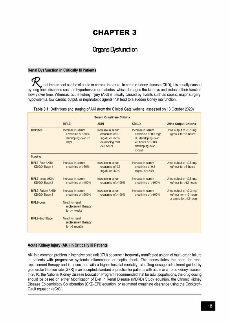
Renal Dysfunction in Critically Ill Patients enal impairment can be of acute or chronic in nature. In chronic kidney disease (CKD), it is usually caused by long-term diseases such as hypertension or diabetes, which damages the kidneys and reduces their function slowly over time. Whereas, acute kidney injury (AKI) is usually caused by events such as sepsis, major surgery, hypovolemia, low cardiac output, or nephrotoxic agents that lead to a sudden kidney malfunction. Table 3.1: Definitions and staging of AKI (from the Clinical Gate website, assessed on 13 October 2020) Acute Kidney Injury (AKI) in Critically Ill Patients AKI is a common problem in intensive care unit (ICU) because it frequently manifested as part of multi-organ failure in patients with progressive systemic inflammation or septic shock. This necessitates the need for renal replacement therapy and is associated with a higher hospital mortality rate. Drug dosage adjustment guided by glomerular filtration rate (GFR) is an accepted standard of practice for patients with acute or chronic kidney disease. In 2010, the National Kidney Disease Education Program recommended that for adult populations, the drug dosing should be based on either Modification of Diet in Renal Disease (MDRD) Study equation, the Chronic Kidney Disease Epidemiology Collaboration (CKD-EPI) equation, or estimated creatinine clearance using the Cockcroft-Gault equation (eCrCl).
R
19
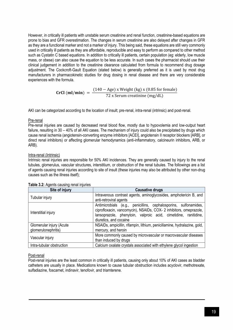
However, in critically ill patients with unstable serum creatinine and renal function, creatinine-based equations are prone to bias and GFR overestimation. The changes in serum creatinine are also delayed after changes in GFR as they are a functional marker and not a marker of injury. This being said, these equations are still very commonly used in critically ill patients as they are affordable, reproducible and easy to perform as compared to other method such as Cystatin C based equations. In addition to critically ill patients, certain population (eg: elderly, low muscle mass, or obese) can also cause the equation to be less accurate. In such cases the pharmacist should use their clinical judgement in addition to the creatinine clearance calculated from formula to recommend drug dosage adjustment. The Cockcroft-Gault Equation (stated below) is generally preferred as it is used by most drug manufacturers in pharmacokinetic studies for drug dosing in renal disease and there are very considerable experiences with the formula.
𝐂𝐫𝐂𝐥 (𝐦𝐥/𝐦𝐢𝐧) = (140 − Age) x Weight (kg) x (0.85 for female)
72 x Serum creatinine (mg/dL)
AKI can be categorized according to the location of insult; pre-renal, intra-renal (intrinsic) and post-renal. Pre-renal Pre-renal injuries are caused by decreased renal blood flow, mostly due to hypovolemia and low-output heart failure, resulting in 30 – 40% of all AKI cases. The mechanism of injury could also be precipitated by drugs which cause renal ischemia (angiotensin-converting enzyme inhibitors [ACEI], angiotensin II receptor blockers [ARB], or direct renal inhibitors) or affecting glomerular hemodynamics (anti-inflammatory, calcineurin inhibitors, ARB, or ARB). Intra-renal (Intrinsic) Intrinsic renal injuries are responsible for 50% AKI incidences. They are generally caused by injury to the renal tubules, glomerulus, vascular structures, interstitium, or obstruction of the renal tubules. The followings are a list of agents causing renal injuries according to site of insult (these injuries may also be attributed by other non-drug causes such as the illness itself); Table 3.2: Agents causing renal injuries
Site of injury Causative drugs
Tubular injury Intravenous contrast agents, aminoglycosides, amphotericin B, and anti-retroviral agents
Interstitial injury
Antimicrobials (e.g., penicillins, cephalosporins, sulfonamides, ciprofloxacin, vancomycin), NSAIDs, COX- 2 inhibitors, omeprazole, lansoprazole, phenytoin, valproic acid, cimetidine, ranitidine, diuretics, and cocaine
Glomerular injury (Acute glomerulonephritis)
NSAIDs, ampicillin, rifampin, lithium, penicillamine, hydralazine, gold, mercury, and heroin
Vascular injury More commonly caused by microvascular or macrovascular diseases than induced by drugs
Intra-tubular obstruction Calcium oxalate crystals associated with ethylene glycol ingestion
Post-renal Post-renal injuries are the least common in critically ill patients, causing only about 10% of AKI cases as bladder catheters are usually in place. Medications known to cause tubular obstruction includes acyclovir, methotrexate, sulfadiazine, foscarnet, indinavir, tenofovir, and triamterene.
20
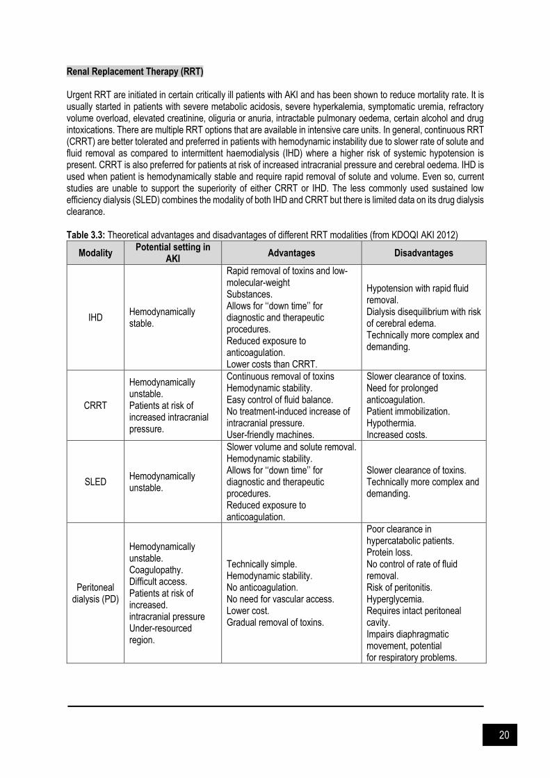
Renal Replacement Therapy (RRT) Urgent RRT are initiated in certain critically ill patients with AKI and has been shown to reduce mortality rate. It is usually started in patients with severe metabolic acidosis, severe hyperkalemia, symptomatic uremia, refractory volume overload, elevated creatinine, oliguria or anuria, intractable pulmonary oedema, certain alcohol and drug intoxications. There are multiple RRT options that are available in intensive care units. In general, continuous RRT (CRRT) are better tolerated and preferred in patients with hemodynamic instability due to slower rate of solute and fluid removal as compared to intermittent haemodialysis (IHD) where a higher risk of systemic hypotension is present. CRRT is also preferred for patients at risk of increased intracranial pressure and cerebral oedema. IHD is used when patient is hemodynamically stable and require rapid removal of solute and volume. Even so, current studies are unable to support the superiority of either CRRT or IHD. The less commonly used sustained low efficiency dialysis (SLED) combines the modality of both IHD and CRRT but there is limited data on its drug dialysis clearance. Table 3.3: Theoretical advantages and disadvantages of different RRT modalities (from KDOQI AKI 2012)
Modality Potential setting in
AKI Advantages Disadvantages
IHD Hemodynamically stable.
Rapid removal of toxins and low-molecular-weight Substances. Allows for ‘‘down time’’ for diagnostic and therapeutic procedures. Reduced exposure to anticoagulation. Lower costs than CRRT.
Hypotension with rapid fluid removal. Dialysis disequilibrium with risk of cerebral edema. Technically more complex and demanding.
CRRT
Hemodynamically unstable. Patients at risk of increased intracranial pressure.
Continuous removal of toxins Hemodynamic stability. Easy control of fluid balance. No treatment-induced increase of intracranial pressure. User-friendly machines.
Slower clearance of toxins. Need for prolonged anticoagulation. Patient immobilization. Hypothermia. Increased costs.
SLED Hemodynamically unstable.
Slower volume and solute removal. Hemodynamic stability. Allows for ‘‘down time’’ for diagnostic and therapeutic procedures. Reduced exposure to anticoagulation.
Slower clearance of toxins. Technically more complex and demanding.
Peritoneal dialysis (PD)
Hemodynamically unstable. Coagulopathy. Difficult access. Patients at risk of increased. intracranial pressure Under-resourced region.
Technically simple. Hemodynamic stability. No anticoagulation. No need for vascular access. Lower cost. Gradual removal of toxins.
Poor clearance in hypercatabolic patients. Protein loss. No control of rate of fluid removal. Risk of peritonitis. Hyperglycemia. Requires intact peritoneal cavity. Impairs diaphragmatic movement, potential for respiratory problems.
21
Drug Dosing Considerations in Critically Ill Patient Receiving RRT It is reasonably common for critically ill patients to have renal impairment with ongoing infectious diseases that requires renal replacement and antimicrobial therapy. The concept of drug dosage adjustment for AKI or CKD patients especially for antimicrobial therapy is an accepted standard of practice. However, for individuals who require the provision of renal replacement therapy, be it continuous or intermittent hemodialysis, they may require additional dosing adjustment for optimal drug therapy. It is crucial for critically ill patients with multi organ failure to have optimized antimicrobial dosing regimens to maximize patient outcomes while limiting adverse drug events. Many evidences on drug dialysis clearance that are used to guide antimicrobial dosing in critically ill patients have become outdated due to introduction of newer RRTs and advances in RRT efficiency. Additionally, data from current dosing regimen in dialysis-dependent patients are frequently derived from stable CKD patients, as compared to critically ill patients with ever-changing factors that can affect the pharmacokinetics and pharmacodynamics properties of antimicrobial. Alterations to the antimicrobial regimen are often necessary throughout the course of therapy. In addition to clinical status of the patient, changes in renal function, the patient’s fluid status (as an index of the drug’s volume of distribution), the RRT prescription (e.g., ultrafiltration and dialysis flow rates), the delivered dose, clinical response, and the presence of adverse events should be monitored to help guide dosage adjustment. While patients are on RRT, dosage adjustments of drugs are determined by the drug dialyzability. This in turn would depend primarily on several physicochemical characteristics of the drug which are protein binding, molecular weight, and volume of distribution. Other factors such as dialysis membrane, molecular charge, blood and dialysate flow rates also affect drug removal although not as significant. The recommendations for antimicrobial dosing in critically ill patients with AKI, CKD, or receiving RRT can be seen in (refer Table 3.4)
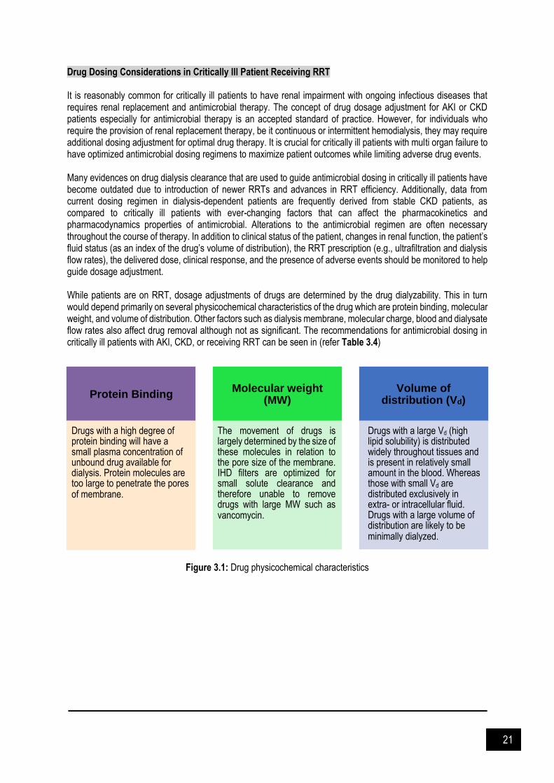
Figure 3.1: Drug physicochemical characteristics
Protein Binding
Drugs with a high degree of protein binding will have a small plasma concentration of unbound drug available for dialysis. Protein molecules are too large to penetrate the pores of membrane.
Molecular weight
(MW)
The movement of drugs is largely determined by the size of these molecules in relation to the pore size of the membrane. IHD filters are optimized for small solute clearance and therefore unable to remove drugs with large MW such as vancomycin.
Volume of
distribution (Vd)
Drugs with a large Vd (high lipid solubility) is distributed widely throughout tissues and is present in relatively small amount in the blood. Whereas those with small Vd are distributed exclusively in extra- or intracellular fluid. Drugs with a large volume of distribution are likely to be minimally dialyzed.
22
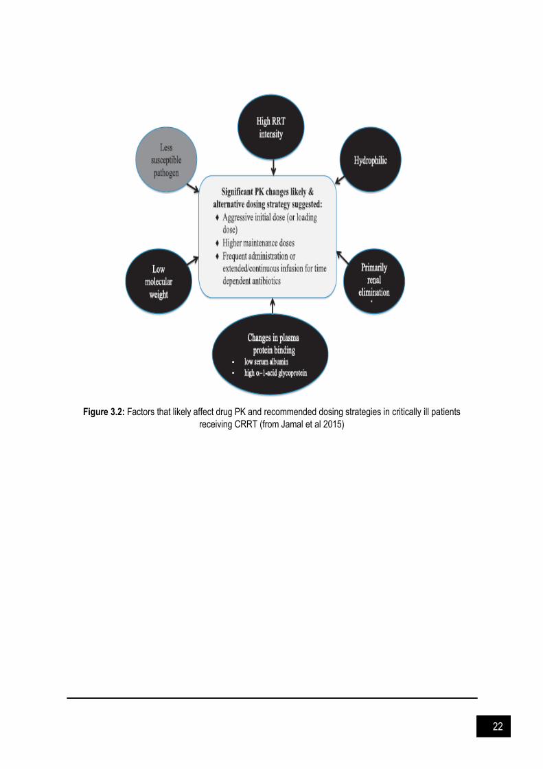
Figure 3.2: Factors that likely affect drug PK and recommended dosing strategies in critically ill patients
receiving CRRT (from Jamal et al 2015)

23
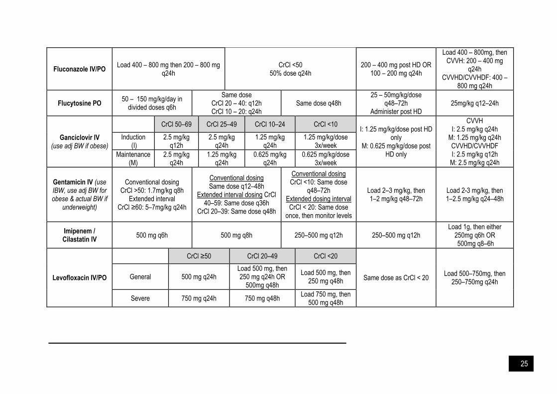
Table 3.4: Antimicrobial dosing in renal impairment
Drug CrCl >50 mL/min CrCl 10–50 mL/min CrCl <10 mL/min IHD# CRRT
Acyclovir IV (use adj BW in
obese) 5–10 mg/kg q8h
Same dose CrCl 25 – 50: q12h CrCl 10 – 25: q24h
50% dose q24h 2.5 – 5 mg/kg q24h CVVH: 5–10 mg/kg q24h CVVHD/CVVHDF: 5–10
mg/kg q12–24h
Acyclovir PO
General dose CrCl >25 CrCl 10–25 CrCl <10
Same dose with CrCl <10 No data 800mg 5x daily No adjustment 800mg q8h 800mg q12h
400mg q8h No adjustment 200mg q8h 200mg q12h
Amikacin IV (use IBW, adj BW for obese & actual BW if
underweight)
15 – 20 mg/kg/day 7.5 mg/kg q24–72h 7.5 mg/kg q48–72hrs
5 – 7.5 mg/kg q48–72h OR post HD only
Monitor level and adjust dose
Load 10 mg/kg, then 7.5 mg/kg q24–48h Monitor level and adjust
dose
Amoxicillin / clavulanic acid
IV/PO
CrCl >30 IV: 1.2 g q8h
PO: 624 mg q8h
CrCl 10–30 Same dose q12h
CrCl <10: Same dose q24h
Same dose with CrCl <10 No data
Ampicillin IV 2g q4–6h 2g q6–12h 2g q12–24h 2g q12h CVVH: 2g q8–12h CVVHD: 2g q8h
CVVHDF: 2g q6–8h
Ampicillin / Sulbactam IV
General CrCl ≥30 CrCl 15–29 CrCl <15 IHD CVVH: 3 g q8–12h
CVVHD: 3g q8h CVVHDF: 3g q6–8h
For MDR or sensitive strain A. baumannii;
9g q8h (9g sulbactam/day)
1.5 – 3 q6h Same dose q12h Same dose q24h Same dose q12–24h
MDR or sensitive strain A.
baumannii
CrCl ≥50 CrCl 20–50 CrCl <20 IHD
9g q8h (9g sulbactam/day)
6g q8h (6g sulbactam/day)
6g q12h (4g sulbactam/day)
6g q12h (4g sulbactam/day)
Cefazolin IV 1–2 g q8h CrCl 11–34
50% dose q12h 50% dose q18–24h
0.5–1 g q24h OR
2g/2g/3g post HD only
Load 2g, then CVVH: 1–2 g q12h
CVVHD/CVVHDF: 1 g q8h OR 2g q12h
Cefepime IV can be given as extended 4h infusion 1 g q24h 0.5 – 1 g q24h OR Load 2g, then

24
General
CrCl >60 CrCl 30–60 CrCl 11–29 1 – 2 g q48–72h CVVH: 1–2 g q12h CVVHD/CVVHDF: 1 g q8h
OR 2g q12h Max 2g q8h for GNR with
MIC ≥ 4mg/L)
2 g q12h OR 1 g q8h
2 g q24h OR 1 g q12h
1 g q24h
Severe 2 g q8h 2 g q12h 2 g q24h
Cefoperazone / Sulbactam IV
CrCl >50 CrCl 20–50 CrCl <20 IHD
No data General 1–2 g q12h 1–2 g q12h 1–2 g q12h 1–2 g q12h
A. baumannii 3 g q4h 2 g q4h 2 g q6h 2 g q6h
Cefotaxime IV 1–2 g q6–8h Same dose q6–12h Same dose q24h 1–2 g q24h CVVH: 1–2 g q8–12h CVVHD: 1–2 g q8h
CVVHDF: 1–2 g q6–8h
Ceftazidime IV 1–2 g q8h Same dose
CrCl 31–50: q12h CrCl 16–30: q24h
CrCl ≤15 0.5–1 g q24h
0.5–1 g q24h OR
1–2 g q48–72h
Load 2g, then CVVH: 1 – 2g q12h
CVVHD/CVVHDF: 1 g q8h OR 2g q12h
Cefuroxime IV 0.75 – 1.5 g q8h CrCl 10 – 20
Same dose q12h Same dose q24h
Administer a supplemental dose at the end of HD
1 g q12h
Ciprofloxacin IV 400 mg q8–12h CrCl 10–30
400 mg q12–24h 400 mg q24h 200–400 mg q24h 200–400 mg q8–24h
Ciprofloxacin PO 500–750 mg q12h CrCl 30–50 500mg q12h
CrCl <30 500mg q24h
250–500 mg q24h 500 mg q12–24h
Daptomycin (use adj BW in
obese) 6–12 mg/kg q24h
CrCl 11–30 Same dose q48h
Same dose q48h Same dose with CrCl <10 6 – 10 mg q48h OR
4 – 6 mg q24h
Ertapenem IV 1 g q24h CrCl <30
500 mg q24h 500 mg q24h Same dose with CrCl <10 1 g q24h
Ethambutol PO (use lean BW in
obese)
15 – 25mg/kg q24h (Max 1.6 g/day)
15 – 25mg/kg q24–36h 15 – 25mg/kg q48h 15 – 25mg/kg 3x/week 15 – 25mg/kg q24–36h
25
Fluconazole IV/PO Load 400 – 800 mg then 200 – 800 mg
q24h CrCl <50
50% dose q24h 200 – 400 mg post HD OR
100 – 200 mg q24h
Load 400 – 800mg, then CVVH: 200 – 400 mg
q24h CVVHD/CVVHDF: 400 –
800 mg q24h
Flucytosine PO 50 – 150 mg/kg/day in
divided doses q6h
Same dose CrCl 20 – 40: q12h CrCl 10 – 20: q24h
Same dose q48h 25 – 50mg/kg/dose
q48–72h Administer post HD
25mg/kg q12–24h
Ganciclovir IV (use adj BW if obese)
CrCl 50–69 CrCl 25–49 CrCl 10–24 CrCl <10 I: 1.25 mg/kg/dose post HD
only M: 0.625 mg/kg/dose post
HD only
CVVH I: 2.5 mg/kg q24h
M: 1.25 mg/kg q24h CVVHD/CVVHDF I: 2.5 mg/kg q12h
M: 2.5 mg/kg q24h
Induction (I)
2.5 mg/kg q12h
2.5 mg/kg q24h
1.25 mg/kg q24h
1.25 mg/kg/dose 3x/week
Maintenance (M)
2.5 mg/kg q24h
1.25 mg/kg q24h
0.625 mg/kg q24h
0.625 mg/kg/dose 3x/week
Gentamicin IV (use IBW, use adj BW for obese & actual BW if
underweight)
Conventional dosing CrCl >50: 1.7mg/kg q8h
Extended interval CrCl ≥60: 5–7mg/kg q24h
Conventional dosing Same dose q12–48h
Extended interval dosing CrCl 40–59: Same dose q36h
CrCl 20–39: Same dose q48h
Conventional dosing CrCl <10: Same dose
q48–72h Extended dosing interval CrCl < 20: Same dose
once, then monitor levels
Load 2–3 mg/kg, then 1–2 mg/kg q48–72h
Load 2-3 mg/kg, then 1–2.5 mg/kg q24–48h
Imipenem / Cilastatin IV
500 mg q6h 500 mg q8h 250–500 mg q12h 250–500 mg q12h Load 1g, then either
250mg q6h OR 500mg q8–6h
Levofloxacin IV/PO
CrCl ≥50 CrCl 20–49 CrCl <20
Same dose as CrCl < 20 Load 500–750mg, then
250–750mg q24h General 500 mg q24h
Load 500 mg, then 250 mg q24h OR
500mg q48h
Load 500 mg, then 250 mg q48h
Severe 750 mg q24h 750 mg q48h Load 750 mg, then
500 mg q48h

26
Meropenem IV (can be given as 3h extended infusion)
1 – 2 g q8h
CrCl 26–50 Same dose q12h
CrCl 10–25 50% dose q12h
50% dose q24h 0.5 – 1 g q24h
Load 1g, then CVVH: 500 mg q8h OR
1g q8–12h CVVHD/CVVHDF: 500 mg
q8–6h OR 1 g q8–12h CNS infection: 2g q12h
Oseltamivir PO
Prophylaxis Treatment Treatment
(severe/ICU) 30mg (low flux HD) OR 75mg (high flux HD) post HD for 5 days
Treatment: 75mg q12h Prophylaxis: 75mg q24h CrCl ≥30 75 mg q24h 75 mg q12h 150 mg q12h
CrCl <30 75 mg q48h 75 mg q24h 150 mg q24h
Penicillin G IV 2–4 mu q4h 2–3 mu q4h
(Administer 75% of normal dose)
1–2 mu q6h (Administer 20–50% of
normal dose)
Load normal dose, then either
0.5–1 mu (25–50% dose) q4–6h OR
1–2 mu (50–100% dose) q8–12h
Load 4 mu, then CVVH: 2 mu q4–6h
CVVHD: 2–3 mu q4–6h CVVHDF: 2–4 mu q4–6h
Piperacillin / Tazobactam IV
CrCl >40 CrCl 20–40 CrCl <20 General: 2.25 g q12h
Severe infection: 3.375 g q12h over 4h OR
2.25 g q8h
CVVH: 2.25-3.375 g q6–8h
CVVHD: 2.25-3.375 g q6h CVVHDF: 3.375g q6h OR
4.5g q8h
Intermittent Dosing
3.375 – 4.5 g q6h 2.25 – 3.375 g q6h 2.25 g q8–6h
4h Extended-Infusion Dosing
3.375 – 4.5 g q8–6h over 4h 3.375g q12 over 4h
Pyrazinamide PO 25 mg/kg q24h (Max 2 g/day)
CrCl <30 25 mg/kg 3x/week
Same dose with CrCl <30 Administer post HD only
No data
27
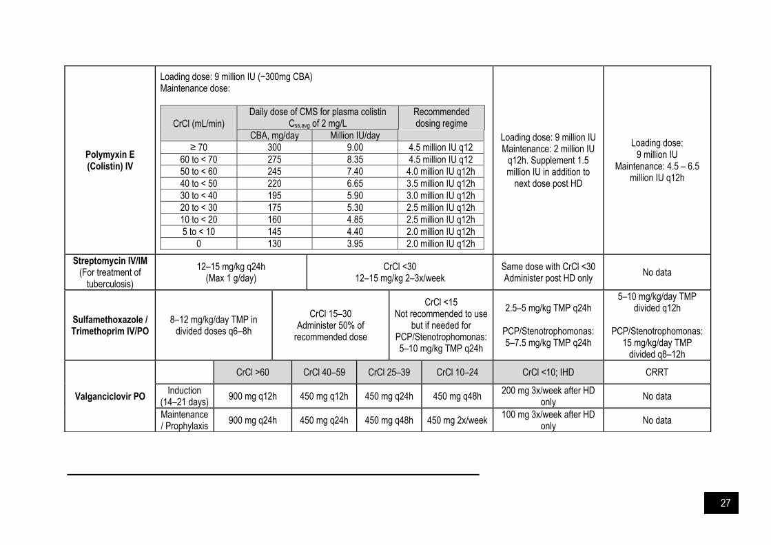
Polymyxin E (Colistin) IV
Loading dose: 9 million IU (~300mg CBA) Maintenance dose:
CrCl (mL/min)
Daily dose of CMS for plasma colistin Css,avg of 2 mg/L
Recommended dosing regime
CBA, mg/day Million IU/day
≥ 70 300 9.00 4.5 million IU q12
60 to < 70 275 8.35 4.5 million IU q12
50 to < 60 245 7.40 4.0 million IU q12h
40 to < 50 220 6.65 3.5 million IU q12h
30 to < 40 195 5.90 3.0 million IU q12h
20 to < 30 175 5.30 2.5 million IU q12h
10 to < 20 160 4.85 2.5 million IU q12h
5 to < 10 145 4.40 2.0 million IU q12h
0 130 3.95 2.0 million IU q12h
Loading dose: 9 million IU Maintenance: 2 million IU
q12h. Supplement 1.5 million IU in addition to
next dose post HD
Loading dose: 9 million IU
Maintenance: 4.5 – 6.5 million IU q12h
Streptomycin IV/IM (For treatment of
tuberculosis)
12–15 mg/kg q24h (Max 1 g/day)
CrCl <30 12–15 mg/kg 2–3x/week
Same dose with CrCl <30 Administer post HD only
No data
Sulfamethoxazole / Trimethoprim IV/PO
8–12 mg/kg/day TMP in divided doses q6–8h
CrCl 15–30 Administer 50% of
recommended dose
CrCl <15 Not recommended to use
but if needed for PCP/Stenotrophomonas: 5–10 mg/kg TMP q24h
2.5–5 mg/kg TMP q24h
PCP/Stenotrophomonas: 5–7.5 mg/kg TMP q24h
5–10 mg/kg/day TMP divided q12h
PCP/Stenotrophomonas:
15 mg/kg/day TMP divided q8–12h
Valganciclovir PO
CrCl >60 CrCl 40–59 CrCl 25–39 CrCl 10–24 CrCl <10; IHD CRRT
Induction (14–21 days)
900 mg q12h 450 mg q12h 450 mg q24h 450 mg q48h 200 mg 3x/week after HD
only No data
Maintenance / Prophylaxis
900 mg q24h 450 mg q24h 450 mg q48h 450 mg 2x/week 100 mg 3x/week after HD
only No data

28
#Unless stated otherwise, all IV preparations should be administered post HD on HD days
Vancomycin IV (use actual BW, not to exceed 2 g per
dose)
CrCl (mL/min) Dose & Frequency
>90 15 mg/kg q8–12h
51–89 10–20 mg/kg q12h
30–50 10–15 mg/kg q12hr to 20 mg/kg q24h
10–29 10–15 mg/kg q24h to 15 mg/kg q48h
<10 or AKI Load 15 mg/kg, then dose by level
Consider loading 25 – 30 mg/kg (max 2.5 g) for severe infections and ICU
Load 15-20mg/kg, then 500-1000mg OR
5-10 mg/kg after each HD session
Monitor level and adjust dose
Load 15-25 mg/kg, then CVVH: 10-15 mg/kg
q24-48h CVVHD: 10-15 mg/kg
q24h CVVHDF: 7.5-10 mg/kg
q12h Monitor level and adjust
dose
29
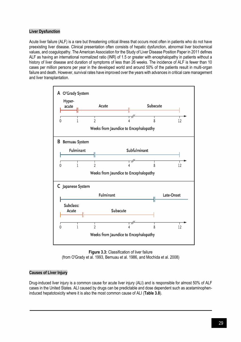
Liver Dysfunction Acute liver failure (ALF) is a rare but threatening critical illness that occurs most often in patients who do not have preexisting liver disease. Clinical presentation often consists of hepatic dysfunction, abnormal liver biochemical values, and coagulopathy. The American Association for the Study of Liver Disease Position Paper in 2011 defines ALF as having an international normalized ratio (INR) of 1.5 or greater with encephalopathy in patients without a history of liver disease and duration of symptoms of less than 26 weeks. The incidence of ALF is fewer than 10 cases per million persons per year in the developed world and around 50% of the patients result in multi-organ failure and death. However, survival rates have improved over the years with advances in critical care management and liver transplantation.
Figure 3.3: Classification of liver failure (from O'Grady et al. 1993, Bernuau et al. 1986, and Mochida et al. 2008)
Causes of Liver Injury Drug-induced liver injury is a common cause for acute liver injury (ALI) and is responsible for almost 50% of ALF cases in the United States. ALI caused by drugs can be predictable and dose dependent such as acetaminophen-induced hepatotoxicity where it is also the most common cause of ALI (Table 3.8).
30
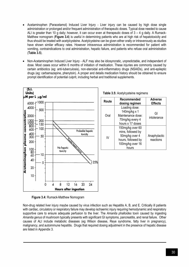
Acetaminophen (Paracetamol) Induced Liver Injury - Liver injury can be caused by high dose single administration or prolonged and/or frequent administration of therapeutic doses. Typical dose needed to cause ALI is greater than 10 g daily; however, it can occur even at therapeutic doses of 3 – 4 g daily. A Rumack-Matthew nomogram (Figure 3.4) is useful in determining patients who are at high risk of hepatotoxicity and thus should be treated with acetylcysteine. Acetylcysteine can be given either orally or intravenously as studies have shown similar efficacy rates. However intravenous administration is recommended for patient with vomiting, contraindications to oral administration, hepatic failure, and patients who refuse oral administration (Table 3.5).
Non-Acetaminophen Induced Liver Injury - ALF may also be idiosyncratic, unpredictable, and independent of dose. Most cases occur within 6 months of initiation of medication. These injuries are commonly caused by certain antibiotics (eg: anti-tuberculosis), non-steroidal anti-inflammatory drugs (NSAIDs), and anti-epileptic drugs (eg: carbamazepine, phenytoin). A proper and details medication history should be obtained to ensure prompt identification of potential culprit, including herbal and traditional supplements.
Table 3.5: Acetylcysteine regimens
Figure 3.4: Rumack-Matthew Nomogram Non-drug related liver injury maybe caused by virus infection such as Hepatitis A, B, and E. Critically ill patients with cardiac, circulatory or respiratory failure may develop ischaemic injury requiring hemodynamic and respiratory supportive care to ensure adequate perfusion to the liver. The Amanita phalloides toxin caused by ingesting Amanita genus of mushroom typically presents with significant GI symptoms, pancreatitis, and renal failure. Other causes of ALI include metabolic diseases (eg Wilson disease, Reye syndrome, fatty liver in pregnancy), malignancy, and autoimmune hepatitis. Drugs that required dosing adjustment in the presence of hepatic disease are listed in Appendix 3.
Route Recommended dosing regimen
Adverse Effects
Oral
Loading dose: 140mg/kg x 1
Maintenance dose: 70mg/kg every 4 hours x 17 doses
GI intolerance
IV
150mg/kg over 60 mins, followed by 50mg/kg over 4
hours, followed by 100mg/kg over 16
hours
Anaphylactic reactions

31
Treatment and Management of Complications Neurologic conditions - encephalopathy may initially present as agitation and confusion but can progress rapidly hence requires close monitoring. The West Haven Coma Scale (Table X) is useful in determining the severity of encephalopathy. The aim of clinical strategy is to prevent onset of encephalopathy, limit its severity, and reduce risk of cerebral edema. Evidence shows both systemic and local inflammation, and circulating neurotoxin (ammonia) plays a role in the pathogenesis of encephalopathy. It can manifest in patients with low systemic blood pressure and vasodilatation, and can be precipitated by sepsis. The inflammatory mediators that are triggered may worsen encephalopathy by altering cerebral endothelial permeability to neurotoxins or cerebral perfusion. There is an association between increased arterial ammonia levels and the development of encephalopathy as detoxification of ammonia to urea is often impaired in liver failure. As the level of ammonia increases, it is removed from the brain via metabolism to glutamine which increases intracellular osmolarity. This eventually leads to cerebral swelling and affects cerebral functions. Levels greater than 200 mcg/dL are associated with cerebral herniation. Cerebral edema can precipitate tissue hypoxia and long-term neurologic deficits. While in severe cases it can lead to intracranial hypertension which is a leading cause of death among patient with ALF. The following approach should be considered in initial phase of managing patient with hepatic encephalopathy;
Patients should be nursed in a 20° – 30° head up tilt to improve venous drainage and maintain head in a neutral position. Excessive stimulation should be avoided by minimizing suctioning and other noxious stimuli. Agitation should be controlled as Valsalva maneuver from psychomotor agitation may lead to increase ICP.
ICP should be kept <25 – 30mmHg and cerebral perfusion pressure (CPP) should be maintained at 50 – 60mmHg. CPP = MAP – ICP
Excessive hyperventilation may lead to cerebral vasoconstriction therefore a PaCO2 of 25 – 30mmHg is recommended.
Moderate hypothermia (32° – 33°C) results in reduction in cerebral blood flow and cerebral metabolism, increase ammonia uptake and glutamine synthesis.
Lactulose is used to reduce serum ammonia levels in patients with low grade encephalopathy. Administer at a dose of 30 – 60ml orally or via nasogastric tube, titrated to maintain 3 – 4 soft stools daily. Caution should be used to avoid overdistention of bowel in patients in need of liver transplantation.
Seizure can potentially increase ICP and therefore must be promptly controlled with phenytoin and/or benzodiazepines. Short acting benzodiazepines can be administered in phenytoin-refractory cases. There is insufficient data to suggest the use of prophylactic antiepileptic drugs.
Mannitol has been shown to correct episodes of elevated ICP in acute liver failure patients. Mannitol (0.5 – 1g/kg) is recommended as first line therapy for intracranial hypertension. The dose may be repeated once or twice as needed as long as the serum osmolality is <320 mOsm/L. However, caution should be used to avoid fluid overload and hyperosmolarity. Mannitol may be less effective when ICP is > 60mmHg. Prophylactic administration of mannitol is not recommended.
Cardiorespiratory dysfunction - Circulatory dysfunction and hypotension are common and multifactorial in origin. High levels of nitric oxide and cGMP leads to a state of high cardiac output, low mean arterial pressure, and low systemic vascular resistance. Most patients are severely dehydrated upon initial presentation due to poor nutritional status. Treatment consists of early restoration of intravascular volume and systemic perfusion with inotropes and/or vasopressor as well adequate oxygen delivery. Coagulopathy & renal dysfunction - Bleeding risk is elevated due to decreased synthesis and depletion of fibrinolytic proteins, anti-coagulants proteins, and pro-coagulant factors. Bleeding risk tends to be higher in patients with low platelet counts in addition to abnormal coagulation laboratory values. Elevated INR without signs and symptoms of acute bleed should be corrected using fresh frozen plasma. Alternatively, vitamin K can be administered orally or intravenously as ALF patients often have vitamin K deficiency. Platelet count should be restored to > 50,000/mm3 before invasive procedure. Patients with elevated INR with significant bleeding may receive Factor VIIa but may increase risk of thrombosis. Renal failure occurs in 40% to 85% of ALF patients, particularly in the elderly or acetaminophen–induced ALF.

32
Table 3.6: The West Haven Coma Scale
Grade Signs and symptoms
1
Trivial lack of awareness Euphoria or anxiety Shortened attention span Impaired performance of addition
2
Lethargy or apathy Minimal disorientation for time or place Subtle personality changes Inappropriate behaviour Impaired performance of subtraction
3 Somnolence to semi-stupor, but responsive to verbal stimulus Confusion Gross disorientation
4 Coma
Infection – The risk of sepsis is high and has been reported in 55% to 90% of patients due to the immunocompromised state which results from dysfunction of monocytes, neutrophils, Kupffer cells, and complement system. Furthermore, it is associated with hemodynamic instability, progression of hepatic encephalopathy, and renal failure. Common infections include urinary tract infections, pneumonia, catheter related blood stream infections, and spontaneous bacterial peritonitis. Fungal infections occur in 30% of the patients with Candida albicans being the most common organism. Regular assessment for infection is therefore crucial. Metabolic abnormalities - Patients with ALF are at higher risk for hypoglycaemia due to high energy expenditure and protein catabolism. However, both hypoglycemia and hyperglycemia can occur and should be avoided in patients with ALF. Serum glucose should be monitored every 2 to 6 hours. If the glucose level is below 100 mg/dL, begin dextrose 10% infusion and maintain a serum glucose level above 100 mg/dL and less than 140 to 180 mg/dL. Drug Dose Modification in Liver Impairment Drug metabolism may be affected in patient with liver impairment. Hence, in order to decide drug dosing in liver failure, three important factors need to be considered, namely;
1. Pharmacokinetic alterations of drugs 2. Pharmacodynamic alteration of drugs 3. Increased susceptibility of patients to adverse events particularly hepatotoxicity.
Table 3.7: Child-Pugh score
Parameter Score
1 2 3
Ascites None Mild Moderate or Severe
Encephalopathy (grade) None 1–2 3–4
Bilirubin (mmol/L) <35 35–50 >50
Bilirubin in Primary Biliary Cirrhosis (mmol/L)
<70 70–170 >170
Albumin (g/L) >35 28–35 <28
INR <1.7 1.8–2.3 >2.3
Grade Score 1st year Survival Rate
A 5–6 100%
B 7–9 81%
C >9 45%
33
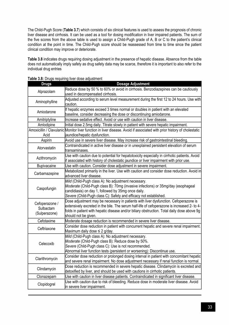
The Child-Pugh Score (Table 3.7) which consists of six clinical features is used to assess the prognosis of chronic liver disease and cirrhosis. It can be used as a tool for dosing modification in liver impaired patients. The sum of the five scores from the above table is used to assign a Child-Pugh grade of A, B or C to the patient’s clinical condition at the point in time. The Child-Pugh score should be reassessed from time to time since the patient clinical condition may improve or deteriorate. Table 3.8 indicates drugs requiring dosing adjustment in the presence of hepatic disease. Absence from the table does not automatically imply safety as drug safety data may be scarce, therefore it is important to also refer to the individual drug entries Table 3.8: Drugs requiring liver dose adjustment
Drugs Dosage Adjustment
Alprazolam Reduce dose by 50 % to 60% or avoid in cirrhosis. Benzodiazepines can be cautiously used in decompensated cirrhosis.
Aminophylline Adjusted according to serum level measurement during the first 12 to 24 hours. Use with caution.
Amiodarone If hepatic enzymes exceed 3 times normal or doubles in patient with an elevated baseline, consider decreasing the dose or discontinuing amiodarone.
Amitriptyline Increase sedative effect. Avoid or use with caution in liver disease.
Amlodipine Initial dose 2.5mg daily. Titrate slowly in patient with severe hepatic impairment.
Amoxicillin / Clavulanic Acid
Monitor liver function in liver disease. Avoid if associated with prior history of cholestatic jaundice/hepatic dysfunction.
Aspirin Avoid use in severe liver disease. May increase risk of gastrointestinal bleeding.
Atorvastatin Contraindicated in active liver disease or in unexplained persistent elevation of serum transaminases.
Azithromycin Use with caution due to potential for hepatotoxicity especially in cirrhotic patients. Avoid if associated with history of cholestatic jaundice or liver impairment with prior use.
Bupivacaine Use with caution. Consider dose adjustment in severe impairment.
Carbamazepine Metabolized primarily in the liver. Use with caution and consider dose reduction. Avoid in advanced liver disease.
Caspofungin
Mild (Child-Pugh class A): No adjustment necessary. Moderate (Child-Pugh class B): 70mg (invasive infections) or 35mg/day (esophageal candidiasis) on day 1, followed by 35mg once daily. Severe (Child-Pugh class C): Safety and efficacy not established.
Cefoperazone / Sulbactam
(Sulperazone)
Dose adjustment may be necessary in patients with liver dysfunction. Cefoperazone is extensively excreted in the bile. The serum half-life of cefoperazone is increased 2- to 4-folds in patient with hepatic disease and/or biliary obstruction. Total daily dose above 9g should not be given.
Cefotaxime Moderate dosage reduction is recommended in severe liver disease.
Ceftriaxone Consider dose reduction in patient with concurrent hepatic and severe renal impairment. Maximum daily dose ≤ 2 g/day.
Celecoxib
Mild (Child-Pugh class A): No adjustment necessary. Moderate (Child-Pugh class B): Reduce dose by 50%. Severe (Child-Pugh class C): Use is not recommended. Abnormal liver function tests (persistent or worsening): Discontinue use.
Clarithromycin Consider dose reduction or prolonged dosing interval in patient with concomitant hepatic and severe renal impairment. No dose adjustment necessary if renal function is normal.
Clindamycin Dose reduction is recommended in severe hepatic disease. Clindamycin is excreted and detoxified by liver, and should be used with cautions in cirrhotic patients.
Clonazepam Use with caution in liver disease patients. Contraindicated in significant liver disease.
Clopidogrel Use with caution due to risk of bleeding. Reduce dose in moderate liver disease. Avoid in severe liver impairment.

34
Drugs Dosage Adjustment
Dantrolene Chronic therapy is contraindicated in active liver disease due to potential for hepatotoxicity.
Dexmedetomidine Consider dose reduction. Clearance is reduced in varying degrees based on the level of impairment.
Diazepam Use with caution due to increase distribution and half-life, and reduced clearance. Oral tablets are contraindicated in severe liver impairment.
Digoxin No specific dosage adjustment is necessary.
Diltiazem Use with caution as diltiazem is extensively metabolized by the liver and half-life is increased in patient with cirrhosis.
Enalapril No adjustment necessary. Hydrolysis may be delayed and/or impaired in severe liver impairment, but the pharmacodynamic effects of the drug do not appear to be significantly altered.
Enoxaparin Use with caution in liver impairment.
Ertapenem Adjustments cannot be recommended (lack of experience and research in this patient population).
Erythromycin Macrolide antibiotics are excreted and detoxified by liver. Use with caution in liver impairment.
Esomeprazole Mild to moderate (Child-Pugh class A or B): No adjustment necessary. Severe (Child-Pugh Class C): Dose should not exceed 20mg.
Felodipine Initial dose 2.5mg daily. Monitor blood pressure closely during titration. Do not use dose above 10mg/day as incidence and severity of adverse event outweighs additional hypotensive effects.
Fluconazole
Use with caution in patient with liver impairment or previous hepatotoxicity from other azole derivatives. Patient who develops abnormal liver function tests during therapy should be monitored closely and discontinued if symptoms consistent with liver disease develop.
Frusemide Cirrhotic patient may have diminished natriuretic effect with increased sensitivity to hypokalemia and volume depletion. Monitor effects particularly with high doses.
Fusidic acid Avoid in hyperbilirubinemia patient.
Granisetron Kinetic studies in patient with liver impairment showed total clearance was approximately halved, however standard doses were much tolerated, and dose adjustments are not necessary.
Hydrocortisone Use with caution in patient with liver impairment including cirrhosis. Long term use has been associated with fluid retention.
Imipenem / Cilastatin Liver impairment may reduce cilastatin clearance. Consider decreasing the dosing frequency.
Insulin Insulin requirements may be reduced. Close monitoring of blood glucose and adjustment of therapy is required in liver impairment.
Isoniazid
No adjustment required. However, use with caution as isoniazid may accumulate, and additional liver damage may occur in patient with pre-existing liver disease. For ALT or AST > 3 x ULN, discontinue or temporarily withhold treatment. Refer Clinical Practice Guidelines: Management of Tuberculosis, 3rd edition for management of tuberculosis in liver impairment.
Itraconazole Use with caution in patient with liver impairment. Monitor closely for signs and symptoms of toxicity.
Ketorolac Use with caution, may cause elevation of liver enzyme. Liver impairment may prolong elimination half-life. Discontinue if clinical signs and symptoms of liver disease develop.
Labetalol Use with caution. Dose reduction may be necessary in liver impairment due to decreased metabolism and increased oral bioavailability.
Lamotrigine Mild (Child-Pugh class A): No adjustment necessary.

35
Drugs Dosage Adjustment
Moderate to severe (Child-Pugh class B or C) without ascites: Initial, escalation, and maintenance dose should reduce by 25%. Moderate to severe (Child-Pugh class B or C) with ascites: Initial, escalation, and maintenance dose should reduce by 50%.
Lansoprazole Mild to moderate (Child-Pugh class A or B): No adjustment necessary. Severe (Child-Pugh class C): 15 mg once daily.
Levetiracetam Mild to moderate (Child-Pugh class A or B): No adjustment necessary. Severe (Child-Pugh class C): Reduce dose by 50%.
Levobupivacaine Use with caution in liver disease.
Lignocaine Consider dose reduction in acute hepatitis and cirrhosis.
Linezolid Mild to moderate (Child-Pugh class A or B): No adjustment necessary. Severe (Child-Pugh class C): Have not been adequately evaluated.
Losartan Initial dose 25mg daily.
Lovastatin Use with caution in patient with past history of liver disease; active liver disease is contraindicated.
Metformin Liver impairment is a risk factor for lactic acidosis. Avoid or use cautiously in patient at risk for lactic acidosis.
Metoprolol Initiate at reduced dose and titrate gradually.
Metronidazole Reduce dose by 50% in patient with severe liver impairment.
Midazolam No adjustment necessary in single dose. Expect longer duration of action and accumulation in multiple or continuous dosing. Dose reduction is likely necessary.
Morphine Pharmacokinetics unchanged in mild liver disease; substantial extrahepatic metabolism may occur. In cirrhosis, increases in half-life and AUC suggest dosage adjustment required.
Nalbuphine Use with caution. Dose reduction is recommended.
Nifedipine Reduce dose by 50% to 60% in patient with cirrhosis.
Ofloxacin Mild to moderate impairment: Use with caution Severe impairment: Maximum dose 400mg daily.
Omeprazole Recommended dose for all degree of impairment 10 to 20mg daily.
Ondansetron Mild to moderate (Child-Pugh class A or B): No adjustment necessary. Severe (Child-Pugh class C): Not to exceed 8mg/day
Oxycodone Initiate therapy at 33% to 50% usual dose and titrate carefully.
Pantoprazole No adjustment necessary. Dose above 40mg daily have not been studied. Use with caution in severe liver impairment.
Paracetamol
Use with caution. Avoid chronic use and large dose in liver impairment. Safely administered in therapeutic dose in stable hepatic disease. Use in contraindicated in severe impairment. Patients with cirrhosis: Recommended dose < 2 – 3 g/day.
Parecoxib
Mild (Child-Pugh class A): No adjustment necessary. Moderate (Child-Pugh class B): 20 mg, followed by 20 mg every 6 - 12 hours; maximum daily dose: 40 mg Severe (Child-Pugh class C): Not recommended.
Pethidine Use with caution. Reduce initial dose in severe liver impairment.
Phenobarbital Use with caution. Dose reduction is recommended.
Phenytoin Dosage adjustment may be necessary as phenytoin under goes hepatic metabolism and clearance may be decreased. Plasma level should be monitored closely.
Piracetam Clearance is reduced in cirrhosis and dose adjustment is necessary.
Prazosin Initial dose 0.5mg daily. Caution is advised when increasing dose.
Propofol Monitor closely in liver impairment.
Propranolol Liver impairment increases systemic exposure to propranolol. Use with caution

36
Drugs Dosage Adjustment
Marked slowing of heart rate may occur during cirrhosis with conventional dose; low initial dose required.
Propylthiouracil May cause severe liver injury and acute liver failure; closely monitor.
Pyrazinamide
Monitor hepatic liver; idiosyncratic hepatoxicity more common. Use in contraindicated in severe liver impairment. Refer Clinical Practice Guidelines: Management of Tuberculosis, 3rd edition for management of tuberculosis in liver impairment.
Rifampicin Use with caution. Monitor hepatic function; idiosyncratic hepatoxicity more common. Refer Clinical Practice Guidelines: Management of Tuberculosis, 3rd edition for management of tuberculosis in liver impairment.
Risperidone Initiate at 0.5mg twice daily. Consider longer titration intervals and dose increment at no more than 0.5mg twice daily.
Rocuronium Use caution in patients with significant liver disease. Duration of neuromuscular blockade may be prolonged. Consider dose reduction.
Ropivacaine Use with caution. Patients may be at a greater risk for developing toxic drug levels
Sevoflurane Use with caution. Safety have not been adequately studied.
Simvastatin Contraindicated in active liver disease.
Sodium bicarbonate Use with caution in cirrhotic liver disease, or edematous or sodium-retaining states.
Suxamethonium Prolonged apnea may occur in severe liver disease due to reduced hepatic synthesis of pseudocholinesterase.
Telmisartan Initiate at low dose; titrate slowly & monitor.
Theophylline Dose reduction and frequent monitoring of serum concentration is required. There is risk of severe and potentially fatal toxicity. Do not exceed 400mg daily unless serum concentration indicated need for larger dose.
Thiopental Severe: Reduce dose for induction.
Tigecycline Mild to moderate (Child-Pugh class A and B): No adjustment necessary. Severe (Child-Pugh class C): Initiate dose 100mg; maintenance 25mg BD.
Topiramate Clearance may decrease; monitor & use with caution.
Tramadol Mild to moderate (Child-Pugh class A and B): No information provided. Severe (Child-Pugh class C): 50mg twice daily.
Valproate (valproic acid)
Reduce dose. Clearance is decreased with liver impairment. Hepatic disease is also associated with decreased albumin concentrations and 2 to 2.6-fold increase in the unbound fraction. Free concentrations of valproate may be elevated while total concentrations appear normal. Use is contraindicated in severe impairment.
Valsartan Mild to moderate: No adjustments necessary; use with caution. Severe: Not studied.
Vecuronium Not recommended; if must be used, lowest effective dose is recommended.
Verapamil Reduce normal dose by 20% to 50%; patient should be monitored for abnormal prolongation of the PR interval (ECG).
Voriconazole
Mild to moderate (Child-Pugh class A and B): Administer standard loading dose, reduce maintenance dose by 50%. Severe (Child-Pugh class C): Used only if benefit outweighs risk. Monitor closely for toxicity.
Warfarin
Avoid in severe liver disease especially if prothrombin time already prolonged. Respond to oral anticoagulant may be enhanced in obstructive jaundice (due to reduced vitamin K absorption), hepatitis, and cirrhosis (due to reduced production of vitamin K dependent clotting factor). INR should be closely monitored.

37
References and Suggested Readings Albers I, Hartmann H, Bircher J, Creutzfeld IN. Superiority of the Child-Pugh Classification to quantitive liver
function tests for assessing prognosis of liver cirrhosis. Scand J Gastroebterol 1989. 24, 269-276. Amarapurkar DN. Prescribing Medications in Patients with Decompensated Liver Cirrhosis. Int J of Hepatol. 2011.
2011: 519526. Amarapurkar DN. Prescribing Medications in Patients with Decompensated Liver Cirrhosis. Int J Hepatol. 2011.
2011:519526 Bentley ML, Corwin HL, Dasta J. Drug-induced acute kidney injury in the critically ill adult: recognition and
prevention strategies. Crit Care Med 2010. 38: S169-S174. Bernuau J, Rueff B, Benhamou JP. Fulminant and subfulminant liver failure: definitions and causes. Semin Liver
Dis 1986. 6: 97-106. Bouchard J, Macedo E, Soroko S, Chertow GM, Himmelfarb J, Ikizler TA, Paganini EP, Mehta RL. Comparison of
methods for estimating glomerular filtration rate in critically ill patients with acute kidney injury. Nephrol Dial Transplant 2010. 25(1): 102-107.
Bragadottir G, Redfors B, Ricksten SE. Assessing glomerular filtration rate (GFR) in critically ill patients with acute kidney injury--true GFR versus urinary creatinine clearance and estimating equations. Crit Care 2013. 17(3): R108.
Carlier M, Dumoulin A, Janssen A, Picavet S, Vanthuyne S, Van Eynde R, Vanholder R, Delanghe J, De Schoenmakere G, De Waele JJ, Hoste EA. Comparison of different equations to assess glomerular filtration in critically ill patients. Intensive Care Med. 2015. 41(3): 427-35.
Eknoyan G, Lameire N, Eckardt K, Kasiske B. Kidney disease: improving global outcomes (KDIGO) acute kidney injury work group. KDIGO clinical practice guideline for acute kidney injury. Kidney Int Suppl. 2012; 2:1-38.
Heintz BH, Matzke GR, Dager WE. Antimicrobial dosing concepts and recommendations for critically ill adult patients receiving continuous renal replacement therapy or intermittent hemodialysis. Pharmacotherapy 2009. 29: 562-77.
Jamal JA, Mueller BA, Choi G, Lipman J, Roberts JA. How can we ensure effective antibiotic dosing in critically ill patients receiving different types of renal replacement therapy? Diag Microb Infect Dis 2015. 82: 91-103.
Lee WM, Stravitz RT, Larson AM. Introduction to the revised American Association for the Study of Liver Diseases Position Paper on acute liver failure 2011. Hepatology (Baltimore, Md.). 2012. 55(3): 965.
Malaysian Society of Intensive Care. Guide to Antimicrobial Therapy in Adult ICU 2017. 1st ed. MSIC; 2017. Matzke GR, Aronoff GR, Atkinson AJ, et al. Drug dosing considerations in patients with acute and chronic kidney
disease - A clinical update from Kidney Disease Improving Global Outcomes (KDIGO). Kidney Int. 2011. 80: 1122-37.
Mochida S, Nakayama N, Matsui A, Nagoshi S, Fujiwara K. Re-evaluation of the Guideline published by the Acute Liver Failure Study Group of Japan in 1996 to determine the indications of liver transplantation in patients with fulminant hepatitis. Hepatol Res 2008. 38: 970-979
O'Grady JG, Schalm SW, Williams R. Acute liver failure: redefining the syndromes. Lancet 1993. 342: 273-275. Palevsky PM. Renal replacement therapy (dialysis) in acute kidney injury in adults: Indications, timing, and dialysis
dose. Uptodate 2020. Panackel C, Thomas R, Sebastian B, Mathai SK. Recent advances in management of acute liver failure. Indian J
Crit Care Med. 2015. 19(1): 27–33. Patton H, Misel M, Gish RG. Acute liver failure in adults: An evidence-based management protocol for clinicians.
Gastroenterol Hepatol (NY). 2012. 8(3): 161-212. Shalimar, Acharya SK. Management in acute liver failure. J Clin Exp Hepatol. 2015. 5(S1): S104–S115. Sunder S, Jayaraman R, Mahapatra HS, Sathi S, Ramanan V, Kanchi P, Gupta A, Daksh SK, Ram P.
Estimation of renal function in the intensive care unit: The covert concepts brought to light. J Intensive Care. 2014. 2(1): 31.
Tolwani A. Continuous renal-replacement therapy for acute kidney injury. N Engl J Med 2012. 367: 2505-14. Uchino S, Kellum JA, Bellomo R, Gordon SD, Morimatsu H, Morgera S, et al. Acute renal failure in critically ill
patients: A multinational, multicenter study. JAMA. 2005. 294(7): 813-818. Wand DW, Yin YM, Yao YM. Advances in the management of acute liver failure. World J Gastroenterol 2013.
19(41): 7069-7077 William Bernal, Julia Wendon. Acute liver failure. N Engl J Med 2013. 369: 2525-2534
38
CHAPTER 4
Cardiovascular Problems
Hypertension in ICU
ypertensive crisis is a severe elevation in blood pressure, generally categorized as hypertensive
emergencies or urgencies, are potentially life-threatening and may occur with chronic essential hypertension. The differences between hypertensive urgency and emergency are on the symptoms, presentation of acute target organ damage and/or complication and effect on the reduction of blood pressure rate. Generally, hypertensive urgency presented with significant symptoms, may not leading to acute target organ damage and/or complication, and require hours to days for the rate reduction in blood pressure (BP). Inversely, hypertensive emergency may exist with no or minimal symptoms, likely to cause acute target organ damage and/or complication, and rapid BP reduction rate (minutes to hours). Hypertensive Urgency Defined as severe increase in blood pressure which is not associated with acute end organ damage/complication and these include patients with grade III or IV retinal changes. It is also known as accelerated and malignant hypertension, but with no overt symptoms and signs of acute target organ damage/complication. Table 4.1: Acute target organ damage and clinical presentations
Eclampsia, preeclampsia Hypertensive encephalopathy
Acute kidney injury/ failure Acute shortness of breath, flash pulmonary edema, or acute left ventricular dysfunction
Acute aortic dissection (type A or B) Acute intracranial bleeding (non-traumatic)
Seizures Acute myocardial ischemia/infarction
Retinopathy Cerebral infarction
The treatment target is aimed for 10-20 mmHg SBP reduction after 2 hours of rest. Failing this, pharmacotherapy should be initiated (Table 4.2). Treat hypertensive with oral antihypertensive with target BP reduction by around 25% within 24 hours. Rapid reduction of BP (within minutes to hours) in hypertensive urgencies should be avoided as it may precipitate ischaemic events. Table 4.2: Oral treatment for hypertensive urgency
Drug Starting dose Onset of action (hr) Duration (hr) Frequency (prn)
Captopril 12.5mg 0.5 6 1-2hr
Nifedipine 10mg 0.5 305 1-2hr
Labetalol 200mg 2.0 6 4hr
Hypertensive Emergency Defined as severe elevation of blood pressure associated with new or progressive end organ damage/complication such as acute heart failure, dissecting aneurysm, acute coronary syndromes, hypertensive encephalopathy, acute renal failure, subarachnoid haemorrhage and/or intracranial haemorrhage. These may occur in patients with BP <180/110 mmHg, particularly if the BP has risen rapidly. There is no RCT evidence comparing different strategies to reduce BP, except in patients with ICH (Table 4.3 & 4.4). Neither is there RCT evidence to suggest how rapidly
H

39
or how much BP should be lowered in a hypertensive emergency4. However, clinical experience indicates that excessive reduction of BP may cause or contribute to renal, cerebral, or coronary ischemia and should be avoided. Table 4.3: Hypertensive emergencies with target BP lowering and first line treatment agents
Clinical presentation
Timeline and target for BP reduction
First-line treatment
Alternative Anti-hypertensive to
avoid
Malignant hypertension with or without acute renal failure
Reduce BP or MAP to around 25% within 3 to
24 hours
Labetalol Nicardipine
Nitroprusside
Hypertensive encephalopathy
Immediately reduce MAP by 20–25%
Labetalol, Nicardipine
Avoid nitroprusside because it can lead to intracranial oedema
Acute coronary event
<25% within 1 hour, then ≤160/100 mmHg
over 2 to 6 hours
Nitroglycerine & Esmolol* Labetalol
Nicardipine Avoid selective ß-blockers if cocaine abuse suspected
Acute cardiogenic pulmonary oedema
<25% within 1 hour, then ≤160/100 mmHg
over 2 to 6 hours
Nitroprusside or nitroglycerine
(with loop diuretic)
ß-blockers and non DHP CCB
contraindicated.
Acute aortic dissection
Reduce BP ≤120/80 mmHg (lower if
tolerable) and HR to <60bpm within 1 hour
Esmolol, labetalol or
Esmolol and Nitroprusside or Nitroglycerine or
Nicardipine
Labetalol ß-blockade should precede vasodilator
administration because of their mechanism of
lowering the rate of rise of ventricular force and
stress on the aorta
Eclampsia, severe preeclampsia or HELLP
Reduce SBP to <140 mmHg within the first
hour.
Labetalol or nicardipine
Consider delivery
ACE inhibitors, ARBs, renin inhibitors, and nitroprusside (risk of
foetal cyanide poisoning)
contraindicated
Acute sympathetic catecholamines excess
Rapid BP lowering until symptoms resolution
Nicardipine Avoid beta blocker monotherapy except
labetalol
Acute ischaemic stroke
SBP >220 or DBP >120: 10–20%
reduction from baseline BP over 24 hours
Labetolol Nitroglycerine Nicardipine
Sodium Nitroprusside
Acute haemorrhagic stroke
SBP 150 – 220 mmHg: Avoid aggressive SBP
lowering to SBP >220 mmHg: SBP lowering towards 140
mmHg
Labetolol Nitroglycerine

40
Table 4.4: Medications used in hypertensive emergencies
Drug Onset of
action Duration of effect
Dose Contraindication Adverse Effect
Labetalol 5-10 mins 3–6 h 20 mg injected slowly for at least 2 min; followed by 40-80 mg every 10 min. Max total dose: 300mg Continuous infusion if response not adequate. IVI 1–2 mg/min until BP stabilises, then stop or reduce to 0.5mg/min. Max 10mg/minute.
Second or third-degree AV block; overt cardiac failure, cardiogenic shock, bradycardia (<60 bpm), reactive airway disease
Bronchoconstriction, foetal bradycardia
Esmolol 1–2 min 10–30 min Loading dose of 80 mg over 15-30 sec, followed by an infusion of 150mcg/kg/min, may increase to 300 mcg/kg/min if necessary.
Second or third-degree AV block; overt cardiac failure, cardiogenic shock, bradycardia (<60 bpm), reactive airway disease
Bradycardia
Nicardipine 5–15 min 30–40 min Initial rate of 5 mg/hr up to max 15 mg/hr. Consider reducing to 3 mg/hr after response is achieved.
Advanced aortic stenosis Caution in acute heart failure and coronary ischaemia.
Headache, reflex tachycardia
Hydralazine 10–30 min 2–4 hr Initial: 5-10 mg via slow inj, may repeat after 20-30 min. Alternatively, as a continuous infusion, initial dose of 0.2-0.3 mg/min. Maintenance: 0.05-0.15 mg/min. Intermittent bolus doses are preferred over continuous IV infusion (Micromedex)
IHD, mitral valve rheumatic heart disease/
Diarrhoea, vomiting, palpitation, angina pectoris (Unpredictable BP-lowering effects)

41
Acute Coronary Syndrome (ACS) ACS refers to a spectrum of conditions compatible with acute myocardial ischemia and/or infarction (MI) that are
usually due to an abrupt reduction in coronary blood flow. It can be classified as
ST-elevation myocardial infarction (STEMI)
Non-ST elevation ACS (NSTE-ACS):
Non-ST-elevation myocardial infarction [NSTEMI] (associated troponin rise).
Unstable angina (no myocardial cell necrosis/troponin increase).
Figure 4.1: Initial assessment of patients with ACS (left) & pathophysiology of MI (from Auguste et al 2019 &
Roffi et al 2015)
Myocardial Infarction (MI) MI is myocardial injury due to ischaemia. It is defined pathologically as myocardial cell death due to prolonged ischaemia. MI is diagnosed when there is a rise and/or fall in cardiac troponins and accompanied with at least one of the following:
Clinical history of chest pain from ischaemic origin lasts more than 30 minutes.
ECG changes of ischaemia/infarction and/or the Development of pathological Q waves. New or presumed new significant ST-T wave changes or left bundle branch block
Imaging evidence of new loss of viable myocardium or new regional wall motion abnormality.
Identification of an intracoronary (IC) thrombus by angiography or autopsy. MI can be divided into 5 types
Type 1: Spontaneous MI due to coronary athero-thrombosis - atherosclerotic plaque rupture, ulceration, fissuring, erosion, or dissection with resulting intraluminal thrombus in coronary artery leading to decreased myocardial blood flow or distal platelet emboli with ensuing myocyte necrosis.
Type 2: MI secondary to an imbalance between myocardial oxygen demand and supply - it may occur in the presence of coronary atherosclerosis without plaque rupture or in the absence of atherosclerosis eg coronary endothelial dysfunction, coronary artery spasm, coronary embolism, coronary artery dissection, tachy / bradyarrhythmias, anaemia, respiratory failure, sepsis, hypotension, and hypertension with or without left ventricular hypertrophy (LVH).
Type 3: MI resulting in death when biomarker values are unavailable unrelated to acute coronary athero-thrombosis

42
Type 4: MI related to PCI
Type 5: MI related to coronary artery bypass surgery (CABG)
Presentations of MI
Typical symptoms Chest pain located in the centre of the chest and may radiate to the jaw or down the left arm. It may occur at rest or with activity. The pain can just be a tightness or heaviness in nature, or more often a squeezing, severe type of crushing pain. They are usually accompanied with sweating, nausea, vomiting and shortness of breath. Atypical symptoms Elderly, females and diabetic patients may present with unexplained fatigue, shortness of breath, dizziness, light-headedness, unexplained sweating and syncope. They may not have chest pain. The clinical spectrum may range from patients free of symptoms at presentation to individuals with ongoing ischaemia, electrical or haemodynamic instability or even cardiac arrest. Some patients may present with ongoing myocardial ischaemia, characterized by one or more of the following:
Recurrent or ongoing chest pain,
Marked ST depression on 12-lead ECG,
Heart failure
Haemodynamic or electrical instability. Upon clinical suspicion of ACS, a 12-lead ECG should be performed and interpreted immediately within 10 minutes of presentation. If the initial ECG is non diagnostic and the index of suspicion of STEMI is high: the ECG should be repeated at close intervals of at least 15 minutes.
To look for progressive ST changes.
Compared with previous ECG’s.
Additional chest leads (V 7-9) and right ventricular leads should be done to identify posterior and right ventricular infarcts.
Figure 4.2: Time course of elevation of serum cardiac biomarkers after STEMI (from Management of Acute ST
Segment Elevation Myocardial Infarction (STEMI), MOH 2020)
Cardiac biomarkers (eg troponins and CK-MB) should be measured at the time of first assessment and repeated 6-9 hours later to document the rise and/or fall exceeding the 99th percentile URL for the diagnosis of MI. If the first measurement is non-diagnostic and the clinical suspicion of MI is high, the preferred cardiac biomarkers for ACS assessment are the troponins (both I and T). Elevation of cardiac troponins indicates myocardial necrosis. ‘Re-infarction’ is used for MI that occurs within 28 days of the incident event (incident MI) while recurrent MI occurs
43
after 28 days. Patients that are suspected of having a re-infarction clinically have ≥20% increase in the value of either troponins or CK-MB between 2 samples 3-6 hours apart. ST-Elevation Myocardial Infarction (STEMI) STEMI is diagnosed when there is:
ST elevation of ≥1 mm in 2 contiguous leads or
New onset LBBB in the resting ECG
Ischaemic type chest pains of > 30 minutes and
Rise and fall in cardiac biomarkers Echocardiography is recommended to evaluate regional and global LV function and to rule out differential diagnoses. Management of STEMI Oxygen Oxygen is administered in patients with hypoxaemia (SpO2 < 95% or PaO2 < 60 mmHg). Routine oxygen is not recommended in patients with SpO2 ≥ 95%. Nitrates Patients with ongoing chest pain should receive S/L GTN 0.5 mg every 5 minutes for a total of 3 doses. If symptoms still persist, intravenous GTN should be considered. During acute ACS, intravenous nitrates may be administered in the following situations:
No symptom relief after 3 doses of sublingual GTN
Presence of dynamic ECG changes
Presence of left ventricular failure
Concomitant high blood pressure. Contraindications to nitrate therapy: hypotension (SBP< 90 mmHg), RV infarction, history of PDE-5 inhibitors ingestion (within 24 hours of sildenafil or vardenafil, or within 48 hours of tadalafil-aha). After the first 48 hours, oral or topical nitrates may be continued in patients with persisting ischaemia and/or HF. Analgesics & tranquilizer Pain is associated with sympathetic activation, which causes vasoconstriction and increases the workload of the heart. IV opioids like morphine can be used when necessary. Watch for adverse events such as hypotension and respiratory depression. Antiemetic such as IV metoclopramide or IV promethazine can be given concurrently. NSAIDs (except aspirin) should not be initiated and should be discontinued during hospitalization for NSTE-ACS because of the associated risk of MACE A mild tranquillizer (usually a benzodiazepine) can be considered in anxious patients. Reperfusion therapy In STEMI patients, reperfusion therapy is indicated in all patients with symptoms of ischaemia of ≤12 h duration and persistent ST-segment elevation. A primary PCI strategy is recommended over fibrinolysis within indicated timeframes. The DNT if fibrinolytic is to be administered should be within 30 minutes. Primary PCI is the preferred strategy in patients who have contraindications to fibrinolytic therapy and the high-risk patients. High-risk patients
Large infarcts
Anterior infarcts
Hypotension and cardiogenic shock.
Significant arrhythmias
Elderly patients
Post-revascularization (post-CABG and post-PCI)
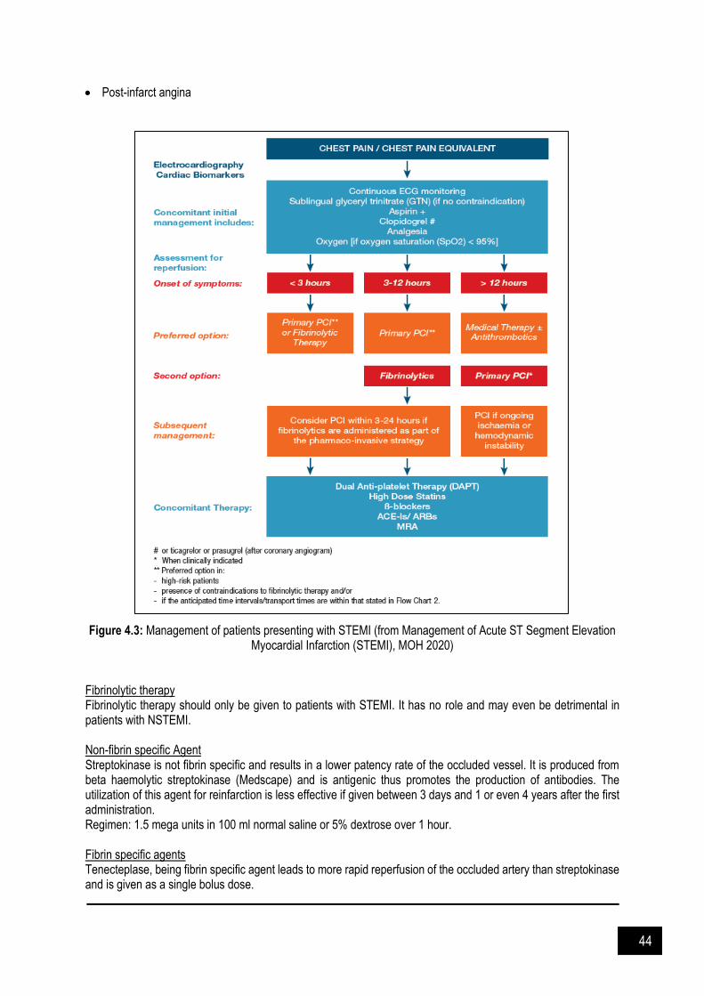
44
Post-infarct angina Figure 4.3: Management of patients presenting with STEMI (from Management of Acute ST Segment Elevation
Myocardial Infarction (STEMI), MOH 2020) Fibrinolytic therapy Fibrinolytic therapy should only be given to patients with STEMI. It has no role and may even be detrimental in patients with NSTEMI. Non-fibrin specific Agent Streptokinase is not fibrin specific and results in a lower patency rate of the occluded vessel. It is produced from beta haemolytic streptokinase (Medscape) and is antigenic thus promotes the production of antibodies. The utilization of this agent for reinfarction is less effective if given between 3 days and 1 or even 4 years after the first administration. Regimen: 1.5 mega units in 100 ml normal saline or 5% dextrose over 1 hour. Fibrin specific agents Tenecteplase, being fibrin specific agent leads to more rapid reperfusion of the occluded artery than streptokinase and is given as a single bolus dose.

45
Table 4.5: Dosage for tenecteplase in MI
Body weight Single IV bolus over 5 secs Volume TNK to be administered
< 60 kg 30 mg 6
60 to < 70 kg 35 mg 7
70 to < 80 kg 40 mg 8
80 to < 90 kg 45 mg 9
> 90 kg 50 mg 10
For patients who present with hypotension (SBP<90mmHg) which might be due to relative hypovolaemia, RV infarction or acute LV failure, these patients should be:
Given IV fluids as necessary.
Started on inotropes, preferably noradrenaline. Fibrinolytic therapy with a fibrin specific agent is preferred. If blood pressure drops during infusion of the fibrinolytic agent, administer fluids or start noradrenaline. When the SBP >90mmHg, fibrinolysis can be given concomitantly. Table 4.6: Contraindications to fibrinolytic therapy
Absolute contraindications Relative contraindications
Risk of intracranial haemorrhage
History of intracranial bleed.
History of ischaemic stroke within 3 months.
Known structural cerebral vascular lesion (e.g arteriovenous malformation).
Known intracranial neoplasm.
Risk of intracranial haemorrhage
Severe uncontrolled hypertension on presentation [blood pressure (BP) >180/110 mmHg]*
Ischaemic stroke more than 3 months.
History of chronic, severe uncontrolled hypertension.
Risk of bleeding
Active bleeding or bleeding diathesis (excluding menses).
Significant head trauma within 3 months.
Suspected aortic dissection.
Risk of bleeding
Current use of anticoagulant in therapeutic doses [International Normalised Ratio (INR) > 2] or direct oral anticoagulant (DOAC).
Recent major surgery < 3 weeks.
Traumatic or prolonged CPR > 10 minutes.
Recent internal bleeding (e.g gastrointestinal or urinary tract haemorrhage) within 4 weeks.
Non-compressible vascular puncture.
Active peptic ulcer.
Others
Pregnancy.
Prior exposure (> 5 days and within 12 months of first usage) to streptokinase (if planning to use same agent)
* The BP should be reduced prior to institution of fibrinolytic therapy

46
Additional Treatment Beta blocker Oral treatment with ß-blockers initiation immediately after STEMI or when patient is haemodynamically stable is recommended in patients with heart failure and/or LV systolic dysfunction, unless contraindicated. Contraindications to ß-blockers:
Bradycardia < 60 beats per minute.
SBP < 100 mmHg.
Pulmonary congestion with crepitations beyond the lung bases.
Signs of peripheral hypoperfusion.
Second or third degree atrio-ventricular (AV) block. Table 4.7: Recommended Dosage of β-blocker in ACS
Agents Initiation Dose Target Dose
Metoprolol 25mg BD 100mg BD
Bisoprolol 1.25mg OD 10mgOD
Carvedilol 3.125mg BD 25mg BD
ACE-Is and ARBs ACE-Is are indicated especially in the high-risk patients (LVEF ≤ 40% or with evidence of heart failure, diabetes or an anterior infarct). Contraindications to ACE-I and ARB therapy:
SBP < 100 mmHg
Bilateral renal artery stenosis. The dose of ACE-I/ARB should be reduced or stopped if there is an increase in serum creatinine of ≥ 30% from baseline within 2 weeks after initiation and/or persistent hyperkalaemia (>5.6 mmol/L). In ACE-I intolerant patients, an ARB may be used preferably valsartan. Mineralocorticoid receptor antagonists Mineralocorticoid antagonists, should be considered in patients post MI with LVEF ≤ 40% and HF in the absence of renal failure or hyperkalaemia. Statins High-intensity statin therapy (atorvastatin 40-80mg daily or rosuvastatin 20-40mg daily) should be initiated early, regardless of initial cholesterol values. Target LDL-C should be <1.8mmol/L or a reduction of at least 50% from the baseline, whichever lower. Addition of non-statin therapy to reduce LDL-C should be considered in patients who remain at high risk with LDL-C ≥1.8mmol/L despite the maximum tolerated statin dose. Contraindications
Active liver disease
Nursing or pregnant mothers
Unexplained persistent elevation of serum transaminases Calcium channel blockers (CCB) There is no data to support the routine use of calcium channel blockers post-ACS. In patients who continue to have angina, CCBs in combination with ß-blockers have been shown to be more effective than monotherapy in the relief of angina. For those with contraindications to ß-blockers, particularly in the presence of obstructive airway disease, CCBs such as verapamil or diltiazem may be considered. Despite providing symptom relief, CCBs have not been shown to improve long term cardiac outcomes. Other treatment Trimetazidine and Ivabradine have been shown to be beneficial in patients post STEMI in registry data and in small studies.

47
Ventricular Tachycardia (VT) Defined as ≥3 beats at a rate 100 beats per min or greater which may be terminated spontaneously within 30 seconds without causing severe symptoms (non-sustained VT). In hemodynamically unstable patient presented with VT, in addition to other first-line measures, anti-arrythmic drugs is often used:
Amiodarone (first-line therapy)
Procainamide (alternative to amiodarone)
Lidocaine (if suspected acute myocardial ischaemia) Other drugs that commonly used for management of VT in the ICU is beta-blockers (class II). Metoprolol is usually preferable in ICU setting, can be given orally or IV [e.g esmolol (short-acting)]. For those remain symptomatic despite beta-blockers therapy, or who are unable to tolerate beta-blocker, a non-dihydropyridine calcium channel blocker maybe used. Table 4.8: Drugs commonly used for management of VT in ICU
Drugs Standard dosing
Amiodarone 150 – 300 mg IV bolus over 10 minutes, followed by infusion of 1mg/min for 6H, then 0.5 mg/min for 18 additional hours or until convert to oral therapy. Additional 150 mg bolus can be given if there is breakthrough arrhythmia (up to total loading 2g per 24H) (some suggest may up to 6-10 g oral loading) Alternative dosing regimens:
Load orally 800 to 1600 mg daily for 2-3 weeks, then maintenance dose of 400mg daily
Oral 200 mg twice daily for 4 weeks, then 200 mg daily (maintenance dose)
Oral 400 mg every 8-12H for 1-2 weeks, followed by 200-400 mg once daily (maintenance)
Procainamide Loading up to 17mg/kg (as IV infusion 20-30 mg/min), followed by maintenance 1-4 mg/min
Lidocaine 1-1.5 mg/kg IV bolus, may repeat up to bolus 3 mg/kg, followed by infusion of 1-4 mg/min
Metoprolol Usual effective dose 50-200mg daily
Carvedilol Usual effective dose 12.5 to 50mg daily
Esmolol 500 mcg/kg IV bolus over 1 minute followed by maintenance dose of 50 mcg/kg/min titrated for effect up to 300 mcg/kg/min
Verapamil Usual effective dose 360-480 mg daily
Diltiazem Usual effective dose 240-360 mg daily
Supraventricular Tachycardia (SVT)
SVT is an abnormal heart rhythm with a ventricular rate of 100 or more beats per minute (faster than normal). It happens because of a problem with the heart's electrical system. It starts in the upper chambers of the heart, called the "atria" It usually starts and stops suddenly, without warning. SVT is also called "paroxysmal supraventricular tachycardia" or PSVT (paroxysmal means a sudden attack). The 3 main types of SVT are called:
Atrioventricular nodal reentry tachycardia (AVNRT)
Atrioventricular reentry tachycardia (AVRT)
Atrial tachycardia Pharmacotherapy for SVT In stable patient, drugs that are used for treatment of SVT:
Adenosine: 6-12 mg rapid IV push followed by saline flush
IV verapamil, diltiazem or beta-blockers
Type I or type III anti-arrhythmic Unstable patient:
Electrical cardioversion

48
Figure 4.4: The heart (from the UptoDate 2020)
References and Suggested Readings Amsterdam E, Wenger N, Brindis R et al. AHA/ACC Guideline for the management of patients with non–ST-
elevation acute coronary syndromes. Circulation. 2014; 130(25): e344–e426. Auguste U, Kanei Y, Collo Go R. Chapter 6: Acute Coronary Syndromes 2019. Available at:
https://accessanesthesiology.mhmedical.com. Borja I, Stefan J, Stefan A et al. 2017 ESC Guidelines for the management of acute myocardial infarction in patients
presenting with ST-segment elevation: The Task Force for the management of acute myocardial infarction in patients presenting with ST-segment elevation of the European Society of Cardiology (ESC). Eur Heart J. 2018. 39(2): 119–177.
Braverman A. Acute aortic dissection. Circulation. 2010. 122(2): 184-188. Clinical practice guidelines: Management of hypertension. 5th ed. Ministry of Health Malaysia; 2020. ESC/ESH Guidelines for the management of arterial hypertension. Eur Heart J. 2018. 39: 3021–3104. Johnson W, Nguyen ML, Patel R. Hypertension crisis in the emergency department. Cardiol Clin 2012. 30: 533-43 Knight BP. Atriventricular nodal reentrant tachycardia. UpToDate 2020. Management of scute ST segment elevation myocardial infarction (STEMI). 4th ed. Ministry of Health Malaysia;
2020. Management of unstable angina / non-ST elevation myocardial infarction (UA /NSTEMI). Ministry of Health
Malaysia; 2011. MIMS Gateway. MIMS gateway. 2020 [cited 2 March 2020]. Available from: https://online1.mimsgateway.com.my Prutkin JM. Overview of the acute management of tachyarrhythmias. UpToDate 2020. Roffi M, Patrono C, Collet J et al. 2015 ESC Guidelines for the management of acute coronary syndromes in
patients presenting without persistent ST-segment elevation. Eur Heart J. 2015; 37(3): 267-315. Whelton P, Carey R, Aronow W et al. 2017 ACC / AHA / AAPA / ABC / ACPM / AGS /APhA / ASH / ASPC / NMA
/ PCNA Guideline for the prevention, detection, evaluation, and management of high blood pressure in adults: Executive summary. J American Col Cardiol. 2018. 71(19): 2199-2269.
Zimetbaum PJ. Nonsustained ventricular tachycardia: Clinical manifestations, evaluation, and management. UpToDate 2020.
49
CHAPTER 5
Pulmonary Disorders
Acute Respiratory Distress Syndrome (ARDS)
RDS is a life-threatening form of respiratory failure characterized by inflammatory pulmonary edema
resulting in severe hypoxemia (Table 5.1). ARDS is common, associated with substantial morbidity and frequently fatal. According to Berlin definition, ARDS is defined as an acute form of diffuse lung injury occurring in patients with a predisposing risk factor. Common risk factors of developing ARDS are pneumonia, sepsis, gastric content aspiration, trauma, pancreatitis, inhalation injury, burns, non-cardiogenic shock, drug overdose, transfusion-related acute lung injury (TRALI), and drowning. Table 5.1: Characteristics and clinical presentations of ARDS (from Ranieri et al 2012)
Characteristic Clinical Presentation
Timing Onset within 1 week after the trigger insult
Imaging Bilateral opacities on chest X-ray, not fully explained by effusion, lobar/lung collapse, or nodules
Non-cardiogenic origin Respiratory failure not fully explained by cardiac failure or fluid overload
Classification Presence of hypoxemia base on PaO2/FiO2 ratio measured with PEEP ≥5 cm H2O:
Mild (PaO2/FiO2 201- 300 mm Hg)
Moderate (PaO2/FiO2 101-200 mm Hg)
Severe (PaO2/FiO2 ≤ 100 mm Hg
Management of ARDS The primary target in ARDS treatment is to ensure adequate gas exchange while minimizing the risk of ventilator-induced lung injury. To date, the mainstay treatment is supportive. Lung protective ventilatory strategy is recommended to ensure adequate oxygenation and CO2 clearance, and minimizing the extent of damage due to mechanical ventilation when required. Table 5.2: Management of ARDS (from Michele et al 2017)
Non-pharmacological intervention
Mechanical ventilation
Lung recruitment and PEEP selection
Tidal volume setting
Oxygen and carbon dioxide target
Pharmacological intervention
Neuromuscular blocking agent - cisatracurium within 48 hours in severe ARDS patient has showed to improve survival, increased the ventilator free days without increasing muscle weakness. A 48-hour continuous infusion of cisatracurium in critically ill patients reduced the risk of death, reduced the risk of barotrauma, and did not affect the duration of mechanical ventilation or the risk of ICU-acquired weakness.
Corticosteroids - American College of Critical Care Medicine consensus recommends that glucocorticoid should be initiated early (<14 days of ARDS). Methylprednisolone of 1 mg/kg/ day for ≥14 days is recommended in patients with severe early ARDS. Glucocorticoids should be weaned and not stopped abruptly. Meduri et al. in its meta-analysis found that methylprednisolone at dose 1-2mg/kg/day for ≥7 days reduced ICU mortality rates.
A

50
Inhaled nitric oxide - Seemed promising in early trials, but in larger controlled trials, did not change mortality rates in adults with ARDS. Inhaled nitric oxide did not reduce mortality and may results in only a transient improvement in oxygenation.
Acute Exacerbation of Asthma Exacerbations of asthma is characterized by a progressive increase in symptoms of shortness of breath, cough, wheezing or chest tightness, and a progressive decrease in lung function. Acute exacerbation of asthma carries a high mortality. Patients who are at high risk of asthma-related death should be assessed carefully and should be managed in a high care setting. Risk of asthma-related death:
History of near-fatal asthma requiring intubation and mechanical ventilation
Hospitalization or emergency care visit for asthma in the past year
Currently using or having recently stopped using oral corticosteroids (a marker of event severity)
Over-use of SABAs, especially the use of more than one canister of salbutamol (or equivalent) monthly
A history of psychiatric disease or psychosocial problems
Poor adherence with asthma medications and/or poor adherence with (or lack of) a written asthma action plan
Food allergy in a patient with asthma Table 5.3: Clinical presentations of acute exacerbation of asthma
Mild to moderate exacerbation Severe exacerbation
Talks in phrases
Prefers sitting to lying
Not agitated
Respiratory rate increased
Accessory muscles not used
Pulse rate 100-120 pulse/min
Oxygen saturation 90-95%
PEF >50% predicted or best
Talk in words
Sit hunched forward
Agitated
Respiratory rate >30 breaths/min
Accessory muscle in use
Pulse rate >120 pulse/min
Oxygen saturation ≤ 90%
PEF ≤50% predicted or best
Management of acute exacerbation of asthma Mainstay management is oxygen therapy. Oxygen therapy is to maintain the SpO2 level between 93–95%. Administration of low concentration oxygen therapy (controlled oxygen therapy) guided by pulse oximetry during severe exacerbation could result in better clinical outcomes than high flow (e.g 100% oxygen therapy). Various pharmacological agents are used in the management of acute exacerbation of asthma (Table 5.4 & 5.5). Table 5.4: Pharmacological agents used in the management of acute exacerbation of asthma
Management Description
Inhaled short-
acting beta2-
agonists (SABA)
Should be given as soon as possible.
In mild to moderate exacerbation, MDI with spacer is effective. Administer high dose
of 4-10 puffs and repeat every 20 minutes for the first hour.
In severe and life-threatening cases, give oxygen driven (6 L/min) continuous
nebulised SABA at rate 5-10mg/hr followed by intermittent on demand nebuliser
when patient is more stable.
Routine intravenous beta2-agonist use does not improve clinical outcome during
severe exacerbation of asthma.
Inhaled short
acting
anticholinergic
(SAAC)
Ipratropium bromide recommended to be combined with inhaled SABA in patient
with moderate to severe exacerbation. Evidences show that it can reduce
hospitalizations and better improvement in PEF and FEV1 compared to SABA alone.
No benefit of combination therapy observed in children.

51
Systemic
Corticosteroids
Proven in moderate-to-severe exacerbation- helps rapid resolution of exacerbation
and reduce relapse.
Administer early within 1 hour of presentation since the effects of corticosteroids
usually only can be seen within 6-12 hours post administration.
Oral administration is as effective as intravenous. Oral prednisolone 1mg/kg/ day to
a maximum dose of 50mg daily or IV hydrocortisone 200mg per day in divided dose
should be given to patient.
Treatment duration of 5 to 7 days in adults is found to be as effective as 10 to 14
days courses.
There is no evidence supporting tapering corticosteroid dose either following short
term or several weeks use provided that the patient receives adequate dose of
inhaled corticosteroids.
Inhaled
corticosteroid
(ICS)
Conflicting evidences on concomitant use of ICS with systemic corticosteroid.
In patient who not receiving systemic corticosteroid, high dose ICS should be given
within 1 hour of presentation. The patient should be discharged home with regular
ICS since an event of severe exacerbation is a risk factor for future exacerbations.
Magnesium
Administer a single 10 mmol magnesium sulphate IV infusion over 20 minutes in
severe exacerbation who has not had a good response to initial treatment. Study
shows that it reduces hospitalization
Adults with FEV1 <25–30% predicted - Adults and children who did not respond to
initial therapy with persistent hypoxemia, Children whose FEV1 fails to reach 60%
predicted after 1 hour of care.
Inhaled magnesium sulphate can possibly improve pulmonary function in severe
asthma exacerbations (FEV1 less than 50% predicted). Some practices
administered nebulised salbutamol in isotonic magnesium sulphate instead of
normal saline.
Aminophylline
and theophylline
Not likely add bronchodilator effect compared to SABA alone. Not recommended to
be used in acute asthma exacerbation due to poor efficacy and potential life-
threatening side effects especially in patient who already on long term theophylline
SR treatment.
Leukotriene
receptor
antagonists
(montelukast)
The clinical evidences that support the use of LTRAs in acute exacerbation of asthma are
limited.
ICS/LABA
combinations
The role of ICS/LABA in the management of acute exacerbation is unclear.
Antibiotics Not recommended unless there are strong evidence of respiratory infection.
Sedative
Sedative should not be given during acute asthma exacerbation due to respiratory
depressant effect that can possibly lead to asthma death.

52
Table 5.5: Common medications used in acute asthma exacerbation.
Medication Dose Adverse reaction Remarks
Inhaled Salbutamol (Short acting beta2 agonist)
Moderate, severe, life threatening acute asthma: NEB: 5mg repeat every 20-30min Continuous neb: 5-10mg/hr MDI: 2-10 puff every 10-20 min
Lactic acidosis (High dose), nausea, tremor, tachycardia
Duration of action: 3-5 hours
Inhaled Ipratropium bromide (Short acting anticholinergic)
Acute bronchospasm: NEB: 500mcg PRN (max: 2mg/day) Severe acute asthma: NEB: 500mcg 4-6 hourly as required Reversible airway obstruction: MDI:250-500mcg tds/qid
Constipation, cough, diarrhoea, dry mouth, headache, sinusitis
Maximal effect: 30-60 min after use Duration of action: 3-6 hours
Inhaled Ipratropium/ salbutamol (Short acting anticholinergic/Short acting beta2 agonist)
Bronchospasm in COPD: NEB: 0.5/2.5 mg tds/qid
Bronchitis, URTI
Tab. Prednisolone (oral corticosteroid) Mild, moderate, severe or life-threatening acute asthma: 1mg/kg/day or 40-50mg OD
Sleep disturbance, reflux, appetite increase, hyperglycemia,
IV Hydrocortisone (parenteral corticosteroid)
Severe, life threatening acute asthma: 100mg QID
Pain and paraesthesia
IV Magnesium sulphate 50% ( 2.5g/5ml)
Severe acute asthma not responding to initial treatment: 8 mmol (4ml) STAT over 20 minutes
Flushing, hypotension 1g MgSO4 = 4mmol
Inhaled corticosteroid
As per table 1 Oropharyngeal candidiasis. Prolonged high dose increase risk of systemic side effect such as adrenal crisis and adrenal supression
No evidence for concomitant use with systemic corticosteroid. In patient not receiving systemic corticosteroid, high dose ICS should be given

53
Figure 5.1: Treatment flow chart: Asthma exacerbation (from Clinical practice guideline: Management of asthma in adults, MOH 2017 & Global initiative for asthma:
Global strategy for asthma management and prevention 2017)

54
Table 5.6: Common doses of inhaled corticosteroids
Inhaled corticosteroid Low dose
(mcg) Medium dose
(mcg) High dose
(mcg)
Beclometasone dipropionate (CFC) 200-500 501-1000 >1000
Beclometasone dipropionate (HFA) 100-200 201-400 >400
Budesonide (DPI) 200-400 401-800 >800
Ciclesonide (HFA) 80-160 161-320 >320
Fluticasone furoate (DPI) 100 NA 200
Fluticasone propionate (DPI) 100-250 251-500 >500
Fluticasone propionate (HFA) 100-250 251-500 >500
Mometasone furoate 110-220 221-440 440
*CFC: chlorofluorocarbon propellant: DPI: dry powder inhalation; HFA: hydrofluoroalkane propellant **The inhaled corticosteroids are in metered dose inhaler (MDI) preparations. AT-connector is used to connect the MDI canister to the endotracheal tube (ETT) for intubated
Acute Exacerbation of Chronic Obstructive Pulmonary Disease (AECOPD) AECOPD is defined as increased in respiratory symptoms from patient baseline symptoms:
Increase dyspnoea
More frequent and severe cough
Increase sputum production and purulence that require change in therapy. Table 5.7: Classification of severity of COPD
Classification Description
Mild Controlled with increased dose of short acting bronchodilators only
Moderate Requires additional treatment with antibiotics and/or oral corticosteroids
Severe Requires hospitalization or emergency department evaluation
Classification of severity in hospitalized patient
Classification Description
Mild (no respiratory
failure)
Respiratory rate: 20-30 breaths per minute
No use of accessory muscles
No changes in mental status
Hypoxemia improved with supplemental oxygen given via Venturi mask 28-35% inspired oxygen (FiO2)
No increase in PaCO2
Moderate (acute
respiratory failure-non
life threatening)
Respiratory rate: > 30 breaths per minute
Using accessory respiratory muscles
No change in mental status
Hypoxemia improved with supplemental oxygen via Venturi mask 25-30% FiO2
Hypercarbia (PaCO2 increased compared with baseline or elevated 50-60 mmHg)
Severe (acute
respiratory failure-life
threatening)
Respiratory rate: > 30 breaths per minute
Using accessory respiratory muscles
Acute changes in mental status
Hypoxemia not improved with supplemental oxygen via Venturi mask or requiring FiO2 > 40%
Hypercarbia (PaCO2 increased compared with baseline or elevated > 60 mmHg or the presence of acidosis (pH < 7.25).
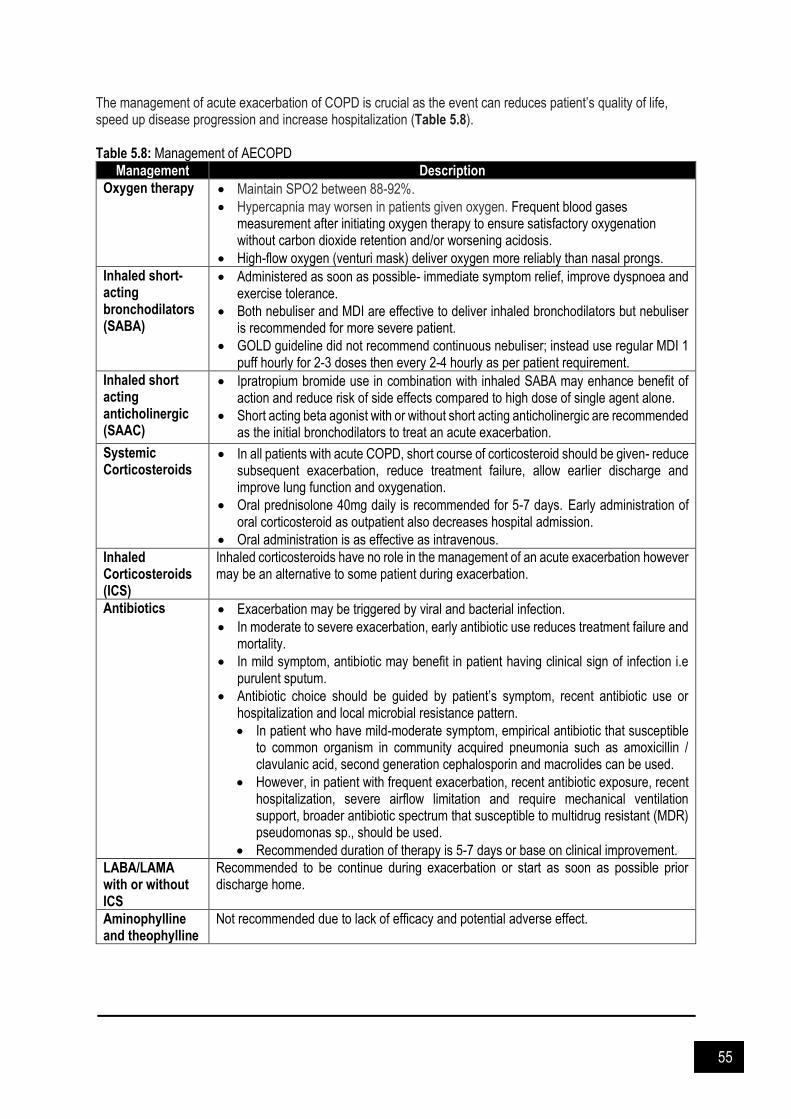
55
The management of acute exacerbation of COPD is crucial as the event can reduces patient’s quality of life, speed up disease progression and increase hospitalization (Table 5.8). Table 5.8: Management of AECOPD
Management Description
Oxygen therapy
Maintain SPO2 between 88-92%.
Hypercapnia may worsen in patients given oxygen. Frequent blood gases measurement after initiating oxygen therapy to ensure satisfactory oxygenation without carbon dioxide retention and/or worsening acidosis.
High-flow oxygen (venturi mask) deliver oxygen more reliably than nasal prongs.
Inhaled short-acting bronchodilators (SABA)
Administered as soon as possible- immediate symptom relief, improve dyspnoea and exercise tolerance.
Both nebuliser and MDI are effective to deliver inhaled bronchodilators but nebuliser is recommended for more severe patient.
GOLD guideline did not recommend continuous nebuliser; instead use regular MDI 1 puff hourly for 2-3 doses then every 2-4 hourly as per patient requirement.
Inhaled short acting anticholinergic (SAAC)
Ipratropium bromide use in combination with inhaled SABA may enhance benefit of action and reduce risk of side effects compared to high dose of single agent alone.
Short acting beta agonist with or without short acting anticholinergic are recommended as the initial bronchodilators to treat an acute exacerbation.
Systemic Corticosteroids
In all patients with acute COPD, short course of corticosteroid should be given- reduce subsequent exacerbation, reduce treatment failure, allow earlier discharge and improve lung function and oxygenation.
Oral prednisolone 40mg daily is recommended for 5-7 days. Early administration of oral corticosteroid as outpatient also decreases hospital admission.
Oral administration is as effective as intravenous.
Inhaled Corticosteroids (ICS)
Inhaled corticosteroids have no role in the management of an acute exacerbation however may be an alternative to some patient during exacerbation.
Antibiotics
Exacerbation may be triggered by viral and bacterial infection.
In moderate to severe exacerbation, early antibiotic use reduces treatment failure and mortality.
In mild symptom, antibiotic may benefit in patient having clinical sign of infection i.e purulent sputum.
Antibiotic choice should be guided by patient’s symptom, recent antibiotic use or hospitalization and local microbial resistance pattern.
In patient who have mild-moderate symptom, empirical antibiotic that susceptible to common organism in community acquired pneumonia such as amoxicillin / clavulanic acid, second generation cephalosporin and macrolides can be used.
However, in patient with frequent exacerbation, recent antibiotic exposure, recent hospitalization, severe airflow limitation and require mechanical ventilation support, broader antibiotic spectrum that susceptible to multidrug resistant (MDR) pseudomonas sp., should be used.
Recommended duration of therapy is 5-7 days or base on clinical improvement.
LABA/LAMA with or without ICS
Recommended to be continue during exacerbation or start as soon as possible prior discharge home.
Aminophylline and theophylline
Not recommended due to lack of efficacy and potential adverse effect.

56
Figure 5.2: Treatment flow chart: COPD exacerbation (from Global strategy for the diagnosis, management, and prevention of chronic obstructive pulmonary disease:
GOLD 2017 & Stoller 2020)

57
References and Suggested Readings Afshari A, Brok J, Møller AM, Wetterslev J. Inhaled nitric oxide for acute respiratory distress syndrome and acute
lung injury in adults and children: a systematic review with meta-analysis and trial sequential analysis. Anesth Analg, 2011. 112(6): 1411-21.
American Thoracic Society, European Respiratory Society Task Force. Standards for the diagnosis and management of patients with COPD. American Thoracic Society 2004.
Balanag VM, Yunus F, Yang PC, Jorup C. Efficacy and safety of budesonide/formoterol compared with salbutamol in the treatment of acute asthma. Pulm Pharmacol Ther, 2006. 19: 139-47.
Barr RG, Rowe BH, Camargo CA. Methylxanthines for exacerbations of chronic obstructive pulmonary disease. Cochrane Database Syst Rev, 2, 2003.
British Thoracic Society & Scottish Intercollegiate Guidelines Network. British guideline on the management of asthma 2016.
Clinical Practise Guideline (CPG): Management of Asthma in Aduts 2017. Ministry of Health Malaysia. Edmonds ML, Milan SJ, Camargo CA, Pollack CV, Rowe BH. Early use of inhaled corticosteroids in the emergency
department treatment of acute asthma. Cochrane Database Systemic Review, 12, 2012. Fan E, Sorbo DL, Goligher EC, et al. An Official American Thoracic Society / European Society of Intensive Care
Medicine / Society of Critical Care Medicine Clinical Practice Guideline: Mechanical ventilation in adult patients with acute respiratory distress syndrome. Am J Respir Crit Care Med 2017. 195(9): 1253–1263.
Fanta CH. Acute exacerbations of asthma in adults: Emergency department and inpatient management. UpToDate 2020.
Global initiative for asthma. Global strategy for asthma management and prevention 2017. Global Initiative for Chronic Obstructive Lung Disease. Global strategy for the diagnosis, management, and
prevention of chronic obstructive pulmonary disease: GOLD 2017. Griffiths B, Ducharme FM. Combined inhaled anticholinergics and short-acting beta2-agonists for initial treatment
of acute asthma in children. Cochrane Database Systemic Review, 8, 2013. Hasegawa T, Ishihara K, Takakura S, et al. Duration of systemic corticosteroids in the treatment of asthma
exacerbation; a randomized study. Intern Med, 2000. 39: 794-7. Kayani S, Shannon DC. Adverse behavioral effects of treatment for acute exacerbation of asthma in children: A
comparison of two doses of oral steroids. Chest, 2002. 122: 624-8. Michele U, Paolo F, Luca B, Davide C. Current Concepts of ARDS: A narrative review. Int. J. Mol. Sci, 2017. 18:
64. Nair P, Milan SJ, Rowe BH. Addition of intravenous aminophylline to inhaled beta-(2)-agonists in adults with acute
asthma. Cochrane Database Systemic Review, 12, 2012. Papazian L. et al. Neuromuscular blockers in early acute respiratory distress syndrome. N Eng J Med, 2010.
363(12): 1107-1116. Ranieri VM, Rubenfeld GD, Thompson BT, Ferguson ND, Caldwell E, et al. ARDS Definition Task Force, Acute
respiratory distress syndrome: The Berlin Definition. JAMA, 2012. 307(23): 2526-33. Stoller JK. Management of exacerbations of chronic obstructive pulmonary disease. UpToDate 2020. Travers AH, Milan SJ, Jones AP, Camargo CA, Rowe BH. Addition of intravenous beta-(2)-agonists to inhaled
beta-(2)-agonists for acute asthma. Cochrane Database Systemic Review,12, 2012.
58
CHAPTER 6
Hematologic Disorders
Coagulation Disorders
oagulation disorders are common seen in ICU, which may cause by various clinical conditions including sepsis, malignancy, trauma, vascular disorders and obstetric condition. Pre-existing medical conditions such as liver disease, renal impairment etc, may provoke the development of coagulopathy. Sepsis is a common factor that leads to coagulopathy and disseminated intravascular coagulation (DIC) in critically ill patients. DIC results from the systemic activation of both the clotting and fibrinolytic systems which would lead to requirement of many coagulation factors and platelets. Fresh frozen plasma (FFP) and platelet transfusion are the mainstay therapeutic options, particularly in ICU for hemodynamic stabilization. Generally, the treatment aim is to maintain the targeted hemoglobin. Hemostatic drugs such as desmopressin may be used, particularly to treat or prevent mild von Willebrand disease or platelet function defects. Table 6.1: Desmopressin standard dosing
Method of administration Standard dosing Onset Duration
Slow IV infusion 0.3 mcg/kg, may repeat dose if needed (based on clinical response and laboratory results) (maximum IV dose of 20 mcg has been recommended)
30-60 minutes 6-12 hours
Subcutaneous (SC) 0.3 mcg/kg (may repeat dose if needed (based on clinical response and laboratory results)
30-60 minutes 6-12 hours
Intranasal <50 kg: 150 mcg (1 spray in a single nostril); ≥50 kg: 300 mcg (1 spray each nostril) (q12-24H, 4-5 doses)
60-90 minutes 6-12 hours
Anemia in ICU Anemia in patients admitted to an intensive care unit (ICU) is common and affects almost all critically ill patients. Red cell transfusion, the most common treatment for anemia, comes with associated risks, which may further reduce the chance of survival of these patients. World Health Organization (WHO) defines anemia as a hemoglobin (Hb) level less than 13 g/dL in men and less than 12 g/dL in women (Astin 2014). Various studies have shown that approximately two-thirds of critical patients admitted to an ICU have a hemoglobin concentration of less than 12 g/dl on the day of admission, and 97% of the patients become anemic after a week in ICU. Consequences of Anemia Anemia is associated with poor outcomes, mostly in elderly patients with chronic disease. The compensatory response of the body in acute or chronic anemia is an extra burden on critical patients, who are often in either newly developed or preexisting cardiopulmonary failure. The body reacts and compensates for anemia by increasing cardiac output (increased heart rate and stroke volume), oxygen unloading (right shift of the oxyhemoglobin dissociation curve) and oxygen extraction. Various studies have shown that anemia is associated
C

59
with adverse outcomes in acute myocardial infarction, chronic kidney disease, and chronic heart failure. Studies have also shown increased risk of re-intubation or weaning failure from mechanical ventilator in those patients having low hemoglobin levels, more so in patients with chronic obstructive pulmonary disease Table 6.2: Causes of anemia in intensive care
Type of Anemia Causes
Blood loss Phlebotomy Gastrointestinal bleeding Trauma Surgery
Erythropoietin deficiencies Inflammatory cytokines Renal insufficiency Drugs Decrease bone marrow response (functional deficiency)
Nutritional deficiencies Low folate levels Low iron levels Low vitamin B levels (vitamin B12)
Hemolysis Drug reactions Toxins
Coagulation abnormalities Sepsis syndrome Thrombocytopenia Liver disease Viral infection Splenomegaly
Management of Anemia Blood transfusions Transfusion of packed red blood cells (PRBC) in critically ill patients serves to increase oxygen delivery thus decreasing tissue hypoxia. However, many studies have not shown any improvement in oxygen uptake post-transfusion of PRBC. This can be explained by the various adverse factors of stored blood like low levels of 2,3-diphosphoglycerate which decreases the ability of the hemoglobin to unload oxygen to the tissues (shift of oxygen disassociation curve to the left), the structural changes in stored red blood cells resulting in increased aggregation and/or hemolysis, and the accumulation of pro-inflammatory cytokines. Erythropoiesis-stimulating agents (ESA) A review of the literature has shown that in critically ill patients there is an inappropriately low erythropoietin release or a decreased response of the bone marrow to erythropoietin. Corwin et al. (2007), in a prospective, randomized, placebo-controlled trial including 146 medical, surgical and trauma patients, concluded that treatment with erythropoietin did not help in reducing the rate of red cell transfusions but resulted in a decrease in mortality in trauma patients. Also, the use of erythropoietin was associated with a greater incidence of thrombotic events. This may be due to the facts that ESAs have a delayed effect, or the critical patients have resistance to ESA and require a high dosage, as concluded by Jelkmann et al. (2013). Thus, ESAs are shown to have a beneficial role only in critically ill patients with a concomitant medical disease (renal disease) or in trauma patients. Blood substitutes These are also known as oxygen carriers and have not shown promising results being still a subject of research. The developed oxygen carriers are of two types:
a. Cell-free hemoglobin-based oxygen carriers (HBOCs). b. Per fluorocarbons.

60
HBOCs have various adverse effects including nephrotoxicity, impaired perfusion, and increased rates of myocardial infarction and death. Also, their short intravascular half-life of 12 to 48 hours, have limited their use to severe cases of acute bleeding in surgical or trauma patient
Iron therapy In critically ill patients, there is iron-restricted erythropoiesis due to iron sequestration and decreased absorption from the gut. This suggests a potential, mainly intravenous role, of iron therapy. However, this type of treatment has been challenged as iron has been associated with the growth and virulence of microbes responsible for nosocomial infections and decreasing the iron levels may be a defense response of the body to sepsis. Further research is needed before this therapy can be accepted on a regular basis. Thrombocytopenia There were five major blood dyscrasias attributable to drugs which are aplastic anemia, agranulocytosis, megaloblastic anemia, hemolytic anemia, and thrombocytopenia. It is usually rare compared to other adverse effects induced by drugs. However, they are important because it is associated with significant morbidity and mortality. Direct toxicity or immune reactions are the two main mechanisms involved in drug-induced haematological disorders. The recommended treatment for drug-induced haematological disorders is the removal of causative drug and initiation of symptomatic support of the patient. Besides that, frequent monitoring of laboratory values is also warranted. The common drugs that can induce haematological disorders are as in Table 6.3. Heparin-induced thrombocytopenia (HIT) and HIT with thrombosis (HITT)
An immune-mediated disorder that is triggered by exposure to any form of heparin. Patient undergoing cardiac or
orthopedic surgery are among those at highest risk of developing HIT as compared to medical patient.
Nevertheless, the incidence of HIT varies depending on the studied population and type of heparin preparation
used. Unfractionated heparin (UFH) may predispose higher risk for development of HIT compared to the use of
low molecular weight heparin (LMWH). When HIT is suspected:
Discontinue all heparin products immediately
Consider non-heparin anti-coagulant to replace heparin in patients suspected of having HIT, at high risk
of developing thrombosis (e.g direct thrombin inhibitors)
Thrombotic thrombocytopenic purpura (TTP)
The disorder or thrombocytopenia, hemolysis (based on blood smears), renal dysfunction and neurologic
abnormalities such as headache, confusion, focal deficits, seizures or coma. The possible etiology are
autoimmune, congenital, drug related or HIV. Plasma exchange is the mainstay therapeutic approach, which
maybe supplemented with immunosuppressive drugs.

61
Figure 6.1: Recommended algorithm for initial management of acquired, autoimmune TTP
(from George 2006 & George 2020)

62
Table 6.3: Drugs that can induce haematological disorder
Drugs associated with apalastic anaemia
Acetazolamide Aspirin Captopril Chloramphenicol
Chloroquine Chlorothiazide Chlorpromazine Dapsone
Felbamate Interferon alfa Lisinopril Lithium
Sulindac Nizatidine Pentoxifylline Quinidine
Ticlopidine
Drugs associated with agranulocytosis
Acetaminophen Acetazolamide Ampicillin Captopril Cefotaxime Cefuroxime Chloramphenicol Chlorpramazine Chlorpropramide Chlorpheniramine
Clindamycin Clozapine Colchicine Doxepin Dapsone Desipramine Ethosuximide Flucystosine Gentamicin
Griseofulvin Hydralazine Hydroxychloroquine Imipenem-cilastatin Imipramine Lamotrigine Levodopa Methazolamide Methyldopa
Metronidazole Nafcillin NSAIDs Olanzapine Oxacillin Penicillamine Penicillin G Phenytoin Pyrimethamine Quinidine
Quinine Rifampicin Streptomycin Terbinafine Ticarcilin Tolbutamide VancomycinPrimidone Procainamide Propylthiouracil
Drugs associated with hemolytic anaemia
Acetaminophen Angiotensin-converting enzyme inhibitors β-Lactam antibiotics Cephalosporins Ciprofloxacin Clavulanate Erythromycin
Hydrochlorothiazide Indinavir Interferon alfa Ketoconazole Lansoprazole Levodopa Levofloxacin Methyldopa
Minocycline NSAIDs Omeprazole p-Aminosalicylic acid Phenazopyridine Probenecid Procainamide Quinidine
Rifabutin Rifampin Streptomycin Sulbactam Sulfonamides Sulfonylureas Tacrolimus Tazobactam
Teicoplanin Tolbutamide Tolmetin Triamterene
Drugs associated with oxidative hemolytic anaemia
Ascorbic acid Metformin Methylene blue
Nalidixic acid Nitrofurantoin Phenazopyridine
Primaquine Sulfacetamide Sulfamethoxazole
Sulfanilamide
Drugs associated with megaloblastic anaemia
Azathioprine Chloramphenicol Colchicine
Cyclophosphamide Cytarabine 5-Fluorodeoxyuridine 5-Fluorouracil
Hydroxyurea 6-Mercaptopurine Methotrexate
p-Aminosalicylate Phenobarbital Phenytoin
Pyrimethamine Sulfasalazine Tetracycline

63
Cotrimoxazole Oral contraceptives Primidone Vinblastine
Drugs associated with thrombocytopenia
Abciximab Acetaminophen Acyclovir Albendazole Aminoglutethimide Aminosalicylic acid Amiodarone Amphotericin B Ampicillin Aspirin Atorvastatin Captopril Chlorothiazide Chlorpromazine Chlorpropamide Cimetidine
Ciprofloxacin Clarithromycin Clopidogrel Danazol Deferoxamine Diazepam Diazoxide Diclofenac Digoxin Ethambutol Felbamate Fluconazole Gold salts Haloperidol Heparin
Hydrochlorothiazide Ibuprofen Indinavir Levamisole Linezolid Lithium Indomethacin Interferon alfa Isoniazid Isotretinoin Itraconazole Low-molecular-weight heparins Measles, mumps, and rubella vaccine
Mesalamine Methyldopa Minoxidil Naproxen Nitroglycerin Octreotide Cloxacillin p-Aminosalicylic acid Penicillamine Ranitidine Recombinant hepatitis B vaccine Rifampicin Simvastatin Sirolimus Sulfasalazine
Sulfonamides Sulindac Tamoxifen Trimethoprim Vancomycin Pentoxifylline Piperacillin Primidone Procainamide Pyrazinamide Quinine Quinidine

64
References and Suggested Readings Astin R, Puthucheary Z. Anaemia secondary to critical illness: an unexplained phenomenon. Extrem Physiol Med.
2014. 3(1): 4. Athar MK, Puri N, Gerber DR. Anemia and blood transfusions in critically ill patients. J Blood Trans. 2012. 2012:
629204. Corwin HL. The role of erythropoietin therapy in the critically ill. Transfus Med Rev. 2006. 20: 27–33. De Souza DA, Silva FG e, Costa PJM de S. Critical evaluation of justifications for the transfusion of red blood cells:
The reality of a government emergency hospital. Rev Bras Hematol Hemoter. 2013. 35: 263-7. George JN. Thrombotic thrombocytopenic purpura. N Eng J Med. 2006; 354: 1927-1935. George JN. Acquired TTP: Initial treatment. UpToDate 2020. Hajjar LA, Auler Junior JO, Santos L, Galas F. Blood tranfusion in critically ill patients: state of the art. Clinics (Sao
Paulo). 2007. 62(4): 507-24. Hayden SJ, Albert TJ, Watkins TR, Swenson ER. Anemia in critical illness: Insights into etiology, consequences,
and management. Am J Respir Crit Care Med. 2012. 185: 1049-1057. Jelkmann I, Jelkmann W. Impact of erythropoietin on intensive care unit patients. Transfus Med Hemother. 2013.
40: 310-8. Khan FA, Fisher MA, Khakoo RA. Association of hemochromatosis with infectious diseases: expanding spectrum.
Int J Infect Dis. 2007. 11: 482-7. Kremer-Hovinga JA, Meyer SC. Current management of thrombotic thrombocytopenic purpura. Curr Opin Hematol.
2008. 15: 445-450. Kutcher ME, Cohen MJ. Acute coagulopathy associated with trauma. UpToDate 2020 Mannucci PM, Remuzzi G, Pusineri F, Lombardi R, Valsecchi C, Mecca G, Zimmerman TS. Deamino-8-D-arginine
vasopressin shortens the bleeding time in uremia. N Engl J Med. 1983. 308(1): 8. Mannucci PM, Ruggeri ZM, Pareti FI, Capitanio A.1-Deamino-8-d-arginine vasopressin: A new pharmacological
approach to the management of haemophilia and von Willebrands' diseases. Lancet. 1977. 1(8017): 869. McEvoy MT, Shander A. Anemia, bleeding, and blood transfusion in the intensive care unit: Causes, risks, costs,
and new strategies. Am J Crit Care. 2013. 22: eS1-13 Rawal G, Kumar R, Yadav S, Singh A. Anemia in intensive care: A review of current concepts. J Crit Care Med
(Targu Mures). 2016. 2(3): 109–114. Stubbs JR. Alternatives to blood product transfusion in the critically ill: erythropoietin. Crit Care Med. 2006. 34:
S160–9. Thomas J, Jensen L, Nahirniak S, Gibney RT. Anemia and blood transfusion practices in the critically ill: A
prospective cohort review. Heart Lung. 2010; 39(3): 217-25. Tsai HM. Thrombotic thrombocytopenic purpura: A thrombotic disorder caused by ADAMTS13 deficiency. Hematol
Oncol Clin North Am. 2007. 21: 609-632. Weinberg ED. Iron loading and disease surveillance. Emerg Infect Dis. 1999. 5: 346–52.

65
CHAPTER 7
Deep Vein Thrombosis & Pulmonary Embolism
Deep Vein Thrombosis (DVT)
VT is a preventable cause of death within the hospital. Critically ill patients are at high risk for the development of DVT as well as PE. Without thromboprophylaxis, the incidence of DVT ranges from 28 to 32% in the general medical surgical ICU, but can be as high as 60% in trauma and 70% in patients with acute ischemic stroke. Omission of prophylaxis is associated with poorer outcomes. Table 7.1: Risk factors for development of VTE against bleeding (from the ICU Management Protocols, MOH Malaysia)
Risk factors of VTE Risk factors of bleeding
Non-surgical: - Active malignancy - Previous VTE - Immobilisation > 3 days - Known thrombophilic conditions - Obesity, BMI > 30kg/m2 - Ischemic stroke - Acute myocardial infarction - Elderly > 60 years old - Cardiac or respiratory failure - Sepsis - Rheumatologic disorder - Ongoing hormonal therapy - Pregnancy and post-partum Surgical: - Orthopaedic (hip or knee arthroplasty, hip, pelvic
and femur fracture) - Multiple trauma - Acute spinal cord injury - Surgery in patient with multiple risk factors - Abdominal-pelvic surgery for malignancy
- Acute gastrointestinal ulcer - Significant bleed in the last 3 months* - Platelet < 75 x 109/L - Age > 85 years old - PT > 2x normal - CrCl < 30ml/min - Acute stroke - Central neuroaxial blockade within previous 4
hours or expected in 12 hours - Concurrent use of anticoagulant - Bleeding disorder - Uncontrolled systolic hypertension (230 or
higher)
* Defined as bleeding associated with a decrease in Hb by >2g/dL, required blood transfusion > 2 units, occurred in a critical site (intracranial, intraocular, retroperitoneal, spinal or pericardial)
Figure 7.1: Evaluation for DVT
Assess bleeding risk before initiating pharmacological prophylaxis Select a pharmacological agent based on its pharmacokinetics and the risk of developing VTE Use mechanical prophylaxis when pharmacological prophylaxis is contraindicated There is no role of routine screening for DVT
D
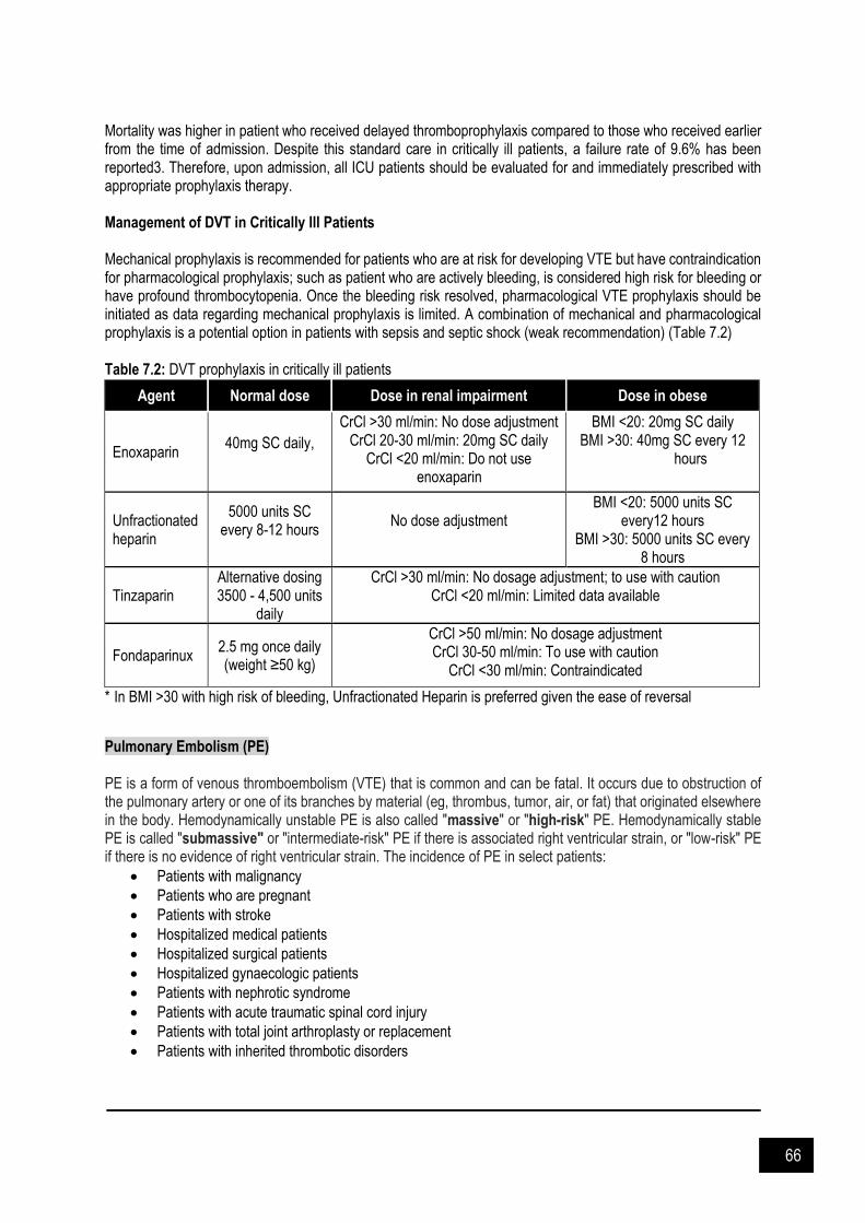
66
Mortality was higher in patient who received delayed thromboprophylaxis compared to those who received earlier from the time of admission. Despite this standard care in critically ill patients, a failure rate of 9.6% has been reported3. Therefore, upon admission, all ICU patients should be evaluated for and immediately prescribed with appropriate prophylaxis therapy. Management of DVT in Critically Ill Patients Mechanical prophylaxis is recommended for patients who are at risk for developing VTE but have contraindication for pharmacological prophylaxis; such as patient who are actively bleeding, is considered high risk for bleeding or have profound thrombocytopenia. Once the bleeding risk resolved, pharmacological VTE prophylaxis should be initiated as data regarding mechanical prophylaxis is limited. A combination of mechanical and pharmacological prophylaxis is a potential option in patients with sepsis and septic shock (weak recommendation) (Table 7.2) Table 7.2: DVT prophylaxis in critically ill patients
Agent Normal dose Dose in renal impairment Dose in obese
Enoxaparin 40mg SC daily,
CrCl >30 ml/min: No dose adjustment CrCl 20-30 ml/min: 20mg SC daily
CrCl <20 ml/min: Do not use enoxaparin
BMI <20: 20mg SC daily BMI >30: 40mg SC every 12
hours
Unfractionated heparin
5000 units SC every 8-12 hours
No dose adjustment
BMI <20: 5000 units SC every12 hours
BMI >30: 5000 units SC every 8 hours
Tinzaparin Alternative dosing 3500 - 4,500 units
daily
CrCl >30 ml/min: No dosage adjustment; to use with caution CrCl <20 ml/min: Limited data available
Fondaparinux 2.5 mg once daily (weight ≥50 kg)
CrCl >50 ml/min: No dosage adjustment CrCl 30-50 ml/min: To use with caution
CrCl <30 ml/min: Contraindicated
* In BMI >30 with high risk of bleeding, Unfractionated Heparin is preferred given the ease of reversal
Pulmonary Embolism (PE) PE is a form of venous thromboembolism (VTE) that is common and can be fatal. It occurs due to obstruction of the pulmonary artery or one of its branches by material (eg, thrombus, tumor, air, or fat) that originated elsewhere in the body. Hemodynamically unstable PE is also called "massive" or "high-risk" PE. Hemodynamically stable PE is called "submassive" or "intermediate-risk" PE if there is associated right ventricular strain, or "low-risk" PE if there is no evidence of right ventricular strain. The incidence of PE in select patients:
Patients with malignancy
Patients who are pregnant
Patients with stroke
Hospitalized medical patients
Hospitalized surgical patients
Hospitalized gynaecologic patients
Patients with nephrotic syndrome
Patients with acute traumatic spinal cord injury
Patients with total joint arthroplasty or replacement
Patients with inherited thrombotic disorders

67
Once the diagnosis of PE is made, the mainstay therapy is anticoagulation, depending on the risk of bleeding. Patients with life-threatening PE (hemodynamically unstable) may require additional pharmacological treatment beyond anticoagulation, including thrombolytic therapy (thrombolysis). Systemic thrombolysis with typically tissue-type plasminogen activator (tPA) is preferred in hemodynamically unstable patients. Recommendation before initiating thrombolysis:
Hemodynamically unstable patients, who are already receiving systemic anticoagulation, which is typically unfractionated heparin, consider discontinuing the anticoagulant therapy, immediately before and during the thrombolytic infusion to minimize the risk of bleeding
If a patient has already received low molecular weight heparin, the administration of the thrombolytic agent should not be delayed
Table 7.3: Thrombolytic agents
Agents Recommended dosing regimens
Alteplase (tPA) Hemodynamically unstable / massive: 100 mg administered over two hours
PE associated with cardiac arrest –
As a bolus infusion over 15 minutes or as a 20 mg IV bolus followed by an infusion of 80 mg over the next two hours OR
50 mg bolus over 2 minutes; repeat a second 50 mg bolus 15 minutes later if the return of spontaneous circulation (ROSC) is not achieved
Tenecteplase (TNK-tPA) Weight-based bolus infusion Administer as a single bolus over 5-10 seconds
<60 kg: 30 mg
≥60 to <70 kg: 35 mg
≥70 to <80 kg: 40 mg
≥80 to <90 kg: 45 mg
≥90 kg: 50 mg
When the infusion of thrombolytic agent is complete, IV UFH may be re-started. Avoid initiating longer-acting anticoagulants (eg, low molecular weight heparin) and oral agents, (eg, direct oral anticoagulants [DOAC], warfarin) for at least 24 hours to ensure that there is no delayed bleeding that would require immediate cessation of anticoagulation. It is suggested to start a heparin infusion without a bolus. To minimize the risk of bleeding, it is recommended to check an activated partial thromboplastin time (aPTT) and resume UFH without a loading dose when the aPTT is less than twice its upper limit of normal. If the aPTT exceeds this value, repeat it every four hours until it is less than twice its upper limit of normal, at which time, resume a heparin infusion. Once stable for 24 to 48 hours, patients should be transitioned to an oral agent (eg, DOAC or warfarin). The duration of long-term anticoagulation following thrombolysis is generally the same as for patients who have not received a thrombolytic agent Table 7.4: Contraindications to fibrinolytic therapy for DVT or acute PE
Absolute contraindications
Prior intracranial hemorrhage Known structural cerebral vascular lesion Known malignant intracranial neoplasm Ischemic stroke within three months (excluding stroke within three hours*) Suspected aortic dissection Active bleeding or bleeding diathesis (excluding menses) Significant closed-head trauma or facial trauma within three months
Relative contraindications
History of chronic, severe, poorly controlled hypertension Severe uncontrolled hypertension on presentation (SBP >180 mmHg or DBP >110 mmHg) History of ischemic stroke more than three months prior Traumatic or prolonged (>10 minutes) CPR or major surgery less than three weeks
68
Recent (within two to four weeks) internal bleeding Noncompressible vascular punctures Recent invasive procedure For streptokinase/anistreplase - Prior exposure (more than five days ago) or prior allergic reaction to these agents Pregnancy Active peptic ulcer Pericarditis or pericardial fluid Current use of anticoagulant (eg, warfarin sodium) that has produced an elevated international normalized ratio (INR) >1.7 or prothrombin time (PT) >15 seconds Age >75 years Diabetic retinopathy
References and Suggested Readings Alhazzani W, Lim W, Jaeschke RZ, Murad MH, Cade J, Cook DJ. Heparin thromboprophylaxis in medical-surgical
critically ill patients: A systematic review and meta-analysis of randomized trials. Crit Care Med. 2013. 41(9): 2088-98.
Benjamin E, Recinos G, Aiolfi A, Inaba K, Demetriades D. Pharmacological thromboembolic prophylaxis in traumatic brain injuries. Ann Surg. 2017. 266(3): 463-469.
Chan C, Shorr A. Venous thromboembolic disease in the intensive care unit. Semin Respir Crit Care Med. 2010. 31(01): 039-046.
Cook D, Crowther M, Meade M, et al. Deep venous thrombosis in medical-surgical critically ill patients: prevalence, incidence, and risk factors. Crit Care Med. 2005. 33(7): 1565-1571
Freeman AL, Pendleton RC, Rondina MT. Prevention of venous thromboembolism in obesity. Expert Rev Cardiovasc Ther. 2010. 8(12): 1711-1721.
Ho KM, Chavan S, Pilcher D. Omission of Early Thromboprophylaxis and mortality in critically ill patients: A multicenter registry study. Chest. 2011. 140(6): 1436-1446.
Kang M, Alahmadi M, Sawh S, Kovacs MJ, Lazo-Langner A. Fondaparinux for the treatment of suspected heparin-induced thrombocytopenia: a propensity score–matched study. Blood. 2015. 125(6): 924-929.
Kearon C, Akl EA, Comerota AJ, Prandoni P, Bounameaux H, Goldhaber SZ, Nelson ME, Wells PS, Gould MK, Dentali F, Crowther M, Kahn SR, American College of Chest Physicians. Antithrombotic therapy for VTE disease: Antithrombotic therapy and prevention of thrombosis, 9th ed: American College of Chest Physicians Evidence-Based Clinical Practice Guidelines. Chest. 2012. 141: e419S.
Kelly J, Rudd A, Lewis R, Hunt BJ. Venous thromboembolism after acute stroke. Stroke. 2001. 32(1): 262-267. Levine M, Hirsh J, Weitz J, Cruickshank M, Neemeh J, Turpie AG, Gent M. A randomized trial of a single bolus
dosage regimen of recombinant tissue plasminogen activator in patients with acute pulmonary embolism. Chest. 1990. 98(6): 1473.
Linkins L-A, Dans AL, Moores LK, et al. Treatment and Prevention of Heparin-Induced Thrombocytopenia: Antithrombotic Therapy and Prevention of Thrombosis, 9th ed: American College of Chest Physicians Evidence-Based Clinical Practice Guidelines. Chest. 2012. 141(2): e495S-e530S.
Meneveau N, Schiele F, Metz D, Valette B, Attali P, Vuillemenot A, Grollier G, Elaerts J, Mossard JM, Viel JF, Bassand JP. Comparative efficacy of a two-hour regimen of streptokinase versus alteplase in acute massive pulmonary embolism: immediate clinical and hemodynamic outcome and one-year follow-up. J Am Coll Cardiol. 1998. 31(5): 1057.
Rhodes A, Evans LE, Alhazzani W, et al. Surviving Sepsis Campaign: International Guidelines for Management of Sepsis and Septic Shock: 2016. Crit Care Med. 2017. 45(3): 486-552.
Rondina MT, Wheeler M, Rodgers GM, Draper L, Pendleton RC. Weight-based dosing of enoxaparin for VTE prophylaxis in morbidly obese, medically-Ill patients. Thromb Res. 2010. 125(3): 220-3
Tapson VF, Weinberg AS. Approach to thrombolytic (fibrinolytic) therapy in acute pulmonary embolism: Patient selection and administration. UpToDate 2020.
Thompson BT, Kabrhel C. Overview of acute pulmonary embolism in adults. UpToDate. 2020.

69
CHAPTER 8
Stress-related Ulcer
tress-related ulcer is an acute condition which erosion of the gastric mucosa occur secondary to a physiologic stress. Two types of mucosal lesions included stress-related injury (diffuse, superficial mucosal damage) and discrete stress ulcers (deep focal lesions that penetrate the submucosa). Endoscopic evidence of superficial mucosal damage occurs in 75-100% of patients within 1–2 days after ICU admission. Mortality from stress-related bleeding in the critically ill ranges from 50-70%, with a 20% mortality rate attributable to stress related ulcer. The exact mechanism of acute stress ulcer is multifactorial and complex (Figure 8.1). Clinical presentation that of overt bleeding are manifested as hematemesis, coffee ground emesis, melena or bloody nasogastric aspirate. Unexplained hypotension or a decrease greater than 2 g/dL in hemoglobin level requires prompt evaluation for gastrointestinal (GI) bleeding.
Figure 8.1: Pathophysiology of stress ulcer in critically ill patient (from Stollman & Metz 2005) Stress Ulcer Prophylaxis The strongest clinical predictors of GI bleeding are mechanical ventilators more than 48 hours and coagulopathy. Patient with head injury or burns are at risk due to higher rate of gastric acid secretion resulting from vagal stimulation also as a result of diminished mucosal defenses and hypo perfusion. Clinically significant stress-related bleeding has decreased during the past decade because of many factors, including early resuscitation and stress ulcer prophylaxis (SUP).
S

70
Table 8.1: Indication for stress ulcer prophylaxis in critically ill patient
Risk Indications Indication for prophylaxis
Low No risk factor No
Moderate Chronic NSAIDs/ aspirin use Peptic ulcer disease > 1 year
To be considered
High Mechanical ventilation > 48 hours Coagulopathy: INR > 1.5, APTT 2x the control, platelet
<50 000/mm Traumatic brain injury with GCS ≤10 or spinal cord injury Significant burn injury (total body surface area >30%) Acute liver failure Renal replacement therapy Use of 2 antiplatelets History of UGIB or gastric ulcer in last 1 year Any 2 of the following
- Sepsis - High dose steroid (hydrocortisone >250mg/day,
prednisolone 60mg /day, methylprednisolone 50mg/day, dexamethasone 10mg/day)
- ICU stay > 7 days - Occult bleeding of > 6 days
Indicated
Treatment strategies need to be evaluated on a case-by-case basis, taking into account the expected benefits of SUP balance against the patients’ risk factor for bleeding and the risk of pneumonia. A propose mechanism that proton pump inhibitors (PPIs) caused pneumonia is increased bacterial colonization by overgrowth of gastric microbes in the upper GI tract due to higher pH after gastric acid suppression. The condition worsening when coupled with micro-aspiration in intubated patient. Maintaining a pH less than 4 will reduce rate of pneumonia and Clostridium difficile infections (CDI) as gastric acid is an important defense against the acquisition of this complication. Once patient is tolerated to full feeding and has no risk factor, prophylactic therapy may be discontinued. This is important to avoid unnecessary drug interaction, complications and cost. In patient with nasogastric feeding, the administration of gastric nutrition reduces but does not eliminate the risk of GI bleeding. Patients who are predicted to be mechanically ventilated more 48 hours and allowed for gastric enteral nutrition are encouraged to have nasogastric nutrition initiated within 48 hours of admission. Considerations in Critically Ill Patients Commonly used medications for SUP in ICU includes H2RAs and PPIs (Table 8.2). The choice of SUP agents depends on balance between efficacy, potential harm and cost. Antacids is not recommended for SUP due to frequent administration (up to every hour), adverse effects (constipation, diarrhea, electrolytes abnormalities) and interactions (interferes the absorption of some dugs). Meanwhile sucralfate was found to be inferior to H2RA and physically can clog nasogastric (NG) tubes, thus limit its usage.

71
Table 8.2: Pharmacological agents for management of stress ulcer in critically ill patient
Drug Classes Histamine Type 2 Receptor
Antagonists Proton Pump Inhibitors
Drugs Ranitidine Esomeprazole Omeprazole Pantoprazole
Dosage Adult Normal Dose Oral: 150 mg bd IV: 50 mg Q8H Dose Adjustment CrCl < 50 ml/min Oral: 150 mg od IV: 50 mg every 18-24 hours; adjust dose cautiously if needed Hemodialysis: Adjust dosing schedule so that dose coincides with the end of hemodialysis.
40 mg daily (IV, nasogastric tube, PO) * Esomeprazole is the S-isomer of omeprazole
20-40 mg daily (PO, nasogastric/ jejunal, duodenal tube)
40 mg daily (IV, nasogastric tube, PO)
Patient who develops significant GI hemorrhage** receiving prophylaxis: Only IV Pantoprazole /Esomeprazole: 80 mg loading dose followed by 8 mg/hr for 3 days - need to consider for endoscopic evaluation and convert to intermittent dosing schedule when no evidence of bleeding for 24 hours, **Defined as bleeding that requires transfusion, causes hemodynamic changes and/or decrease in haemoglobin ≥2 g/dL
Monitoring Liver enzyme, serum creatinine, sign and symptoms of PUD, occult blood with GI bleeding, renal function.
Liver enzyme
Contraindication Hypersensitivity to the component of the formulation
Precaution Use in caution in patient with hepatic impairment and renal impairment
Severe liver dysfunction may require dose adjustment
Bioavailability may increase in the elderly, Asian population, and with hepatic dysfunction
IV preparation contain edentate sodium (EDTA); use caution in patient who are at risk for zinc deficiency if other EDTA containing solution are co-administered
Adverse Reactions
Arrythmias, dizziness, headache, mental confusion, rash, anemia, thrombocytopenia, leucopenia, hepatic failure and pneumonia
Headache, nausea, flatulence, diarrhea, constipation, abdominal pain, hypertension, hyponatremia, pneumonia
Headache, dizziness, diarrhea, abdominal pain, pneumonia
Headache, diarrhea, flatulence, abdominal pain, abnormal liver function test

72
Drug Classes Histamine Type 2 Receptor
Antagonists Proton Pump Inhibitors
Drugs Ranitidine Esomeprazole Omeprazole Pantoprazole
Drug Interaction CYP450 effect Increase effect: Phenytoin, Midazolam Decrease effect: Atazanavir, Alprazolam, Ketoconazole, Itraconazole
CYP450 effect Increase effect: Methotrexate, Phenytoin, Warfarin Decrease effect: Atazanavir, Indinavir, Ketoconazole, Itraconazole, Clopidogrel, Mycophenolate mofetil, Cyanocobalamine
CYP450 effect Increase effect: Methotrexate, Clonazepam, Midazolam, Diazepam, Digoxin, Warfarin Decrease effect: Ketoconazole, Itraconazole, Atazanavir, Indinavir, Clopidogrel, Mycophenolate mofetil
CYP450 effect Increase effect: Methotrexate, Warfarin Decrease effect: Atazanavir, Indinavir, Ketoconazole, Itraconazole, Mycophenolate mofetil, Cyanocobalamine
Stability/ Storage
Reconstitute with 20 ml NS or D5 and can be administered over at least 2 minutes. Diluted solution stable for 24 hours at room temperature.
Reconstitute with 5 ml NS or D5 and can be administered over at least 3 minutes. Reconstituted solution stable for 12 hours with NS or 6 hours D5 at room temperature.
Reconstitute with provided solvent. Administer over not less than 2.5 min (not greater than 4mL/min). Reconstituted solution stable for 4 hours in 2-8 °C.
Reconstitute with 10 ml NS. Can be further diluted with isotonic sodium chloride solution, 5% dextrose in water, or lactated Ringer’s solution. 2-minute infusion: The volume of reconstituted solution (4 mg/mL) may be administered IV over at least 2 minutes. 15-minute infusion: Infuse over ~15 minutes at a rate of ~7 mL/minute. Continuous infusion: May also be administered as a continuous infusion for the prevention of rebleeding with in peptic ulcer bleed (off-label use)
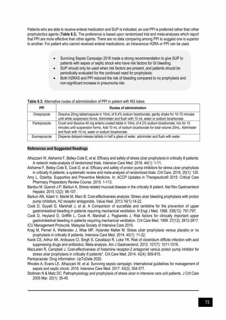
73
Patients who are able to receive enteral medication and SUP is indicated, an oral PPI is preferred rather than other prophylactics agents (Table 8.3). The preference is based upon randomized trial and meta-analyses which report that PPI are more effective than other agents. There are no data comparing among PPI to suggest one is superior to another. For patient who cannot received enteral medications, an intravenous H2RA or PPI can be used. Table 8.3: Alternative routes of administration of PPI in patient with NG tubes
PPI Routes of administration
Omeprazole Dissolve 20mg tablet/capsule in 10mL of 8.4% sodium bicarbonate, gently shake for 10-15 minutes until white suspension forms. Administer and flush with 10 mL water or sodium bicarbonate.
Pantoprazole Crush and dissolve 40 mg enteric-coated tablet in 10mL of 4.2% sodium bicarbonate, mix for 10 minutes until suspension forms. Add 10 mL of sodium bicarbonate for total volume 20mL. Administer and flush with 10 mL water or sodium bicarbonate.
Esomeprazole Disperse delayed-release tablets in half a glass of water, administer and flush with water.
References and Suggested Readings Alhazzani W, Alshamsi F, Belley-Cote E, et al. Efficacy and safety of stress ulcer prophylaxis in critically ill patients:
A network meta-analysis of randomized trials. Intensive Care Med. 2018. 44(1): 1-11. Alshamsi F, Belley-Cote E, Cook D, et al. Efficacy and safety of proton pump inhibitors for stress ulcer prophylaxis
in critically ill patients: a systematic review and meta-analysis of randomized trials. Crit Care. 2016. 20(1): 120. Amy L. Dzierba. Supportive and Preventive Medicine. In: ACCP Updates in Therapeutics® 2015: Critical Care
Pharmacy Preparatory Review Course. 2015: 1-113. Bardou M, Quenot J-P, Barkun A. Stress-related mucosal disease in the critically ill patient. Nat Rev Gastroenterol
Hepatol. 2015.12(2): 98-107. Barkun AN, Adam V, Martel M, Marc B. Cost-effectiveness analysis: Stress ulcer bleeding prophylaxis with proton
pump inhibitors, H2 receptor antagonists. Value Heal. 2013.16(1):14-22. Cook D, Guyatt G, Marshall J, et al. A Comparison of sucralfate and ranitidine for the prevention of upper
gastrointestinal bleeding in patients requiring mechanical ventilation. N Engl J Med. 1998. 338(12): 791-797. Cook D, Heyland D, Griffith L, Cook R, Marshall J, Pagliarello J. Risk factors for clinically important upper
gastrointestinal bleeding in patients requiring mechanical ventilation. Crit Care Med. 1999. 27(12): 2812-2817. ICU Management Protocols. Malaysia Society of Intensive Care 2019. Krag M, Perner A, Wetterslev J, Wise MP, Hylander Møller M. Stress ulcer prophylaxis versus placebo or no
prophylaxis in critically ill patients. Intensive Care Med. 2014. 40(1): 11-22. Kwok CS, Arthur AK, Anibueze CI, Singh S, Cavallazzi R, Loke YK. Risk of clostridium difficile infection with acid
suppressing drugs and antibiotics: Meta-analysis. Am J Gastroenterol. 2012. 107(7): 1011-1019. MacLaren R, Campbell J. Cost-effectiveness of histamine receptor-2 antagonist versus proton pump inhibitor for
stress ulcer prophylaxis in critically ill patients*. Crit Care Med. 2014. 42(4): 809-815. Pantoprazole: Drug Information. UpToDate 2020. Rhodes A, Evans LE, Alhazzani W, et al. Surviving sepsis campaign: International guidelines for management of
sepsis and septic shock: 2016. Intensive Care Med. 2017. 43(3): 304-377. Stollman N & Metz DC. Pathophysiology and prophylaxis of stress ulcer in intensive care unit patients. J Crit Care
2005 Mar. 20(1): 35-45.
Surviving Sepsis Campaign 2016 made a strong recommendation to give SUP to patients with sepsis or septic shock who have risk factors for GI bleeding.
SUP should only be used when risk factors are present, and patients should be periodically evaluated for the continued need for prophylaxis.
Both H2RAS and PPI reduced the risk of bleeding compared to no prophylaxis and
non-significant increase in pneumonia risk.

74
CHAPTER 9
Delerium
elirium, a common manifestation of acute brain dysfunction in critically ill patients, is associated with poor short-term outcomes and may result in adverse sequelae years after ICU discharge. The prevalence of delirium reported in medical and surgical ICU cohort studies has varied from 20% to 80% and is often goes unrecognized by clinicians. Onset of delirium may be triggered by a number of physiological processes, including metabolic derangement, infectious processes, central nervous system pathology, and medication-induced side effects. Medications commonly associated with the development of delirium include benzodiazepines, opioids, and agents with high anticholinergic activity or side effects. Based on ICU Protocol Management, Ministry of Health Malaysia, patients with a revised Riker >-2 or RASS >-3 should be screened for delirium using the Confusion Assessment Method for ICU (CAM-ICU) every 8 hours.
Figure 9.1: Confusion Assessment Method for ICU (CAM-ICU) (from Management Protocols in ICU, MOH 2012)
D

75
Recommended strategies for addressing delirium include treating the reversible factors, such as infectious disease, drug withdrawal, and medication toxicity, adequately treating pain, correcting metabolic, nutritional, endocrine disorders and finally, managing sleep deprivation. Non-pharmacological management Non-pharmacological approached is preferred in the management of delirium. The strategies include:
Providing psychological support and orientation consistently Involving family members to re-orientate patient Early mobilization Minimize noise and light at night Minimize nocturnal interventions if clinically possible and use night sedation as a last resort Early liner/catheher removal
Pharmacological management Recent guidelines advocate against the use of pharmacologic agent to “prevent” delirium in critically ill patients. Reasons are due to poor available evidence and lack of generalizability from available studies. When adjunct medical management is required, following drugs may be used to treat delirium:
However, they should not be routinely used due to poor quality of evidence. In addition, patients who start with an antipsychotic for delirium often remain on these medications unnecessarily upon discharge. Continued exposure of these antipsychotic may result in morbidity and increase financial cost
Although no IV drugs approved by FDA for delirium in critically ill patients, IV haloperidol is used nevertheless and remains as the first line of delirium treatment in ICU
Dexmedetomidine, the alpha-2 agonists have demonstrated clinical efficacy in ICU settings in achieving tranquilization-sedation more safely and potentially help in preventing delirium.
References and Suggested Readings Barr J, Fraser GL, Puntillo K, et al. Clinical practice guidelines for the management of pain, agitation, and delirium
in adult patients in the intensive care unit. Crit Care Med. 2013. 41(1): 262-306. Dasta, J.F. Current and future pharmacologic strategies to prevent and manage delirium. 40th Critical Care
Congress Review 2011. Gilchrist, N.A., Asoh, I., & Greenberg, B. Analytical review: Atypical antipsychotics for the treatment of ICU delirium.
J Intensive Care Med. 2011. 27(6): 354-61. Girard TD, Pandharipande PP, Ely EW. Delirium in the intensive care unit. Critical Care. 2008. 12(3):S3. Jones SF, Pisani MA. ICU delirium: An update. Curr Opin Crit Care. 2012. 18(2): 146-51. Maldonado JR, Maccioli GA, Riker RR, Dasta JF, Szabo E. Delirium in the ICU: Prevention and treatment.
Presentations in FOCUS, Rogers Medical Intelligence Solutions 2005. Management Protocols in ICU: Sedation and delirium 2012. Ministry of Health Malaysia & Malaysian Society of
Intensive Care. NICE Clinical Guideline. Delirium: Diagnosis, prevention and management. 2010: 103.
IV Haloperidol: <60yrs: 5-10mg PRN/ q4-6h, >60yrs: 2.5-5mg PRN/q4-6h *Continuous infusion (unlabeled use): 3-25mg/hour
T. Chlorpromazine 12.5-25 mg q6-8 hourly T. Risperidone 0.5-1mg q12h IV Dexmedetomidine 0.2-1.5 mcg/kg/hr (consider in patients who are ready to be weaned but
agitated) T. Olanzapine 5-10 mg q12-24h, 2.5 mg OD in patients >60 years T. Quetiapine 50-100 mg q12h
Caution: Prolonged QT may occur with the use of all the above drugs except Dexmedetomidine 6

76
CHAPTER 10
Fluids and Electrolytes
t is estimated that 60% of lean body weight (LBW) in male and 50% LBW in women consist of water. Total
body water is further divided to intracellular (IC) space and extracellular (EC) space which is separated by cell membrane. EC compartment is further divided to interstitial space (75%) and intravascular space (25%) which is separated by capillary membrane. The distribution of intravenous fluid is explained with this approximate distribution of total body fluid (TBF) into the mentioned compartment. Table 10.1: Intravenous fluids and volume expansion
Intravenous fluid Infused volume (ml) Equivalent intravascular volume expansion (ml)
0.9% NaCl 1000 250
Lactated Ringer’s 1000 250
5% Dextrose 1000 250
5% albumin 500 500
20% albumin 100 400
25% albumin 100 500
The plasma osmolality is between 275 and 290 mOsm/kg. a) Hypertonic fluid cause fluid to shift from the IC to the EC compartment with subsequent cellular dehydration
and shrinkage. b) Isotonic fluid (similar osmolarity with plasma) - will not result in a fluid shift between fluid compartments. c) Hypotonic fluid is solution with osmolarity less than 150 mOsm/L. It caused fluid to shift from the EC to the IC
compartment with subsequent cellular overhydration and swelling.
RBC swelling may cause cell rupture (i.e., hemolysis)
Brain cells may swell, causing cerebral edema and herniation; most likely to occur with acute hyponatremia (<2 days)
Sterile water is NEVER meant for intravenous administration
Table 10.2: Comparison between crystalloids and collloids
Crystalloids Colloids
Fluids that may contain a combination of water, dextrose and/or electrolytes
High molecular weight substances primarily remain in the intravascular compartment → generating an oncotic pressure.
Example: 0.9% NaCl, 5% Dextrose, 5% Dextrose in NaCl 0.9%, Lactated Ringer’s, Sterofundin
Example: Natural: blood and blood products, albumin Artificial: dextran, hydroxyethyl starch (HES), modified fluid gelatine
Na+ and Cl- do not freely cross into cells (distribute evenly in EC space – 25% intravascular space, 75% in interstitial space)
Dextrose = free water (Dextrose → CO2 + H2O), freely crosses any membrane, evenly distributed in TBF
Too large to cross the capillary membrane – primarily remain in the intravascular space (small portion “leak” into the interstitial space)
I
77
When administered in large volumes, 0.9% NaCl can cause normal anion gap metabolic acidosis (hyperchloremic metabolic acidosis), while this is not an issue in balanced crystalloid solution eg: sterofundin.
Free water (D5W) should be avoided in patient with elevated intracranial pressure (ICP) as it may cross cerebral cells and cause further ICP elevation.
HES and dextran – risk of coagulopathy, avoid in patients with increased risk of haemorrhage.
All colloids can cause anaphylactoid reactions.
HES should no longer be used to treat septic or critically ill patients as current studies shows it may increase risk of kidney injury and mortality.
Require larger volume for equivalent intravascular expansion.
Require less volume for equivalent intravascular expansion.
Inexpensive (balanced solution is expensive compared to conventional crystalloid solution).
Expensive.
Shorter intravascular half-life Longer intravascular half-life
Fluid Resuscitation versus Fluid maintenance in ICU
The goal of fluid resuscitation is to restore intravascular volume and prevent organ hypoperfusion while fluid maintenance is to prevent dehydration and maintain a normal fluid and electrolyte balance. The osmolarity and electrolytes content of various intravenous fluids are listed in (refer Table 10.4). Table 10.3: Fluid resuscitation versus fluid maintenance
Fluid resuscitation Fluid maintenance
Indication: In patients with sign and symptoms of fluid depletion.
Indication: in patients who are unable to tolerate oral fluids.
Recommendation: crystalloids (0.9% NaCl or Lactated Ringer’s). Colloids not superior to crystalloids, more expensive.
Recommendation: 5% Dextrose with 0.45% NaCl + 20-40 mmol KCl/L (KCl content – adjust individually).
Infuse rapidly, preferably through central venous catheter (CVC).
Continuous infusion, through CVC or peripheral catheter. Remember to evaluate IV fluids daily.
500- to 1000-mL bolus (20ml/kg) and re-evaluate; this process continues as long as the sign and symptoms of intravascular volume depletion are improving or there are sign of acute pulmonary oedema (APO).
Common methods of estimating daily maintenance volume in children and adults
1st 10kg - 100 ml/kg; 2nd 10kg - 50 ml/kg plus 20 ml/kg for every kg greater than 20 kg (i.e if weight >20kg = 1500ml + 20ml for each kg over 20kg)
“4:2:1” Rule (ml/kg/hr)
20–40 mL/kg/day (for adults only)
Adjust fluids based on pt’s input, output, insensible loss

78
Table 10.4: IV fluids osmolarity and electrolytes content
IV Fluids mOsm/L Glucose g/L Na+ Cl- *Lactate K+ Ca2+ Mg2+ **Acetate **Malate
mmol/L
CRYSTALLOIDS
5% dextrose and water (D5) 278 50
10% dextrose and water (D10) 555 100
20% dextrose and water (D20) 1110 200
50% dextrose and water (D50) 2523 500
0.9% sodium chloride (NS) 308 154 154
0.45% sodium chloride (HS) 154 77 77
3% sodium chloride (S3) 1024 513 513
0.9% sodium chloride + dextrose 5% (NSD5) 585 50 154 154
0.45% sodium chloride + 5% dextrose (HSD5) 430 50 77 77
0.45% sodium chloride + 10% dextrose (HSD10)
709 100 77 77
0.18% sodium chloride + 4.23% dextrose (QSD1) (1/5SD5)
296 42.3 31 31
0.18% sodium chloride + 10% dextrose (QSD10) (1/5SD10)
615 100 31 31
Hartmann’s Solution 278 131 111 29 5 2
Sterofundin 304 140 127 4 2.5 1.0 24 5
COLLOIDS
Albumin 20% Sodium ranges from 113.5 – 138.7 mmol/L
Gelofusine 4% 274 154 120
Voluven 6% HES 130/0.4/9:1 308 154 154
Venofundin 6% HES 130/0.42/6:1 309 154 154
Volulyte 6% HES 130/0.4/9:1 286.5 137 110 4 1.5 34
Tetraspan 6% HES 130/0.42/6:1 296 140 118 4 2.5 1.0 24 5
mEq = mmol/L x ion charge. *1 mmol lactate produce 1 mmol bicarbonate (in liver) **1 mmol acetate produce 1 mmol bicarbonate and 1 mmol malate produce 2 mmol bicarbonate (in muscle)
79
Plasma Sodium (Normal: 135-145 mmol/L)
Sodium is the principal cation of the ECF and primary determinant of plasma osmolality. Serum sodium concentration and serum osmolarity normally are maintained under precise control by homeostatic mechanisms involving stimulation of thirst, secretion of antidiuretic hormone (ADH), and renal handling of filtered sodium. The physician must have a thorough understanding of the pathophysiology of hyponatremia to initiate safe and effective corrective therapy. The patient's fluid status must be accurately assessed upon presentation, as it guides the approach to correction. Table 10.5: Sodium abnormalities
Classification Signs and symptoms Causes
Hyponatremia (<135 mmol/L)
The clinical features of acute hyponatremia are related to osmotic water shifted that leads to increased ICF volume and brain cells swelling. Mild hyponatremia is usually asymptomatic.
Serum Na+ < 120 mmol/L may be associated with disturbed mental state, restlessness, confusion and irritability.
As the Na+ approaches 110 mmol/L, seizures and coma may occur.
Replacement of lost solute with water
Volume depletion and organ hypoperfusion
Syndrome of inappropriate ADH secretion and cortisol deficiency
Medications
Renal failure
Pseudohyponatremia condition when Na+ content in the body is not actually reduced, but rather, shifts from the EC compartment into the cells to maintain plasma osmolality in a normal range. Normally associated with normal osmolality
Primary or secondary hyperlipidaemia disorder, severe hyperglycaemia (DKA).
Plasma protein elevations above 10 g/dL, as seen in multiple myeloma or macroglobulinemia
Hypernatremia (>145 mmol/L)
Features are tremulousness, irritability, ataxia, spasticity, mental confusion and coma.
Loss of water (Fever, GI loss, burn, infection, renal loss).
Administration of hypertonic saline/exogenous source of Na+.
Management of Sodium Abnormalities Sodium correction:
Total Na+ deficit (mmol): (130 – current Na+) x 60% of body weight in kg
Volume (ml) required to replace total Na+ deficit using 0.9% NaCl: Total Na+ deficit (mmol)/154 (mmol/L) x 1000
Volume (ml) required to replace total Na+ deficit using 3% NaCl: Total Na+ deficit (mmol)/513 (mmol/L) x 1000
The volume calculated should be given within the calculated number of hour: (130 – current Na+) / 0.5 The rate of rise in plasma Na should not exceed 0.5 mmol/L/ hour (10-12 mmol/day) to avoid cerebral pontine myelinolysis.
Hypernatremia
Free H2O replacement (use dextrose 5%): Water deficit (L) = 0.6 x BW in kg x [(measured Na+/140) - 1]
To be given over 48-72 hours. Sterile Water (Should only be administered through NG)
Rate of correction should not exceed 0.5 mmol/L/ hour (10-12 mmol/day) to avoid cerebral pontine myelinolysis.
80
Table 10.6: Classification of Hyponatremia
Classification Hypervolemic hyponatremia Normovolemic hyponatremia Hypovolemic hyponatremia
Description Caused by excess Na+ and fluid but fluid excess predominates.
Normal total body Na+ with excess fluid volume (i.e dilutional).
Deficit of both Na+ and fluid, but total Na+ is decreased more than total body water.
Diagnosis Urine Na+ <25 mmol/L: edematous disorders (i.e heart failure, cirrhosis, nephrotic syndrome) Urine Na+ >25 mmol/L: acute or chronic renal failure.
Urine osmolality >100 mOsm/kg: Impaired water excretion Urine Na+ >40 mmol/L: Glucocorticoid deficiency, hypothyroidism, stress, drugs.
Urine Na+ <25 mmol/L: Nonrenal loss of Na+ (e.g emesis, diarrhea) Urine Na+ >40 mmol/L: Renal loss of Na+
Treatment Sodium and water restriction; treat underlying cause; vasopressin receptor antagonists (e.g conivaptan, tolvaptan*).
If drug-induced SIADH, remove offending agent; fluid restriction; demeclocycline; vasopressin receptor antagonists (e.g conivaptan, tolvaptan*)
Fluid resuscitation (see above)
Example Heart failure, cirrhosis, nephrotic syndrome
SIADH Fluid loss (e.g., emesis, diarrhea, fever), third-spacing, renal loss (diuretics)
*Not available in Malaysia upon publication.
Plasma Potassium (3.5-5.0 mmol/L)
Potassium is the principal intracellular ion. Normal potassium is regulated by renal excretion, to maintain electroneurality with sodium. Nearly 98% of the body's potassium is intracellular. The ratio of intracellular to extracellular potassium is important in determining the cellular membrane potential. Small changes in the extracellular potassium level can have profound effects on the function of the cardiovascular and neuromuscular systems. Table 10.7: Potassium abnormalities
Classification Signs and symptoms Causes
Hypokalemia (<3.5 mmol/L)
(usually present when K <2.5 mmol/L)
Malaise, fatigue
Neuromuscular disturbances: Weakness, hyporeflexia, paraesthesias, cramps, restlessness leg syndrome, rhabdomylysis, paralysis
Gastrointestinal: Constipation, ileus
Polyuria, polydipsia, metabolic alkalosis
ECG changes: small or inverted T waves, prominent U wave, depressed ST segments, prolonged PR interval
Arrhythmias: First- and second-degree heart block, atrial fibrillation, ventricular tachycardia, ventricular fibrillation
Decreased potassium intake
Increased entry into cell
Increased urinary losses
Increased GI losses
Increased sweat loss
Dialysis
Plasmapheresis

81
Hyperkalemia (>5.0 mmol/L)
Muscle weakness and paralysis (typically occur when K+ exceeds 8 mmol/L).
Abnormal cardiac conduction (peaked, narrowed T waves, widening of QRS) ventricular fibrilation, asystole. ECG has limitation in predicting cardiac toxicity. Thus, patient with K+ more than 6 mmol/L should be treated even in the absence of ECG changes.
Conduction disturbances are enhanced by hypocalcaemia, hypomagnaesemia, acidosis, rapid increase in potassium concentration.
Pseudohyperkalemia.
Increased in potassium intake.
Increased cell diffusion of potassium to extracellular fluid (Acidosis, insulin deficiency, β adrenergic blockade, digoxin toxicity, rewarming after cardiac surgery, succinylcholine).
Reduced potassium excretion (kidney dysfunction, intravascular volume depletion, hypoaldosteronism, potassium sparing diuretics, ACEi/ARB).
Potassium released from cells while or after obtaining the blood specimen; because of trauma during venipuncture
Measurement of serum rather than plasma potassium concentration; caused by potassium release during coagulation.
Management of Potassium Abnormalities Hypokalaemia
Replace magnesium first if concurrent hypomagnesemia
Generally, reduction of 1 mmol/L serum K+ results in 200 - 400 mmol potassium deficit
Replacement should be guided by K+ concentration; important to recheck level (2 - 4 hourly)
Potassium deficit (mmol) = 4 – Current K+ x 0.4 x Body Weight Calculation for potassium deficit (mmol) = [(4.5 – Current K+) x 0.4 + Body Weight]/13.4 + [ 1 x Body Weight]/13.4
Replacement - Infusion rate: 10-20 mmol/hr (peripheral); max 40 mmol/hr (central line) - Peripheral line administration should not be more than 60 mmol/L to avoid irritation
Caution: - Avoid mixing K+ and dextrose solution as this may cause insulin release which result in IC shift of K+ - Renal impaired and elderly patient
Table 10.8: Potassium correction
Potassium level (mmol/L) Treatment Comments
3.0-3.5 Oral KCl 60–80 mmol/day (> 60 mmol/day should be divided)
Daily K+ check up
2.5-3.0 Oral KCl 120 mmol/day or IV 60–80 mmol administered at 10–20 mmol/hour
Monitor closely (2 hrs after infusion)
2.0-2.5 IV KCL at 10–20 mmol/hr Consider frequent monitoring with ECG
<2.0 IV 20–40 mmol/hr Requires continuous monitoring with ECG
*1g IV KCl = 10 mmol K+
Hyperkalemia
Asymptomatic: Ca2+/Na+ Polystyrene Sulfonate.
Urgent and immediate treatment is required for patients with: - Plasma K+ above 6.5 mmol/L - Severe muscle weakness - ECG changes

82
Table 10.9: Treatment for symptomatic hyperkalaemia
Agents Management
Calcium To prevent hyperkalemia-induced arrhythmias even if patients are normocalcemic.
Calcium gluconate 10% 10 ml (1 g calcium gluconate = 90 mg of calcium elemental) over 2-10 minutes can be administered peripherally and is preferred over calcium chloride because of reduced risk of tissue necrosis.
Onset is fast, within minutes, but duration is short (30–60 minutes).
Not for plasma K+ reduction, but will antagonize the effect of K+ in cardiac conduction cells.
Use in urgent circumstances while waiting for other measures (e.g., insulin and glucose) to lower plasma K+.
Warning: Avoid use in patients receiving digoxin because hypercalcaemia can precipitate digoxin toxicity especially cardiac arythmia and may cause sudden death.
Insulin and glucose
10 units of regular insulin, given intravenously plus, 25–50 g of glucose administered as a 50% dextrose intravenous push in order to prevent hypoglycaemia.
Lowers plasma K+ by 0.5–1.5 mmol/L within an hour and may last for several hours.
If patients are hyperglycemic, insulin may be administered alone.
Sodium bicarbonate
50 mmol of sodium bicarbonate infused slowly over 5 minutes; may repeat in 30 minutes if needed.
May lower plasma K+ within 30–60 minutes and persist for several hours.
The efficacy of bicarbonate is disputed, it seems least effective in patients with advanced kidney disease; may be preferred and effective in patients with underlying metabolic acidosis.
β2-adrenergic agonists (off-label use, Medscape)
Salbutamol 10–20 mg nebulized over 10 minutes or 0.5 mg given intravenously.
Lower plasma K+ by 0.5–1.5 mmol/L.
Onset is within 90 minutes with inhalation.
Avoid use in patients with coronary ischemia due to risk of tachycardia.
Not recommended as single agent for urgent treatment as 40% patients may not respond to inhaled salbutamol, consider use as combination with insulin.
The treatment options above should be followed by one of the following agents to remove excess K+ from the body. i. Diuretics
Loop or thiazide-type diuretics increase K+ renal excretion
Ineffective in patients with advanced kidney disease
ii. Cation-exchange resin
Exchanges Na+ for K+, thus resulting in GI excretion of K+
As onset is slow (2 hours) and efficacy is unpredictable, it is not indicated for emergency treatment of hyperkalemia
Kalimate (Calcium Polystyrene Sulfonate): Oral: 15g 3-4 times daily, Rectal: 30 – 50 g as a retention enema. (oral Kalimate contraindicated in obstructive bowel disease)
iii. Dialysis
Used in severe hyperkalemia or when other treatments are ineffective
Need to monitor for rebound increases in K+ post dialysis
More common in advanced kidney disease patients

83
Plasma Calcium (2.1 - 2.65 mmol/L) [Ionised Calcium (1.1 – 1.3 mmol/L)] Calcium regulation is critical for normal cell function, neural transmission, membrane stability, bone structure, blood coagulation, and intracellular signaling. Normal ionised calcium has been advocated to prevent neurologic and cardiovascular complications. The essential functions of this divalent cation continue to be elucidated, particularly in head injury/stroke and cardiopulmonary effects. Depending on the cause, unrecognized or poorly treated hypocalcemic emergencies can lead to significant morbidity or death. Table 10.10: Calcium abnormalities
Classification Signs and symptoms Causes
Hypocalcemia
- Paraesthesia, tetany, psychosis, circumoral numbness, dystonia, papilloedema (severe), cramp, convulsion
- Prolonged Q-T interval on ECG - Chvostek’s sign – gentle tapping over facial nerve cause
twitching of facial muscles. - Trousseau’s sign – inflation of sphygmomanometer cuff
above diastolic pressure for 5 min causes carpopaedal spasm
- Long-standing hypocalcemia may result in dry skin, coarse hair, alopecia, brittle nails and hypoplastic teeth
- Hypoparathyroidism - Hyperphosphatemia - Receives large amount of
blood products transfusion - Undergoing continuos
renal replacement therapy - Metabolic alkalosis - Vitamin D deficiency - Chronic kidney disease - Alcoholism
Hypercalcemia
- Neurology: Depression, proximal myopathy, fatigue, confusion, stupor and coma
- Renal: Hypertension, renal colic (nephrolithiasis), polyuria and nocturia (nephrogenic diabetes insipidus), dehydration
- Bones: Pain, pathological fractures – osteitis fibrosa cystics in hyperthyroidism (sub-periosteal resorption, bone cysts)
- Abdominal: Nausea, vomiting, constipation, abdominal colic, peptic ulcer disease, pancreatitis
- General: Soft tissue and corneal calcification (band keratopathy)
- ECG changes: Shortened QT intervals
- Malignancy - Hyperparathyroidism
Management of Calcium Abnormalities Hypocalcemia
10 mls 10% Ca gluconate (can be given through peripheral line) contains 90 mg elemental calcium)
Treatment directed at underlying cause
If symptomatic (muscle spasm, laryngeal spasms or cardiac involvement)
If ionised Ca2+ <0.65 mmol/L or corrected Ca2+ <2.0 mmol/L: - IV 10-20mls 10% Ca gluconate over 10 min followed by 1-2 mg/kg/h x 6-12h i.e 30 mls 10% Ca gluconate
diluted to 100 mls N/S run at 25-40ml/h x 6-12h. Administer Mg if deficient. Daily maintenance dose of elemental Ca 1 – 4g orally
Calcium correction to be tailored according to clinical condition and serum calcium level Hypercalcemia
Restoration of intravascular volume with normal saline
Loop diuretic such as furosemide to promote calcium excretion.
Bisphosphonates, such as pamidronate (30–90 mg by i.v. infusion), is recommended for severe cases especially malignant related hypercalcemia
Other therapies include steroids and calcitonin
Calcium adjustment in hypoalbuminemic patients Adjusted calcium level = Current calcium (mmol/L) + 0.02 [40 – Current albumin in g/L]
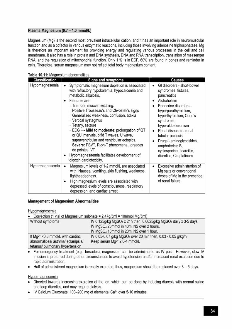
84
Plasma Magnesium (0.7 – 1.0 mmol/L) Magnesium (Mg) is the second most prevalent intracellular cation, and it has an important role in neuromuscular function and as a cofactor in various enzymatic reactions, including those involving adenosine triphosphatase. Mg is therefore an important element for providing energy and regulating various processes in the cell and cell membrane. It also has a role in protein and DNA synthesis, DNA and RNA transcription, translation of messenger RNA, and the regulation of mitochondrial function. Only 1 % is in ECF, 60% are found in bones and reminder in cells. Therefore, serum magnesium may not reflect total body magnesium content. Table 10.11: Magnesium abnormalities
Classification Signs and symptoms Causes
Hypomagnesemia
Symptomatic magnesium depletion is associated with refractory hypokalemia, hypocalcemia and metabolic alkalosis.
Features are: - Tremors, muscle twitching. - Positive Trousseau’s and Chvostek’s signs - Generalized weakness, confusion, ataxia - Vertical nystagmus - Tetany, seizure - ECG → Mild to moderate: prolongation of QT
or QU intervals, bifid T waves, U wave, supraventricular and ventricular ectopics. Severe: PSVT, R-on-T phenomena, torsades de pointes, VT
Hypomagnesaemia facilitates development of digoxin cardiotoxicity.
GI disorders - short-bowel syndromes, fistulas, pancreatitis
Alchoholism
Endocrine disorders - hyperparathyroidism, hyperthyroidism, Conn’s syndrome, hyperaldosteronism
Renal diseases - renal tubular acidosis
Drugs - aminoglycosides, amphotericin B, cyclosporine, ticarcillin, diuretics, Cis-platinum
Hypermagnesemia
Magnesium levels of 1-2 mmol/L are associated with: Nausea, vomiting, skin flushing, weakness, lightheadedness.
High magnesium levels are associated with depressed levels of consciousness, respiratory depression, and cardiac arrest.
Excessive administration of Mg salts or conventional doses of Mg in the presence of renal failure.
Management of Magnesium Abnormalities Hypomagnesemia
Correction (1 vial of Magnesium sulphate = 2.47g/5ml = 10mmol Mg/5ml)
Without symptoms IV 0.125g/kg MgSO4 x 24h then, 0.0625g/kg MgSO4 daily x 3-5 days. IV MgSO4 20mmol in 40ml NS over 2 hours. IV MgSO4 10mmol in 20ml NS over 1 hour.
If Mg2+ <0.6 mmol/L with cardiac abnormalities/ asthma/ eclampsia/ tetanus/ pulmonary hypertension
IV 0.05-0.07 g/kg MgSO4 over 20 min then, 0.03 - 0.05 g/kg/h Keep serum Mg2+ 2.0-4 mmol/L
For emergency treatment (e.g.: torsades), magnesium can be administered as IV push. However, slow IV infusion is preferred during other circumstances to avoid hypotension and/or increased renal excretion due to rapid administration.
Half of administered magnesium is renally excreted, thus, magnesium should be replaced over 3 – 5 days. Hypermagnesemia
Directed towards increasing excretion of the ion, which can be done by inducing diuresis with normal saline and loop diuretics, and may require dialysis.
IV Calcium Gluconate: 100–200 mg of elemental Ca2+ over 5-10 minutes.
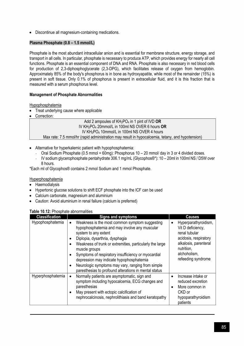
85
Discontinue all magnesium-containing medications. Plasma Phosphate (0.8 – 1.5 mmol/L) Phosphate is the most abundant intracellular anion and is essential for membrane structure, energy storage, and transport in all cells. In particular, phosphate is necessary to produce ATP, which provides energy for nearly all cell functions. Phosphate is an essential component of DNA and RNA. Phosphate is also necessary in red blood cells for production of 2,3-diphosphoglycerate (2,3-DPG), which facilitates release of oxygen from hemoglobin. Approximately 85% of the body's phosphorus is in bone as hydroxyapatite, while most of the remainder (15%) is present in soft tissue. Only 0.1% of phosphorus is present in extracellular fluid, and it is this fraction that is measured with a serum phosphorus level. Management of Phosphate Abnormalities Hypophosphatemia
Treat underlying cause where applicable
Correction:
Add 2 ampoules of KH2PO4 in 1 pint of IVD OR IV KH2PO4 20mmol/L in 100ml NS OVER 6 hours OR
IV KH2PO4 10mmol/L in 100ml NS OVER 4 hours Max rate: 7.5 mmol/hr (rapid administration may result in hypocalcemia, tetany, and hypotension)
Alternative for hyperkalemic patient with hypophosphatemia: - Oral Sodium Phosphate (0.5 mmol = 60mg): Phosphorus 10 – 20 mmol/ day in 3 or 4 divided doses. - IV sodium glycerophosphate pentahydrate 306.1 mg/mL (Glycophos®*): 10 – 20ml in 100ml NS / D5W over
8 hours. *Each ml of Glycophos® contains 2 mmol Sodium and 1 mmol Phosphate. Hyperphosphatemia
Haemodialysis
Hypertonic glucose solutions to shift ECF phosphate into the ICF can be used
Calcium carbonate, magnesium and aluminium
Caution: Avoid aluminium in renal failure (calcium is preferred) Table 10.12: Phosphate abnormalities
Classification Signs and symptoms Causes
Hypophosphatemia
Weakness is the most common symptom suggesting hypophosphatemia and may involve any muscular system to any extent
Diplopia, dysarthria, dysphagia
Weakness of trunk or extremities, particularly the large muscle groups
Symptoms of respiratory insufficiency or myocardial depression may indicate hypophosphatemia
Neurologic symptoms may vary, ranging from simple paresthesias to profound alterations in mental status
Hyperparathyroidism, Vit D deficiency, renal tubular acidosis, respiratory alkalosis, parenteral nutrition, alchoholism, refeeding syndrome
Hyperphosphatemia
Normally patients are asymptomatic, sign and symptom including hypocalcemia, ECG changes and paresthesias
May present with ectopic calcification of nephrocalcinosis, nephrolithiasis and band keratopathy
Increase intake or reduced excretion
More common in CKD or hypoparathyroidism patients

86
References and Suggested Readings Annane D, Siami S, Jaber S, et al. Effects of fluid resuscitation with colloids vs crystalloids on mortality in critically
ill patients presenting with hypovolemic shock: the CRISTAL randomized trial. JAMA 2013. 310: 1809. Awad S, Allison SP, Lobo DN. The history of 0.9% saline. Clin Nutr 2008. 27: 179. Bellomo R, Morimatsu H, French C, et al. The effects of saline or albumin resuscitation on acid-base status and
serum electrolytes. Crit Care Med 2006. 34: 2891. Cuesta M, Thompson C. The relevance of hyponatraemia to perioperative care of surgical patients. Surgeon 2015.
13: 163. Delaney A, Finfer S. Metabolic homeostasis - Fluid and electrolyte therapy. Oh’s Intensive Care Manual. 6th Ed.
Elsevier: Butterworth-Heinemann. 2009. 963-974. Finfer S, Bellomo R, Boyce N, et al. A comparison of albumin and saline for fluid resuscitation in the intensive care
unit. N Engl J Med 2004. 350: 2247. Gaudio AR. Severe sepsis. Oh’s Intensive Care Manual. 6th Ed. Elsevier: Butterworth-Heinemann. 2009. 709 -
717. Gheorghe C, Dadu R, Blot C, et al. Hyperchloremic metabolic acidosis following resuscitation of shock. Chest
2010. 138: 1521. Guyton AC, Hall JE. The body fluid compartments: Extracellular and intracellular fluids, interstitial fluid and edema.
Textbook of Medical Physiology. Elsevier Saunders 2006. 291-295. Kraft MD, Btaiche IF, Sacks GS, Kudsk KA. Treatment of electrolyte disorders in adult patients in the intensive
care unit. Am J Health Syst Pharm 2005. 62: 1663. Lee Hamm L, Hering-Smith KS, Nakhoul NL. Acid-base and potassium homeostasis. Semin Nephrol 2013. 33:
257. McCowen KC, Malhotra A, Bistrian BR. Stress-induced hyperglycemia. Crit Care Clin 2001. 17: 107. Mitra S, Khandelwal P. Are all colloids same? How to select the right colloid. Indian J Anaest 2009. 53(5): 592 –
607. Myburgh J, Finder S, Bellomo R, et al. Hydroxyethyl starch or saline for fluid resuscitation in intensive care. N Engl
J Med 2012. 367: 1901-1. Palmer BF. Regulation of Potassium Homeostasis. Clin J Am Soc Nephrol 2015. 10: 1050. Perel P, Roberts I, Ker K. Colloids versus crystalloids for fluid resuscitation in critically ill patients. Cochrane
Database Syst Rev 2013. CD000567. Perner A, Haase N, Guttormsen AB., et al. Hydroxyethyl starch 130 / 0.42 versus ringer’s acetate in severe sepsis.
N Engl J Med 2012. 367(2): 124–34. Pritchard JA, Cunningham G, Pritchard SA. The Parkland Memorial Hospital protocol for treatment of eclampsia:
Evaluation of 245 cases. Am J Obstet Gynaecol 1984. 148(7): 951-963. SAFE Study Investigators, Australian and New Zealand Intensive Care Society Clinical Trials Group, Australian
Red Cross Blood Service, et al. Saline or albumin for fluid resuscitation in patients with traumatic brain injury. N Engl J Med 2007. 357: 874.
SAFE Study Investigators, Finfer S, McEvoy S, et al. Impact of albumin compared to saline on organ function and mortality of patients with severe sepsis. Intensive Care Med 2011. 37: 86.
Schierhout G, Roberts I. Fluid resuscitation with colloid or crystalloid solutions in critically ill patients: a systematic review of randomised trials. BMJ 1998. 316: 961.
Schramko A, Suojaranta-Ylinen R, Kuitunen A, et al. Hydroxyethylstarch and gelatin solutions impair blood coagulation after cardiac surgery: a prospective randomized trial. Br J Anaesth 2010. 104: 691.
Shafiee MA, Bohn D, Hoorn EJ, Halperin ML. How to select optimal maintenance intravenous fluid therapy. QJM 2003. 96: 601.
Sterns RH. Disorders of plasma sodium--causes, consequences, and correction. N Engl J Med 2015. 372: 55. Verbalis JG. Disorders of body water homeostasis. Best Pract Res Clin Endocrinol Metab 2003. 17: 471. Viera AJ, Wouk N. Potassium Disorders: Hypokalemia and Hyperkalemia. Am Fam Physician 2015. 92: 487. Young P, Bailey M, Beasley R, et al. Effect of a Buffered Crystalloid Solution vs Saline on Acute Kidney Injury
Among Patients in the Intensive Care Unit: The SPLIT Randomized Clinical Trial. JAMA 2015. 314: 1701.

87
CHAPTER 11
Nutritional Support
ritical illness is usually associated with a catabolic stress in which patients demonstrate a systemic inflammatory response together with complications of increased infectious morbidity, multiple organ dysfunction, prolonged hospitalization, and disproportionate mortality. Nutrition therapy refers specifically to the provision of either enteral nutrition (EN) by enteral access device and/or parenteral nutrition (PN) by central venous access. Is indicated for critically ill patients to attenuate the metabolic response to stress, to prevent oxidative cellular injury, and to modulate the immune response in a good manner. By giving early nutrition therapy primarily using the enteral route, it reduces the disease severity, diminishes complications, decreases the ICU length of stay and improves patient’s clinical outcome.
Figure 11.1: Methods of nutritional support: Enteral nutrition (EN) and parenteral nutrition (PN) (from www.rxkinetic.com assessed on 13 October 2020)
C

88
Nutritional Assessment General clinical assessment should be performed to assess malnutrition in ICU could include anamnesis, report of unintentional weight loss or decrease in physical performance before ICU admission, physical examination, general assessment of body composition and muscle mass and strength, if possible. Every critically ill patient staying for more than 48 h in ICU should be considered at risk for malnutrition.
Figure 11.2: Recommended nutritional status assessment in critically ill patients (from Heyland et al 2011)
Determination of nutrition risk (e.g Nutritional Risk Score [NRS-2002], NUTRIC score) should be performed on all patients admitted to the Intensive Care Unit (ICU) for whom volitional intake is anticipated to be insufficient. High nutrition risk identifies those patients most likely to benefit from early EN therapy. In the absence of variables that affect the accuracy, Indirect Calorimetry (IC) should be used to determine energy requirement when available. In the absence of Indirect Calorimetry (IC), energy requirement may be calculated by using a published predictive equation or a simplistic weight-based equation (25–30 kcal/kg/day). Nutritional Support: Enteral nutrition (EN) & Parenteral Nutrition (PN) Oral diet is preferred over EN and PN on critically ill patient who are able to eat. If oral diet not possible, early EN (within 48 hours) should be initiated rather than delaying EN or early PN. The use of EN over PN is recommended in critically ill patient who require nutrition support as it will reduce in infectious morbidity (pneumonia, central line infections, abdominal abscess in trauma patients) and ICU length of stay (LOS). Over the first week of hospitalization, patients (high risk or severely malnourished) requiring PN in the ICU may benefit from a feeding strategy that is hypocaloric (≤ 20 kcal/kg/day or no more than 80% of estimated energy needs) but provides adequate protein (≥ 1.2 g protein/kg/day). The amount of PN energy should be reduced and discontinued when the patient is receiving > 60% of target energy requirements from EN. Supplemental PN should

89
be considered after 7 to 10 days in patients at either low or high nutrition risk (if unable to meet > 60% of energy and protein requirements by the enteral route alone).
Figure 11.3: NUTRIC score scoring system (from www.criticalcarenutrition.com assessed on 13 October 2020)

90
Enteral Nutrition (EN) Enteral feeding plays an important part in managing the critically ill patients. Enteral feeding is the preferred method of providing nutritional support in haemodynamically stable patients who have a functioning GI tract but cannot maintain an adequate oral intake. The goals are to provide adequate calories and proteins, to prevent or to correct the nutrient deficiencies, to preserve the gastric mucosa function, thus avoiding gut failure and to promote wound healing, as well as enhance host immune function. It should be started within 24 – 48 hours upon ICU admission using an appropriate amount of feed if the patients are not expected to be on full oral diet within 3 days.
Table 11.1: Methods of EN administration
Continuous feedings
Delivered at a slow and continuous rate over a period of 24 hours with sporadic interruptions for medications deliver or medical procedures. It can be directly infused into the small bowel.
Cyclic EN
Administration of enteral feedings via a continuous way over a specified period (i.e 8-20 hours per day) and it is generally infused at night thus allowing oral intake during the daytime. Like continuous feedings method, this cyclic EN can be directly infused into the small bowel too.
Bolus feedings
This method mimics the usual eating patterns and involves the infusion of EN over a short period of time at specified intervals, usually 4 – 6 times per day. Therefore, minimal drug-nutrient interactions and interruptions of feeding are achieved with this kind of method.
Intermittent feedings
Similar technique to that of bolus feeding but it is used over a longer duration, which may help to improve tolerance.
Not recommended for direct feeding into the small bowel.
Parenteral Nutrition (PN) In the patient determined to be at high nutrition risk or severely malnourished, it is recommended to initiate PN as soon as possible following ICU admission when EN is not feasible. PN support refers to the provision of calories, amino acids, electrolytes, vitamins, minerals, trace elements, and fluids via a parenteral route. Parenteral nutrition given for more than a few days must be delivered via a central venous catheter because its high osmotic load is not tolerated by peripheral veins. Parenteral nutrition may be given via a peripheral vein if it is significantly more dilute, so called peripheral parenteral nutrition (PPN). PPN is a type of parenteral nutrition that can be delivered through a peripheral intravenous catheter because it has an osmolarity lower than that of conventional parenteral nutrition (<900 mOsm). Monitoring of PN Routine monitoring of parenteral nutrition includes measurement of fluid intake and output, as well as selected laboratory studies. It is reasonable to measure serum electrolytes, glucose, calcium, magnesium, and phosphate daily until they are stable, or more frequently when the patient is at high risk for or exhibiting refeeding syndrome. It is similarly reasonable to measure aminotransferases, bilirubin, and triglyceride at least once each week during treatment. More frequent measurements should be performed in the period immediately after parenteral nutrition is initiated, or after changes in the composition. Frequency of monitoring should be adjusted to the individual patient’s acuity, stability, and risks for deficiencies. Some of possible complications of PN administration:
Bloddstream infection
Hyperglycaemia
Serum electrolytes alterations
Macro- or micronutrient excess or deficiency
Refeeding syndrome
Wernicke’s encephalopathy
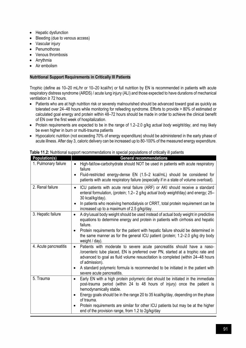
91
Hepatic dysfunction
Bleeding (due to venous access)
Vascular injury
Penumothorax
Venous thrombosis
Arrythmia
Air embolism
Nutritional Support Requirements in Critically Ill Patients
Trophic (define as 10–20 mL/hr or 10–20 kcal/hr) or full nutrition by EN is recommended in patients with acute respiratory distress syndrome (ARDS) / acute lung injury (ALI) and those expected to have durations of mechanical ventilation ≥ 72 hours.
Patients who are at high nutrition risk or severely malnourished should be advanced toward goal as quickly as tolerated over 24–48 hours while monitoring for refeeding syndrome. Efforts to provide > 80% of estimated or calculated goal energy and protein within 48–72 hours should be made in order to achieve the clinical benefit of EN over the first week of hospitalization.
Protein requirements are expected to be in the range of 1.2–2.0 g/kg actual body weight/day, and may likely be even higher in burn or multi-trauma patients
Hypocaloric nutrition (not exceeding 70% of energy expenditure) should be administered in the early phase of acute illness. After day 3, caloric delivery can be increased up to 80-100% of the measured energy expenditure.
Table 11.2: Nutritional support recommendations in special populations of critically ill patients
Population(s) General recommendations
1. Pulmonary failure High-fat/low-carbohydrate should NOT be used in patients with acute respiratory failure
Fluid-restricted energy-dense EN (1.5–2 kcal/mL) should be considered for patients with acute respiratory failure (especially if in a state of volume overload).
2. Renal failure ICU patients with acute renal failure (ARF) or AKI should receive a standard enteral formulation, (protein; 1.2– 2 g/kg actual body weight/day) and energy; 25–30 kcal/kg/day).
In patients who receiving hemodialysis or CRRT, total protein requirement can be increased up to a maximum of 2.5 g/kg/day.
3. Hepatic failure A dry/usual body weight should be used instead of actual body weight in predictive equations to determine energy and protein in patients with cirrhosis and hepatic failure.
Protein requirements for the patient with hepatic failure should be determined in the same manner as for the general ICU patient (protein; 1.2–2.0 g/kg dry body weight / day).
4. Acute pancreatitis Patients with moderate to severe acute pancreatitis should have a naso-/oroenteric tube placed, EN is preferred over PN, started at a trophic rate and advanced to goal as fluid volume resuscitation is completed (within 24–48 hours of admission).
A standard polymeric formula is recommended to be initiated in the patient with severe acute pancreatitis.
5. Trauma Early EN with a high protein polymeric diet should be initiated in the immediate post-trauma period (within 24 to 48 hours of injury) once the patient is hemodynamically stable.
Energy goals should be in the range 20 to 35 kcal/kg/day, depending on the phase of trauma.
Protein requirements are similar for other ICU patients but may be at the higher end of the provision range, from 1.2 to 2g/kg/day

92
Population(s) General recommendations
6. Traumatic brain injury
Early EN should be initiated in the immediate post-trauma period (within 24 to 48 hours of injury) once the patient is hemodynamically stable.
Protein requirements maybe in the range of 1.5 to 2.5 g/kg/day.
7. Open abdomen Early EN (24–48 hours post-injury) should be provided in patients treated with an open abdomen (OA) in the absence of a bowel injury.
An additional 15 to 30 grams protein per liter of exudate lost for patients with OA should be provided.
8. Burns PN should be reserved for those burn patients for whom EN is not feasible or not tolerated.
Early initiation of EN (if possible, within 4–6 hours of injury) in a patient with burn injury is suggested.
Patients with burn injury should receive protein in the range of 1.5–2 g/kg/day
9. Sepsis Trophic feeding (10-20 kcal/hr or up to 500 kcal/day) is recommended for the initial phase of sepsis, advancing as tolerated after 24-48 hr to > 80% of target energy goal over the first week.
1.2-3 g/kg/day protein should be delivered.
Immune modulating formula should NOT be used routinely in patient with severe sepsis.
10. Post-operative major surgery
Patient who has undergone major upper GI surgery and EN is not feasible, PN should be initiated (only if the duration of therapy is anticipated to be ≥7 days)
Unless the patient is at high nutrition risk, PN should NOT be started in the immediate postoperative period, but should be delayed for 5–7 days.
Upon advancing the diet postoperatively, patients be allowed solid food as tolerated, and that clear liquids are not required as the first meal
11. Obesity in critical illness
Early EN should be initiated within 24–48 hours of admission to the ICU for obese patients who cannot sustain volitional intake. For all classes of obesity, the goal of the EN regimen should not exceed 65–70% of target energy requirements as measured by Indirect Calorimetry (IC). If IC is unavailable, the weight-based equation should be used to calculate energy requirement.
BMI 30 to 50: 11–14 kcal/kg actual body weight / day BMI > 50: 22–25 kcal/kg ideal body weight / day
Protein requirement: BMI 30–40: 2 g/kg ideal body weight / day BMI ≥ 40: up to 2.5 g/kg ideal body weight / day
(If urinary nitrogen losses or lean body mass determination are not available, protein intake can be 1.3g/kg ‘’adjusted body weight’’ per day)
*Ideal body weight instead of actual body weight is recommended as reference (adjusted) body weight at BMI >25 kg/m2. *A pragmatic approach is to add 20 – 25% of the excess weight (actual body weight – ideal body weight) all calculations of energy requirements.

93
Refeeding Syndrome The refeeding syndrome is defined as the clinical complications that can occur as a result of fluid and electrolyte shifts during aggressive nutritional rehabilitation of malnourished patients. Hypophosphatemia is the hallmark of the syndrome and predominant cause of the refeeding syndrome. Other clinical features of possible development of refeeding syndrome are:
Hypokalemia
Congestive heart failure
Peripheral edema
Rhabdomyolysis
Seizures
Hemolysis. Pathogenesis of hypophosphatemia begins when stores of phosphate are depleted during episodes of starvation or malnutrition. When nutritional replenishment starts and patients are fed with carbohydrates and glucose, these provoke the release of insulin, which triggers cellular uptake of phosphate (and potassium and magnesium) and a decrease in serum phosphorous levels. Insulin released also causes cells to produce a variety of depleted molecules that require phosphate (eg, adenosine triphosphate and 2,3-diphosphoglycerate), which further depletes the body’s stores of phosphate. The subsequent lack of phosphorylated intermediates causes tissue hypoxia, myocardial dysfunction, respiratory failure due to an inability of the diaphragm to contract, hemolysis, rhabdomyolysis, and seizures. Criteria patients at risk of developing refeeding syndrome:
Patient has one of the following: BMI less than 16 kg/m2 Unintentional weight loss more than 15% within the last 3-6 months Little or no nutritional intake for more than 10 days Low level of potassium, phosphate or magnesium prior to feeding
Or patient has two or more of the following: BMI less than 18.5 kg/m2 Unintentional weight loss greater than 15% within the last 3-6 months Little or no nutritional intake for more than 5 days history of alcohol abuse or drugs including insulin, chemotherapy, antacids or diuretics
Management of Refeeding Syndrome Electrolytes such as potassium, magnesium and phosphate should be monitored at least once daily for the first week. Energy supply should be restricted for 48 hours (eg: 20 kcal/hr for 48 hours) and then gradually increased in patients with refeeding hypophosphatemia (<0.65 mmol/l or a drop of >0.16 mmol/l phosphate). Provide immediately before and during the first 10 days of feeding with oral thiamine 200-300 mg daily, vitamin B and a balanced multivitamin/trace element supplement once daily. Consider feeding protocol as follow in patient at risk of refeeding syndrome: Nutritional support started at maximum rate of 10 kcal/kg/day, increasing slowly to meet full requirement by 4-
7 days (using 5 kcal/kg/day in extreme cases (eg: BMI less than 14 kg/m2 or negligible intake more than 15 days).
Provide oral, enteral or IV supplements of potassium, phosphate, and magnesium unless pre feeding plasma level is high.

94
Figure 11.4: Pathogenesis of refeeding syndrome (from Boateng et al 2010)

95
Drug Administration Issues during Nutritional Support in Critically Ill Patient Several issues have to be considered before drug being administered through enteral tube. Evaluate the tube type, tube location in the GI tract, site of drug action and absorption and effects of food on drug absorption (e.g sucralfate is not suitable for intestinal feeding tubes administration as it acts locally in the stomach). Enteral Feeding Tubes Enteral feeding tubes are made up of polyvinylchloride (PVC), polyurethane (PUR), silicone or latex. External diameter of the feeding tube is measured in French unit (Fr) where 1 Fr = 0.33mm. Types of enteral feeding tubes
Nasoenteric tubes [e.g nasoenteric tubes are nasogastric tube (NGJ), nasoduodenal tube (NDT), and nasojejunal tube (NJT)] are normally used for short- to medium-term feeding (days to weeks) as it has lots of drawbacks on long-term management (e.g nasoduodenal tube tends to recoil into the stomach, cause nasal pressure sores, and sometimes it can be pushed out of place accidentally)
Ostomy tubes are used for long-term feeding (months to years). Generally, the tubes are inserted into the desired place endoscopically, radiologically or surgically. Examples of ostomy tubes are percutaneous gastrostomy tube, percutaneous jejunostomy tube, and percutaneous gastrojejunostomy tube.
Fluid Osmolarity Many commercial liquids are extremely hyperosmolar with osmolalities over 1000 mOsm/kg and the osmolality of GI secretions, on the other hand, ranges from 100 to 400 mOsm/kg. Diarrhea, cramping, abdominal distension and vomiting may occur after administration of hyperosmolar products through the feeding tube and this can be overcome by diluting medication with 10-30 ml of sterile water before administration. Sterile water contains no solute thus it does not contribute to the mixture’s osmolality. Osmolality of the resulting mixture can be calculated using formula below:
Inactive ingredients or excipients present in liquid products, such mannitol, lactose, saccharin, sorbitol, and sucrose, may also cause or worsen diarrhea. Sorbitol, used as a sweetening agent to improve medication taste as well as tolerability, may cause bloating and flatulence at a total daily dose of 10gm, and cause an osmotic laxative effect (resulting in cramping and diarrhea) at a total daily dose of 20 gm Alteration of Drug PK due to EN The absorption of the drug at the GI tract is via a passive process of diffusion from the gut lumen across the mucosa into the splanchnic circulation. However, some drugs (e.g methyldopa and levodopa) are being absorbed via the same way as the nutrients, i.e active transport system. Thus, a change in diet will affect the drug absorption (e.g high-protein diet will result in decreased absorption of methyldopa and levodopa). Drug absorption also depends on the physiological affect of food/feed on the GI tract, owing to the ability of food and feeds to alter the pH of the GI tract. The presence of some components in the feed formulations (e.g calcium, iron) will bind with the drug, therefore result in changing of the molecular size or solubility thus reduce the absorption. Drug distribution is altered in severely malnourished patient (e.g low plasma proteins for drug binding results in increasing free circulating drug concentrations). There is a potential interaction between drugs and the enteral feeding tubes (Table 11.4).
(osmolality of drug / volume of drug) Osmolality of diluted mixture = --------------------------------------------------
total volume of mixture

96
Table 11.3: Drug considerations during enteral feeding
Considerations
1. Drug bioavailability may increase with intrajejunal administration of drugs with extensive first-pass metabolism, such as opioids, tricyclics, beta-blockers, or nitrates
2. For drugs that require administration on empty stomach, feeding should be stopped 30 minutes before and after dosing if the tube is placed in the stomach
3. If the tablets can be crushed into fine powder, the powder should be mixed in slurry with a suitable diluent and given through large-bore feeding tubes. Sometimes, pellets inside some microencapsulated products may be poured down the large-bore enteral feeding tube, provided that the pellets are not crushed (e.g Creon® – pancreatic enzymes).
4. Drug formulations that should not be crushed are sustained-release or modified-release, enteric-coated and teratogenic, carcinogenic or cytotoxic medication (e.g antineoplastics, hormones, and prostaglandin analogs as aerosolized particles might be harmful to the health care workers).
5. Feeding tubes should be flushed (eg: 15-30 ml of water) before and after administration of medication via the tube to prevent tube blockage (in the case of fluid-restricted patients, revise the flush volumes to meet the patient’s prescribed fluid restriction, air flushes may be used to replace water flushes).
6. Medication should not be added to enteral formula to reduce the risk of microbial contamination and to avoid drug-nutrient incompatibilities. A possible exception for mixing medications with feeding formulae involves the addition of liquid electrolytes, such as sodium or potassium, to the enteral preparation.
7. When several medications are to be given at the same time, all medications should be administered separately and the tube flushed with at least 5 ml of water after each dose.
8. Liquid preparations are the preferred formulations whenever possible as they are less likely to cause blockage of feeding tubes. However, medication dosage or frequency may need adjustment when switching from solid to liquid preparations, this is particularly important when switching from an extended-release product to a liquid preparation, which is immediate release and requires frequent dosing (e.g extended-release phenytoin capsules may be given once daily, however phenytoin suspension is an immediate-release product that need to be dosed 2 to 4 times daily).
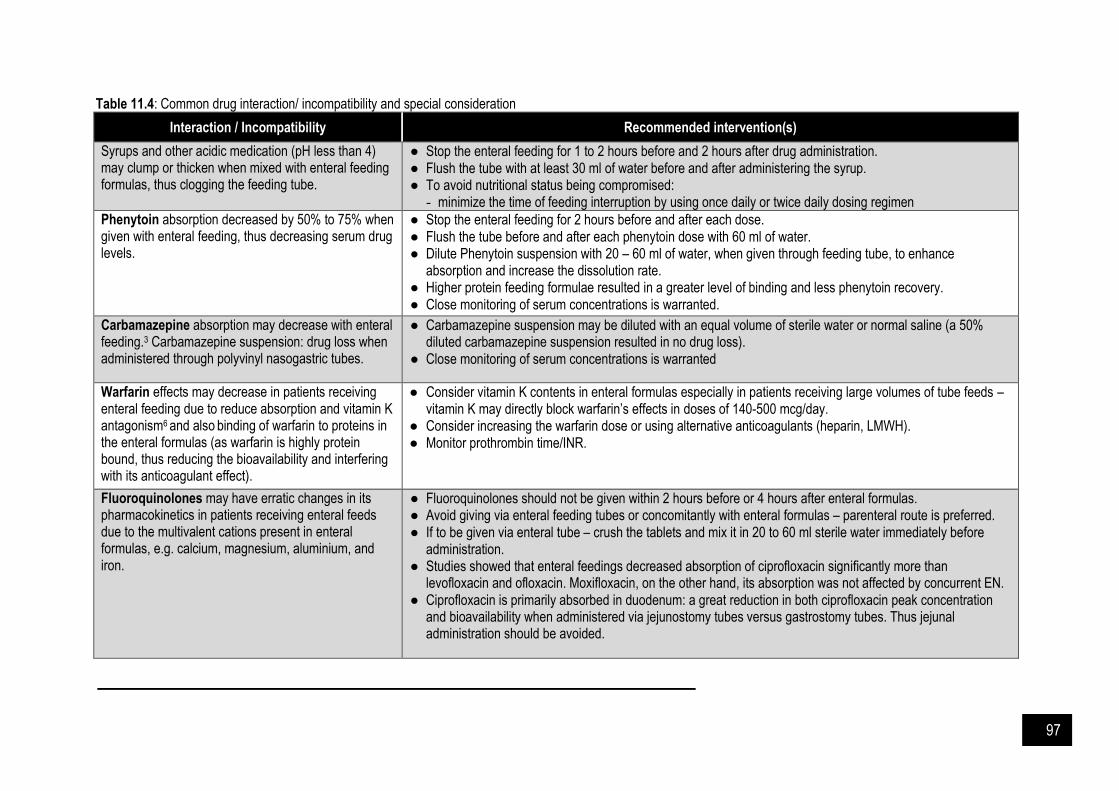
97
Table 11.4: Common drug interaction/ incompatibility and special consideration
Interaction / Incompatibility Recommended intervention(s)
Syrups and other acidic medication (pH less than 4) may clump or thicken when mixed with enteral feeding formulas, thus clogging the feeding tube.
● Stop the enteral feeding for 1 to 2 hours before and 2 hours after drug administration. ● Flush the tube with at least 30 ml of water before and after administering the syrup. ● To avoid nutritional status being compromised:
- minimize the time of feeding interruption by using once daily or twice daily dosing regimen
Phenytoin absorption decreased by 50% to 75% when given with enteral feeding, thus decreasing serum drug levels.
● Stop the enteral feeding for 2 hours before and after each dose. ● Flush the tube before and after each phenytoin dose with 60 ml of water. ● Dilute Phenytoin suspension with 20 – 60 ml of water, when given through feeding tube, to enhance
absorption and increase the dissolution rate. ● Higher protein feeding formulae resulted in a greater level of binding and less phenytoin recovery. ● Close monitoring of serum concentrations is warranted.
Carbamazepine absorption may decrease with enteral feeding.3 Carbamazepine suspension: drug loss when administered through polyvinyl nasogastric tubes.
● Carbamazepine suspension may be diluted with an equal volume of sterile water or normal saline (a 50% diluted carbamazepine suspension resulted in no drug loss).
● Close monitoring of serum concentrations is warranted
Warfarin effects may decrease in patients receiving enteral feeding due to reduce absorption and vitamin K antagonism6 and also binding of warfarin to proteins in the enteral formulas (as warfarin is highly protein bound, thus reducing the bioavailability and interfering with its anticoagulant effect).
● Consider vitamin K contents in enteral formulas especially in patients receiving large volumes of tube feeds – vitamin K may directly block warfarin’s effects in doses of 140-500 mcg/day.
● Consider increasing the warfarin dose or using alternative anticoagulants (heparin, LMWH). ● Monitor prothrombin time/INR.
Fluoroquinolones may have erratic changes in its pharmacokinetics in patients receiving enteral feeds due to the multivalent cations present in enteral formulas, e.g. calcium, magnesium, aluminium, and iron.
● Fluoroquinolones should not be given within 2 hours before or 4 hours after enteral formulas. ● Avoid giving via enteral feeding tubes or concomitantly with enteral formulas – parenteral route is preferred. ● If to be given via enteral tube – crush the tablets and mix it in 20 to 60 ml sterile water immediately before
administration. ● Studies showed that enteral feedings decreased absorption of ciprofloxacin significantly more than
levofloxacin and ofloxacin. Moxifloxacin, on the other hand, its absorption was not affected by concurrent EN. ● Ciprofloxacin is primarily absorbed in duodenum: a great reduction in both ciprofloxacin peak concentration
and bioavailability when administered via jejunostomy tubes versus gastrostomy tubes. Thus jejunal administration should be avoided.
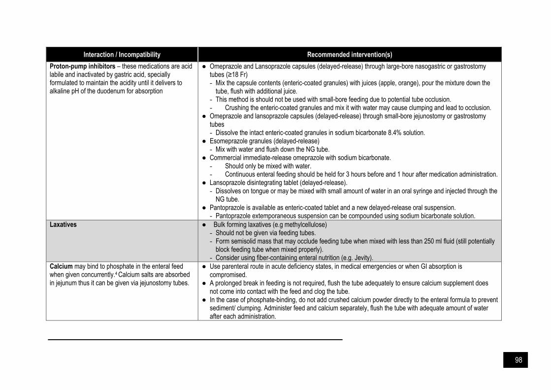
98
Interaction / Incompatibility Recommended intervention(s)
Proton-pump inhibitors – these medications are acid labile and inactivated by gastric acid, specially formulated to maintain the acidity until it delivers to alkaline pH of the duodenum for absorption
● Omeprazole and Lansoprazole capsules (delayed-release) through large-bore nasogastric or gastrostomy tubes (≥18 Fr) - Mix the capsule contents (enteric-coated granules) with juices (apple, orange), pour the mixture down the
tube, flush with additional juice. - This method is should not be used with small-bore feeding due to potential tube occlusion. - Crushing the enteric-coated granules and mix it with water may cause clumping and lead to occlusion.
● Omeprazole and lansoprazole capsules (delayed-release) through small-bore jejunostomy or gastrostomy tubes - Dissolve the intact enteric-coated granules in sodium bicarbonate 8.4% solution.
● Esomeprazole granules (delayed-release) - Mix with water and flush down the NG tube.
● Commercial immediate-release omeprazole with sodium bicarbonate. - Should only be mixed with water. - Continuous enteral feeding should be held for 3 hours before and 1 hour after medication administration.
● Lansoprazole disintegrating tablet (delayed-release). - Dissolves on tongue or may be mixed with small amount of water in an oral syringe and injected through the
NG tube. ● Pantoprazole is available as enteric-coated tablet and a new delayed-release oral suspension.
- Pantoprazole extemporaneous suspension can be compounded using sodium bicarbonate solution.
Laxatives ● Bulk forming laxatives (e.g methylcellulose) - Should not be given via feeding tubes. - Form semisolid mass that may occlude feeding tube when mixed with less than 250 ml fluid (still potentially
block feeding tube when mixed properly). - Consider using fiber-containing enteral nutrition (e.g. Jevity).
Calcium may bind to phosphate in the enteral feed when given concurrently.4 Calcium salts are absorbed in jejunum thus it can be given via jejunostomy tubes.
● Use parenteral route in acute deficiency states, in medical emergencies or when GI absorption is compromised.
● A prolonged break in feeding is not required, flush the tube adequately to ensure calcium supplement does not come into contact with the feed and clog the tube.
● In the case of phosphate-binding, do not add crushed calcium powder directly to the enteral formula to prevent sediment/ clumping. Administer feed and calcium separately, flush the tube with adequate amount of water after each administration.
99
References and Suggested Readings American Society for Parenteral and Enteral Nutrition (ASPEN) 2016. Beckwith MC, Feddema SS, Barton RG, et al. A guide to drug therapy in patients with enteral feeding tubes:
Dosage form selection and administration method. Hospital Pharmacy. 2004. 39(3): 225-37. Boateng AA, Sriram K, Meguid MM, Crook M. Refeeding syndrome: Treatment considerations based on collective
analysis of literature case reports. Nutrition. 2010. 26(2): 156. Dickerson RN, Melnik G. Osmolality of oral drug solutions & suspensions. Am J Hosp Pharm. 1988. 45(4): 832–
34. ESPEN guideline on clinical nutrition in the intensive care unit 2018 Estoup M. Approaches and limitations of medication delivery in patients with enteral feeding tubes. Crit Care Nurse.
1994. 14: 68-72. Gora ML, Tschampel MM, Visconti JA. Considerations of drug therapy in patients receiving enteral nutrition. Nutr
Clin Pract. 1989. 4: 105-10. Heyland DK, Dhaliwal R, Drover JW, et al. Canadian clinical practice guidelines for nutrition support in mechanically
ventilated, critically ill adult patients. J Parenter Enteral Nutr. 2003. 27: 355-73. Heyland DK, Dhaliwal R, Jiang X, Day AG. Identifying critically ill patients who benefit the most from nutrition
therapy: The development and initial validation of a novel risk assessment tool. Crit Care. 2011. 15(6): R268 Kreymann KG, Berger MM, Deutz N, et al. ESPEN guidelines on enteral nutrition: Intensive care. Clinical Nutrition.
2006. 25: 210-23. Mehanna HM, Moledina J, Travis J. Refeeding syndrome: What it is, and how to prevent and treat it. BMJ. 2008.
336(7659):1495. Mehler P. Anorexia nervosa inj adults and adolescents: The refeeding syndrome. UpToDate 2020 National Collaborating Centre for Acute Care (UK). Nutrition support for adults: Oral nutrition support, enteral tube
feeding and parenteral nutrition 2006. Rahman A, Hasan RM, Agarwala R, Martin C, Day AG, Heyland DK. Identifying critically-ill patients who will benefit
most from nutritional therapy: Further validation of the “modified NUTRIC” nutritional risk assessment tool. Clinical Nutrition. 2016. 35(1): 158–162.
Seres D. Nutrition support in critically ill patients: Parenteral nutrition. UpToDate 2020 Singer P, Blaser A, Berger M, Alhazzani W, Calder P, Casaer M et al. ESPEN guideline on clinical nutrition in the
intensive care unit. Clinical Nutrition. 2018. Society of Critical Care Medicine (SCCM). Guidelines for the Provision and Assessment of Nutrition Support
Therapy in the Adult Critically Ill Patient. Taylor B, McClave S, Martindale R, Warren M, Johnson D, Braunschweig C et al. Guidelines for the provision and
assessment of nutrition support therapy in the adult critically ill patient. Criti Care Med. 2016. 44(2): 390-438. The Nutric Score. Available at https://www.criticalcarenutrition.com/resources/nutric-score Thomson FC, Naysmith MR, Lindsay A. Managing drug therapy in patients receiving enteral and parenteral
nutrition. Hosp Pharmacist. 2000. 7: 155–64. TPN Tutorial. Available at http://www.rxkinetics.com/tpntutorial/1_4.html Williams NT. Medication administration through enteral feeding tubes. Am J Health Syst Pharm 2008. 65(24):
2347-57.
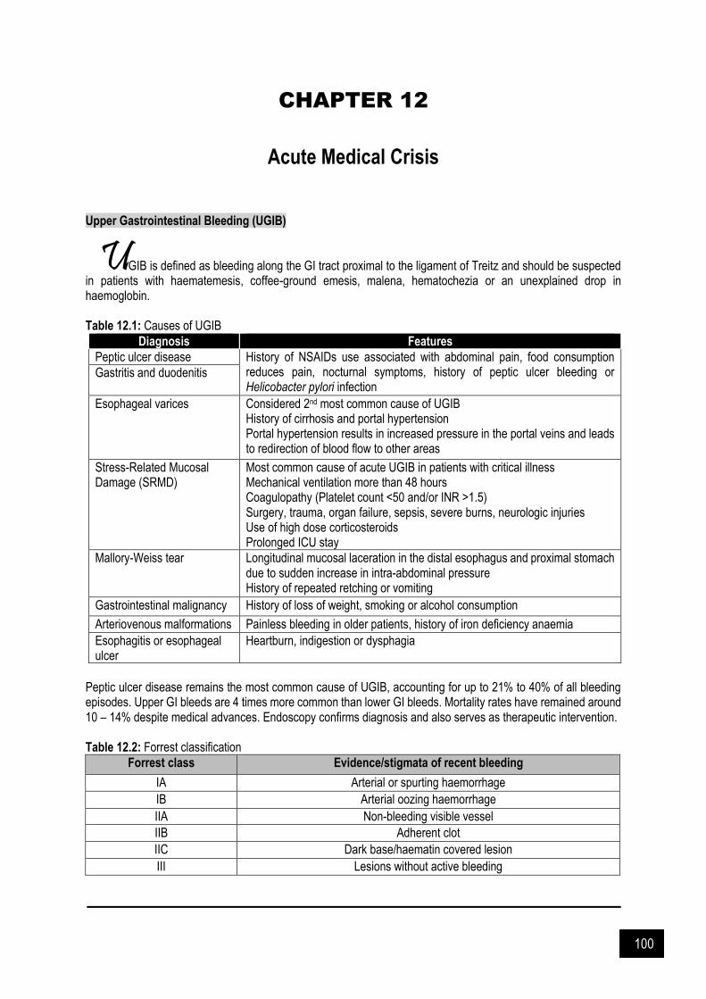
100
CHAPTER 12
Acute Medical Crisis
Upper Gastrointestinal Bleeding (UGIB)
GIB is defined as bleeding along the GI tract proximal to the ligament of Treitz and should be suspected in patients with haematemesis, coffee-ground emesis, malena, hematochezia or an unexplained drop in haemoglobin. Table 12.1: Causes of UGIB
Diagnosis Features
Peptic ulcer disease History of NSAIDs use associated with abdominal pain, food consumption reduces pain, nocturnal symptoms, history of peptic ulcer bleeding or Helicobacter pylori infection
Gastritis and duodenitis
Esophageal varices Considered 2nd most common cause of UGIB History of cirrhosis and portal hypertension Portal hypertension results in increased pressure in the portal veins and leads to redirection of blood flow to other areas
Stress-Related Mucosal Damage (SRMD)
Most common cause of acute UGIB in patients with critical illness Mechanical ventilation more than 48 hours Coagulopathy (Platelet count <50 and/or INR >1.5) Surgery, trauma, organ failure, sepsis, severe burns, neurologic injuries Use of high dose corticosteroids Prolonged ICU stay
Mallory-Weiss tear Longitudinal mucosal laceration in the distal esophagus and proximal stomach due to sudden increase in intra-abdominal pressure History of repeated retching or vomiting
Gastrointestinal malignancy History of loss of weight, smoking or alcohol consumption
Arteriovenous malformations Painless bleeding in older patients, history of iron deficiency anaemia
Esophagitis or esophageal ulcer
Heartburn, indigestion or dysphagia
Peptic ulcer disease remains the most common cause of UGIB, accounting for up to 21% to 40% of all bleeding episodes. Upper GI bleeds are 4 times more common than lower GI bleeds. Mortality rates have remained around 10 – 14% despite medical advances. Endoscopy confirms diagnosis and also serves as therapeutic intervention. Table 12.2: Forrest classification
Forrest class Evidence/stigmata of recent bleeding
IA Arterial or spurting haemorrhage
IB Arterial oozing haemorrhage
IIA Non-bleeding visible vessel
IIB Adherent clot
IIC Dark base/haematin covered lesion
III Lesions without active bleeding
U
text here

101
Blatchford score is used to identify low risk patients ready for discharge whereas the Rockall score using both clinical and endoscopic data, identifies patients at highest risk of mortality.
Figure 12.1: Blatchford score (left) & Rockall score (right) General Management of UGIB Assess hemodynamic status upon presentation and provide adequate resuscitative measures as needed. Colloids or crystalloid fluids should be administered to restore adequate blood pressure. Blood transfusion should be initiated with target haemoglobin ≥ 7.0 g/dL to compensate ongoing blood loss, substantial haemorrhage, or cardiac ischemia. Goal of therapy include:
Correct hypovolemic shock and prevent its complications
Compensate ongoing blood loss
Stop bleeding and prevent re-bleeding

102
Fresh frozen plasma or vitamin K administered with INR greater than 1.5. Endoscopic interventions include application of clips, argon plasma coagulation, injection of epinephrine or sclerosants, bipolar electrocoagulation, band ligation, heater probe coagulation and laser therapy. Choice of treatment technique depends on the size of bleeding vessels. Endoscopic hemostatic therapy is recommended for small vessels and in PUD patients with highest risk of re-bleeding while variceal banding or sclerotherapy is recommended for variceal bleeds of larger vessels or when endoscopic hemostatic therapy is inadequate. Pharmacological Management a. Non – variceal bleeding Initiate PPI infusion with a bolus (80 mg bolus followed by 8 mg/h) and continue for 72 hours in patients with higher risk of bleeding at endoscopy and those who had undergone endoscopy. Meta-analysis of randomized trials of intravenous PPI therapy (80mg bolus followed by 8 mg/h continuous infusion) vs. placebo/no treatment for 72 hours after endoscopic therapy of high-risk stigmata reveals a significant reduction in further bleeding. If endoscopy is delayed or cannot be performed, intravenous PPI is recommended to reduce further bleeding. b. Variceal bleeding All patients should receive 7 days of prophylactic therapy with ciprofloxacin or ceftriaxone due to high risk of infectious complications. A review of 12 trials involving 1,241 patients with variceal hemorrhage found that broad-spectrum antibiotics (e.g., ceftriaxone, norfloxacin, ciprofloxacin) reduced overall mortality and risk of rebleeding. Intravenous infusion Octreotide 50 µg bolus followed by 25 – 50 µg/h or intravenous terlipressin 1 – 2 mg q4h may be used as adjuvants. Acute Pancreatitis In 2009, acute pancreatitis (AP) was the most common discharge diagnosis with an annual cost of 2.6 billion dollars in the US. Causes of AP including gallstones, alcohol, drugs (e.g statins, ACE inhibitors, estrogen / hormone replacement therapy, HAART medications, valproic acid, azathioprine, metronidazole), virus, vasculitis, hypercalcaemia, hypertriglyceridemia, trauma, malignancy and congenital. The diagnosis of AP required two of the following features:
Abdominal pain consistent with AP
Elevated serum lipase (or amylase) at least 3 times greater than upper limit of normal
Characteristic findings of AP on CECT, MRI or transabdominal US Serum amylase concentration may be normal in alcohol induced AP and hypertriglyceridemia. Newly revised Atlanta classification stratifies the acute pancreatitis in three grades of severity: mild, moderate and severe. Two distinctive phases of AP have been identified:
Early (within a week) : Characterized by SIRS and/or organ failure
Late (>1 week) : Characterized by local complications (eg: peripancreatic fluid collections, Pancreatic and peripancreatic necrosis-infected or sterile, pseudocysts and walled-off necrosis
Table 12.3: Types of acute pancreatitis
Types
1. Interstitial edematous pancreatitis ● Presence of diffuse enlargement of pancreas due to inflammatory edema. CECT shows relatively homogeneous
enhancement and some inflammatory changes of haziness or mild stranding of peripancreatic fat. Clinical symptoms usually resolve within the first week

103
2. Necrotising pancreatitis ● Necrosis of parenchyma, peripancreatic tissue or both developed in 5 – 10% of AP patients; commonly manifest
in both pancreas and peripancreatic tissue. Impairment of pancreatic perfusion and signs of necrosis develop over several days resulting in underestimation of even if CECT is performed early. Necrosis may remain solid or liquefy, sterile or become infected, persist or disappear over time
3. Infected pancreatic necrosis ● Rare during the first week. Infection should be suspected with the presence of extraluminal gas in the pancreas
and/or peripancreatic tissue on CECT or when culture is positive bacteria and/or fungi. Development of secondary infection is associated with increased morbidity and mortality.
Management of Acute Pancreatitis Early hydration AP patients are frequently hypovolemic due to multiple factors including vomiting, reduced oral intake, third spacing of fluids and diaphoresis. Aggressive fluid resuscitation during the first 12-24 hours has been shown to decrease risk and incidence of pancreatic necrosis. Surrogate markers such as haematocrit, BUN and creatinine can be used as monitoring parameters. The aim is to decrease haematocrit and BUN and maintain normal creatinine levels. Lactated Ringer’s solution was reported to be more beneficial, resulting in fewer patients developing SIRS as compared as normal saline in a prospective randomized trial. Better electrolyte balance and improved outcomes were also observed. Lactated Ringer’s provides a higher than required pH, inhibiting activation of trypsinogen preventing acinar cells to be more susceptible to injury. Caution when administering aggressive hydration in the elderly, patients with history of cardiac and renal disease. Endoscopic retrograde cholangiopancreatography (ERCP) Persistent choledocholithiasis leads to obstruction of pancreatic duct/biliary tree which leads to severe AP. Patients with AP and concurrent acute cholangitis should undergo ERCP within 24 hours of admission however is not advocated early in most patients with gallstone pancreatitis who lack laboratory or clinical evidence of ongoing biliary obstruction. Acute pancreatitis is a complication of ERCP, seen in 5-10% of cases and up to 40% of certain high-risk procedures which since reduced with better recognition of high-risk patients and high-risk procedures. Endoscopic US or MRCP should be used in patients with normal bile duct and normal bilirubin levels as the risk of post – ERCP pancreatitis is higher among this group. Infection Both pancreatic and extra-pancreatic infections contribute to morbidity and mortality in patients with AP. Infected necrotizing pancreatitis has significantly higher mortality rate than sterile necrotising pancreatitis. Patients who deteriorate or fail to improve within 7-10 days of hospitalization should be suspected of infected necrosis. Appropriate cultures should be obtained and antibiotics should be started such as:
IV piperacillin/tazobactam 4.5 gm q6h-q8h (to be given as extended infusion over 3-4 hours) OR
IV cefoperazone 1-2 gm q12h PLUS IV metronidazole 500 mg q8h
Modify antibiotics once culture and sensitivity is available. Reserve carbapenem for infection caused by resistant pathogen(s). Antimicrobial therapy for established infection should be limited to 4-7 days, unless source control is difficult to obtain. The use of prophylactic antibiotics in sterile necrotising pancreatitis is not suggested. Routine administration of antifungal alongside prophylactic or therapeutic antibiotics is not recommended. Nutrition Clinical and experimental studies showed that bowel rest is associated with intestinal mucosal atrophy and increased infectious complications from bacterial translocation. In mild AP, oral feeding with a low–fat solid or liquid diet can be started immediately in the absence of nausea and vomiting, and resolved abdominal pain. In severe AP, enteral feeding via nasogastric or naso-jejunal route is recommended to prevent infectious complications. Parenteral nutrition is considered if enteral route is not available, not well tolerated or not meeting caloric requirements.

104
Surgery Cholecystectomy should be performed in patients with mild gallstone pancreatitis however, should be deferred in patients with necrotising biliary AP until active inflammation has subsided. In stable patients with infected necrosis, surgical, radiologic, and / or endoscopic drainage should be delayed preferably for more than 4 weeks to allow liquefication of the contents and the development of a fibrous wall around the necrosis. In symptomatic patients with infected necrosis, minimally invasive methods of necrosectomy are preferred to open necrosectomy. In presence of known or suspected pre-existing infections, use of antibiotic treatment regime is recommended. Antimicrobial is indicated for 4-7 days, unless source control is inadequate. Diabetic Ketoacidosis (DKA) & Hyperosmolar Hyperglycaemic State (HHS) DKA and HHS are the most common diabetic emergencies. Most common precipitating factor for both conditions is infection. Other precipitating conditions include cardiovascular accident, myocardial infarction, trauma, drugs and non-compliance to therapy. Table 12.4: Comparison between DKA and HHS
Characteristics DKA HHS
Clinical Presentation ● Blood glucose >13.8 mmol/L ● Blood pH <7.3 ● Serum bicarbonate <18 mmol/L ● Anion gap >10 ● Presence of ketone in blood
● Blood glucose >33.3 mmol/L ● Blood pH >7.3 ● Serum bicarbonate >15 mmol/L ● Serum osmolarity >320 mOsm ● Presence of small amounts of ketone
Pathophysiology Insulin deficiency with increased counter-regulatory hormones leads to lipolysis and production of ketone bodies
Relative reduction in circulating insulin with concomitant rise in counter-regulatory hormones. Sufficient amount of insulin inhibits lipolysis and ketogenesis but is unable to prevent hyperglycemia
Onset Within 24 hours Several days to weeks
Symptoms Vomiting and abdominal pain (in DKA), polyuria, polydipsia, weight loss and confusion (more common in HHS)
Management of DKA & HHS
Goal of therapy for DKA and HHS include (Table 12.5): 1. Correct fluid depletion 2. Correct electrolyte imbalances 3. Correct acid-base abnormalities 4. Glycaemic control 5. Treat underlying cause
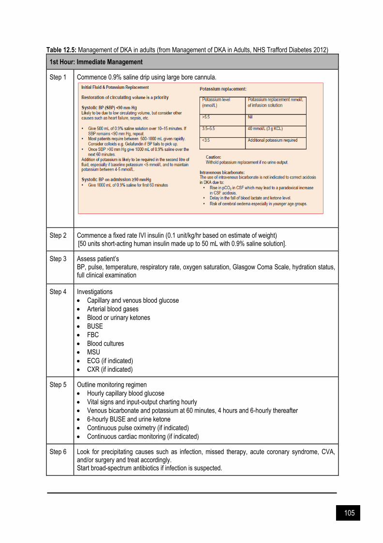
105
Table 12.5: Management of DKA in adults (from Management of DKA in Adults, NHS Trafford Diabetes 2012)
1st Hour: Immediate Management
Step 1 Commence 0.9% saline drip using large bore cannula.
Step 2 Commence a fixed rate IVI insulin (0.1 unit/kg/hr based on estimate of weight) [50 units short-acting human insulin made up to 50 mL with 0.9% saline solution].
Step 3 Assess patient’s BP, pulse, temperature, respiratory rate, oxygen saturation, Glasgow Coma Scale, hydration status, full clinical examination
Step 4 Investigations
Capillary and venous blood glucose
Arterial blood gases
Blood or urinary ketones
BUSE
FBC
Blood cultures
MSU
ECG (if indicated)
CXR (if indicated)
Step 5 Outline monitoring regimen
Hourly capillary blood glucose
Vital signs and input-output charting hourly
Venous bicarbonate and potassium at 60 minutes, 4 hours and 6-hourly thereafter
6-hourly BUSE and urine ketone
Continuous pulse oximetry (if indicated)
Continuous cardiac monitoring (if indicated)
Step 6 Look for precipitating causes such as infection, missed therapy, acute coronary syndrome, CVA, and/or surgery and treat accordingly. Start broad-spectrum antibiotics if infection is suspected.
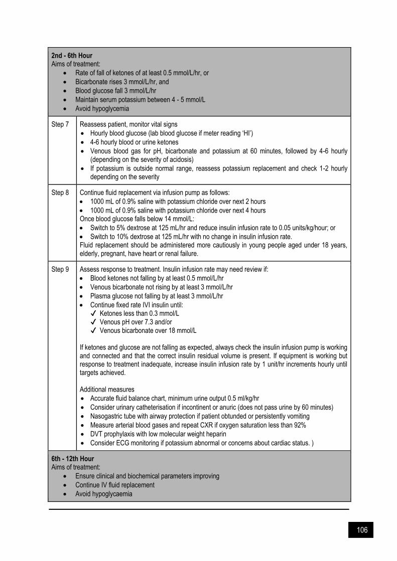
106
2nd - 6th Hour Aims of treatment:
Rate of fall of ketones of at least 0.5 mmol/L/hr, or
Bicarbonate rises 3 mmol/L/hr, and
Blood glucose fall 3 mmol/L/hr
Maintain serum potassium between 4 - 5 mmol/L
Avoid hypoglycemia
Step 7 Reassess patient, monitor vital signs
Hourly blood glucose (lab blood glucose if meter reading ‘HI’)
4-6 hourly blood or urine ketones
Venous blood gas for pH, bicarbonate and potassium at 60 minutes, followed by 4-6 hourly (depending on the severity of acidosis)
If potassium is outside normal range, reassess potassium replacement and check 1-2 hourly depending on the severity
Step 8 Continue fluid replacement via infusion pump as follows:
1000 mL of 0.9% saline with potassium chloride over next 2 hours
1000 mL of 0.9% saline with potassium chloride over next 4 hours Once blood glucose falls below 14 mmol/L:
Switch to 5% dextrose at 125 mL/hr and reduce insulin infusion rate to 0.05 units/kg/hour; or
Switch to 10% dextrose at 125 mL/hr with no change in insulin infusion rate. Fluid replacement should be administered more cautiously in young people aged under 18 years, elderly, pregnant, have heart or renal failure.
Step 9 Assess response to treatment. Insulin infusion rate may need review if:
Blood ketones not falling by at least 0.5 mmol/L/hr
Venous bicarbonate not rising by at least 3 mmol/L/hr
Plasma glucose not falling by at least 3 mmol/L/hr
Continue fixed rate IVI insulin until: ✔ Ketones less than 0.3 mmol/L ✔ Venous pH over 7.3 and/or ✔ Venous bicarbonate over 18 mmol/L
If ketones and glucose are not falling as expected, always check the insulin infusion pump is working and connected and that the correct insulin residual volume is present. If equipment is working but response to treatment inadequate, increase insulin infusion rate by 1 unit/hr increments hourly until targets achieved. Additional measures
Accurate fluid balance chart, minimum urine output 0.5 ml/kg/hr
Consider urinary catheterisation if incontinent or anuric (does not pass urine by 60 minutes)
Nasogastric tube with airway protection if patient obtunded or persistently vomiting
Measure arterial blood gases and repeat CXR if oxygen saturation less than 92%
DVT prophylaxis with low molecular weight heparin
Consider ECG monitoring if potassium abnormal or concerns about cardiac status. )
6th - 12th Hour Aims of treatment:
Ensure clinical and biochemical parameters improving
Continue IV fluid replacement
Avoid hypoglycaemia
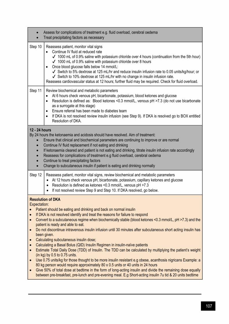
107
Assess for complications of treatment e.g. fluid overload, cerebral oedema
Treat precipitating factors as necessary
Step 10 Reassess patient, monitor vital signs
Continue IV fluid at reduced rate ✔ 1000 mL of 0.9% saline with potassium chloride over 4 hours (continuation from the 5th hour) ✔ 1000 mL of 0.9% saline with potassium chloride over 8 hours
Once blood glucose falls below 14 mmol/L: ✔ Switch to 5% dextrose at 125 mL/hr and reduce insulin infusion rate to 0.05 units/kg/hour; or ✔ Switch to 10% dextrose at 125 mL/hr with no change in insulin infusion rate.
Reassess cardiovascular status at 12 hours; further fluid may be required. Check for fluid overload.
Step 11 Review biochemical and metabolic parameters
At 6 hours check venous pH, bicarbonate, potassium, blood ketones and glucose
Resolution is defined as: Blood ketones <0.3 mmol/L, venous pH >7.3 (do not use bicarbonate as a surrogate at this stage)
Ensure referral has been made to diabetes team
If DKA is not resolved review insulin infusion (see Step 9). If DKA is resolved go to BOX entitled Resolution of DKA.
12 - 24 hours By 24 hours the ketonaemia and acidosis should have resolved. Aim of treatment:
Ensure that clinical and biochemical parameters are continuing to improve or are normal
Continue IV fluid replacement if not eating and drinking
If ketonaemia cleared and patient is not eating and drinking, titrate insulin infusion rate accordingly
Reassess for complications of treatment e.g fluid overload, cerebral oedema
Continue to treat precipitating factors
Change to subcutaneous insulin if patient is eating and drinking normally
Step 12 Reassess patient, monitor vital signs, review biochemical and metabolic parameters
At 12 hours check venous pH, bicarbonate, potassium, capillary ketones and glucose
Resolution is defined as ketones <0.3 mmol/L, venous pH >7.3
If not resolved review Step 9 and Step 10. If DKA resolved, go below.
Resolution of DKA Expectation:
Patient should be eating and drinking and back on normal insulin
If DKA is not resolved identify and treat the reasons for failure to respond
Convert to a subcutaneous regime when biochemically stable (blood ketones <0.3 mmol/L, pH >7.3) and the patient is ready and able to eat.
Do not discontinue intravenous insulin infusion until 30 minutes after subcutaneous short acting insulin has been given.
Calculating subcutaneous insulin dose;
Calculating a Basal Bolus (QID) Insulin Regimen in insulin-naïve patients
Estimate Total Daily Dose (TDD) of Insulin. The TDD can be calculated by multiplying the patient’s weight (in kg) by 0.5 to 0.75 units.
Use 0.75 units/kg for those thought to be more insulin resistant e.g obese, acanthosis nigricans Example: a 80 kg person would require approximately 80 x 0.5 units or 40 units in 24 hours
Give 50% of total dose at bedtime in the form of long-acting insulin and divide the remaining dose equally between pre-breakfast, pre-lunch and pre-evening meal. E.g Short-acting insulin 7u tid & 20 units bedtime

108
Figure 12.2: Management of adult with HHS (from Malaysian Clinical Practice Guidelines on Management of Type 2 Diabetes Mellitus 2015)
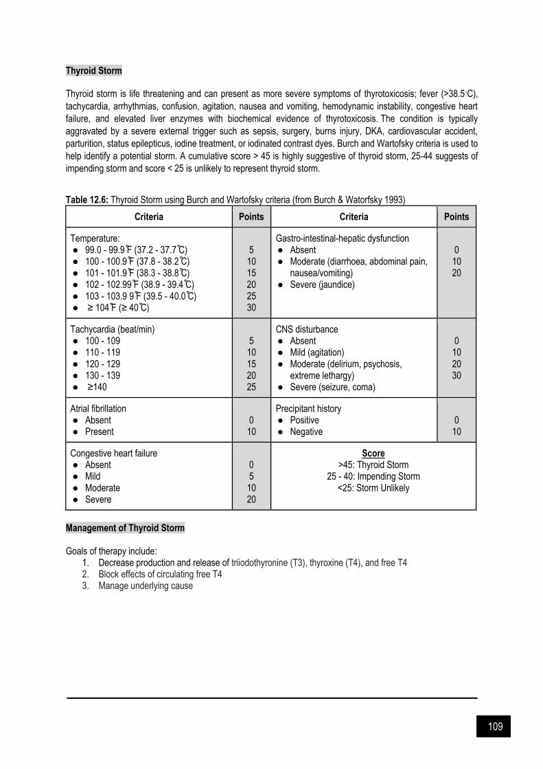
109
Thyroid Storm Thyroid storm is life threatening and can present as more severe symptoms of thyrotoxicosis; fever (>38.5◦C),
tachycardia, arrhythmias, confusion, agitation, nausea and vomiting, hemodynamic instability, congestive heart
failure, and elevated liver enzymes with biochemical evidence of thyrotoxicosis. The condition is typically
aggravated by a severe external trigger such as sepsis, surgery, burns injury, DKA, cardiovascular accident,
parturition, status epilepticus, iodine treatment, or iodinated contrast dyes. Burch and Wartofsky criteria is used to
help identify a potential storm. A cumulative score > 45 is highly suggestive of thyroid storm, 25-44 suggests of
impending storm and score < 25 is unlikely to represent thyroid storm.
Table 12.6: Thyroid Storm using Burch and Wartofsky criteria (from Burch & Watorfsky 1993)
Criteria Points Criteria Points
Temperature: ● 99.0 - 99.9 ͒F (37.2 - 37.7 ͒C) ● 100 - 100.9 ͒F (37.8 - 38.2 ͒C) ● 101 - 101.9 ͒F (38.3 - 38.8 ͒C) ● 102 - 102.99 ͒F (38.9 - 39.4 ͒C) ● 103 - 103.9 9 ͒F (39.5 - 40.0 ͒C) ● ≥ 104 ͒F (≥ 40 ͒C)
5
10 15 20 25 30
Gastro-intestinal-hepatic dysfunction ● Absent ● Moderate (diarrhoea, abdominal pain,
nausea/vomiting) ● Severe (jaundice)
0
10 20
Tachycardia (beat/min) ● 100 - 109 ● 110 - 119 ● 120 - 129 ● 130 - 139 ● ≥140
5
10 15 20 25
CNS disturbance ● Absent ● Mild (agitation) ● Moderate (delirium, psychosis,
extreme lethargy) ● Severe (seizure, coma)
0
10 20 30
Atrial fibrillation ● Absent ● Present
0
10
Precipitant history ● Positive ● Negative
0
10
Congestive heart failure ● Absent ● Mild ● Moderate ● Severe
0 5
10 20
Score >45: Thyroid Storm
25 - 40: Impending Storm <25: Storm Unlikely
Management of Thyroid Storm Goals of therapy include:
1. Decrease production and release of triiodothyronine (T3), thyroxine (T4), and free T4 2. Block effects of circulating free T4 3. Manage underlying cause

110
Table 12.7: Pharmacological management of thyroid storm
Medication Dose Notes
Inhibition of hormone synthesis
Propylthiouracil (PTU)
600 mg loading dose, followed by 200 - 250 mg PO q4 - 6h
Additional inhibition of peripheral deiodination. However, recent warning from FDA regarding severe liver toxicity with PTU makes either carbimazole or methimazole first-choice thionamide.
Carbimazole (or methimazole)
20 - 30 mg PO q4 - 6h
Inhibition of hormone release
SSKI (Potassium Iodide)*
5 drops PO q6 - 8h
*Administer at least 1 hour after thionamide.
Lugol’s Solution*
5 - 10 drops PO q6 - 8h
Lapanoic acid* 1000 mg q8hr for 24 hours, followed by 500 mg BD
Inhibition of peripheral effects of excess thyroid hormone
Propanolol
1 - 2 mg/min IV q15min up to max 10 mg
40 - 80 mg PO q4 - 6h
IV dose initially if hemodynamically unstable
Esmolol
50 mcg/kg/min IV May increase by 50 mcg/kg/min
q4min as required to max 300 mcg/kg/min
Short acting
Metoprolol 100 mg PO q6h Cardioselective, use in known airway disease.
Diltiazem 60 - 90 mg q6 - 8hr Use if beta-blocker contraindicated.
Supplementary management
Hydrocortisone 100 mg IV q6h
Dexamethasone 2 mg IV q6h
Paracetamol 1 g PO q6h Caution in significant hepatic dysfunction.
Additional therapies
Lithium Carbonate 300 mg PO q8h Monitor for toxicity.
Potassium Perchlorite
1 g PO OD Associated with aplastic anemia and nephritic syndrome
Cholestyramine 4 g PO q6 - 12h
Acute Adrenal Insufficiency Cortisol plays a role in maintenance of vascular tone, endothelial integrity, vascular permeability and potentiates vasoconstrictor actions of endogenour and exogenous catecholamines. Critically ill patients are at risk of critical illness-related corticosteroid insufficient (CIRCI) and may present as hypotension, unresponsiveness to catecholamine infusion, and/or ventilator dependence. It occurs due to decreased cortisol levels and corticosteroid tissue resistance. Consyntropin stimulation tests are usually used to measure adrenal response however is not recommended due to potential inaccuracies in corticol immunoassays. Adrenal Crisis Adrenal crisis refers to acute adrenal insufficiency. It is most common in patients with primary adrenal insufficiency, but may also occur in those with secondary or tertiary adrenal insufficiency. Adrenal crisis may also occur in
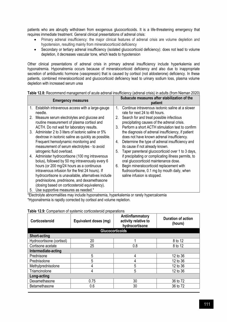
111
patients who are abruptly withdrawn from exogenous glucocorticoids. It is a life-threatening emergency that requires immediate treatment. General clinical presentations of adrenal crisis:
Primary adrenal insufficiency: the major clinical features of adrenal crisis are volume depletion and hypotension, resulting mainly from mineralocorticoid deficiency
Secondary or tertiary adrenal insufficiency (isolated glucocorticoid deficiency): does not lead to volume depletion, it decreases vascular tone, which leads to hypotension
Other clinical presentations of adrenal crisis in primary adrenal insufficiency include hyperkalemia and hyponatremia. Hyponatremia occurs because of mineralocorticoid deficiency and also due to inappropriate secretion of antidiuretic hormone (vasopressin) that is caused by cortisol (not aldosterone) deficiency. In these patients, combined mineralocorticoid and glucocorticoid deficiency lead to urinary sodium loss, plasma volume depletion with increased serum urea Table 12.8: Recommend management of acute adrenal insufficiency (adrenal crisis) in adults (from Nieman 2020)
Emergency measures Subacute measures after stabilization of the
patient
1. Establish intravenous access with a large-gauge needle.
2. Measure serum electrolytes and glucose and routine measurement of plasma cortisol and ACTH. Do not wait for laboratory results.
3. Administer 2 to 3 liters of isotonic saline or 5% dextrose in isotonic saline as quickly as possible. Frequent hemodynamic monitoring and measurement of serum electrolytes - to avoid iatrogenic fluid overload.
4. Administer hydrocortisone (100 mg intravenous bolus), followed by 50 mg intravenously every 6 hours (or 200 mg/24 hours as a continuous intravenous infusion for the first 24 hours). If hydrocortisone is unavailable, alternatives include prednisolone, prednisone, and dexamethasone (dosing based on corticosteroid equivalency).
5. Use supportive measures as needed.*
1. Continue intravenous isotonic saline at a slower rate for next 24 to 48 hours.
2. Search for and treat possible infectious precipitating causes of the adrenal crisis.
3. Perform a short ACTH stimulation test to confirm the diagnosis of adrenal insufficiency, if patient does not have known adrenal insufficiency.
4. Determine the type of adrenal insufficiency and its cause if not already known.
5. Taper parenteral glucocorticoid over 1 to 3 days, if precipitating or complicating illness permits, to oral glucocorticoid maintenance dose.
6. Begin mineralocorticoid replacement with fludrocortisone, 0.1 mg by mouth daily, when saline infusion is stopped.
*Electrolyte abnormalities may include hyponatremia, hyperkalemia or rarely hypercalcemia *Hyponatremia is rapidly corrected by cortisol and volume repletion.
Table 12.9: Comparison of systemic corticosteroid preparations
Corticosteroid Equivalent doses (mg) Antiinflammatory activity relative to
hydrocortisone
Duration of action (hours)
Glucocorticoids
Short-acting
Hydrocortisone (cortisol) 20 1 8 to 12
Cortisone acetate 25 0.8 8 to 12
Intermediate-acting
Prednisone 5 4 12 to 36
Prednisolone 5 4 12 to 36
Methylprednisolone 4 5 12 to 36
Triamcinolone 4 5 12 to 36
Long-acting
Dexamethasone 0.75 30 36 to 72
Betamethasone 0.6 30 36 to 72

112
Mineralocorticoids
Fludrocortisone The typical dose of fludrocortisone for mineralocorticoid replacement is 0.1 to 0.2 mg.
12 to 36
Myasthenia Gravis & Myasthenic Crisis Myasthenia gravis (MG) is an autoimmune disorder of the postsynaptic neuromuscular junction, which characterised by fluctuating weakness involving variable combinations of ocular, bulbar, limb and respiratory muscles. Myasthenic crisis is a life-threatening condition due to worsening of myasthenic weakness. It may involve varying degrees of skeletal muscle weakness. The most commonly affected muscles are those of the eyes, face, and swallowing. It can result in double vision, drooping eyelids, trouble talking, and trouble walking. In severe condition, it may require intubation or non-invasive ventilation. Typically, patient may develop generalized or bulbar weakness as a warning. Generalized weakness may mask the usual signs of respiratory distress. Myasthenic crisis may be precipitated by a variety of factors which include concurrent infection, surgical intervention, pregnancy, childbirth or tapering of immunosuppressive medication. Medication can also precipitate the weakness in myasthenia (Table 12.11). Management of Myasthenia Gravis and Myasthenic Crisis Primary therapies used to treat MG are:
Symptomatic treatment (e.g acetylcholinesterase inhibition)
Chronic immunosuppressive therapies (e.g glucocorticoids and nonsteroidal immunosupporessive agents)
Rapid and short acting immunomodulating treatments (e.g plasma exchange and IVIG)
Surgical intervention (e.g thymectomy) General approach to the management of patients with myasthenic crisis or severe exacerbations of myasthenia gravis include the following
Admit to intensive care unit
Frequent monitoring of respiratory muscle strength
Elective intubation (if evalution suggesting for impending respiratory collapse)
Rapid therapy with plasma exchange or intravenous immune globulin (IVIG)
Immunomodulating therapy (e.g high dose glucocorticoids, azathioprine, mycophenolate mofetil, cyclosporine)
Initiate weaning from mechanical ventilation when respiratory muscle strength is improving Among intubated patients:
Pyridostigmine is generally held initially to reduce airway secretions and increase sensitivity to pyridostigmine (a brief drug holiday may increase responsiveness when treatment is resumed).
Pyridostigmine should be restarted prior to extubation.
Non-intubated patients:
If the patient is pyridostigmine naive, it may be reasonable to start a low dose (e.g 60 mg PO q6hr).
In most other cases, it may be reasonable to continue the patient's home dosing regimen. Table 12.10: Common drugs used in management of MG and myasthenic crisis
Initial symptomatic therapy
Pyridostigmine Starting dose: 30mg TDS for 2-3 days then asses the cholinergic side effects. May increase the dose by 30mg increments until achieve good therapeutic effect or limited by side effects (usual maximum dose: 120 mg QID, usual

113
adult patients require a total daily dose of ≤960 mg divided into 4 to 8 doses)
Rapid therapy
Plasma exchange Typical course consisted of 5 exchanges (3-5L of plasma each) over 7-14 days
IVIG Total dose is 2g/kg split evenly over 2-5 days
Management pyridostigmine of side effect
Glycopyrrolate 1mg orally with each pyridostigmine dose
Propantheline 15mg orally with each pyridostigmine dose
Hyoscyamine sulfate 0.125mg orally with each pyridostigmine dose
In patient who developed refractory disease, may require unacceptably high dose of glucocorticoids despite concurrent use of first line steroid-sparing agents (e.g oral or nasogastric glucocorticoids at moderate to high dose (e.g prednisone 60-80mg daily). Others: azathioprine, mycophenolate, cyclosporine, rituximab, eculizumab, cyclophosphamide. Respiratory support Respiratory support is imperative in the management of myasthenic crisis. 67% to 90% of patients with myasthenic crisis require intubation and mechanical ventilation. Elective intubation of a myasthenic patient with impending respiratory failure is favored over emergent intubation. Neuromuscular blocking agents Should be used with caution when intubating MG patients. Depolarizing agents such as succinylcholine are less potent in myasthenics because fewer functional post-synaptic AChR are available. This decrease in receptors also results in a decrease in the safety margin or remaining AChR available for neuromuscular transmission. Nondepolarizing agents such as atracurium, vecuronium, rocuronium and pancuronium have increased potency, and reduced doses are required for paralysis. Removal of auto-antibodoes Plasma exchange directly removes anti-acetylcholine receptor antibodies from the body and is often the first-line for severe exacerbations. Plasma exchange offers advantage in term of rapid response as compared to IV Immunoglobulin (IVIg). In plasma exchange, 5 exchanges (1 plasma volume or 3-4 L per exchange) are usually performed every other day over 10 days. Intravenous immune globulin (IVIg) May be useful for less severe exacerbations. It takes longer to show its effect, but the efficacy may be more sustained. IVIg can be used in situations where plasma exchange is unavailable or contraindicated. The most common side effect associated with IVIg is headache. Other complications include fever, nausea, IV site discomfort, rash, malaise, aches and pain. More serious adverse events can include aseptic meningitis, hypertension, cardiac arrhythmia, thrombocytopenia, and thrombotic events, including stroke, myocardial infarction, and pulmonary embolism. Response to treatment generally occurs after 2 days with plasma exchange and after 4-5 days with IVIg. Treat / remove underlying cause The most common precipitant for Myasthenic Crisis is infection, especially pneumonia. Other precipitants include electrolyte abnormality, thyroid disease, surgery including thymectomy, trauma, perimenstrual state, pregnancy, delivery, certain medications and tapering of immune modulating medications. These medications should be used cautiously in myasthenic patients, especially after surgery. Any medication suspected of precipitating myasthenic crisis should be discontinued. Other antecedent factors include exposure to extreme temperature, pain, sleep deprivation, and physical or emotional stress.

114
Complications Most common complications associated with myasthenic crisis are:
Various causes of fever and infection, including pneumonia, bronchitis, urinary tract infection, colitis cause by clostridioides difficile, bacteremia and sepsis
Risk of vascular complications (e.g DVT, heart failure, acute MI, cardiac arryhtmias, cardiac arrest, stress-induced cardiomyopathy)
Table 12.11: Drugs that may unmask / exacerbate Myasthenia Gravis
Drugs that impair neuromuscular transmission and may increase weakness in patients with underlying
neuromuscular junction disorders
Drugs implicated as potentially harmful in myasthenia gravis patients based on anecdotal case reports and /
or in-vitro microelectrode studies
Antibiotics Aminoglycosides Tobramycin Gentamicin Netilmicin Neomycin Streptomycin Kanamycin Fluoroquinolones Ciprofloxacin Norfloxacin Ofloxacin Ketolides Telithromycin Other antibiotics Macrolides Azithromycin Clarithromycin
Others Tetracyclines Sulfonamides Penicillins Amino acid antibiotics Ritonavir Nitrofurantoin Quinidine Quinine Chloroquine
Beta blockers Propranolol Oxprenolol Timolol Practolol Atenolol Labetalol Metoprolol Nadolol Calcium channel blockers Verapamil Other cardiac drugs Procainamide Bretylium Trimethaphan Anticonvulsant medication Phenytoin Barbiturates Ethosuximide Carbamazepine Gabapentin
Ophthalmologic medications Timolol Betaxolol hydrochloride Echothiophate (a long-acting cholinesterase inhibitor used in the treatment of open angle glaucoma) Psychiatric drugs Lithium carbonate Phenothiazines Amitriptyline Imipramine Amphetamines Haloperidol Miscellaneous drugs Riluzole Glatiramer acetate Fludarabine Cisplatin Interleukin-2
Guillain-Barré Syndrome (GBS) GBS also called acute inflammatory demyelinating polyradiculoneuropathy (AIDP), is an inflammatory disorder which affects the peripheral nervous system, characterized by acute or subacute onset and typically presents with ascending paralysis severe enough to warrant hospital admission for management. GBS occurs as a result of autoimmune response to an immune activating event, such as:
Campylobacter jejuni enteritis (25 - 50%)
Mycoplasma infection
Viral infection (e.g Cytomegalovirus, Epstein–Barr, hepatitis A-E viruses, HIV, influenza, arboviruses)
Vaccination (e.g influenza, rabies) Management of Guillain-Barré Syndrome Goals of therapy include: 1. Respiratory support
115
2. Removal of auto-antibodies. 3. Supportive care Respiratory support Intubation and ventilatory support are required when patients are too weak to support a satisfactory minute volume, poor cough effort, and/or when bulbar cranial nerves become involved. Tracheostomy should be offered when pulmonary function tests fail to improve with treatment and two weeks of intubation has passed without change. Removal of auto-antibodies. Plasma exchange removes aetiological auto-antibodies. Efficacy of plasma exchange has been demonstrated. Evidence supports 4-5 exchanges of 1-2 plasma volume (3-8 L per exchange) performed over a span of 10-14 days. Every other day plasmapheresis reduces the risk of coagulopathy by permitting hepatic synthesis to regenerate serum clotting factors. It allows time for antibodies initially located outside of the plasma compartment to redistribute back into the serum, where they can be removed during subsequent exchange. Replacement with albumin is preferred over fresh frozen plasma due to a lower complication rate. Intravenous immune globulin (IVIg) It is suggested that IVIg given within 2 weeks of the onset was as efficacious as plasma exchange, and more likely to be completed than plasma exchange. Their combination is not supported. The most common IVIg dose is 2 g/kg split evenly over 5 days. Since treatment with plasmapheresis and IVIg are equally effective, the choice between them is dictated by treatment availability, facility expertise, and patient comorbidities or contraindications. Supportive care Although corticosteroids are highly effective therapy for chronic inflammatory demyelinating polyneuropathy (CIDP), it is ineffective for treating GBS. Deep Venous Thrombosis (DVT) prophylaxis, enteral feeding and analgesia are among supportive care for GBS unless contraindicated. Early initiation of physiotherapy and rehabilitation as an inpatient and continuing upon discharge is associated with better outcomes and recommended for all but the mildest cases. Status Epilepticus (SE) SE is defined as a seizure which lasts longer than 5 minutes or when seizures (2 or more) occur close together without return of consciousness. Status epilepticus are classified as convulsive and non-convulsive. Convulsive status epilepticus may involve jerking motions, grunting sounds, drooling, and rapid eye movements. Patients with non-convulsive status epilepticus may appear confused or look like daydreaming, unable to speak and/or behaving in an irrational way. Meanwhile, pseudoseizures are an important differential diagnosis characterised by volitional or non-volitional, preserved consciousness or purposeful movements, asymmetric movements, poorly coordinated thrashing, back arching, eyes held shut, head rolling, pelvic thrusting and/or pedalling movements of legs. Causes of SE includes epilepsy, infection (meningitis, encephalitis, cerebral malaria, cerebral toxoplasmosis), hypoxia, vascular (stroke, embolism), metabolic (hypoglycemia, electrolyte imbalance), structural (tumour), physical (hyperthermia, head trauma, stress, menstrual), drug-induced and/or related to drug-withdrawal. Table 12.12: Drugs which have been implicated in inducing seizure
Drug-induced Drug-withdrawal
Cough & Cold medicine Diphenhydramine Pseudoephedrine Phenylephrine
Antimicrobial
Analgesics Morphine Pethidine
Tramadol Cardiovascular drugs
Alcohol Amphetamine Methylenedioxymethamphetamine (MDMA) Cocaine Marijuana
116
Cefazolin Ceftazidime Cefoperazone Cefepime Doripenem Ertapenem Imipenem Meropenem
Antivirals Acyclovir Amantadine Ganciclovir
Anaesthetic agent
Bupivacaine Lidocaine Lignocaine Etomidate Isoflurane Ketamine Propofol Sevoflurane Ropivacaine
Others
Contrast agent
Digoxin Ergometrine Ergotamine Esmolol Lignocaine Metoprolol Methyldopa
Anti-asthmatics Salbutamol Theophylline Terbutaline
Antidepressants Amitriptyline Clomipramine Dothiepin Fluoxetine Fluvoxamine Sertraline Lithium
Antipsychotics Clozapine Chlorpromazine Haloperidol Olanzapine Risperidone
Morphine Heroin Pethidine
Management of Status Epilepticus Goals of therapy include:
● Resuscitation to prevent secondary brain injury and maintain cerebral perfusion pressure ● Terminate seizure ● Treat underlying cause ● Treat complications, if any
Resuscitation
Manage airway with recovery position, airway adjuncts and intubation if required. Optimise oxygenation and provide ventilatory support as needed (prone to hypercapnia). Treat hypoglycaemia and life-threatening electrolyte disturbance if present. Maintain normothermia. Table 12.13: Pharmacological agents used in management of SE
Therapy Dose Note
First line therapies
Midazolam
0.1 mg/kg IV / IM / buccal (Max 10 mg)
IM Midazolam not inferior to IV lorazepam
Lorazepam 0.1 mg/kg IV Onset in 3 - 5 minutes and last hours; preferred for longer acting effects
Diazepam 5 mg IV/PR Avoid IM due to pain. Onset in ~1 minute but lasts~20 min for anticonvulsant activity.
Second line therapies (Typically requires intubation and mechanical ventilation)
117
Phenytoin
15-20 mg/kg IV over 30 minutes or longer, may give additional 5 - 10 mg/kg if still seizing
Should be used to terminate seizures as a sole agent, always with benzodiazepines. Avoid use in patients with on-going phenytoin treatment. Avoid giving rapid push due to risk of cardiotoxicity from propylene glycol diluent.
Sodium valproate
20-40 mg/kg IV over 10 min, may give an additional 20 mg/kg over 5 min if still seizing
Use with caution in patients with traumatic head injury; may be a preferred agent in patients with glioblastoma multiforme.
Leviteracetam 2-3 g IV over 15 min Off label use for status epilepticus
Third line therapies
Propofol 1-2 mg/kg IV loading dose followed by 20 mcg/kg/min
Requires mechanical ventilation
Midazolam
0.2 mg/kg IV then 0.05 mg/kg/hr IVI
Tachyphylaxis occurs after prolonged use
Phenobarbitone 5-15 mg/kg IV boluses followed by 0.5 - 5 mg/kg/hr IVI
Fourth line therapies
Thiopentone
2-7 mg/kg IV followed by 0.5-5 mg/kg/hr IVI
Requires mechanical ventilation. Metabolized to phenobarbitone
Ketamine 1-5 mg/kg IV followed by 1-10 mg/kg/hr IVI
Potential neurotoxicity
Complications Complications depend on the underlying cause and can range from no complications to death. Complication is unlikely if the underlying cause such as poor epilepsy control is fixed. If the underlying cause is a stroke or brain injury, complications may include physical disability from the cause or even death. Table 12.14: Complications of status epilepticus
Classification Complications
Cardiac Hypertension, tachycardia (reversing after 30 minutes), arrhythmias, cardiac arrest
Pulmonary Apnea, respiratory failure, hypoxia, neurogenic pulmonary, edema, aspiration pneumonia
Autonomic Fever, sweating, hypersecretion (including tracheobronchial), vomiting
Metabolic Hyperkalemia, hyperglycemia then hypoglycemia, volume depletion, venous stasis, possible thrombosis
Endocrine Increased prolactin and cortisol
Other Leukocytosis, cerebrospinal fluid pleocytosis, vertebral and other fractures, physical injury, rhabdomyolysis, renal failure, disseminated intravascular coagulation
Cerebral Neuronal damage similar to that of hypoxia, hyperthermia: cortical layers 3 and 5, cerebellum, and hippocampus Cerebral edema, raised intracranial pressure Cortical vein thrombosis
Neurologic sequelae Increased seizure frequency, recurrent status epilepticus Decreased cognitive function (controversial) Drug effects, increased exposure to anticonvulsants
118
References and Suggested Readings Ahmed S, et al. An update on myasthenic crisis. Curr Treat Options Neurol. 2005. 7(2): 129-141. Arlt W, Allolio B. Adrenal insufficiency. Lancet 2003. 361: 1881. Arlt W. The approach to the adult with newly diagnosed adrenal insufficiency. J Clin Endocrinol Metab 2009. 94:
1059. Bajwa S, Jindal R. Endocrine emergencies in critically ill patients: Challenges in diagnosis and management. Indian
J Endocrinol Metab. 2012. 16(5): 722–727. Barkun AN, Bardou M, Kuipers EJ, Sung J, Hunt RH, Martel M, Sinclair P. International consensus
recommendations on the management of patients with nonvariceal upper gastrointestinal bleeding. Ann Intern Med. 2010. 152(2): 101.
Bedlack RS, Sanders DB. On the concept of myasthenic crisis. J Clin Neuromuscul Dis. 2002. 4(1): 40. Bird SJ. Overview of the treatment of myasthenia gravis. UpToDate 2020. Brophy GM, Bell R, Claassen J, Alldredge B, Bleck TP, Glauser T, Vespa PM. Guidelines for the evaluation and
management of status epilepticus. Neurocritical Care. 2012. 17(1): 3–23. Carroll R, Matfin G. Endocrine and metabolic emergencies: Thyroid storm. Ther Adv Endocrinol Metab. 2010. 1(3):
139 – 145. Chavez-Tapia NC, Barrientos-Gutierrez T, Tellez-Avila FI, Soares-Weiser K, Uribe M. Antibiotic prophylaxis for
cirrhotic patients with upper gastrointestinal bleeding. Cochrane Database Syst Rev. 2010. (9): CD002907 Davidson I, Wilson C, Walton T, Brissenden S. Physiotherapy and Guillain-Barré syndrome: Results of a national
survey. Physiotherapy. 2009. 95(3): 157–163. Díaz-Manera J, Rojas García R, Illa I. Treatment strategies for myasthenia gravis: An update. Expert Opin
Pharmacother. 2012. (13):1873-83. French Cooperative Group on Plasma Exchange in Guillain-Barré syndrome. Efficiency of plasma exchange in
Guillain-Barré syndrome: role of replacement fluids. Ann Neurol. 1987. 22(6): 753-61. Garcia-Tsao et al. Portal hypertension bleeding in cirrhosis: Risk stratification, diagnosis, and management: 2016
Practice Guidance by the AASLD. Hepatology. 2016. 65(1): 2017. Gilhus NE. Myasthenia gravis. N Engl J Med. 2016. 375(26): 2570. Gralnek IM et al. Nonvariceal upper gastrointestinal hemorrhage: ESGE guideline. Endoscopy. 2015. 47: a1–a46 Greenberg JA et al. Clinical practice guideline: Management of acute pancreatitis. Can J Surg. 2016. 59: 2. Kearney T, Dang C. Diabetic and endocrine emergencies. Postgrad Med J. 2007. 83: 79–86. Lizarraga A, Lizarraga K, Benatar M. Getting rid of weakness in the ICU: An updated approach to the acute
management of myasthenia gravis and Guillain-Barré syndrome. Semin Neurol. 2016. 36(6): 615-624. Mah PM, Jenkins RC, Rostami-Hodjegan A, et al. Weight-related dosing, timing and monitoring hydrocortisone
replacement therapy in patients with adrenal insufficiency. Clin Endocrinol (Oxf) 2004; 61:367. Malaysian Clinical Practice Guidelines on Management of Type 2 Diabetes Mellitus 2015. NHS Trafford Diabetes 2012. Management of DKA in Adults. Nieman LK. Treatment of adrenal insufficiency in adults. UpToDate 2020 Raphaël JC, Chevret S, Hughes RA, Annane D. Plasma exchange for Guillain-Barré syndrome. Cochrane
Database Syst Rev. 2012. (7): CD001798. Sanders DB, Wolfe GI, Benatar M, Evoli A, Gilhus NE, Illa I, Kuntz N, Massey JM, Melms A, Murai H, Nicolle M,
Palace J, Richman DP, Verschuuren J, Narayanaswami P. International consensus guidance for management of myasthenia gravis: Executive summary. Neurology. 2016. 87(4): 419.
Wells M, Chande N, Adams P, Beaton M, Levstik M, Boyce E, et al. Meta-analysis: Vasoactive medications for the management of acute variceal bleeds. Aliment Pharmacol Ther. 2012. 35: 1267-1278.
Wilkins T, Khan N, Nabh A, Schade RR. Diagnosis and management of upper gastrointestinal bleeding. AAFP. 2012. 85: 5.

119
CHAPTER 13
Feeding Intolerance
arly enteral feeding is one of the fundamentals of critical care practice. Enteral nutrition (EN) preserved gut integrity by stimulates mesenteric blood flow and gut growth-factor and promotes normal GI secretions. However, EN is often poorly tolerated in a critically ill patient. More than 50% of them experiencing delayed gastric emptying or gastroparesis.
Figure 13.1. Motor activity of the stomach and duodenum in (a) healthy person and (b) critical illness (from Fraser & Bryant 2010)
Feeding intolerance due to gastroparesis is manifested by high gastric residual. The best-recognized motor dysfunctions which contributed to intolerance are (Figure 13.1):
Reduction in the frequency and amplitude of antral contraction, particularly loss of gastric phase 3 activity. Duodenal phase 3 activity is often preserved in this patient but in a disorganized form that may promote a retrograde flow
E
120
Disturbance in fundic motor activity. Relaxation is delayed in response to stimulation by nutrients in the gut, but more importantly, gastric tone fails to recover when this nutrient stimulus is terminated. This prolonged relaxation response is likely to lead to failure of redistribution of gastric content
Abnormal duodenal contractility. The lack of clearance may also potentially result in continuing contact between nutrient and small intestinal leading to prolonged feedback stimulation
Increase in pyloric response to duodenal nutrients and subsequently raise the motility pattern that will obstruct the gastric outflow
Rise feedback from the small intestine receptor. The mechanism is unclear but may relate to enhance secretion of cholecystokinin, especially with high lipid content feeding
Factors contributed to gastroparesis include medication used (i.e opioid, catecholamines), hyperglycemia, renal dysfunction, mechanical ventilation, recumbent position, and the disease process itself (i.e head injury, abdominal injury). Unresolved gastroparesis may lead to vomiting, aspiration of gastric content, pneumonia, and inadequate calorie intake. There are significant associations between feeding intolerance, prolonged ICU stay, and increased risk of death. Even with these complications, EN is preferred to PN as it is associated with lower septic complications, lower risk of bacterial translocation, and cheaper. Management of Gastroparesis
Initial management of gastroparesis includes dietary modification, optimization of glycemic control, and hydration. Pharmacological strategies include prokinetic drugs such as metoclopramide and erythromycin alone or in combination (Figure 13.2). Management of Refractory and Acute Exacerbations of Gastroparesis
Re-evaluation of compliance with dietary modification and pharmacotherapy is required in patients presented with refractory symptoms of gastroparesis despite dietary modification and pharmacological management. This is also to identify the necessity for the provision of nutritional support. Jejunostomy or venting gastrostomy tube placement is the other approach for enteral nutrition and decompression. Parenteral nutrition is indicated only in patients who cannot tolerate enteral nutrition despite concomitant pharmacotherapy. Gastric electrical stimulation can be considered only in patients with gastroparesis with intractable nausea and vomiting despite medical therapy for at least one year.
For acute exacerbations of delayed gastric emptying, it is recommended to administer erythromycin 3 mg/kg
intravenously (IV) every eight hours. In patients without permanent enterostomy tubes, IV erythromycin relieves
acute gastric stasis rapidly, obviating the need for nasogastric decompression. However, IV erythromycin is
effective in relieving symptoms in approximately 40 percent of patients. In patients who fail IV erythromycin,
subcutaneous metoclopramide (5 to 10 mg three times daily) can be the alternative approach.

121
Figure 13.2: Management of gastroparesis (adapted from UpToDate 2020)
Prokinetic Agents
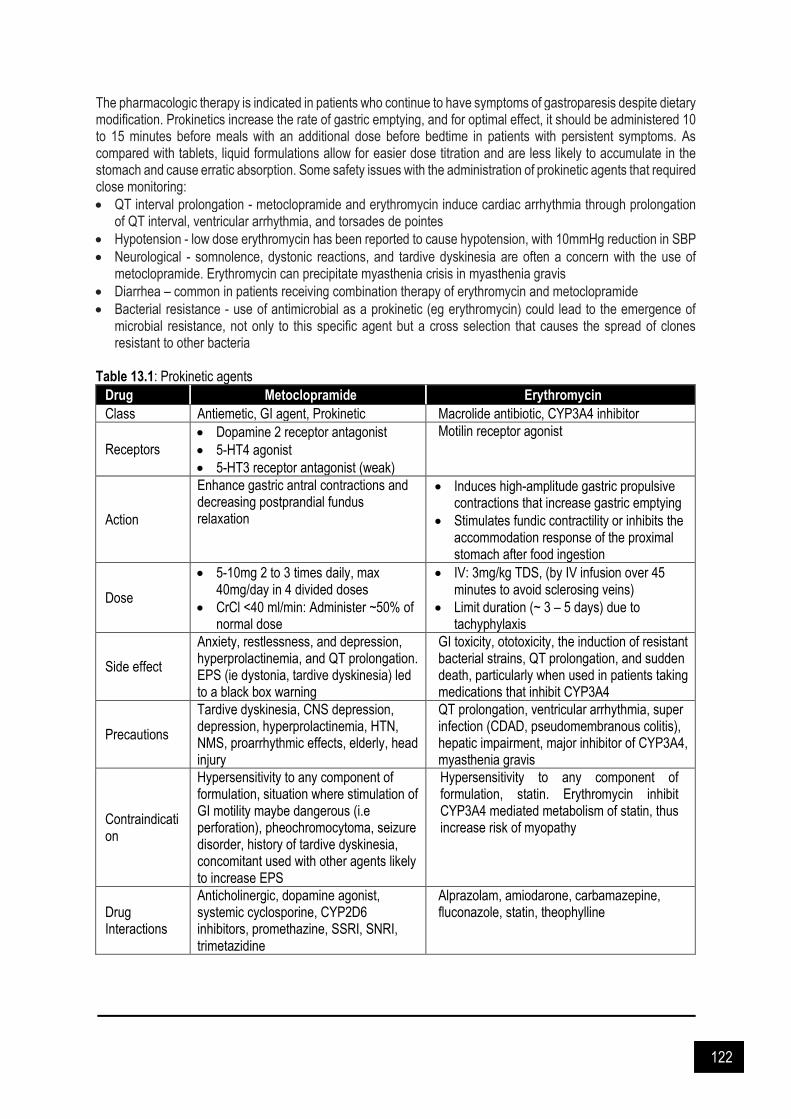
122
The pharmacologic therapy is indicated in patients who continue to have symptoms of gastroparesis despite dietary modification. Prokinetics increase the rate of gastric emptying, and for optimal effect, it should be administered 10 to 15 minutes before meals with an additional dose before bedtime in patients with persistent symptoms. As compared with tablets, liquid formulations allow for easier dose titration and are less likely to accumulate in the stomach and cause erratic absorption. Some safety issues with the administration of prokinetic agents that required close monitoring:
QT interval prolongation - metoclopramide and erythromycin induce cardiac arrhythmia through prolongation of QT interval, ventricular arrhythmia, and torsades de pointes
Hypotension - low dose erythromycin has been reported to cause hypotension, with 10mmHg reduction in SBP
Neurological - somnolence, dystonic reactions, and tardive dyskinesia are often a concern with the use of metoclopramide. Erythromycin can precipitate myasthenia crisis in myasthenia gravis
Diarrhea – common in patients receiving combination therapy of erythromycin and metoclopramide
Bacterial resistance - use of antimicrobial as a prokinetic (eg erythromycin) could lead to the emergence of microbial resistance, not only to this specific agent but a cross selection that causes the spread of clones resistant to other bacteria
Table 13.1: Prokinetic agents
Drug Metoclopramide Erythromycin
Class Antiemetic, GI agent, Prokinetic Macrolide antibiotic, CYP3A4 inhibitor
Receptors
Dopamine 2 receptor antagonist
5-HT4 agonist
5-HT3 receptor antagonist (weak)
Motilin receptor agonist
Action
Enhance gastric antral contractions and decreasing postprandial fundus relaxation
Induces high-amplitude gastric propulsive contractions that increase gastric emptying
Stimulates fundic contractility or inhibits the accommodation response of the proximal stomach after food ingestion
Dose
5-10mg 2 to 3 times daily, max 40mg/day in 4 divided doses
CrCl <40 ml/min: Administer ~50% of normal dose
IV: 3mg/kg TDS, (by IV infusion over 45 minutes to avoid sclerosing veins)
Limit duration (~ 3 – 5 days) due to tachyphylaxis
Side effect
Anxiety, restlessness, and depression, hyperprolactinemia, and QT prolongation. EPS (ie dystonia, tardive dyskinesia) led to a black box warning
GI toxicity, ototoxicity, the induction of resistant bacterial strains, QT prolongation, and sudden death, particularly when used in patients taking medications that inhibit CYP3A4
Precautions
Tardive dyskinesia, CNS depression, depression, hyperprolactinemia, HTN, NMS, proarrhythmic effects, elderly, head injury
QT prolongation, ventricular arrhythmia, super infection (CDAD, pseudomembranous colitis), hepatic impairment, major inhibitor of CYP3A4, myasthenia gravis
Contraindication
Hypersensitivity to any component of formulation, situation where stimulation of GI motility maybe dangerous (i.e perforation), pheochromocytoma, seizure disorder, history of tardive dyskinesia, concomitant used with other agents likely to increase EPS
Hypersensitivity to any component of formulation, statin. Erythromycin inhibit CYP3A4 mediated metabolism of statin, thus increase risk of myopathy
Drug Interactions
Anticholinergic, dopamine agonist, systemic cyclosporine, CYP2D6 inhibitors, promethazine, SSRI, SNRI, trimetazidine
Alprazolam, amiodarone, carbamazepine, fluconazole, statin, theophylline

123
References and Suggested Readings Booth CM, Heyland DK, Paterson WG. Gastrointestinal promotility drugs in the critical care setting: A systematic
review of the evidence. Crit Care Med 2002. 30(7): 1429-35. Camilleri M, Parkman HP, Shafi MA, Abell TL, Gerson L. Clinical guideline: management of gastroparesis. Am J
Gastroenterol 2013. 108(1): 18. Camilleri M. Treatment of gastroparesis. UpToDate 2020. Chapman MJ, Nguyen NQ, Fraser RJ. Gastrointestinal motility and prokinetics in the critically ill. Curr Opinion Crit
Care 2007. 13(2): 187-94. Dive A, Miesse C, Jamart J, Evrard P, Gonzalez M, Installe E. Duodenal motor response to continuous enteral
feeding is impaired in mechanically ventilated critically ill patients. Clin Nutr 1994. 13(5): 302-6. Dive A, Moulart M, Jonard P, Jamart J, Mahieu P. Gastroduodenal motility in mechanically ventilated critically ill
patients: a manometric study. Crit Care Med 1994. 22(3): 441-7. Doherty WL, Winter B. Prokinetic agents in critical care. Crit Care 2003. 7(3): 206. Fraser RJ, Bryant L. Current and future therapeutic prokinetic therapy to improve enteral feed intolerance in the
ICU patient. Nutr Clin Prac 2010. 25(1): 26-31. Grant K, Thomas R. Prokinetic drugs in the intensive care unit: Reviewing the evidence. J Intensive Care Soc
2009. 10(1): 34-7. Hawkyard CV, Koerner RJ. The use of erythromycin as a gastrointestinal prokinetic agent in adult critical care:
Benefits versus risks. J Antimicrob Chemother 2007. 59(3): 347-58. Lewis K, Alqahtani Z, Mcintyre L, Almenawer S, Alshamsi F, Rhodes A, et al. The efficacy and safety of prokinetic
agents in critically ill patients receiving enteral nutrition: a systematic review and meta-analysis of randomized trials. Crit Care 2016. 20(1): 259.
Nguyen NQ, Chapman MJ, Fraser RJ, Bryant LK, Holloway RH. Erythromycin is more effective than Metoclopramide in the treatment of feed intolerance in critical illness. Crit Care Med 2007. 35(2): 483-9.
Nguyen NQ, Yi Mei SLC. Current issues on safety of prokinetics in critically ill patients with feed intolerance. Ther Adv Drug Saf 2011. 2(5): 197-204.
Roden DM. Drug-induced prolongation of the QT interval. N Eng J Med 2004. 350(10): 1013-22.

124
CHAPTER 14
Neuromuscular Blocking Agents
euromuscular blocking agents (NMBA) are used in the ICU for paralysis in rapid sequence intubation, tracheostomy, facilitating mechanical ventilation in patients with acute respiratory distress syndrome (ARDS) or acute lung injury (ALI) and to prevent shivering in patients undergoing therapeutic hypothermia. NMBAs may also be useful in managing increased intracranial pressure or seizures. NMBAs may improve chest wall compliance, prevent respiratory dys-synchrony, reduce peak airway pressures and reduce oxygen consumption by decreasing the work of breathing and respiratory muscle blood flow. NMBAs are structurally related to acetylcholine (ACh) and act by interfering the binding of ACh to the motor endplate. NMBA classifications: ● Classification according to structure
✔ Aminosteroidal: pancuronium, vecuronium, rocuronium
✔ Benzylisoquinolinium: atracurium, cisatracurium
● Classification according to mechanism of action (MOA)
✔ Depolarising: e.g suxamethonium
✔ Non-depolarising: e.g atracurium, pancuronium, rocuronium, vecuronium, cisatracurium
NMBA Complications ● Thrombosis - patient with NMBA has 8 times more likely to have DVT than those not on a NMBA. DVT
prophylxis is needed ● Prolonged weakness - include myopathy, polyneuropathy or neuromyopathy. Other risk factors include
concomitant use of corticosteroids, persistent hyperglycemia and type of NMBA used ● Corneal abrasions - paralysis eliminates the ability of the eyes to close and blink. Apply lubricating eye
ointments/eye covers as eye protection ● Resistance to paralysis and/or potentiation - certain disease may produce an up-regulation in acetylcholine
skeletal muscle receptors, leading to higher than the normal doses of the NMBA (i.e muscle trauma, muscle atrophy, burns). Acid-base disorders, electrolyte imbalances and adrenal insufficiency may cause unpredictable alterations in dosing requirements.
● Anaphylaxis - IgE against the substituted ammonium ion might be induced by exposure to different chemical
substances; in turn puts the patient at risk for IgE-mediated anaphylaxis to succinylcholine due to cross-reactivity. However, the recent identification of the MRGPRX2 receptor indicates that other NMBA might induce pseudo-allergic reactions.
PA
RT
III: OT
HE
R P
HA
RM
AC
OT
HE
RA
PY
IN C
RIT
ICA
L C
AR
E
N
text here
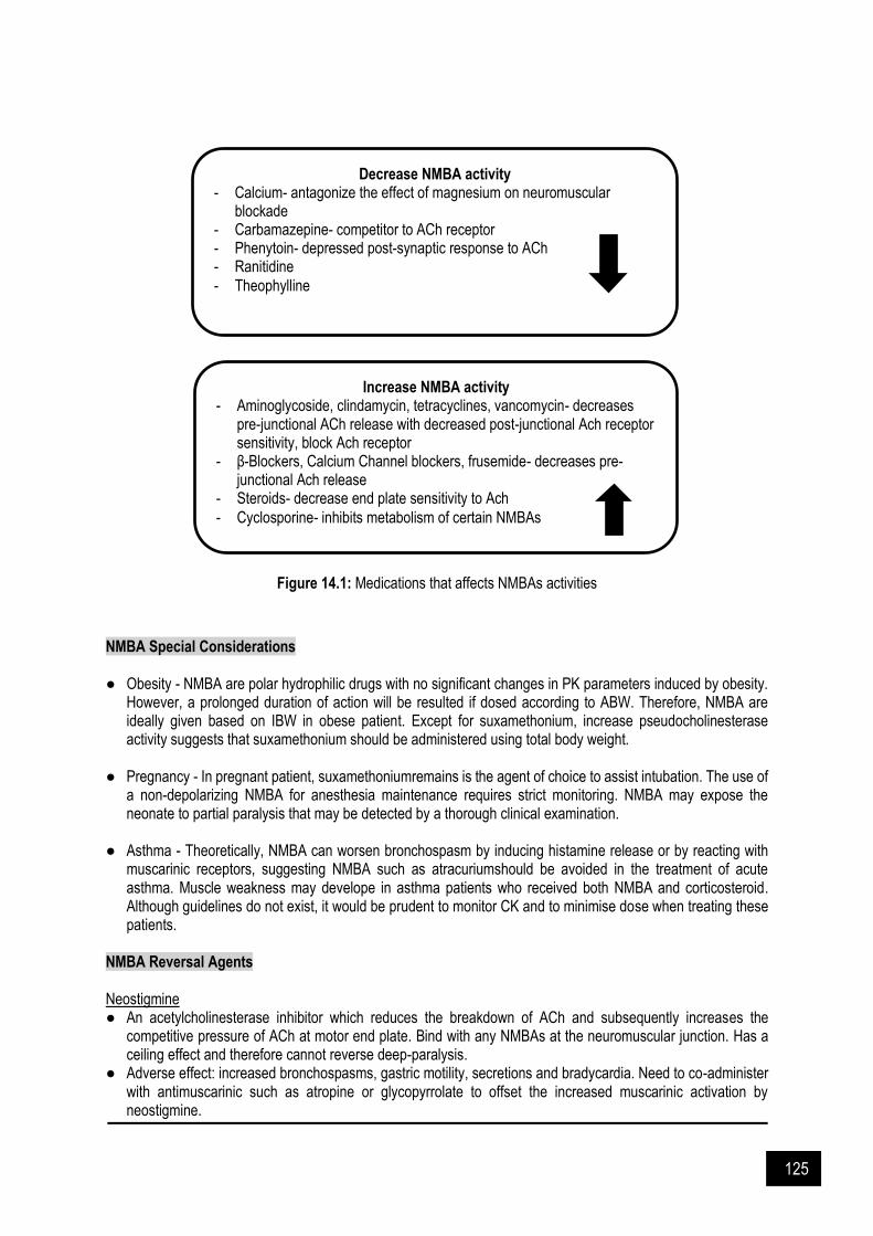
125
Figure 14.1: Medications that affects NMBAs activities
NMBA Special Considerations ● Obesity - NMBA are polar hydrophilic drugs with no significant changes in PK parameters induced by obesity.
However, a prolonged duration of action will be resulted if dosed according to ABW. Therefore, NMBA are ideally given based on IBW in obese patient. Except for suxamethonium, increase pseudocholinesterase activity suggests that suxamethonium should be administered using total body weight.
● Pregnancy - In pregnant patient, suxamethoniumremains is the agent of choice to assist intubation. The use of
a non-depolarizing NMBA for anesthesia maintenance requires strict monitoring. NMBA may expose the neonate to partial paralysis that may be detected by a thorough clinical examination.
● Asthma - Theoretically, NMBA can worsen bronchospasm by inducing histamine release or by reacting with
muscarinic receptors, suggesting NMBA such as atracuriumshould be avoided in the treatment of acute asthma. Muscle weakness may develope in asthma patients who received both NMBA and corticosteroid. Although guidelines do not exist, it would be prudent to monitor CK and to minimise dose when treating these patients.
NMBA Reversal Agents Neostigmine ● An acetylcholinesterase inhibitor which reduces the breakdown of ACh and subsequently increases the
competitive pressure of ACh at motor end plate. Bind with any NMBAs at the neuromuscular junction. Has a ceiling effect and therefore cannot reverse deep-paralysis.
● Adverse effect: increased bronchospasms, gastric motility, secretions and bradycardia. Need to co-administer with antimuscarinic such as atropine or glycopyrrolate to offset the increased muscarinic activation by neostigmine.
Decrease NMBA activity - Calcium- antagonize the effect of magnesium on neuromuscular
blockade - Carbamazepine- competitor to ACh receptor - Phenytoin- depressed post-synaptic response to ACh - Ranitidine - Theophylline
Increase NMBA activity - Aminoglycoside, clindamycin, tetracyclines, vancomycin- decreases
pre-junctional ACh release with decreased post-junctional Ach receptor sensitivity, block Ach receptor
- β-Blockers, Calcium Channel blockers, frusemide- decreases pre-junctional Ach release
- Steroids- decrease end plate sensitivity to Ach
- Cyclosporine- inhibits metabolism of certain NMBAs

126
● Dose: Dosage: 0.03-0.07mg/kg IV (maximum total dose 0.07mg/kg or 5mg (whichever is less))4. Administer slow IV injection over 1 min after or with atropine sulphate 0.6-1.2mg; to administer with separate syringe. May be given undiluted or dilute with WFI.
Sugammadex ● A direct-reversal agent for rocuronium and vecuronium. Classified as a gamma-cyclodextrin, it creates a drug-
drug complex with free NMBA. Faster reversal effect than neostigmine (2 min compared to 17 min). Has no ceiling effect and therefore can rapidly reverse any depth of paralysis
● Adverse effect: No effect on muscarinic receptors, significantly avoiding the need to co-administer with antimuscarinic agents.
● Dose: 2 mg/kg for moderate blockade to 16 mg/kg for immediate reversal. Administer via IV bolus over 10 sec. Maybe given undiluted or may inject into the line of a running infusion of NS, D5W, 1/2 NSD2.5W, Lactate Ringers, Ringers solution or D5NS.
Figure 14.2: MOA of neostigmine and Sugammedex (from Bruell et al 2010)

127
Table 14.1: Neuromuscular blockage agents (NMBA)
Agents Suxamethonium Atracurium Rocuronium Pancuronium Vecuronium Cisatracurium
Dose
Adjunct to general anaesthesia IV:0.6mg/kg (range: 0.3 – 1.1mg/kg) Max: 100mg IM:3-4mg/kg. Max total dose:150mg
Adjunct to general anaesthesia Initial: IV: 0.3- 0.6 mg/kg
Maintenance: IV: 0.1- 0.2 mg/kg
OR IV infusion: 5 -10 mcg/kg/min (300- 600 mcg/kg/hr)
Intensive care Initial: IV: 0.3-0.6 mg/kg
Maintenance: IV: 4.5-29.5 mcg/kg/min (usual: 11-13 mcg/kg/min)
Surgery procedures (Intubation) Initial: IV: 0.6 mg/kg
Maintenance: IV: 0.15 mg/kg (Elderly: 0.075- 0.1 mg/kg) OR IV infusion: 0.3-0.6 mg/kg/h (Elderly: 0.4 mg/kg/h)
Intensive care Initia: IV: 0.6 mg/kg
Maintenance: IV infusion: 0.3-0.6 mg/kg/h for first hour then adjusted according to response
Neuromuscular blockade Initial: IV:0.06-0.1 mg/kg or 0.05 mg/kg after initial dose of suxamethonium for intubation
Maintenance: IV: 0.01 mg/kg 60 – 100 min after initial dose, then 0.01 mg/kg every 25-60 min
Intensive care IV: 0.05-0.1 mg/kg bolus followed by 0.8-1.7 mcg/kg/min once recovery from bolus seen or 0.1-0.2 mg/kg every 1-3 hours
Surgery procedures (Intubation) Initial: IV: 0.08-0.1 mg/kg or 0.04-0.06 mg/kg after initial dose of suxamethonium for intubation
Maintenance: IV: 0.01-0.015 mg/kg 25-40 min after initial dose, then 0.01-0.015 mg/kg every 12-15 min. May be given as continuous infusion at 0.8-2 mcg/kg/min Intensive care IV: 0.05-0.1 mg/kg bolus followed by 0.8-1.7 mcg/kg/min once recovery from bolus seen or 0.1- 0.2 mg/kg every 1-3 hours
Surgery procedures (Intubation) Initial: IV: 0.1mg/kg after initial dose of suxamethonium for intubation
Maintenance: IV:0.03 mg/kg 40-60 min after initial dose, then at 20 min intervals
Intensive care Begin infusion at a dose of 3mcg/kg/min with dosage ranges of 0.5-10mcg/kg/min
Onset 2–3 min 2–3 min 1–2 min (within 4 min)
2–3 min 2.5–3 min 2–3 min
Duration 10–30 min 20–35 min ~30 min 60–100 min 20–40 min 20–35 min
Administration IV bolus
Given undiluted or dilute with NS or D5
IV bolus/ infusion Given undiluted or dilute with NS or D5

128
Agents Suxamethonium Atracurium Rocuronium Pancuronium Vecuronium Cisatracurium
Monitoring Train-of-four; with a goal of adjusting the degree of neuromuscular blockade to achieve one or two twitches
Vital sign (HR, BP, RR), degree of muscle paralysis, serum potassium, calcium
Precautions
Acute rhabdomyolysis with hyperkalemia, ventricular arrhythmias and cardiac arrests reported in children with undiagnosed skeletal muscle myopathy. Caution in patients with extensive or severe burns; risk of hyperkalemia increased following injury
Reduce initial dose and inject slowly over 1-2 min in patients in whom substantial histamine release will be potentially hazardous. Increase sensitivity in patients with MG and Eaton-Lambert Syndrome
Caution in patients with valvular heart disease, pulmonary disease, hepatic impairment. Ventilation must be supported during NMB. Increased sensitivity in patients with MG and Eaton-Lambert Syndrome
Ventilation must be supported during NMB. Caution in patients with renal or hepatic impairment. Increased sensitivity in patients with myasthenia gravis and Eaton-Lambert Syndrome
Increased sensitivity in patients with myasthenia gravis and Eaton-Lambert Syndrome
Side effects
Cardiovascular: Arrhythmias, bradycardia, cardiac arrest, hyper / hypotension, tachycardia Dermatologic: Rash
GI: Excessive salivation
Neuromuscular/ skeletal: Jaw rigidity, postoperative muscle pain, rhabdomyolysis
Respiratory: Apnea, respiratory depression
Endocrine/metabolic: hyperkalemia
1-10%: Bradycardia, flushing, hypotension, tachycardia
<1%: Broncheal secretions, erythema, itching, urticaria, wheezing
>1%: Cardiovascular: Transient hyper/hypotension
<1%: Abnormal ECG, anaphylaxis, arrhythmia, bronchospasm, edema, hiccups, nausea, rash, rhonchi, shock, tachycardia, wheezing, vomiting
Cardiovascular: Elevation in PR, elevated BP/CO, tachycardia, edema, skin flushing, circulatory collapse Dermatologic: Rash, itching, erythema, burning sensation along the vein
GI: excessive salivation
Neuromuscular/ skeletal: Profound muscle weakness
Respiratory: wheezing, bronchospasm
Serious immunologic: Anaphylaxis, hypersensitivity reaction Musculoskeletal: Muscle weakness, prolonged NMB Respiratory: Bronchospasm
Cardiovascular: Bradyarrhymia, hypotension Respiratory: Bronchospasm, gasping for breath Endocrine/metabolic: Metabolic acidosis Immunologic: Anaphylaxis Neurologic: CNS depression, paralysis, seizure

129
References and Suggested Readings American Society of Health care Pharmacist (ASHP). Therapeutic Guidelines for sustained neuromuscular
blockade in the adult critically ill patient: 2002. Bittner EA. Neuromuscular blocking agents in critically ill patients: Use, agent selection, administration and adverse
effects. UpToDate 2020. Brull SJ et al. Residual neuromuscular block: Lessons unlearned. Part II: Methods to reduce the risk of residual
weakness. Anest Analg 2010. 111: 129-140. Casati A, Putzu M. Anesthesia in the obese patient: pharmacokinetic considerations. J Clin Anest. 2005. 17(2):134-
45. Craig RG, Hunter JM. Neuromuscular blocking drugs and their antagonists in patients with organ disease.
Anaesthesia. 2009. 64 Suppl 1:55. Elliot JM, Bion JF. The use of neuromuscular blocking drugs in intensive care practice. Acta Anaesthesiol Scand
Suppl. 1995. 106:70. Greenberg SB, Vender J. The use of neuromuscular blocking agents in the ICU: where are we now? Crit Care Med
2013. 41:1332. Guay J, Grenier Y, Varin F. Clinical pharmacokinetics of neuromuscular relaxants in pregnancy. Clin
Pharmacokinet. 1998. 34(6): 483-96. Hunter JM. New neuromuscular blocking drugs. N Engl J Med 1995. 332: 1691. Lee LA, Athanassoglou V, Pandit JJ. Neuromuscular blockade in the elderly patient. J Pain Res 2016. 9: 437. Payen JF, Chanques G, Mantz J, et al. Current practices in sedation and analgesia for mechanically ventilated
critically ill patients: a prospective multicenter patient-based study. Anesthesiology 2007. 106: 687. Price DR, Mikkelsen ME, Umscheid CA, Armstrong EJ. Neuromuscular blocking agents and neuromuscular
dysfunction acquired in critical illness: A systematic review and meta-analysis. Crit Care Med 2016. 44: 2070. Prielipp RC, Coursin DB. Applied pharmacology of common neuromuscular blocking agents in critical care. New
Horiz 1994. 2: 34. Rhodes A, Evans LE, Alhazzani W, et al. Surviving sepsis campaign: International guidelines for management of
sepsis and septic shock: 2016. Crit Care Med 2017. 45:486. Topulos GP. Neuromuscular blockade in adult intensive care. New Horiz 1993. 1:447. Tripathi S, Hunter J. Neuromuscular blocking drugs in the critically ill. Continuing Education in Anaesthesia, Critical
Care & Pain. 2006. 6(3): 119-23. Vender JS, Szokol JW, Murphy GS, Nitsun M. Sedation, analgesia, and neuromuscular blockade in sepsis: An
evidence-based review. Crit Care Med. 2004. 32(11): S554-S61.
130
CHAPTER 15
Analgesic & Sedative Agents
Analgesia ritically ill patients experience moderate to severe pain at rest and during standard care procedure. Severe pain may compromise on cardiac instability, respiratory function and immunosuppression. Pain management is often complicated. High inter- and intra-individual variability (acute, chronic, and acute on chronic), experiencing different types of pain (somatic, visceral and neuropathic) and subjective pain perceptions result in the increase complexity of pain management. Factors that affect pain are divided into: ✔ Pain at rest: influenced by physiologic (anxiety and depression) and demographic (age, comorbidities) ✔ Pain during procedure: due to pre-procedural pain intensity, type of procedure, underlying diagnoses and
demographic
Recent guideline advocate titration of analgesic dosing to balance benefits and versus potential risk of opioid exposure and the use of assessment driven, protocol based, stepwise approach for pain and sedation management in critically ill patients. Analgosedation is recommended in institution. It can be implemented by incorporating analgesia first sedation (opioid analgesic is used before a sedative to reach sedation goal) or analgesia-based sedation (opioid analgesic is used instead of sedative to reach sedative goal). Sedation Sedatives agents and analgesics are often used to facilitate patient tolerance to invasive mechanical ventilation. The goals of sedation and analgesia in this context include decreasing pain and anxiety, reducing the stress response and facilitating nursing care. Evidenced showed that targeting light levels of sedation or using daily awakening trials and minimizing benzodiazepines improve short term outcomes (duration of mechanical ventilation and ICU length of stay). Over-sedation can increase time on ventilator support and prolonged ICU stay and under-sedation can cause hyper catabolism, immunosuppression, hypercoagulability and increased sympathetic activity. Recent guideline advocated the use of light sedation (versus deep sedation) in critically ill, mechanically ventilated patients. Sedation-agitation scales can be used to identify and quantify agitation and to grade the depth of sedation. ICU Management Protocol 2012, by Ministry of Health Malaysia, suggested that patients are to be assessed for sedation and agitation based on the Revised Riker Sedation-Agitation Scale (SAS) (Table 15.2) or Richmond Agitation and Sedation Scale (RASS) (Table 15.3) every 4 hours and titrate the sedative infusion rate with the aim of keeping the SAS between -1 to +1 or RASS between -2 to +1. Exception to keeping within the targeted score (should be deeply sedated to achieve SAS of -2 to -3 or RASS of -3 to -5):
✔ Head injury on cerebral protection
✔ Septic shock on high inotropic support
✔ ARDS on high ventilator support (FiO2 >0.6 and PEEP >12)
✔ Tetanus
C
text here

131
Table 15.1: Commonly used analgesic agents in ICU
Morphine Fentanyl Remifentanil
Dose
Initial dose: 0.01-0.15mg/kg every 1-2 hours as needed
Maintenance dose: 0.07-0.5mg/kg/hour
Initial dose: 0.35-1.5mcg/kg every 30-60min as needed
Maintenance dose:
0.7-10mcg/kg/hr
Initial dose: 1mcg/kg over 30-60sec
Maintenance dose: 0.05-0.2mcg/kg/hr
Onset 5-20 min 2 - 5min 1-3min
Duration 2-4 hours 0.5 – 1 hours 0.3 – 0.6 hours
Elimination Hepatic conjugation; active metabolite excreted renally4
Hepatic CYP3A4 Hydrolysis by esterases
Monitoring Pain relief, respiratory and CNS status, BP Pain relief, respiratory and CNS status, BP,
HR2,6 Respiratory and cardiovascular status
Contraindication ▪ Hypersensitivity to morphine sulfate or any product component
▪ Acute or severe asthma ▪ Known or suspected paralytic ileus ▪ Opioid non-tolerant patients2,3 ▪ Significant respiratory depression, especially in
unmonitored settings that lack resuscitative equipment6
▪ During labor when a premature birth is anticipated2,3
▪ Within 2 weeks of MAO inhibitors
▪ Hypersensitivity to fentanyl or any product component
▪ Acute or severe asthma ▪ Known or suspected paralytic ileus ▪ Opioid non-tolerant patients ▪ Significant respiratory depression,
especially in unmonitored settings that lack resuscitative equipment
▪ Within 2 weeks of MAO inhibitors
▪ Hypersensitivity to remifentanil, fentanyl analogs or any component of the component of the formulation
▪ Not for intrathecal or epidural administration due to the presence of glycine in the formulation
Precaution
▪ May cause CNS depression, hypotension, orthostatic hypotension and syncope
▪ May cause respiratory depression, especially in elderly debilitated patients, hypoxia or hypercapnia
▪ Use with caution in patients with adrenal insufficiency, biliary tract dysfunction, acute pancreatitis, renal/ hepatic impairment, head trauma, seizure, thyroid dysfunction, history of drug abuse or acute alcoholism
▪ May cause CNS depression, respiratory depression, especially in elderly debilitated patients, hypoxia or hypercapnia
▪ Use with caution in bradycardia, bradyarrhythmias, renal/ hepatic impairment, head trauma, history of drug abuse or acute alcoholism
▪ May cause hypotension ▪ Caution in patient with hypovolemia,
cardiovascular disease or drugs which may exaggerate hypotensive effects
▪ Safety and efficacy for postoperative analgesic have not been established in children
▪ Rapid IV infusion may result in skeletal muscle and chest wall rigidity, impaired ventilation or respiratory distress/ arrest;

132
Morphine Fentanyl Remifentanil
nondepolarizing skeletal muscle relaxant may be required2
Side effects6 -common
GI: Constipation, nausea, vomiting Neurologic: Dizziness, headache, somnolence. Dermatologic: Pruritus Ophthalmic: Miosis Renal: Urinary retention
GI: Abdominal pain, nausea, vomiting, constipation, xerostomia Neurologic: Asthenia, confusion, dizziness, nervous, headache, insomnia, somnolence Psychiatric: Anxiety, depression, euphoria, hallucination Renal: Urinary retention Respiratory: Dyspnea, upper respiratory, depression Other: Fatigue, influenza-like symptoms
GI: Nausea, vomiting Neurologic: Headache Dermatologic: Pruritus Cardiovascular: Hypotension Musculoskeletal: Muscle rigidity
-serious
Cardiovascular: Cardiac arrest, circulatory depression, orthostatic hypotension, shock, syncope Immunologic: Anaphylaxis Musculoskeletal: Myoclonus Neurologic: Coma, raised intracranial pressure, seizure Respiratory: Dyspnea, respiratory depression
Cardiovascular: Bradyarrhythmia, cardiac dysrhythmia, chest pain Respiratory: Apnea, respiratory depression
Hematologic: Hemorrhage Respiratory: Respiratory depression Other: Anaphylaxis
Advantages Reduces tachypnea4 Less hypotensive than morphine4 No accumulation in hepatic/ renal failure
Disadvantages Reduces BP, respiratory depression, accumulation in hepatic/ renal failure
CYP450 3A4 inhibitors may increase fentanyl level
Reduces HR and BP, increase ICP, associated with opioid induced hyperalgesia

133
Table 15.2: Revised Riker Sedation Agitation scale
Score Description Definition
+3 Agitated and restless When awaken or otherwise, pulling at ETT, trying to remove catheters or requires physical restraints
+2 Awake but mildly agitated Anxious but mildly agitated. Attempts to sit up but calms down with verbal instructions
+1 Awake and calm Awake, calm and easily follows commands
0 Aroused by voice and remains calm
Awakens easily to verbal stimuli. Remains awake, calm and easily follows command
-1 Aroused by movement
Awakens to loud verbal stimuli or gentle shaking. Has eye contact for at least 10 seconds but drifts off to sleep OR awakens to loud verbal stimuli or gentle shaking and follows simple commands
-2 Aroused by painful stimuli Localizing or flexion to pain. Does not communicate or follow commands
-3 Unarousable Extension, minimal or no response to painful stimuli
In addition, sedation delivery protocol and choice of sedation can have important impact on post ICU outcomes (90-day mortality, physical functioning, neurocognitive and psychological outcomes). Recent guideline advocates that non-benzodiazepine sedative (either propofol or dexmedetomidine) are preferable to benzodiazepine sedatives (midazolam). Evidence showed that the use of non-benzodiazepine improved short term (ICU length of stay, duration of mechanical ventilation and delirium) and long-term (90-day mortality, cognitive and physical functioning, institutionalization and physiologic dysfunction) outcome. Table 15.3: Richmond Agitation and Sedation scale
Score Description Definition
+4 Combative Overtly combative or violent; immediate danger to staff
+3 Very agitated Pulls on or removes tube(s) or catheter(s) or has aggressive behavior toward staff
+2 Agitated Frequent non purposeful movement or patient-ventilator dyssynchrony
+1 Restless Anxious or apprehensive but movements not aggressive or vigorous
0 Alert and calm -
-1 Drowsy Not fully alert, but has sustained (more than 10 seconds) awakening, with eye contact to voice i.e eye contact >10 seconds with verbal stimulation
-2 Light sedation Briefly (less than 10 seconds) awakening with eye contact to voice ie eye contact <10 seconds with verbal stimulation
-3 Moderate sedation Any movement but no eye contact to verbal stimulation
-4 Deep sedation No response to voice, but any movement to physical stimulation
-5 Unarousable No response to voice or physical stimulation, comatous
Pharmacokinetic Considerations for Sedative Agents ● Onset of effect: absorption and distribution to brain Sedatives agents vary in their lipid solubility, which accounts the differences for their onset of effect. Following absorption, drug from blood stream (central compartment) will distribute into peripheral tissues (peripheral compartment). It distributes by diffusing through loosely-joined capillary membrane (highly vascularized) to tissue in proportion to their degree of perfusion (brain>muscle>adipose tissue). However, brain requires additional

134
consideration because the endothelium capillaries within central nervous system are joined tightly and wrapped by astrocytes, described as blood brain barrier. Because of this, drug need some degree of lipid solubility for it to leave the blood stream and distribute to the brain tissue. As distribution proceeds, serum level declines and high concentration in brain redistribute into the blood stream. The process occurred more rapidly with high lipid solubility drugs and account for rapid onset. ● Duration of effect: Distribution versus elimination The decline in serum concentration of drug administered intravenously occurs in 2 major phases. The initial decline is designated as distribution half-life (T ½ α) and further decline attributed to elimination half-life (T ½ β). The values provide 2 clinical impact; first, a drug may consider completely eliminated after 4 half-lives and, secondly, drugs steady state can be achieved following 4 half-lives provided the drug is administered at a consistent dose and schedule. The duration of sedation is more dependent on drug distribution and redistribution because this process proceeded well before elimination. In continuous infusion, duration of sedation may increase somewhat because tissue depots become more saturated and elimination process begin to come into play. During continuous infusion using midazolam, the duration of sedation following first few increments may last for 10-15 minutes- correspond to T ½ α. As infusion continued, subsequent IV increments may lead to progressively longer duration of sedation before additional increments is needed. Besides, drug elimination must be considered as well because residual serum concentration can have impact on subsequent psychomotor recovery. Consideration for Continuous Infusion As the duration of a continuous infusion increased, time for recovery can also be prolonged. However, some drugs possess unique properties that prelude this problem, and their duration is described using a concept called context-sensitive half-time (Figure 15.1). Like drug half-life, it represents 50% reduction in serum concentration following discontinued the infusion. The subsequent decline in serum concentration reflects the effect of distributions and elimination. Propofol and remifentanil are well suited for continuous infusion because they have short context-sensitive half-time that do not rise during lengthy continuous infusion. While they accumulate in poorly perfuse tissues over time, they are rapidly cleared as they slowly redistribute back into the blood stream. Remifentanil is impressive because it is hydrolysed by esterase within tissues. Diazepam and fentanyl are inappropriate because they are cleared from blood stream more slowly. While for midazolam, it is not cleared rapidly as propofol, but rate of hepatic clearance is far greater for diazepam (6-11mL/kg/min versus 0.2 -0.5mL/kg/min).
Figure 15.1: Context-sensitive half-time (from Hughes et al 1992)

135
Table 15.4: Commonly used sedative agents in ICU
Midazolam Propofol Dexmedetomidine Ketamine
Dose
Initial dose: 0.01 - 0.05 mg/kg Maintenance dose: 0.02 – 0.1 mg/kg/hour
1) Monitored anaesthesia care sedation Initial dose: 100 – 150 mcg/kg/min (6 – 9 mg/kg/h) IV infusion
Maintenance dose: 25 – 75 mcg/kg/min (1.5 – 4.5 mg/kg/h) IV 2) Sedation for mechanically ventilated ICU patient Initial dose: 5 mcg/kg/min (0.3 mg/kg/h) IV infusion for 5 min then titrate in 5 - 10 mcg/kg/min (0.3 - 0.6 mg/kg/h) increments every 5-10 min to achieve desired sedation level Maintenance dose 5 - 50 mcg/kg/min (0.3 - 3 mg/kg/h) or higher
Initial dose: 1 mcg/kg over 10 mins
Maintenance dose: 0.2 – 0.7 mcg/kg/hour for a maximum of 24 hours
Initial dose: 0.2 - 0.75mg/kg
Maintenance dose: 2 - 7 mcg/kg/min (0.12-0.42 mg/kg/hr)
Onset 3- 5 min 30 - 45 sec 5 – 10 min 30 – 40 sec
Duration ~ 2 hours (single dose) 10-15 min, if dose is titrated on a
daily basis 60 – 120 min (dose dependent) 5 – 10 min
Elimination Hepatic cytochrome CYP3A4. Active metabolite excreted renally
Hepatic conjugation Hepatic including glucuronidation and CYP2A6
Hepatic N-demethylation
Cardiac effects Important depressant effect2,6 Important stimulant effects
Respiratory effects Important depressant effect Bronchodilatory, important
depressant effect Minimal depressant effect
Bronchodilatory effect
Analgesia None Yes Yes
Monitoring Respiratory and cardiovascular status, blood pressure2,3
Anaphylactic reactions, BP, cardiorespiratory depression,
Level of sedation, BP, HR, respiration, rhythm, pain control2
HR, BP and cardiac function.
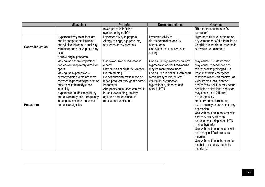
136
Midazolam Propofol Dexmedetomidine Ketamine
fever, propofol infusion syndrome, hyperTG6
RR and transcutaneous O2 saturation2
Contra-indication
▪ Hypersensitivity to midazolam and its components including benzyl alcohol (cross-sensitivity with other benzodiazepines may exist)
▪ Narrow-angle glaucoma
▪ Hypersensitivity to propofol ▪ Allergy to eggs, egg products,
soybeans or soy products
▪ Hypersensitivity to dexmedetomidine and its components
▪ Use outside of intensive care setting
▪ Hypersensitivity to ketamine or any component of the formulation
▪ Condition in which an increase in BP would be hazardous
Precaution
▪ May cause severe respiratory depression, respiratory arrest or apnea
▪ May cause hypotension – hemodynamic events are more common in paediatric patients or patients with hemodynamic instability
▪ Hypotension and/or respiratory depression may occur frequently in patients who have received narcotic analgesics
▪ Use slower rate of induction in elderly
▪ May cause anaphylactic reaction, life threatening
▪ Do not administer with blood or blood products through the same IV catheter
▪ Abrupt discontinuation can result in rapid awakening, anxiety, agitation and resistance to mechanical ventilation
▪ Use cautiously in elderly patients; hypotension and/or bradycardia may be more pronounced
▪ Use caution in patients with heart block, bradycardia, severe ventricular dysfunction, hypovolemia, diabetes and chronic HTN
▪ May cause CNS depression ▪ May cause dependence and
tolerance with prolonged use ▪ Post anesthetic emergence
reactions which can manifest as vivid dreams, hallucinations, and/or frank delirium may occur; confusion or irrational behavior may occur up to 24hours postoperatively
▪ Rapid IV administration or overdose may cause respiratory depression
▪ Use with caution in patients with coronary artery disease, catecholamine depletion, HTN and tachycardia
▪ Use with caution in patients with cerebrospinal fluid pressure elevation
▪ Use with caution in the chronic alcoholic or acutely alcoholic intoxicated
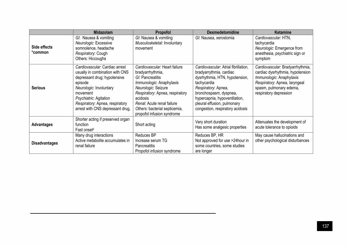
137
Midazolam Propofol Dexmedetomidine Ketamine
Side effects *common
GI: Nausea & vomiting Neurologic: Excessive somnolence, headache Respiratory: Cough Others: Hiccoughs
GI: Nausea & vomiting Musculoskeletal: Involuntary movement
GI: Nausea, xerostomia Cardiovascular: HTN, tachycardia Neurologic: Emergence from anesthesia, psychiatric sign or symptom
Serious
Cardiovascular: Cardiac arrest usually in combination with CNS depressant drug; hypotensive episode Neurologic: Involuntary movement Psychiatric: Agitation Respiratory: Apnea, respiratory arrest with CNS depressant drug,
Cardiovascular: Heart failure bradyarrhythmia, GI: Pancreatitis Immunologic: Anaphylaxis Neurologic: Seizure Respiratory: Apnea, respiratory acidosis Renal: Acute renal failure Others: bacterial septicemia, propofol infusion syndrome
Cardiovascular: Atrial fibrillation, bradyarrythmia, cardiac dysrhythmia, HTN, hypotension, tachycardia Respiratory: Apnea, bronchospasm, dyspnea, hypercapnia, hypoventilation, pleural effusion, pulmonary congestion, respiratory acidosis
Cardiovascular: Bradyarrhythmia, cardiac dysrhythmia, hypotension Immunologic: Anaphylaxis Respiratory: Apnea, laryngeal spasm, pulmonary edema, respiratory depression
Advantages ▪ Shorter acting if preserved organ
function ▪ Fast onset4
▪ Short acting ▪ Very short duration ▪ Has some analgesic properties
▪ Attenuates the development of acute tolerance to opioids
Disadvantages
▪ Many drug interactions ▪ Active metabolite accumulates in
renal failure
▪ Reduces BP ▪ Increase serum TG ▪ Pancreatitis ▪ Propofol infusion syndrome
▪ Reduces BP, HR ▪ Not approved for use >24hour in
some countries, some studies are longer
▪ May cause hallucinations and other psychological disturbances

138
References and Suggested Readings Arroliga A, Frutos-Vivar F, Hall J, et al. Use of sedatives and neuromuscular blockers in a cohort of patients
receiving mechanical ventilation. Chest. 2005. 128(2): 496-506. Barr J, Fraser GL, Puntillo K, et al. Clinical practice guidelines for the management of pain, agitation, and delirium
in adult patients in the intensive care unit. Crit Care Med. 2013. 41(1): 263-306. Becker DE. Pharmacodynamic considerations for moderate and deep sedation. Anesth Prog. 2012. 59(1): 28-42. Devlin JW, Skrobik Y, Gélinas C, et al. Clinical practice guidelines for the prevention and management of pain,
agitation / sedation, delirium, immobility, and sleep Disruption in adult patients in the ICU. Crit Care Med. 2018. 46(9): e825-e873.
Girard TD, Pandharipande PP, Ely EW. Delirium in the intensive care unit. Crit Care. 2008. 12(S3): S3. Hughes MA, Glass PSA, Jacobs JR. Context-sensitive half-time in multicompartment pharmacokinetic models for
intravenous anesthetic drugs. Anesthesiology 1992. 76: 334-341. Jackson DL, Proudfoot CW, Cann KF, Walsh TS. The incidence of sub-optimal sedation in the ICU: A systematic
review. Crit Care. 2009. 13(6): R204. Kronenberg RH. Ketamine as an analgesic. J Pain Palliat Care Pharmacother. 2002. 16(3): 27-35. Ministry of Health Malaysia & Malaysian Society of Intensive Care. Management protocols in ICU. 2019. Reschreiter H, Maiden M, Kapila A. Sedation practice in the intensive care unit: A UK national survey. Crit Care.
2008. 12(6): R152 Santonocito C, Noto A, Crimi C, Sanfilippo F. Remifentanil-induced postoperative hyperalgesia: Current
perspectives on mechanisms and therapeutic strategies. Local Reg Anesth. 2018. 11:15-23. Sessler CN, Grap MJ, Ramsay MA. Evaluating and monitoring analgesia and sedation in the intensive care unit.
Crit Care. 2008. 12(S3): S2. Sessler CN, Varney K. Patient-focused sedation and analgesia in the ICU. Chest. 2008. 133(2): 552-565. Shehabi Y, Ruettimann U, Adamson H, Innes R, Ickeringill M. Dexmedetomidine infusion for more than 24-hours
in critically ill patients: Sedative and cardiovascular effects. Intensive Care Med. 2004. 30(12): 2188-2196.

139
CHAPTER 16
Special Populations: Obese, Hypoalbuminaemia & Burn
Obese
besity is defined by the CDC as a BMI of >30kg/m2, and morbid obesity is defined as a BMI of >40kg/m2. Physiological changes in obesity patient, can alter pharmacodynamic and pharmacokinetic of a drug which includes dramatically increased adipose tissue, slightly increased lean tissue mass, increased cardiac output, increased glomerular filtration rate, and fatty infiltration of liver. A higher proportion of body tissue can influence drug with lipophilic properties whereas increased organ mass, lean body mass, and blood volume in obesity can affect hydrophilic medications. Table 16.1 listed weight-based dosing scalar recommendations for commonly used drugs in critically ill patient. Failure to adjust doses in obesity may result either in sub therapeutic failure or increased toxicity. Table 16.1: Weight-based dosing scalar recommendations for commonly used drugs in critically ill patient
Ideal body weight IBW (as described by Devine 1974) Male IBW (kg) = 50 kg + [2.3 x (Ht-60)]
Female IBW (kg) = 45.5 kg + [2.3 x (Ht-60)] (Height measures in inches)
Body Mass Index (BMI) BMI = Weight (kg) / Height (m)2
Lean Body Weight (LBW2005) (formula by Janmahasatian et al. 2005) Male LBW = [9270 x weight (kg)] / [6680+216 x BMI]
Female LBW = [9270 x weight (kg)] / [8780+244 x BMI]
Adjusted Body Weight (ABW) ABW = IBW + 0.4(ActualBW-IBW)
Estimation of Creatinine Clearance in Obese Patient
Overestimation or underestimation of clearance can occur in obesity when considering actual body weight versus ideal body weight, respectively. The Cockcroft-Gault equation is commonly used to calculate glomerular filtration rate (GFR) in lean patients, however its use in obesity is questionable due to the disparity between muscle mass and body weight ratio observed in obesity. The Salazar-Corcoran equation takes into account multiple factors to provide a better estimation of ClCr in obesity including serum creatinine, gender, actual weight, age, and height. Although some drugs have established dosing adjustments for obesity, it remains unknown for the majority of drugs if dosing adjustment is warranted.
Figure 16.1: Salazar-Corcoran equation
ClCr (Male) = (137-age) x [(0.285xWt) + (12.1xHt2)] / (51xSCr) ClCr (Female) = (146-age) x [(0.287xWt) + (9.74xHt2)] / (60xSCr)
*Wt = actual body weight in kg, Ht = height in meters, SCr = serum creatinine in mg/dl
O
PA
RT
IV: S
PE
CIA
L C
ON
SID
ER
AT
ION
S IN
CR
ITIC
AL
LY
ILL
PA
TIE
NT
S

140
Table 16.2: Dosing approaches of selected drugs in obese patient.
Drugs Suggested dosing weight
Acyclovir IBW or ABW
Aminoglycosides ABW
Amphotericin B ActualBW for conventional preparation; IBW for lipid preparation
Atracurium IBW
Benzodiazepines IBW
Ciprofloxacin ABW
Colistimethate IBW, dosage expressed in terms of colistin
Daptomycin ActualBW
Digoxin IBW
Enoxaparine ActualBW
Additional recommendation: - Up to 150 kg
- VTE prophylaxis: if BMI ≥ 40 mg/m2 increase dose by 30% - VTE treatment: avoid once-daily dosing if BMI > 27 kg/m2 (e.g do not use
1.5 mg/kg daily)
Erythromycin IBW
Fentanyl LBW
Fluconazole ActualBW
Additional recommendation: Consider higher doses in obese patient
Flucytosine IBW
Ganciclovir ABW
Heparin - unfractionated ABW
Immunoglobulin (IVIG) ABW
Methylprednisolone IBW
Oseltamivir Use standard dosing (no adjustment for obesity)
Pancuronium IBW
Phenytoin LD = IBW+1.33(TBW-IBW), MD = IBW
Additional recommendation: Drug level monitoring
Propofol Induction: LBW, Maintenance: ActualBW
Remifentanil LBW
Rocuronium IBW
Succinylcholine ActualBW
Thiopental Induction: LBW, Maintenance: ActualBW
Trimethoprim/sulfamethoxazole IBW or ABW (if BMI ≥ 35 kg/m2)
Vancomycin Total body weight for loading doses and class I obese; adjusted body weight for class II and III obese
Voriconazole ABW
ActualBW = actual body weight; LBW =Lean Body Weight; IBW = ideal body weight; ABW = adjusted body weight Hypoalbuminemia Critical care patients often have capillary leak, increased cardiac output and altered protein levels which can have profound effects on the volume of distribution (Vd) and clearance (CL) of antibacterial agents, both of which may affect the pharmacokinetics (PK) and pharmacodynamics (PD) of the drug. Low serum albumin levels are very common in critically ill patients, with reported incidences as high as 40–50%. The effects of hypoalbuminaemia on pharmacokinetics are driven by the decrease in the extent of antibacterial bound to albumin, which increases the unbound fraction of the drug. Unlike the fraction bound to plasma proteins, the unbound fraction is the only fraction available for distribution and clearance from the plasma (central compartment).
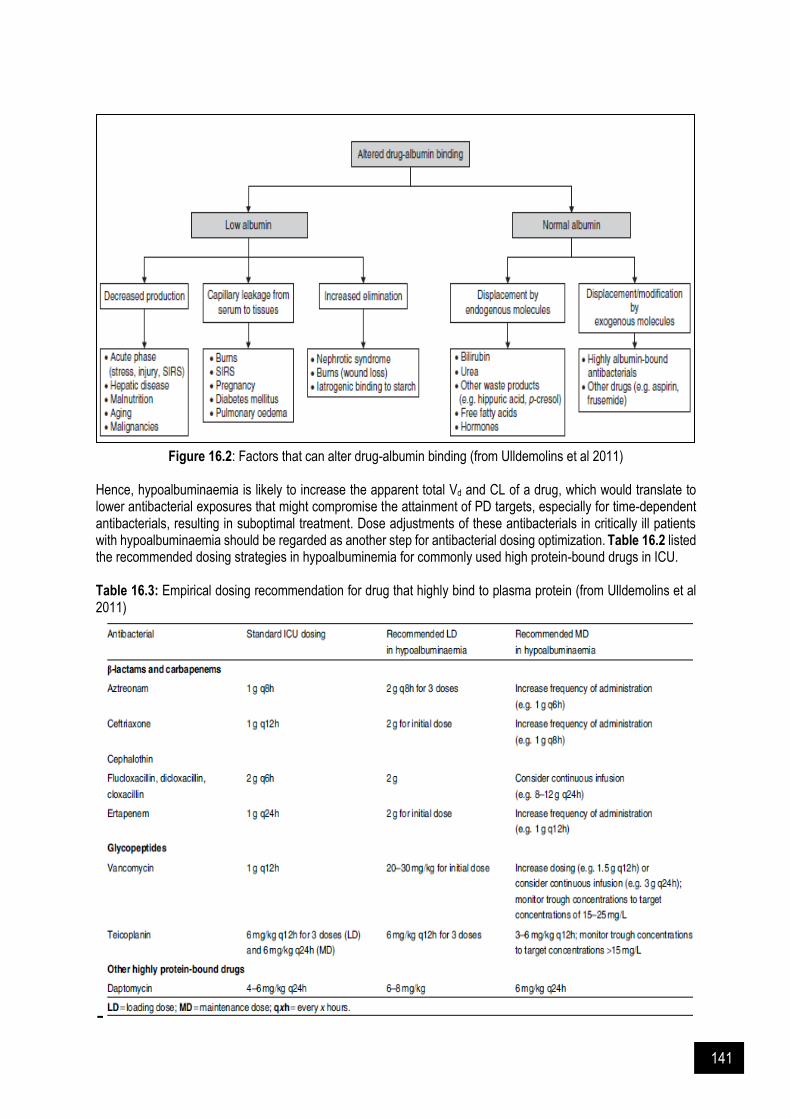
141
Figure 16.2: Factors that can alter drug-albumin binding (from Ulldemolins et al 2011) Hence, hypoalbuminaemia is likely to increase the apparent total Vd and CL of a drug, which would translate to lower antibacterial exposures that might compromise the attainment of PD targets, especially for time-dependent antibacterials, resulting in suboptimal treatment. Dose adjustments of these antibacterials in critically ill patients with hypoalbuminaemia should be regarded as another step for antibacterial dosing optimization. Table 16.2 listed the recommended dosing strategies in hypoalbuminemia for commonly used high protein-bound drugs in ICU.
Table 16.3: Empirical dosing recommendation for drug that highly bind to plasma protein (from Ulldemolins et al 2011)

142
Burn Major burn injury is defined as a surface area involving >20–30% TBSA, and classically is characterized by a bi-phasic systemic response. During the first 48 h, the severity and extent of local tissue injury results in inflammatory mediators being released into the systemic circulation. These induce specific haemodynamic alternations, including increased capillary permeability. Peripheral and splanchnic vasoconstriction, and myocardial depression. Large volumes of protein rich fluid is lost into the interstitial space resulting in relative intravascular hypovolaemia, systemic hypotension and organ hypoperfusion.
The second phase of major burns injury is hypermetabolic state, which is characterize by hyperdynamic circulation, increased tissue perfusion, and hypoalbuminemia that results from significant increases in catecholamines cortisol, glucagon, and prostaglandins. This hypermetabolic and hyperinflammatory physiologic response maybe sustained 1 to 3 years after initial burn injury.
Figure 16.3: Recommended drug dosing approaches in critically ill burn injury patients
(from Udy et al 2017).

143
Table 16.4: Proposed dosing regimens of various antibiotics in burn injury patients
Antibiotic(s) Empirical dosage PK/PD target In case of moderate-severe renal impairment (without renal replacement therapy)
Amikacin Loading dose 30 mg/kg Maintenance dose based
on TDM, usually once daily
Cmax / MIC ≥8–10 AUC / MIC >70 Cmin <2 mg/L
Maintain high doses if possible; prolong dosing
interval (36- to 48-h intervals are acceptable)
Gentamicin Loading dose 7–10 mg/kg Maintenance dose based
on TDM, usually once daily
Cmax / MIC ≥10 AUC / MIC >70 Cmin <0.5 mg/L
Maintain high doses if possible; prolong dosing
interval (36- to 48-h intervals are acceptable)
Meropenem 1 g at 0, 4 and 8 h, thereafter 1 g every 8 h
Consider prolonged infusion
(1 g infused over 3 h)
fT>MIC 40%; fT>MIC 100% if
immunocompromised
In case of intermittent dosing, dose can be reduced or dosing interval prolonged.
Piperacillin / Tazobactam
4/0.5 g at 0, 3 and 6 h, thereafter 4/0.5 g every
6H. Consider continuous infusion
fT>MIC 50% (piperacillin); fT>MIC 100% if
immunocompromised
In case of intermittent dosing, dose can be reduced or dosing interval prolonged.
Ciprofloxacin 400 mg/8 h or 600 mg every 12H
AUC / MIC ≥125 Cmax / MIC ≥8
Maintain high dose if possible while prolonging
dosing interval
Vancomycin Loading dose 30 mg/kg, thereafter 1 g every 8H,
1.5g every 12H or continuous infusion 30–40
mg/kg/day
Cmin 15–20 mg/L or steady state concentration
20-25 mg/L AUC / MIC >400
Reduce total daily dose
Colistin Loading dose 9 million international units (IU) colistimethate sodium
(CMS), thereafter 9 million IU/day divided in 2–3
doses
Steady state concentration ≥2 mg/L
fAUC / MIC >25–35
Reduce dose or prolong dosing interval
Tigecycline Loading dose 100 mg, thereafter 50 mg every
12H or a 200 mg loading dose followed by 100 mg
every 12H
Varying AUC / MIC targets for different pathologies (e.g
pneumonia, intra-abdominal infection, soft tissue infections
No adaptations required
Linezolid 600 mg every 12H AUC / MIC ≥85 T>MIC 85%
Cmin >6 mg/L
In case of severe renal impairment: No dosage
adjustment necessary, use with caution, consider TDM if
possible.

144
References and Suggested Readings
Abernethy DR, Greenblatt DJ, Smith, TW. Digoxin disposition in obesity: Clinical pharmacokinetic investigation. Am Heart J. 1981. 102(4): 740-4.
Cota JM, FakhriRavari A, Rowan MP, Chung K, Murray CK, Akers KS. Intravenous antibiotic and antifungal agent pharmacokinetic-pharmacodynamic dosing in adults with severe burn injury. Clin Ther. 2016. 38(9): 2016-31.
Dunn TE, Ludwig EA, Slaughter RL, Camara DS, Jusko WJ. Pharmacokinetics and pharmacodynamics of methylprednisolone in obesity. Clin Pharmacol Ther. 1991. 49(5): 536-49.
Erstad BL. Dosing of medications in morbidly obese patients in the intensive care unit setting. Intensive Care Med. 2004. 30(1): 18-32.
Hume, R. Prediction of lean body mass from height and weight. J Clin Pathol. 1966. 19(4): 389–391. Ingrande J, Lemmen H. Dose adjustment of anaesthetics in the morbidly obese. Br J Anaesth. 2010. 105 (S1): i16
- i23. Janmahasatian S, Duffull SB, Ash S, Ward LC, Byrne NM, Green B. Quantification of lean body weight. Clin
Pharmacokinet. 2005. 44: 1051– 65. Lee JB, Winstead PS, Cook AM. Pharmacokinetics alterations in obesity. Orthopedics. 2006. 29: 984. Myzienski AE, Lutz MF, Smythe MA. Unfractionated heparin dosing for venous thromboembolism in morbidly
obese patients: Case report and review of the literature. Pharmacotherapy. 2010. 30(3): 324. Pai, MP, Lodise Jr TP. Oseltamivir and oseltamivir carboxylate pharmacokinetics in obese adults: Dose
modification for weight is not necessary. Antimicrob Agents Chemother. 2011. 55(12): 5640-5. Shah S, Barton G, Fischer A. Pharmacokinetic considerations and dosing strategies of antibiotics in the critically
ill patient. J Intensive Care Soc. 2015.16(2): 147–153. Udy AA, Roberts JA, Jeffrey Lipman, Stijn B. The effects of major burn related pathophysiological changes on the
pharmacokinetics and pharmacodynamics of drug use: An appraisal utilizing antibiotics. Adv Drug Delivery Rev. 2018; 65–74
Ulldemolins M, Rello J, Paterson DL, Lipman J. The effects of hypoalbuminaemia on optimizing antibacterial dosing in critically ill patients. Clin Pharmacokinet. 2011. 50(2): 99-110.
Van Kralingen, S., van de Garde., E.M., Knibbe, C.A. Comparative evaluation of atracurium dosed on ideal body weight vs. total body weight in morbidly obese patients. Br J Clin Pharmacol 2011. 71(1): 34 - 40.
Wurtz R, Itokazu G, Rodvold K. Antimicrobial dosing in obesity. Clin Infect Dis. 1997. 25: 112C.
145
CHAPTER 17
Critically Ill Trauma Patient
ajority of critically ill trauma patients admitted to ICU with traumatic brain injury (TBI). The severity of TBI commonly graded using the Glasgow Coma Scale (GCS), with 3 being the worst GCS, and 15 is the best GCS. Mild brain injury might present with GCS ≥13, moderate brain injury with GCS of 9-12, and severe brain injury with GCS ≤8.
Management of Critically Ill Trauma Patients Prior to ICU admission The assessment and treatment start before a patient reaches the hospital. At pre-hospital, the primary goal of treatment is to prevent and treat hypotension (target SBP <90 mmHg) and hypoxia (target PaO2 <60 mmHg). These are the major causes of secondary injury after TBI, within the first 24 hours. In patients with severe TBI or a SpO2 <90% despite supplemental oxygen, airway protection, and endotracheal intubation are recommended. While for non-intubated patients, SpO2 is maintaining at >90 to 93%. Adequate fluid resuscitation using isotonic crystalloids could assist in blood pressure control. Patients with TBI might be assumed to have spinal fractures. Thus, stabilizing and immobilizing the spine during transport should be done with caution. At the emergency department, other required monitorings included the vital signs, evaluation and management of increased intracranial pressure (ICP), and all necessary blood works. Tranexamic acid - the CRASH-3 trial, a randomized, placebo-controlled trial done in 175 hospitals in 29 countries, involving 9202 adults, showed that tranexamic acid is safe in patients with TBI and that treatment within 3 hours of injury reduces head-injury related death. They recommend IV tranexamic acid 1 g, infused over 10 minutes, followed by an IV infusion of 1 g over 8 hours, to patients who present with moderate to severe TBI within 3 hours of injury. Surgical treatment - indications for surgical treatment are based upon neurologic status and findings on CT brain. ICU management The main objective of critical care management for severe TBI is to limit secondary brain injury. Generally, treatment efforts are aim at ICP management, maintenance of cerebral perfusion, as well as optimizing oxygenation and blood pressure and managing temperature, glucose, seizures, and other potential secondary brain insults. Hemodynamic management
Fluids – Isotonic fluids (normal saline) should be used to maintain euvolemia. Albumin has been associated with increased mortality in the SAFE clinical trial. Although balanced crystalloid solutions such as lactated Ringer’s are used to decrease the risk of acute kidney injury in other critically ill patients, they are relatively hypotonic and may worsen cerebral edema. In the SMART ICU trial, no benefit was seen with the use of balanced fluids over normal saline in TBI patients.
Blood pressure – Systolic BP ≥100 mmHg in patients 50-69 years old and ≥110 mmHg for patients 15 to 49 or >70 years old should be maintained.
Cerebral perfusion pressure – Cerebral autoregulation is disrupted in 30% of TBI patients. In these patients, a rise in MAP can lead to elevated ICP due to increased cerebral blood volume and hyperaemia, while drops in MAP may be associated with hypoperfusion and ischemia.
M

146
Cerebral perfusion pressure (CPP) = Mean arterial pressure (MAP) – Intracranial pressure (ICP)
Episodes of hypotension (low MAP), raised ICP, or low CPP is associated with secondary brain injury and worse clinical outcomes. The recommended goal of CPP is 60 to 70 mmHg. Optimize the CPP by treating ICP elevation if preferred over elevating the MAP with vasopressors. Hypertension is more likely to worsen cerebral edema when protective autoregulation is impaired. Ventilation Hypoxia must be avoided, and PaO2 is maintaining >60 mmHg. Hyperventilation is not recommended as a routine approach to reduce ICP. It can decrease PaCO2 and cause cerebral vasoconstriction, decrease the cerebral blood volume and ICP. Although hyperventilation can reduce ICP, it should be avoided, especially in the first 24 to 48 hours. Mild to moderate hyperventilation can be considered later, but PaCO2 <30 mmHg should be avoided, except as a temporary intervention to resolve ICP crises. Antiepileptic drugs Antiepileptic is recommended for the prevention and treatment of post-traumatic seizures in severe TBI. Patients with TBI who develop seizures will require ongoing therapy with antiepileptic. The use of antiepileptic drugs in the acute management of TBI has been shown to reduce the incidence of early-seizures but does not prevent the later development of epilepsy. Early seizures can lead to a high fatality rate in TBI. Phenytoin or levetiracetam is recommended for 7 days as seizure prophylaxis. However, studies revealed that patients treated with levetiracetam have improved six-month functional outcomes and suffered less adverse drug events compared to phenytoin. Venous thromboembolism (VTE) prophylaxis Patients with TBI are at increased risk of venous thromboembolism (VTE). All patients with TBI upon admission must be put on mechanical prophylaxis using intermittent pneumatic compression. VTE prophylaxis using the pharmacological agents (chemoprophylaxis) is suitable in most of TBI patients. Chemoprophylaxis agents can be given either the unfractionated heparin 5000 units BD or enoxaparin 40mg OD. The risk of VTE is high in the first 24 to 48 hours. Therefore, chemoprophylaxis has to be weighed against the potential risk of hemorrhage expansion. Coagulopathy management Approximately 30% of patients with severe TBI demonstrate coagulopathy. It is associated with an increased risk of hemorrhage enlargement, poor neurologic outcomes, and death. Vitamin K is the antidote used to treat coagulopathy caused by medication (e.g warfarin or antiplatelet agents). Fresh frozen plasma (FFP) and/or prothrombin complex concentrate (PCC) are used to treat other causes of coagulopathy. Glucose management The incidence of hypo- and hyperglycemia must be avoided in patients with severe TBI. Both are associated with worsened outcome in a variety of neurologic conditions, including TBI. Target glucose level is suggested from 8-10 mmol/L. However, intensive insulin therapy to achieve this target is not recommended. Tight glycemic control was associated with reduced cerebral glucose availability and elevated lactate: pyruvate ratio, which in turn, is associated with increased mortality. This may also cause a greater risk of hypoglycaemia and worse outcomes in other critically ill patients. Temperature management Fever is associated with worse outcome after stroke and probably severe head injury. Fever also worsens ICP control through an increase in metabolic demand, blood flow and blood volume. Maintenance of normothermia can be attempted using antipyretics, surface-cooling devices or endovascular temperature management catheters. Paroxysmal sympathetic hyperactivity
147
Paroxysmal sympathetic hyperactivity may occur in about 10% of patients with severe TBI. It is characterised by episodes of hypertension, tachycardia, tachypnea, hyperthermia, diaphoresis, increased tone and posturing of varying severity that are often but not always triggered by external stimuli. Abortive and preventive medications may be applied. Nutritional support Basic nutritional goal should be attained no later than 5-7 days from injury. Early enteral nutrition may decrease rates of pneumonia and mortality following TBI. Intracranial pressure (ICP) Management Elevated ICP is associated with increased mortality and worse outcome. Initial treatment such as head of bed elevation to 30o, keeping neck in neutral position and loosening of neck braces if too tight should be instituted as soon as possible. This is to permit adequate venous drainage from the brain to allow cerebral perfusion. Impending cerebral herniation Clinical signs of cerebral herniation include significant papillary asymmetry, unilateral or bilateral fixed and dilated pupils, decorticate or decerebrate posturing, respiratory depression and the ‘Cushing triad’ of hypertension, bradycardia and irregular respiration. In such patients, the following steps must be taken immediately:
Endotracheal intubation, if not already performed.
Elevation of head of bed to 30o to 45o, with the head maintained in the neutral position.
Brief hyperventilation to a pCO2 of approximately 30 mmHg, as it can rapidly and reliably lower the ICP in cerebral herniation. However, prolonged use is not recommended.
A bolus dose of an osmotic agent can transiently reverse cerebral herniation. Mannitol 1 – 1.5g/kg or 23.4% NaCl 30 to 60 mL over 10 minutes. 23.4% NaCl is preferred over mannitol as the latter may result in volume depletion, especially in patients with ongoing blood loss, hemodynamic instability or renal failure. That being said, 23.4% NaCl can provoke transient but profound hypotension.
Aim MAP 80 to 100 mmHg. Both fluid resuscitation and vasopressor use may be necessary to maintain MAP.
These interventions are temporary measures while awaiting more definitive interventions, like neurosurgical treatment ICP and CPP monitoring ICP monitoring is started in severe TBI (e.g GCS ≤ 8) and an abnormal CT scan showing evidence of mass effect from lesions such as hematomas, contusions or swelling. ICP monitoring is also indicated in severe TBI if 2 of the following features are present: age >40 years, motor posturing, systolic BP < 90 mmHg. An ICP goal ≤ 22 mmHg is recommended for survival and favourable outcome of TBI. Cerebrospinal fluid drainage CSF drainage is the first method in lowering ICP in patients with a ventricular catheter. Drainage may be continuous or intermittent, with studies favouring the former; however excessive drainage can lead to ventricular collapse. Sedation and analgesia TBI patients with significant ICP elevation is preferred to have a sedative agent together with an opioid infusion. Sedation and analgesia may lower ICP by reducing cerebral metabolic demand, cerebral blood flow and cerebral blood volume. Sedation may also improve ventilator asynchrony and blunt sympathetic responses of hypertension and tachycardia. Fentanyl infusions are commonly used as analgesia in this setting, while propofol is the preferred sedative. Fentanyl has greater efficacy than morphine and is able to minimize hemodynamic instability. Propofol is able to decrease cerebral metabolic demand and ICP and its short duration of action allows for intermittent clinical neurologic assessment. It also has neuroprotective effect. However, its side effects such as hypotension, dyskalemia and propofol infusion syndrome need to be taken into consideration.
148
Neuromuscular blockade may decrease ICP but they are generally discouraged as they may prolong neuromuscular weakness and delay weaning from mechanical ventilation. Osmotic therapy Intravascular injection of hyperosmolar agents, like hypertonic saline and mannitol, creates an osmolar gradient, drawing water across the blood-brain barrier. This leads to a decrease in brain volume and a decrease in ICP. The effect of hyperosmolar therapy diminished with time, as a compensatory increase in brain osmolarity occurs within 24 hours. Thus, they should be weaned slowly after prolonged use to prevent a reversal in osmotic gradient and rebound cerebral edema.
Hypertonic saline – NaCL 3% (infusion) to 23.4% (bolus). Target sodium level is between 145 to 155 mmol/L and the saline should be infused via central line.
Mannitol – Bolus 0.25-1g/kg 4-6 hourly. Benefit of hypertonic saline over mannitol is that volume depletion and hypovolemia do not occur with hypertonic saline. Also, it is less likely to leak into brain tissue. Its potential side effects include circulatory overload, pulmonary edema and non-anion gap metabolic acidosis. Mannitol, on the other hand, may leak into brain tissue in patients with disrupted blood-brain barrier, leading to reversal of osmolar gradient and rebound cerebral edema. Although theoretical, it may be a serious concern. No studies have shown superiority of one over the other to improve outcomes like mortality or functional recovery. Electrolytes and fluid balance should be monitored. Hypernatremia is associated with increased mortality in severe TBI. Its association with diabetes insipidus is a marker of more extensive brain injury that includes the hypothalamic-pituitary axis. Renal function tests should be checked as mannitol may cause dehydration and acute kidney injury. Refractory ICP elevation This can be managed with decompressive craniectomy, barbiturate coma or hypothermia
Barbiturate coma – The use of barbiturate coma has not been shown to improve TBI outcomes, but it has effectively reduced ICP. IV thiopentone loading dose: 2-3mg/kg, maintenance dose: 3-5 mg/kg/hr. Maximum dose 15mg/kg/hr.
Thiopentone is associated with morbidity. It has long half-life which delays neurologic examination and determination of brain death. Other side effects include hypotension, adynamic ileus, poor pulmonary mucus clearance, ventilator-associated pneumonia and severe metabolic acidosis from propylene-glycol toxicity due to thiopentone infusions.
Hypothermia – Potential to reduce ICP as well as provide neuroprotection and prevent secondary brain injury. However, it has not been convincingly shown to improve outcomes.
Neuroprotection To date, no neuroprotective agents or strategies, including induced hypothermia have been shown to produce improved outcome. The use of glucocorticoid therapy following head trauma was found to be harmful in a large trial of patients with moderate to severe TBI.

149
ICP: intracranial pressure; TBI: traumatic brain injury; CPP: cerebral perfusion pressure; PbtO2: brain tissue oxygen; pO2: partial pressure of oxygen; pCO2: partial pressure of carbon dioxide; RASS: Richmond Agitation-Sedation Scale; CSF: cerebrospinal fluid; CT: computed tomography; FiO2: fraction of inspired oxygen.* If advanced monitoring available.
Figure 17.1: Stepwise approaches of elevated ICP in patients with severe TBI (Glasgow Coma Scale <9) (from Cadena et al 2017 & UpToDate 2020)

150
References and Suggested Readings Bratton SL, Chestnut RM, Ghajar J, McConnell FH, Harris OA, Hartl R, Manley GT, Nemecek A, Newell DW,
Rosenthal G, Schouten J. Guidelines for the management of severe traumatic brain injury. J Neurotrauma. 2007. 24: S7-13.
Cadena R, Shoykhet M, Ratcliff JJ. Emergency neurological life support: Intracranial hypertension and herniation. Neurocrit Care 2017. 27: 82.
Carney N, Totten AM, O'Reilly C, Ullman JS, Hawryluk GW, Bell MJ, Bratton SL, Chesnut R, Harris OA, Kissoon N, Rubiano AM. Guidelines for the management of severe traumatic brain injury. Neurosurgery. 2017. 80(1): 6-15.
Crash-2 Collaborators. Effect of tranexamic acid in traumatic brain injury: A nested randomised, placebo-controlled trial (CRASH-2 Intracranial Bleeding Study). BMJ. 2011. 343: d3795.
Haddad SH, Arabi YM. Critical care management of severe traumatic brain injury in adults. Scand J Trauma, Resusc Emerg Med. 2012. 20(1): 12.
Jeremitsky E, Omert LA, Dunham CM, Wilberger J, Rodriguez A. The impact of hyperglycemia on patients with severe brain injury. J Trauma Acute Care Surg. 2005. 58(1): 47-50.
Koenig MA, Bryan M, Lewin JL, Mirski MA, Geocadin RG, Stevens RD. Reversal of transtentorial herniation with hypertonic saline. Neurology. 2008. 70(13): 1023-9.
Norwood SH, Berne JD, Rowe SA, Villarreal DH, Ledlie JT. Early venous thromboembolism prophylaxis with enoxaparin in patients with blunt traumatic brain injury. J Trauma Acute Care Surg. 2008. 65(5): 1021-7.
Puccio AM, Fischer MR, Jankowitz BT, Yonas H, Darby JM, Okonkwo DO. Induced normothermia attenuates intracranial hypertension and reduces fever burden after severe traumatic brain injury. Neurocrit Care. 2009. 11(1): 82.
Rajajee V. Management of acute moderate and severe traumatic brain injury. UpToDate 2020. Roberts I, Sydenham E. Barbiturates for acute traumatic brain injury. Cochrane Database Syst Rev. 2012. 12(12):
CD000033. Rovlias A, Kotsou S. The influence of hyperglycemia on neurological outcome in patients with severe head injury.
Neurosurgery. 2000. 46(2): 335-42. Rudehill A, Bellander BM, Weitzberg E, Bredbacka S, Backheden M, Gordon E. Outcome of traumatic brain injuries
in 1,508 patients: Impact of prehospital care. J Neurotrauma. 2002. 19(7): 855-68. Sadaka F, Veremakis C. Therapeutic hypothermia for the management of intracranial hypertension in severe
traumatic brain injury: A systematic review. Brain Inj. 2012. 26(7-8): 899-908. Sorrentino E, Diedler J, Kasprowicz M, Budohoski KP, Haubrich C, Smielewski P, Outtrim JG, Manktelow A,
Hutchinson PJ, Pickard JD, Menon DK. Critical thresholds for cerebrovascular reactivity after traumatic brain injury. Neurocrit Care. 2012. 16(2): 258-66.
Szaflarski JP, Sangha KS, Lindsell CJ, Shutter LA. Prospective, randomized, single-blinded comparative trial of intravenous levetiracetam versus phenytoin for seizure prophylaxis. Neurocrit Care. 2010. 12(2): 165-72.

151
CHAPTER 18
Antimicrobial Dosing in Critically Ill Patient: Pharmacokinetic & Pharmacodynamic (PKPD) Consideration
harmacokinetic (PK) is defined as the time-course of drug absorption, distribution, Metabolism and excretion in our body. Meanwhile pharmacodynamics (PD) describes the relationship between drug concentrations and pharmacological effects. Sometimes PK refer as “what the body does to the drugs” while PD refer as “what the drug does to the body”. Hence, the PK/PD describes the relationship between drug dose and pharmacological effects with changes in drug concentrations leading to different pharmacological effects.
Figure 18.1: Basic drug pharmacokinetics: absorption, distribution, metabolism and elimination In the presence of critical illness, it may affect all process which is absorption, distribution, metabolism and elimination of drugs thereby significantly influencing the PK of drugs. Hence, in critical illness the disposition of many drugs changes, resulting vary concentrations across patients and within the same patient during different phases of the disease and leading to unpredictable pharmacological and toxicological effects. Thus, when standard dosing regimens are used in critically ill patients with assumptions that drugs information dosing is appropriate, suboptimal concentrations and therapeutic failure may result. On the other hand, supra-therapeutic concentration leads to drug toxicity also possible.
P
152
Pharmacokinetic considerations: Linear & Non-Linear For most drugs, a proportional relationship is observed between concentration at steady-state (Css) or area under the concentration-time curve (AUC) and administered dose. The PK of these drugs is described as linear or dose independent and is characterized by first-order processes. For drugs exhibiting linear PK, semi-log concentration-versus-time plots will be parallel at different doses. In contrast, nonlinearity occurs when the relationships between dose administered and Css, AUC or other PK parameters are not directly proportional. These drugs demonstrate dose-dependent PK that is described by mixed-order, saturable, Michaelis–Menten, or capacity-limited processes. Pharmacodynamic Considerations: Minimum Inhibitory Concentration (MIC) PD refers to the biochemical and physiologic effects of a drug, specifically related to the mechanism of action. It is also related to drug/receptor binding and clinical effect. Based on PK of the drug, its PD properties and the likelihood of treatment success, clinical susceptibility breakpoints are defined to classify bacteria as either susceptible or resistant in reference to measured minimum inhibitory concentration (MIC). The MIC is quantified based on exposure to static concentrations over 18-24 hours and does not provide any information about possibilities of regrowth after an initial kill MIC is not necessarily bactericidal concentration but rather bacteriostatic (inhibit growth) which means that exposure to such concentration may not necessarily kill all of the bacteria. The limitations of MIC in relating a static concentration to clinical efficacy can be addressed by characterizing the relationships between the dynamic antibiotic exposure (pharmacokinetics) and antibiotic effects (e.g microbial killing). In describing these relationships, the MIC should be considered in combination with the exposure of the drug, that is, to relate observed concentrations profiles to the potency of the antibiotic, or MIC. The PD index of antibiotic classes may differ from one another. These describe the optimal ‘shape’ of the concentration-time curve and can be influenced by the presence of a post-antibiotic effect. Three major exposure antibacterial activity relationships have been described for antibiotics based on the observation of correlations between antibacterial activity and either;
duration of antibiotic exposure relative to the MIC
magnitude of exposure relative to MIC
time course of the magnitude of exposure relative the MIC. The three main PK/PD indices associated with the effect of the antibiotics and used for PK/PD analysis to predict the antimicrobial efficacy are:
T>MIC - the percentage of time in one dosing interval which the concentration of the drug was above the MIC
Cmax/MIC - The ratio of peak concentration and MIC
AUC/MIC - The ratio of the 24-h area under the concentration-time curve and MIC
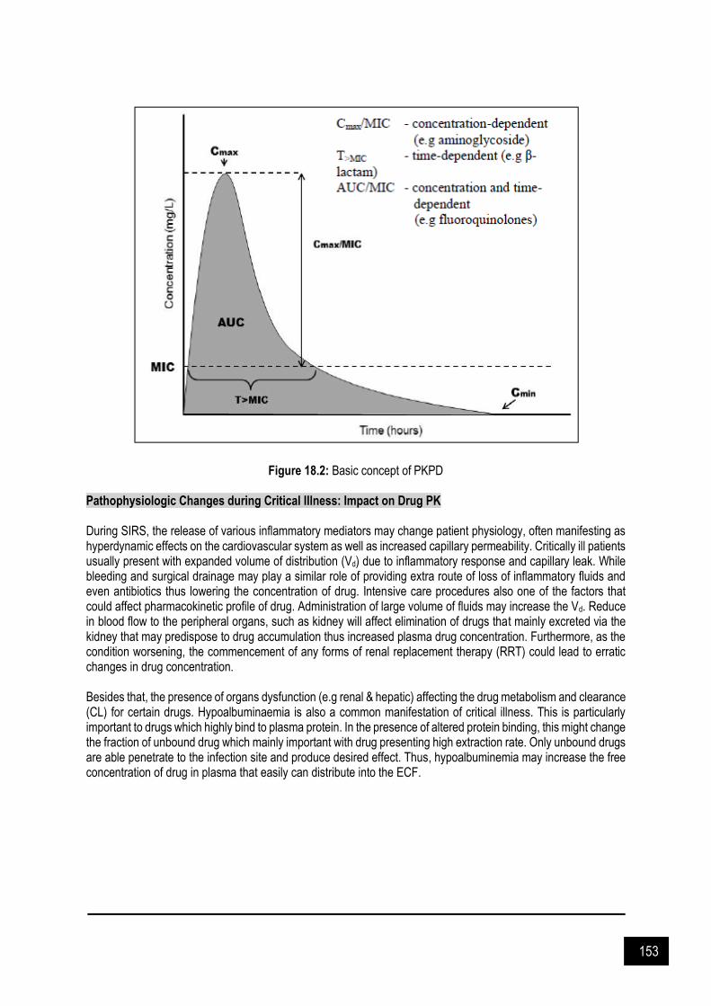
153
Figure 18.2: Basic concept of PKPD Pathophysiologic Changes during Critical Illness: Impact on Drug PK During SIRS, the release of various inflammatory mediators may change patient physiology, often manifesting as hyperdynamic effects on the cardiovascular system as well as increased capillary permeability. Critically ill patients usually present with expanded volume of distribution (Vd) due to inflammatory response and capillary leak. While bleeding and surgical drainage may play a similar role of providing extra route of loss of inflammatory fluids and even antibiotics thus lowering the concentration of drug. Intensive care procedures also one of the factors that could affect pharmacokinetic profile of drug. Administration of large volume of fluids may increase the Vd. Reduce in blood flow to the peripheral organs, such as kidney will affect elimination of drugs that mainly excreted via the kidney that may predispose to drug accumulation thus increased plasma drug concentration. Furthermore, as the condition worsening, the commencement of any forms of renal replacement therapy (RRT) could lead to erratic changes in drug concentration. Besides that, the presence of organs dysfunction (e.g renal & hepatic) affecting the drug metabolism and clearance (CL) for certain drugs. Hypoalbuminaemia is also a common manifestation of critical illness. This is particularly important to drugs which highly bind to plasma protein. In the presence of altered protein binding, this might change the fraction of unbound drug which mainly important with drug presenting high extraction rate. Only unbound drugs are able penetrate to the infection site and produce desired effect. Thus, hypoalbuminemia may increase the free concentration of drug in plasma that easily can distribute into the ECF.
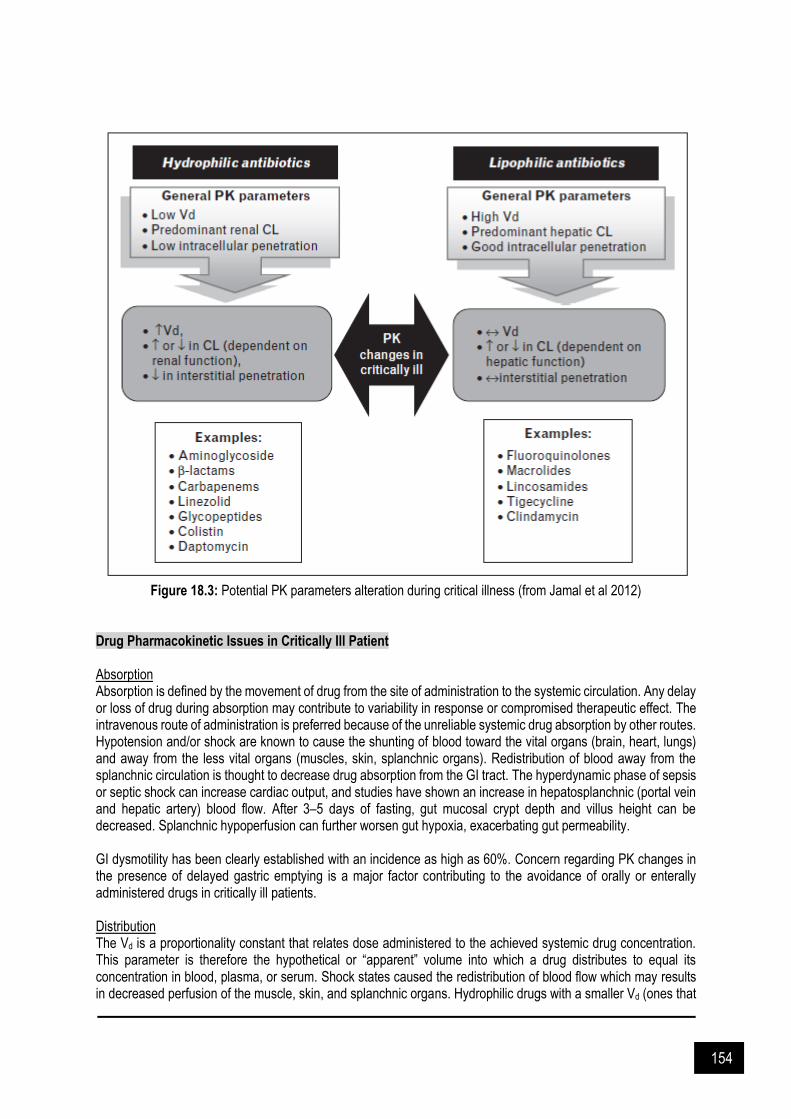
154
Figure 18.3: Potential PK parameters alteration during critical illness (from Jamal et al 2012) Drug Pharmacokinetic Issues in Critically Ill Patient Absorption Absorption is defined by the movement of drug from the site of administration to the systemic circulation. Any delay or loss of drug during absorption may contribute to variability in response or compromised therapeutic effect. The intravenous route of administration is preferred because of the unreliable systemic drug absorption by other routes. Hypotension and/or shock are known to cause the shunting of blood toward the vital organs (brain, heart, lungs) and away from the less vital organs (muscles, skin, splanchnic organs). Redistribution of blood away from the splanchnic circulation is thought to decrease drug absorption from the GI tract. The hyperdynamic phase of sepsis or septic shock can increase cardiac output, and studies have shown an increase in hepatosplanchnic (portal vein and hepatic artery) blood flow. After 3–5 days of fasting, gut mucosal crypt depth and villus height can be decreased. Splanchnic hypoperfusion can further worsen gut hypoxia, exacerbating gut permeability. GI dysmotility has been clearly established with an incidence as high as 60%. Concern regarding PK changes in the presence of delayed gastric emptying is a major factor contributing to the avoidance of orally or enterally administered drugs in critically ill patients. Distribution The Vd is a proportionality constant that relates dose administered to the achieved systemic drug concentration. This parameter is therefore the hypothetical or “apparent” volume into which a drug distributes to equal its concentration in blood, plasma, or serum. Shock states caused the redistribution of blood flow which may results in decreased perfusion of the muscle, skin, and splanchnic organs. Hydrophilic drugs with a smaller Vd (ones that
155
remain in the plasma water volume) may have decreased distribution to parts of the body with decreased blood flow. Critically ill patients can receive significant volumes of IV fluid for resuscitation purposes which could eventually results in increased volumes of total body water and interstitial fluid. Other than that sepsis, burns, ARDS, AKI, heart failure, cirrhosis and surgery may affect drug Vd to various extent. Acid-base disorders presented in critically ill may affect the changes degree of drug ionization as most drugs are either weak acids or bases. Non-ionized drugs penetrate cell membranes more easily than do ionized drugs. Metabolism The predominant location for drug metabolism is the liver, but it can include tissues such as the GI tract, kidneys, lung, and brain. The greatest extent of knowledge regarding drug metabolism, and more importantly changes in critically ill patients, relates to hepatic metabolism. Critically ill patients with AKI have clinically relevant changes in insulin metabolism, as evidenced by increased hypoglycemic events and lower insulin requirements upon developing AKI. Hepatic clearance refers to the volume of blood that is completely cleared of drug by the liver per unit of time. The ability of the liver to metabolize drugs depends on three physiologic parameters: hepatic blood flow, drug protein binding, and the intrinsic activity of hepatic enzymes. Elimination For most drugs, the kidneys are the primary site for excretion of the parent drug, metabolites, or both. Urinary excretion of a drug is dependent on filtration, secretion, and reabsorption. Patients in the ICU may experience increased, decreased, or normal renal excretion of drugs. The state of renal excretion depends on many variables and can change rapidly. Decreased renal excretion of drugs during AKI is the most widely applicable change occurring in critically ill patients. AKI significantly affects the excretion of renally eliminated drugs, and dosing modifications must be made in these situations. Special Considerations in Critically Ill Patients Augmented renal clearance (ARC) Critically ill patient is associated with an elevated cardiac output which causes increased renal blood flow and subsequently increased glomerular filtration. Other factors such as aggressive fluid therapy, use of vasoactive drugs and hypoalbuminemia may also contribute to increases in renal blood flow and glomerular filtration rate. Therefore, there is enhanced clearance of renally cleared antibiotics, a phenomenon referred to as augmented renal clearance (ARC). ARC is defined as creatinine clearance >130 mL/min/1.73 m². Increased elimination through this phenomenon has a particularly marked influence on time-dependent antibiotics, which require maintenance of optimal concentrations for a prolonged time within a dosing interval. Critically ill obese patient (refer Chapter 16) Obesity is not commonly a standalone disease but always linked to other pathologies (such as diabetes, hypercholesterolemia, cardiac failure, renal and hepatic impairment, and so on). Obesity has been associated to antibiotic treatment failure as one of a significant risk factor. However, the need of changes on the standard dosing regimens is still not clear. There is relationship between obesity and higher Vd and CL for several drugs. Larger doses might be needed to achieve the same PD exposures as in non-obese patients. Elderly patient Pharmacokinetic changes in elderly patients include a reduction in the renal and hepatic clearance and increase the volume of distribution of lipid soluble drugs hence prolong the drug elimination half-life. The need of adjustment is not only for renal clearance but also to consider the age of patient in the case of some fluoroquinolones. Tissue penetration Free tissue concentration at the target site which are commonly lower than total plasma concentrations, are responsible for the therapeutic effect. Some studies have demonstrated differences on the limited cerebral penetration of some antibiotics and the implications regarding PK/PD. The ratio of the area under the concentration-time curve in brain to that in plasma for the different antibiotics ranged from 0.01 to 1, and thus the PK/PD analysis based on plasma concentrations might overestimate the antibacterial activity in the cerebral tissue.
156
Recommended Dosing Strategies in Critically Ill Patients Inappropriate drug choice and suboptimal dosing of antibiotics (due to low doses or short durations of treatment) are two major factors affecting and followed by the emergence of drug resistance and consequently poorer clinical outcomes. Thus, good knowledge of the mechanisms involved on the effect of the antibiotics (PD) and the evolution of the antibiotics concentration in the body of the patients (PK). PK/PD analysis integrates them and studies the dosing required to enhance the possibility of success of the antibiotic therapy as well as minimize the side effects and the emergence of resistance. During severe illness, increased exposure may be necessary to achieve desirable clinical outcomes. For instance, even though non–critically ill patients may only require minimal β-lactam antibiotic exposures of 40% to 70%ƒT>MIC, recent clinical studies suggest 100%ƒT>MIC may be a minimum requirement to ensure optimal outcomes in the critically ill patient. Recommended alternative dosing strategies to improve antimicrobial dosing in critically ill patients:
Aggressive dosing, either by administration of a larger front-loading dosing doses or higher dosing regimen
Changing the method of drug administration for certain drug such as beta-lactam antibiotics, from conventional intermittent dosing to a prolong infusion (such as by continuous or extended infusion) has proven to be beneficial in achieving the desired therapeutic target.
Therapeutic drug monitoring (TDM) to guide dosing
PK/PD modeling – in the absence of TDM Table 18.1: Recommended dosing approach for altered antimicrobial PK/PD during critical illness (from Udy et al 2013)
PK alterations Recommended dosing approach
Larger volume of distribution (Vd) Appropriately weight adjusted loading doses
AKI requiring CRRT Individualised patient dosing based on physicochemical properties, intensity of CRRT and residual native renal function TDM
Augmented renal clearance (ARC) Increased total daily dose Consider more frequent dosing (shorter dosing interval) Consider prolong infusion (e.g extended / continuous) TDM
Hypoalbuminaemia (highly protein bound drugs)
Larger loading doses Consider more frequent dosing (shorter dosing interval) Consider prolong infusion (e.g extended / continuous) TDM (unbound concentrations)
Reduced bacterial susceptibility (e.g high MIC)
Increased total daily dose Consider prolong infusion (e.g extended / continuous) Application of PK/PD models TDM (for agreesive dosing during early in the antimicrobial therapy)

157
References and Suggested Readings Asín-Prieto E, Rodríguez-Gascón A, Isla A. Applications of the pharmacokinetic / pharmacodynamic (PK/PD)
analysis of antimicrobial agents. J Infect Chemother 2015. 21(5): 319-329. Blot S, Pea F, Lipman J. The effect of pathophysiology on pharmacokinetics in the critically ill patient - Concepts
appraised by the example of antimicrobial agents. Adv Drug Del Rev 2014. 77: 3-11. Blot SI, Pea F, Lipman J. The effect of pathophysiology on pharmacokinetics in the critically ill patient—concepts
appraised by the example of antimicrobial agents. Adv Drug Deliv Rev 2014. 77: 3-11. Jamal JA, Economou CJ, Lipman J, Roberts JA. Improving antibiotic dosing in special situations in the ICU: burns,
renal replacement therapy and extracorporeal membrane oxygenation. Curr Opin Crit Care 2012. 18: 460-47. McKindley DS, Fabian TC, Boucher BA, Croce MA, Proctor KG. Antibiotic pharmacokinetics following fluid
resuscitation from traumatic shock. Arch Surg 1995. 130: 1321-1328. Roberts J, Abdul-Aziz MH, Lipman J, Mouton J, Vinks A., Felton T. et al. Individualised antibiotic dosing for patients
who are critically ill: Challenges and potential solutions. Lancet Infect Dis 2014. 14(6): 498-509. Roberts J, Taccone F, Lipman J. Understanding PK/PD. Intensive Care Med 2015. 42(11): 1797-1800. Roberts JA, Lipman J. Antibacterial dosing in intensive care: pharmacokinetics, degree of disease and
pharmacodynamics of sepsis. Clin Pharmacokinet 2006. 45: 755-773. Roberts JA, Lipman J. Pharmacokinetic issues for antibiotics in the critically ill patient. Crit Care Med 2009. 37:
840-851. Roberts JA, Paul SK, Akova M, Bassetti M, De Waele JJ, Dimopoulos G. et al. DALI: Defining antibiotic levels in
intensive care unit patients: are current beta-lactam antibiotic doses sufficient for critically ill patients? Clin Infect Dis 2014. 58: 1072-1083.
Sime F, Roberts M, Roberts J. Optimization of dosing regimens and dosing in special populations. Clinl Microbiol Infect. (2015). 21(10): 886-893.
Tängdén T, Ramos Martín V, Felton T, Nielsen E, Marchand S, Brüggemann R, et al. The role of infection models and PK/PD modelling for optimising care of critically ill patients with severe infections. Intensive Care Medicine 2017. 43(7): 1021-1032.
Tsai D, Lipman J, Roberts J. Pharmacokinetic / pharmacodynamic considerations for the optimization of antimicrobial delivery in the critically ill. Curr Op Crit Care 2015. 21(5): 412-420.
Turnidge J.D. The pharmacodynamics of beta-lactams. Clin Infect Dis 1998. 27: 10-22. Udy AA, Roberts JA, Lipman J. Implications of augmented renal clearance in critically ill patients. Nat Rev Nephrol
2011. 7: 539-543. Udy AA, Roberts JA, Lipman J. Clinical implications of antibiotic pharmacokinetic principles in the critically ill.
Intensive Care Med 2013. 39: 2070-2082. Udy AA, Roberts JA, Shorr AF, Boots RJ, Lipman J. Augmented renal clearance in septic and traumatized patients
with normal plasma creatinine concentrations: Identifying at-risk patients. Crit Care 2013. 28: 17(1): R35. Udy AA, Varghese JM, Altukroni M, Briscoe S, McWhinney BC, Ungerer JP, et al. Subtherapeutic initial beta-
lactam concentrations in select critically ill patients: association between augmented renal clearance and low trough drug concentrations. Chest 2012. 142: 30-39.
Varghese J, Roberts J, Lipman J. Antimicrobial pharmacokinetic and pharmacodynamic issues in the critically Ill with severe sepsis and septic shock. Crit Care Clin 2011. 27(1): 19-34.
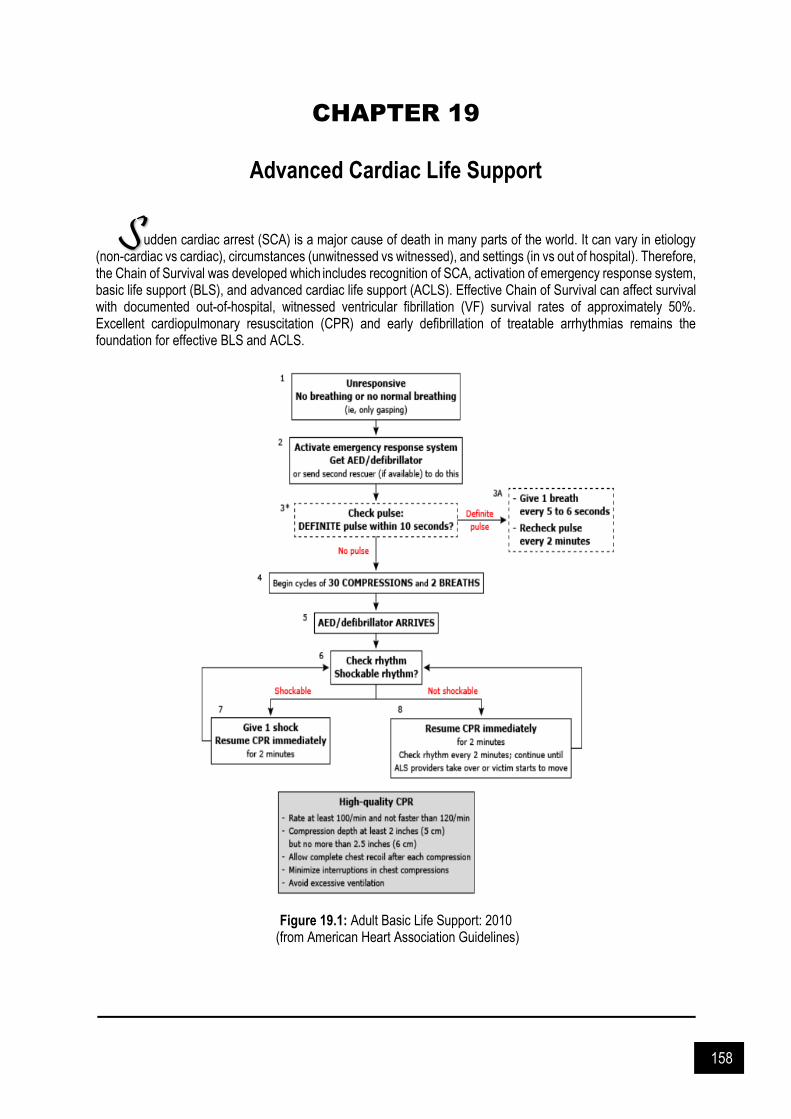
158
CHAPTER 19
Advanced Cardiac Life Support
udden cardiac arrest (SCA) is a major cause of death in many parts of the world. It can vary in etiology (non-cardiac vs cardiac), circumstances (unwitnessed vs witnessed), and settings (in vs out of hospital). Therefore, the Chain of Survival was developed which includes recognition of SCA, activation of emergency response system, basic life support (BLS), and advanced cardiac life support (ACLS). Effective Chain of Survival can affect survival with documented out-of-hospital, witnessed ventricular fibrillation (VF) survival rates of approximately 50%. Excellent cardiopulmonary resuscitation (CPR) and early defibrillation of treatable arrhythmias remains the foundation for effective BLS and ACLS.
Figure 19.1: Adult Basic Life Support: 2010
(from American Heart Association Guidelines)
S
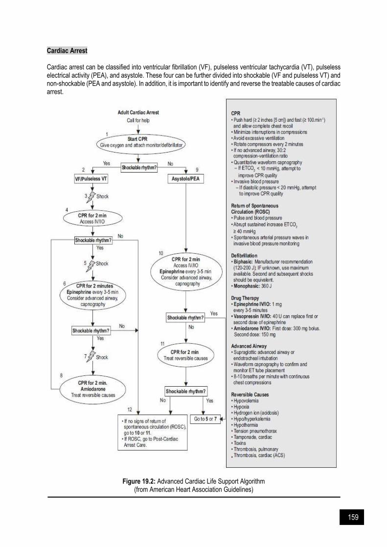
159
Cardiac Arrest Cardiac arrest can be classified into ventricular fibrillation (VF), pulseless ventricular tachycardia (VT), pulseless electrical activity (PEA), and asystole. These four can be further divided into shockable (VF and pulseless VT) and non-shockable (PEA and asystole). In addition, it is important to identify and reverse the treatable causes of cardiac arrest.
Figure 19.2: Advanced Cardiac Life Support Algorithm (from American Heart Association Guidelines)

160
Table 19.1: Treatable causes of cardiac arrest
H’s T’s
Hypoxia Toxins
Hypovolemia Tamponade (cardiac)
Hydrogen ion (acidosis) Thrombosis
Pulmonary embolism
Coronary thrombosis Hypoglycemia
Hypo / hyperkalemia Tension pneumothorax
Hypothermia
Medications administered during cardiac arrest are to increase myocardial blood flow during CPR and to help achieve return of spontaneous circulation (ROSC), with the exception of amiodarone in out-of hospital arrest (Table 19.2). These medications have not been shown to increase cardiac arrest survival rates. CPR and defibrillation should never be interrupted by vascular access insertion and medication delivery. Table 19.2: Medications used during sudden cardiac arrest
Medication Mechanism of
action Dosage, route,
frequency Compatibility & administration
Clinical benefits
Adrenaline α-Adrenergic agonist effects leading to vasoconstriction
1 mg IV/IO q3–5 min 2–2.5 mg ET q3–5 min
Compatibility: D5%, NS Administration: Undiluted, bolus, infusion
Increases coronary and cerebral perfusion pressure during CPR Increases ROSC
Amiodarone Na+/K+/Ca2+ channel and β-receptor antagonist
First dose: 300 mg or 5 mg/kg IV/IO x 1 Second dose: 150 mg IV/IO x 1 Max: 2.2 g/day
Compatibility: D5% Administration: Bolus, infusion
Increases survival to hospital admission compared with lidocaine or placebo for VF/ pulseless VT arrest
Lidocaine Na+ channel antagonist
First dose: 1–1.5 mg/kg IV/IO x 1 Subsequent dosing: 0.5–0.75 mg/kg q5–10 min Max 3-mg/kg cumulative dose
Compatibility: D5%, D5/NS, D5/½NS, NS,
½NS
Administration: Undiluted, bolus, infusion
Increases ROSC and possibly survival to admission for out-of-hospital VF cardiac arrest. No improvement in overall or discharge survival Insufficient evidence to recommend for VF/pulseless VT unless amiodarone unavailable
Magnesium Stops EAD in torsades de pointes by inhibiting Ca2+ channel influx
1–2 g diluted in 10 mL of 5% dextrose or sterile water IV/IO x 1
Compatibility: D5%, NS Administration: Bolus, infusion
Can aid in stopping torsades de pointes in patients with prolonged QT interval
EAD = early afterdepolarization; ET = endotracheal; IO = intraosseously; IV = intravenously; min = minute; q = every. It is recommended to deliver drug through central intravenous access to achieve higher peak and shorter drug circulation period compared to peripheral access. If only peripheral access available, 20 mL bolus of an intravenous fluid should be given after the bolus administration of a drug to improve drug flow from the extremity to the central circulation. When intravenous access cannot be obtained, intraosseous (IO) lines are safe and effective alternative.The dose of drugs administered through IO lines is the same as intravenous lines. Administration and drug delivery through proximal humerus intraosseous access are similar to central venous access while

161
administration and drug delivery though proximal or distal tibial intraosseous access are similar to peripheral venous access. Peak effect of intravenous and IO medications will be delayed around 1 minute during CPR. Studies have shown that naloxone, atropine, vasopressin, adrenaline, and lidocaine can be absorbed through tracheal (ET) administration. However, the serum concentration of medications administered through this route is lesser and unpredictable compared to intravenous and IO route. Recommended doses for tracheal administration are 2 to 2.5 times of the standard intravenous doses and to be diluted in 5-10mL of sterile water or 0.9% isotonic saline before injecting down the tracheal tube. Ventricular Fibrilation (VF) or Pulseless Ventricular Tachycardia (VT) Medication therapy can be considered after one defibrillation and 2 minutes of CPR (one cycle). First line medication is IV adrenaline 1mg every 3-5 minutes with CPR performed concurrently. Adrenaline administration within 2 minutes of defibrillation has been associated with decreased survival. Vasopressin was removed from the algorithm as studies showed no difference in terms of ROSC, survival to hospital admission, and survival to hospital discharged compared to adrenaline. Antiarrhythmic drugs provide little survival benefits in refractory VF or pulseless VT. However, they may be considered in refractory cases defined as unresponsive to defibrillation, CPR, and a vasopressor. Amiodarone is recommended over lidocaine as it increases survival to hospital admission compared to lidocaine. Lidocaine can still be considered if amiodarone is unavailable. Amiodarone is to be administered as intravenous or IO in pulseless patient and as slow intravenous piggyback once pulse is recovered. Pulseless Electrical Activity (PEA) or Asystole PEA and asystole do not respond to defibrillation. CPR should be initiated immediately and first line vasopressor IV Adrenaline should be given as soon as possible. Vasopressin was removed from the algorithm as the benefit is limited. Atropine is no longer recommended due to the lack of therapeutic benefit. The use of sodium bicarbonate is conflicting with most data showing no benefit or poor outcome with its use. The mainstays of restoring acid-base balance are high quality chest compressions and appropriate oxygenation or ventilation. However, cases such as tricyclic antidepressant overdose, bicarbonate-wasting causes of metabolic acidosis, and hyperkalemia, may warrant the use of sodium bicarbonate. With regards to the administration of calcium, there is currently no trial-based evidence on the impact and survival of in- and out-of-hospital cardiac arrest. It can be considered in patients with pre-existing hypocalcemia and signs and symptoms of acute hypocalcemia such as tetany and seizures. Cardiac pacing is ineffective for cardiac arrest and therefore not recommended. Common vasoactive agents that can be used post-cardiac arrest are listed in Table 19.3. Table 19.3: Common vasoactive agents used after cardiac arrest
Medication Standard dosing (mcg/kg/minute)
Special considerations
Adrenaline 0.03-0.3 Mixed α and β activity
Used to treat severe hypotension (e.g., SBP <70 mm Hg), symptomatic bradycardia and in hemodynamically unstable anaphylactic reactions
Noradrenaline 0.03-0.3 α- >β-receptor activity
Used to treat severe hypotension (e.g., SBP <70 mm Hg)
Should be used in volume-resuscitated patients
first line for septic shock
Phenylephrine 0.3-3 Pure α agonist
Used to treat severe hypotension (e.g., SBP <70 mm Hg), should be used in volume-resuscitated patients
Dopamine 2-20 Dose-related receptor activity: 2–5 mcg/kg/minute dopamine receptor, 5–10 mcg/kg/minute β1-receptor, > 10 α1-receptor
Does not provide exclusive receptor activity across dosing ranges
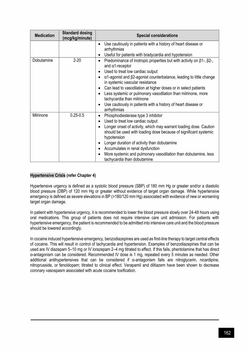
162
Medication Standard dosing (mcg/kg/minute)
Special considerations
Use cautiously in patients with a history of heart disease or arrhythmias
Useful for patients with bradycardia and hypotension
Dobutamine 2-20 Predominance of inotropic properties but with activity on β1-, β2-, and α1-receptor
Used to treat low cardiac output
α1-agonist and β2-agonist counterbalance, leading to little change in systemic vascular resistance
Can lead to vasodilation at higher doses or in select patients
Less systemic or pulmonary vasodilation than milrinone, more tachycardia than milrinone
Use cautiously in patients with a history of heart disease or arrhythmias
Milrinone 0.25-0.5 Phosphodiesterase type 3 inhibitor
Used to treat low cardiac output
Longer onset of activity, which may warrant loading dose. Caution should be used with loading dose because of significant systemic hypotension
Longer duration of activity than dobutamine
Accumulates in renal dysfunction
More systemic and pulmonary vasodilation than dobutamine, less tachycardia than dobutamine
Hypertensive Crisis (refer Chapter 4) Hypertensive urgency is defined as a systolic blood pressure (SBP) of 180 mm Hg or greater and/or a diastolic blood pressure (DBP) of 120 mm Hg or greater without evidence of target organ damage. While hypertensive emergency is defined as severe elevations in BP (>180/120 mm Hg) associated with evidence of new or worsening target organ damage. In patient with hypertensive urgency, it is recommended to lower the blood pressure slowly over 24-48 hours using oral medications. This group of patients does not require intensive care unit admission. For patients with hypertensive emergency, the patient is recommended to be admitted into intensive care unit and the blood pressure should be lowered accordingly.
In cocaine induced hypertensive emergency, benzodiazepines are used as first-line therapy to target central effects of cocaine. This will result in control of tachycardia and hypertension. Examples of benzodiazepines that can be used are IV diazepam 5–10 mg or IV lorazepam 2–4 mg titrated to effect. If this fails, phentolamine that has direct α-antagonism can be considered. Recommended IV dose is 1 mg, repeated every 5 minutes as needed. Other additional antihypertensives that can be considered if α-antagonism fails are nitroglycerin, nicardipine, nitroprusside, or fenoldopam; titrated to clinical effect. Verapamil and diltiazem have been shown to decrease coronary vasospasm associated with acute cocaine toxification.
163
References and Suggested Readings Andersen LW, Kurth T, Chase M, Berg KM, Cocchi MN, Callaway C, et al. Early administration of epinephrine
(adrenaline) in patients with cardiac arrest with initial shockable rhythm in hospital: propensity score matched analysis. BMJ 2016. 353: i1577.
Aung K, Htay T. Vasopressin for cardiac arrest: a systematic review and meta-analysis. Arch Intern Med. 2005. 165(1): 17-24.
Carey RM, Whelton PK. Prevention, detection, evaluation, and management of high blood pressure in adults: Synopsis of the 2017 American College of Cardiology / American Heart Association hypertension guideline. Ann Intern Med 2018.168(5): 351-8.
Craig-Brangan KJ, Day MP. Update: 2015 AHA BLS and ACLS guidelines. Nursing 2016. 46(2): 40-5 Dorian P, Cass D, Schwartz B, Cooper R, Gelaznikas R, Barr A. Amiodarone as compared with lidocaine for shock-
resistant ventricular fibrillation. N Eng J Med 2002. 346(12): 884-90. Fazekas T, Scherlag BJ, Vos M, Wellens HJ, Lazzara R. Magnesium and the heart: Antiarrhythmic therapy with
magnesium. Clin Cardiol 1993. 16(11): 768-74. Jennings PA, Cameron P, Walker T, Bernard S, Smith K. Out-of-hospital cardiac arrest in Victoria: Rural and urban
outcomes. Med J of Aust 2006. 185(3): 135-9. Johnson W, Nguyen ML, Patel R. Hypertension crisis in the emergency department. Cardiol Clin 2012. 30: 533-
43. Løvstad RZ, Granhus G, Hetland S. Bradycardia and asystolic cardiac arrest during spinal anaesthesia: A report
of five cases. Acta Anaesthesiol Scand 2000. 44(1): 48-52. Marik PE, Varon J. Hypertensive crises: Challenges and management. Chest 2007. 131(6): 1949-62. Mark S, Lauren L, Berkow C, Peter J, Henry K, Halperin R, Erik P, Vivek H, Moitra K, Robert W, Brian N. Adult
advanced cardiovascular life support 2015: American Heart Association guidelines update for cardiopulmonary resuscitation and emergency cardiovascular care. Circulation 2015. 132(S2): S444-64.
Pazner CN. Advanced cardiac life support (ACLS) in adults. Uptodate 2020. Qureshi AI, Harris-Lane P, Jawad F, Kirmani JF. Treatment of acute hypertension in patients with intracranial
hemorrhage using the American Heart Association guidelines. Critl Care Med 2006. 34: 1975-80. van Walraven C, Stiell IG, Wells GA, Hebert PC, Vandemheen K. Do advanced cardiac life support drugs increase
resuscitation rates from in-hospital cardiac arrest? The OTAC Study Group. Ann Emerg Med 1998. 32(5): 544-53.
Velissaris D, Karamouzos V, Pierrakos C, Koniari I, Apostolopoulou C, Karanikolas M. Use of sodium bicarbonate in cardiac arrest: Current guidelines and literature review. J Clin Med Res 2016. 8(4): 277-83.
Wong ML, Carey S, Mader TJ, Wang HE. Time to invasive airway placement and resuscitation outcomes after inhospital cardiopulmonary arrest. Resusc 2010. 81(2): 182-6.

164
CHAPTER 20
Drug Overdose and Poisoning
rug poisoning can produce a wide range of symptoms and clinical findings. Presentation depends upon the
agent ingested, whether the ingestion is acute or chronic, baseline prescription medications a patient may be taking, and whether the ingestion involves a single drug or several co-ingestants.
Patients who develop only mild toxicity with a low predicted severity can be observed in the emergency department until they are asymptomatic.
Patients presented with moderate observed toxicity or those who are at risk, evidenced from the history or initial laboratory data should be admitted for intermediate-care or an appropriate observation unit for continued monitoring and treatment.
Patients presented with significant toxicity should be admitted to an intensive care unit (ICU) (Table 20.1).
Table 20.1: Criteria for ICU admission of patient with poisoning (from UpToDate 2020)
Criteria
CNS depression, including significant lethargy, coma Agitation requiring chemical or physical restraint Respiratory depression (PCO2 >45 mmHg), hypoxia or respiratory failure (ARDS), and/or endotracheal intubation Hypotension (SBP ≤80 mmHg) Seizures that are prolonged or recurring Second- or third-degree AV block on ECG Non-sinus cardiac rhythm on ECG Significant acid-base disturbances (eg, metabolic acidosis with pH ≤7.2) Significant metabolic abnormalities requiring close monitoring or aggressive correction (eg, symptomatic hypoglycemia following sulfonylurea or insulin overdose) Extremes of temperature (eg, hyperthermia with T >104°F) Poisoning with a "toxic time bomb"
Ingested drug packets, sustained-release preparations
Quantitative level of drug which predicts unfavorable outcome Need for invasive hemodynamic monitoring (eg, pulmonary artery catheter or arterial line) or cardiac pacing Need for whole bowel irrigation to enhance GI elimination of poison Need for emergency hemodialysis, hemoperfusion, hemofiltration
Initial Poisoning Management Initial management is focused on acute stabilization. The history and physical examination are of great importance in recognizing that poisoning has occurred. Management is directed to the provision of supportive care, prevention of poison absorption, and, when applicable, the use of antidotes and enhanced elimination techniques. Generally, patient presented with altered consciousness should prompt administration of (unless these diagnoses can be rapidly excluded):
Intravenous thiamine to prevent Wernicke's encephalopathy
25 g of dextrose (50 mL of a 50 percent solution) to treat hypoglycemia
Intravenous naloxone to patients with signs, symptoms, or a history suggestive of opioid intoxication.
D
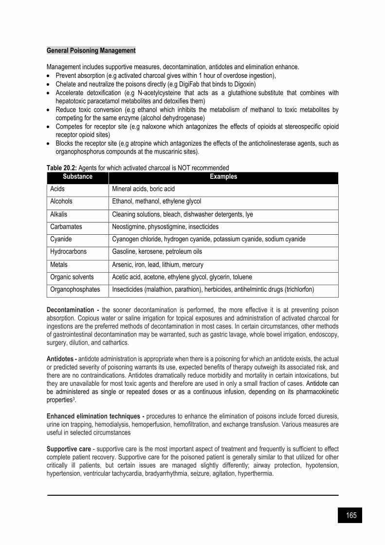
165
General Poisoning Management Management includes supportive measures, decontamination, antidotes and elimination enhance.
Prevent absorption (e.g activated charcoal gives within 1 hour of overdose ingestion),
Chelate and neutralize the poisons directly (e.g DigiFab that binds to Digoxin)
Accelerate detoxification (e.g N-acetylcysteine that acts as a glutathione substitute that combines with hepatotoxic paracetamol metabolites and detoxifies them)
Reduce toxic conversion (e.g ethanol which inhibits the metabolism of methanol to toxic metabolites by competing for the same enzyme (alcohol dehydrogenase)
Competes for receptor site (e.g naloxone which antagonizes the effects of opioids at stereospecific opioid receptor opioid sites)
Blocks the receptor site (e.g atropine which antagonizes the effects of the anticholinesterase agents, such as organophosphorus compounds at the muscarinic sites).
Table 20.2: Agents for which activated charcoal is NOT recommended
Substance Examples
Acids Mineral acids, boric acid
Alcohols Ethanol, methanol, ethylene glycol
Alkalis Cleaning solutions, bleach, dishwasher detergents, lye
Carbamates Neostigmine, physostigmine, insecticides
Cyanide Cyanogen chloride, hydrogen cyanide, potassium cyanide, sodium cyanide
Hydrocarbons Gasoline, kerosene, petroleum oils
Metals Arsenic, iron, lead, lithium, mercury
Organic solvents Acetic acid, acetone, ethylene glycol, glycerin, toluene
Organophosphates Insecticides (malathion, parathion), herbicides, antihelmintic drugs (trichlorfon)
Decontamination - the sooner decontamination is performed, the more effective it is at preventing poison absorption. Copious water or saline irrigation for topical exposures and administration of activated charcoal for ingestions are the preferred methods of decontamination in most cases. In certain circumstances, other methods of gastrointestinal decontamination may be warranted, such as gastric lavage, whole bowel irrigation, endoscopy, surgery, dilution, and cathartics. Antidotes - antidote administration is appropriate when there is a poisoning for which an antidote exists, the actual or predicted severity of poisoning warrants its use, expected benefits of therapy outweigh its associated risk, and there are no contraindications. Antidotes dramatically reduce morbidity and mortality in certain intoxications, but they are unavailable for most toxic agents and therefore are used in only a small fraction of cases. Antidote can be administered as single or repeated doses or as a continuous infusion, depending on its pharmacokinetic properties3. Enhanced elimination techniques - procedures to enhance the elimination of poisons include forced diuresis, urine ion trapping, hemodialysis, hemoperfusion, hemofiltration, and exchange transfusion. Various measures are useful in selected circumstances Supportive care - supportive care is the most important aspect of treatment and frequently is sufficient to effect complete patient recovery. Supportive care for the poisoned patient is generally similar to that utilized for other critically ill patients, but certain issues are managed slightly differently; airway protection, hypotension, hypertension, ventricular tachycardia, bradyarrhythmia, seizure, agitation, hyperthermia.

166
Table 20.3: Common poisoning syndromes (toxidromes)
Toxidrome Mental status Pupils Vital signs Other manifestations Examples of toxic agents
Sympathomimetic Hyperalert, agitation, hallucinations, paranoia
Mydriasis Hyperthermia, tachycardia, hypertension, widened pulse pressure, tachypnea, hyperpnea
Diaphoresis, tremors, hyperreflexia, seizures
Cocaine, amphetamines, cathinones, ephedrine, pseudoephedrine, phenylpropanolamine, theophylline, caffeine
Anticholinergic Hypervigilance, agitation, hallucinations, delirium with mumbling speech, coma
Mydriasis Hyperthermia, tachycardia, hypertension, tachypnea
Dry flushed skin, dry mucous membranes, decreased bowel sounds, urinary retention, myoclonus, choreoathetosis, picking behaviour, seizures (rare)
Antihistamines, tricyclic antidepressants, cyclobenzaprine, orphenadrine, antiparkinson agents, antispasmodics, phenothiazines, atropine, scopolamine, belladonna alkaloids (eg, Jimson Weed)
Hallucinogenic Hallucinations, perceptual distortions, depersonalization, synesthesia, agitation
Mydriasis (usually)
Hyperthermia, tachycardia, hypertension, tachypnea
Nystagmus Phencyclidine, LSD, mescaline, psilocybin, designer amphetamines (eg, MDMA ["Ecstasy"], MDEA)
Opioid CNS depression, coma Miosis Bradypnea, apnea characteristic; may develop: hypothermia, bradycardia, hypotension
Hyporeflexia, pulmonary edema, needle marks
Opioids (eg, heroin, morphine, methadone, oxycodone, hydromorphone), diphenoxylate
Sedative-hypnotic CNS depression, confusion, stupor, coma
Variable Often normal, but may develop: hypothermia, bradycardia, hypotension, apnea, bradypnea
Hyporeflexia Benzodiazepines, barbiturates, carisoprodol, meprobamate, glutethimide, alcohols, zolpidem
Cholinergic Confusion, coma Miosis Bradycardia, hypertension or hypotension, tachypnea or bradypnea
Salivation, urinary and fecal incontinence, diarrhea, emesis, diaphoresis, lacrimation, GI cramps, bronchoconstriction, muscle fasciculations and weakness, seizures
Organophosphate and carbamate insecticides, nerve agents, nicotine, pilocarpine, physostigmine, edrophonium, bethanechol, urecholine
Serotonin syndrome Confusion, agitation, coma Mydriasis Hyperthermia, tachycardia, hypertension, tachypnea
Tremor, myoclonus, hyperreflexia, clonus, diaphoresis, flushing, trismus, rigidity, diarrhea
MAOIs alone or with: SSRIs, meperidine, dextromethorphan, TCAs, L-tryptophan
LSD: lysergic acid diethylamide; MDMA: 3,4-methylenedioxymethamphetamine; MDEA: methylenedioxymethamphetamine; CNS: central nervous system; GI: gastrointestinal; MAOI:
monoamine oxidase inhibitor; SSRI: selective serotonin reuptake inhibitor; TCA: tricyclic antidepressant.
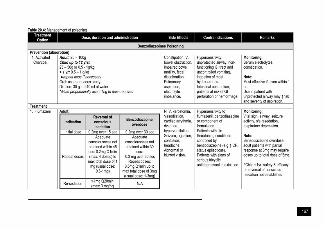
167
Table 20.4: Management of poisoning
Treatment Option
Dose, duration and administration Side Effects Contraindications Remarks
Benzodiazepines Poisoning
Prevention (absorption)
1. Activated Charcoal
Adult: 25 – 100g Child up to 12 yrs: 25 – 50g or 0.5 - 1g/kg < 1 yr: 0.5 – 1 g/kg ● repeat dose if necessary
Oral: as an aqueous slurry Dilution: 30 g in 240 ml of water *dilute proportionally according to dose required
Constipation, V, bowel obstruction, impaired bowel motility, fecal discoloration. Pulmonary aspiration, electrolyte imbalance.
Hypersensitivity, unprotected airway, non-functioning GI tract and uncontrolled vomiting, ingestion of most hydrocarbons. Intestinal obstruction, patients at risk of GI perforation or hemorrhage.
Monitoring: Serum electrolytes, constipation. Note: Most effective if given within 1 hr. Use in patient with unprotected airway may ↑risk and severity of aspiration.
Treatment
1. Flumazenil
Adult:
Indication Reversal of conscious sedation
Benzodiazepine overdose
Initial dose 0.2mg over 15 sec 0.2mg over 30 sec
Repeat doses
Adequate consciousness not obtained within 45 sec: 0.2mg Q1min (max: 4 doses) to
max total dose of 1 mg (usual dose:
0.6-1mg)
Adequate consciousness not obtained within 30
sec: 0.3 mg over 30 sec
Repeat doses: 0.5mg Q1min up to
max total dose of 3mg (usual dose: 1-3mg)
Re-sedation ≤1mg Q20min (max: 3 mg/hr)
N/A
N, V, xerostomia, Vasodilation, cardiac arrythmia, dyspnea, hyperventilation, Seizure, agitation, confusion, headache, Abnormal or blurred vision.
Hypersensitivity to flumazenil, benzodiazepine or component of formulation. Patients with life-threatening conditions controlled by benzodiazepine (e.g ↑ICP, status epilepticus). Patients with signs of serious tricyclic antidepressant intoxication.
Monitoring: Vital sign, airway, seizure activity, s/s resedation, respiratory depression. Note: Benzodiazepine overdose: adult patients with partial response at 3mg may require doses up to total dose of 5mg. *Child <1yr: safety & efficacy in reversal of conscious sedation not established

168
Treatment Option
Dose, duration and administration Side Effects Contraindications Remarks
Cont. infusion (alternative) N/A 0.1 - 0.4mg/hr
Child ≥ 1yr:
Indication Reversal of conscious sedation
Benzodiazepine overdose
Initial dose 0.01 mg/kg
(up to 0.2mg) over 15 sec
0.01 mg/kg (up to 0.2mg)
Repeat doses
Adequate consciousness not obtained after 45
sec: 0.01 mg/kg (up to 0.2 mg) Q1min up
to 4 times (max total dose:
0.05 mg/kg or 1mg
Repeat doses: 0.01 mg/kg
(up to 0.2 mg) Q1 min up to max total dose of
0.05 mg/kg or 1mg
Resedation Safety & efficacy
for repeated doses not established
N/A
Continuous infusion
(alternative) N/A 0.005-0.01mg/kg/hr
*Neonates (benzodiazepine overdose following maternal ingestion): Initial: 0.02mg/kg, Maintenance infusion: 0.05 mg/kg/hr for 6H Administration: Reversal of conscious sedation: over 15 sec, overdose: over 30 sec. Administer through freely running IV infusion into large vein (compatible with D5 or NS), remain stable in syringe for 24hrs
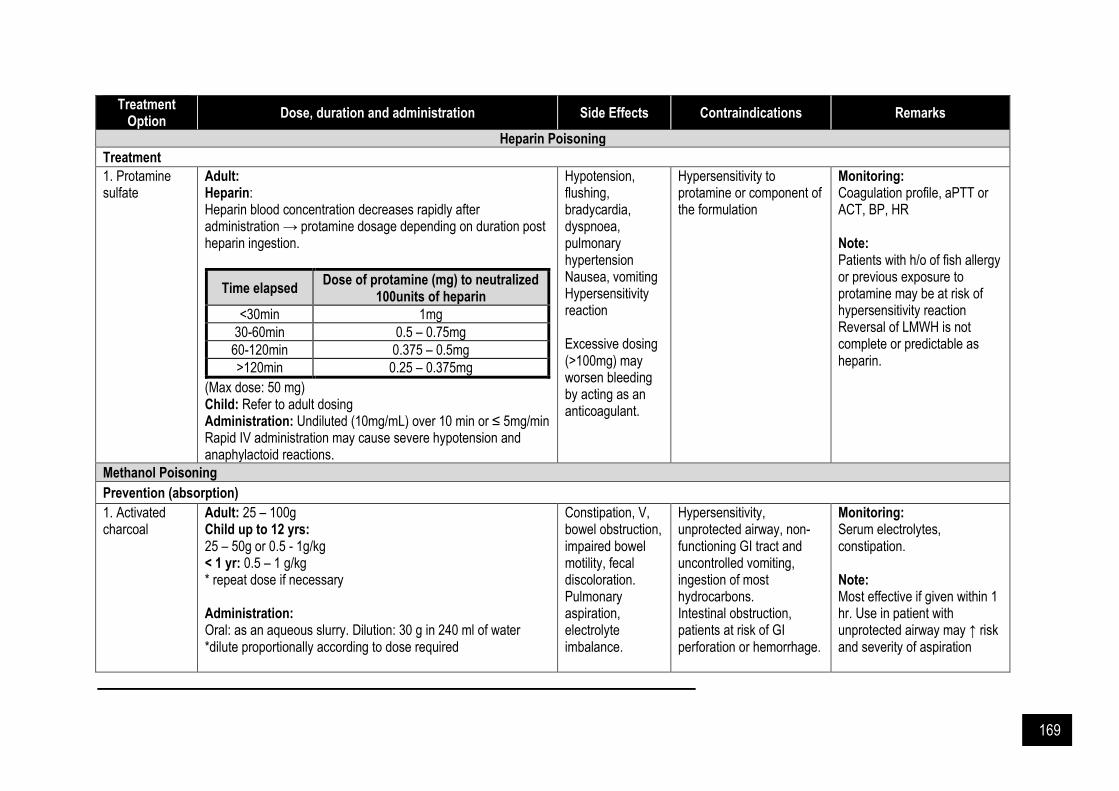
169
Treatment Option
Dose, duration and administration Side Effects Contraindications Remarks
Heparin Poisoning
Treatment
1. Protamine sulfate
Adult: Heparin: Heparin blood concentration decreases rapidly after administration → protamine dosage depending on duration post heparin ingestion.
Time elapsed Dose of protamine (mg) to neutralized
100units of heparin
<30min 1mg
30-60min 0.5 – 0.75mg
60-120min 0.375 – 0.5mg
>120min 0.25 – 0.375mg
(Max dose: 50 mg) Child: Refer to adult dosing Administration: Undiluted (10mg/mL) over 10 min or ≤ 5mg/min Rapid IV administration may cause severe hypotension and anaphylactoid reactions.
Hypotension, flushing, bradycardia, dyspnoea, pulmonary hypertension Nausea, vomiting Hypersensitivity reaction Excessive dosing (>100mg) may worsen bleeding by acting as an anticoagulant.
Hypersensitivity to protamine or component of the formulation
Monitoring: Coagulation profile, aPTT or ACT, BP, HR
Note: Patients with h/o of fish allergy or previous exposure to protamine may be at risk of hypersensitivity reaction Reversal of LMWH is not complete or predictable as heparin.
Methanol Poisoning
Prevention (absorption)
1. Activated charcoal
Adult: 25 – 100g Child up to 12 yrs: 25 – 50g or 0.5 - 1g/kg < 1 yr: 0.5 – 1 g/kg * repeat dose if necessary Administration: Oral: as an aqueous slurry. Dilution: 30 g in 240 ml of water *dilute proportionally according to dose required
Constipation, V, bowel obstruction, impaired bowel motility, fecal discoloration. Pulmonary aspiration, electrolyte imbalance.
Hypersensitivity, unprotected airway, non-functioning GI tract and uncontrolled vomiting, ingestion of most hydrocarbons. Intestinal obstruction, patients at risk of GI perforation or hemorrhage.
Monitoring: Serum electrolytes, constipation. Note: Most effective if given within 1 hr. Use in patient with unprotected airway may ↑ risk and severity of aspiration

170
Treatment Option
Dose, duration and administration Side Effects Contraindications Remarks
Treatment
1. Ethanol
Adult: (Target: maintain Serum ethanol conc. 100-150mg/dL) Loading dose:
IV (10%V/V) Oral (20%V/V)
LD 0.8g/kg
(or 10mL/kg) 0.8g/kg
(or 5mL/kg)
Maintenance dose:
IV (10%V/V) Oral (20%V/V)
Non-alcoholic
0.08 -0.13g/kg/hr (or 1.0-1.6mL/kg/hr)
0.08-0.13g/kg/hr (or 0.5-0.8mL/kg/hr)
Chronic alcoholic
0.15 g/kg/hr (or 1.9mL/kg/hr)
0.15 g/kg/hr (or 0.9 mL/kg/hr)
During dialysis
0.25-0.35 g/kg/hr (or 3.1-4.4mL/kg/hr)
0.25-0.35 g/kg/hr (or 1.6-2.2 mL/kg/hr)
Child (unlabelled use): Refer to adult dosing (Target: maintain serum ethanol concentration 100mg/dL) Administration: - IV: LD over 20 – 60 min * Oral ethanol may be used if IV ethanol is unavailable.
Flushing, hypotension, nerve and tissue destruction. Hypoglycemia (especially common in paediatricss), urinary retention, intoxication. Disorientation, encephalopathy, sedation, seizure, vertigo.
Hypersensitivity to ethyl alcohol, seizure disorder, diabetic coma. Pregnancy (prolonged use or high doses at term).
Monitoring: Blood ethanol concentration, electrolytes, arterial pH, blood gases, blood glucose, methanol blood level, HR, BP. Note: IV ethanol 10% V/V = 0.08g ethanol/mL. Dialyzable; ↑ infusion (approximately doubled, possibly tripled) during dialysis. Endpoint of therapy: ● Methanol blood
concentration <10mg/dL ● Formate blood
concentration <1.2mg/dL ● Acidosis, CNS clinical
findings, electrolyte abnormalities, serum amylase, and osmolal gap resolved.
2. Fomepizole Adult: Loading dose: 15mg/kg, maintenance dose: 10mg/kg BD x 4 doses followed by 15mg/kg BD Dosing adjustment in hemodialysis: Loading dose: 15 mg/kg, maintenance dose: 10 mg/kg Q4H x 4 doses followed by 15 mg/kg Q4H
Headache, N, dizziness, drowsiness and metallic taste. Hypertriglyceridemia, ↑ LFT, bradycardia,
Hypersensitivity to fomepizole, other pyrazoles, or component of formulation.
Monitoring: Plasma/ urinary ethylene glycol or methanol level, plasma/ urinary osmolality Renal & hepatic function, sr. electrolytes, ABG, anion & osmolar gaps.
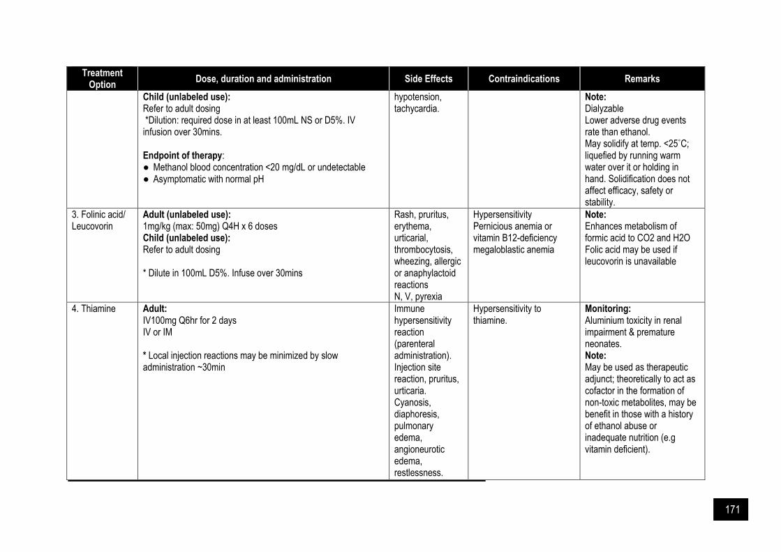
171
Treatment Option
Dose, duration and administration Side Effects Contraindications Remarks
Child (unlabeled use): Refer to adult dosing *Dilution: required dose in at least 100mL NS or D5%. IV infusion over 30mins. Endpoint of therapy: ● Methanol blood concentration <20 mg/dL or undetectable ● Asymptomatic with normal pH
hypotension, tachycardia.
Note: Dialyzable Lower adverse drug events rate than ethanol. May solidify at temp. <25˚C; liquefied by running warm water over it or holding in hand. Solidification does not affect efficacy, safety or stability.
3. Folinic acid/ Leucovorin
Adult (unlabeled use): 1mg/kg (max: 50mg) Q4H x 6 doses Child (unlabeled use): Refer to adult dosing
* Dilute in 100mL D5%. Infuse over 30mins
Rash, pruritus, erythema, urticarial, thrombocytosis, wheezing, allergic or anaphylactoid reactions N, V, pyrexia
Hypersensitivity Pernicious anemia or vitamin B12-deficiency megaloblastic anemia
Note: Enhances metabolism of formic acid to CO2 and H2O Folic acid may be used if leucovorin is unavailable
4. Thiamine Adult: IV100mg Q6hr for 2 days IV or IM * Local injection reactions may be minimized by slow administration ~30min
Immune hypersensitivity reaction (parenteral administration). Injection site reaction, pruritus, urticaria. Cyanosis, diaphoresis, pulmonary edema, angioneurotic edema, restlessness.
Hypersensitivity to thiamine.
Monitoring: Aluminium toxicity in renal impairment & premature neonates. Note: May be used as therapeutic adjunct; theoretically to act as cofactor in the formation of non-toxic metabolites, may be benefit in those with a history of ethanol abuse or inadequate nutrition (e.g vitamin deficient).

172
Treatment Option
Dose, duration and administration Side Effects Contraindications Remarks
Opioids Poisoning
Prevention (absorption)
1. Activated Charcoal
Adult: 25 – 100g Child up to 12 yrs: 25 – 50g or 0.5 - 1g/kg < 1 yr: 0.5 – 1 g/kg * repeat dose if necessary Oral: as aqueous slurry Dilution: 30 g in 240 ml of water *dilute proportionally according to dose required
Constipation, V, bowel obstruction, impaired bowel motility, fecal discoloration. Pulmonary aspiration, electrolyte imbalance.
Hypersensitivity, unprotected airway, non-functioning GI tract and uncontrolled vomiting, ingestion of most hydrocarbons. Intestinal obstruction, patients at risk of GI perforation or hemorrhage.
Monitoring: Serum electrolytes, constipation. Note: Most effective if given within 1 hour. Use in patient with unprotected airway may ↑ risk and severity of aspiration.
Treatment
1. Naloxone
Adult:
Indication
Opioids intoxication: Respiratory depression Post-
operative reversal
Opioids- naïve
Opioids dependent
Initial dose 0.4 – 2 mg Q2-3min; may
need to repeat doses Q20-60min up to total dose of
10mg
0.04-0.4mg; may repeat or escalate dose up to
2mg if inadequate response
0.1 - 0.2 mg Q2-3min;
repeat doses may be
needed within 1-2hr
Cont. infusion
(unlabeled dosing)
2/3 of the initial bolus dose on an hourly basis (typically 0.25-
6.25mg/hr) *1/2 of initial bolus dose
should be re-administered 15min after initiation of
infusion to prevent ↓ level
N/A
Cardiac arrest, ventricular fibrillation, pulmonary edema, dyspnea Abrupt reversal may cause N, V, tachycardia, ↑BP, sweating
Hypersensitivity to naloxone or component of formulation5
Monitoring: RR, HR, BP, cardiorespiratory status, temp., level of consciousness, ABG or pulse oximetry Note: Consider lower dose in known or suspected opioids -dependent patients to minimize withdrawal syndrome May be given IM or SC when IV access is not available
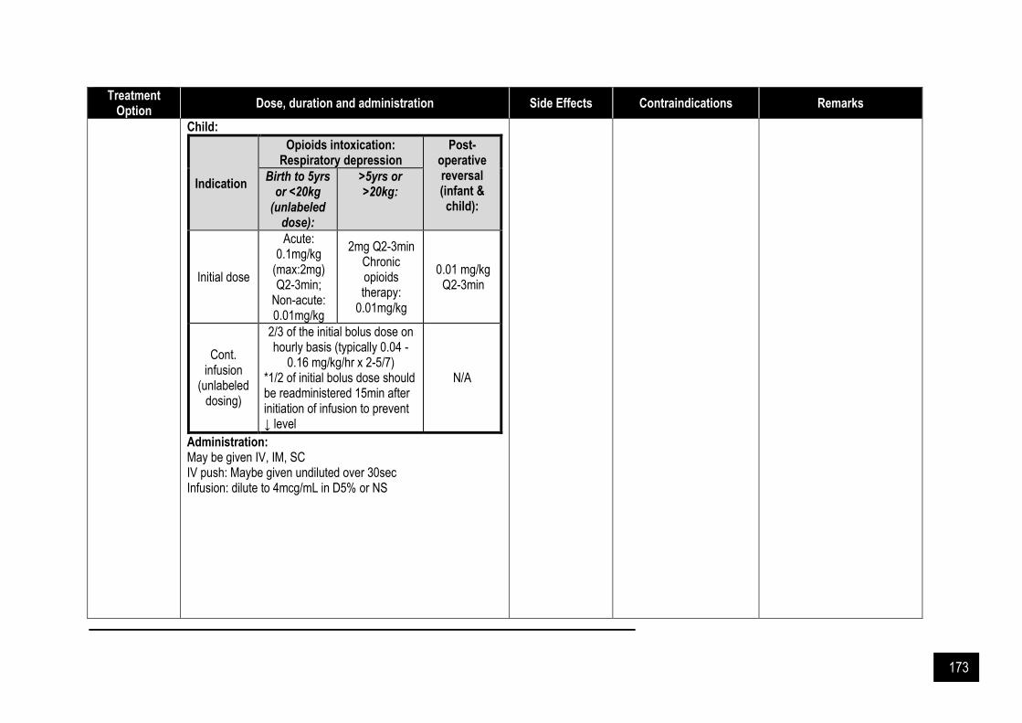
173
Treatment Option
Dose, duration and administration Side Effects Contraindications Remarks
Child:
Indication
Opioids intoxication: Respiratory depression
Post-operative reversal (infant & child):
Birth to 5yrs or <20kg
(unlabeled dose):
>5yrs or >20kg:
Initial dose
Acute: 0.1mg/kg
(max:2mg) Q2-3min;
Non-acute: 0.01mg/kg
2mg Q2-3min Chronic opioids therapy:
0.01mg/kg
0.01 mg/kg Q2-3min
Cont. infusion
(unlabeled dosing)
2/3 of the initial bolus dose on hourly basis (typically 0.04 -
0.16 mg/kg/hr x 2-5/7) *1/2 of initial bolus dose should be readministered 15min after initiation of infusion to prevent ↓ level
N/A
Administration: May be given IV, IM, SC IV push: Maybe given undiluted over 30sec Infusion: dilute to 4mcg/mL in D5% or NS
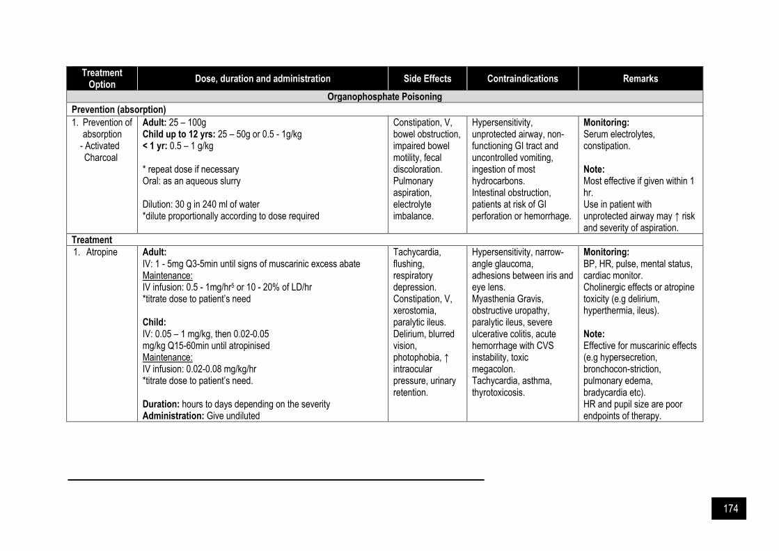
174
Treatment Option
Dose, duration and administration Side Effects Contraindications Remarks
Organophosphate Poisoning
Prevention (absorption)
1. Prevention of absorption
- Activated Charcoal
Adult: 25 – 100g Child up to 12 yrs: 25 – 50g or 0.5 - 1g/kg < 1 yr: 0.5 – 1 g/kg * repeat dose if necessary Oral: as an aqueous slurry Dilution: 30 g in 240 ml of water *dilute proportionally according to dose required
Constipation, V, bowel obstruction, impaired bowel motility, fecal discoloration. Pulmonary aspiration, electrolyte imbalance.
Hypersensitivity, unprotected airway, non-functioning GI tract and uncontrolled vomiting, ingestion of most hydrocarbons. Intestinal obstruction, patients at risk of GI perforation or hemorrhage.
Monitoring: Serum electrolytes, constipation. Note: Most effective if given within 1 hr. Use in patient with unprotected airway may ↑ risk and severity of aspiration.
Treatment
1. Atropine Adult: IV: 1 - 5mg Q3-5min until signs of muscarinic excess abate Maintenance: IV infusion: 0.5 - 1mg/hr5 or 10 - 20% of LD/hr *titrate dose to patient’s need Child: IV: 0.05 – 1 mg/kg, then 0.02-0.05 mg/kg Q15-60min until atropinised Maintenance: IV infusion: 0.02-0.08 mg/kg/hr *titrate dose to patient’s need. Duration: hours to days depending on the severity Administration: Give undiluted
Tachycardia, flushing, respiratory depression. Constipation, V, xerostomia, paralytic ileus. Delirium, blurred vision, photophobia, ↑ intraocular pressure, urinary retention.
Hypersensitivity, narrow-angle glaucoma, adhesions between iris and eye lens. Myasthenia Gravis, obstructive uropathy, paralytic ileus, severe ulcerative colitis, acute hemorrhage with CVS instability, toxic megacolon. Tachycardia, asthma, thyrotoxicosis.
Monitoring: BP, HR, pulse, mental status, cardiac monitor. Cholinergic effects or atropine toxicity (e.g delirium, hyperthermia, ileus). Note: Effective for muscarinic effects (e.g hypersecretion, bronchocon-striction, pulmonary edema, bradycardia etc). HR and pupil size are poor endpoints of therapy.

175
Treatment Option
Dose, duration and administration Side Effects Contraindications Remarks
2. Pralidoxime Adult: LD: 30mg/kg (IV), maintenance: IV infusion 8mg/kg/hr OR LD: IV/IM: 1 - 2g, maintenance: (IV) repeat bolus 1 -2g after 1 hr and then Q10-12hr as needed Child up to 16 yrs: LD: 20 – 50 mg/kg (IV) (max: 2g/dose), maintenance: IV 10 – 20 mg/kg/hr OR LD: 20 – 50 mg/kg (IV) (max: 2g/dose), maintenance: IV repeat bolus 20-50mg/kg (max: 2g/dose) after 1 hr and then Q10-12hr as needed Duration: ≥ 24 hrs after cholinergic manifestation have resolved. Administration: IV: LD - dilute to 10- 20 mg/ml with NS & infuse over 15-30min. (fluid restriction: give as 50mg/ml solution over ≥5min) Maintenance: max infusion rate 200mg/min. IM: reconstitute 1g vial with 3ml of WFI or NS (300mg/ml).
Blurred vision, diplopia, dizziness, drowsiness, headache, N. ↑ LFT, ↑ CK. Tachycardia, hyperventilation, apnea. Rapid injection: laryngospasm, muscle rigidity, tachycardia.
Hypersensitivity to any component of the product.
Monitoring: Vital signs, ECG, HR, BP, RR, muscle fasciculations and strength, pulse oximetry, cardiac monitor, fluid balance. Note: Primarily effective for nicotinic effects (diaphragmatic and respiratory muscles weakness). Administer as soon as possible after exposure (ideally within 36 hrs), however pt presenting late (2-6 days) may still benefit. Concomitant use may enhance the S/E of atropine.
Paracetamol Poisoning
Prevention (absorption)
1. Activated Charcoal
Adult: 25 – 100g Child up to 12 yrs: 25 – 50g or 0.5 - 1g/kg < 1 yr: 0.5 – 1 g/kg * repeat dose if necessary Administration: Oral: as aqueous slurry
Constipation, V, bowel obstruction, impaired bowel motility, fecal discoloration. Pulmonary aspiration, electrolyte imbalance
Hypersensitivity, unprotected airway, non-functioning GI tract and uncontrolled vomiting, ingestion of most hydrocarbons. Intestinal obstruction, patients at risk of GI perforation or hemorrhage.
Monitoring: Serum electrolyes, constipation. Note: Well absorbed by activated charcoal. Most effective if given within 1 hr.

176
Treatment Option
Dose, duration and administration Side Effects Contraindications Remarks
Dilution: 30 g in 240 ml of water (dilute proportionally according to dose required)
Use in patient with unprotected airway may ↑ risk and severity of aspiration.
Treatment
1. N-acetyl-cysteine (NAC)
Adult (21-hour Regimen): LD:150mg/kg (max: 15g) in 200mL D5% over 60min 2nd dose: 50mg/kg (max: 5g) in 500mL D5% over 4hr 3rd dose: 100mg/kg (max: 10g) in 1L D5% over 16hr Child: Refer to adult dosing. Fluid volume should be reduced in patients weighing <40kg:
Dilution Volume with D5% or half saline (mL)
Body weight
(kg)
LD: 150mg/kg
over 60min
2nd dose: 50mg/kg over
4hr
3rd dose: 100mg/kg over
16hr
30 100 250 500
25 100 250 500
20 60 140 280
15 45 105 210
10 30 70 140
*prolonged courses may be needed for patients with hepatotoxicity or hepatic failure.
Rash, pruritus, angioedema, anaphylactoid reactions. N, V, hypocalcemia. Tachycardia, ECG changes, ↓ BP, ↑ BP, bronchospasm.
Hypersensitivity to acetylcysteine or any of its components.
Monitoring: Vital signs, Ca2+ level, ECG, PT, INR, serum glucose, LFT, Cr, BUN, Hb, HCT, anaphylactoid reaction. Note: Fluid volume should be reduced in patients <40kg as large amount of free water may cause ↓ Na and seizure in children. Average time to the onset of dose-related ADR is 30 min; ↓ the rate of NAC infusion LD to 60 min may avoid some of the ADR. Asthmatic patients are at higher risk for ADR; use cautiously.
Paraquat Poisoning
Prevention (absorption)
1. Activated Charcoal
Adult: 25 – 100g Child up to 12 yrs: 25 – 50g or 0.5 - 1g/kg <1 yr: 0.5 – 1 g/kg
Constipation, V, bowel obstruction, impaired bowel motility, fecal discoloration.
Hypersensitivity, unprotected airway, non-functioning GI tract and uncontrolled vomiting, ingestion of most hydrocarbons.
Monitoring: Serum electrolytes, constipation.

177
Treatment Option
Dose, duration and administration Side Effects Contraindications Remarks
* repeat dose if necessary Administration: Oral: as an aqueous slurry Dilution: 30 g in 240 ml of water *dilute proportionally according to the dose required
Pulmonary aspiration, electrolyte imbalance.
Intestinal obstruction, patients at risk of GI perforation or hemorrhage.
Note: Most effective if given within 1 hr. Use in patient with unprotected airway may ↑ risk and severity of aspiration.
2. Fuller’s Earth Adult: 200 - 500 ml of Fuller's Earth (30% suspension) together with magnesium sulphate or mannitol every 2 hours for several days OR 100-150g (30% suspension) Child up to 12 yrs: 2g/kg (30% suspension)
N/A N/A Note: Should be administered as soon as possible via NG tube.
Warfarin Overdose
Prevention (absorption)
1. Activated Charcoal
Adult: 25 – 100g Child up to 12 yrs: 25 – 50g or 0.5 - 1g/kg < 1 yr: 0.5 – 1 g/kg * repeat dose if necessary Administration: Oral: as an aqueous slurry. Dilution: 30 g in 240 ml of water. *dilute proportionally according to dose required.
Constipation, V, bowel obstruction, impaired bowel motility, fecal discoloration. Pulmonary aspiration, electrolyte imbalance.
Hypersensitivity, unprotected airway, non-functioning GI tract and uncontrolled vomiting, ingestion of most hydrocarbons. Intestinal obstruction, patients at risk of GI perforation or hemorrhage.
Monitoring: Serum electrolytes, constipation. Note: Most effective if given within 1 hr. Use in patient with unprotected airway may ↑ risk and severity of aspiration.
Treatment
2. Vitamin K
Adults maintained on warfarin:
INR Treatment
< 5 No bleeding
● Lower or omit next dose, monitor frequently
● Resume at a lower dose when INR therapeutic. If only minimally above
Cyanosis, flushing, hypotension, dyspnea, dizziness, abnormal taste,
Hypersensitivity to Vitamin K or component of formulation.
Monitoring: PT, INR, HCT. Note:

178
Treatment Option
Dose, duration and administration Side Effects Contraindications Remarks
the therapeutic range, no dose reduction may be required
Rapid reversal
● Hold warfarin. Administer IV infusion Vitamin K 1mg or oral 2mg
≥5-<9
No bleeding
● Omit the next 1 or 2 doses, monitor frequently. Resume at a lower dose when INR therapeutic OR
● Omit dose. Administer oral vitamin K ≤ 5mg (particularly if at increased risk of bleeding)
Rapid reversal
● Hold warfarin. Administer IV infusion vitamin K 1-2mg or oral 2-5mg (with the expectation that INR ↓ within 24hr)
≥9 No bleeding
● Hold warfarin. Administer oral vitamin K 2.5-5mg or IV 1-2mg (with expectation that the INR ↓ within 24-48hr)
● Monitor frequently. Additional vitamin K if needed. Resume at a lower dose when INR therapeutic
Rapid reversal
● Hold warfarin. Administer IV vitamin K 1-10mg (may repeat 6-24hr as necessary
Any Serious bleeding
● Hold warfarin. Administer Vitamin K 10mg slow IV infusion.
● Supplement with FFP or PCC. rVIIa may be considered as an alternative for PCC. Vitamin K can be repeated every 12hr
Life threaten-
● Hold warfarin. Give PCC supplement with vitamin K 10mg slow IV infusion
injection site reaction, scleroderma-like lesions Anaphylaxis (greater risk with rapid IV administration).
IV route should be restricted to patients with significant bleeding or severe coagulopathy.
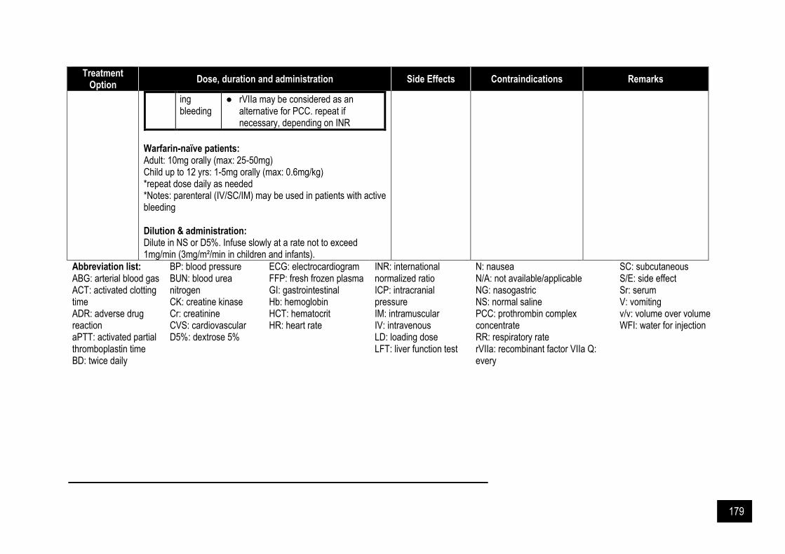
179
Treatment Option
Dose, duration and administration Side Effects Contraindications Remarks
ing bleeding
● rVIIa may be considered as an alternative for PCC. repeat if necessary, depending on INR
Warfarin-naïve patients: Adult: 10mg orally (max: 25-50mg) Child up to 12 yrs: 1-5mg orally (max: 0.6mg/kg) *repeat dose daily as needed *Notes: parenteral (IV/SC/IM) may be used in patients with active bleeding Dilution & administration: Dilute in NS or D5%. Infuse slowly at a rate not to exceed 1mg/min (3mg/m²/min in children and infants).
Abbreviation list: ABG: arterial blood gas ACT: activated clotting time ADR: adverse drug reaction aPTT: activated partial thromboplastin time BD: twice daily
BP: blood pressure BUN: blood urea nitrogen CK: creatine kinase Cr: creatinine CVS: cardiovascular D5%: dextrose 5%
ECG: electrocardiogram FFP: fresh frozen plasma GI: gastrointestinal Hb: hemoglobin HCT: hematocrit HR: heart rate
INR: international normalized ratio ICP: intracranial pressure IM: intramuscular IV: intravenous LD: loading dose LFT: liver function test
N: nausea N/A: not available/applicable NG: nasogastric NS: normal saline PCC: prothrombin complex concentrate RR: respiratory rate rVIIa: recombinant factor VIIa Q: every
SC: subcutaneous S/E: side effect Sr: serum V: vomiting v/v: volume over volume WFI: water for injection

180
References and Suggested Readings Battaglia J, Moss S, Rush J, Kang J, Mendoza R, Leedom L, Dubin W, McGlynn C, Goodman L. Haloperidol,
lorazepam, or both for psychotic agitation? A multicenter, prospective, double-blind, emergency department study. Am J Emerg Med 1997. 15(4): 335.
Blake KV, Massey KL, Hendeles L, Nickerson D, Neims A. Relative efficacy of phenytoin and phenobarbital for the prevention of theophylline-induced seizures in mice. Ann Emerg Med 1988. 17(10): 1024.
Boyer EW. Management of opioid analgesic overdose. N Engl J Med 2012. 367(2): 146-55. Buchman AL, Dauer J, Geiderman J. The use of vasoactive agents in the treatment of refractory hypotension seen
in tricyclic antidepressant overdose. J Clin Psychopharmacol 1990. 10(6): 409. Burns MJ, Linden CH, Graudins A, Brown RM, Fletcher KE. A comparison of physostigmine and benzodiazepines
for the treatment of anticholinergic poisoning. Ann Emerg Med 2000. 35(4): 374. Erickson TB, Thompson TM, Lu JJ. The approach to the patient with an unknown overdose. Emerg Med Clin North
Am 2007. 25(2): 249-81; Hoffman JR, Schriger DL, Luo JS. The empiric uses of naloxone in patients with altered mental status: a
reappraisal. Ann Emerg Med 1991; 20:246. Höjer J, Baehrendtz S, Magnusson A, Gustafsson LL. A placebo-controlled trial of flumazenil given by continuous
infusion in severe benzodiazepine overdosage. Acta Anaesthesiol Scand 1991; 35:584 Hollander JE. The management of cocaine-associated myocardial ischemia. N Engl J Med 1995. 333(19): 1267. Isbister GK, Downes F, Sibbritt D, Dawson AH, Whyte IM. Aspiration pneumonitis in an overdose population:
frequency, predictors, and outcomes. Crit Care Med 2004. 32(1): 88. Kreshak AA, Cantrell FL, Clark RF, Tomaszewski CA. A poison center's ten-year experience with flumazenil
administration to acutely poisoned adults. J Emerg Med 2012; 43:677. Lange RA, Cigarroa RG, Flores ED, McBride W, Kim AS, Wells PJ, Bedotto JB, Danziger RS, Hillis LD. Potentiation
of cocaine-induced coronary vasoconstriction by beta-adrenergic blockade. Ann Intern Med 1990. 112(12): 897.
Laskowski LK, Landry A, Vassallo SU, Hoffman RS. Ice water submersion for rapid cooling in severe drug-induced hyperthermia. Clin Toxicol (Phila) 2015. 53(3): 181-4.
Litovitz TL, Klein-Schwartz W, Rodgers GC Jr, Cobaugh DJ, Youniss J, Omslaer JC, May ME, Woolf AD, Benson BE. 2001 Annual report of the American Association of Poison Control Centers Toxic Exposure Surveillance System. Am J Emerg Med 2002. 20(5): 391.
Lu J Da, Xue J. Poisoning: Kinetics to Therapeutics. In: Critical Care Nephrology: Third Edition. 2019. Pohjola-Sintonen S, Kivistö KT, Vuori E, et al. Identification of drugs ingested in acute poisoning: Correlation of
patient history with drug analyses. Ther Drug Monit 2000. 22: 749. Salyer SW. Essential Emergency Medicine. Essential Emergency Medicine. 2007. Schabelman E, Kuo D. Glucose before thiamine for Wernicke encephalopathy: A literature review. J Emerg Med
2012. 42: 488. Tran TP, Panacek EA, Rhee KJ, Foulke GE. Response to dopamine vs norepinephrine in tricyclic antidepressant-
induced hypotension. Acad Emerg Med 1997. 4(9): 864. Yasaka M, Oomura M, Ikeno K, et al. Effect of prothrombin complex concentrate on INR and blood coagulation
system in emergency patients treated with warfarin overdose. Ann Hematol 2003; 82:121.