hard metal disease
TRANSCRIPT

Brit. J. industr. Med., 1962, 19, 239.
HARD METAL DISEASEBY
A. 0. BECH, M. D. KIPLING, and J. C. HEATHER
From the Coventry Group of Hospitals and H.M. Medical Inspector of Factories, Birmingham
(RECEIVED FOR PUBLICATION DECEMBER 23, 1961)
In Great Britain there have been no published reports of respiratory disease occurring amongstworkers in the hard metal (tungsten carbide) industry. In this paper the clinical and radiologicalfindings in six cases and the pathological findings in one are described. In two cases physiologicalstudies indicated mild alveolar diffusion defects. Histological examination in a fatal case revealeddiffuse pulmonary interstitial fibrosis with marked peribronchial and perivascular fibrosis andbronchial epithelial hyperplasia and metaplasia. Radiological surveys revealed the sporadicoccurrence and low incidence of the disease. The alterations in respiratory mechanics whichoccurred in two workers following a day's exposure to dust are described. Airborne dust con-centrations are given.The industrial process is outlined and the literature is reviewed. The toxicity of the metals is
discussed, and our findings are compared with those reported from Europe and the United States.We are of the opinion that the changes which we would describe as hard metal disease are
caused by the inhalation of dust at work and that the component responsible may be cobalt.
Hard metal is manufactured by a process ofpowder metallurgy from tungsten and carbon withcobalt as a binder. The industry, starting in Germanyafter the first world war, has greatly increased insize. The extremely hard product (90 to 95% thehardness of diamond) is used for the cutting edgesof tools, rock drills, broaches, and dies. It is alsoused for radiotherapy screens and armaments.The process of manufacture involves the pro-
duction of tungsten metal powder and cobaltpowder from their respective oxides or salts. Thetungsten metal powder is mixed with carbon andheated in an atmosphere of hydrogen to formtungsten carbide. Titanium carbide is added insome grades and is similarly formed. Small amountsof tantalum carbide and vanadium carbide may beincorporated in particular grades.The percentage by weight of tungsten carbide
varies from 80 to 90, of titanium carbide 8 to 18 andof cobalt 5 to 25. More than three-quarters ofproduction is of grades containing between 6% and9% of cobalt.
Carbides and cobalt are mixed wet in a ball-milland dried. The dried powder is pressed to therequired shape combined with wax using a solventsuch as trichlorethylene, which is then driven off byheat in an oven in an atmosphere of hydrogen (pre-sintering). After shaping, the product is heated
again in an atmosphere of hydrogen (sintering). Itis then ground, wet or dry, with diamond andcarborundum wheels to its final shape.Dust may be produced during mixing, filtering,
shaping, and grinding, but since the material isvaluable and contamination of the product by otherdust undesirable, as much dust as possible is re-covered by local exhaust.The sequence of the various manufacturing pro-
cesses is outlined in the chart (Fig. 1).
Historical ReviewJobs and Ballhausen (1940) examined 27 workers
in a factory which had been in production for twoyears in Germany and found reticular shadowingwith areas of fine nodulation suggestive of earlypneumoconiosis in the radiographs of eight menwho had been exposed to hard metal dust. Wiele(1951) in Germany reported that the radiographs oftwo men showed a pneumoconiosis. These men hadbeen employed in powder mixing for 10 and 30years respectively. The first suffered from severedyspnoea and the other died of cardiac failure dueto emphysema and chronic bronchitis.
Moschinski, Jurisch, and Reinl (1959) examined696 hard metal workers between the years 1948 and1949 and found a high incidence of bronchitis with
239
copyright. on D
ecember 16, 2021 by guest. P
rotected byhttp://oem
.bmj.com
/B
r J Ind Med: first published as 10.1136/oem
.19.4.239 on 1 October 1962. D
ownloaded from

20BRITISH JOURNAL OF INDUSTRIAL MEDICINE
Wolfram
Cobalt oxide
ISieved
Heated in reduction oven inhydrogen at 600-1000°C.
Titanium oxideor
Tantalum oxideor
Pure tungsten powder Vanadium oxideCarbon
Sieved Reduced
Mixed< Mixed
Heated in carbonizing oven Heated in carbonizing ovenin hydrogen at 1400-1500°C. in hydrogen at 1400-1500°C.
Il
Pure tungsten carbideTV
Titaniumor
Tantalumor
Cobalt metal powder Sieved Vanadium
Sieved ," Mixed Sieved
IBall-milled
Centrifuged
Vacuum dried
Finished powder
ICompressed
IPre-sintered:
Heated in hydrogen at 1000°C.
IShaped
Sintered:Heated in hydrogen at 1500°C.
Finished hard metal
IGround
Finished hard metal product
FIG. I.-Diagram of stages of manufacture.
carbide
carbide
carbide
240
copyright. on D
ecember 16, 2021 by guest. P
rotected byhttp://oem
.bmj.com
/B
r J Ind Med: first published as 10.1136/oem
.19.4.239 on 1 October 1962. D
ownloaded from

HARD METAL DISEASE
bronchospasm. They quote a report of Magos,Timar, and Szandanyi (1956) on Hungarian hardmetal workers. Of 40 workers 23 complained ofrespiratory difficulties, and in the radiographs offive of these there was a stippled network appearanceand in the rest a demonstrable increase in lungmarkings.
In 1955 and 1956 Moschinski et al. (1959) carriedout an investigation amonst 331 hard metal workers.They found a high incidence of respiratory disordersand radiographic changes in 59 men; of these 20showed early fibrosis, 38 a slightly more advanceddegree, and one marked changes. The changes mostfrequently seen were diffuse finely striated increasedlung markings in the mid and lower zones and inmore advanced cases soft patchy nodular opacitiesextending over the lung fields, also thickening of thehilar shadows and fairly large irregular shadows inthe mid-zones. The particulars of three cases werereported in detail. These authors concluded thatrespiratory disorders developed after varying periodsof exposure, the main symptoms being difficulty inbreathing, cough, and expectoration, and that theclinical picture was characterized by the develop-ment of bronchitis with bronchospasm and in severecases by emphysema and respiratory insufficiency.They had observed fibrosis develop in a few instancesfollowing a preliminary phase with respiratorysymptoms. The possibility of susceptibility wasalso discussed.Husten (1959) described the post-mortem findings
in a man whose case had been originally describedby Wiele. He found diffuse pulmonary fibrosis,emphysematous changes and interstitial fibrosisparticularly in the pulmonary lobules, also anassociated pulmonary tuberculosis. Titanium andtungsten were detected chemically in the pulmonaryand lymphatic tissues but not cobalt.
In Sweden, Karth (1948) described three cases.These men suffered from cough and markeddyspnoea, and the appearances of their radiographswere suggestive of chronic interstitial pneumonia.Two of the cases at autopsy showed non-specificinterstitial pneumonia.Lundgren and Ohman (1954) quote Forssman as
stating he had seen 11 cases of pulmonary diseaseamongst hard metal workers while in the U.S.A. in1947 and Frederick that 15 to 16 cases had occurredin two Detroit factories, one of which had beenvisited by Forssman. In two fatal cases there werefibrosis, thickening of alveolar walls, and giant cellsin the alveoli and small bronchi. Analysis of thelungs revealed the presence of titanium and tungstenbut virtually no cobalt. This was attributed to thegreater solubility of cobalt. Lundgren and Ohman(1954) reported personal experience of five cases of
pulmonary fibrosis among Swedish hard metalworkers and the autopsy findings in two fatal cases.They also carried out a survey amongst approxi-mately 200 hard metal workers in five factories andgave a detailed account of their findings.Ahlmark, Bruce, and Nystr6m (1961) in a sum-
mary of four recorded cases of pneumoconiosisattributed to hard metal state that after relativelyshort periods of employment, at most eight years,pulmonary lesions were found characterized byproliferation of interstitial tissue and a tendency topulmonary contraction, and that the pneumoconiosisshowed a marked tendency to progress.
In the U.S.A., Fairhall, Castberg, Carrozzo, andBrinton (1947) examined 1,802 tungsten carbideworkers. They found changes in the conjunctivae,the upper respiratory tract, and the mucous mem-branes. Pruritus and sensitivity to cobalt as shownby patch tests were found in some workers. In theradiographs of 36 there were granular or conglomer-ate markings, but 64% of these men had beenpreviously employed in mining or metal fabricatingindustries.
Miller, Davis, Goldman, and Wyatt (1953)described three cases. These men were engaged ingrinding finished hard metal tools. They complainedof persistent cough with sputum and dyspnoea onexertion, and their radiographs showed prominenthilar shadows and increased lung markings. Theyquote Olsen in a personal report as having studiedcases of hard metal workers with radiographicchanges during the war in Sweden.
Schwartz, Peck, Blair, and Markuson (1945)investigated dermatitis in a tungsten carbide plantand came to the conclusion that sensitivity tocobalt was the cause of the dermatitis. Moschinskiet al. (1959) confirmed that cobalt was a cutaneoussensitizer.
In Russia, Kaplun (1957) examined the workersin two hard metal factories and found cases oflung disease. In Czechoslovakia, Kaplun andMezencewa (1960) analysed the records of periodicmedical examinations of 283 hard metal workersand found that disorders of the upper respiratorytract, bronchitis, and an early type ofpneumoconiosisoccurred.
In this country A.I.G. McLaughlin (1961, personalcommunication) investigated a hard metal workerwhose chest radiographs revealed marked changes.[In 1951 the workers in a hard metal factory weresurveyed by Davidson and Cottrell (P. T. Davidsonand J. E. Cottrell, personal communication); thelatter is quoted by Browning (1960). Davidson(1962, personal communication) carried out a furthersurvey at the same factory.]
241
copyright. on D
ecember 16, 2021 by guest. P
rotected byhttp://oem
.bmj.com
/B
r J Ind Med: first published as 10.1136/oem
.19.4.239 on 1 October 1962. D
ownloaded from

BRITISH JOURNAL OF INDUSTRIAL MEDICINE
Toxicity of Cobalt, Tungsten and TitaniumThe literature relating to the toxicity of tungsten,
cobalt, titanium and tantalum has been reviewed byBrowning (1961), and of cobalt by Caujolle andMeynier (1959).
Frederick and Bradley (1946) found tungsten,tantalum and titanium carbides, and titanium oxideessentially inert in white rats when injected intra-peritoneally. Cobalt metal produced markedvasodilatation, swelling of the liver, moderate bloodchanges characterized by an increased number ofimmature red cells, and initial inflammation withresidual fibrosis in all tissue surfaces in contact withthe material. Cobalt was not stored in the animalorganism. Miller et al. (1953) produced an intenseforeign body reaction and early collagenic sclerosisby injection of dust derived from grinding into theperitoneal cavities of rats. Intratracheal inoculationof pure tungsten carbide produced no changes otherthan those of an inert dust after 18 weeks. Delahant(1955) compared the effect of intratracheal injectioninto guinea-pigs of tantalum oxide, tungsten metal,cobalt oxide, tungsten carbide and carbon (ratio94 to 6), tungsten carbide and cobalt (ratios 3 to 1and 91 to 9), and cobalt metal. He found cobaltmetal was intensely irritant to pulmonary tissue andthat the property remained when cobalt was com-bined with tungsten carbide. A second injection ofcobalt after one week produced a fatal reaction infive out of six animals.
Schepers (1955), using a similar technique, foundtantalum oxide was essentially benign but there wereminor bronchial epithelial hyperplasia and hyper-trophic focal emphysema. Cobalt oxide produced atransient inflammatory reaction. He considered thattungsten was relatively benign but found that cobaltproduced a chemical pneumonitis. In the subacutelesions there was a predominance of eosinophil cellsand incipient adenoma formation. There wasevidence of the development of tolerance to cobalt.With intratracheal injection of tungsten carbide andcarbon (ratio 94 to 6) there was an acute responsewith hyperaemia and bronchial catarrh and minorresidual changes. Tungsten carbide and cobalt(ratio 10 to 1) caused a transient inflammatoryresponse with residual fibrosis. The inhalation oftungsten carbide and cobalt (ratio 3 to 1) producedacute inflammation followed by focal pneumonitiswith residual bronchial epithelial hyperplasia andmetaplasia. He stressed the unusual epithelialreaction following intratracheal injection of tungstencarbide and cobalt mixtures.Kaplun (1957) exposed dogs for four months to an
atmosphere containing a concentration of 200 mg./m.3 of metallic cobalt, and found a lowering of bloodpressure and pathological changes in the lungs.
Kaplun and Mezencewa (1960) administeredtungsten carbide, metallic tungsten, tungsten oxide,titanium carbide, cobalt oxide, metallic cobalt, and amixture of tungsten, titanium, and cobalt to whiterats in a dust chamber or by intratracheal injection.Metallic tungsten, tungsten oxide, tungsten carbide,and titanium carbide produced only very minorchanges. Cobalt produced perivascular and peri-bronchial inflammatory changes with fibrosis and inthe bronchi small rounded epithelial glandularformations. They found that cobalt oxide producedsimilar changes. Mixtures of tungsten, titanium, andcobalt were more toxic than metallic cobalt alone.The changes seen were perivascular and peri-bronchial inflammatory changes and fibrosis. Theepithelium of the smaller bronchi was hypertrophiedforming tumours and adenoma-like structures.These changes were similar to those caused by cobaltbut much more advanced. They considered that theincreased effect of cobalt in mixtures with tungstenwas due to the fact that tungsten dust increased thesolubility of cobalt.Harding (1950) in experiments on rats found that
intratracheal injection of cobalt metal powder hadan acute irritant reaction and produced markedalterations in the pulmonary capillaries with anoutpouring of fluid and haemorrhages. He con-sidered that this action was related to the solubilityof cobalt in protein-containing fluids, which is 500times greater than in saline. Tungsten and puretungsten carbide injections did not produce similartoxic changes.Heath (1960) produced malignant tumours in 17
out of 30 rats by a single injection of pure metalliccobalt.Changes in the serum protein fractions of the
blood after cobalt administration were describedby Volta and Marinoni (1951), by Uzman andRosen (1954) and by Stokinger and Wagner (1958).Erythrocytaemia has been demonstrated by manyobservers in humans and animals following admin-istration of cobalt (Browning, 1961) and a rise inhaemoglobin was found in cobalt workers (J.Gwynne Morgan, 1961, personal communication).S. G. Rainsford (1960, personal communication)found haemoglobin levels above 16-0 g. per 100 ml.in three out of five hard metal workers examined byhim.
Present SeriesPreliminary Observations.-During the past 14
years four men had been referred to hospital in theCoventry area, in whom either the occupational orclinical histories suggested the possibility of hardmetal disease. The cases of these men together withtwo others are reported below. Our clinical impres-
242
copyright. on D
ecember 16, 2021 by guest. P
rotected byhttp://oem
.bmj.com
/B
r J Ind Med: first published as 10.1136/oem
.19.4.239 on 1 October 1962. D
ownloaded from

HARD METAL DISEASE
TABLE 1
RESULTS OF PHYSIOLOGICAL TESTS
iDuration F.E.V.,., Inspiratory Airways ResistanceSubject Type of Work (years) ()
A.M. P.M. % Change A.M. P.M. % Change
Powder DepartmentA Supervisor 20 3 40 3-57 5 0 0-61 0-52 -14 8B Sieving 8 months 3-47 3-32 4-3 0-48 0-44 - 8-3C Powder weighing 1 3-37 3-68 - 9-2 1-21 0-89 -26-4D Granulater 2 2-93 2 90 - 10 0-56 0-56 0E Foreman 25 3-13 3-21 + 2-6 0-71 0 77 8-5F Process 14 2-10 2 09 - 0-5 1-08 0 90 -16 7G Powder milling I week 3-57 3-49 - 2-2 0 77 0-78 - 1-3
Mean 3-14 3-18 + 1-3 0-77 0-69 - 8-1
Shaping DepartmentH Turner 10 2-27 1-87 -17-6 0-66 0-82 +24-3
Grinder 161, 4-15 4-10 - 1-2 0 45 0 58 +28 9J Cutter grinder 3.,i 2-88 2-95 - 2-4 0-78 0-80 + 2-6K Cutter grinder 6 2-27 2-43 - 7-0 1-33 1-69 +27-1L Hot press operator 3 3-73 3-23 -13-4 0 66 0-87 + 31 8M Press operator 10 2-78 3-63 - 5-4 0-62 0-57 - 8-1N Turner I 1 3-67 3-73 +- 1-6 0-39 0-55 +41-00 Cutter grinder 7 4 15 3 94 - 5-1 1-43 0-88 -38-5P Cutter grinder I1 1-77 2-24 - 26-6 1-03 0-88 -14-6Q Grinder 6 3-48 3.54 + 17 0-71 0-51 -28-2R Inspection 5 2-70 2-79 -i- 3-2 0-63 0-61 - 3-2S Progress 10 2-65 2-67 + 0-8 0-69 0-55 -20-3
Mean 3-04 3-01 -0-4 0-78 0-78 + 3-6
sion was that these men had contracted a pulmonarydisease from exposure to dust at work. One man,who had been under clinical and radiological super-vision for several years, developed this disorder aftera short period of exposure. A physiological evalua-tion was obtained in two, which indicated pul-monary fibrosis and minor alveolar diffusion defectsand supported our clinical impressions of the disease.A study of these cases led us to undertake a
radiological survey of 255 hard metal workers atsix factories. As there had been complaints that anumber of workers noticed tightness of the chestduring the working day we carried out some simplephysiological tests on 19 volunteers at one factoryto see whether the ventilatory capacity and airwaysresistance could be shown to change during a normalday's exposure. At the same time dust concentrationswere estimated at the same factory.
Survey of 255 Hard Metal Workers.Radiography.-Radiological surveys of 232 hard
metal workers (22 powder workers, 113 shapers, and97 grinders) were carried out by the Mass Radio-graphy Units using 70 mm. films and of 23 grindersusing full-sized films. One case of hard metal disease(Case 5) was discovered, and in the radiographs ofseveral other workers there were slight changeswhich we considered to be due to early fibrosis.Cases 1, 2, 4, and 6 were discovered during routinevisits of the Mass Radiography Unit in the past15 years.
Physiological Tests.-Seven powder workers and12 shapers had estimations of the forced expiratoryvolume (F.E.V.1.0) and of airways resistance(A.W.R.) (McKerrow, Roach, Gilson, and Schilling,1962) carried out at the start and at the end of aworking day. The results are in Table 1.Among those tested there were two subjects "H"
and "L" in whom a considerable fall in ventilatorycapacity was accompanied by an appreciable risein airways resistance. There were two other subjects"I" and "N" whose A.W.R. rose considerablyduring the day, but this was not accompanied byany changes in the F.E.V. It was thought that thesechanges could possibly be due to a technical causein the A.W.R. measurement. Subjects "J" and "S"showed no physiological changes, although in thepast, before they wore masks, they had complainedof tightness in the chest during work.The results showed that there was little change in
respiratory mechanics with the exception of subjects"K" and "L" both of whom complained of wheezingand tightness.
Environmental Studies.-Airborne dust sampleswere collected at breathing level in the differentdepartments of the factory by a thermal precipitator.The dust counts comprised all particles in the sizerange 0-5 to 5-0 ,u projected diameter, thus includingany general atmospheric pollution component. Thegravimetric samples obtained with a conicycle(Wolff and Roach, 1961) were taken in the sievingroom (subject "B") and in the hot press department
243
copyright. on D
ecember 16, 2021 by guest. P
rotected byhttp://oem
.bmj.com
/B
r J Ind Med: first published as 10.1136/oem
.19.4.239 on 1 October 1962. D
ownloaded from
244BRITISH JOURNAL OF INDUSTRIAL MEDICINE
TABLE 2AIRBORNE DUST CONCENTRATIONS
ConcentrationDepartment Operation p.p. mi. mg./m.3
(0 5 to 50) (<5A)
Powder Weighing 505Sieving 483 0 30Granulating 405
Forming Turning 405 0 34Cutter grinding 230-475Slitting 1,230Hot pressing 410 0-30Cold pressing 195
Outside factory 100-150
TABLE 2aANALYSIS OF INCOMBUSTIBLE
COMPONENT OF GRAVIMETRIC SAMPLES, BYSPECTROGRAPHIC METHODS
Element Sieving (%) Hot Pressing (%)
Tungsten 90 In quantity*Cobalt 6 In quantity*Titanium 1-5 -
Silica 1-5 1-5Aluminium 1 1-5Magnesium 01 01-1Iron 1 1-5Any other element -
*Sample collected was too small for accurate measurement-probably of the same order as that found in sample from sieving room.
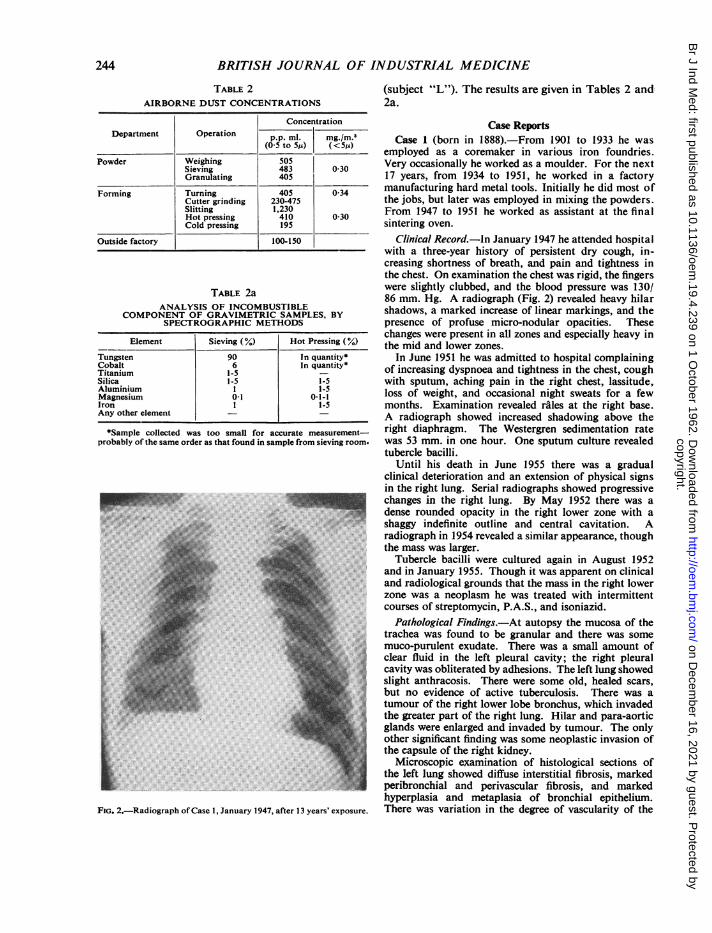
FIG. 2.-Radiograph ofCase 1, January 1947, after 13 years' exposure.
(subject "L"). The results are given in Tables 2 and2a.
Case ReportsCase 1 (born in 1888).-From 1901 to 1933 he was
employed as a coremaker in various iron foundries.Very occasionally he worked as a moulder. For the next17 years, from 1934 to 1951, he worked in a factorymanufacturing hard metal tools. Initially he did most ofthe jobs, but later was employed in mixing the powders.From 1947 to 1951 he worked as assistant at the finalsintering oven.
Clinical Record.-ln January 1947 he attended hospitalwith a three-year history of persistent dry cough, in-creasing shortness of breath, and pain and tightness inthe chest. On examination the chest was rigid, the fingerswere slightly clubbed, and the blood pressure was 130/86 mm. Hg. A radiograph (Fig. 2) revealed heavy hilarshadows, a marked increase of linear markings, and thepresence of profuse micro-nodular opacities. Thesechanges were present in all zones and especially heavy inthe mid and lower zones.
In June 1951 he was admitted to hospital complainingof increasing dyspnoea and tightness in the chest, coughwith sputum, aching pain in the right chest, lassitude,loss of weight, and occasional night sweats for a fewmonths. Examination revealed rales at the right base.A radiograph showed increased shadowing above theright diaphragm. The Westergren sedimentation ratewas 53 mm. in one hour. One sputum culture revealedtubercle bacilli.
Until his death in June 1955 there was a gradualclinical deterioration and an extension of physical signsin the right lung. Serial radiographs showed progressivechanges in the right lung. By May 1952 there was adense rounded opacity in the right lower zone with ashaggy indefinite outline and central cavitation. Aradiograph in 1954 revealed a similar appearance, thoughthe mass was larger.
Tubercle bacilli were cultured again in August 1952and in January 1955. Though it was apparent on clinicaland radiological grounds that the mass in the right lowerzone was a neoplasm he was treated with intermittentcourses of streptomycin, P.A.S., and isoniazid.
Pathological Findings.-At autopsy the mucosa of thetrachea was found to be granular and there was somemuco-purulent exudate. There was a small amount ofclear fluid in the left pleural cavity; the right pleuralcavity was obliterated by adhesions. The left lung showedslight anthracosis. There were some old, healed scars,but no evidence of active tuberculosis. There was atumour of the right lower lobe bronchus, which invadedthe greater part of the right lung. Hilar and para-aorticglands were enlarged and invaded by tumour. The onlyother significant finding was some neoplastic invasion ofthe capsule of the right kidney.
Microscopic examination of histological sections ofthe left lung showed diffuse interstitial fibrosis, markedperibronchial and perivascular fibrosis, and markedhyperplasia and metaplasia of bronchial epithelium.There was variation in the degree of vascularity of the
..
Fi
244
copyright. on D
ecember 16, 2021 by guest. P
rotected byhttp://oem
.bmj.com
/B
r J Ind Med: first published as 10.1136/oem
.19.4.239 on 1 October 1962. D
ownloaded from

HARD METAL DISEASE
fibrous tissue, some areas showing marked vascularitywhilst other areas, mainly hyalinized, were entirely freefrom vessels. There was slight anthracotic pigmentation.No giant cells were seen and there was no evidence oftuberculosis. Sections from the right lung showedsimilar features complicated by the presence of ananaplastic adenocarcinoma. The fibrosis present in thelung did not appear to be a stromal reaction that mightbe related to carcinoma. The general appearance wasthat of a non-specific fibrosis (Figs. 3a and 3b).
Case 2 (born 1897).-Until he joined the Army in 1915,he was employed in a machine tool factory boring steel,then as a bread roundsman, and from 1920 to 1942pressing wood pulp in a textile factory. Since 1942 he hasbeen employed as a millwright in general maintenanceat a factory manufacturing hard metal tools. He alsostripped and cleaned 10 ovens and their flues twice aweek. Since 1956 he has been doing light work, includingstock taking.
Clinical Record.-A radiograph in January 1952 (Fig. 4)revealed some increase in the transverse diameter of theheart, very prominent hilar shadows, and in the mid andlower zones a marked increase of linear markings and thepresence of scattered micro-nodular opacities. Someconfluence of these opacities was present in the lowerzones, especially on the left side. He was first seenin January 1954 and gave a five-year history of coughwith copious frothy sputum, of dyspnoea on exertion,and of recent left chest pain and haemoptysis. Onexamination the chest was rigid and emphysematous, thebreath sounds were distant, rales were audible at thebases, and the blood presure was 190/120 mm. Hg.A radiograph at this time revealed increased prominenceof the hilar shadows and the same distribution of thelinear and micro-nodular opacities, though there was anapparent contraction and hardening of the linear opacitiesand an increased profusion of the micro-nodular opacities.The Westergren sedimentation rate was 10 mm. in onehour. The haemoglobin was 14 6 g./100 ml., the redblood cell count 4,900,000/c.mm., and the leucocyte count8,400/c.mm. (68% polymorphs, 25% lymphocytes, 5%monocytes, and 2% eosinophils). The Wasserman andKahn tests were negative. Sputum cultures were negativefor tubercle bacilli.
In March 1956 he was admitted to hospital with anacute respiratory infection. He was cyanosed, breathless,and had increased cough with purulent sputum. Therewere increased signs at the bases of both lungs, slighthepatic enlargement, tachycardia, slight oedema of theankles, and elevation of the jugular pressure. The bloodpressure was 120/70 mm. Hg. The haemoglobin was15-6 g./l00 ml., the red cell count 5,400,000/c.mm., andthe leucocyte count 9,800/c.mm. (83% polymorphs, 13°%lymphocytes, 2% large monocytes, and 2% eosinophils).Urine analysis revealed 0 35 g. of albumin and the pre-sence of casts. An electrocardiogram showed left axisdeviation. The signs of early cardiac failure clearedrapidly as the respiratory infection responded to anti-biotics.For the past five years he has suffered from severe
persistent dyspnoea on exertion with wheezing and
AW~~~~~~~~~~~~~
, .
FIG. 3a.-Section of lung from Case I showing diffuse fibrosis andmarked peribronchial and perivascular fibrosis. In the bottom-left hand corner is illustrated a small bronchus showing markedhyperplasia and metaplasia of the lining epithelium. ( x 75.)
FIG. 3b.-Shows the airway seen in Fig. 3a at higher magnification.(x 290.)
245
copyright. on D
ecember 16, 2021 by guest. P
rotected byhttp://oem
.bmj.com
/B
r J Ind Med: first published as 10.1136/oem
.19.4.239 on 1 October 1962. D
ownloaded from

BRITISH JOURNAL OF INDUSTRIAL MEDICINE
FIG. 4.-Radiograph of Case 2, January 1952, 10 years' exposure.
tightness, persistent cough and sputum with infrequentsputum staining and occasional palpitations. During thewinters, he has been away from work for periods ofseveral months with respiratory infections and exacer-bations of symptoms. In March 1961 examinationrevealed a blood pressure of 170/100 mm. Hg, a rigid
FIG. 5.-Radiograph of Case 2, March 1961, 19 years' exposure.
emphysematous chest, and the presence of basal rales.The liver and spleen were palpable. A Heaf test waspositive. The urine analysis was normal. A radiograph(Fig. 5) revealed the presence of heavy linear markingsand profuse micro-nodular opacities in the right upperzones, further contraction of the linear opacities in themid and lower zones, and increased translucency in theright mid and lower zones. An electrocardiogram showedsupraventricular and ventricular ectopic beats in thechest leads and left axis deviation. The haemoglobin was13-7 g./100 ml. and the leucocyte count 5,000/c.mm.(65% polymorphs, 30% lymphocytes, 4% large mono-cytes, and 1% eosinophils). The blood urea was 28 mg./100 ml. The plasma proteins were 7-5 g./lOOml. (albumin4 1, globulin 3-4: a, globulin 0 3 g./lOOml., a2 0 7g./100 ml., P 09 g./100 ml., and y 1l5 g./100 ml.). Liverfunction tests were normal.
Physiological Assessment.-The physiological findingsare summarized in the Appendix. The ventilatorycapacity was moderately reduced and increased slightlyafter adrenalin. The percentage of the forced vitalcapacity which could be expelled in one second wasnormal. The lung airways resistance and static compliancewere normal; the dynamic compliance was reduced. Thetotal lung capacity and its subdivisions were normal. Thepulmonary diffusing capacity was low. The patienthyperventilated on exercise breathing air; the hyper-ventilation was associated with a low arterial carbondioxide tension, and a fall in the arterial oxygen tensionfrom its resting value.The patient's dyspnoea on exertion was probably the
result of hyperventilation and some reduction in ventila-tory capacity. The low diffusing capacity and the arterialblood gas changes, together with the normal forcedexpiratory volume ratio and airways resistance, suggestedsome degree of alveolar capillary block. But the differencebetween static and dynamic compliance and the moderatereduction in ventilatory capacity indicated also somedegree of obstructive lung disease.
Case 3 (born 1910).-He started work in 1924 and wasemployed in turn as a spinner in various cotton millsfor nine years, as a maintenance fitter for six years, andas a storekeeper for seven years. He was next employedas a steel hardener for nine years. In June 1955 he startedwork in a factory manufacturing hard metal tools. Hewas employed mixing the powders with a hand-operatedmixer in a separate room with a low ceiling. He then fedthe mixed powder into an extruding machine, placed theextruded material in an oven and later removed it. After21 months he developed symptoms and gave up thiswork in March 1957. He restarted work in the samefactory in July 1957 partly as a gardener and partly as astorekeeper. Apart from three months' sickness absencein 1958 he remained in this employment until May 1960when he returned to steel hardening.
Clinical Record.-In November 1952 he underwent aleft orchidectomy for tuberculous epididymo-orchitis.His health was otherwise good and he had no respiratorysymptoms. A radiograph was normal. For the next fouryears he remained well and periodic radiographs werenormal.
246
copyright. on D
ecember 16, 2021 by guest. P
rotected byhttp://oem
.bmj.com
/B
r J Ind Med: first published as 10.1136/oem
.19.4.239 on 1 October 1962. D
ownloaded from

HARD METAL DISEASE
He reattended in March 1957, 17 months after the lastnormal radiograph, with six weeks' history of cough withsputum, moderate dyspnoea on exertion, tightness acrossthe chest, and lassitude. Examination revealed harshbreath sounds and the presence of basal rales. A radio-graph (Fig. 6) revealed a well-marked increase in linearmarkings, the presence of profuse micro-nodular opacitiesin all zones, but particularly heavy in the mid and lowerzones. Repeated sputum cultures for tubercle bacilliwere negative. The Westergren sedimentation rate was3 mm. in one hour. The haemoglobin was 15 5 g./100 ml.,and the leucocyte count 8,700/c.mm. (69% polymorphs,23% lymphocytes, 6% large monocytes, and 2% eosino-phils). Urine analysis was normal. Following sympto-matic treatment his symptoms slowly improved and werenoticeably less by July 1958.
In October 1958 he developed a tuberculous rightepididymo-orchitis. A radiograph at that time revealedno change. He was treated with streptomycin, isoniazid,and P.A.S. until September 1959. He was by this timevirtually free of respiratory symptoms. His subsequentprogress has been uneventful.
In March 1961 examination was normal, the bloodpressure being 140/80 mm. Hg. A Heaf test was positive.A chest radiograph revealed some contraction of thelinear opacities and the same distribution and profusionof the micronodular opacities. The haemoglobin was13-1 g./100 ml., the leucocyte count 5,000/c.mm. (65%polymorphs, 30% lymphocytes, 4O% large monocytes,and 1% eosinophils) and the Westergren sedimentationrate 11 mm. in one hour. The plasma proteins were7-7 g./100 ml. (albumin 40, globulin 3-7: a, globulin,a2 0 7 g./100 ml., f 1 0 g./100 ml., and y 1-6 g./100 ml.).Liver function tests were normal.
Physiological Assessment.-The physiological findingsare summarized in the Appendix. The ventilatorycapacity, lung airways resistance, total lung capacity,and its subdivisions were within normal limits; thelung compliance (both static and dynamic) and diffusingcapacity were somewhat reduced. The patient ventilatednormally on exercise, but the arterial blood oxygentension, which was normal at rest, fell on near maximalexercise. This finding confirmed the small diffusion defectwhich, in the presence of a reduced compliance, wasprobably due to slight pulmonary fibrosis.
Case 4 (born 1894).-In 1903 he was employed in turnin cycle repairing, brake testing, and again in cyclerepairing, until he joined the Army in 1916. In 1919he worked in a furniture shop and then as a packer.From 1924 to 1946 he was employed in a factory manu-facturing hard metal tools as a shaper. He then retiredfor health reasons. For the last 10 years of the perioddust extraction was operating.
Clinical Record.-From 1937 he suffered from slightdyspnoea on exertion, slight cough, and occasionalpalpitations. A radiograph taken by the miniature massradiography unit in October 1947 (Fig. 7) revealed anenlarged heart, punctiform nodules, and increased linearmarkings over two-thirds of each lung field, and an areaof increased translucency at the right base with oblitera-tion of both costophrenic angles.
..I
FIG. 6.-Radiograph of Case 3, March 1961, 17 months' exposure.
In July 1948 he first attended hospital complaining ofincreased dyspnoea on exertion, cough, and palpitations,also loss of weight, lassitude, headache, and soreness of
FIG. 7.-Radiograph of Case 4, October 1947, 23 years' exposure.
247
A.-
copyright. on D
ecember 16, 2021 by guest. P
rotected byhttp://oem
.bmj.com
/B
r J Ind Med: first published as 10.1136/oem
.19.4.239 on 1 October 1962. D
ownloaded from

BRITISH JOURNAL OF INDUSTRIAL MEDICINE
Fio. 8.-Radiograph of Case 5, December 1961, 15 years' exposure.
the throat and mouth. The blood pressure was 275/105mm. Hg. Aortic incompetence was present, and the chestwas rigid and emphysematous. Urine analysis revealedthe presence of albumin. The haemoglobin was 14-3 g./100 ml., the red blood cell count 4,760,000/c.mm. (1 %reticulocytes) and the leucocyte count 8,500/c.mm. (63 %polymorphs, 30% lymphocytes, 5% monocytes, and 2 %eosinophils).He was admitted to hospital in January 1949 with a
coronary thrombosis and a left pleural effusion, due topulmonary infarction. An electrocardiogram showedchanges consistent with anterior myocardial infarction,complete heart block, and left axis deviation. Thehaemoglobin was 15 5 g./100 ml., the red blood cellcount 5,110,000/c.mm. (0'5% of reticulocytes) and theleucocyte count 22,000/c.mm. (73% polymorphs). Thepleural fluid was lymphocytic and sterile on culture.The sputum was negative for tubercle bacilli on culture.In 1951 he had a cerebral haemorrhage and died. Noautopsy was performed.
Case 5 (born 1913).-He first worked as a grocer for10 years and then as a turner for eight years making steeldies, until 1946. For 16 years, since 1946 he has beenemployed as a turner in the shaping department of afactory manufacturing hard metal tools. The work in-volves the use of a diamond wheel and is carried outbefore final sintering.
Clinical Record.-His health has always been good, hisonly symptom being a recent dry cough. Examinationwas normal, the blood pressure being 125/80 mm. Hg.A radiograph in December 1961 (Fig. 8) revealed anincrease of linear markings and the presence of scatteredpunctiform opacities particularly in the mid and lowerzones. Urine analysis was normal. A Heaf test was
positive. The haemoglobin was 14-9 g./100 ml. and theleucocyte count 7,000/c.mm. (68% polymorphs, 21 %lymphocytes, 5% large monocytes, 4% eosinophils, and2% basophils). The Westergren sedimentation rate was2 mm. in one hour. The plasma proteins, the globulinfractions, and liver function tests were normal.
Case 6 (born 1921).-After one year in the boot andshoe trade he started work at an engineering factory as asurface grinder in 1938. Three years later he transferredto the tool department. Initially he was partly engagedin brazing hard metal tips onto steel tools and partly insharpening their cutting edges by dry grinding withdiamond and carborundum wheels. After a few years hebecame wholly engaged in dry grinding hard metal tools.
Clinical Record.-In October 1946 a routine radio-graph was normal. He first attended hospital in August1949 with six months' history of dry cough. Examinationwas normal. A radiograph (Fig. 9) revealed the pre-sence of increased linear markings and faint nodularopacities in the mid and upper zones.During the next seven years he remained well and his
weight increased, though his dry cough persisted inter-mittently. Periodic examinations were normal. Serialradiographs showed slowly increasing changes. A radio-graph in June 1954 (Fig. 10) revealed slight widening ofthe superior mediastinum and prominence of the hilarshadows especially on the left side, also a marked increasein linear markings, and the presence of profuse nodularopacities in all zones, these changes being most evident inthe right lung in the upper and lower zones.
In June 1956 he reattended hospital following arespiratory infection with two months' history of slightdiscomfort in the chest, nasal catarrh, and cough withsputum. Examination revealed some restriction of chestexpansion and the presence of occasional mid and lowerzone rhonchi. A radiograph (Fig. 11) revealed furtherwidening of the superior mediastinum with rotation of theheart and traction of the trachea to the right, a very heavyleft hilar shadow and a very marked increase of linearmarkings particularly in the upper and right lower zones,also the presence of extremely profuse nodular opacitiesin all zones with areas of confluent shadowing in theright lower zone and the left mid-zone near the lefthilum. Direct examination of sputum for tuberclebacilli was negative. From July 1956 to April 1957 hewas given 200 mg. of isoniazid and 16 g. of P.A.S. daily,whilst remaining at work. Following this treatmentthere was no radiographic clearing. A Heaf test wasnegative, a Mantoux test was positive to 10 tuberculinunits, a Westergren sedimentation rate was 6 mm. in onehour, the plasma proteins were normal and a Wassermantest was negative. The findings on examination wereunchanged, the blood pressure being 150/70 mm. Hg.From March 1959 to September 1959 he was treated
with 10 mg. of prednisolone, 200 mg. of isoniazid, and16 g. P.A.S. daily, whilst remaining at work. No radio-graphic clearing was seen following this treatment.A radiograph in September 1961 (Fig. 12) revealedincreased distortion of the pulmonary architecture.There were further widening of the superior mediastinum,
248
copyright. on D
ecember 16, 2021 by guest. P
rotected byhttp://oem
.bmj.com
/B
r J Ind Med: first published as 10.1136/oem
.19.4.239 on 1 October 1962. D
ownloaded from

HARD METAL DISEASE
FIG. 9.-Radiograph of Case 6, August 1949, eight years' exposure.
increased rotation of the heart and traction of the tracheato the right, and additional areas of confluence in theupper zones.During the past five years he has remained well and
has continued to gain weight. He has complained inter-mittently of a dry cough and latterly of slight dyspnoeaon exertion. Serial radiographs have shown slowlyprogressing changes in both lungs.
Table 3 provides a summary of these six cases.
FIG. 10.-Radiograph of Case 6, June 1954, 13 years' exposure.
DiscussionPast experience in a number of hard metal
factories has shown that there have been complaintsof cough, expectoration, shortness of breath, andtightness of the chest. These symptoms are com-paratively mild in the majority of workers. A fewindividuals experience well-marked respiratory dis-tress towards the end of the working day. The
.-. ;Egis«
.l..... W:Ri
............*: ......
::: y
0
......
*.Sb
FIG. 12.-Radiograph of Case 6, September 1961, 20 years' exposure.
249III
FIG. I I.-Radiograph of Case 6, June 1956, 15 years' exposure.
copyright. on D
ecember 16, 2021 by guest. P
rotected byhttp://oem
.bmj.com
/B
r J Ind Med: first published as 10.1136/oem
.19.4.239 on 1 October 1962. D
ownloaded from

BRITISH JOURNAL OF INDUSTRIAL MEDICINE
TABLE 3
SUMMARY OF CASES
Case Year of Birth Occupation Exposure (years) Date of Radiological Diagnosis
1888 Powder worker 1934-1951: 17 19472 1897 Millwright 1942-1962: 20 19523 1910 Powder worker 1955-1957: 1 yr 10 mths 19574 1894 Shaper 1924-1946: 22 19475 1913 Shaper 1946-1962: 16 19616 1921 Grinder 1941-1962: 21 1949
results of the physiological tests obtained at thefactory survey showed that a change in respiratorymechanics was occurring during a day's exposureto dust in two of the 19 workmen tested, thoughsome other of the workers did complain of cough,wheezing, and shortness of breath. Both of thesemen experienced less distress when wearing masks.In the men with severe symptoms there appears tobe considerable variation in the length of exposurebefore symptoms arise but in some these maydevelop quite quickly. The majority of thosedeveloping severe symptoms leave and are thusexcluded from the industry. At an early stage suchsymptoms may be reversible on withdrawal fromexposure.The extent of the relation between susceptible
individuals with respiratory symptoms and acutephysiological changes and those individuals withestablished hard metal disease and permanentpulmonary damage is uncertain.Hard metal disease may arise abruptly without
preliminary respiratory symptoms. One man(Case 3), who had been under regular clinical andradiological surveillance for five years beforeexposure started, developed symptoms after 21months, and his radiograph showed widespreadpulmonary changes six weeks later. The disease maydevelop with little or no preceding respiratorysymptoms and with no symptoms of significancewith established minimal disease as in Case 5, andmarked radiographic changes may be associatedwith mild symptoms as in Case 6. Morbidityappears to be extremely low and only a very smallproportion of those exposed become affected.Moschinski et al. (1959) believe that factors ofindividual susceptibility play a significant part in thedevelopment and subsequent course of the disease.They cite the example of a father and son who bothdeveloped the disease. Degrees of susceptibilitywould account for the marked variation in the lengthof exposure, the severity of the disease, and the ratesof progression.
There have been reports of radiological clearingon withdrawal from exposure (Miller et al., 1953;Lundgren and bhman, 1954). This has not been
obvious in our cases. It seems likely that the progressof the disease is affected by continued exposure.Since the disease was first detected radiologicallyCases 2, 3, and 6 have been studied for nine, four,and 12 years respectively. There has been gradualradiological progression and increasing respiratoryinsufficiency with continued exposure in Case 2,whereas in Case 3 without further exposure minimalradiological progression has been accompanied bygradual disappearance of respiratory symptoms.In Case 6 there has been steady radiological pro-gression accompanied by only slight symptoms withcontinued exposure.
It is possible that the pulmonary response is of thenature of a hypersensitivity. This is suggested bythe reversibility of the symptoms, by the occasionalradiological clearing on withdrawal from exposure,and by the precipitation of the symptoms on re-exposure (Miller et al., 1953). The animal experi-ments of Delahant (1955) support this view. Thistype of response has been suggested by Miller et al.(1953), and by Moschinski et al. (1959). Lundgrenand Ohman (1954) have pointed out that there is nocorrelation between skin allergy as demonstrated byskin tests and pulmonary symptoms.The pathological findings in Case 1 of our series
are broadly similar to those described in the literatureand correlate well with the physiological changesfound in Cases 2 and 3, which indicated mild alveolardiffusion defects.As far as we are aware Case 1 of our series is the
first example reported of carcinoma of the bronchusdeveloping in a patient suffering from hard metaldisease. The finding of bronchial epithelial hyper-plasia and metaplasia is of interest in the light of theanimal experiments of Schepers (1955) and of Kaplunand Mezencewa (1960), and of Heath (1960).
Miller et al. (1953) found raised plasma proteinlevels with elevation of globulin in the cases hedescribed. In our series the plasma proteins wereestimated by electrophoresis in three cases. Themethods and normals were based on the paper bySalt (1956). In Cases 2 and 3 minor abnormalitieswere found. High haemoglobin levels were found inthree of our series. These levels though within the
250
copyright. on D
ecember 16, 2021 by guest. P
rotected byhttp://oem
.bmj.com
/B
r J Ind Med: first published as 10.1136/oem
.19.4.239 on 1 October 1962. D
ownloaded from

HARD METAL DISEASE
TABLE 4RANGE OF CONCENTRATIONS (p.p. ml.) FOUND
BY OTHER WORKERS
Department Mosehinsky |Ludgren Fairhall Our Ownetal.and Ohman et hal.
Powder 300-1,200 10-2,000 1 100 405- 505Forming 200-1,400 10-2,000 f(average) 195-1,230
upper limits of normal are in line with the findingsof Miller et al. (1953) and S. G. Rainsford (1959,personal communication).Dust concentrations reported by other workers
who have investigated hard metal disease are givenin Table 4 (Fairhall, Keenan, and Brinton, 1949;Lundgren and Ohman, 1954; Moschinski et al.,1959). Unfortunately differences in samplingtechnique and instruments make comparison ofdoubtful validity, but it appears that the dust con-centrations we measured were of the same order asreported elsewhere, and this perhaps indicates thatit should not be assumed that previous cases of thisdisease have only occurred under conditions of highdust concentration. In Russia maximum acceptableconcentrations are 0 5 mg./m.3 for cobalt (cobaltoxide) and 6 mg./m.3 for tungsten and tungstencarbide (Elkins, 1961). We found a concentration ofbetween 400 to 500 particles per ml. (p.p.ml.) closeto the working positions of the men and about200 p.p.ml. away from these positions, and atslitting (cutting with a thin grinding wheel) 1,230p.p.ml. Background dust levels contributed 100 to150 p.p.ml. The analysis of the dust depended uponthe composition of the materials being handled ormachined and therefore varied throughout thefactory.The agent responsible for hard metal disease is as
yet unknown. Cobalt in animal experiments hasbeen shown to produce pulmonary lesions, hyper-globulinaemia, and erythrocytaemia, and to be thetoxic constituent in hard metal. In man cobalt isknown to produce a skin allergy and an erythro-cytaemia. Tungsten, tungsten carbide, and titaniumhave been found to be relatively inert in animalexperiments, and tungsten and titanium are com-paratively harmless in man. Post-mortem analysis oflungs of hard metal workers have shown the presenceof tungsten and titanium, but cobalt has not beenfound, perhaps due to its high solubility in plasma.Nagelschmidt (1960) has suggested that the reactionwith a soluble dust is an interstitial or disseminatedfibrosis and with an insoluble one a nodular or focalfibrosis.The acquisition of more information on the risks
of grinding hard metal is desirable as this process iscarried out in many engineering factories. The three
cases described by Miller et al. (1953) and our sixthcase were grinders, although Moschinski et al. (1959)were of the opinion that after final sintering therewas no significant dust hazard. Miller et al. (1953)have demonstrated the toxic properties of dustderived from grinding hard metal in animal experi-ments.
We are grateful to Dr. P. Tyson Davidson and Dr.J. E. Cottrell who first surveyed hard metal workers inthis country.We are deeply indebted to the staff of the Pneumo-
coniosis Research Unit for their help and advice; toDr. C. B. McKerrow and Dr. J. C. Gilson for carrying outthe physiological tests at the factory survey and forinterpreting the results, to Mr. J. W. Skidmore for dustmeasurements, and Dr. J. E. Cotes for carrying out thephysiological studies in Cases 2 and 3 and for interpretingthe results; also to Miss Pamela Edwards of this unit.We are also grateful to Dr. A. A. White and Dr. R. B.
Mayfield for allowing us to include Case 6; and to Dr.G. E. Smith, Dr. Ethel Browning, Dr. J. GwynneMorgan, Dr. D. Frost, Dr. J. A. Duncan, and Dr. A. 1.G. McLaughlin for assistance; to Dr. A. P. Prior, Dr.D. Rivers, and Professor J. P. Wyatt for advice onpathology and to Dr. Gordon Evans for the MassRadiography Surveys; to Miss A. L. Hebdeb for radio-graph reproductions, and to Mr. S. Gaunt for photo-micrographs, also to Miss J. M. Gick for secretarialassistance.
Finally we should like to thank Dr. T. A. Lloyd Daviesfor his encouragement and the members of the firmsconcerned for their wholehearted co-operation.
REFERENCES
AhImark, A., Bruce, T., and Nystrom, A. (1961). Silicosis and OtherPneumoconioses in Sweden, p. 390. Svenska Bokforlaget,Stockholm.
Browning, E. (1960). In Modern Trends in Occupational Health,ed. R. S. F. Shilling, p. 85. Butterworth, London.
(1961). ToxicitY of Industrial Metals, p. 115. Butterworth,London.
Caujolle, F., and Meynier, D. (1959). Rev. Path. gen., 59, 245.Delahant, A. B. (1955). A.M.A. Arch. industr. Hlth, 12, 116.Elkins, H. B. (1961). Arch. environm. Hlth, 2, 45.Fairhall, L. T., Castberg, H. T., Carrozzo, N. J., and Brinton, H. P.
(1947). Occup. Med., 4, 371.-, Keenan, R. G., and Brinton, H. P. (1949). P,ibl. Hlth Rep.
(Wash.), 64, 485.Frederick, W. G., and Bradley, W. R. (1946). Rep. of 8th Ann.
Meeting of Amer. Industr. Hyg. Ass. Chicago.Harding, H. E. (1950). Brit. J. industr. Med., 7, 76.Heath, J. C. (1960). Brit. J. Cancer, 14, 478.Husten, K. (1959). Arch. Gewerbepath. Gewerbehyg., 16, 721.Jobs, H., and Ballhausen, C. (1940). Vertrauensarzt, 8, 142.Kaplun, Z. S. (1957). Iseretyne Met., 9, 42.
, and Mezencewa, N. W. (1960). J. Hyg. Epidem. (Praha), 4, 390.Karth, B. (1948). Rep. of meeting of Swedish Ass. Int. Med.Lundgren, K. B., and Ohman, H. (1954). Virchows Arch. path. Anat.,
325, 259.McKerrow, C. B., Roach, S. A., Gilson, J. C., and Schilling, R. S. F.
(1962). Brit. J. industr. Med., 19, 1.Magos, L. N., Timar, U. A., and Szandanyi (1956). Quoted by
Moschinski et al. (1959).Miller, C. W., Davis, M. W., Goldman, A., and Wyatt, J. P. (1953).
A.M.A. Arch. industr. Hyg., 8, 453.Moschinski, G., Jurisch, A., and Reinl, W. (1959). Arch. Gewerbepath.
Gewerbehyg., 16, 697.Nagelschmidt, G. (1960). Brit. J. indiestr. Med., 17, 247.
251
copyright. on D
ecember 16, 2021 by guest. P
rotected byhttp://oem
.bmj.com
/B
r J Ind Med: first published as 10.1136/oem
.19.4.239 on 1 October 1962. D
ownloaded from

BRITISH JOURNAL OF INDUSTRIAL MEDICINE
Salt, H. B. (1956). Clin. Chem., 21, 35.Schepers, G. W. H. (1955). A.M.A. Arch. industr. Hlth, 12, 121, also
127 134, 137, and 140.Schwartz, L., Peck, S. M., Blair, K. E., and Markuson, K. E. (1945).
J. Allergy, 16, 51.Stokinger, H. F., and Wagner, W. D. (1958). A.M.A. Arch. industr.
Hlth, 17, 273.
Uzman, L. L., and Rosen, H. (1954). Science, 120,1031.Volta, A., and Marinoni, U. (1951). IlPoliclinico, Sez. Med., 58, 145.Wiele, G. (1951). Rep. symp. Rhine-Westphalia Inst. intern. Med.Wolff, H. S., and Roach, S. A. (1961). In InhaledParticles and Vapours,
Proc. Int. Symposium organized by the British OccupationalHygiene Society, ed. C. N. Davies, p. 460. Pergamon Press,London.
APPENDIXPulmonary Function Studies
Age (years)Ventilatory CapacityForced expiratory volume (F.E.V.,.,) (1.)Forced expiratory volume after adrenalin (I.)Forced vital capacity (F.V.C.) (1.)Forced vital capacity after adrenalin (1.)F.E.V. % F.V.C.F.E.V. % F.V.C. after adrenalin
Lung Capacity (helium dilution method)Total lung capacity (T.L.C.) (I.)Functional residual capacity (F.R.C.) (1.)Residual volume (R.V.) (1.)Rd. V% T.L.C.
Pulmonary Diffusing Capacity (single breath method)Diffusing capacity of lung (DLCO) (ml./mm./mm. Hg)Diffusing capacity of membrane (Dmco) (ml./mm./mm. Hg)Volume of lung capillary (Vc) (ml.)
Lung MechanicsAirways resistance on inspiration by body plethysmograph (cm. H,O/1./m.)
Compliance by oesophageal balloon (I./cm. H2O)StaticDynamic
Blood Gas and Exercise StudiesRest (on air)
Arterial gas tensions (PaCOO) (mm. Hg)(PaO2) (mm. Hg)
Arterial blood saturation (SaO, %)
ExerciseTreadmill speed (m.p.h.)Treadmill slope (%)
Time (min.)Exercise ventilation V1. (I./min.)PaCO2 (mm. Hg)PO, (mm. Hg)S.0, %Exercise ability improved by 0,
PaCO2 indirect (Campbell method)
Case 2
63
2-032-182-902-80
7078
5 983-732-61
43-6
13-324-043
1*37Flow rate up to 0 5 1./sec.
0-1770074
On Air1 75
51-631-761-779-0
32-172-293.9
2
10
On Oxygen1-75
35.542-2
100-2Yes42-6
On Air2
46-639-17293-2
Case 3
51
3-083-083-653 70
8483
5-723 701-78
31-2
18 832-066
074
0-100-129
41-394099.5
3
16
On Oxygen2
42-645 8
99-1Yes39.4
252
copyright. on D
ecember 16, 2021 by guest. P
rotected byhttp://oem
.bmj.com
/B
r J Ind Med: first published as 10.1136/oem
.19.4.239 on 1 October 1962. D
ownloaded from