hazard assessment of bisphenol a - minister of economy ... · hazard assessment of bisphenol a...
TRANSCRIPT

Bisphenol A
328
Hazard assessment of Bisphenol A
[Bisphenol A, CAS No. 80-05-7]
Chemical name : Bisphenol A
Synonyms : 2,2-bis (p-hydroxyphenyl) propane, 4,4'-(1-methylethylidine)
diphenol, 4,4'-isopropylidenediphenol, BPA
Molecular formula : C15H16O2
Molecular weight : 228.29
Structural formula :
C
CH3
CH3
OHHO
Appearance : White flakes 1)
Melting point : 150 - 155°C 1)
Boiling point : 220°C (533 Pa) 1)
Specific gravity : 2525d = 1.195 1)
Vapor pressure : 5.3 × 10⁻⁶ Pa (25°C) 1)
Partition coefficient : Log Pow = 3.32 (observed value) 1)
Degradability : Hydrolyzability: No Report
Biodegradability: Slightly degradable (BOD=0%, 14 days) 2)
Solubility : Water, 120 mg/l (25°C) 1) , Soluble in aqua alkaline solution
Organic solvent: Soluble in acetone, ethanol, ether and
benzene. Slightly soluble in carbon tetrachloride.
Amout of production/import : 150,697t in 1998 (manufactured: 149,984t, imported: 713t)5)
Usage : Raw material for epoxy resin and polycarbonate resin. Raw
material for phenol resin and antioxidant, etc.1).
Applied laws ®ulations : Law Concerning Reporting, etc. of Release Special Chemical
Substances to the Environment and Promotion of the
Improvement of Their Management

Bisphenol A
329
1) HSDB, 2001; 2) Tsusanshou Koho (Daily), 1977; 3) Ministry of International Trade and Industry,
1999.
1. Toxicity Data
1) Information on adverse effects on human health
Mild skin irritation is reported by contact with BPA dust (Handbook of Industrial
Poisoning). There is a report that positive patch test reactions to BPA and resins
containing 0.014 or 0.015% of BPA were demonstrated, but not to major component of the
resins, when allergic reaction tests were conducted for a female dental technician who had
used for 4 years dental composite resin, major component of which was epoxide of BPA
containing trace amount of BPA, and had dermatitis at her hands. In this case the patient
showed positive reaction also to formaldehyde contained in the resin as an impurity.
Thus, synergy between BPA and formaldehyde was suspected. Furthermore, the
composition of the resin actually in use was not know and it was not clear which
substance caused the dermatitis as well as whether synergy between BPA and
formaldehyde existed (Jolanki et al., 1995).
There is no report on the carcinogenicity of this substance in humans.
2) Information on endocrine system and reproductive system
(1) In vitro test result related to receptor binding (Attachment-1)
In a receptor binding test, BPA binds to estrogen receptor (ER) in human and rat ( its
affinity is 1/500 - 1/15,000 of that to estradiol (E2)) (Sheeler et al., 2000; Blair et al.,
2000; Nagel et al., 1997; CERI, 2001). The genetic activation dependent on ERE
(estrogen responsive sequence) was also noted in a reporter gene assay using yeast
introduced with human ER (including two-hybrid assay) and animal cells in which human
or rat estrogen receptors were introduced, and the extent of activation was 1/600 -
1/130,000 of that of E2 (Sheeler et al., 2000; Nishihara et al., 2000; Coldham et al., 1997;
Gaido et al.,1997; Hiroi et al., 1999; Legler et al., 1999; CERI, 2001; Yamasaki et al.,
2001). The EC50 of BPA was 3.1 × 10⁻⁶ M in a human ER dimer formation test using
yeast two hybrid assay, indicating that concentration of 26,000 times higher than that of
E2 (1.2 × 10⁻¹⁰ M) was needed for dimer formation of estrogen receptor (Sheeler et al.,
2000). In an experiment to investigate influence on endogenous estrogen responsive

Bisphenol A
330
gene, induction of pS2, etc. was noted but at the concentration of 100,000 - 1,000,000
times higher than that of E2 (Jorgensen et al., 2000). BPA (1 nM) activated the gene in a
reporter gene assay using promoter domain of prolactin gene (Steinmetz et al., 1997).
(2) in vivo test results in mammals (Attachment-2, 3)
The results of short term test to detect estrogenic action in mammals and the results
of reproductive and fertility toxicity test are shown in Attachment-2 and 3 respectively.
Uterotrophic assay (according to the OECD guideline draft) has been conducted
using rats and mice (Attachment-2).
The uterotrophic assay by subcutaneous administration of BPA at 0, 0.02, 0.2, 0.8, 2
and 8 mg/kg/day for 4 days in ovariectomized female B6C3F1 mice (35 - 60 days old)
revealed increased uterus weight at 0.8 mg/kg/day or more (Papaconstantinous et al.,
2000). On the other hand, no changes in uterus weight occurred in uterotrophic assay by
subcutaneous administration of BPA at 0, 0.01, 0.1, 1, 10 and 100 mg/kg/day for 3 days in
juvenile female CD-1 mice (21 days old) (Mehmood et al., 2000).
The uterotrophic assay of BPA at 0, 40, 160 and 800 mg/kg/day by gavage as well as
by subcutaneous administration at 0, 8, 40 and 160 mg/kg/day both for 3 days in juvenile
female SD (18 days old) or DA/Han (130g body weight) rat revealed increase in uterus
weight by oral administration at 160 mg/kg/day or more and by subcutaneous
administration at 8 mg/kg/day or more (Yamasaki et al., 2000; Diel et al., 2000).
Furthermore, when subcutaneous administration of BPA at 0, 2, 20 and 200 mg/kg/day
was conducted for 3 days in juvenile female SD rats (20 days old), increase in uterus
weight occurred at 20 mg/kg/day or more (Yamasaki et al., 2001). In another
uterotrophic assay in which a capsule corresponding to 0.3 mg/kg/day was subcutaneous ly
implanted in ovariectomized female SD rats (7 - 8 week old) and F344 rats (7 - 8 week
old), increase in uterus weight and increase in height of uterine epithelial cells occurred in
F344 rats, but nothing abnormal was noted in SD rats (Steinmetz et al., 1998). By oral
administration by gavage of BPA at 0, 100, 200 and 400 mg/kg/day for 3 days in juvenile
female Long Evans rats (21 days old), increase in uterine weight was noted at 200
mg/kg/day or more when the animals had been examined after 6 hours, but nothing
abnormal was detected after 24 hours from the final administration (Laws et al., 2000).
When oral administration by gavage of BPA at 0, 500, 750, 1,000 and 1,250

Bisphenol A
331
mg/kg/day was conducted in female CD-1 mice (age unknown) from day 6 to 15 of
gestation, relative liver weight of dams increased at 500 mg/kg/day or more, and
suppression of body weight increase, decreased pregnant uterus weight and increased
embryo resorption in the dams as well as decreased fetal body weight were noted at 1,250
mg/kg. However, no deformation was observed (Morrissey et al., 1987).
When BPA at 0, 2,500, 5,000 and 10,000 ppm (corresponding to 0, 437, 875 and
1,750 mg/kg/day respectively) in diet were administered to male and female CD-1 mice
(age unknown) in a 2-generation reproductive test in which F0 males or females given BPA
were mated with untreated females or males, decrease in number of offsprings born and
their survival were noted at 875 mg/kg/day or more, as well as decreased body weight
(females), increased liver and kidney weights (males and females), decreased seminal
vesicle weight and spermatozoic motility, and increased mortality of offsprings before
weaning at 1,750 mg/kg/day in regard to F0 generation, while increased liver and kidney
weights (males and females) and decreased epididymis and seminal vesicle weights were
observed at 437 mg/kg/day or more for F1 generation (Reel et al., 1997).
By oral administration of BPA at 0, 160, 320, 640 and 1,280 mg/kg/day in female
SD rats (age unknown), decreased body weight at 160 mg/kg/day or more and death at
1,280 mg/kg/day occurred in the dams, but nothing abnormal was observed in fetuses
(Morrissey et al., 1987).
When BPA at 0, 1,000, 3,000 and 9,000 ppm (equivalent to 0, 50, 150 and
450 mg/kg/day, respectively), or BPA at 0, 100, 250, 500, 750 and 1,000 ppm (equivalent
to 0, 5, 13, 25, 38 and 50 mg/kg/day, respectively) in diet were administered to male and
female SD rats (age unknown) for 17 weeks in a single generation reproductive test,
decreased body weight at 150 mg/kg/day or more occurred in F0 generation, while
decreased body weight at 50 mg/kg/day or more was noted in F1 generation. In the
repeated study the decreased body weight was observed at 50mg/kg/day or more in F0, but
not at 50mg/kg/day in F1 (German Chemical Society, 1995).
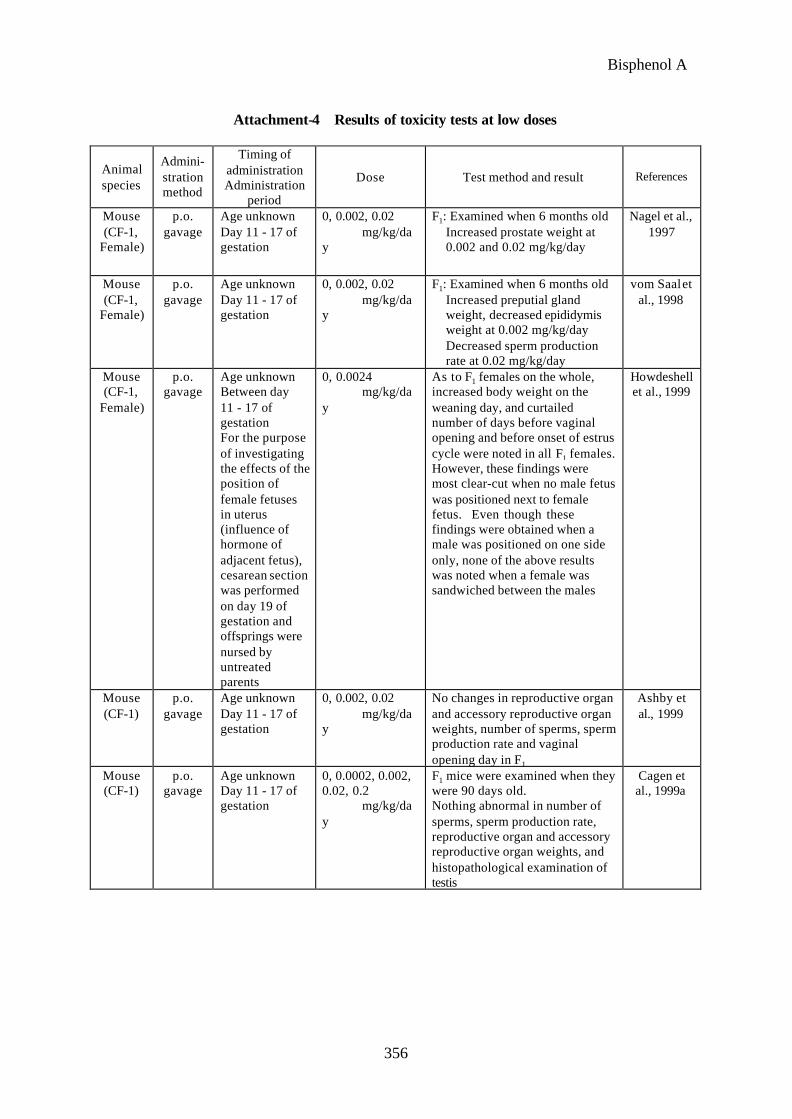
(3) Investigation of the effect at low dose (Attachment-4)
Since 1998, the effect of a low dose of BPA has been discussed.
When oral administration of BPA at 0, 0.002 and 0.02 mg/kg/day was conducted in
female CF-1 mice (age unknown) from day 11 to 17 of gestation, increased prostate and

Bisphenol A
332
preputial gland weights, and decreased epididymis weight at 0.002 mg/kg/day, and
increased prostate weight and decreased sperm production rate at 0.02 mg/kg/day were
noted in F1 (Nagel et al., 1997; vom Saal et al., 1998). However, reliability of this report
was in doubt, because 1) the changes observed in 0.002 mg/kg/day group (e.g. increased
preputial gland weight) was not observed in 0.02 mg/kg/day group, 2) the study was not
conducted in compliance with GLP and 3) the number of animals per group was too small.
To demonstrate the reproducibility, similar tests were conducted, but the results of the first
test were not reproducible (Ashby et al., 1999; Cagen et al., 1999a). When oral
administration of BPA at 0 and 0.0024 mg/kg/day was conducted in female CF-1 mice
(age unknown) from day 11 to 17 of gestation, decreased body weight on the weaning day,
and curtailed number of days before vaginal opening and before onset of estrus cycle were
noted in F1, and the relative positions of male and female fetuses were reported to be
involved in the extent of these effects (Howdeshell et al., 1999), but the results of tests to
confirm reproducibility have not been disclosed yet.
When 1 ppm of BPA was incorporated in water and given to rats for 8 - 9 weeks
from the time before mating, during pregnancy up to weaning, testis weight decreased in
F1 males (Sharpe, 1996, though the report was presented at the 10th International Congress
of Endocrinology, San Francisco, June 1996, the complete report has not yet been
published). However, when BPA at 0, 0.01, 0.1, 1.0 and 10 ppm was incorporated in
water and administered in a reproductive test to female Wistar rats (10 weeks old) for total
10 weeks including 2 weeks before mating, 2 weeks during cohabitation, 21 - 22 days
during pregnancy and 22 days during nursing and in about 90-day old males the
reproductive organ and accessory reproductive organ weights, number of sperms and
histopathology of testis were examined, no influence of BPA was observed (Cagen et al.,
1999b). As described in the above, the results are divided as to the effect of low dose
BPA. Whereas researchers such as vom Saal insisted that the effects were noted by
administration during gestation, other reports indicated that the results were not
reproducible. The low dose effect of BPA is still in the controversial stage.
Based on these background, the reports indicating the presence of a low dose of BPA
and those indicating no influence were reviewed and discussed at NTP Endocrine
Disrupters Low Dose Peer Review Meeting (NTP, USA, 10th - 12th October, 2000).
Prior to the Meeting, the low dose panel of NTP requested the statistics subpanel to

Bisphenol A
333
conduct statistic analysis of the data submitted again to review whether the facts
discovered or those not discovered as the results of experiments correctly reflected the raw
data. The subpanel of BPA discussed the analysis results presented by the statistics
subpanel and the review report was presented at the Meeting, and concluded as in the
following.
The subpanel of BPA considered that the data submitted (the researchers who denied
the effects of BPA submitted the whole data, while those who although submitting a part
of data insisted on the effects were all statistically reliable. With this as the basis, the
subpanel placed importance on the report that the results assumed to exist the low dose
effect (increased uterus weight and elevated serum prolactin level, etc.) were obtained not
only from the experiment in mice conducted by vom Saal but also from the experiment in
rats conducted by Ben-Jonathan (subcutaneous embedding of implant containing BPA), as
well as on the report that there was difference in reaction between the strains of rats (raw
data were not submitted in this report). Bearing these in mind, the subpanel indicated the
following: "There is definite evidence that BPA at a low dose influenced specific
endpoints such as prostate weight. However, considering the result that the low dose
effect was not reproducible in the experiments conducted at a plural number of
laboratories under the same conditions, this subpanel is not persuaded that a low dose
effect of BPA has been conclusively established as a general or reproducible findings.”
That is, according to the statement of the specialist panel, the low dose effect of BPA is a
phenomenon observed under considerably limited experimental conditions at present, and
therefore this phenomenon is not considered as a general phenomenon.
In addition, the toxicological significance and a long term involvement of low dose
effect reportedly observed in prostate and female reproductive organs as examples are
unclear, and were not taken up as the subjects of discussion in the peer review. As a
mechanism of low dose effect of BPA, the theory that some fetuses are susceptible to the
effect depending on implantation position in the uterus (a male sandwiched between 2
females, or a male adjacent to a female is susceptible to female endogenous estrogen) has
been proposed, but this is a task to be clarified in the future. The subpanel
simultaneously suggested a possibility that the analysis based on introduction of new
technology including genomix, proteomix, etc. as well as the dose-tissue concentration at
a low dose may provide useful data for solution of the problem (NTP, 2001).
Bisphenol A
334
At NTP's Low Dose Peer Review in October 2,000, at International Congress related
to the low dose effect held in Berlin in November the same year (Bisphenol A: Low dose
effects - high dose effects; Berlin, Germany, 18th - 20th November, 2000) and at the 3rd
International Symposium on Environmental Endocrine Disrupters 2000 (Yokohama, Japan,
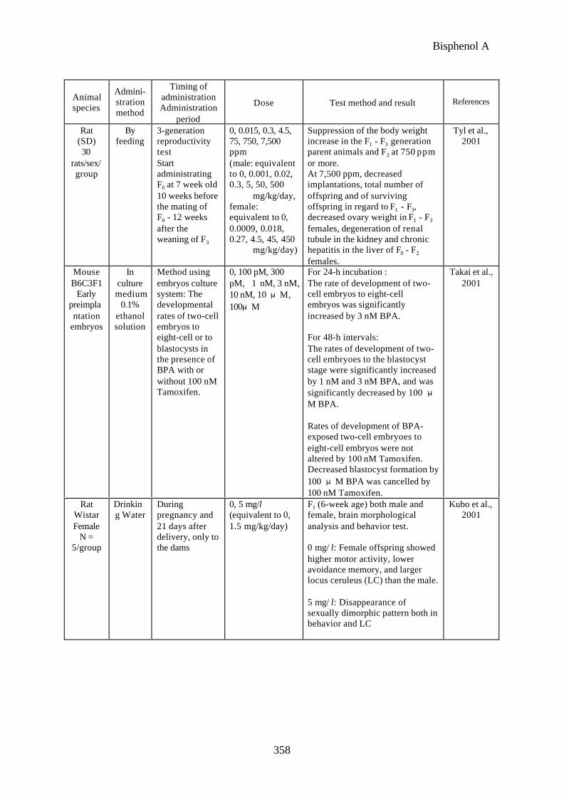
16th - 18th, December, 2000), Tyl et al. reported on a 3 generation reproductive toxicity
test in rats (Tyl et al., 2000). When a test was conducted using BPA at low doses of 0,
0.015, 0.3, 4.5, 75 ppm (equivalent to 0, 0.001, 0.02, 0.3, 5 mg/kg/day, respectively), as
well as at a dose of 750, 7,500 ppm (equivalent to 50, 500 mg/kg/day, respectively) known
to manifest toxicity, no general toxic effects on parent animals for each generation were
observed for low dose groups and that no abnormality was found in fertility and
development of offspring. On the other hand, at the high dose group of 7,500 ppm
abnormality was found in fertility of parent animals for each generation and in
development of offspring. And at the dose of 750 ppm, suppression in body weight
increase was observed for parent animals of each generation, however, no abnormality
was found in fertility of the parental animals and in development of offspring. Based on
these findings, NOAEL in the rat 3-generation study was estimated to be 75 ppm
(equivalent to 5 mg/kg/day) for systemic toxicity to parental animals and 750 ppm
(50mg/kg/day) for reproductive toxicity.
When oral administration by gavage of BPA at 0, 0.0002, 0.002, 0.02 and 0.2
mg/kg/day was conducted in male and female SD rats from the time before the mating of
F0 (10 weeks and 5 weeks before mating in the case of males and females respectively) up
to weaning of F2 in a 2-generation reproductive test, nothing abnormal was reported in
reproductive function of parent animal and in development & growth of offsprings in each
generation (Ema et al., 2001). The BPA subpanel assessment of these tests was
considered by the NTP low dose effect panel as the data that were statistically reliable,
showing many endpoints without however much emphasis placed as the experiment to
investigate the low dose action. In this regard, the subpanel maintained that a multi-
generation reproductive test is intended to investigate reproductivity and is not appropriate
for fully assessing the effect of a substance on development & growth process of
offsprings, unless administration is conducted at the window period using pregnant
animals (NTP, 2001).
Apart from the low dose effect discussion, HSE (Health and Safety Executive) in

Bisphenol A
335
UK proposed to EU in May 2001 to attach a label with a warning that BPA corresponds to
category 2 of reproductive toxicity substance (substance that is regarded to harm the
fertility of human) and is an R60 (may harm fertility) to relevant products (HSE Bootle,
2001). The basis of the proposal is the results of a test in mice conducted by NTP (Reel
et al., 1997) and a 3-generation reproductivity test in rats conducted by Tyl et al (2000).
In the former, the middle and high doses of 5,000 and 10,000 ppm (equivalent to 875 and
1,750 mg/kg/day, respectively) developed decrease in the number of offspring born (by
5% and 9% respectively) and the number of surviving offspring per litter (by 20% and
48% respectively). As to the latter in which the doses 0.015-7,500 ppm (equivalent to
0.001 - 500 mg/kg/day) were given, decrease in the number of siblings per litter was
observed in F1 to F3 generations at 7,500 ppm (the number of surviving offspring per litter
in the control group and a group administered 500 mg/kg/day of BPA: 14.3 and 11.5
respectively in F1, 14.6 and 10.8 respectively in F2, 14.8 and 10.9 respectively in F3).
After mentioning that the above dose was known to cause toxicity in the dams including
inhibition on body weight increase and degeneration of renal tubule, HSE still seriously
considered the reproductive disorder obtained from mice (HSE Bootle, 2001). Later, the
evaluation of reproductive toxicity of BPA by EU was so amended at the conference of
January 2002 for BPA to be a substance toxic to reproduction category 3 (substances
which cause concern for human fertility) with risk phrase of R62 (possible risk of
impaired fertility). As to the developmental effect, the view was given that at least one
year would be needed before more researchers are completed (HSE Health Directorate,
2002).
Recently, the results suggesting the low dose action of BPA have been reported.
When oral administration by gavage of BPA at 0, 0.02, 0.2, 2, 20 and 200 mg/kg/day
was conducted in male SD rats (13 weeks old) for 6 days, decrease in daily
spermatogenesis was noted in all the BPA groups. When oral administration by gavage
of BPA at 0, 0.000002, 0.00002, 0.0002, 0.002, 0.02, 0.2 and 2.0 mg/kg/day was
conducted for 6 days under the same conditions, decrease in daily spermatogenesis was
also noted at 0.002 mg/kg/day or more (Sakaue et al., 2001).
Effects of BPA on development of cultured mouse two-cell embryoes were studied.
The rate of in vitro development of two-cell embryoes to eight-cell embryoes was
significantly increased by exposure to BPA at concentration of 3 nM (94%; 172/182)

Bisphenol A
336
compared with the rate in the control group (88%; 334/378). The rate of development to
the blastocyst stage in 48-h cultures of two-cell embryoes were also significantly
increased (69.2%; 126/182) compared with the rate in control group (58.7%; 222/378).
On the other hand, the frequency of development to blastocyst stage at 48-h was
significantly decreased by exposure to BPA at 100 μM (31.2%; 93/298), although
development to eight-cell embryoes at 24-h was not inhibited by 100 μM BPA (Takai et
al., 2000).
The effects of exposure to BPA early in life on sexual differentiation in brain and
behavior in Wistar rats were studied. Mother rats during pregnancy and lactation were
administered at approximately 1.5 mg/kg BPA per day in drinking water. The control
female offspring showed higher activity, lower avoidance memory, and larger locus
coeruleus than the male controls, while the BPA-exposed group did not show any sexual
dimorphism. These results may suggest that BPA affects sexual differentiation in brain
and behavior from late fatal period to first week after birth in rats (Kubo et al., 2001).
3) Information on general toxicity
(1) Acute toxicity (Table 1) (German Chemical Society, 1995)
Table 1 Acute toxicity test
Mouse Rat Rabbit Guinea pig
Oral LD50 1,600 - 5,280 mg/kg* 3,250 - 5,660 mg/kg* 2,230-4,000mg/kg
4,000 mg/kg
Percutaneous LD50 - - 3,000-6,400mg/kg
-
Intraperitoneal LD50 200 mg/kg 400-800 mg/kg 150 mg/kg -
Subcutaneous LD50 - 2,400 mg/kg - -
*: There are differences between the literatures cited.
(2) Repeated-dose toxicity (Attachment-5)
When BPA at 0, 2,000, 5,000, 10,000, 20,000 and 40,000 ppm (equivalent to 0, 500,
1,000, 2,200, 5,500 and 14,600 mg/kg/day, respectively in males, and 0, 600, 1,300, 2,500,
6,300 and 22,000 mg/kg/day, respectively in females) in diet and administered to male
and female B6C3F1 mice (6-week-old) for 13 weeks, no changes attributable to BPA was
observed at 2,000 ppm. However, decreased RBC and Ht at 5,000 ppm or more,
Bisphenol A
337
decreased Hb concentration, cystic dilation of renal tubules, fibrosis in the periphery of
cyst, degeneration and regeneration of tubular epithelium and increased hyaline cast at
10,000 ppm or more, suppression on body weight increase, increased liver weight,
decreased ovary weight, fibrous osteodystrophy in femoral bone and sternum, and atrophy
of myofibril at 20,000 ppm or more, and leanness, death assumed to be attributable to food
rejection, increased platelet and kidney weight, and enhanced extramedullary
hematopoiesis in spleen at 40,000 ppm were observed (Furukawa et al.; 1994).
When BPA in diet was administered for 2 years to mice, large polyploidy hepatocytes
were noted in liver of male B6C3F1 mice (5 weeks old) at 1,000 and 5,000 ppm
(equivalent to 150 and 250 mg/kg/day respectively). Decreased body weight was observed
in both male and female at 5,000 ppm (NTP, 1982).
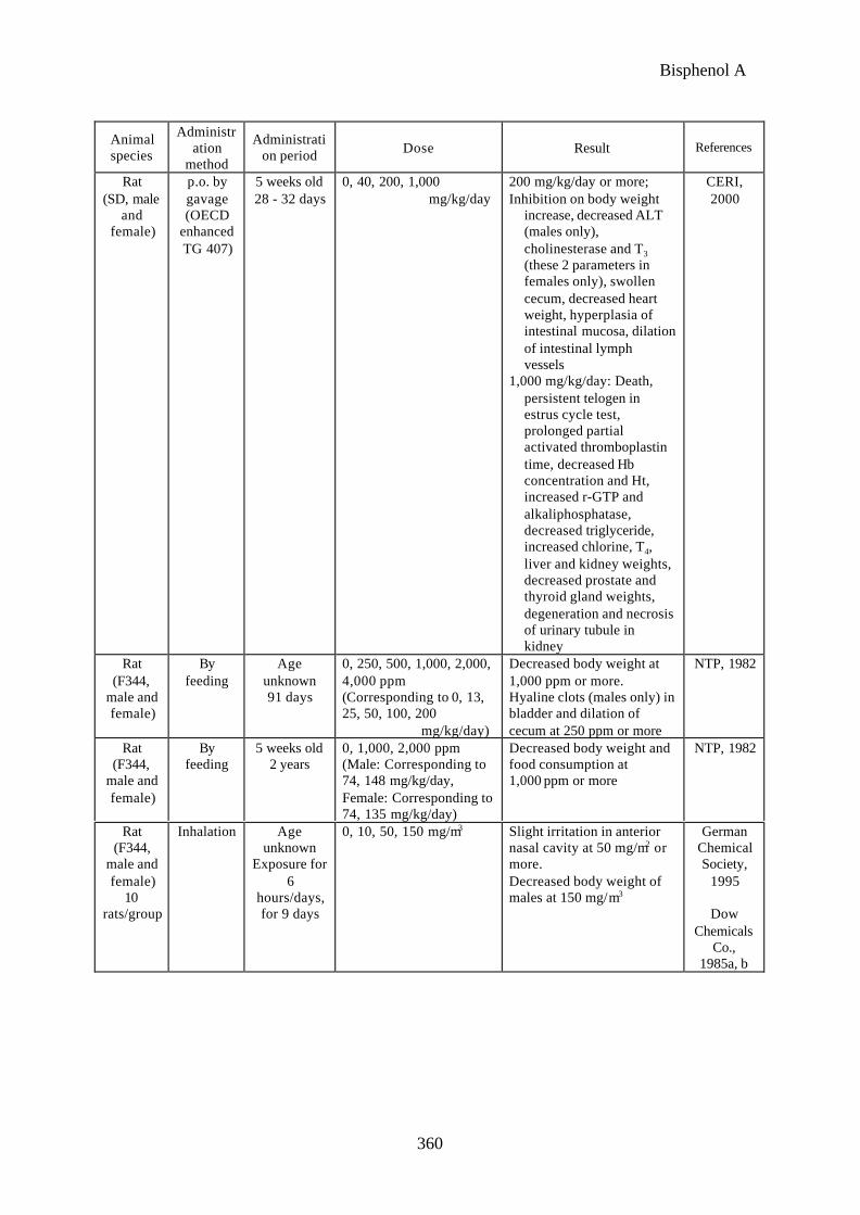
When BPA at 0, 250, 500, 1,000, 2,000 and 4,000 ppm (equivalent to 0, 13, 25, 50,
100 and 200 mg/kg/day, respectively) in diet was administered to male and female F344
rats (age unknown) for 91 days, dilation of cecum in males and females, and hyaline clots
in bladder of males were observed at 250 ppm (equivalent to 13 mg/kg/day) or more,
while body weight decreased at 1,000 ppm (equivalent to 50 mg/kg/day) (NTP, 1982).
When oral administration by gavage of BPA at 0, 40, 200 and 1,000 mg/kg/day was
conducted in male and female SD rats (5 weeks old) for 28 - 32 days in compliance with
the revised OECD test guideline 407 (enhanced TG 407), the changes mainly in kidney,
liver and intestinal tract were observed at 200 mg/kg/day or more, while abnormal estrus
cycle was noted at 1,000 mg/kg/day (CERI, 2000).
When BPA in diet was administered for 2 years to F344 rats both male and female (5
weeks old) at 1,000 and 2,000 ppm (equivalent to 74 and 148 mg/kg/day to male,
equivalent to 74 and 135 mg/kg/day to female, respectively), decrease in body weight and
in food intake were observed in both male and female at 5,000 ppm (NTP, 1982)(EPA
calculated 1000 ppm to be 50 mg/kg/day).
When BPA in diet at 0, 1,000, 3,000 and 9,000 ppm (equivalent to 25, 75, 225
mg/kg/day) was administered to male and female dogs (beagle) for 90 days, liver weight
increased at 9,000 ppm (equivalent to 225 mg/kg/day) (German Chemical Society, 1995;
General Electric, 1976b).
Concerning the inhalation exposure, when male and female F344 rats (age
unknown) was exposed to BPA at 0, 10, 50 and 150 mg/m3 for 6 hours/day for 9 days,

Bisphenol A
338
slight irritation in the anterior nasal cavity was noted at 50 mg/m3 or more, and body
weight of males decreased at 150 mg/m3 (German Chemical Society, 1995; Dow
Chemicals Co., 1985a; Dow Chemicals Co., 1985b). When male and female F344 rats
(age unknown) were exposed to BPA at 0, 10, 50 and 150 mg/m3 for 6 hours/day,
5 days/week for 13 weeks, decreased body weight, dilated cecum, inflammation in nasal
cavity and respiratory mucosa, and hyperplasia of squamous epithelium were noted at
50 mg/m3 or more, while liver and kidney weights decreased at 150 mg/m3 (German
Chemical Society, 1995; Dow Chemicals Co., 1988).
4) Information related to mutagenicity/genotoxicity and carcinogenicity
(1) Mutagenicity/genotoxicity (Table 2)
Concerning the in vitro tests, the results of sister chromatid exchange test, murine
lymphoma test, chromosome aberration test and reverse mutation test using Salmonella
typhimurium, Escherichia coli and yeast were all negative regardless of metabolic
activation. There is a report that mutagenicity of BPA was tested using human Rsa cells,
which has been utilized for identification of novel mutagens. In the BPA exposed cells
base substitution mutations at K-ras codon 12 were detected. (Takahashi et al., 2001).
Concerning the in vivo tests, result of DNA adduct formation test using rat was positive,
but this was assessed to have questionable toxicological significance including
carcinogenicity, because the covalent index was too small to induce carcinoma (Atkinson
& Roy, 1995b).
Table 2 Results of mutagenicity, genetic toxicity tests
Test method Used cell/strain/Animal species Result* References
Salmonella typhimurium TA98, TA100,TA1535, TA1537 0.33-333.3 µg/plate S9(+/-)
-
German ChemicalSociety, 1995
Haworth et al.,1983
in vitro Reversemutation test
Salmonella typhimurium TA1538 0.1-1.0mg/mL S9(+/-)
-
German ChemicalSociety, 1995
Shell Oil Co.,1978
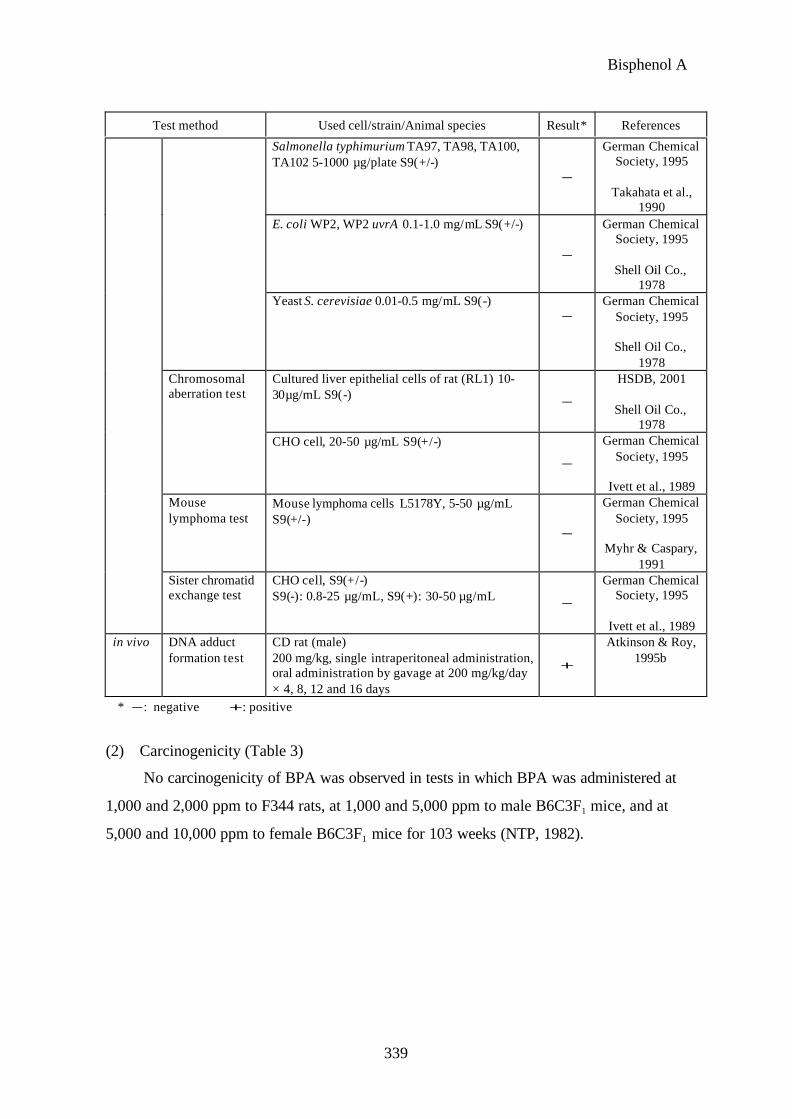
Bisphenol A
339
Test method Used cell/strain/Animal species Result* References
Salmonella typhimurium TA97, TA98, TA100,TA102 5-1000 µg/plate S9(+/-)
-
German ChemicalSociety, 1995
Takahata et al.,1990
E. coli WP2, WP2 uvrA 0.1-1.0 mg/mL S9(+/-)
-
German ChemicalSociety, 1995
Shell Oil Co.,1978
Yeast S. cerevisiae 0.01-0.5 mg/mL S9(-)-
German ChemicalSociety, 1995
Shell Oil Co.,1978
Cultured liver epithelial cells of rat (RL1) 10-30µg/mL S9(-) -
HSDB, 2001
Shell Oil Co.,1978
Chromosomalaberration test
CHO cell, 20-50 µg/mL S9(+/-)
-
German ChemicalSociety, 1995
Ivett et al., 1989Mouselymphoma test
Mouse lymphoma cells L5178Y, 5-50 µg/mLS9(+/-)
-
German ChemicalSociety, 1995
Myhr & Caspary,1991
Sister chromatidexchange test
CHO cell, S9(+/-)S9(-): 0.8-25 µg/mL, S9(+): 30-50 µg/mL -
German ChemicalSociety, 1995
Ivett et al., 1989in vivo DNA adduct
formation testCD rat (male)200 mg/kg, single intraperitoneal administration,oral administration by gavage at 200 mg/kg/day× 4, 8, 12 and 16 days
+
Atkinson & Roy,1995b
* -: negative +: positive
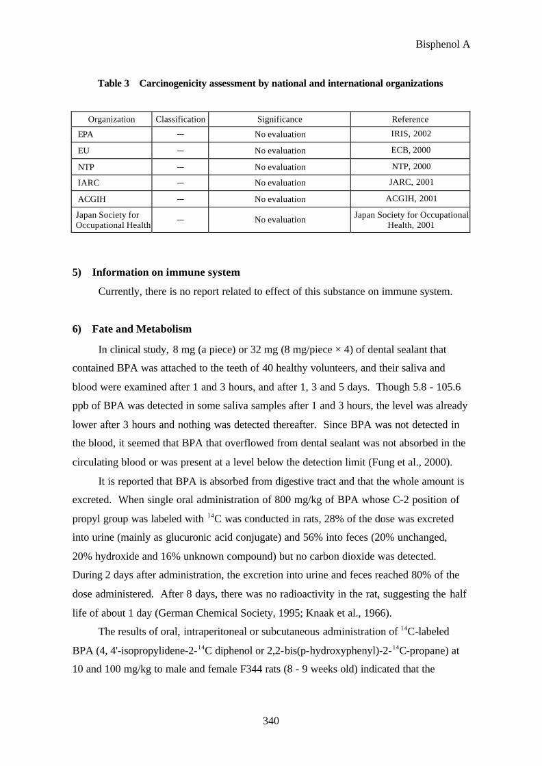
(2) Carcinogenicity (Table 3)
No carcinogenicity of BPA was observed in tests in which BPA was administered at
1,000 and 2,000 ppm to F344 rats, at 1,000 and 5,000 ppm to male B6C3F1 mice, and at
5,000 and 10,000 ppm to female B6C3F1 mice for 103 weeks (NTP, 1982).
Bisphenol A
340
Table 3 Carcinogenicity assessment by national and international organizations
Organization Classification Significance Reference
EPA - No evaluation IRIS, 2002
EU - No evaluation ECB, 2000
NTP - No evaluation NTP, 2000
IARC - No evaluation JARC, 2001
ACGIH - No evaluation ACGIH, 2001
Japan Society forOccupational Health
- No evaluation Japan Society for OccupationalHealth, 2001
5) Information on immune system
Currently, there is no report related to effect of this substance on immune system.
6) Fate and Metabolism
In clinical study, 8 mg (a piece) or 32 mg (8 mg/piece × 4) of dental sealant that
contained BPA was attached to the teeth of 40 healthy volunteers, and their saliva and
blood were examined after 1 and 3 hours, and after 1, 3 and 5 days. Though 5.8 - 105.6
ppb of BPA was detected in some saliva samples after 1 and 3 hours, the level was already
lower after 3 hours and nothing was detected thereafter. Since BPA was not detected in
the blood, it seemed that BPA that overflowed from dental sealant was not absorbed in the
circulating blood or was present at a level below the detection limit (Fung et al., 2000).
It is reported that BPA is absorbed from digestive tract and that the whole amount is
excreted. When single oral administration of 800 mg/kg of BPA whose C-2 position of
propyl group was labeled with 14C was conducted in rats, 28% of the dose was excreted
into urine (mainly as glucuronic acid conjugate) and 56% into feces (20% unchanged,
20% hydroxide and 16% unknown compound) but no carbon dioxide was detected.
During 2 days after administration, the excretion into urine and feces reached 80% of the
dose administered. After 8 days, there was no radioactivity in the rat, suggesting the half
life of about 1 day (German Chemical Society, 1995; Knaak et al., 1966).
The results of oral, intraperitoneal or subcutaneous administration of 14C-labeled
BPA (4, 4'-isopropylidene-2-14C diphenol or 2,2-bis(p-hydroxyphenyl)-2-14C-propane) at
10 and 100 mg/kg to male and female F344 rats (8 - 9 weeks old) indicated that the

Bisphenol A
341
pharmacokinetics became different by administration route and between male and female.
The maximum blood concentration was observed within 1 hour after oral and
intraperitoneal administration, while the peak was noted at 4 hours after subcutaneous
administration. The compound was quickly excreted to reach the level below detection
limit within 72 hours after subcutaneous and intraperitoneal administration and within 18
hours after oral administration. The bioavailability and plasma radioactivity
concentration were the highest by subcutaneous administration, followed by
intraperitoneal administration. The levels were markedly low after oral administration
probably because of low absorption of BPA from digestive tract and conjugation in the
first pass effect in liver. The plasma radioactivity was noted mainly as glucuronic acid
conjugate in the case of oral administration, but mainly unchanged BPA was detected after
intraperitoneal and subcutaneous administration. Other than the unchanged BPA, 4
metabolites were noted by these 2 administration routes. The hydroxide reported in the
past experiment was very small in quantity. Depending on the dose, it was assumed that
hydroxylation occurs after other metabolic pathways have become saturated. Most of the
radioactive compound was excreted into feces unchanged by each route. BPA was
mainly excreted into urine as monoglucuronide. Urinary excretion was about twice
higher in females by each administration route. The residue of BPA and its metabolites
was very small and only 1.3%, 0.8% and 0.4% of the radioactivity administered remained
after 7 days from subcutaneous, intraperitoneal and oral administration respectively
(Pottenger, 2000).
An in vitro experiment demonstrated that sulfate conjugation took place in BPA by
recombinant human sulfate transferase. Furthermore, sulfate conjugation of BPA was
observed in an experiment in which BPA and sulfuric acid were added to HepG2 cell
derived from human liver cancer, suggesting that sulfate conjugation of BPA proceeds in
living body (Suiko et al., 2000).
It was demonstrated that when BPA was allowed to react with an oxidant in vitro,
bisphenol-o-quinone was generated, and that when this was incubated with DNA, binding
to DNA occurred (Figure 1)(Atkinson and Roy, 1995b). When single intraperitoneal
administration of BPA at 200 mg/kg was conducted in rats in an experiment or when oral
administration by gavage at 200 mg/kg/day for 4, 8, 12 and 16 days was conducted in
another experiment, covalent binding to DNA occurred in the liver (Atkinson and Roy,

Bisphenol A
342
1995a). Based on these results, it was assumed that, after BPA was metabolized to 5-
hydroxybisphenol in liver, the reactive metabolites bisphenol semiquinone and 4,5-
bisphenol-o-quinone were produced (Figure 1) and they were bound to DNA. However,
based on the calculation of index of covalent binding to DNA, this reaction was not
intense so that no carcinogenicity was induced (German Chemical Society, 1995).
CH3H3C
OHHO
CH3H3C
OHHO
CH3H3C
OHO
CH3H3C
OHO
HO HO
O
(1) (2) (3)
(4)
グルクロン酸抱合体硫酸抱合体
(1) Bisphenol-A
(2) 5-hydroxybisphenol
(3) Bisphenol semiquinone
(4) 4,5-bisphenol-o-quinone
Fig. 1: Metabolic pathway of bisphenol-A
2. Hazard Assessment at present
There is no report on the effect of BPA on endocrine system and reproductive system
of humans.
To investigate the effect on endocrine system, many in vitro tests, as well as in vivo
tests (uterotrophic assay) have been conducted, and the results indicated that BPA has
estrogenic action. However, in in vitro tests its activity mediated by estrogen receptors is
weak (its receptor binding affinity is 1/500 - 1/15,000 of that of E2 and its transcription
activation capability 1/600 and 1/130,000 of that of E2).
Concerning the effect on reproductive system, a test was conducted in compliance
glucuronic acid conjugationsulfate conjugation

Bisphenol A
343
with the revised OECD test guideline 407 (enhanced TG 407). The results indicated
various toxicological changes at 200 mg/kg/day or more, and at 1,000 mg/kg/day
abnormal estrus cycle suggesting endocrine disruption. Regarding reproductive and
developmental toxicity or fertility toxicity, there are reports ranging from single generation
reproductive tests to 3-generation reproductive tests. In single generation reproductive
test in rats body weight decrease in F1 was observed at 50 mg/kg/day, but no reproductive
toxicity was observed. In 2-generation reproductive test in mice declined seminal vesicle
weight and spermatozoic motility, etc. in F1 was observed at 437 mg/kg/day or more. In a
3-generation reproductive test in rats at lower doses of 0.015, 0.3, 4.5 and 75 ppm
(equivalent to 0.001, 0.02, 0.3 and 5 mg/kg/day, respectively) and higher doses of 750,
7500 ppm (equivalent to 50, 500 mg/kg/day, respectively), suppression of body weight
increase was observed for parental animals of each generation at 50 mg/kg/day. But no
abnormality was found in fertility of parental animals and in development of offspring.
Reproductive and developmental toxicity was observed at the dose equivalent to 500
mg/kg/day. At present NOAEL for reproductive and developmental toxicity and fertility
toxicity is estimated to be 50 mg/kg/day.
On the other hand, the problem related to toxicity of BPA is the effect on fetus and
offspring by administration during gestation as was reported in some animal experiments.
According to positive reports, changes were observed at considerably low doses of
0.002 mg/kg/day by oral administration and at 1 ppm mixed in drinking water. However,
reproducibility tests conducted for various administration periods and at many doses in a
larger number of animals and test parameters all indicated negative results.
In the final report of NTP Peer Review results about low dose effects given in
October 2,000 (published on May 14, 2001), all the data submitted regarding the low dose
effects of BPA on prostate, etc. were considered reliable whether the result was positive or
negative. The subpanel concluded that at present the effect is observed only under
considerably limited conditions and therefore this is hardly considered to be the general
phenomenon. In this regard, the subpanel indicated the following: "There is definite
evidence that BPA at a low dose influenced specific endpoints such as prostate weight.
However, this subpanel is not persuaded that a low dose effect of BPA has been
conclusively established as general or reproducible findings." Even though BPA does
have the low dose effect, current findings are unable to prove that its action is harmful.
Bisphenol A
344
As the information related to the harmful nature of BPA in humans, mild skin
irritation and allergic dermatitis were reported. The patient of dermatitis showed positive
allergic reaction to both BPA and formaldehyde, accordingly substances causing the effect
as well as synergy of both substances was not identified,.
As repeated oral dose toxicity, the effects mainly on large intestine, cecum, liver and
kidney were noted in animal experiments. Anemia was also observed. Additionally,
body weight decrease was observed at dose of 50 mg/kg/day and above in a 2-years rat
oral chronic toxicity test, and EPA estimated LOAEL as 50 mg/kg/day.
Mutagenicity test results were mostly negative and carcinogenicity was also negative.
There is no report on carcinogenicity of this substance in humans.
3. Risk assessment and other necessary future measures
BPA has weak estrogenic activity (receptor binding affinity is 1/500 - 1/15,000 of E2
and its transcription activation capability 1/600 and 1/130,000 of E2) and reproductive &
developmental toxicity was observed at high doses of approximately not lower than 500
mg/kg/day in oral tests. Since sufficient scientific findings in in vitro as well as in vivo
animal test data were already obtained to assess the effect of BPA on endocrine system and
reproductive system for humans, there seems to be no urgent need to plan additional tests
in the future.
Though it is necessary to collect further information on so-called "low dose effects"
represented by BPA from academic point of view, it seems unnecessary to take any
specific measure other than the above, considering the view expressed by NTP Low Dose
Effect Panel that the low dose effect of BPA at present is a phenomenon observed under
considerably limited experimental conditions and it is hardly considered to be the general
phenomenon.
On the other hand, since effects of BPA on reproductive & developmental toxicity
was observed, independent of the existence of endocrine disruption, risk assessment based
on hazard and exposure assessment is to be conducted and appropriate risk management is
to be examined.
Bisphenol A
345
Reference
ACGIH (2001) Booklet of the threshold limit values and biological exposure indices.
Ashby, J., Tinwell, H. and Haseman, J. (1999) Lack of effects for low dose levels of bisphenol A and
diethylstilbestrol on the prostate gland of CF1 mice exposed in utero. Regul. Toxicol.
Pharmcol., 30, 156 - 166.
Atkinson, A. and Roy, D. (1995a) In vivo DNA adduct formation by bisphenol A. Environ. Mol.
Mutagen., 26, 60 - 66.
Atkinson, A. and Roy, D. (1995b) In vitro conversion of environmental estrogenic chemical bisphenol
A to DNA binding metabolite(s)., Biochem. Biophys. Res. Commun., 210, 424 - 433.
Blair, R.M., Fang, H, Branham, W.S., Hass, B.S., Dial, S.L., Moland, C.L., Tong, W., Shi, L., Perkins,
R. and Sheehan, D.M.. (2000) The estrogen receptor relative binding affinities of 188 natural
and xenochemicals: structural diversity of ligands. Toxicol. Sci., 54, 138 - 153.
Cagen, S.Z., Waechter, J.M., Diamond, S.S., Breslin, W.J., Butala, J.H., Jetat, F.W., Jointer, R. L.,
Shiotsuka, R.N., Veenstra, G.E. and Harris, L.R. (1999a) Normal reproductive organ
development in CF-1 mice following prenatal exposure to Bisphenol A. Toxicol. Sci.50, 36 -
44.
Cagen, S.Z., Waechter, J.M., Dimond, S.S., Breslin, W.J., Butala, J.H., Jekat, F.W., Joiner, R.L.,
Shiotsuka, R.N., Veenstra, G.E. and Harris, L.R. (1999b) Normal reproductive organ
development in Wistar rats exposed to Bisphenol A in the drinking water. Regul. Toxicol.
Pharmacol., 30, 130 - 139.
Coldham, N.G., Dave, M., Sivapathasundaram, S., McDonnell, D., Connor, C. and Sauer M.J. (1997)
Evaluation of a recombinant yeast cell estrogen screening assay. Environ. Health Perspect.,
105, 734-742.
Diel, P., Schulz, T., Smolnikar, K., Strunck, E., Vollmer, G. and Michna, H. (2000) Ability of xeno- and
phytoestrogens to modulate expression of estrogen-sensitive genes in rat uterus: estrogenicity
profiles and uterotropic activity. J. Steroid Biochem. Mol. Biol., 73, 1 - 10.
Dow Chemical Co (1985a) Bisphenol A: 2-week aerosol toxicity study with Fischer 344 rats. EPA/OTS,
Document #878216052; Order No. 0206803 (NTIS), 1-54.
Dow Chemical Co (1985b) Bisphenol A: 2-week aerosol toxicity study with Fischer 344 rats. EPA/OTS,
Document #40-8586071; Order No. 51007 (NTIS).
Dow Chemical Co (1988) Bisphenol A: 13-week aerosol toxicity study with Fischer 344 rats. Study
Report K-001304-011, Dow chemical Co., 1-22.
ECB (2000) Council Directive 67/548/EEC on the approximation of the laws, regulations and
administrative provisions relating to the classification, packaging and labeling of dangerous
substances : ANNEX I (http://ecb.jrc.it/).
Ema, M., Fujii, S., Furukawa, M., Kiguchi, M., Ikka, T. and Harazono, A. (2001) Rat two-generation
reproductive toxicity study of bisphenol A. Reprod. Toxicol., 15, 505-523.
Bisphenol A
346
Fung, E.Y. K., Ewoldsen, N.O., St.Germain, Jr H.A., Marx, D.B., Miaw, C.L., Siew, C., Chou, H.N.,
Grunninger, S.E. and Meyer, D.M. (2000) Pharmacokinetics of bisphenol A released from a
dental sealant. J. Am. Dent. Assoc., 131, 51 - 58.
Gaido KW, Leonard LS, Lovell S, Gould JC, Babai D, Portier CJ, McDonnell DP. (1997) Evaluation of
chemicals with endocrine modulating activitiy in a yeast-based steroid hormone receptor gene
transcription assay. Toxicol. Appl. Pharmacol,, 143, 205 - 212.
General Electric (1976a) Reproduction and ninety day oral toxicity study in rats. EPA/OTS, Document
#878214681; Order No. 206618 (NTIS), 1-51.
General Electric (1976b) Ninety day oral toxicity study in dogs. EPA/OTS, Document #878214681;
Order No. 206618 (NTIS), 1-32.
General Electric (1978) Reproduction and ninety day oral toxicity study in rats. EPA/OTS, Document
#878214683; Order No. 206618 (NTIS), 1-89.
German Chemical Society (1995) Bisphenol A, BUA Report, No.203.
Haworth, S., Lawlor, T., Mortelmans, K., Speck, W. and Zaiger, E. (1983) Salmonella mutagenicity test
results for 250 chemicals. Environ. Mutagen. 5 (Suppl. 1), 3-142.
HSDB (2001) Hazardous Substances Data Bank, U.S. National Library of Medicine,
(http://toxnet.nlm.nih.gov/cgi-bin/sis/htmlgen?HSDB).
Hiroi, H., Tsutsumi, O., Momoeda, M., Takai, Y., Osuga, Y. and Taketani, Y. (1999) Differential
interactions of bisphenol A and 17β-estradiol with estrogen receptor α (ERα) and ERβ.
Endcrine J., 46, 773 - 778.
Howdeshell, K. L., Hotchkiss, A. K., Thayer, K. A., Vandenbergm J.G. and vom Saal, F. S. (1999)
Exposure to bisphenol A advances puberty. Nature, 401, 763 - 764.
HSE, Bootle (2001) Working Group on the Classification and Labelling of Dangerous Substances: May
2001 Meeting. http://ecbntlib.ei.jrc.it/claalab/public.htm.
HSE Health Directorate (2002) Table of substances under review for Annex I of 67/548/EEC.
(http://www.hse.gov.uk/hthdir/noframes/chip/chip7.htm)
IARC (2001) IARC Monograph on the Evaluation of Carcinogenic Risks to Humans.
http://www.iarc.fr
IRIS (2002) Intergated Risk Information System. National Library of Medicine,
(http://toxnet.nlm.nih.gov/cgi-bin/sis/htmlgen?IRIS).
Ivett, J. L., Brown, B. M., Rodgers, C., Anderson, B. E., Resnick, M. A. and Zeiger, E. (1989)
Chromosomal aberrations and sister chromatid exchange test in Chinese hamster ovary cells in
vitro. IV. Results with 15 chemicals. Environ. Mol. Mutagen. 14, 165-187.
Jolanki, R., Kanerva, L. and Estlander, T. (1995). Occupational allergic contact dermatitis caused by
epoxy diacrylate in ultraviolet-light-cured paint, and bisphenol A in dental composite resin.
Contact Dermatitis, 33, 94 - 99.
Jorgensen, M., Vendelbo, B., Skakkebaek, N.E. and Leffers, H. (2000) Assaying estrogenicity by
quantitating the expression levels of endogenous estrogen-regulated genes. Environ. Health

Bisphenol A
347
Perspect., 108, 403.
Knaak, J.B. and Sullivan, L.J. (1966) Metabolisim of bisphenol A in the rat. Toxicol. Appl. Pharmacol.,
8, 175 - 184.
Kubo K, Arai O, Ogata R, Omura M, Hori T, Aou S. (2001) Exposure to bisphenol A during the fetal
and suckling periods disrupts sexual differentiation of the locus coeruleus and of behavior in
the rat. Neurosci. Lett., 304, 73-76.
Kwon, S., Stedman, D.B., Elswick, B.A., Cattley, R.C. and Welsch, F. (2000) Pubertal development
and reproductive functions of Crl:CD BR Sprague-Dawley rats exposed to bisphenol A during
prenatal and postnatal development. Toxicol. Sci., 55, 399 - 406.
Laws, S.C., Carey, S.A., Ferrell, J.M., Bodman, G.J. and Cooper, R.L. (2000) Estrogenic activity of
octylphenol, nonylphenol, bisphenol A and methoxychlor in rats. Toxicol. Sci., 54, 154 - 167.
Legler J, van den Brink CE, Brouwer A, Murk AJ, van der Saag PT, Vethaak AD, van der Burg B..
(1999) Developmental of a stably transfected estrogen recepror-mediated luciferase reporter
gene assay in the human T47D breast cancer cell line, Toxicol. Sci., 48, 55 - 66.
Mehmood, Z., Smith, A.G., Tucker, M.J., Chuzel, F. and Carmichael, N.G. (2000) The development of
methods for assessing the in vivo oestrogen-like effects of xenobiotics in CD-1 mice. Food
Chem.Toxicol., 38, 493 - 501.
Morrissey, R.E., George, J.D., Price, C.J., Yye, R.W., Marr, M.C. and Kimmel, C.A. (1987) The
developmental toxicity of bisphenol A in rats and mice. Fundam. Appl. Toxicol., 8, 571 - 582.
Myhr, B. C. and Caspary, W. J. (1991) Chemical muragenesis at the thymidine kinase locus in L5178Y
mouse lymphoma cells. Results for 31 coded compounds in the National Toxicology Program.
Environ. Mol. Mutagen. 18, 51-83.
Nagel, S.C., vom Saal, F.S., Thayer, K.A., Dhar, M.G., Boechler, M. and Welshons, W.V. (1997)
Relative binding affinity-serum modified access (RBA-SMA) assay predicts the relative in
vivo bioactivity of the xenoestrogens bisphenol A and octylphenol. Environ. Health Perspect.,
105, 70 - 76.
Nishihara, T., Nishikawa, J., Kanayama, T., Dakeyama, F., Saito, K., Imagawa, M., Takatori, S.,
Kitagawa, Y., Hori, S., and Utsumi, H. (2000) Estrogenic activites of 517 chemicals by yeast
two-hybrid assay. J. Health Sci., 46, 282-298.
NTP (2001) Final Report of the Endocrine Disruptors Low-Dose Peer Review, published in May 14th,
2001.
NTP (2000) U.S. Department of Health and Human Services Public Health Service, National
Toxicology Program, 9th Report on Carcinogens.
NTP (1982) NTP techinical report on the carcinogenesis bioassay of bisphenol A in F344 rats and
B6C3F1 mice.
Papaconstantinou, A.D., Umbreit, T.H., Fisher, B.R., Goering, P.L., Lappas, N.T. and Brown, K.M.
(2000) Bisphenol A - Induced increase in uterine weight and alterations in uterine morphology
in ovariectomized B6C3F1 mice: Role of the estrogen receptor. Toxicol. Sci., 56, 332 - 339.
Bisphenol A
348
Pottenger, L.H., Domoradzki, J.Y., Markham, D.A., Hansen, S.C., Cagen, S.Z. and Waechter, Jr. J. M.
(2000) The relative bioavailability and metabolism of bisphenol A in rats is dependent upon the
route of administration. Toxicol. Sci., 54, 3 - 18.
Reel, J., George M., Lawton, A. and Meyers, C. (1997) Bisphenol A. Environ. Health Perspect., 105,
273 - 274.
RTECS (1998) Registry of Toxic Effects of Chemical Substances, US NIOSH.
Sakaue, M., Ohsako, S., Ishimura, R., Kurosawa, S., Kurohmaru, M., Hayashi, Y., Aoki, Y., Yonemoto,
J. and Tohyama, C. (2001) Bisphenol-A affects spermatogenesis in the adult rat even at a low
dose. J. Occup. Health, 43, 185-190.
Sharpe, R. Majdic, G., Fisher, J., Parte, P., Millar, M.R. and Saunders, P.T.K. (1996) Effects on
testicular development and function. Abstract S23-4, 10th International Congress of
Endocrinology.
Shell Oil Co (1978) Toxicity test with diphenylol propane (DPP): In vivo mutation studies, with cover
letter. EPA/OTS Document #878214488; Order No. 206596 (NITS), 1-18.
Sheeler, C.Q., Dudley, M.W. and Khan, S.A. (2000) Environmental estrogens induce transcriptionally
active estrogen receptor dimers in yeast: Activity potentiated by the coactivator RIP140.
Environ. Health Perspect.,108, 97 - 103.
Steinmetz, R., Brown, N. G., Allen, D. L., Bigsby, R. M. and Ben-Jonathan, N. (1997) The
Environmental estrogen bisphenol A stimulates prolactin release in vitro and in vivo.
Endocrinology, 138, 1780 -1786.
Steinmetz, R., Mitchner, N. A., Grant, A., Allen, D.L., Bigsby, R.M. and Ben-Jonathan, N. (1998) The
xenoestrogen bisphenol A induces growth, differentiation, and c-fos gene expression in the
female reproductive tract. Endocrinology, 139, 2741-2747.
Suiko, M., Sakakibara, Y. and Liu, M.C. (2000) Sulfation of environmental estrogen-like chemicals by
human cytosolic sulfotransferases. Biochem. Biophys. Res. Commun., 267, 80 - 84.
Takahashi S, Chi XJ, Yamaguchi Y, Suzuki H, Sugaya S, Kita K, Hiroshima K, Yamamori H, Ichinose
M, Suzuki N. (2001) Mutagenicity of bisphenol A and its suppression by interferon-alpha in
human RSa cells. Mut. Res., 490, 199-207.
Takahata, J., Tamakawa, K., Takahashi, Y., Seki, T., Tsuda, A., Nohmi, T. and Sofuni, T. Mutagenicity
of environmental chemicals. II. Bisphenol A. Sendai-shi Eisei Kenkyushoho 20, 245-247.
Takai, Y., Tsutsumi, O., Ikezuki, Y., Hiroi, H., Osuga, Y., Momoeda, M., Yano, T. and Taketani, Y.
(2000) Estrogen receptor-mediated effects of a xenoestrogen, bisphenol A, on preimplantation
mouse embryos. Biochem. Biophys. Res. Commun., 270, 918 - 921.
Tyl, R.W., Myers, C., Marr, M., Chang, T., Seely, J., Brine, D., Veselica, M., Fail, P., Joiner, R., Butala,
J. et al., (2002) Three-generation reproductive toxicity evaluation of bisphenol A administered
in the feed to CD(Sprague-Dawley) rats. The Toxicologist, 60, 297.
vom Saal, F., Cooke, P.S., Buchanan, D.L., Palanza, P., Thayer, K. A., Nagel, S. C., Parmigiani, S. and
Welshons, W. (1998) A physiologically based approach to the study of bisphenol A and other

Bisphenol A
349
estrogenic chemicals on the size of reproductive organs, daily sperm production, and behavior.
Toxicol. Ind. Health, 14, 239 - 260.
Yamasaki, K., Sawaki, M., Takatsuki, M. (2000) Immature rat uterotrophic assay of bisphenol A.
Environ.Health Perspect., 108, 1147 - 1150.
Yamasaki, K., Takeyoshi, M., Yakabe, Y. , Sawaki, M., Imatanaka, M. and Takatsuki, M. (2001)
Comparison of Reporter Gene Assay and Immature Rat Uterotrophic Assay of Twenty-three
Chemicals. Toxicology, 170, 21-30.
CERI (Chemicals Evaluation and Research Institute, Japan) (2000) : Contract task on behalf of the
New Energy and Industrial Technology Development Organization – Ministry of International
Trade and Industry : Development of High Precision Screening Methods for Endocrine
Disrupters and Database Maintenance
CERI (Chemicals Evaluation and Research Institute, Japan) (2001): Report on evaluation and method
development for hormone-like effects of exogenous substances. 2000 Contract
investigation/research on environment-compatible technology development on behalf of the
Ministry of Environment and Industry.
Furukawa, F., Nishikawa, A., Mitsui. M., Sato, M., Suzuki, J., Imazawa, T., Takahashi, M. (1994) A 13-
week Subchronic Toxicity Study of Bisphenol A in B6C3F1 Mice. Bulletin of National Institute
of Health Sciences., 112, 89-96.
Handbook of industrial Poisoning (1994), Ishiyaku Publisher Inc .
IPCS (2000) International Chemical safety Cards(ICSC), Japanese Version, http://www.nihs.go.jp.
Japan Society for Occupational Health (2001): Advice on the tolerance limit. San Ei Shi, 43: 95-119.
Tsusansho Koho (Daily) 1977.
Ministry of International Trade and Industry (1999): Survey on the production/import of existing
chemical substances in 1998
Bisphenol A
350
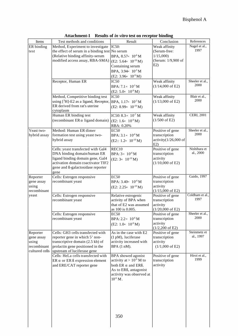
Attachment-1 Results of in vitro test on receptor bindingItems Test methods and conditions Result Conclusion References
Method, Experiment to investigatethe effect of serum in a binding test(Relative binding affinity-serummodified access assay, RBA-SMA)
IC50:No serumBPA, 8.57×10⁻⁶ M(E2: 5.64×10⁻¹⁰ M)Containing serumBPA, 3.94×10⁻⁵ M(E2: 3.96×10⁻⁹ M)
Weak affinity(Serum-free:1/15,000)(Serum: 1/9,900 ofE2)
Nagel et al.,1997
Receptor, Human ER IC50BPA: 7.1×10⁻⁵ M(E2: 5.0×10⁻⁹ M)
Weak affinity(1/14,000 of E2)
Sheeler et al.,2000
Method, Competitive binding testusing [3H]-E2 as a ligand, Receptor,ER derived from rat's uterinecytoplasm
IC50BPA, 1.17×10⁻⁵ M(E2: 8.99×10⁻¹⁰ M)
Weak affinity(1/13,000 of E2)
Blair et al.,2000
ER bindingtest
Human ER binding test(recombinant ER α ligand domain)
IC50: 8.3×10⁻⁷ M(E2: 1.6×10⁻⁹ M)RBA: 0.20%
Weak affinity(1/500 of E2)
CERI, 2001
Method: Human ER dimerformation test using yeast two-hybrid assay
EC50BPA: 3.1×10⁻⁶ M(E2:: 1.2×10⁻¹⁰ M)
Positive of genetranscriptionactivity(1/26,000 ofE2)
Sheeler et al.,2000
Yeast two-hybrid assay
Cells: yeast transfected with Gal4DNA binding domain/human ERligand binding domain gene, Gal4activation domain coactivator TIF2gene and ß-galactosidase reportergene
REC10BPA: 3×10⁻⁶ M(E2: 3×10⁻¹⁰ M)
Positive of genetranscriptionactivity(1/10,000 of E2)
Nishihara etal., 2000
Cells: Estrogen responsiverecombinant yeast
EC50BPA: 3.40×10⁻⁶ M(E2: 2.25×10⁻¹⁰ M)
Positive of genetranscriptionactivity(1/15,000 of E2)
Gaido, 1997
Cells: Estrogen responsiverecombinant yeast
Relative estrogenicactivity of BPA whenthat of E2 was assumedas 100 is 0.005.
Positive of genetranscriptionactivity(1/20,000 of E2)
Coldham et al.,1997
Reportergene assayusingrecombinantyeast
Cells: Estrogen responsiverecombinant yeast
EC50BPA: 2.2×10⁻⁶ M(E2: 1.0×10⁻⁹ M)
Positive of genetranscriptionactivity(1/2,200 of E2)
Sheeler et al.,2000
Cells: GH3 cells transfected withreporter gene in which 5’ non-transcriptive domain (2.5 kb) ofprolactin gene positioned in theupstream of luciferase gene
As in the case with E2(1 pM), luciferaseactivity increased withBPA (1 nM).
Positive of genetranscriptionactivity (1/1,000 of E2)
Steinmetz etal., 1997
Reportergene assayusingrecombinantcultured cells
Cells: HeLa cells transfected withER α or ER ß expression elementand ERE/CAT reporter gene
BPA showed agonistactivity at > 10-9 M toboth ER α and ERß.As to ERß, antagonistactivity was observed at10-6 M.
Positive of genetranscriptionactivity
Hiroi et al.,1999

Bisphenol A
351
Items Test methods and conditions Result Conclusion ReferencesMethod: Reporter gene assaymediated by estrogen receptorCells: T47D cells transfected withestrogen responsive element andluciferase gene
EC50BPA: 7.70×10⁻⁷ M(E2: 6×10-12 M)
Positive of genetranscriptionactivity(1/130,000 of E2)
Legler et al.,1999
Cells: HeLa cells transfected withhuman ER expression gene and ERresponding sequence.Concentration: 10-11 -10-5 M
PC50:BPA: 2.9×10⁻⁷ M(E2: <10-11 M)
Positive(1/29,000 of E2)
CERI, 2001
Cells: HeLa cells transfected withrat ER expression gene and ERresponding sequence.Concentration: 10-11 - 10-5 M
PC50:BPA: 6.0×10⁻⁷ M(E2=<10-11 M)
Positive(1/600 of E2)
Yamasaki etal., 2002
Method: Experiment to determinethe secretion of prolactin in GH3cells cultured in the presence ofBPA and E2
BPA and E2 dose-dependently promotedprolactin secretionwithin the range of 10-8 -10-6 M and 10-12- 10-9
respectively.
Promoting prolactinsecretion
Steinmetz etal., 1997
Method: Experiment by singleintraperitoneal administration ofBPA to F344 and SD rats at 18.75,37.5, 75, 150 and 200 mg/kg
The expression of c-fosincreased by 14 times inuterus and vagina ofF344 at 2 hours afteradministration of BPA(50 mg/kg).
Promoting geneexpression
Steinmetz etal., 1998
Method: Experiment to investigateeffect on endogenous estrogenresponding gene expression level(the expression levels of pS2, TGFß3, monoamine oxidase A (MAO-A), α1-antichymotrypsin (α1-ACT)were quantitated by PCR method)
The concentration ofBPA required to inducepS2 gene was 105 - 106
times that of E2.
Promoting geneexpression
Jorgensen,2000
Changes ingene andproteinexpression
Method: After administration ofBPA at 5, 50 and 200 mg/kg toovariectomized DA/Han rats for3 days, uterus was collected andgene expression in the tissue wasdetermined by Northern blotmethod and semi-quantitative PCRmethod
Inhibition on expressionof AR, ER and PR geneand an increase in theexpression of C3 genewere noted in 200 mg/kggroup.
Promoting geneexpression
Diel et al.,2000
ER: Estrogen receptor; E2: 17ß-estradiol; REC10: Concentration corresponding to 10% of E2 activityvalue at 10-7 M PC50: Concentration corresponding to 50% of the maximum activity of E2; IC50: 50%inhibitory concentration; RBA: Relative binding affinity (%)

Bisphenol A
352
Attachment-2 Results of short-term detection test of estrogenic activity in mammals
Animalspecies
Admini-strationmethod
Administrationperiod Dose Result References
Mouse(B6C3F1, female)
s.c.(uterotro
troficassay,
ovariectomizedmice)
35 - 60 daysold, for 4 days
0, 0.02, 0.2, 0.8,2, 8 mg/kg/day
Increased uterus weight at0.8 mg/kg/day or more
Papaconst-antinous etal., 2000
Mouse(CD-1,female)
s.c.(uterotro
troficassay,
juvenilemice
21 days old3 days
0, 0.01, 0.1, 1, 10,100 mg/kg/day
No increase in uterus weight.No changes in lactoferrin,peroxidase activity or BrdUlabeling index in uterine mucusepithelium
Mehmood etal., 2000
Rat(F344,female)
i.p.(uterinegrowthassay,
ovariectomized
rat)
7-8 weeks oldSingle dose
0, 18.75, 37.5, 75,150, 200 mg/kg
Significant increase in BrdUlabeling index in uterine andvaginal epithelia at 37.5 mg/kg ormore
Rat(F344 or
SD,female)
s.c.capsuleimplant
(uterotrotroficassay,
ovariectomized
rat)
7-8 weeks old3-day
implantation
Correspondingto 0.3 mg/kg/day
In F344 rat increased uterusweight, hypertrophy andhyperplasia of uterus, mucousfluid secretion from uterus,epithelial hyperplasia andkeratinization of vagina.The height of uterine epithelialcells increased by 2.5 times.No influence on SD rat.
Steinmetz etal., 1998
Rat(Alpk:A
P/SD,female)
s.c.(uterotro
troficassay,
ovariectomized
rat)
8-10 weeksold
3 days
33 mg/rat/day Increased uterus weight Ashby et al.,2000
p.o.gavage
(uterotrotroficassay,
juvenilerat)
18 days old3 days
0, 40, 160, 800 mg/kg/day
Increased uterus weight at160 mg/kg/day or more
Rat(SD,
female)
s.c.(uterotro
troficassay,
juvenilerat)
18 days old3days
0, 8, 40, 160 mg/kg/day
Increased uterus weight at8 mg/kg/day or more
Yamasaki etal., 2000

Bisphenol A
353
Animalspecies
Admini-strationmethod
Administrationperiod Dose Result References
p.o.gavage
(uterotrotroficassay,
juvenilerat)
21 days old3 days
Comparativetest by autopsyat 6 hours and24 hours after
finaladministration
0, 100, 200 , 400 mg/kg/day
Increased uterus weight at200 mg/kg/day or moreThe above result was obtainedafter 6 hours but no differencefrom the control was noted after24 hours
Rat(LongEvans,female)
p.o.gavage
(uterotrotroficassay,
ovariectomized
rat)
60 days old3 days
0, 100 mg/kg/day No influence on uterus weight
Laws et al.,2000
Rat(SD,
female)
s.c.(uterotro
troficassay,
juvenilerat)
20 days old3 days
0, 2, 20, 200 mg/kg/day
Increased uterus weight at20 mg/kg/day or more
Yamasaki etal, 2002

Bisphenol A
354
Attachment-3 Results of reproductivity and reproductive toxicity test in mammals
Animalspecies
Admini-strationmethod
Administrationperiod Dose Result References
Mouse(CD-1,female)
p.o.gavage
Age unknownDay 6 - 15 ofgestation(sacrificed onday 17 ofgestation)
0, 500, 750, 1,000,1,250 mg/kg/day
Dams: Increased relative weight ofliver at 500 mg/kg/day or more,suppression on body weightincrease and decreased pregnantuterus weight at 1,250mg/kg/day
Fetus: Increased embryoresorption, decreased bodyweight at 1,250 mg/kg group
No deformation was noted
Morrisseyet al., 1987
Mouse(CD-1,maleand
female)
Byfeeding
2-generationreproductivetest from1week beforeF0 mating to F2
weaning
Mating of F0
generation(males orfemales) withuntreatedanimals.
0, 2,500, 5,000,10,000 ppm(Corresponding to0, 437, 875, 1,750 mg/kg/day)
F0: Decreased number of offspringborn and surviving at 875mg/kg/day or more; at 1,750mg/kg/day or more decreasedbody weight, increased liver andkidney weights, decreasedseminal vesicle weight andspermatozoic motility, andincreased mortality of offspringbefore weaning
F1: Increased liver and kidneyweights (both males andfemales) and decreasedepididymis and seminal vesicleweights at 437 mg/kg/day ormore
Both mating of males in thehigh dose group with untreatedfemales and that of females inthe high dose group withuntreated males resulted in adecrease in the number ofoffspring born
Reel et al.,1997
Rat (SD,female)
p.o.gavage
Day 6 - 15 ofgestation(sacrificed onday 20 ofgestation)
0, 160, 320, 640,1,280 mg/kg/day
Parent animal: Decreased bodyweight at 160 mg/kg/day or more,death at 1,280 mg/kg/dayFetus: No abnormality
Morrisseyet al., 1987
Rat (SD,maleand
female)
Byfeeding
Singlegenerationreproductivetest for 17days in F0 (ageunknown) andfor 90 days inF1
0, 1,000, 3,000,9,000 ppm(Corresponding to0, 50, 150, 450 mg/kg/day)
F0: Decreased body weight at150 mg/kg/day or moreF1: Decreased body weight at50 mg/kg/day or more
GermanChemicalSociety,
1995
GeneralElectric,1976a

Bisphenol A
355
Animalspecies
Admini-strationmethod
Administrationperiod Dose Result References
Rat (SD,maleand
female)
Byfeeding
Singlegenerationreproductivetest for 17days in F0 (ageunknown) andfor 90 days inF1
0, 100, 250, 500,750, 1,000 ppm(Corresponding to0, 5, 13, 25, 38, 50 mg/kg/day)
F0: Decreased body weight at50 mg/kg/day or moreF1: No effects
GermanChemicalSociety,
1995
GeneralElectric,
1978
Bisphenol A
356
Attachment-4 Results of toxicity tests at low doses
Animalspecies
Admini-strationmethod
Timing ofadministrationAdministration
period
Dose Test method and result References
Mouse(CF-1,
Female)
p.o.gavage
Age unknownDay 11 - 17 ofgestation
0, 0.002, 0.02 mg/kg/day
F1: Examined when 6 months oldIncreased prostate weight at0.002 and 0.02 mg/kg/day
Nagel et al.,1997
Mouse(CF-1,
Female)
p.o.gavage
Age unknownDay 11 - 17 ofgestation
0, 0.002, 0.02 mg/kg/day
F1: Examined when 6 months oldIncreased preputial glandweight, decreased epididymisweight at 0.002 mg/kg/dayDecreased sperm productionrate at 0.02 mg/kg/day
vom Saal etal., 1998
Mouse(CF-1,
Female)
p.o.gavage
Age unknownBetween day11 - 17 ofgestationFor the purposeof investigatingthe effects of theposition offemale fetusesin uterus(influence ofhormone ofadjacent fetus),cesarean sectionwas performedon day 19 ofgestation andoffsprings werenursed byuntreatedparents
0, 0.0024 mg/kg/day
As to F1 females on the whole,increased body weight on theweaning day, and curtailednumber of days before vaginalopening and before onset of estruscycle were noted in all F1 females.However, these findings weremost clear-cut when no male fetuswas positioned next to femalefetus. Even though thesefindings were obtained when amale was positioned on one sideonly, none of the above resultswas noted when a female wassandwiched between the males
Howdeshellet al., 1999
Mouse(CF-1)
p.o.gavage
Age unknownDay 11 - 17 ofgestation
0, 0.002, 0.02 mg/kg/day
No changes in reproductive organand accessory reproductive organweights, number of sperms, spermproduction rate and vaginalopening day in F1
Ashby etal., 1999
Mouse(CF-1)
p.o.gavage
Age unknownDay 11 - 17 ofgestation
0, 0.0002, 0.002,0.02, 0.2 mg/kg/day
F1 mice were examined when theywere 90 days old.Nothing abnormal in number ofsperms, sperm production rate,reproductive organ and accessoryreproductive organ weights, andhistopathological examination oftestis
Cagen etal., 1999a

Bisphenol A
357
Animalspecies
Admini-strationmethod
Timing ofadministrationAdministration
period
Dose Test method and result References
Rat(SD,
female)
p.o.gavage
13 weeks old6 days
0, 0.02, 0.2, 2, 20,200 mg/kg/day
0, 0.000002,0.00002, 0.0002,0.002, 0.02, 0.2, 2 mg/kg/day
The testis was weighed andspermatogenic function was testedwhen the rats were 14 weeks oldand 18 weeks old.Daily spermatogenic function aswell as daily spermatogenicfunction per testis weightdecreased at 0.02 mg/kg/day ormore of BPA
Sakaue etal., 2001
Rat Drinkingwater
Administrationfor 8 to 9 weeks(before mating,duringpregnancy andlactation)
1 ppm F1 rats were examined when theywere 90 days oldTestis weight decreased andnumber of sperms decreased
Sharpe,1996
Rat(Wistar,female)
Drinkingwater
10 weeks old.10 weeks ofadministration(2 weeks beforemating, 2 weeksduringcohabitation,21 - 22 daysduringpregnancy and22 days duringnursing)
0, 0.01, 0.1, 1.0,10 ppm(BPA intakeCorresponding to0, 0.001-0.004,0.008-0.038,0.100-0.391,0.775-4.022 mg/kg/day)
F1 rats were examined when theywere 90 days oldNo changes in the reproductiveorgan and accessory reproductiveorgan weights, number of sperms,sperm production rate andhistopathological examination oftestis
Cagen et al,1999b
Rat(SD,
female)
p.o.gavage
From day 11 ofgestation to theweaning (up to20 days afterbirth)
0, 3.2, 32, 320 mg/kg/day
F0: No abnormality in fertility andreproductive organ weightsF1: No abnormality in the time ofsexual maturity of females, andreproductive organ weights offemales and males
Kwon et al,2000
Rat(SD)25
rats/sex/group
p.o.gavage
2-generationreproductivitytestMale: 5 weeksoldFemales: 10weeks old.From the timebefore mating ofF0 (from 10weeks beforeand 5 weeksbefore in thecase of malesand femalesrespectively) upto the weaningof F2
0, 0.0002, 0.002,0.02, 0.2 mg/kg/day
No abnormality in reproductivefunction of parent animal anddevelopment & growth ofoffspring in each generation
Ema et al.,2001
Bisphenol A
358
Animalspecies
Admini-strationmethod
Timing ofadministrationAdministration
period
Dose Test method and result References
Rat(SD)30
rats/sex/group
Byfeeding
3-generationreproductivitytestStartadministratingF0 at 7 week old10 weeks beforethe mating ofF0 - 12 weeksafter theweaning of F3
0, 0.015, 0.3, 4.5,75, 750, 7,500ppm(male: equivalentto 0, 0.001, 0.02,0.3, 5, 50, 500 mg/kg/day,female:equivalent to 0,0.0009, 0.018,0.27, 4.5, 45, 450 mg/kg/day)
Suppression of the body weightincrease in the F1 - F3 generationparent animals and F3 at 750 ppmor more.At 7,500 ppm, decreasedimplantations, total number ofoffspring and of survivingoffspring in regard to F1 - F3,decreased ovary weight in F1 - F3
females, degeneration of renaltubule in the kidney and chronichepatitis in the liver of F0 - F2
females.
Tyl et al.,2001
MouseB6C3F1
Earlypreimplantation
embryos
Inculturemedium
0.1%ethanolsolution
Method usingembryos culturesystem: Thedevelopmentalrates of two-cellembryos toeight-cell or toblastocysts inthe presence ofBPA with orwithout 100 nMTamoxifen.
0, 100 pM, 300pM, 1 nM, 3 nM,10 nM, 10 μM,100μM
For 24-h incubation :The rate of development of two-cell embryos to eight-cellembryos was significantlyincreased by 3 nM BPA.
For 48-h intervals:The rates of development of two-cell embryoes to the blastocyststage were significantly increasedby 1 nM and 3 nM BPA, and wassignificantly decreased by 100 μM BPA.
Rates of development of BPA-exposed two-cell embryoes toeight-cell embryos were notaltered by 100 nM Tamoxifen.Decreased blastocyst formation by100 μM BPA was cancelled by100 nM Tamoxifen.
Takai et al.,2001
RatWistarFemale
N =5/group
Drinking Water
Duringpregnancy and21 days afterdelivery, only tothe dams
0, 5 mg/l(equivalent to 0,1.5 mg/kg/day)
F1 (6-week age) both male andfemale, brain morphologicalanalysis and behavior test.
0 mg/ l: Female offspring showedhigher motor activity, loweravoidance memory, and largerlocus ceruleus (LC) than the male.
5 mg/ l: Disappearance ofsexually dimorphic pattern both inbehavior and LC
Kubo et al.,2001

Bisphenol A
359
Attachment-5 Repeated dose toxicity test results
Animalspecies
Administration
method
Administration period Dose Result References
Mouse(B6C3F1,male andfemale)
Byfeeding
6 weeks old13 weeks
0, 2,000, 5,000, 10,000,20,000, 40,000 ppm(Male : Corresponding to0, 500, 1,000, 2,200,5,500, 14,600 mg/kg/dayFemale : Corresponding to0, 600, 1,300, 2,500,6,300, 22,000 mg/kg/day)
5,000 pm or more:Decreased RBC and Ht
10,000 ppm or more:Decreased Hb, cyst-likedilation of urinary tubule,fibrous hyperplasia in theperiphery of cysts,degeneration andregeneration of tubularepithelium and increasedvitreous urinary casts
20,000 ppm or more:Suppression of bodyweight increase, increasedliver weight, decreasedovary weight, fibrousosteodystrophy of femoralbone and sternum andatrophy of myofibrils
40,000 ppm: Leanness,death assumed to beattributable to foodrejection, increasedplatelet count and kidneyweight, and enhancedextramedullaryhematopoiesis in spleen
Furukawaet al., 1994
Mouse(B6C3F1,male andfemale)
Byfeeding
5 weeks old2 years
Male: 0, 1,000, 5,000 ppm(Corresponding to 0, 150,750 mg/kg/day)Female: 0, 5,000, 10,000ppm(Corresponding to 0, 750,1,500 mg/kg/day)
Increased polyploidyhepatocytes in liver of malesat 1,000 ppm or more,decreased body weight ofmales at 5,000 ppm and thatof females at 5,000 ppm ormore
NTP, 1982
Bisphenol A
360
Animalspecies
Administration
method
Administration period Dose Result References
Rat(SD, male
andfemale)
p.o. bygavage(OECD
enhancedTG 407)
5 weeks old28 - 32 days
0, 40, 200, 1,000 mg/kg/day
200 mg/kg/day or more;Inhibition on body weight
increase, decreased ALT(males only),cholinesterase and T3(these 2 parameters infemales only), swollencecum, decreased heartweight, hyperplasia ofintestinal mucosa, dilationof intestinal lymphvessels
1,000 mg/kg/day: Death,persistent telogen inestrus cycle test,prolonged partialactivated thromboplastintime, decreased Hbconcentration and Ht,increased r-GTP andalkaliphosphatase,decreased triglyceride,increased chlorine, T4,liver and kidney weights,decreased prostate andthyroid gland weights,degeneration and necrosisof urinary tubule inkidney
CERI,2000
Rat(F344,
male andfemale)
Byfeeding
Ageunknown91 days
0, 250, 500, 1,000, 2,000,4,000 ppm(Corresponding to 0, 13,25, 50, 100, 200 mg/kg/day)
Decreased body weight at1,000 ppm or more.Hyaline clots (males only) inbladder and dilation ofcecum at 250 ppm or more
NTP, 1982
Rat(F344,
male andfemale)
Byfeeding
5 weeks old2 years
0, 1,000, 2,000 ppm(Male: Corresponding to74, 148 mg/kg/day,Female: Corresponding to74, 135 mg/kg/day)
Decreased body weight andfood consumption at1,000 ppm or more
NTP, 1982
Rat(F344,
male andfemale)
10rats/group
Inhalation Ageunknown
Exposure for6
hours/days,for 9 days
0, 10, 50, 150 mg/m3 Slight irritation in anteriornasal cavity at 50 mg/m2 ormore.Decreased body weight ofmales at 150 mg/m3
GermanChemicalSociety,
1995
DowChemicals
Co.,1985a, b

Bisphenol A
361
Animalspecies
Administ-ration
method
Administrat-ion period
Dose Result References
Rat(F344,
male andfemale)
10rats/group
Inhalation Ageunknown
Exposure for6 hours/
days, 5 days/weeks,
for 13 weeks
0, 10, 50, 150 mg/m3 Decreased body weight,dilation of cecum,inflammation in nasal cavityand respiratory mucosa andhyperplasia of squamousepithelium at 50 mg/m3 ormore.Decreased liver and kidneyweights at 150 mg/m3
GermanChemicalSociety,
1995
DowChemicalsCo., 1988
Dog(beagle)
Byfeeding
Ageunknown90 days
0, 1,000, 3,000, 9,000ppm(Corresponding to 0, 25,75, 225 mg/kg/day)
Increased liver weight at9,000 ppm
GermanChemicalSociety,
1995
GeneralElectric,1976b