hba1c. glycated proteins monitoring long term glucose control retrospective index of the integrated...
TRANSCRIPT

HbA1c

Glycated proteins
• Monitoring long term glucose control
• Retrospective index of the integrated plasma glucose
• Is not subject to the wide fluctuations
• Adjunct to blood glucose determination
• not for the diagnosis of diabetes mellitus

Hemoglobin
• Human adult hemoglobin – HbA 97%of the total, HbA2 2.5%, HbF 0.5%
• Minor hemoglobins – HbA1a HbA1b, HbA1c collectively referred to
as HbA1 – fast hemoglobins

• Glycation – Nonenzymatic addition of a sugar residue to amino groups – Neoglycoprotein, Glycation
• HbA1a1; – fructose 1, 6 diphosphate
• HbA1a2– glucose 6 phosphate
• HbA1b– pyruvic acid
• HbA1C– glucose– major fraction 80% of HbA1


• Hb A0 – Glycation at lysine residues , or α chain – measured by affinity chromatography
• Blood levels of Glycated hemoglobin – Depends
• on the life span of red cells• the blood glucose concentration

Glycated Hb
• Free of day to day fluctuations • Unaffected by exercise or recent food ingestion • Recent glucose values provide larger
contributions to glycated Hb than earlier values.• The plasma glucose in the preceding one month
makes up 50% of the HbA1c whereas days 60-120 determine only 25%.
• blood glucose over the preceding 6-8 week

Interpretation of Glycated hemoglobin
• sources of errors – Low Glycated hemoglobin
• hemolytic disease • shortened red blood cell survival • recent blood loss
• High Glycated hemoglobin– Iron deficiency anemia – the effect of hemoglobin variants such as Hb
F, S and C

• Carbamylated Hb • Labile intermediates pre Hb A1C, Schiff
base • depends on the specific method of
analysis • Labile fraction
– changes rapidly with acute changes in blood glucose
– spuriously alter Glycated Hb values

• Pre-Hb A1c – amounts to 5-8% of total Hb A1 in normal
people – 8-30 % in patients with diabetes
• Glycated Hb should be routinely monitored at least every 3 month in all insulin treated patients

Clinical utility of Glycated Hb
• For glycemic control to decrease long term complications of diabetes mellitus
• To reduce the risk of retinopathy, nephropathies, and neuropathy
• to delay the onset and to slow the progression of these complications

• Study; a 10% lower Hb A1c was assocated with a 45% lower risk of retinopathy
• An index of long term blood glucose concentration in patients with diabetes mellitus
• The goal is blood glucose control

Methods for the determination of glycated hemoglobin
• selection of method – Including sample volume, patient population,
and cost
• most widely used technique– affinity chromatography In the United States
• methods based on charge


• Total glycated hemoglobin (A1+A0), HbA1 (HbA1a1+A1a2+A1b+A1c).
• In Europe – HPLC and ion-exchange with less use of
affinity chromatography

Ion exchange chromatography
• Hemoglobin variants are separated based on charge difference
• Bed– cation exchange resin (negatively charged)
• Procedure– hemolysis of the patient sample, a buffer is applied
and the eluent collected. – Elution
• The ionic strength and pH of the eluent buffer are selected so that glycated hemoglobins are less positively charged

Ion exchange chromatography
• A second buffer of different ionic strength to elute the more positively charged main Hb fraction – this is read as total Hb
• glycated Hb is expressed as percentage of total Hb

Ion exchange chromatography
• Modifications – Flow rates are accelerated by centrifugation – Batch technique
• agitation of resin with hemolysate to adsorb Hb A
– Using two different buffers to separate HbA1a+b from A1c

Factors affecting Ion exchange chromatography
• The temperature of the reagents and columns – thermostatting the columns – applying a correction factor
• Control of pH and ionic strength
• Sample storage condition
• different minicolumns exhibit wide variability in performance

Factors affecting Ion exchange chromatography
• The labile pre-Hb A1 fraction– produce elevated results
• HbF– elutes with HbA1 produce falsely elevated results
• Alteration of charge on Hb – carbamylated Hb, alcoholism,lead poisoning and
acetylated Hb
• HbS,HbC and their glycated derivatives; misleading low values for HbA1

HPLC
• The principle – Cation exchange chromatography
• Procedure – Application of hemolysate – Elution
• stepped elution – phosphate buffer of increasing ionic strength
– Detection • absorbance at 415 and 690 nm
HPLC
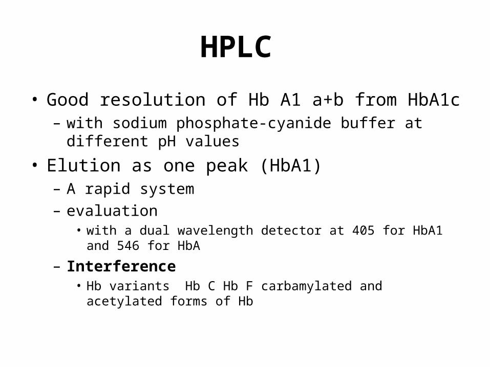
• Good resolution of Hb A1 a+b from HbA1c – with sodium phosphate-cyanide buffer at different pH
values
• Elution as one peak (HbA1)– A rapid system – evaluation
• with a dual wavelength detector at 405 for HbA1 and 546 for HbA
– Interference • Hb variants Hb C Hb F carbamylated and acetylated forms
of Hb

HPLC
• Quantification – Integrating the area under the peaks
• An automated system – Step gradients
• using three phosphate buffers of increasing ionic strength
– Detection• at 415 and 690nm
– both Hb A1c and HbA1 is reported – Variant Hb are resolved (Hb F, S and C)

HPLC
• HPLC methods – have excellent precision – recommended as reference method
• interference – Carbamylated and acetylated Hb and possibly
other derivatives• slightly higher results

Electrophoresis
• Agar gel at pH 6.3 resolution of Hb A and HbA1• The gel contains negatively charged moieties• Quantification performed by scanning densitometry at
415 nm• HbA1c is also commercially available • Results agree with that of HPLC or column but are less
precise• Minor variations in pH, ionic strength or temperature
have little effect on results • HbF migrates the same as HbA1and causes falsely
elevated value • Hb C and S do not • The labile form should be removed

Isoelectric focusing
• Principle; migration in gel containing a pH gradient
• Matrix; acrylamide gel • pH range of 6-8 • On completion the gels are fixed and then
scanned by a microdensitometer • Hb A1c resolved from HbA1a, A1b, S and F • Results showed close agreement with other
methods • The equipment is expensive

Immunoassay
• Anti serum raised against purified human HbA1c
• Available methods
• RIA format
• Enzyme immunoassay format
• Agglutination inhibition

Immunoassay
• Antibodies raised against the Amadori product of glucose (ketoamine linkage) plus the first few amino acids at the N-terminal of β-chain
• Agglutinator; a synthetic polymer containing multiple copies of the immunoreactive portion of HbA1c, light scattering

Immunoassay
• Excellent precision • The antibodies do not recognize labile
intermediates or other glycated hemoglobins • Other Hb variants such as HbF, A2, S,
carbamylated Hb are not detected.• Correlate well with HPLC but exhibit lower
values • Due to different calibration, detection by HPLC
of substances other than HbA1c

Affinity chromatography
• Principle– m-aminophenyl boronic acid is immobilized by cross
linking to beaded agarose or other matrix (e.g., glass fiber)
– The boronic acid react with the cis-diol groups of glucose
– Dissociation • By Sorbitol
– Detection • Absorbance of bound and non bound fractions measured at
415 nm


Affinity chromatography
• Advantage – No interference non glycated Hb – Negligible interference from the labile
intermediate form – Unaffected by variations in temperature – Reasonably good precision – Hemoglobin variants
• Hb F, S, and C produce little effect

Affinity chromatography
• Report – Affinity methods measure total glycated Hb – Commercially available systems are
calibrated to also report a HbA1c standardized value
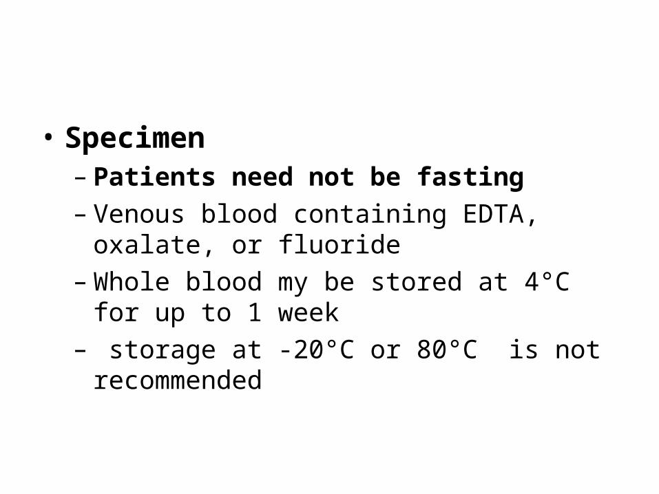
• Specimen– Patients need not be fasting – Venous blood containing EDTA, oxalate, or
fluoride – Whole blood my be stored at 4°C for up to 1
week– storage at -20°C or 80°C is not
recommended

• heparinized samples– should be assayed within 2 days and may not
be suitable for other methods (electrophoresis)

• Preparation of hemolysate– Packed cell
• Centrifuge – remove the plasma and buffy coat
• Wash with saline
– Removal of labile glycated Hb • Incubation of RBC in saline • in buffer solutions at pH 5 to 6 • by dialysis or ultrafiltration of hemolysate

• Preparation of column– Bring the column to room temperature – Remove the caps – Pour off upper buffer – Add equilibration buffer let drain and discard
the eluate

Assay standardization
• The absence of a reference method and a single glycated Hb standard has generated confusion
• Interlaboratory comparisons are not possible
• calibration – significantly improves precision and facilitates
direct comparison of results obtained by different methods

Assay standardization
• Calibrator– lyophilized hemolysate assayed by a precise
HPLC method for Hb A1c
• adoption of a universal standard will enhance the clinical utility of glycated Hb

Reference interval
• Values for glycated Hb are expressed as a percentage of total Hb
• Three major glyacted Hb species – HbA1, HbA1c, or total glycated Hb
• Reference intervals vary depending on – method – the glycated Hb component – whether the labile fraction is included


Reference intervals
• Reference intervals show some increase with age
• poorly controlled diabetes – values may extent to twice the upper limit of the
reference interval
• Values grater than 20% should prompt further studies
• There is no specific level of HbA1c below which the risk of diabetic complications is eliminated completely

Reference intervals
• Each laboratory should establish its own nondiabetic reference interval
• Assay precision is important; each 1% change = 25-35 mg/dl change