head injury - emergency care institute
TRANSCRIPT
Head Injury
Dr Sally McCarthy
Medical Director
ECI
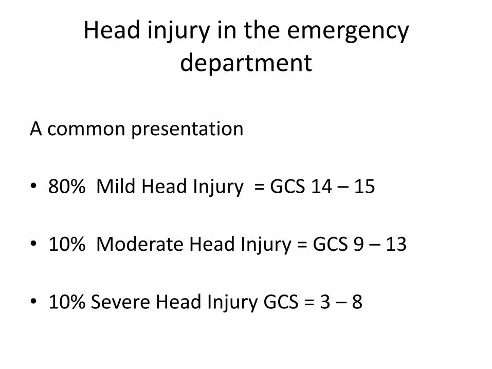
Head injury in the emergency department
A common presentation
• 80% Mild Head Injury = GCS 14 – 15
• 10% Moderate Head Injury = GCS 9 – 13
• 10% Severe Head Injury GCS = 3 – 8

Aetiology
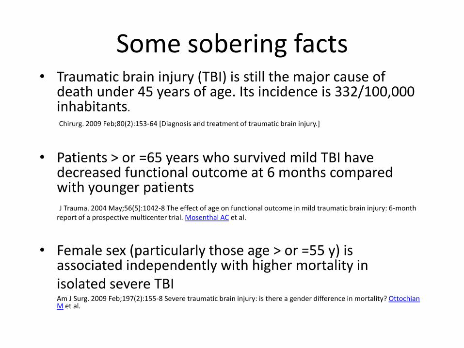
Some sobering facts• Traumatic brain injury (TBI) is still the major cause of
death under 45 years of age. Its incidence is 332/100,000 inhabitants.
Chirurg. 2009 Feb;80(2):153-64 [Diagnosis and treatment of traumatic brain injury.]
• Patients > or =65 years who survived mild TBI have decreased functional outcome at 6 months compared with younger patientsJ Trauma. 2004 May;56(5):1042-8 The effect of age on functional outcome in mild traumatic brain injury: 6-month
report of a prospective multicenter trial. Mosenthal AC et al.
• Female sex (particularly those age > or =55 y) is associated independently with higher mortality in isolated severe TBIAm J Surg. 2009 Feb;197(2):155-8 Severe traumatic brain injury: is there a gender difference in mortality? Ottochian M et al.
Adult Trauma Clinical Practice
Guidelines
Anatomy and Physiology
What are the unique features of
brain anatomy and physiology,
and how do they affect patterns of
brain injury?
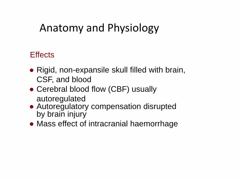
Anatomy and Physiology
● Rigid, non-expansile skull filled with brain,
CSF, and blood
● Cerebral blood flow (CBF) usually
autoregulated● Autoregulatory compensation disrupted
by brain injury
● Mass effect of intracranial haemorrhage
Effects
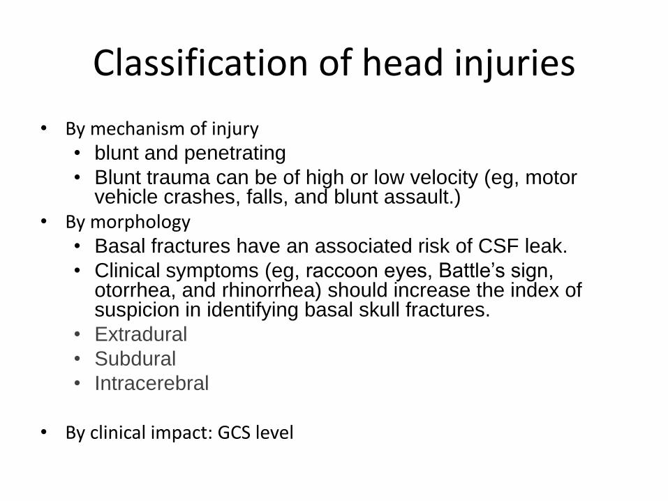
Classification of head injuries
• By mechanism of injury• blunt and penetrating
• Blunt trauma can be of high or low velocity (eg, motor vehicle crashes, falls, and blunt assault.)
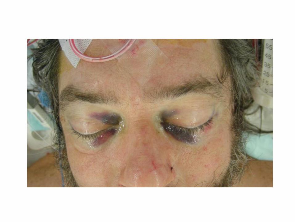
• By morphology• Basal fractures have an associated risk of CSF leak.
• Clinical symptoms (eg, raccoon eyes, Battle’s sign, otorrhea, and rhinorrhea) should increase the index of suspicion in identifying basal skull fractures.
• Extradural
• Subdural
• Intracerebral
• By clinical impact: GCS level
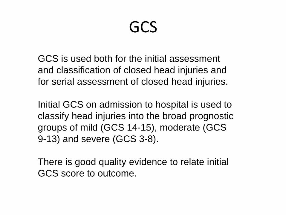
GCS is used both for the initial assessment
and classification of closed head injuries and
for serial assessment of closed head injuries.
Initial GCS on admission to hospital is used to
classify head injuries into the broad prognostic
groups of mild (GCS 14-15), moderate (GCS
9-13) and severe (GCS 3-8).
There is good quality evidence to relate initial
GCS score to outcome.
GCS
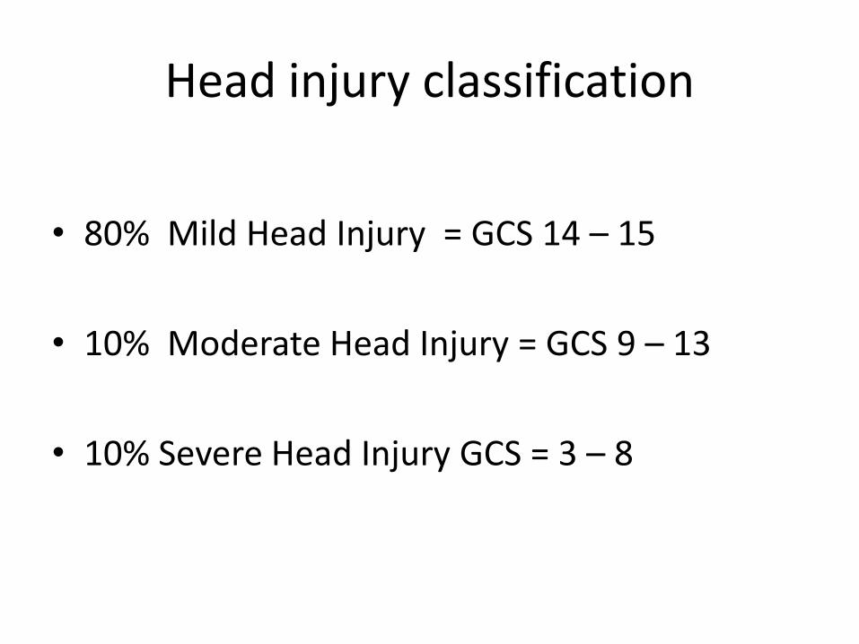
Head injury classification
• 80% Mild Head Injury = GCS 14 – 15
• 10% Moderate Head Injury = GCS 9 – 13
• 10% Severe Head Injury GCS = 3 – 8
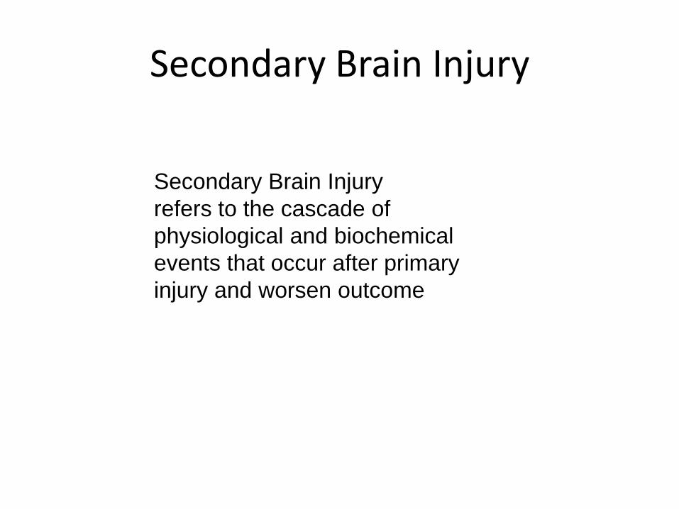
Secondary Brain Injury
refers to the cascade of
physiological and biochemical
events that occur after primary
injury and worsen outcome
Secondary Brain Injury

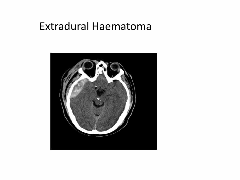
Extradural Haematoma
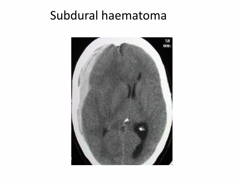
Subdural haematoma

Intracerebral Haematoma / Contusion
Large Frontal Contusion with Shift

Diffuse Brain Injury
Normal CT Diffuse Injury

Minor Head Injury
GCS 14 -15
High Risk= any RF present
Low Risk= no RF, normal CT-scan
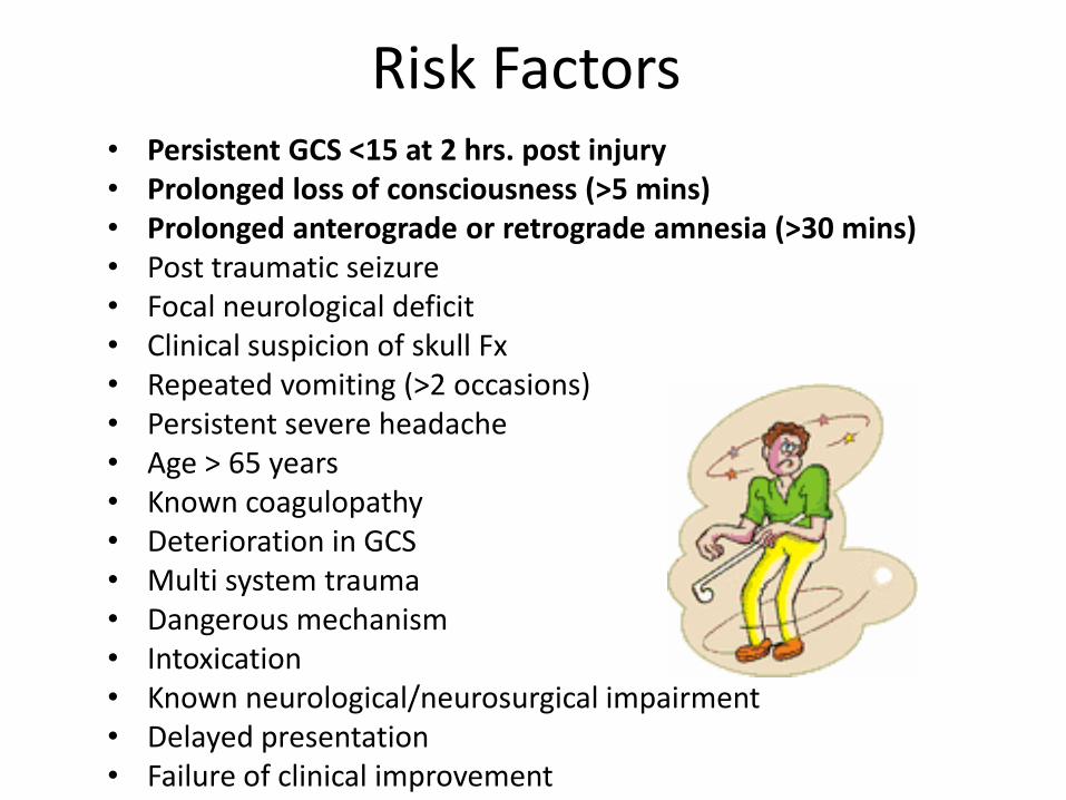
Risk Factors• Persistent GCS <15 at 2 hrs. post injury• Prolonged loss of consciousness (>5 mins)• Prolonged anterograde or retrograde amnesia (>30 mins)• Post traumatic seizure• Focal neurological deficit• Clinical suspicion of skull Fx• Repeated vomiting (>2 occasions)• Persistent severe headache• Age > 65 years• Known coagulopathy• Deterioration in GCS• Multi system trauma• Dangerous mechanism• Intoxication• Known neurological/neurosurgical impairment• Delayed presentation• Failure of clinical improvement
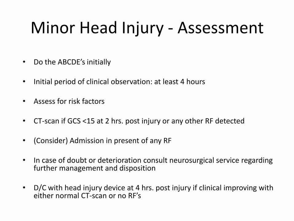
Minor Head Injury - Assessment
• Do the ABCDE’s initially
• Initial period of clinical observation: at least 4 hours
• Assess for risk factors
• CT-scan if GCS <15 at 2 hrs. post injury or any other RF detected
• (Consider) Admission in present of any RF
• In case of doubt or deterioration consult neurosurgical service regarding further management and disposition
• D/C with head injury device at 4 hrs. post injury if clinical improving with either normal CT-scan or no RF’s
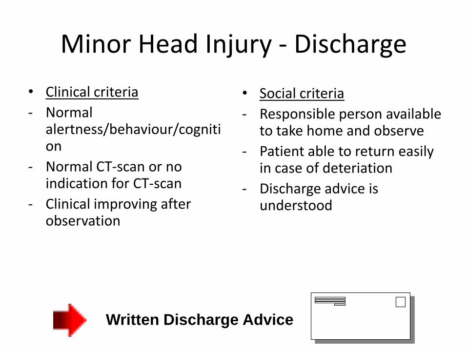
Minor Head Injury - Discharge
• Clinical criteria
- Normal alertness/behaviour/cognition
- Normal CT-scan or no indication for CT-scan
- Clinical improving after observation
• Social criteria
- Responsible person available to take home and observe
- Patient able to return easily in case of deteriation
- Discharge advice is understood
Written Discharge Advice
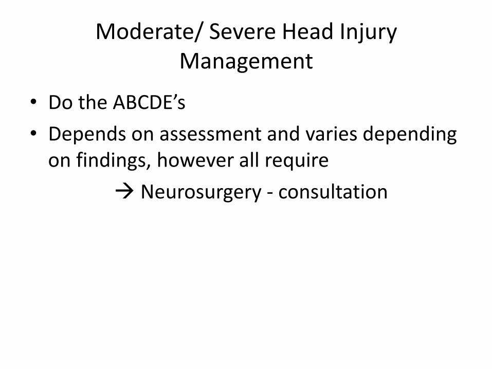
Moderate/ Severe Head Injury Management
• Do the ABCDE’s
• Depends on assessment and varies depending on findings, however all require
Neurosurgery - consultation

Indications for CT-scan
• Mild head injury with at least 1 risk factor present
• Any moderate head injury
• Any severe head injury
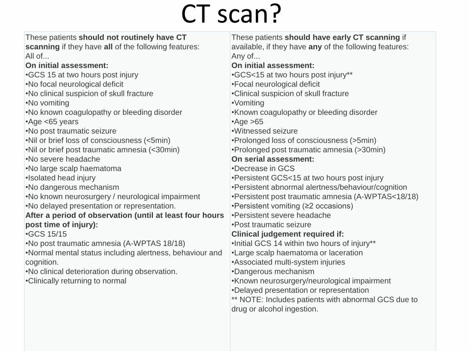
These patients should not routinely have CT
scanning if they have all of the following features:
All of...
On initial assessment:
•GCS 15 at two hours post injury
•No focal neurological deficit
•No clinical suspicion of skull fracture
•No vomiting
•No known coagulopathy or bleeding disorder
•Age <65 years
•No post traumatic seizure
•Nil or brief loss of consciousness (<5min)
•Nil or brief post traumatic amnesia (<30min)
•No severe headache
•No large scalp haematoma
•Isolated head injury
•No dangerous mechanism
•No known neurosurgery / neurological impairment
•No delayed presentation or representation.
After a period of observation (until at least four hours
post time of injury):
•GCS 15/15
•No post traumatic amnesia (A-WPTAS 18/18)
•Normal mental status including alertness, behaviour and
cognition.
•No clinical deterioration during observation.
•Clinically returning to normal
These patients should have early CT scanning if
available, if they have any of the following features:
Any of...
On initial assessment:
•GCS<15 at two hours post injury**
•Focal neurological deficit
•Clinical suspicion of skull fracture
•Vomiting
•Known coagulopathy or bleeding disorder
•Age >65
•Witnessed seizure
•Prolonged loss of consciousness (>5min)
•Prolonged post traumatic amnesia (>30min)
On serial assessment:
•Decrease in GCS
•Persistent GCS<15 at two hours post injury
•Persistent abnormal alertness/behaviour/cognition
•Persistent post traumatic amnesia (A-WPTAS<18/18)
•Persistent vomiting (≥2 occasions)
•Persistent severe headache
•Post traumatic seizure
Clinical judgement required if:
•Initial GCS 14 within two hours of injury**
•Large scalp haematoma or laceration
•Associated multi-system injuries
•Dangerous mechanism
•Known neurosurgery/neurological impairment
•Delayed presentation or representation
** NOTE: Includes patients with abnormal GCS due to
drug or alcohol ingestion.
CT scan?
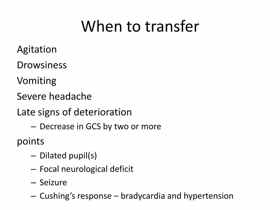
When to transferAgitation
Drowsiness
Vomiting
Severe headache
Late signs of deterioration
– Decrease in GCS by two or more
points
– Dilated pupil(s)
– Focal neurological deficit
– Seizure
– Cushing’s response – bradycardia and hypertension

Standard management of head injuries• Assess and stabilise ABCDEs
• Commence at least hourly clinical observations of vital signs, GCS, pupils, PTA and clinical symptoms.
• The initial assessment should be followed by a period clinical observation to detect risk factors for significant intracranial injury. The patient should be risk stratified into “low” or “high” risk groups based on the presence or absence of identified clinical risk factors.
• CT scan not routinely indicated unless one or more high risk factors are present.
• Discharge for home observation with head injury advice sheet at 4 hours post injury if clinically improving with either no risk factors indicating need for CT scan or normal CT scan if performed.
• Consider hospital admission and consult regional neurosurgical service if abnormal CT scan.
• Consider hospital admission for observation if clinically not improving at 4 hours post injury irrespective of CT scan result.
• Consider hospital admission for observation if elderly, known coagulopathy or socially isolated.
• Advise patients to see their local doctor if they do not return to normal within 48 hours so they can be reassessed and monitored for post concussion symptoms.
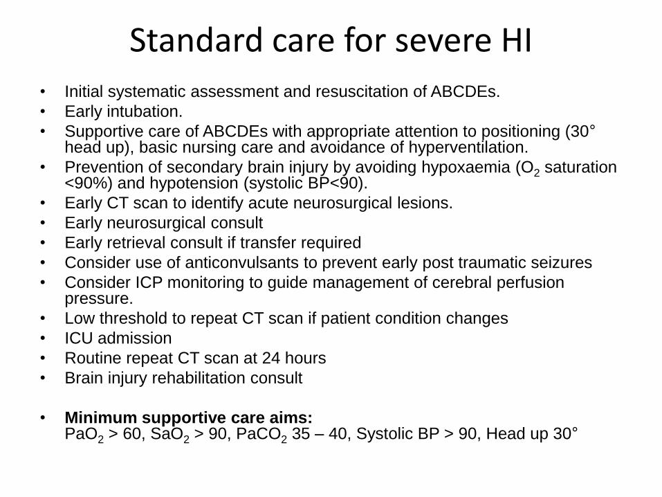
Standard care for severe HI• Initial systematic assessment and resuscitation of ABCDEs.
• Early intubation.
• Supportive care of ABCDEs with appropriate attention to positioning (30°head up), basic nursing care and avoidance of hyperventilation.
• Prevention of secondary brain injury by avoiding hypoxaemia (O2 saturation <90%) and hypotension (systolic BP<90).
• Early CT scan to identify acute neurosurgical lesions.
• Early neurosurgical consult
• Early retrieval consult if transfer required
• Consider use of anticonvulsants to prevent early post traumatic seizures
• Consider ICP monitoring to guide management of cerebral perfusion pressure.
• Low threshold to repeat CT scan if patient condition changes
• ICU admission
• Routine repeat CT scan at 24 hours
• Brain injury rehabilitation consult
• Minimum supportive care aims:PaO2 > 60, SaO2 > 90, PaCO2 35 – 40, Systolic BP > 90, Head up 30°
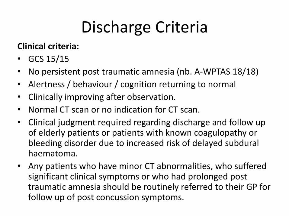
Discharge CriteriaClinical criteria:
• GCS 15/15
• No persistent post traumatic amnesia (nb. A-WPTAS 18/18)
• Alertness / behaviour / cognition returning to normal
• Clinically improving after observation.
• Normal CT scan or no indication for CT scan.
• Clinical judgment required regarding discharge and follow up of elderly patients or patients with known coagulopathy or bleeding disorder due to increased risk of delayed subdural haematoma.
• Any patients who have minor CT abnormalities, who suffered significant clinical symptoms or who had prolonged post traumatic amnesia should be routinely referred to their GP for follow up of post concussion symptoms.

Summary
• Assess
– Trauma approach
– ? High or low risk
• Manage
– Avoid secondary brain injury
• High or low risk?
• Monitor
• Analgesia
• Seek assistance, consultation, retrieval or transfer