health care provider’s guide to sickle cell disease
TRANSCRIPT

Health Care Provider’s Guide to Sickle Cell
Health Care Provider’s Guide to Sickle Cell Disease

Health Care Provider’s Guide to Sickle Cell

Health Care Provider’s Guide to Sickle Cell
Table of Contents
ii
47889
1011131517171820212121212222222223232323242425252526
IntroductionOverview of Genetics and PathophysiologyNeonatal Screening and DiagnosisOverview of Comprehensive CARE Medical Home Family and Patient Education Preventive Care and Health MaintenanceAcute Illness Fever and Infection Pain ACS Splenic Sequestration Aplastic Crisis Acute CNS Events PriapismChronic Manifestations Growth and Development Eyes Heart Lungs Renal System Hepatobiliary System Skin Bones SpleenTreatment Transfusion Therapy Surgery Bone Marrow Transplantation Hydroxyurea Antisickling AgentsPrognosis References
Health Care Provider’s Guide to Sickle Cell
Introduction
The term sickle cell disease (SCD) describes a group of complex, chronic disorders characterized by hemolysis, unpredictable acute complications that can rapidly become life-threatening, and the variable development of chronic organ damage. Expert, comprehensive medical care decreases morbidity and prolongs life expectancy for individuals with SCD. Many children with SCD in the United States receive much of their medical care from pediatricians. This statement is intended to provide pediatricians and family practitioners in primary care and subspecialist with an overview of the essential components of comprehensive care for children with SCD and their families. A detailed discussion of the treatment of individual acute and chronic complications of SCD is beyond the scope of this booklet but is available elsewhere.[1,2,3]
Health Care Provider’s Guide to Sickle Cell
Overview of Genetics and Pathophysiology

Health Care Provider’s Guide to Sickle Cell
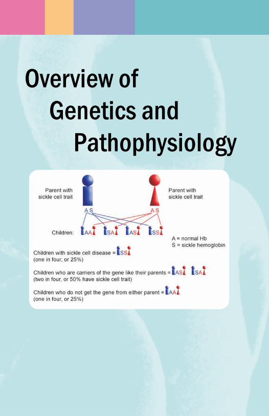
SCD is an autosomal recessive genetic disorder characterized by the presence of sickle hemoglobin (HbS) in red blood cells. Heterozygous individuals have sickle cell anemia (HbSS), sickle-hemoglobin C disease (HbSC), and two types of sickle β-thalasemia (Sβ+-thalassemia and Sβ◦-thalassemia)—account for most SCD in the United States. Less common forms of SCD are caused by coinheritance of HbS with other hemoglobin variants. Genes for SCD are common in persons of African, Mediterranean, Middle Eastern and Indian ancestry and persons from the Caribbean and parts for Central and South America. SCD is the most prevalent disorder identified by neonatal blood screening, with approximately 2000 affected infants born in the United States each year. Overall, the incidence of SCD exceeds that of most other serious genetic disorders, including cystic fibrosis and hemophilia.
The protean clinical manifestations of SCD results from variable degrees of hemolysis and intermittent episodes of vascular occlusion that cause tissue ischemia and acute and chronic organ dysfunction. Consequences of hemolysis may include chronic anemia, jaundice, predisposition to aplastic crisis, cholelithiasis, and delayed growth and sexual maturation. Vaso-occlusion and tissue ischemia can result in acute and chronic injury to virtually every organ of the body. Important clinical manifestations of SCD during childhood and adolescence are shown in Table 1. Generally, children with HbSS and Sβ◦-thalassemia are more severely affected than are children with HbSC or Sβ+-thalassemia. However, each genotype is characterized by marked and largely unpredictable variability in clinical expression and severity.
Sickled Cell
Normal Cell
2

Health Care Provider’s Guide to Sickle Cell 3
Table 1. Important Clinical Manifestations of SCD During Childhood and AdolescenceAcute Manifestations Bacterial sepsis or meningitis* Recurrent vaso-occlusive pain (dactylitis, musculoskeletal or abdominal pain) Splenic sequestration* Aplastic crisis* Acute chest syndrome* Stroke* Priapism Hematuria, including papillary necrosis
Chronic Manifestations Anemia Jaundice Splenomegaly Functional asplenia Cardiomegaly and functional murmurs Hyposthenuria and enuresis Proteinuria Cholelithiasis Delayed growth and sexual maturation Restrictive lung disease* Pulmonary hypertension* Avascular necrosis Proliferative retinopathy Leg Ulcers Transfusional hemosiderosis*
* Potential cause of mortality
Health Care Provider’s Guide to Sickle Cell4
Neonatal Screening and Diagnosis

Health Care Provider’s Guide to Sickle Cell
Most infants with SCD are healthy at birth and become symptomatic later in infancy or childhood after fetal hemoglobin (HbF) levels decrease. Most infants with SCD born in the United States are now identified by routine neonatal screening. Affected infants not identified through neonatal screening generally present clinically during infancy or early childhood with painful swelling of the hands and feet (dactylitis), pneumococcal sepsis or meningitis, severe anemia and acute splenic enlargement (splenic sequestration), acute chest syndrome, pallor, jaundice, or splenomegaly. Clinical presentations in older children include anemia, severe or recurrent musculoskeletal or abdominal pain, aplastic crisis, acute chest syndrome, splenomegaly or splenic sequestration, and cholelithiasis.
Forty-seven states, the District of Columbia, Puerto Rico, and the Virgin Islands currently provide universal neonatal screening for SCD; screening is available by request in the other three states. It is essential that pediatricians be familiar with their particular state’s screening program, that a screening sample always be obtained before any newborn blood transfusion (regardless of gestational or postnatal age), and that the results of neonatal screening tests be routinely and promptly documented for all infants. In states that have not yet implemented universal screening, neonatal screening for SCD should be requested for all high risk infants (those of African, Mediterranean, Middle Eastern, Indian, Caribbean, and Central and South American ancestry). Any high-risk infant not screened at birth, or for whom neonatal screening results cannot be documented, should be screened with hemoglobin electrophoresis as soon as possible after birth. For infants with positive screening results, confirmatory testing should be accomplished before 2 months of age so that parental education, penicillin prophylaxis, and arrangements for comprehensive care can be promptly initiated.
Confirmatory testing of infants with positive neonatal screening results and diagnosis of older patients who present with symptoms require hemoglobin separation by electrophoresis (cellulose acetate and citrate agar), isoelectric focusing, and diagnostic test results for the four most common genotypes of SCD are shown in Table 2. Solubility testing (eg. Sickledex) has no place in the diagnosis of SCD, because it does not differentiate SCD from sickle cell trait and because high levels of HbF cause false-negative results in infants with SCD.
5

Health Care Provider’s Guide to Sickle Cell6
TABL
E 2. S
CD: N
eona
tal S
cree
ning
and
Diag
nost
ic Te
st R
esul
ts
Dis
ord
er
Ap
pro
xim
ate
Neo
nat
al
Hem
oglo
bin
S
eria
l CB
C a
nd
Hem
atol
ogic
Stu
die
s by
Age
2 Y
ears
P
erce
nta
ge o
f S
cree
nin
g S
epar
atio
n b
y R
etic
ulo
cyte
Cou
nts
M
CV
† H
bA₂‡
H
bF
US
Pat
ien
ts
Res
ult
s A
ge 6
Wee
ks
(per
cen
t)
(per
cen
t)
Wit
h S
CD
HbS
S
65
FS
F
S
Hem
olys
is a
nd
an
emia
N
orm
al o
r in
crea
sed
§
<3.
6§
<25
B
y ag
e 6-
12 m
oH
bSC
25
F
SC
F
SC
M
ild
or
no
anem
ia b
y N
orm
al o
r d
ecre
ased
N
A‖
<15
a
ge 2
yS
β+-t
hal
asse
mia
8
FS
A o
r F
S
FS
A
Mil
d o
r n
o an
emia
by
Nor
mal
or
dec
reas
ed
>3.
6 <
25
age
2 y
Sβ◦
-th
alas
sem
ia
2 F
S
FS
H
emol
ysis
an
d a
nem
ia
Dec
reas
ed
>3.
6 <
25
Tabl
e sh
ows
typ
ical
res
ult
s-ex
cep
tion
s oc
cur.
Rar
e fo
rms
of S
CD
, su
ch a
s S
D-P
un
jab,
SO
-Ara
b, S
C-H
arle
m, S
δβ-t
hal
asse
mia
, SE
, an
d S
Lep
ore,
not
in
clu
ded
.*H
emog
lobi
ns
rep
orte
d i
n o
rder
of
quan
tity
(eg
, FS
A =
F>
S>
A).
†Nor
mal
or
refe
ren
ce r
ange
of
MC
V i
s >
70f
L a
t ag
e 6-
12 m
o; l
ower
lim
its
of r
efer
ence
ran
ge s
ubs
equ
entl
y in
crea
se w
ith
age
to
80 f
L d
uri
ng
adol
esce
nce
.‡H
bA₂ r
esu
lts
vary
som
ewh
at d
epen
din
g on
lab
orat
ory
met
hod
olog
y.§
HbS
S w
ith
coe
xist
ent
α-th
alas
sem
ia m
ay s
how
dec
reae
d M
CV
an
d H
bA₂>
3.6
per
cen
t; h
owev
er, n
eon
atal
scr
een
ing
resu
lts
from
su
ch i
nfa
nts
usu
ally
sh
ow B
art’s
h
emog
lobi
n.
‖ N
A =
not
ap
pli
cabl
e−qu
anti
ty o
f H
bA₂ u
sual
ly n
ot m
easu
red
in
pre
sen
ce o
f H
bC.
Qu
anti
ty o
f H
bA a
t bi
rth
is
som
etim
es i
nsu
ffici
ent
for
det
ecti
on.

Health Care Provider’s Guide to Sickle Cell 7
Overview Of Comprehensive Care

Health Care Provider’s Guide to Sickle Cell8
SCD is a complex disorder with multisystem manifestations that requires specialized comprehensive care to achieve an optimal outcome. Appropriate treatment for children requires the active involvement of health care professionals with expertise in the management and treatment of SCD, usually a pediatric hematologist-oncologist working in conjunction with a multidisciplinary team.
Medical HomeIt is essential that every child with SCD receive care that is provided and coordinated through an appropriate medical home. For many patients, the most appropriate medical home is a multidisciplinary sickle cell clinic that coordinates all aspects of comprehensive care in collaboration with the child’s primary care provider that provides specialty and primary care in one setting. In other cases, the medical home may be provided by a knowledgeable primary care pediatrician or other health care professional from whom patients receive day to day care, with periodic referrals to sickle cell specialists for comprehensive evaluations and for the management and treatment of severe, life threatening complications. Some SCD programs support primary care pediatricians by conducting outreach clinics in communities distant from tertiary care centers. The location of the medical home and the extent to which the care outlined in this statement is provided by the primary care pediatrician versus the multidisciplinary SCD team will vary among patients, depending on the distance of the community from a tertiary care centers. Access to a multidisciplinary SCD team, family preference, and the frequency and severity of disease manifestations. Appropriate management of many aspects of SCD requires time and expertise beyond levels provided by most primary care providers. In some cases, ongoing access to the pediatric hematologists-oncologist and other subspecialists may require advocacy by the primary care providers with managed care organizations or other payers.
Family and Patient EducationIdentification of an infant with SCD through neonatal screening provides an opportunity to educate parents and other caregivers about the child’s disorders before symptoms develop. Initially, the focus should include the genetics (including the availability of carrier testing and prenatal diagnosis) and basic pathophysiology of SCD and the importance of regularly scheduled health maintenance visits,

Health Care Provider’s Guide to Sickle Cell 9
penicillin prophylaxis, and immunizations, including pneumoccocal vaccines. Education about the need for urgent medical evaluation for and treatment of febrile illness, acute splenic sequestration, aplastic crisis, and acute chest syndrome is critical. Education about splenic sequestration includes the need to seek medical attention immediately if the child is pale and listless and instruction about abdominal palpation for determining spleen size. Recognition and appropriate management of dactylitis and other painful events should be reviewed. As the child ages, other topics such as stroke, enuresis, priapism, cholelithiasis, delayed puberty, proliferative retinopathy, avascular necrosis of the hip or shoulder, and leg ulcers are introduced. During middle childhood and adolescence, education is increasingly directed toward the patient in addition to the parents, and during adolescence, it includes the genetic basis of SCD and issues related to contraception, carrier testing of partners, genetic counseling, and prenatal diagnosis. The ultimate goal is to enable families to functionally cope with the child’s complex chronic illness and enhance the child’s potential for successful transition to adulthood.
Preventive Care and Health MaintenanceComprehensive medical care has been shown to contribute to increased life expectancy in patients with SCD. Patients for whom a neonatal electrophoresis screen is suggestive of SCD should be referred to a center knowledgeable in sickle cell syndromes. Early teaching about the implications of SCD and confirmation testing are performed, preferably before the patient is 2 months old. These measures allow physicians to begin penicillin prohylaxis, greatly reducing the risk of morbidity and mortality in children. Penicillin prophylaxis in sickle cell anemia (SCA) patients has been shown to reduce the incidence of pneumococcal infection by 84 percent, in comparison with placebo. [4] For SCA or sickle cell β◦-thalassemia, prophylactic penicillin is given in dosages of 125 mg orally twice a day for patients younger than 3 years, 250 mg orally twice a day for patients aged 3 to 12 years, and 500 mg orally twice a day for patients older than 12 years. Erythromycin ethylsuccinate is given to patients allergic to penicillin. Patients should remain on penicillin or erythromycin prohylaxis until 5 years of age. If serious pneumococcal infection is documented, penicillin or erythromycin prophylaxis is continued indefinitely. Parents are taught the signs

Health Care Provider’s Guide to Sickle Cell
and symptoms of sepsis, aplastic crisis, splenic sequestration, and vaso-occlusive episodes. A plan of care should be identified for the parents. Specifically, any child with SCD and a temperature above 101.5 degrees F, an enlarging spleen, or new onset of pallor should be examined on an emergency basis.
Routine comprehensive clinic visits are scheduled on at least a yearly basis. These visits should include a thorough physical examination to document the spleen size, baseline murmurs, and jaundice. Laboratory evaluation at a well visit can define a patient’s typical hemoglobin, platelet count, white blood cell (WBC) count, reticulocyte level and O² saturation level. This information can be extremely helpful when the patient seeks emergency care with possible acute chest or splenic sequestration syndrome. At two years of age, transcranial Doppler (TCD) examinations should be initiated [5]. Renal and liver functions are monitored yearly after the age of 5. Eye examinations are performed yearly after 7 years of age. Chest radiographs and pulmonary function should be considered every three to five years after the age of 5. X-ray of the hips, gall bladder ultrasound and echocardiogram are performed routinely at age 12 years.
Patients are immunized against pneumoccocal Haemophilus influenzae and hepatitis B as infants on the standard schedule. Pneumovax vaccine (23-valent) is given at age 2, with a booster at 5 years of age.
Anticipatory guidance is an important component of the regular clinic visit. The symptoms of infection, aplastic crisis, and splenic sequestration should be reviewed. Plans for coping with painful episodes at home will reduce hospital admissions and emergency room visits. Monitoring of growth and nutrition, yearly dental screening, genetic counseling, and birth control are other significant issues to include in the visit. Psychologic aspects, such as fear of death, chronic pain, and worry about growth delays should be addressed. As the patient reaches adulthood, a smooth transition from the pediatric facility to an adult center is essential.
Acute IllnessAcute illness characterized by relatively common childhood signs and symptoms, such as fever, cough, abdominal pain, pallor, and limp, can rapidly become life-threatening. Unfortunately, delayed or inadequate evaluation and treatment of acute illness remains an
10

Health Care Provider’s Guide to Sickle Cell 11
important cause of preventable morbidity and mortality. Thus, it is imperative that every child with SCD have a plan for around-the-clock access to a medical facility where knowledge and perspective about SCD is available and where evaluation and treatment can be promptly delivered. For example, a child with fever or pallor, and listlessness should always be initially evaluated, if possible, at a site where complete blood cells counts (CBC) and reticulocyte counts, blood cultures, intravenous antibiotics, and red blood transfusions are readily available. Health care professionals who treat acute illness need ready access to baseline information about the patient (e.g., SCD genotype, the presence or absence of splenomegaly, and baseline CBC and reticulocyte counts). Strategies for ensuring the availability of baseline information about individual patients include computerized patient databases and the provision of baseline information directly to patients and families on medical alert cards or on emergency information forms recommended by the AAP. The goal ensuring timely medical treatment for acute illness also is facilitated by providing anticipatory guidance to patients and families about early recognition, appropriate medical evaluation, and treatment of common acute complications.
Examples of acute illness that require urgent evaluation and treatment are outlined briefly below. More than one of these acute complications may be present simultaneously, and the information provided here lacks many important details about the management of each. Additional details are provided in references cited throughout this statement. Because blood transfusions play a central role in the treatment of some acute complications, the patient’s red blood cell antigen phenotype should be determined ahead of time if minor antigen-matched red blood cells, selected to prevent alloimmunization are available locally.
Fever and InfectionBecause patients with SCD develop splenic dysfunction at as early as 3 months of age, they are at high risk for septicemia and meningitis with pneumococci and other encapsulated bacteria. Thus, all patients with temperature greater than 101.5 degrees C require rapid triage and physical assessment, urgent CBC and reticulocyte counts, blood culture (plus cerebrospinal fluid analysis and other cultures as indicated), and prompt administration of a broad-spectrum parenteral antibiotic, such as ceftriaxone sodium or cefotaxime sodium. Because of its long half-life, ceftriaxone is usually chosen for selected cases in which outpatient management
Health Care Provider’s Guide to Sickle Cell12
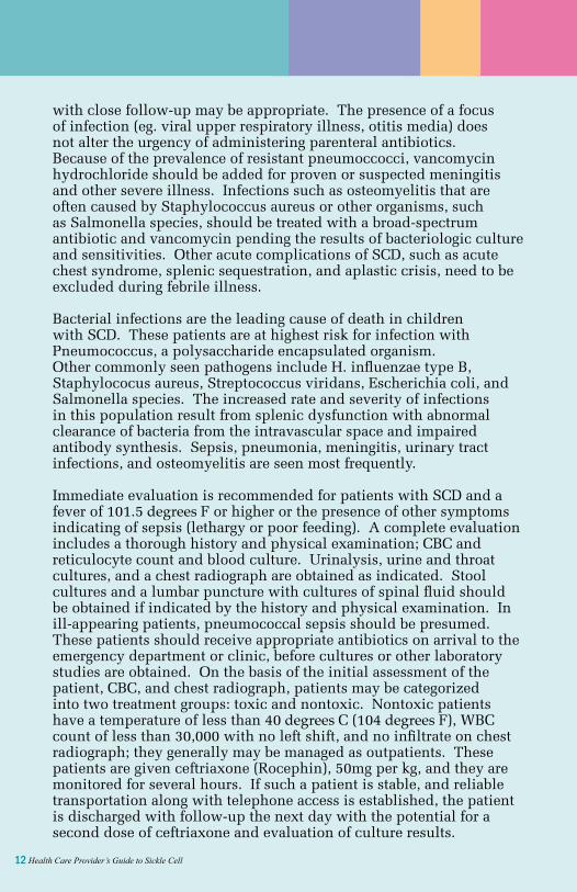
with close follow-up may be appropriate. The presence of a focus of infection (eg. viral upper respiratory illness, otitis media) does not alter the urgency of administering parenteral antibiotics. Because of the prevalence of resistant pneumoccocci, vancomycin hydrochloride should be added for proven or suspected meningitis and other severe illness. Infections such as osteomyelitis that are often caused by Staphylococcus aureus or other organisms, such as Salmonella species, should be treated with a broad-spectrum antibiotic and vancomycin pending the results of bacteriologic culture and sensitivities. Other acute complications of SCD, such as acute chest syndrome, splenic sequestration, and aplastic crisis, need to be excluded during febrile illness.
Bacterial infections are the leading cause of death in children with SCD. These patients are at highest risk for infection with Pneumococcus, a polysaccharide encapsulated organism. Other commonly seen pathogens include H. influenzae type B, Staphylococus aureus, Streptococcus viridans, Escherichia coli, and Salmonella species. The increased rate and severity of infections in this population result from splenic dysfunction with abnormal clearance of bacteria from the intravascular space and impaired antibody synthesis. Sepsis, pneumonia, meningitis, urinary tract infections, and osteomyelitis are seen most frequently.
Immediate evaluation is recommended for patients with SCD and a fever of 101.5 degrees F or higher or the presence of other symptoms indicating of sepsis (lethargy or poor feeding). A complete evaluation includes a thorough history and physical examination; CBC and reticulocyte count and blood culture. Urinalysis, urine and throat cultures, and a chest radiograph are obtained as indicated. Stool cultures and a lumbar puncture with cultures of spinal fluid should be obtained if indicated by the history and physical examination. In ill-appearing patients, pneumococcal sepsis should be presumed. These patients should receive appropriate antibiotics on arrival to the emergency department or clinic, before cultures or other laboratory studies are obtained. On the basis of the initial assessment of the patient, CBC, and chest radiograph, patients may be categorized into two treatment groups: toxic and nontoxic. Nontoxic patients have a temperature of less than 40 degrees C (104 degrees F), WBC count of less than 30,000 with no left shift, and no infiltrate on chest radiograph; they generally may be managed as outpatients. These patients are given ceftriaxone (Rocephin), 50mg per kg, and they are monitored for several hours. If such a patient is stable, and reliable transportation along with telephone access is established, the patient is discharged with follow-up the next day with the potential for a second dose of ceftriaxone and evaluation of culture results.
Health Care Provider’s Guide to Sickle Cell 13
Patients are considered potentially toxic if the WBC is greater than 30,000 or less than 5000, a left shift is noted on the differential, or the temperature is greater than 40 degrees C (104 degrees F). Patients who have a pulmonary infiltrate or have no reliable transportation generally require admission to the hospital for intravenous antibiotics and monitoring. Cefotaxime sodium, 50 mg per kg per dose every eight hours, provides broad-spectrum coverage for the common infecting organisms. However, the institutional sensitivities should be considered in the choice of a specific regimen, especially in areas with known penicillin- and cephalosporin-resistant Pneumococcus strains. Antibiotics are continued until the patient has been afebrile for 24 hours and cultures have been negative for 48 hours. Prevention of infection in the population with SCD has been previously discussed in the section on health maintenance.
PainAcute painful episodes are the most common symptom of SCD. Health providers must be aware that all pain in sickle cell patients is not a result of vaso-occlusion. Painful episodes of sickle cell disease are caused by ischemia or infarction of bones, bone marrow, or other organs. The most common sites for painful episodes after the third or fourth year of life are the long bones (more frequently in the humerus, tibia, and femur), the vertebrae, the abdomen, and the chest. In infancy, dactylitis, or “hand-foot” syndrome, is the most common manifestation of SCD. Infarction of the metacarpals and metatarsals causes this syndrome and results in swelling and pain in the hands and feet. Patients may refuse to bear weight on them. Fever and leukocytosis may accompany dactylitis. Treatment for dactylitis is the same as for other painful episodes.
Painful episodes in the abdomen may mimic peritonitis or appendicitis. An elevation of the WBC count, fever, or lack of response to the usual measures make a diagnosis that requires surgical exploration more likely. In this case, the patient should take nothing by mouth and a surgical consultation should be obtained. Obstruction of the biliary tree may be the cause of right upper quadrant pain. Abdominal ultrasonography and evaluation of bilirubin and liver function tests are performed if this is suspected.
Signs and symptoms of painful episodes include tenderness to palpitation and swelling over the affected site. Warmth, erythema, and diminished range of motion may be present. Painful episodes can mimic osteomyelitis, and it may be difficult to differentiate the
Health Care Provider’s Guide to Sickle Cell14
two. A culture is needed for a definitive diagnosis of osteomyelitis. However, it is important to remember that painful episodes due to infarction are much more common than osteomyelitis in the patient who is responding to the usual measures for episodic pain, and osteomyelitis is less likely.
Painful episodes have been attributed to infection, fever, dehydration, stress, and climate changes. However, no cause is identified in most patients. The severity of pain varies widely from mild events that can be managed at home, to severe events necessitating hospitalization. Episodes usually last three to five days but may be prolonged in some patients.
The diagnosis of a painful episode is best made with careful history and physical examination. Older children and adults can determine whether the pain is typical for their SCD. Pain level should be documented with numerical or visual analogue scales for older children and adults. Face scales for younger children help determine the severity of pain. In infants, parental ratings or behavioral indicators are used. There is no objective method for measuring pain, and so the report of the patient or parent should be accepted. Radiographs of the affected area are usually not helpful in acute episodes. Likewise, painful episodes are not associated with definitive changes in the complete blood count. Hemoglobin levels or amount of sickled cells on the smear cannot predict severity of an episode.
Treatment for painful episodes depends on the severity of the event. Attempts at home therapy can be made initially. Combinations of acetaminophen (10 to 15 mg per kg per dose every four to six hours; 24-hour maximum of five doses per day) and a nonsteroidal anti-inflammatory drug (ibuprofen, 10 mg per kg per dose every six hours) are used for milder episodes. Acetaminophen with codeine (0.5 to 1.0 mg codeine per kg per dose every four to six hours) may be used to strengthen the home regimen. Sufficient oral hydration should be stressed.
If treatment at home fails, the patient is evaluated in the clinic or emergency department. Adequate hydration is ensured with hypotonic intravenous fluids generally given at a rate of 1.5 times the maintenance level. Parenteral narcotics are the next line of therapy for patients with moderate to severe pain. The drug of choice in our institution is nalbuphine (Nubain) with a bolus dose of 0.3 mg per kg intravenously (maximal dose, 20 mg). This bolus may be repeated every 20 minutes for two additional doses (total of three doses). If adequate pain control is achieved, the patient is discharged

Health Care Provider’s Guide to Sickle Cell
to home with a sufficient supply of oral narcotics, NSAIDs, and acetaminophen. The patient should be instructed to take the oral pain medications on a scheduled basis for at least 48 hours. Oral fluid intake is again stressed.
If pain is not tolerably controlled in the emergency department or clinic, the patient is admitted for continuous parenteral narcotics. Nalbuphine is the first choice, at a starting dose of 0.075 mg per kg per hour. This infusion may be gradually increased to a maximum of 0.15 mg per kg per hour. The maximal daily dose for nalbuphine, 160 mg, must also be considered when this drug is used: Some patients cannot receive the hourly maximum without surpassing the daily limit. Although nalbuphine is preferred in our institution as a means of lowering the incidence of acute chest syndrome, therapy must be individualized. Some patients prefer certain medications. Hydromorphone hydrochloride (Dilaudid) and fentanyl (Sublimaze) are other options, followed by morphine. The dosage of narcotics is routinely divided between a continuous infusion and an additional patient-controlled analgesia (PCA). PCA empowers patients, gives them more control in their treatment, and allows for an immediate bolus when required (within set parameters). This method is also a useful tool for monitoring a patient’s course, because the PCA demands decrease as the pain improves. PCA is helpful in allowing a patient to self-wean as well.
Adjuvant therapies are used in addition to intravenous narcotics. Acetaminophen (Tylenol) and a non-steroidal anti-flammatory drug are scheduled every six hours. Hydroxyzine (Atarax) at a dose of 0.5 mg per kg orally every 6 hours may be used to potentiate the effects of the narcotics. Heat, transcutaneous electrical nerve stimulation (TENS) units, whirlpool, and relaxation techniques are other measures that may be beneficial. Transfusion of packed RBCs and provision of oxygen at the onset of uncomplicated painful episodes do not shorten the course of the event and are not indicated.
Acute Chest SyndromeThe syndrome of acute chest is the leading cause of death in patients over 10 years of age with SCD. The simplest definition of acute chest syndrome is a new infiltrate on chest radiograph. Some clinicians, including the author require that the patient show signs of hypoxia. Fever, tachypnea, and chest pain are other presenting symptoms. Abdominal pain may be the initial complaint in some patients. Acute chest syndrome is seen more commonly in patients with higher
15

Health Care Provider’s Guide to Sickle Cell16
baseline hemoglobin levels and lower fetal hemoglobin levels. The cause of acute chest syndrome may be infection, infarction, or a combination of factors. The source of the event is usually difficult to detect. In most cases, an infectious cause is not proven. When infection is documented, Pneumococcus, Mycoplasma, Salmonella, Klebsiella species, and H Influenzae are the organisms most commonly involved. Pulmonary fat embolization may be a significant cause of acute chest syndrome. In a study of 27 patients with acute chest syndrome, pulmonary fat embolus (thought to be secondary to bone marrow necrosis) was identified in 12 of those patients. [6]
In evaluation of a patient for acute chest syndrome, the complete blood cell count (CBC) may reveal a drop in the hemoglobin level in comparison with baseline levels. The WBC count is often increased, and reticulocytosis may be present. A drop in platelet count may be an ominous sign. The chest radiograph may not be abnormal initially. However, an infiltrate may appear or become more prominent as the patient is hydrated. An arterial blood gas measurement with room air is needed if acute chest syndrome is suspected. If the baseline oxygen saturation for the patient is known, trends in transcutaneous oxygen saturation may provide some assistance.
Treatment involves the use of supplemental oxygen, if the patient is hypoxic. Incentive spirometry, percussion therapy, and albuterol nebulizations may be useful in preventing atelectasis. An effort should be made to ensure that the patient is out of bed several times a day when possible. Intravenous fluids are given at a rate of one to two times the maintenance level until the patient is rehydrated. At that time, the infusion is decreased to maintenance levels to prevent fluid overload. Empirical antibiotic therapy is given to cover the typical organisms. Cefotaxime sodium (50 mg per kg intravenously every eight hours) and erythromycin are chosen in our institution. Analgesics are administered carefully to avoid oversedation or undersedation, either of which may cause atelectasis. In milder episodes and when the hemoglobin level is below baseline, patients are treated with a simple transfusion. More severe or progressive episodes necessitate exchange transfusion. One article promoted the use of intravenous dexamethasone (0.3 mg per kg every 12 hours for four doses) in patients with mild to moderately severe acute chest syndrome. The patients who received dexamethasone have shorter hospital stays, a reduced need for blood transfusion, and shorter duration of analgesic and oxygen therapy than did patients who received placebo. [7]

Health Care Provider’s Guide to Sickle Cell 17
Splenic Sequestration Splenic sequestration is the syndrome of rapid entrapment of RBCs within the spleen, with splenic enlargement and a fall in the hemoglobin by more than two to three grams per dL. This has been reported to be in approximately five to 10 percent of patients with SCD. This syndrome can occur as early as 2 months of age and is rarely seen after the age of 5 years in patients with SCA. Sequestration may occur later in children with HbSC or sickle cell β+-thalassemia in whom autosplenectomy is delayed. Episodes of splenic sequestration may occur alone or in combination with acute chest syndrome, a painful episode, or infection. Symptoms include pallor, weakness, abdominal distention, and left-sided abdominal pain. Patients are noted to be tachycardic and dyspneic with splenomegaly and may deteriorate rapidly to hypovolemic shock. Laboratory evaluation reveals an acute drop in hemoglobin below the patient’s baseline, with marked reticulocytosis. Thrombocytopenia is often present and is caused by splenic entrapment of platelets. Episodes of sequestration of the spleen may be mild, necessitating no definitive therapy, or can progress rapidly and lead to death. Adequate hydration with intravenous fluids at 1.5 times maintenance levels may reverse less severe episodes. Patients should be closely monitored for signs of progression. Hypovolemia and lack of oxygen-carrying capacity should be corrected rapidly with transfusion of packed RBCs or whole blood.
Approximately half the patients who survive the first episode have a recurrence of splenic sequestration. This recurrent episode often occurs within four months after the first event. Approaches to maintenance care include close monitoring by instructing parents to monitor spleen size, institution of a maintenance transfusion program, or surgical splenectomy. Some authorities advocate splenectomy after the first episode of splenic sequestration; others promote splenectomy after repeated episodes. The current plan of care in our institution is to defer splenoctomy until age 5 if possible, because the risk for postsplenectomy infection is lower after this age
Aplastic CrisisAplastic Crisis, or transient erythroblastopenia, is often secondary to infection with parvovirus but can occur with other infections. Aplastic crisis is a result of diminished RBC production in addition to the underlying chronic hemolytic anemia RBC survival in SCA

Health Care Provider’s Guide to Sickle Cell18
is shortened to 10 to 15 days, in comparison with the normal RBC survival of 120 days. Patients with SCA compensate with increased hematopoietic activity. In the wake of parvovirus or other infections, erythroid differentiation is halted, and the precarious balance between RBC production and destruction is lost. Reticulocytosis drops, with a resulting fall in hemoglobin. Symptoms of aplastic crisis are those of any acute anemia, including pallor and lethargy. Parvovirus infection can be documented with viral titers. Patients usually show signs of recovery within approximately 10 days but may require treatment with transfusion if the anemia is severe.
Acute Central Nervous System EventsCerebrovascular accidents (CVAs) are responsible for devastating morbidity and are the second leading cause of death in one study of patients with SCD. The incidence of CVAs in children with SCA is approximately seven to eight percent. The Cooperative Study of Sickle Cell Disease (CSSCD) reported a four percent overall incidence of CVAs. This complication is less common in patients with HbSC or sickle cell β+-thalassemia but should be considered. CVA recurs in approximately 70 percent of untreated patients, usually within three years of the previous attack, and with a more severe outcome. CVA usually occurs without warning but may manifest with painful episodes, aplastic crisis, or infectious illnesses. CVA occurs in patients after the age of 1 year; ischemic events are more common in children, and hemorrhagic events are most common in adults aged 20 to 29 years. A combination of the two types of stroke can also take place. Ischemic stroke usually occurs in larger vessels, secondary to stenosis or obstruction. The underlying pathophysiologic process consists of thrombus formation, embolization, or proliferation of smooth muscle and fibroblasts over damaged endothelium. The middle cerebral, anterior cerebral, and distal internal carotid arteries are often involved with ischemic CVA. Hemorrhagic stroke may be secondary to aneurysms of collateral or weakened vessels.
Risk factors for ischemic CVA include a prior episode of a transient ischemic attack (TIA), a recent episode of acute chest syndrome, and elevated systolic blood pressure. Patients most commonly come to medical attention with dysarthria, dysphagia, focal seizures, gait difficulties, hemiparesis, or hemiplegia. With hemorrhagic stroke, a severe headache, syncope, meningismus, or photophobia may be the initial complaint. A physical examination with a thorough neurologic

Health Care Provider’s Guide to Sickle Cell 19
evaluation is indicated in any patient with SCD and potential neurologic symptoms. Computed tomography or magnetic resonance imaging should be performed to rule out intracranial hemorrhage. Noninvasive magnetic resonance angiography allows definition of cerebral vessels without the use of hypertonic radiocontrast and its associated risk of sickling.
Treatment for CVA includes immediate hydration, frequent neurologic assessment, and antiepileptic medications if seizures are present. Exchange transfusion is the standard initial therapy. Alternatively, simple transfusion may be used primarily in stable patients with no progressive symptoms and a hemoglobin level below steady state. A hemoglobin level for more than 12 grams per dL must be avoided in order to avoid further difficulties from hyperviscosity. Simple or exchange transfusion is followed by a program of maintenance transfusion therapy every three to six weeks to maintain an HbS level of less than 30 percent. Up to 50 percent of patients may have complete or nearly complete recovery with rapid, aggressive transfusion therapy. The risk of CVA recurrence is approximately 70 percent in untreated patients and is highest in the first three years after the initial event. Maintenance transfusion therapy reduces the risk to 10 percent; most of these events consist of TIAs. Maintenance transfusion therapy clearly improves the mortality rate as well. Patients with a history of stroke should be maintained on a routine transfusion program indefinitely. Attempts have been made to liberalize the HbS level to 40 to 50 percent or to stop transfusion therapy after a period of time. However, CVA has recurred after completion of five- to 12-year maintenance transfusion. The dangers of maintenance transfusions include infection risks, sensitization, and iron overload. Partial exchange transfusions decrease the overall iron burden. Iron chelation should be included in the maintenance transfusion protocol once iron overload is established.
One of the greatest difficulties has been identifying patients at risk for stroke and preventing the first CVA. Significant advances have been made in this area. Elevated velocities in the internal carotid or middle cerebral artery as evaluated by transcranial Doppler (TCD) ultrasonography have been strongly associated with an increased risk of stroke. [5] This knowledge has been used in the Stroke Prevention Trial in Sickle Cell Anemia (STOP). These patients at risk for stroke were defined as having two TCDs with an elevated mean blood flow velocity. Patients were randomly assigned to prophylactic transfusions that maintained the HbS below 30 percent or to standard care. Maintenance transfusion therapy was shown to significantly reduce the incidence of first stroke in patients with abnormal TCD. [8] Attempts should be made to screen patients with SCA disease or S-

Health Care Provider’s Guide to Sickle Cell20
β◦-thalassemia beginning at the age 2 years to determine the need for prophylactic transfusions.
Silent cerebral infarcts occur in 22 percent of children with SCA by 14 years of age who have not been identified as having a clinical evident stroke (overt stroke) [9]. Presently, the Silent Infarct Transfusion Trial (SITT) is being conducted to determine if prophylactic blood transfusion therapy in children with silent infarcts will result in a dramatic reduction in the proportion of patients with clinically evident strokes or new or progressive silent cerebral infarcts.
PriapismPriapism can occur at any age in males with SCD. In one study, the median age at onset was 21 years. The pathophysiologic process consists of obstruction of venous outflow from the corpora cavernosum. Two forms of which consists of multiple short episodes, and severe prolonged priapism, in which painful erection lasts more than 24 hours. In one study, stuttering priapism was shown to advance to prolonged episodes in 28 percent of patients. Acute urinary retention may complicate priapism. In a study of Jamaican
patients with a history of priapism, a 46 percent incidence of sexual dysfunction was reported.
Initial treatment for priapism consists of hydration with hypotonic fluids and Sudafed has been effective in many instances. If no improvement is seen in four to six hours, simple or exchange transfusion is warranted. Surgical procedures are available and should be considered if the patient is unresponsive to less aggressive measures. The approaches include corporal aspiration and irrigation or construction of a fistula between the glans and corpora cavernosum. Patients with frequent episodes of priapism may benefit from a short course of maintenance transfusion therapy.

Health Care Provider’s Guide to Sickle Cell 21
Chronic ManifestationsGrowth and DevelopmentGrowth curves generated by the Cooperative Study of Sickle Cell Disease (CSSCD) demonstrate delays in both weight and height, with greater improvement in height by the end of adolescence. Delays are greater in patients with SCA and S-β◦thalassemia. Growth delays have been attributed to increased caloric requirements as a result of amplified hematopoetic activity and cardiac output. Delayed sexual development has also been noted in patients with SCD. Decreased fertility has not been reported in females. However, abnormal sperm motility and lower sperm counts have been documented in some males.
EyesProliferative retinopathy is a serious ocular complication of SCD. This complication is more common in sickle cell patients with higher hemoglobin levels (ie HgbSC, Sickle B+thal). Manifestations range in severity from stage I retinopathy with peripheral arteriolar occlusion to stage 4 with retinal detachment. In a Jamaican investigation, 20 percent of the eyes examined showed evidence for proliferative retinopathy during the course of the study. Treatment with laser therapy may be helpful but remains controversial because blindness may be a complication of occlusion of a central retinal artery.
HeartSystolic and diastolic ejection murmurs, a split S₁, and an S₂ may be auscultated in patients with SCD. Cardiomegaly is present in the majority of older patients, and left ventricular hypertrophy is seen on electrocardiograms in approximately 50 percent of patients. These findings usually result from compensation for the chronic hemolytic anemia. Myocardial infarctions are surprisingly rare in patients with SCD. Increased sickling is not noted in the coronary arteries, possibly as a result of the short transit time for the RBCs in the coronary vessels. SCD also appears to confer a protective effect toward development of atherosclerosis.

Health Care Provider’s Guide to Sickle Cell22
LungChronic lung disease secondary to recurrent infarction ranges in severity from a chronic cough to severe pulmonary hypertension. Patients with SCD maintain a lower resting partial oxygen pressure. These patients also have lower vital and total lung capacities.
Renal SystemAn inability to produce concentrated urine results in hyposthenuria in patients with SCD. This promotes dehydration and negates the use of the urine specific gravity for assessment of hydration status. Nocturia or enuresis ensues in approximately 50 percent of patients with SCD. Nephrotic syndrome may also be seen in association with SCD. Affected patients are often unresponsive to therapy, and the condition may advance to renal failure.
Hepatobiliary SystemGallstones are noted frequently in patients with SCD as a result of the chronic hemolytic anemia. In one investigation, 42 percent of 15- to 18-year-old patients with SCA were reported to have gallstones. In addition, 13 percent of patients between the ages of 5 and 13 years had this problem. Prophylactic cholescystectomy is not recommended for asymptomatic patients. Cholecystectomy may be performed in a patient with recurrent right upper quadrant pain and gallstones, but it does not resolve the pain episodes in every patient. Moderate hepatomegaly is often noted in patients with SCD. Hepatitis related to transfusion is also seen in the population with SCD.
Skin Leg ulcers usually occur in regions of the medial tibia and posterior to the medial malleolus. This is a complication in as many as 75 percent of patients in certain geographic areas. Skin breakdown may be secondary to elevations in the venous pressure of the legs with increased hematopoietic activity and expansion of the bone marrow volume. Impairments in wound healing may also contribute to the process. Treatment involves routine wound care with rest, elevation of the lower extremities, and debridement if necessary. Compression hose may be used to prevent the occurrence of ulcers. Oral zinc has been reported to improve healing.

Health Care Provider’s Guide to Sickle Cell 23
BonesThe characteristic “hair on end” and flattened “codfish” vertebrae result from the marrow expansion in patients with the chronic hemolytic anemia. The more serious bony complication of SCD is avascular necrosis (AVN), usually of the femoral head but also occurring in the head of the humerus. SCD is the most common cause of AVN of the femoral head in the pediatric population. By one report, AVN occurs in 19 percent of patients with SCA and nine percent of patients with HbSC. Approximately half the affected patients are symptomatic with pain and decreased mobility. AVN may develop as a result of elevations in pressure within the bone marrow after ischemic events. Vasoocclusive infarctions within the femoral head may affect the vascular supply. Bone is remodeled, and resorption occurs with collapse of the femoral head.
The diagnosis of AVN is made with radiographs or MRI, showing a widened joint space, flattened epiphysis, and widening of the femoral neck. Treatment for mild cases involves avoidance of weight bearing. Patients who have completed growth with severe AVN may benefit from prosthetic hip replacement.
SpleenFunctional asplenia occurs by 5 to 36 months of age. Continued infarction leads to autosplenectomy, in which the spleen is reduced to a small, fibrotic piece of tissue. The consequences of asplenia are discussed in the section on infections.
TreatmentTransfusion TherapyA simple transfusion usually suffices for patients with aplastic crisis and splenic sequestration. Simple transfusions are sometimes indicated in milder cases of acute chest syndrome or for central nervous system events if the hemoglobin level is below baseline. A hemoglobin level of more than 12 grams per dL should be avoided in patients with SCD in order to reduce the effects of hyperviscosity. Simple transfusion is also used in preparation for surgery, as
Health Care Provider’s Guide to Sickle Cell24
discussed later. Partial exchange transfusion is conventionally employed with most central nervous system events and with more severe or progressive episodes of acute chest syndrome. Maintenance transfusion programs are indicated for patients at risk for CVA and for patients with recurrent splenic sequestration. Short transfusion programs may be considered for recurrent acute chest syndrome and debilitating painful episodes. The goal of maintenance transfusion therapy is to reduce and maintain a lowered HbS level, usually less than 30 percent. This lessens the frequency of vaso-occlusive events. Iron overload is a complication of maintenance transfusion therapy and should be monitored closely. Standard treatment consists of chelation with desferrioxamine (Desferal). A new oral cheleton (Exjade) has recently become available. Alloimmunization, another complication of maintenance transfusion, occus in less than 25 percent of patients. Antigen-matched packed RBCs would therefore be preferred but are expensive and more difficult to obtain. We suggest antigen matching for the most common antibody-inciting antigens (Rh Group, Kell, Duffy)
SurgeryComplications of surgery are known to occur in patients with SCD. However, with careful management, these problems may potentially be avoided. A simple transfusion should be given to reach a preoperative hemoglobin level of 10 grams per dL. Strict attention to oxygenation, avoidance of temperature extremes and provision of maintenance intravenous fluids are important prophylactic measures in the preoperative period. Even with preoperative transfusion, a 10 percent incidence of acute chest syndrome was noted after surgery in a multi-institutional study. [10] A history of acute chest syndrome was noted to be a risk factor for postoperative episodes.
Bone Marrow TransplantationAllogeneic transplantation is a potentially curative treatment for SCD. An ongoing multicenter trial reports promising data. Of the 34 children with SCD who have received transplants from HLA-identical siblings, 32 have survived; 28 of these patients had stable engraftment. A 93 percent survival rate and a 79 percent event free survival rate are reported. [11] Patients who underwent transplantation had a history of CVA, recurrent acute chest syndrome, or recurrent vasoocclusive crises. Despite the encouraging results, difficulties with transplantation exist. Only a low percentage of patients have

Health Care Provider’s Guide to Sickle Cell
a matched-sibling, sickle cell-negative donor. Furthermore, older patients do not tolerate the complications of allogeneic bone marrow transplantation as well as do younger patients without significant organ toxicity. However, it is difficult to identify which patients with severe disease would benefit most from transplantation. Further studies are needed to identify early which patients are at risk for complication of SCD.
HydroxyureaAs previously discussed, higher levels of HbF inhibit HbS polymerization in vitro. Patients with higher HbF levels generally have less severe disease. This knowledge is the basis for the use of hydroxyurea (Hydrea), which increase HbF production. Use of hydroxyurea in adults has been shown to reduce the frequency of painful episodes, acute chest syndrome, and transfusions. [12] Concerns regarding the potential teratogenicity and carcinogenicity of the drug warrant limited pediatric use until ongoing studies are completed. A phase II multicenter trial is currently under way to evaluate the effects of hydroxyurea in infants with SCD.
Antisickling AgentsWith increasing knowledge of the molecular pathophysiologic processes of SCD, numerous agents are being evaluated for potential antisickling effects. Medications are being studied for possible inhibition of the transport pumps that cause erythrocyte dehydration.
PrognosisThe overall life expectancy for patients with SCA is 42 years for males and 48 years for females. With HbSC, the life expectancy is 60 for males and 68 years for females. Most childhood deaths are a result of sepsis, the highest mortality rate occurring between the ages of 1 and 3 years. Other causes of death in pediatric patients include acute chest syndrome, acute splenic sequestration, and stroke. The majority of adult deaths in patients with SCD occurred in conjunction with acute chest syndrome, stroke or painful episodes.
25

Health Care Provider’s Guide to Sickle Cell26
References1. Health supervision for children with Sickle Cell disease. Pediatrics 2002; 109:526-35.
2. Fixler, J., Styles. Sickle Cell Disease. Ped Clinic NAM 2002; 49:1193-1210.
3. National Institutes of Health. The Management of Sickle Cell disease ? 2002. 4th edition. NIH Publication No. 02-2117.
4. Gaston MH, Verter JI, Woods, G. et. Al. Prophylaxis with Oral Penicillin in Children with Sickle Cell Anemia. A Randomized Trial. NEJM 1986; 314: 1593-9.
5. Adams, R. McKie, V, Nichols, F. et. al. The Use of Transcranial Ultrasonography to Predict Stroke in Sickle Cell disease. NEJM 1992; 326:605-610.
6. Vichinsky. EP., Neumayr LD, Earles ANet al. Causes and Outcome of the Acute Chest Syndrome in Sickle Cell disease. National Acute Chest Syndrome Study Group. NEJM 2000; 342-1855-65.
7. Quinn, CT, Buchannan GR. The Acute Chest syndrome of Sickle Cell Disease. J. Peds. 1999;135-:416-422.
8. Adams, R. J, McKieVC, HsuL. et al. Prevention of First Stroke by Transfusions in Children with Sickle Cell Anemia. NEJM 1998; 339:5-11
9. Miller ST, Macklin, EA, Pegelow CHetal, Silent Infarction as a Risk Factor for Overt Stroke in Children with Sickle Cell Anemia: A Report from The Cooperative Study of Sickle Cell Disease. J. Peds. 2001; 139:385-390.
10. Vichinsky EP, HaberkernCM, Neumayr, L et al. A Comparison of Conservatiove and Aggressive Transfusion Regiments in the Perioperative Management of Sickle Cell Disease. NEJM 1995; 333:206-13.
11. Hoppe CC, Walters MC. Bone Marrow Transplantation in Sickle Cell Anemia. Curr. Opin Oncol. 2001;13:85-90.
12. Charache, S., Terrin ML, Moore RD et al. Effect of Hydroxyurea on The Frequency of Painful Crises in Sickle Cell Anemia. NEJM 1995;332:1317-22.

Health Care Provider’s Guide to Sickle Cell

Health Care Provider’s Guide to Sickle Cell
Children’s Mercy Kansas CityHematology/Oncology
2401 Gillham RoadKansas City, Missouri 64108
816.234.3000www.childrensmercy.org
This publication is supported by funds from the Missouri Department of Health and Senior Services
“Hope”by Hertz Nazaire
www.sicklecellart.com