hearing impaired physiology of cochlear implants jay b. dean, ph.d
TRANSCRIPT
Hearing ImpairedHearing Impaired
Physiology of Cochlear ImplantsPhysiology of Cochlear Implants
Jay B. Dean, Ph.D.Jay B. Dean, Ph.D.
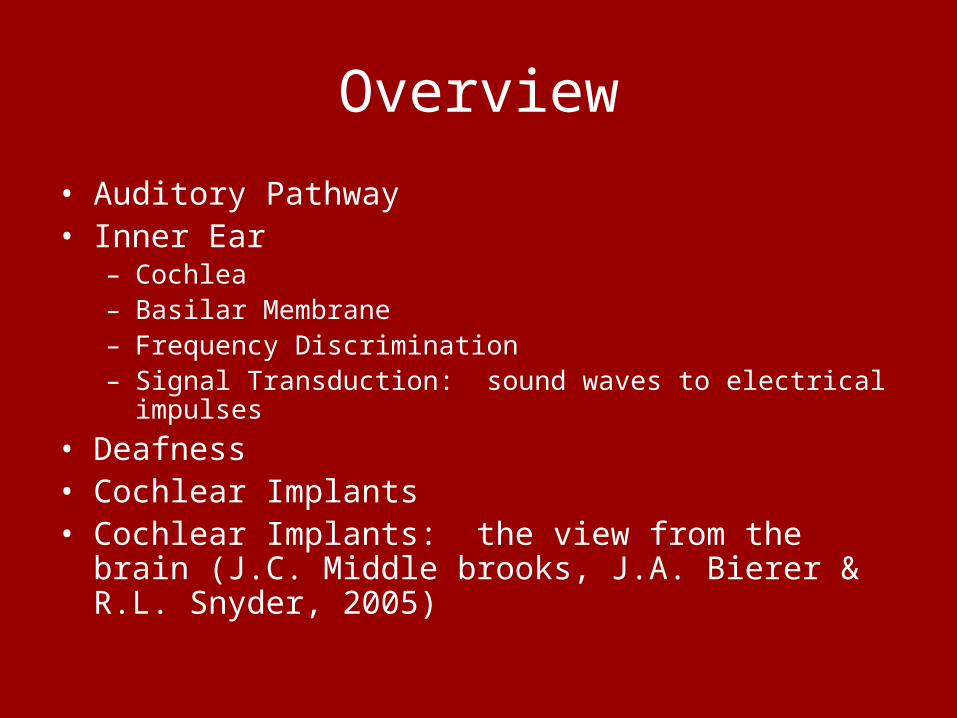
Overview
• Auditory Pathway• Inner Ear
– Cochlea– Basilar Membrane– Frequency Discrimination– Signal Transduction: sound waves to electrical impulses
• Deafness• Cochlear Implants• Cochlear Implants: the view from the brain (J.C. Middle
brooks, J.A. Bierer & R.L. Snyder, 2005)

What is a Disability?
• Neuromuscular impairment
• Sensory deprivation (vision, hearing or speech)
• Cognitive dysfunction (learning, higher processing)
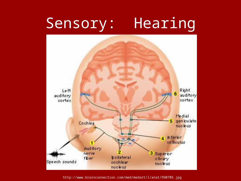
Sensory: Hearing
http://www.brainconnection.com/med/medart/l/anat/990705.jpg

Outer, middle & inner ear
http://www.american-hearing.org/images/ear.jpg

Sound Waves
http://www.privateline.com/TelephoneHistory/soundwaves.html
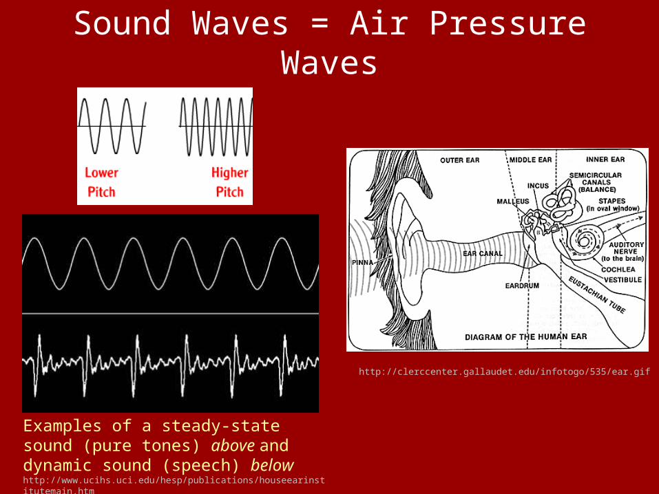
Sound Waves = Air Pressure Waves
http://clerccenter.gallaudet.edu/infotogo/535/ear.gif
Examples of a steady-state sound (pure tones) above and dynamic sound (speech) below http://www.ucihs.uci.edu/hesp/publications/houseearinstitutemain.htm
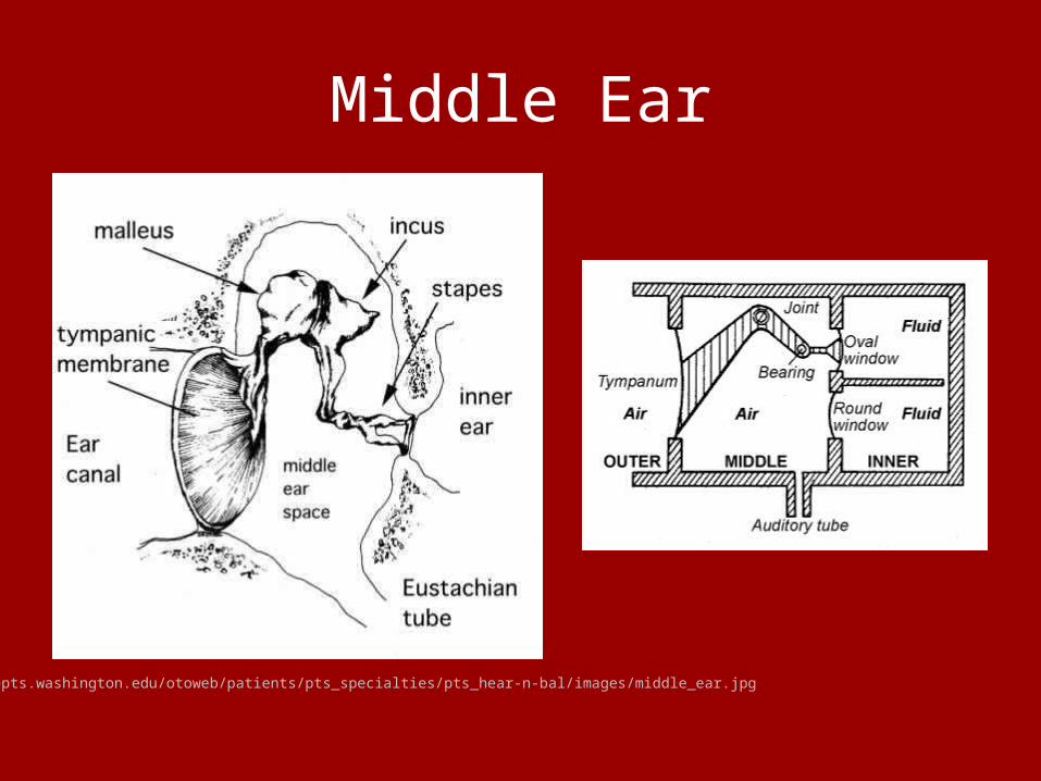
Middle Ear
http://depts.washington.edu/otoweb/patients/pts_specialties/pts_hear-n-bal/images/middle_ear.jpg

Sensitivity of the Ear
A young, healthy ear can respond over a frequency range of 20 Hz to 20,000 Hz.
The minimum sound pressure level perceptible to the ear at a particular frequency is called the threshold of hearing at that frequency. This is different for each individual, even between people with 'normal' hearing capacities. It is also age related, with a progressive loss in sensitivity at the high frequencies occurring with increasing age.
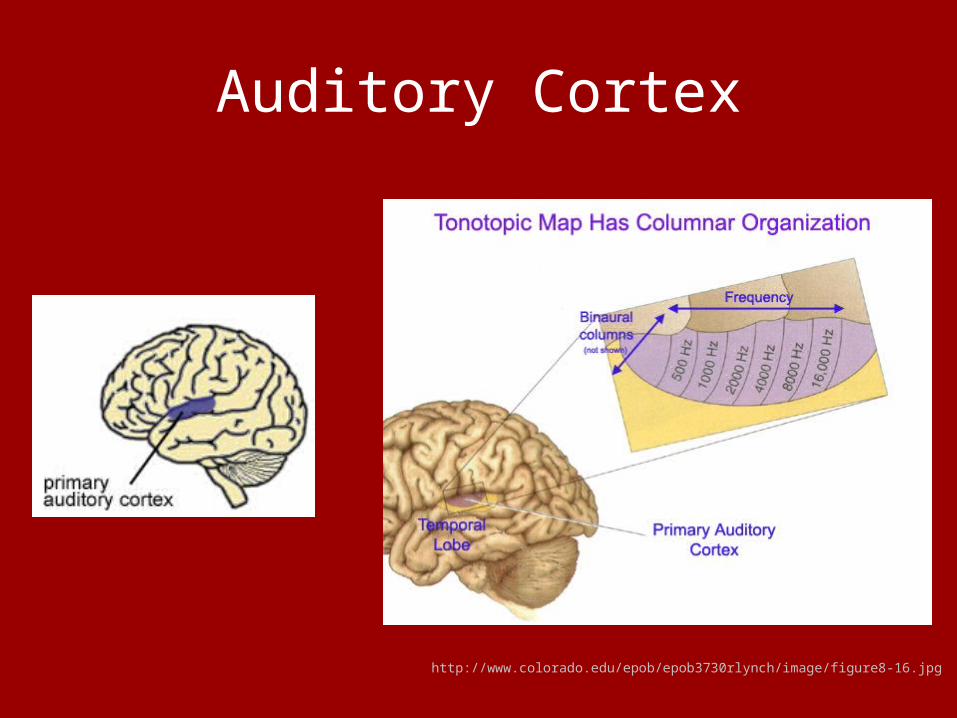
Auditory Cortex
http://www.colorado.edu/epob/epob3730rlynch/image/figure8-16.jpg
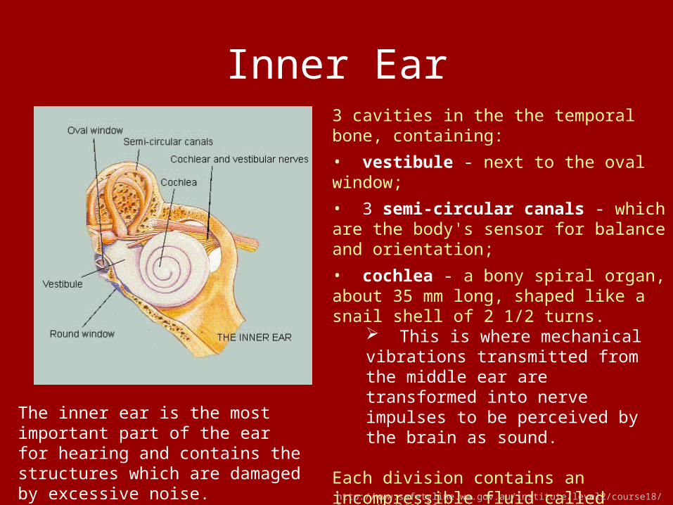
Inner Ear
The inner ear is the most important part of the ear for hearing and contains the structures which are damaged by excessive noise.
3 cavities in the the temporal bone, containing:
• vestibule - next to the oval window;
• 3 semi-circular canals - which are the body's sensor for balance and orientation;
• cochlea - a bony spiral organ, about 35 mm long, shaped like a snail shell of 2 1/2 turns.
This is where mechanical vibrations transmitted from the middle ear are transformed into nerve impulses to be perceived by the brain as sound.
Each division contains an incompressible fluid called perilymph.
http://www.safetyline.wa.gov.au/institute/level2/course18/lecture101/l101_08.asp

Cochlea
The cochlea is itself divided lengthwise into three chambers:
• scala vestibuli - which has the oval window at its base;
• scala tympani - which ends in the round window (a simple membrane which acts as a pressure release); and
• scala media - which contains the true hearing sensory structure - the organ of Corti.
http://www.safetyline.wa.gov.au/institute/level2/course18/lecture101/l101_08.asp

Cochlea

Endolymph vs. PerilymphThe dividing membranes are called the basilar membrane and Reissner's membrane.
The organ of Corti, which contains the sensory hearing cells, is supported on the basilar membrane in the scala media which is filled with endolymph fluid.
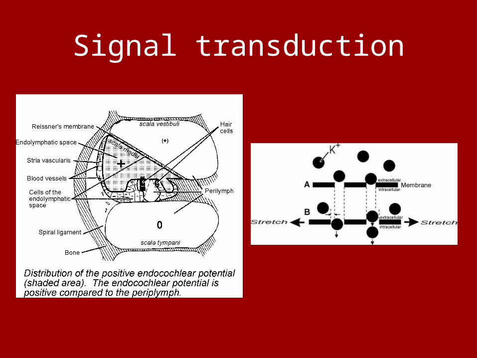
The scala vestibuli and scala tympani are connected at the apex of the cochlea by an opening called the helicotrema, and are filled with perilymph fluid. The scala media is at a slightly higher electrical potential than the other two chambers (+80 mV). This potential difference is important for the correct functioning of the cochlea.http://www.safetyline.wa.gov.au/institute/level2/course18/lecture101/l101_08.asp

Basilar Membrane
http://www.safetyline.wa.gov.au/institute/level2/course18/lecture101/images/l101_12.jpg
The organ of Corti contains the sensory hair cells which are embedded in supporting cells attached to the basilar membrane. There are two types of hair cells - inner and outer.
The inner hair cells, of which there are about 10,000, form a single row along the inside spiral of the cochlea.
The outer hair cells, of which there are about 20,000, are in three parallel rows towards the outside of the spiral.
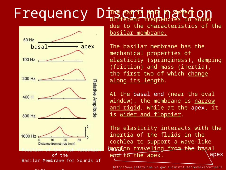
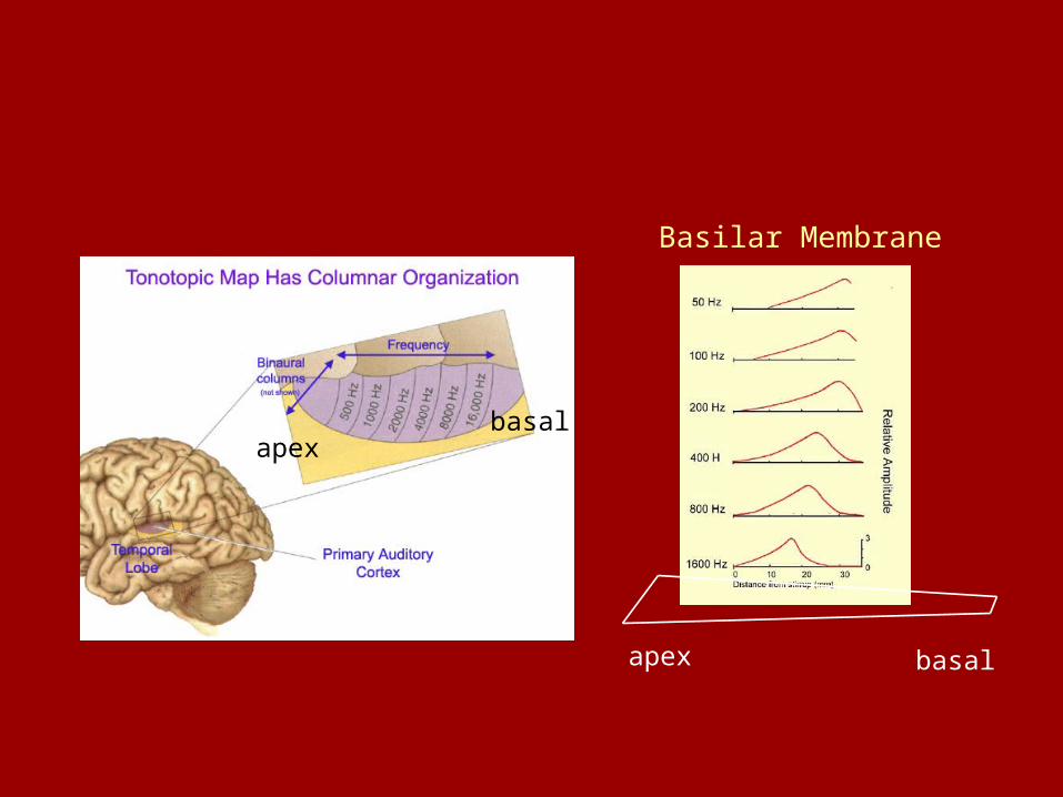
Frequency DiscriminationThe ear is able to detect different frequencies in sound due to the characteristics of the basilar membrane.
The basilar membrane has the mechanical properties of elasticity (springiness), damping (friction) and mass (inertia), the first two of which change along its length.
At the basal end (near the oval window), the membrane is narrow and rigid, while at the apex, it is wider and floppier.
The elasticity interacts with the inertia of the fluids in the cochlea to support a wave-like motion traveling from the basal end to the apex.
apexbasal
basalapexPosition of the Peak Vibration of the
Basilar Membrane for Sounds of Different Frequency http://www.safetyline.wa.gov.au/institute/level2/course18/lecture101/l101_08.asp
Frequency Discrimination (continued)
Basilar Membrane
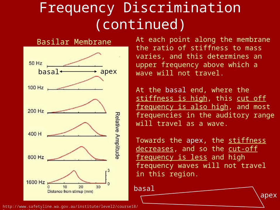
At each point along the membrane the ratio of stiffness to mass varies, and this determines an upper frequency above which a wave will not travel.
At the basal end, where the stiffness is high, this cut off frequency is also high, and most frequencies in the auditory range will travel as a wave.
Towards the apex, the stiffness decreases, and so the cut-off frequency is less and high frequency waves will not travel in this region.
apexbasal
basalapex
http://www.safetyline.wa.gov.au/institute/level2/course18/lecture101/l101_08.asp
Frequency Discrimination (continued)
Basilar Membrane
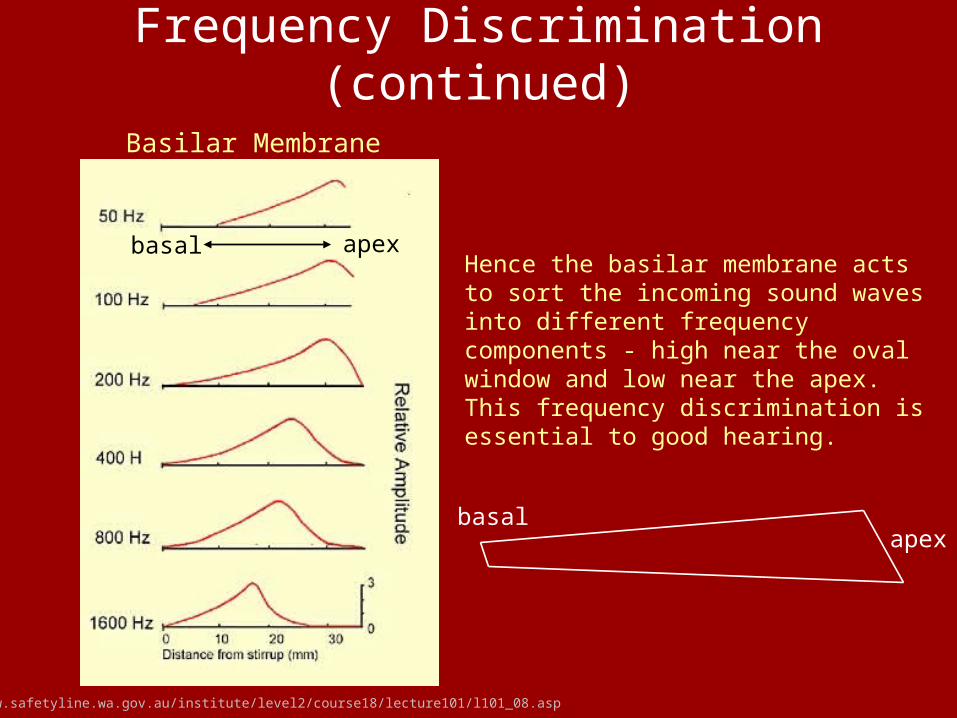
Hence the basilar membrane acts to sort the incoming sound waves into different frequency components - high near the oval window and low near the apex. This frequency discrimination is essential to good hearing.
apexbasal
basalapex
http://www.safetyline.wa.gov.au/institute/level2/course18/lecture101/l101_08.asp
Stereocilia
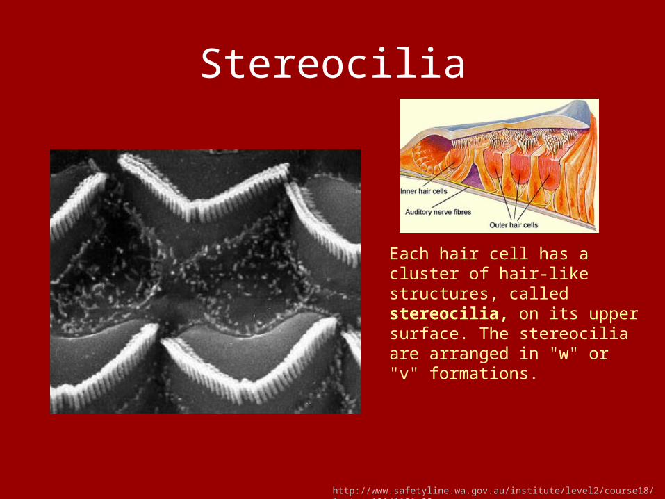
Each hair cell has a cluster of hair-like structures, called stereocilia, on its upper surface. The stereocilia are arranged in "w" or "v" formations.
http://www.safetyline.wa.gov.au/institute/level2/course18/lecture101/l101_08.asp

Stereocilia, Tectorial Membrane
Above the hair cells is the tectorial membrane, which is attached to the lining of the cochlea wall and may be attached to the outer hair cell stereocilia.
When a traveling wave displaces the basilar membrane, a shearing movement of the stereocilia occurs.
These function much like a microphone - small back and forth movements of the cilia change the flow of electric current through the hair cells.
http://www.safetyline.wa.gov.au/institute/level2/course18/lecture101/l101_08.asp

Inner/Outer Hair CellsThe inner hair cells are the primary sensory cells. They directly connect to individual nerve fibres of the auditory nerve. The sound-induced voltage changes within the inner hair cells lead to electrical activity in the nerve, which is sent to the brain.
The outer hair cells appear to serve an additional, mechanical purpose, only recently discovered. Experiments have shown that they are likely to lengthen and shorten in sympathy with the electrical signals passed by their hairs.
Signal transduction

Auditory Nerve
AUDITORY NERVENerve fibres carry the impulses from the hair cells. They pass through the spiral ganglia, to join together to become the auditory nerve.
This connects to the cochlea nuclei in the brain stem and hence to the higher auditory centres in the temporal lobe of the brain.
Here the messages, received and analysed by the ear, are interpreted.

Causes of Deafness1. EAR CANAL - Most ear canal problems are due to either
excessive wax, or foreign bodies in the ear, most of these problems are easily treatable.
2. EAR DRUM - This can either be perforated or broken, perforated ear drums can be assisted by a hearing-aid, but badly damaged ear-drums can lead to complete loss of hearing.
3. EAR BONES - There can be a bone growth which may cause the bones to weld together (Otosclerosis) and not work as they should. A hearing aid may help but more often surgery is needed.
4. COCHLEAR - Age related hearing loss (Presbyacusis) is due to the tiny hair cells dying. Sometimes these may be damaged due to illness, this is helped by use of a hearing aid.
5. NERVE DEAFNESS - Mainly due to illness (e.g. mumps) the nerves are damaged thus the sound signal is not sent to the brain as it should normally. can be helped with use of a hearing aid.
6. GLUE EAR - This is where the ear draws fluid from Eustachian Tube which is connected to the nose, this is easily helped by minor surgery. http://www.norfolkdeaf.org.uk/types.htm

Deafness
The cochlea converts the sound waves into electrical signals. These signals are then passed to the brain.
Around 80% of deafness occurs due to damage to the cochlea cells.
Deafness is in fact one of the most common of all disabilities, and still very little is known about it.

What is a cochlear implant?
A cochlear implant is an implanted electronic hearing device, designed to produce useful hearing sensations to a person with severe to profound nerve deafness by electrically stimulating nerves inside the inner ear.
http://www.wasa-shhh.org/cochlear_implants.htm

What is a cochlear implant?
These implants usually consist of 2 main components:
• The externally worn microphone (5), speech processor (3) and transmitter system (4).
• The implanted receiver (2) and electrode system (1), which contains the electronic circuits that receive signals from the external system and send electrical currents to the inner ear.

Cochlear Implant
http://depts.washington.edu/otoweb/patients/pts_specialties/pts_hear-n-bal/pts_hear-n-bal_cochlear-implant.htm
1. Sounds in the environment are picked up by the small directional microphone.2. A thin cable (cord) sends the sound from the microphone to the Spectra 22 speech processor.3. The speech processor amplifies, filters and digitizes sound into coded signals.4. These coded signals are sent from the speech processor to the transmitting coil via the cables.5. The transmitting coil sends the signals across the skin to the implanted receiver/stimulator via an FM radio signal.6. The receiver/stimulator delivers the correct amount of electrical stimulation to the appropriate electrodes on the array.7. The electrodes along the array stimulate the remaining auditory nerve fibers in the cochlea.8. The resulting electrical sound information is sent through the auditory system to the brain for interpretation .

Implanted Electrode
The implanted part is an electronic device that is put under the skin behind the ear.
An electrode connected to the device is inserted into the inner ear.
The electrode is simply a bundle of tiny wires that have open contacts spread out along the length of the cochlea. Thus, the electrical signals can be sent to different areas of the cochlea and represent different frequency sounds.
http://www.bcm.edu/oto/jsolab/cochlear_implants/cochlear_implant.htm

http://www.hearingloss-wa.org/implant%20image.JPG
Who uses cochlear implants?
• severely to profoundly deaf adults and children who get little or no benefit from hearing aids.
• even individuals with severe or profound "nerve deafness" may be able to benefit from cochlear implants.

What determines the success of cochlear implants?
• How long the patient has been deaf patients who have been deaf for a short time do better than those who have been deaf a long time
• How old they were when they became deaf--whether they were deaf before they could speak
• How old they were when they got the cochlear implant--younger patients, as a group, do better than older patients who have been deaf for a long time
• How long they have used the implant
• How quickly they learn
What determines the success of cochlear implants (continued)?
• How good and dedicated their learning support structure is
• The health and structure of their cochlea--number of nerve (spiral ganglion) cells that they have
• Implanting variables, such as the depth and type of implanted electrode and signal processing technique
• Intelligence and communicativeness of patient
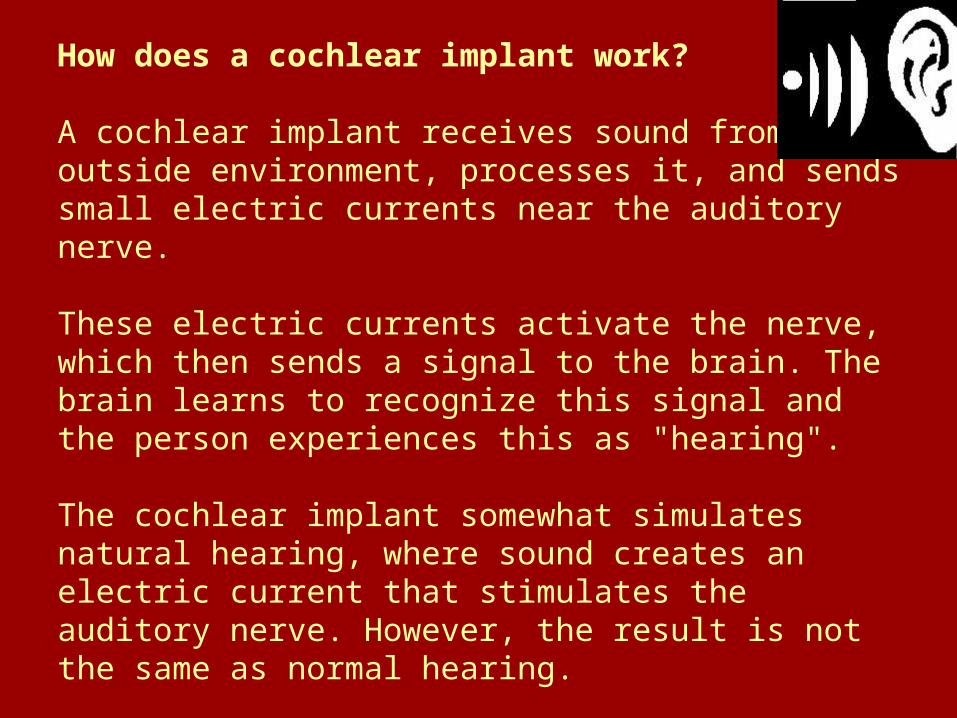
How does a cochlear implant work?
A cochlear implant receives sound from the outside environment, processes it, and sends small electric currents near the auditory nerve.
These electric currents activate the nerve, which then sends a signal to the brain. The brain learns to recognize this signal and the person experiences this as "hearing".
The cochlear implant somewhat simulates natural hearing, where sound creates an electric current that stimulates the auditory nerve. However, the result is not the same as normal hearing.

Reference no. 16

Cochlear implants: the view from the brain, 1
• Cochlear implants (therapeutic option)– Patients who lack cochlear hair cells – And who have surviving auditory nerve fibers
• C.I. used for > 2 decades• >60,000 devices implanted• However, few studies of how CNS
responds to stimulation of the auditory nerve by intracochlear electrodes

Implanted Electrode
The implanted part is an electronic device that is put under the skin behind the ear.
An electrode connected to the device is inserted into the inner ear.
The electrode is simply a bundle of tiny wires that have open contacts spread out along the length of the cochlea. Thus, the electrical signals can be sent to different areas of the cochlea and represent different frequency sounds.
http://www.bcm.edu/oto/jsolab/cochlear_implants/cochlear_implant.htm

Reference no. 15
Multi-site neural recording technology

Multi-site neural recording technology

• Normal ear – spectral analysis of sound (frequencies) is accomplished by the mechanical frequency sensitivity of the cochlea
• Map of sound frequency = tonotopic organization
• Frequency analysis – inner hair cells
• Synaptic activation auditory nerve fibers
Cochlear implants: the view from the brain, 2

• Cochlear prosthesis– Cochlear signal transduction microphone– Spectral analysis of fluid waves (cochlea)
band pass filters within the speech processor
Cochlear implants: the view from the brain, 3
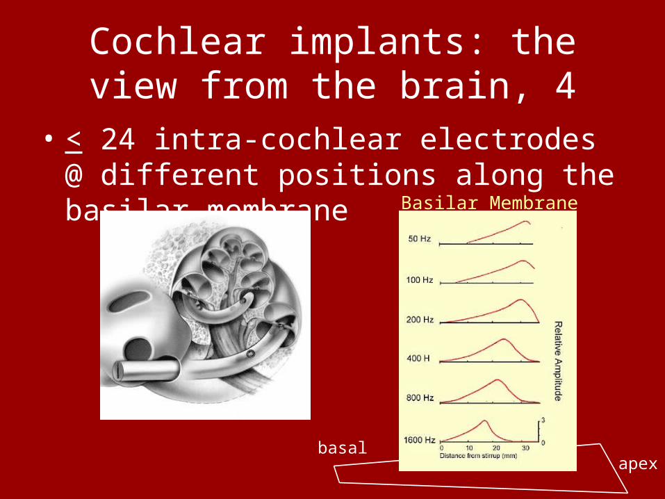
• < 24 intra-cochlear electrodes @ different positions along the basilar membrane
Cochlear implants: the view from the brain, 4
Basilar Membrane
basalapex

Cochlear implants: the view from the brain, 5
• Amplitude-modulated electrical pulse train (recorded by the electrode) that stimulates the auditory nerve
• Normally, this informationNormally, this information is relayed tonotopically to is relayed tonotopically to the ICC & A.C.the ICC & A.C.
ICC, low freq. (dorsolaterally) high freq. (ventromedially)
“Spatial tuning curve”Tonotopic axis of the ICC & A.C.
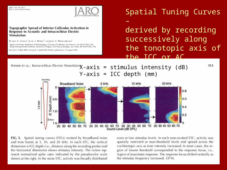
Spatial Tuning Curves –derived by recording successively along the tonotopic axis of the ICC or AC
X-axis = stimulus intensity (dB)Y-axis = ICC depth (mm)

• Multi-site recording probes: simultaneous recordings of action potentials activity along the tonotopic axis of the ICC or AC.
• Thus spatial tuning curves for cochlear implants vs. visually placed recording electrodes
• Result… ?
Central representation of spectral information, pp. 488-490

Multi-site neural recording technology
Cochlear implant

• Result…– Cochlear implant: most focused or restricted
activation (i.e. “V” shaped spatial tuning curve) was similar to that activated by 1/3-octave-wide noise burst in normal hearing
– Careful placement of bipolar electrode pairs:• Radial dimension of cochlear spiral spatial
tuning curve produced by pure tones.
• Importance…optimizing design of clinical electrode arrays !
Central representation of spectral information, pp. 488-490

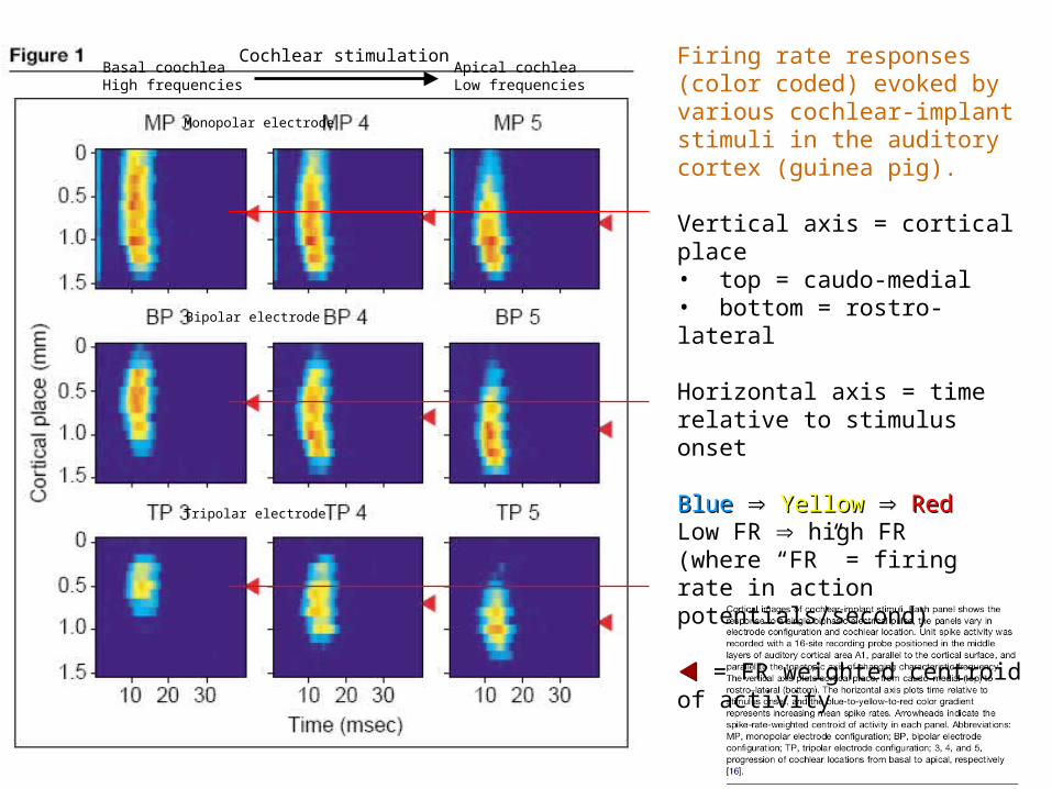
• Auditory cortex (Figure 1)– Patterns of excitation vary with cochlear
electrode configuration and place of excitation• MP, BP or TP electrodes• MP TP electrodes infer increasingly restricted
cochlear electrical fields; i.e. more focused higher spatial resolution activation of auditory cortical neurons
Central representation of spectral information, pp. 488-490
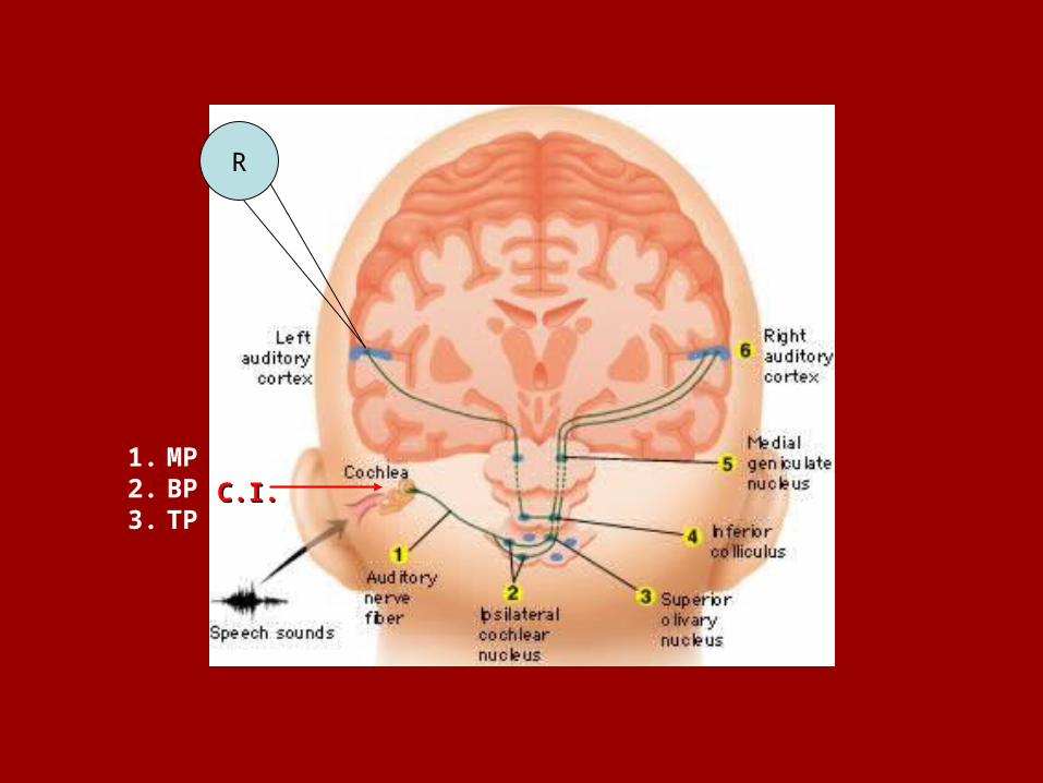
R
C.I.C.I.1. MP2. BP3. TP
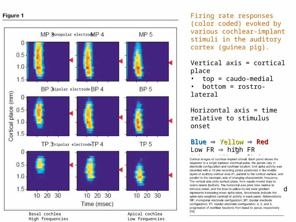
Firing rate responses (color coded) evoked by various cochlear-implant stimuli in the auditory cortex (guinea pig).
Vertical axis = cortical place• top = caudo-medial• bottom = rostro-lateral
Horizontal axis = time relative to stimulus onset
BlueBlue YellowYellow RedRedLow FR high FR(where “FR” = firing rate in action potentials/second)
= FR weighted centroid of activity
Monopolar electrode
Bipolar electrode
Tripolar electrode
Basal cochleaHigh frequencies
Apical cochleaLow frequencies
Basilar Membrane
apex basal
apexbasal
Firing rate responses (color coded) evoked by various cochlear-implant stimuli in the auditory cortex (guinea pig).
Vertical axis = cortical place• top = caudo-medial• bottom = rostro-lateral
Horizontal axis = time relative to stimulus onset
BlueBlue YellowYellow RedRedLow FR high FR(where “FR” = firing rate in action potentials/second)
= FR weighted centroid of activity
Monopolar electrode
Bipolar electrode
Tripolar electrode
Basal coochleaHigh frequencies
Apical cochleaLow frequencies
Cochlear stimulation

• Figure 1, cortical images of neural activity shift:– Activation of caudal-to-rostral cortical loci
(top-to-bottom) which represents high-to-low frequencies in the auditory cortex
– Notice the restricted area of activation in the AC using TP electrodes at the cochlea
• Spectral information is transmitted best by restricted electrode configurations (i.e., BP & TP).
Central representation of spectral information, pp. 488-490
• Temporal information (timing, direction, tempo, rhythm, etc.)– Amplitude envelope of sound waves– C.I. transmit amplitude envelopes by
amplitude modulation of constant-rate electrical pulse trains. How effective is this?
• Normal hearing – neurons in the auditory pathway fire action potentials in synchrony (phase lock) to amplitude-modulated stimuli
Central representation of temporal information, pp. 490-491
• In the C.I.– Initial evidence indicates that neurons in the
auditory pathway also phase lock to stimuli applied to cochlear devices
– Auditory cortical neurons can phase lock across the range of modulation frequencies relevant for speech perception in cochlear implants
Central representation of temporal information, pp. 490-491

• Neural plasticity in the auditory system is caused by:– Deafness– Chronic electrical stimulation of the cochlea
• Deafness– Disrupts tonotopic organization– Disruption of tonotopy increases with time
• Chronic cochlear electrical stimulation– Distorts ICC tonotopy (MP)– However, MP stimulation preserves tonotopy
Central auditory plasticity, p. 491
• Neural plasticity in the auditory system is caused by:– Deafness– Chronic electrical stimulation of the cochlea
• Deafness– Disrupts tonotopic organization– Disruption of tonotopy increases with time
• Chronic cochlear electrical stimulation– Distorts ICC tonotopy (MP)– However, TP stimulation preserves tonotopy
Central auditory plasticity, p. 491