heart rhythm society expert consensus ... - pic1.cmt.com.cn
TRANSCRIPT
Heart Rhythm Society Expert Consensus Statement onElectrophysiology Laboratory Standards: Process,Protocols, Equipment, Personnel, and SafetyDavid E. Haines, MD, FHRS, (Chair)1 Salwa Beheiry, MSN, CCRN, (Chair)2
Joseph G. Akar, MD, PhD,3 Janice L. Baker, MSN, CCRN, CEPS, FHRS,4 Doug Beinborn, RN, MA,5
John F. Beshai, MD, FHRS, FACC,6* Neil Brysiewicz, MS,7 Christine Chiu-Man, MS, CEPS, CCDS, FHRS,8
Kathryn K. Collins, MD, FHRS,9†Matthew Dare, CEPS,10 Kenneth Fetterly, PhD,11 John D. Fisher, MD,FHRS,12 Richard Hongo, MD, FHRS,13 Samuel Irefin, MD,14 John Lopez, RN,14
John M. Miller, MD, FHRS,15 James C. Perry, MD, FHRS,16 David J. Slotwiner, MD,17
Gery F. Tomassoni, MD, FHRS, FACC,18 Esther Weiss, APN, CNS, MSN, CCDS, CEPS19
From the 1William Beaumont Hospital, Royal Oak, Michigan, 2California Pacific Medical Center, SanFrancisco, California, 3Yale University School of Medicine, New Haven Connecticut, 4Chester CountyHospital, West Chester, Pennsylvania, 5Mayo Medical Center, Rochester, Minnesota, 6Mayo Clinic, Phoenix,Arizona, 7Yale New Haven Hospital, New Haven, Connecticut, 8Hospital for Sick Children, Toronto, Canada,9The Children’s Hospital, Aurora, Colorado, 10St. David’s Medical Center, Austin, Texas, 11Mayo Clinic,Rochester, Minnesota, 12Montefiore Medical Center, Bronx, New York, 13Sutter Pacific Medical Foundation,San Francisco, California, 14Cleveland Clinic, Cleveland, Ohio, 15Indiana University School of Medicine,Indianapolis, Indiana, 16UCSD/Rady Children’s Hospital, San Diego, California, 17Hofstra School ofMedicine, North Shore-Long Island Jewish Health System, New Hyde Park, New York, 18LexingtonCardiology Consultants, Lexington, Kentucky, and 19Advocate Sherman Hospital, Elgin, Illinois.
TABLE OF CONTENTS
1. Introduction ................................................................................ 12. Evolution of the EP Laboratory ......................................... 23. Laboratory Environment ....................................................... 2
4. Laboratory Design ................................................................... 45. Laboratory Equipment ........................................................... 96. Laboratory Staffing ............................................................... 137. Laboratory Personnel Credentialing ............................... 188. Procedural Issues ................................................................... 219. Pediatric and Adult Congenital Heart Disease ........... 2710. Quality ....................................................................................... 2911. Occupational Health Concerns ......................................... 3112. Ethical Concerns .................................................................... 35
1. IntroductionThe modern electrophysiology (EP) laboratory is a complexenvironment providing an array of interventions for thediagnosis and treatment of heart rhythm disorders and is a resultof many transformations over the last three decades. The EPfield has witnessed rapid expansion in the number of therapeuticprocedures treating a wide range of arrhythmias and in the newtechnologies available to perform these procedures. Because ofthe increasing complexity of equipment and procedures and anever-expanding knowledge base, it was concluded that the fieldwould benefit from a consensus document that would define thecritical components and processes of a modern EP laboratory.To this end, the Heart Rhythm Society (HRS) convened amultidisciplinary team to review EP laboratory design, ergo-nomics, personnel, equipment, occupational hazards, and patient
*Representative for American College of Cardiology (ACC);
†Representative for Pediatric and Congenital Electrophysiology Society (PACES)
KEYWORDS Cardiac electrophysiology laboratory; Laboratory equipment;Cardiac electrophysiology laboratory staffing; Cardiac electrophysiology staffcredentialing; Quality assurance; Occupational safetyABBREVIATIONS CIED = cardiovascular implantable electronicdevice; CT= computed tomography; EP= electrophysiology; FDA=U.S. Food and Drug Administration; ICD=implantable cardioverter-defibrillator; MRI = magnetic resonance imaging; QA = qualityassurance; QI = quality improvement; RF = radiofrequency; VT =ventricular tachycardia (Heart Rhythm 2014;0:1–43)
Developed in collaboration with and endorsed by the American College ofCardiology (ACC), the American Heart Association (AHA), and the Pediatricand Congenital Electrophysiology Society (PACES). Endorsed by EuropeanHeart Rhythm Society (EHRA), and Sociedad Latinoamericana de Estimula-cion Cardiaca y Electrofisiologia (SOLAECE)-Latin American Society ofCardiac Pacing and Electrophysiology. EHRA endorses the recommendationsof the expert consensus statement, with the exception of those statementsspecifically addressing US regulations. Address correspondence: David E.Haines, MD. E-mail address: [email protected].
1547-5271/$-see front matter B 2014 Heart Rhythm Society. All rights reserved. http://dx.doi.org/10.1016/j.hrthm.2014.03.042
safety, as well as clinical and ethical issues related to diagnosticand therapeutic EP procedures. The goal is to provide physi-cians, administrators, and regulatory personnel with the recom-mended requirements for building, staffing, and running amodern EP laboratory to optimize patient outcomes, minimizepatient risk, and provide a safe and positive environment forphysicians and staff.
The writing committee was formed by the Scientific andClinical Documents Committee of the HRS, with approval bythe President of the HRS and the HRS Executive Committee.The composition of the committee was meant to represent therange of stakeholders in the EP laboratory. The choice of thewriting committee members was in accordance with the HRSRelationships With Industry policy.1 All members of thewriting committee were required to fully disclose all potentialconflicts of interest (see Appendix 1).
Relatively little published literature addresses the EP labora-tory environment, staffing, and processes. Therefore, many of thestatements in this document are the product of expert consensusby the writing committee and reviewers. For cases in which therewere divergent opinions on a statement, a vote among writingcommittee members was taken, and if a two-third majoritysupported the statement, it was adopted in the document. Thesections pertaining to pediatric and adult congenital heart diseasewere reviewed and approved by the Pediatric and CongenitalElectrophysiology Society (PACES), a nonprofit organizationdedicated to the treatment of arrhythmia disorders in childrenand individuals with congenital heart disease (CHD). The finaldocument was approved by the Board of Trustees of the HRS.This document is directed to all health care professionals whodesign, manage, and/or work in the EP laboratory environment.
2. Evolution of the EP LaboratoryThe field of clinical cardiac electrophysiology (CCEP) hasgrown from its origin as a field of clinical research for
arrhythmogenesis to its present-day incarnation as an impor-tant specialty offering advanced therapies for a wide varietyof disorders. Clinical EP laboratories emerged in the late1960s, and by the early 1970s, formal fellowships had beenestablished and EP laboratories were taking shape. First-generation EP laboratories often shared space with cardiaccatheterization laboratories and were typically subordinate tocoronary angiographic and hemodynamic procedures. Whena space was dedicated for electrophysiological testing, it wasoften small, and fluoroscopy was delivered with portable C-arm units. These laboratories were sufficient for diagnosticEP studies and electropharmacological testing. Second-generation EP laboratories developed in the 1980s with theintroduction of catheter ablation and cardiac implantableelectronic devices (CIEDs) to the electrophysiologist’sarmamentarium. Pacemaker implantation was shifting fromthe domain of surgery to that of cardiac EP. With increas-ingly complex procedures being performed in EP laborato-ries, more space was allocated to new dedicated laboratoriesand fluoroscopy equipment began to be upgraded to systemscommensurate with those used in cardiac catheterizationlaboratories.
The third generation of interventional cardiac EP has beendriven by the success of catheter ablation and advanced devicetherapy. The precise anatomy and physiology of a wide varietyof arrhythmias has been elucidated through the development ofadvanced mapping systems and improvements in ablationcatheter technologies. Modern device therapy incorporatesmultimodal multisite pacing, sophisticated therapies fortachyarrhythmias, and advanced diagnostics. With the increas-ing complexity of EP procedures and equipment has comeincreasing sophistication of laboratory processes and greaterdemands on laboratory personnel. The cost and complexity ofthe modern EP laboratory now demands that standards aredeveloped to ensure a high level of care.
3. Laboratory Environment
Laboratory Environment Recommendations
� Highly complex procedures or procedures on patients with certain conditions and comorbidities that are associated with higherprocedural risk should not be performed in a freestanding laboratory (i.e., an EP laboratory that is not physically attached to a hospital).
� Emergency cardiovascular surgical support should be immediately available in case of life-threatening bleeding complications fromthe extraction of chronic device leads and complex mapping/ablation procedures, particularly those requiring pericardial access.
� High-risk procedures in critically ill patients, such as ablation of ventricular tachycardia in patients requiring extracorporealhemodynamic support, can only be safely performed in institutions offering comprehensive programs with active engagement fromelectrophysiologists, surgeons, intensivists, and anesthesiologists.
3.1. Procedure Room OptionsThere are multiple options and practice settings for performingEP and implantable device procedures. Medical centers mayadopt one or more of the following laboratory operations fortheir practice. The choice among the following options involvesa trade-off between increasing capability for procedure com-plexity and increasing construction and operating costs.
3.1.1. Dedicated EP LaboratoryIn a dedicated EP laboratory, the staff space and procedureroom space are separate from the cardiac catheterizationlaboratory and/or radiology laboratory, although the staff spaceand procedure room space often exist within a common area.The preparatory and recovery rooms are often shared with othersubspecialties. Procedures that can be performed in thislaboratory setting include diagnostic EP studies, ablation
Heart Rhythm, Vol 0, No 0, Month 20142
procedures, use of cardiac implantable devices, implantabledevice extractions, use of temporary pacemakers, three-dimensional (3D) mapping, intracardiac echocardiography(ICE), and use of robotics. The advantages of using a dedicatedEP laboratory include greater availability of more highly trainedallied personnel, room equipment dedicated to only EPprocedures, and decreased overall equipment costs per room.
3.1.2. Shared EP and Catheterization LaboratoryA shared procedural laboratory program is usually in associ-ation with a cardiac catheterization laboratory program, but canalso be shared with an interventional radiology program. Ashared room allows for two or more practices to share commonequipment that includes fluoroscopic equipment, recordingsystems, emergency equipment, and anesthesia equipment, aswell as the space. This is helpful in circumstances of lowoverall volumes when sharing a room allows for flexibility inpatient care while controlling costs and space requirements.
3.1.3. Device-Only LaboratoryThese types of procedure rooms have been created at large-volume institutions that can support a procedure room dedicatedonly to CIED surgery. The procedures performed in this type ofroom include the use of pacemakers and defibrillators that aresingle chamber, dual chamber, or biventricular in operation.Other procedures can include the use of temporary pacemakers,the use of implantable loop recorders, and lead and deviceextractions. Device and lead extractions may also be performedin a surgical operating room (OR) on the basis of the patient’scondition or on the standard agreed on by the institution.Advanced mapping and EP recording systems are not required,and the costs of equipping this type of laboratory are lower,which is the key advantage of this type of room. Device-onlylaboratories are appropriate for high-volume centers that alreadyhave one or more fully outfitted EP laboratories.
3.1.4. Advanced Mapping, Ablation, and Combined HybridLaboratoriesThese procedure rooms are designed to the rigorous standards ofORs (positive airflow, medical gas availability, surgical lighting,and substerile scrub area) but have high-quality fixed fluoro-scopy and a full complement of EP and/or cardiac catheterizationequipment. These rooms are ideal for procedures that may becombined with open or minimally invasive cardiac surgery andfor lead extraction procedures. When not being used for hybridsurgical procedures, these laboratories can function either as fullyfunctional ORs or as fully functional EP/catheterization suites.Procedures that can be performed include complex ablationprocedures that involve EP and surgical components, left atrialappendage occlusion or clipping, epicardial lead placement, andminimally invasive valve replacement.
3.1.5. Special Procedure RoomsSome organizations incorporate special noninvasive rooms intotheir practice to accommodate patient care that does not requirefluoroscopy or other specialty equipment. These rooms are oftenused to perform minor procedures such as cardioversions, tilt
table studies, and noninvasive programmed stimulation defib-rillation threshold testing. Autonomic testing with head-up tilttable testing requires a procedure table that has the capability for70º head-up tilt, an electrocardiogram (ECG) monitor, non-invasive blood pressure monitor, supplemental oxygen, andbasic supplies. Equipping these rooms is much less expensivethan equipping a full procedural laboratory and can help im-prove patient flow and volume through a busy EP department.
3.1.6. Pediatric EP LaboratoryThe room and equipment standards for pediatric EP proceduresare similar to those for adult EP procedures, except for the avai-lability of pediatric resuscitation equipment and drug doses aswell as a wider inventory of smaller catheters. Pediatric and con-genital EP patients can require a combined procedure of EP andthe need for cardiac catheterization, including angiography andpossible intervention. Thus, it is optimal (although not a necessity)for a pediatric/congenital EP laboratory to meet all the standardsof a pediatric catheterization laboratory. Pediatric EP proceduresin young children should be performed in pediatric hospitals orhospitals that have a pediatric cardiology and EP service.
3.2. Freestanding Cardiac EP LaboratoryAn EP laboratory that is not physically attached to a hospital isconsidered a freestanding laboratory. Freestanding EP laborato-ries can be privately owned, and when owned by physicians,there may be concerns about conflicts of interest (as discussed inSection 12). This arrangement presents challenges that stem fromthe separation of the laboratory from vital hospital services. In theevent of a life-threatening complication, such as pericardialtamponade2 or endovascular tear during lead extractions,3 anemergency response from certain hospital-based services such ascardiothoracic surgery can become necessary, and even possiblylifesaving. Performing EP procedures in freestanding EP labo-ratories on patients with clinical conditions that confer increasedrisk are relatively contraindicated. These include preexistingadvanced heart failure and severe left ventricular dysfunction4;recent myocardial infarction, recent stroke, chronic kidneydisease, severe chronic obstructive pulmonary disease, pulmo-nary hypertension, and severe/morbid obesity5; and severevalvular dysfunction or prosthetic heart valve, CHD (includingatrial septal defect repair), active oral anticoagulation, advancedage, and pediatric age. Procedures that necessitate lesion creationclose to coronary arteries, such as aortic cusp ablation6 andepicardial ablation,7 carry a higher risk of intraprocedural myo-cardial infarction and should not be performed outside a hospital.As part of the consent process, patients should be informed thatthe procedure is being performedwithout on-site surgical backup.In order to ensure the safety of a patient undergoing a procedurein a freestanding EP laboratory, a functional and tested systemmust be in place to quickly transfer patients to a hospital withimmediate surgical support in case of an unanticipated compli-cation. The receiving program should be familiar with compli-cations unique to the EP laboratory. There must be a standingagreement between the laboratory and the receiving hospital sothat there is no unnecessary delay in the transfer process.
3Haines et al HRS Expert Consensus Statement on Electrophysiology Laboratory Standards

3.3. Hospital and EP LaboratoryThe hospital environment plays an important role in shaping thestructure and function of the EP laboratory. A “closed EPlaboratory” is commonly present in academic institutions andlimits physician practice to faculty members of the particularinstitution or university. In contrast, “open EP laboratories” allowcredentialing and the participation of multiple physician groups,including thosewho do not hold faculty level appointments. Suchlaboratory structuring is common in community and privateinstitutions and is also present in some academic settings.Whether an EP laboratory is open or closed is determined bythe institution’s leadership on the basis of economic, historical,political, and geographical factors that are often beyond physiciancontrol. An inherent difficulty in the open EP laboratory formatlies in procedure scheduling for multiple physicians; a centralizedscheduling structure that can arrange schedulingwhile organizingand prioritizing procedures on the basis of urgency and acuity isimportant to avoid conflicts and optimize patient care.
The complexity and degree of invasiveness of EP proceduresis dependent on the level of support provided by the hospital orother health care organization in terms of personnel, facilities, andequipment. Anesthesia support is desirable for the safe perform-ance of potentially lengthy and complex procedures. The role ofanesthesia services in the EP laboratory is detailed in Section 6.Surgery backup must be immediately present for lead extractionprocedures in which a lead to be removed is older than 1 year (orrequire tools other than a standard stylet to be removed if youngerthan 1 year from implantation)8, andmapping/ablation proceduresrequire pericardial access. Complex ablation procedures, such asatrial fibrillation and ventricular tachycardia (VT) ablation, shouldbe performed only in hospitals equipped and prepared to managethese types of emergencies, with access to emergency surgicalsupport when required. Finally, high-risk procedures in criti-cally ill patients, such as ablation of VT in patients requir-ing hemodynamic support with extracorporeal membrane
oxygenation, can only be safely performed in institutions offeringcomprehensive programs with active engagement from electro-physiologists, surgeons, and anesthesiologists. Although suchcollaborations were limited to advanced tertiary care institutionsin the past, the increasing availability of institutional resources andsupport has expanded the range of facilities in which complexprocedures are performed to include private institutions.
3.4. Regulatory Standards Related to EP LaboratoriesFederal guidelines for the construction and retrofitting of healthcare facilities have been influenced by recent catastrophicevents, such as the Northridge earthquake of 1994, HurricaneKatrina in 2005, and the F5 tornado that made a direct hit on ahospital in Joplin, MO, in 2011. In the mid-1990s, threeformerly competing code writing agencies united to form theInternational Code Council. Their mission was to develop anational construction code that, among other entities, wouldregulate the construction of health care facilities to mitigate therisk of damage due to seismic, wind, and flood dangers. Knownas the International Building Code, one of its versions has beenadopted by every state. In addition, the Federal EmergencyManagement Agency, a branch of the Department of Home-land Security, published revised guidelines for improvinghospital safety in earthquakes, floods, and high winds.
The primary legislative avenues for controlling the dissemina-tion of expensive health care services are Certificate of Need(CON) laws. As of 2009, 39 states still have a CON process, law,or set of requirements. Inmost cases, the approval of CON is basedon the actual or projected volume of services provided in the pro-cedural laboratories. As procedural volumes for percutaneouscoronary arterial interventions have diminished at most tertiaryreferral hospitals, many hospitals have shifted some coronary inter-ventional laboratory CONs to EP laboratories. Once an EP lab-oratory is established, the primary government body overseeing itsoperations, policies, and procedures is the Joint Commission (TJC).
4. Laboratory Design
Laboratory Design Recommendations
� The Guidelines for Design and Construction of Hospitals and Health Care Facilities published by the American Institute of Architects and theFacility Guidelines Institute provide space and functionality standards for EP laboratories with a goal to improve work flow in the EPenvironment. (Specific recommendations not derived from this document are based on the consensus opinion of the writing committee.)
� The minimal procedural area of a complete EP laboratory (not including control room space) is 350 sq ft of clear floor area.� Current electrical system regulations for health care facilities should follow Article 517 of the National Electrical Code (NEC) Handbook.� An uninterruptible power supply for all computer equipment is required.� The air flow/heating, ventilation, and air conditioning design should comply with the Guidelines for Environmental Infection Control in
Health-Care Facilities Recommendations of the Centers for Disease Control and Prevention and the Healthcare Infection Control PracticesAdvisory Committee document.
� Lighting should include an overhead light on an articulating arm, 2 � 2 ft lighting squares to flood the main procedure area, and adedicated workspace light for the nursing/anesthesia area.
� The ideal sound/communication system is an always-on, full-duplex, two-way intercom system.� Network cabling and hardware should have a minimum capability of support for gigabit Ethernet speed.� Electronic storage of EP data should be Health Insurance Portability and Accountability Act (HIPAA) compliant. Data should be
maintained for at least the minimum duration as determined by each state.
Heart Rhythm, Vol 0, No 0, Month 20144

The American Institute of Architects and the Facility Guide-lines Institute regularly publish the Guidelines for Design andConstruction of Hospitals and Health Care Facilities.9 Thisdocument is recognized by federal and state authorities, andrecently this document has included EP laboratories. It providesdefined standards in terms of the space and functionality of EPlaboratories with a goal to specifically improve work flow in theEP environment, acknowledging that the EP laboratory requiresmore space than an angiographic/interventional laboratory forsupporting equipment and supplies. Traditionally, however, theconstruction of an EP laboratory had no specific guidelinesbecause of its special applications. The typical layout is generallyderived from a cardiac catheterization laboratory,9 which is notideal for the performance of the full range of EP procedures. Thelimitations of direct adaptation of an angiography suite design tothe practice of cardiac EP include space constraints relative to thespecial equipment used in EP procedures, the necessity to workon either side of the patient table, and the requirement to accessthe patient’s upper chest for device implantation. EP laboratoryplans should take into account not only the available spacewithin the procedure room but also its location relative topertinent services such as the patient prep area, recovery area,OR, intensive care unit, the ward, and specialized resources suchas an adjacent magnetic resonance imaging (MRI) suite thatmight permit real-time MRI imaging during procedures in thefuture. The rationale is to consider the proximity of all neededservices in the overall design during the planning stage so thatenhanced patient flow can be achieved. The aim of the planningcommittee should be to build a consensus on a minimum set ofspecifications that will meet the needs of the clinicians andsupport staff, and enable them to provide optimal patient care,while maintaining occupational safety for the staff.
4.1. Space RequirementsThe EP laboratory needs as much space as is practical to ensurethe freedom of movement of the operator and staff, to accom-modate all equipment used, and to facilitate movement of staffin emergency situations. The recommended procedural area ofa complete EP laboratory (not including control room space) is500 sq ft or greater of clear floor area, although 350 sq ft is theabsolute minimum requirement. There should be a minimumof 8 ft of clear space between the wall and the edges of eachside of the patient table when it is positioned at the isocenter.Enough clearance at the head of the bed should be allocated foranesthesia equipment on either side and sterile access tojugular vein entry sites, if employed, while allowing for freerange of movement of the fluoroscopy C-arm. The ceilingheight is dependent on the requirements of the X-ray/fluoro-scopic equipment9 (Figure 1). Preexisting laboratories that arebeing renovated where it is impossible to expand the gross areabecause of building and location constraints should followfederal and state code requirements, but due caution should betaken to meet suggested recommendations.
4.2. Room LayoutThe fluoroscopic equipment plays a major role in determin-ing the amount of ideal space in the procedural area and
could serve as the reference point. Equipment can be eithermounted on the floor or suspended from the ceiling. Thelatter configuration allows for the floor to be optimallycleaned; however, because of the amount of equipment thatwould need to be suspended from the ceiling (monitors,surgical lights, X-ray barriers, equipment racks, and anes-thesia gas supply), a floor-mounted configuration may bemore practical in some laboratories. It is best if X-raygenerators and tanks are located in a space separate fromthe procedure and control rooms. The size and portability ofthe fluoroscopy unit is important in planning room size,especially when cabinetry and other fixtures are planned forinstallation on the walls within the procedural area. Installa-tion of cabinetry at the head of the bed is discouragedbecause it further limits space to allow free movement of theX-ray arm, anesthesia supply cart, and life support equip-ment. Cabinetry for supplies frequently used during casesshould be positioned on the side walls for easy access. Theroom should be wide enough to accommodate the cabinetand open door swing without impinging on the sterile fieldand traffic flow through the laboratory.
Most peripheral equipment such as recording systems,stimulators, and radiofrequency (RF) generators are madefrom multiple components, some of which need to be in acontrol room and others in the laboratory itself. It is stronglyrecommended that none of the modules sit on the floor. Thiscan reduce sterility and cleanliness as well as put theequipment at risk of being damaged by fluids. A ceiling-mounted boom removes all equipment from the floor andreduces damage to cables by allowing them to remainconnected at all times. By placing the recording systemamplifier, the RF generator, the mapping system amplifier,the stimulator amplifier and router, and other peripheralequipment together on a ceiling-mounted equipment boom,all cabling will be permanently placed and connected,reducing cable wear. The removal of rolling equipment cartsfrom the room improves staff access to the patient. Remov-ing cables and equipment from the floor reduces the trippinghazard to the staff and risk of equipment damage. Becauseadditional portable EP equipment is often employed during aprocedure, it is necessary to have ample power outletsinstalled to accommodate such needs.
Anesthesia gases are best supplied via a ceiling-mountedanesthesia boom, which should include two oxygen lines,one nitrous oxide line, one medical air line, two vacuumlines, and one waste anesthetic gas disposal line.10 It shouldbe equipped with at least one slide clamp for vacuum canisterplacement, which should allow the canisters to be locatedwithin 4 in. of the floor for ease of removal when full. Theanesthesia boom should have a minimum of six electricaloutlets, at least some of which should be on emergency (redplug) circuits in case of general power outage during aprocedure. A mounted light controlled independently fromthe room lighting for charting in a dark room is a usefuloption. Video can be routed from the anesthesia boom todisplay data from an anesthesia cart to monitors placedaround the room.
5Haines et al HRS Expert Consensus Statement on Electrophysiology Laboratory Standards

4.3. Hybrid LaboratoryThe hybrid laboratory has all the requirements of a full EPlaboratory but has added features that allow it to serve as a fullyfunctional operating suite. These laboratories are often largerand have the fluoroscopy equipment on a track so that it can beentirely removed from the surgical field. It is typically locatedwithin or contiguous to the other ORs and has a full substerilescrub and supply area. The use of a hybrid laboratory for EPprocedures is evolving. Hybrid laboratories in which EPprocedures are performed need to be outfitted with theappropriate EP-specific equipment, including EP recordingsystems, mapping systems, and programmed stimulators.Procedures that might benefit from performance in this settingwould include those where surgical intervention or extracor-poreal hemodynamic support might be required, such as leadextractions, VT ablation procedures in patients with structuralheart disease, and hybrid atrial fibrillation ablation procedures.
4.4. Control RoomAlthough some EP laboratories house all the monitoring andstimulating equipment in the procedure room, it may be
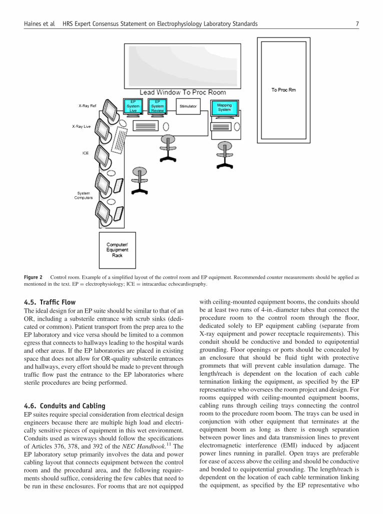
preferable to have a contiguous control roomwith an interposedleaded wall and large viewing window so that members of theteam (apart from the primary operator, the circulating nurse, andthe anesthesia professional) can work without exposure toionizing radiation. The control rooms can be shared among twoor more laboratories. A separate control room demands a fullduplex intercom system so that there is no barrier to commu-nication. The space required for a control room is not inclusiveof the procedural area measurements. Adequate ventilationshould be supplied to account for excess heat production fromthe electronics. The counters should be at least 30 in. deep sothat the monitors can be 20 in. away from the user. At least 160in. of desk space is suggested for a laboratory with a single-plane fluoroscopy system and 180 in. of desk space for abiplane fluoroscopy system to allow for fluoroscopymonitors, amapping system, a recording system, and a stimulator. Anadditional 45 in. of desk space is suggested for a two-monitorreading station or a single-monitor workstation (Figure 2). Theparticipation of an ergonomics expert in the planning should beconsidered as a measure to comply with Occupational Safetyand Health Administration standards.
Figure 1 Space requirements. The sample layout of EP laboratory with adjacent control room area. Note the availability of enough free space at the head ofbed area allowing freedom of movement of fluoroscopy arm and anesthesia equipment. EP ¼ electrophysiology.
Heart Rhythm, Vol 0, No 0, Month 20146
4.5. Traffic FlowThe ideal design for an EP suite should be similar to that of anOR, including a substerile entrance with scrub sinks (dedi-cated or common). Patient transport from the prep area to theEP laboratory and vice versa should be limited to a commonegress that connects to hallways leading to the hospital wardsand other areas. If the EP laboratories are placed in existingspace that does not allow for OR-quality substerile entrancesand hallways, every effort should be made to prevent throughtraffic flow past the entrance to the EP laboratories wheresterile procedures are being performed.
4.6. Conduits and CablingEP suites require special consideration from electrical designengineers because there are multiple high load and electri-cally sensitive pieces of equipment in this wet environment.Conduits used as wireways should follow the specificationsof Articles 376, 378, and 392 of the NEC Handbook.11 TheEP laboratory setup primarily involves the data and powercabling layout that connects equipment between the controlroom and the procedural area, and the following require-ments should suffice, considering the few cables that need tobe run in these enclosures. For rooms that are not equipped
with ceiling-mounted equipment booms, the conduits shouldbe at least two runs of 4-in.-diameter tubes that connect theprocedure room to the control room through the floor,dedicated solely to EP equipment cabling (separate fromX-ray equipment and power receptacle requirements). Thisconduit should be conductive and bonded to equipotentialgrounding. Floor openings or ports should be concealed byan enclosure that should be fluid tight with protectivegrommets that will prevent cable insulation damage. Thelength/reach is dependent on the location of each cabletermination linking the equipment, as specified by the EPrepresentative who oversees the room project and design. Forrooms equipped with ceiling-mounted equipment booms,cabling runs through ceiling trays connecting the controlroom to the procedure room boom. The trays can be used inconjunction with other equipment that terminates at theequipment boom as long as there is enough separationbetween power lines and data transmission lines to preventelectromagnetic interference (EMI) induced by adjacentpower lines running in parallel. Open trays are preferablefor ease of access above the ceiling and should be conductiveand bonded to equipotential grounding. The length/reach isdependent on the location of each cable termination linkingthe equipment, as specified by the EP representative who
Figure 2 Control room. Example of a simplified layout of the control room and EP equipment. Recommended counter measurements should be applied asmentioned in the text. EP ¼ electrophysiology; ICE ¼ intracardiac echocardiography.
7Haines et al HRS Expert Consensus Statement on Electrophysiology Laboratory Standards
oversees the room project and design. Backup temporarycabling should be available in case of failure of conduitcabling during a case.
4.7. Electrical System/Noise ImmunityCurrent regulations for health care facilities should followArticle 517 of the NEC Handbook. Because the EP procedureroom is classified as a “wet procedure location,” the installa-tion of an isolated power system with line isolation monitoringis required, which provides a layer of protection from thehazards of electric shock with the added benefit of line noiseisolation because of its design.9 In addition, all computerequipment directly related to the ongoing monitoring andtreatment of a patient must have an uninterruptible powersupply (UPS). The UPS may be integrated into the power forthe entire suite, or individual UPS may be placed in line foreach central processing unit. The main purpose of the UPS isto prevent the EP system, mapping system, or other criticalimaging or monitoring system from going through a hardshutdown and full reboot procedure in case of a transientpower outage or surge. Other important electrical componentsof the laboratory, such as the imaging train, should beconnected to emergency backup power so that cases can becompleted even if line power is lost. Power lines and data linesshould be run separately and isolated from each other indifferent conduits to prevent EMI from power line wiringinduced through data line wiring that could affect optimalperformance of the EP equipment. If open cable trays are usedabove the ceiling, careful consideration should be given to theplacement of power lines and other fixtures that can be sourcesof EMI. Although power lines used on these runs do notnecessarily involve enough energy to induce heating, it is stilla good rule to follow the specifications of Article 300.20 of theNEC Handbook as a reference.11 Adequate spacing of EPlaboratory equipment in the procedural area should befollowed. Interface cables between the patient and the equip-ment (e.g., ECG cables and intracardiac catheter cables)should not dangle by the X-ray tube and should be keptneatly arranged by the side of the patient to provide easyaccess for troubleshooting purposes during the procedure.
4.8. Air Flow/Heating, Ventilation, and AirConditioningAir flow should be of OR quality. The design should complywith the Guidelines for Environmental Infection Control inHealth-Care Facilities Recommendations from the Centersfor Disease Control and Prevention and Sections 5 and 6 of theHealthcare Infection Control Practices Advisory Committeedocument.10 Emphasis should be placed on the use of in-linefilters or mechanical smoke evacuation systems to preventairborne infective and toxic particles from the plume producedby electrocautery and similar equipment. The temperaturecontrol should support effective configuration for temper-atures as low as 60ºF. This allows comfort for practitionerswho are wearing sterile gowns, hats, and masks on top of leadaprons during long procedures. Patient comfort should also be
addressed, particularly as they are fully draped and may beonly lightly sedated.
4.9. LightingThe patient table should be flanked by large lighting squaresor the equivalent to flood the main procedure area with light.Appropriate grounding is required to prevent EMI from theselights. The lighting squares should be tied to an X-ray pedalswitch that can be turned on and off at will by the X-rayoperator. Additional spotlights that are dimmable from adistant wall switch are also recommended for procedures thatrequire a darker environment to optimize glare reduction andvisualization of display systems in front of the operator.There should be at least one overhead OR light of surgicalquality mounted on an articulating arm, strategically placedto be accessible for use on the left shoulder, right shoulder, orabdomen at either side of the patient. There should besufficient range of motion to be able to focus light intensity ata steeper angle toward and into the implant pocket. Twolights are optimal for reducing shadows. The preferred ORlight is mounted on a boom that extends from the ceiling andhas free range of movement to focus the beam at the anglesand distance optimal to adequately light the surgical field anddevice implant pockets. Anesthesia and/or nursing shouldhave a light over their workspace that is independent of theroom lighting on either side of the patient table, whichshould be oblique at a distance from the X-ray C-arm.9
4.10. Sound Systems/Communications EquipmentFor laboratory designs that employ a separate control room,there may be difficulties with the use of communicationsystems that link the operator in the procedure room to thecontrol room staff. Because critical processes such as timingof ablation onset and offset require close coordinationbetween the bedside and the control room, the importanceof good two-way communication for patient safety andquality of care cannot be overstated. The ideal equipmentis capable of a always-on, two-way system because of theconstant and instantaneous need to communicate. The idealsystem is an always-on, full-duplex, two-way intercomsystem, with a toggle to silence unnecessary chatter fromthe control room. This requires electronic noise cancellationto prevent acoustical feedback and has variable effectivenessdepending on room acoustics. A simpler solution is a one-way push-to-talk intercom, but this does not allow sponta-neous back-and-forth communication. The use of wirelessheadsets is a favorable solution, which broadcasts spokenwords directly to the headphone users, with simultaneoustalk paths open as needed. Whatever system is selectedshould be high fidelity, spectrum friendly, and encrypted toprevent eavesdropping and potential HIPAA violations,making it a more expensive solution.
4.11. Data NetworkProcedural charting and operative reports should be part ofthe institution’s electronic medical record. The network
Heart Rhythm, Vol 0, No 0, Month 20148

cabling and hardware should have a minimum capability ofsupport for gigabit Ethernet speed.9 The data demands ofimaging systems, including 3D electroanatomic mappingsystems, are great and require larger storage repositories incomparison with the compressed images of major imagingequipment such as ultrasound and X-ray radiograph systems.There is an increased use of imaging created by computedtomography (CT) and MRI, which are 3D in nature,necessitating high transfer speeds between the picturearchiving and communication system (PACS) and the EPlaboratory environment. Collaboration with the informationtechnology (IT) department and its infrastructure within theinstitution is necessary in this venture. EP systems gatherinformation in digitized format for patient records andreview at a later time. It will be important for industry todevelop a better and unified standard for storing andretrieving cardiac electrogram information. Waveform infor-mation in EP is constructively different from image infor-mation and needs to be handled in a different manner. Thecomplexity involved in translating the files without losingthe ability to utilize the tool sets needed during review, and toscroll through the whole EP study, is a challenge. The DigitalImaging and Communications in Medicine standard is a
more robust model to follow and should be the preferredmethod, when feasible.12
For current equipment standards and needs, the recom-mendation is to involve the IT department in the safekeep-ing of digital records of patient information. Storinginformation in an enterprise-wide network repositorymanaged by the health care IT staff within the institutionis recommended, as they are adequately equipped tocomply with policies governing hospital data. Data storagemust be HIPAA compliant13 and must be maintainedaccording to the laws of each individual state—typically5–7 years for adults and 5–7 years past the age of maturityfor pediatric patients. Practically, the duration of datastorage should be longer than the minimum requirement,because old invasive study data are often important in themanagement of patients decades later. Electronic storage ofall EP laboratory information could require 5–10 terabytesof space annually; therefore, the IT department mustanticipate commitment of these resources for this process.Regardless of the equipment’s capability to store to thenetwork, the IT department should be involved as longas they comply with the EP equipment manufactureres’recommendations.
5. Laboratory Equipment
Laboratory Equipment Recommendations
� Both single-plane and biplane fluoroscopic systems are suitable for the modern EP laboratory.� A basic EP laboratory should be equipped with a monitoring system that includes 12-lead surface ECG and 24 intracardiac electrogram
channels; advanced laboratories (e.g., those performing complex ablation procedures) require EP systems with 64–128-channel capabilities.� A biphasic external defibrillator is required in each EP laboratory, with a backup defibrillator immediately accessible.� An anesthesia cart that contains endotracheal intubation equipment, as well as sedative, paralytic, and anesthetic agents, should be
readily accessible for all EP procedures.� Emergency trays should be immediately available for pericardiocentesis, thoracentesis, and thoracotomy.� Programmable electrical stimulators must provide reliable, accurate, and effective electrical stimulation.� It is recommended that all EP laboratory personnel using the ablation systems are able to demonstrate familiarity and proficiency with
the setup, operation, and characteristics of all ablation system(s) employed at their site.� Advanced mapping systems should be available for complex ablation procedures.� ICE may be useful as an adjunctive imaging modality during complex procedures.� Transthoracic echocardiography and transesophageal echocardiography should be readily available for emergency use and for adjunctive
imaging in selected cases.� Integrated data display systems provide flexibility and efficiency in data display; it is advisable to have separate backup monitors in case
of failure.
5.1. Procedure TablePatient safety and comfort are the most important consider-ations for the modern EP laboratory table. The ability tosupport a heavy patient is one of the most important features ofthe modern EP procedure table, with tables capable ofsupporting more than 200 kg being commercially available.The length and width of the table are also important consid-erations. Although standard table lengths are usually sufficientto accommodate most patients, there is growing need for the
increased width provided by bariatric surgical tables. Motor-ized tables with adjustable height and a tilting capacity of up to20º have become standard. Tilting into the Trendelenburgposition may be helpful in cases of difficult subclavian venousaccess or internal jugular venous access in ablation and deviceprocedures. Reverse Trendelenburg positioning can be helpfulfor patients unable to lie flat because of musculoskeletal orrespiratory difficulties. Table rotation up to 180º facilitatespatient transport but more importantly provides better access to
9Haines et al HRS Expert Consensus Statement on Electrophysiology Laboratory Standards

the table head in cases of emergency. This feature, as well asthe ability to tilt sideways, may also be helpful for maximizingsurgical exposure in hybrid OR laboratories. Given the need toperform both right- and left-sided procedures, having rails onboth sides of the table is particularly useful for mountingequipment and tableside controls. Finally, given the length ofsome EP procedures, in which patients may lay supine forseveral hours, a comfortable and supportive EP table pad isimportant. Foam material is commonly used in EP table pads,but other materials are also available.
5.2. Radiographic EquipmentAlthough fluoroscopy remains the mainstay of EP proce-dures, it is imperative to reduce ionizing radiation exposureto patients, operators, and staff as best possible. Specificissues related to radiation and limiting exposure are detailedin Section 11. The complexity of procedures performed inthe laboratory is the primary determinant of the specificfluoroscopy features needed. Both single- and biplanefluoroscopic systems are suitable for the modern EP labo-ratory, and the choice of the system is dictated by the specificneeds of the laboratory. In basic EP laboratories designedprimarily for device implantation, a single-plane system isusually sufficient. Biplane systems are often preferred in moreadvanced laboratories where ablation is performed, as thesebiplane systems can be converted to single-plane units fordevice insertion; however, the advent of 3D mapping technol-ogy has diminished operator reliance on biplane fluoroscopy.
The introduction of digital imaging has been the mostimportant recent change in fluoroscopic imaging. Digital flatpanel detectors permit reduction in radiation and provideexcellent image quality with a physically smaller and thinnerdetector. These systems allow greater temporal resolution andcontrast ratio with less image distortion and veiling glare andallow the acquisition of high-quality still images. The latterfeature is particularly useful for procedures depending on theimaging of vascular structures such as coronary arteries, thecoronary sinus, and its branches. Floor- and ceiling-mountedunits are available depending on the exact specifications andsetup of the laboratory space. Some digital fluoroscopicsystems offer advanced imaging capabilities, which may beuseful in EP procedures including rotational angiography,rotational CT imaging, and multimodality integration of 3Dmagnetic resonance and CT images. These features aregenerally more suited for advanced laboratories performingcomplex ablation procedures. Three-dimensional recon-structed images from CT, MRI, and rotational fluoroscopycan guide ablation planning, catheter navigation, and catheterablation.14 The pattern of myocardial scarring defined bydelayed enhancement MRI scanning can influence themethod of access (endocardial vs. epicardial), catheter type,and type of mapping technology.15 In the setting of atrialfibrillation ablation, a preprocedural 3D image can be helpfulin cases of unusual atrial or pulmonary vein anatomy.Creation of a 3D map during the procedure using a mappingsystem can obviate the need for a preprocedural 3D image.
5.3. EP SystemsAn EP system refers to the hardware and software programsthat allow clinicians to record, display, store, and review dataacquired during EP procedures. The monitoring systemincludes a computer workstation with both local and bedsidehigh-resolution color display monitors, a recorder, amplifiersand filters for signal acquisition and processing, a printer, anddevice interface cables. The workstation contains an integratedcomputer that uses data processing software with amplifiersand adjustable filters to process and display electrogram signalsand waveforms. At a minimum, the system should contain12-lead surface ECG and 24 intracardiac electrogram channels,which is sufficient for the basic EP laboratory. Advancedlaboratories performing complex ablation procedures requireEP systems with 64–128-channel capabilities to simultane-ously record signals from different multipolar catheters anddisplay hemodynamic data from arterial and/or left atrialpressure transducers. Useful features for EP systems includea triggered sweep, template matching, and capability to savefluoroscopic images. These data are displayed on colormonitors that include both real-time and review screens forvisualization and analysis of electrogram signals duringmapping and ablation. The number of available channelsdisplayed on color monitors is configurable and differs amongthe various EP systems. Storage capabilities are often includedin EP systems with various hard disk capacities and digitalmedia for archival purposes and retrieval of data. Ideally, datashould be stored in a central repository and be available to anyworkstation over the network. Integration and interfacing withRF-generating devices, fluoroscopy, mapping, and ablationsystems are also important components of the system. Finally,the systems should be capable of communicating with institu-tional information systems and electronic medical records.
5.4. Resuscitation EquipmentResuscitation equipment is mandatory, given the potential forinduction of malignant arrhythmias. A biphasic externaldefibrillator is required in each EP laboratory, with a backupdefibrillator immediately accessible. Routine preventativemaintenance of external defibrillators should be performed,according to U.S. Food and Drug Administration (FDA)guidelines and manufacturer recommendations.16 A crash cartcontaining standard advanced cardiac life support (ACLS)medications must be available to assist with the managementof tachy- and bradyarrhythmias. Standard ACLS medicationsshould be available, including, but not limited to, epinephrine,atropine, dopamine, vasopressin, adenosine, amiodarone, andlidocaine, in addition to magnesium sulfate, calcium chloride,potassium chloride, and sodium bicarbonate. Sedative reversalagents should also be available, including flumazenil andnaloxone. It is essential that the laboratory be stocked withappropriate long needles, guide wires, and catheters foremergency pericardiocentesis and that all operators and staffare familiar with the use of this equipment.
Given the increasing complexity of EP procedures and thepotential need for general anesthesia, an anesthesia cart that
Heart Rhythm, Vol 0, No 0, Month 201410

contains endotracheal intubation equipment as well assedative, paralytic, and anesthetic agents is highly recom-mended. This includes a resuscitator bag and mask, a non-rebreather mask, suction equipment, and arterial blood gaskits. Such a cart should also contain a separate monitoringsystem for ECG and hemodynamics, including a pressuretransducer and end-tidal carbon dioxide monitor, and shouldbe available even in cases not staffed by an anesthesiologist.Finally, all modern EP laboratories should possess high-flowoxygen and vacuum for suctioning as detailed in Section 9.
5.5. StimulatorsProgrammable electrical stimulators are the mainstay of EPstudies and must provide reliable, accurate, and effectiveelectrical stimulation. Modern programmable electrical stim-ulators have multiple output channels, usually ranging fromtwo to four channels. It is important for these channels to beindependent and isolated and to accurately provide stimuli ofadjustable amplitude and pulse duration. Burst pacing anddelivery of one or more premature extrastimuli are standardfeatures of all stimulators. In addition, some modernstimulators are fully automated and have the capacity ofdelivering several types of preprogrammed stimulationprotocols to assess physiological parameters such as thresh-olds, sinus node recovery times, refractory periods, andWenckebach periods.
5.6. Ablation SystemsIn order to perform catheter ablation of cardiac arrhythmias,an ablation system is required in the EP laboratory. Ablationsystems generally consist of a generator, cables, and cathetersfor the delivery of energy and may or may not include a groundpatch, depending on the energy source. The ablation systemsshould interface with EP monitoring and electroanatomicmapping systems. Energy sources can be in the form of RFablation, cryoablation, ultrasound ablation, microwave abla-tion, and laser ablation. RF and cryotherapy sources are themost widely clinically utilized, and a discussion of the othersources is beyond the scope of this document.
RF ablation as a therapeutic modality is the most commonlyused and has been proven to be highly effective and safe for thetreatment of a wide array of arrhythmias.17 Irrigated RF energyablation systems require an irrigation pump to infuse saline ineither a closed- or an open-irrigated tip catheter. Cryoablationsystems consist of a cryocatheter, a refrigeration console withnitrous oxide, a coaxial tube for the delivery of nitrous oxide,and an electrical cable. During cryoablation, heat is removedfrom the tissue by using a refrigerant (nitrous oxide) in a closed-irrigated tip catheter. Cryoablation can be delivered at a singlesite (catheter based) or over a larger tissue area (balloon device).The selection of ablation modality depends on operatorpreference, patient size,18 and ablation target. RF energyremains the most established modality for ablation. CooledRF technologies are generally employed where deep and/ortransmural lesions are required, such as with VT ablation.Either irrigated RF energy or the cyrothermic balloon ablation
system is commonly used for atrial fibrillation ablationprocedures, depending on operator preference.
It is desirable for an EP laboratory to have more than onetype of ablation system, but the selection of an ablationsystem and energy type is entirely discretionary. Differentcatheters have different handling characteristics, and differ-ent ablation systems have different strengths and weak-nesses. It is recommended that all EP laboratory personnelusing the ablation systems are able to demonstrate familiarityand proficiency with the setup, operation, and characteristicsof all ablation system(s) employed at their site.
5.7. Mapping SystemsThree-dimensional electroanatomic mapping systems arecommonly used in the EP laboratory for the acquisition ofaccurate and reproducible electrical and anatomic informa-tion and display in 3D. Reconstruction of complex cardiacgeometry with direct nonfluoroscopic catheter visualizationis combined with endocardial electrogram data to create a 3Dmap of the cardiac chamber. Advanced signal processing canpresent acquired electrophysiological data in a variety offormats to direct the operator to optimal ablation targets. Inaddition, standard fluoroscopy, CT, MRI, and intracardiacultrasound images can be integrated with electroanatomicmapping systems to link electrogram information withanatomical structures. This allows nonfluoroscopic catheterlocalization, reducing radiation exposure during catheterablation procedures.19 Mapping systems consist of a work-station computer, local and bedside monitors, fiber-opticmedia converter with a fiber-optic cable, an amplifier,diagnostic and ablative catheters, and a patient interface unitthat provides the central connection of the computer systemto catheters, cables, and the amplifier. The system caninterface with recording systems and integrate with ultra-sound, fluoroscopy, and CT/MRI systems. The systemconsists of a workstation computer, local and bedsidemonitors, an amplifier, fiber-optic media converter with afiber-optic cable, and a multielectrode array catheter.
5.8. ICE SystemsICE is often useful as an adjunctive imaging modality duringcomplex procedures. It has the potential to improve both thesafety and the efficacy of a procedure. Dynamic visualizationof intracardiac structures, catheters, and other proceduraldevices is possible using ICE. The ability to use this modalityin real time is an advantage that improves the work flow of theprocedure compared with using other pre- or postproceduralaugmentative imaging modalities. Using ICE to directlyvisualize and confirm the proper position of the transseptalneedle on the atrial septum can minimize procedural com-plications, such as cardiac perforation. Pulmonary veinstenosis can be avoided by using ICE to confirm an ostialposition of the lasso catheter during pulmonary vein iso-lation.20 Early detection of complications, such as pericardialeffusion or intracardiac thrombus formation, can lead toearlier and more effective interventions.21 Fluoroscopic
11Haines et al HRS Expert Consensus Statement on Electrophysiology Laboratory Standards
exposure and its associated risks can be minimized whennavigation of catheters and procedural devices are guided byusing ICE.22 The success of a procedure can depend on therecognition and successful navigation of challenging anat-omy that can be detectable through ICE, such as a prominentEustachian ridge during atrial flutter ablation, a cristaterminalis ectopic tachycardia focus, or a ventricular arrhyth-mia involving the papillary muscles or aortic cusps.23 Contactof the ablation catheter with tissue can be verified before thedelivery of ablative energy, and ablative effects on the tissuecan be monitored by assessing morphological changes,including tissue swelling and increased tissue echogenicity.Presently, two different types of ICE systems are available:systems using a linear phased array transducer that produces a90º image longitudinal to the catheter and systems that use arotational transducer to display a 360º image perpendicular tothe catheter. Each system has relative advantages anddisadvantages, and their selection is based on operatorpreference. Some ultrasound catheters can work with 3Delectroanatomic mapping systems and can import 2D ultra-sound images to augment 3D electroanatomic mapping.24
Despite the potential value of ICE, reviewed in detailabove, it is important to recognize that clinical trials are notavailable to demonstrate that the use of ICE improves theoutcomes or safety of ablation procedures. Although someoperators and centers depend heavily on ICE, many othersuse it only in selective situations. ICE substantially increasesprocedure costs, requires an additional site for vascularaccess, and requires extensive training in order to accuratelyinterpret the images.25
5.9. Robotic Navigation SystemsCatheter movement can be performed using robotic navi-gation systems, allowing for reproducible complex cathetermanipulation, improved tissue contact and stability, and thepotential for more efficient and efficacious lesion formation.Because of the automated nature of catheter navigation using3D anatomic mapping systems, fluoroscopic exposure maybe reduced, especially for the primary operator, whotypically performs the ablation procedure seated in thecontrol room. This may also translate into less orthopedicstrain from the use of lead aprons.
Two distinctly different types of robotic navigation sys-tems are currently available. Robotic arm systems use steer-able sheaths to direct catheter movement. These systems canuse a full array of conventional catheters, including irrigatedablation catheters. The rigidity of the sheath and the lack oftactile feedback increase the risk of cardiac perforation andpericardial tamponade.26 Pressure sensor technology is used toassess appropriate tissue contact and to avoid perforation, butcan be confounded by indirect forces and tortuous catheterpositions. A simpler robotic approach to control the cathetermovement involves the use of a robotic arm to remotelymanipulate a steerable ablation catheter exactly as an operatorwould manipulate the catheter directly.27 Although theoperator sacrifices the tactile feel of catheter manipulation
with this system, it allows the operator to move to a radiation-free space and to perform the ablation from a seated position.
Magnetic systems use two large banks of externalmagnets to manipulate a magnetized catheter. These magnetscan be either solid magnets that are physically moved orelectromagnets using electromagnetic field manipulation.Specialized ablation catheters for these systems are available,including open-irrigated tip catheters. Because the body ofthe catheter has no rigidity and the catheters are directedsolely by a limited low-intensity magnetic field, the risk ofcardiac perforation is virtually eliminated.28 The constantmagnetic force holds the catheter in contact with tissue, evenduring cardiac and respiratory motion, translating to poten-tially more precise and efficacious lesions.29 The use ofrobotic navigation systems takes the primary operator awayfrom the patient’s side during the procedure; thus, subtlechanges in clinical status that are usually noticed in closeproximity to the patient or the tactile sensation of a steam popmay no longer be detectable. Hence, close monitoring by ananesthesiologist and the nursing staff is of paramountimportance when robotic navigation is being used.
5.10. Integrated Data Display SystemsAs the breadth of technologies in the modern EP laboratory hasgrown, so too has the challenge of displaying information in ameaningful and useful way. The model using a fixed number ofseparate monitors, each displaying a single signal, is not wellsuited for laboratories using multiple systems and performingcomplex procedures. Modern advanced laboratories haveincreasingly taken advantage of integrated data display systems(IDDSs). These IDDSs replace the multiple fixed monitorswith a single large screen that displays multiple signals, therebyallowing the physician and laboratory staff to display as manyimages as required in whatever layout they choose. Not only doIDDSs enhance flexibility, they also diminish the physicalrequirements for monitoring, thereby liberating space withinthe EP laboratory. The drawback of IDDSs is the addition ofanother layer between the operator and the source systems thatmay be susceptible to image distortion or complete failure thatwould affect all signals. Thus, it is necessary to have separatebackup monitors for critical functions in case of failure. Lastly,IDDSs should have a simple, intuitive user interface; other-wise, any benefit they provide would be outweighed by issuesrelating to the complexity of use.
5.11. Telemedicine ApplicationsTelemedicine has grown in many areas of medicine over thepast decade, and EP is no exception. In fact, EP is bettersuited than most specialties to leverage this growing trend,thanks in part to the integration of many laboratory systemsinto a single interface and to advances in remote catheternavigation systems. Remote diagnostics are already a realitybecause of the growth of several networks that link variouslaboratories and facilities together. Physicians from a numberof institutions can broadcast live and prerecorded proceduresand perform real-time consultations with other participating
Heart Rhythm, Vol 0, No 0, Month 201412
facilities. Remote surgery has been demonstrated using thecurrent generation of remote catheter navigation technologiesand has been further bolstered by the addition of newerlaboratory integration systems. While the requirements forremote surgery are similar to those of remote diagnostics,
there should be much less tolerance for latency and systemresponsiveness as well as enhanced fail-safe measures and theability for local override. Significant gaps in state, federal, andinternational regulations will need to be addressed beforetelemedicine can reach its full potential in this field.
6. Laboratory Staffing
Laboratory Staffing Recommendations
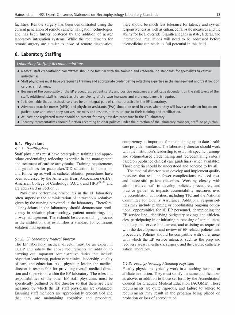
� Medical staff credentialing committees should be familiar with the training and credentialing standards for specialists in cardiacarrhythmias.
� Staff physicians must have prerequisite training and appropriate credentialing reflecting expertise in the management and treatment ofcardiac arrhythmias.
� Because of the complexity of the EP procedures, patient safety and positive outcomes are critically dependent on the skill levels of thestaff. Additional staff is needed as the complexity of the case increases and more equipment is required.
� It is desirable that anesthesia services be an integral part of clinical practice in the EP laboratory.� Advanced practice nurses (APNs) and physician assistants (PAs) should be used in areas where they will have a maximum impact on
patient care and where they can assume roles and responsibilities unique to their training and certification.� At least one registered nurse should be present for every invasive procedure in the EP laboratory.� Industry representatives should function according to clear policies under the direction of the laboratory manager, staff, or physician.
6.1. Physicians6.1.1. QualificationsStaff physicians must have prerequisite training and appro-priate credentialing reflecting expertise in the managementand treatment of cardiac arrhythmias. Training requirementsand guidelines for pacemaker/ICD selection, implantation,and follow-up as well as catheter ablation procedures havebeen addressed by the American Heart Association (AHA),American College of Cardiology (ACC), and HRS30–34 andare addressed in Section 7.
Physicians performing procedures in the EP laboratoryoften supervise the administration of intravenous sedativesgiven by the nursing personnel in the laboratory. Therefore,all physicians in the laboratory should demonstrate profi-ciency in sedation pharmacology, patient monitoring, andairway management. There should be a credentialing processin the institution that establishes a standard for conscioussedation management.
6.1.2. EP Laboratory Medical DirectorThe EP laboratory medical director must be an expert inCCEP and satisfy the above requirements, in addition tocarrying out important administrative duties that includephysician leadership, patient care clinical leadership, qualityof care, and education. As a physician leader, the medicaldirector is responsible for providing overall medical direc-tion and supervision within the EP laboratory. The roles andresponsibilities of the other EP staff physicians must bespecifically outlined by the director so that there are clearmeasures by which the EP staff physicians are evaluated.Ensuring staff members are appropriately credentialed andthat they are maintaining cognitive and procedural
competency is important for maintaining up-to-date healthcare provider standards. The laboratory director should workwith the institution’s leadership to establish specific training-and volume-based credentialing and recredentialing criteriabased on published clinical care guidelines (when available).Those criteria should be understood and adhered to by all.
The medical director must develop and implement qualitymeasures that result in fewer complications, reduced cost,and successful patient outcomes. Working closely withadministrative staff to develop policies, procedures, andpractice guidelines impacts accountability measures usedby accreditation authorities, including TJC and the NationalCommittee for Quality Assurance. Additional responsibil-ities may include planning or coordinating ongoing educa-tional opportunities for all EP personnel, championing theEP service line, identifying budgetary savings and efficien-cies, participating in or initiating purchasing of capital itemsthat keep the service line current, and assisting as requestedwith the development and review of EP-related policies andprocedures. Policies should be compatible with other areaswith which the EP service interacts, such as the prep andrecovery areas, anesthesia, surgery, and the cardiac catheteri-zation laboratory.
6.1.3. Faculty/Teaching Attending PhysicianFaculty physicians typically work in a teaching hospital oraffiliate institution. They must satisfy the same qualificationsas above, in addition to those set forth by the AccreditationCouncil for Graduate Medical Education (ACGME). Theserequirements are quite rigorous, and failure to adhere torequirements may result in the program being placed onprobation or loss of accreditation.
13Haines et al HRS Expert Consensus Statement on Electrophysiology Laboratory Standards
6.1.4. EP Laboratory Attending PhysicianAlthough certain components of the procedure can bedelegated to a trainee or other secondary operator, thelaboratory’s attending physician of record is ultimatelyresponsible for all activities within the laboratory and forpatient welfare. It is important for the staff physician torecognize that patient safety and successful outcomes dependgreatly on effective communication in the EP laboratory. Thiscommunication should include preoperative discussions withall members of the team before the case is underway regardingspecific patient needs. The physician should review thediagnosis, indications for the procedure, anticipated equipmentneeded, and potential findings of the procedure. The patientshould have a clear understanding of what to expect post-procedure in order to minimize anxiety. After the procedure,clear communication of the procedure findings, postprocedureorders, and recommendations should be exchanged with thetreatment team, including physicians, APNs, PAs, and nurses.
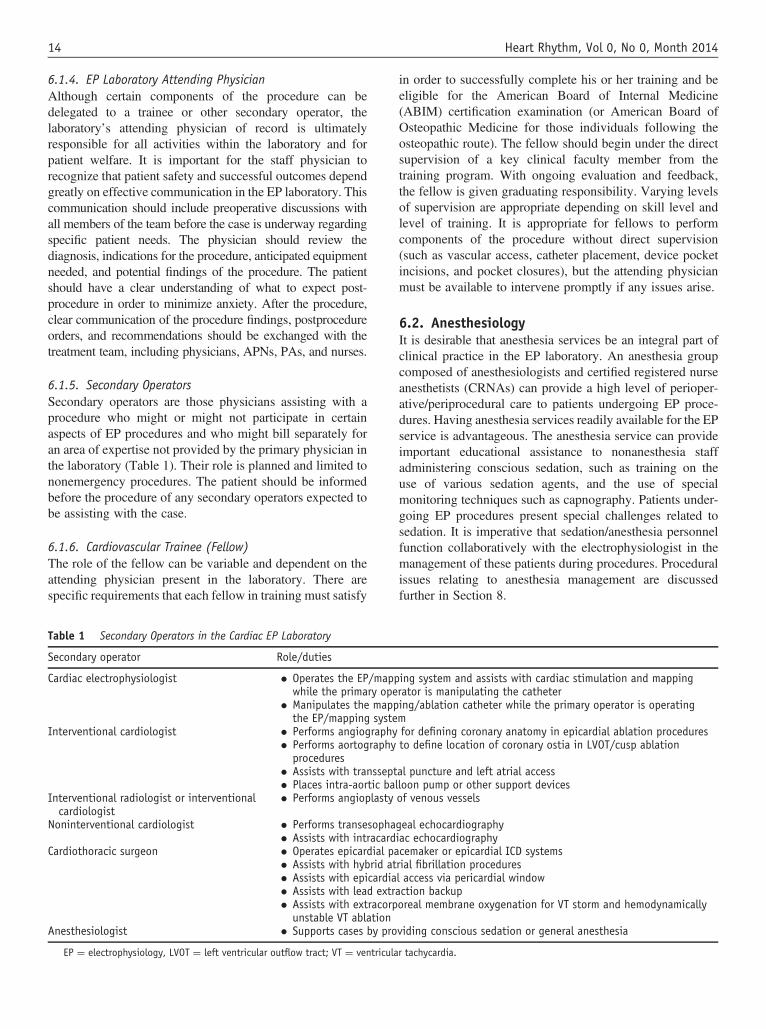
6.1.5. Secondary OperatorsSecondary operators are those physicians assisting with aprocedure who might or might not participate in certainaspects of EP procedures and who might bill separately foran area of expertise not provided by the primary physician inthe laboratory (Table 1). Their role is planned and limited tononemergency procedures. The patient should be informedbefore the procedure of any secondary operators expected tobe assisting with the case.
6.1.6. Cardiovascular Trainee (Fellow)The role of the fellow can be variable and dependent on theattending physician present in the laboratory. There arespecific requirements that each fellow in training must satisfy
in order to successfully complete his or her training and beeligible for the American Board of Internal Medicine(ABIM) certification examination (or American Board ofOsteopathic Medicine for those individuals following theosteopathic route). The fellow should begin under the directsupervision of a key clinical faculty member from thetraining program. With ongoing evaluation and feedback,the fellow is given graduating responsibility. Varying levelsof supervision are appropriate depending on skill level andlevel of training. It is appropriate for fellows to performcomponents of the procedure without direct supervision(such as vascular access, catheter placement, device pocketincisions, and pocket closures), but the attending physicianmust be available to intervene promptly if any issues arise.
6.2. AnesthesiologyIt is desirable that anesthesia services be an integral part ofclinical practice in the EP laboratory. An anesthesia groupcomposed of anesthesiologists and certified registered nurseanesthetists (CRNAs) can provide a high level of perioper-ative/periprocedural care to patients undergoing EP proce-dures. Having anesthesia services readily available for the EPservice is advantageous. The anesthesia service can provideimportant educational assistance to nonanesthesia staffadministering conscious sedation, such as training on theuse of various sedation agents, and the use of specialmonitoring techniques such as capnography. Patients under-going EP procedures present special challenges related tosedation. It is imperative that sedation/anesthesia personnelfunction collaboratively with the electrophysiologist in themanagement of these patients during procedures. Proceduralissues relating to anesthesia management are discussedfurther in Section 8.
Table 1 Secondary Operators in the Cardiac EP Laboratory
Secondary operator Role/duties
Cardiac electrophysiologist � Operates the EP/mapping system and assists with cardiac stimulation and mappingwhile the primary operator is manipulating the catheter
� Manipulates the mapping/ablation catheter while the primary operator is operatingthe EP/mapping system
Interventional cardiologist � Performs angiography for defining coronary anatomy in epicardial ablation procedures� Performs aortography to define location of coronary ostia in LVOT/cusp ablationprocedures
� Assists with transseptal puncture and left atrial access� Places intra-aortic balloon pump or other support devices
Interventional radiologist or interventionalcardiologist
� Performs angioplasty of venous vessels
Noninterventional cardiologist � Performs transesophageal echocardiography� Assists with intracardiac echocardiography
Cardiothoracic surgeon � Operates epicardial pacemaker or epicardial ICD systems� Assists with hybrid atrial fibrillation procedures� Assists with epicardial access via pericardial window� Assists with lead extraction backup� Assists with extracorporeal membrane oxygenation for VT storm and hemodynamicallyunstable VT ablation
Anesthesiologist � Supports cases by providing conscious sedation or general anesthesia
EP ¼ electrophysiology, LVOT ¼ left ventricular outflow tract; VT ¼ ventricular tachycardia.
Heart Rhythm, Vol 0, No 0, Month 201414
6.3. Allied Professional PersonnelTo ensure optimal safety and efficacy of interventional EP, it isimportant to emphasize the necessity of a multidisciplinary teamapproach. In this respect, the term allied professionals has beenemployed. Allied professionals are defined as all nonphysicianmembers of the health care team involved with the care of thepatient in the EP laboratory. This includes, but is not limited to,registered nurses (RNs), EP technologists, radiological technol-ogists, certified nurse practitioners (NPs), PAs, CRNAs, patientprep and recovery staff, and OR staff. Other key personnel thatare important for the safe and efficient function of the laboratoryinclude quality assurance (QA) staff; information technologists;biomedical engineers; scheduling coordinators; purchasing,inventory, and supply personnel; and housekeeping. Based onevidence-based practice and best practice patterns, it is importantto acknowledge that there is limited published research regard-ing the roles and responsibilities inherent in EP. Recommenda-tions as to how these positions may be filled by any one of theseveral categories of personnel are discussed below.
6.3.1. Advanced Practice Nurses and Physician AssistantsAPNs and PAs can play major roles and serve many functionsin the EP laboratory, as determined by the director of thelaboratory. They should be placed in those areas where theywill have maximum impact on patient care and assume rolesand responsibilities unique to their training and certification.APNs are often placed in clinic settings where they mayevaluate and treat arrhythmia or device-related issues. Theycan make rounds on inpatients, make assessments, developplans for care, write histories and physical exams, and admitand discharge patients. They can perform pre- and postpro-cedural evaluations and follow-up. Particularly in nonaca-demic institutions or practices, an APN or PA may functionas the most experienced or skilled nonphysician practitionerin the laboratory setting and thus function as a first assistantfor many technical aspects of the procedure. Each institutionshould have established policies defining the role of the APNand/or PA in the care of hospital patients.
6.3.2. Registered NursesAn RN should be present for every invasive procedure in theEP laboratory. The nurse must be familiar with the overallfunction of the laboratory as well as coordinate with thephysician operator and the other team members. The nurse(either RN or CRNA) is the primary individual responsible forthe direct observation, sedation, and nursing care of the patientduring the EP procedure and must be prepared to respond toany emergency. The number and type of nursing personnelrequired in the EP laboratory will vary depending on the typeof procedure, equipment used, and additional support staffassigned to the procedure.35 EP procedures are complex bytheir nature, and it is essential that the nursing staff participat-ing in such procedures provide safe, evidence-based care.
In institutions where nurses are responsible for theadministration of intraprocedural sedation, they are to followinstitutional training and guidelines for the care of the patient.
When a nurse is administering deep sedation, his or herfocus should be only on monitoring patient status, vital signs,oxygenation, and level of sedation. However, during moderateor light sedation, this individual may assist with minor,interruptible tasks once the patient’s level of sedation/analge-sia and vital signs have stabilized, provided that adequatemonitoring of the patient’s level of sedation is maintained.36
The nurse can also manage point-of-care testing for activatedclotting times (ACTs), oxygen saturation, and blood gasmeasurements. In most states, only RNs may administermedications and blood products. The nurse optimizes patientsafety by adhering to policies, protocols, and procedures, suchas completing the “active time-out” preprocedure and ensur-ing that the proper airway assessments are completed beforethe administration of sedation. Keeping a record or chartingduring the procedure is generally the responsibility of nurses.In addition, training on the use of stimulators, infusers, andablation generators is recommended so that nurses are able tofunction in multiple roles. Overall, nurses are coordinators ofall patient care in the laboratory and they oversee the careother allied professional-EP personnel are providing.
6.3.3. TechnologistsBecause of the extremely complex and technical aspects ofmany EP procedures, there should be at least one additionalperson involved in the more complex procedures, in addition tothe nurse who provides direct patient care. Depending on thecomplexity of the procedure, there may need to be more thanone additional person. In this arena, specific training, experi-ence, and certifications may determine which team memberoccupies each role. For example, a nurse and a technologistmay be equally capable of performing a certain duty orresponsibility but economics, staffing availability, and thesimultaneous performance of multiple duties can dictate whodoes each job in the laboratory. Because of the multiplicity ofroles, it is useful for members of the EP team to be cross-trainedand be able to function inmultiple roles and situations. There is awide array of additional equipment that requires training tooperate. This includes, but is not limited to, lasers, energy sourcegenerators, electroanatomic mapping systems, robotic andmagnetic catheter navigation systems, echocardiography (trans-esophageal and intracardiac), and CT and MRI imaging.
In many laboratories, it is the technologist or nurse whomonitors and operates the recording system. This activityrequires a thorough understanding and knowledge of theelectrophysiological properties of the heart as well as pacingprotocols and ablation. The operator must be able to trouble-shoot pacing problems and remain calm and functional inemergency situations. All technologists must have basiccardiac life support certification, and ACLS certification ispreferred. In the pediatric laboratory, pediatric cardiac lifesupport certification is required. As with the nurse, thetechnologist should have the ability to review, understand,and synthesize into practice new knowledge and practices.
EP technologists perform as essential team members. Theymay be a first assistant, which requires in-depth knowledge of
15Haines et al HRS Expert Consensus Statement on Electrophysiology Laboratory Standards
percutaneous procedures, catheters, sterile technique, energygenerators, and integrated noninvasive imaging. They shouldbe trained in the use, maintenance, and troubleshooting of allthe equipment. EP technologists should be skilled in steriletechnique, passing sterile supplies, and obtaining and perform-ing point-of-care testing on blood samples. They are often theperson who assists on device implant cases and lead extractions—roles that require fastidious adherence to sterile techniqueand an in-depth understanding of the implant process alongwith its risks and goals. A technologist or nurse may serve asthe first assistant for an invasive case. The circulator is typicallya nurse, but this role can be filled by a technologist, dependingon the staffingmix in the laboratory, the scope of practice in thisjob description, and the institutional requirements.
At least one department member should be a certifiedradiological technologist or equivalent technologist withexpertise in the operation of fluoroscopic equipment as wellas expertise in radiographic and angiographic imagingprinciples and techniques. Requirements for the participationof a radiological technologist in fluoroscopic proceduresvary from state to state. In conjunction with a qualifiedmedical physicist, the radiological technologist shouldmonitor radiation safety techniques for patients and labo-ratory personnel. In many states, the Nuclear RegulatoryCommission has specific regulations for who may operateionizing radiation equipment and under what circumstances.It is imperative that these regulations are understood andfollowed in the laboratory for the protection of patientsand staff.
6.3.4. Industry Employed Allied ProfessionalsDevice programmers, mapping and recording systems, andsome ablation systems may sometimes be operated byindustry representatives. Industry representatives must func-tion according to clear policies under the direction of thelaboratory manager, staff, or physician. They are oftenrequired to provide the institution with evidence of appro-priate immunizations, competency documentation, andendorsement from their company before being allowed inthe laboratory. They are generally allowed to have patientcontact only under direct staff supervision.25
During device implants or other device-related proce-dures, a clinical industry representative may be present underthe direct supervision of the attending physician. They maybring device equipment to the laboratory, provide intra-procedural programming and testing, and may even be askedto participate in data collection related to device registries. Amember of the EP laboratory staff, however, should beassigned the ultimate responsibility for the accurate andcomplete submission of data to national device registries.These industry representatives are often excellent sources ofinformation and education for the regular EP laboratory staff.
They may assist in the provision of formal training andeducation on device-related issues.25
6.3.5. Staffing PatternsTo ensure optimal safety and efficacy of interventional EP, itis important to emphasize the necessity of a multidisciplinaryteam approach. EP procedures are complex and includediagnostic, interventional, and therapeutic measures andshould be performed by experienced personnel. Because ofthe complexity of the EP procedures, patient safety andpositive outcomes are highly dependent on the skill levels ofthe staff (Table 2). Therefore, personnel dedicated to EPlaboratory procedures are recommended. Additional person-nel are needed as the complexity of the case increases, andmore equipment is required. The staffing mix may beinfluenced by regulations, regional practice patterns, typeof institution (academic vs. nonacademic), credentialingbodies, and economics. Cross-training of staff within theEP department maximizes staffing flexibility and is stronglyrecommended.
6.4. Administrator/ManagerThe role of the EP department administrator is typicallyheld by someone with broad knowledge of the field of EP.Depending on the size and volume of the laboratory, theadministrator may have no clinical obligations or mayserve as the head nurse of the laboratory. The responsibil-ities can include, but are not limited to, the following:strategic planning in association with the medical director,managing operational issues, capital planning, budgeting,hiring, planning orientations and training programs forallied professionals, and other general administrationduties. A nurse or a cardiovascular technologist, preferablywith some business training or experience, is best suitedfor this role. In shared or combined cardiac catheterizationlaboratories and EP laboratories, there may be a commonadministrator overseeing both areas.
In many departments, the manager is a nurse. The responsi-bilities of a nurse manager include an overall understanding ofthe day-to-day operations of the laboratory, management of pre-and postprocedural care areas, and direct participation in theobservation and care of patients undergoing EP procedures.Additional areas of responsibility include application of institu-tional guidelines for patient monitoring, medication adminis-tration, procedural sedation, and patient safety. Staffcompetencies and proficiency in performing tasks requiredbefore, during, and after the procedure must be developed,updated, and reviewed on a regular basis. The nurse managerwill collaborate with anesthesia, pharmacy, biomedical engi-neering, purchasing, equipment vendors, and housekeeping tocoordinate the operation of the EP laboratory.
Heart Rhythm, Vol 0, No 0, Month 201416
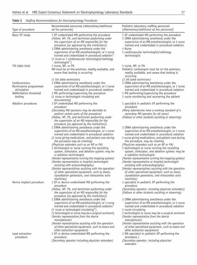
Table 2 Staffing Recommendations for Electrophysiology Procedures
Type of procedureRecommended personnel (Alternatives/additionalad hoc personnel)
Pediatric laboratory staffing personnel(Alternatives/additional ad hoc personnel)
Basic EP study 1 EP credentialed MD performing the procedure 1 EP credentialed MD performing the procedure(Fellow, NP, PA, and technician performing underthe supervision of an MD responsible for theprocedure [as approved by the institution])
1 CRNA administering anesthesia under thesupervision of an MD anesthesiologist, or 1 nursetrained and credentialed in procedural sedation
1 CRNA administering anesthesia under thesupervision of an MD anesthesiologist, or 1 nursetrained and credentialed in procedural sedation*
1 Nurse1 cardiovascular technologist/radiologytechnologist
(1 nurse or 1 cardiovascular technologist/radiologytechnologist*)
Tilt table tests 1 nurse, NP, or PA 1 nurse, NP, or PAMD must be on the premises, readily available, andaware that testing is occurring
Pediatric cardiologist must be on the premises,readily available, and aware that testing isoccurring
(1 tilt table technician) (1 tilt table technician)Cardioversions 1 CRNA administering anesthesia under the
supervision of an MD anesthesiologist, or 1 nursetrained and credentialed in procedural sedation
1 CRNA administering anesthesia under thesupervision of an MD anesthesiologist, or 1 nursetrained and credentialed in procedural sedation
Noninvasive programmedstimulation
Defibrillation thresholdtesting
1 MD performing/supervising the procedure 1 MD performing/supervising the procedure(1 nurse or technologist circulating anddocumenting)
1 nurse monitoring and recovering the patient
Ablation procedures 1 EP credentialed MD performing theprocedure
1 specialist in pediatric EP performing theprocedure
(Secondary MD operators may be desirable toperform certain parts of the procedure)
(Many laboratories have a working standard of asecondary MD operator for all cases)
(Fellow, NP, PA, and technician performing underthe supervision of an MD responsible for theprocedure [as approved by the institution])
(Fellows or other students assisting or observing)
1 CRNA administering anesthesia under thesupervision of an MD anesthesiologist, or 1 nursetrained and credentialed in procedural sedation
1 CRNA administering anesthesia under thesupervision of an MD anesthesiologist, or 1 nursetrained and credentialed in procedural sedation
(1 nurse giving medications, and patient care duringthe procedure may do charting)
1 nurse giving medications, and patient care duringthe procedure, may do charting
(Physician extenders such as an NP or PA) (Physician extenders such as an NP or PA)1 technologist or nurse running the recordingsystem, stimulator, and ablation system; may bea radiation technologist
1 technologist or nurse running the recordingsystem, stimulator, and ablation system; may bea radiation technologist
(Vendor representative running the mapping system) (Vendor representative running the mapping system)(Vendor representative or hospital technologistassisting with echocardiography)
(Vendor representative or hospital technologistassisting with echocardiography)
(Vendor representatives assisting with the operationof other specialized equipment, such as lasers,cryoablation generators, and intracardiac echomachines)
(Vendor representatives assisting with the operationof other specialized equipment, such as lasers,cryoablation generators, and intracardiac echomachines)
Device implant procedure 1 EP or device-credentialed MD performing theprocedure
1 specialist in pediatric EP performing theprocedure
(Fellow, NP, PA, and technician performing underthe supervision of an MD responsible for theprocedure [as approved by the institution])
(Secondary operator, including physician extenders)(Fellows or other students assisting or observing)
1 CRNA administering anesthesia under thesupervision of an MD anesthesiologist, or 1 nursetrained and credentialed in procedural sedation*
1 CRNA administering anesthesia under thesupervision of an MD anesthesiologist, or 1 nursetrained and credentialed in procedural sedation
(1 nurse or technologist circulating*) 1 nurse circulating(1 technologist or nurse may be a surgical assistant) 1 technologist or nurse may be a surgical assistant(Vendor representative from the devicemanufacturer)
(Vendor representative from the devicemanufacturer)
(Vendor representative assisting with the operationof other specialized equipment, such as lasers andother extraction equipment)
(Vendor representative assisting with the operationof other specialized equipment, such as lasers andother extraction equipment)
Lead extractionprocedure
1 EP or device-credentialed MD performing theprocedure
1 MD specialist in pediatric EP performing theprocedure )
(Secondary operator including physician extenders) (Secondary operator, including physicianextenders
17Haines et al HRS Expert Consensus Statement on Electrophysiology Laboratory Standards

7. Laboratory Personnel Credentialing
Laboratory Personnel Credentialing Recommendations
� All clinicians working in the EP laboratory have a responsibility to achieve and maintain the recommended credentials and continuemedical education to optimize patient care.
� It is recommended that any non–CCEP-certified physician who wants privileges for implantable cardioverter-defibrillator implantationshould complete formal training in this field as defined by the COCATS criteria, achieve certification of Competency in Cardiac RhythmDevice Therapy for the Physician (CCDS) from the International Board of Heart Rhythm Examiners (IBHRE), and maintain an adequatevolume of device implants to meet hospital-based credentialing criteria.
� For the benefit of patients, it is paramount that physicians be held to a high performance standard and that remediation, withholdingrecredentialing, or revocation of privileges occurs if criteria are not met.
7.1. Attending Physicians7.1.1. CredentialingA range of procedures is performed in cardiac EP suites.Procedures that fall within the domain of physicians trainedand ABIM certified in cardiovascular diseases includeperformance of electrical cardioversions and placement oftemporary pacemaker wires. Invasive cardiac EP proce-dures in adults, including diagnostic electrophysiologicaltesting and catheter ablation, should be restricted tophysicians who are ABIM certified in CCEP.31 All CCEPboard–certified physicians have completed at least 1 year of
comprehensive subspecialty training in CCEP at anACGME-approved training program30 or had substantialexperience and a career focus in CCEP if they trained in anera before the development of formal CCEP trainingprograms. The majority of programs encourage a secondyear non-ACGME advanced fellowship. The 2013 guide-lines for advanced training in pediatric and congenital EPrepresent procedural requirements for those completingtraining.37 The clinical competence statement on invasiveelectrophysiology studies, catheter ablation, and cardiover-sion,34 supplemented by expert consensus statements on
Table 2 (continued )
Type of procedureRecommended personnel (Alternatives/additionalad hoc personnel)
Pediatric laboratory staffing personnel(Alternatives/additional ad hoc personnel)
(Fellow, NP, PA, and technician performing underthe supervision of an MD responsible for theprocedure [as approved by the institution])
(Fellows or other students assisting orobserving)
1 CRNA administering anesthesia under thesupervision of an MD anesthesiologist, or 1 nursetrained and credentialed in procedural sedation
1 CRNA administering anesthesia under thesupervision of an MD anesthesiologist
1 CV surgeon to be immediately available (may berequired to be in the room for the critical part ofthe procedure)
1 congenital CV surgeon in the operating room atthe time of extraction and immediately availableperiprocedure
1 CV surgery fellow on call for surgical assistance1 nurse circulating 1 perfusionist(1 technologist or nurse scrubbing) 1 nurse
1 cardiovascular technologist/radiology technologist(May require a second scrub person for the surgicalprocedure)
1 CV OR scrub nurse1 CV OR circulating nurse
(An operator to monitor a TEE that may be in place.This function is sometimes performed by theanesthesiologist)
(An operator to monitor a TEE that may be in place.This function is sometimes performed by theanesthesiologist)
(Vendor representative from the devicemanufacturer)
(Vendor representative from the devicemanufacturer)
(Vendor representative assisting with the operationof other specialized equipment, such as lasers andother extraction equipment)
(Vendor representative assisting with the operationof other specialized equipment, such as lasers andother extraction equipment)
CRNA ¼ certified registered nurse anesthetist; CV ¼ cardiovascular; EP ¼ electrophysiology; MD ¼ physician; NP ¼ nurse practitioner; PA ¼ physicianassistant; TEE ¼ transesophageal echocardiography.*In procedures performed with deep sedation/analgesia, the CRNA or nurse administering sedation/anesthesia should have no responsibilities other thanmonitoring the patient. A second nurse or technologist must be available to circulate and document. However, in procedures performed with moderate or lightsedation, this individual may assist with minor, interruptible tasks once the patient’s level of sedation/analgesia and vital signs have stabilized, provided thatadequate monitoring for the patient’s level of sedation is maintained.52
Heart Rhythm, Vol 0, No 0, Month 201418

transvenous lead extraction,8 catheter and surgical ablationof atrial fibrillation,38 and catheter ablation of ventriculararrhythmias,39 provides guidelines for appropriate trainingin CCEP. This document is scheduled to be updated in thenear future. As training standards evolve, these minimumrequirements will be updated regularly. A successfulpassage of the ABIM CCEP board examination is requiredto receive the Certificate of Added Qualification in CCEP.A similar but alternate pathway is available for doctors ofosteopathy through the American Board of OsteopathicMedicine. Physicians for whom these pathways areunavailable because of international training or pathwaychoices and who are actively involved in the clinicalmanagement of EP patients may choose to certify throughthe IBHRE with the Certification Examination for Com-petency in Cardiac Electrophysiology. Physicians perform-ing complex catheter ablation procedures, such as atrialfibrillation/complex atrial tachycardia ablation and VTablation, should treat at least 25 cases of each with anexperienced mentor before becoming independent. Alter-natively, they should perform these procedures during theirCCEP training program, as recommended by the 2012HRS/EHRA/ECAS expert consensus statement on catheterand surgical ablation of atrial fibrillation38 and the 2009EHRA/HRS expert consensus on catheter ablation ofventricular arrhythmias.39
A number of training pathways can lead to the practice ofimplanting CIEDs. Although physicians who are boardcertified in CCEP have met minimum training standardsfor CIED implantation, some cardiovascular diseases(CVD) board–certified physicians and some AmericanBoard of Surgery–certified cardiac, thoracic, and generalsurgeons also devote a substantial portion of their practice tothe prescription, implantation, and follow-up of CIEDs. Thecriteria established by the ABIM program requirements28
and the Recommendations for Training in Adult Cardiovas-cular Medicine Core Cardiology Training document33
strongly recommend that any non–CCEP-certified physicianwho wants privileges for ICD implantation in adultscompletes formal training in this field. Achieving CCDSfrom the IBHRE for CIED prescription, implantation, andfollow-up is strongly recommended.
Credentialing is ultimately the responsibility of thecredentialing committee of the individual hospital, whoshould be familiar with the training and credentialing stand-ards for specialists in cardiac arrhythmias. Although mostmajor centers already follow the guidelines above, somecenters allow less qualified practitioners to perform these EPlaboratory procedures. Unfortunately, hospital credentialingcommittees have substantial conflicts of interest that couldlead to granting privileges to physicians without appropriateboard certification and experience, as refusal of thesecredentials usually results in a loss of patients and revenuesto competing institutions. Ultimately, the patient pays theprice when inappropriate credentials are allowed.
7.1.2. Evaluation and RecredentialingAll EP laboratory physicians should be subject to periodicpeer review and recredentialing. Important components inthe recredentialing process should include a review of ABIMboard certification and IBHRE CCDS status, case volume,patient outcomes, peer evaluation, and continuing medicaleducation (CME). The specific criteria for recredentialing aredetermined by each individual hospital, but should generallyparallel the following recommendations: ABIM CCEP boardcertification and IBHRE certification are limited to 10 years;to stay current for CCEP, the physician must complete aseries of CME and/or practice improvement activities9;recertification examination for CCEP and CCDS are eachrequired at 10-year intervals; to ensure that cognitiveand technical skills are maintained, the physician’sclinical competence must be evaluated and documented ona regular basis; it is the responsibility of the medical staffcredentialing committee to ensure that physicians performthe necessary number of evaluations and procedures neededto maintain their expertise31 and also that they participate inregular CME activities. The EP laboratory should have arobust QA process (see Section 9), and physician outcomesshould be compared with national benchmarks derived fromthe literature or databases such as the National Cardiovas-cular Data Registry (NCDR) on a regular basis; regular 360ºevaluations, including evaluations from physician cow-orkers, fellows, nursing staff, technical staff, and patients,should be considered as part of the recredentialing reviewprocess.
For the benefit of patients, it is paramount that physiciansbe held to a high performance standard and that remediation,withholding recredentialing, or revocation of privilegesoccurs if criteria are not met. The physician leaders mustbe committed to working aggressively to maintain thehighest standards of patient care in their laboratories.40
7.1.3. Pediatric Training and CredentialingPACES, working in conjunction with HRS, has developedguidelines for advanced training in EP focused on pediatricpatients and patients of any age with CHD. The originalguidelines, endorsed by the HRS and published in 2005,required at least 12 months of specialized training, and werefollowed by a 2008 document focused on the implantation ofpacemakers and defibrillators in these populations.41,42
There are currently no third-tier board examinations fordiplomates of the pediatric cardiology subboard of theAmerican Board of Pediatrics. However, pediatric electro-physiologists are eligible to take the IBHRE examination forphysicians with special competency in EP, as that examina-tion includes a pediatric module. PACES has recently writtena competency statement for training in this field.43 Thatcompetency statement strongly recommended that all grad-uating fellows and active pediatric EP clinicians take theIBHRE EP examination.
19Haines et al HRS Expert Consensus Statement on Electrophysiology Laboratory Standards
7.1.4. Adult Congenital Heart Disease Training andCredentialingThe field of adult congenital heart disease (ACHD) is animportant and expanding clinical domain that is typicallystaffed by clinicians who are competent in both pediatricsand internal medicine. Cardiologists providing invasive EPcare for this unique patient population can enter the fieldfrom either specialty, but a portion of their formal trainingmust be focused on the complex anatomy and unique EP ofACHD. Further recommendations on the expertise necessaryto care for this patient group can be found in a recentconsensus statement supported by AHA/ACC/HRS.44 Aboard certification process for ACHD is being developedand is scheduled for implementation by 2015.
7.2. Nurses7.2.1. Training and CredentialingNursing licensure, credentialing, recredentialing, continuingeducation, and laboratory training are affected by therequirements of multiple agencies, including federal andstate governments, the health care organization, and Occupa-tional Safety and Health Administration.
The standards of professional practice for nursesemployed in the EP laboratory environment have beendefined.33 All EP nurses should have a critical care or astrong cardiology background, ACLS certification, and,in the pediatric EP laboratory, pediatric ACLS. Anextensive knowledge of cardiac anatomy and physiology,electrocardiography, pharmacology, and training in ster-ile technique is also required. Nurses need to have athorough understanding of catheter-based interventionsand surgical procedures, cardioversion, arrhythmia dis-crimination, and emergency treatment of life-threateningarrhythmias and complications/emergencies. Familiaritywith fluoroscopic, electroanatomic, and echocardio-graphic imaging is a required skill set. Annual or biannualcompetencies required for nurses working in heart rhythmservice operations should include basic life support,ACLS, infection control, emergency and TJC prepared-ness, training in conscious sedation, charting, and patientsafety. Demonstration of competency in radiation safety,sterile technique, external defibrillator operation, unit-specific nursing protocols, ACT operation, and temporarypacemaker operation should also be mandatory. Depend-ing on the roles and responsibilities assumed by RNs,competencies may be needed in the EP recording systemand programmed stimulator operation, ablation, generatoroperation, mobile laboratory operations, sheath insertion/removal, operation of vascular ultrasound, 3D mappingoperation, and ICE operation.
Certification offered by the (IBHRE is an integral part ofheart rhythm education, training expectations, and require-ments. In addition to the standard certification requirementsfor being an RN, the IBHRE offers two certificationexaminations for allied professionals designed to demonstratea mastery of knowledge in cardiac rhythm management.
7.2.2. Evaluation and RecredentialingContinuing education requirements are highly variable bystate and nurse specialty. Nurses need to review requirementsfor the state in which they are practicing to ensure propercompliance and maintenance of certification, as requirementscan range from no required continuing education to as manyas 30 hours every 2 years.45 The Certification Examinationfor Competency in Cardiac Rhythm Device Therapy and theCertification Examination for Competency in Cardiac Elec-trophysiology are required at 10-year intervals.
7.3. Advanced Practice Nurses7.3.1. Training and CredentialingThe APN (usually a NP) is educated through a certifiedgraduate level NP program, meets the requirements of thestate credentialing bodies, and practices according to theAmerican Nurses Association consensus model.46 An NP istrained to perform preprocedural evaluation, order and inter-pret diagnostic tests, and conduct postprocedural follow-up.After on-the-job training, NPs may assist in diagnostic EPstudies, catheter ablation procedures, and device implants butcannot serve as a primary operator. NPs working in heartrhythm services should be certified in basic and advancedcardiac life support and have knowledge of radiation safety,sterile technique, external defibrillator operation, ICE oper-ation, and temporary pacemaker operation. Among laborato-ries represented by writing committee members, NPs (or PAs)are involved in pre- and postprocedural care in more than 75%of the centers but only 19% of the centers employ theseindividuals for assistance in the procedural laboratory.
7.3.2. Evaluation and RecredentialingThere are no uniform recredentialing criteria for NPs in theEP laboratory. NPs are expected to maintain certification andlicensure as per certification and state guidelines. Theinstitution should establish volume criteria for the main-tenance of procedural competencies.
7.4. Technologists7.4.1. Training and CredentialingIndividuals with a variety of backgrounds and qualifications canwork in an EP laboratory as a cardiovascular technologist ortechnician. There is no professional regulatory body for cardiactechnologists, although efforts are underway in some regions toachieve this goal. Most technologists have postsecondaryeducation (university degree or college diploma) with extensiveon-the-job training. Industry-sponsored courses often providesupplemental education specific to technologies used in the EPlaboratory environment. Some cardiovascular technologistprograms offer EP as a component within a cardiovasculartechnology program; there are also certificate programs avail-able at some accredited colleges.47 Cardiovascular technolo-gists can be credentialed as a registered cardiovascular invasivespecialist, registered cardiology technologist, registered cardi-opulmonary technologist, or registered cardiac electrophysiol-ogy specialist through an accredited association such as
Heart Rhythm, Vol 0, No 0, Month 201420
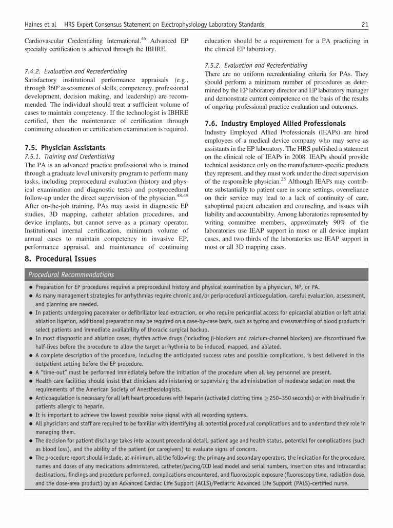
Cardiovascular Credentialing International.46 Advanced EPspecialty certification is achieved through the IBHRE.
7.4.2. Evaluation and RecredentialingSatisfactory institutional performance appraisals (e.g.,through 360º assessments of skills, competency, professionaldevelopment, decision making, and leadership) are recom-mended. The individual should treat a sufficient volume ofcases to maintain competency. If the technologist is IBHREcertified, then the maintenance of certification throughcontinuing education or certification examination is required.
7.5. Physician Assistants7.5.1. Training and CredentialingThe PA is an advanced practice professional who is trainedthrough a graduate level university program to perform manytasks, including preprocedural evaluation (history and phys-ical examination and diagnostic tests) and postproceduralfollow-up under the direct supervision of the physician.48,49
After on-the-job training, PAs may assist in diagnostic EPstudies, 3D mapping, catheter ablation procedures, anddevice implants, but cannot serve as a primary operator.Institutional internal certification, minimum volume ofannual cases to maintain competency in invasive EP,performance appraisal, and maintenance of continuing
education should be a requirement for a PA practicing inthe clinical EP laboratory.
7.5.2. Evaluation and RecredentialingThere are no uniform recredentialing criteria for PAs. Theyshould perform a minimum number of procedures as deter-mined by the EP laboratory director and EP laboratory managerand demonstrate current competence on the basis of the resultsof ongoing professional practice evaluation and outcomes.
7.6. Industry Employed Allied ProfessionalsIndustry Employed Allied Professionals (IEAPs) are hiredemployees of a medical device company who may serve asassistants in the EP laboratory. The HRS published a statementon the clinical role of IEAPs in 2008. IEAPs should providetechnical assistance only on the manufacturer-specific productsthey represent, and they must work under the direct supervisionof the responsible physician.25 Although IEAPs may contrib-ute substantially to patient care in some settings, overrelianceon their service may lead to a lack of continuity of care,suboptimal patient education and counseling, and issues withliability and accountability. Among laboratories represented bywriting committee members, approximately 90% of thelaboratories use IEAP support in most or all device implantcases, and two thirds of the laboratories use IEAP support inmost or all 3D mapping cases.
8. Procedural Issues
Procedural Recommendations
� Preparation for EP procedures requires a preprocedural history and physical examination by a physician, NP, or PA.� As many management strategies for arrhythmias require chronic and/or periprocedural anticoagulation, careful evaluation, assessment,
and planning are needed.� In patients undergoing pacemaker or defibrillator lead extraction, or who require pericardial access for epicardial ablation or left atrial
ablation ligation, additional preparation may be required on a case-by-case basis, such as typing and crossmatching of blood products inselect patients and immediate availability of thoracic surgical backup.
� In most diagnostic and ablation cases, rhythm active drugs (including β-blockers and calcium-channel blockers) are discontinued fivehalf-lives before the procedure to allow the target arrhythmia to be induced, mapped, and ablated.
� A complete description of the procedure, including the anticipated success rates and possible complications, is best delivered in theoutpatient setting before the EP procedure.
� A “time-out” must be performed immediately before the initiation of the procedure when all key personnel are present.� Health care facilities should insist that clinicians administering or supervising the administration of moderate sedation meet the
requirements of the American Society of Anesthesiologists.� Anticoagulation is necessary for all left heart procedures with heparin (activated clotting timeZ250–350 seconds) or with bivalirudin in
patients allergic to heparin.� It is important to achieve the lowest possible noise signal with all recording systems.� All physicians and staff are required to be familiar with identifying all potential procedural complications and to understand their role in
managing them.� The decision for patient discharge takes into account procedural detail, patient age and health status, potential for complications (such
as blood loss), and the ability of the patient (or caregivers) to evaluate signs of concern.� The procedure report should include, at minimum, all the following: the primary and secondary operators, the indication for the procedure,
names and doses of any medications administered, catheter/pacing/ICD lead model and serial numbers, insertion sites and intracardiacdestinations, findings and procedure performed, complications encountered, and fluoroscopic exposure (fluoroscopy time, radiation dose,and the dose-area product) by an Advanced Cardiac Life Support (ACLS)/Pediatric Advanced Life Support (PALS)-certified nurse.
21Haines et al HRS Expert Consensus Statement on Electrophysiology Laboratory Standards

8.1. Patient Preparation8.1.1. History, Physical Examination, and LaboratoryExaminationPreparation for EP procedures requires a careful preproce-dural history and physical examination by a physician, NP,or PA to confirm the reason for the procedure that day andidentify all comorbidities that could adversely impactprocedure outcome. A thorough medication history, includ-ing allergies, must be gathered. The patient needs to beevaluated for factors that will impact anesthesia management(adequacy of airway, history of anesthesia experiences,obstructive sleep apnea, and physical indicators for difficultintubation). All adult patients should have recent (usuallywithin 2 weeks) laboratory work, including electrolytes,blood urea nitrogen, creatinine, complete blood count, and, iftaking anticoagulants, prothrombin time. All women ofchildbearing potential, including girls older than 12 years,should have serum or urine pregnancy testing within 2 weeksbefore the procedure. The need for a preprocedure laboratoryexam in healthy children undergoing elective electrophysio-logical testing is not clear and is not common practice.
8.1.2. Patients Receiving Oral Anticoagulants or AntiplateletMedicationsAs many management strategies for arrhythmias requirechronic and/or periprocedural anticoagulation, careful eval-uation, assessment, and planning are needed. Among theconsiderations are the agent used, thromboembolic risk,bleeding risk, comorbidities, laboratory values, and avail-ability of reversal agents or blood products such as freshfrozen plasma. Consideration should be given to performingadditional preprocedural transesophageal echocardiogramsor the use of intracardiac ultrasound to reduce the risk ofcomplications. In patients undergoing pacemaker or defib-rillator lead extraction or who require pericardial access forepicardial ablation or left atrial ablation ligation, additionalpreparations may be required, including typing and cross-matching of blood products, availability of thoracic surgicalbackup and/or OR, and, in some cases, intraproceduraltransesophageal echocardiogram.
8.1.3. Patients Receiving Antiarrhythmic DrugsMany patients are taking one or more medications to control theheart rate and/or rhythm at the time of an EP procedure. In mostcases, rhythm active drugs (including β-blockers and calcium-channel blockers) are discontinued five half-lives before theprocedure to allow the target arrhythmia to be induced,mapped, and ablated. In patients undergoing anatomicallybased ablation, withholding these drugs may not be necessary.
8.1.4. Patient Education and ConsentFor most patients, the EP laboratory is an unfamiliar andintimidating environment, one in which an equally unfami-liar procedure is about to be performed. A completedescription of the anticipated events is best delivered in theoutpatient setting before the procedure day. Education as to
the planned agenda, the other participants (nurses, technol-ogists, EP doctors, and anesthetists/anesthesiologists), andthe nature of some of the equipment in the laboratory isimportant to ease the patient’s anxiety and aid in theircooperation during the procedure. Many of the technicalterms used to describe the procedure are foreign to thepatient; the staff must take care to use lay language in theirdescriptions, to evaluate the patient’s ability to learn andpreference how to learn, and to assess the patient’s compre-hension. This role is usually filled by an RN familiar with theprocedure. The requisite process for informed consent isdetailed in Section 12. Critical components include ensuringpatient understanding, full disclosure of the risks andalternatives to the planned procedure, and the opportunityfor the patients to ask questions and fully discuss theirconcerns. The patient education and consent process must becompleted before the administration of any sedative oranxiolytic agents. In the case of pediatric patients or adultpatients with cognitive impairment, education must be givenand consent requested of the patient and the legal guardian.
8.1.5. Time-OutA time-out must be performed immediately before theinitiation of the procedure when all key personnel arepresent. All members of the team are to cease their activitieswhile one member recites two patient identifiers (i.e., name,date of birth, and medical record number), the type andlaterality of the procedure, the name of the operator, and anyknown allergies. All members of the team must agree on allpoints before the procedure can commence.50
8.2. Procedural Issues—EP Catheter Procedures8.2.1. Sedative agents, Relaxants, and AnesthesiaThe goal of analgesia and anesthesia in the EP laboratoryshould be to provide a safe, nontraumatic experience for thepatient. The administration of anesthesia varies among casetypes and among institutions, from anxiolysis or moderateprocedural sedation by an Advanced Cardiac Life Support(ACLS)/Pediatric Advanced Life Support (PALS)-certifiednurse under the supervision of the cardiac electrophysio-logist51,52 to monitored anesthesia care or general anesthesiaadministered by an anesthesiologist or CRNA under thesupervision of the anesthesiologist. The health care institutionmust require those administering or supervising moderatesedation who are not anesthesiologists to meet the require-ments of the American Society of Anesthesiologists to obtainprivileges.53 Credentialing for this privilege must be periodi-cally renewed. If intravenous procedural sedation will be used,the physician must establish an American Society of Anes-thesiologists classification and Mallampati score for the patientbefore the procedure.36 It is necessary to have these assess-ments done before the procedure so that, if necessary, alternateplans for sedation may be arranged to optimize patient safetyand minimize procedural delays. Medications typicallyemployed include etomidate, propofol, ketamine, fentanyl,midazolam, methohexital, and inhalational agents. Individual
Heart Rhythm, Vol 0, No 0, Month 201422

states regulate what medications can be administered bynonanesthesiologists. As the provision of sedation is acontinuum and the depth of sedation may vary, best practiceis that all patients receiving moderate or deep sedation beevaluated by continual observation of qualitative clinical signs,pulse oximetry, noninvasive blood pressure monitoring, heartrate and rhythm, and monitoring for the presence of exhaledcarbon dioxide to ensure the adequacy of ventilation unlessprecluded or invalidated by the nature of the patient, procedure,or equipment.51 Monitoring equipment should be in workingorder and have appropriate audible alarms. In pediatric cases,factors to be considered for the choice of sedation or generalanesthesia include young age, preexisting medical conditions,presence of CHD, airway issues, physician or family choice,and the length and complexity of the procedure. The 2002NASPE Position Statement on Pediatric Ablation delineatedthe types of anesthesia (conscious sedation, moderate sedation,and general anesthesia), and these remain applicable.54
Regardless of whether the administration of sedative agentsis under the control of the electrophysiologist or anothercaregiver, the electrophysiologist must have a working knowl-edge of the effects of the agents used and how they mightimpact the electrical aspects of the procedure (such asarrhythmia inducibility and effects on blood pressure) orinteract with other medications that may be given. Deepsedation or general anesthesia can minimize patient discom-fort, can benefit the EP procedure by preventing patientmovement, and is necessary during defibrillation thresholdtesting. An immobile patient facilitates accurate and precise 3Dmapping and reduces risk during transseptal puncture, peri-cardial access, or ablation in close proximity to criticalstructures. Note that in cases where the assessment of phrenicnerve function is important for a favorable case outcome (suchas placement of a coronary vein branch pacing lead or ablationwithin the right superior pulmonary vein), paralytic dosingshould be reduced or eliminated. Improved safety, efficacy,and procedure times have been shown with the use of generalanesthesia with certain procedures such as atrial fibrillationablation.55 Using high-frequency ventilation can further min-imize respiration-related cardiac movement during ablation.56
Although adequate sedation should be administered toensure patient comfort because certain arrhythmias such asatrial tachycardias and outflow tract VTs can be dependent onadrenergic tone, excessive sedation can result in the inability toinduce the clinical arrhythmia. In cases in which an adrenergic-dependent arrhythmia is suspected, sedation must be mini-mized until the clinical arrhythmia is induced and mapped.Deep sedation is usually administered while performingablation to prevent patient movement. When assessing theend point in these cases, care must be used to differentiate theeffect of sedation from the actual elimination of the arrhythmia.
8.2.2. Sterile Preparation of the Access Site and VascularAccessAlthough the risk of infection is extremely low with EPcatheter procedures,57 appropriate sterile techniques should
be maintained. This includes sterile preparation of all accesssites, such as the groin and neck. If there is the potential forpericardial access, the subxiphoid region, and possibly theparasternal and apical regions, should also be prepped anddraped. In cases with a higher risk of cardiac tamponade,sterile preparation of the subxiphoid region can be consid-ered at the onset of the procedure.
8.2.3. Diagnostic Catheter SelectionCatheters with a smaller French size and fewer electrodes aremore flexible, exert lower axial force, and may carry a lowerrisk of perforation,58 but should perhaps be avoided becauseof difficulty maintaining stable catheter position. Catheterswith more electrodes can facilitate rapid recognition ofarrhythmia activation patterns and are particularly beneficialat sites such as the coronary sinus. Smaller electrodes andnarrower interelectrode spacing detect a more local activationsignal and can provide more precise activation mapping, butmay be less maneuverable. Activation confined to a smallstructure or circuit, such as the His bundle or an accessorypathway, however, may be difficult to localize with narrowelectrode spacing. The field of signal detection can beincreased either by changing to a catheter with widerelectrode spacing or by reconfiguring the electrode pairing.With this wider field of view, anatomical localization isdecreased. Some diagnostic catheters are designed withelectrodes in a specialized spatial configuration, such ascircular/ring, basket, or star-shaped catheters. These catheterscan enable rapid deciphering of an activation pattern evenwith a paucity of arrhythmia. The number of catheters andrecording sites should be adequate to achieve the desired endpoints of the procedure, but not so many that vascular damageor obstruction or intracardiac entanglement could occur.
8.2.4. AnticoagulationAnticoagulation is necessary for all left heart procedureswith heparin or bivalirudin in patients allergic to heparin.38,39
Even in patients with therapeutic international normalizedratio on warfarin, heparin must be administered for left heartprocedures, though typically in lower doses than in patientsnot taking warfarin. For right heart procedures, there is noevidence favoring routine use of anticoagulation; in caseswhere there is concern that the patient is at an increased riskfor thromboembolic complications (prolonged procedure,known or discovered patent foramen ovale), some centersadminister heparin for anticoagulation.
8.2.5. Selection of Ablation CathetersSelection of the mode and catheter for catheter ablation isoperator dependent. Catheter factors such as torque delivery,axial stiffness, steerability, and introducer diameter affectdevice selection. Ablation modes, including RF, cooled RF,laser, and cryothermy all have their strengths and weak-nesses. The goal is to achieve a therapeutic ablation byidentifying the ablation target, maneuvering to that site, andthen destroying enough tissue to prevent arrhythmia
23Haines et al HRS Expert Consensus Statement on Electrophysiology Laboratory Standards
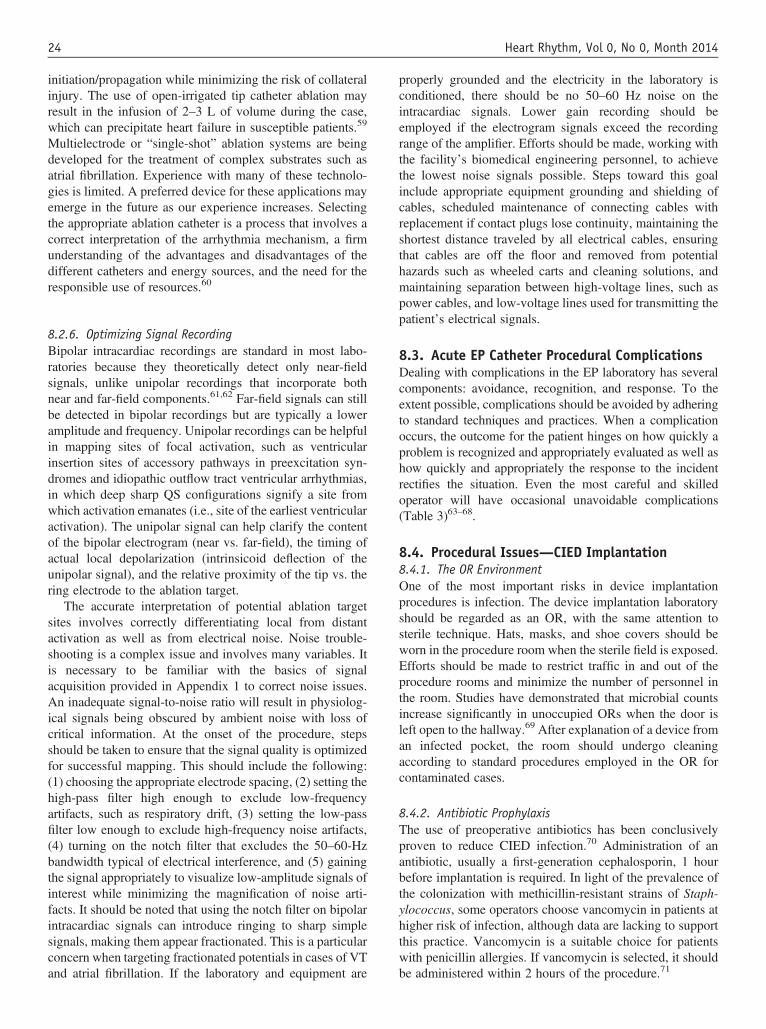
initiation/propagation while minimizing the risk of collateralinjury. The use of open-irrigated tip catheter ablation mayresult in the infusion of 2–3 L of volume during the case,which can precipitate heart failure in susceptible patients.59
Multielectrode or “single-shot” ablation systems are beingdeveloped for the treatment of complex substrates such asatrial fibrillation. Experience with many of these technolo-gies is limited. A preferred device for these applications mayemerge in the future as our experience increases. Selectingthe appropriate ablation catheter is a process that involves acorrect interpretation of the arrhythmia mechanism, a firmunderstanding of the advantages and disadvantages of thedifferent catheters and energy sources, and the need for theresponsible use of resources.60
8.2.6. Optimizing Signal RecordingBipolar intracardiac recordings are standard in most labo-ratories because they theoretically detect only near-fieldsignals, unlike unipolar recordings that incorporate bothnear and far-field components.61,62 Far-field signals can stillbe detected in bipolar recordings but are typically a loweramplitude and frequency. Unipolar recordings can be helpfulin mapping sites of focal activation, such as ventricularinsertion sites of accessory pathways in preexcitation syn-dromes and idiopathic outflow tract ventricular arrhythmias,in which deep sharp QS configurations signify a site fromwhich activation emanates (i.e., site of the earliest ventricularactivation). The unipolar signal can help clarify the contentof the bipolar electrogram (near vs. far-field), the timing ofactual local depolarization (intrinsicoid deflection of theunipolar signal), and the relative proximity of the tip vs. thering electrode to the ablation target.
The accurate interpretation of potential ablation targetsites involves correctly differentiating local from distantactivation as well as from electrical noise. Noise trouble-shooting is a complex issue and involves many variables. Itis necessary to be familiar with the basics of signalacquisition provided in Appendix 1 to correct noise issues.An inadequate signal-to-noise ratio will result in physiolog-ical signals being obscured by ambient noise with loss ofcritical information. At the onset of the procedure, stepsshould be taken to ensure that the signal quality is optimizedfor successful mapping. This should include the following:(1) choosing the appropriate electrode spacing, (2) setting thehigh-pass filter high enough to exclude low-frequencyartifacts, such as respiratory drift, (3) setting the low-passfilter low enough to exclude high-frequency noise artifacts,(4) turning on the notch filter that excludes the 50–60-Hzbandwidth typical of electrical interference, and (5) gainingthe signal appropriately to visualize low-amplitude signals ofinterest while minimizing the magnification of noise arti-facts. It should be noted that using the notch filter on bipolarintracardiac signals can introduce ringing to sharp simplesignals, making them appear fractionated. This is a particularconcern when targeting fractionated potentials in cases of VTand atrial fibrillation. If the laboratory and equipment are
properly grounded and the electricity in the laboratory isconditioned, there should be no 50–60 Hz noise on theintracardiac signals. Lower gain recording should beemployed if the electrogram signals exceed the recordingrange of the amplifier. Efforts should be made, working withthe facility’s biomedical engineering personnel, to achievethe lowest noise signals possible. Steps toward this goalinclude appropriate equipment grounding and shielding ofcables, scheduled maintenance of connecting cables withreplacement if contact plugs lose continuity, maintaining theshortest distance traveled by all electrical cables, ensuringthat cables are off the floor and removed from potentialhazards such as wheeled carts and cleaning solutions, andmaintaining separation between high-voltage lines, such aspower cables, and low-voltage lines used for transmitting thepatient’s electrical signals.
8.3. Acute EP Catheter Procedural ComplicationsDealing with complications in the EP laboratory has severalcomponents: avoidance, recognition, and response. To theextent possible, complications should be avoided by adheringto standard techniques and practices. When a complicationoccurs, the outcome for the patient hinges on how quickly aproblem is recognized and appropriately evaluated as well ashow quickly and appropriately the response to the incidentrectifies the situation. Even the most careful and skilledoperator will have occasional unavoidable complications(Table 3)63–68.
8.4. Procedural Issues—CIED Implantation8.4.1. The OR EnvironmentOne of the most important risks in device implantationprocedures is infection. The device implantation laboratoryshould be regarded as an OR, with the same attention tosterile technique. Hats, masks, and shoe covers should beworn in the procedure room when the sterile field is exposed.Efforts should be made to restrict traffic in and out of theprocedure rooms and minimize the number of personnel inthe room. Studies have demonstrated that microbial countsincrease significantly in unoccupied ORs when the door isleft open to the hallway.69 After explanation of a device froman infected pocket, the room should undergo cleaningaccording to standard procedures employed in the OR forcontaminated cases.
8.4.2. Antibiotic ProphylaxisThe use of preoperative antibiotics has been conclusivelyproven to reduce CIED infection.70 Administration of anantibiotic, usually a first-generation cephalosporin, 1 hourbefore implantation is required. In light of the prevalence ofthe colonization with methicillin-resistant strains of Staph-ylococcus, some operators choose vancomycin in patients athigher risk of infection, although data are lacking to supportthis practice. Vancomycin is a suitable choice for patientswith penicillin allergies. If vancomycin is selected, it shouldbe administered within 2 hours of the procedure.71
Heart Rhythm, Vol 0, No 0, Month 201424
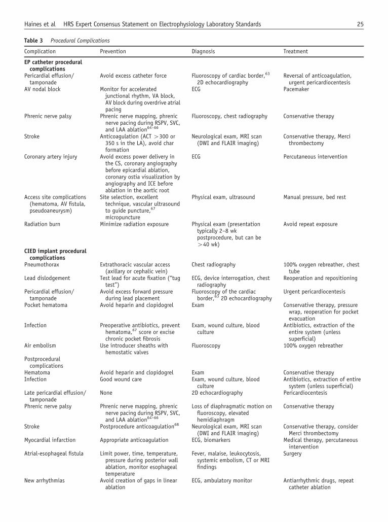
Table 3 Procedural Complications
Complication Prevention Diagnosis Treatment
EP catheter proceduralcomplications
Pericardial effusion/tamponade
Avoid excess catheter force Fluoroscopy of cardiac border,63
2D echocardiographyReversal of anticoagulation,urgent pericardiocentesis
AV nodal block Monitor for acceleratedjunctional rhythm, VA block,AV block during overdrive atrialpacing
ECG Pacemaker
Phrenic nerve palsy Phrenic nerve mapping, phrenicnerve pacing during RSPV, SVC,and LAA ablation64–66
Fluoroscopy, chest radiography Conservative therapy
Stroke Anticoagulation (ACT 4300 or350 s in the LA), avoid charformation
Neurological exam, MRI scan(DWI and FLAIR imaging)
Conservative therapy, Mercithrombectomy
Coronary artery injury Avoid excess power delivery inthe CS, coronary angiographybefore epicardial ablation,coronary ostia visualization byangiography and ICE beforeablation in the aortic root
ECG Percutaneous intervention
Access site complications(hematoma, AV fistula,pseudoaneurysm)
Site selection, excellenttechnique, vascular ultrasoundto guide puncture,67
micropuncture
Physical exam, ultrasound Manual pressure, bed rest
Radiation burn Minimize radiation exposure Physical exam (presentationtypically 2–8 wkpostprocedure, but can be440 wk)
Avoid repeat exposure
CIED implant proceduralcomplications
Pneumothorax Extrathoracic vascular access(axillary or cephalic vein)
Chest radiography 100% oxygen rebreather, chesttube
Lead dislodgement Test lead for acute fixation (“tugtest”)
ECG, device interrogation, chestradiography
Reoperation and repositioning
Pericardial effusion/tamponade
Avoid excess forward pressureduring lead placement
Fluoroscopy of the cardiacborder,63 2D echocardiography
Urgent pericardiocentesis
Pocket hematoma Avoid heparin and clopidogrel Exam Conservative therapy, pressurewrap, reoperation for pocketevacuation
Infection Preoperative antibiotics, preventhematoma,67 score or excisechronic pocket fibrosis
Exam, wound culture, bloodculture
Antibiotics, extraction of theentire system (unlesssuperficial)
Air embolism Use introducer sheaths withhemostatic valves
Fluoroscopy 100% oxygen rebreather
Postproceduralcomplications
Hematoma Avoid heparin and clopidogrel Exam Conservative therapyInfection Good wound care Exam, wound culture, blood
cultureAntibiotics, extraction of entiresystem (unless superficial)
Late pericardial effusion/tamponade
None 2D echocardiography Pericardiocentesis
Phrenic nerve palsy Phrenic nerve mapping, phrenicnerve pacing during RSPV, SVC,and LAA ablation64–66
Loss of diaphragmatic motion onfluoroscopy, elevatedhemidiaphragm
Conservative therapy
Stroke Postprocedure anticoagulation68 Neurological exam, MRI scan(DWI and FLAIR imaging)
Conservative therapy, considerMerci thrombectomy
Myocardial infarction Appropriate anticoagulation ECG, biomarkers Medical therapy, percutaneousintervention
Atrial-esophageal fistula Limit power, time, temperature,pressure during posterior wallablation, monitor esophagealtemperature
Fever, malaise, leukocytosis,systemic embolism, CT or MRIfindings
Surgery
New arrhythmias Avoid creation of gaps in linearablation
ECG, ambulatory monitor Antiarrhythmic drugs, repeatcatheter ablation
25Haines et al HRS Expert Consensus Statement on Electrophysiology Laboratory Standards

8.4.3. Sterile TechniqueThe instruments and components used in the device implantlaboratory must be opened in a clean air environment. Bydoing so, bacterial contamination of these instruments andcomponents will be kept to a minimum. All personnel in theroom must wear a cap and mask at all times, and all who arein contact with the sterile field must perform a completesurgical scrub and must be gowned and gloved.
8.4.4. Sterile Preparation of the Surgical SiteThe operative site(s) should be prepared with an antisepticagent. Although these agents eliminate the immediatebacterial count on the skin surface at the operative site, hairfollicles may prevent complete sterilization of the skin. Hairclipping close to the skin in the prep room, rather thanshaving, is recommended because bacteria on the skinsurface begin to recolonize within 30 minutes in the presenceof hair follicles despite complete sterilization.72 Somecenters instruct patients to use preprocedural home cleansingkits. Site preparation with alcohol-based solutions should beallowed to dry completely before draping. The combinationof evaporating alcohol, supplemental oxygen, and electro-cautery poses a significant fire risk in the surgical field.
8.4.5. Concomitant Groin Access During Implant ProceduresIn some cases, additional femoral venous access for theplacement of a temporary pacing catheter may be warranted(e.g., lead extraction and pacemaker-dependent patients withan inadequate escape rhythm who are undergoing generatorchange).8 Large-bore venous sheaths are useful for rapidvolume resuscitation in the event of vascular tears duringlead extraction, and arterial access facilitates beat-to-beatmonitoring of blood pressure.
8.5. Acute CIED Implant Procedural Complications(Table 4)Complications associated with acute CIED implantation areoften technique related. Attention to detail will minimize therisk of difficulties related to the implant procedure.
8.6. Postprocedural Issues8.6.1. Vascular HemostasisVenous sheaths may be removed at the end of the procedureif no anticoagulant has been administered, with pressure heldfor 10–20 minutes. If heparin has been administered, waiting
until the ACT is more than 175 seconds (o250 seconds ifthe patient is receiving therapeutic warfarin) before sheathsare removed decreases the likelihood of bleeding andhematoma formation. Reversal of heparin effect with
Table 3 (continued )
Complication Prevention Diagnosis Treatment
Radiation burn Minimize radiation exposure Physical exam (presentationtypically 2–8 wkpostprocedure, but can be440 wk)
Avoid repeat exposure
2D ¼ two-dimensional; ACT ¼ activated clotting time; AV ¼ atrioventricular; CS ¼ coronary sinus; CT ¼ computed tomography; DWI ¼ diffusion weightedimaging; ECG¼ electrocardiogram; EP¼ electrophysiology; FLAIR¼ fluid-attenuated inversion recovery; ICE¼ intracardiac echocardiography; LA¼ left atrium;LAA ¼ left atrial appendage; MRI ¼ magnetic resonance imaging; RSPV ¼ right superior pulmonary vein; SVC ¼ superior vena cava; VA ¼ ventricular atrial.
Table 4 Definitions and Terminology Used in Clinical Fluoroscopy
Roentgen (R) is the unit of radiation exposure in air. The totalcharge produced in air per unit mass by ionizing radiation iseasily measurable with survey instruments.
Gray (Gy) is the International System of Units (SI) of absorbedradiation dose of ionizing radiation, which has replaced the termradiation absorbed dose (in rad). One gray is the absorption of 1 Jof ionizing radiation by 1 kg of matter (equivalent to 100 rad),and it assesses the potential biological risk to that tissue. The U.S. unit for absorbed dose is rad. 1 Gy ¼ 100 rad.
Equivalent dose Sievert (Sv) is a measure of equivalent dose, andis also an SI unit. It takes into account the different probabilityof effects that occur when the same amount of absorbed dose isdelivered by different types of radiation (protons vs. X-rays). It isequal to the absorbed dose in Gy multiplied by the radiationweighting factor, WR, and other modifying factors. The WR is 1 forX-rays. The equivalent dose can be used to assess radiation risk ifthe person’s whole body is uniformly irradiated. For partial bodyexposure, such as cardiac electrophysiology, additionalcorrection is necessary to assess the radiation risk.
Effective dose is used to assess the risk when only a part of thebody absorbs energy from radiation. Since some organs in thebody are more sensitive to radiation effects than others, theequivalent dose is multiplied by the appropriate tissue weightingfactors. This terminology is used to assess the risk of radiation-induced cancer and hereditary effects. For example, the effectivedose for a typical chest X-ray radiograph is 0.0001 Sv (0.1 mSv).The U.S. unit for sievert (Sv) is rem. 1 Sv ¼ 100 rem.
As low as reasonably achievable (ALARA) standard is a system forlimiting the amount of radiation a person receives. Radiationexposure should be justified on the basis of the assumption thatthere is no threshold below which ionizing radiation is free fromharmful biological effects and that shielding from radiationexposure is needed, no matter how low the dose.
Kerma is an acronym for kinetic energy released in the material.Kerma is measured in Gy.
Kerma-area product (PKA) is the integral of air kerma (absorbeddose to air) across the entire X-ray beam emitted from the X-raytube. PKA is a surrogate measurement for the entire amount ofenergy delivered to the patient by the beam. PKA is measured inGy � cm2. Another term for this is the dose-area product (inGy � cm2).
Reference air kerma (Ka,r) is the kerma-area product at a specificpoint in space relative to the fluoroscopic gantry (theinterventional reference point) during a procedure. It ismeasured in Gy.
Peak skin dose is the highest dose of radiation exposure on anyportion of a patient’s skin during a procedure.
Heart Rhythm, Vol 0, No 0, Month 201426

protamine is employed in many laboratories to more rapidlyreverse heparin effect and allow almost immediate sheathremoval, although one must be prepared to treat uncommonbut sometimes severe protamine reactions.64 Vascular clo-sure devices are uncommonly used in EP,64,65 but are anappropriate choice for arterial closure. After atrial fibrillationablation, the reestablishment of therapeutic anticoagulationsoon after sheath removal is desirable to lessen periproce-dural stroke risk but there is no consensus on the optimalregimen or timing.
8.6.2. Postanesthesia RecoveryWhen mild anesthesia is used, vital signs and oxygensaturation should be monitored continuously until the patientis conscious and communicative. Access sites, cardiacrhythm, and neurological state should be assessed every 15minutes during the first hour and then periodically thereafter.Late complications, such as access site hematomas andhemodynamically significant pericardial effusion, candevelop after the patient leaves the postprocedural recoveryarea. If a patient received midazolam during the procedureand a dose of its antidote (Romazicon/flumazenil) wasadministered, the patient must be monitored for a reboundeffect of midazolam. If general anesthesia was used, patientsusually recover in a postanesthesia care unit.
8.6.3. Postprocedural ComplicationsProcedural complications that can arise after the patientleaves the laboratory area (or even after hospital discharge)are listed in Table 4. A process for tracking postproceduralcomplications should be in place as part of the laboratory’sQA process (see Section 9).
8.6.4. MedicationPostprocedure anticoagulation is recommended in patientswho are at high risk of stroke on the basis of evaluation toolssuch as CHADS2 or CHA2DS2-VASc scores. In manylaboratories, chronic warfarin therapy is not interrupted
during either device implantation or ablation procedures inpatients who are taking it for stroke prevention in the settingof atrial fibrillation or mechanical heart valves; the procedureis safe, with the international normalized ratio ranging from2.0 to 3.5.35,64
In patients with insulin-dependent diabetes, the morninginsulin dose is typically halved on the day of the procedureand glucose is periodically monitored during the procedureand the patient is treated accordingly.
8.7. Hospital DischargeThe setting for EP procedures may be outpatient, 23-hourobservation, or inpatient. The decision for discharge takesinto account procedural detail, patient age, and health status,the potential for complications (such as blood loss), and theability of the patient (or caregivers) to evaluate signs ofconcern.67 This is a medical decision and should bedetermined irrespective of reimbursement issues.
8.8. Reporting Procedural ResultsThe procedure report should include, at a minimum, thefollowing: the primary and secondary operators; the indicationfor the procedure; names and doses of any medicationsadministered; intake, output, and estimated blood loss;catheter/pacing/ICD lead model numbers, serial numbers,insertion sites, and intracardiac destinations; findings andprocedure performed; complications encountered; and fluoro-scopic exposure (minutes; mGy; dose-area product). Patientswho receive excessive radiation exposure during a procedure(typically 43000 mGy, but requirements vary by state) mustbe notified and followed up for evidence of skin damage.Ideally, this information is stored in a database for QApurposes. Recordings made during the procedure (electro-grams and fluoroscopic and mapping system images) shouldbe archived on digital media (ideally on a network, oralternatively on a CD or DVD) for future reference, ifneeded.64
9. Pediatric and Adult Congenital HeartDisease
Pediatric and Adult Congenital Heart Disease Recommendations
� Pediatric EP procedures on small and young children should be performed in centers where there is pediatric surgical backup.� Procedures on adult patients with CHD can be performed in pediatric or adult facilities by physicians who have expertise in the area of
CHD and the potential arrhythmia substrates of patients with CHD.� There are special considerations for performing pediatric EP procedures, including unusual arrhythmia mechanisms, small patient size,
and the effect on future patient growth.
9.1. Patient Factors Different From AdultsIssues that pertain to EP laboratory standards and practice forpediatric EP and patients with pediatric and adult CHD withrhythm abnormalities differ from those pertaining to adults
and are not confined to issues of patient and cardiac sizecompared with the adult patient. The decision-making processfor interventions has implications for patient quality of life anddevelopment. Success, failure, and procedural factors relatedto intervention therefore span many decades. A significant
27Haines et al HRS Expert Consensus Statement on Electrophysiology Laboratory Standards
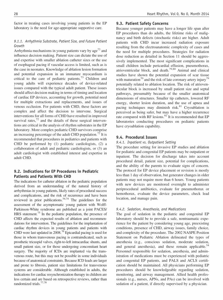
factor in treating cases involving young patients in the EPlaboratory is the need for age-appropriate supportive care.
9.1.1. Arrhythmia Substrate, Patient Size, and Future PatientGrowthArrhythmia mechanisms in young patients vary by age72 andinfluence decision making. Patient size can dictate the use ofand expertise with smaller ablation catheter sizes or the useof esophageal pacing if vascular access is limited, such as isthe case in neonates. Knowledge of ablation lesion formationand potential expansion in an immature myocardium iscritical to the care of pediatric patients.73 Children andyoung adults will experience decades of device-relatedissues compared with the typical adult patient. These issuesshould affect decision making in terms of timing and locationof cardiac EP devices, accounting for growth, potential needfor multiple extractions and replacements, and issues ofvenous occlusion. For patients with CHD, these factors arecomplex and affect the decision to intervene. Surgicalinterventions for all forms of CHD have resulted in improvedsurvival rates,72 and the details of these surgical interven-tions are critical in the analysis of rhythm substrates in the EPlaboratory. More complex pediatric CHD survivors comprisean increasing percentage of the adult CHD population.72 It isrecommended that procedures in pediatrics and patients withCHD be performed by (1) pediatric cardiologists, (2) acollaboration of adult and pediatric cardiologists, or (3) anadult cardiologist with established interest and expertise inadult CHD.
9.2. Indications for EP Procedures in PediatricPatients and Patients With CHDThe indications for catheter ablation in the pediatric populationderived from an understanding of the natural history ofarrhythmias in young patients, likely rates of procedural successand complications, and the risk of recurrence and have beenreviewed in prior publications.54,74 The guidelines for theassessment of the asymptomatic young patient with Wolff-Parkinson-White syndrome are published as a joint PACES/HRS statement.75 In the pediatric population, the presence ofCHD affects the expected results of ablation and recommen-dations for intervention. The guidelines for the implantation ofcardiac rhythm devices in young patients and patients withCHD were last updated in 2008.76 Epicardial pacing is used forthose in whom transvenous pacing is contraindicated, such asprosthetic tricuspid valves, right-to-left intracardiac shunts, andsmall patient size, or for those undergoing concomitant heartsurgery. The majority of ICDs are implanted via the trans-venous route, but this may not be possible in some individualsbecause of anatomical constraints. Because ICD leads are largerand prone to fibrosis, patient size limitations for transvenoussystems are considerable. Although established in adults, theindications for cardiac resynchronization therapy in children areless certain and are based on retrospective reviews, rather thanrandomized trials.77,78
9.3. Patient Safety ConcernsBecause younger patients may have a longer life span afterEP procedures than do adults, the lifetime risks of malig-nancy and birth defects (stochastic risks) are higher. Adultpatients with CHD incur increased radiation exposureresulting from the electroanatomic complexity of cases andthe need for multiple procedures. Strategies for radiationdose reduction as detailed in Section 11 should be aggres-sively implemented. The most significant complications insmall children include pericardial effusion, pneumothorax,atrioventricular block, and death.79,80 Animal and clinicalstudies have shown the potential expansion of scar tissuewith maturation70 and the risk of late coronary artery injury79
potentially related to ablation location. The risk of atrioven-tricular block is increased by small patient size and septalpathways, presumably because of the smaller anatomicaldimensions of structures. Smaller tip catheters, lowered RFenergy, shorter lesion duration, and the use of apnea andpacing techniques may diminish risk.81 Cryoablation isperceived as being safer, but may have a higher recurrencerate compared with RF lesions.82 It is recommended that EPlaboratories conducting procedures on pediatric patientshave cryoablation capability.
9.4. Procedural Issues9.4.1. Inpatient vs. Outpatient SettingThe procedure setting for invasive EP studies and ablationfor pediatric and congenital EP patients may be outpatient orinpatient. The decision for discharge takes into accountprocedural detail, patient size, potential for complications,and the ability of the parents to evaluate signs of concern.The protocol for EP device placement or revision is mostlyless than 1 day of observation, but generator changes in olderpatients may not require more than 6 hours. Young patientswith new devices are monitored overnight to administerperiprocedural antibiotics, evaluate for pneumothorax orhemothorax, evaluate the device parameters, check leadlocation, and manage pain.
9.4.2. Sedation, Anesthesia, and MedicationsThe goal of sedation in the pediatric and congenital EPlaboratory should be to provide a safe, nontraumatic expe-rience for the patient by considering young age, preexistingconditions, presence of CHD, airway issues, family choice,and complexity of the procedure. The 2002 NASPE PositionStatement on Pediatric Ablation delineated the types ofanesthesia (e.g., conscious sedation, moderate sedation,and general anesthesia), and these remain applicable.54
Personnel responsible for sedation, anesthesia, and admin-istration of medications must be experienced with pediatricand congenital EP patients, and PALS and ACLS certifi-cation should be maintained. All physicians performing EPprocedures should be knowledgeable regarding sedation,monitoring, and airway management. Allied health profes-sionals (e.g., nurses, APNs, and PAs) can be involved withsedation of a patient, if directly supervised by a physician.
Heart Rhythm, Vol 0, No 0, Month 201428
9.4.3. FacilitiesThe room and equipment standards for pediatric EP proce-dures are similar to those for adult EP procedures (seeSections 4 and 5) but must have a cardiac defibrillatorspecifically for use with children, a code cart meetingpediatric needs, and age-appropriate anesthesia equipment.Pediatric and congenital EP patients may require a combinedprocedure of EP and hemodynamic catheterization, includ-ing angiography and possible intervention. Thus, it isdesirable that a pediatric/CHD EP laboratory meet the samestandards as a pediatric catheterization laboratory.
9.5. Lab StaffingGeneral recommendations for EP laboratory staffing aredetailed in Section 6 and Table 2. Pediatric and congenitalEP guidelines recommend that the facility and laboratory staffshould be appropriate for the patient population and pediatricand CHD interventional and surgical experts be urgentlyavailable during laboratory procedures.42 All members of theteam should be trained in PALS (when treating a child/infant)and ACLS (if treating an adult).
9.5.1. PhysiciansTraining and board certification pathways employed byphysicians who perform EP procedures on pediatric andcongenital EP patients include the pediatrics and internalmedicine pathways (see Sections 6 and 7).
9.5.2. EP Laboratory PersonnelRecommended staffing for pediatric cases is detailed inTable 3. While laboratory staff roles are similar to thoseoutlined in Section 6 for pediatric cases, these staff shouldhave expertise in performing cardiac procedures in pediatricand congenital EP patients.
9.6. Emergency Supportive Care and Surgical/Intensive Care Unit BackupPediatric EP procedures should be performed (earlier than theteenage years) in centers where there is pediatric cardiovas-cular surgical availability. For procedures performed onpatients in their teenage years or procedures on adult patientswith CHD that are performed in adult or mixed pediatric andadult institutions, a CV surgical plan should be formalized toaccommodate on-site emergencies (notably, cardiac perfora-tion) and a rapid transfer made to a pediatric cardiovascularcenter should the anatomy of the patient require this expertise.
9.7. Postprocedural CareThe postprocedural care can be performed in a separateperiprocedural area, the general cardiology floor, or theintensive care unit. Continuous telemetry should be availablefor the evaluation of heart rhythm. The environment forpostprocedural care should be appropriate for patient age anddevelopment. The nursing and physician staff should beexperienced in the care of pediatric and congenital EP patients.
10. Quality
Quality Recommendations
� A process for tracking postprocedural complications should be in place as part of the laboratory’s QA process.� An essential component of a successful EP laboratory is to have an internal QA/quality improvement (QI) process in place, in addition to
public reporting requirements.� Components of the QA/QI process should include national requirements for tracking (e.g., device implants), minimum acceptable
complication rates (e.g., infections), and compliance with national registries, including the NCDR ICD Registry.� It is the responsibility of each institution to ensure that staff credentialing, maintenance of certification, and necessary continuing
medical education requirements are met.� Procedure outcomes, including success rates and complications, should be documented and recorded. Data acquired from the EP laboratory
QA process should be used to benchmark the complication rates and outcomes of both individual practitioners and the overall EP laboratory.� Physicians should participate in regularly scheduled QI and/or peer review meetings to maintain privileges and evaluate procedural
appropriateness.� A quarterly EP laboratory morbidity and mortality conference should be mandatory, with attendance documented.� A QA process for the equipment should be established that provides a mechanism to demonstrate optimal function and operation of the
equipment and that offers staff training in equipment maintenance, setup, and operation.� Given the often poorly defined relationship between case volumes and outcomes, a more appropriate measure is to ensure that all major
complications are reviewed by the QA committee and handled as described in the previous section.
10.1. The QA/QI ProcessHigh-quality, consistent care delivery in a busy cardiac EPlaboratory requires standard protocols for procedures, com-munication channels, and documentation. An essentialcomponent of a successful EP laboratory is to have an
internal QA/QI process in place, in addition to publicreporting requirements. This requires a commitment fromfacility administrators to provide adequate staffing, includinga committee chair, staff coordinator, and funding forcollecting and managing data. The goal of the programshould be to motivate and encourage physicians and staff to
29Haines et al HRS Expert Consensus Statement on Electrophysiology Laboratory Standards

participate and to take initiative in the QA/QI process andoverall success of the laboratory. It is acknowledged that thereis little published data on the QA/QI process in the EPlaboratory and that expert consensus is the primary basis forour recommendations. Further research and development ofquality metrics specific to the practice of cardiac EP is ongoing.
To begin the QA/QI process, its components should beidentified. These components include national requirementsfor tracking (e.g., device implants), minimum acceptablecomplication rates (e.g., infections), and compliance withnational registries, including the NCDR ICD Registry. AQA/QI program must ensure that key data are collectedprospectively and systematically. When the QA/QI team isidentifying potential quality metrics, either strong scientificevidence or expert consensus must support the metric. Themetric must measure areas important to patient care, and it isbest if it covers an aspect of practice where there is a gap inpatient care. The data must be available in a usable format forfuture analysis. These data allow the laboratory to bench-mark its performance and provides a reference by whichappropriate changes can be made.
Provider qualifications are typically well established bynational guidelines and certification bodies. It is the respon-sibility of each institution to ensure staff credentialing,maintenance of certification, and necessary CME require-ments are met (see Section 7). Practitioners should beexpected to adhere to published practice guidelines unlessthe reason for deviation is documented. Clinical situationsnot directly addressed by the guidelines inevitably arise andrequire judgment and skill to address. Periodic peer review ofcases that fall outside the guidelines is recommended toensure appropriate delivery of care. The guidelines evolveand, by definition, require a consensus of data before theycan be written and revised. The writing group recognizes andencourages the development of new clinical pathways andtools. This development is best achieved through researchprotocols that adequately capture patient demographic char-acteristics, procedure characteristics, and outcomes. Theresearch consent process ensures that patients are aware thatthe treatment being offered is beyond the current recom-mendations of the practice guidelines.
Procedure outcomes, including success rates and compli-cations (and ideally including 30-day outcomes), should bedocumented and recorded. The writing group recognizes thatsuccess for some procedures requires clinical follow-up(particularly atrial fibrillation and VT ablation procedures).In these instances, an acute end point for the procedure shouldbe specified (e.g., pulmonary vein isolation, noninducibility forVT, or creation of a planned RF ablation lesion set based on theresults of an endocardial voltage scar map) and documentationshould indicate if the end point was achieved. An assessmentof freedom from arrhythmia recurrence after 1 year of follow-up should be performed. Risk-adjusted models are not welldeveloped by which the relative frequency of complicationsand successful outcomes among different patient populationscan be interpreted and physician results compared. Therefore,the interpretation of success rates and complication rates
requires judgment by peers. Physicians should participate inregularly scheduled QI and/or peer review meetings tomaintain privileges and evaluate procedural appropriateness.A quarterly EP laboratory morbidity and mortality conference(stand-alone or as a component of another conference) shouldbe mandatory, with attendance documented.
The modern cardiac EP laboratory depends on manycomplex hemodynamic and physiological recording systems,advanced imaging systems, advanced mapping systems, andmultiple ablation systems. Rigorous processes must be estab-lished to ensure that (1) a QA process for equipment isestablished; (2) equipment is tested and demonstrated to befunctioning appropriately, both on a routine basis and imme-diately before a case in which the specific item will be used;verification of equipment function should be included in thetime-out procedure; (3) EP laboratory staff are appropriatelytrained in the maintenance, setup, and operation of the equip-ment; and/or (4) representatives from the vendor are available,qualified, and cleared by administration and occupational healthto participate in the operation of the equipment before, during,and after the procedures. Competencies in clinical skills, ACLS,PALS (when appropriate), sterile technique, radiation safety,and fire safety should be assessed on a regular basis.
The EP laboratory requires processes be in place to ensureproper communication within the EP laboratory and with otherhospital services. Within the laboratory, protocols for emer-gency situations (such as tamponade or ventricular fibrillationrefractory to defibrillation) can make the difference between anorganized, streamlined, successful resuscitation effort and achaotic effort during which leadership is absent, teammembersduplicate some activities and neglect others, and ultimately atragic but potentially avoidable outcome occurs. Beyond theboundaries of the EP laboratory, communication between theEP team and other health care professionals is essential,particularly during handoffs from one care team to anotherand at the time of patient discharge. It is the role of the QA/QIcommittee to oversee that excellent communication processesare developed, maintained, and adhered to by the staff.
10.2. Clinical Outcomes and ComplicationsData acquired from the EP laboratory QA process should beused to benchmark the complication rates and outcomes ofboth individual practitioners and the overall EP laboratory.For practitioners with complication rates above the bench-mark, an objective unbiased peer review of the relevant casesis critical to determining whether a deviation in the standardof care occurred. Because event rates are low and risk-adjusted models are not well developed for the EP laboratory,peer review is particularly important. Practitioners should notbe penalized for accepting higher risk and/or more challeng-ing cases. However, if reckless behavior or inadequate skillsor knowledge is deemed to be present and a deviation in careoccurs, verbal and written communication by the chair of QAis imperative. This communication should include a clearplan for corrective action and documentation of potentialfuture actions if corrective action is not successful.
Heart Rhythm, Vol 0, No 0, Month 201430

10.3. Case VolumesA link between operator case volume, skills, and outcomeshas been documented in some but not all areas of cardiac EP,yet controversy and conflicting data remain. Specific casevolumes for training are outlined by the ABIM29 (Table 3),and for clinical competency they are available in the HRSclinical competency statements.8,17,33,38,39,42 Given the oftenpoorly defined relationship between case volumes and out-comes, a more appropriate measure is to ensure that all majorcomplications (see Table 3) are reviewed by the QAcommittee and handled as described in the previous section.
10.4. DatabaseA prospective plan to acquire data in an accessible andfunctional database is an essential building block for anyQA/QI process. Without objective, reliable data to measureoutcomes, no meaningful effort at QI can be undertaken.Minimum data that should be recorded in a searchableaggregate form include patient demographic characteristics,relevant history of present illness, medications, CIEDproduct information data, and data on outcome and compli-cations from any invasive procedures. Patients with CIEDshould be identifiable by the device that has been implantedto permit rapid identification of patients who may havereceived defective hardware in the event of a recall or othernotification from the manufacturer or FDA.
The field of medical informatics is on the verge ofexponential growth. Registries such as the ACC/NCDRICD Registry have identified specific data fields that shouldbe captured for participation in the registry and compliancewith payer mandates.83 Beyond these basic requirements,recommendations for more detailed database fields areincluded in clinical guidance documents for the managementof patients with atrial fibrillation and for the management ofpatients with VT.38,39 These standard data elements providean opportunity for EP laboratories invested in research toaggregate and/or compare their data with that of otherlaboratories working in the same field.
10.5. Pediatric and Adult Congenital Heart DiseaseTo date, QA efforts in pediatric and congenital EP have centeredon the creation of large EP procedural registries.84,85 In late 2010,a PACES taskforce began to develop and implement a self-sustaining multicenter QI registry known as MAP-IT. Presently,the MAP-IT taskforce is creating a registry of patient-centeredlate outcome measures of catheter ablation procedures. For thefirst time, an empirical and data-derivedmethod of risk/complex-ity adjustment for pediatric and CHD EP procedures, known asthe COMPASS score, has been developed. The future of QAefforts for procedure-based subspecialties will require thebenchmarking and reporting of risk-adjusted “patient-centered”outcome measures. Widespread implementation of the MAP-ITinitiative within PACES should satisfy this need.
11. Occupational Health Concerns
Occupational Health Concern Recommendations
� Fluoroscopy equipment should report three parameters: fluoroscopy time; radiation dose (air kerma, in Gy), a measure of deterministicinjury potential; and the dose-area product (in cGy � cm2), a measure of stochastic injury potential. A minimum of 0.5-mm lead-equivalent protective apron, thyroid shield, and eye protection should be used by EP laboratory personnel.
� All lead should be tested at 6-month intervals to check for cracks or leaks.� Risks of acquisition of infectious diseases by health care workers can be minimized by adherence to current infection control guidelines.
11.1. Radiation SafetyThe field of cardiac EP is greatly dependent on fluoroscopicimaging for the placement of catheters and device leads intothe heart. This results in significant radiation exposure to thepatient, the operator, and the laboratory staff. While thisexposure cannot be eliminated in most cases, attention tofluoroscopic technique can minimize radiation dose. Compe-tencies in radiation safety should be completed yearly by theEP laboratory staff. Some states mandate fluoroscopy licensesbe obtained by all personnel using fluoroscopy and renewedperiodically, including physicians and radiation technologists.
11.1.1. Terms for Understanding Radiation Exposure in theCardiac EP LaboratoryNonionizing radiation, such as microwave or infraredradiation, can cause heating but not molecular damage tocells. Ionizing radiation, such as β, γ, and X radiation, strips
electrons from atoms and causes molecular injury to DNA.Ionizing radiation has great potential for damage to tissue,including burns and malignancy. Understanding the basicdefinitions and terminology used to describe ionizing radi-ation is helpful when trying to understand the potentialeffects on the human body (see Table 4).86
11.1.2. Biological Risks From Radiation ExposureRadiation effects are described by their deterministic andstochastic effects.87 Deterministic effects are harmful tissuereactions that are determined by an absorbed threshold dose.Radiation-induced skin burns are an example of determin-istic effects.88 Stochastic effects include malignancy andheritable effects and are not determined directly by the dose,but a higher dose increases the probability of an adverseoutcome. Human tissue radiosensitivity varies directly with
31Haines et al HRS Expert Consensus Statement on Electrophysiology Laboratory Standards

the rate of cellular proliferation and number of futuredivisions and indirectly with the degree of morphologicaland functional differentiation. The most sensitive tissuesinclude the bone marrow, spermatocytes, and intestinal cryptcells.87 Local skin injury is the most commonly encountereddeterministic effect in cardiovascular medicine, with changesnoted at doses above 2 Gy. Findings often do not appear untilweeks after exposure.
Radiation exposure increases lifetime risk of fatal malig-nancy. The as low as reasonably achievable standard wasderived from the Biological Effects of Ionizing Radiation VIIReport.89 This report makes the assumption that cancer riskincreases proportionally with radiation exposure and that thereis no radiation dose that is without risk. All personnel workingin the laboratory must be aware of the as low as reasonablyachievable standard. A skin threshold dose of 2 Sv should notbe exceeded. Because the prevalence of fatal malignanciescontinues to increase over time after radiation exposure,children and young adults are more susceptible to thesecomplications in their lifetimes. Adult patients with CHD incurincreased radiation exposure resulting from the electroanatomiccomplexity of cases and the need for multiple procedures.
11.1.3. Measuring Radiation ExposureA normalized X-ray dose at a specific kilovolt peak 1 m fromthe source, total fluoroscopic time, backscatter correctionfactor, and source-to-skin distance can be used to estimateradiation exposure.90 A direct measurement of radiation dosesat multiple sites can be performed with lithium fluoridethermoluminescent dosimeter sensors and optically stimulatedluminescence. The highest exposure to patients has beenobserved with a median skin entrance dose of 7.26 rem (range0.31-135.7 rem) at the ninth vertebral body. This dose ispredicted to be associated with a lifetime excess risk ofmalignancy to the female breast, active bone marrow, andlung of greater than 700 cases per million undergoing routinecatheter ablation. Operator exposure was highest at the lefthand, waist, and left maxilla.91 Fluoroscopy equipment shouldreport three parameters: fluoroscopy time; radiation dose (airkerma, in Gy), a measure of deterministic injury potential; andthe dose-area product (in cGy � cm2), a measure of stochasticinjury potential. Radiation exposure is a superior metric tofluoroscopy time; reliance on fluoroscopy time is discouraged.The United States Nuclear Regulatory Commission annualdose limits for radiation are 0.50 Sv for skin, arms, and legs;0.15 Sv for eyes; and 0.05 Sv for the whole body. Thefluoroscopy dose for each case should be recorded in themedical record and accessible to patients.
11.1.4. Minimizing Radiation Exposure to PatientsThe predominant strategy for minimizing radiation exposureto patients is to minimize the radiation dose.86,92 The mosteffective approach is to minimize fluoroscopy pedal time.Operators should develop the habit of tapping the fluoro-scopy pedal rather than standing on the pedal for a longperiod of time. If the eyes stray from the fluoroscopy screen,the foot should come off the pedal immediately. Decreasing
the fluoroscopy pulse rate will significantly reduce dose atthe cost of temporal resolution of the image. Supplementa-tion of fluoroscopic imaging with nonfluoroscopic electro-anatomic guidance systems by using stored fluoroscopyloops rather than cine loops and using pulsed fluoroscopywill reduce the total procedural dose. As the X-ray tube getscloser to the patient, X-rays at the skin entry point increaseand the risk of deterministic injury also increases; thus, theoperator should position the table at a comfortable heightwith some distance between the tube and the patient. If theimage intensifier is not positioned as close to the patient aspossible, the image will be magnified but the radiation dosewill be much higher. To magnify the image, use theappropriate magnification mode. Both geometric and elec-tronic magnification increases dose to the patient. Doses maybe limited effectively by collimation, limiting the field ofview with shutters as much as possible. For example,reducing the diameter of the field of view by 29% willreduce the radiation dose by half. Steeply angulated projec-tions should be avoided, and if used, the C-arm should berepositioned somewhat throughout the procedure to avoiddelivering radiation to the identical skin entry site. Depend-ing on which procedure is being performed, local shieldingof the patient’s thyroid and gonads can be employed.93
Since the stochastic effects of radiation exposure arecumulative, the caregiver should be sensitive to the lifetimeexposure of the patient to ionizing radiation. There has beenactive discussion among regulators on implementing a systemfor lifetime tracking of radiation exposure to patients, but thetools for this type of system are not available, and therefore itis not presently mandated.94 It is important to emphasize thatradiation exposure is dependent on the age and condition ofthe equipment. Thus, aggressive limitation of fluoroscopypedal time is desirable, but those efforts may be futile if thedose rate is high because of a high frame rate or employmentof an old imaging train. It is now an accepted standard thatfluoroscopy dose, not only fluoroscopy time, is entered in thepermanent medical record for each fluoroscopic procedure.Ideally, the peak skin dose, the reference air kerma, the kerma-area product, and the fluoroscopy time should be recorded forevery case.95 A review of fluoroscopy use by individualoperators should be part of every laboratory’s QA process.
11.1.5. Minimizing Occupational Radiation ExposureThe primary approaches used to reduce radiation exposure tothe operator and laboratory staff are increasing distance fromthe source, scatter reduction, and dose limitation. Radiationdissipates in proportion to the square of the distance from thesource, and so even a modest effort to move away from thetube will significantly reduce exposure. Radiation scatteroccurs as radiation from the generator tube enters the patientand is partially reflected or refracted by body tissues. Scatterfrom the patient is the main source of radiation exposure tothe patient outside the imaging field and to the operator. Theoperator and laboratory personnel must be protected fromexposure to the scatter radiation with shielding. A minimum
Heart Rhythm, Vol 0, No 0, Month 201432

of 0.5-mm lead-equivalent protective apron, thyroid shield,and eye protection should be used.
Proper table shielding can dramatically reduce the scatterradiation escaping into the environment. Scattered radiationexits the body at all angles but is greatest on the same side of thepatient as the X-ray source because only 1%–5% of radiationcompletely penetrates the patient’s body and exits on the otherside.91 Therefore, proper undertable shielding is paramount.Shield extensions above the table rail and a contoured ceiling-mounted shield in contact with the patient’s torso will substan-tially reduce operator exposure. Since radiation doses decreasewith the square of the distance from the source, the operatorshould perform the procedure as far from the radiation tube as ispractical. Barium-impregnated drapes can further reduce radi-ation scatter in the procedure field.96 All recommendationsdescribed above for limiting total fluoroscopy dose will alsoreduce the operator and laboratory staff radiation exposure.
Radiation exposure to a pregnant EP laboratory worker isa special situation and should be resolved on a case-by-casebasis. It is recommended that radiation exposure to thepregnant staff member, as measured by a waist dosimeter(under the lead apron), should not exceed 0.05 rem/mo, or0.5 rem for the entire pregnancy.35 Additional layers of leadcan be worn over the abdomen to further protect the fetus.
11.1.6. Quality ManagementThe FDA regulates fluoroscopy equipment manufacturing andhas dose limits for systems with automatic exposure control.97
States regulate the safe use and operation of radiation-producing machines, such as fluoroscopic imaging systems.TJC’s sentinel event is an unexpected occurrence involvingdeath or serious injury. Prolonged fluoroscopy that exceedsdoses of 1500 rad (15 Gy) to the skin is a reportable sentinelevent and requires an institutional response. A qualifiedmedical physicist should perform initial acceptance testingand annual testing to ensure optimal image quality andradiation dose. Unfortunately, assuming that fluoroscopicequipment is functioning properly, the most important factorin radiation exposure to patients and staff is operator knowl-edge and behavior. TJC recommended (but did not mandate)credentialing standards for fluoroscopists,98 and the recom-mended curriculum was endorsed by HRS, American Collegeof Cardiology Foundation, AHA, and the Society for Car-diovascular Angiography and Interventions (SCAI).86 Thetraining of EP physicians and staff in radiation physics,radiation biology, and technological developments in X-rayimaging systems and X-ray dose management is highlyvariable, and physician credentialing and recredentialing hasno requirement regarding knowledge of radiation safety. It istherefore incumbent on the hospital leaders to establish highlocal standards and to track fluoroscopy use and behavior.
11.2. Occupational Health Risks of Wearing Lead11.2.1. Lead ApronsLead-equivalent aprons required for radiation protection ofstaff are heavy and present a substantial physical burden to
the interventional cardiologist.99 Historically, lead apronswere made using 0.5-mm lead-equivalent materials, withweight per unit area of these garments being 7 kg/m2.98 Anincreased risk of cervical spondylosis is a known conse-quence for cardiologists who wear protective garments whilestanding for long hours performing procedures.87 Orthopedicproblems and user fatigue associated with the continued useof heavy aprons contributed to the development of lower-weight lead-equivalent materials that are commonly used forprotective garments today. Modern nonlead 0.5-mm lead-equivalent protective garments have a weight reduction of30% or more than do traditional lead aprons.98 That modernaprons are lighter might be expected to lessen, but noteliminate, orthopedic discomfort and injury. It is important toemphasize that all lead should be tested at 6-month intervalsto check for cracks or leaks.
Along with weight, there are several other physical andergonomic aspects of radiation protection garments thatshould be considered. To minimize discomfort, garmentsmust fit correctly and should be tightened around themidsection to shift weight from the shoulders to the hips asmuch as possible. There are several garment designs that arecustomized for various uses. Many interventional cardiolo-gists choose a two-piece garment consisting of a skirt andvest (including internal frame), both of which should besnugly tightened around the midsection. Such garments arefrequently designed to “wraparound” the wearer and typi-cally provide the fully specified lead-equivalent protectionfrom radiation only in the front where the garment overlaps.A thyroid shield should always be used,100 and leadedglasses should be worn to minimize the risk of developingcataracts.101
11.2.2. Alternatives to Wearing LeadTableside alternatives to lead-equivalent garments include afloor-mounted radiation protection cabin and a ceiling- organtry-mounted suspended radiation protection sys-tem.99,102,103 Both these systems are designed to removethe weight burden of radiation protection while allowingtableside access in a sterile working environment. Becausethe weight of the shielding is not borne by the operator,thicker, heavier materials can be used. This results in a16–78-fold decrease in radiation exposure to the operator.103
The disadvantages of these systems are that they restrict themotion of the operator to some degree, and they increase theequipment that is present in an already crowded procedurelaboratory environment.
Robotic manipulation of ablation catheters can be accom-plished using external magnetic fields to guide magnetictipped catheters or robotic armatures that actuate sheath andcatheter movement remotely. These systems allow electro-physiologists to perform most of the procedure behind fixedradiation barriers, thereby eliminating exposure to scatteredradiation for the operator (but not the patient or in-laboratorystaff) and eliminating the need to wear protective lead.26,104,105
Although there is a learning curve associated with these
33Haines et al HRS Expert Consensus Statement on Electrophysiology Laboratory Standards

technologies, they appear to yield results equivalent toconventional manual catheter procedures. Recent develop-ments in electroanatomic mapping and ICE have been used toperform catheter ablation of atrial fibrillation22 with minimaluse of X-ray imaging. Future developments of these and otherimaging technologies that do not use ionizing radiation mayobviate the need for radiation protection garments in the EPlaboratory of the future.
11.3. Laboratory ErgonomicsThe physical stresses associated with working in the EP/interventional laboratory have been identified as a highprevalence of orthopedic problems, particularly those relatedto the spine, hips, knees, and ankles.102,106 The primarycontributor to orthopedic problems is wearing personalradiation protection apparel. Other factors may be thoserelated to ergonomic design, increasing complexity andduration of interventional procedures, falls, and lengthycareers. A Multi-Specialty Occupational Health Group hasbeen formed to evaluate risks and hazards and advocate forefforts to reduce these hazards.107 Table 5 provides a list ofmeasures to be considered in laboratory design and proce-dural processes to reduce the incidence of ergonomic stresseson the EP laboratory physicians and staff.
11.4. Operator Safety During Cardiac EP in PatientsWith Communicable DiseasesPreventing the transmission of infectious agents to ensure thesafety of the operator and other staff in the EP laboratory asmuch as possible involves, first and foremost, adherence tostandardized uniformly applied universal precautions inevery aspect of patient care. Risks of acquisition of infectiousdiseases by health care workers can be minimized byadherence to current infection control guidelines.108
11.4.1. Individual Personal PrecautionsInstitutions have policies on required annual or biannual staffinoculations. These may include inoculations such as hepatitisB, influenza, pertussis, and rubeola (measles). Such inoculationscan provide immunity from certain highly transmissible dis-eases, enabling staff to care for these patients without inordinaterisk. For example, health care workers not immune to chickenpox should not be required to care for patients with chickenpox.109 Because of the specialization of staff in the EPlaboratory, it can be difficult to provide adequate staffing ifmultiple concessions have to be made for noncompliant staff.The interval for regular testing of staff for tuberculosis is definedby every institution. In special situations, staff may be fitted forN95 filter masks, which are particulate respirators that are usedto prevent inhalation of small infectious airborne particlestransmitted by patients with active tuberculosis. Institutionalpolicies should be followed closely in these situations.
11.4.2. Standard PrecautionsThe major features of universal precautions and bodysubstance isolation are incorporated in standard precautions
and are used universally to prevent transmission of highlycontagious or virulent infectious agents that may be spreadby air and/or contact. The basic principle is that all blood,body fluids, secretions, excretions (except sweat), nonintactskin, and mucous membranes may contain transmissibleinfectious agents. Since it is impossible to identify all sourcesof all infectious agents, the same precautions are applied toevery patient at every encounter. Each institution has adetailed description of how standard precautions are imple-mented. The following text highlights certain aspects partic-ularly pertinent to EP laboratory processes.
Hand hygiene is the single most important factor incontrolling and preventing the transmission of germs. Addi-tional protection is offered by the appropriate use of medicalgloves; however, the unnecessary and inappropriate use ofgloves results in a waste of resources and may increase therisk of germ transmission.110 Hands are washed each timeafter gloves are removed. Hand-washing supplies should bereadily accessible in the EP laboratory and environments.Sterile gloves are indicated for any surgical procedure,invasive radiologic procedure, and procedures involvingvascular access (central lines). Examination gloves areindicated in clinical situations where there is potential fortouching blood, body fluids, secretions, excretions, and itemsvisibly soiled by body fluids. Splash protection with masksand eye shields is recommended when working with arterialand venous catheterization. Gloves are not indicated (exceptfor contact precautions) for tasks involving direct patientexposure, including taking blood pressure, pulse, andtemperature; administering intramuscular and subcutaneousinjections; bathing and dressing the patient; transporting thepatient; and any vascular line manipulation in the absence ofblood leakage, or with indirect patient exposure, includingusing the phone, writing in the chart, removing and replacinglinen for the patient’s bed, and placing noninvasive ventila-tion equipment and oxygen cannula.110
Safe injection practices, including the use of needlelessinjection systems and proper disposal of any sharps and otherequipment used for invasive procedures, are paramount.Single-dose vials are preferable to multiple-dose vials,particularly if the vial will be used for multiple patients.
Transmission-based precautions are as described in eachinstitution’s infection control policies. Contact precautionsrequire the use of gowns and gloves when there is anycontact with the patient or the surrounding area. Dropletprecautions are invoked when infectious respiratory dropletscan travel directly from the patient’s respiratory tract to therecipient, generally over short distances. This necessitatesfacial protection, such as surgical mask, eye protection, and/or face shield. Personal eyeglasses and contact lenses are notconsidered adequate eye protection.110 Historically, the areaof defined risk has been a distance of 3 ft or more around thepatient, although depending on the size of the droplets(smallpox and SARS are examples of small droplets), theinfective area may be up to 6 ft from the patient. Droplets donot remain infectious over long periods of time and thereforedo not require special air handling and ventilation. Airborne
Heart Rhythm, Vol 0, No 0, Month 201434
infection isolation precautions prevent transmission ofinfectious agents that remain infectious over long distanceswhen suspended in the air (e.g., rubeola virus [measles],varicella virus [chicken pox], Mycobacterium tuberculosis,and possibly SARS coronavirus). If at all possible, EPprocedures should be deferred in these patients unless anegative airflow procedure room is available. If an EPprocedure is unavoidable, a regular positive pressure proce-dure room with airflow similar to an OR and a portable high-efficiency particulate air filtration unit may be used.110,111
The patient should wear a surgical mask, and the staff shouldwear N95 filter masks. The procedure should be the last caseof the day, and the room left empty until adequate airexchange has taken place to clear the room.
11.4.3. Laboratory ProcessesProtocols and regulations as established by the institution forthe disposal or processing of infectious material, drapes, andfluids should be followed. Care should be taken to avoidshaking drapes or linens, which can aerosolize contaminants.All sharps that are to be reprocessed should be placed inpuncture-resistant containers, leak proof on all sides andbottom, and labeled with a biohazard label. Blood, suctionmaterial, or other contaminated liquids can be converted to asolid by the addition of a gel-forming substance to avoid riskof fluid leaking out of containers. “Clean” and “dirty” areasof the laboratory should be maintained during procedures.Personnel touching a contaminated patient/wound/areashould try to avoid touching surrounding objects beforeremoving gloves and washing hands. After procedures, alltouched surfaces need to be cleaned with a hospital-approveddisinfectant, leaving the surfaces wet for the amount of timeas directed on the bottle. This cleaning includes monitoringcables, intravenous pumps, transfer equipment, and allnondisposable objects within 3–6 ft of a patient on dropletprecautions and within 6 ft if the patient has been coughing.
11.4.4. Catheter ReprocessingCatheter reprocessing is being adopted by many EP labo-ratories nationwide, which is aimed at reducing the cost ofexpensive single-use device and to lessen the environmentalimpact from discarded supplies. With the passage andenactment of the Medical Device User Fee and Moderniza-tion Act of 2002, the reprocessing of single-use devices isnow supported in federal law. Physicians and the entire EPteam must come to an agreement on whether reprocessing isgoing to positively impact their clinical operation and patientcare from the financial, legal, and/or ethical point of view.Ablation catheters are classified by the FDA as class IIIdevices; therefore, they cannot be reprocessed at this time.There are a few third-party FDA-approved reprocessors usedby many EP and catheterization laboratories. In choosing avendor for reprocessing, each team should consider not onlythe price of the reprocessed catheters but also the particularvendor’s turnaround time, catheter collection logistics, andreprocessing success rate.
11.4.5. TransportationWhen a patient must be transported to other units ordepartments, the referring area should communicate thepatient’s isolation needs to the receiving area before trans-porting the patient. All precautions should be followedthroughout the transport process. Gloves and other appro-priate barriers should be worn by the patient and/or healthcare worker during all transfers. Protective equipment is thento be removed, the hands washed, and the patient is to betransported to or from the laboratory. Patients on dropletprecautions must have their mouths and/or tracheostomycovered with a surgical mask. Transporting a patient onairborne precautions should be discouraged, but if absolutelynecessary, consult with the infection control officer as tospecific transportation methods.
12. Ethical Concerns
Laboratory Environment Recommendations
� Obtaining an informed consent is necessary for all procedures.
12.1. Informed ConsentObtaining an informed consent is a process wherebycommunication between a patient and a physician resultsin the patient authorizing the medical procedure. This is anintegral procedure that safeguards patient autonomy andprovides the patient with information about the procedurethat allows them to understand and agree to the procedurebeing recommended. This process is not only a legalrequirement but also an ethical obligation.
To ensure appropriate communication, sufficient timeshould be allocated to this process. A physician, APN, or PAthat is part of the EP team should obtain the informed
consent, and it should be made clear if trainees, PAs, orAPNs will be operators. The process must take place beforepremedication with sedation so that the patient is able tounderstand the discussion and communicate a decision. Thediscussion should take place out of the procedure room andbe in a language that the patient adequately understands,with a qualified interpreter when needed. Common risks,even if not considered serious, should be discussed, asshould serious risks that are potentially life-threatening,even if exceedingly rare. All aspects of the procedure thatcan be reasonably anticipated should be included in thediscussion.
35Haines et al HRS Expert Consensus Statement on Electrophysiology Laboratory Standards

Informed consent in the majority of pediatric cases will begranted by the parent or guardian acting on the patient’sbehalf. Adult patients with CHD are similar to other adultsexcept in cases of cognitive handicaps, as in some geneticsyndromes. A minor should be invited to participate in theprocess, but the consent should be obtained from a legalguardian. Consent/assent forms for young patients older than12 years are frequently used. Serum or urine pregnancytesting is generally applied for females older than 12 yearswithin 2 weeks before the procedure.
The physician should be aware that they can have asubstantial influence on their patient’s decision. As an expert,the patient looks to the physician for guidance. It is an ethicalobligation for the physician to present a rational argument forundergoing the procedure. In contrast, any form of coercionwith the use of verbal or nonverbal threat or manipulationthrough the incomplete or nontruthful presentation of infor-mation is always unethical. Patients should be given ampleopportunity to discuss their concerns about the procedure andto have their questions answered satisfactorily. If the patientis incapacitated, every effort should be made to seek out andobtain consent from an appropriate surrogate.
12.2. Ethics of Teaching in the EP Laboratory SettingTeaching invasive procedural skills is a necessary part ofmaintaining the availability of widespread medical expertiseto patients. The use of simulators can be a helpful adjunct toteaching invasive procedures but cannot replace the experi-ence gained from actual patient procedures.112 Althoughteaching is the charge of institutions with postgraduatetraining programs and affiliations with medical schools, theteaching of invasive procedures can occur in any type ofinstitution.
It is the ethical duty of physicians to communicate topatients when and in what way trainees will be involved intheir procedures. When asked before the procedure, themajority of patients consent to allowing trainees to practicetheir skills.113 Most teaching institutions have patients sign aform during the admission process that acknowledges theirimplicit consent to allow trainees to perform proceduresunder the supervision of senior physicians. Although thismay be sufficient communication to patients for minorprocedures such as venipuncture or acquisition of a surfaceECG, in more invasive procedures it is mandatory that aseparate discussion takes places during the informed con-sent process that clearly states the participation and the roleof the trainee. Allowing a trainee to practice on a patientunder general anesthesia without the patient’s explicitconsent does not respect the patient’s autonomy andcompromises the trainee’s moral integrity. Trainees shouldbe invited to participate in the procedure only as an integralpart of their training, not for mere curiosity. It is theresponsibility of the supervising physician to allow thetrainee to participate in the capacity that they are capable.The trainee should not be left performing the procedureunsupervised.
12.3. Clinical Research Studies During ClinicalProceduresWithout clinical research, promising potentially lifesavingtechnologies and therapies cannot be safely made availablefor patients. Our understanding of disease processes pro-gresses because of ongoing research. Clinical research,however, exposes patients to some risk without proven directbenefit. As a researcher, the operator’s primary responsibilityis generating knowledge, whereas as a physician, theoperator’s primary responsibility is the well-being of thepatient. When there is a conflict between these two roles, therole of the physician must override the role of the researcher.
Patients should be considered for a research protocol ifthey are looking for improved outcomes compared with thoseof current therapies. Recruitment of patients should be carriedout in a manner that does not coerce participation. Similarly,the opportunity for participation in available investigationprotocols should be presented to all patients fulfillinginclusion and exclusion criteria even if the attending physi-cian does not believe that the patient is an ideal candidate. Anecessary amount of time should be given to the informedconsent process in sufficient advance of the procedure so thatthe patient is not unduly pressured into making a decision.When children or adult patients unable to provide aninformed consent are being considered for a research proto-col, it may be helpful to have an independent advocate ensurethat parents or legally authorized representatives are making arational decision exposing the subject to risk.
It is not enough to obtain a patient’s consent to participate ina study. It is the ethical duty of an investigator to ensure that theresearch protocol does not place the participants at unreason-able risk that is disproportionate to the expected benefits of theresearch study. The protocol must have scientific merit with anadequate likelihood of success as well as an expectation ofsocial benefit. All research protocols performed in the EPlaboratory must be reviewed and approved by an independentgroup, such as an institutional review board, to ensure that thestudy is ethically acceptable. Results must be reportedhonestly, not only to honor the risk that research participantstook but also to protect future recipients of the technology andtherapies made available through research.
12.4. Physician-Industry RelationsIndustry can partner with physicians to promote medicalknowledge and improve patient care through examples suchas supporting CME programs, physician training, andclinical research. Although physicians and industry mayshare a common goal of advancing medical care, the primaryinterest of the physician is the well-being of the patientwhereas that of the industry is profitability. Any interactionwith industry, therefore, has the potential to influence aphysician’s decision regarding a company’s product orservice and a physician must stay vigilant to avoid beingbiased by these interactions.1
Any gift or amenity that is offered has the potential toexert influence.114 Institutions and physician groups may
Heart Rhythm, Vol 0, No 0, Month 201436
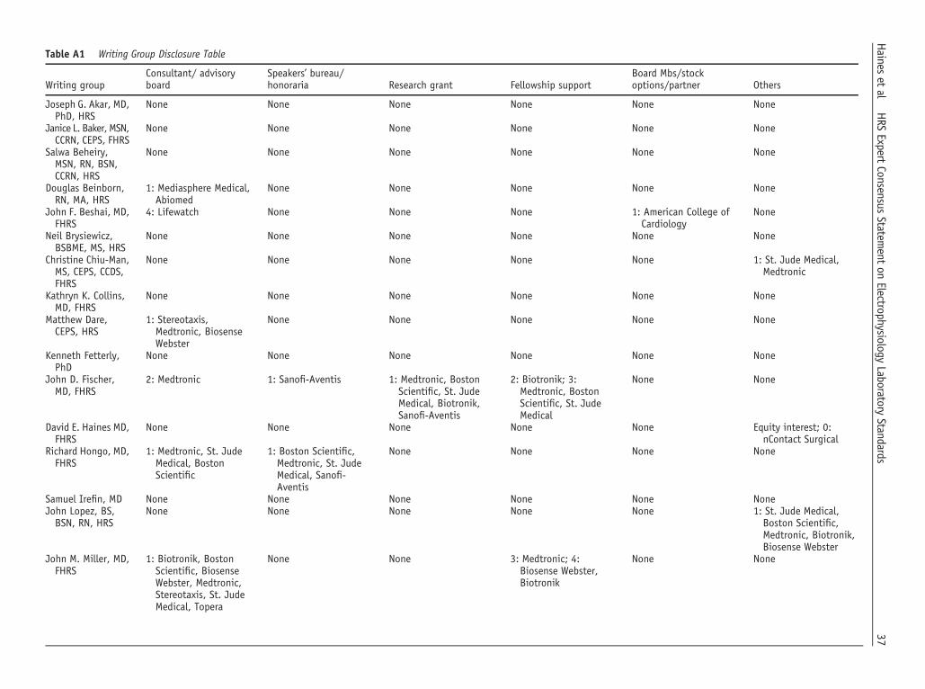
Table A1 Writing Group Disclosure Table
Writing groupConsultant/ advisoryboard
Speakers’ bureau/honoraria Research grant Fellowship support
Board Mbs/stockoptions/partner Others
Joseph G. Akar, MD,PhD, HRS
None None None None None None
Janice L. Baker, MSN,CCRN, CEPS, FHRS
None None None None None None
Salwa Beheiry,MSN, RN, BSN,CCRN, HRS
None None None None None None
Douglas Beinborn,RN, MA, HRS
1: Mediasphere Medical,Abiomed
None None None None None
John F. Beshai, MD,FHRS
4: Lifewatch None None None 1: American College ofCardiology
None
Neil Brysiewicz,BSBME, MS, HRS
None None None None None None
Christine Chiu-Man,MS, CEPS, CCDS,FHRS
None None None None None 1: St. Jude Medical,Medtronic
Kathryn K. Collins,MD, FHRS
None None None None None None
Matthew Dare,CEPS, HRS
1: Stereotaxis,Medtronic, BiosenseWebster
None None None None None
Kenneth Fetterly,PhD
None None None None None None
John D. Fischer,MD, FHRS
2: Medtronic 1: Sanofi-Aventis 1: Medtronic, BostonScientific, St. JudeMedical, Biotronik,Sanofi-Aventis
2: Biotronik; 3:Medtronic, BostonScientific, St. JudeMedical
None None
David E. Haines MD,FHRS
None None None None None Equity interest; 0:nContact Surgical
Richard Hongo, MD,FHRS
1: Medtronic, St. JudeMedical, BostonScientific
1: Boston Scientific,Medtronic, St. JudeMedical, Sanofi-Aventis
None None None None
Samuel Irefin, MD None None None None None NoneJohn Lopez, BS,BSN, RN, HRS
None None None None None 1: St. Jude Medical,Boston Scientific,Medtronic, Biotronik,Biosense Webster
John M. Miller, MD,FHRS
1: Biotronik, BostonScientific, BiosenseWebster, Medtronic,Stereotaxis, St. JudeMedical, Topera
None None 3: Medtronic; 4:Biosense Webster,Biotronik
None None
37Haines
etal
HRS
ExpertConsensus
Statement
onElectrophysiology
LaboratoryStandards

Table A1 (continued )
Writing groupConsultant/ advisoryboard
Speakers’ bureau/honoraria Research grant Fellowship support
Board Mbs/stockoptions/partner Others
James C. Perry, MD,FHRS
1: Medtronic, U.S.Department of Justice
None 2: Medtronic None None None
David J. Slotwiner,MD, HRS
None None None None None None
Gery F. Tomassoni,MD, FHRS, FACC
1: St. Jude Medical,Biosense Webster,Boston Scientific,Stereotaxis,Medtronic
1: Biosense Webster,Boston Scientific,Medtronic,Stereotaxis, Pfizer,Sanofi-Aventis; 2: St.Jude Medical
None None None 1: Stereotaxis
Esther Weiss, RN None None None None None None
0 ¼ $0; 1 ¼ o$10,000; 2 ¼ 4$10,000 to o$25,000; 3 ¼ 4$25,000 to o$50,000; 4 ¼ 4$50,000 to o$100,000; 5 ¼ 4$100,000.
Heart
Rhythm,Vol0,
No0,
Month
201438

Table A2 Peer Reviewer Disclosure Table
Peer reviewerConsultant/ advisoryboard
Speakers’ bureau/honoraria Research grant Fellowship support
Board Mbs/stockoptions/partner Others
Alfred Buxton, MD, HRS 1: Medtronic, BostonScientific, St. JudeMedical
None 0: Biosense Webster,Medtronic
0: Biosense Webster,Medtronic, BostonScientific
None None
Anne Dubin, MD , FHRS None None None 2: Medtronic None NoneCraig Swygman, CEPS,FHRS
1: St. Jude Medical None None None None None
David Benditt, MD,FHRS, CCDS
0: Medtronic; 1:CardioNet; 2: St. JudeMedical; 3: CVRx
None None None None Equity interests; 0:Medtronic; 1:Advanced CirculatorySystems; 2:CardioNet; 3: St. JudeMedical
George Crossley III, MD,FHRS, CCDS
2: Medtronic, BostonScientific
2: Sanofi-Aventis,Boston Scientific,Medtronic
2: Medtronic None None None
James Tcheng, MD 1: American Board ofInternal Medicine,Philips MedicalSystems; 3: AmericanCollege of Cardiology
None 2: U.S. Food and DrugAdministration,National Institutes ofHealth
None None Fiduciary role and salaryposition funding; 3:American College ofCardiology
Kenneth Ellenbogen,MD, FHRS
1: Boston ScientificCorp, CardioNet,American HeartAssociation,American College ofCardiologyFoundation, CameronHealth, Biotronik; 2:Medtronic, NorthAmerican Center forContinuing MedicalEducation; 3: St. JudeMedical
None 0: National Institutes ofHealth, Blue AshTherapeutics; 2:Medtronic; 3:Biosense Webster,Boston Scientific
3: Biosense Webster,Boston Scientific,Medtronic
None Royalty; 0: Wiley-Blackwell; 2: Elsevier
Marianne Beardsall, NP,FHRS, CCDS
1: Medtronic, St. JudeMedical
None None None None None
Michael Lloyd, MD, FHRS 1: St. Jude Medical,Biotectix Corporation
None 1: Boston Scientific,Medtronic
None None None
Noel Boyle, MD, PhD,FHRS
None None None 3: Medtronic, St. JudeMedical, BostonScientific
None None
Paul Wang, MD, FHRS,CCDS
1: Medtronic, BiosenseWebster, BostonScientific, St. JudeMedical
None None 2: Medtronic, BostonScientific, BionsenseWebster, St. JudeMedical
None Equity interests; 1:Vytronus
39Haines
etal
HRS
ExpertConsensus
Statement
onElectrophysiology
LaboratoryStandards

Table A2 (continued )
Peer reviewerConsultant/ advisoryboard
Speakers’ bureau/honoraria Research grant Fellowship support
Board Mbs/stockoptions/partner Others
Sumeet Chugh, MD,FHRS
None None 5: National Institutes ofHealth
2: Boston Scientific,Medtronic
None Salary; 5: NationalInstitutes of Health
Susan Etheridge, MD,FHRS, CEPS
None None None None None Officer, 0: AmericanCollege of Cardiology
Timm-Michael Dickfeld,MD, PhD, FHRS
1: Biosense Webster None 5: GE Healthcare,Biosense Webster
None None None
Ulrika MariaBirgersdotter-Green,MD, FHRS
0: Biotronik, St. JudeMedical, Medtronic
0: St. Jude Medical,Medtronic
2: St. Jude Medical,Medtronic
None None None
0 ¼ $0; 1 ¼ o$10,000; 2 ¼ 4$10,000 to o$25,000; 3 ¼ 4$25,000 to o$50,000; 4 ¼ 4$50,000 to o$100,000; 5 ¼ 4$100,000.
Heart
Rhythm,Vol0,
No0,
Month
201440

define policies regarding the acceptance of industry gifts, butanyone approached by industry must be able to judgewhether accepting the gift carries the danger of biasing amedical decision and compromising patient care. It isunethical to accept any gift that is predicated on using aparticular product or service. It is imperative that the conflictof interest disclosures to employers, editors, professionalorganizations, and audiences be accurate and up to date. Theconflict of interest policies of each physician’s institution andprofessional organization must be followed. Physicians whoare on product review and new product introduction com-mittees for their hospital or professional organization andwho have consulting or other relationships with industrymust disclose those relationships and recuse themselves fromproduct decisions in those cases.
Appendix 1See Tables A1 and A2.
References1. Lindsay BD, Asirvatham SJ, Curtis AB, et al. Guidance for the Heart Rhythm
Society pertaining to interactions with industry endorsed by the Heart RhythmSociety on April 26, 2011. Heart Rhythm 2011;8(7):e19–e23.
2. Tokuda M, Kojodjojo PP, Epstein LM, Koplan BA, Michaud GF, Tedrow UB,Stevenson WG, John RM. Outcomes of cardiac perforation complicatingcatheter ablation of ventricular arrhythmias. Circ Arrhythm Electrophysiol2011;4(5):660–666.
3. Rickard J, Wilkoff BL. Extraction of implantable cardiac electronic devices.Curr Cardiol Rep 2011;13(5):407–414.
4. Stevenson WG, Wilber DJ, Natale A, et al. Irrigated radiofrequency catheterablation guided by electroanatomic mapping for recurrent ventricular tachycar-dia after myocardial infarction: the multicenter thermocool ventricular tachy-cardia ablation trial. Circulation 2008;118(25):2773–2782.
5. Ramakrishna G, Sprung J, Ravi BS, Chandrasekaran K, McGoon MD. Impactof pulmonary hypertension on the outcomes of noncardiac surgery: predictors ofperioperative morbidity and mortality. J Am Coll Cardiol 2005;45(10):1691–1699.
6. Yokokawa M, Good E, Crawford T, et al. Ventricular tachycardia originatingfrom the aortic sinus cusp in patients with idiopathic dilated cardiomyopathy.Heart Rhythm 2011;8(3):357–360.
7. Sacher F, Roberts-Thomson K, Maury P, et al. Epicardial ventricular tachy-cardia ablation: a multicenter safety study. J Am Coll Cardiol 2010;55(21):2366–2372.
8. Wilkoff BL, Love CJ, Byrd CL, Bongiorni MG, Carillo RG, Crossley GH,Epstein LM, Friedman RA, Kennergren CE, Mitkowski P, Schaerf RH, WazniOM. Transvenous lead extraction: Heart Rhythm Society expert consensus onfacilities, training, indications, and patient management. Heart Rhythm 2009;6(7):1085–1104.
9. Facilities Guidelines Institute. Guidelines for design and construction of healthcare facilities. http://www.fgiguidelines.org/. Published January 1, 2014.Accessed December 5, 2012.
10. Sehulster L, & Chin R. Guidelines for Environmental Infection Control inHealth-Care Facilities. Center for Disease Control and the Healthcare InfectionControl Practices Advisory Committee (HICPAC). 2003. http://www.cdc.gov/mmwr/preview/mmwrhtml/rr5210a1.htm Published December 5, 2011.Accessed December 5, 2012.
11. Earley MW, Sargent JS, Sheehan JV & Buss W. NEC 2008 Handbook. Quiny,MA: National Fire Protection Association; 2008.
12. Medical Imaging & Technology Alliance. The DICOM Standard: DigitalImaging and Communications in Medicine. http://medical.nema.org/standard.html. Accessed December 5, 2012.
13. US Department of Health and Human Services. Health Information Privacy.http://www.hhs.gov/ocr/privacy/hipaa/understanding/special/healthit/index.html Accessed December 5, 2012.
14. Gupta S, Desjardins B, Baman T, Ilg K, Good E, Crawford T, Oral H, Pelosi F,Chugh A, Morady F, Bogun F, Delayed-enhanced MR. scar imaging andintraprocedural registration into an electroanatomical mapping system in post-infarction patients. JACC Cardiovasc Imaging 2012;5(2):207–210.
15. Haqqani HM, Tschabrunn CM, Tzou WS, et al. Isolated septal substrate forventricular tachycardia in nonischemic dilated cardiomyopathy: incidence,characterization, and implications. Heart Rhythm 2011;8(8):1169–1176.
16. U.S. Food and Drug Administration. Strategies for Clinical and BiomedicalEngineers to Maintain Readiness of External Defibrilators. http://www.fda.gov/MedicalDevices/ProductsandMedicalProcedures/CardiovascularDevices/ExternalDefibrillators/ucm233451.htm. 2010. Published March 21, 2013. AccessedDecember 5, 2012.
17. Scheinman M, Calkins H, Gillette P, Klein R, Lerman BB, Morady F, SaksenaS, Waldo A. NASPE policy statement on catheter ablation: personnel, policy,procedures, and therapeutic recommendations. Pacing Clin Electrophysiol2003;26(3):789–799.
18. Chanani NK, Chiesa NA, Dubin AM, Avasarala K, Van Hare GF, Collins KK.Cryoablation for atrioventricular nodal reentrant tachycardia in young patients:predictors of recurrence. Pacing Clin Electrophysiol 2008;31(9):1152–1159.
19. Khoury DS, Taccardi B, Lux RL, Ershler PR, Rudy Y. Reconstruction ofendocardial potentials and activation sequences from intracavitary probemeasurements. Localization of pacing sites and effects of myocardial structure.Circulation 1995;91(3):845–863.
20. Banchs JE, Patel P, Naccarelli GV, Gonzalez MD. Intracardiac echocardio-graphy in complex cardiac catheter ablation procedures. J Interv Card Electro-physiol 2010;28(3):167–184.
21. Ren JF, Marchlinski FE, Callans DJ. Left atrial thrombus associated withablation for atrial fibrillation: identification with intracardiac echocardiography.J Am Coll Cardiol 2004;43(10):1861–1867.
22. Ferguson JD, Helms A, Mangrum JM, Mahapatra S, Mason P, Bilchick K,McDaniel G, Wiggings D, DiMarco JP. Catheter ablation of atrial fibrillationwithout fluoroscopy using intracardiac echocardiography and electroanatomicmapping. Circ Arrhythm Electrophysiol 2009;2(6):611–619.
23. Yokokawa M, Good E, Desjardins B, Crawford T, Jongnarangsin K, Chugh A,Pelosi F, Oral H, Morady F, Bogun F. Predictors of successful catheter ablationof ventricular arrhythmias arising from the papillary muscles. Heart Rhythm2010;7(11):1654–1659.
24. Schwartzman D, Zhong H. On the use of CartoSound for left atrial navigation.J Cardiovasc Electrophysiol 2010;21(6):656–664.
25. Lindsay BD, Estes NA III, Maloney JD, Reynolds DW. Heart Rhythm SocietyPolicy Statement Update: Recommendations on the Role of Industry EmployedAllied Professionals (IEAPs). Heart Rhythm 2008;5(11):e8–10.
26. Bai R, Di Biase L, Valderrabano M, et al. Worldwide experience with therobotic navigation system in catheter ablation of atrial fibrillation: methodology,efficacy and safety. J Cardiovasc Electrophysiol 2012;23(8):820–826.
27. Khan E, Frumkin W. A Novel Remote Catheter System, Demonstrates Safetyand Efficacy. Heart Rhythm 2013. (ARTICLE IN PRINT).
28. Faddis MN, Chen J, Osborn J, Talcott M, Cain ME, Lindsay BD. Magneticguidance system for cardiac electrophysiology: a prospective trial of safety andefficacy in humans. J Am Coll Cardiol 2003;42(11):1952–1958.
29. Burkhardt JD, Saliba WI, Schweikert RA, Cummings J, Natale A. Remotemagnetic navigation to map and ablate left coronary cusp ventricular tachy-cardia. J Cardiovasc Electrophysiol 2006;17(10):1142–1144.
30. American Board of Internal Medicine: Accreditation Coucil for GraduateMedical Education. Program Requirements for Graduate Medical Educationin Clinical Cardiac Electrophysiology (Internal Medicine). http://www.acgme.org/acgmeweb/Portals/0/PFAssets/ProgramRequirements/154_clinical_card_electrophys_int_med_07012012_1-YR.pdf. 2012. Published July 1, 2012.Accessed December 5, 2012.
31. American Board of Internal Medicine. Policies and Procedures for Certifica-tion. http://www.abim.org/pdf/publications/Policies-and-Procedures-Certification-August-2012.pdf. 2012. Published December 1, 2012. Accessed December 5,2012.
32. Baughman KL, Duffy FD, Eagle KA, Faxon DP, Hills LD, Lange RA. Taskforce 1: training in clinical cardiology. J Am Coll Cardiol 2008;51(3):339–348.
33. Naccarelli GV, Conti JB, DiMarco JP, Tracy CM. Task force 6: training inspecialized electrophysiology, cardiac pacing, and arrhythmia managementendorsed by the Heart Rhythm Society. J Am Coll Cardiol 2008;51(3):374–380.
34. Tracy CM, Akhtar M, DiMarco JP, Packer DL, Weitz HH, Creager MA, HolmesDR Jr, Merli G, Rodgers GP, Tracy CM, Weitz HH. American College ofCardiology/American Heart Association 2006 update of the clinical competencestatement on invasive electrophysiology studies, catheter ablation, and cardio-version: a report of the American College of Cardiology/American HeartAssociation/American College of Physicians Task Force on Clinical Compe-tence and Training developed in collaboration with the Heart Rhythm Society.J Am Coll Cardiol 2006;48:1503–1517.
35. Bashore TM, Balter S, Barac A, et al. 2012 American College of CardiologyFoundation/Society for Cardiovascular Angiography and Interventions expertconsensus document on cardiac catheterization laboratory standards update.
41Haines et al HRS Expert Consensus Statement on Electrophysiology Laboratory Standards

A report of the American College of Cardiology Foundation Task Force onExpert Consensus documents. J Am Coll Cardiol 2012;59(24):2221–2305.
36. American Society of Anesthesiologists Task Force on Sedation and Analgesiaby Non-Anesthesiologist. Practice guidelines for sedation and analgesia by non-anesthesiologists. Anesthesiology 2002;96:1004–1017.
37. Walsh EP, Bar-Cohen Y, Batra AS, DickM II, Erickson C, Fish F, Hamilton RM,Kanter RJ, Reed JH, Van Hare GF, Vetter VL, Webster G. Recommendations foradvanced fellowship training in clinical pediatric and congenital electrophysiol-ogy: a report from the training and credentialing committee of the pediatric andcongenital electrophysiology society. Heart Rhythm 2013;10:775–781.
38. Calkins H, Kuck KH, Cappato R, et al. 2012 HRS/EHRA/ECAS expertconsensus statement on catheter and surgical ablation of atrial fibrillation:recommendations for patient selection, procedural techniques, patient manage-ment and follow-up, definitions, endpoints, and research trial design. HeartRhythm 2012;9(4):632–696 e21.
39. Aliot EM, Stevenson WG, Almendral-Garrote JM, et al. EHRA/HRS ExpertConsensus on Catheter Ablation of Ventricular Arrhythmias: developed in apartnership with the European Heart Rhythm Association (EHRA), a RegisteredBranch of the European Society of Cardiology (ESC), and the Heart RhythmSociety (HRS); in collaboration with the American College of Cardiology(ACC) and the American Heart Association (AHA). Heart Rhythm 2009;6(6):886–933.
40. Accreditation Council for Graduate Medical Education. Frequently AskedQuestions: Clinical Cardiac Electrophysiology: Review Committee for InternalMedicine. http://www.acgme.org/acgmeweb/Portals/0/PDFs/FAQ/154_ClinicalCardiacElectrophysiology_FAQ.pdf. 2012. Published July 1, 2012. AccessedDecember 5, 2012.
41. Vetter VL, Silka MJ, Van Hare GF, Walsh EP. ACCF/AHA/AAP recommen-dations for training in pediatric cardiology. Task force 4: recommendations fortraining guidelines in pediatric cardiac electrophysiology endorsed by the HeartRhythm Society. J Am Coll Cardiol 2005;46(7):1391–1395.
42. Saul JP, Epstein AE, Silka MJ, Berul CI, Dick M, Dimarco JP, Friedman RA,Rosentahl E, Stephenson EA, Vetter VL. Heart Rhythm Society/Pediatric andCongenital Electrophysiology Society Clinical Competency Statement: trainingpathways for implantation of cardioverter-defibrillators and cardiac resynchro-nization therapy devices in pediatric and congenital heart patients. HeartRhythm 2008;5(6):926–933.
43. Walsh EP, Bar-Cohen Y, Batra AS, et al. Recommendations for AdvancedFellowship Training in Clinical Pediatric and Congenital Electrophysiology: AReport from the Training and Credentialing Committee of the Pediatric andCongenital Electrophysiology Society. Heart Rhythm 2013;10:775–781.
44. Warnes CA, Williams RG, Bashore TM, et al. ACC/AHA 2008 guidelines forthe management of adults with congenital heart disease: a report of theAmerican College of Cardiology/American Heart Association Task Force onPractice Guidelines (Writing Committee to Develop Guidelines on the Manage-ment of Adults With Congenital Heart Disease). Developed in CollaborationWith the American Society of Echocardiography, Heart Rhythm Society,International Society for Adult Congenital Heart Disease, Society for Cardio-vascular Angiography and Interventions, and Society of Thoracic Surgeons.J Am Coll Cardiol 2008;52(23):e143–e263.
45. Nurse.com. Nursing Continuing Education Requirements by State. http://ce.nurse.com/RStateReqmnt.aspx. Published December 1, 2012. Accessed Decem-ber 13, 2012.
46. National Council of State Boards of Nursing. Model for APRN Regulation:Licensure, Accreditation, Certification and Education. https://www.ncsbn.org/Consensus_Model_for_APRN_Regulation_July_2008.pdf. 2008. PublishedJuly 7, 2008. Accessed January 30, 2013.
47. Commission on Accreditation of Allied Health Education Programs. ProfessionDescription & Certficaition Information. www.caahep.org/. 2012. PublishedDecember 1, 2012. Accessed December 7, 2012.
48. Mittman DE, Cawley JF, Fenn WH. Physician assistants in the United States.BMJ 2002;325(7362):485–487.
49. Mikhael N, Ozon P, Rhule C. Defining the Physician Assistant Role in Ontario:Ontario Physician Assistant Scope of Practice Statement and Ontario PhysicianAssistant Competency Profile. HealthForce Ontario 2007.
50. The Joint Commission. National Patient Safety Goals: Hospital AccreditationProgram. http://www.jointcommission.org/assets/1/6/NPSG_Chapter_Jan2012_HAP.pdf. 2012. Accessed December 5, 2012.
51. American Society of Anesthesiologists. Standards for Basic Anesthetic Monitor-ing. http://www.asahq.org/For-Members/Standards-Guidelines-and-Statements.aspx. 2011.Pubblished July 1, 2011. Accessed December 5, 2012.
52. Sayfo S, Vakil KP, Alqaqa'a A, Flippin H, Bhakta D, Yadav AV, Miller JM,Groh WJ. A retrospective analysis of proceduralist-directed, nurse-administeredpropofol sedation for implantable cardioverter-defibrillator procedures. HeartRhythm 2012;9(3):342–346.
53. American Society of Anesthesiologists. Statement on Granting Privileges forAdministration of Moderate Sedation to Practitioners Who Are Not AnesthesiaProfessionals. http://www.asahq.org/For-Members/Standards-Guidelines-and-Statements.aspx. 2011. Published October 18, 2006. Accessed December 5,2012.
54. Friedman RA, Walsh EP, Silka MJ, Calkins H, Stevenson WG, Rhodes LA,Deal BJ, Wolff GS, Demaso DR, Hanisch D, Van Hare GF. NASPE ExpertConsensus Conference: Radiofrequency catheter ablation in children with andwithout congenital heart disease. Report of the writing committee. NorthAmerican Society of Pacing and Electrophysiology. Pacing Clin Electrophysiol2002;25(6):1000–1017.
55. Di Biase L, Conti S, Mohanty P, et al. General anesthesia reduces the prevalenceof pulmonary vein reconnection during repeat ablation when compared withconscious sedation: results from a randomized study. Heart Rhythm 2011;8(3):368–372.
56. Elkassabany N, Garcia F, Tschabrunn C, Raiten J, Gao W, Chaichana K, DixitS, Speck RM, Zado E, Marchlinski F, Mandel J. Anesthetic management ofpatients undergoing pulmonary vein isolation for treatment of atrial fibrillationusing high-frequency jet ventilation. J Cardiothorac Vasc Anesth 2012;26(3):433–438.
57. Doppalapudi H, Yamada T, Kay GN. Complications during catheter ablation ofatrial fibrillation: identification and prevention. Heart Rhythm 2009;6(12Suppl):S18–S25.
58. Perna F, Heist EK, Danik SB, Barrett CD, Ruskin JN, Mansour M. Assessmentof catheter tip contact force resulting in cardiac perforation in swine atria usingforce sensing technology. Circ Arrhythm Electrophysiol 2011 Apr;4(2):218–224.
59. Latchamsetty R, Oral H. Ablation of atrial fibrillation using an irrigated-tipcatheter: open or closed? Pacing Clin Electrophysiol 2012;35(5):503–505.
60. Andrade JG, Khairy P, Guerra PG, Deyell MW, Rivard L, Macle L, Thbault B,Talajic M, Roy D, Dubuc M. Efficacy and safety of cryoballoon ablation foratrial fibrillation: a systematic review of published studies. Heart Rhythm2011;8(9):1444–1451.
61. Venkatachalam KL, Herbrandson JE, Asirvatham SJ. Signals and signalprocessing for the electrophysiologist: part I: electrogram acquisition. CircArrhythm Electrophysiol 2011;4(6):965–973.
62. Venkatachalam KL, Herbrandson JE, Asirvatham SJ. Signals and signalprocessing for the electrophysiologist: part II: signal processing and artifact.Circ Arrhythm Electrophysiol 2011;4(6):974–981.
63. Mangram AJ, Horan TC, Pearson ML, Silver LC, Jarvis WR. Guideline forprevention of surgical site infection, 1999. Hospital Infection Control PracticesAdvisory Committee. Infect Control Hosp Epidemiol 1999;20(4):250–278.(quiz 279-280).
64. Bashore TM, Bates ER, Berger PB, et al. American College of Cardiology/Society for Cardiac Angiography and Interventions Clinical Expert ConsensusDocument on cardiac catheterization laboratory standards. A report of theAmerican College of Cardiology Task Force on Clinical Expert ConsensusDocuments. J Am Coll Cardiol 2001;37(8):2170–2214.
65. Malloy PC, Grassi CJ, Kundu S, et al. Consensus guidelines for periproceduralmanagement of coagulation status and hemostasis risk in percutaneous image-guided interventions. J Vasc Interv Radiol 2009;20(7 Suppl):S240–S249.
66. Cano O, Munoz B, Tejada D, Osca J, Sancho-Tello MJ, Olague J, Castro JE,Salvador A. Evaluation of a new standardized protocol for the perioperativemanagement of chronically anticoagulated patients receiving implantablecardiac arrhythmia devices. Heart Rhythm 2012;9(3):361–367.
67. Haines DE, Wang Y, Curtis J. Implantable cardioverter-defibrillator registry riskscore models for acute procedural complications or death after implantablecardioverter-defibrillator implantation. Circulation 2011;123(19):2069–2076.
68. Haeusler KG, Kirchhof P, Endres M. Left atrial catheter ablation and ischemicstroke. Stroke 2012;43(1):265–270.
69. Scaltriti S, Cencetti S, Rovesti S, Marchesi I, Bargellini A, Borella P. Riskfactors for particulate and microbial contamination of air in operating theatres. JHosp Infect 2007;66(4):320–326.
70. Baddour LM, Epstein AE, Erickson CC, et al. Update on cardiovascularimplantable electronic device infections and their management: a scientific state-ment from the American Heart Association. Circulation 2010;121(3):458–477.
71. Movahed MR, Kasravi B, Bryan CS. Prophylactic use of vancomycin in adultcardiology and cardiac surgery. J Cardiovasc Pharmacol Ther 2004;9:13–20.
72. Perry JC. Sudden cardiac death and malignant arrhythmias: the scope of theproblem in adult congenital heart patients. Pediatr Cardiol 2012;33(3):484–490.
73. Saul JP, Hulse JE, Papagiannis J, Van Praagh R, Walsh EP. Late enlargement ofradiofrequency lesions in infant lambs. Implications for ablation procedures insmall children. Circulation 1994;90(1):492–499.
74. Dubin AM, Van Hare GF. Radiofrequency catheter ablation: indications andcomplications. Pediatr Cardiol 2000;21(6):551–556.
Heart Rhythm, Vol 0, No 0, Month 201442

75. Cohen MI, Triedman JK, Cannon BC, et al. PACES/HRS expert consensusstatement on the management of the asymptomatic young patient with a Wolff-Parkinson-White (WPW, ventricular preexcitation) electrocardiographic pat-tern: developed in partnership between the Pediatric and Congenital Electro-physiology Society (PACES) and the Heart Rhythm Society (HRS). Endorsedby the governing bodies of PACES, HRS, the American College of CardiologyFoundation (ACCF), the American Heart Association (AHA), the AmericanAcademy of Pediatrics (AAP), and the Canadian Heart Rhythm Society(CHRS). Heart Rhythm 2012;9(6):1006–1024.
76. Epstein AE, DiMarco JP, Ellenbogen KA, et al. ACC/AHA/HRS 2008Guidelines for Device-Based Therapy of Cardiac Rhythm Abnormalities: areport of the American College of Cardiology/American Heart Association TaskForce on Practice Guidelines (Writing Committee to Revise the ACC/AHA/NASPE 2002 Guideline Update for Implantation of Cardiac Pacemakers andAntiarrhythmia Devices): developed in collaboration with the AmericanAssociation for Thoracic Surgery and Society of Thoracic Surgeons. Circulation2008;117(21):e350–e408.
77. Janousek J, Gebauer RA. Cardiac resynchronization therapy in pediatric andcongenital heart disease. Pacing Clin Electrophysiol 2008;31(Suppl 1):S21–S23.
78. Cecchin F, Frangini PA, Brown DW, Fynn-Thompson F, Alexander ME,Triedman JK, Gauvreau K, Walsh EP, Berul CI. Cardiac resynchronizationtherapy (and multisite pacing) in pediatrics and congenital heart disease: five yearsexperience in a single institution. J Cardiovasc Electrophysiol 2009;20(1):58–65.
79. Kugler JD, Danford DA, Houston KA, Felix G. Pediatric radiofrequencycatheter ablation registry success, fluoroscopy time, and complication rate forsupraventricular tachycardia: comparison of early and recent eras. J CardiovascElectrophysiol 2002;13:336–341.
80. Blaufox AD, Felix G, Saul JP. Radiofrequency catheter ablation in infantso 18months old: when is it done and how do they fare? Short-term data from thePediatric Ablation Registry. Circulation 2001;104:2803–2808.
81. Pecht B, Maginot KR, Boramanand NK, Perry JC. Techniques to avoidatrioventricular block during radiofrequency catheter ablation of septal tachycardiasubstrates in young patients. J Interv Card Electrophysiol 2002 Aug;7(1):83–88.
82. Chanani NK, Chiesa NA, Dubin AM, Avasarala K, Van Hare GF, Collins KK.Cryoablation for atrioventricular nodal reentrant tachycardia in young patients:predictors of recurrence. Pacing Clin Electrophysiol 2008;31:1152–1159.
83. Buxton AE, Calkins H, Callans DJ, et al. ACC/AHA/HRS 2006 key dataelements and definitions for electrophysiological studies and procedures: areport of the American College of Cardiology/American Heart Association TaskForce on Clinical Data Standards (ACC/AHA/HRS Writing Committee toDevelop Data Standards on Electrophysiology). J Am Coll Cardiol 2006;48(11):2360–2396.
84. Collins KK, Schaffer MS. Use of cryoablation for treatment of tachyarrhythmiasin 2010: survey of current practices of pediatric electrophysiologists. PacingClin Electrophysiol 2011;34(3):304–308.
85. Van Hare GF, Colan SD, Javitz H, Carmelli D, Knilans T, Schaffer M, Kugler J,Byrum CJ, Saul JP. Prospective assessment after pediatric cardiac ablation: fateof intracardiac structure and function, as assessed by serial echocardiography.Am Heart J 2007;153(5):815–820. (820 e1-6).
86. Hirshfeld JW Jr, Balter S, Brinker JA, et al. ACCF/AHA/HRS/SCAI clinicalcompetence statement on physician knowledge to optimize patient safety andimage quality in fluoroscopically guided invasive cardiovascular procedures. Areport of the American College of Cardiology Foundation/American HeartAssociation/American College of Physicians Task Force on Clinical Compe-tence and Training. J Am Coll Cardiol 2004;44(11):2259–2282.
87. Vano E, Rosenstein M, Liniecki J, Rehani MM, Martin CJ, Vetter RJ. ICRPPublication 113. Education and training in radiological protection for diagnosticand interventional procedures. Ann ICRP 2009;39(5):7–68.
88. Glazier JJ, Dixon SR. Skin injury following prolonged fluoroscopy: early andlate appearances. QJM 2012;105(6):571–573.
89. Committee to Assess Health Risks from Exposure to Low Levels of IonizingRadiation National Research Council. Health Risks from Exposure to LowLevels of Ionizing Radiation: BEIR VII Phase 2 2006.
90. Rosenthal LS, Mahesh M, Beck TJ, et al. Predictors of fluoroscopy time andestimated radiation exposure during radiofrequency catheter ablation proce-dures. Am J Cardiol 1998;82(4):451–458.
91. Calkins H, Niklason L, Sousa J, el-Atassi R, Langberg J, Morady F. Radiationexposure during radiofrequency catheter ablation of accessory atrioventricularconnections. Circulation 1991;84(6):2376–2382.
92. Einstein AJ, Moser KW, Thompson RC, et al. Radiation dose to patients fromcardiac diagnostic imaging. Circulation 2007;116(11):1290–1305.
93. United States Nuclear Regulatory Commission. NRC: Annual Dose Limits forRadiation Workers. http://www.nrc.gov/reading-rm/doc-collections/nuregs/staff/sr1556/v6/fig012.html. 2012. Published June 28, 2013. Accessed Decem-ber 5, 2012.
94. Gerber TC, Carr JJ, Arai AE, et al. Ionizing radiation in cardiac imaging: ascience advisory from the American Heart Association Committee on CardiacImaging of the Council on Clinical Cardiology and Committee on Cardiovas-cular Imaging and Intervention of the Council on Cardiovascular Radiology andIntervention. Circulation 2009;119(7):1056–1065.
95. Miller DL, Balter S, Dixon RG, Nikoli B, Bartal G, Cardell JF, Dauer LT,Stecker MS. Quality improvement guidelines for recording patient radiationdose in the medical record for fluoroscopically guided procedures. J Vasc IntervRadiol 2012;23(1):11–18.
96. Germano JJ, Day G, Gregorious D, Natarajan V, Cohen T. A novel radiationprotection drape reduces radiation exposure during fluoroscopy-guided electro-physiology procedures. J Invasive Cardiol 2005;17:469–472.
97. U.S. Food and Drug Administration. CFR-Code of Federal Regualtions Title 21.http://www.accessdata.fda.gov/scripts/cdrh/cfdocs/cfcfr/CFRSearch.cfm?fr=1020.32. 2012. Published April 1, 2013. Accessed December 14, 2012.
98. Joint Commission on Accreditation of Healthcare Organizations. AccreditationManual for Hospitals Update 3. Oakbrook Terrace, IL; 2002.
99. Birnie D, Healey JS, Krahn AD, et al. Prevalence and risk factors for cervicaland lumbar spondylosis in interventional electrophysiologists. J CardiovascElectrophysiol 2011;22(9):957–960.
100. von Boetticher H, Lachmund J, Hoffmann W. Cardiac catheterization: impact offace and neck shielding on new estimates of effective dose. Health Phys 2009;97(6):622–627.
101. Jacob S, Boveda S, Bar O, Brezin A, Maccia C, Laurier D, Bernier MO.Interventional cardiologists and risk of radiation-induced cataract: results of aFrench multicenter observational study. Int J Cardiol 2013;167(5):1843–1847.
102. Ross AM, Segal J, Borenstein D, Jenkins E, Cho S. Prevalence of spinal discdisease among interventional cardiologists. Am J Cardiol 1997;79(1):68–70.
103. Dragusin O, Weerasooriya R, Jais P, Hocini M, Ector J, Takahashi Y,Haissaguerre M, Bosmans H, Heidbuchel H. Evaluation of a radiation protectioncabin for invasive electrophysiological procedures. Eur Heart J 2007;28(2):183–189.
104. Di Biase L, Wang Y, Horton R, et al. Ablation of atrial fibrillation utilizingrobotic catheter navigation in comparison to manual navigation and ablation:single-center experience. J Cardiovasc Electrophysiol 2009;20(12):1328–1335.
105. Pappone C, Vicedomini G, Manguso F, et al. Robotic magnetic navigation foratrial fibrillation ablation. J Am Coll Cardiol 2006;47(7):1390–1400.
106. Goldstein JA, Balter S, Cowley M, Hodgson J, Klein LW. Occupational hazardsof interventional cardiologists: prevalence of orthopedic health problems incontemporary practice. Catheter Cardiovasc Interv 2004;63(4):407–411.
107. Klein LW, Miller DL, Balter S, Laskey W, Haines D, Norbash A, Mauro MA,Goldstein JA. Occupational health hazards in the interventional laboratory: timefor a safer environment. Heart Rhythm 2009;6(3):439–444.
108. Weber DJ, Rutala WA, Schaffner W. Lessons learned: protection of healthcareworkers from infectious disease risks. Crit Care Med 2010;38(8 Suppl):S306–S314.
109. Siegel JD, Rhinehart E, Jackson M, et al. 2007 Guideline for IsolationPrecautions: Preventing Transmission of Infectious Agents in Health CareSettings. Am J Infect Control 2007;35(10 Suppl 2): (S65-164).
110. World Health Organization. Glove Use Information Leaflet. http://www.who.int/gpsc/5may/Glove_Use_Information_Leaflet.pdf. 2009. Published August 1,2009. Accessed December 5, 2012.
111. Ninomura P, Bartley J. New Ventilation Guidelines for Health-Care Facilities.American Society of Heating, Refrigerating and Air Conditioning EngineersJournal 2001:29–33.
112. Engum SA, Jeffries P, Fisher L. Intravenous catheter training system: computer-based education versus traditional learning methods. Am J Surg 2003;186(1):67–74.
113. McNamara RM, Monti S, Kelly JJ. Requesting consent for an invasiveprocedure in newly deceased adults. JAMA 1995;273(4):310–312.
114. Chren MM, Landefeld CS. Physicians’ behavior and their interactions with drugcompanies. A controlled study of physicians who requested additions to ahospital drug formulary. JAMA 1994;271(9):684–689.
43Haines et al HRS Expert Consensus Statement on Electrophysiology Laboratory Standards