heat-shock protein 70 modulates apoptosis signal-regulating kinase 1 in stressed hepatocytes of...
TRANSCRIPT

Heat-shock protein 70 modulates apoptosis signal-regulatingkinase 1 in stressed hepatocytes of Mugil cephalus
Ekambaram Padmini • Jayachandran Tharani
Received: 18 March 2014 / Accepted: 19 May 2014
� Springer Science+Business Media Dordrecht 2014
Abstract Oxidative stress causes damage at the
cellular level and activates a number of signaling
pathways. Heat-shock proteins (HSPs) play an impor-
tant role in repair and protective mechanisms under
cell response to stress conditions. HSP70 has been
shown to act as an inhibitor of apoptosis. Apoptosis
signal-regulating kinase-1 (ASK1) activity is regu-
lated at multiple levels, one of which is through
inhibition by cytosolic chaperons HSP70. The current
study was aimed to investigate the alteration in
signaling molecules that allow the fish to survive
under stressed natural field conditions. The study also
investigates the variation in biomolecular composition
of hepatocytes by using Fourier transform infrared
spectroscopy. The impact of stress on hepatocytes was
assessed by measuring the level of lipid peroxides
(LPO), catalase activity (CAT) and assessing the
changes in hepatocytes of Mugil cephalus inhabiting
Kovalam and Ennore estuaries. The expression of
HSP70 and ASK1 were analyzed by immunoblot
analysis and ELISA, respectively. The spectral ana-
lysis showed variations in biomolecular composition
of hepatocytes at a wave number region of
4,000–400 cm-1. There was significant decrease of
CAT activity (p \ 0.01) (25 %) with significant
increase of LPO (p \ 0.001) (35 %) and HSP70
(p \ 0.001) and insignificant increase of ASK1
(p \ 0.05) (16 %) in fish hepatocytes inhabiting
Ennore estuary than Kovalam estuary. In conclusion,
the present study suggests that the survival of fish in
the Ennore estuary under stressed condition may be
due to the upregulation of HSP70 that mediates the
altered signal pathway which promotes cellular resis-
tance against apoptosis.
Keywords Catalase (CAT) � Fourier transform
infrared spectroscopy (FTIR) � Heat-shock
protein (HSP) � Hepatocytes � Lipid peroxides
(LPO) � Mugil cephalus
Introduction
Estuaries, the main contributors of fisheries in India,
suffer from severe damage due to increased industri-
alization and urbanization along the coastal areas. Fish
are the most at threat from aquatic pollution caused by
various internal and external (physicochemical and
biotic) environmental factors and hence considered to
be the bioindicators of water pollution (Adams and
Marshall 1999). Gray mullets, the natural inhabitant of
the estuaries studied, are found in the shallow water of
the intertidal zone that experiences dual fluctuations in
both temperature and salinity. These economically
important fish are under threat because of their
E. Padmini (&) � J. Tharani
P.G. Department of Biochemistry, Bharathi Women’s
College, Affiliated to University of Madras,
Chennai 600108, Tamilnadu, India
e-mail: [email protected]; [email protected]
123
Fish Physiol Biochem
DOI 10.1007/s10695-014-9949-0

continuous exposure to toxic chemical-rich industrial
effluents that are discharged into the estuaries (Pad-
mini and Kavitha 2005). These species have high
economic importance and gastronomic value, which
means that the information provided by our study may
be important both to people using the lagoon resources
and to those responsible for fishery management. Also
they have been demonstrated to be suitable for
biomarker studies (Ferreira et al. 2004; Gorbi et al.
2005; Pacheco et al. 2005).
Oxidative stress reflects a disturbance in the pro-
oxidant and antioxidant systems in favor of the pro-
oxidant. Free radicals are the potent toxic compounds
that are produced continuously in cells during exposure
to environmental toxins and exert a deleterious effect
via their chain reactions (Das and Das 2006), covalent
modification and oxidation of functional groups
thereby causing significant damage to biological
macromolecules and bringing about alterations in the
cellular redox balance (Abele et al. 2002). However,
the harmful effects of free radicals are blocked to a
great extent by antioxidant defense systems.
To maintain the homeostasis under oxidative stress,
the cells apart from the induction of antioxidant
enzymes produce high levels of stress proteins or heat-
shock proteins (HSPs), which protect them against the
damage. HSP70 protein plays appreciable role in
stress tolerance, protein folding, posttranslational
control of the stability and regulation of signal
transduction pathways that control cell growth and
survival (Zugel and Kaufmann 1999). It is expressed
constitutively in most cells and is upregulated by
thermal stress, heat-shock, heavy metal exposure,
oxidative stress and developmental and mitogenic
stimuli. HSPs are potential biomarkers for environ-
mental stress in fish (Iwama et al. 1998). Various
studies demonstrate that Hsp-induced cytoprotection
can be attributed partly to the suppression of apoptosis
(Samali and Orrenius 1998). Apoptosis resistance is
associated with the overexpression of HSPs.
Apoptosis signal-regulating kinase 1 (ASK1) is a
155-kDa ubiquitously expressed protein belonging to
the member of MAPKKK, a serine–threonine protein
kinase. It is activated in response to reactive oxygen
species (ROS), hydrogen peroxide, tumor necrosis
factor (TNF) and other stress stimuli (Morita et al.
2001). It plays a key role in the regulation of signaling
in response to oxidative stress (Goldman et al. 2003).
ASK1 activity is regulated at multiple levels, one of
which is through inhibition by cytosolic chaperons of
the HSP 70 family (Hwang et al. 2005).
Fourier transform infrared (FTIR) spectroscopy is a
non-disturbing technique which provides quantitative
biochemical information about biological samples. It
is a valuable technique due to its high sensitivity in
detecting changes in the molecular constituent of
tissues, such as lipids, proteins and nucleic acids. The
shifts in the peak positions, bandwidths and intensities
of the bands all give valuable structural and functional
information, which may have diagnostic value. With
FTIR spectroscopy, it is possible to monitor changes in
the structure and properties of biomolecules such as
DNA, RNA, proteins, carbohydrates, lipids in biolog-
ical tissues and cell simultaneously (Ci et al. 1999).
The advantages of FTIR method enable it to examine
the initial response to stress with high sensitivity
through spectra from very small amounts of samples.
(Corte et al. 2010) reported that this technique offers a
fast method to fingerprint the global cellular features
under specific conditions (Alvarez-Ordonez et al.
2010). In this context, the current study was aimed
to investigate the level of HSP70 along with ASK1 by
immunoblot and ELISA. An attempt was made to
correlate the FTIR study of HSP70 and ASK1 in
hepatocytes of M. cephalus inhabiting Kovalam
(unpolluted site) and Ennore (polluted site) estuaries.
Materials and methods
Study site
Kovalam and Ennore estuaries were chosen as the two
study sites for the current research work (Fig. 1).
Kovalam estuary (12�47016N, 80�14058E) is situated
on the east coast of India and is about 35 km south of
Chennai. It runs parallel to the sea coast and extends to
a distance of 20 km. It was chosen as the unpolluted
site for the present investigation as it is surrounded by
high vegetation, and it is free from industrial or urban
pollution. Ennore estuary (13�14051N, 80�19031E)
also situated on the east coast of India and is about
15 km north of Chennai. It runs parallel to the sea
coast and extends over a distance of 36 km. This
estuary was chosen as the polluted site as in its
immediate coastal neighborhood are situated a number
of industries, which include petrochemicals, fertiliz-
ers, pesticides, oil refineries, rubber factory and
Fish Physiol Biochem
123

thermal power stations that discharge their effluents
directly into this estuary. Contamination of this
estuary by heavy metals such as lead, cadmium,
mercury, zinc and iron to a significant extent com-
pared to unpolluted estuary has also been confirmed by
previous studies (Raghunathan and Srinivasan 1983;
Padmini and Vijaya Geetha 2007a). It has also been
reported that Ennore estuary significantly differs from
Kovalam estuary in its physical, chemical and biolog-
ical factors (Padmini and Vijaya Geetha 2007b), thus
it has been chosen as the polluted site.
Study animal and sampling
M. cephalus, a natural inhabitant of the estuaries,
identified by the use of Food and Agriculture Organi-
zation (FAO) species identification sheets (Fischer and
Bianchi 1984). M. cephalus with an average length of
30–32 cm were collected from unpolluted and polluted
estuaries using baited minnow traps. Collected fish was
placed immediately into insulated containers filled
with aerated estuarine water at ambient temperature
(25–30 �C) and salinity (24–29 ppt). Fish were main-
tained in the above-specified conditions for 4–5 h until
the start of the experimental procedures. Fish were
killed by severing the spinal cord, and the liver was
removed immediately.
Isolation of hepatocytes
The isolation of hepatocytes was carried out according
to established protocols (Krumschnabel et al. 1994;
Buckley et al. 2004) with slight modification as
described by Padmini and Usha Rani (2008). In brief,
after the fish was anesthetized with a solution of ethyl
m-aminobenzoate (MS-222; 0.5 g l-1 of water), a
INDIA
N
TAMILNADU
CHENNAI
ENNORE
KOVALAM
CHENNAI
BAY
O F
BENGAL
UTHANDI
INJAMBAKKAM
PALAVAKKAM
THIRUVANMIYUR
THIRUVETTIYUR
80°00” 80°06” 80°12” 80°18” 80°24” 80°30”
80°00” 80°06” 80°12” 80°18” 80°24” 80°30”
13°12”
13°06”
13°00”
12°54”
12°48”
13°12”
13°06”
13°00”
12°54”
12°48”
10 0 10 20 km
North Chennai Coast
Central Chennai Coast
South Chennai Coast
Fig. 1 Study area. Geographical locations of the Kovalam (unpolluted) and Ennore (polluted) estuaries
Fish Physiol Biochem
123

midventral incision was made to expose the liver, and
the portal vein was cannulated in the direction of the
liver. The liver was perfused with a perfusion buffer
containing 290 mmol l-1 NaCl, 2 mmol l-1 KCl,
10 mmol l-1 N-2-hydroxyethylpiperazine-N-2-etha-
nesulfonic acid (HEPES), 0.5 mmol l-1 ethylene
glycol-bis (2-aminoethyl)-tetraacetic acid (EGTA)
and 25 mmol l-1 tricine, (pH 7.8), to remove red
blood cells. Liver was then removed, perfused initially
and then incubated in a cell suspension buffer (SB)
(292.5 mmol l-1 NaCl, 5 mmol l-1 KCl,
2.5 mmol l-1 MgCl2, 3 mmol-1 CaCl2, 2 mmol l-1
NaHCO3, 2 mmol l-1 NaHPO4, 5 mmol l-1 glucose
and 50 mmol l-1 Hepes, pH 7.8) that contained
5 units ml-1 type IV collagenase (Sigma, USA) for
1 h, to separate cells. Cells were sieved through 60-
and 200-mm mesh screens and were then pelleted via
centrifugation at 100 g for 10 min. The cells were then
suspended in SB and allowed to recover for 1 h. The
cells were stored at -20 �C prior to assay.
Cell viability assay
The cell viability of hepatocyte preparations was
assessed using trypan blue staining (Strober 2001).
This dye exclusion polluted is used to determine the
number of viable cells present in a cell suspension and
is based on the principle that live cells possess intact
cell membrane that exclude dyes such as trypan blue,
whereas dead cells do not. In brief, suspension cells
were harvested by centrifugation. An equal volume of
0.4 % (w/v) trypan blue was added to a cell suspension
at a concentration of approximately 1 9 106 per ml.
The cells were then incubated for 3 min and loaded
onto a hemocytometer. Nonviable, deep blue cells as
well as viable, clear cells were counted in three
separate fields using bright field optics, and the
viability percentage was calculated by dividing the
number of viable cells by the number of total cells and
multiplying it by 100.
Protein preparation
Hepatocytes were harvested in cell suspension buffer,
centrifuged and resuspended in cell lysis buffer
(20 mM Tris pH 7.5, 1 % Triton X-100, 1 mM
ethylenediamine tetraacetic acid (EDTA), 1 mM eth-
ylene glycol-bis (2-aminoethyl)-tetraacetic acid
(EGTA), 1 mM phenylmethyl sulfonyl fluoride
(PMSF), 5 mM sodium pyrophosphate, 2 mM sodium
orthovanadate and protease inhibitor). The cell sus-
pension was incubated for 30 min at 4 �C, with
occasional shaking, or it was sonicated and centrifuged
at 16,0009g for 10 min in a 4 �C refrigerated micro-
fuge to remove the cellular debris. The supernatant was
the cell lysate, whose protein concentration was
determined by the classical method of Bradford
(1976) with coomassie brilliant blue (CBB) G-250,
using bovine serum albumin as a standard.
Field emission scanning electron microscopy
assay
Hepatocytes were diluted to a concentration of 1:100
(v/v). The cells were then fixed with 4 % glutaralde-
hyde overnight at 4 �C followed by centrifugation at
1009g for 5 min. The supernatant was discarded, and
the pellet was resuspended in 1 % osmium tetroxide
prepared in 0.1 M PBS for 2 h at room temperature.
The centrifugation process was repeated, and the
samples were dehydrated with an ascending ethanol
series (10–100 %). The absolute ethanol was finally
displaced by liquid carbon dioxide which served as the
transitional fluid for critical point drying. Dried
samples were mounted on aluminum stubs and sputter
coated with gold for 60 s (Hitachi, E1010, Europe).
Electron accelerators for FESEM were operated at
10 kV.
Estimation of lipid peroxides (LPO)
The level of lipid peroxides of fish hepatocytes
inhabiting Kovalam and Ennore estuaries was evalu-
ated by the method of Ohkawa et al. (1970). The
results were expressed in terms of nanomoles of MDA/
mg protein.
Estimation of catalase (CAT)
Catalase activity (CAT) of fish hepatocytes inhabiting
Kovalam and Ennore estuaries was evaluated by the
method of Beers and Sizer (1952). The CAT was
expressed as units/mg protein.
Immunoblot analysis of HSP70
Hepatocytes protein aliquots containing 50 lg pro-
teins were ran on 10 % sodium dodecylsulfate-
Fish Physiol Biochem
123

polyacrylamide gel (SDS-PAGE) simultaneously. The
gels were then blotted on to PVDF membranes
(BioTrace PVDF 0.4 lm, Pall Corporation, Germany)
according to the method of Towbin et al. (1979). The
antibodies used were anti-HSP70 (SPA-810) and anti-
b-actin (CSA-400), and followed by goat antimouse
IgG secondary antibody treatment, color development
was done using BCIP-NBT substrate system. The band
intensities were scanned with the Hp Scan Imager and
quantified using the TotalLab software, gels, USA.
The results were confirmed by individually perform-
ing the blotting studies of these proteins.
Quantification of ASK1 using ELISA
The inducible form of ASK1 in unpolluted and
polluted hepatocytes of M. cephalus was quantified
using ASK1 (E91358Hu 96T, Uscn Life Science,
Inc, USA) according to the manufacturer’s
instructions.
Sodium dodecylsulfate-polyacrylamide gel (SDS-
PAGE) Electrophoresis
Sodium dodecylsulfate-polyacrylamide gel electro-
phoresis was carried out in the discontinuous SDS-
PAGE electrophoresis system of Laemmli (1970)
using 10 % (w/v) separating gel and 4 % (w/v)
stacking gel. Protein profile of unpolluted and
polluted hepatocytes of M. cephalus was observed
by SDS-PAGE. The electrophoresis was carried out
using slab type SDS-PAGE with 1.5 % polyacryl-
amide gel. A marker of known molecular weight
(SDS marker, Genei) was also loaded (40 ll) along
with the samples. The apparatus was connected
with constant electric current (30 mA) till the
bromophenol blue (BPB) reached the bottom of
the plate.
Staining and destaining of gel
The gels were put into a container with staining
solution-containing CBB R-250 dissolved in methanol
with acetic acid and double distilled water. Gels were
left in the staining solution for overnight and destained
in methanol, acetic acid and water with shaking until
the bands became visible above the background. Both
staining and destaining steps were carried out while
shaking.
Determination of molecular weight of proteins
Molecular weights of whole cell proteins are analyzed
by SDS-PAGE by using a protein molecular weight
marker. The distance travelled by bands of the marker
was measured. By taking the distance travelled by the
bands of marker (in mm) along x-axis and molecular
weight (in kDa) of the proteins present in the marker
along y-axis, a standard graph was obtained. The
distance covered by each band of the sample was
calculated and with the help of standard graph,
molecular weights of proteins present in the sample
were determined. Based on the graph, bands (MW 70
and 155 kDa) were excised from the gel piece.
Elution of protein from polyacrylamide gel pieces
Place excised gel pieces in clean screw-cap culture or
microcentrifuge tubes. Add 0.5–1 ml of elution buffer
(50 mM Tris–HCl, 150 mM NaCl and 0.1 mM
EDTA; pH 7.5) so that the gel pieces are completely
immersed. Crush the gel pieces using a clean pestle
and incubate in a rotary shaker at 30 �C overnight.
Centrifuge at 5,000–10,0009g for 10 min and care-
fully pipette supernatant into a new microcentrifuge
tube. 70 kDa (HSP70) and 155 kDa (ASK1) proteins
present in an aliquot of the supernatant were quantified
using Western blot analysis and ASK1 ELISA kit
(E91358Hu 96T, Uscn Life Science, Inc, USA),
respectively. The confirmed aliquot of the supernatant
was tested for FTIR analysis.
FTIR spectroscopic analysis
The isolated samples and potassium bromide (all dry
solid state) were lyophilized in order to remove most
bound water, which might interfere with the measure-
ment of amide I, band. Sample was mixed with dried
Kbr and subjected to a pressure of 5 9 106 Pa and
made into a clear pellet of 13 mm diameter and 1 mm
thickness. The spectrometer was continuously purged
with dry nitrogen. The absorption intensity of the peak
was calculated using the baseline method. Each
observation was confirmed by taking at least three
replicates. The spectra were recorded in the range of
4,000–400 cm-1 using FTIR (PerkinElmer FTIR
Spectrometer RX I). In the present study, it is possible
to directly relate the intensities of the absorption bands
of the corresponding functional groups.
Fish Physiol Biochem
123
Statistical analysis
Data were analyzed using statistical software package
version 7.0. Student’s t test was used to ascertain the
significance of variations between unpolluted and
polluted fish hepatocytes. All data were presented as
mean ± SD of 20 fish per estuary. Differences were
considered significant at p \ 0.05, p \ 0.01 and
p \ 0.001.
Results
Cell viability
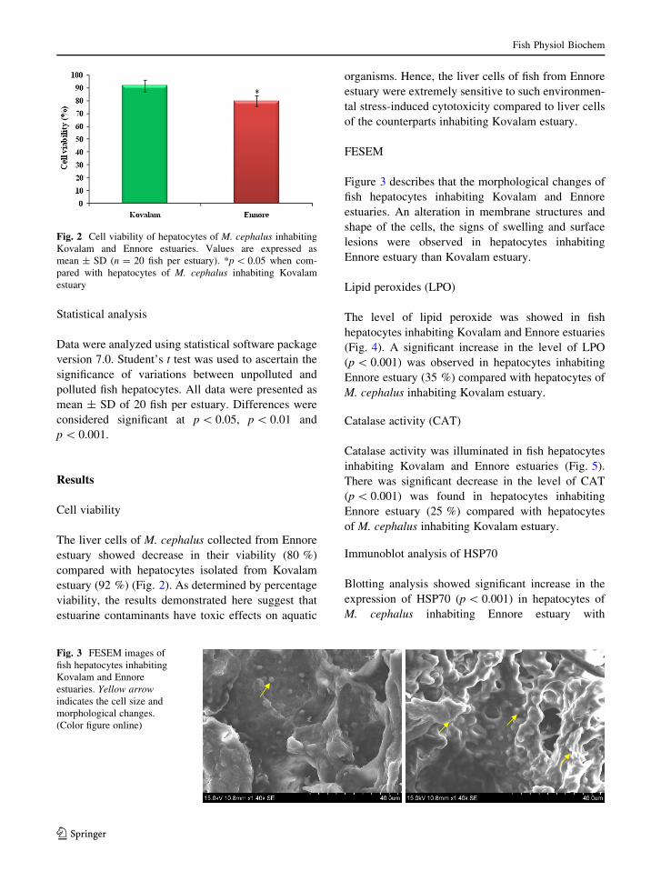
The liver cells of M. cephalus collected from Ennore
estuary showed decrease in their viability (80 %)
compared with hepatocytes isolated from Kovalam
estuary (92 %) (Fig. 2). As determined by percentage
viability, the results demonstrated here suggest that
estuarine contaminants have toxic effects on aquatic
organisms. Hence, the liver cells of fish from Ennore
estuary were extremely sensitive to such environmen-
tal stress-induced cytotoxicity compared to liver cells
of the counterparts inhabiting Kovalam estuary.
FESEM
Figure 3 describes that the morphological changes of
fish hepatocytes inhabiting Kovalam and Ennore
estuaries. An alteration in membrane structures and
shape of the cells, the signs of swelling and surface
lesions were observed in hepatocytes inhabiting
Ennore estuary than Kovalam estuary.
Lipid peroxides (LPO)
The level of lipid peroxide was showed in fish
hepatocytes inhabiting Kovalam and Ennore estuaries
(Fig. 4). A significant increase in the level of LPO
(p \ 0.001) was observed in hepatocytes inhabiting
Ennore estuary (35 %) compared with hepatocytes of
M. cephalus inhabiting Kovalam estuary.
Catalase activity (CAT)
Catalase activity was illuminated in fish hepatocytes
inhabiting Kovalam and Ennore estuaries (Fig. 5).
There was significant decrease in the level of CAT
(p \ 0.001) was found in hepatocytes inhabiting
Ennore estuary (25 %) compared with hepatocytes
of M. cephalus inhabiting Kovalam estuary.
Immunoblot analysis of HSP70
Blotting analysis showed significant increase in the
expression of HSP70 (p \ 0.001) in hepatocytes of
M. cephalus inhabiting Ennore estuary with
Fig. 2 Cell viability of hepatocytes of M. cephalus inhabiting
Kovalam and Ennore estuaries. Values are expressed as
mean ± SD (n = 20 fish per estuary). *p \ 0.05 when com-
pared with hepatocytes of M. cephalus inhabiting Kovalam
estuary
Fig. 3 FESEM images of
fish hepatocytes inhabiting
Kovalam and Ennore
estuaries. Yellow arrow
indicates the cell size and
morphological changes.
(Color figure online)
Fish Physiol Biochem
123

hepatocytes of M. cephalus inhabiting Kovalam
estuary. The representative blots for proteins are
given in Fig. 6.
ELISA of ASK1
The expression of ASK1 (16 %) was insignificantly
increased in hepatocytes of M. cephalus inhabiting
Ennore estuary compared to Kovalam estuary (Fig. 7).
Expression of HSP70 and ASK1 eluted from SDS-
PAGE
The expression of HSP70 and ASK1 (eluted from
SDS-PAGE) was observed in hepatocytes of M.
cephalus inhabiting Kovalam and Ennore estuaries
(Figs. 8, 9). The expression of HSP70 (p \ 0.001) was
significantly increased along with the expression of
ASK1 (15 %) was insignificantly increased in hepa-
tocytes inhabiting Ennore estuary compared to Kova-
lam estuary.
FTIR analysis
FTIR spectra of M. cephalus inhabiting Kovalam and
Ennore estuaries with structural changes of HSP70 and
ASK1 in the 4,000–400 cm-1 range were demon-
strated in Figs. 10 and 11. The spectrum is quite
complex and contains several bands arising from the
contribution of different functional groups belonging
Fig. 4 Level of lipid peroxides in hepatocytes of M. cephalus
inhabiting Kovalam and Ennore estuaries. Values are expressed
as mean ± SD (n = 20 fish per estuary). #p \ 0.001 when
compared with hepatocytes of M. cephalus inhabiting Kovalam
estuary
Fig. 5 Activity of catalase in hepatocytes of M. cephalus
inhabiting Kovalam and Ennore estuaries. Values are expressed
as mean ± SD (n = 20 fish per estuary). @p \ 0.01 when
compared with hepatocytes of M. cephalus inhabiting Kovalam
estuary
Fig. 6 Immunoblot analysis of HSP70 in hepatocytes of M.
cephalus inhabiting Kovalam and Ennore estuaries. b-actin has
been used as the loading control. a Hepatocytes of M. Cephalus
inhabiting Kovalam estuary. b Hepatocytes of M. Cephalus
inhabiting Ennore estuary
Fig. 7 Level of ASK1 in hepatocytes of M. cephalus inhabiting
Kovalam and Ennore estuaries. Values are expressed as
mean ± SD (n = 20 fish per estuary). *p \ 0.05 when com-
pared with hepatocytes of M. cephalus inhabiting Kovalam
estuary
Fig. 8 Immunoblot analysis of HSP70 (eluted from SDS-
PAGE) in hepatocytes of M. cephalus inhabiting Kovalam and
Ennore estuaries. b-actin has been used as the loading control.
c Hepatocytes of M. Cephalus inhabiting Kovalam estuary.
d Hepatocytes of M. Cephalus inhabiting Ennore estuary
Fish Physiol Biochem
123

to protein, lipids and other biomolecules. The absorp-
tion bands and assignments were shown in Tables 1
and 2.
Discussion
Oxidative stress is a state of unbalanced tissue
oxidation characterized by a disturbance in the free
radical and antioxidant systems (Abele and Puntarulo
2004). The redox cycling of heavy metals as well as
their interactions with organic pollutants is docu-
mented as a major contributor to the oxidative stress
resulting from aquatic pollution (Padmini et al. 2009).
The studies on oxidative stress in fish inhabiting
polluted environment have demonstrated significant
pollution impact on various organs of fish like gill
(Padmini and Sudha 2004), brain (Padmini and
Kavitha 2007), liver (Padmini and Usha Rani 2009)
and erythrocytes (Padmini et al. 2006). Accumulation
of damaged and oxidized macromolecules like lipid,
proteins and DNA in various organs would lead to
decrease in reproduction rate, susceptibility to quick
infection and sudden death of fish in large numbers
(Padmini et al. 2004). The enhancement of oxidative
and nitrative stress status and association of this with
alteration in cell viability are also well documented
(Padmini et al. 2009).
Scanning electron microscopy studies are useful for
the evaluation of toxicant-induced changes in liver
cells (Battle et al. 1997). Hence, comparison of fish
hepatocytes inhabiting Kovalam and Ennore estuaries
using FESEM revealed the structural changes. Regular
surfaces, smooth membranes and characteristic
arrangement of some areas were observed in fish
hepatocytes inhabiting Kovalam estuary (Fig. 3).
However, the signs of swelling, surface lesions and
alteration in membrane structures and shape of the
cells were demonstrated in fish hepatocytes inhabiting
Ennore estuary, indicating the impact of pollutant
stress on the surface morphology of the liver. Consis-
tent with the current findings, profound alterations of
cell surface topography, cytoskeletal disruption and
surface blebs formation have been demonstrated under
Fig. 9 Level of ASK1 (eluted from SDS-PAGE) in hepatocytes
of M. cephalus inhabiting Kovalam and Ennore estuaries.
Values are expressed as mean ± SD (n = 20 fish per estuary).
*p \ 0.05 when compared with hepatocytes of M. cephalus
inhabiting Kovalam estuary
Kovalam Ennore
3733
.37
3669
.08
3388
.91
2993
.00
1774
.20
1666
.35
1456
.59
1355
.48
1067
.42
100015002000250030003500
Wavenumber cm-1
0.0
0.1
0.2
0.3
0.4
0.5
0.6
0.7
0.8
Abs
orba
nce
Uni
ts
3778
.13
2889
.12
2828
.51
2396
.44
2351
.04
1834
.92
1773
.40
1681
.80
1648
.83
1539
.76
1515
.45
1364
.62
1339
.68
1244
.00
1056
.93
100015002000250030003500
Wavenumber cm-1
0.00
0.05
0.10
0.15
0.20
0.25
0.30
Abs
orba
nce
Uni
ts
Fig. 10 FTIR spectra of hepatocytes of M. cephalus inhabiting Kovalam and Ennore estuaries with HSP70 expression in the
4,000–400 cm-1 range
Fish Physiol Biochem
123

various stress conditions (Lemasters et al. 1983;
Okanoue et al. 1988).
The present result demonstrates that environmental
pollutants have cytotoxic effects on aquatic organ-
isms, significantly increasing stress, thereby decreas-
ing cell viability in hepatocytes of fish from Ennore
compared to Kovalam. Heavy metals are potent
prooxidants that catalyze lipid peroxidation and may
ultimately lead to oxidative stress (Sampaio et al.
2008). Enhanced free radical production and defective
antioxidant status, a condition usually suggestive of
oxidative/nitrative stress (Tepel et al. 2000), are well
authenticated, and the current results characterized by
increase in LPO along with decrease in CAT levels
confirm both the direction and extent of the previous
observations.
The knowledge of the signaling pathways and
physiological responses to environmental stress con-
dition is essential to understand the mechanism of
adaptation for survival in the stressed condition. HSPs
are a group of inducible proteins some of which are
constitutively expressed and increase in response to
stress, whereas others are expressed only after stress
(Hartl 1996). The induction of increased level of the
stress protein is associated with the development of
resistance to contaminated conditions. It is apparent
that induced stress proteins can act to protect cells
Kovalam Ennore
4000.0 3600 3200 2800 2400 2000 1800 1600 1400 1200 1000 800 600 400.0
0.00
0.1
0.2
0.3
0.4
0.5
0.6
0.7
0.8
0.9
1.0
1.1
1.2
1.3
1.4
1.5
1.6
1.71.75
cm-1
A
3916.8
3903.9
3891.2
3853.6
3838.0
3752.3
3456.1
2927.1
2860.0
2367.7
2345.2 1640.2
4000.0 3600 3200 2800 2400 2000 1800 1600 1400 1200 1000 800 600 400.0
0.00
0.1
0.2
0.3
0.4
0.5
0.6
0.7
0.8
0.9
1.0
1.1
1.2
1.3
1.4
1.5
1.6
1.70
cm-1
A
3916.4
3902.8
3890.5
3852.5
3840.9
3767.6
3723.8
3688.8
3675.6
3647.4
3627.6
3453.0
3064.8
2925.1
2859.9
2779.2
2679.6
2576.1
2483.1
2364.9
2345.5
2275.4
2202.7
2083.1
1653.0
1577.1
1465.6
1248.5
1108.2
1019.0 670.1
Fig. 11 FTIR spectra of hepatocytes of M. cephalus inhabiting Kovalam and Ennore estuaries with ASK1 expression in the
4,000–400 cm-1 range
Table 1 General band assignments of the FTIR spectra of
hepatocytes of M. cephalus inhabiting Kovalam and Ennore
estuaries
Wave number (cm-1) Functional group
*2,828.51 C–H stretch
*2,396.44 C=O stretch
*2,351.04 C=O stretch
*1,834.92 C=O stretch
*1,648.83 C=C stretch
*1,539.76 N–O asymmetric stretch
*1,339.68 N–O symmetric stretch
*1,244 C–N Stretch
Table 2 General band assignments of the FTIR spectra of
hepatocytes of M. cephalus inhabiting Kovalam and Ennore
estuaries
Wave number (cm-1) Functional group
*3,600 to *700 O–H stretch
*3,064.8 C–H stretch
*2,500 to *2,800 C–H stretch
*2,000 to *2,400 C:C and C:N stretch
*1,000 to *1,600 Amide C=O Stretch (1�2� and 3�)
*670 C–H stretch (fingerprint region)
Fish Physiol Biochem
123

from stress-induced damage by preventing protein
denaturation and/or by repairing such damage (Li and
Werb 1982).
Vijayan et al. (1998) have also demonstrated that
significant increase in the inducible HSP70 expression
serves as a sensitive indicator of the cellular stress
response associated with exposure to the contaminant.
Enhanced levels of HSP70 in polluted site fish may
reflect a protective response against environmental
pollutant-related stress. In this study, an increased
expression of HSP70 was observed in fish hepatocytes
inhabiting Ennore estuary (Fig. 6). HSP70 is a pow-
erful chaperon whose expression is induced in
response to a wide variety of physiological and
environmental insults, thus allowing the cell to survive
in lethal conditions. In addition to the protein recovery
functions, HSP70 is also involved in the control of
signaling pathway that control the onset of apoptosis,
as well as protein complexes that are central in the
activation of the apoptotic pathways. HSP70 cytopro-
tective properties may be explained by its anti-
apoptotic function (Rerole et al. 2011). HSP 70
(HSP70) has been shown to act as an inhibitor of
apoptosis (Li et al. 2000).
FTIR spectroscopy is currently used as a detection
tool to study stress-induced changes in the molecular
structure level (Venkataramana et al. 2010). In this
research, FTIR spectra were acquired to detect the
conformational changes and content variations of the
functional groups contributed from protein and lipids of
hepatocytes inhabiting Kovalam and Ennore estuaries.
Figure 10 shows the FTIR spectra of fish hepatocytes
inhabiting Kovalam and Ennore estuaries with structural
changes of HSP70. The bands observed at func-
tional region (*2,828.51, *2,396.44, *2,351.04,
*1,834.92, *1,648.83 and *1,539.76 cm-1) due to
C–H stretch, C=O stretch, C=C stretch and N–O
asymmetric stretching molecules in hepatocytes of M.
cephalus inhabiting Ennore estuaries. The peaks at
*1,339.68 and *1,244 cm-1 were observed in the
fingerprint region due to the presence of deformation and
dipole movement of the molecule in hepatocytes of M.
cephalus inhabiting Ennore estuary.
Apoptosis signal-regulating kinase 1 is a mitogen-
activated protein kinase (MAPK) kinase of the c-Jun
N-terminal kinase (JNK) and p38 MAPK pathways.
ASK1 is preferentially activated by various cytotoxic
stressors and plays pivotal roles in a wide variety of
cellular response to them (Takeda et al. 2008). ASK1
is well-known as a proapoptotic, stress-activated
signaling molecule, and it is under tight regulation at
multiple levels. Molecular chaperons and cochaperons
play a protective role under conditions of cellular
stress by facilitating both refolding and degradation of
misfolded proteins (Gabai et al. 1997; Beere et al.
2000). In addition, HSPs directly modulate stress-
dependent signaling pathways to attenuate cell dam-
age and repress apoptotic events during the response to
stress. ASK1 signaling cascades are regulated by
molecular chaperons. HSP70 affects ASK1 by direct
interactions with JNK (through its peptide-binding
domain) and ASK1 (through the N-terminal ATP-
binding domain). These interactions inhibit JNK and
p38 MAP kinase activities and block ASK1-dependent
apoptosis. The chaperons of the HSP70 family may
inhibit the activity of ASK-1 by having the physical
association with ASK1, thereby inhibiting the homo-
oligomerization of the kinase, and hence act as an
endogenous inhibitor of ASK-1 (Park et al. 2002).
Figure 11 shows the FTIR spectra of fish hepato-
cytes inhabiting Kovalam and Ennore estuaries with
structural changes of ASK1. The ratio of the peak
intensities of the bands observed between *3,600 and
*3,800 cm-1 due to N–H bending and O–H stretch-
ing, respectively, could be used as indicators of the
relative concentration of the protein to water of
biological tissues. The band observed at functional
region *2,000 to *2,400 cm-1 due to C:C and
C:N stretching molecules in hepatocytes of M.
cephalus inhabiting Ennore estuary. The protein
absorption bands mainly located between 1,600 and
1,500 cm-1 contained amide I and amide II bands
(Warnau et al. 1996). The bands between 1,400 and
1,000 cm-1 were of the ‘‘fingerprint’’ region (Pala-
niappan and Renju 2009), amide III and the function
group of nucleic acid and carbohydrates contributed to
these absorption bands in samples. The band at
*670 cm-1 was observed in the fingerprint region
due to the presence of deformation and dipole
movement of the molecule in hepatocytes of M.
cephalus inhabiting Ennore estuary. Overall, the
spectrum of hepatocytes inhabiting Kovalam and
Ennore estuaries with ASK1 protein changes differs
in the shape of absorbance curve, indicating to obvious
changes in structure and contents of biological com-
ponents due to pollution-induced stress.
The analysis of the FTIR spectra collected from
hepatocytes of M. cephalus inhabiting Ennore and
Fish Physiol Biochem
123

Kovalam estuaries revealed that biomolecules were
sensitive to pollution-induced stress. The results of the
current study have provided insight on the stress-
induced conformational changes of biomolecules
including proteins, as well as on the content variation
of these components. The FTIR analysis constructs a
direct link between the functional biomolecules and
the physiological status under heavy metal stress. The
macromolecular characteristics and their contents are
fundamental factors whereby the cell maintains its
normal development and growth. In the present work,
we have clearly demonstrated the functional and
structural changes of protein in the ASK1 signaling
molecule in fish hepatocytes inhabiting Ennore estu-
ary compared to Kovalam estuary. The functional
changes of proteins were also evident from ELISA
study.
The chaperons of the HSP70 family may inhibit the
activity of ASK1 by having physical association
thereby inhibiting the homo-oligomerization of the
kinase and act as an endogenous inhibitor of ASK1
(Park et al. 2002). As HSP70 may inhibit the activity
of ASK1, the expression of ASK1 may be downreg-
ulated and hence there is only 16 % increase in its
expression levels. The evidence presented by the
above study indicates that the overexpression of
HSP70 could reduce ASK1 expression to prevent
stress-induced cellular injury thereby promoting cell
survival in Ennore estuary fish.
Conclusion
The current study results confirm the impact of
environmental pollutant-mediated oxidative stress on
hepatocytes structures. The results also indicated that
the relationship between the degree of antioxidant
deficiency and lipid oxidation could also be used as
biomarkers for toxicity. Upregulation of HSP70, an
early and sensitive biomarker of environmental stress,
downregulates ASK-1 thereby acting as an anti-
apoptotic molecule and promote cell resistance against
contaminating stress-induced apoptosis in the gray
mullet surviving under pollution conditions. In addi-
tion, we demonstrate that HSP70 and ASK-1
expressed under stress conditions show distinct vari-
ation through the FTIR studies which goes to indicate
that tight regulation of signaling proteins can also be
monitored by FTIR. FTIR spectroscopy offers a fast
and efficient tool for detection of qualitative and
quantitative pollutants-induced changes in the context
of molecular structure level in hepatocytes of M.
cephalus. The conformational changes at the biomo-
lecular level indicated by FTIR may be useful tool for
detecting the stress-induced changes.
Acknowledgments The project funded by University Grants
Commission, New Delhi, India. Project referral number-UGC:
41-1281/2012 (SR) is acknowledged.
Conflict of interest The authors report no conflicts of interest.
The authors alone are responsible for the content and writing of
the paper.
References
Abele D, Puntarulo S (2004) Formation of reactive species and
induction of antioxidant defense systems in polar and
temperate marine invertebrates and fish. Comp Biochem
Physiol B 138:405–415
Abele D, Heise K, Portner HO, Puntarulo S (2002) Temperature
dependence of mitochondrial function and production of
reactive oxygen species in the intertidal mud clam Mya
arenaria. J Exp Biol 205:1831–1841
Adams S, Marshall C (1999) Ecological risk assessment in a
large river reservoir: bioindicators of fish population
health. Environ Toxicol Chem 18(4):628–640
Alvarez-Ordonez A, Halisch J, Prieto M (2010) Changes in Fourier
transform infrared spectra of Salmonella enterica serovars
Typhimurium and Enteritidis after adaptation to stressful
growth conditions. Int J Food Microbiol 142:97–105
Battle T, Touchard C, Moulsdale HJ, Dowsett B, Stacey GN
(1997) New cell substrates for in vitro evaluation of micr-
ocystin hepato-cytotoxicity. Toxicol In Vitro 11:557–567
Beere HM, Wolf BB, Cain K, Mosser DD, Mahboubi A,
Kuwana T, Tailor P, Morimoto RI, Cohen GM, Green DR
(2000) Heat-shock protein 70 inhibits apoptosis by pre-
venting recruitment of procaspase-9 to the Apaf-1 apop-
tosome. Nat Cell Biol 2:469–475
Beers RF Jr, Sizer IW (1952) A spectrophotometric method for
measuring the breakdown of hydrogen peroxide by cata-
lase. J Biol Chem 195:133–140
Bradford MM (1976) A rapid and sensitive method for the
quantitation of microgram quantities of protein utilizing
the principle of protein-dye binding. Anal Biochem
72:248–254
Buckley BA, Place SP, Hofmann GE (2004) Regulation of heat
shock genes in isolated hepatocytes from an Antarctic fish,
Trematomus bernacchii. J Exp Biol 207:3649–3656
Ci YX, Gao TY, Jun F, Zhen QG (1999) Fourier transform
infrared spectroscopic characterization of human breast
tissue: implications for breast cancer diagnosis. Appl
Spectrosc 53:312–315
Corte L, Rellini P, Roscini L, Fatichenti F, Cardinali G (2010)
Development of a novel, FTIR (Fourier transform infrared
Fish Physiol Biochem
123

spectroscopy) based, yeast bioassay for toxicity testing and
stress response study. Anal Chim Acta 659:258–265
Das S, Das DK (2006) Antioxidant paradox. SFRR India Bull
5:8–11
Ferreira M, Antunes P, Gil O, Vale C, Reis-Henriques MA
(2004) Organochlorine contaminants in flounder (Platich-
thys flesus) and mullet (Mugil cephalus) from Douro
estuary and their use as sentinel species for environmental
monitoring. Aquat Toxicol 69:347–357
Fischer W, Bianchi G (1984) FAO species identification sheets
for fishery purposes. Western Indian Ocean (fishing area
51), vol 1–6. FAO, Rome
Gabai VL, Meriin AB, Mosser DD, Caron AW, Rits S, Shifrin
VI, Sherman MY (1997) Hsp70 prevents activation of
stress kinases. J Biol Chem 272:18033–18037
Goldman EH, Chen L, Fu H (2003) Activation of apoptosis
signal-regulating kinase 1 by reactive oxygen species
through dephosphorylation at Ser967 and 14–3–3 dissoci-
ation. J Biol Chem 279:10442–10449
Gorbi S, Baldini C, Regoli F (2005) Seasonal variability of
metallothioneins, cytochrome P450, bile metabolites and
oxyradical metabolism in the European eel Anguilla angu-
illa L. (Anguillidae) and striped mullet Mugil cephalus L.
(Mugilidae). Arch Environ Contam Toxicol 49:62–70
Hartl FU (1996) Molecular chaperones in cellular protein
folding. Nature 381:571–579
Hwang JR, Zhang C, Patterson C (2005) C-terminus of heat
shock protein 70-interacting protein facilitates degradation
of apoptosis signal-regulating kinase 1 and inhibits apop-
tosis signal-regulating kinase 1-dependent apoptosis. Cell
Stress Chaperon 10(2):147–156
Iwama GK, Thomas PT, Forsyth RB, Vijayan MM (1998) Heat
shock protein expression in fish. Rev Fish Biol Fish
8:35–56
Krumschnabel G, Schwarzbaum PJ, Wieser W (1994) Coupling
of energy supply and energy demand in isolated goldfish
hepatocytes. Physiol Zool 67:438–448
Laemmli UK (1970) Cleavage of structural proteins during the
assembly of the head of bacteriophage T4. Nature
227:680–685
Lemasters JJ, Stemkowski CJ, Ji S, Thurman RG (1983) Cell
surface changes and enzyme release during hypoxia and
reoxygenation in the isolated, perfused rat liver. J Cell Biol
97:778–786
Li GC, Werb Z (1982) Correlation between synthesis of het
shock proteins and development of thermotolerance in
chinese hamster fibroblasts. Proc Natl Acad Sci USA
79:3218–3222
Li CY, Lee JS, Ko YG, Kim JI, Seo JS (2000) Heat shock protein
70 inhibits apoptosis downstream of cytochrome c release
and upstream of caspase-3 activation. J Biol Chem
275:25665–25671
Morita K, Saitoh M, Tobiume K, Matsuura H, Enomoto S,
Nishitoh H, Ichijo H (2001) Negative feedback regulation
of ASK1 by protein phosphatase 5 (PP5) in response to
oxidative stress. EMBO J 20:6028–6036
Ohkawa H, Ohishi N, Yagi K (1970) Assay for lipid peroxides in
animal tissue with thiobarbituric acid reaction. Anal Bio-
chem 95:352–358
Okanoue T, Ohta M, Kachi K, Ohta Y, Sawa Y, Kanaoka H,
Kagawa K, Takino T, French SW (1988) Intermediate
filaments of hepatocytes and biliary epithelial cells in bile
duct obstruction: transmission and scanning electron
microscope study. Gastroenterol Jpn 23:428–434
Pacheco M, Santos MA, Teles M, Oliveira M, Rebelo JE,
Pombo L (2005) Biotransformation and genotoxic bio-
markers in mullet species (Liza sp.) from a contaminated
coastal lagoon (Ria de Aveiro, Portugal). Environ Monit
Assess 107:133–153
Padmini E, Kavitha M (2005) Contaminant induced stress
impact on biochemical changes in brain of estuarine grey
mullets. Pollut Res 24:647–651
Padmini E, Kavitha M (2007) Comparative assessment of con-
taminant induced oxidative stress in brain of Mugil ceph-
alus. Environ Poll Con J 10:75–79
Padmini E, Sudha D (2004) Environmental impact on gill mito-
chondrial function in Mugil cephalus. Aquacult 5:89–92
Padmini E, Usha Rani M (2008) Impact of seasonal variation on
HSP70 expression quantitated in stressed fish hepatocytes.
Comp Biochem Physiol B 151:278–285
Padmini E, Usha Rani M (2009) Evaluation of oxidative stress
biomarkers in hepatocytes of grey mullet inhabiting natural
and polluted estuaries. Sci Total Environ 407:4533–4541
Padmini E, Vijaya Geetha B (2007a) A comparative seasonal
pollution assessment study on estuary with respect to metal
accumulation in Mugil cephalus. Oceanol Hydrobiol Stud
35:1–13
Padmini E, Vijaya Geetha B (2007b) Seasonal influences on
water quality parameters and pollution status of the Ennore
estuary, Tamilnadu, India. J Environ Hydrol 15:1–9
Padmini E, Thendral Hepsibha B, Shanthalin Shellomith AS
(2004) Lipid alteration as stress markers in grey mullets
(Mugil cephalus Linnaeus) caused by industrial effluents in
Ennore estuary. Aquacult 5:115–118
Padmini E, Sridevi S, Vijaya Geetha B (2006) Environmental
stress in Ennore estuary and enhanced erythrocyte micro-
nuclei formation in mullets. Environ Poll Con J 9:51–56
Padmini E, Usha Rani M, Vijaya Geetha B (2009) Studies on
antioxidant status in Mugil cephalus in response to heavy
metal pollution at Ennore estuary. Environ Monit Assess
155:215–225
Palaniappan PLRM, Renju VB (2009) FTIR study of the effect
of zinc exposure on the biochemical contents of the muscle
of Labeo rohita. Infrared Phys Technol 52:37–41
Park HS, Cho SG, Kim CK, Hwang HS, Noh KT, Kim MS, Huh
SH, Kim MJ, Ryoo K, Kim EK, Kang WJ, Lee JS, Seo JS,
Ko YG, Kim S, Choi EJ (2002) Hsp72 is a negative regu-
lator of apoptosis signal regulating kinase1. Mol Cell Biol
22:7721–7730
Raghunathan MB, Srinivasan M (1983) Zooplankton dynamics
and hydrographic features of Ennore estuary, Madras. Rec
Zool Sur India 40:1–30
Rerole AL, Jego G, Garrido C (2011) Hsp70: anti-apoptotic and
tumorigenic protein. Methods Mol Biol 787:205–230
Samali A, Orrenius S (1998) Heat shock proteins: regulations of
stress response and apoptosis. Cell Stress Chaperon
3:228–236
Sampaio FG, Boijink CDL, Oba EL, Santos LRBD, Kalinin AL,
Rantin FT (2008) Antioxidant defenses and biochemical
changes in pacu (Piaractus mesopotamicus) in response to
single and combined copper and hypoxia exposure. Comp
Biochem Physiol C 147:43–51
Fish Physiol Biochem
123

Strober W (2001) Trypan blue exclusion test of cell viability.
Curr Protoc Immunol May, Appendix 3, Appendix 3B
18432654 (P, S, E, B, D)
Takeda K, Noguchi T, Naguro I, Ichijo H (2008) Apoptosis
signal-regulating kinase 1 in stress and immune response.
Annu Rev Pharmacol Toxicol 48:199–225
Tepel M, Echelmeyer M, Orie NN, Zidek W (2000) Increased
intracellular reactive oxygen species in patients with end-
stage renal failure: effect of hemodialysis. Kidney Int
58:867–872
Towbin H, Staehelin T, Gordon J (1979) Electrophoretic
transfer of proteins from polyacrylamide gels to nitrocel-
lulose sheets: procedure and some applications. Proc Natl
Acad Sci USA 76:4350–4354
Venkataramana GV, Komal Kumar J, Devi Prasad AG, Karimi
P (2010) Fourier transform infrared spectroscopic study on
liver of freshwater fish Oreochromis mossambicus. Rom J
Biophys 20(4):315–322
Vijayan MM, Pereira C, Kruzynski G, Iwama GK (1998) Sub-
lethal concentrations of contaminant induce the expression
of hepatic heat shock protein 70 in 2 salmonids. Aquat
Toxicol 40:101–108
Warnau M, Temera A, Jangoux M, Dubois P, Iaccarino M, De-
Biase A, Temara A, Pagano G (1996) Spermiotoxicity and
embryotoxicity of heavy metals in the echinoid Paracen-
trotus lividus. Environ Toxicol Chem 15:1931–1936
Zugel U, Kaufmann SHE (1999) Role of heat shock proteins in
protection from and pathogenesis of infectious diseases.
Clin Microbiol Rev 12:19–39
Fish Physiol Biochem
123