heel pain plantar fasciitis: revision 2014 pain... · heel pain—plantar fasciitis: revision 2014...
TRANSCRIPT

Clinical Practice Guidelines
ROBROY L. MARTIN, PT, PhD • TODD E. DAVENPORT, DPT • STEPHEN F. REISCHL, DPT • THOMAS G. MCPOIL, PT, PhDJAMES W. MATHESON, DPT • DANE K. WUKICH, MD • CHRISTINE M. MCDONOUGH, PT, PhD
Heel Pain—Plantar Fasciitis: Revision 2014
Clinical Practice Guidelines Linked to the International Classification of Functioning,
Disability and Health From the Orthopaedic Section of the American Physical Therapy Association
J Orthop Sports Phys Ther. 2014;44(11):A1-A23. doi:10.2519/jospt.2014.0303
REVIEWERS: Roy D. Altman, MD • Paul Beattie, PT, PhD • Mark Cornwall, PT, PhDIrene Davis, PT, PhD • John DeWitt, DPT • James Elliott, PT, PhD • James J. Irrgang, PT, PhD
Sandra Kaplan, PT, PhD • Stephen Paulseth, DPT, MS • Leslie Torburn, DPT • James Zachazewski, DPT
For author, coordinator, contributor, and reviewer affiliations, see end of text. Copyright ©2014 Orthopaedic Section, American Physical Therapy Association (APTA), Inc, and the Journal of Orthopaedic & Sports Physical Therapy®. The Orthopaedic Section, APTA, Inc, and the Journal of Orthopaedic & Sports Physical Therapy consent to the reproduction and distribution of this guideline for educational purposes. Address correspondence to: Joseph Godges, DPT, ICF-based Clinical Practice Guidelines Coordinator, Orthopaedic Section, APTA, Inc, 2920 East Avenue South, Suite 200, La Crosse, WI 54601. E-mail: [email protected]
SUMMARY OF RECOMMENDATIONS . . . . . . . . . . . . . . . . . . . . . . . . . . . . . . A2
INTRODUCTION. . . . . . . . . . . . . . . . . . . . . . . . . . . . . . . . . . . . . . . . . . . . . . . . . . . . . . . . . . . . A3
METHODS . . . . . . . . . . . . . . . . . . . . . . . . . . . . . . . . . . . . . . . . . . . . . . . . . . . . . . . . . . . . . . . . . . . A4
CLINICAL GUIDELINES: Impairment/Function-Based Diagnosis . . . . . . . . . . . . . . . . . . A7
CLINICAL GUIDELINES: Examination . . . . . . . . . . . . . . . . . . . . . . . . . . . . . . . . . . . . . . . . . . . . . . . . . . . . . . . . . . . A10
CLINICAL GUIDELINES: Interventions . . . . . . . . . . . . . . . . . . . . . . . . . . . . . . . . . . . . . . . . . . . . . . . . . . . . . . . . . . . A11
AUTHOR/REVIEWER AFFILIATIONS AND CONTACTS . . . . . . A20
REFERENCES . . . . . . . . . . . . . . . . . . . . . . . . . . . . . . . . . . . . . . . . . . . . . . . . . . . . . . . . . . . . . A21
44-11 Guidelines.indd 1 10/20/2014 7:10:39 PM
Jour
nal o
f O
rtho
paed
ic &
Spo
rts
Phys
ical
The
rapy
®
Dow
nloa
ded
from
ww
w.jo
spt.o
rg a
t on
Dec
embe
r 26
, 201
4. F
or p
erso
nal u
se o
nly.
No
othe
r us
es w
ithou
t per
mis
sion
. C
opyr
ight
© 2
014
Jour
nal o
f O
rtho
paed
ic &
Spo
rts
Phys
ical
The
rapy
®. A
ll ri
ghts
res
erve
d.

Heel Pain—Plantar Fasciitis: Clinical Practice Guidelines Revision 2014
a2 | november 2014 | volume 44 | number 11 | journal of orthopaedic & sports physical therapy
RISK FACTORS
Clinicians should assess the presence of limited ankle dorsiflexion range of motion, high body mass index in
nonathletic individuals, running, and work-related weight-bearing activities—particularly under conditions with poor shock absorp-tion—as risk factors for the development of heel pain/plantar fasciitis.
DIAGNOSIS/CLASSIFICATION
Physical therapists should diagnose the International Clas-sification of Diseases (ICD) category of plantar fasciitis
and the associated International Classification of Functioning, Disability and Health (ICF) impairment-based category of heel pain (b28015 Pain in lower limb, b2804 Radiating pain in a segment or region) using the following history and physical examination findings:
• Plantar medial heel pain: most noticeable with initial steps after a period of inactivity but also worse following prolonged weight bearing
• Heel pain precipitated by a recent increase in weight-bearing activity
• Pain with palpation of the proximal insertion of the plantar fascia
• Positive windlass test• Negative tarsal tunnel tests• Limited active and passive talocrural joint dorsiflexion range
of motion• Abnormal Foot Posture Index score• High body mass index in nonathletic individuals
DIFFERENTIAL DIAGNOSIS
Clinicians should assess for diagnostic classifications other than heel pain/plantar fasciitis, including spondyloarthritis,
fat-pad atrophy, and proximal plantar fibroma, when the individual’s reported activity limitations or impairments of body function and structure are not consistent with those presented in the Diagnosis/Classification section of this guideline, or when the individual’s symptoms are not resolving with interventions aimed at normaliza-tion of the individual’s impairments of body function.
EXAMINATION – OUTCOME MEASURES
Clinicians should use the Foot and Ankle Ability Measure (FAAM), Foot Health Status Questionnaire (FHSQ), or the
Foot Function Index (FFI) and may use the computer-adaptive ver-sion of the Lower Extremity Functional Scale (LEFS) as validated self-report questionnaires before and after interventions intended to alleviate the physical impairments, activity limitations, and participation restrictions associated with heel pain/plantar fasciitis.
EXAMINATION – ACTIVITY LIMITATION AND PARTICIPATION RESTRICTION MEASURES
Clinicians should utilize easily reproducible performance-based measures of activity limitation and participation re-
striction measures to assess changes in the patient’s level of function associated with heel pain/plantar fasciitis over the episode of care.
EXAMINATION – PHYSICAL IMPAIRMENT MEASURES
When evaluating a patient with heel pain/plantar fasciitis over an episode of care, assessment of impairment of body
function should include measures of pain with initial steps after a pe-riod of inactivity and pain with palpation of the proximal insertion of the plantar fascia, and may include measures of active and passive ankle dorsiflexion range of motion and body mass index in nonath-letic individuals.
INTERVENTIONS – MANUAL THERAPY
Clinicians should use manual therapy, consisting of joint and soft tissue mobilization, procedures to treat relevant lower
extremity joint mobility and calf flexibility deficits and to decrease pain and improve function in individuals with heel pain/plantar fasciitis.
INTERVENTIONS – STRETCHING
Clinicians should use plantar fascia–specific and gastroc-nemius/soleus stretching to provide short-term (1 week to
4 months) pain relief for individuals with heel pain/plantar fasciitis. Heel pads may be used to increase the benefits of stretching.
INTERVENTIONS – TAPING
Clinicians should use antipronation taping for immediate (up to 3 weeks) pain reduction and improved function for
individuals with heel pain/plantar fasciitis. Additionally, clinicians may use elastic therapeutic tape applied to the gastrocnemius and plantar fascia for short-term (1 week) pain reduction.
INTERVENTIONS – FOOT ORTHOSES
Clinicians should use foot orthoses, either prefabricated or custom fabricated/fitted, to support the medial longitudinal
arch and cushion the heel in individuals with heel pain/plantar fasci-itis to reduce pain and improve function for short- (2 weeks) to long-term (1 year) periods, especially in those individuals who respond positively to antipronation taping techniques.
INTERVENTIONS – NIGHT SPLINTS
Clinicians should prescribe a 1- to 3-month program of night splints for individuals with heel pain/plantar fasciitis who
consistently have pain with the first step in the morning.
Summary of Recommendations*
B
B
C
A
F
B
A
A
A
A
A
44-11 Guidelines.indd 2 10/20/2014 7:10:39 PM
Jour
nal o
f O
rtho
paed
ic &
Spo
rts
Phys
ical
The
rapy
®
Dow
nloa
ded
from
ww
w.jo
spt.o
rg a
t on
Dec
embe
r 26
, 201
4. F
or p
erso
nal u
se o
nly.
No
othe
r us
es w
ithou
t per
mis
sion
. C
opyr
ight
© 2
014
Jour
nal o
f O
rtho
paed
ic &
Spo
rts
Phys
ical
The
rapy
®. A
ll ri
ghts
res
erve
d.

Heel Pain—Plantar Fasciitis: Clinical Practice Guidelines Revision 2014
journal of orthopaedic & sports physical therapy | volume 44 | number 11 | november 2014 | a3
AIM OF THE GUIDELINES
The Orthopaedic Section of the American Physical Therapy
Association (APTA) has an ongoing effort to create evidence-
based clinical practice guidelines (CPGs) for orthopaedic
physical therapy management of patients with musculoskel-
etal impairments described in the World Health Organiza-
tion’s International Classification of Functioning, Disability
and Health (ICF).97
Introduction
INTERVENTIONS – PHYSICAL AGENTS
Electrotherapy: clinicians should use manual therapy, stretching, and foot orthoses instead of electrotherapeutic
modalities, to promote intermediate and long-term (1-6 months) improvements in clinical outcomes for individuals with heel pain/plantar fasciitis. Clinicians may or may not use iontophoresis with dexamethasone or acetic acid to provide short-term (2-4 weeks) pain relief and improved function.
Low-level laser: clinicians may use low-level laser therapy to reduce pain and activity limitations in individuals with heel
pain/plantar fasciitis.
Phonophoresis: clinicians may use phonophoresis with keto-profen gel to reduce pain in individuals with heel pain/plantar
fasciitis.
Ultrasound: the use of ultrasound cannot be recommended for individuals with heel pain/plantar fasciitis.
INTERVENTIONS – FOOTWEAR
To reduce pain in individuals with heel pain/plantar fasciitis, clinicians may prescribe (1) a rocker-bottom shoe construc-
tion in conjunction with a foot orthosis, and (2) shoe rotation during the work week for those who stand for long periods.
INTERVENTIONS – EDUCATION AND COUNSELING FOR WEIGHT LOSS
Clinicians may provide education and counseling on exercise strategies to gain or maintain optimal lean body mass in
individuals with heel pain/plantar fasciitis. Clinicians may also refer individuals to an appropriate health care practitioner to address nutrition issues.
INTERVENTIONS – THERAPEUTIC EXERCISE AND NEUROMUSCULAR RE-EDUCATION
Clinicians may prescribe strengthening exercises and movement training for muscles that control pronation and
attenuate forces during weight-bearing activities.
INTERVENTIONS – DRY NEEDLING
The use of trigger point dry needling cannot be recommend-ed for individuals with heel pain/plantar fasciitis.
*These recommendations and clinical practice guidelines are based on the scientific literature published prior to January 2013.
Summary of Recommendations* (continued)
D
C
C
C
C
E
F
F
List of Acronyms
APTA: American Physical Therapy AssociationCI: confidence intervalCPG: clinical practice guidelineESWT: extracorporeal shockwave therapyFAAM: Foot and Ankle Ability MeasureFFI: Foot Function IndexFHSQ: Foot Health Status QuestionnaireFPI-6: Foot Posture Index-6ICD: International Classification of Diseases
ICF: International Classification of Functioning, Disability and HealthICSI: intralesional corticosteroid injectionLEFS: Lower Extremity Functional ScaleMCID: minimal clinically important differenceNSAID: nonsteroidal anti-inflammatory drugSF-36: Medical Outcomes Study 36-Item Short-Form Health SurveyVAS: visual analog scale
44-11 Guidelines.indd 3 10/20/2014 7:10:40 PM
Jour
nal o
f O
rtho
paed
ic &
Spo
rts
Phys
ical
The
rapy
®
Dow
nloa
ded
from
ww
w.jo
spt.o
rg a
t on
Dec
embe
r 26
, 201
4. F
or p
erso
nal u
se o
nly.
No
othe
r us
es w
ithou
t per
mis
sion
. C
opyr
ight
© 2
014
Jour
nal o
f O
rtho
paed
ic &
Spo
rts
Phys
ical
The
rapy
®. A
ll ri
ghts
res
erve
d.

Heel Pain—Plantar Fasciitis: Clinical Practice Guidelines Revision 2014
a4 | november 2014 | volume 44 | number 11 | journal of orthopaedic & sports physical therapy
The purposes of these clinical guidelines are to:• Describe evidence-based physical therapy practice, includ-
ing diagnosis, prognosis, intervention, and assessment of outcome for musculoskeletal disorders commonly managed by orthopaedic physical therapists
• Classify and define common musculoskeletal conditions using the World Health Organization’s terminology relat-ed to impairments of body function and body structure, activity limitations, and participation restrictions
• Identify interventions supported by current best evidence to address impairments of body function and structure, activity limitations, and participation restrictions associ-ated with common musculoskeletal conditions
• Identify appropriate outcome measures to assess chang-es resulting from physical therapy interventions in body function and structure as well as in activity and participa-tion of the individual
• Provide a description to policy makers, using internation-ally accepted terminology, of the practice of orthopaedic physical therapists
• Provide information for payers and claims reviewers regarding the practice of orthopaedic physical therapy for common musculoskeletal conditions
• Create a reference publication for orthopaedic physical therapy clinicians, academic instructors, clinical instruc-tors, students, interns, residents, and fellows regarding the best current practice of orthopaedic physical therapy
STATEMENT OF INTENT
These guidelines are not intended to be construed or to serve as a standard of medical care. Standards of care are determined on the basis of all clinical data available for an individual patient and are subject to change as scientific knowledge and technology advance and patterns of care evolve. These parameters of prac-tice should be considered guidelines only. Adherence to them will not ensure a successful outcome in every patient, nor should they be construed as including all proper methods of care or excluding other acceptable methods of care aimed at the same results. The ultimate judgment regarding a particular clinical procedure or treatment plan must be made based on clinician experience and expertise in light of the clinical presentation of the patient; the available evidence; the available diagnostic and treatment options; and the patient’s values, expectations, and preferences. However, we suggest that significant departures from accepted guidelines should be documented in the patient’s medical records at the time the relevant clinical decision is made.
Introduction (continued)
Content experts were appointed by the Orthopaedic Section, APTA to conduct a review of the literature and to develop an updated heel pain/plantar fasciitis CPG as indicated by the current state of the evidence in the field. The aims of the revision were to provide a concise summary of the evi-dence since publication of the original guideline and to de-velop new recommendations or revise previously published recommendations to support evidence-based practice. The authors of this guideline revision worked with research li-brarians with expertise in systematic review to perform a systematic search for concepts associated with heel pain or plantar fasciitis in articles published since 2007 related to classification, examination, and intervention strategies for heel pain or plantar fasciitis, consistent with previous guide-line development methods related to ICF classification.91 Briefly, the following databases were searched from 2007 to between December 13 and 19, 2012: MEDLINE (PubMed) (2007 to date), Cochrane Library (2007 to date), Web of Sci-ence (2007 to date), CINAHL (2007 to date), ProQuest Dis-sertations and Theses (2007 to date), PEDro (2007 to date),
and ProQuest Nursing and Allied Health Source (2007 to date). See APPENDIX A (available online) for full search strate-gies and APPENDIX B (available online) for search dates and results.
The authors declared relationships and developed a conflict management plan, which included submitting a conflict-of-interest form to the Orthopaedic Section, APTA. Articles that were authored by a reviewer were assigned to an al-ternate reviewer. Funding was provided to the CPG devel-opment team for travel and expenses for CPG development training. The CPG development team maintained editorial independence.
Articles contributing to recommendations were reviewed based on specified inclusion and exclusion criteria, with the goal of identifying evidence relevant to physical therapist clin-ical decision making for adult persons with heel pain/plantar fasciitis. The title and abstract of each article were reviewed independently by 2 members of the CPG development team
Methods
44-11 Guidelines.indd 4 10/20/2014 7:10:40 PM
Jour
nal o
f O
rtho
paed
ic &
Spo
rts
Phys
ical
The
rapy
®
Dow
nloa
ded
from
ww
w.jo
spt.o
rg a
t on
Dec
embe
r 26
, 201
4. F
or p
erso
nal u
se o
nly.
No
othe
r us
es w
ithou
t per
mis
sion
. C
opyr
ight
© 2
014
Jour
nal o
f O
rtho
paed
ic &
Spo
rts
Phys
ical
The
rapy
®. A
ll ri
ghts
res
erve
d.

Heel Pain—Plantar Fasciitis: Clinical Practice Guidelines Revision 2014
journal of orthopaedic & sports physical therapy | volume 44 | number 11 | november 2014 | a5
for inclusion. See APPENDIX C (available online) for inclusion and exclusion criteria. Full-text review was then similarly conducted to obtain the final set of articles for contribution to recommendations. The team leader (R.L.M.) provided the final decision for discrepancies that were not resolved by the review team. See APPENDIX D (available online) for a flow chart of articles and APPENDIX E (available online) for articles includ-ed in recommendations by topic. For selected relevant topics that were not appropriate for the development of recommen-dations, such as shockwave therapy, injection, and imaging, articles were not subject to the systematic review process and were not included in the flow chart. Evidence tables for this CPG are available on the CPG pages of the Orthopaedic Section of the APTA's website (www.orthopt.org).
This guideline was issued in 2014 based on the published literature up to December 2012. This guideline will be con-sidered for review in 2017, or sooner if new evidence becomes available. Any updates to the guideline in the interim peri-od will be noted on the Orthopaedic Section of the APTA's website (www.orthopt.org).
LEVELS OF EVIDENCEIndividual clinical research articles were graded accord-ing to criteria adapted from the Centre for Evidence-based Medicine, Oxford, UK for diagnostic, prospective, and therapeutic studies.62 In 3 teams of 2, each reviewer independently assigned a level of evidence and evaluated the quality of each article using a critical appraisal tool. See APPENDICES F and G (available online) for the evidence table and details on procedures used for assigning levels of evidence. An abbreviated version of the grading system is provided below.
IEvidence obtained from high-quality diagnostic studies, prospective studies, or randomized controlled trials
II
Evidence obtained from lesser-quality diagnostic studies, prospective studies, or randomized controlled trials (eg, weaker diagnostic criteria and reference standards, improper randomization, no blinding, less than 80% follow-up)
III Case-control studies or retrospective studies
IV Case series
V Expert opinion
GRADES OF EVIDENCEThe strength of the evidence supporting the recommenda-tions was graded according to the previously established methods for the original guideline and those provided be-low. Each team developed recommendations based on the
strength of evidence, including how directly the studies ad-dressed the question and heel pain/plantar fasciitis popu-lation. In developing their recommendations, the authors considered the strengths and limitations of the body of evi-dence and the health benefits, side effects, and risks of tests and interventions.
GRADES OF RECOMMENDATION BASED ON STRENGTH OF EVIDENCE
AStrong evidence A preponderance of level I and/or level II
studies support the recommendation. This must include at least 1 level I study
BModerate evidence
A single high-quality randomized controlled trial or a preponderance of level II studies support the recommendation
C
Weak evidence A single level II study or a preponderance of level III and IV studies, including statements of consensus by content experts, support the recommendation
D
Conflicting evidence
Higher-quality studies conducted on this topic disagree with respect to their conclusions. The recommendation is based on these conflicting studies
E
Theoretical/ foundational evidence
A preponderance of evidence from animal or cadaver studies, from conceptual models/principles, or from basic science/bench research supports this conclusion
FExpert opinion Best practice based on the clinical
experience of the guidelines- development team
REVIEW PROCESSThe Orthopaedic Section, APTA selected content experts and stakeholders to serve as reviewers of the early drafts of these CPGs. The draft was posted for public comment on the web-site of the Orthopaedic Section of the APTA. The authors used the feedback from the reviewer and website comments to inform final revisions.
CLASSIFICATIONThe primary International Classification of Diseases 10th re-vision (ICD-10) code and condition associated with heel pain is M72.2 Plantar fascial fibromatosis/Plantar fasciitis.96 Secondary ICD-10 codes and conditions associated with heel pain are G57.5 Tarsal tunnel syndrome and G57.6 Lesion of plantar nerve/Morton’s metatarsalgia.96
The primary ICF body function codes associated with plantar fasciitis, tarsal tunnel syndrome, and plantar nerve lesions are the sensory functions related to pain. These body function
Methods (continued)
44-11 Guidelines.indd 5 10/20/2014 7:10:40 PM
Jour
nal o
f O
rtho
paed
ic &
Spo
rts
Phys
ical
The
rapy
®
Dow
nloa
ded
from
ww
w.jo
spt.o
rg a
t on
Dec
embe
r 26
, 201
4. F
or p
erso
nal u
se o
nly.
No
othe
r us
es w
ithou
t per
mis
sion
. C
opyr
ight
© 2
014
Jour
nal o
f O
rtho
paed
ic &
Spo
rts
Phys
ical
The
rapy
®. A
ll ri
ghts
res
erve
d.
Heel Pain—Plantar Fasciitis: Clinical Practice Guidelines Revision 2014
a6 | november 2014 | volume 44 | number 11 | journal of orthopaedic & sports physical therapy
Methods (continued)
codes are b28015 Pain in lower limb and b2804 Radiating pain in a segment or region.
The primary ICF body structure codes associated with plan-tar fasciitis are s75023 Ligaments and fasciae of ankle and foot and s75028 Structures of ankle and foot, neural.
The primary ICF activities and participation codes associated with plantar fasciitis are d4500 Walking short distances, d4501 Walking long distances, and d4154 Maintaining a standing position.
A comprehensive list of codes was published in the previous guideline.56
ORGANIZATION OF THE GUIDELINEFor each topic, the summary recommendation and grade of evidence from the 2008 guideline are presented, followed by a synthesis of the recent literature with the corresponding evidence levels. Each topic concludes with the 2014 summary recommendation and its updated grade of evidence.
44-11 Guidelines.indd 6 10/20/2014 7:10:41 PM
Jour
nal o
f O
rtho
paed
ic &
Spo
rts
Phys
ical
The
rapy
®
Dow
nloa
ded
from
ww
w.jo
spt.o
rg a
t on
Dec
embe
r 26
, 201
4. F
or p
erso
nal u
se o
nly.
No
othe
r us
es w
ithou
t per
mis
sion
. C
opyr
ight
© 2
014
Jour
nal o
f O
rtho
paed
ic &
Spo
rts
Phys
ical
The
rapy
®. A
ll ri
ghts
res
erve
d.
Heel Pain—Plantar Fasciitis: Clinical Practice Guidelines Revision 2014
journal of orthopaedic & sports physical therapy | volume 44 | number 11 | november 2014 | a7
PREVALENCE2008 SummaryPlantar fasciitis is the most common foot condition treated by health care providers. It has been estimated that plantar fasciitis occurs in approximately 2 million Americans each year and af-fects as much as 10% of the population over the course of a life-time. In 2000, the Foot and Ankle Special Interest Group of the Orthopaedic Section, APTA surveyed over 500 members and received responses from 117 therapists. Of those responding, 100% indicated that plantar fasciitis was the most common foot condition seen in their clinic. Rome et al68 reported that plantar fasciitis accounts for 15% of all adult foot complaints requir-ing professional care and is prevalent in both nonathletic and athletic populations. Taunton et al82 conducted a retrospective case-control analysis of 2002 individuals with running-related injuries who were referred to the same sports medicine center. They reported that plantar fasciitis was the most common con-dition diagnosed in the foot and represented 8% of all injuries.
Evidence UpdateA systematic review of ankle and foot overuse in-juries occurring in numerous sporting activities (54 851 athletes in total) found that 50% of the
studies included in the review involved participation in soc-cer, running, gymnastics, and dance.76 In this review, Achilles tendinopathy, plantar fasciitis, and stress fractures were the most commonly reported injuries.76
In a systematic review assessing the frequency of running-related musculoskeletal injuries (8 stud-ies; pooled n = 3500 runners), the incidence of
plantar fasciitis ranged from 4.5% to 10%, with the preva-lence ranging from 5.2% to 17.5%.50
In a 2-year longitudinal cohort study involving 3206 individuals ranging from 20 to more than 75 years of age living in southern Australia, 17.4% reported hav-
ing foot pain.33 Of these individuals, the hind foot was the second most common site of pain, with the highest prevalence noted in those 20 to 34 years of age and greater than 75 years of age.33
In a retrospective assessment of previous overuse injuries in 748 high school runners (aged 13 to 18 years), 481 runners reported a previous injury.83
Plantar fasciitis accounted for 8% of the reported previ-ous injuries, with the incidence being greater in female runners.83
In a prospective assessment of nontraumatic foot and lower-limb injuries in 166 runners involved in various running specialties, 98 (59%) indicated
they had developed an overuse injury, with 30 (31%) report-ing plantar fasciitis.19
2014 SummaryThe prevalence of pain in the hind foot or heel region is high in both nonathletic and athletic populations. In athletic pop-ulations, plantar fasciitis is a common injury reported by high school, competitive, and recreational distance runners.
PATHOANATOMICAL FEATURES2008 SummaryClinicians should assess for impairments in muscles, ten-dons, and nerves, as well as the plantar fascia, when a patient presents with heel pain.
2014 SummaryIncreased plantar fascia thickness was found to be associ-ated with symptoms22,92,98 and altered compressive proper-ties of the fat pad in those with plantar heel pain.93 Changes in plantar fascia thickness were found to be positively as-sociated with changes in pain levels for individuals with plantar fasciitis receiving treatment.52 In individuals with general foot- and ankle-related disability, pain-related fear of movement was the strongest single contributor to dis-ability.48 An area of future research may be fear-avoidance behaviors and their role in disability in individuals with plantar fasciitis.48,79
CLINICAL COURSE2008 SummaryBased on long-term follow-up data in case series composed primarily of patients seen in an orthopaedic outpatient set-ting, the clinical course for most patients was positive, with 80% reporting resolution of symptoms within a 12-month period.55,95
CLINICAL GUIDELINES
Impairment/Function-Based Diagnosis
II
II
III
III
III
44-11 Guidelines.indd 7 10/20/2014 7:10:41 PM
Jour
nal o
f O
rtho
paed
ic &
Spo
rts
Phys
ical
The
rapy
®
Dow
nloa
ded
from
ww
w.jo
spt.o
rg a
t on
Dec
embe
r 26
, 201
4. F
or p
erso
nal u
se o
nly.
No
othe
r us
es w
ithou
t per
mis
sion
. C
opyr
ight
© 2
014
Jour
nal o
f O
rtho
paed
ic &
Spo
rts
Phys
ical
The
rapy
®. A
ll ri
ghts
res
erve
d.

Heel Pain—Plantar Fasciitis: Clinical Practice Guidelines Revision 2014
a8 | november 2014 | volume 44 | number 11 | journal of orthopaedic & sports physical therapy
2014 SummaryHeel pain/plantar fasciitis usually presents as a chronic con-dition, with symptom duration greater than 1 year prior to seeking treatment. In 2 retrospective cohort studies involving 432 individuals diagnosed with chronic plantar heel pain, the mean duration of symptoms ranged from 13.3 to 14.1 months.39,99
RISK FACTORS2008 Recommendation
Clinicians should consider limited ankle dorsiflex-ion range of motion and a high body mass index in nonathletic populations as factors predisposing
patients to the development of heel pain/plantar fasciitis.
Evidence UpdateRunning was found to be a risk factor for devel-oping plantar fasciitis.50,76 Street running, spiked shoes, cavus foot, and hind-foot varus were related
to the onset of plantar fasciitis in a group of runners.19
Other studies have also found plantar fasciitis to be common among runners,83 with increased arch height as a potential risk factor.67 Greater rates of
increase in vertical ground reaction forces and a lower medial longitudinal arch were found in female runners with a history of plantar fasciitis.63
A systematic review found a strong association be-tween greater body mass index and chronic plantar heel pain in a nonathletic population.8 Two addition-
al studies found body mass index to be a risk factor for devel-oping plantar fasciitis,36,39 but did not find a difference in body mass index between those with an acute or chronic condition.39
In assembly-line workers, risk factors for plantar fasciitis included time spent standing on hard sur-faces, time spent walking, number of times jumping
in and out of vehicles (for the truck/forklift drivers), and 4 to 7 years of factory work. Shoe rotation during the work week was found to reduce the risk of plantar fasciitis.94
A high-arch foot type71 and decreased ankle dor-siflexion range of motion60 were identified as risk factors for developing plantar fasciitis. Also, a pos-
itive association was found between hamstring tightness,42 leg-length discrepancy (with pain in the longer limb),51 and plantar fasciitis.
An area of future research may include the role of decreased intrinsic muscle strength in development of heel pain/plantar fasciitis.9
2014 RecommendationClinicians should assess the presence of limited ankle dorsiflexion range of motion, high body mass index in nonathletic individuals, running, and
work-related weight-bearing activities—particularly under conditions with poor shock absorption—as risk factors for the development of heel pain/plantar fasciitis.
DIAGNOSIS/CLASSIFICATION2008 Recommendation
Pain in the plantar medial heel region, most notice-able with initial steps after a period of inactivity but also worse following prolonged weight bearing
and often precipitated by a recent increase in weight-bearing activity, is a useful clinical finding for classifying a patient with heel pain into the ICD category of plantar fasciitis and the associated ICF impairment-based category of heel pain (b28015 Pain in lower limb, b2804 Radiating pain in a segment or region).
In addition, the following physical examination measures may be useful in classifying a patient with heel pain into the ICD category of plantar fasciitis and the associated ICF im-pairment-based category of heel pain (b28015 Pain in lower limb, b2804 Radiating pain in a segment or region).
• Palpation of proximal plantar fascia insertion• Active and passive talocrural joint dorsiflexion range
of motion• The tarsal tunnel tests• The windlass test• The longitudinal arch angle
Evidence UpdateIn a case-control study in which 80 individuals with chronic plantar heel pain were matched with 80 control participants, the chronic plantar heel
pain group had a more pronated foot posture than the con-trols when assessed with the Foot Posture Index (FPI-6). The mean FPI-6 score for the chronic plantar heel pain group was 2.4 3.3, versus 1.1 2.3 for the controls.36 The FPI-615 is based on 6 criteria to assess foot posture in individuals with chronic plantar heel pain.65
A leg-length discrepancy51 and limitation in ham-string flexibility42 were present in individuals diag-nosed with plantar fasciitis.
2014 RecommendationPhysical therapists should diagnose the ICD catego-ry of plantar fasciitis and the associated ICF impair-ment-based category of heel pain (b28015 Pain in
B
B
B
II
III
III
III
IV
IV
III
IV
B
44-11 Guidelines.indd 8 10/20/2014 7:10:41 PM
Jour
nal o
f O
rtho
paed
ic &
Spo
rts
Phys
ical
The
rapy
®
Dow
nloa
ded
from
ww
w.jo
spt.o
rg a
t on
Dec
embe
r 26
, 201
4. F
or p
erso
nal u
se o
nly.
No
othe
r us
es w
ithou
t per
mis
sion
. C
opyr
ight
© 2
014
Jour
nal o
f O
rtho
paed
ic &
Spo
rts
Phys
ical
The
rapy
®. A
ll ri
ghts
res
erve
d.
Heel Pain—Plantar Fasciitis: Clinical Practice Guidelines Revision 2014
journal of orthopaedic & sports physical therapy | volume 44 | number 11 | november 2014 | a9
lower limb, b2804 Radiating pain in a segment or region) using the following history and physical examination findings:
• Plantar medial heel pain: most noticeable with initial steps after a period of inactivity but also worse following prolonged weight bearing
• Heel pain precipitated by a recent increase in weight-bearing activity
• Pain with palpation of the proximal insertion of the plan-tar fascia
• Positive windlass test• Negative tarsal tunnel tests• Limited active and passive talocrural joint dorsiflexion
range of motion• Abnormal FPI score• High body mass index in nonathletic individuals
DIFFERENTIAL DIAGNOSIS2008 Recommendation
Clinicians should consider diagnostic classifica-tions other than heel pain/plantar fasciitis when the patient’s reported activity limitations or impair-
ments of body function and structure are not consistent with those presented in the Diagnosis/Classification section of this guideline, or when the patient’s symptoms are not resolving with interventions aimed at normalization of the patient’s impairments of body function.
Evidence UpdateIn a retrospective study of 250 individuals with signs and symptoms of plantar heel pain, 53.2% were diagnosed with plantar fasciitis and 15% with
fat-pad atrophy. The individuals with fat-pad atrophy were more likely to have pain aggravated by prolonged standing (odds ratio [OR] = 20.91), night pain (OR = 20.94), and bilateral pain (OR = 24.95) without first-step pain in the morning.99
The heel pad in individuals with unilateral plantar heel pain had a reduced ability to dissipate energy when compared to the uninvolved side.93
In a retrospective study of 275 individuals diag-nosed with spondyloarthritis, plantar heel pain was reported in 47.1%, and plantar heel pain was the
first symptom reported by 15.7%, of all individuals.40
In a retrospective study of 100 pathology specimens from 97 individuals diagnosed with recalcitrant plantar fasciitis, 25% of the specimens had a histo-
logical appearance of plantar fibroma.30
2014 RecommendationClinicians should assess for diagnostic classifica-tions other than heel pain/plantar fasciitis, in-cluding spondyloarthritis, fat-pad atrophy, and
proximal plantar fibroma, when the individual’s reported activity limitations or impairments of body function and structure are not consistent with those presented in the Di-agnosis/Classification section of this guideline, or when the individual’s symptoms are not resolving with interventions aimed at normalization of the individual’s impairments of body function.
IMAGING STUDIES2008 SummaryImaging studies are typically not necessary for the diagnosis of plantar fasciitis. Imaging would appear to be most useful to rule out other possible causes of heel pain or to establish a diagnosis of plantar fasciitis if the health care provider is in doubt. Plantar fascia thickness and fat-pad abnormalities observed from radiographs are the 2 best factors for group differentiation of plantar fasciitis.59 Evidence of calcaneal spurs is not a key radiographic feature to distinguish differ-ences in individuals with plantar fasciitis in comparison to controls.59
Evidence UpdateDiagnostic ultrasound may be used to assess plan-tar fascia thickness, as a decrease in plantar fascia thickness has been associated with a reduction in
heel pain symptoms. In a case-control prospective study, 30 individuals with plantar fascia pain who underwent a diag-nostic ultrasound examination had a significantly thicker fascia in comparison to a control group of 33 individuals. In addition, individuals with plantar fascia pain who reported an improvement in symptoms demonstrated a decrease in fascia thickness.22 In a case series of 30 individuals (39 feet) diagnosed with plantar fasciitis, 29 feet (74.4%) demonstrat-ed a decrease in pain that was associated with a reduction in the thickness of the plantar fascia as determined by diagnos-tic ultrasound.52
F
III
IV
IV
IV
C
III
44-11 Guidelines.indd 9 10/20/2014 7:10:42 PM
Jour
nal o
f O
rtho
paed
ic &
Spo
rts
Phys
ical
The
rapy
®
Dow
nloa
ded
from
ww
w.jo
spt.o
rg a
t on
Dec
embe
r 26
, 201
4. F
or p
erso
nal u
se o
nly.
No
othe
r us
es w
ithou
t per
mis
sion
. C
opyr
ight
© 2
014
Jour
nal o
f O
rtho
paed
ic &
Spo
rts
Phys
ical
The
rapy
®. A
ll ri
ghts
res
erve
d.

Heel Pain—Plantar Fasciitis: Clinical Practice Guidelines Revision 2014
a10 | november 2014 | volume 44 | number 11 | journal of orthopaedic & sports physical therapy
OUTCOME MEASURES2008 Recommendation
Clinicians should use validated self-report ques-tionnaires, such as the Foot Function Index (FFI), Foot Health Status Questionnaire (FHSQ), or the
Foot and Ankle Ability Measure (FAAM), before and after interventions intended to alleviate the physical impairments, functional limitations, and activity restrictions associated with heel pain/plantar fasciitis. Physical therapists should consider measuring change over time using the FAAM, as it has been validated in a physical therapy practice setting.
Evidence UpdateA computer-adaptive version of the Lower Extrem-ity Functional Scale (LEFS) was found to have evidence of validity, reliability, and responsiveness
using 10 287 patients with foot- and ankle-related impair-ments (46% were missing diagnoses).31 Seven items were found to produce an estimate of functional status on aver-age, and a change score of 8 functional units (0-100 scale) represented a minimal clinically important improvement.31
Minimal clinically important difference (MCID) values for the FHSQ and visual analog scale (VAS) for pain levels were defined in 2 interventional
studies for patients with plantar fasciitis.44,45 The MCID val-ues for the FHSQ were as follows: pain subscale, 13 points45 and 14 points44; function subscale, 7 points44,45; and footwear domain, 2 points.45 The general foot health domain was not responsive to change in pain or function.45 The MCID on the VAS was 8 mm45 and 9 mm44 for average pain and 19 mm45 for pain on first step.
A review found the FAAM and FHSQ to have evi-dence for content validity, construct validity, reli-ability, and responsiveness for patients with plantar
fasciitis in orthopaedic physical therapy.54
2014 RecommendationClinicians should use the FAAM, FHSQ, or the FFI and may use the computer-adaptive version of the LEFS as validated self-report questionnaires before
and after interventions intended to alleviate the physical im-pairments, activity limitations, and participation restrictions associated with heel pain/plantar fasciitis.
ACTIVITY LIMITATION MEASURES2008 and 2014 Recommendations
Clinicians should utilize easily reproducible per-formance-based measures of activity limitation and participation restriction measures to assess
changes in the patient’s level of function associated with heel pain/plantar fasciitis over the episode of care.
PHYSICAL IMPAIRMENT MEASURES2008 RecommendationPhysical impairment measures of ankle dorsiflexion range of motion, dorsiflexion-eversion test, windlass test, and longitudinal arch angle were recommended. No grade was assigned for the strength of the evidence supporting the recommendations.
Evidence UpdateTreatment directed to reducing plantar fascia strain has been shown to be effective in reducing pain with initial steps and palpation of the proximal in-
sertion of the plantar fascia.21,43,78
High body mass index8,36,39 and decreased ankle dorsiflexion range of motion60 were found to be risk factors for developing heel pain/plantar
fasciitis.
2014 RecommendationWhen evaluating a patient with heel pain/plantar fasciitis over an episode of care, assessment of im-pairment of body function should include measures
of pain with initial steps after a period of inactivity and pain with palpation of the proximal insertion of the plantar fascia, and may include measures of active and passive ankle dorsi-flexion range of motion and body mass index in nonathletic individuals.
CLINICAL GUIDELINES
Examination
A
III
III
III
A
F
IV
II
B
44-11 Guidelines.indd 10 10/20/2014 7:10:42 PM
Jour
nal o
f O
rtho
paed
ic &
Spo
rts
Phys
ical
The
rapy
®
Dow
nloa
ded
from
ww
w.jo
spt.o
rg a
t on
Dec
embe
r 26
, 201
4. F
or p
erso
nal u
se o
nly.
No
othe
r us
es w
ithou
t per
mis
sion
. C
opyr
ight
© 2
014
Jour
nal o
f O
rtho
paed
ic &
Spo
rts
Phys
ical
The
rapy
®. A
ll ri
ghts
res
erve
d.

Heel Pain—Plantar Fasciitis: Clinical Practice Guidelines Revision 2014
journal of orthopaedic & sports physical therapy | volume 44 | number 11 | november 2014 | a11
MANUAL THERAPY2008 Recommendation
There is minimal evidence to support the use of manual therapy and nerve mobilization procedures in the short term (1 to 3 months) for pain and func-
tion improvement. Suggested manual therapy procedures include talocrural joint posterior glide, subtalar joint lateral glide, anterior and posterior glides of the first tarsometatarsal joint, subtalar joint distraction manipulation, soft tissue mo-bilization near potential nerve entrapment sites, and passive neural mobilization procedures.
Evidence UpdateBrantingham and colleagues7 conducted a system-atic review of studies that documented the clinical effect of manual therapy on various lower-quarter
conditions. The authors included a study by Cleland and colleagues,12 who compared the effects of iontophoresis and manual therapy, respectively, combined with exercise on clin-ical outcomes associated with plantar heel pain. The home exercise program consisted of calf and plantar fascia stretch-ing. All patients received a total of 6 treatment sessions over a 4-week period. Patients randomized to receive manual ther-apy (n = 30) underwent calf soft tissue mobilization, followed by pragmatically applied manual therapy to the hip, knee, ankle, and/or foot combined with specific follow-up home exercises for self-mobilization. Numeric pain rating scale (0-10), self-reported foot and ankle function measured using the LEFS and the FAAM, and a self-reported global rating of change were obtained before treatment, as well as 4 weeks and 6 months following enrollment. A small but significant between-group difference favoring the manual therapy group for changes in pain scores was found at 4 weeks (–1.5; 95% confidence interval [CI]: –0.4, –2.5) but was not present at 6 months. However, clinically and statistically significant be-tween-group differences in self-reported function and global patient self-rating that favored the manual therapy group were noted at both 4 weeks and 6 months.12
A randomized clinical trial found that soft tissue mobilization techniques directed to the muscula-ture of the lower leg were associated with improved
disability and pressure pain threshold measurements in individuals with plantar heel pain. Renan-Ordine and col-leagues66 randomized 60 individuals with plantar heel pain to receive either a self-stretching protocol (n = 30) or soft tissue mobilization pragmatically directed to gastrocnemius and so-
leus trigger points in addition to the self-stretching protocol. All patients received intervention 4 times weekly for 4 weeks. Outcome measures were assessed before and immediately af-ter intervention, including the Medical Outcomes Study 36-Item Short-Form Health Survey (SF-36) physical function and bodily pain subscales, and mechanical pressure algometry over the gastrocnemius, soleus, and calcaneus of the affected foot. Both groups demonstrated significant improvement in SF-36 subscale scores and mechanical pressure algometry im-mediately following 4 weeks of intervention. Further analysis found a significant group-by-time effect favoring the group receiving self-stretching and trigger point manual therapy. However, the 95% CI for change in disability measures in each group included the MCID, so the clinical relevance of the documented change in disability should be interpreted with caution. Pressure pain threshold measurements demon-strated significant improvement in both groups, with a signifi-cant group-by-time interaction effect favoring the group that received self-stretching and trigger point manual therapy.66
2014 RecommendationClinicians should use manual therapy, consisting of joint and soft tissue mobilization, procedures to treat relevant lower extremity joint mobility and
calf flexibility deficits and to decrease pain and improve func-tion in individuals with heel pain/plantar fasciitis.
STRETCHING2008 Recommendation
Calf muscle and/or plantar fascia–specific stretch-ing can be used to provide short-term (2-4 months) pain relief and improvement in calf muscle flexibil-
ity. The dosage for calf stretching can be either 3 times a day or 2 times a day, utilizing either a sustained (3 minutes) or intermittent (20 seconds) stretching time, as neither dosage produced a better effect.
Evidence UpdateEvidence from 2 systematic reviews suggests stretching of the ankle and foot provides short-term clinical benefit for individuals with heel pain/plan-
tar fasciitis.43,80 Landorf and Menz43 found no studies that compared the effect of stretching to no stretching in individu-als with plantar heel pain. The review by Landorf and Menz43 found that the addition of a heel pad to gastrocnemius/so-leus and plantar aponeurosis stretching could improve clini-
CLINICAL GUIDELINES
Interventions
I
I I
E
A
B
44-11 Guidelines.indd 11 10/20/2014 7:10:43 PM
Jour
nal o
f O
rtho
paed
ic &
Spo
rts
Phys
ical
The
rapy
®
Dow
nloa
ded
from
ww
w.jo
spt.o
rg a
t on
Dec
embe
r 26
, 201
4. F
or p
erso
nal u
se o
nly.
No
othe
r us
es w
ithou
t per
mis
sion
. C
opyr
ight
© 2
014
Jour
nal o
f O
rtho
paed
ic &
Spo
rts
Phys
ical
The
rapy
®. A
ll ri
ghts
res
erve
d.

Heel Pain—Plantar Fasciitis: Clinical Practice Guidelines Revision 2014
a12 | november 2014 | volume 44 | number 11 | journal of orthopaedic & sports physical therapy
cal outcomes,61 and that plantar fascia stretching may be of more benefit than Achilles stretching.20 A more recent sys-tematic review by Sweeting and colleagues80 concluded that the main pain-relieving benefits of stretching appear to occur within the first 2 weeks to 4 months, but could not support one method of stretching over another as being more effec-tive for reducing pain or improving function. This review did include a study by Radford et al,64 who noted adverse effects, which included increased pain in the heel, calf, and other areas of the lower limb, in 10 of 46 participants within the calf stretching group.
In 102 patients with proximal plantar fasciopathy, Rompe et al69 reported significantly improved FFI scores when comparing plantar fascia–specific
stretching to shockwave therapy at 2- and 4-month follow-up (P<.002). However, at 15-month follow-up, no significant between-group difference was found.69
2014 RecommendationClinicians should use plantar fascia–specific and gastrocnemius/soleus stretching to provide short-term (1 week to 4 months) pain relief for individu-
als with heel pain/plantar fasciitis. Heel pads may be used to increase the benefits of stretching.
TAPING2008 Recommendation
Calcaneal or low-Dye taping can be used to provide short-term (7-10 days) pain relief. Studies indicate that taping does cause improvements in function.
Evidence UpdateThe results of a systematic review looking at the efficacy of taping on plantar heel pain (fasciosis) performed by van de Water and Speksnijder87
noted strong evidence for decreasing pain at 1-week follow-up, inconclusive results for change in level of disability, and evidence that taping can have an additional benefit when added to a stretching program. Similar results were found in the systematic review by Landorf and Menz,43 as they found moderate evidence that taping was more effective than no taping at 1 week for reducing pain with first step and that tap-ing was more effective than sham taping at improving pain at 1 week. However, taping was not more effective than no treatment at 1 week for improving function.43
Tsai et al85 found that elastic therapeutic tape ap-plied to the gastrocnemius and plantar fascia im-proved pain scores and reduced plantar fascia
thickness when compared to ultrasound and electrotherapy alone at 1-week follow-up in patients with plantar fasciitis.
In patients with plantar fasciitis, antipronation (low-Dye) taping was found to reduce pain and im-prove function over a 3-week period. Taping was
not more effective than a medial longitudinal arch support.1 Also, antipronation taping (augmented low-Dye) produced an immediate decrease in mean walking plantar pressure and pain when walking and jogging compared with the controls.88
Antipronation taping was found to reduce calcane-al eversion,10 increase arch height,25,27,28,100 increase plantar pressures in the lateral midfoot, decrease
pressure in the medial forefoot and rearfoot,91 reduce tibialis posterior and tibialis anterior muscle activity,27-29 decrease foot motion, and limit ankle abduction and plantar flexion.29 These changes were diminished 48 hours after application.100 Also, low-Dye taping was less effective than the other taping techniques, such as high-Dye and stirrups taping.10 These findings were consistent with a review performed by Franet-tovich et al.26
2014 RecommendationClinicians should use antipronation taping for im-mediate (up to 3 weeks) pain reduction and im-proved function for individuals with heel pain/
plantar fasciitis. Additionally, clinicians may use elastic therapeutic tape applied to the gastrocnemius and plantar fascia for short-term (1 week) pain reduction.
FOOT ORTHOSES2008 Recommendation
Prefabricated or custom foot orthoses can be used to provide short-term (3 months) reduction in pain and improvement in function. There appear
to be no differences in the amount of pain reduction or im-provement in function created by custom foot orthoses in comparison to prefabricated orthoses. There is currently no evidence to support the use of prefabricated or custom foot orthoses for long-term (1 year) pain management or function improvement.
Evidence UpdateThe Cochrane review by Hawke et al32 found the fol-lowing results regarding individuals diagnosed with plantar fasciitis: custom foot orthoses were more
effective than sham orthoses in improving function, but not for reducing pain after 3 and 12 months; custom foot ortho-ses were not more effective than noncustom foot orthoses in reducing pain or improving function after 8 to 12 weeks or 12 months; custom foot orthoses were not more effective than night splints but increased the effectiveness of night splints in reducing pain and improving function after 6 to 12 weeks; cus-tom foot orthoses did not increase the effectiveness of Achilles
I
II
II
C
AA
I
IV
A
I
44-11 Guidelines.indd 12 10/20/2014 7:10:43 PM
Jour
nal o
f O
rtho
paed
ic &
Spo
rts
Phys
ical
The
rapy
®
Dow
nloa
ded
from
ww
w.jo
spt.o
rg a
t on
Dec
embe
r 26
, 201
4. F
or p
erso
nal u
se o
nly.
No
othe
r us
es w
ithou
t per
mis
sion
. C
opyr
ight
© 2
014
Jour
nal o
f O
rtho
paed
ic &
Spo
rts
Phys
ical
The
rapy
®. A
ll ri
ghts
res
erve
d.

Heel Pain—Plantar Fasciitis: Clinical Practice Guidelines Revision 2014
journal of orthopaedic & sports physical therapy | volume 44 | number 11 | november 2014 | a13
tendon and plantar fascia stretching or night-splint interven-tion in reducing pain after 6 to 8 weeks; and custom foot orthoses were less effective than a combined treatment of ma-nipulation, mobilization, and/or stretching in reducing pain after 2 weeks, but not after 4 to 8 weeks. Similar conclusions were reported by others,43,46 including a meta-analysis that noted that short-, intermediate-, and long-term improvements occur regardless of specific orthotic design,46 and findings that custom foot orthoses may be no better than prefabricated foot orthoses in those with heel pain/plantar fasciitis.43
The review by Hume et al34 found prefabricated semi-rigid foot orthoses to have a moderately ben-eficial effect compared to sham foot orthoses in re-
ducing pain and improving function over a 3- to 12-month period in individuals with plantar fasciitis. Customized rigid foot orthoses were found to have moderately beneficial ef-fect compared with anti-inflammatories and when compared with stretching for a positive final assessment and perceived better outcome, respectively.34 Similar findings were noted in the systematic review by Uden et al,86 who concluded that a customized functional foot orthosis can lead to a decrease in pain and increase in functional ability in those with plantar fasciitis.
In individuals with plantar fasciitis, Lee et al47 found that an accommodative pressure-relieving foot orthosis, when combined with night-splint in-
tervention, reduced pain and improved function at 2- and 8-week follow-up periods.
Al-Bluwi et al2 noted that a foot orthosis that sup-ported the medial arch and cushioned the heel, when combined with nonsteroidal anti-inflammatory
drugs (NSAIDs), produced a decrease in pain at the 6-month follow-up period when compared to NSAIDs and physical therapy and NSAIDs, physical therapy, and local injection.
Marabha et al53 reported that a silicon heel pad combined with plantar fascia stretching, intrinsic foot muscle strengthening, and steroid injection re-
duced pain at 1- and 3-month follow-up periods in patients with plantar fasciitis.
In patients with plantar fasciitis, Stratton et al78 noted that the use of plantar fascia–specific stretching and prefabricated foot orthoses provid-
ed pain relief and improvement in function at the 3-month follow-up.
Drake et al21 found that first-step heel pain de-creased and function improved at 2-, 4-, and 12-week follow-up periods in individuals with plantar
fasciitis treated with a temporary custom foot orthosis used for 2 weeks, followed by a stretching program.
In patients with plantar fasciitis, Chia et al11 re-ported that both prefabricated and custom ortho-ses were useful in distributing rearfoot pressure,
whereas heel pads increased rearfoot pressure. Bonanno et al6 found that prefabricated foot orthoses were more effective at reducing pressure under the heel when compared to a sili-con heel cup, soft foam heel pad, and heel lift in older people (greater than 65 years of age) with heel pain.
Van Lunen et al88 noted that a heel pain orthosis (heel cup with rearfoot control) produced immedi-ate decrease in walking mean plantar pressure and
pain when walking and jogging compared with controls.
A systematic review and meta-analysis performed by Collins et al13 supported the use of foot orthoses in the prevention of overuse conditions but found
no difference between the use of custom and prefabricated foot orthoses. Cheung et al10 performed a meta-analysis and found custom foot orthoses to be more effective than prefab-ricated foot orthoses, but not as effective as taping, in control-ling rearfoot motion.
Ferber and Benson23 studied healthy individuals and found that plantar fascia strain was reduced by 34% when walking in either the molded or non-
molded semi-custom foot orthoses. However, they did not find differences in peak rearfoot eversion, tibial internal rotation, or medial longitudinal arch angles between no orthosis and molded or nonmolded semi-custom orthoses.23 In those with common foot symptoms, an insole created specifically for foot symptoms and arch height did not produce any difference in plantar pressure redistribution. Therefore, it was concluded that basic insoles may be sufficient for all patient groups.77 Improvement in economy of gait was found with both prefab-ricated and custom foot orthoses. However, only the custom foot orthoses maintained this improvement over 4 weeks.84
A systematic review investigated evidence for the ki-nematic, shock attenuation, and neuromotor control paradigms for orthosis selection.58 Under the kine-
matic and shock absorption paradigms, this review found that posted nonmolded orthoses could decrease peak rearfoot ever-sion and tibial internal rotation, whereas nonposted and posted molded orthoses could reduce loading rate and vertical impact force compared to posted nonmolded orthoses. The neuromo-tor control paradigm found that orthoses could increase tibialis anterior and fibularis longus muscle activity. Overall, a great deal of variability in an individual’s response was noted, and further research to guide orthosis selection is needed.58
I
I
I
II
II
II
III
III
IV
IV
IV
44-11 Guidelines.indd 13 10/20/2014 7:10:43 PM
Jour
nal o
f O
rtho
paed
ic &
Spo
rts
Phys
ical
The
rapy
®
Dow
nloa
ded
from
ww
w.jo
spt.o
rg a
t on
Dec
embe
r 26
, 201
4. F
or p
erso
nal u
se o
nly.
No
othe
r us
es w
ithou
t per
mis
sion
. C
opyr
ight
© 2
014
Jour
nal o
f O
rtho
paed
ic &
Spo
rts
Phys
ical
The
rapy
®. A
ll ri
ghts
res
erve
d.

Heel Pain—Plantar Fasciitis: Clinical Practice Guidelines Revision 2014
a14 | november 2014 | volume 44 | number 11 | journal of orthopaedic & sports physical therapy
Antipronation taping techniques have been used as a means to assess and determine the appropriate-ness of foot orthoses.74,89,90 If the taping technique as
described by Vicenzino89 is effective, orthoses are fabricated according to the change in foot posture created by the tape.57 The results of a case series indicated that orthoses created based on taping technique resulted in a substantial short-term (4-week) reduction in pain and an increase in function.57
2014 RecommendationClinicians should use foot orthoses, either prefab-ricated or custom fabricated/fitted, to support the medial longitudinal arch and cushion the heel in
individuals with heel pain/plantar fasciitis to reduce pain and improve function for short- (2 weeks) to long-term (1 year) periods, especially in those individuals who respond positively to antipronation taping techniques.
NIGHT SPLINTS2008 Recommendation
Night splints should be considered as an interven-tion for patients with symptoms greater than 6 months in duration. The desired length of time for
wearing the night splint is 1 to 3 months. The type of night splint used (ie, posterior, anterior, sock type) does not appear to affect the outcome.
Evidence UpdateLee et al47 randomized patients with plantar fas-ciitis into 2 groups: foot orthoses and night splint versus foot orthoses alone. At 8 weeks following
intervention, the group with the combination of night splint and orthoses had greater reduction in mean pain VAS and greater improvement in self-reported function, as measured by the FFI, than the group with foot orthoses alone.47
Sheridan et al73 randomized patients with plantar fasciopathy into a control group receiving NSAIDs, foot orthoses, and corticosteroid injections and an
experimental group that had the same intervention with the addition of an ankle dorsiflexion dynamic splint. There was a significant positive difference in the mean of the change in pain/disability scores in the group treated with the ankle dorsi-flexion dynamic splint when compared to the control group.73
Beyzadeoğlu et al5 used a prospective nonrandom-ized design to study the effect of the addition of a night splint to a program of heel cushions, medica-
tion, and stretching in patients with plantar fasciitis. This study compared a group of patients who did not want to use a night splint versus those who agreed to use the splint for 8 weeks. The results show that the patients with the night
splint had significantly better improvement compared to those who chose not to use a night splint.5
Attard and Singh3 compared posterior versus ante-rior night splints in 15 patients with heel pain. Each patient used both devices for a 6-week period. Both
devices reduced pain via the VAS, but the posterior night splint was tolerated less, with more complaints of sleep disruption.3
A systematic review by Landorf and Menz43 did not find a benefit for the addition of night splints over oral NSAIDs for individuals with heel pain
and plantar fasciitis. Comparing patients using casted foot orthoses versus casted foot orthoses and a night splint also showed no difference.43
2014 RecommendationClinicians should prescribe a 1- to 3-month pro-gram of night splints for individuals with heel pain/plantar fasciitis who consistently have pain with the
first step in the morning.
PHYSICAL AGENTS – ELECTROTHERAPY2008 Recommendation
Dexamethasone 0.4% or acetic acid 5% delivered via iontophoresis can be used to provide short-term (2-4 weeks) pain relief and improved function.
Evidence UpdateData from a randomized clinical study failed to sup-port the use of iontophoresis over manual therapy for patients with plantar heel pain. Cleland and col-
leagues12 compared the effects of iontophoresis and manual therapy, respectively, combined with exercise on clinical out-comes associated with plantar heel pain. All patients received a home exercise program that consisted of calf and plantar fascia stretching. Patients who were randomized to receive iontophoresis (n = 30) underwent therapeutic ultrasound (3 MHz, 1.5 W/cm2, 100-Hz frequency, 20% duty cycle for 5 minutes) to enhance transdermal permeability, followed by iontophoresis with dexamethasone (40 mA/min total dose). All patients received a total of 6 treatment sessions over a 4-week period. Numeric pain rating scale (0-10), foot and ankle function (LEFS and FAAM), and global patient self-rating (global rating of change) measures were obtained be-fore treatment, as well as 4 weeks and 6 months following enrollment. A small but significant between-group difference in numeric pain rating scores was present at 4 weeks (–1.5; 95% CI: –0.4, –2.5) favoring the manual therapy group, but this difference in pain scores was not present at 6 months. However, clinically and statistically significant between-group differences in self-reported foot and ankle function
IV
A
B
I
I
II
II
II
A
B
I
44-11 Guidelines.indd 14 10/20/2014 7:10:44 PM
Jour
nal o
f O
rtho
paed
ic &
Spo
rts
Phys
ical
The
rapy
®
Dow
nloa
ded
from
ww
w.jo
spt.o
rg a
t on
Dec
embe
r 26
, 201
4. F
or p
erso
nal u
se o
nly.
No
othe
r us
es w
ithou
t per
mis
sion
. C
opyr
ight
© 2
014
Jour
nal o
f O
rtho
paed
ic &
Spo
rts
Phys
ical
The
rapy
®. A
ll ri
ghts
res
erve
d.

Heel Pain—Plantar Fasciitis: Clinical Practice Guidelines Revision 2014
journal of orthopaedic & sports physical therapy | volume 44 | number 11 | november 2014 | a15
and global patient self-rating that favored the manual thera-py group were noted at both 4 weeks and 6 months.12
A randomized trial by Stratton et al78 found that the addition of low-frequency electrical stimulation did not provide any benefit to the effectiveness of
plantar fascia–specific stretching and prefabricated foot or-thoses over a 3-month period. Stratton and colleagues78 pro-vided prefabricated foot orthoses and plantar fascia–specific stretching to patients with plantar fasciitis (n = 26). These interventions were to be used daily in the context of a home-based program. In addition, the authors randomized patients with plantar fasciitis to receive either low-frequency electrical stimulation (10-Hz frequency for 20 minutes) in the context of a home-based program (n = 13) or no additional treatment (n = 13). Outcome measurements consisted of VAS pain rat-ings and the FAAM activities of daily living subscale, which were collected before intervention, after 4 weeks of interven-tion, and at the 3-month follow-up. Both treatment groups demonstrated significant reductions in pain based on the VAS and significant improvements in function measurements over time. There were no significant between-group differences in either pain reduction or function improvement.78
2014 RecommendationClinicians should use manual therapy, stretching, and foot orthoses instead of electrotherapeutic modalities to promote intermediate and long-term
(1-6 months) improvements in clinical outcomes for individ-uals with heel pain/plantar fasciitis. Clinicians may or may not use iontophoresis to provide short-term (2-4 weeks) pain relief and improved function.
PHYSICAL AGENTS – LOW-LEVEL LASER THERAPY2008 RecommendationNo recommendation.
Evidence UpdateA randomized and placebo-controlled study pro-vided evidence for using low-level laser therapy for pain reduction, but not for altering plantar fascia
morphology, in individuals with heel pain/plantar fasciitis. Kiritsi and colleagues38 studied the effects of gallium-arsenide infrared diode laser and placebo irradiation, respectively, on VAS pain rating and sonographic measurements of plantar fascia morphology. Treatments were provided 3 times weekly for 6 weeks. Data for 25 patients who completed the entire study protocol were analyzed. Pain measurements demon-strated statistically significant but clinically small effects fa-voring low-level laser therapy for night rest pain (laser group, 21 24.3; placebo group, 38 10.3) and daily activities (laser group, 28 24.3; placebo group, 50 15.9). Pretreatment
and posttreatment plantar fascia thickness measurements were not significantly different between groups, although both groups demonstrated significant improvement posttreatment.
2014 RecommendationClinicians may use low-level laser therapy to reduce pain and activity limitations in individuals with heel pain/plantar fasciitis.
Supplemental Note Regarding Low-Level Laser TherapyData from 1 randomized study that was published outside the review time frame for this guideline re-vision failed to support the clinical effectiveness of
low-level laser therapy to address symptoms in individuals with plantar fasciitis. Basford and colleagues4 analyzed data from 31 patients with plantar heel pain who were random-ized to receive either gallium-arsenide infrared diode laser or placebo irradiation 3 times weekly for 4 weeks. Dependent measures included morning pain, pain with toe walking, ten-derness to palpation, windlass test response, medication con-sumption, and foot orthosis use. All dependent measures were obtained before the study, at the treatment midpoint, at the end of treatment, as well as 1 month following the last study treatment. In addition, data regarding potential adverse effects were collected. No significant difference between treatment groups was documented for any measures at any study time point. The active low-level laser therapy treatment was well tolerated, with 96% of patients reporting no adverse effects.
PHYSICAL AGENTS – PHONOPHORESIS2008 RecommendationNo recommendation.
Evidence UpdateData from 1 small, randomized study support the use of phonophoresis compared to ultrasound. Ja-siak-Tyrkalska and colleagues37 randomized patients
with plantar heel pain and plantar calcaneal spur (n = 40) to receive a warm whirlpool bath, orthopaedic shoe inserts, and exercise followed by either phonophoresis (n = 20; ketoprofen gel—dose was undocumented) or ultrasound (n = 20; 1-MHz frequency, 1 W/cm2 maximum power, 20% pulsed duty cycle). Treatments were performed for 6 to 8 minutes on 5 days per week for 3 consecutive weeks. Outcome measurements includ-ed VAS pain rating, range-of-motion measurements of ankle plantar flexion and supination, and muscle strength of the an-kle plantar flexor and foot supinator muscle groups using the Lovett scale. Measurements were taken at the beginning of the study and immediately following the final intervention. Small but significant improvements in pain intensity, range of mo-tion, and muscle strength were observed in both groups. A be-tween-group difference was reported for postintervention pain
I
D
I
C
I
II
44-11 Guidelines.indd 15 10/20/2014 7:10:44 PM
Jour
nal o
f O
rtho
paed
ic &
Spo
rts
Phys
ical
The
rapy
®
Dow
nloa
ded
from
ww
w.jo
spt.o
rg a
t on
Dec
embe
r 26
, 201
4. F
or p
erso
nal u
se o
nly.
No
othe
r us
es w
ithou
t per
mis
sion
. C
opyr
ight
© 2
014
Jour
nal o
f O
rtho
paed
ic &
Spo
rts
Phys
ical
The
rapy
®. A
ll ri
ghts
res
erve
d.

Heel Pain—Plantar Fasciitis: Clinical Practice Guidelines Revision 2014
a16 | november 2014 | volume 44 | number 11 | journal of orthopaedic & sports physical therapy
intensity, which was small but statistically significant (mean difference, 2.1; 95% CI: 1.4, 2.8) in favor of phonophoresis.
2014 RecommendationClinicians may use phonophoresis with ketoprofen gel to reduce pain in individuals with heel pain/plantar fasciitis.
PHYSICAL AGENTS – ULTRASOUND2008 RecommendationNo recommendation.
Evidence UpdateA review by Shanks et al72 concluded that there is currently no high-quality evidence available to support therapeutic ultrasound in the treatment
of musculoskeletal conditions of the lower limb. This review included a study by Crawford and Snaith,18 who found ultra-sound (0.5 W/cm2 power, 3-MHz frequency, 1:4 pulsed duty cycle) delivered for eight 8-minute sessions at a frequency of twice weekly for 4 weeks no more effective than a sham treatment in treating those with heel pain.
2014 RecommendationThe use of ultrasound cannot be recommended for individuals with heel pain/plantar fasciitis.
FOOTWEAR2008 RecommendationNo recommendation.
Evidence UpdateRyan and colleagues70 randomized 24 patients with chronic plantar fasciitis to receive a standardized exercise program and either ultraflexible running
shoes or conventional training shoes. Three patients, all from the ultraflexible shoe group, were lost to follow-up; 2 (17%) dropped out of the study secondary to increased pain. Both groups demonstrated a statistically significant decrease in pain ratings over time, but there was no difference in improve-ment based on type of footwear.70 Losses to follow-up and methodological weaknesses limited the strength of this study.
Fong et al24 reported that the combination of rocker shoes and foot orthoses produced an immediate lower VAS pain score (9.7 mm) when compared to
rocker shoes (30.9 mm) and foot orthoses (29.5 mm) alone. The combination of rocker shoes and foot orthoses also sig-nificantly reduced medial heel pain when compared to rocker shoes and foot orthoses alone.24
Werner et al94 reported that shoe rotation during the work week was found to reduce the risk of plan-tar fasciitis.
Cheung and colleagues,10 in their systematic re-view of motion-control interventions, found that foot orthoses, motion-control footwear, and taping
all controlled rearfoot eversion, with taping being the most effective. In healthy individuals, plantar heel pressures are positively associated with shoe heel height.14 In addition, rocker shoes reduced loading of the plantar aponeurosis.49
2014 RecommendationTo reduce pain in individuals with heel pain/plan-tar fasciitis, clinicians may prescribe (1) a rocker-bottom shoe construction in conjunction with a
foot orthosis, and (2) shoe rotation during the work week for those who stand for long periods.
EDUCATION AND COUNSELING FOR WEIGHT LOSS2008 RecommendationNo recommendation.
Evidence UpdateIn a systematic review by Butterworth et al8 focus-ing on the relationship between body mass index and foot disorders, 12 of the 25 articles in their
search results were related to chronic plantar heel pain conditions. These authors reported a strong association be-tween greater body mass index and chronic plantar heel pain in nonathletic populations. Limited, weak evidence showed some change in pain following weight loss.8
Tanamas et al81 reported higher body mass index, and specifically fat mass as opposed to muscle mass, to be strongly associated with generalized
foot pain and disability in their cohort.
2014 RecommendationClinicians may provide education and counseling on exercise strategies to gain or maintain optimal lean body mass for individuals with heel pain/
plantar fasciitis. Clinicians may also refer individuals to an appropriate health care practitioner to address nutrition issues.
THERAPEUTIC EXERCISE AND NEUROMUSCULAR RE-EDUCATION2008 RecommendationNo recommendation.
C
III
C
III
III
III
IV
C
IV
E
IV
44-11 Guidelines.indd 16 10/20/2014 7:10:45 PM
Jour
nal o
f O
rtho
paed
ic &
Spo
rts
Phys
ical
The
rapy
®
Dow
nloa
ded
from
ww
w.jo
spt.o
rg a
t on
Dec
embe
r 26
, 201
4. F
or p
erso
nal u
se o
nly.
No
othe
r us
es w
ithou
t per
mis
sion
. C
opyr
ight
© 2
014
Jour
nal o
f O
rtho
paed
ic &
Spo
rts
Phys
ical
The
rapy
®. A
ll ri
ghts
res
erve
d.
Heel Pain—Plantar Fasciitis: Clinical Practice Guidelines Revision 2014
journal of orthopaedic & sports physical therapy | volume 44 | number 11 | november 2014 | a17
Evidence UpdateStrength deficits in the hip musculature have been identified in those with lower extremity overuse injuries.41 A 6-week training program to
strengthen the hip abductors and external rotators resulted in improved lower extremity joint load response during running.75
2014 RecommendationClinicians may prescribe strengthening exercises and movement training for muscles that control pronation and attenuate forces during weight-
bearing activities.
DRY NEEDLING2008 RecommendationNo recommendation.
Evidence UpdateA systematic review indicates there is limited evidence to support the clinical benefit of trigger point dry needling for patients with plantar heel
pain to reduce treatment duration.16 Included in the system-atic review, Imamura and colleagues35 conducted a nonran-domized study in which they compared a group receiving trigger point dry needling with a group receiving a standard-ized program of physical agents and home exercises. Trigger point dry needling consisted of repetitive insertion of 22- to 25-gauge needles into the medial head of the gastrocnemius, soleus, tibialis posterior, popliteus, abductor hallucis, fibu-laris longus, and flexor digitorum brevis, followed by 0.1% lidocaine injection into the identified trigger points. Out-come measurements included pain rating on the VAS (0-10) and pressure pain threshold by way of algometry, which were obtained at discharge, 6 months after discharge, and 2 years after discharge. Duration of treatment was significantly less in the trigger point dry needling group (3.2 2.2 weeks) compared to the physical agents and exercise group (21.1 19.5 weeks). At discharge, significant improvement in rela-tive pain intensity was documented in both groups (trigger point dry needling group, 58.4% improvement; physical agents/exercise group, 54.9% improvement). However, be-tween-group differences were not substantially different for discharge pain ratings and were unreported at the 6-month and 2-year time points. Between-group differences for pres-sure pain algometry were unreported at all measurement time points.35
2014 RecommendationThe use of trigger point dry needling cannot be rec-ommended for individuals with heel pain/plantar fasciitis.
Supplemental Note Regarding Trigger Point Dry Needling Recommendation
One noteworthy randomized clinical trial was pub-lished after the inclusive search dates for this CPG. Cotchett and colleagues17 investigated the effect of
trigger point dry needling compared to sham dry needling intervention on symptoms and disablement associated with plantar heel pain. The authors randomized 84 patients with a clinical diagnosis of plantar fasciitis to receive one 30-minute treatment per week for 6 weeks of either penetrating needles (n = 41) or nonpenetrating needles (n = 41) over pragmatically assessed myofascial trigger points in the ankle, foot, and lower leg. Primary outcome measures included VAS rating of pain with the first step out of bed in the morning (0-100 mm), patient global foot health rating on a scale from 0 (worst foot health) to 100 (best foot health), and FHSQ score, which were assessed at baseline and 2, 4, 6, and 12 weeks after enrollment into the study. There was a significant effect of decreased pain and improved FHSQ score over time in the study, and the dif-ference between groups was significant at 6-week follow-up but at no other time point. The clinical relevance of the ob-served statistically significant mean difference in FHSQ score between groups remains questionable, because the mean dif-ference did not meet the MCID. Overall, the observed number needed to treat to achieve the MCID on VAS first-step pain rating and FHSQ score was 4 (95% CI: 2, 12). Adverse events were noted in approximately one third of patients in the dry needling group. Harms were minor and transient in nature, including immediate needle insertion pain, increased plan-tar heel pain symptoms, and delayed bruising. The observed number needed to harm for immediate and delayed adverse events was 3 (95% CI: 1, 3).17
INTERVENTIONS – OTHERPatients may seek advice from clinicians regarding the poten-tial efficacy of extracorporeal shockwave therapy (ESWT) and medications as part of a comprehensive nonsurgical man-agement plan for heel pain/plantar fasciitis. In particular, intralesional corticosteroid injection (ICSI) is a widespread practice for the management of heel pain/plantar fasciitis. This section is intended to assist physical therapists, patients, and other stakeholders in effective multidisciplinary manage-ment of heel pain/plantar fasciitis.
EXTRACORPOREAL SHOCKWAVE THERAPYEvidence Update
Extracorporeal shockwave therapy does not appear to be more effective in reducing pain than stretch-ing and therapeutic ultrasound. The systematic re-
view by Landorf and Menz43 found 6 randomized controlled
F
IV
III
F
I
I
44-11 Guidelines.indd 17 10/20/2014 7:10:45 PM
Jour
nal o
f O
rtho
paed
ic &
Spo
rts
Phys
ical
The
rapy
®
Dow
nloa
ded
from
ww
w.jo
spt.o
rg a
t on
Dec
embe
r 26
, 201
4. F
or p
erso
nal u
se o
nly.
No
othe
r us
es w
ithou
t per
mis
sion
. C
opyr
ight
© 2
014
Jour
nal o
f O
rtho
paed
ic &
Spo
rts
Phys
ical
The
rapy
®. A
ll ri
ghts
res
erve
d.

Heel Pain—Plantar Fasciitis: Clinical Practice Guidelines Revision 2014
a18 | november 2014 | volume 44 | number 11 | journal of orthopaedic & sports physical therapy
trials and noted that the better-quality studies did not favor ESWT and identified the potential for adverse effects as a result of treatment.
CORTICOSTEROID INJECTIONSEvidence Update
There is limited evidence supporting the effective-ness of ICSI as a first-tier intervention for heel pain/plantar fasciitis, because the benefits do not
offset the risk for harms, including long-term disablement.
The results of 2 systematic reviews failed to yield evidence favoring any substantive clinical benefit of ICSI for pa-tients with heel pain/plantar fasciitis.43,86 Potential harms associated with ICSI may include injection-site pain, infec-tion, subcutaneous fat atrophy, skin pigmentation changes, plantar fascia rupture, peripheral nerve injury, and muscle damage.43,86
A model to guide clinical decisions regarding evaluation, diagnosis, and treatment planning for individuals with heel pain/plantar fasciitis is depicted in the FIGURE.
I
Key Clinical Findings of Heel Pain/Plantar Fasciitis• Plantar medial heel pain: most noticeable with initial steps after a period of inactivity but also worse following prolonged weight bearing (B)• Heel pain precipitated by a recent increase in weight-bearing activity (B)• Reproduction of the reported heel pain with palpation/provocation of the proximal insertion of the plantar fascia (B)• Positive windlass test (B)• Negative tarsal tunnel tests as well as other signs of peripheral nerve entrapment to include lower-limb tension and sensation tests (B)• Negative examination findings suggesting lumbopelvic region referred or radiating pain, to include reports of low back pain, provocation of
lumbar and pelvic girdle structures, lower-limb nerve tension, and neurological status examination (F)
Measures to Assess Level of Functioning, Presence of Associated Physical Impairments to Address With Treatment, and Response to Treatment• A self-report outcome measure, such as the Foot and Ankle Ability Measure (A)• Visual analog scale to assess pain with initial steps after a period of inactivity (B)• Active and passive talocrural dorsiflexion range of motion (B)• Foot Posture Index-6 score (C)• Body mass index in nonathletic individuals (B)• Lower-quarter musculoskeletal and biomechanical assessment, to include the following required elements of gait (F):
– First metatarsophalangeal joint range of motion and accessory mobility to attain 65° of extension at preswing– Rearfoot/talocalcaneal range of motion and accessory mobility to attain 4° to 6° of eversion at loading response– Tibialis posterior strength and movement coordination to control mid-tarsal joint motion at loading response– Fibularis longus strength and movement coordination to control mid-tarsal joint motion at terminal stance– Talocrural dorsiflexion range of motion, accessory mobility, and gastrocnemius/soleus muscle length and tissue mobility to attain 10° of
dorsiflexion at terminal stance– Gastrocnemius/soleus strength and movement coordination to control tibial advancement at midstance and propulsion at terminal stance– Knee joint and thigh muscle flexibility to attain 0° of extension at terminal stance and 60° of flexion at initial swing– Quadriceps femoris strength and movement coordination to control knee flexion at loading response– Hip joint mobility and muscle flexibility to attain 10° of extension at terminal stance– Trunk, buttock, and thigh strength and movement coordination to control lower-limb internal rotation at loading response and hip
abduction at loading response and midstance
FIGURE. Heel pain/plantar fasciitis evaluation/intervention decision-making model. A, guidelines based on strong evidence; B, guidelines based on moderate evidence; C, guidelines based on weak evidence; E, guidelines based on theoretical/foundational evidence; F, guidelines based on expert opinion.
Figure continues on page A19.
44-11 Guidelines.indd 18 10/20/2014 7:10:46 PM
Jour
nal o
f O
rtho
paed
ic &
Spo
rts
Phys
ical
The
rapy
®
Dow
nloa
ded
from
ww
w.jo
spt.o
rg a
t on
Dec
embe
r 26
, 201
4. F
or p
erso
nal u
se o
nly.
No
othe
r us
es w
ithou
t per
mis
sion
. C
opyr
ight
© 2
014
Jour
nal o
f O
rtho
paed
ic &
Spo
rts
Phys
ical
The
rapy
®. A
ll ri
ghts
res
erve
d.

Heel Pain—Plantar Fasciitis: Clinical Practice Guidelines Revision 2014
journal of orthopaedic & sports physical therapy | volume 44 | number 11 | november 2014 | a19
Interventions – Targeted to Directly Address Plantar Fascia–Related Physical Impairments• Therapeutic exercises (A)
– Plantar fascia stretching– Gastrocnemius/soleus stretching
• Manual therapy (A)– Joint mobilization to improve identified restrictions in joint mobility of the lower extremity, with an emphasis on improving talocrural
dorsiflexion– Soft tissue mobilization of the plantar fascia– Soft tissue mobilization of gastrocnemius and soleus myofascia, specifically targeting trigger points and areas of soft tissue restriction
• Taping (A)– Application of antipronation taping
• Patient education and counseling (E)– Address/discuss strategies to modify relevant weight-bearing loads during occupational, recreational, or daily activities– Address/discuss footwear options to mitigate commonly occurring weight-loading stresses– Address/discuss strategies to gain or maintain optimal lean body mass, especially in nonathletic individuals with a high body mass index
• Foot orthoses (A)– Use of over-the-counter/prefabricated or custom foot orthoses that support the medial arch and/or provide cushion to the heel region,
especially in individuals who exhibit Foot Posture Index-6 scores indicating excessive pronation, demonstrate lower-quarter strength and movement coordination deficits, and/or positively respond to antipronation taping
– Use of an over-the-counter heel cushion, footwear modification that provides heel cushioning, and/or orthotic strategies that incorpo-rate heel cushioning, especially in individuals with decreased shock-absorption capacity, indicated by a Foot Posture Index-6 score that indicates excessive supination and/or coexisting lower-quarter strength and movement coordination deficits
• Night splints (A)– As appropriate, depending on the response to other interventions, utilization of night splints for a 1- to 3-month period
• Physical agents (C)– Application of iontophoresis, low-level laser, or phonophoresis for individuals who present with acute pain, proceeding with the
interventions noted above as the pain diminishes and those other interventions are tolerated
Interventions – Targeted to Directly Address Lower-Limb Physical Impairments Potentially Associated With the Individual's Heel Pain/Plantar Fasciitis, With the Primary Focus of Reducing Walking and Running Gait Abnormalities, as Well as Relevant and Lower-Quarter Musculoskele-tal/Biomechanical Assessment Findings
• Manual therapy (F)– Joint mobilization and manual stretching procedures to restore normal first metatarsophalangeal joint, tarsometatarsal joints,
talocalcaneal, talocrural, knee, and hip mobility– Soft tissue mobilization and manual stretching procedures to restore normal muscle length to the calf, thigh, and hip myofascia,
primarily required at terminal stance• Therapeutic exercises and neuromuscular re-education (F)
– Strengthening and training of the muscles that work eccentrically to control mid-tarsal pronation (tibialis posterior and fibularis longus), ankle plantar flexion (tibialis anterior), knee flexion (quadriceps femoris), hip adduction (gluteus medius), and lower-limb internal rotation (hip external rotators) at loading response, to lessen the individual's pronatory tendencies and improve the individual's ability to attenuate and absorb weight-bearing forces
FIGURE (CONTINUED). Heel pain/plantar fasciitis evaluation/intervention decision-making model. A, guidelines based on strong evidence; B, guidelines based on moderate evidence; C, guidelines based on weak evidence; E, guidelines based on theoretical/foundational evidence; F, guidelines based on expert opinion.
44-11 Guidelines.indd 19 10/20/2014 7:10:46 PM
Jour
nal o
f O
rtho
paed
ic &
Spo
rts
Phys
ical
The
rapy
®
Dow
nloa
ded
from
ww
w.jo
spt.o
rg a
t on
Dec
embe
r 26
, 201
4. F
or p
erso
nal u
se o
nly.
No
othe
r us
es w
ithou
t per
mis
sion
. C
opyr
ight
© 2
014
Jour
nal o
f O
rtho
paed
ic &
Spo
rts
Phys
ical
The
rapy
®. A
ll ri
ghts
res
erve
d.

Heel Pain—Plantar Fasciitis: Clinical Practice Guidelines Revision 2014
a20 | november 2014 | volume 44 | number 11 | journal of orthopaedic & sports physical therapy
AUTHORSRobRoy L. Martin, PT, PhDProfessorDepartment of Physical
TherapyDuquesne UniversityPittsburgh, [email protected] Physical TherapistCenter for Rehab ServicesUniversity of Pittsburgh
Medical CenterPittsburgh, Pennsylvania
Todd E. Davenport, DPTAssociate ProfessorDepartment of Physical
TherapyUniversity of the PacificStockton, [email protected]
Stephen F. Reischl, DPTAdjunct Associate Professor
of Clinical Physical TherapyDivision of Biokinesiology and
Physical TherapyHerman Ostrow School of DentistryUniversity of Southern CaliforniaLos Angeles, [email protected] Physical Therapy, IncSignal Hill, California
Thomas G. McPoil, PT, PhDProfessorSchool of Physical TherapyRueckert-Hartman College
of Health ProfessionsRegis UniversityDenver, [email protected]
James W. Matheson, DPTPresident and Clinic DirectorCatalyst Sports MedicineHudson, [email protected]
Dane K. Wukich, MDChief, Division of Foot and
Ankle SurgeryAssistant Professor of Orthopaedic
SurgeryUniversity of Pittsburgh
Comprehensive Foot and Ankle Center
Pittsburgh, [email protected]
Christine M. McDonough, PT, PhDResearch Assistant ProfessorHealth and Disability Research
InstituteBoston University School
of Public HealthBoston, [email protected] Clinical Assistant
ProfessorDepartment of Orthopaedic SurgeryGeisel School of Medicine at
DartmouthDartmouth-Hitchcock Medical CenterLebanon, New HampshireandICF-based Clinical Practice Guidelines
Revisions CoordinatorOrthopaedic Section, APTA, IncLa Crosse, Wisconsin
REVIEWERSRoy D. Altman, MDProfessor of MedicineDivision of Rheumatology
and ImmunologyDavid Geffen School of MedicineUniversity of California Los AngelesLos Angeles, [email protected]
Paul Beattie, PT, PhDClinical ProfessorDivision of Rehabilitative SciencesUniversity of South CarolinaColumbia, South [email protected]
Mark Cornwall, PT, PhDProfessorDepartment of Physical TherapyNorthern Arizona UniversityFlagstaff, [email protected]
Irene Davis, PT, PhDDirector, Spaulding National
Running CenterDepartment of Physical Medicine
and RehabilitationHarvard Medical SchoolSpaulding-Cambridge Outpatient CenterCambridge, [email protected]
John DeWitt, DPTDirector of Physical Therapy Sports
and Orthopaedic ResidenciesThe Ohio State UniversityColumbus, [email protected]
James M. Elliott, PT, PhDAssistant ProfessorPhysical Therapy and Human
Movement SciencesFeinberg School of MedicineNorthwestern UniversityChicago, [email protected]
James J. Irrgang, PT, PhDAssociate Professor and Director
of Clinical ResearchDepartment of Orthopaedic SurgeryUniversity of Pittsburgh School
of MedicinePittsburgh, [email protected]
Sandra Kaplan, PT, PhDClinical Practice Guidelines CoordinatorPediatric Section, APTA, IncAlexandria, VirginiaandProfessorDoctoral Programs in Physical
Therapy
Rutgers [email protected] Paulseth, DPT, MSPaulseth and Associates
Physical TherapyLos Angeles, CaliforniaandClinical FacultyOrthopedic Physical Therapy
Residency ProgramDivision of Biokinesiology
and Physical TherapyHerman Ostrow School of DentistryUniversity of Southern CaliforniaLos Angeles, [email protected]
Leslie Torburn, DPTPrincipal and ConsultantSilhouette Consulting, IncSacramento, [email protected]
James Zachazewski, DPTClinical DirectorDepartment of Physical and
Occupational TherapyClinical Content Lead, Health
ProfessionsPartners eCareMassachusetts General HospitalBoston, [email protected]
COORDINATORJoseph J. Godges, DPT, MAICF-based Clinical Practice
Guidelines CoordinatorOrthopaedic Section, APTA, IncLa Crosse, [email protected] Associate Professor
of Clinical Physical TherapyDivision of Biokinesiology and
Physical TherapyHerman Ostrow School of DentistryUniversity of Southern CaliforniaLos Angeles, California
AFFILIATIONS AND CONTACTS
ACKNOWLEDGEMENTS: The authors would like to acknowledge the contributions of Dartmouth Biomedical Libraries Research and Education Librarians Karen V. Odato and Pamela Bagley, for their guidance and assistance in the design and implementation of the literature search. The authors would also like to acknowledge the assistance in developing the evidence tables provided by the following University of the Pacific Doctor of Physical Therapy students: Pete Charukesnant, Dinah Compton, Rachel Eng, Megan Jackson, Steven Jew, Meiying Lam, and Katherine Samstag.
44-11 Guidelines.indd 20 10/20/2014 7:10:47 PM
Jour
nal o
f O
rtho
paed
ic &
Spo
rts
Phys
ical
The
rapy
®
Dow
nloa
ded
from
ww
w.jo
spt.o
rg a
t on
Dec
embe
r 26
, 201
4. F
or p
erso
nal u
se o
nly.
No
othe
r us
es w
ithou
t per
mis
sion
. C
opyr
ight
© 2
014
Jour
nal o
f O
rtho
paed
ic &
Spo
rts
Phys
ical
The
rapy
®. A
ll ri
ghts
res
erve
d.

Heel Pain—Plantar Fasciitis: Clinical Practice Guidelines Revision 2014
journal of orthopaedic & sports physical therapy | volume 44 | number 11 | november 2014 | a21
1. Abd El Salam MS, Abd Elhafz YN. Low-Dye taping versus medial arch support in managing pain and pain-related disability in patients with plantar fasciitis. Foot Ankle Spec. 2011;4:86-91. http://dx.doi.org/10.1177/1938640010387416
2. Al-Bluwi MT, Sadat-Ali M, Al-Habdan IM, Azam MQ. Efficacy of EZStep in the management of plantar fasciitis: a prospective, randomized study. Foot Ankle Spec. 2011;4:218-221. http://dx.doi.org/10.1177/1938640011407318
3. Attard J, Singh D. A comparison of two night ankle-foot orthoses used in the treatment of inferior heel pain: a preliminary investigation. Foot Ankle Surg. 2012;18:108-110. http://dx.doi.org/10.1016/j.fas.2011.03.011
4. Basford JR, Malanga GA, Krause DA, Harmsen WS. A randomized con-trolled evaluation of low-intensity laser therapy: plantar fasciitis. Arch Phys Med Rehabil. 1998;79:249-254.
5. Beyzadeoğlu T, Gökçe A, Bekler H. [The effectiveness of dorsiflexion night splint added to conservative treatment for plantar fasciitis]. Acta Orthop Traumatol Turc. 2007;41:220-224.
6. Bonanno DR, Landorf KB, Menz HB. Pressure-relieving properties of various shoe inserts in older people with plantar heel pain. Gait Posture. 2011;33:385-389. http://dx.doi.org/10.1016/j.gaitpost.2010.12.009
7. Brantingham JW, Bonnefin D, Perle SM, et al. Manipulative therapy for low-er extremity conditions: update of a literature review. J Manipulative Physi-ol Ther. 2012;35:127-166. http://dx.doi.org/10.1016/j.jmpt.2012.01.001
8. Butterworth PA, Landorf KB, Smith SE, Menz HB. The associa-tion between body mass index and musculoskeletal foot disorders: a systematic review. Obes Rev. 2012;13:630-642. http://dx.doi.org/10.1111/j.1467-789X.2012.00996.x
9. Chang R, Kent-Braun JA, Hamill J. Use of MRI for volume estimation of tibialis posterior and plantar intrinsic foot muscles in healthy and chronic plantar fasciitis limbs. Clin Biomech (Bristol, Avon). 2012;27:500-505. http://dx.doi.org/10.1016/j.clinbiomech.2011.11.007
10. Cheung RT, Chung RC, Ng GY. Efficacies of different external controls for excessive foot pronation: a meta-analysis. Br J Sports Med. 2011;45:743-751. http://dx.doi.org/10.1136/bjsm.2010.079780
11. Chia KK, Suresh S, Kuah A, Ong JL, Phua JM, Seah AL. Comparative trial of the foot pressure patterns between corrective orthotics, formthotics, bone spur pads and flat insoles in patients with chronic plantar fasciitis. Ann Acad Med Singapore. 2009;38:869-875.
12. Cleland JA, Abbott JH, Kidd MO, et al. Manual physical therapy and ex-ercise versus electrophysical agents and exercise in the management of plantar heel pain: a multicenter randomized clinical trial. J Orthop Sports Phys Ther. 2009;39:573-585. http://dx.doi.org/10.2519/jospt.2009.3036
13. Collins N, Bisset L, McPoil T, Vicenzino B. Foot orthoses in lower limb overuse conditions: a systematic review and meta-analysis. Foot Ankle Int. 2007;28:396-412. http://dx.doi.org/10.3113/FAI.2007.0396
14. Cong Y, Cheung JT, Leung AK, Zhang M. Effect of heel height on in-shoe localized triaxial stresses. J Biomech. 2011;44:2267-2272. http://dx.doi.org/10.1016/j.jbiomech.2011.05.036
15. Cornwall MW, McPoil TG, Lebec M, Vicenzino B, Wilson J. Reliability of the modified Foot Posture Index. J Am Podiatr Med Assoc. 2008;98:7-13. http://dx.doi.org/10.7547/0980007
16. Cotchett MP, Landorf KB, Munteanu SE. Effectiveness of dry needling and injections of myofascial trigger points associated with plantar heel pain: a systematic review. J Foot Ankle Res. 2010;3:18. http://dx.doi.org/10.1186/1757-1146-3-18
17. Cotchett MP, Munteanu SE, Landorf KB. Effectiveness of trigger point dry needling for plantar heel pain: a randomized controlled trial. Phys Ther.
2014;94:1083-1094. http://dx.doi.org/10.2522/ptj.20130255
18. Crawford F, Snaith M. How effective is therapeutic ultrasound in the treat-ment of heel pain? Ann Rheum Dis. 1996;55:265-267.
19. Di Caprio F, Buda R, Mosca M, Calabrò A, Giannini S. Foot and lower limb diseases in runners: assessment of risk factors. J Sports Sci Med. 2010;9:587-596.
20. Digiovanni BF, Nawoczenski DA, Malay DP, et al. Plantar fascia-specific stretching exercise improves outcomes in patients with chronic plantar fasciitis. A prospective clinical trial with two-year follow-up. J Bone Joint Surg Am. 2006;88:1775-1781. http://dx.doi.org/10.2106/JBJS.E.01281
21. Drake M, Bittenbender C, Boyles RE. The short-term effects of treating plantar fasciitis with a temporary custom foot orthosis and stretching. J Orthop Sports Phys Ther. 2011;41:221-231. http://dx.doi.org/10.2519/jospt.2011.3348
22. Fabrikant JM, Park TS. Plantar fasciitis (fasciosis) treatment outcome study: plantar fascia thickness measured by ultrasound and correlated with patient self-reported improvement. Foot (Edinb). 2011;21:79-83. http://dx.doi.org/10.1016/j.foot.2011.01.015
23. Ferber R, Benson B. Changes in multi-segment foot biomechanics with a heat-mouldable semi-custom foot orthotic device. J Foot Ankle Res. 2011;4:18. http://dx.doi.org/10.1186/1757-1146-4-18
24. Fong DT, Pang KY, Chung MM, Hung AS, Chan KM. Evaluation of combined prescription of rocker sole shoes and custom-made foot orthoses for the treatment of plantar fasciitis. Clin Biomech (Bristol, Avon). 2012;27:1072-1077. http://dx.doi.org/10.1016/j.clinbiomech.2012.08.003
25. Franettovich M, Chapman A, Blanch P, Vicenzino B. Continual use of augmented low-Dye taping increases arch height in standing but does not influence neuromotor control of gait. Gait Posture. 2010;31:247-250. http://dx.doi.org/10.1016/j.gaitpost.2009.10.015
26. Franettovich M, Chapman A, Blanch P, Vicenzino B. A physiological and psychological basis for anti-pronation taping from a critical review of the literature. Sports Med. 2008;38:617-631.
27. Franettovich M, Chapman A, Vicenzino B. Tape that increases medial longitudinal arch height also reduces leg muscle activity: a preliminary study. Med Sci Sports Exerc. 2008;40:593-600. http://dx.doi.org/10.1249/MSS.0b013e318162134f
28. Franettovich M, Chapman AR, Blanch P, Vicenzino B. Augmented low-Dye tape alters foot mobility and neuromotor control of gait in individuals with and without exercise related leg pain. J Foot Ankle Res. 2010;3:5. http://dx.doi.org/10.1186/1757-1146-3-5
29. Franettovich MM, Murley GS, David BS, Bird AR. A comparison of aug-mented low-Dye taping and ankle bracing on lower limb muscle activity during walking in adults with flat-arched foot posture. J Sci Med Sport. 2012;15:8-13. http://dx.doi.org/10.1016/j.jsams.2011.05.009
30. Hafner S, Han N, Pressman MM, Wallace C. Proximal plantar fibroma as an etiology of recalcitrant plantar heel pain. J Foot Ankle Surg. 2011;50:153-157. http://dx.doi.org/10.1053/j.jfas.2010.12.016
31. Hart DL, Wang YC, Stratford PW, Mioduski JE. Computerized adaptive test for patients with foot or ankle impairments produced valid and responsive measures of function. Qual Life Res. 2008;17:1081-1091. http://dx.doi.org/10.1007/s11136-008-9381-y
32. Hawke F, Burns J, Radford JA, du Toit V. Custom-made foot orthoses for the treatment of foot pain. Cochrane Database Syst Rev. 2008:CD006801. http://dx.doi.org/10.1002/14651858.CD006801.pub2
33. Hill CL, Gill TK, Menz HB, Taylor AW. Prevalence and correlates of foot pain in a population-based study: the North West Adelaide health study. J Foot Ankle Res. 2008;1:2. http://dx.doi.org/10.1186/1757-1146-1-2
34. Hume P, Hopkins W, Rome K, Maulder P, Coyle G, Nigg B. Effectiveness of
REFERENCES
44-11 Guidelines.indd 21 10/20/2014 7:10:47 PM
Jour
nal o
f O
rtho
paed
ic &
Spo
rts
Phys
ical
The
rapy
®
Dow
nloa
ded
from
ww
w.jo
spt.o
rg a
t on
Dec
embe
r 26
, 201
4. F
or p
erso
nal u
se o
nly.
No
othe
r us
es w
ithou
t per
mis
sion
. C
opyr
ight
© 2
014
Jour
nal o
f O
rtho
paed
ic &
Spo
rts
Phys
ical
The
rapy
®. A
ll ri
ghts
res
erve
d.

Heel Pain—Plantar Fasciitis: Clinical Practice Guidelines Revision 2014
a22 | november 2014 | volume 44 | number 11 | journal of orthopaedic & sports physical therapy
foot orthoses for treatment and prevention of lower limb injuries: a review. Sports Med. 2008;38:759-779.
35. Imamura M, Fischer AA, Imamura ST, Kaziyama HS, Carvalho AE, Sa-lomao O. Treatment of myofascial pain components in plantar fasciitis speeds up recovery: documentation by algometry. J Musculoskelet Pain. 1998;6:91-110.
36. Irving DB, Cook JL, Young MA, Menz HB. Obesity and pronated foot type may increase the risk of chronic plantar heel pain: a matched case-control study. BMC Musculoskelet Disord. 2007;8:41. http://dx.doi.org/10.1186/1471-2474-8-41
37. Jasiak-Tyrkalska B, Jaworek J, Frańczuk B. Efficacy of two different phys-iotherapeutic procedures in comprehensive therapy of plantar calcaneal spur. Fizjoter Polska. 2007;7:145-154.
38. Kiritsi O, Tsitas K, Malliaropoulos N, Mikroulis G. Ultrasonographic evaluation of plantar fasciitis after low-level laser therapy: results of a double-blind, randomized, placebo-controlled trial. Lasers Med Sci. 2010;25:275-281. http://dx.doi.org/10.1007/s10103-009-0737-5
39. Klein SE, Dale AM, Hayes MH, Johnson JE, McCormick JJ, Racette BA. Clinical presentation and self-reported patterns of pain and function in patients with plantar heel pain. Foot Ankle Int. 2012;33:693-698. http://dx.doi.org/10.3113/FAI.2012.0693
40. Koumakis E, Gossec L, Elhai M, et al. Heel pain in spondyloarthritis: results of a cross-sectional study of 275 patients. Clin Exp Rheumatol. 2012;30:487-491.
41. Kulig K, Popovich JM, Jr., Noceti-Dewit LM, Reischl SF, Kim D. Women with posterior tibial tendon dysfunction have diminished ankle and hip muscle performance. J Orthop Sports Phys Ther. 2011;41:687-694. http://dx.doi.org/10.2519/jospt.2011.3427
42. Labovitz JM, Yu J, Kim C. The role of hamstring tightness in plantar fasciitis. Foot Ankle Spec. 2011;4:141-144. http://dx.doi.org/10.1177/1938640010397341
43. Landorf KB, Menz HB. Plantar heel pain and fasciitis. Clin Evid (Online). 2008;2008:1111.
44. Landorf KB, Radford JA. Minimal important difference: values for the Foot Health Status Questionnaire, Foot Function Index and Visual Analogue Scale. Foot. 2008;18:15-19. http://dx.doi.org/10.1016/j.foot.2007.06.006
45. Landorf KB, Radford JA, Hudson S. Minimal Important Difference (MID) of two commonly used outcome measures for foot problems. J Foot Ankle Res. 2010;3:7. http://dx.doi.org/10.1186/1757-1146-3-7
46. Lee SY, McKeon P, Hertel J. Does the use of orthoses improve self-reported pain and function measures in patients with plantar fasciitis? A meta-analysis. Phys Ther Sport. 2009;10:12-18. http://dx.doi.org/10.1016/j.ptsp.2008.09.002
47. Lee WC, Wong WY, Kung E, Leung AK. Effectiveness of adjustable dorsi-flexion night splint in combination with accommodative foot orthosis on plantar fasciitis. J Rehabil Res Dev. 2012;49:1557-1564.
48. Lentz TA, Sutton Z, Greenberg S, Bishop MD. Pain-related fear contrib-utes to self-reported disability in patients with foot and ankle pathology. Arch Phys Med Rehabil. 2010;91:557-561. http://dx.doi.org/10.1016/j.apmr.2009.12.010
49. Lin SC, Chen CP, Tang SF, Wong AM, Hsieh JH, Chen WP. Changes in windlass effect in response to different shoe and insole designs dur-ing walking. Gait Posture. 2013;37:235-241. http://dx.doi.org/10.1016/j.gaitpost.2012.07.010
50. Lopes AD, Hespanhol Junior LC, Yeung SS, Costa LO. What are the main running-related musculoskeletal injuries? A systematic review. Sports Med. 2012;42:891-905. http://dx.doi.org/10.1007/BF03262301
51. Mahmood S, Huffman LK, Harris JG. Limb-length discrepancy as a cause
of plantar fasciitis. J Am Podiatr Med Assoc. 2010;100:452-455. http://dx.doi.org/10.7547/1000452
52. Mahowald S, Legge BS, Grady JF. The correlation between plantar fascia thickness and symptoms of plantar fasciitis. J Am Podiatr Med Assoc. 2011;101:385-389. http://dx.doi.org/10.7547/1010385
53. Marabha T, Al-Anani M, Dahmashe Z, Rashdan K, Hadid A. The relation between conservative treatment and heel pain duration in plantar fasciitis. Kuwait Med J. 2008;40:130-132.
54. Martin RL, Irrgang JJ. A survey of self-reported outcome instruments for the foot and ankle. J Orthop Sports Phys Ther. 2007;37:72-84. http://dx.doi.org/10.2519/jospt.2007.2403
55. Martin RL, Irrgang JJ, Conti SF. Outcome study of subjects with insertional plantar fasciitis. Foot Ankle Int. 1998;19:803-811.
56. McPoil TG, Martin RL, Cornwall MW, Wukich DK, Irrgang JJ, Godges JJ. Heel pain—plantar fasciitis: clinical practice guidelines linked to the International Classification of Function, Disability, and Health from the Orthopaedic Section of the American Physical Therapy Association. J Orthop Sports Phys Ther. 2008;38:A1-A18. http://dx.doi.org/10.2519/jospt.2008.0302
57. Meier K, McPoil TG, Cornwall MW, Lyle T. Use of antipronation taping to determine foot orthoses prescription: a case series. Res Sports Med. 2008;16:257-271. http://dx.doi.org/10.1080/15438620802310842
58. Mills K, Blanch P, Chapman AR, McPoil TG, Vicenzino B. Foot orthoses and gait: a systematic review and meta-analysis of literature pertaining to po-tential mechanisms. Br J Sports Med. 2010;44:1035-1046. http://dx.doi.org/10.1136/bjsm.2009.066977
59. Osborne HR, Breidahl WH, Allison GT. Critical differences in lateral X-rays with and without a diagnosis of plantar fasciitis. J Sci Med Sport. 2006;9:231-237. http://dx.doi.org/10.1016/j.jsams.2006.03.028
60. Patel A, DiGiovanni B. Association between plantar fasciitis and isolated contracture of the gastrocnemius. Foot Ankle Int. 2011;32:5-8. http://dx.doi.org/10.3113/FAI.2011.0005
61. Pfeffer G, Bacchetti P, Deland J, et al. Comparison of custom and prefab-ricated orthoses in the initial treatment of proximal plantar fasciitis. Foot Ankle Int. 1999;20:214-221.
62. Phillips B, Ball C, Sackett D, et al. Oxford Centre for Evidence-based Medi-cine - Levels of Evidence (March 2009). Available at: http://www.cebm.net/index.aspx?o=1025. Accessed August 4, 2009.
63. Pohl MB, Hamill J, Davis IS. Biomechanical and anatomic factors associ-ated with a history of plantar fasciitis in female runners. Clin J Sport Med. 2009;19:372-376. http://dx.doi.org/10.1097/JSM.0b013e3181b8c270
64. Radford JA, Landorf KB, Buchbinder R, Cook C. Effectiveness of calf muscle stretching for the short-term treatment of plantar heel pain: a randomised trial. BMC Musculoskelet Disord. 2007;8:36. http://dx.doi.org/10.1186/1471-2474-8-36
65. Redmond AC, Crosbie J, Ouvrier RA. Development and validation of a nov-el rating system for scoring standing foot posture: the Foot Posture Index. Clin Biomech (Bristol, Avon). 2006;21:89-98. http://dx.doi.org/10.1016/j.clinbiomech.2005.08.002
66. Renan-Ordine R, Alburquerque-Sendín F, de Souza DP, Cleland JA, Fernán-dez-de-las-Peñas C. Effectiveness of myofascial trigger point manual therapy combined with a self-stretching protocol for the management of plantar heel pain: a randomized controlled trial. J Orthop Sports Phys Ther. 2011;41:43-50. http://dx.doi.org/10.2519/jospt.2011.3504
67. Ribeiro AP, Trombini-Souza F, Tessutti V, Lima FR, de Camargo Neves Sacco I, João SM. Rearfoot alignment and medial longitudinal arch configurations of runners with symptoms and histories of plantar fasci-itis. Clinics (São Paulo). 2011;66:1027-1033. http://dx.doi.org/10.1590/
44-11 Guidelines.indd 22 10/20/2014 7:10:47 PM
Jour
nal o
f O
rtho
paed
ic &
Spo
rts
Phys
ical
The
rapy
®
Dow
nloa
ded
from
ww
w.jo
spt.o
rg a
t on
Dec
embe
r 26
, 201
4. F
or p
erso
nal u
se o
nly.
No
othe
r us
es w
ithou
t per
mis
sion
. C
opyr
ight
© 2
014
Jour
nal o
f O
rtho
paed
ic &
Spo
rts
Phys
ical
The
rapy
®. A
ll ri
ghts
res
erve
d.

Heel Pain—Plantar Fasciitis: Clinical Practice Guidelines Revision 2014
journal of orthopaedic & sports physical therapy | volume 44 | number 11 | november 2014 | a23
S1807-59322011000600018
68. Rome K, Howe T, Haslock I. Risk factors associated with the development of plantar heel pain in athletes. Foot. 2001;11:119-125.
69. Rompe JD, Cacchio A, Weil L, Jr., et al. Plantar fascia-specific stretching versus radial shock-wave therapy as initial treatment of plantar fasciopa-thy. J Bone Joint Surg Am. 2010;92:2514-2522. http://dx.doi.org/10.2106/JBJS.I.01651
70. Ryan M, Fraser S, McDonald K, Taunton J. Examining the degree of pain re-duction using a multielement exercise model with a conventional training shoe versus an ultraflexible training shoe for treating plantar fasciitis. Phys Sportsmed. 2009;37:68-74. http://dx.doi.org/10.3810/psm.2009.12.1744
71. Sahin N, Öztürk A, Atici T. Foot mobility and plantar fascia elasticity in pa-tients with plantar fasciitis. Acta Orthop Traumatol Turc. 2010;44:385-391. http://dx.doi.org/10.3944/AOTT.2010.2348
72. Shanks P, Curran M, Fletcher P, Thompson R. The effectiveness of thera-peutic ultrasound for musculoskeletal conditions of the lower limb: a lit-erature review. Foot (Edinb). 2010;20:133-139. http://dx.doi.org/10.1016/j.foot.2010.09.006
73. Sheridan L, Lopez A, Perez A, John MM, Willis FB, Shanmugam R. Plantar fasciopathy treated with dynamic splinting: a randomized con-trolled trial. J Am Podiatr Med Assoc. 2010;100:161-165. http://dx.doi.org/10.7547/1000161
74. Smith M, Brooker S, Vicenzino B, McPoil T. Use of anti-pronation taping to assess suitability of orthotic prescription: case report. Aust J Physiother. 2004;50:111-113.
75. Snyder KR, Earl JE, O’Connor KM, Ebersole KT. Resistance training is ac-companied by increases in hip strength and changes in lower extremity biomechanics during running. Clin Biomech (Bristol, Avon). 2009;24:26-34. http://dx.doi.org/10.1016/j.clinbiomech.2008.09.009
76. Sobhani S, Dekker R, Postema K, Dijkstra PU. Epidemiol-ogy of ankle and foot overuse injuries in sports: a system-atic review. Scand J Med Sci Sports. 2013;23:669-686. http://dx.doi.org/10.1111/j.1600-0838.2012.01509.x
77. Stolwijk NM, Louwerens JW, Nienhuis B, Duysens J, Keijsers NL. Plantar pressure with and without custom insoles in patients with common foot complaints. Foot Ankle Int. 2011;32:57-65. http://dx.doi.org/10.3113/FAI.2011.0057
78. Stratton M, McPoil TG, Cornwall MW, Patrick K. Use of low-frequency elec-trical stimulation for the treatment of plantar fasciitis. J Am Podiatr Med Assoc. 2009;99:481-488.
79. Sutton Z, Greenburg S, Bishop M. Association of pain related beliefs with disability and pain in patients with foot and/or ankle pain: a case series. Orthop Phys Ther Pract. 2008;20:200-207.
80. Sweeting D, Parish B, Hooper L, Chester R. The effectiveness of manual stretching in the treatment of plantar heel pain: a systematic review. J Foot Ankle Res. 2011;4:19. http://dx.doi.org/10.1186/1757-1146-4-19
81. Tanamas SK, Wluka AE, Berry P, et al. Relationship between obesity and foot pain and its association with fat mass, fat distribution, and muscle mass. Arthritis Care Res (Hoboken). 2012;64:262-268. http://dx.doi.org/10.1002/acr.20663
82. Taunton JE, Ryan MB, Clement DB, McKenzie DC, Lloyd-Smith DR, Zumbo BD. A retrospective case-control analysis of 2002 running injuries. Br J Sports Med. 2002;36:95-101.
83. Tenforde AS, Sayres LC, McCurdy ML, Collado H, Sainani KL, Freder-icson M. Overuse injuries in high school runners: lifetime prevalence and prevention strategies. PM R. 2011;3:125-131; quiz 131. http://dx.doi.org/10.1016/j.pmrj.2010.09.009
84. Trotter LC, Pierrynowski MR. Changes in gait economy between full-
contact custom-made foot orthoses and prefabricated inserts in patients with musculoskeletal pain: a randomized clinical trial. J Am Podiatr Med Assoc. 2008;98:429-435.
85. Tsai CT, Chang WD, Lee JP. Effects of short-term treatment with Kinesio-taping for plantar fasciitis. J Musculoskelet Pain. 2010;18:71-80.
86. Uden H, Boesch E, Kumar S. Plantar fasciitis – to jab or to support? A systematic review of the current best evidence. J Multidiscip Healthc. 2011;4:155-164. http://dx.doi.org/10.2147/JMDH.S20053
87. van de Water AT, Speksnijder CM. Efficacy of taping for the treatment of plantar fasciosis: a systematic review of controlled trials. J Am Podiatr Med Assoc. 2010;100:41-51.
88. Van Lunen B, Cortes N, Andrus T, Walker M, Pasquale M, Onate J. Immedi-ate effects of a heel-pain orthosis and an augmented low-dye taping on plantar pressures and pain in subjects with plantar fasciitis. Clin J Sport Med. 2011;21:474-479. http://dx.doi.org/10.1097/JSM.0b013e3182340199
89. Vicenzino B. Foot orthotics in the treatment of lower limb conditions: a musculoskeletal physiotherapy perspective. Man Ther. 2004;9:185-196. http://dx.doi.org/10.1016/j.math.2004.08.003
90. Vicenzino B, Griffiths SR, Griffiths LA, Hadley A. Effect of antipronation tape and temporary orthotic on vertical navicular height before and after exercise. J Orthop Sports Phys Ther. 2000;30:333-339. http://dx.doi.org/10.2519/jospt.2000.30.6.333
91. Vicenzino B, McPoil T, Buckland S. Plantar foot pressures after the aug-mented low Dye taping technique. J Athl Train. 2007;42:374-380.
92. Wearing SC, Smeathers JE, Sullivan PM, Yates B, Urry SR, Dubois P. Plantar fasciitis: are pain and fascial thickness associated with arch shape and loading? Phys Ther. 2007;87:1002-1008. http://dx.doi.org/10.2522/ptj.20060136
93. Wearing SC, Smeathers JE, Yates B, Urry SR, Dubois P. Bulk compres-sive properties of the heel fat pad during walking: a pilot investigation in plantar heel pain. Clin Biomech (Bristol, Avon). 2009;24:397-402. http://dx.doi.org/10.1016/j.clinbiomech.2009.01.002
94. Werner RA, Gell N, Hartigan A, Wiggerman N, Keyserling WM. Risk factors for plantar fasciitis among assembly plant workers. PM R. 2010;2:110-116. http://dx.doi.org/10.1016/j.pmrj.2009.11.012
95. Wolgin M, Cook C, Graham C, Mauldin D. Conservative treatment of plan-tar heel pain: long-term follow-up. Foot Ankle Int. 1994;15:97-102.
96. World Health Organization. ICD-10: International Statistical Classification of Diseases and Related Health Problems: Tenth Revision. Geneva, Swit-zerland: World Health Organization; 2005.
97. World Health Organization. International Classification of Functioning, Dis-ability and Health: ICF. Geneva, Switzerland: World Health Organization; 2009.
98. Wu CH, Chang KV, Mio S, Chen WS, Wang TG. Sonoelastography of the plantar fascia. Radiology. 2011;259:502-507. http://dx.doi.org/10.1148/radiol.11101665
99. Yi TI, Lee GE, Seo IS, Huh WS, Yoon TH, Kim BR. Clinical characteristics of the causes of plantar heel pain. Ann Rehabil Med. 2011;35:507-513. http://dx.doi.org/10.5535/arm.2011.35.4.507
100. Yoho R, Rivera JJ, Renschler R, Vardaxis VG, Dikis J. A biomechanical analysis of the effects of low-Dye taping on arch deformation dur-ing gait. Foot (Edinb). 2012;22:283-286. http://dx.doi.org/10.1016/j.foot.2012.08.006
@ MORE INFORMATIONWWW.JOSPT.ORG
44-11 Guidelines.indd 23 10/20/2014 7:10:48 PM
Jour
nal o
f O
rtho
paed
ic &
Spo
rts
Phys
ical
The
rapy
®
Dow
nloa
ded
from
ww
w.jo
spt.o
rg a
t on
Dec
embe
r 26
, 201
4. F
or p
erso
nal u
se o
nly.
No
othe
r us
es w
ithou
t per
mis
sion
. C
opyr
ight
© 2
014
Jour
nal o
f O
rtho
paed
ic &
Spo
rts
Phys
ical
The
rapy
®. A
ll ri
ghts
res
erve
d.
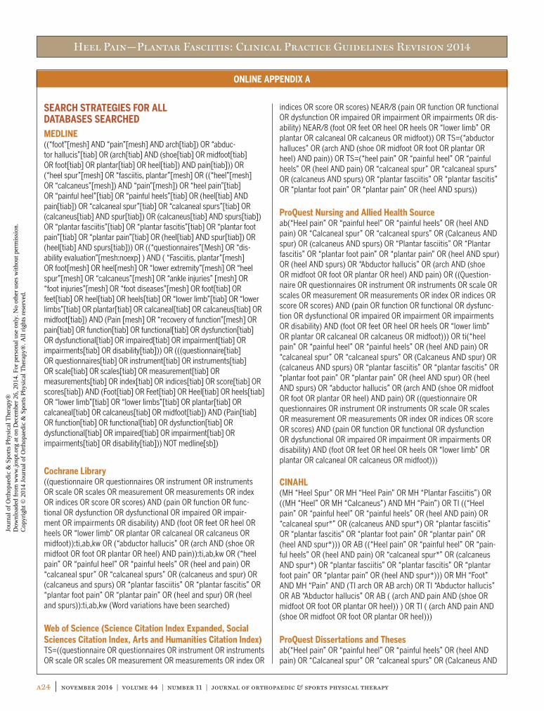
Heel Pain—Plantar Fasciitis: Clinical Practice Guidelines Revision 2014
a24 | november 2014 | volume 44 | number 11 | journal of orthopaedic & sports physical therapy
ONLINE APPENDIX A
SEARCH STRATEGIES FOR ALL DATABASES SEARCHEDMEDLINE((“foot”[mesh] AND “pain”[mesh] AND arch[tiab]) OR “abduc-tor hallucis”[tiab] OR (arch[tiab] AND (shoe[tiab] OR midfoot[tiab] OR foot[tiab] OR plantar[tiab] OR heel[tiab]) AND pain[tiab])) OR (“heel spur”[mesh] OR “fasciitis, plantar”[mesh] OR ((“heel”[mesh] OR “calcaneus”[mesh]) AND “pain”[mesh]) OR “heel pain”[tiab] OR “painful heel”[tiab] OR “painful heels”[tiab] OR (heel[tiab] AND pain[tiab]) OR “calcaneal spur”[tiab] OR “calcaneal spurs”[tiab] OR (calcaneus[tiab] AND spur[tiab]) OR (calcaneus[tiab] AND spurs[tiab]) OR “plantar fasciitis”[tiab] OR “plantar fascitis”[tiab] OR “plantar foot pain”[tiab] OR “plantar pain”[tiab] OR (heel[tiab] AND spur[tiab]) OR (heel[tiab] AND spurs[tiab])) OR ((“questionnaires”[Mesh] OR “dis-ability evaluation”[mesh:noexp] ) AND ( “Fasciitis, plantar”[mesh] OR foot[mesh] OR heel[mesh] OR “lower extremity”[mesh] OR “heel spur”[mesh] OR “calcaneus”[mesh] OR “ankle injuries” [mesh] OR “foot injuries”[mesh] OR “foot diseases”[mesh] OR foot[tiab] OR feet[tiab] OR heel[tiab] OR heels[tiab] OR “lower limb”[tiab] OR “lower limbs”[tiab] OR plantar[tiab] OR calcaneal[tiab] OR calcaneus[tiab] OR midfoot[tiab]) AND (Pain [mesh] OR “recovery of function”[mesh] OR pain[tiab] OR function[tiab] OR functional[tiab] OR dysfunction[tiab] OR dysfunctional[tiab] OR impaired[tiab] OR impairment[tiab] OR impairments[tiab] OR disability[tiab])) OR (((questionnaire[tiab] OR questionnaires[tiab] OR instrument[tiab] OR instruments[tiab] OR scale[tiab] OR scales[tiab] OR measurement[tiab] OR measurements[tiab] OR index[tiab] OR indices[tiab] OR score[tiab] OR scores[tiab]) AND (Foot[tiab] OR Feet[tiab] OR Heel[tiab] OR heels[tiab] OR “lower limb”[tiab] OR “lower limbs”[tiab] OR plantar[tiab] OR calcaneal[tiab] OR calcaneus[tiab] OR midfoot[tiab]) AND (Pain[tiab] OR function[tiab] OR functional[tiab] OR dysfunction[tiab] OR dysfunctional[tiab] OR impaired[tiab] OR impairment[tiab] OR impairments[tiab] OR disability[tiab])) NOT medline[sb])
Cochrane Library((questionnaire OR questionnaires OR instrument OR instruments OR scale OR scales OR measurement OR measurements OR index OR indices OR score OR scores) AND (pain OR function OR func-tional OR dysfunction OR dysfunctional OR impaired OR impair-ment OR impairments OR disability) AND (foot OR feet OR heel OR heels OR “lower limb” OR plantar OR calcaneal OR calcaneus OR midfoot)):ti,ab,kw OR (“abductor hallucis” OR (arch AND (shoe OR midfoot OR foot OR plantar OR heel) AND pain)):ti,ab,kw OR (“heel pain” OR “painful heel” OR “painful heels” OR (heel and pain) OR “calcaneal spur” OR “calcaneal spurs” OR (calcaneus and spur) OR (calcaneus and spurs) OR “plantar fasciitis” OR “plantar fascitis” OR “plantar foot pain” OR “plantar pain” OR (heel and spur) OR (heel and spurs)):ti,ab,kw (Word variations have been searched)
Web of Science (Science Citation Index Expanded, Social Sciences Citation Index, Arts and Humanities Citation Index)TS=((questionnaire OR questionnaires OR instrument OR instruments OR scale OR scales OR measurement OR measurements OR index OR
indices OR score OR scores) NEAR/8 (pain OR function OR functional OR dysfunction OR impaired OR impairment OR impairments OR dis-ability) NEAR/8 (foot OR feet OR heel OR heels OR “lower limb” OR plantar OR calcaneal OR calcaneus OR midfoot)) OR TS=(“abductor halluces” OR (arch AND (shoe OR midfoot OR foot OR plantar OR heel) AND pain)) OR TS=(“heel pain” OR “painful heel” OR “painful heels” OR (heel AND pain) OR “calcaneal spur” OR “calcaneal spurs” OR (calcaneus AND spurs) OR “plantar fasciitis” OR “plantar fascitis” OR “plantar foot pain” OR “plantar pain” OR (heel AND spurs))
ProQuest Nursing and Allied Health Sourceab(“Heel pain” OR “painful heel” OR “painful heels” OR (heel AND pain) OR “Calcaneal spur” OR “calcaneal spurs” OR (Calcaneus AND spur) OR (calcaneus AND spurs) OR “Plantar fasciitis” OR “Plantar fascitis” OR “plantar foot pain” OR “plantar pain” OR (heel AND spur) OR (heel AND spurs) OR “Abductor hallucis” OR (arch AND (shoe OR midfoot OR foot OR plantar OR heel) AND pain) OR ((Question-naire OR questionnaires OR instrument OR instruments OR scale OR scales OR measurement OR measurements OR index OR indices OR score OR scores) AND (pain OR function OR functional OR dysfunc-tion OR dysfunctional OR impaired OR impairment OR impairments OR disability) AND (foot OR feet OR heel OR heels OR “lower limb” OR plantar OR calcaneal OR calcaneus OR midfoot))) OR ti(“heel pain” OR “painful heel” OR “painful heels” OR (heel AND pain) OR “calcaneal spur” OR “calcaneal spurs” OR (Calcaneus AND spur) OR (calcaneus AND spurs) OR “plantar fasciitis” OR “plantar fascitis” OR “plantar foot pain” OR “plantar pain” OR (heel AND spur) OR (heel AND spurs) OR “abductor hallucis” OR (arch AND (shoe OR midfoot OR foot OR plantar OR heel) AND pain) OR ((questionnaire OR questionnaires OR instrument OR instruments OR scale OR scales OR measurement OR measurements OR index OR indices OR score OR scores) AND (pain OR function OR functional OR dysfunction OR dysfunctional OR impaired OR impairment OR impairments OR disability) AND (foot OR feet OR heel OR heels OR “lower limb” OR plantar OR calcaneal OR calcaneus OR midfoot)))
CINAHL(MH “Heel Spur” OR MH “Heel Pain” OR MH “Plantar Fasciitis”) OR ((MH “Heel” OR MH “Calcaneus”) AND MH “Pain”) OR TI ((“Heel pain” OR “painful heel” OR “painful heels” OR (heel AND pain) OR “calcaneal spur*” OR (calcaneus AND spur*) OR “plantar fasciitis” OR “plantar fascitis” OR “plantar foot pain” OR “plantar pain” OR (heel AND spur*))) OR AB ((“Heel pain” OR “painful heel” OR “pain-ful heels” OR (heel AND pain) OR “calcaneal spur*” OR (calcaneus AND spur*) OR “plantar fasciitis” OR “plantar fascitis” OR “plantar foot pain” OR “plantar pain” OR (heel AND spur*))) OR MH “Foot” AND MH “Pain” AND (TI arch OR AB arch) OR TI “Abductor hallucis” OR AB “Abductor hallucis” OR AB ( (arch AND pain AND (shoe OR midfoot OR foot OR plantar OR heel)) ) OR TI ( (arch AND pain AND (shoe OR midfoot OR foot OR plantar OR heel)))
ProQuest Dissertations and Thesesab(“Heel pain” OR “painful heel” OR “painful heels” OR (heel AND pain) OR “Calcaneal spur” OR “calcaneal spurs” OR (Calcaneus AND
44-11 Guidelines.indd 24 10/20/2014 7:10:48 PM
Jour
nal o
f O
rtho
paed
ic &
Spo
rts
Phys
ical
The
rapy
®
Dow
nloa
ded
from
ww
w.jo
spt.o
rg a
t on
Dec
embe
r 26
, 201
4. F
or p
erso
nal u
se o
nly.
No
othe
r us
es w
ithou
t per
mis
sion
. C
opyr
ight
© 2
014
Jour
nal o
f O
rtho
paed
ic &
Spo
rts
Phys
ical
The
rapy
®. A
ll ri
ghts
res
erve
d.

Heel Pain—Plantar Fasciitis: Clinical Practice Guidelines Revision 2014
journal of orthopaedic & sports physical therapy | volume 44 | number 11 | november 2014 | a25
ONLINE APPENDIX A
spur) OR (calcaneus AND spurs) OR “Plantar fasciitis” OR “Plantar fascitis” OR “plantar foot pain” OR “plantar pain” OR (heel AND spur) OR (heel AND spurs) OR “Abductor hallucis” OR (arch AND (shoe OR midfoot OR foot OR plantar OR heel) AND pain) OR ((Question-naire OR questionnaires OR instrument OR instruments OR scale OR scales OR measurement OR measurements OR index OR indices OR score OR scores) AND (pain OR function OR functional OR dysfunc-tion OR dysfunctional OR impaired OR impairment OR impairments OR disability) AND (foot OR feet OR heel OR heels OR “lower limb” OR plantar OR calcaneal OR calcaneus OR midfoot))) OR ti(“heel pain” OR “painful heel” OR “painful heels” OR (heel AND pain) OR “calcaneal spur” OR “calcaneal spurs” OR (Calcaneus AND spur) OR (calcaneus AND spurs) OR “plantar fasciitis” OR “plantar fascitis” OR “plantar foot pain” OR “plantar pain” OR (heel AND spur) OR (heel AND spurs) OR “abductor hallucis” OR (arch AND (shoe OR midfoot OR foot OR plantar OR heel) AND pain) OR ((questionnaire OR questionnaires OR instrument OR instruments OR scale OR scales OR measurement OR measurements OR index OR indices OR score
OR scores) AND (pain OR function OR functional OR dysfunction OR dysfunctional OR impaired OR impairment OR impairments OR disability) AND (foot OR feet OR heel OR heels OR “lower limb” OR plantar OR calcaneal OR calcaneus OR midfoot)))
PEDro (Physiotherapy Evidence Database)“heel pain” OR “painful heel” OR “painful heels” OR (heel AND pain) OR “calcaneal spur” OR “calcaneal spurs” OR (calcaneus AND spur) OR (calcaneus AND spurs) OR “plantar fasciitis” OR “plantar fascitis” OR “plantar foot pain” OR “plantar pain” OR (heel AND spur) OR (heel AND spurs) OR “abductor hallucis” OR (arch AND (shoe OR midfoot OR foot OR plantar OR heel) AND pain) OR ((questionnaire OR questionnaires OR instrument OR instruments OR scale OR scales OR measurement OR measurements OR index OR indices OR score OR scores) AND (pain OR function OR functional OR dysfunc-tion OR dysfunctional OR impaired OR impairment OR impairments OR disability) AND (foot OR feet OR heel OR heels OR “lower limb” OR plantar OR calcaneal OR calcaneus OR midfoot))
ONLINE APPENDIX B
SEARCH RESULTS
Database Date Conducted Results, nMEDLINE 12/13/12 2408
Cochrane Library 12/13/12 653
Cochrane reviews 49
Other reviews 3
Trials 597
Methods studies 3
Technology assessments 1
Web of Science 12/13/12 1382
ProQuest Nursing and Allied Health Source 12/17/12 1101
CINAHL 12/17/12 1101
ProQuest Dissertations and Theses 12/17/12 168
PEDro 12/19/12 532
Total 7345
Total with duplicates removed 5764
44-11 Guidelines.indd 25 10/20/2014 7:10:48 PM
Jour
nal o
f O
rtho
paed
ic &
Spo
rts
Phys
ical
The
rapy
®
Dow
nloa
ded
from
ww
w.jo
spt.o
rg a
t on
Dec
embe
r 26
, 201
4. F
or p
erso
nal u
se o
nly.
No
othe
r us
es w
ithou
t per
mis
sion
. C
opyr
ight
© 2
014
Jour
nal o
f O
rtho
paed
ic &
Spo
rts
Phys
ical
The
rapy
®. A
ll ri
ghts
res
erve
d.

Heel Pain—Plantar Fasciitis: Clinical Practice Guidelines Revision 2014
a26 | november 2014 | volume 44 | number 11 | journal of orthopaedic & sports physical therapy
ONLINE APPENDIX C
ARTICLE INCLUSION AND EXCLUSION CRITERIAInclusion CriteriaWe included articles providing evidence of the following types: systematic reviews, meta-analyses, experimental and quasi- experimental, cohort, case series, and cross-sectional studies reporting on:• The functional anatomy (abductor hallucis, longitudinal arch,
muscles, tendons, and nerves, as well as the plantar fascia) of the heel and foot relevant to plantar fasciitis
OR• Tests and measures for diagnosis and/or differential diagnosis
of heel pain/plantar fasciitis within the scope of physical therapist practice, including but not limited to tarsal tunnel syndrome test, windlass test, longitudinal arch angle, Foot Posture Index
OR• Measurement properties of instruments and tests specific
to measuring heel pain/plantar fasciitis–related outcomes (including but not limited to symptoms, functions, activity, and participation)
OR• Measurement properties of instruments that are not specific
to heel pain/plantar fasciitis BUT are specific to lower extremity outcomes
OR• Measurement properties of instruments using data from
a sample of patients with heel pain/plantar fasciitisOR• Primarily adults (16 years old or greater) – Studies reporting on persons less than 16 years old IF
the proportion in the sample is small (less than 5%)AND• Plantar heel pain due to plantar fasciitis, including the following
topics: – Risk of heel pain/plantar fasciitis, including but not limited
to ankle range of motion and body mass index
– Diagnostic characteristics of heel pain/plantar fasciitis, including but not limited to pain location, duration, and quality, and related impairments and functional limitations
– Interventions within the scope of practice of physical therapists, to include modalities (including but not limited to iontophoresis, manual therapy, stretching exercises, taping, orthotic devices, dry needling, and splints)
All outcomes were included.
Exclusion CriteriaWe excluded nonsystematic review articles and reports, and articles reporting on:• Primarily infants and children (less than 16 years old)• Heel pain related primarily to conditions other than plantar
fasciitis: – Fractures (including stress fractures) – Compartment syndrome – Tumors – Postoperative heel pain from foot surgery – Posterior or lateral heel pain related to Achilles
or peroneal tendinitis – Nonmusculoskeletal heel pain:
• Diabetes• Ulcers• Primary peripheral nerve entrapment
• Topics outside the scope of physical therapist practice:
– Decision to order radiologic tests (magnetic resonance imaging, etc)
– Extracorporeal shockwave therapy (unless it is compared to physical therapy intervention)
– Diagnostic ultrasound
44-11 Guidelines.indd 26 10/20/2014 7:10:49 PM
Jour
nal o
f O
rtho
paed
ic &
Spo
rts
Phys
ical
The
rapy
®
Dow
nloa
ded
from
ww
w.jo
spt.o
rg a
t on
Dec
embe
r 26
, 201
4. F
or p
erso
nal u
se o
nly.
No
othe
r us
es w
ithou
t per
mis
sion
. C
opyr
ight
© 2
014
Jour
nal o
f O
rtho
paed
ic &
Spo
rts
Phys
ical
The
rapy
®. A
ll ri
ghts
res
erve
d.

Heel Pain—Plantar Fasciitis: Clinical Practice Guidelines Revision 2014
journal of orthopaedic & sports physical therapy | volume 44 | number 11 | november 2014 | a27
ONLINE APPENDIX D
FLOW CHART OF ARTICLES
Duplicates removed, n = 1581
Articles found from other sources, n = 3
Records identified through database search, n = 7345
Articles assessed for eligibility, n = 238
Articles used in recommendations, n = 81
Relevant articles, n = 122
Relevant articles, n = 125
Records screened (title and abstract), n = 5764
Records excluded, n = 5526
Articles found from other sources, n = 2
Full-text articles excluded, n = 116• Methodology, n = 61• Outside scope, n = 25• Redundant, n = 14• Not English, n = 12• Could not locate, n = 4
Full-text articles excluded, n = 46• Methodology, n = 12• Outside scope, n = 26• Not English, n = 1• Redundant, n = 7
44-11 Guidelines.indd 27 10/20/2014 7:10:49 PM
Jour
nal o
f O
rtho
paed
ic &
Spo
rts
Phys
ical
The
rapy
®
Dow
nloa
ded
from
ww
w.jo
spt.o
rg a
t on
Dec
embe
r 26
, 201
4. F
or p
erso
nal u
se o
nly.
No
othe
r us
es w
ithou
t per
mis
sion
. C
opyr
ight
© 2
014
Jour
nal o
f O
rtho
paed
ic &
Spo
rts
Phys
ical
The
rapy
®. A
ll ri
ghts
res
erve
d.

Heel Pain—Plantar Fasciitis: Clinical Practice Guidelines Revision 2014
a28 | november 2014 | volume 44 | number 11 | journal of orthopaedic & sports physical therapy
ONLINE APPENDIX E
ARTICLES INCLUDED IN RECOMMENDATIONS BY TOPICImpairment/Function-Based DiagnosisPrevalenceDi Caprio F, Buda R, Mosca M, Calabrò A, Giannini S. Foot and lower
limb diseases in runners: assessment of risk factors. J Sports Sci Med. 2010;9:587-596.
Hill CL, Gill TK, Menz HB, Taylor AW. Prevalence and correlates of foot pain in a population-based study: the North West Ad-elaide health study. J Foot Ankle Res. 2008;1:2. http://dx.doi.org/10.1186/1757-1146-1-2
Lopes AD, Hespanhol Junior LC, Yeung SS, Costa LO. What are the main running-related musculoskeletal injuries? A systematic review. Sports Med. 2012;42:891-905. http://dx.doi.org/10.1007/BF03262301
Sobhani S, Dekker R, Postema K, Dijkstra PU. Epidemiology of ankle and foot overuse injuries in sports: a systematic review. Scand J Med Sci Sports. 2013;23:669-686. http://dx.doi.org/10.1111/j.1600-0838.2012.01509.x
Tenforde AS, Sayres LC, McCurdy ML, Collado H, Sainani KL, Freder-icson M. Overuse injuries in high school runners: lifetime preva-lence and prevention strategies. PM R. 2011;3:125-131; quiz 131. http://dx.doi.org/10.1016/j.pmrj.2010.09.009
Pathoanatomical FeaturesFabrikant JM, Park TS. Plantar fasciitis (fasciosis) treatment outcome
study: plantar fascia thickness measured by ultrasound and correlated with patient self-reported improvement. Foot (Edinb). 2011;21:79-83. http://dx.doi.org/10.1016/j.foot.2011.01.015
Lentz TA, Sutton Z, Greenberg S, Bishop MD. Pain-related fear con-tributes to self-reported disability in patients with foot and ankle pathology. Arch Phys Med Rehabil. 2010;91:557-561. http://dx.doi.org/10.1016/j.apmr.2009.12.010
Mahowald S, Legge BS, Grady JF. The correlation between plantar fascia thickness and symptoms of plantar fasciitis. J Am Podiatr Med Assoc. 2011;101:385-389. http://dx.doi.org/10.7547/1010385
Sutton Z, Greenburg S, Bishop M. Association of pain related beliefs with disability and pain in patients with foot and/or ankle pain: a case series. Orthop Phys Ther Pract. 2008;20:200-207.
Wearing SC, Smeathers JE, Sullivan PM, Yates B, Urry SR, Dubois P. Plantar fasciitis: are pain and fascial thickness associated with arch shape and loading? Phys Ther. 2007;87:1002-1008. http://dx.doi.org/10.2522/ptj.20060136
Wearing SC, Smeathers JE, Yates B, Urry SR, Dubois P. Bulk compressive properties of the heel fat pad during walking: a pilot investigation in plantar heel pain. Clin Biomech (Bris-tol, Avon). 2009;24:397-402. http://dx.doi.org/10.1016/j.clinbiomech.2009.01.002
Wu CH, Chang KV, Mio S, Chen WS, Wang TG. Sonoelastography of the plantar fascia. Radiology. 2011;259:502-507. http://dx.doi.org/10.1148/radiol.11101665
Clinical CourseKlein SE, Dale AM, Hayes MH, Johnson JE, McCormick JJ, Racette
BA. Clinical presentation and self-reported patterns of pain and function in patients with plantar heel pain. Foot Ankle Int. 2012;33:693-698. http://dx.doi.org/10.3113/FAI.2012.0693
Yi TI, Lee GE, Seo IS, Huh WS, Yoon TH, Kim BR. Clinical charac-teristics of the causes of plantar heel pain. Ann Rehabil Med. 2011;35:507-513. http://dx.doi.org/10.5535/arm.2011.35.4.507
Risk FactorsButterworth PA, Landorf KB, Smith SE, Menz HB. The association
between body mass index and musculoskeletal foot disorders: a systematic review. Obes Rev. 2012;13:630-642. http://dx.doi.org/10.1111/j.1467-789X.2012.00996.x
Chang R, Kent-Braun JA, Hamill J. Use of MRI for volume estima-tion of tibialis posterior and plantar intrinsic foot muscles in healthy and chronic plantar fasciitis limbs. Clin Biomech (Bristol, Avon). 2012;27:500-505. http://dx.doi.org/10.1016/j.clinbiomech.2011.11.007
Di Caprio F, Buda R, Mosca M, Calabrò A, Giannini S. Foot and lower limb diseases in runners: assessment of risk factors. J Sports Sci Med. 2010;9:587-596.
Irving DB, Cook JL, Young MA, Menz HB. Obesity and pronated foot type may increase the risk of chronic plantar heel pain: a matched case-control study. BMC Musculoskelet Disord. 2007;8:41. http://dx.doi.org/10.1186/1471-2474-8-41
Klein SE, Dale AM, Hayes MH, Johnson JE, McCormick JJ, Racette BA. Clinical presentation and self-reported patterns of pain and function in patients with plantar heel pain. Foot Ankle Int. 2012;33:693-698. http://dx.doi.org/10.3113/FAI.2012.0693
Labovitz JM, Yu J, Kim C. The role of hamstring tightness in plan-tar fasciitis. Foot Ankle Spec. 2011;4:141-144. http://dx.doi.org/10.1177/1938640010397341
Lopes AD, Hespanhol Junior LC, Yeung SS, Costa LO. What are the main running-related musculoskeletal injuries? A systematic review. Sports Med. 2012;42:891-905. http://dx.doi.org/10.1007/BF03262301
Mahmood S, Huffman LK, Harris JG. Limb-length discrepancy as a cause of plantar fasciitis. J Am Podiatr Med Assoc. 2010;100:452-455. http://dx.doi.org/10.7547/1000452
Patel A, DiGiovanni B. Association between plantar fasciitis and iso-lated contracture of the gastrocnemius. Foot Ankle Int. 2011;32:5-8. http://dx.doi.org/10.3113/FAI.2011.0005
Pohl MB, Hamill J, Davis IS. Biomechanical and anatomic factors associated with a history of plantar fasciitis in female runners. Clin J Sport Med. 2009;19:372-376. http://dx.doi.org/10.1097/JSM.0b013e3181b8c270
Ribeiro AP, Trombini-Souza F, Tessutti V, Lima FR, de Camargo Neves Sacco I, João SM. Rearfoot alignment and medial longitudinal arch configurations of runners with symptoms and histories of plantar fasciitis. Clinics (São Paulo). 2011;66:1027-1033. http://dx.doi.org/10.1590/S1807-59322011000600018
Sahin N, Öztürk A, Atici T. Foot mobility and plantar fascia elastic-ity in patients with plantar fasciitis. Acta Orthop Traumatol Turc. 2010;44:385-391. http://dx.doi.org/10.3944/AOTT.2010.2348
Sobhani S, Dekker R, Postema K, Dijkstra PU. Epidemiology of
44-11 Guidelines.indd 28 10/20/2014 7:10:49 PM
Jour
nal o
f O
rtho
paed
ic &
Spo
rts
Phys
ical
The
rapy
®
Dow
nloa
ded
from
ww
w.jo
spt.o
rg a
t on
Dec
embe
r 26
, 201
4. F
or p
erso
nal u
se o
nly.
No
othe
r us
es w
ithou
t per
mis
sion
. C
opyr
ight
© 2
014
Jour
nal o
f O
rtho
paed
ic &
Spo
rts
Phys
ical
The
rapy
®. A
ll ri
ghts
res
erve
d.

Heel Pain—Plantar Fasciitis: Clinical Practice Guidelines Revision 2014
journal of orthopaedic & sports physical therapy | volume 44 | number 11 | november 2014 | a29
ONLINE APPENDIX E
ankle and foot overuse injuries in sports: a systematic review. Scand J Med Sci Sports. 2013;23:669-686. http://dx.doi.org/10.1111/j.1600-0838.2012.01509.x
Tenforde AS, Sayres LC, McCurdy ML, Collado H, Sainani KL, Freder-icson M. Overuse injuries in high school runners: lifetime preva-lence and prevention strategies. PM R. 2011;3:125-131; quiz 131. http://dx.doi.org/10.1016/j.pmrj.2010.09.009
Werner RA, Gell N, Hartigan A, Wiggerman N, Keyserling WM. Risk factors for plantar fasciitis among assembly plant workers. PM R. 2010;2:110-116. http://dx.doi.org/10.1016/j.pmrj.2009.11.012
Diagnosis/ClassificationCornwall MW, McPoil TG, Lebec M, Vicenzino B, Wilson J. Reliability
of the modified Foot Posture Index. J Am Podiatr Med Assoc. 2008;98:7-13. http://dx.doi.org/10.7547/0980007
Irving DB, Cook JL, Young MA, Menz HB. Obesity and pronated foot type may increase the risk of chronic plantar heel pain: a matched case-control study. BMC Musculoskelet Disord. 2007;8:41. http://dx.doi.org/10.1186/1471-2474-8-41
Labovitz JM, Yu J, Kim C. The role of hamstring tightness in plan-tar fasciitis. Foot Ankle Spec. 2011;4:141-144. http://dx.doi.org/10.1177/1938640010397341
Mahmood S, Huffman LK, Harris JG. Limb-length discrepancy as a cause of plantar fasciitis. J Am Podiatr Med Assoc. 2010;100:452-455. http://dx.doi.org/10.7547/1000452
Redmond AC, Crosbie J, Ouvrier RA. Development and validation of a novel rating system for scoring standing foot posture: the Foot Posture Index. Clin Biomech (Bristol, Avon). 2006;21:89-98. http://dx.doi.org/10.1016/j.clinbiomech.2005.08.002
Differential DiagnosisHafner S, Han N, Pressman MM, Wallace C. Proximal plantar fibroma
as an etiology of recalcitrant plantar heel pain. J Foot Ankle Surg. 2011;50:153-157. http://dx.doi.org/10.1053/j.jfas.2010.12.016
Koumakis E, Gossec L, Elhai M, et al. Heel pain in spondyloarthritis: results of a cross-sectional study of 275 patients. Clin Exp Rheu-matol. 2012;30:487-491.
Wearing SC, Smeathers JE, Yates B, Urry SR, Dubois P. Bulk compressive properties of the heel fat pad during walking: a pilot investigation in plantar heel pain. Clin Biomech (Bris-tol, Avon). 2009;24:397-402. http://dx.doi.org/10.1016/j.clinbiomech.2009.01.002
Yi TI, Lee GE, Seo IS, Huh WS, Yoon TH, Kim BR. Clinical charac-teristics of the causes of plantar heel pain. Ann Rehabil Med. 2011;35:507-513. http://dx.doi.org/10.5535/arm.2011.35.4.507
ExaminationOutcome MeasuresHart DL, Wang YC, Stratford PW, Mioduski JE. Computerized adap-
tive test for patients with foot or ankle impairments produced valid and responsive measures of function. Qual Life Res. 2008;17:1081-1091. http://dx.doi.org/10.1007/s11136-008-9381-y
Landorf KB, Radford JA. Minimal important difference: values for the Foot Health Status Questionnaire, Foot Function Index and Visual
Analogue Scale. Foot. 2008;18:15-19. http://dx.doi.org/10.1016/j.foot.2007.06.006
Landorf KB, Radford JA, Hudson S. Minimal Important Differ-ence (MID) of two commonly used outcome measures for foot problems. J Foot Ankle Res. 2010;3:7. http://dx.doi.org/10.1186/1757-1146-3-7
Martin RL, Irrgang JJ. A survey of self-reported outcome instruments for the foot and ankle. J Orthop Sports Phys Ther. 2007;37:72-84. http://dx.doi.org/10.2519/jospt.2007.2403
InterventionManual TherapyBrantingham JW, Bonnefin D, Perle SM, et al. Manipulative therapy
for lower extremity conditions: update of a literature review. J Manipulative Physiol Ther. 2012;35:127-166. http://dx.doi.org/10.1016/j.jmpt.2012.01.001
Cleland JA, Abbott JH, Kidd MO, et al. Manual physical therapy and exercise versus electrophysical agents and exercise in the man-agement of plantar heel pain: a multicenter randomized clinical trial. J Orthop Sports Phys Ther. 2009;39:573-585. http://dx.doi.org/10.2519/jospt.2009.3036
Renan-Ordine R, Alburquerque-Sendín F, de Souza DP, Cleland JA, Fernández-de-las-Peñas C. Effectiveness of myofascial trigger point manual therapy combined with a self-stretching protocol for the management of plantar heel pain: a randomized controlled trial. J Orthop Sports Phys Ther. 2011;41:43-50. http://dx.doi.org/10.2519/jospt.2011.3504
StretchingLandorf KB, Menz HB. Plantar heel pain and fasciitis. Clin Evid
(Online). 2008;2008:1111.Radford JA, Landorf KB, Buchbinder R, Cook C. Effectiveness of calf
muscle stretching for the short-term treatment of plantar heel pain: a randomised trial. BMC Musculoskelet Disord. 2007;8:36. http://dx.doi.org/10.1186/1471-2474-8-36
Rompe JD, Cacchio A, Weil L, Jr., et al. Plantar fascia-specific stretch-ing versus radial shock-wave therapy as initial treatment of plan-tar fasciopathy. J Bone Joint Surg Am. 2010;92:2514-2522. http://dx.doi.org/10.2106/JBJS.I.01651
Sweeting D, Parish B, Hooper L, Chester R. The effectiveness of manual stretching in the treatment of plantar heel pain: a systematic review. J Foot Ankle Res. 2011;4:19. http://dx.doi.org/10.1186/1757-1146-4-19
TapingAbd El Salam MS, Abd Elhafz YN. Low-Dye taping versus medial arch
support in managing pain and pain-related disability in patients with plantar fasciitis. Foot Ankle Spec. 2011;4:86-91. http://dx.doi.org/10.1177/1938640010387416
Cheung RT, Chung RC, Ng GY. Efficacies of different external controls for excessive foot pronation: a meta-analysis. Br J Sports Med. 2011;45:743-751. http://dx.doi.org/10.1136/bjsm.2010.079780
Franettovich M, Chapman A, Blanch P, Vicenzino B. Continual use of augmented low-Dye taping increases arch height in standing
44-11 Guidelines.indd 29 10/20/2014 7:10:50 PM
Jour
nal o
f O
rtho
paed
ic &
Spo
rts
Phys
ical
The
rapy
®
Dow
nloa
ded
from
ww
w.jo
spt.o
rg a
t on
Dec
embe
r 26
, 201
4. F
or p
erso
nal u
se o
nly.
No
othe
r us
es w
ithou
t per
mis
sion
. C
opyr
ight
© 2
014
Jour
nal o
f O
rtho
paed
ic &
Spo
rts
Phys
ical
The
rapy
®. A
ll ri
ghts
res
erve
d.

Heel Pain—Plantar Fasciitis: Clinical Practice Guidelines Revision 2014
a30 | november 2014 | volume 44 | number 11 | journal of orthopaedic & sports physical therapy
ONLINE APPENDIX E
but does not influence neuromotor control of gait. Gait Posture. 2010;31:247-250. http://dx.doi.org/10.1016/j.gaitpost.2009.10.015
Franettovich M, Chapman A, Blanch P, Vicenzino B. A physiological and psychological basis for anti-pronation taping from a critical review of the literature. Sports Med. 2008;38:617-631.
Franettovich M, Chapman A, Vicenzino B. Tape that increases medial longitudinal arch height also reduces leg muscle activity: a pre-liminary study. Med Sci Sports Exerc. 2008;40:593-600. http://dx.doi.org/10.1249/MSS.0b013e318162134f
Franettovich M, Chapman AR, Blanch P, Vicenzino B. Augmented low-Dye tape alters foot mobility and neuromotor control of gait in in-dividuals with and without exercise related leg pain. J Foot Ankle Res. 2010;3:5. http://dx.doi.org/10.1186/1757-1146-3-5
Franettovich MM, Murley GS, David BS, Bird AR. A comparison of augmented low-Dye taping and ankle bracing on lower limb muscle activity during walking in adults with flat-arched foot pos-ture. J Sci Med Sport. 2012;15:8-13. http://dx.doi.org/10.1016/j.jsams.2011.05.009
Landorf KB, Menz HB. Plantar heel pain and fasciitis. Clin Evid (Online). 2008;2008:1111.
Meier K, McPoil TG, Cornwall MW, Lyle T. Use of antiprona-tion taping to determine foot orthoses prescription: a case series. Res Sports Med. 2008;16:257-271. http://dx.doi.org/10.1080/15438620802310842
Tsai CT, Chang WD, Lee JP. Effects of short-term treatment with Kinesiotaping for plantar fasciitis. J Musculoskelet Pain. 2010;18:71-80.
van de Water AT, Speksnijder CM. Efficacy of taping for the treatment of plantar fasciosis: a systematic review of controlled trials. J Am Podiatr Med Assoc. 2010;100:41-51.
Van Lunen B, Cortes N, Andrus T, Walker M, Pasquale M, Onate J. Immediate effects of a heel-pain orthosis and an augmented low-dye taping on plantar pressures and pain in subjects with plantar fasciitis. Clin J Sport Med. 2011;21:474-479. http://dx.doi.org/10.1097/JSM.0b013e3182340199
Vicenzino B, McPoil T, Buckland S. Plantar foot pressures af-ter the augmented low Dye taping technique. J Athl Train. 2007;42:374-380.
Yoho R, Rivera JJ, Renschler R, Vardaxis VG, Dikis J. A biomechani-cal analysis of the effects of low-Dye taping on arch deforma-tion during gait. Foot (Edinb). 2012;22:283-286. http://dx.doi.org/10.1016/j.foot.2012.08.006
Foot OrthosesAl-Bluwi MT, Sadat-Ali M, Al-Habdan IM, Azam MQ. Efficacy of
EZStep in the management of plantar fasciitis: a prospective, randomized study. Foot Ankle Spec. 2011;4:218-221. http://dx.doi.org/10.1177/1938640011407318
Bonanno DR, Landorf KB, Menz HB. Pressure-relieving properties of various shoe inserts in older people with plantar heel pain. Gait Posture. 2011;33:385-389. http://dx.doi.org/10.1016/j.gaitpost.2010.12.009
Cheung RT, Chung RC, Ng GY. Efficacies of different external controls for excessive foot pronation: a meta-analysis. Br J Sports Med.
2011;45:743-751. http://dx.doi.org/10.1136/bjsm.2010.079780Chia KK, Suresh S, Kuah A, Ong JL, Phua JM, Seah AL. Com-
parative trial of the foot pressure patterns between corrective orthotics, formthotics, bone spur pads and flat insoles in pa-tients with chronic plantar fasciitis. Ann Acad Med Singapore. 2009;38:869-875.
Collins N, Bisset L, McPoil T, Vicenzino B. Foot orthoses in lower limb overuse conditions: a systematic review and meta-analysis. Foot Ankle Int. 2007;28:396-412. http://dx.doi.org/10.3113/FAI.2007.0396
Drake M, Bittenbender C, Boyles RE. The short-term effects of treat-ing plantar fasciitis with a temporary custom foot orthosis and stretching. J Orthop Sports Phys Ther. 2011;41:221-231. http://dx.doi.org/10.2519/jospt.2011.3348
Ferber R, Benson B. Changes in multi-segment foot biomechanics with a heat-mouldable semi-custom foot orthotic device. J Foot Ankle Res. 2011;4:18. http://dx.doi.org/10.1186/1757-1146-4-18
Hawke F, Burns J, Radford JA, du Toit V. Custom-made foot ortho-ses for the treatment of foot pain. Cochrane Database Syst Rev. 2008:CD006801. http://dx.doi.org/10.1002/14651858.CD006801.pub2
Hume P, Hopkins W, Rome K, Maulder P, Coyle G, Nigg B. Effective-ness of foot orthoses for treatment and prevention of lower limb injuries: a review. Sports Med. 2008;38:759-779.
Landorf KB, Menz HB. Plantar heel pain and fasciitis. Clin Evid (On-line). 2008;2008:1111.
Lee SY, McKeon P, Hertel J. Does the use of orthoses improve self-reported pain and function measures in patients with plantar fasciitis? A meta-analysis. Phys Ther Sport. 2009;10:12-18. http://dx.doi.org/10.1016/j.ptsp.2008.09.002
Lee WC, Wong WY, Kung E, Leung AK. Effectiveness of adjustable dor-siflexion night splint in combination with accommodative foot or-thosis on plantar fasciitis. J Rehabil Res Dev. 2012;49:1557-1564.
Marabha T, Al-Anani M, Dahmashe Z, Rashdan K, Hadid A. The rela-tion between conservative treatment and heel pain duration in plantar fasciitis. Kuwait Med J. 2008;40:130-132.
Mills K, Blanch P, Chapman AR, McPoil TG, Vicenzino B. Foot orthoses and gait: a systematic review and meta-analysis of literature pertaining to potential mechanisms. Br J Sports Med. 2010;44:1035-1046. http://dx.doi.org/10.1136/bjsm.2009.066977
Stolwijk NM, Louwerens JW, Nienhuis B, Duysens J, Keijsers NL. Plantar pressure with and without custom insoles in patients with common foot complaints. Foot Ankle Int. 2011;32:57-65. http://dx.doi.org/10.3113/FAI.2011.0057
Stratton M, McPoil TG, Cornwall MW, Patrick K. Use of low-frequency electrical stimulation for the treatment of plantar fasciitis. J Am Podiatr Med Assoc. 2009;99:481-488.
Trotter LC, Pierrynowski MR. Changes in gait economy between full-contact custom-made foot orthoses and prefabricated inserts in patients with musculoskeletal pain: a randomized clinical trial. J Am Podiatr Med Assoc. 2008;98:429-435.
Uden H, Boesch E, Kumar S. Plantar fasciitis – to jab or to support? A systematic review of the current best evidence. J Multidiscip Healthc. 2011;4:155-164. http://dx.doi.org/10.2147/JMDH.S20053
44-11 Guidelines.indd 30 10/20/2014 7:10:50 PM
Jour
nal o
f O
rtho
paed
ic &
Spo
rts
Phys
ical
The
rapy
®
Dow
nloa
ded
from
ww
w.jo
spt.o
rg a
t on
Dec
embe
r 26
, 201
4. F
or p
erso
nal u
se o
nly.
No
othe
r us
es w
ithou
t per
mis
sion
. C
opyr
ight
© 2
014
Jour
nal o
f O
rtho
paed
ic &
Spo
rts
Phys
ical
The
rapy
®. A
ll ri
ghts
res
erve
d.

Heel Pain—Plantar Fasciitis: Clinical Practice Guidelines Revision 2014
journal of orthopaedic & sports physical therapy | volume 44 | number 11 | november 2014 | a31
ONLINE APPENDIX E
Van Lunen B, Cortes N, Andrus T, Walker M, Pasquale M, Onate J. Immediate effects of a heel-pain orthosis and an augmented low-dye taping on plantar pressures and pain in subjects with plantar fasciitis. Clin J Sport Med. 2011;21:474-479. http://dx.doi.org/10.1097/JSM.0b013e3182340199
Night SplintsAttard J, Singh D. A comparison of two night ankle-foot orthoses used
in the treatment of inferior heel pain: a preliminary investigation. Foot Ankle Surg. 2012;18:108-110. http://dx.doi.org/10.1016/j.fas.2011.03.011
Beyzadeoğlu T, Gökçe A, Bekler H. [The effectiveness of dorsiflexion night splint added to conservative treatment for plantar fasciitis]. Acta Orthop Traumatol Turc. 2007;41:220-224.
Landorf KB, Menz HB. Plantar heel pain and fasciitis. Clin Evid (Online). 2008;2008:1111.
Lee WC, Wong WY, Kung E, Leung AK. Effectiveness of adjustable dor-siflexion night splint in combination with accommodative foot or-thosis on plantar fasciitis. J Rehabil Res Dev. 2012;49:1557-1564.
Sheridan L, Lopez A, Perez A, John MM, Willis FB, Shanmugam R. Plantar fasciopathy treated with dynamic splinting: a random-ized controlled trial. J Am Podiatr Med Assoc. 2010;100:161-165. http://dx.doi.org/10.7547/1000161
Physical Agents – ElectrotherapyCleland JA, Abbott JH, Kidd MO, et al. Manual physical therapy and
exercise versus electrophysical agents and exercise in the man-agement of plantar heel pain: a multicenter randomized clinical trial. J Orthop Sports Phys Ther. 2009;39:573-585. http://dx.doi.org/10.2519/jospt.2009.3036
Stratton M, McPoil TG, Cornwall MW, Patrick K. Use of low-frequency electrical stimulation for the treatment of plantar fasciitis. J Am Podiatr Med Assoc. 2009;99:481-488.
Physical Agents – Low-Level Laser TherapyKiritsi O, Tsitas K, Malliaropoulos N, Mikroulis G. Ultrasonographic
evaluation of plantar fasciitis after low-level laser therapy: re-sults of a double-blind, randomized, placebo-controlled trial. Lasers Med Sci. 2010;25:275-281. http://dx.doi.org/10.1007/s10103-009-0737-5
Physical Agents – PhonophoresisJasiak-Tyrkalska B, Jaworek J, Frańczuk B. Efficacy of two different
physiotherapeutic procedures in comprehensive therapy of plan-tar calcaneal spur. Fizjoter Polska. 2007;7:145-154.
Physical Agents – UltrasoundShanks P, Curran M, Fletcher P, Thompson R. The effectiveness of
therapeutic ultrasound for musculoskeletal conditions of the lower limb: A literature review. Foot (Edinb). 2010;20:133-139.
FootwearCheung RT, Chung RC, Ng GY. Efficacies of different external controls
for excessive foot pronation: a meta-analysis. Br J Sports Med. 2011;45:743-751. http://dx.doi.org/10.1136/bjsm.2010.079780
Cong Y, Cheung JT, Leung AK, Zhang M. Effect of heel height on in-shoe localized triaxial stresses. J Biomech. 2011;44:2267-2272. http://dx.doi.org/10.1016/j.jbiomech.2011.05.036
Fong DT, Pang KY, Chung MM, Hung AS, Chan KM. Evaluation of combined prescription of rocker sole shoes and custom-made foot orthoses for the treatment of plantar fasciitis. Clin Biomech (Bristol, Avon). 2012;27:1072-1077. http://dx.doi.org/10.1016/j.clinbiomech.2012.08.003
Lin SC, Chen CP, Tang SF, Wong AM, Hsieh JH, Chen WP. Changes in windlass effect in response to different shoe and insole designs during walking. Gait Posture. 2013;37:235-241. http://dx.doi.org/10.1016/j.gaitpost.2012.07.010
Ryan M, Fraser S, McDonald K, Taunton J. Examining the degree of pain reduction using a multielement exercise model with a con-ventional training shoe versus an ultraflexible training shoe for treating plantar fasciitis. Phys Sportsmed. 2009;37:68-74. http://dx.doi.org/10.3810/psm.2009.12.1744
Education and Counseling for Weight LossButterworth PA, Landorf KB, Smith SE, Menz HB. The association
between body mass index and musculoskeletal foot disorders: a systematic review. Obes Rev. 2012;13:630-642. http://dx.doi.org/10.1111/j.1467-789X.2012.00996.x
Tanamas SK, Wluka AE, Berry P, et al. Relationship between obesity and foot pain and its association with fat mass, fat distribution, and muscle mass. Arthritis Care Res (Hoboken). 2012;64:262-268. http://dx.doi.org/10.1002/acr.20663
Therapeutic Exercise and Neuromuscular Re-educationKulig K, Popovich JM, Jr., Noceti-Dewit LM, Reischl SF, Kim D.
Women with posterior tibial tendon dysfunction have diminished ankle and hip muscle performance. J Orthop Sports Phys Ther. 2011;41:687-694. http://dx.doi.org/10.2519/jospt.2011.3427
Snyder KR, Earl JE, O’Connor KM, Ebersole KT. Resistance train-ing is accompanied by increases in hip strength and changes in lower extremity biomechanics during running. Clin Biomech (Bristol, Avon). 2009;24:26-34. http://dx.doi.org/10.1016/j.clinbiomech.2008.09.009
Trigger Point Dry NeedlingCotchett MP, Landorf KB, Munteanu SE. Effectiveness of dry needling
and injections of myofascial trigger points associated with plantar heel pain: a systematic review. J Foot Ankle Res. 2010;3:18. http://dx.doi.org/10.1186/1757-1146-3-18
44-11 Guidelines.indd 31 10/20/2014 7:10:50 PM
Jour
nal o
f O
rtho
paed
ic &
Spo
rts
Phys
ical
The
rapy
®
Dow
nloa
ded
from
ww
w.jo
spt.o
rg a
t on
Dec
embe
r 26
, 201
4. F
or p
erso
nal u
se o
nly.
No
othe
r us
es w
ithou
t per
mis
sion
. C
opyr
ight
© 2
014
Jour
nal o
f O
rtho
paed
ic &
Spo
rts
Phys
ical
The
rapy
®. A
ll ri
ghts
res
erve
d.

Heel Pain—Plantar Fasciitis: Clinical Practice Guidelines Revision 2014
a32 | november 2014 | volume 44 | number 11 | journal of orthopaedic & sports physical therapy
ONLINE APPENDIX F
LEVELS OF EVIDENCE TABLE*
LevelIntervention/Prevention
Pathoanatomic/Risk/ Clinical Course/Prognosis/Differential Diagnosis
Diagnosis/Diagnostic Accuracy
Prevalence of Condition/Disorder Exam/Outcomes
I Systematic review of high-quality RCTs
High-quality RCT†
Systematic review of prospective cohort studies
High-quality prospective cohort study‡
Systematic review of high-quality diagnostic studies
High-quality diagnostic study§ with validation
Systematic review, high-quality cross-sectional studies
High-quality cross-sectional study11║
Systematic review of prospective cohort studies
High-quality prospective cohort study
II Systematic review of high-quality cohort studies
High-quality cohort study‡
Outcomes study or ecological study
Lower-quality RCT¶
Systematic review of retrospective cohort study
Lower-quality prospective cohort study
High-quality retrospective cohort study
Consecutive cohortOutcomes study or ecological
study
Systematic review of explor-atory diagnostic studies or consecutive cohort studies
High-quality exploratory diagnostic studies
Consecutive retrospective cohort
Systematic review of studies that allows relevant estimate
Lower-quality cross- sectional study
Systematic review of lower-quality prospective cohort studies
Lower-quality prospective cohort study
III Systematic reviews of case-control studies
High-quality case-control study
Lower-quality cohort study
Lower-quality retrospective cohort study
High-quality cross-sectional study
Case-control study
Lower-quality exploratory diagnostic studies
Nonconsecutive retrospective cohort
Local nonrandom study High-quality cross- sectional study
IV Case series Case series Case-control study Lower-quality cross- sectional study
V Expert opinion Expert opinion Expert opinion Expert opinion Expert opinion
Abbreviation: RCT, randomized clinical trial.*Adapted from Phillips et al62 (http://www.cebm.net/index.aspx?o=1025). See also APPENDIX G.†High quality includes RCTs with greater than 80% follow-up, blinding, and appropriate randomization procedures.‡High-quality cohort study includes greater than 80% follow-up.§High-quality diagnostic study includes consistently applied reference standard and blinding.║High-quality prevalence study is a cross-sectional study that uses a local and current random sample or censuses.¶Weaker diagnostic criteria and reference standards, improper randomization, no blinding, and less than 80% follow-up may add bias and threats to validity.
44-11 Guidelines.indd 32 10/20/2014 7:10:51 PM
Jour
nal o
f O
rtho
paed
ic &
Spo
rts
Phys
ical
The
rapy
®
Dow
nloa
ded
from
ww
w.jo
spt.o
rg a
t on
Dec
embe
r 26
, 201
4. F
or p
erso
nal u
se o
nly.
No
othe
r us
es w
ithou
t per
mis
sion
. C
opyr
ight
© 2
014
Jour
nal o
f O
rtho
paed
ic &
Spo
rts
Phys
ical
The
rapy
®. A
ll ri
ghts
res
erve
d.
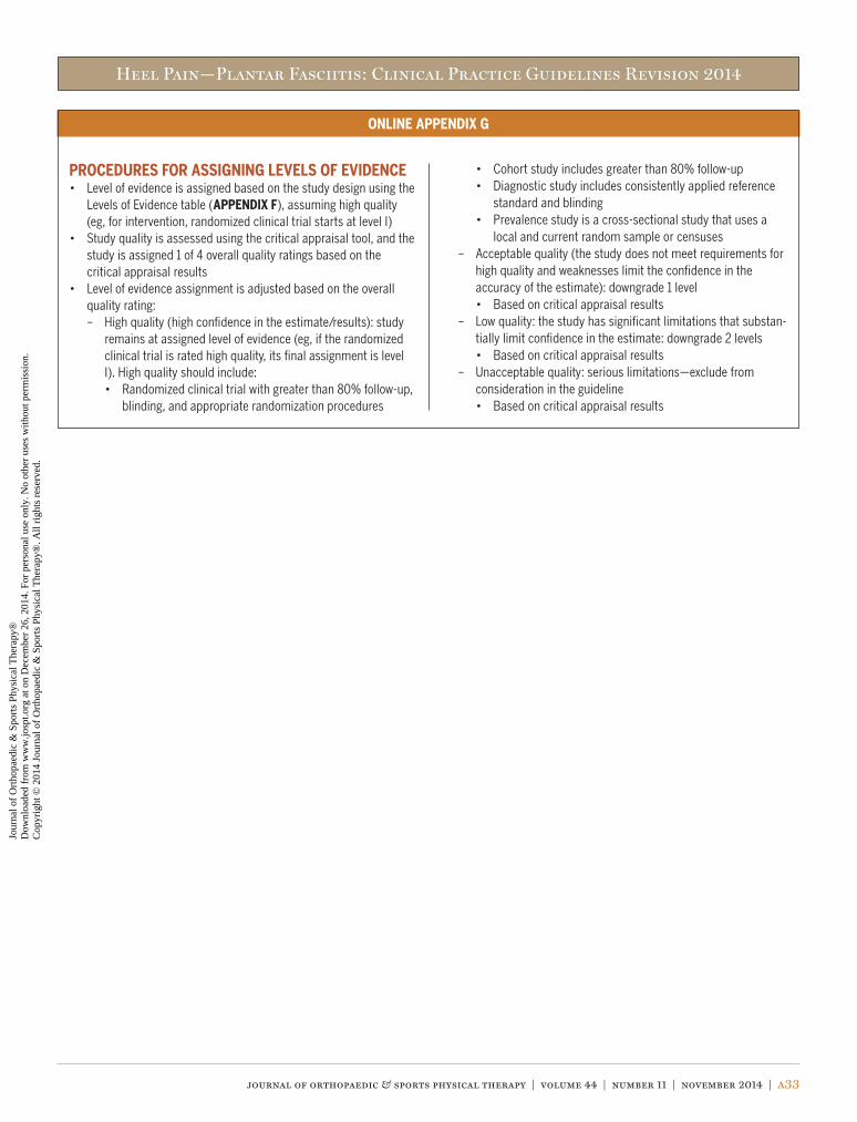
Heel Pain—Plantar Fasciitis: Clinical Practice Guidelines Revision 2014
journal of orthopaedic & sports physical therapy | volume 44 | number 11 | november 2014 | a33
ONLINE APPENDIX G
PROCEDURES FOR ASSIGNING LEVELS OF EVIDENCE• Level of evidence is assigned based on the study design using the
Levels of Evidence table (APPENDIX F), assuming high quality (eg, for intervention, randomized clinical trial starts at level I)
• Study quality is assessed using the critical appraisal tool, and the study is assigned 1 of 4 overall quality ratings based on the critical appraisal results
• Level of evidence assignment is adjusted based on the overall quality rating:
– High quality (high confidence in the estimate/results): study remains at assigned level of evidence (eg, if the randomized clinical trial is rated high quality, its final assignment is level I). High quality should include:
• Randomized clinical trial with greater than 80% follow-up, blinding, and appropriate randomization procedures
• Cohort study includes greater than 80% follow-up • Diagnostic study includes consistently applied reference
standard and blinding • Prevalence study is a cross-sectional study that uses a
local and current random sample or censuses – Acceptable quality (the study does not meet requirements for
high quality and weaknesses limit the confidence in the accuracy of the estimate): downgrade 1 level
• Based on critical appraisal results – Low quality: the study has significant limitations that substan-
tially limit confidence in the estimate: downgrade 2 levels • Based on critical appraisal results – Unacceptable quality: serious limitations—exclude from
consideration in the guideline • Based on critical appraisal results
44-11 Guidelines.indd 33 10/20/2014 7:10:51 PM
Jour
nal o
f O
rtho
paed
ic &
Spo
rts
Phys
ical
The
rapy
®
Dow
nloa
ded
from
ww
w.jo
spt.o
rg a
t on
Dec
embe
r 26
, 201
4. F
or p
erso
nal u
se o
nly.
No
othe
r us
es w
ithou
t per
mis
sion
. C
opyr
ight
© 2
014
Jour
nal o
f O
rtho
paed
ic &
Spo
rts
Phys
ical
The
rapy
®. A
ll ri
ghts
res
erve
d.