hemangioma of the facial nerve: role of the geniculate capillary plexus
TRANSCRIPT
Thomas Balkany, M.D., F.A.C.S., Milo Fradis, M.D.,Bruce W Jafek, M.D., F.A.C.S.,
and Nolan C. Rucker, D.V.M., M.S.
HEMANGIOMA OF THE FACIAL
NERVE: ROLE OF THE
GENICULATE CAPILLARY PLEXUS
Hemangioma of the facial nerve, once thought to beextremely rare, has recently been shown to occur more
commonly. 1-5 The terms "hemangioma" and "benignvascular tumor" are used synonymously. They may besubclassified by their predominant histologic appearance,such as capillary, venous, arteriovenous, and cavernous.
Hemangiomas may invade bony trabecula or may formintratumoral bony spicules. The latter have been termedossifying hemangiomas.
Although hemangiomas of the facial nerve have beenknown to occur most commonly at the geniculate gan-glion,1 4--6 the reason for this site of predilection is notknown. Based on our previous cat facial nerve studies,7which showed a highly vascular geniculate capillaryplexus (GCP), we hypothesize that a similar vascularplexus of the geniculate ganglion may occur in man andthat it may be the reason for this site of predilection for
benign vascular tumors. Fisch and Ruttnerl previouslysuspected that this site of predilection corresponded to an
area of vascular anastomosis.In our previous study, we evaluated 28 fresh cat facial
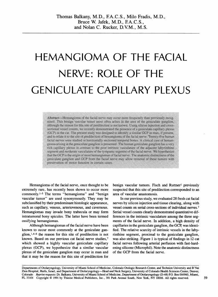
nerves by silicon injection and tissue clearing, along withvessel counts on serial cross-sections of individual nerves.7Serial vessel counts clearly demonstrated quantitative dif-ferences in the intrinsic vasculature among the three seg-ments of the facial nerve. In addition, a high density ofcapillaries in the geniculate ganglion, the GCP, was identi-fied. The relative scarcity of intrinsic vessels in the laby-rinthine segment, proximal to the geniculate ganglion,was also striking. Figure 1 is typical of findings in 12 catfacial nerves following arterial perfusion with fast-hard-ening silicone (Microphil). Note the anatomic distinctnessof the GCP from the facial nerve.
59
Departments of Otolaryngology, University of Miami School of Medicine, Colorado Otologic Research Center, and Technion University and B'NaiZion Hospital, Haifa, Israel, and Department of Otolaryngology-Head and Neck Surgery, University of Colorado Health Sciences Center, Denver,Colorado Reprint requests: Dr. Balkany, University of Miami School of Medicine, Department of Otolaryngology (D-48) PO. Box 016960, Miami,FL 33101 Copyright © 1991 by Thieme Medical Publishers, Inc., 381 Park Avenue South, New York, NY 10016. All rights reserved.
SKULL BASE SURGERYNOLUME 1, NUMBER 1 JANUARY 1991
GG.S.&
~~~~~~~~~~~~~~~~~~~~... /..........
650,
Figure 1. Cat facial nerve following intravital injection of fast-hardening silicone (Microfil) and tissue clearing. Note thehigh vascular density and anatomic distinctness of the GCP from the adjacent nerve. TS: tympanic segment; GG: genicularganglion; GPn: greater petrosal nerve; LS: labyrinthine segment.
MATERIALS AND METHODS
Twenty-five human temporal bones from the Univer-sity of Colorado Department of Otolaryngology-Head andNeck Surgery collection were selected for study accordingto the following criteria:
1.2.
3.4.
No history of temporal bone diseaseNo systemic disease that may affect the temporalboneNo congenital disease of any kindAdequate sections of all labyrinthine and tym-panic segments along with the geniculate gan-glion available for study
All temporal bones had previously been fixed in 10%formalin, progressively dehydrated in ethanol at roomtemperature, imbedded in celloidan, and sectioned hori-zontally at 20 ,um. Every tenth section was stained withhematoxylin and eosin (H & E).
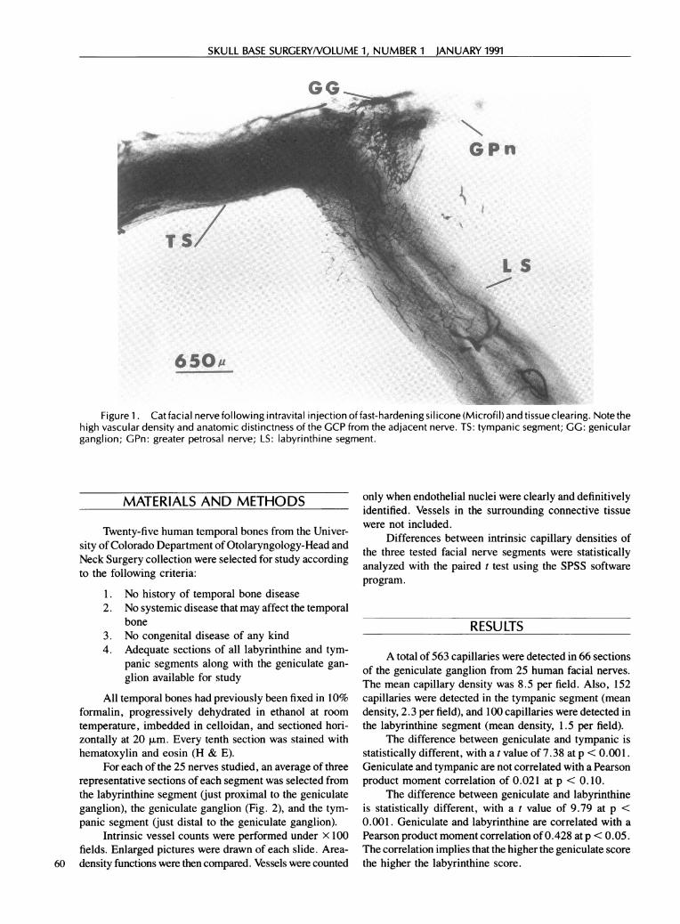
For each of the 25 nerves studied, an average of threerepresentative sections of each segment was selected fromthe labyrinthine segment (just proximal to the geniculateganglion), the geniculate ganglion (Fig. 2), and the tym-panic segment (just distal to the geniculate ganglion).
Intrinsic vessel counts were performed under x 100fields. Enlarged pictures were drawn of each slide. Area-
60 density functions were then compared. Vessels were counted
only when endothelial nuclei were clearly and definitivelyidentified. Vessels in the surrounding connective tissuewere not included.
Differences between intrinsic capillary densities ofthe three tested facial nerve segments were statisticallyanalyzed with the paired t test using the SPSS softwareprogram.
RESU LTS
A total of 563 capillaries were detected in 66 sectionsof the geniculate ganglion from 25 human facial nerves.The mean capillary density was 8.5 per field. Also, 152capillaries were detected in the tympanic segment (meandensity, 2.3 per field), and 100 capillaries were detected inthe labyrinthine segment (mean density, 1.5 per field).
The difference between geniculate and tympanic isstatistically different, with a t value of 7.38 at p < 0.001.Geniculate and tympanic are not correlated with a Pearsonproduct moment correlation of 0.021 at p < 0.10.
The difference between geniculate and labyrinthineis statistically different, with a t value of 9.79 at p <0.001. Geniculate and labyrinthine are correlated with aPearson product moment correlation of 0.428 at p < 0.05.The correlation implies that the higher the geniculate scorethe higher the labyrinthine score.
GPN
L:-,z.L.S
HEMANGIOMA OF THE FACIAL NERVE/BALKANY, FRADIS, JAFEK, RUCKER
Figure 2. Human geniculate ganglion. Large arroworiginal magnification, xI00.)
The difference between tympanic and labyrinthine isstatistically different, with a t value of 2.42 at p < 0.05.Tympanic and labyrinthine are not correlated with a Pear-son product moment correlation of 0.245 at p < 0.10.
CASE REPORT
A 41 -year-old man was referred by his neurologist forevaluation of "left Bell's palsy" of 5 months' duration.Paralysis was complete approximately 4 days after onset.There was no history of pain or facial twitching and thepatient did not respond to two courses of oral steroids.
On examination, peripheral-type facial paralysis wascomplete and tearing in the right eye was reduced. Facialelectroneurography showed a 95% denervation on theright. Pure-tone speech and auditory brainstem responseaudiometry were normal. Computed tomography (CT)examination was normal. Transmastoid exploration wasnormal but middle fossa exploration demonstrated a softred mass overlying the geniculate ganglion. A frozensection biopsy demonstrated benign vascular tumor. Fur-ther bony removal permitted excision of this 0.9 x 0.6 cmsoft tissue mass along with its origin at the geniculateganglion without resection of the facial nerve. Approx-imately 6 months later facial tone improved and voluntarymovement began to return. The patient has good volun-tary movement with moderate synkinesis 2 years post-operatively (grade III/VI).
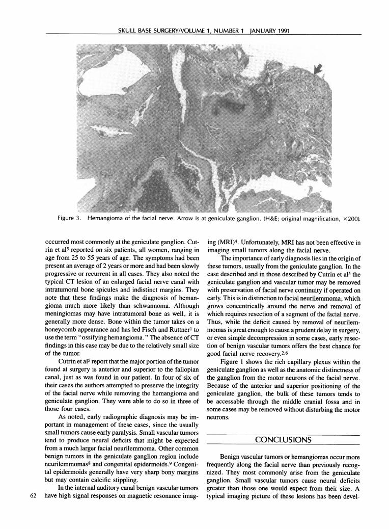
Pathologic sections demonstrate a central cluster ofcompact large vascular channels containing blood and
a .Xt" *
iatganglion cell, and small arrows are at capillaries. (H&E;
lined with small well-differentiated endothelial cells. Thewalls were thick and fibrous with spindle-shaped nuclei.Particles of calcium were seen interspersed in the lesion(Fig. 3)
DISCUSSION
The presence of a dense capillary plexus within thegeniculate ganglion is evident from study of both the catand man. The technique used in the current study hascertain inherent shortcomings due to the limitations ofhorizontal sectioning. However, intravital perfusion ofsilicone, which confirmed the cross-sectional vesselcounts in the cat, is not feasible in man. In spite of thelimitations of technique, we believe that vessel countsalone are adequate to confirm in man the previous findingsdemonstrated by more exhaustive techniques in the catmodel. The paucity of intrinsic vessels in the labyrinthinesegment of the facial nerve may have implications in otherfacial nerve disorders and will be the subject of a separatearticle.
Clinical differentiation of facial paralysis betweenBell's palsy and tumor is sometimes difficult. In this casethe typical slow progression of paralysis or recurrent pa-ralysis did not occur. The typical CT imaging findings ofbenign vascular tumor of the facial nerve have been welldescribed."- Lo et a14 reported CT findings in 11 casesand noted enlargment of the facial nerve canals withirregular margins and a honeycomb appearance. The aver-age age of these patients was 24 to 51 years and the tumors 61
tz.
SKULL BASE SURGERYNOLUME 1, NUMBER 1 JANUARY 1991
Figure 3. Hemangioma of the facial nerve. Arrow is at geniculate ganglion. (H&E; original magnification, x200).
occurred most commonly at the geniculate ganglion. Cut-rin et a15 reported on six patients, all women, ranging inage from 25 to 55 years of age. The symptoms had beenpresent an average of 2 years or more and had been slowlyprogressive or recurrent in all cases. They also noted thetypical CT lesion of an enlarged facial nerve canal withintratumoral bone spicules and indistinct margins. Theynote that these findings make the diagnosis of heman-gioma much more likely than schwannoma. Althoughmeningiomas may have intratumoral bone as well, it isgenerally more dense. Bone within the tumor takes on ahoneycomb appearance and has led Fisch and Ruttnerl touse the term "ossifying hemangioma. " The absence ofCTfindings in this case may be due to the relatively small sizeof the tumor.
Cutrin et a15 report that the major portion of the tumorfound at surgery is anterior and superior to the fallopiancanal, just as was found in our patient. In four of six oftheir cases the authors attempted to preserve the integrityof the facial nerve while removing the hemangioma andgeniculate ganglion. They were able to do so in three ofthose four cases.
As noted, early radiographic diagnosis may be im-portant in management of these cases, since the usuallysmall tumors cause early paralysis. Small vascular tumorstend to produce neural deficits that might be expectedfrom a much larger facial neurilemmoma. Other commonbenign tumors in the geniculate ganglion region includeneurilemmomas8 and congenital epidermoids.9 Congeni-tal epidermoids generally have very sharp bony marginsbut may contain calcific stippling.
In the internal auditory canal benign vascular tumors62 have high signal responses on magnetic resonance imag-
ing (MRI)4. Unfortunately, MRI has not been effective inimaging small tumors along the facial nerve.
The importance of early diagnosis lies in the origin ofthese tumors, usually from the geniculate ganglion. In thecase described and in those described by Cutrin et a15 thegeniculate ganglion and vascular tumor may be removedwith preservation of facial nerve continuity if operated onearly. This is in distinction to facial neurilemmoma, whichgrows concentrically around the nerve and removal ofwhich requires resection of a segment of the facial nerve.Thus, while the deficit caused by removal of neurilem-momas is great enough to cause a prudent delay in surgery,or even simple decompression in some cases, early resec-tion of benign vascular tumors offers the best chance forgood facial nerve recovery.2,6
Figure 1 shows the rich capillary plexus within thegeniculate ganglion as well as the anatomic distinctness ofthe ganglion from the motor neurons of the facial nerve.Because of the anterior and superior positioning of thegeniculate ganglion, the bulk of these tumors tends tobe accessable through the middle cranial fossa and insome cases may be removed without disturbing the motorneurons.
CONCLUSIONS
Benign vascular tumors or hemangiomas occur morefrequently along the facial nerve than previously recog-nized. They most commonly arise from the geniculateganglion. Small vascular tumors cause neural deficitsgreater than those one would expect from their size. Atypical imaging picture of these lesions has been devel-

HEMANGIOMA OF THE FACIAL NERVE/BALKANY, FRADIS, JAFEK, RUCKER
oped and facial paralysis, not typical of Bell's palsy,should be evaluated for a lesion at the geniculate ganglionwith both MRI and CT. Early diagnosis provides the bestchance of functional recovery because removal of thesetumors, along with the geniculate ganglion, with preserva-tion of motor nerve fibers may be possible in certainlesions. A geniculate capillary plexus has been demon-strated in man.
REFERENCES
1. Fisch U, Ruttner J: Pathology of intratemporal tumors involving thefacial nerve. In Fisch U (ed): Facial Nerve Surgery, Birmingham,AL: Aesculapius, 1977, pp. 456-488
2. Mangham, CA, Carberry JN, Brackmann DE: Management ofintratemporal vascular tumors. Laryngoscope 91:867-876, 1981
3. Glasscock MA, Smith PG, Schwabber MK, Nissan AJ: Clinicalaspects of osseous hemangiomas of the skull base. Laryngoscope94:869-873, 1984
4. Lo WWM, Horn KL, Carberry JN, Wade CT: Intratemporal vascu-lar tumors: Evaluation with CT. Radiology 159:181-185, 1986
5. Cutrin HD, Jensen HE, Barnes L, May M: "Ossifying" heman-giomas of the temporal bone: Evaluation with CT. Radiology164:831-835, 1987
6. Lo WWM, Brackmann DE, Shelton C: Facial nerve hemangioma.Ann Otol Rhinol Laryngol 98:160-161, 1989
7. Balkany TJ: The intrinsic vasculature of the cat facial nerve. Laryn-goscope 96:70-77, 1986
8. Latack JT, Gabrielsen TO, Knake JE: Facial nerve neuromas: Radio-logic evaluation. Radiology 149:731-739, 1983
9. Latack JT, Kartush JM, Kemink JL, Graham MD: Epidermoidomasof the cerebellar pontine angle and temporal bone: CT and MRaspects. Radiology 157:361-366, 1985
63