hemolytic uremic syndrome (hus) and thrombotic
TRANSCRIPT
Hemolytic Uremic Syndrome (HUS)
and Thrombotic
Thrombocytopenic Purpura (TTP)
Jean Francis MDBoston University Medical Center
Associate Professor of Medicine, Renal Section
Associate physician BWH
Jean Francis, MD
• Boston University School of Medicine
• Medicine Residency @Yale-HSR Campus
• Nephrology Fellowship @ Yale-HSR Campus
• Transplant Fellowship @ BIDMC
• Associate Professor of Medicine @ BUSM– Clinical focus: kidney and pancreas
Transplant
– Research focus: Thrombosis/TMA
DISCLOSURERelevant Financial Relationships
Advisory board and consulting: Alexion
Euroimmun-US, Mallinckrodt, UpToDate
Off Label UsageYes
Case Presentation: 1
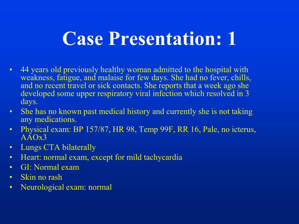
• 44 years old previously healthy woman admitted to the hospital with weakness, fatigue, and malaise for few days. She had no fever, chills, and no recent travel or sick contacts. She reports that a week ago she developed some upper respiratory viral infection which resolved in 3 days.
• She has no known past medical history and currently she is not taking any medications.
• Physical exam: BP 157/87, HR 98, Temp 99F, RR 16, Pale, no icterus, AAOx3
• Lungs CTA bilaterally
• Heart: normal exam, except for mild tachycardia
• GI: Normal exam
• Skin no rash
• Neurological exam: normal
Case Presentation: 1
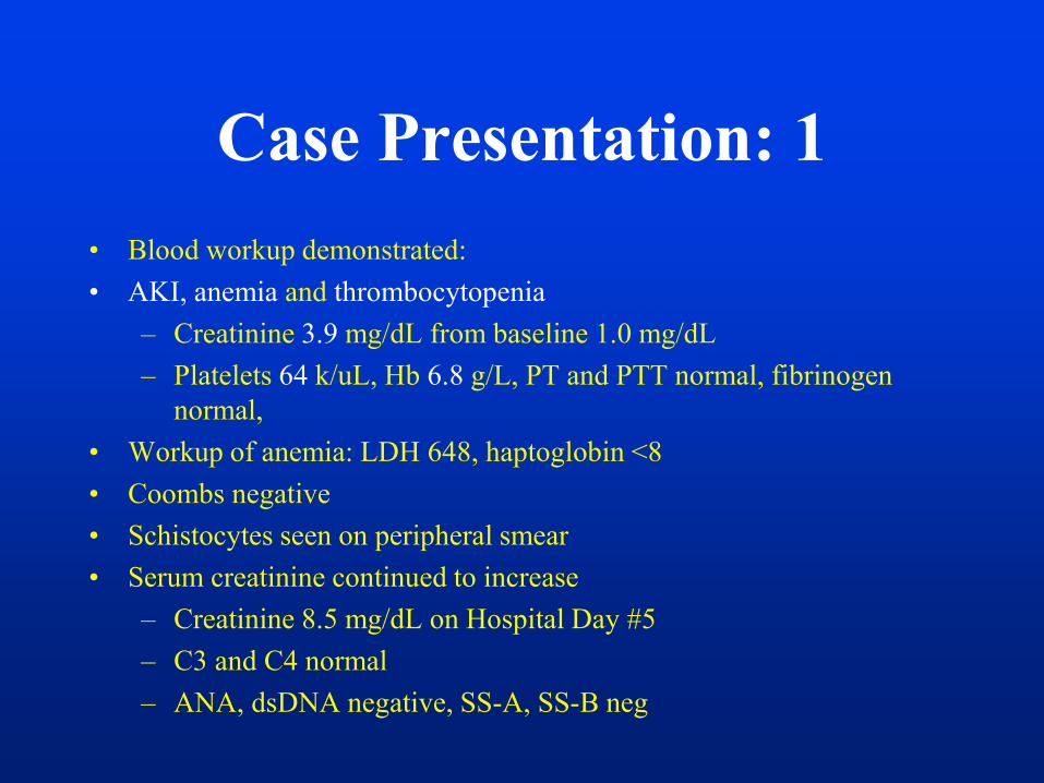
• Blood workup demonstrated:
• AKI, anemia and thrombocytopenia
– Creatinine 3.9 mg/dL from baseline 1.0 mg/dL
– Platelets 64 k/uL, Hb 6.8 g/L, PT and PTT normal, fibrinogen
normal,
• Workup of anemia: LDH 648, haptoglobin <8
• Coombs negative
• Schistocytes seen on peripheral smear
• Serum creatinine continued to increase
– Creatinine 8.5 mg/dL on Hospital Day #5
– C3 and C4 normal
– ANA, dsDNA negative, SS-A, SS-B neg
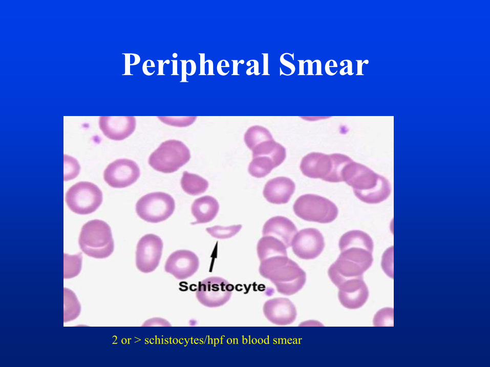
Peripheral Smear
2 or > schistocytes/hpf on blood smear
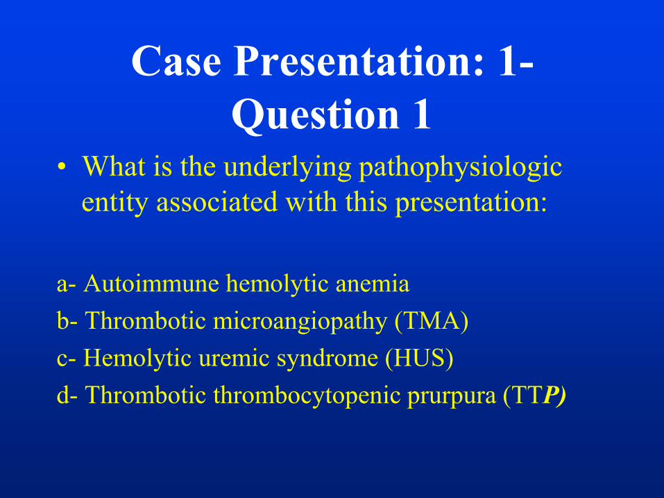
Case Presentation: 1-
Question 1• What is the underlying pathophysiologic
entity associated with this presentation:
a- Autoimmune hemolytic anemia
b- Thrombotic microangiopathy (TMA)
c- Hemolytic uremic syndrome (HUS)
d- Thrombotic thrombocytopenic prurpura (TTP)
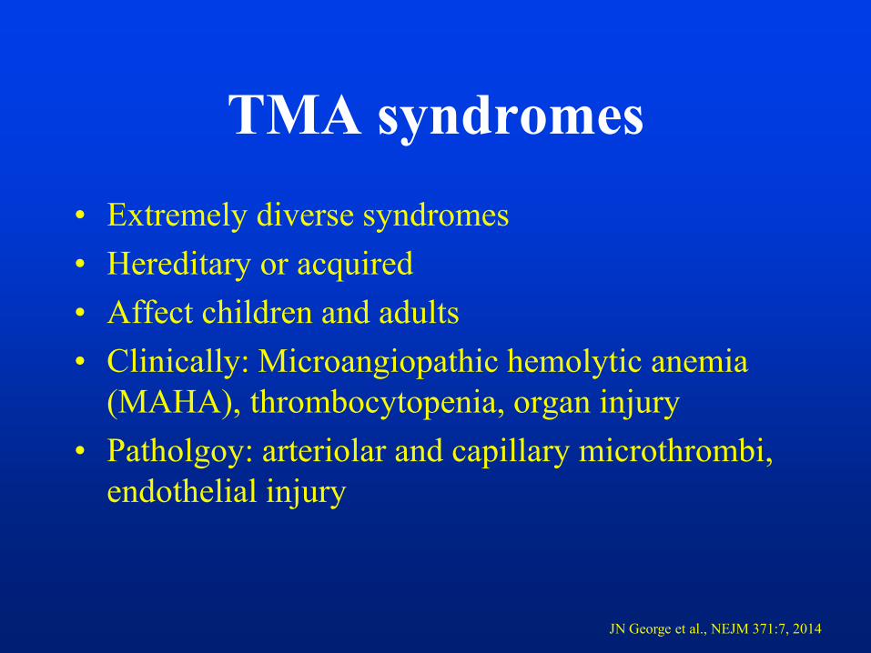
TMA syndromes
• Extremely diverse syndromes
• Hereditary or acquired
• Affect children and adults
• Clinically: Microangiopathic hemolytic anemia
(MAHA), thrombocytopenia, organ injury
• Patholgoy: arteriolar and capillary microthrombi,
endothelial injury
JN George et al., NEJM 371:7, 2014
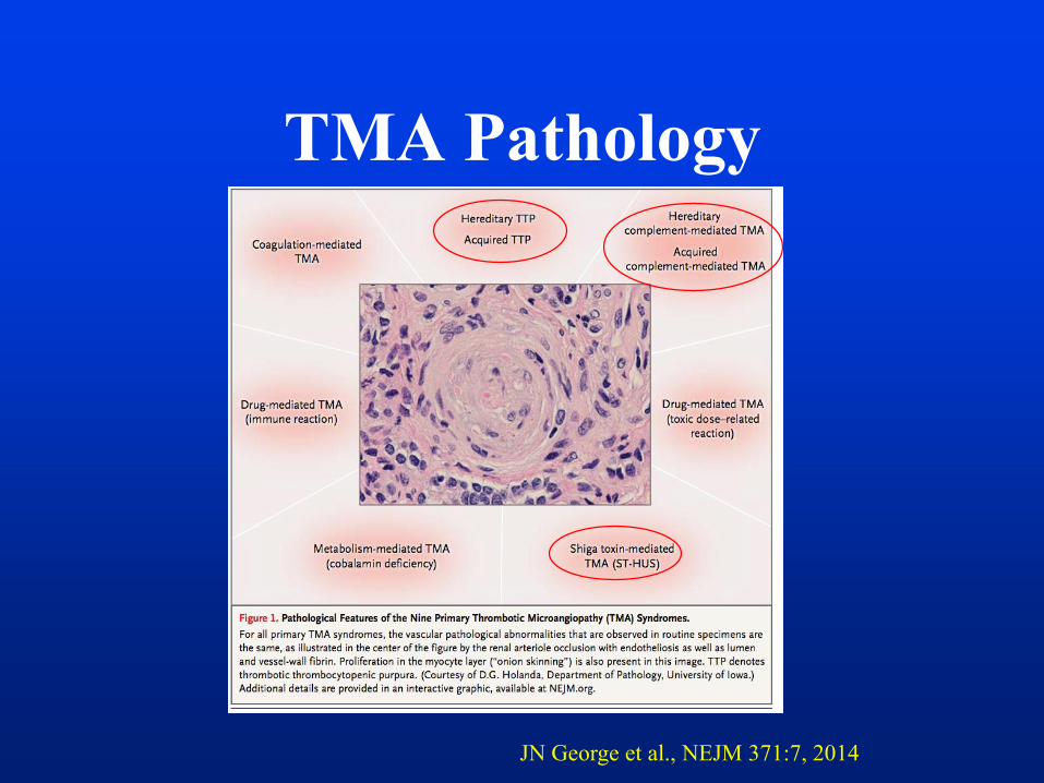
TMA Pathology
JN George et al., NEJM 371:7, 2014

Carla M. Nester CM, et al, Mol Immunology, Vol 67, Issue 1, 2015, 31–42
Differential Diagnosis of TMA
ADAMTS13
deficiency
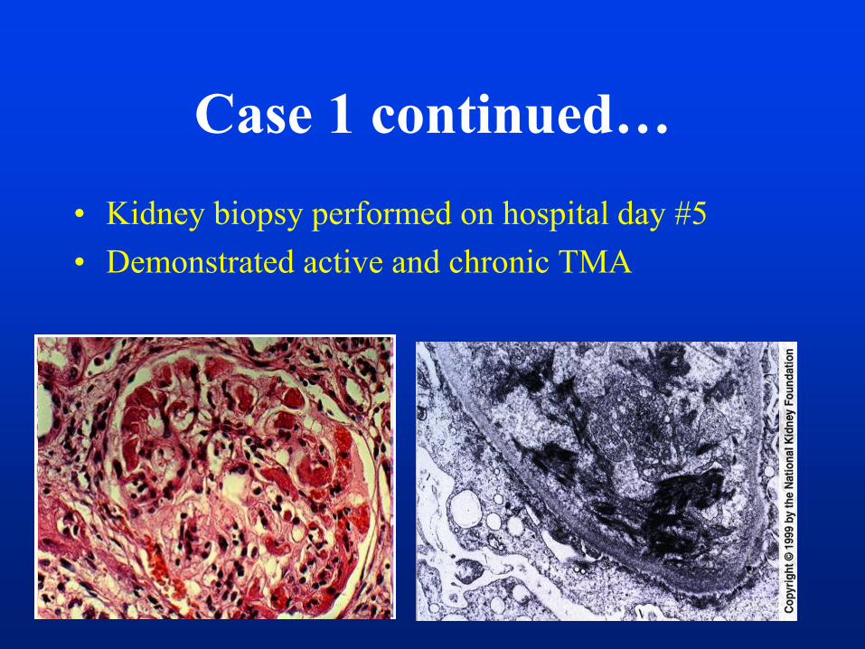
• Kidney biopsy performed on hospital day #5
• Demonstrated active and chronic TMA
Case 1 continued…
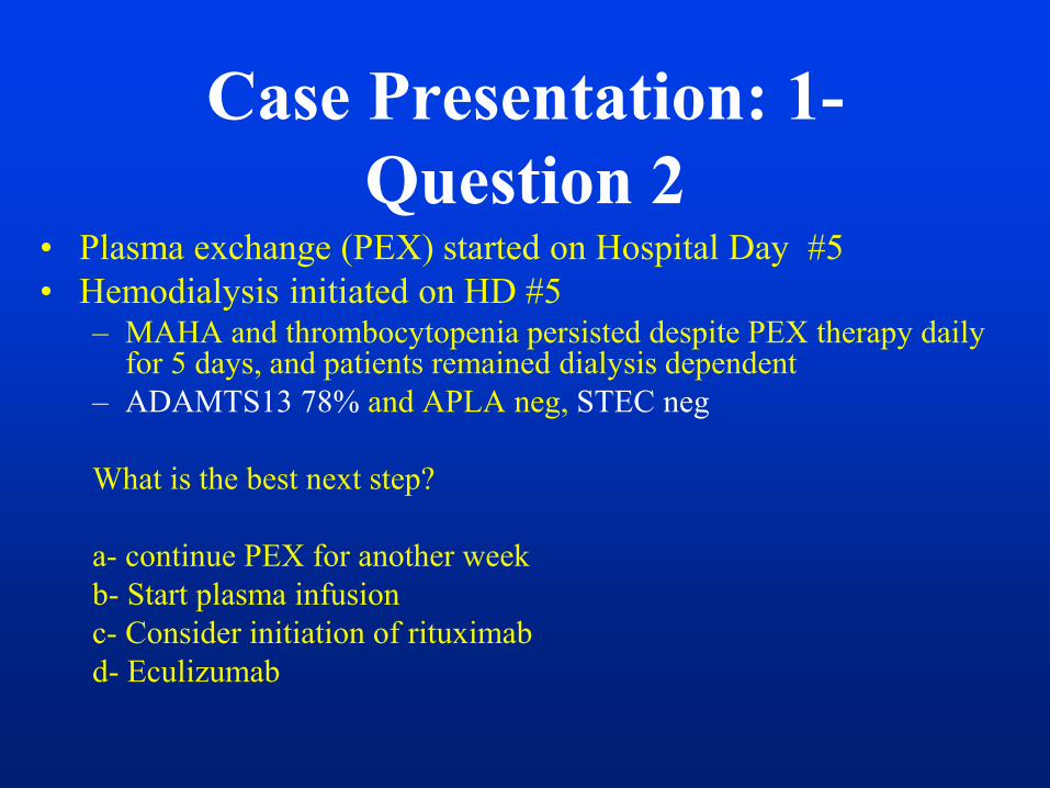
Case Presentation: 1-
Question 2• Plasma exchange (PEX) started on Hospital Day #5
• Hemodialysis initiated on HD #5– MAHA and thrombocytopenia persisted despite PEX therapy daily
for 5 days, and patients remained dialysis dependent
– ADAMTS13 78% and APLA neg, STEC neg
What is the best next step?
a- continue PEX for another week
b- Start plasma infusion
c- Consider initiation of rituximab
d- Eculizumab
Case Presentation: 1-
Question 3
• Before the administration of eculizumab, you should:
a- vaccinate the patient for Neisseria meningitidis
b- vaccinate the patient for Neisseria meningitidis and start at least
2 weeks of antibiotic prophylaxis for N. Meningitidis (e.g
ciprofloxacin)
c- Monitor for any signs of meningitis and treat as needed
d- no further intervention is needed, proceed with eculizumab
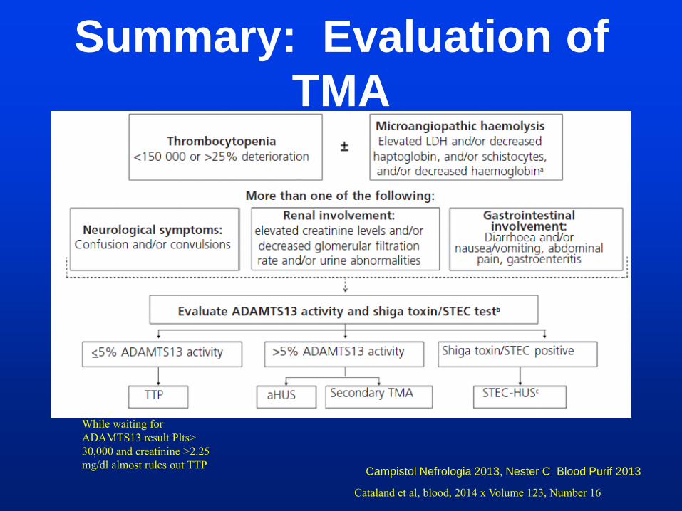
Summary: Evaluation of
TMA
Campistol Nefrologia 2013, Nester C Blood Purif 2013
While waiting for
ADAMTS13 result Plts>
30,000 and creatinine >2.25
mg/dl almost rules out TTP
Cataland et al, blood, 2014 x Volume 123, Number 16
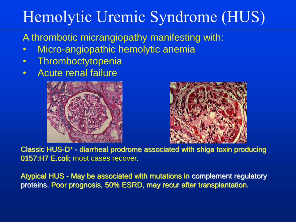
Hemolytic Uremic Syndrome (HUS)
Classic HUS-D+ - diarrheal prodrome associated with shiga toxin producing
0157:H7 E.coli; most cases recover.
Atypical HUS - May be associated with mutations in complement regulatory
proteins. Poor prognosis, 50% ESRD, may recur after transplantation.
A thrombotic micrangiopathy manifesting with:
• Micro-angiopathic hemolytic anemia
• Thromboctytopenia
• Acute renal failure

Shiga Toxin HUS
• Induced by enteric infection with shiga toxin
secreting strain of Enterohemorrhagic E Coli
(O157:H7, O104:H4) or Shigella (contaminated
water, vegetables, beef products etc..)
• Common in children presenting with AKI
• 6-9% of STEC infected children develop HUS
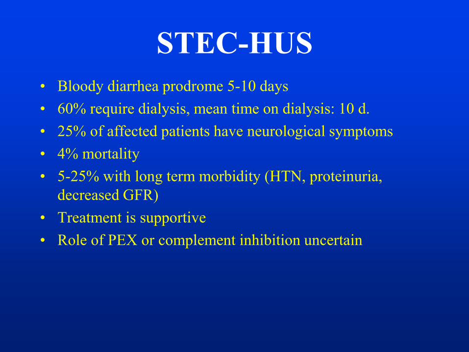
STEC-HUS
• Bloody diarrhea prodrome 5-10 days
• 60% require dialysis, mean time on dialysis: 10 d.
• 25% of affected patients have neurological symptoms
• 4% mortality
• 5-25% with long term morbidity (HTN, proteinuria,
decreased GFR)
• Treatment is supportive
• Role of PEX or complement inhibition uncertain
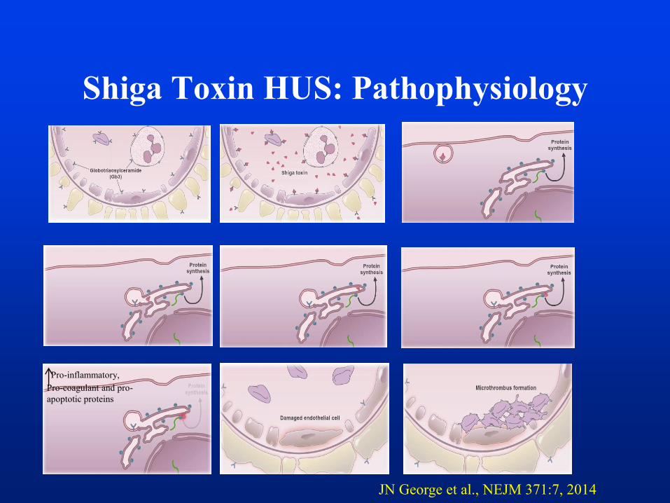
Shiga Toxin HUS: Pathophysiology
Pro-inflammatory,
Pro-coagulant and pro-
apoptotic proteins
JN George et al., NEJM 371:7, 2014
Case Presentation: 1-
Question 4• Which one of the following statements is correct:
a- Normal complement levels rule out aHUS
b- The absence of complement system related
genetic mutations rules out aHUS
c- the absence of family history of aHUS, rules
out aHUS
d- none of the above
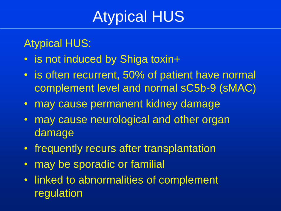
Atypical HUS:
• is not induced by Shiga toxin+
• is often recurrent, 50% of patient have normal
complement level and normal sC5b-9 (sMAC)
• may cause permanent kidney damage
• may cause neurological and other organ
damage
• frequently recurs after transplantation
• may be sporadic or familial
• linked to abnormalities of complement
regulation
Atypical HUS

Mele et al, Semin Immunopathol (2014) 36:399–420
22
FIMAC SRCR LRDR-1 LRDR-2 S-S SP
H Chain L Chain
T72SA240G G261D (n=2)
G349R
I357M
W399R
L484V+Q485G+W486X
E554V
D519N
MG2 MG3 MG4 MG5
MG
6β
LNK
β-chain α-chain
α’NT
MG
6α
MG7
CU
B g
TED MG8AN
A
CU
B f
Anchor
C345C
R570W K1029M
R1041S
D1093N
I1135T
T1361M
1 2 3
SCRslinker
Ba Bb
R138W
1 2 3
SCRslinker
VWA SP
R317W
T140R
T140K Q163E
Regulatorydomain
C3b
Recognitiondoman
C3bproteoglycansproteoglycans C3b proteoglycans
S890I (n=2)
Y899X
W920R
2759del15bp
479X
N516KQ950H
I970V1014X
Q1137L
C1163W
Dup(3546-3581)
E1172X (n=2)
W1183R
W1183X
S1191L (n=7)
G1194D
V1197A (n=4)
E1198A
V1200L
R1210C (n=9)
R1215Q (n=2)
R1215G
3675-3699del
R78G
T267fs270X
(858-872)del+D277N+P278S
F242C
SCRs
STP
TM
CT
2
3
4
1
C35X
C35Y (n=3)
R59X (n=4)
48aa del
A353V
192T>C+193-198del
C99R
(96-129)del+G130I+Y132T+L133X
Y155D
MG1
R456L
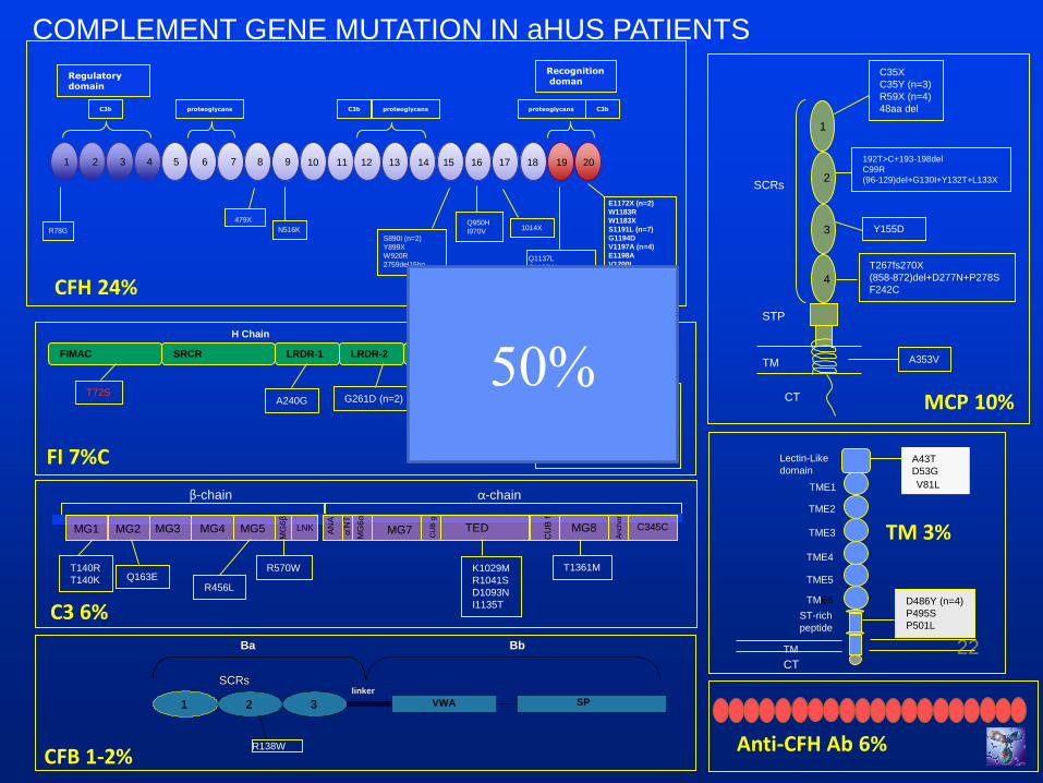
CFH 24%
MCP 10%
FI 7%C
C3 6%
CFB 1-2%
COMPLEMENT GENE MUTATION IN aHUS PATIENTS
Anti-CFH Ab 6%
A43T
D53G
V81L
D486Y (n=4)
P495S
P501L
CT
Lectin-Like
domain
TME1
TME2
TME3
TME4
TME5
TME6
ST-rich
peptide
TM
V81L
TM 3%
1 2 3 4 5 6 7 8 9 10 11 12 13 14 15 16 17 18 19 20
50%
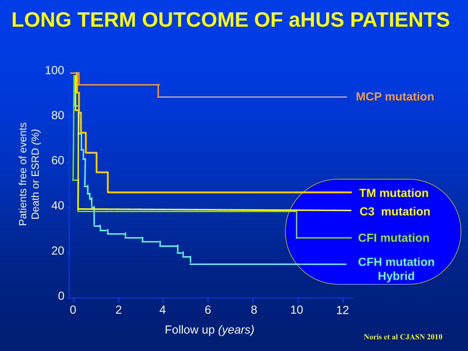
LONG TERM OUTCOME OF aHUS PATIENTS
CFH mutation
Hybrid
Pa
tie
nts
fre
e o
f e
ve
nts
Death
or
ES
RD
(%
)
100
80
60
40
20
0
0 2 4 6 8 10 12
CFI mutation
C3 mutation
Follow up (years)
MCP mutation
Noris et al CJASN 2010
TM mutation
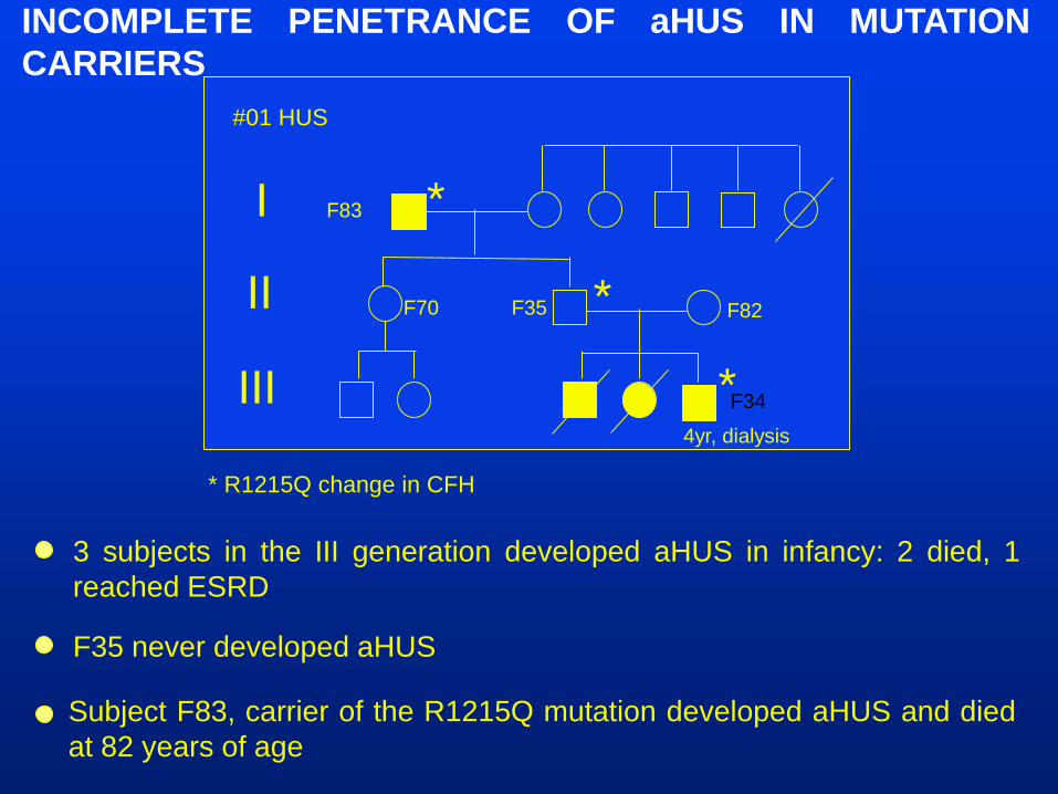
#01 HUS
I
II
III F34
4yr, dialysis
F82F35F70
F83 *
*
*
* R1215Q change in CFH
3 subjects in the III generation developed aHUS in infancy: 2 died, 1
reached ESRD
F35 never developed aHUS
INCOMPLETE PENETRANCE OF aHUS IN MUTATION
CARRIERS
Subject F83, carrier of the R1215Q mutation developed aHUS and died
at 82 years of age
Atypical HUS may arise when there is:
• Deficiency, dysfunction or autoantibody-mediated
inhibition of one or more of the regulatory proteins
• Resistance of C3b or factor B to decay by factors H, I and
MCP
• Mutation of thrombomodulin, an endothelial glycoprotein
with cytoprotective and anticoagulant properties
• The disease may be quiescent for months or years, even in
patients with homozygous mutations and complete
deficiency, only to be triggered by an otherwise innocuous
infection, drug exposure or pregnancy
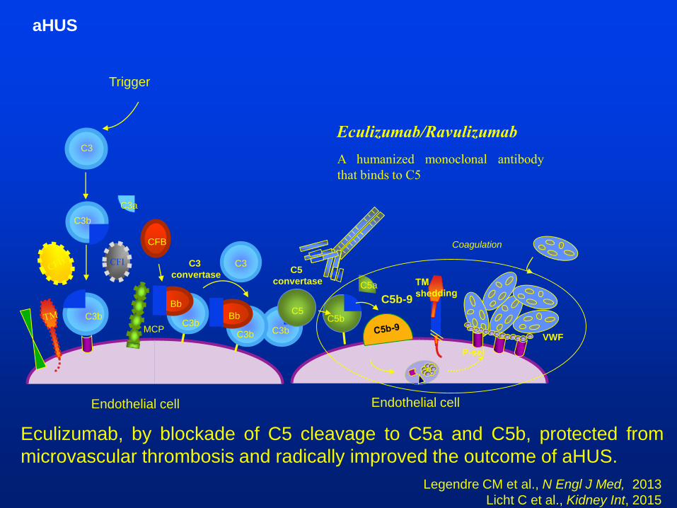
aHUS
C3b
C3
C3b
C3a
Endothelial cell
MCP
CFB
CFI
C3b
C5b
C5a
C5
C5b-9
C3b
Bb
C3b
Bb
C3C3
convertaseC5
convertase
Endothelial cell
Trigger
Coagulation
VWF
Eculizumab, by blockade of C5 cleavage to C5a and C5b, protected from
microvascular thrombosis and radically improved the outcome of aHUS.
Legendre CM et al., N Engl J Med, 2013
Licht C et al., Kidney Int, 2015
Eculizumab/Ravulizumab
A humanized monoclonal antibody
that binds to C5
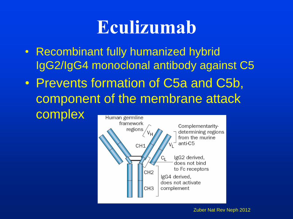
Eculizumab
• Recombinant fully humanized hybrid
IgG2/IgG4 monoclonal antibody against C5
• Prevents formation of C5a and C5b,
component of the membrane attack
complex
Zuber Nat Rev Neph 2012
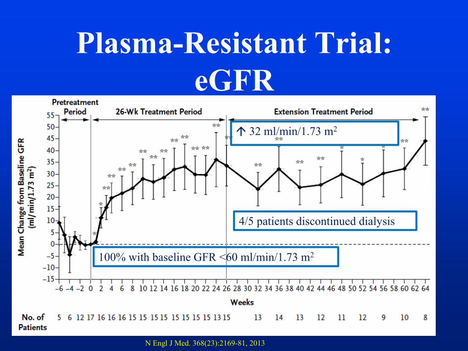
Plasma-Resistant Trial:
eGFR
28
4/5 patients discontinued dialysis
100% with baseline GFR <60 ml/min/1.73 m2
32 ml/min/1.73 m2
N Engl J Med. 368(23):2169-81, 2013
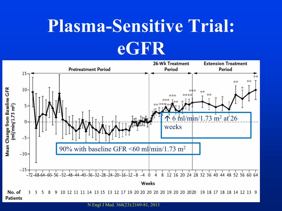
Plasma-Sensitive Trial:
eGFR
29
90% with baseline GFR <60 ml/min/1.73 m2
6 ml/min/1.73 m2 at 26
weeks
N Engl J Med. 368(23):2169-81, 2013
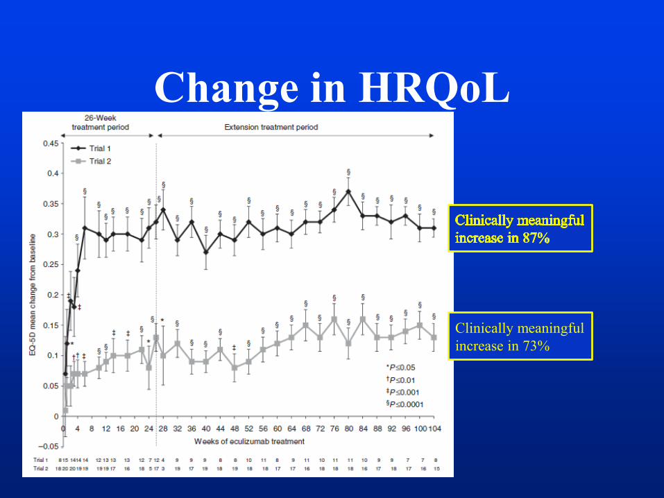
Change in HRQoL
Clinically meaningful
increase in 73%
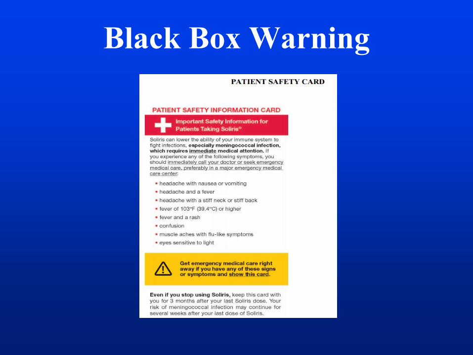
Black Box Warning
FH
FH
transplant
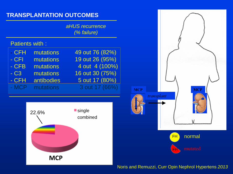
TRANSPLANTATION OUTCOMES
Patients with :
- CFH mutations
- CFI mutations
- CFB mutations
- C3 mutations
- CFH antibodies
- MCP mutations
aHUS recurrence
(% failure)
49 out 76 (82%)
19 out 26 (95%)
4 out 4 (100%)
16 out 30 (75%)
5 out 17 (80%)
3 out 17 (66%)
Noris and Remuzzi, Curr Opin Nephrol Hypertens 2013
FH
normal
mutated
FH
MCP
transplant
MCP
MCP
single
combined 22.6%
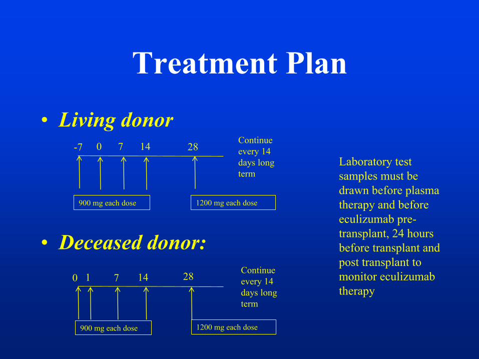
Treatment Plan
• Living donor
• Deceased donor:
-7 0 7 14 28Continue
every 14
days long
term
900 mg each dose 1200 mg each dose
0 1 7 14 28Continue
every 14
days long
term
900 mg each dose 1200 mg each dose
Laboratory test
samples must be
drawn before plasma
therapy and before
eculizumab pre-
transplant, 24 hours
before transplant and
post transplant to
monitor eculizumab
therapy
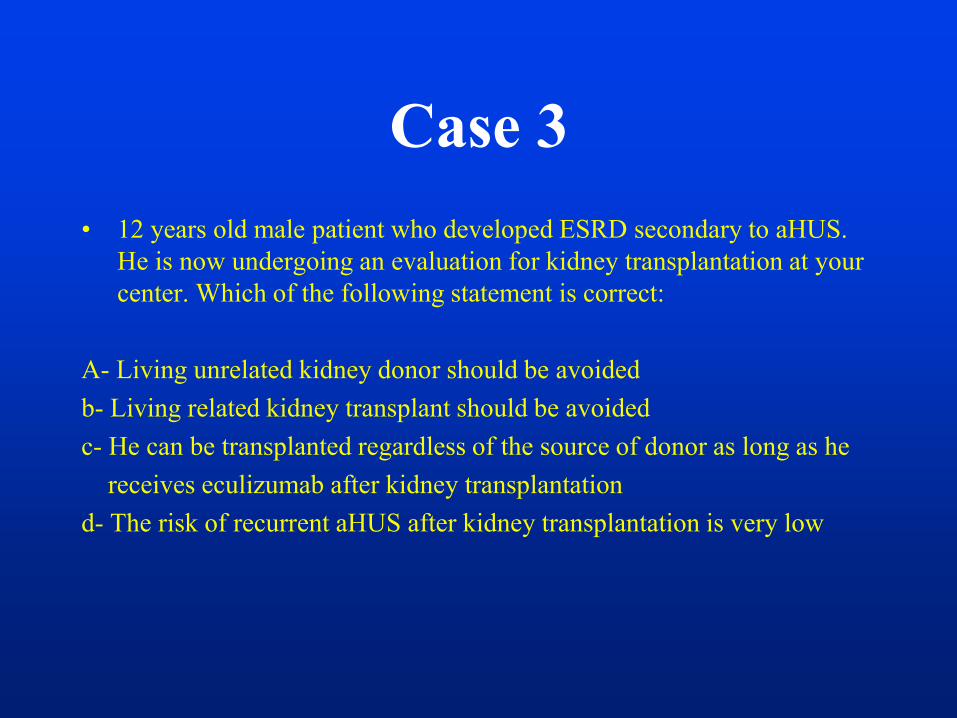
Case 3
• 12 years old male patient who developed ESRD secondary to aHUS.
He is now undergoing an evaluation for kidney transplantation at your
center. Which of the following statement is correct:
A- Living unrelated kidney donor should be avoided
b- Living related kidney transplant should be avoided
c- He can be transplanted regardless of the source of donor as long as he
receives eculizumab after kidney transplantation
d- The risk of recurrent aHUS after kidney transplantation is very low
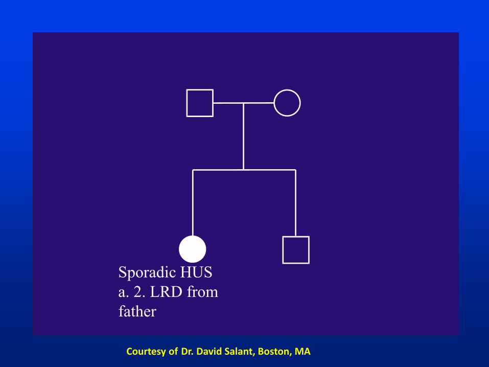
Sporadic HUS
a. 2. LRD from
father
Courtesy of Dr. David Salant, Boston, MA
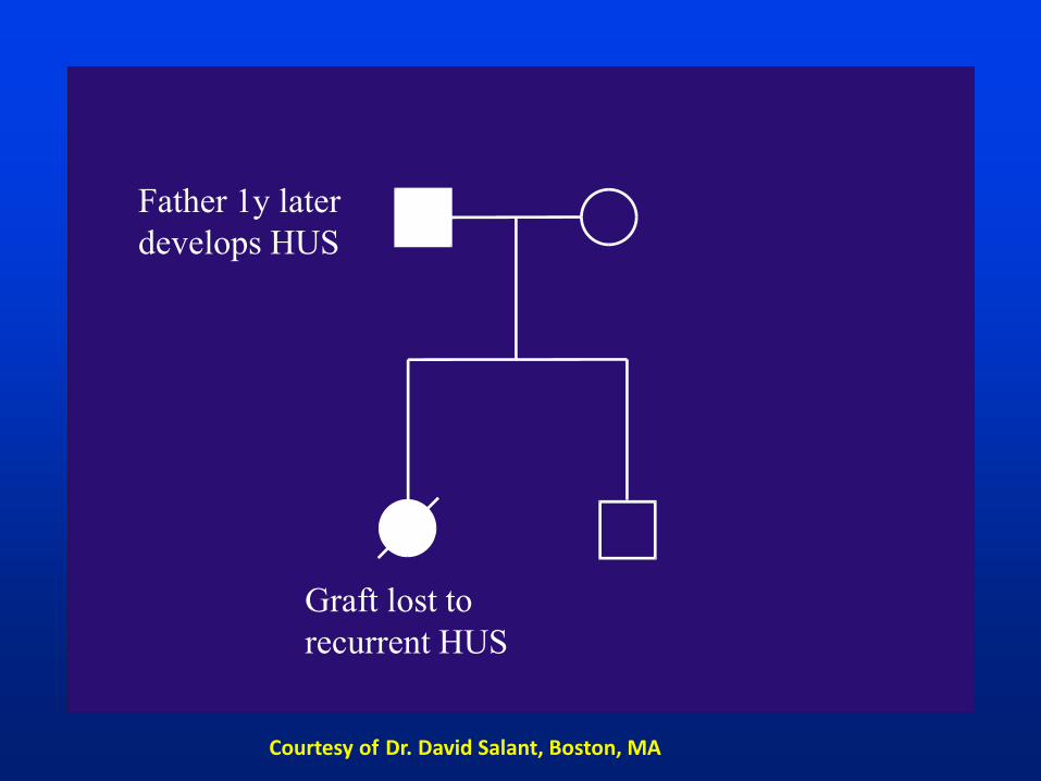
Graft lost to
recurrent HUS
Father 1y later
develops HUS
Courtesy of Dr. David Salant, Boston, MA
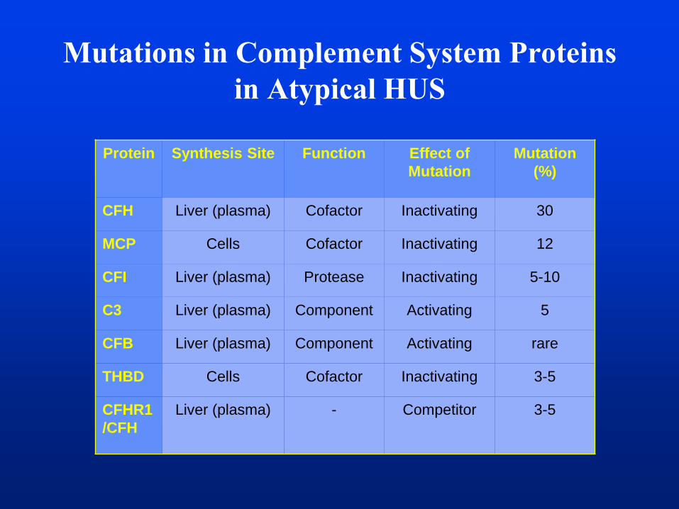
Protein Synthesis Site Function Effect of
Mutation
Mutation
(%)
CFH Liver (plasma) Cofactor Inactivating 30
MCP Cells Cofactor Inactivating 12
CFI Liver (plasma) Protease Inactivating 5-10
C3 Liver (plasma) Component Activating 5
CFB Liver (plasma) Component Activating rare
THBD Cells Cofactor Inactivating 3-5
CFHR1
/CFH
Liver (plasma) - Competitor 3-5
Mutations in Complement System Proteins
in Atypical HUS
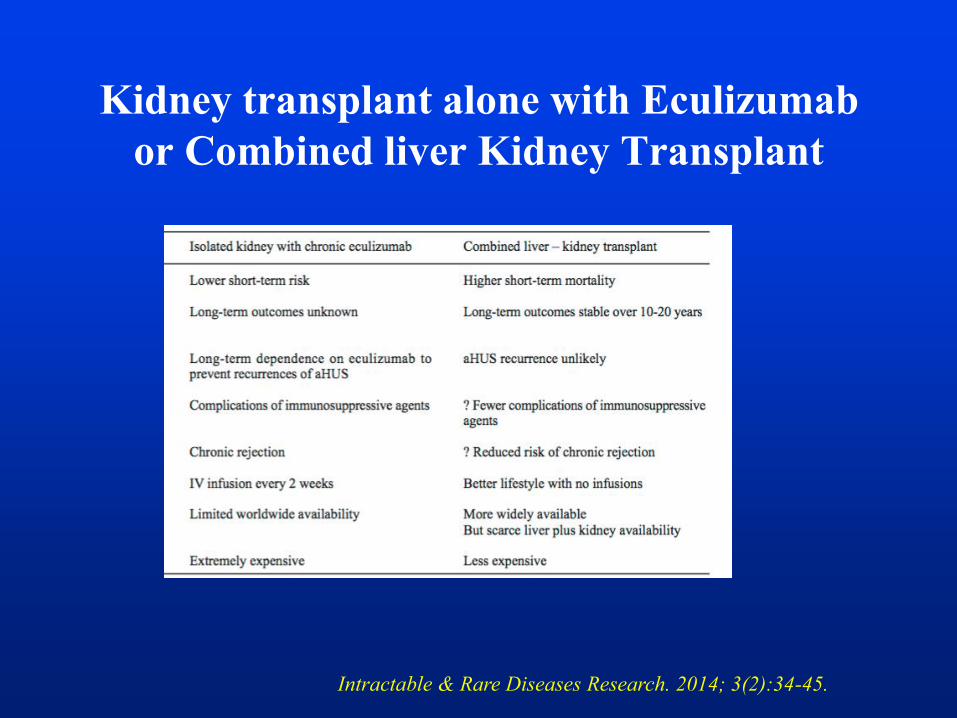
Kidney transplant alone with Eculizumab
or Combined liver Kidney Transplant
Intractable & Rare Diseases Research. 2014; 3(2):34-45.
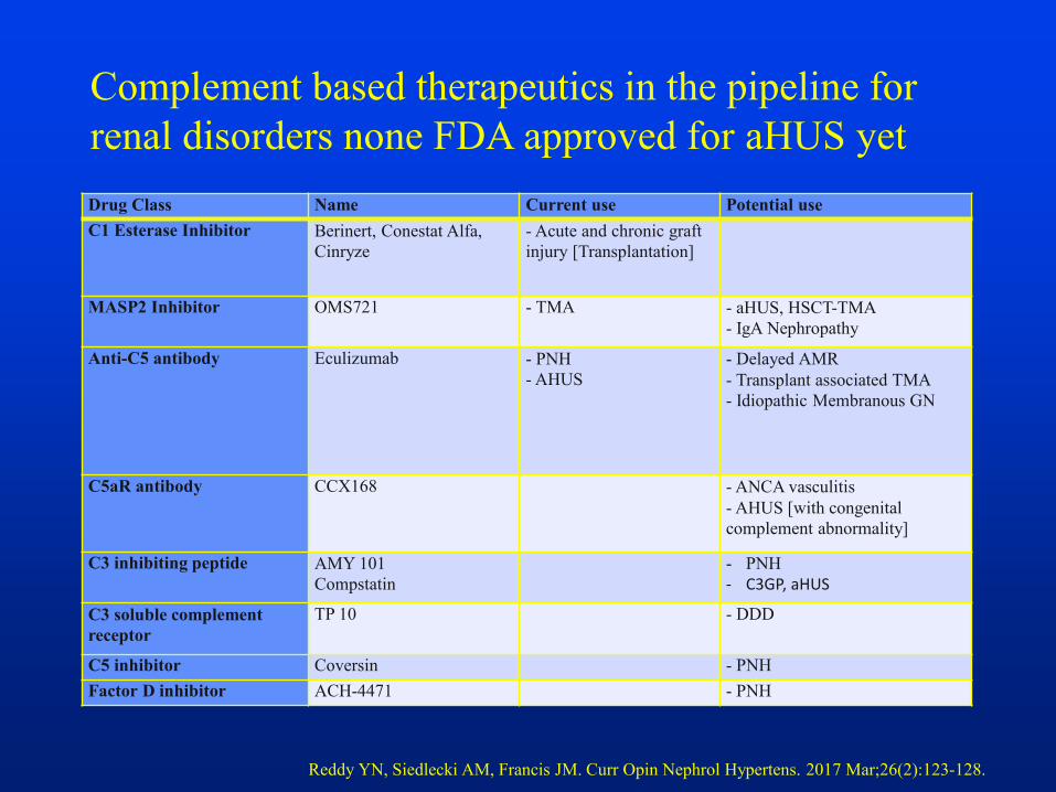
Drug Class Name Current use Potential use
C1 Esterase Inhibitor Berinert, Conestat Alfa,
Cinryze
- Acute and chronic graft
injury [Transplantation]
MASP2 Inhibitor OMS721 - TMA - aHUS, HSCT-TMA
- IgA Nephropathy
Anti-C5 antibody Eculizumab - PNH
- AHUS
- Delayed AMR
- Transplant associated TMA
- Idiopathic Membranous GN
C5aR antibody CCX168 - ANCA vasculitis
- AHUS [with congenital
complement abnormality]
C3 inhibiting peptide AMY 101
Compstatin
- PNH
- C3GP, aHUS
C3 soluble complement
receptor
TP 10 - DDD
C5 inhibitor Coversin - PNH
Factor D inhibitor ACH-4471 - PNH
Complement based therapeutics in the pipeline for
renal disorders none FDA approved for aHUS yet
Reddy YN, Siedlecki AM, Francis JM. Curr Opin Nephrol Hypertens. 2017 Mar;26(2):123-128.
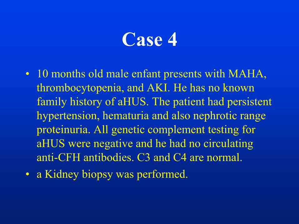
Case 4
• 10 months old male enfant presents with MAHA,
thrombocytopenia, and AKI. He has no known
family history of aHUS. The patient had persistent
hypertension, hematuria and also nephrotic range
proteinuria. All genetic complement testing for
aHUS were negative and he had no circulating
anti-CFH antibodies. C3 and C4 are normal.
• a Kidney biopsy was performed.
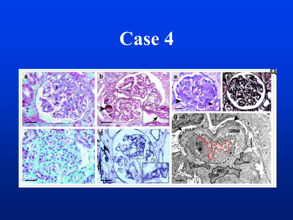
Case 4
Case 4: Question 1
• What is the most likely diagnosis:
a- complement mediated aHUS
b- TTP
c- HUS secondary to diacylglycerol kinase
e (DGKE) mutation
d- Lupus nephritis
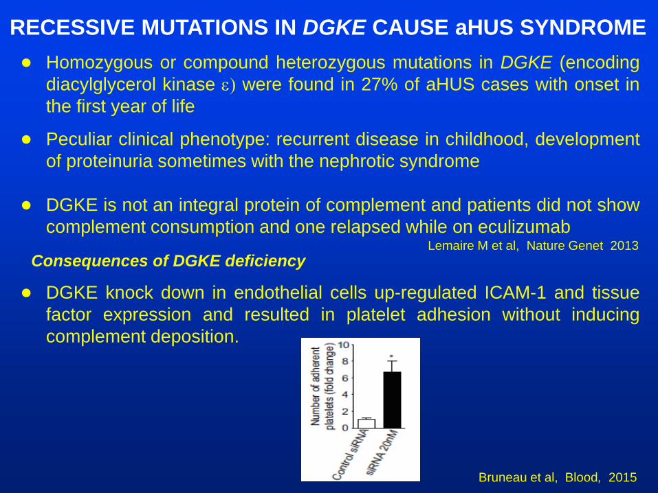
● Homozygous or compound heterozygous mutations in DGKE (encoding
diacylglycerol kinase e) were found in 27% of aHUS cases with onset in
the first year of life
● Peculiar clinical phenotype: recurrent disease in childhood, development
of proteinuria sometimes with the nephrotic syndrome
● DGKE is not an integral protein of complement and patients did not show
complement consumption and one relapsed while on eculizumab
● DGKE knock down in endothelial cells up-regulated ICAM-1 and tissue
factor expression and resulted in platelet adhesion without inducing
complement deposition.
Lemaire M et al, Nature Genet 2013
RECESSIVE MUTATIONS IN DGKE CAUSE aHUS SYNDROME
Consequences of DGKE deficiency
Bruneau et al, Blood, 2015
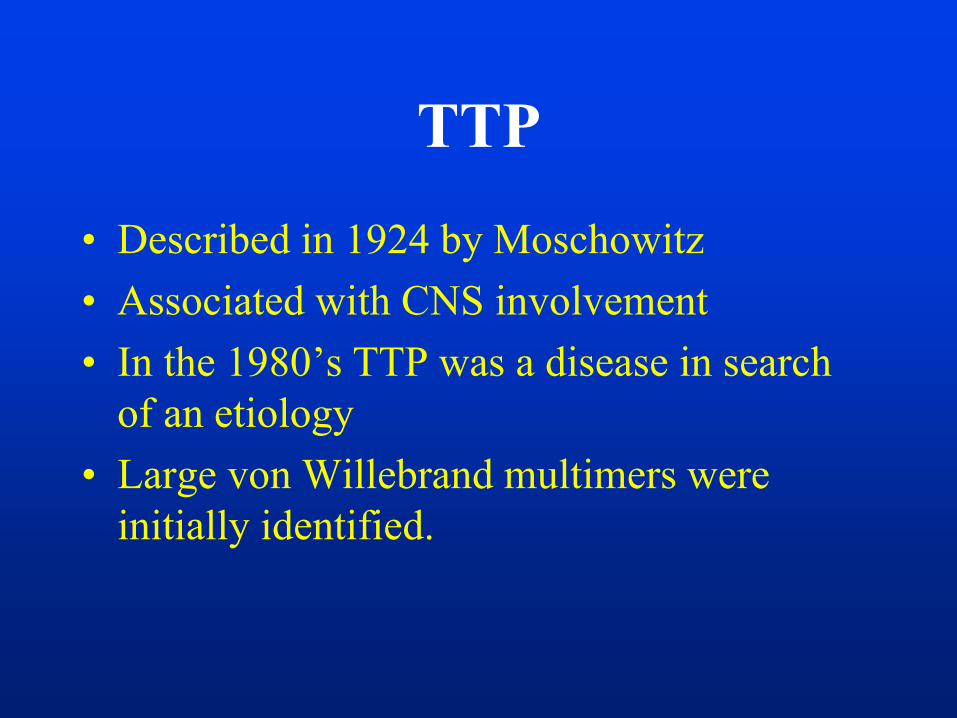
TTP
• Described in 1924 by Moschowitz
• Associated with CNS involvement
• In the 1980’s TTP was a disease in search
of an etiology
• Large von Willebrand multimers were
initially identified.
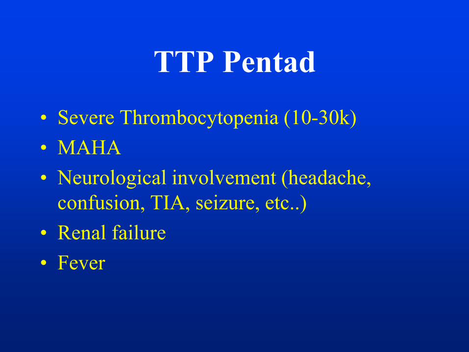
TTP Pentad
• Severe Thrombocytopenia (10-30k)
• MAHA
• Neurological involvement (headache,
confusion, TIA, seizure, etc..)
• Renal failure
• Fever
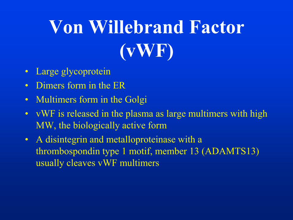
Von Willebrand Factor
(vWF)• Large glycoprotein
• Dimers form in the ER
• Multimers form in the Golgi
• vWF is released in the plasma as large multimers with high
MW, the biologically active form
• A disintegrin and metalloproteinase with a
thrombospondin type 1 motif, member 13 (ADAMTS13)
usually cleaves vWF multimers
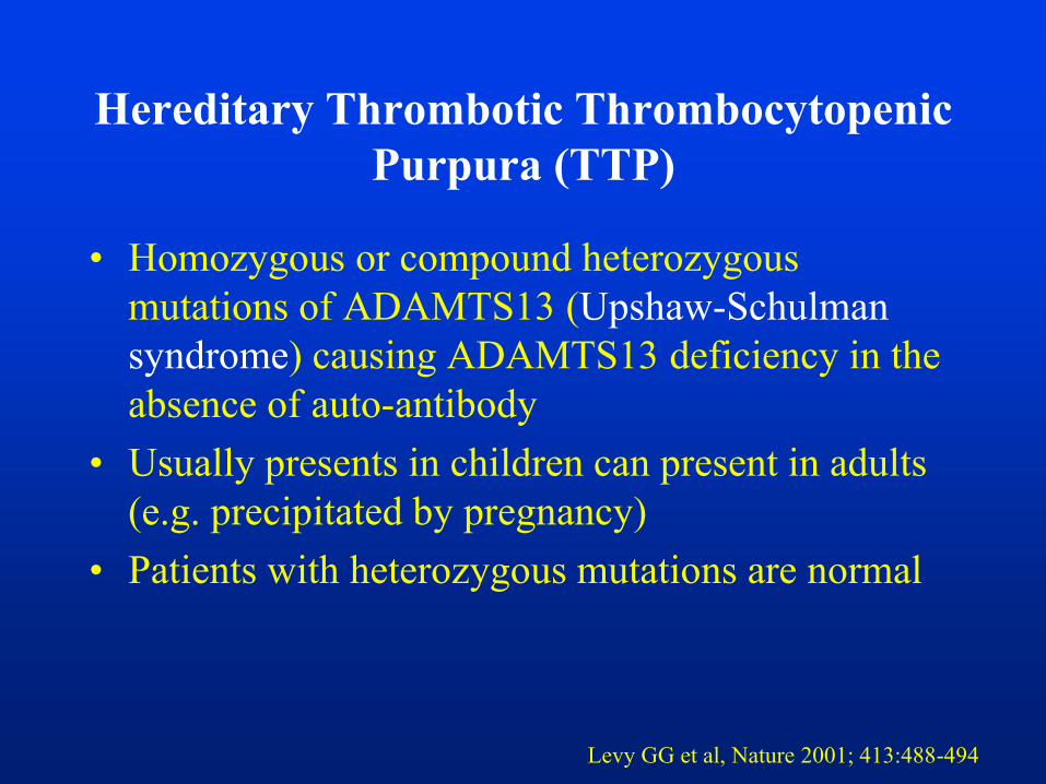
Hereditary Thrombotic Thrombocytopenic
Purpura (TTP)
• Homozygous or compound heterozygous
mutations of ADAMTS13 (Upshaw-Schulman
syndrome) causing ADAMTS13 deficiency in the
absence of auto-antibody
• Usually presents in children can present in adults
(e.g. precipitated by pregnancy)
• Patients with heterozygous mutations are normal
Levy GG et al, Nature 2001; 413:488-494
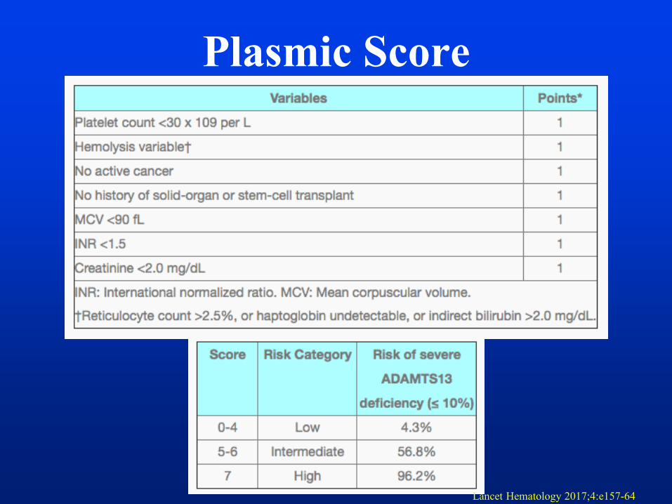
Plasmic Score
Lancet Hematology 2017;4:e157-64
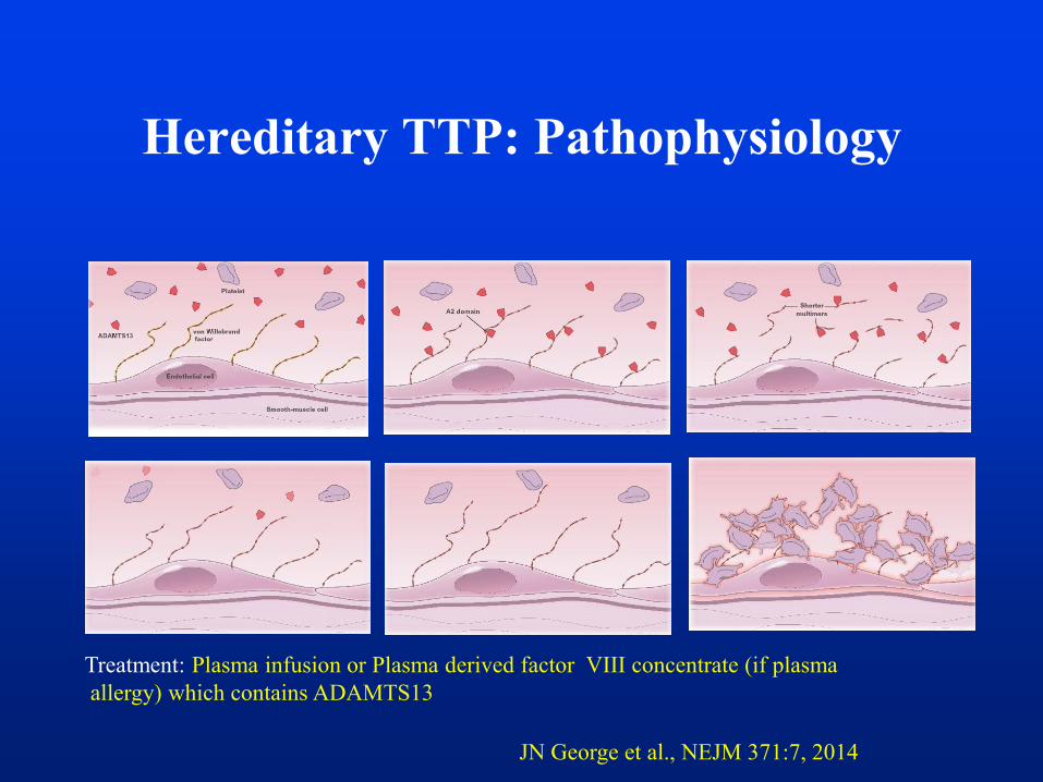
Hereditary TTP: Pathophysiology
JN George et al., NEJM 371:7, 2014
Treatment: Plasma infusion or Plasma derived factor VIII concentrate (if plasma
allergy) which contains ADAMTS13
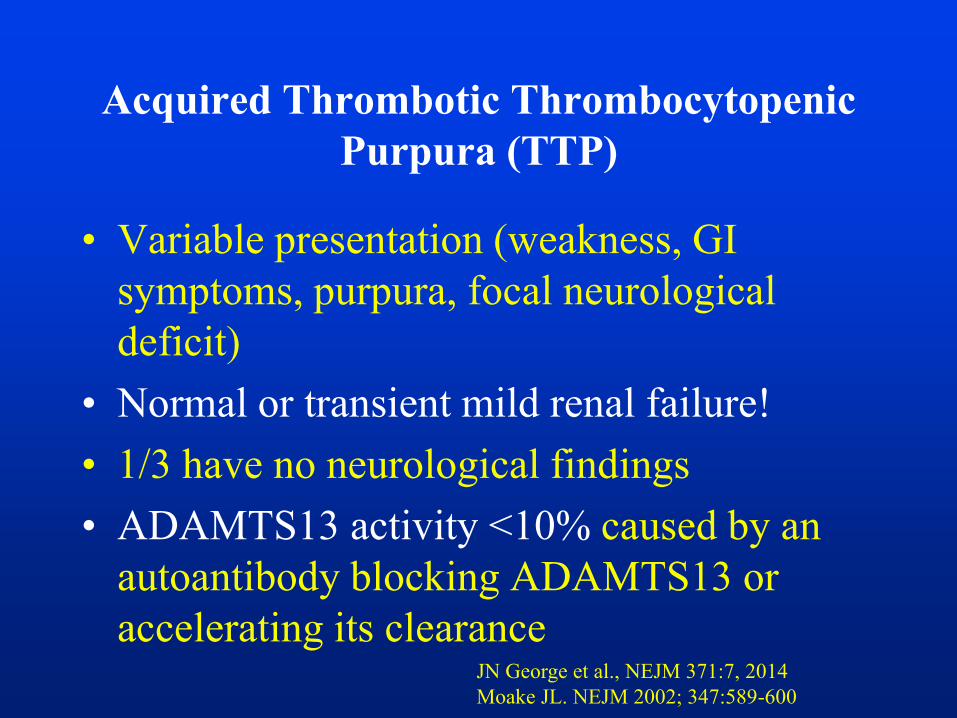
Acquired Thrombotic Thrombocytopenic
Purpura (TTP)
• Variable presentation (weakness, GI
symptoms, purpura, focal neurological
deficit)
• Normal or transient mild renal failure!
• 1/3 have no neurological findings
• ADAMTS13 activity <10% caused by an
autoantibody blocking ADAMTS13 or
accelerating its clearanceJN George et al., NEJM 371:7, 2014
Moake JL. NEJM 2002; 347:589-600
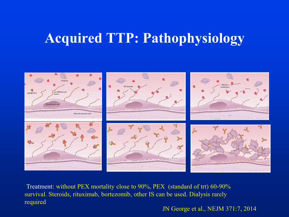
Acquired TTP: Pathophysiology
JN George et al., NEJM 371:7, 2014
:Treatment: without PEX mortality close to 90%, PEX (standard of trt) 60-90%
survival. Steroids, rituximab, bortezomib, other IS can be used. Dialysis rarely
required
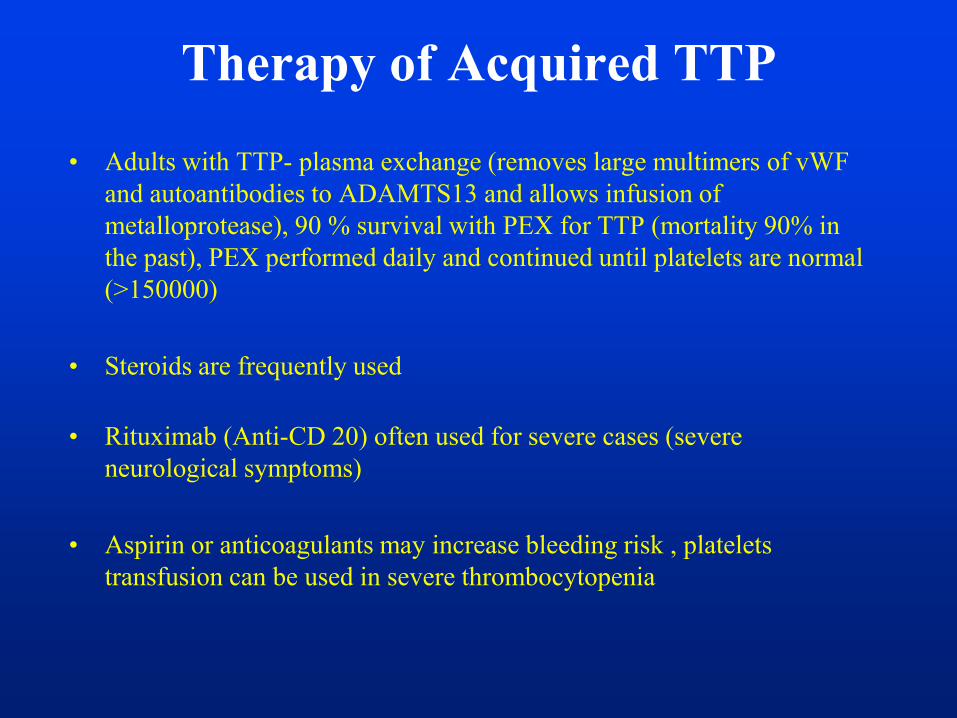
Therapy of Acquired TTP
• Adults with TTP- plasma exchange (removes large multimers of vWF
and autoantibodies to ADAMTS13 and allows infusion of
metalloprotease), 90 % survival with PEX for TTP (mortality 90% in
the past), PEX performed daily and continued until platelets are normal
(>150000)
• Steroids are frequently used
• Rituximab (Anti-CD 20) often used for severe cases (severe
neurological symptoms)
• Aspirin or anticoagulants may increase bleeding risk , platelets
transfusion can be used in severe thrombocytopenia
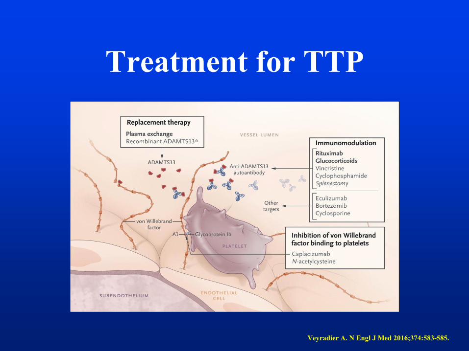
Treatment for TTP
Veyradier A. N Engl J Med 2016;374:583-585.

Case 5
35 y/o female with no PMH presents with 5d headache, N/V and non-bloody diarrhea
• No recent travel
• Husband and kids are healthy, and ate same foods
• Has never had this constellation of symptoms before
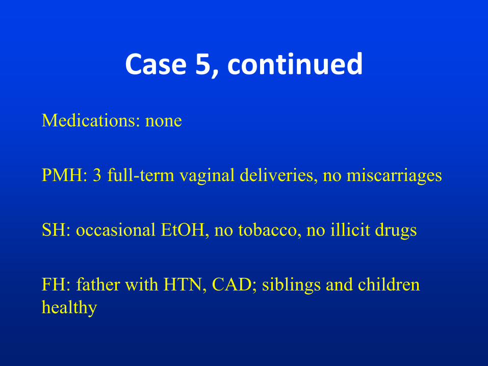
Case 5, continued
Medications: none
PMH: 3 full-term vaginal deliveries, no miscarriages
SH: occasional EtOH, no tobacco, no illicit drugs
FH: father with HTN, CAD; siblings and children
healthy
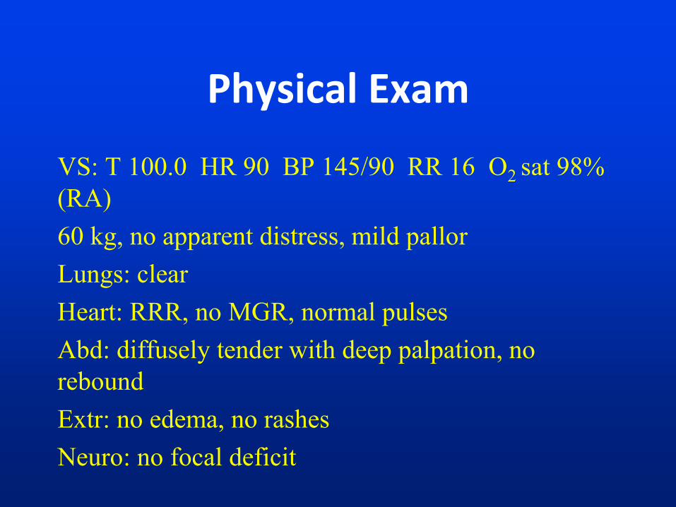
Physical Exam
VS: T 100.0 HR 90 BP 145/90 RR 16 O2 sat 98%
(RA)
60 kg, no apparent distress, mild pallor
Lungs: clear
Heart: RRR, no MGR, normal pulses
Abd: diffusely tender with deep palpation, no
rebound
Extr: no edema, no rashes
Neuro: no focal deficit
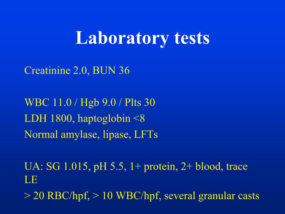
Laboratory tests
Creatinine 2.0, BUN 36
WBC 11.0 / Hgb 9.0 / Plts 30
LDH 1800, haptoglobin <8
Normal amylase, lipase, LFTs
UA: SG 1.015, pH 5.5, 1+ protein, 2+ blood, trace
LE
> 20 RBC/hpf, > 10 WBC/hpf, several granular casts
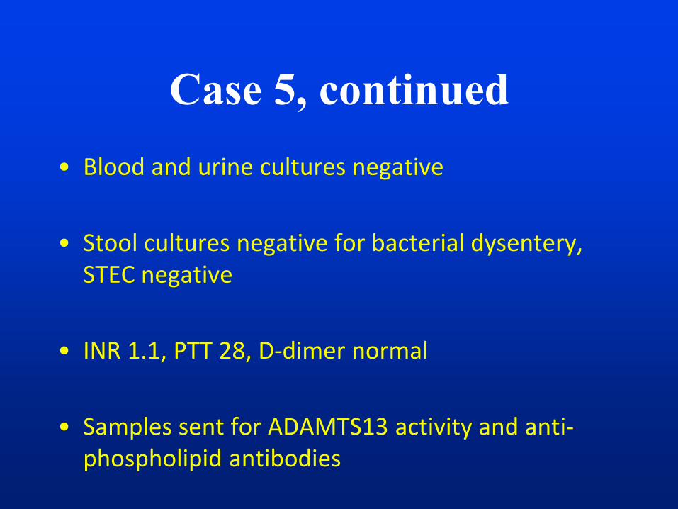
Case 5, continued
• Blood and urine cultures negative
• Stool cultures negative for bacterial dysentery, STEC negative
• INR 1.1, PTT 28, D-dimer normal
• Samples sent for ADAMTS13 activity and anti-phospholipid antibodies
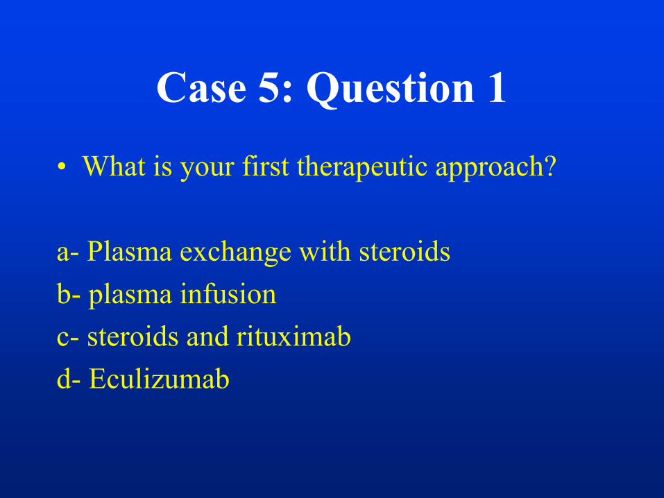
Case 5: Question 1
• What is your first therapeutic approach?
a- Plasma exchange with steroids
b- plasma infusion
c- steroids and rituximab
d- Eculizumab
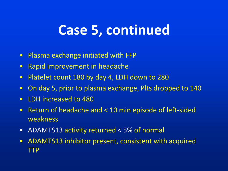
Case 5, continued
• Plasma exchange initiated with FFP
• Rapid improvement in headache
• Platelet count 180 by day 4, LDH down to 280
• On day 5, prior to plasma exchange, Plts dropped to 140
• LDH increased to 480
• Return of headache and < 10 min episode of left-sided weakness
• ADAMTS13 activity returned < 5% of normal
• ADAMTS13 inhibitor present, consistent with acquired TTP
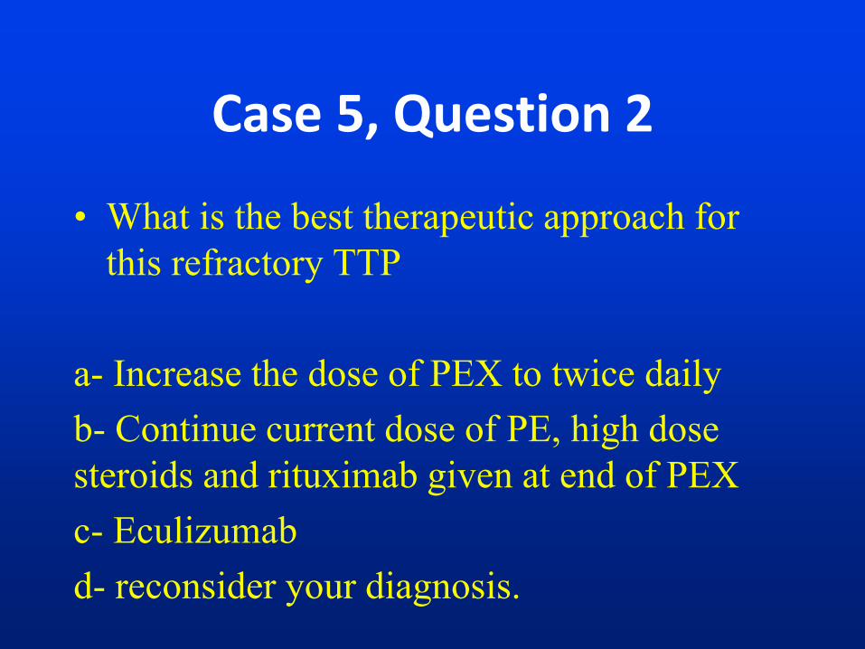
Case 5, Question 2
• What is the best therapeutic approach for
this refractory TTP
a- Increase the dose of PEX to twice daily
b- Continue current dose of PE, high dose
steroids and rituximab given at end of PEX
c- Eculizumab
d- reconsider your diagnosis.
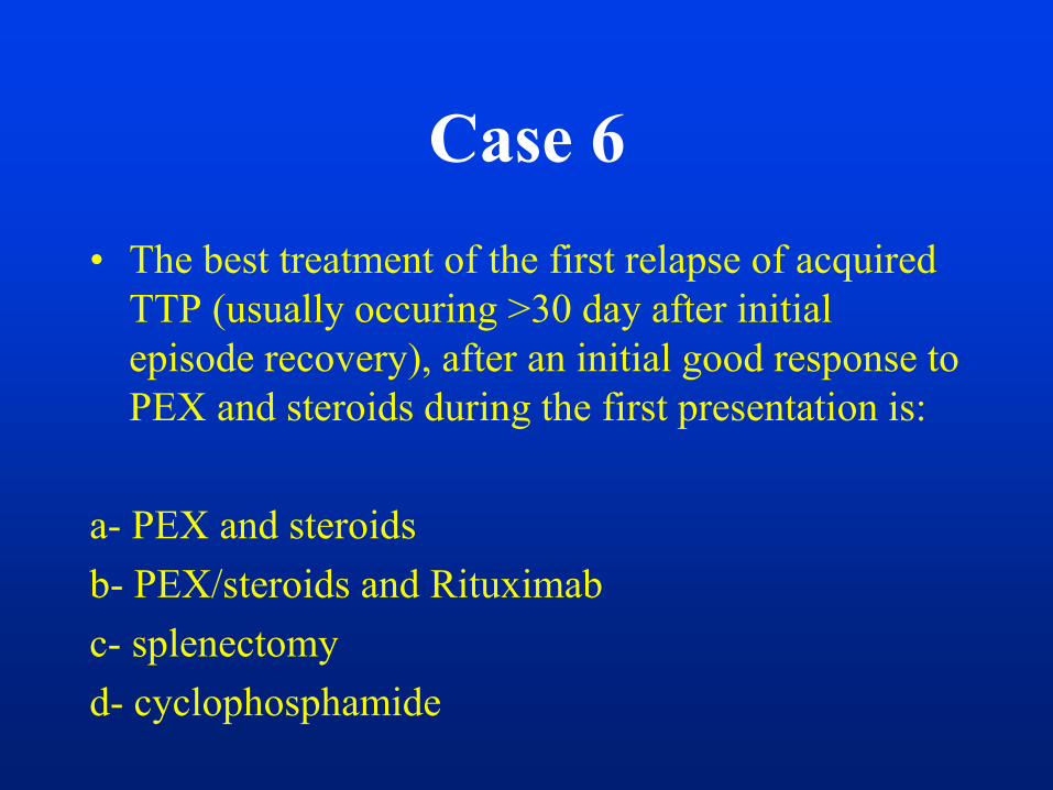
Case 6
• The best treatment of the first relapse of acquired
TTP (usually occuring >30 day after initial
episode recovery), after an initial good response to
PEX and steroids during the first presentation is:
a- PEX and steroids
b- PEX/steroids and Rituximab
c- splenectomy
d- cyclophosphamide
Bibliography
• JN George et al., NEJM 371:7, 2014
• Carla M. Nester CM, et al, Mol Immunology, Vol 67, Issue 1, 2015,
31–42
• Cataland et al, blood, 2014 x Volume 123, Number 16
• Legendre CM et al., N Engl J Med, 2013
• Licht C et al., Kidney Int, 2015
• Noris and Remuzzi, Curr Opin Nephrol Hypertens 2013
• J Zuber et al, Am J Transplant 2012
• Mele et al, Semin Immunopathol (2014) 36:399–420
Bibliography
• Veyradier A. N Engl J Med 2016;374:583-585
• Bendapudi et al BrJ Hematol 2015, Aug 28
• Moake JL. NEJM 2002; 347:589-600
• Levy GG et al, Nature 2001; 413:488-494
• Intractable & Rare Diseases Research. 2014; 3(2):34-45.
• Lemaire M et al, Nature Genet 2013
• Bruneau et al, Blood, 2015

Thank you!
• Questions