hepatic artery embolization for treatment of patients with hereditary hemorrhagic telangiectasia and...
TRANSCRIPT
Received: 22 January 2004Revised: 9 July 2004Accepted: 19 July 2004Published online: 17 August 2004© Springer-Verlag 2004
Abstract At present there is no established therapy for treating pa-tients with hereditary hemorrhagictelangiectasia (HHT) and symptom-atic hepatic involvement. We presentthe results of a prospective studywith 15 consecutive patients whowere treated with staged hepatic artery embolization (HAE). Branchesof the hepatic artery were selectivelycatheterized and embolized in stagesusing polyvinyl alcohol particles(PVA) and platinum microcoils orsteel macrocoils. Prophylactic anti-biotics, analgesics and anti-emeticswere administered after every embo-lization. Clinical symptomatologyand cardiac output were assessed before and after therapy as well as at the end of follow-up (median 28months; range 10–136 months). Fivepatients had abdominal pain and fourpatients had symptoms of portal hypertension. The cardiac outputwas raised in all patients, with cardiacfailure being present in 11 patients.After treatment, pain resolved in all five patients, and portal hyperten-sion improved in two of the four pa-tients. The mean cardiac output de-creased significantly (P<0.001) from12.57±3.27 l/min pre-treatment to
8.36±2.60 l/min at the end of follow-up. Symptoms arising from cardiacfailure resolved or improved marked-ly in all but one patient. Cholangitisand/or cholecystitis occurred in threepatients of whom two required acholecystectomy. One patient withpre-existent hepatic cirrhosis died asa complication of the procedure.Staged HAE yields long-term reliefof clinical symptoms in patients with HHT and hepatic involvement.Patients with pre-existing hepaticcirrhosis may be poor candidates forHAE.
Keywords Hereditary hemorrhagictelangiectasia · Arterio-venous malformations · Cardiac failure ·Embolization · Liver transplantation
Eur Radiol (2004) 14:2079–2085DOI 10.1007/s00330-004-2455-5 H E PAT O B I L I A RY- PA N C R E A S
Ajay ChavanMartin CaselitzKarl-Friedrich GratzJoachim LotzTimm KirchhoffPlinio PisoSiegfried WagnerMichael MannsMichael Galanski
Hepatic artery embolization for treatment of patients with hereditary hemorrhagic telangiectasia and symptomatic hepatic vascular malformations
Introduction
Hereditary hemorrhagic telangiectasia (HHT) is a rareautosomal dominant familial disorder characterized bytelangiectasias and arterio-venous malformations (AVM)
in various organs such as the skin, lungs, central nervousand gastrointestinal system. Hepatic involvement is encountered in about 8–31% of the patients with HHT,of which about half may be symptomatic [1–7]. Whereassome symptomatic patients can be managed conserva-
A. Chavan · J. Lotz · T. KirchhoffM. GalanskiDepartment of Diagnostic Radiology,Hannover Medical School,Hannover, Germany
A. Chavan (✉)Department of Radiology and Nuclear Medicine,Klinikum Oldenburg,Dr Eden Strasse 10, 26133 Oldenburg,Germanye-mail: [email protected].: +49-441-4032521Fax: +49-441-4032515
M. Caselitz · S. Wagner · M. MannsDepartment of Gastroenterology and Hepatology,Hannover Medical School,Hannover, Germany
K.-F. GratzDepartment of Nuclear Medicine,Hannover Medical School,Hannover, Germany
P. PisoDepartment of Abdominal and Transplantation Surgery,Hannover Medical School,Hannover, Germany

tively, the treating physician is often in a dilemma whenconfronted with a patient whose symptoms are refractoryto all forms of conservative therapy. Experience de-scribed in the literature with the treatment of such pa-tients so far consists largely of case reports about hepaticartery ligation, hepatic artery embolization (HAE) andliver transplantation as possible therapy options. Wepresent the results of a prospective study with 15 consec-utive patients who were treated with staged HAE.
Materials and methods
Over a 12-year period, altogether 29 patients having HHT with he-patic involvement presented at our hospital, of which 15 patientshad incapacitating abdominal pain or symptoms arising from car-diac failure or portal hypertension. These 15 patients were treatedwith staged HAE after reaching an interdisciplinary consensus between the radiologists, gastroenterologists and the transplantsurgeons. Informed consent was obtained from all patients.
The pre-interventional diagnostic work-up included a colorDoppler sonography of the liver as well as a spiral CT examina-tion of the entire abdomen. In addition to routine laboratory pa-rameters, hepatitis serology, prothrombin time and serum albumin,cholinesterase, bilirubin and aminotransferase levels were record-ed before and after every intervention as well as at the end of fol-low-up. Cardiac output was determined using echocardiographyprior to therapy, after every embolization session and at the end offollow-up. In the patients who died, the last cardiac output mea-sured prior to death was used for purposes of statistical evaluation.
To test for shunting of embolic particles through the hepaticAVMs into the lungs, polyvinyl alcohol (PVA) particles betweenthe sizes of 250–1,180 µm (Contour, Target Therapeutics, Fre-mont, CA) were labeled with technetium-99m. These test particleswere injected into the hepatic artery, and planar scintigrams of the
liver and lungs were obtained to rule out passage of the particlesthrough the hepatic AVMs into the lungs. The detailed methodolo-gy has been described previously [8]. At subsequent emboliza-tions, only pre-tested particle sizes were used.
HAEs in stages were carried out in one to five sessions at intervals ranging from 1 to 15 weeks. The intervals between theembolization sessions were determined by the time required forthe patient to recover from the previous embolization as well as bythe time required for the hepatic parameters to return to pre-embo-lization levels.
All embolizations were performed under local anaesthesia viathe trans-femoral approach. After establishing portal venous pa-tency by an indirect portography, branches of the right or left he-patic artery were selectively catheterized using 5F Cobra II,Sidewinder I or Sidewinder II catheters (Terumo Europe N.V.,Leuven, Belgium). For superselective catheterization of some he-patic artery branches, co-axial catheters were necessary (Tracker,Boston Scientific, Cork, Ireland). The peripheral vascular bed wasfirst embolized with a mixture of PVA particles of sizes rangingbetween 250 and 1,180 µm followed by embolization of thebranch vessel with platinum microcoils (Target Therapeutics, Fremont, CA) or steel macrocoils (William Cook Europe A/S,Bjaerverskov, Denmark) using a previously described technique[8] (Fig. 1). The macrocoils were delivered via the 5-F diagnosticcatheters, whereas the microcoils were introduced through the co-axial catheters. After experiencing an episode of severe ischemiccholangitis in one patient, only particles larger than 500 µm wereused in subsequent patients in order to prevent occlusion of verysmall peripheral vessels. With the exception of one patient whounderwent five embolization sessions, only tertiary branches ofthe hepatic arteries (and beyond) were embolized, leaving the cen-tral vessels open. The vessels in the right and the left lobe wereembolized in separate sessions. An embolization session was con-cluded either when the patient developed moderately severe painor when the angiogram showed marked reduction or disappear-ance of the peripheral AVMs in the portion of the liver that wasembolized. As the entire liver was affected in all cases, the orderin which the vessels were embolized was determined according tothe “first come, first served” principle: those vessels that could becatheterized first were embolized first, followed by others at sub-sequent sessions.
Prophylactic antibiotic coverage was provided for at least 5 days following the procedure and longer if clinically indicated.Analgesics and anti-emetics were administered as and when nec-essary during and after every embolization. In order to monitor theoccurrence of hepatic necrosis, abdominal sonography was per-formed before and after every embolization. The decision to ter-minate therapy was based on improvement/alleviation of clinicalsymptomatology or the occurrence of severe complications. Themedian follow-up period was 28 months (range 10–136 months).
2080
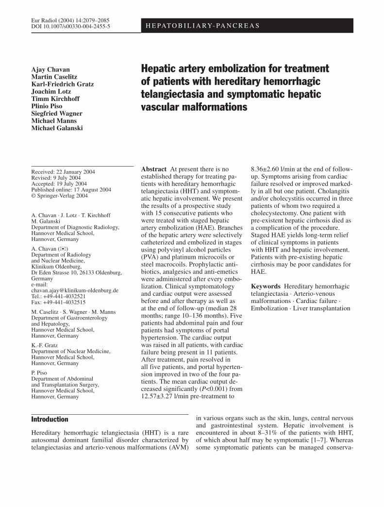
Fig. 1 a Flush aortogram showing massively dilated common andproper hepatic arteries and numerous intra-hepatic arterio-venousshunts. b Parenchymal phase of a selective hepatic angiogram de-picting early opacification of dilated hepatic veins. c Followingperipheral embolization of a portion of the right liver lobe usingpolyvinyl alcohol particles, note the lack of peripheral opacifica-tion inferolaterally in the right lobe of the liver. Hereafter, twobranches of the right hepatic artery were embolized further proxi-mally using metal coils

Statistical analysis
Continuous variables are expressed as means ± SD. Comparisonof cardiac output before treatment and at the end of the observa-tion period was performed applying the Student’s paired t-test andthe Fisher’s exact test (SPSS for Windows 10.0; SPSS, Chicago,IL); P<0.05 was considered statistically significant.
Results
Of the 15 patients, 4 were men and 11 were women withages ranging between 30 and 72 years (median age 56years). Abdominal pain (capsular pain or abdominal angina) as a presenting feature, necessitating a regularintake of analgesics including opiate derivatives, was observed in five patients. Symptoms related to portal hypertension (ascites, esophageal varices) were presentin four patients. The cardiac output was raised in all patients and ranged between 7.4 l/min and 19.5 l/min.Cardiac failure/insufficiency with symptoms such asdyspnea, early fatigue, limitation of physical activity, orthopnea and pedal edema was present in 11 patients.
The embolizations could be performed successfully inall patients. Apart from the right and the left hepatic arte-ries arising from the proper hepatic artery, the left lobe ofthe liver was supplied by a dilated left gastric artery inseven patients, which was also embolized; as with the he-patic arteries, the catheterization of this vessel was tech-nically possible without great difficulty in all patients. Intwo patients, dilated branches arising from the inferiormesenteric artery, which supplied the hepatic AVMs,were embolized with microcoils (Fig. 2). A total of 45embolization procedures were performed. Each patientreceived one to five HAEs with a median of three proce-dures. On an average, 17.53±7.95 vials of PVA particlesand 25.07±14.53 coils were used per patient. The sizes ofthe PVA particles varied between 250 and 1,180 µm; thecoil diameters ranged between 2 and 8 mm.
Shunting of PVA particles into the pulmonary circula-tion was not observed in any of the patients. Post-inter-ventional pain was experienced by all but one patient.Administration of analgesics was necessary for a medianperiod of 9 days (range 1–31 days) following every em-bolization. Anti-emetics were administered for a medianof 3 days (range 1–9 days).
There was a transient increase of serum aminotrans-ferases following embolization, which returned to pre-embolization levels at or shortly following discharge.Other than this, there was no significant long-term im-pairment of liver synthetic function.
Ischemic cholangitis and/or cholecystitis was ob-served in three patients. One of them was managed con-servatively; the pain receded 2 weeks after discharge, butunder antibiotics and analgesic medication; medicationcould be weaned off between 4 and 5 weeks followingtreatment. At follow-up after 107 months, she continuesto be completely free of pain. Cholecystectomy was nec-essary in the other two patients, with additional debride-ment of necrotic liver tissue in one of them.
One patient with hepatitis C, pre-existing Child-Pughstage A hepatic cirrhosis, portal hypertension, hepaticencephalopathy and cardiac insufficiency died within 30 days of the first HAE. The cause of death was hepaticnecrosis leading initially to renal and subsequently tomulti-organ failure.
During the course of follow-up, three further patientsdied. One patient with portal hypertension resulting fromarterio-portal shunting responded excellently with thedisappearance of massive ascites and regression of gradeIII esophageal varices to grade I varices. Her cardiacoutput diminished from 17.3 l/min pre-treatment to5.8 l/min at follow-up. However, 7 months followingembolization, she was readmitted with critical varicealbleeding. A surgical attempt to create a porto-systemicshunt was unsuccessful due to excessive intra-operativebleeding and only a ligation of the common hepatic
2081
Fig. 2 a Selective angiogramof the inferior mesenteric artery; tortuous vessels arisingfrom this artery causing opaci-fication of dilated hepatic arte-ries. b After coil embolizationof the feeding vessels

artery could be performed. The patient died of postoper-ative sepsis 13 days following surgery. The second pa-tient had a history of severe recurrent epistaxis for over20 years and progressively worsening recurrent episodesof hepatic encephalopathy for over 8 years. He was ad-mitted with Child-Pugh stage B hepatic cirrhosis andcardiac failure with a cardiac output of 14 l/min. Follow-ing treatment, his dyspnea on exertion as well as pedaledema regressed, and the cardiac output decreased to8.8 l/min. The bouts of hepatic encephalopathy showedonly marginal improvement. Five months after treat-ment, he was readmitted with severe epistaxis and gradeIII hepatic encephalopathy, which could not be con-trolled, and the patient ultimately died of multi-organfailure. Treatment could not be completed in the thirdpatient with co-existent severe Parkinson’s disease andcardiac insufficiency with a cardiac output of 13.5 l/min;he underwent only two embolization sessions, afterwhich the cardiac output was 12.4 l/min. The severe Parkinson features remained unchanged and led to a falland a fracture of the left femoral neck. The patient succumbed to complications of Parkinsonism and of thefracture, prior to the third embolization.
At the end of follow-up, the remaining 11 patientscontinue to do well and all have been relieved of theirclinical symptoms. Of the five patients with abdominalpain, two patients had intractable abdominal angina(caused by a steal phenomenon) with severe resultantweight loss. Following treatment, the abdominal anginaresolved and the patients gained 6 and 10 kg in bodyweight, respectively, in the year following the conclusionof therapy. One of them had an uneventful pregnancy atthe age of 34 years, 31 months after HAE. Similarly, inthe remaining three patients capsular pain resolved com-pletely following treatment.
The mean cardiac output reduced significantly(P<0.001) from 12.57±3.27 l/min pre-treatment to 8.36±2.60 l/min at the end of follow-up (Figs. 3, 4). Except inthe patient who died within 30 days after the first embo-lization, symptoms arising from cardiac failure resolvedor improved markedly in all patients.
Of the four patients with portal hypertension, ascitesand grade I esophageal varices disappeared completelyin two patients. As mentioned above, one patient withportal hypertension and massive ascites responded excel-lently with disappearance of ascites, fall in cardiac out-put and regression of esophageal varices (from grade IIIto grade I), but succumbed 7 months later to complica-tions of critical variceal bleeding. The fourth patient wasthe one who died in the hospital of complications of theembolization within 30 days of the procedure.
Discussion
In patients with HHT and hepatic involvement, symp-toms, if present, include abdominal pain (in the form ofcapsular pain or abdominal angina often exacerbated after meals) or those arising from cardiac failure, portalhypertension or biliary disease [1, 3, 5, 6]. Due to therarity of the disease, as well as due to its complexity,there is no established therapy form at present for themanagement of these patients.
In patients non-responsive to supportive medical ther-apy, the various therapy options reported in the literatureinclude hepatic artery ligation or banding, HAE and livertransplantation. A review of the relevant literature hasyielded 7 cases that have been treated with hepatic arteryligation or banding [9–14], 25 patients who have under-gone HAE [11, 15–30] and 16 patients who have been
2082
Fig. 3 Cardiac output beforeand at the end of follow-up after hepatic artery embolization(HAE) in 15 patients with hereditary hemorrhagic telangi-ectasia (HHT)

2083
Fig. 4 a Chest radiograph of apatient with HHT presentingwith cardiac failure and intrac-table abdominal pain. b Repeatchest radiograph 2 years fol-lowing five sessions of HAE;marked reduction in cardiacsize. The cardiac output decreased from 13 l/min to6 l/min
subjected to liver transplantation [12, 25, 29–36]. Ofthese patients, one underwent hepatic artery ligation aswell as liver transplantation [12], and another patientwas subjected to HAE followed by hepatic artery liga-tion [11]; five patients underwent HAE followed by livertransplantation [25, 29, 30].
The least experience exists with hepatic artery liga-tion [9–14]. The reported follow-up periods are short. Ofthe seven reported patients, one had previously under-gone HAE [11]. Another patient required liver transplan-tation after 14 months because of the development ofnew collaterals to the hepatic AVMs leading to portal hypertension and recurrent esophageal bleeding [12].Given the tendency of the hepatic AVMs to draw theirblood supply from numerous vessels other than the hepatic artery (such as the pancreato-duodenal vessels,the superior mesenteric, inferior mesenteric, left gastricand splenic arteries), the development of such collateralswith the passage of time after proximal hepatic artery ligation is conceivable. Consequently, this therapy formhas not gained widespread acceptance [37, 38].
The largest experience in literature is with HAE. Thisconsists of 19 other reported cases [11, 15–22, 24, 25,27, 30] in addition to the 15 patients included in thepresent report. Eleven of our 15 patients (73%) are aliveand completely free of symptoms at a median follow-uptime of 28 months (range 10–136 months). Follow-upexceeding 5 years was possible for six patients and ex-ceeding 10 years for two patients. Partial symptomaticresponse with resolution of cardiac failure and symptomsof portal hypertension was observed in two further pa-tients. Thus, complete or partial symptomatic responsecould be achieved in 13 of 15 patients (87%). Good clin-ical response to HAE has been reported by other authorsas well [16, 18, 19, 20, 22, 24, 27].
The therapy was well tolerated by 11 of our 15 pa-tients in whom no major complications occurred. Com-plications in the remaining four patients included pro-tracted ischemic cholangitis or cholecystitis in three pa-tients and one procedure-related death (30-day mortality:
7%). Similar complications have been reported by otherauthors [15, 21, 30], and occurrence of biliary sepsis orliver failure may even necessitate emergency liver trans-plantation [25, 29, 30]. Consequently, reservations re-garding this therapy form exist in some institutions [29,38–41]. Possible means of reducing the severity of thesecomplications would be embolizing smaller portions ofthe liver at each session and placing the catheter tip dis-tal to the origin of the cystic artery in order to avoid theoccurrence of ischemic cholecystitis.
Liver transplantation is an alternative to HAE; this,however, involves major surgery followed by life-longimmune-suppression along with its associated side ef-fects. The spectrum of operative and postoperative com-plications that can occur during and after liver transplan-tation may include excessive intra-operative bleeding,sepsis, renal, respiratory or multi-organ failure, lympho-proliferative disorders, the appearance of de novo malig-nancies as well as postoperative vascular or biliary com-plications, which may necessitate re-operation or evenre-transplantation [25, 30, 34, 42, 43]. As with HAE,death following attempted liver transplantation [6] or abdominal surgery [26] in patients with HHT has beenreported.
Against this backdrop, a reasonable treatment algo-rithm that is acceptable to all is difficult to suggest. Atpresent, a differential approach appears to be the mostprudent in this special group of patients with HHT. Given the complications that may result from HAE aswell as from liver transplantation, it is advisable to man-age the patient conservatively on supportive or medicaltherapy as long as possible. Only those patients refrac-tory to conservative management should be consideredfor invasive therapy. Patients without hepatic cirrhosiscould be managed well by HAE as these patients appearto respond well to the procedure and are at a low risk offatal complications. Pre-requisite is that the HAE be carried out in stages with embolization of small portionsof the liver at each session [8]. As compared to livertransplantation, HAE is less invasive and does not

require life-long immune suppression. Furthermore, ageis not a limiting factor for carrying out HAE.
In our set of 15 patients, both patients with cirrhosisdied, one as a complication of the procedure and the other of an unresolved hepatic encephalopathy. Thus, thesub-group of patients in whom cirrohotic changes haveset in appear to be poor candidates for primary HAE andcould benefit better from primary liver transplantation
[25, 30]. At any rate, until more experience with thetreatment of such patients becomes available, it is advis-able that the therapy be carried out by physicians withadequate experience with HAE or liver transplantationand that the choice of therapy be made only by interdis-ciplinary consensus. It is worthy of mention that reluc-tance on the part of the patient to undergo major surgerymay influence the choice of therapy in some cases.
2084
References
1. Martini GA (1978) The liver in hereditary haemorrhagic teleangiectasia:an inborn error of vascular structurewith multiple manifestations: a reappraisal. Gut 19:531–537
2. Reilly PJ, Nostrant TT (1984) Clinical manifestations of hereditaryhaemorrhagic teleangiectasia. Am JGastroenterol 79:363–367
3. Bernard G, Mion F, Henry L, PlauchuH, Paliard P (1993) Hepatic involve-ment in hereditary haemorrhagicteleangiectasia: clinical, radiologicaland hemodynamic studies of 11 cases.Gastroenterology 105:482–487
4. Guttmacher AE, Marchuk DA, WhiteRI (1995) Hereditary hemorrhagic telangiectasia. N Engl J Med333(14):918–924
5. Haitjema T, Westermann CJ, OvertoomTT et al (1996) Hereditary hemorrhagictelangiectasia (Osler-Weber-Rendu dis-ease): new insights into pathogenesis,complications and treatment. Arch Intern Med 156:714–719
6. Garcia-Tsao G, Korzenik JR, Young L,Henderson KJ, Jain D, Byrd B, PollakJS, White RI (2000) Liver disease inpatients with hereditary hemorrhagictelangiectasia. N Engl J Med 343:931–936
7. Geisthoff UW, Schneider G, FischingerJ, Plinkert PK (2002) Hereditärehämorrhagische Teleangiektasie
(Morbus Osler): Eine interdisziplinäreHerausforderung. HNO 50:114–128
8. Chavan A, Galanski M, Wagner S,Caselitz M, Schlitt HJ, Gratz KF,Manns M (1998) Hereditary hemor-rhagic telangiectasia: effective protocolfor embolization of hepatic vascularmalformations—experience in five patients. Radiology 209:735–739
9. DeLorimier A, Simpson E, Baum R et al (1967) Hepatic artery ligation forhepatic hemangiomatosis. N Engl JMed 277:333–337
10. Radtke WE, Smith HC, Fulton RE, Adson MA (1978) Misdiagnosis ofatrial septal defect in patients with hereditary telangiectasia (Osler-Rendu-Weber disease) and hepatic arteriove-nous fistulas. Am Heart J 95:235–242
11. Zentler-Munro PL, Howard ER, Karani J, Williams R (1989) Varicealhaemorrhage in hereditary hemorrhagictelangiectasia. Gut 30:1293–1297
12. Neumann U-P, Knoop M, Langrehr J-M et al (1998) Effective therapy forhepatic M. Osler with systemic hyper-circulation by ligation of the hepaticartery and subsequent liver transplanta-tion. Transpl Int 11:323–326
13. Zieren J, Buttemeyer R, Muller JM(1998) Adjustable “banding” of the hepatic artery in treatment of shunt-in-duced heart failure in Osler-Rendu-Weber disease. Chirurg 69:639–641
14. Graham WP, Eiseman B, Pryor R(1964) Hepatic artery aneurysm withportal vein fistula in a patient with familial hereditary telangiectasia. AnnSurg 159:362–367
15. Göthlin JH, Nordgard K, Jonsson K,Nyman U (1982) Hepatic telangiectasiain Osler’s disease treated with arterialembolisation. Report of 2 cases. Eur JRadiol 2:27–30
16. Brohee D, Franken P, Fievez M et al(1984) High-output right ventricularfailure secondary to hepatic arteriove-nous microfistulae. Selective arterialembolization treatment. Arch InternMed 144:1282–1284
17. Allison D, Jordon J, Hennessey O(1985) Therapeutic embolization of thehepatic artery: a review of seventy-fiveprocedures. Lancet 1:595–599
18. Roman CF, Do Cha S, Incarvito J,Cope C, Maranhao V (1987) Trans-catheter embolization of hepatic arteriovenous fistula in Osler-Weber-Rendu disease—a case report. Angiology 38:484–488
19. Derauf BJ, Hunter DW, Sirr SA,Cardella JF, Castaneda-Zuniga W, Amplatz K (1987) Peripheral emboli-zation of diffuse hepatic arteriovenousmalformations in a patient with hereditary hemorrhagic telangiectasia.Cardiovasc Intervent Radiol 10:80–83
20. Nikolopoulos N, Xynos E, VassilakisJS (1988) Familial occurrence of hyperdynamic circulation status due to intrahepatic fistulae in hereditaryhemorrhagic telangiectasia. Hepatogastroenterology 35:167–168
21. Bourgeois N, Delcour C, Deviere J etal (1990) Osler–Weber–Rendu diseaseassociated with hepatic involvementand high output heart failure. J ClinGastroenterol 12:236–238
22. Whiting JH Jr, Morton KA, Datz FL,Patch GG, Miller FJ Jr (1992) Emboli-zation of hepatic arteriovenous malfor-mations using radiolabeled and nonra-diolabeled polyvinyl alcohol sponge ina patient with hereditary hemorrhagictelangiectasia: case report. J Nucl Med33(2):260–262
23. Caselitz M, Wagner S, Chavan A,Gebel M, Bleck JS, Wu A, Schlitt HJ,Galanski M, Manns M (1998) Clinicaloutcome of transfemoral embolisationin patients with arteriovenous malfor-mations of the liver in hereditaryhaemorrhagic telangiectasia (Weber-Rendu-Osler disease). Gut 42:123–126
24. Trotter JF, Suhocki PV, Lina JR, Martin LW, Parrish JL, Swantkowski T(1998) Hereditary hemorrhagic telangi-ectasia causing high output cardiacfailure: treatment with transcatheterembolization. Am J Gastroenterol93:1569–1571
25. Odorico JS, Hakim NM, Becker YT et al (1998) Liver transplantation as definitive therapy for complications after arterial embolization for hepaticmanifestations of hereditary hemor-rhagic telangiectasia. Liver TransplSurg 4:483–490
26. Chavan A, Galanski M, Wagner S,Caselitz M, Schlitt HJ, Gratz KF,Manns M (1999) Hereditary hemor-rhagic telangiectasia (reply—letters tothe editor). Radiology 213:928–930

27. Stockx L, Raat H, Caerts B et al (1999)Transcatheter embolization of hepaticarteriovenous fistulas in Rendu-Osler-Weber disease: a case report and review of the literature. Eur Radiol9(7):1434–1437
28. Hisamatsu K, Ueeda M, Ando M et al(1999) Peripheral arterial coil emboli-zation for hepatic arteriovenous malformation in Osler–Weber–Rendudisease; useful for controlling high output heart failure, but harmful to theliver. Intern Med 38:962–968
29. Whiting JH Jr, Korzenik JR, Miller FJJr et al (2000) Fatal outcome after em-bolotherapy for hepatic arteriovenousmalformations of the liver in two patients with hereditary hemorrhagictelangiectasia. J Vasc Interv Radiol11:855–858
30. Pfitzmann R, Heise M, Langrehr JM,Jonas S, Steinmüller T, Podrabsky P,Ewert R, Settmacher U, Neuhaus R,Neuhaus P (2001) Liver transplantationfor treatment of intrahepatic Osler’sdisease: first experiences. Transplanta-tion 72:237–241
31. Bauer T, Britton P, Lomas D, WightDG, Friend PJ, Alexander GJ (1995)Liver transplantation for hepatic arte-riovenous malformation in hereditaryhaemorrhagic telangiectasia. J Hepatol22:586–590
32. Saxena R, Hytiroglou P, Atillasoy EOet al (1998) Coexistence of hereditaryhemorrhagic telangiectasia and fibropolycystic liver disease. Am JSurg Pathol 22:368–372
33. McInroy B, Zajko AB, Pinna AD(1998) Biliary necrosis due to hepaticinvolvement with hereditary hemor-rhagic telangiectasia. Am J Roentgenol170:413–415
34. Boillot O, Bianco F, Viale J-P et al(1999) Liver transplantation resolvesthe hyperdynamic circulation in heredi-tary hemorrhagic telangiectasia withhepatic involvement. Gastroenterology116:187–192
35. Le Corre F, Golkar B, Tessier C et al(2000) Liver transplantation for hepaticarteriovenous malformation with high-output cardiac failure in hereditaryhemorrhagic telangiectasia; hemody-namic study. J Clin Anesth 12:339–342
36. Hillert C, Broering DC, Gundlach M et al (2001) Hepatic involvement in hereditary hemorrhagic telangiectasia:an unusual indication for liver trans-plantation. Liver Transpl 7:266–268
37. Caselitz M, Chavan A, Manns MP,Wagner S (2001) Die Hereditäre Hämorrhagische Teleangiectasie (Morbus Osler–Rendu–Weber) undihre Manifestation an der Leber. Z Gastroenterol 39:533–542
38. Larson AM (2003) Liver disease in hereditary hemorrhagic telangiectasia.J Clin Gastroenterol 36(2):149–158
39. Jackson JE (1999) Hepatic vascularmalformations in hereditary hemor-rhagic telangiectasia: treatment withembolization (Letter to the editor). Radiology 213:927–928
40. Miller FJ, Whiting JH, Korzenik JR,White RI (1999) Caution with use ofhepatic embolization in treatment ofhereditary hemorrhagic telangiectasia(Letter to the editor). Radiology213:928
41. Miller FJ, Mineau DE (1983) Trans-catheter arterial embolization—majorcomplications and their prevention.Cardiovasc Intervent Radiol 6:141–149
42. Jain A, Reyes J, Kashyap R et al(2000) Long-term survival after livertransplantation in 4,000 consecutivepatients at a single center. Ann Surg232(4):490–500
43. Hatzidakis AA, Gogas C, PapanikolaouN, Samonakis D, Kofteridis D, Gourtsoyiannis NC (2002) Hepatic involvement in hereditary hemorrhagictelangiectasia (Rendu-Osler-Weber disease). Eur Radiol 12:S51–S55
2085