hepatoprotective effect of ethanolic extract of curcuma longa on
TRANSCRIPT
Salama et al. BMC Complementary and Alternative Medicine 2013, 13:56http://www.biomedcentral.com/1472-6882/13/56
RESEARCH ARTICLE Open Access
Hepatoprotective effect of ethanolic extract ofCurcuma longa on thioacetamide induced livercirrhosis in ratsSuzy M Salama1, Mahmood Ameen Abdulla1*, Ahmed S AlRashdi1, Salmah Ismail2, Salim S Alkiyumi1
and Shahram Golbabapour1,2
Abstract
Background: Hepatology research has focused on developing traditional therapies as pharmacological medicinesto treat liver cirrhosis. Thus, this study evaluated mechanisms of the hepatoprotective activity of Curcuma longarhizome ethanolic extract (CLRE) on thioacetamide-induced liver cirrhosis in rats.
Methods: The hepatoprotective effect of CLRE was measured in a rat model of thioacetamide-induced livercirrhosis over 8 weeks. Hepatic cytochrome P450 2E1 and serum levels of TGF-β1 and TNF-α were evaluated.Oxidative stress was measured by malondialdehyde, urinary 8-hydroxyguanosine and nitrotyrosine levels. Theprotective activity of CLRE free-radical scavenging mechanisms were evaluated through antioxidant enzymes.Protein expression of pro-apoptotic Bax and anti-apoptotic Bcl-2 proteins in animal blood sera was studied andconfirmed by immunohistochemistry of Bax, Bcl2 proteins and proliferating cell nuclear antigen.
Results: Histopathology, immunohistochemistry and liver biochemistry were significantly lower in the Curcumalonga-treated groups compared with controls. CLRE induced apoptosis, inhibited hepatocytes proliferation but hadno effect on hepatic CYP2E1 levels.
Conclusion: The progression of liver cirrhosis could be inhibited by the antioxidant and anti-inflammatory activitiesof CLRE and the normal status of the liver could be preserved.
Keywords: Curcuma longa, Antioxidant enzymes, Cytochrome P450 2E1 (CYP2E1), Histology, Oxidative stress,Immunohistochemistry
BackgroundCirrhosis is the damage of liver cells and their gradual re-placement with scar tissue that impairs blood flowthrough the liver causing hepatocyte death and loss ofliver function [1]. Hepatic fibrosis occurs in response toliver damage and regenerates apoptotic cells after repeatedinjury [2]. This inflammatory response is accompanied bylimited deposition of extra cellular matrix (ECM), so thatif the regeneration of dying cells fails during persistentliver injury, hepatocytes are replaced by abundant ECM,including fibrillar collagen, depending on the origin of in-jury [3]. Treatment options for common liver disease such
* Correspondence: [email protected] of Molecular Medicine, Faculty of Medicine, University ofMalaya, 50603, Kuala Lumpur, MalaysiaFull list of author information is available at the end of the article
© 2013 Salama et al.; licensee BioMed CentralCommons Attribution License (http://creativecreproduction in any medium, provided the or
as cirrhosis, fatty liver and chronic hepatitis are problem-atic. The effectiveness of treatments such as interferons,colchicines, penicillamine and corticosteroids are incon-sistent at best and the incidence of side-effects profound[4]. Because of the role of oxidative stress in liver cirrhosis,antioxidants have been proposed as a treatment for cirrho-sis [5]. Several studies have demonstrated the protectiveeffects of antioxidants against induced liver injury by redu-cing oxidative stress in cells [6,7]. A number of herbalsshow promising activity, including Silymarin for livercirrhosis, glycyrrhizin for chronic viral hepatitis, andherbal combinations from China and Japan that have beenproven for treatment of liver diseases [8]. Silymarin, a ref-erence drug, is a flavonolignan from ″milk thistle″Silybum marianum, and widely used for the treatment ofhepatitis and liver cirrhosis [9].
Ltd. This is an Open Access article distributed under the terms of the Creativeommons.org/licenses/by/2.0), which permits unrestricted use, distribution, andiginal work is properly cited.

Salama et al. BMC Complementary and Alternative Medicine 2013, 13:56 Page 2 of 17http://www.biomedcentral.com/1472-6882/13/56
Curcuma longa is a rhizomatous perennial herb thatbelongs to the family Zingiberaceae, native to South Asiaand is commonly known as turmeric. In Malaysia, com-monly known as Kunyit, turmeric plant is a popular in-gredient for preparing culinary dishes. In addition, it isused as herbal remedy due to the prevalent belief thatthe plant has medical properties. In folk medicine, therhizome juice from C. longa is used in the treatment ofmany diseases such as anthelmintic, asthma, gonorrheaand urinary, and its essential oil is used in the treatmentof carminative, stomachic and tonic [10]. In traditionalmedicine, several plants and herbs have been usedexperimentally to treat liver disorders, including livercirrhosis, [11,12]. C. longa possesses antioxidant [13],anti-tumor [14], antimicrobial [15], anti-inflammatory[16], wound healing [17], and gastroprotective activities[18]. The previous studies have also shown that theaqueous extract of C. longa has hepatoprotective activityagainst carbon tetrachloride toxicity [19]. In this study,we assessed the hepatoprotective effect of the ethanolicextract of C. longa rhizomes against TAA-induced livercirrhosis in Sprague Dawley rats.
MethodsPreparation of CLREC. longa rhizomes were obtained from Ethno Company,Kuala Lumpur, Malaysia and identified by comparisonwith the voucher specimen (KLU41829) deposited atthe Herbarium of Rimba Ilmu, Institute of BiologicalSciences, University of Malaya, Kula Lumpur, MalaysiaThe rhizomes were cleaned, dried, ground, weighed, andhomogenized in 95% ethanol at a ratio of 1:10 of plantto ethanol and left to soak for 3 days at 25°C with occa-sional shaking and stirring. The mixture was then fil-tered and the resulting liquid was concentrated underreduced pressure at 45°C in an EYELA rotary evaporatorto yield a dark gummy-yellow extract (7%, w/w). Theconcentrated extract was then kept in the incubator at45°C for 3 days to evaporate the ethanol residue yieldingthe crude rhizome extract. Extracts were then dissolvedin 10% Tween-20 before being orally administrated toanimals in concentrations of 250 and 500 mg/kg bodyweight (5ml/kg body weight).
Total phenol content (TPC) of CLREThe Total Phenol content (TPC) of the CLRE extractwas determined by the Folin Denis calorimetric methodusing Folin-Ciocalteau reagent (Merck, Darmstadt,Germany) in gallic acid equivalent in mg (GAE/mgextract) [20]. CLRE (1 mg) was first dissolved in 1 mLdimethyl sulfoxide (DMSO). Next, 20 μL of the extractwas added into 100 μL of Folin-Ciocalteau reagent, and
the resulting mixture was incubated in the dark for 3min. Then, 100 μL of sodium carbonate (1 g/10 mL) so-lution was added to the mixture, and mixed thoroughly.The final mixture was kept in the dark for 1 h and itsabsorbance (750 nm wavelength) was read by an ELISAreader (UV 1601 spectrophotometer, Shimadzu, Japan).All procedures were carried out in triplicate. Linearstandard curves were produced by serial dilution ofgallic acid (1 mg/mL DMSO) and the absorbance wasread at 750 nm.
Ferric reducing anti-oxidant power of CLREThe ferric reducing anti-oxidant power (FRAP) of CLREwas assayed according to the previously describedmethod [21] with slight modification. FRAP reagent wasprepared by adding 300 mM acetate buffer (3.1 mg so-dium acetate/mL, pH 3.6) to 10 mM 2,4,6-tripyridyl-S-triazine (TPTZ) solution (Merck, USA) and 20 mMFeCl3.H2O (5.4 mg/mL). Ten μL of 1 mg/mL of CLRE(equivalent to 500 mg/kg dose administrated daily to an-imals) and the standards gallic acid, quercetin, ascorbicacid, retin, trolox and 2,6-di-tert-butyl-4 methyl phenyl(BHT) were each sampled with 10 μL of 0.1 mg/mLSilymarin (equivalent to 50 mg/kg dose administrateddaily to animals) and added to 290 μL of TPTZ reagentin triplicate wells. Absorbance was read at 593 nm usingan ELISA reader (Shimadzu, Japan) every 4 min for 2 h.
Experimental animalsSixty-six healthy Sprague Dawley rats (180-250 g) wereused in the experiments. All rats were kept in wire-bottomed cages at 25 ± 2°C, given tap water and stand-ard pellet diet and exposed to a 12 h:12 h light–darkcycle at 50–60% humidity in an animal room. Through-out the experiments, all animals received human careaccording to the criteria outlined in the “Guide for theCare and Use of Laboratory Animals” prepared by theNational Academy of Sciences and published by the na-tional Institute of health. The study was approved by theEthics Committee for Animal Experimentation, Facultyof Medicine, University of Malaya, Malaysia PM/28/08/2009/MAA.
Acute toxicity studyEighteen males and eighteen females healthy rats wereassigned equally into 3 groups of 6 rats: vehicle (receiv-ing 10% Tween-20 w/v, 5 mL/kg); or treated with 2 g/kgor 5 g/kg of CLRE preparation, respectively. The animalswere fasted overnight but water prior to dosing. Foodwas withheld for a further 3-4 h after dosing. The ani-mals were observed for 30 min and at 2, 4, 8, 24 and 48h after administration for the onset of clinical or toxico-logical symptoms. The animals were sacrificed on the

Figure 1 Antioxidant activity of the CLRE compared with thestandards: gallic acid, quercetin, ascorbic acid, rutin, trolox,BHT and the standard drug Silymarin. Values were expressed asmean ± SEM.
Salama et al. BMC Complementary and Alternative Medicine 2013, 13:56 Page 3 of 17http://www.biomedcentral.com/1472-6882/13/56
15th day. Histological and serum biochemical parameterswere determined using standard methods [22].
Induction of liver cirrhosis in ratsThioacetamide (TAA, CH3-C(S)NH2) is a hepatotoxinand hepatocarcinogenic when administered in the diet ofexperimental animals, and is widely used as a model ofacute and chronic liver disease [23] Briefly, after admin-istration of TAA in the diet, it is converted to TAA-S-oxide (TASO) by hepatic microsomal cytochrome P4502E1 (CYP2E1), then transformed to toxic thioacetamideS-dioxide (TASO2) [24]. TASO2 damages biomoleculesof the liver leading to cirrhosis [25].Male animals were randomly divided into 5 groups of 6
rats. Rats of Group 1 (normal control group) were orallyadministrated with 10% Tween-20 (5 mL/kg) daily andintraperitoneally (ip) injected with sterile distilled water(1 mg/kg) thrice weekly. Groups 2–5 were administeredwith TAA by intraperitoneal injection (200 mg/kg/mL)three times a week to induce liver cirrhosis. Constant ex-posure of this concentration of TAA induces pathologicalchanges in the liver comparable to the etiology of cirrhosisin humans [26]. The stock solution was prepared (5 g/L)by dissolving TAA crystals (Sigma-Aldrich, USA) in steriledistilled water and stirred till completely dissolved [27].Rats of Group 2 (cirrhosis control group) were orallyadministrated with 10% Tween-20 (5 mL/kg) daily. Ratsof Group 3 (Silymarin-treated group) were orally
Table 1 Effect of CLRE on renal function tests in rats
Dose Sodium Potassium
(mM/L) (mM/L)
Vehicle (10% Tween-20) 139.79 ± 1.34 4.87 ± 0.47
Low dose CLRE (2 g/kg) 143.31 ± 2.11 5.14 ± 0.39
High dose CLRE (5 g/kg) 140.67 ± 2.67 5.09 ± 0.40
Values expressed as mean ± SEM. There are no significant differences between gro
administrated with Silymarin (50 mg/kg) daily. Silymarin(International Laboratory, USA) was properly dissolved in10% Tween-20 and used as a standard drug. Rats ofGroups 4 and 5 (treatment groups) were orally adminis-trated with CLRE at daily doses of 250 mg/kg and 500mg/kg, respectively. The treatment procedure was consid-ered an 8-week period due to the preventive nature of theexperiment (Silymarin and CLRE), protecting the liverfrom further damage. At the end of the 8 weeks, the ratswere fasted for 24 h after the last treatment and perfusedunder ketamine (30 mg/kg, 100 mg/mL) and xylazil(3 mg/kg, 100 mg/mL) anesthesia [28]. Blood was with-drawn through the jugular vein and collected for pro-thrombin time ratio evaluation, biochemical examinations,cytokines and apoptotic proteins assessment. Liver tissueswere excised, washed with ice cold normal saline, blottedon filter paper and weighed. The tissues were examinedthoroughly for gross cirrhosis. They were prepared forevaluation of the oxidative damages and histopathologyassessment. Liver tissues were homogenized (10% w/v) in50 mM cold potassium phosphate buffer (pH 7.4) using aTeflon homogenizer (Polytron, Heidolph RZR 1, Germany).Then the tissue homogenates were centrifuged at 3500 rpmfor 15 min at 4°C in a centrifuge (Heraeus, Germany). Thesupernatant of each sample was collected and frozen in ali-quots for later use.
Biochemical analysisBlood samples from animals were collected in sodiumcitrate tubes for determining prothrombin time or in gel-activated tubes for the assessment of specific liver markers.The gel-activated tubes were allowed to clot, thencentrifuged at 3400 rpm for 10 min at 4°C. The serumsamples were collected for measuring liver markers,alkaline phosphatase (AP), alanine aminotransferase (ALT),aspartate aminotransferase (AST), total protein, albumenand bilirubin. The markers were assayed with a spectro-photometer at Central Diagnostic Laboratory of the Med-ical Center of University Malaya.
Assessment of hepatic CYP2E1 levelsThe level of CYP2E1 in the liver tissue homogenate of allrats was evaluated by following the instructions of a sand-wich enzyme immunoassay (Uscn Life Science, China).
Chloride Urea Creatinine
(mM/L) (mM/L) (µM/L)
104.81 ± 1.42 4.69 ± 0.42 40.10 ± 2.63
103.46 ± 2.04 4.97 ± 0.58 39.00 ± 2.71
103.70 ± 1.52 5.27 ± 0.52 38.82 ± 3.14
ups, significant value at P<0.05. CLRE: C. longa rhizomes ethanolic extract.
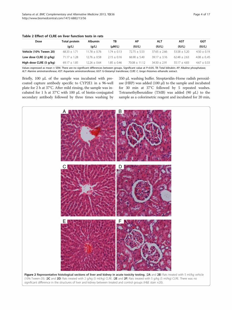
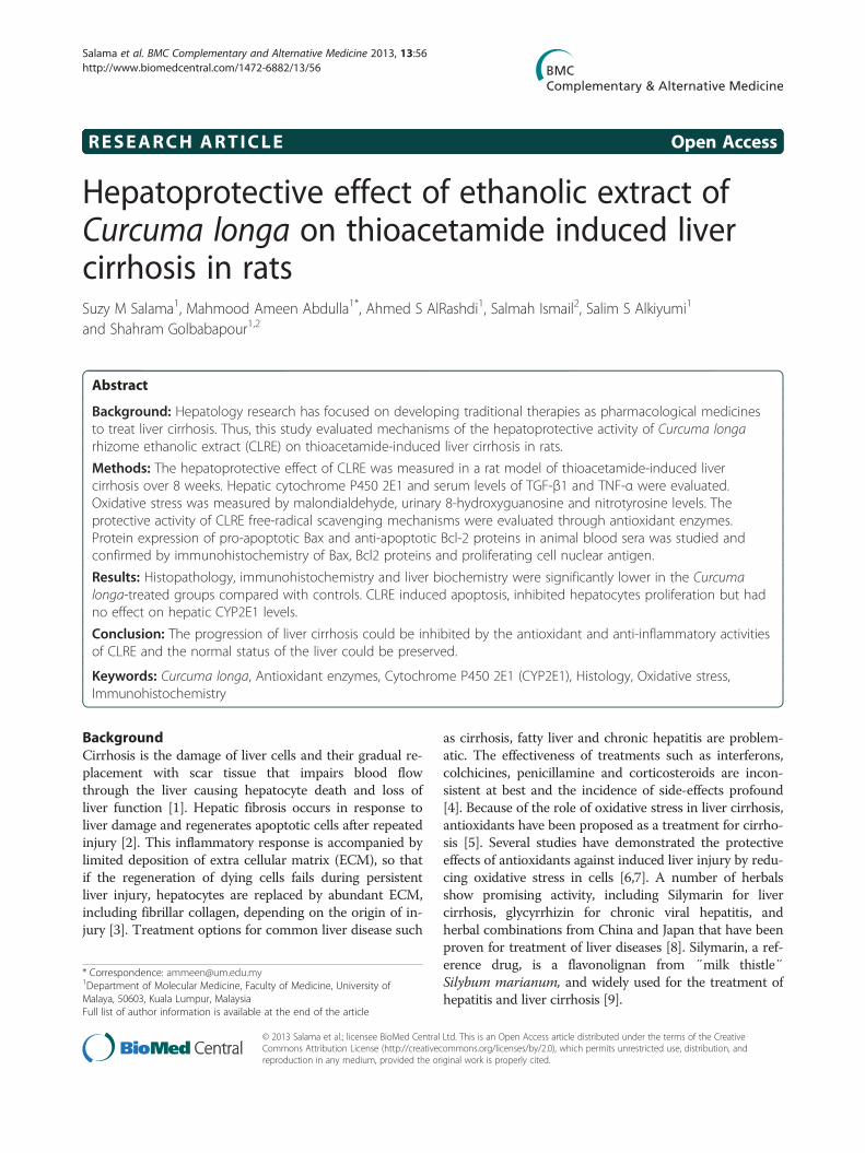
Table 2 Effect of CLRE on liver function tests in rats
Dose Total protein Albumin TB AP ALT AST GGT
(g/L) (g/L) (µM/L) (IU/L) (IU/L) (IU/L) (IU/L)
Vehicle (10% Tween 20) 68.33 ± 1.71 11.78 ± 0.76 1.74 ± 0.13 72.75 ± 5.53 37.65 ± 2.66 53.58 ± 5.20 4.50 ± 0.19
Low dose CLRE (2 g/kg) 71.17 ± 1.28 12.76 ± 0.58 2.15 ± 0.16 66.90 ± 5.40 39.17 ± 3.16 62.48 ± 2.63 4.08 ± 0..45
High dose CLRE (5 g/kg) 69.17 ± 1.85 12.26 ± 0.64 1.85 ± 0.46 70.08 ± 11.12 34.50 ± 2.91 55.17 ± 4.83 4.67 ± 0.33
Values expressed as mean ± SEM. There are no significant differences between groups. Significant value at P<0.05, TB: Total bilirubin; AP: Alkaline phosphatase;ALT: Alanine aminotransferase; AST: Aspartate aminotransferase; GGT: G-Glutamyl transferase; CLRE: C. longa rhizomes ethanolic extract.
Salama et al. BMC Complementary and Alternative Medicine 2013, 13:56 Page 4 of 17http://www.biomedcentral.com/1472-6882/13/56
Briefly, 100 μL of the sample was incubated with pre-coated capture antibody specific to CYP2E1 in a 96-wellplate for 2 h at 37°C. After mild rinsing, the sample was in-cubated for 1 h at 37°C with 100 μL of biotin-conjugatedsecondary antibody followed by three times washing by
A
C
E
Figure 2 Representative histological sections of liver and kidney in ac(10% Tween-20). (2C and 2D) Rats treated with 2 g/kg (5 ml/kg) CLRE. (2Esignificant difference in the structures of liver and kidney between treated
350 μL washing buffer. Streptavidin-Horse radish peroxid-ase (HRP) was added (100 μl) to the sample and incubatedfor 30 min at 37°C followed by 5 repeated washes.Tetramethylbenzidine (TMB) was added (90 μL) to thesample as a colorimetric reagent and incubated for 20 min,
B
D
F
ute toxicity testing. (2A and 2B) Rats treated with 5 ml/kg vehicleand 2F) Rats treated with 5 g/kg (5 ml/kg) CLRE. There was noand control groups (H&E stain ×20).

Table 3 Effect of CLRE on liver index measurements from rats at the end of 8 weeks study
Treatment Body weight (g) Liver weight (g) Liver weight × 100/body weight
Normal rats 341.33 ± 6.18 8.43 ± 0.34 2.47 ± 0.06
Cirrhosis rats 216.83 ± 10.96 10.13 ± 0.54 4.70 ± 0.22**
Silymarin-treated rats 336 ± 9.187 9.12 ± 0.34 2.72 ± 0.14*
Low dose CLRE-treated rats (250 mg/kg) 249.17 ± 14.89 8.80 ± 0.29 3.58 ± 0.19*
High dose CLRE-treated rats (500 mg/kg) 351 ± 19.73 9.48 ± 0.19 2.74 ± 0.15*
Data are expressed as mean ± SEM. *P<0.001 compared with the cirrhosis control Group 2. **P<0.001 compared with normal control Group 1. CLRE: C. longarhizomes ethanolic extract.
Salama et al. BMC Complementary and Alternative Medicine 2013, 13:56 Page 5 of 17http://www.biomedcentral.com/1472-6882/13/56
stopped by H2SO4 (50 μL) and the absorbance was read at450 nm.
Evaluation of oxidative stress markersUrine 8-OH-dG8-hydroxy-2-deoxyguanosine (8-OH-dG) is a product ofDNA oxidative damage through reactive oxygen species(ROS) and serves as an established oxidative stressmarker [29]. To evaluate the DNA oxidative damage,urine samples from all animals were collected 24 h be-fore sacrifice and stored (-80°C). The levels of 8-OH-dGwere measure according to the instructions of themanufacturer (Genox KOG-HS10E, USA). In brief, in a96-well microtiter plate, pre-coated with monoclonalantibody specific for 8-OH-dG, a urine sample (50 μL)was incubated at 4°C overnight. The plate was thenwashed (5 times) with concentrated buffered saline(pH=7.4). 100 μL of biotinylated secondary antibody wasadded to the sample and incubated for 1 h at roomtemperature followed by a 3-time rinsing. The chromaticsolution tetramethylbenzidine (TMB) was added (100μL) and incubated at room temperature for 15 min indark. The reaction terminating solution (1M phosphoricacid) was added (100 μL) and the plate was read with aspectrophotometer at 450 nm.
Hepatic nitrotyrosineNitrotyrosine, a marker for protein oxidation [30] wasassessed in liver tissue homogenate of all animals by ELISAaccording to the manufacturer protocol (MyBiosource,USA). Succinctly, 100 μL of the sample was incubated withmonoclonal Nitrotyrosine-HRP conjugate in a microtiterplate. After 1 h of incubation, the plate was washed 5 timesby 350 μL wash solution. Then substrate specific to HRPenzyme was added (100 μL) to the sample followed by 50μL stop solution and the intensity of the produced colourwas measured with a spectrophotometer at 450 nm.
Hepatic malondialdehydeMalondialdehyde (MDA) levels were measured in theliver tissue homogenate of all experimental groups as ameasure for lipid peroxidation using thiobarbituric acid
according to the manufacturer’s instructions (Cayman,Sigma). Ready to use SDS solution was added (100 μL)to the samples/standard (100 μL). Then 4 mL of thecolour reagent were added to the mixture. Samples andstandard solutions vials were immersed in boiling waterfor 1 h. The reaction was stopped when they were incu-bation in an ice bath for 10 min. All vials were thencentrifuged at 1600 × g for 10 min at 4°C. A set of dupli-cated samples or standards were loaded into 96-wellplate, and their absorbance were read at 532 nm by aspectrophotometer.
Antioxidant enzyme assessmentSuperoxide dismutase (SOD) and catalase (CAT) en-zymes were measured for each liver tissue homogenate(Cayman, USA). SOD activity was evaluated by tetrazo-lium salt detecting superoxide radicals produced by theaction of xanthine oxidase on hypxanthine. Bovineerythrocyte SOD was used to represent SOD standardcurve. 200 μL of tetrazolium salt solution was added to10 μL standards or samples followed by fast addition of20 μL of xanthine oxidase to initiate the reaction. Theplate was covered and incubated for 20 min on a plateshaker (Barnstead Dubuque, USA) and the reading ofthe spectrophotometer was recorded at 450 nm. CAT ac-tivity was evaluated by the chromagen, 4-amino-3hydrazino-5-mercapto-1,2,4-triazole which measures theformaldehyde produced by the reaction of CAT enzymewith methanol in the presence of H2O2. The standardcurve was obtained by catalase formaldehyde standard.Assay buffer (100 μL) and methanol (30 μL) were addedto the standards or samples (20 μL) and the reactionwas initiated when 20 μL of H2O2 was added. The platewas incubated in dark at room temperature for 20 min.Then Potassium Phosphate buffer (30 μL) was added toterminate the reaction. Chromagen was added (30 μL)and the plate was incubated on a shaker at roomtemperature in the dark. After 10 min, catalase potas-sium periodate was added (10 μL) and the plate was cov-ered and left on the shaker at room temperature for 5min. The absorbance was measure with a spectropho-tometer at 540 nm.

*
** ** **
*
*****
**
*
** ****
050
100150200250300
No
rmal
Rat
s
Cir
rho
sis
Rat
s
Sily
mar
in-t
reat
edR
ats
Lo
w D
ose
CL
RE
-tr
eate
d R
ats
(250
mg
/kg
)
Hig
h D
ose
CL
RE
-tre
ated
Rat
s (5
00 m
g/k
g)
Sp
ecif
ic L
iver
En
zym
es
AP IU/LALT IU/LAST IU/L
Figure 3 Effect of CLRE on the plasma level of specific liver enzymes from rats at the end of 8 weeks study. AP: alkaline phosphatase;ALT: alanine transferase; AST: aspartate transferase. Data were expressed as mean ± SEM. *P<0.001 compared with the normal control Group 1.**P<0.001 compared with cirrhosis control Group 2. ***P<0.05 compared with cirrhosis control Group 2.
Salama et al. BMC Complementary and Alternative Medicine 2013, 13:56 Page 6 of 17http://www.biomedcentral.com/1472-6882/13/56
Assessment of cytokinesBlood samples from each group was centrifuged at3500 rpm and the sera were stored (-80°C) in aliquots forassessment of transforming growth factor-beta (TGF-β), afibrogenesis-driving cytokine, and tumornecrosisfactor-alpha (TNF-α), according to the manufacturers’ instruc-tions (Abnova, USA). Briefly, the captured antibody wasdiluted with the sample buffer provided and added onto96-plate pre-coated with anti-rat antibody specific toTGF-β or TNF-α. After the recommended incubationperiod, biotinylated anti-rat specific antibody was addedand left incubated at 37°C in the dark. Then the samplesand the standards were incubated with streptavidin-HRPconjugate which was washed with the washing buffer. Thewells were then incubated with the colorimetric reagentTMB and the reaction was stopped so that to read the ab-sorbance at 450 nm. The concentrations were calculatedby the optical density measurements from the obtainedstandard curve.
Pro-apoptotic Bax and anti-apoptotic Bcl-2 assessmentRat Bax ELISA kit and Rat Bcl-2 ELISA kit (Uscn LifeScience, China) were used to evaluate the expression ofBax and Bcl-2 proteins in the rat sera. Prior to use, sera
***
*#
* **0
102030405060708090
No
rmal
Rat
s
Cir
rho
sis
Rat
s
Sily
mar
in-
trea
ted
Rat
s
Pro
tein
pro
file
Figure 4 Effect of CLRE on the total protein, albumen and bilirubin oSEM. *P<0.001 compared with normal control Group 1. **P<0.001 comparecontrol Group1. *#P<0.01 compared with cirrhosis control Group 2.
samples stored at -80°C were warmed up in 37°C bathand then protein concentration in the samples was mea-sured. The absorbance was read at 450 nm and the con-centration ratio of Bax/ Bcl-2 was then calculatedaccordingly.
Histopathological analysisLiver samples were fixed in 10% buffered formaldehyde,processed by an automated tissue processing machinefollowed by paraffin wax embedding. Sections (5 μm inthickness) were prepared and stained with hematoxylinand eosin (H&E) for histopathological examination ofthe liver tissue. Staining with Masson’s Trichrome(Sigma, USA) was used as a marker of fibrosis to assessthe degree of fibrosis by identifying collagen fibers inliver tissues. All the slides were examined under a lightmicroscope and images were captured with a Nikonmicroscope (Y-THS, Japan).
ImmunohistochemistryLiver tissue sections were heated at 60°C for 25 min inan oven (Venticell, MMM, Einrichtungen. Germany)and then deparaffinized in xylene and rehydrated usinggraded alcohol. The process of antigen retrieval was
*#
** **
Lo
w D
ose
CL
RE
-tre
ated
Rat
s (2
50m
g/k
g)
Hig
h D
ose
CL
RE
-tre
ated
Rat
s (5
00m
g/k
g)
Protein g/L
Albumen g/L
Bilirubin g/L
f rats at the end of 8 weeks study. Data were expressed as mean ±d with cirrhosis control Group 2. ***P<0.01 compared with normal

*
*****
**
0.00.20.40.60.81.01.21.41.6
No
rmal
Rat
s
Cir
rho
sis
Rat
s
Sily
mar
in-t
reat
edR
ats
Lo
w D
ose
CL
RE
-tr
eate
d R
ats
(250
mg
/kg
)
Hig
h D
ose
CL
RE
-tr
eate
d R
ats
(500
mg
/kg
)
Pro
thro
mb
in T
ime
rati
o
Figure 5 Effect of CLRE on the prothrombin time ratio of ratsat the end of 8 weeks study. Data were expressed as mean ±SEM. *P<0.001 compared with the normal control Group 1.**P<0.001 compared with cirrhosis Group 2. ***P<0.01 comparedwith cirrhosis control Group 2.
Salama et al. BMC Complementary and Alternative Medicine 2013, 13:56 Page 7 of 17http://www.biomedcentral.com/1472-6882/13/56
performed in 10 mM sodium citrate buffer boiled in amicrowave. Immunohistochemistry staining steps wereperformed following the manufacturer’s instructions(DakoCytomation, USA). In brief, endogenous peroxid-ase was blocked using 0.03% hydrogen peroxide sodiumazide for 5 min. Tissue sections were washed gently withwash buffer and then incubated with Bcl-2–associated Xprotein (Bax) (1:500), Proliferating Cell Nuclear Antigen(PCNA) (1:200) and anti-apoptotic protein Bcl2 (1:50)biotinylated primary antibodies for 15 min. Sectionswere gently washed with wash buffer and kept in thebuffer bath in a humid chamber. A sufficient amount ofstreptavidin-HRP was then added and incubated for 15min followed by washing. Diaminobenzidine-substratechromagen was added to the sections and incubated forover 7 min followed by washing and counterstainingwith hematoxylin for 5 sec. The sections were then
*
**
.0
.5
1.0
1.5
2.0
2.5
3.0
3.5
No
rmal
Rat
s
Cir
rho
sis
Rat
s
Sily
mar
in-t
reat
edR
ats
Lo
w d
ose
CL
RE
-tr
eate
d R
ats
(250
mg
/kg
)
Hig
h d
ose
CL
RE
-tr
eate
d R
ats
(500
mg
/kg
)
CY
P2E
1 n
g/m
L
Figure 6 Effect of CLRE on hepatic levels of CYP2E1 in rats atthe end of 8 weeks study. Data were expressed as mean ± SEM.*P<0.01 compared with the normal control Group 1. **P<0.05compared with cirrhosis control Group 2. No significant differencewas observed between the low dose CLRE-treated Group 4 andhigh dose CLRE-treated Group 5 when compared with cirrhosisGroup 2.
dipped in weak ammonia (0.037 M/L) 10 times, washedand cover slipped. Positive antigens stained brown underlight microscopy.
Statistical analysisStatistical analysis of the results was performed using one-way ANOVA (Tukey Post-Hoc Test analysis) using SPSSversion 18 (SPSS Inc, USA). All values were reported asmean ± SEM and a value of P<0.05 was consideredstatistically.
ResultsTPC and FRAP resultsThe TPC of the CLRE was 517.54 ± 0.049 mg GAE/mgextract, while the calibration curve equation was y =0.15× + 0.0557, R2 = 0.9867. The FRAP of 1 mg/mL ofCLRE measured 1736.7 ± 0.032 nM/1 mg (Figure 1),which is relatively lower than the standards for gallicacid, quercetin, ascorbic acid, rutin, trolox and BHT.However, the measured value of CLRE was comparableto the reference drug Silymarin which is 600.56 ± 0.003nM/ 0.1 mg (Figure 1). This suggested that CLREcontained sufficient anti-oxidant efficacy to maintain theliver status quo.
CLRE does not induce acute toxicityFollowing CLRE administration, all animals remained aliveand did not manifest any visible toxicity at the doses used.Clinical observations and serum biochemistry did notshow any significant differences between the control andthe treated groups (Tables 1 and 2). Histopathology resultsof both liver and kidney (Figure 2) did not show anysignificant differences between controls and the treatedgroups.
Effect of CLRE on liver cirrhosisBody weightBefore sacrifice, the total body weight of each rat wasmeasured. Rats from the normal control group (Group1) followed a normal pattern of growth and attained anormal weight gain reaching 341.33 ± 6.184 g over 8weeks. The cirrhosis rats (Group 2) suffered growth re-tardation and had a significantly (P<0.05) lower weightthan other groups. When the body weights were fac-tored in, rats from Group 2 measured the highest liverindex. Silymarin-treated rats (Group 3) and the ratstreated with a high dose (500 mg/kg) of CLRE extract(Group 5) attained weights equivalent to Group 1, thenormal rats. Rats treated with the low dose (250 mg/kg)of CLRE extract (Group 4) gained more weight thanthose of Group 2 but not as much as those attained inGroup 3 and 5 (Table 3). These findings suggested that

Table 4 Effect of CLRE on OHdG, Nitrotyrosine and MDA from rats at the end of 8 weeks study
Treatment 8-OH-dG (ng/mL) Nitrotyrosine (ng/mL) MDA (nM/mg protein)
Normal rats 2.17 ± 0.33 1.06 ± 0.07 2.17 ± 0.33
Cirrhosis rats 5.40 ± 0.34** 3.87 ± 0.13** 5.40 ± 0.34**
Silymarin-treated Rats 2.80 ± 0.15* 1.67 ± 0.07* 2.80 ± 0.15*
Low Dose CLRE-treated rats (250 mg/kg) 2.83 ± 0.33* 1.40 ± 0.20* 2.83 ± 0.33*
High Dose CLRE-treated rats (500 mg/kg) 2.37 ± 0.88* 1.33 ± 0.13* 2.37 ± 0.88*
Data are expressed as mean ± SEM. *P<0.001 compared with the cirrhosis control Group 2. **P<0.001 compared with normal control Group 1. CLRE: C. longarhizomes ethanolic extract.
Salama et al. BMC Complementary and Alternative Medicine 2013, 13:56 Page 8 of 17http://www.biomedcentral.com/1472-6882/13/56
high dose CLRE could be optimal since it was as effect-ive as Silymarin in attenuating cirrhosis progression.
Specific liver markers and total protein, albumen andBilirubinThe plasma levels of specific liver enzymes and proteinprofile was measured to determine the liver function ofeach rat (Figures 3, 4 and 5). The liver damage inducedby TAA toxicity significantly (P<0.001) elevated theplasma level of specific liver enzymes (AP, ALT, AST,bilirubin and prothrombin time ratio) and significantly(P<0.001) lowered protein and albumen levels in thehepatotoxic rats of Group 2 compared with the othergroups. The high dose CLRE-treated rats (Group 5)resulted in comparable biochemical marker readings tothose of normal control Group 1 and Silymarin-treatedGroup 3, and better than those recorded from thelow dose CLRE-treated rats (Group 4). These data dem-onstrated that the effects of toxicity induced by TAA onthe liver function could be effectively counterbalancedby CLRE treatment.
Hepatic CYP2E1 levelsAs shown in Figure 6, animals from the cirrhosis Group2 had significantly (P<0.001) higher levels of CYP2E1compared with the normal Group 1 and Silymarin-treated Group 3. However, there was no difference be-tween the low dose CLRE-treated animals of Group 4and high dose CLRE-treated animals of Group 5 whichhad similar CYP2E1 levels, or between these groups andthe cirrhosis Group 2.
Oxidative stress markersOxidative stress parameters (liver tissue homogenateMDA, nitrotyrosine, and urinary 8-OH-dG) are shown inTable 4. Generally, the cirrhosis rats treated with TAAonly, had significantly higher levels of oxidative stressbiomarkers (P<0.001) than the normal rats and the experi-mental treatment groups. Notably, the experimental ratstreated with low dose and high dose CLRE had
significantly lower levels (P< 0.001) of liver MDA andnitrotyrosine compared with the cirrhosis rats of Group 2.In addition, low and high dose treated rats had signifi-cantly lower levels (P<0.001) of urinary 8-OH-dG contentsin comparison to the cirrhosis rats. Moreover, there wereno significant differences in the tested oxidative stress bio-markers between CLRE-treated animals and Silymarin-treated animals. These results suggest that treatment withCLRE may protect hepatic cells from further damage dur-ing cirrhosis.
Hepatocellular antioxidant enzymesThe loss of hepatocytes in the cirrhotic livers of animalswas indirectly analyzed by the activity of the antioxidantenzymes (SOD and CAT). SOD and CAT results weresimilar to that of the oxidative stress biomarkers(Figures 7 and 8), but inversely, so the values of SODand CAT in the cirrhotic rats were lower than in thenormal rats. These results indicated the occurrence ofsevere damage in the cells of cirrhotic livers. Treatingthe cirrhotic animals with low and high dose CLRE sig-nificantly (P<0.05) increased the levels of SOD and CATand induced the survival of hepatocytes. These resultscollectively supported the suggestion that treatment withCLRE could provide a favorable host environment forprotecting the hepatocytes from progressive damage.
Cytokine assessmentThe serum levels of the cytokines TGF-β and TNF-α fromsamples collected from all sacrificed rats are shown inFigure 9. TGF-β1 and TNF-α levels were significantly ele-vated (P<0.001) in serum samples from the cirrhosis Group2 (100.11 ± 10.67 and 343.41 ± 4.66 pg/mL, respectively)compared with all other groups. Administration of CLRE toanimals reduced the serum levels of the fibrogenic factorTGF-β1 in the low dose CLRE-treated rats (61.72 ± 6.27pg/mL) and in the high dose CLRE-treated rats (34.11 ±0.84 pg/mL). In addition, the serum levels of the inflamma-tory mediator TNF-α decreased in the low dose CLRE-treated group 4 (272.73 ± 1.61 pg/mL) and in the high doseCLRE-treated Group 5 (226.30 ± 2.01 pg/mL). Levels ofTGF-β1 and TNF-α from the high dose CLRE-treated ratsapproached the values from the Silymarin-treated group

Salama et al. BMC Complementary and Alternative Medicine 2013, 13:56 Page 9 of 17http://www.biomedcentral.com/1472-6882/13/56
(52.44 ± 2.96 and 240.54 ± 4.66 pg/mL, respectively) com-pared with higher values in the low dose CLRE-treated rats.
Pro-apoptotic Bax and anti-apoptotic Bcl-2 assessmentThe level of the pro-apoptotic protein Bax and the anti-apoptotic protein Bcl-2 in the rat sea and the ratio of Bax/Bcl-2 are shown in Figures 10 and 11. Results of Baxshowed no significance differences between the cirrhosisgroup 2 and the normal group 1. On the other hand, therewas significant increase (P<0.001) in the level of Bax insilymarin-treated Group 3, low dose CLRE-treated Group4 and high dose CLRE-treated group 5 (4.98 ± 0.11, 4.47 ±0.15 and 5.43 ± 0.12 ng/mL respectively) compared to thecirrhosis group 2. The level of anti-apoptotic protein Bcl-2showed significant increase (P<0.05) in the cirrhosis group2 compared with the normal group 1 (2.57 ± 0.23 and0.89 ± 0.09 ng/mL respectively), whereas no significancedifferences were observed between the high dose and thelow dose treated groups when compared with the cirrhosisgroup 2 indicating enhanced apoptosis in silymarin andCLRE- treated groups as confirmed by the ratio Bax/Bcl-2in Figure 11.
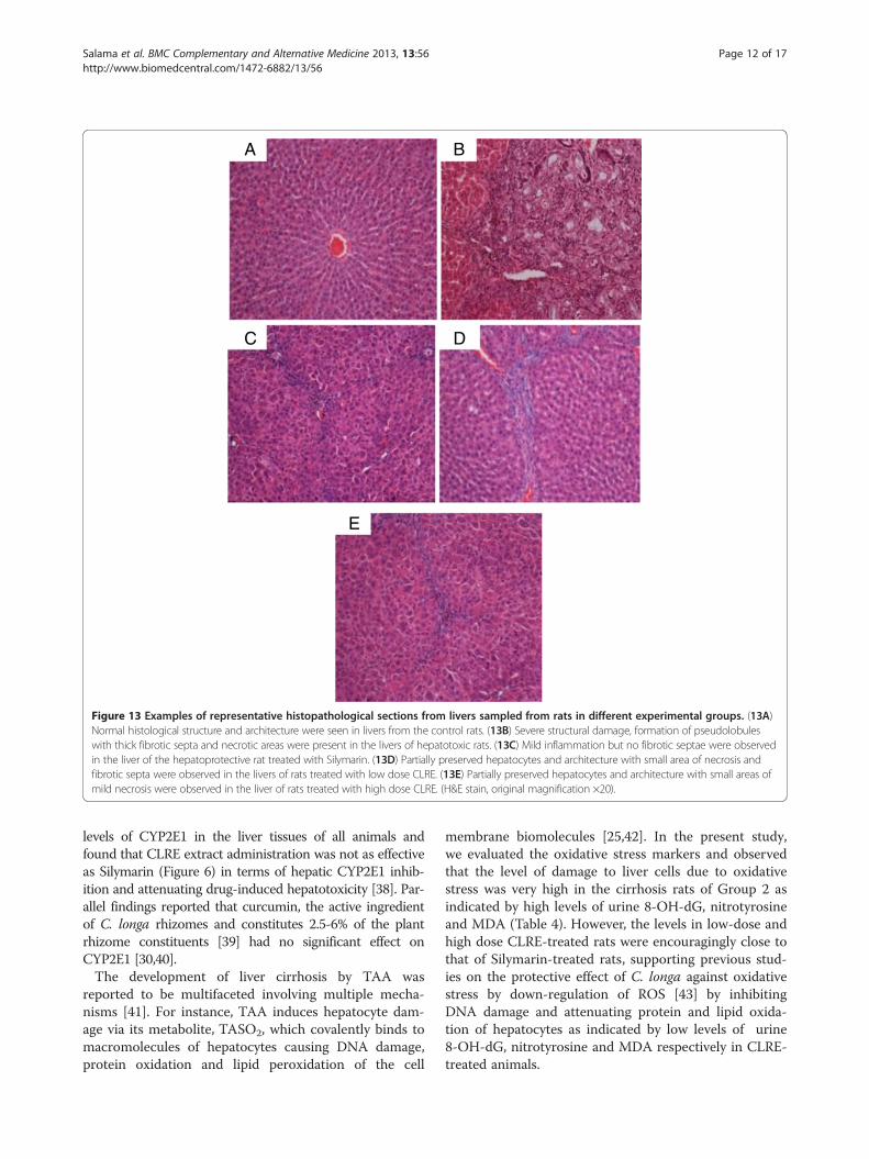
Gross anatomy and histopathologyThe gross appearances of the liver samples and micro-scopic assessment (H&E staining) of their sections in theexperimental Groups 1–5 are shown in Figures 12 and 13.The gross appearance of livers from the normal rats inGroup 1 (Figure 12A) appeared reddish with smooth sur-faces and without signs of nodules, and histology showednormal architecture (Figure 13A). Livers from the cir-rhotic rats of Group 2 appeared congested with numerousmicro- and macro-nodules (Figure 12B). The normal liverarchitecture was lost and replaced by regenerating nodulesthat were separated by fibrous septae extending from thecentral vein to the portal triad (Figure 13B) and accom-panied by intensive proliferation of the bile duct together
*
** **
02468
1012141618
No
rmal
Rat
s
Cir
rho
sis
Rat
s
Sily
mar
in-t
reat
edR
ats
Lo
w D
ose
CL
RE
-tr
eate
d R
ats
(250
mg
/kg
)
Hig
h D
ose
CL
RE
-tr
eate
d R
ats
(500
mg
/kg
)
SO
D U
/mg
pro
tein
Figure 7 Effect of CLRE on the levels of SOD enzyme in livertissue homogenate at the end of 8 weeks study. Data wereexpressed as mean ± SEM. *P<0.05 compared with the normalcontrol Group 1. **P<0.05 compared with cirrhosis control Group 2.
with invasive inflammatory cells. In addition, thick purplebundles of collagen fibers appeared in the cirrhotic nod-ules. The livers of Silymarin-treated Group 3 (Figures 12Cand 13C) and the high dose CLRE-treated Group 5(Figures 12E and 13E) showed minor- micro-nodules withless fibrous septae and more extension of normal hepaticparenchyma. In contrast, the livers of the low dose CLRE-treated Group 4 (Figures 12D and 13D) showed less fi-brotic macro-nodules than those of the Silymarin-treatedGroup 3, but the improvements were not great as thoseseen in Groups 3 and 5. These visual evaluations providefurther independent confirmation that CLRE treatment ef-fectively protected the liver from further cirrhosis in adose dependent manner.
Masson’s Trichrome stainingThe degree of fibrosis determined by Masson’s trichromestaining of the liver sections from all of the treated groupsis illustrated in Figure 14. Liver sections from the normalrats (Figure 14A) appeared normal without signs of colla-gen deposition. Liver sections from the cirrhosis rats ofGroup 2 revealed increased deposition of collagen fibersaround the congested central vein indicating severe fibrosis(Figure 14B). Liver tissues from Silymarin-treated Group 3(Figure 14C) showed minimal collagen deposition indicat-ing minimal fibrosis. Livers from rats treated with low doseCLRE showed moderate deposition of collagen fibers andmoderate congestion around the central vein (Figure 14D),while those from rats treated with high dose CLRE showedmild collagen deposition and mild congestion around thecentral vein (Figure 12E). This evaluation of the degree offibrosis confirms the previous findings that CLRE treatmentprotected the livers of animals from progressive fibrosis.
Immunohistochemistry of Bax, Bcl2 and PCNABax, Bcl2 and PCNA staining of hepatocytes from thelivers of all experimental groups are shown in Figures 15A,
*
** ** **
0
10
20
30
40
50
60
No
rmal
Rat
s
Cir
rho
sis
Rat
s
Sily
mar
in-t
reat
edR
ats
Lo
w D
ose
CL
RE
-tr
eate
d R
ats
(250
mg
/kg
)
Hig
h D
ose
CL
RE
-tr
eate
d R
ats
(500
mg
/kg
)
CA
T n
M/m
in/m
g p
rote
in
Figure 8 Effect of CLRE on the levels of CAT in liver tissuehomogenate at the end of 8 weeks study. Data were expressedas mean ± SEM. *P<0.001 compared with the normal control Group1. **P<0.001 compared with cirrhosis control Group 2.

P
*** *
*
**
**
*
050
100150200250300350400
No
rmal
Rat
s
Cir
rho
sis
Rat
s
Sily
mar
in-t
reat
edR
ats
Lo
w D
ose
CL
RE
-tr
eate
d R
ats
(250
mg
/kg
)
Hig
h D
ose
CL
RE
-tr
eate
d R
ats
(500
mg
/kg
)
pg
/mL
TGF-beta
TNF-alpha
Figure 9 Effect of CLRE on the serum levels of cytokines at theend of 8 weeks study. Data were expressed as mean ± SEM.*P<0.001compared with the cirrhosis control Group 2. **P<0.001compared with normal control Group 1.
** ****
0102030405060708090
Nor
mal
Rat
s
Cir
rhos
is R
ats
Sily
mar
in-t
reat
edR
ats
Low
Dos
e C
L-
trea
ted
Rat
s (2
50m
g/kg
)
Hig
h D
ose
CL
-tr
eate
d R
ats
(500
mg/
kg)
Bax
/Bcl-
2 R
atio
*
Figure 11 The ratio between pro-apoptotic protein Bax andanti-apoptotic protein Bcl-2. Data were expressed as mean ± SEM.*P<0.01compared with normal control Group 1. **P<0.001 comparedwith cirrhosis control Group.
Salama et al. BMC Complementary and Alternative Medicine 2013, 13:56 Page 10 of 17http://www.biomedcentral.com/1472-6882/13/56
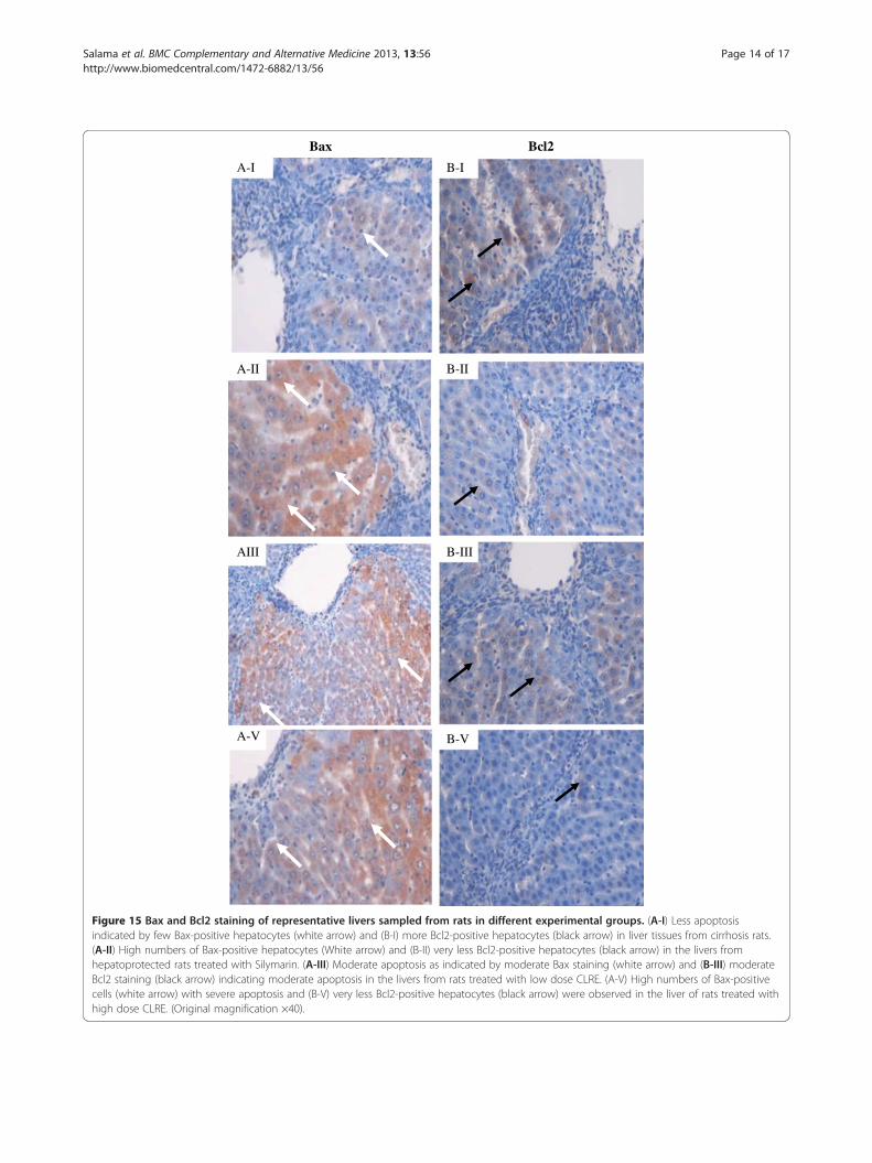
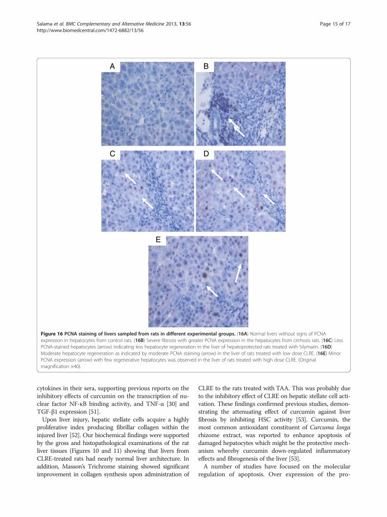
15B and 16. Hepatocytes of liver tissues from the cirrhosisrats of Group 2 showed down-regulation of Bax staining(Figure 15A-15I) with up-regulation of Bcl2-positive hepa-tocytes (Figure 15B-15I) and more PCNA staining(Figure 16B) indicating severe damage with increasednumber of necrotic cells than their apoptosis. Hepatocytesfrom Silymarin-treated rats of Group 3 showed up-regulated Bax expression (Figure 15A-II), down-regulatedBcl2 expression (Figure 15B-II) and, a few PCNA staining(Figure 16C) indicating lower levels of proliferation ofnecrotized hepatocytes and enhanced apoptosis. Liver tis-sues treated with low dose CLRE and high dose CLRE in-duced hepatocyte apoptosis as indicated by up-regulatedBax expression (Figures 15A-III and 15A-V), down-regulated Bcl2 (Figures 15B-III and 15B-V) and down-regulated necrotized hepatocytes’ proliferation as indicatedby reduced PCNA staining (Figures 16D and 16E). Thesefindings supported the idea that CLRE extract might
**
**
0
1
2
3
4
5
6
Nor
mal
Rat
s
Cir
rhos
is R
ats
Sily
mar
in-t
reat
ed R
ats
Con
cent
rati
on n
g/m
L
Figure 10 Effect of CLRE on the serum levels of the pro-apoptotic Baexpressed as mean ± SEM. *P<0.001compared with the cirrhosis control Gr
induce hepatoprotective activity against progressive liverdamage by increasing apoptosis of damaged hepatocytesand ameliorating their proliferation.
DiscussionPrescription drugs with side effects have become widelyused in modern life and as a result, liver cirrhosis has be-come a serious health problem. Consequently, the currentstudy focused on finding new therapeutic solutions tominimize liver damage [31]. Natural products, especiallyplants in folk medicine with an anecdotal history of posi-tive effects against liver diseases or other organs, are con-sidered an alternative therapeutic approach [32]. In thepresent research, the ethanol extract of C. longa rhizomeswas examined as a promising therapy for treating liver cir-rhosis. This study evaluated the toxicity of CLRE alongwith the clinical biochemistry values, which confirmed bythe biochemical results (Tables 1 and 2). In addition, histo-logical examination showed no significant pathological ab-normalities in both the liver and the kidney, even at high
*
Low
Dos
e C
LR
E-t
reat
edR
ats
(250
mg/
kg)
Hig
h D
ose
CL
RE
-tre
ated
Rat
s (5
00 m
g/kg
)
Pro-apoptotic Bax
Anti-apoptotic Bcl-2
x and anti-apoptotic Bcl-2 at the end of 8 weeks study. Data wereoup 2. **P<0.05 compared with normal control Group 1.

A
C D
E
B
Figure 12 Representative images showing the macroscopic appearances of livers sampled from rats in different experimental groups.(12A) The liver of a control rat exhibiting a regular smooth surface. (12B) The liver of a hepatotoxic rat depicting numerous irregular whitishmicro- and macronodules on its surface and a large area of ductular cholangiocellular proliferation (arrow) embedded within fibrotic areas. (12C)The liver of a hepatoprotected rat treated with Silymarin showing a normal smooth surface. (12D) The liver of a rat treated with low dose CLREwith a nearly smooth surface with few granules (arrow head). (12E) The liver of a rat treated with high dose CLRE with a normal smooth surface.
Salama et al. BMC Complementary and Alternative Medicine 2013, 13:56 Page 11 of 17http://www.biomedcentral.com/1472-6882/13/56
doses of 5 g/kg (Figure 2). The hepatoprotective effects ofCLRE on the development of liver cirrhosis, induced byprolonged exposure to TAA (200 mg/kg ) were assessedthough this study. The protocol induced cirrhosis withsimilar pathology and etiology pattern to the human livercirrhosis with the same biochemical values for typical hu-man cirrhosis markers [33]. The results were reconfirmedquantitatively by measurement of the liver index of thecirrhotic animals (Table 3), the biochemical imbalances inthe liver markers (Figures 3 and 5) and the altered totalprotein content, albumen and bilirubin levels (Figure 4). Amarked reduction of plasma total protein levels wereobserved in the cirrhosis control Group 2 compared withthe normal healthy animals of Group 1 (Figure 4), as
described in other TAA intoxication models [34]. Hepaticfactors (AP, ALT, and AST) were significantly increased inthe cirrhosis control rats, as previously described [34].CLRE-treatment caused significant recovery of these en-zymatic activities (Figure 3). Parallel findings were alsopreviously reported [35].TAA has been used to induce hepatotoxicity in the
experimental animals to produce various grades ofliver damage including nodular cirrhosis, liver cell prolifera-tion, production of pseudolobules, and parenchymal cellnecrosis [36]. It is a potent hepatotoxic agent metabolizedby CYP2E1 enzymes present in liver microsomes andis converted to a toxic reactive intermediate calledthioacetamide by oxidation [37]. Here, we measured the
A B
C D
E
Figure 13 Examples of representative histopathological sections from livers sampled from rats in different experimental groups. (13A)Normal histological structure and architecture were seen in livers from the control rats. (13B) Severe structural damage, formation of pseudolobuleswith thick fibrotic septa and necrotic areas were present in the livers of hepatotoxic rats. (13C) Mild inflammation but no fibrotic septae were observedin the liver of the hepatoprotective rat treated with Silymarin. (13D) Partially preserved hepatocytes and architecture with small area of necrosis andfibrotic septa were observed in the livers of rats treated with low dose CLRE. (13E) Partially preserved hepatocytes and architecture with small areas ofmild necrosis were observed in the liver of rats treated with high dose CLRE. (H&E stain, original magnification ×20).
Salama et al. BMC Complementary and Alternative Medicine 2013, 13:56 Page 12 of 17http://www.biomedcentral.com/1472-6882/13/56
levels of CYP2E1 in the liver tissues of all animals andfound that CLRE extract administration was not as effectiveas Silymarin (Figure 6) in terms of hepatic CYP2E1 inhib-ition and attenuating drug-induced hepatotoxicity [38]. Par-allel findings reported that curcumin, the active ingredientof C. longa rhizomes and constitutes 2.5-6% of the plantrhizome constituents [39] had no significant effect onCYP2E1 [30,40].The development of liver cirrhosis by TAA was
reported to be multifaceted involving multiple mecha-nisms [41]. For instance, TAA induces hepatocyte dam-age via its metabolite, TASO2, which covalently binds tomacromolecules of hepatocytes causing DNA damage,protein oxidation and lipid peroxidation of the cell
membrane biomolecules [25,42]. In the present study,we evaluated the oxidative stress markers and observedthat the level of damage to liver cells due to oxidativestress was very high in the cirrhosis rats of Group 2 asindicated by high levels of urine 8-OH-dG, nitrotyrosineand MDA (Table 4). However, the levels in low-dose andhigh dose CLRE-treated rats were encouragingly close tothat of Silymarin-treated rats, supporting previous stud-ies on the protective effect of C. longa against oxidativestress by down-regulation of ROS [43] by inhibitingDNA damage and attenuating protein and lipid oxida-tion of hepatocytes as indicated by low levels of urine8-OH-dG, nitrotyrosine and MDA respectively in CLRE-treated animals.

B
C D
E
A
Figure 14 Masson’s Trichrome staining of representative livers sampled from rats in different experimental groups. (14A) Normal liverstructure without signs of collagen deposition in livers from control rats. (14B) Severe collagen deposition (arrow) and severe fibrosis were seenin the livers from cirrhosis rats. (14C) Minimal collagen deposition in the liver of the hepatoprotective rat treated with Silymarin. (14D) Moderatedcollagen deposition and moderate congestion around the central vein in the livers of rats treated with low dose CLRE. (14E) Mild collagendeposition was observed in the livers of rats treated with high dose CLRE. (Original magnification ×20).
Salama et al. BMC Complementary and Alternative Medicine 2013, 13:56 Page 13 of 17http://www.biomedcentral.com/1472-6882/13/56
Reduced hepatic antioxidant functions have also beensuggested to be one mechanism of TAA-induced hep-atotoxicity [44]. Our results revealed that administrationof CLRE to the cirrhotic rats significantly alleviated theTAA-suppressive effect on antioxidant enzymes SODand CAT by maintaining the activity of these enzymes athigher levels (Figures 7 and 8). Optimizing the level ofhepatocellular antioxidant enzymes led to removal of oxi-dative stress by scavenging the free radicals resulting fromTAA-toxicity. Antioxidant activity of CLRE may be attrib-uted to the antioxidant properties of phenol compoundconstituents which constitute 3-15% of rhizomes [45] andthat TPC is equivalent to gallic acid (517.54 ± 0.049 mgGAE/mg extract). Toxins target metabolically active hepa-tocytes [46] leading to hepatocyte dysfunction and the
release of ROS, and fibrogenic and inflammatory media-tors. Several studies have suggested that part ofhepatocellular injury induced by TAA is mediated throughoxidative stress caused by the action of cytokines throughlipid peroxidation [47]. The free radicals resultingfrom TAA metabolism may activated myofibroblasts,that secrete fibrinogen and growth factors [48]. TGF-β1,a prominent profibrogenic cytokine with antiproliferativeeffects that can up-regulate the deposition of ECM [49],was present at high levels in the cirrhosis rats of Group 2compared with the other groups (Figure 9). Inaddition, the pro-inflammatory cytokine TNF-α [50] waselevated in the cirrhosis rats indicating a high inflamma-tory state in the cirrhotic liver. Low or high dose CLRE ad-ministration to the rats reduced the high levels of
A-V
AIII
A-II
A-I
Ba Bx cl2
B-I
B-II
B-III
B-V
Figure 15 Bax and Bcl2 staining of representative livers sampled from rats in different experimental groups. (A-I) Less apoptosisindicated by few Bax-positive hepatocytes (white arrow) and (B-I) more Bcl2-positive hepatocytes (black arrow) in liver tissues from cirrhosis rats.(A-II) High numbers of Bax-positive hepatocytes (White arrow) and (B-II) very less Bcl2-positive hepatocytes (black arrow) in the livers fromhepatoprotected rats treated with Silymarin. (A-III) Moderate apoptosis as indicated by moderate Bax staining (white arrow) and (B-III) moderateBcl2 staining (black arrow) indicating moderate apoptosis in the livers from rats treated with low dose CLRE. (A-V) High numbers of Bax-positivecells (white arrow) with severe apoptosis and (B-V) very less Bcl2-positive hepatocytes (black arrow) were observed in the liver of rats treated withhigh dose CLRE. (Original magnification ×40).
Salama et al. BMC Complementary and Alternative Medicine 2013, 13:56 Page 14 of 17http://www.biomedcentral.com/1472-6882/13/56
A
DC
E
B
Figure 16 PCNA staining of livers sampled from rats in different experimental groups. (16A) Normal livers without signs of PCNAexpression in hepatocytes from control rats. (16B) Severe fibrosis with greater PCNA expression in the hepatocytes from cirrhosis rats. (16C) LessPCNA-stained hepatocytes (arrow) indicating less hepatocyte regeneration in the liver of hepatoprotected rats treated with Silymarin. (16D)Moderate hepatocyte regeneration as indicated by moderate PCNA staining (arrow) in the liver of rats treated with low dose CLRE. (16E) MinorPCNA expression (arrow) with few regenerative hepatocytes was observed in the liver of rats treated with high dose CLRE. (Originalmagnification ×40).
Salama et al. BMC Complementary and Alternative Medicine 2013, 13:56 Page 15 of 17http://www.biomedcentral.com/1472-6882/13/56
cytokines in their sera, supporting previous reports on theinhibitory effects of curcumin on the transcription of nu-clear factor NF-κB binding activity, and TNF-α [30] andTGF-β1 expression [51].Upon liver injury, hepatic stellate cells acquire a highly
proliferative index producing fibrillar collagen within theinjured liver [52]. Our biochemical findings were supportedby the gross and histopathological examinations of the ratliver tissues (Figures 10 and 11) showing that livers fromCLRE-treated rats had nearly normal liver architecture. Inaddition, Masson’s Trichrome staining showed significantimprovement in collagen synthesis upon administration of
CLRE to the rats treated with TAA. This was probably dueto the inhibitory effect of CLRE on hepatic stellate cell acti-vation. These findings confirmed previous studies, demon-strating the attenuating effect of curcumin against liverfibrosis by inhibiting HSC activity [53]. Curcumin, themost common antioxidant constituent of Curcuma longarhizome extract, was reported to enhance apoptosis ofdamaged hepatocytes which might be the protective mech-anism whereby curcumin down-regulated inflammatoryeffects and fibrogenesis of the liver [53].A number of studies have focused on the molecular
regulation of apoptosis. Over expression of the pro-

Salama et al. BMC Complementary and Alternative Medicine 2013, 13:56 Page 16 of 17http://www.biomedcentral.com/1472-6882/13/56
apoptotic proteins Fas, FasL and Bax were reported inchronic hepatitis [54]. Toxicity induced by TAA was foundto be accompanied by elevation in Bax protein levels andreduction in the anti-apoptotic protein Bcl2 and its trans-location into the mitochondria, causing apoptosis [55]. Inthe current study, we observed significant increase in theserum level of Bax protein and decrease in Bcl-2 proteinin silymarin-treated and CLRE- treated animals comparedwith the cirrhosis group animals. This was confirmed bythe ratio Bax/Bcl-2 which was high in the treated groupscompared with the cirrhosis group and the large numberof Bax positive-stained hepatocytes together with few Bcl2positive-stained hepatocytes both doses of CLRE-treatedanimals, and in Silymarin-treated animals compared withthe cirrhosis Group (Figures 10, 11, 13A and 13B) indicat-ing the susceptibility of these cells to apoptosis and therole of curcuminoids in inducing apoptosis [53]. Further-more, those animals on daily feeding with CLRE alongwith TAA injections thrice weekly for 8 weeks attenuatedhepatocyte proliferation and regeneration as indicated bya significant decrease in PCNA positive-stained cells inthe liver sections from the low dose and high dose-treatedgroups similar to that in the Silymarin-treated group(Figure 14) [56]. These results were consistent with previ-ous reports that curcumin the active ingredient of CLREextract had inhibitory effect on hepatocyte proliferation[53]. Treating the animals with CLRE extract inhibited thenecrotic effect due to thioacetamide administration bymodifying necrosis into apoptosis, which might be throughcytochrome release from mitochondria and caspase activa-tion [57]. This modification in vivo would scale down therelease of inflammatory mediators that would prevent pro-gressive live damage. The ethanolic extract of C. longarhizomes showed a significant hepatoprotective effect whenorally administrated in doses of 250 mg/kg and 500 mg/kg,and the protective effect was dose-dependent. The mainconstituents of CLRE extract are the flavonoid curcuminand various volatile oils, including tumerone, atlantone, andzingiberene. The hepatoprotective effects of turmeric andcurcumin might be due to direct antioxidant and free-radical scavenging mechanisms, as well as the ability toindirectly augment glutathione levels, thereby aiding inhepatic detoxification [58]. The volatile oils and curcuminof C. longa exhibit potent anti-inflammatory effects [59].
ConclusionIn conclusion, our results demonstrated that the progres-sion of TAA-induced liver cirrhosis could be prevented orreduced using the ethanol extract of C. longa rhizomes.The plant natural extract exerted its hepatoprotective ef-fect by preventing the harmful cascade of events inducedby TAA toxicity. This hepatoprotective capability of CLREpreserved the liver’s status quo in terms of its properties,functions and structure against toxins, and warranted
further study to explore its pharmacologic potential intreating liver cirrhosis. In addition, Curcumin might bepredominantly responsible for the hepatoprotective effectof CLRE rhizome extract. These findings would encouragefurther studies on the pharmacological significance ofusing plant extracts as alternative medicines for treatingliver cirrhosis.
Competing interestsNo competing interests of either a financial or non-financial nature.
Authors’ contributionsSMS: Designing the research project, collection, analysis, and interpretationof data; writing of the manuscript and the decision to submit the manuscriptfor publication. MAA: Designing the research project; Animal experiment andthe decision to submit the manuscript for publication. AS: Interpretation ofdata and writing of the manuscript. SS: Collection of data and revision of themanuscript. SG: Interpretation of data. SI: Involved in the decision to submitthe manuscript. All authors read and approved the final manuscript.
AcknowledgmentsThis study was financially supported by the University of Malaya throughUniversity Malaya Research Grant PV042-2011A and HIR Grant (F000009-21001). The authors are thankful to the staffs of Department of MolecularMedicine, and Clinical Diagnostic Laboratory of University Malaya.
Author details1Department of Molecular Medicine, Faculty of Medicine, University ofMalaya, 50603, Kuala Lumpur, Malaysia. 2Institute of Biological science,Faculty of Science, University of Malaya, 50603, Kuala Lumpur, Malaysia.
Received: 24 July 2012 Accepted: 20 February 2013Published: 5 March 2013
References1. Wang S, Nagrath D: Liver Tissue Engineering. Biomaterials for Tissue
Engineering Applications: A Review of the Past and Future Trends 2010, 14:389.2. Friedman SL, McQuaid KR, Grendell JH: Current diagnosis & treatment in
gastroenterology. New York: Lang Medical Books/McGraw-Hill; 2002.3. O'Connell M, Rushworth S: Curcumin: potential for hepatic fibrosis
therapy? Br J Pharmacol 2008, 153(3):403–405.4. Strader DB, Wright T, Thomas DL, Seeff LB: Diagnosis, management, and
treatment of hepatitis C. Hepatology 2004, 39(4):1147–1171.5. Loguercio C, Federico A: Oxidative stress in viral and alcoholic hepatitis.
Free Radic Biol Med 2003, 34(1):1–10.6. Bansal AK, Bansal M, Soni G, Bhatnagar D: Protective role of Vitamin E
pre-treatment on nitrosodiethylamine induced oxidative stress in ratliver. Chem Biol Interact 2005, 156(2):101–111.
7. Cederbaum AI, Lu Y, Wu D: Role of oxidative stress in alcohol-inducedliver injury. Arch Toxicol 2009, 83(6):519–548.
8. Stickel F, Schuppan D: Herbal medicine in the treatment of liver diseases.Dig Liver Dis 2007, 39(4):293–304.
9. Dvorák Z, Kosina P, Walterová D, Simánek V, Bachleda P, Ulrichová J:Primary cultures of human hepatocytes as a tool in cytotoxicity studies:cell protection against model toxins by flavonolignans obtained fromSilybum marianum. Toxicol Lett 2003, 137(3):201–212.
10. Phansawan B, Poungbangpho S: Antioxidant capacities of Puerariamirifica, Stevia rebaudiana Bertoni, Curcuma longa Linn., Andrographispaniculata (Burm. f.) Nees. and Cassia alata Linn. for the development ofdietary supplement. Kasetsart J 2007, 41(3):407–413.
11. Alshawsh MA, Abdulla MA, Ismail S, Amin ZA: Hepatoprotective Effects ofOrthosiphon stamineus Extract on Thioacetamide-Induced Liver Cirrhosisin Rats. Evid Based Complement Alternat Med 2011, 2011:1–6.
12. Kadir FA, Othman F, Abdulla MA, Hussan F, Hassandarvish P: Effect ofTinospora crispa on thioacetamide-induced liver cirrhosis in rats. Indian JPharmacol 2011, 43(1):64.
13. Maizura M, Aminah A, Wan Aida W: Total phenolic content andantioxidant activity of kesum (Polygonum minus), ginger (Zingiber
Salama et al. BMC Complementary and Alternative Medicine 2013, 13:56 Page 17 of 17http://www.biomedcentral.com/1472-6882/13/56
officinale) and turmeric (Curcuma longa) extract. Int Food Res J 2011,18:526–531.
14. Kunnumakkara AB, Guha S, Krishnan S, Diagaradjane P, Gelovani J, AggarwalBB: Curcumin Potentiates Antitumor Activity of Gemcitabine in anOrthotopic Model of Pancreatic Cancer through Suppression ofProliferation, Angiogenesis, and Inhibition of Nuclear Factor-κB–Regulated Gene Products. Cancer Res 2007, 67(8):3853.
15. Kim KJ, Yu HH, Cha JD, Seo SJ, Choi NY, You YO: Antibacterial activity ofCurcuma longa L. against methicillin‐resistant Staphylococcus aureus.Phytother Res 2005, 19(7):599–604.
16. Kohli K, Ali J, Ansari M, Raheman Z: Curcumin: a natural antiinflammatoryagent. Indian J Pharmacol 2005, 37(3):141–147.
17. Panchatcharam M, Miriyala S, Gayathri VS, Suguna L: Curcumin improveswound healing by modulating collagen and decreasing reactive oxygenspecies. Mol Cell Biochem 2006, 290(1):87–96.
18. Miriyala S, Panchatcharam M, Rengarajulu P: Cardioprotective effects ofcurcumin. In: The molecular targets and therapeutic uses of curcumin inhealth and disease 2007, 595:359–377.
19. Sengupta M, Sharma GD, Chakraborty B: Hepatoprotective andimmunomodulatory properties of Aqueous extract of Curcuma longa incarbon tetra chloride intoxicated Swiss albino mice. Asian Pac J TropBiomed 2011, 1(3):193–199.
20. Zhang Q, Zhang J, Shen J, Silva A, Dennis DA, Barrow CJ: A simple 96-wellmicroplate method for estimation of total polyphenol content inseaweeds. J Appl Phycol 2006, 18(3):445–450.
21. Jing LJ, Mohamed M, Rahmat A, Bakar MFA: Phytochemicals, antioxidantproperties and anticancer investigations of the different parts of severalgingers species (Boesenbergia rotunda, Boesenbergia pulchella varattenuata and Boesenbergia armeniaca). J Med Plants Res 2010, 4(1):27–32.
22. Mahmood AA, Mariod AA, Abdelwahab SI, Ismail S, Al-Bayaty F: Potentialactivity of ethanolic extract of Boesenbergia rotunda (L.) rhizomes extractin accelerating wound healing in rats. J Med Plants Res 2010, 4(15):1570–1576.
23. Ramaiah SK, Apte U, Mehendale HM: Diet restriction as a protectivemechanism in noncancer toxicity outcomes: a review. Int J Toxicol 2000,19(6):413–424.
24. Chilakapati J, Shankar K, Korrapati MC, Hill RA, Mehendale HM: Saturationtoxicokinetics of thioacetamide: role in initiation of liver injury. DrugMetab Dispos 2005, 33(12):1877–1885.
25. Djordjević VB: Free radicals in cell biology. Int Rev Cytol 2004, 237:57–89.26. Beale G, Chattopadhyay D, Gray J, Stewart S, Hudson M, Day C, Trerotoli P,
Giannelli G, Manas D, Reeves H: AFP, PIVKAII, GP3, SCCA-1 and follisatin assurveillance biomarkers for hepatocellular cancer in non-alcoholic andalcoholic fatty liver disease. BMC Cancer 2008, 8(1):200.
27. AydIn AF, Küskü-Kiraz Z, Dogru-Abbasoglu S, Güllüoglu M, Uysal M,Koçak-Toker N: Effect of carnosine against thioacetamide-induced livercirrhosis in rat. Peptides 2010, 31(1):67–71.
28. Fatemi F, Allameh A, Khalafi H, Ashrafihelan J: Hepatoprotective effects of[gamma]-irradiated caraway essential oils in experimental sepsis. ApplRadiat Isot 2010, 68(2):280–285.
29. Beckman KB, Ames BN: Oxidative decay of DNA. J Biol Chem 1997, 272(32):19633–19636.
30. Bruck R, Ashkenazi M, Weiss S, Goldiner I, Shapiro H, Aeed H, Genina O,Helpern Z, Pines M: Prevention of liver cirrhosis in rats by curcumin. LiverInt 2007, 27(3):373–383.
31. Daly AK, Donaldson PT, Bhatnagar P, Shen Y, Pe'er I, Floratos A, Daly MJ,Goldstein DB, John S, Nelson MR: HLA-B* 5701 genotype is a majordeterminant of drug-induced liver injury due to flucloxacillin. Nat Genet2009, 41(7):816–819.
32. Khanna D, Sethi G, Ahn KS, Pandey MK, Kunnumakkara AB, Sung B,Aggarwal A, Aggarwal BB: Natural products as a gold mine for arthritistreatment. Curr Opin Pharmacol 2007, 7(3):344–351.
33. Plonné D, Schulze HP, Kahlert U, Meltke K, Seidolt H, Bennett AJ, CartwrightIJ, Higgins JA, Till U, Dargel R: Postnatal development of hepatocellularapolipoprotein B assembly and secretion in the rat. J Lipid Res 2001, 42(11):1865.
34. Alshawsh MA, Abdulla MA, Ismail S, Amin ZA: Hepatoprotective Effects ofOrthosiphon stamineus Extract on Thioacetamide-Induced Liver Cirrhosisin Rats. Evidence-Based Complement Altern Med 2011:1–6.
35. Kumar A: A review on hepatoprotective herbal drugs. IJRPC 2012, 2(1):92–102.36. Sadasivan S, Latha PG, Sasikumar JM, Rajashekaran S, Shyamal S, Shine VJ:
Hepatoprotective studies on Hedyotis corymbosa (L.) Lam.J Ethnopharmacol 2006, 106(2):245–249.
37. Kim KH, Bae JH, Cha SW, Han SS, Park KH, Jeong TC: Role of metabolicactivation by cytochrome P450 in thioacetamide-induced suppression ofantibody response in male BALB/c mice. Toxicol Lett 2000, 114(1–3):225–235.
38. Upadhyay G, Kumar A, Singh MP: Effect of silymarin on pyrogallol-andrifampicin-induced hepatotoxicity in mouse. Eur J Pharmacol 2007,565(1–3):190–201.
39. Parthasarathy VA, Chempakam B, Zachariah TJ: Chemistry of spices. UK: CABInternational; 2008: 1-20
40. Guangwei X, Rongzhu L, Wenrong X, Suhua W, Xiaowu Z, Shizhong W, YeZ, Aschner M, Kulkarni SK, Bishnoi M: Curcumin pretreatment protectsagainst acute acrylonitrile-induced oxidative damage in rats.Toxicology 2010, 267(1–3):140–146.
41. Ahmad A, Pillai KK, Najmi AK, Ahmad SJ, Pal SN, Balani DK: Evaluation ofhepatoprotective potential of jigrine post-treatment against thioacetamideinduced hepatic damage. J Ethnopharmacol 2002, 79(1):35–41.
42. Chilakapati J, Korrapati MC, Hill RA, Warbritton A, Latendresse JR, MehendaleHM: Toxicokinetics and toxicity of thioacetamide sulfoxide: a metaboliteof thioacetamide. Toxicology 2007, 230(2–3):105–116.
43. Elaziz E, Ibrahim Z, Elkattawy A: Protective effect of Curcuma longa againstCCL4 induced oxidative stress and cellular degeneration in rats. GlobalVeterinaria 2010 (5):272–281.
44. Wang H, Peng R, Kong R, Li Y: Serum glutathione S-transferase activity asan early marker of thioacetimide-induced acute hepatotoxicity in mice.Wei sheng yan jiu 1999, 28(3):179.
45. Li S, Yuan W, Deng G, Wang P, Yang P, Aggarwal BB: Chemicalcomposition and product quality control of turmeric (Curcuma longa L.).Phytochemistry 2011, 2:28–54.
46. Mehendale HM: Tissue repair: an important determinant of final outcomeof toxicant-induced injury. Toxicol Pathol 2005, 33(1):41–51.
47. Okuyama H, Shimahara Y, Nakamura H, Araya S, Kawada N, Yamaoka Y,Yodoi J: Thioredoxin prevents thioacetamide-induced acute hepatitis.Comp Hepatol 2004, 3(Suppl 1):S6.
48. Bassiouny AR, Zaky AZ, Abdulmalek SA, Kandeel KM, Ismail A, Moftah M:Modulation of AP-endonuclease1 levels associated with hepatic cirrhosisin rat model treated with human umbilical cord blood mononuclearstem cells. Int J Clin Exp Pathol 2011, 4(7):692.
49. Gressner AM, Weiskirchen R, Breitkopf K, Dooley S: Roles of TGF-beta inhepatic fibrosis. Front Biosci 2002, 7(1):d793–807.
50. Zaret KS, Grompe M: Generation and regeneration of cells of the liverand pancreas. Science 2008, 322(5907):1490–1494.
51. Gaedeke J, Noble NA, Border WA: Curcumin blocks multiple sites of theTGF-β signaling cascade in renal cells. Kidney Int 2004, 66(1):112–120.
52. Elsharkawy A, Oakley F, Mann D: The role and regulation of hepatic stellatecell apoptosis in reversal of liver fibrosis. Apoptosis 2005, 10(5):927–939.
53. Wang ME, Chen YC, Chen IS, Hsieh SC, Chen SS, Chiu CH: Curcuminprotects against thioacetamide-induced hepatic fibrosis by attenuatingthe inflammatory response and inducing apoptosis of damagedhepatocytes. J Nutr Biochem 2012.
54. Chen NL, Bai L, Li L, Chen PL, Zhang C, Liu CY, Deng T, Chen H, Jia KM,Zhou ZQ: Apoptosis pathway of liver cells in chronic hepatitis. World JGastroenterology 2004, 10(21):3201–3204.
55. Chen LH, Hsu CY, Weng CF: Involvement of P53 and Bax/Bad triggeringapoptosis in thioacetamide-induced hepatic epithelial cells. World JGastroenterol 2006, 12(32):5175.
56. Sakr SA, Shalaby SY: Metiram-induced histological and histochemicalalterations in Liver and kidney of pregnant mice. Life Sci J 2012, 9(1).
57. Malhi H, Gores GJ, Lemasters JJ: Apoptosis and necrosis in the liver: a taleof two deaths? Hepatology 2006, 43(S1):S31–S44.
58. Girish C, Koner BC, Jayanthi S, Ramachandra Rao K, Rajesh B, Pradhan SC:Hepatoprotective activity of picroliv, curcumin and ellagic acidcompared to silymarin on paracetamol induced liver toxicity in mice.Fundam Clin Pharmacol 2009, 23(6):735–745.
59. Organization WH: WHO monographs on selected medicinal plants. vol. 1.Geneva: World Health Organization; 2002.
doi:10.1186/1472-6882-13-56Cite this article as: Salama et al.: Hepatoprotective effect of ethanolicextract of Curcuma longa on thioacetamide induced liver cirrhosis inrats. BMC Complementary and Alternative Medicine 2013 13:56.